Serum from aged F344 rats conditions the activation of young macrophages
7
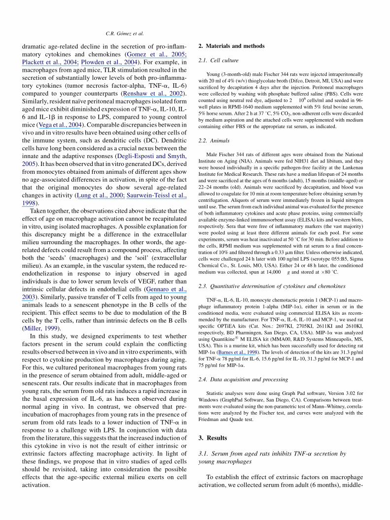
Serum from aged F344 rats conditions the activation of young macrophages Christian R. Go ´mez a,1 , Claudio Acun ˜a-Castillo a,1 , Sumiyo Nishimura a , Viviana Pe ´rez a , Alejandro Escobar a , Flavio Salazar-Onfray a , Valeria Sabaj a , Claudio Torres c , Robin Walter b,c , Felipe Sierra b,c, * a Instituto de Ciencias Biome ´dicas, Programas de Biologı ´a Celular y Molecular, and Inmunologı ´a, Facultad de Medicina, Universidad de Chile, Independencia 1027, Santiago, Chile b Centro FONDAP de Estudio Moleculares de la Ce ´lula, Facultad de Medicina, Universidad de Chile, Independencia 1027, Santiago, Chile c Lankenau Institute for Medical Research, 100 Lancaster Avenue, Wynnewood, PA 19096, USA Abstract There is considerable controversy about the molecular mechanisms responsible for the variations in innate immunity associated with age. While in vivo, aged animals and humans react to an inflammatory signal with an excessive production of pro-inflammatory cytokines, studies in vitro generally show that this response is attenuated in macrophages from old individuals. In an effort to examine possible extrinsic factors that might affect the response of macrophages to lipopolysaccharide (LPS), we have challenged peritoneal macrophages obtained from young rats with sera obtained from rats of different ages. Our results indicate that the serum from aged rats significantly impairs the capacity of young macrophages to induce tumor necrosis factor-alpha (TNF-a) production, while at the same time it increases the basal levels of interleukin-6 (IL-6). The effect of serum from aged donors on TNF-a secretion requires pre-incubation and is sensitive to heat inactivation. In contrast, the stimulating effect on IL-6 is resistant to heat, and thus should not be due to a protein factor. Therefore, our results indicate that the age-related changes in macrophage activity are not only the consequence of intrinsic changes, but there also appears to be a modulatory effect imparted by the external milieu. Keywords: Aging; Macrophages; Cytokines; Lipopolysaccharide; TNF-a; IL-6 1. Introduction Aging is characterized by a decline in both innate and adaptive immunological functions. In the case of lymphocytes, there is considerable coherence between the defects found in vivo and those described using isolated cells in vitro. For example, in vitro studies have shown that the proliferative capacity of lymphocytes is generally attenuated during aging (DeVeale et al., 2004), and a similar behavior could be extrapolated from the decreased response to influenza vaccination observed in old individuals, in vivo (Boon et al., 2002; Sambhara et al., 2001; Webster, 2000). In contrast, there is considerable discordance between in vivo and in vitro data in relation to age-related changes in macrophage activation. This is exemplified by the production of pro-inflammatory cytokines. Most in vivo studies indicate that aged individuals have an increased basal level of serum interleukin-6 (IL-6) and other pro-inflammatory cytokines (Franceschi et al., 2000). In addition, production of cytokines in vivo in response to external stimuli, such as lipopolysaccharide (LPS) has also been found to be strongly exacerbated in aged individuals (primarily rodents). It is generally believed that macrophages represent a central source of these molecules during inflammatory episodes. However, the majority of studies done with isolated macrophages incubated in vitro indicate a Abbreviations: DC, dendritic cells; FBS, fetal bovine serum; IL, interleu- kin; LPS, lipopolysaccharide; NO, nitric oxide; TLR, toll like receptors; TNF- a, tumor necrosis factor-alpha * Corresponding author. Present address: National Institute on Aging, Biol- ogy of Aging Program, Gateway Bldg., Suite 2C231, 7201 Wisconsin Ave, Bethesda, MD 20850. Tel.: +1 301 496 6402; fax: +1 301 402 0010. E-mail address: [email protected] (F. Sierra). 1 Both authors contributed equally.
-
Upload
independent -
Category
Documents
-
view
5 -
download
0
Transcript of Serum from aged F344 rats conditions the activation of young macrophages
Serum from aged F344 rats conditions the activation
of young macrophages
Christian R. Gomez a,1, Claudio Acuna-Castillo a,1, Sumiyo Nishimura a,Viviana Perez a, Alejandro Escobar a, Flavio Salazar-Onfray a,
Valeria Sabaj a, Claudio Torres c, Robin Walter b,c, Felipe Sierra b,c,*a Instituto de Ciencias Biomedicas, Programas de Biologıa Celular y Molecular, and Inmunologıa,
Facultad de Medicina, Universidad de Chile, Independencia 1027, Santiago, Chileb Centro FONDAP de Estudio Moleculares de la Celula, Facultad de Medicina,
Universidad de Chile, Independencia 1027, Santiago, Chilec Lankenau Institute for Medical Research, 100 Lancaster Avenue, Wynnewood, PA 19096, USA
Abstract
There is considerable controversy about the molecular mechanisms responsible for the variations in innate immunity associated with age. While
in vivo, aged animals and humans react to an inflammatory signal with an excessive production of pro-inflammatory cytokines, studies in vitro
generally show that this response is attenuated in macrophages from old individuals. In an effort to examine possible extrinsic factors that might
affect the response of macrophages to lipopolysaccharide (LPS), we have challenged peritoneal macrophages obtained from young rats with sera
obtained from rats of different ages. Our results indicate that the serum from aged rats significantly impairs the capacity of young macrophages to
induce tumor necrosis factor-alpha (TNF-a) production, while at the same time it increases the basal levels of interleukin-6 (IL-6). The effect of
serum from aged donors on TNF-a secretion requires pre-incubation and is sensitive to heat inactivation. In contrast, the stimulating effect on IL-6
is resistant to heat, and thus should not be due to a protein factor. Therefore, our results indicate that the age-related changes in macrophage activity
are not only the consequence of intrinsic changes, but there also appears to be a modulatory effect imparted by the external milieu.
Keywords: Aging; Macrophages; Cytokines; Lipopolysaccharide; TNF-a; IL-6
1. Introduction
Aging is characterized by a decline in both innate and
adaptive immunological functions. In the case of lymphocytes,
there is considerable coherence between the defects found in
vivo and those described using isolated cells in vitro. For
example, in vitro studies have shown that the proliferative
capacity of lymphocytes is generally attenuated during aging
Abbreviations: DC, dendritic cells; FBS, fetal bovine serum; IL, interleu-
kin; LPS, lipopolysaccharide; NO, nitric oxide; TLR, toll like receptors; TNF-
a, tumor necrosis factor-alpha
* Corresponding author. Present address: National Institute on Aging, Biol-
ogy of Aging Program, Gateway Bldg., Suite 2C231, 7201 Wisconsin Ave,
Bethesda, MD 20850. Tel.: +1 301 496 6402; fax: +1 301 402 0010.
E-mail address: [email protected] (F. Sierra).1 Both authors contributed equally.
(DeVeale et al., 2004), and a similar behavior could be
extrapolated from the decreased response to influenza
vaccination observed in old individuals, in vivo (Boon et al.,
2002; Sambhara et al., 2001; Webster, 2000). In contrast, there
is considerable discordance between in vivo and in vitro data in
relation to age-related changes in macrophage activation. This
is exemplified by the production of pro-inflammatory
cytokines. Most in vivo studies indicate that aged individuals
have an increased basal level of serum interleukin-6 (IL-6) and
other pro-inflammatory cytokines (Franceschi et al., 2000). In
addition, production of cytokines in vivo in response to
external stimuli, such as lipopolysaccharide (LPS) has also
been found to be strongly exacerbated in aged individuals
(primarily rodents). It is generally believed that macrophages
represent a central source of these molecules during
inflammatory episodes. However, the majority of studies done
with isolated macrophages incubated in vitro indicate a
C.R. Gomez et al.
dramatic age-related decline in the secretion of pro-inflam-
matory cytokines and chemokines (Gomez et al., 2005;
Plackett et al., 2004; Plowden et al., 2004). For example, in
macrophages from aged mice, TLR stimulation resulted in the
secretion of substantially lower levels of both pro-inflamma-
tory cytokines (tumor necrosis factor-alpha, TNF-a, IL-6)
compared to younger counterparts (Renshaw et al., 2002).
Similarly, resident naıve peritoneal macrophages isolated form
aged mice exhibit diminished expression of TNF-a, IL-10, IL-
6 and IL-1b in response to LPS, compared to young control
mice (Vega et al., 2004). Comparable discrepancies between in
vivo and in vitro results have been obtained using other cells of
the immune system, such as dendritic cells (DC). Dendritic
cells have long been considered as a crucial nexus between the
innate and the adaptive responses (Degli-Esposti and Smyth,
2005). It has been observed that in vitro generated DCs, derived
from monocytes obtained from animals of different ages show
no age-associated differences in activation, in spite of the fact
that the original monocytes do show several age-related
changes in activity (Lung et al., 2000; Saurwein-Teissl et al.,
1998).
Taken together, the observations cited above indicate that the
effect of age on macrophage activation cannot be recapitulated
in vitro, using isolated macrophages. A possible explanation for
this discrepancy might be a difference in the extracellular
milieu surrounding the macrophages. In other words, the age-
related defects could result from a compound process, affecting
both the ‘seeds’ (macrophages) and the ‘soil’ (extracellular
milieu). As an example, in the vascular system, the reduced re-
endothelization in response to injury observed in aged
individuals is due to lower serum levels of VEGF, rather than
intrinsic cellular defects in endothelial cells (Gennaro et al.,
2003). Similarly, passive transfer of T cells from aged to young
animals leads to a senescent phenotype in the B cells of the
recipient. This effect seems to be due to modulation of the B
cells by the T cells, rather than intrinsic defects on the B cells
(Miller, 1999).
In this study, we designed experiments to test whether
factors present in the serum could explain the conflicting
results observed between in vivo and in vitro experiments, with
respect to cytokine production by macrophages during aging.
For this, we cultured peritoneal macrophages from young rats
in the presence of serum obtained from adult, middle-aged or
senescent rats. Our results indicate that in macrophages from
young rats, the serum from old rats induces a rapid increase in
the basal expression of IL-6, as has been observed during
normal aging in vivo. In contrast, we observed that pre-
incubation of macrophages from young rats in the presence of
serum from old rats leads to a lower induction of TNF-a in
response to a challenge with LPS. In conjunction with data
from the literature, this suggests that the increased induction of
this cytokine in vivo is not the result of either intrinsic or
extrinsic factors affecting macrophage activity. In light of
these findings, we propose that in vitro studies of aged cells
should be revisited, taking into consideration the possible
effects that the age-specific external milieu exerts on cell
activation.
2. Materials and methods
2.1. Cell culture
Young (3-month-old) male Fischer 344 rats were injected intraperitoneally
with 20 ml of 4% (w/v) thioglycolate broth (Difco, Detroit, MI, USA) and were
sacrificed by decapitation 4 days after the injection. Peritoneal macrophages
were collected by washing with phosphate buffered saline (PBS). Cells were
counted using neutral red dye, adjusted to 2 � 106 cells/ml and seeded in 96-
well plates in RPMI-1640 medium supplemented with 5% fetal bovine serum,
5% horse serum. After 2 h at 37 8C, 5% CO2, non-adherent cells were discarded
by medium aspiration and the attached cells were supplemented with medium
containing either FBS or the appropriate rat serum, as indicated.
2.2. Animals
Male Fischer 344 rats of different ages were obtained from the National
Institute on Aging (NIA). Animals were fed NIH31 diet ad libitum, and they
were housed individually in a specific pathogen-free facility at the Lankenau
Institute for Medical Research. These rats have a median lifespan of 24 months
and were sacrificed at the ages of 6 months (adult), 15 months (middle-aged) or
22–24 months (old). Animals were sacrificed by decapitation, and blood was
allowed to coagulate for 10 min at room temperature before obtaining serum by
centrifugation. Aliquots of serum were immediately frozen in liquid nitrogen
until use. The serum from each individual animal was evaluated for the presence
of both inflammatory cytokines and acute phase proteins, using commercially
available enzyme-linked immunosorbent assay (ELISA) kits and western blots,
respectively. Sera that were free of inflammatory markers (the vast majority)
were pooled using at least three different animals for each pool. For some
experiments, serum was heat inactivated at 50 8C for 30 min. Before addition to
the cells, RPMI medium was supplemented with rat serum to a final concen-
tration of 10% and filtered through a 0.33 mm filter. Unless otherwise indicated,
cells were challenged 24 h later with 100 ng/ml LPS (serotype 055:B5, Sigma
Chemical Co., St. Louis, MO, USA). Either 24 or 48 h later, the conditioned
medium was collected, spun at 14,000 � g and stored at �80 8C.
2.3. Quantitative determination of cytokines and chemokines
TNF-a, IL-6, IL-10, monocyte chemotactic protein 1 (MCP-1) and macro-
phage inflammatory protein 1-alpha (MIP-1a), either in serum or in the
conditioned media, were evaluated using commercial ELISA kits as recom-
mended by the manufacturer. For TNF-a, IL-6, IL-10 and MCP-1, we used rat
specific OPTiEA kits (Cat. Nos.: 2697KI, 2705KI, 2611KI and 2610KI,
respectively, BD Pharmingen, San Diego, CA, USA). MIP-1a was analyzed
using Quantikine1 M ELISA kit (MMA00, R&D Systems Minneapolis, MS,
USA). This is a murine kit, which has been successfully used for detecting rat
MIP-1a (Barnes et al., 1998). The levels of detection of the kits are 31.3 pg/ml
for TNF-a 78 pg/ml for IL-6, 15.6 pg/ml for IL-10, 31.3 pg/ml for MCP-1 and
75 pg/ml for MIP-1a.
2.4. Data acquisition and processing
Statistic analyses were done using Graph Pad software, Version 3.02 for
Windows (GraphPad Software, San Diego, CA). Comparisons between treat-
ments were evaluated using the non-parametric test of Mann–Whitney, correla-
tions were analyzed by the Fischer test, and curves were analyzed with the
Friedman and Quade test.
3. Results
3.1. Serum from aged rats inhibits TNF-a secretion by
young macrophages
To establish the effect of extrinsic factors on macrophage
activation, we collected serum from adult (6 months), middle-
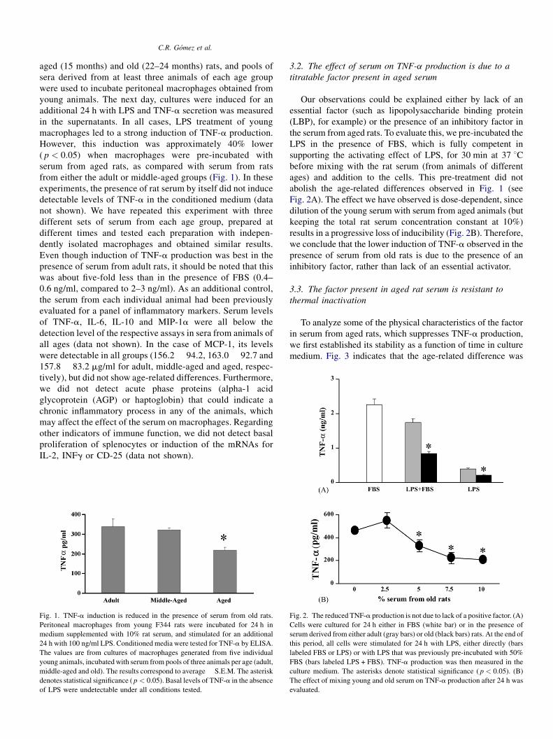
C.R. Gomez et al.
aged (15 months) and old (22–24 months) rats, and pools of
sera derived from at least three animals of each age group
were used to incubate peritoneal macrophages obtained from
young animals. The next day, cultures were induced for an
additional 24 h with LPS and TNF-a secretion was measured
in the supernatants. In all cases, LPS treatment of young
macrophages led to a strong induction of TNF-a production.
However, this induction was approximately 40% lower
( p < 0.05) when macrophages were pre-incubated with
serum from aged rats, as compared with serum from rats
from either the adult or middle-aged groups (Fig. 1). In these
experiments, the presence of rat serum by itself did not induce
detectable levels of TNF-a in the conditioned medium (data
not shown). We have repeated this experiment with three
different sets of serum from each age group, prepared at
different times and tested each preparation with indepen-
dently isolated macrophages and obtained similar results.
Even though induction of TNF-a production was best in the
presence of serum from adult rats, it should be noted that this
was about five-fold less than in the presence of FBS (0.4–
0.6 ng/ml, compared to 2–3 ng/ml). As an additional control,
the serum from each individual animal had been previously
evaluated for a panel of inflammatory markers. Serum levels
of TNF-a, IL-6, IL-10 and MIP-1a were all below the
detection level of the respective assays in sera from animals of
all ages (data not shown). In the case of MCP-1, its levels
were detectable in all groups (156.2 � 94.2, 163.0 � 92.7 and
157.8 � 83.2 mg/ml for adult, middle-aged and aged, respec-
tively), but did not show age-related differences. Furthermore,
we did not detect acute phase proteins (alpha-1 acid
glycoprotein (AGP) or haptoglobin) that could indicate a
chronic inflammatory process in any of the animals, which
may affect the effect of the serum on macrophages. Regarding
other indicators of immune function, we did not detect basal
proliferation of splenocytes or induction of the mRNAs for
IL-2, INFg or CD-25 (data not shown).
Fig. 1. TNF-a induction is reduced in the presence of serum from old rats.
Peritoneal macrophages from young F344 rats were incubated for 24 h in
medium supplemented with 10% rat serum, and stimulated for an additional
24 h with 100 ng/ml LPS. Conditioned media were tested for TNF-a by ELISA.
The values are from cultures of macrophages generated from five individual
young animals, incubated with serum from pools of three animals per age (adult,
middle-aged and old). The results correspond to average � S.E.M. The asterisk
denotes statistical significance ( p < 0.05). Basal levels of TNF-a in the absence
of LPS were undetectable under all conditions tested.
3.2. The effect of serum on TNF-a production is due to a
titratable factor present in aged serum
Our observations could be explained either by lack of an
essential factor (such as lipopolysaccharide binding protein
(LBP), for example) or the presence of an inhibitory factor in
the serum from aged rats. To evaluate this, we pre-incubated the
LPS in the presence of FBS, which is fully competent in
supporting the activating effect of LPS, for 30 min at 37 8Cbefore mixing with the rat serum (from animals of different
ages) and addition to the cells. This pre-treatment did not
abolish the age-related differences observed in Fig. 1 (see
Fig. 2A). The effect we have observed is dose-dependent, since
dilution of the young serum with serum from aged animals (but
keeping the total rat serum concentration constant at 10%)
results in a progressive loss of inducibility (Fig. 2B). Therefore,
we conclude that the lower induction of TNF-a observed in the
presence of serum from old rats is due to the presence of an
inhibitory factor, rather than lack of an essential activator.
3.3. The factor present in aged rat serum is resistant to
thermal inactivation
To analyze some of the physical characteristics of the factor
in serum from aged rats, which suppresses TNF-a production,
we first established its stability as a function of time in culture
medium. Fig. 3 indicates that the age-related difference was
Fig. 2. The reduced TNF-a production is not due to lack of a positive factor. (A)
Cells were cultured for 24 h either in FBS (white bar) or in the presence of
serum derived from either adult (gray bars) or old (black bars) rats. At the end of
this period, all cells were stimulated for 24 h with LPS, either directly (bars
labeled FBS or LPS) or with LPS that was previously pre-incubated with 50%
FBS (bars labeled LPS + FBS). TNF-a production was then measured in the
culture medium. The asterisks denote statistical significance ( p < 0.05). (B)
The effect of mixing young and old serum on TNF-a production after 24 h was
evaluated.
C.R. Gomez et al.
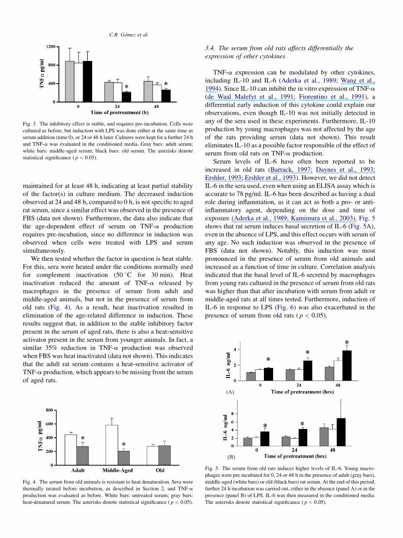
Fig. 3. The inhibitory effect is stable, and requires pre-incubation. Cells were
cultured as before, but induction with LPS was done either at the same time as
serum addition (time 0), or 24 or 48 h later. Cultures were kept for a further 24 h
and TNF-a was evaluated in the conditioned media. Gray bars: adult serum;
white bars: middle-aged serum; black bars: old serum. The asterisks denote
statistical significance ( p < 0.05).
maintained for at least 48 h, indicating at least partial stability
of the factor(s) in culture medium. The decreased induction
observed at 24 and 48 h, compared to 0 h, is not specific to aged
rat serum, since a similar effect was observed in the presence of
FBS (data not shown). Furthermore, the data also indicate that
the age-dependent effect of serum on TNF-a production
requires pre-incubation, since no difference in induction was
observed when cells were treated with LPS and serum
simultaneously.
We then tested whether the factor in question is heat stable.
For this, sera were heated under the conditions normally used
for complement inactivation (50 8C for 30 min). Heat
inactivation reduced the amount of TNF-a released by
macrophages in the presence of serum from adult and
middle-aged animals, but not in the presence of serum from
old rats (Fig. 4). As a result, heat inactivation resulted in
elimination of the age-related difference in induction. These
results suggest that, in addition to the stable inhibitory factor
present in the serum of aged rats, there is also a heat-sensitive
activator present in the serum from younger animals. In fact, a
similar 35% reduction in TNF-a production was observed
when FBS was heat inactivated (data not shown). This indicates
that the adult rat serum contains a heat-sensitive activator of
TNF-a production, which appears to be missing from the serum
of aged rats.
Fig. 4. The serum from old animals is resistant to heat denaturation. Sera were
thermally treated before incubation, as described in Section 2, and TNF-a
production was evaluated as before. White bars: untreated serum; gray bars:
heat-denatured serum. The asterisks denote statistical significance ( p < 0.05).
3.4. The serum from old rats affects differentially the
expression of other cytokines
TNF-a expression can be modulated by other cytokines,
including IL-10 and IL-6 (Aderka et al., 1989; Wang et al.,
1994). Since IL-10 can inhibit the in vitro expression of TNF-a
(de Waal Malefyt et al., 1991; Fiorentino et al., 1991), a
differential early induction of this cytokine could explain our
observations, even though IL-10 was not initially detected in
any of the sera used in these experiments. Furthermore, IL-10
production by young macrophages was not affected by the age
of the rats providing serum (data not shown). This result
eliminates IL-10 as a possible factor responsible of the effect of
serum from old rats on TNF-a production.
Serum levels of IL-6 have often been reported to be
increased in old rats (Barrack, 1997; Daynes et al., 1993;
Ershler, 1993; Ershler et al., 1993). However, we did not detect
IL-6 in the sera used, even when using an ELISA assay which is
accurate to 78 pg/ml. IL-6 has been described as having a dual
role during inflammation, as it can act as both a pro- or anti-
inflammatory agent, depending on the dose and time of
exposure (Aderka et al., 1989; Kamimura et al., 2003). Fig. 5
shows that rat serum induces basal secretion of IL-6 (Fig. 5A),
even in the absence of LPS, and this effect occurs with serum of
any age. No such induction was observed in the presence of
FBS (data not shown). Notably, this induction was most
pronounced in the presence of serum from old animals and
increased as a function of time in culture. Correlation analysis
indicated that the basal level of IL-6 secreted by macrophages
from young rats cultured in the presence of serum from old rats
was higher than that after incubation with serum from adult or
middle-aged rats at all times tested. Furthermore, induction of
IL-6 in response to LPS (Fig. 6) was also exacerbated in the
presence of serum from old rats ( p < 0.05).
Fig. 5. The serum from old rats induces higher levels of IL-6. Young macro-
phages were pre incubated for 0, 24 or 48 h in the presence of adult (gray bars),
middle-aged (white bars) or old (black bars) rat serum. At the end of this period,
further 24 h incubation was carried out, either in the absence (panel A) or in the
presence (panel B) of LPS. IL-6 was then measured in the conditioned media.
The asterisks denote statistical significance ( p < 0.05).
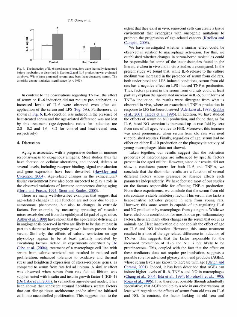
C.R. Gomez et al.
Fig. 6. The induction of IL-6 is resistant to heat. Sera were thermally denatured
before incubation, as described in Section 2, and IL-6 production was evaluated
as above. White bars: untreated serum; gray bars: heat-denatured serum. The
asterisks denote statistical significance ( p < 0.05).
In contrast to the observations regarding TNF-a, the effect
of serum on IL-6 induction did not require pre-incubation, as
increased levels of IL-6 were observed even after co-
application of the serum and LPS (Fig. 5A). Furthermore, as
shown in Fig. 6, IL-6 secretion was induced in the presence of
heat-treated serum and the age-related difference was not lost
by this treatment (age-dependent ratios for induction are
2.0 � 0.2 and 1.6 � 0.2 for control and heat-treated sera,
respectively).
4. Discussion
Aging is associated with a progressive decline in immune
responsiveness to exogenous antigens. Most studies thus far
have focused on cellular alterations, and indeed, defects at
several levels, including receptor binding, signal transduction
and gene expression have been described (Hawkley and
Cacioppo, 2004). Age-related changes in the extracellular/
tisular environment have also been suspected to play a role in
the observed variations of immune competence during aging
(Doria and Frasca, 1994; Stout and Suttles, 2005).
There are many well-described examples that suggest that
age-related changes in cell function are not only due to cell-
autonomous phenomena, but also to changes in extrinsic
factors. For example, by measuring sprouting of vascular
microvessels derived from the epididymal fat pad of aged mice,
Arthur et al. (1998) have shown that the age-related deficiencies
in angiogenesis observed in vivo are likely to be due at least in
part to a decrease in angiogenic growth factors present in the
serum. Similarly, the effects of caloric restriction on age
physiology appear to be at least partially mediated by
circulating factors. Indeed, in experiments described by De
Cabo et al. (2004), treatment of a macrophage cell line with
serum from caloric restricted rats resulted in reduced cell
proliferation, enhanced tolerance to oxidative and thermal
stress and heightened expression of stress–response genes, as
compared to serum from rats fed ad libitum. A similar effect
was observed when serum from rats fed ad libitum was
supplemented with insulin and insulin growth factor-1 (IGF-1)
(De Cabo et al., 2003). In yet another age-relevant model, it has
been shown that senescent stromal fibroblasts secrete factors
that can disrupt tissue architecture and/or stimulate initiated
cells into uncontrolled proliferation. This suggests that, to the
extent that they exist in vivo, senescent cells can create a tissue
environment that synergizes with oncogenic mutations to
promote the progression of age-related cancers (Krtolica and
Campisi, 2003).
We have investigated whether a similar effect could be
observed in relation to macrophage activation. For this, we
established whether changes in serum-borne molecules could
be responsible for some of the inconsistencies found in the
literature when in vivo and in vitro studies are compared. In the
present study we found that, while IL-6 release to the culture
medium was increased in the presence of serum from old rats,
both under basal and LPS-induced conditions, serum from old
rats has a negative effect on LPS-induced TNF-a production.
Thus, factors present in the serum from old rats could at least
partially explain the age-related increase in IL-6, but in terms of
TNF-a induction, the results were divergent from what is
observed in vivo, where an exacerbated TNF-a production in
response to LPS has been observed (Aderka et al., 1989; Krabbe
et al., 2001; Tateda et al., 1996). In addition, we have studied
the effects of serum on NO production, and found that, as for
IL-6, basal NO secretion is increased up to two-fold by sera
from rats of all ages, relative to FBS. Moreover, this increase
was most pronounced when serum from old rats was used
(unpublished results). Finally, regardless of age, serum had no
effect on either IL-10 production or the phagocytic activity of
young macrophages (data not shown).
Taken together, our results suggest that the activation
properties of macrophages are influenced by specific factors
present in the aged milieu. However, since our results did not
show a consistent pattern for both IL-6 and TNF-a, we
conclude that the dissimilar results are a function of several
different factors whose presence or absence affects each
parameter independently. We have focused most of our studies
on the factors responsible for affecting TNF-a production.
From these experiments, we conclude that the serum from old
rats contains a stable inhibitory factor. This serum also lacks a
heat-sensitive activator present in sera from young rats.
However, this same serum is capable of up regulating IL-6
and NO production by macrophages from young rats. While we
have ruled out a contribution for most known pro-inflammatory
factors, there are many other changes in the serum that occur as
animals age. Heat inactivation did not abolish the effect of age
on IL-6 and NO induction. However, this same treatment
resulted in a loss of the age-related difference in induction of
TNF-a. This suggests that the factor responsible for the
increased production of IL-6 and NO is not likely to be
proteinaceous. This, coupled with the fact that the effect on
these mediators does not require pre-incubation, suggests a
possible role for advanced glycosylation end products (AGEs),
whose serum levels are known to increase with age (Ulrich and
Cerami, 2001). Indeed, it has been described that AGEs can
induce higher levels of IL-6, TNF-a and NO in macrophages
(Chang et al., 2004; Iida et al., 1994; Morohoshi et al., 1995;
Rojas et al., 1996). It is, therefore, possible (though admittedly
speculative) that AGEs could play a role in our observations, at
least with regards to the effects of serum from old rats on IL-6
and NO. In contrast, the factor lacking in old sera and
C.R. Gomez et al.
responsible for the effect on TNF-a is more likely to be a
protein, since it is lost from serum from adult rats following
heat inactivation. Of course, more complex interpretations have
not been formally excluded. For example, even though the
initial serum did not contain detectable levels of IL-6, basal
expression of this cytokine was noted during the experimental
period, even in the absence of LPS. It has been shown that low
doses of IL-6 (within the range observed in our studies) can
significantly inhibit production of TNF-a by human peripheral
monocytes (Aderka et al., 1989). It is thus possible that this
early induction of IL-6 could have played a role in the reduced
production of TNF-a in response to LPS.
To date, most studies of cytokine production by macro-
phages from old individuals have focused on possible changes
either in CD-14 and LTR expression (Boehmer et al., 2004;
Chelvarajan et al., 2005; Renshaw et al., 2002; Vega et al.,
2004) or in the activation of molecules involved in LPS-linked
signal transduction (such as p38 and JNK MAP kinases
(Boehmer et al., 2004). Our studies indicate that age-related
changes in macrophage activity are also a consequence of age-
dependent changes in the extracellular milieu surrounding these
cells in vivo. Important questions remain, including the
possibility of reversing the effects by removal of the serum
from aged rats, as well as the use of different combinations of
cells and sera. It would be indeed very informative to
investigate the effect of serum from adult and aged animals
on cells from aged subjects, to see if the effects are additive or
synergistic. Nevertheless, our studies clearly point out the need
to consider the aged environment in molecular studies of the
effect of aging on cell behavior. Of course, characterization of
the factors responsible for each of the effects observed is of
paramount importance, as their identification could lead to
treatments designed to alleviate the effects of advanced age on
the immune response. Our results also reinforce the idea that
experimental conditions commonly employed in the literature,
including the culture of cells in FBS, could result in masking of
important age-related changes.
Acknowledgments
The authors want to thank Dr. Elizabeth J. Kovacs for useful
discussions. This work was supported by FONDECYT [Grants
2000038 (V.P.), 2010071 (C.A.-C.)], FONDAP 15010006 (F.S.)
and NIH/NIA AG13902 (F.S.).
References
Aderka, D., Le, J.M., Vilcek, J., 1989. IL-6 inhibits lipopolysaccharide-induced
tumor necrosis factor production in cultured human monocytes, U937 cells,
and in mice. J. Immunol. 143, 3517–3523.
Arthur, W.T., Vernon, R.B., Sage, E.H., Reed, M.J., 1998. Growth factors
reverse the impaired sprouting of microvessels from aged mice. Microvasc.
Res. 55, 260–270.
Barnes, D.A., Tse, J., Kaufhold, M., Owen, M., Hesselgesser, J., Strieter, R.,
Horuk, R., Perez, H.D., 1998. Polyclonal antibody directed against human
RANTES ameliorates disease in the Lewis rat adjuvant-induced arthritis
model. J. Clin. Invest. 101, 2910–2919.
Barrack, E.R., 1997. TGF beta in prostate cancer: a growth inhibitor that can
enhance tumorigenicity. Prostate 31, 61–70.
Boehmer, E.D., Goral, J., Faunce, D.E., Kovacs, E.J., 2004. Age-dependent
decrease in Toll-like receptor 4-mediated pro-inflammatory cytokine pro-
duction and mitogen-activated protein kinase expression. J. Leukoc. Biol.
75, 342–349.
Boon, A.C., Fringuelli, E., Graus, Y.M., Fouchier, R.A., Sintnicolaas, K., Iorio,
A.M., Rimmelzwaan, G.F., Osterhaus, A.D., 2002. Influenza A virus
specific T cell immunity in humans during aging. Virology 299, 100–108.
Chang, P.C., Chen, T.H., Chang, C.J., Hou, C.C., Chan, P., Lee, H.M., 2004.
Advanced glycosylation end products induce inducible nitric oxide synthase
(iNOS) expression via a p38 MAPK-dependent pathway. Kidney Int. 65,
1664–1675.
Chelvarajan, R.L., Collins, S.M., Van Willigen, J.M., Bondada, S., 2005. The
unresponsiveness of aged mice to polysaccharide antigens is a result of a
defect in macrophage function. J. Leukoc. Biol. 77, 503–512.
Daynes, R.A., Araneo, B.A., Ershler, W.B., Maloney, C., Li, G.Z., Ryu, S.Y.,
1993. Altered regulation of IL-6 production with normal aging: possible
linkage to the age-associated decline in dehydroepiandrosterone and its
sulfated derivative. J. Immunol. 150, 5219–5230.
De Cabo, R., Furer-Galban, S., Anson, R.M., Gilman, C., Gorospe, M., Lane,
M.A., 2003. An in vitro model of caloric restriction. Exp. Gerontol. 38, 631–
639.
De Cabo, R., Cabello, R., Rios, M., Lopez-Lluch, G., Ingram, D.K., Lane, M.A.,
Navas, P., 2004. Calorie restriction attenuates age-related alterations in the
plasma membrane antioxidant system in rat liver. Exp. Gerontol. 39, 297–
304.
de Waal Malefyt, R., Abrams, J., Bennett, B., Figdor, C.G., de Vries, J.E., 1991.
Interleukin 10 (IL-10) inhibits cytokine synthesis by human monocytes: an
autoregulatory role of IL-10 produced by monocytes. J. Exp. Med. 174,
1209–1220.
Degli-Esposti, M.A., Smyth, M.J., 2005. Close encounters of different kinds:
dendritic cells and NK cells take centre stage. Nat. Rev. Immunol. 5, 112–
124.
DeVeale, B., Brummel, T., Seroude, L., 2004. Immunity and aging: the enemy
within? Aging Cell 3, 195–208.
Doria, G., Frasca, D., 1994. Ageing and genetic control of immune respon-
siveness. Immunol. Lett. 40, 231–233.
Ershler, W.B., 1993. Interleukin-6: a cytokine for gerontologists. J. Am. Geriatr.
Soc. 41, 176–181.
Ershler, W.B., Sun, W.H., Binkley, N., Gravenstein, S., Volk, M.J., Kamoske,
G., Klopp, R.G., Roecker, E.B., Daynes, R.A., Weindruch, R., 1993.
Interleukin-6 and aging: blood levels and mononuclear cell production
increase with advancing age and in vitro production is modifiable by dietary
restriction. Lymphokine Cytokine Res. 12, 225–230.
Fiorentino, D.F., Zlotnik, A., Mosmann, T.R., Howard, M., O’Garra, A., 1991.
IL-10 inhibits cytokine production by activated macrophages. J. Immunol.
147, 3815–3822.
Franceschi, C., Bonafe, M., Valensin, S., Olivieri, F., De Luca, M., Ottaviani, E.,
De Benedictis, G., 2000. Inflamm-aging: an evolutionary perspective on
immunosenescence. Ann. N. Y. Acad. Sci. 908, 244–254.
Gennaro, G., Menard, C., Michaud, S.E., Rivard, A., 2003. Age-dependent
impairment of reendothelialization after arterial injury: role of vascular
endothelial growth factor. Circulation 107, 230–233.
Gomez, C.R., Boehmer, E.D., Kovacs, E.J., 2005. The aging innate immune
system. Curr. Opin. Immunol.
Hawkley, L.C., Cacioppo, J.T., 2004. Stress and the aging immune system.
Brain Behav. Immun. 18, 114–119.
Iida, Y., Miyata, T., Inagi, R., Sugiyama, S., Maeda, K., 1994. Beta 2-micro-
globulin modified with advanced glycation end products induces interleu-
kin-6 from human macrophages: role in the pathogenesis of hemodialysis-
associated amyloidosis. Biochem. Biophys. Res. Commun. 201, 1235–
1241.
Kamimura, D., Ishihara, K., Hirano, T., 2003. IL-6 signal transduction and its
physiological roles: the signal orchestration model. Rev. Physiol. Biochem.
Pharmacol. 149, 1–38.
Krabbe, K.S., Bruunsgaard, H., Hansen, C.M., Moller, K., Fonsmark, L., Qvist,
J., Madsen, P.L., Kronborg, G., Andersen, H.O., Skinhoj, P., Pedersen, B.K.,
2001. Ageing is associated with a prolonged fever response in human
endotoxemia. Clin. Diagn. Lab. Immunol. 8, 333–338.
C.R. Gomez et al.
Krtolica, A., Campisi, J., 2003. Integrating epithelial cancer, aging stroma and
cellular senescence. Adv. Gerontol. 11, 109–116.
Lung, T.L., Saurwein-Teissl, M., Parson, W., Schonitzer, D., Grubeck-Loeben-
stein, B., 2000. Unimpaired dendritic cells can be derived from monocytes
in old age and can mobilize residual function in senescent T cells. Vaccine
18, 1606–1612.
Miller, R.A., 1999. Fundamental immunology. In: Paul, W.E. (Ed.), Aging and
Immune Function. Lippincott–Raven Publishers, Philadelphia, (Chapter
28), pp. 947–966.
Morohoshi, M., Fujisawa, K., Uchimura, I., Numano, F., 1995. The effect of
glucose and advanced glycosylation end products on IL-6 production by
human monocytes. Ann. N. Y. Acad. Sci. 748, 562–570.
Plackett, T.P., Boehmer, E.D., Faunce, D.E., Kovacs, E.J., 2004. Aging and
innate immune cells. J. Leukoc. Biol. 76, 291–299.
Plowden, J., Renshaw-Hoelscher, M., Engleman, C., Katz, J., Sambhara, S.,
2004. Innate immunity in aging: impact on macrophage function. Aging
Cell 3, 161–167.
Renshaw, M., Rockwell, J., Engleman, C., Gewirtz, A., Katz, J., Sambhara, S.,
2002. Cutting edge: impaired Toll-like receptor expression and function in
aging. J. Immunol. 169, 4697–4701.
Rojas, A., Caveda, L., Romay, C., Lopez, E., Valdes, S., Padron, J., Glaria, L.,
Martinez, O., Delgado, R., 1996. Effect of advanced glycosylation end
products on the induction of nitric oxide synthase in murine macrophages.
Biochem. Biophys. Res. Commun. 225, 358–362.
Sambhara, S., Kurichh, A., Miranda, R., James, O., Underdown, B., Klein, M.,
Tartaglia, J., Burt, D., 2001. Severe impairment of primary but not memory
responses to influenza viral antigens in aged mice: costimulation in vivo
partially reverses impaired primary immune responses. Cell Immunol. 210,
1–4.
Saurwein-Teissl, M., Schonitzer, D., Grubeck-Loebenstein, B., 1998. Dendritic
cell responsiveness to stimulation with influenza vaccine is unimpaired in
old age. Exp. Gerontol. 33, 625–631.
Stout, R.D., Suttles, J., 2005. Immunosenescence and macrophage functional
plasticity: dysregulation of macrophage function by age-associated micro-
environmental changes. Immunol. Rev. 205, 60–71.
Tateda, K., Matsumoto, T., Miyazaki, S., Yamaguchi, K., 1996. Lipopolysac-
charide-induced lethality and cytokine production in aged mice. Infect.
Immun. 64, 769–774.
Ulrich, P., Cerami, A., 2001. Protein glycation, diabetes, and aging. Recent
Prog. Horm. Res. 56, 1–21.
Vega, V.L., De Cabo, R., De Maio, A., 2004. Age and caloric restriction diets are
confounding factors that modify the response to lipopolysaccharide by
peritoneal macrophages in C57BL/6 mice. Shock 22, 248–253.
Wang, P., Wu, P., Siegel, M.I., Egan, R.W., Billah, M.M., 1994. IL-10 inhibits
transcription of cytokine genes in human peripheral blood mononuclear
cells. J. Immunol. 153, 811–816.
Webster, R.G., 2000. Immunity to influenza in the elderly. Vaccine 18, 1686–
1689.