
Serotonin Transporter Genotype Modulates Subgenual Response to Fearful Faces Using an Incidental...
20
Serotonin Transporter Genotype Modulates Subgenual Response to Fearful Faces Using an Incidental Task Elizabeth J. P. O’Nions 1,2 , Raymond J. Dolan 1 , and Jonathan P. Roiser 1 1 University College London, UK 2 King’s College London, UK Abstract This study assessed the impact of serotonin transporter genotype (5-HTTLPR) on regional responses to emotional faces in the amygdala and subgenual cingulate cortex (sgACC), while subjects performed a gender discrimination task. Although we found no evidence for greater amygdala reactivity or reduced amygdala–sgACC coupling in short variant 5-HTTLPR homozygotes (s/s), we observed an interaction between genotype and emotion in sgACC. Only long variant homozygotes (la/la) exhibited subgenual deactivation to fearful versus neutral faces, whereas the effect in s/s subjects was in the other direction. This absence of subgenual deactivation in s/s subjects parallels a recent finding in depressed subjects [Grimm, S., Boesiger, P., Beck, J., Schuepbach, D., Bermpohl, F., Walter, M., et al. Altered negative BOLD responses in the default-mode network during emotion processing in depressed subjects. Neuropsychopharmacology, 34, 932–943, 2009]. Taken together, the findings suggest that subgenual cingulate activity may play an important role in regulating the impact of aversive stimuli, potentially conferring greater resilience to the effects of aversive stimuli in la/la subjects. Using dynamic causal modeling of functional magnetic resonance imaging data, we explored the effects of genotype on effective connectivity and emotion-specific changes in coupling across a network of regions implicated in social processing. Viewing fearful faces enhanced bidirectional excitatory coupling between the amygdala and the fusiform gyrus, and increased the inhibitory influence of the amygdala over the sgACC, although this modulation of coupling did not differ between the genotype groups. The findings are discussed in relation to the role of sgACC and serotonin in moderating responses to aversive stimuli [Dayan, P., & Huys, Q. J., Serotonin, inhibition, and negative mood. PLoS Comput Biol, 4, e4, 2008; Mayberg, H. S., Liotti, M., Brannan, S. K., McGinnis, S., Mahurin, R. K., Jerabek, P. A., et al. Reciprocal limbic–cortical function and negative mood: Converging PET findings in depression and normal sadness. Am J Psychiatry, 156, 675–682, 1999]. INTRODUCTION Processing facial emotions depends on a distributed network of cortical and subcortical structures (Haxby, Hoffman, & Gobbini, 2000). This network, comprising the lateral occipital cortex (LOC), fusiform gyrus (FG), amygdala, inferior frontal gyrus (IFG), posterior superior temporal sulcus (pSTS), and anterior cingulate cortex, plays a pivotal role in the detection and decoding of socially relevant visual stimuli (Castelli, Happe, Frith, & Frith, 2000; Baron-Cohen et al., 1999; Brothers, Ring, & Kling, 1990). Functional specialization across this network has been explored extensively with respect to face © 2011 Massachusetts Institute of Technology Reprint requests should be sent to Elizabeth O’Nions, MRC Social Genetic and Developmental Psychiatry Centre, Institute of Psychiatry, King’s College London, DeCrespigny Park, London, SE5 8AF, UK, or via [email protected]. . Europe PMC Funders Group Author Manuscript J Cogn Neurosci. Author manuscript; available in PMC 2012 September 07. Published in final edited form as: J Cogn Neurosci. 2011 November ; 23(11): 3681–3693. doi:10.1162/jocn_a_00055. Europe PMC Funders Author Manuscripts Europe PMC Funders Author Manuscripts
-
Upload
independent -
Category
Documents
-
view
5 -
download
0
Transcript of Serotonin Transporter Genotype Modulates Subgenual Response to Fearful Faces Using an Incidental...
Serotonin Transporter Genotype Modulates SubgenualResponse to Fearful Faces Using an Incidental Task
Elizabeth J. P. O’Nions1,2, Raymond J. Dolan1, and Jonathan P. Roiser1
1University College London, UK2King’s College London, UK
AbstractThis study assessed the impact of serotonin transporter genotype (5-HTTLPR) on regionalresponses to emotional faces in the amygdala and subgenual cingulate cortex (sgACC), whilesubjects performed a gender discrimination task. Although we found no evidence for greateramygdala reactivity or reduced amygdala–sgACC coupling in short variant 5-HTTLPRhomozygotes (s/s), we observed an interaction between genotype and emotion in sgACC. Onlylong variant homozygotes (la/la) exhibited subgenual deactivation to fearful versus neutral faces,whereas the effect in s/s subjects was in the other direction. This absence of subgenualdeactivation in s/s subjects parallels a recent finding in depressed subjects [Grimm, S., Boesiger,P., Beck, J., Schuepbach, D., Bermpohl, F., Walter, M., et al. Altered negative BOLD responses inthe default-mode network during emotion processing in depressed subjects.Neuropsychopharmacology, 34, 932–943, 2009]. Taken together, the findings suggest thatsubgenual cingulate activity may play an important role in regulating the impact of aversivestimuli, potentially conferring greater resilience to the effects of aversive stimuli in la/la subjects.Using dynamic causal modeling of functional magnetic resonance imaging data, we explored theeffects of genotype on effective connectivity and emotion-specific changes in coupling across anetwork of regions implicated in social processing. Viewing fearful faces enhanced bidirectionalexcitatory coupling between the amygdala and the fusiform gyrus, and increased the inhibitoryinfluence of the amygdala over the sgACC, although this modulation of coupling did not differbetween the genotype groups. The findings are discussed in relation to the role of sgACC andserotonin in moderating responses to aversive stimuli [Dayan, P., & Huys, Q. J., Serotonin,inhibition, and negative mood. PLoS Comput Biol, 4, e4, 2008; Mayberg, H. S., Liotti, M.,Brannan, S. K., McGinnis, S., Mahurin, R. K., Jerabek, P. A., et al. Reciprocal limbic–corticalfunction and negative mood: Converging PET findings in depression and normal sadness. Am JPsychiatry, 156, 675–682, 1999].
INTRODUCTIONProcessing facial emotions depends on a distributed network of cortical and subcorticalstructures (Haxby, Hoffman, & Gobbini, 2000). This network, comprising the lateraloccipital cortex (LOC), fusiform gyrus (FG), amygdala, inferior frontal gyrus (IFG),posterior superior temporal sulcus (pSTS), and anterior cingulate cortex, plays a pivotal rolein the detection and decoding of socially relevant visual stimuli (Castelli, Happe, Frith, &Frith, 2000; Baron-Cohen et al., 1999; Brothers, Ring, & Kling, 1990). Functionalspecialization across this network has been explored extensively with respect to face
© 2011 Massachusetts Institute of Technology
Reprint requests should be sent to Elizabeth O’Nions, MRC Social Genetic and Developmental Psychiatry Centre, Institute ofPsychiatry, King’s College London, DeCrespigny Park, London, SE5 8AF, UK, or via [email protected]. .
Europe PMC Funders GroupAuthor ManuscriptJ Cogn Neurosci. Author manuscript; available in PMC 2012 September 07.
Published in final edited form as:J Cogn Neurosci. 2011 November ; 23(11): 3681–3693. doi:10.1162/jocn_a_00055.
Europe PM
C Funders A
uthor Manuscripts
Europe PM
C Funders A
uthor Manuscripts
processing. Dorsal components (pSTS and IFG) appear to process dynamic aspects of facialstimuli, including gaze direction and expression (Pelphrey, Morris, & McCarthy, 2004;George, Driver, & Dolan, 2001). Ventral components (LOC, FG, and amygdala) are thoughtto process invariant features (Haxby et al., 2000; Kanwisher, McDermott, & Chun, 1997)such as large eye whites, which signal fear (Whalen et al., 2004).
In a seminal study, Fairhall and Ishai (2007) used dynamic causal modeling (DCM) toexplore connectivity across this network during face processing. They identified hierachialorganization in the “core” network, with LOC exerting direct influence over both the FG andpSTS, with no evidence that including feedback or lateral connections increased model fit.Responses in the “extended” components of the network (IFG, amygdala, and orbito-frontalcortex) were predominantly driven by inputs from the FG, with a reduction in model fitobserved when connections were modeled from the STS.
In addition to examining connectivity related to viewing face stimuli, Fairhall and Ishai alsoexamined the modulatory effects of emotional valence on coupling. Viewing emotionalfaces increased effective connectivity in the ventral component of this network, from theLOC to the FG, and from the FG to the amygdala. Their findings are consistent with thewealth of evidence indicating the centrality of the amygdala in processing emotions,particularly fear; consistent with its more general role as a threat-detection system (Dolan,2002; Adolphs, Tranel, Damasio, & Damasio, 1994).
A plethora of studies have demonstrated that genetic factors contribute to individualdifferences in responsivity to threat-relevant stimuli at the behavioral and neural levels(Gotlib, Joormann, Minor, & Hallmayer, 2008; Strange, Kroes, Roiser, Tan, & Dolan, 2008;Roiser, Cook, Cooper, Rubinsztein, & Sahakian, 2005; Hariri et al., 2002; Garpenstrand,Annas, Ekblom, Oreland, & Fredrikson, 2001). In particular, exaggerated amygdalaresponse to emotional faces, particularly those displaying fear or anger, has been associatedwith the short allele of a polymorphism in the promoter region of the serotonin transportergene (5-HTTLPR) (Dannlowski et al., 2010; Bertolino et al., 2005; Pezawas et al., 2005;Hariri et al., 2002), although others have failed to report this effect (Thomason et al., 2010;Shah et al., 2009). Differential responses to aversive stimuli related to 5-HTTLPR genotypehave also been observed in subgenual cingulate cortex (Fortier et al., 2010; Fukudo et al.,2009; Shah et al., 2009), a region strongly implicated in mood disorders and pivotal in theregulation of amygdala response (Schardt et al., 2010; Grimm et al., 2009; Drevets et al.,1997). Volumetric reductions in both the amygdala and sgACC, as well as altered functionaland structural connectivity between the two, have also been linked with the short allele(Schardt et al., 2010; Pacheco et al., 2009; Pezawas et al., 2005).
Although convergent evidence suggests modulation of limbic and medial prefrontalresponse related to 5-HTTLPR genotype, evidence for transient expression of serotonintransporters across diverse glutamatergic cortical networks during pre- and postnataldevelopment raises the possibility that the effects of 5-HTTLPR may extend to diverseneural pathways (Gaspar, Cases, & Maroteaux, 2003). However, to date, studies assessingthe influence of 5-HTTLPR polymorphism on functional coupling in the brain have utilizedmethods that only permit the assessment of connectivity between two regions at a time (i.e.,“functional connectivity”; e.g., Schardt et al., 2010; Canli et al., 2006; Pezawas et al., 2005).These studies have also tended to focus on coupling with the amygdala. Moreover, suchapproaches do not provide insights into the direction of connectivity.
The primary aim of this study was to investigate whether 5-HTTLPR genotype influencesresponses to emotional faces in the amygdala and sgACC, using an incidental task. We alsoused DCM to explore whether effective connectivity was influenced by 5-HTTLPR
O’Nions et al. Page 2
J Cogn Neurosci. Author manuscript; available in PMC 2012 September 07.
Europe PM
C Funders A
uthor Manuscripts
Europe PM
C Funders A
uthor Manuscripts
genotype across a distributed network that participates in emotional face processing. UsingDCM allowed us to ascertain not only the strength and direction of connectivity but alsotask-specific effects on coupling, providing insight into the mechanisms behind changes inregional responses (Friston, Harrison, & Penny, 2003). Based on previous findings, wepredicted greater amygdala responses to fearful versus neutral faces and reduced amygdala–sgACC coupling in s/s homozygotes while viewing fearful faces (Pezawas et al., 2005;Hariri et al., 2002). As both enhanced and attenuated sgACC response to emotional stimulihave been associated with the short variant, we explored changes in response in this regionwithout a directional hypothesis (Fukudo et al., 2009; Shah et al., 2009). Given that transientserotonin transporter expression influences the development of diverse cortical networks(Gaspar et al., 2003), we predicted differences in connectivity related to serotonintransporter genotype across the network.
METHODSSubjects
Thirty right-handed volunteers of European ancestry selected for 5-HTTLPR genotype tookpart in the study. None of these subjects carried a low-frequency A-to-G substitution withinthe long allele (Hu et al., 2006; Nakamura, Ueno, Sano, & Tanabe, 2000). Fifteen subjectswere homozygous for the short allele (s/s) and 15 for the long allele (la/la). Subjects werescreened for previous psychiatric disorders using the Mini International NeuropsychiatricInventory (Sheehan et al., 1998). IQ was measured using the Weschler Test of AdultReading (Wechsler, 2001). All subjects provided written informed consent beforeparticipation, as approved by the National Hospital for Neurology and Neurosurgery and theInstitute of Neurology joint ethics committee.
Experimental ParadigmSubjects were told they would see a series of faces and were instructed to indicate thegender of the face via an MRI-compatible keypad. The task comprised four cycles of threeblocks. Each block consisted of faces representing one single emotion (happy, fearful, orneutral faces). Each facial emotion block consisted of eight trials, each lasting 2 sec.Between blocks, there was a 16-sec period during which a central fixation cross wasdisplayed. The scanning session lasted 6 min and 24 sec in total.
Image Acquisition and AnalysisA 3-T head scanner (Magnetom Allegra, Siemens Medical) was used to acquire gradient-echo T2*-weighted images. There were 32 slices per volume, positioned to ensuremaximum coverage of prefrontal cortex and subcortical regions. The 30° tilted sequence wasdesigned to minimize dropout in ventral prefrontal cortex and amygdala (Weiskopf, Hutton,Josephs, & Deichmann, 2006). Echo time was 30 msec, repetition time per slice was 65msec, slice thickness was 2 mm, interslice gap was 1 mm, and in-plane resolution was 2 × 2× 2 mm. fMRI data were analyzed using Statistical Parametric Mapping (SPM5; WellcomeTrust Centre for Neuroimaging, London; www. fil.ion.uck.ac.uk/spm) in Matlab 7.1. Thefirst six image volumes were removed from the analysis to allow for T1 equilibration. Thevolumes were realigned to the seventh image, normalized into standardized space [MontrealNeurological Institute (MNI) template], and smoothed using an 8-mm FWHM, to allow foranatomical variability across subjects.
Regressors for each facial emotion were constructed (happy, fearful, and neutral) andconvolved with a synthetic hemodynamic response function, with onsets modeled as box-carfunctions at the presentation of the first face in each 16-sec block. There was one box-carper block. For subjects who made errors or omissions in the gender judgment task, an extra
O’Nions et al. Page 3
J Cogn Neurosci. Author manuscript; available in PMC 2012 September 07.
Europe PM
C Funders A
uthor Manuscripts
Europe PM
C Funders A
uthor Manuscripts
regressor was included to model instances where they may not have been attending to thetask. For the five subjects who made no errors, the non-attending regressor was not included.The 16-sec fixation period between blocks constituted an implicit baseline. A high-passfilter with a cutoff of 128 sec was used to remove low-frequency fluctuations. Regressioncoefficients (parameter estimates, or betas) were then estimated using the general linearmodel, creating a beta image for each regressor. Parameter estimate images for the facialemotion regressors were linearly combined to calculate the contrasts of interest (happy vs.neutral, fear vs. neutral, and happy vs. fear). These were then used to compare s/s and la/lasubjects in random-effects group-level analyses using the t statistic.
The aim of the group-level comparison was to measure differences in response to fearfulversus neutral faces associated with 5-HTTLPR genotype in our primary ROIs: theamygdala and sgACC. To define our amygdala ROI, we used a bilateral mask from theWake Forest University PickAtlas toolbox with the Automated Anatomical Labeling (AAL)atlas (Maldjian, Laurienti, Kraft, & Burdette, 2003). For the sgACC ROI, we constructed an8-mm sphere surrounding the peak coordinate identified by Drevets et al. (1997) (Talairachand Tournoux coordinates: x = −2, y = 32, z = −2; maximum volumetric reduction acrossunmedicated unipolar and bipolar depressed subjects, relative to controls). The rationalebehind the selection of this ROI was that s/s subjects may be more vulnerable to depressionin the context of stress (Uher & McGuffin, 2010), thus by choosing the group maxima ofchanges associated with depression reported by Drevets et al., we hoped to maximize ourchances of detecting group differences in functional response at a potential biomarker forsusceptibility. We used the small-volume family-wise error corrected (SV-FWE) p value atthe voxel level reported by SPM to determine statistical significance (threshold p = .05, SV-FWE).
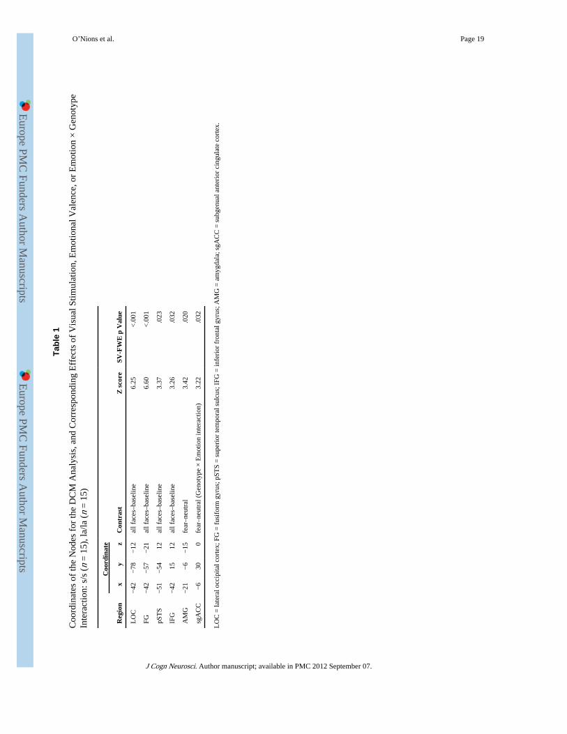
For these ROIs, we extracted parameter estimates for each of the regressors (happy, fearful,and neutral) relative to the implicit baseline at the peak voxel for the fear versus neutralcontrast (either the main effect of emotion or the Genotype group × Emotion interaction, asappropriate). Where a significant interaction was detected, these parameter estimates wereused to identify simple main effects in post hoc analyses. In addition, for completeness anddescriptive purposes, we also include the results of whole-brain comparisons at p < .001(uncorrected) with a spatial extent threshold of 5 voxels (Supplementary Table S3).Although the above analyses were conducted in the context of the mass-univariateframework, we have also included descriptive statistics relating to the mean response acrossanatomically defined amygdala ROIs (left, right, and combined) for each of the relevantcontrasts in the supplementary materials for future meta-analytic studies (SupplementaryTable S4). In Table 1, we restrict our reporting of the main effects of visual stimulation (allfaces vs. baseline) to the regions or nodes considered by the DCM (see below), along withthose in the amygdala and sgACC. The p values were SV-FWE corrected and a threshold ofp < .05 was adopted, although without correction for multiple comparisons across regions.The ROIs in this instance comprised spheres of between 4 and 8 mm centered on locationsdetermined a priori from Fairhall and Ishai (2007).
Power AnalysisGiven that we were attempting to replicate the findings of Hariri et al. (2002), the power ofthe present study to detect an effect of the size they reported warrants particularconsideration. In their sample, mean response across the amygdala ROI was 0.28 (SD 0.299)for the s-carriers (n = 14), and 0.03 (SD 0.187) for the l/l group (n = 14); this corresponds toan effect size of d = 1.0016 (Munafò, Brown, & Hariri, 2009). Power calculations indicatethat in the present study, at α = .05, an effect of this size was detected at 75% power with atwo-tailed test and at 85% power with a one-tailed test, warranted on the grounds of areplication study.
O’Nions et al. Page 4
J Cogn Neurosci. Author manuscript; available in PMC 2012 September 07.
Europe PM
C Funders A
uthor Manuscripts
Europe PM
C Funders A
uthor Manuscripts
Dynamic Causal ModelingWe selected the nodes for our DCM analysis from the network used by Fairhall and Ishai(2007) in their analysis of effective connectivity during face processing. The decision to usethe anatomical regions described by Fairhall and Ishai as nodes for the DCM was based onevidence that viewing faces versus nonface objects or scrambled faces elicits a preferentialresponse across these regions that is independent of valence and task (Ishai, 2008). First, amodel was specified consisting of fixed connections across the network and modulators(changes in coupling) related to viewing particular emotional expressions. It should be notedthat the modulators on the connections in the models examined changes in coupling relatedto viewing a particular emotion per se, rather than for a contrast (e.g., fear vs. neutral) as inthe univariate analysis.
Having established the position of the nodes for each subject (described in detail in theResults section), we constructed models to describe the dynamic connectivity across thenetwork. We referred to anatomical evidence when specifying the connections for ourmodels, supporting the existence of the fixed connectivity model we assumed (Schmahmannet al., 2007; Croxson et al., 2005; Molnar-Szakacs, Iacoboni, Koski, & Mazziotta, 2005;Catani, Jones, Donato, & Ffytche, 2003; Ongur & Price, 2000).
We elaborated a model space that contained several plausible models. Parameter estimatesfor the fixed connections and modulators were estimated by fitting the model to eachsubject’s time series. Following an initially exploratory model identification strategy(Models 1 and 2), models were identified using a heuristic search by successively removingconnections/modulators from a model with a fairly complete connectivity structure (Model 3onward; see Supplementary Figure 1). We continued this process until the model evidencepooled over subjects (see below) stopped increasing, and we were unable to supersede thepooled model evidence for the best model. One-sample t tests on the mean parameterestimates across subjects determined whether the connections were significantly differentfrom zero. Connections were removed from subsequent models if they failed to surviveuncorrected thresholds of p < .005 or p < .01 for fixed connections and modulatory effects,respectively.
A formal comparison of the alternative models was based on the model evidence. The modelevidence indicates which model provides the best fit to the measured time-series data foreach subject. It penalizes model complexity to prevent “overfitting,” whereby the modeldoes not generalize well to other datasets (Friston et al., 2003). This analysis was performedusing the group Bayesian Model Comparison tool in SPM8 (Stephan, Penny, Daunizeau,Moran, & Friston, 2009).
Once we had established the best model across all subjects (Model 11), we conductedanalyses to compare parameter estimates for connections and modulators between thegenotype groups, using independent-samples t tests. In these analyses, we corrected formultiple comparisons according to the number of fixed connections (n = 14) and modulators(n = 5) using the Bonferroni method. However, we report results reaching a nominalthreshold of p < .05 using two-tailed p values in Table 2.
Analysis of Behavioral and Demographic DataBehavioral and demographic data were analyzed with the Statistical Package for the SocialSciences (SPSS) 16, using repeated measures analyses of variance or t tests as appropriate(Supplementary Tables S1 and S2).
O’Nions et al. Page 5
J Cogn Neurosci. Author manuscript; available in PMC 2012 September 07.
Europe PM
C Funders A
uthor Manuscripts
Europe PM
C Funders A
uthor Manuscripts
RESULTSDemographic Data
There were no differences in age, gender, or IQ between the genotype groups(Supplementary Table S1).
Behavioral DataThere were no differences between the genotype groups for either response times or genderjudgment accuracy, and no evidence for an Emotion × Genotype interaction for eitherresponse times [F(2, 56) < 1] or accuracy [F(2, 56) = 2.14, p = .13] (Supplementary TableS2).
Univariate fMRI AnalysisAmygdala—Left amygdala responses were greater for fearful relative to neutral faces,irrespective of genotype (main effect of emotion depicted in Figure 1A; MNI coordinates x= 21, y = −6, z = −15; Z = 3.42, p < .05, SV-FWE). t tests on the parameter estimates at thepeak voxel for this contrast confirmed that the effect was present in both genotype groups[Figure 1B; s/s: t(14) = 2.28, p = .04 (uncorrected); la/la: t(14) = 3.07, p = .008(uncorrected)]. There was no evidence that the amygdala response to fearful relative toneutral faces was greater in s/s subjects within the bilateral amygdala ROI, with no voxelssurviving even the liberal threshold of p < .005, uncorrected.
Because Hariri et al. (2002) did not use a neutral face condition as a baseline, we repeatedthese analyses comparing the fearful faces versus fixation contrast between groups forcomparability with their study. Again, when comparing the genotype groups, no amygdalavoxels survived SV-FWE correction, or even the liberal threshold of p < .005 (uncorrected),for this contrast.
Subgenual Cingulate—sgACC responses to fearful versus neutral faces differedsignificantly between the genotype groups (Genotype × Emotion interaction depicted inFigure 2A; MNI coordinates x = −6, y = 30, z = 0; Z = 3.22, p = .032 SV-FWE). Post hocanalysis revealed that in la/la subjects the sgACC response was decreased for fearful relativeto neutral faces [Figure 2B; t(14) = 2.89, p = .01, uncorrected], and was increased in the s/sgroup [Figure 2B; t(14) = 2.21, p = .04, uncorrected]. la/la subjects showed significantlyreduced responses to fearful faces (vs. baseline) compared to the s/s subjects at this voxel[t(28) = 5.51, p < .001, uncorrected], while the groups did not differ in terms of responses toneutral faces (t < 1) (Figure 2B).
Dynamic Causal ModelingHaving established that contrasting fearful and neutral faces produced robust responses inthe amygdala and an interaction with group in sgACC, we next used DCM to explore themechanisms underpinning these effects by examining functional coupling across regionsinvolved in social processing. The coordinates of the nodes of interest are listed in Table 1.These were the main effects of our task (all faces minus baseline) identified within theanatomical regions described by Fairhall and Ishai (2007). Note that because we hadidentified a Genotype × Emotion interaction in sgACC, and because sgACC–amygdalacoupling was of particular interest in our analysis, we substituted the OFC node used byFairhall and Ishai (2007) with sgACC. We restricted our connectivity modeling to the lefthemisphere, as the emotion-specific effects we aimed to understand in the amygdala andsgACC were both in the left hemisphere.
O’Nions et al. Page 6
J Cogn Neurosci. Author manuscript; available in PMC 2012 September 07.
Europe PM
C Funders A
uthor Manuscripts
Europe PM
C Funders A
uthor Manuscripts
Identifying the Position of the Nodes for Each Subject—We identified the locationof each node for each subject on the basis of their own responses and anatomy using a two-stage process. For the LOC, FG, pSTS, and IFG, an automated procedure (using a custom-written Matlab script) identified each subject’s peak response for all faces versus baselinewithin a specified distance of the group maximum (code available from the correspondingauthor on request). The size of the spheres around the group maxima used to constrain thesearch varied according to the size of the region (LOC: 12 mm; FG: 10 mm; pSTS: 10 mm;IFG: 16 mm). Given our interest in detecting changes in connectivity in emotion processingnetworks when viewing fearful faces, the automated procedure identified each subject’speak responses for the fear versus neutral contrast for the amygdala and sgACC nodes. Forthe amygdala, the AAL atlas was used to generate a mask (PickAtlas toolbox; Maldjian etal., 2003), which constrained the search for the subject-specific peaks. For sgACC, an 8-mmsphere around the peak for the Genotype × Emotion interaction was used (MNI coordinatesx = −6, y = 30, z = 0). Given that only la/la subjects exhibited sgACC deactivation to fearfulversus neutral faces, while the effect in the s/s subjects was, if anything, in the oppositedirection (Figure 2B), we used an F-contrast to identify responses in sgACC (because tcontrasts in SPM are unidirectional). We used the peak response within each region withouta statistical threshold. This allowed us to include all nodes in each subject’s DCM, andhence to fit DCMs for all subjects, alleviating the potential difficulty that a differentialresponse related to emotion may be stronger in one genotype, as for very few cases therewas no response significant at a threshold of p < .05, uncorrected within the appropriateanatomical region.
Once the location of the nodes had been identified for each subject, their responses wereoverlaid onto their own anatomical scan to manually check that responses were located inthe correct anatomical location. Where necessary, a more anatomically appropriate peak wasselected. This procedure was carried out blind to genotype. Once each subject’s nodes hadbeen selected, we used the SPM eigenvariate tool to extract the principal component acrossthe time series, adjusting for effects of interest.
Our strategy of selecting subject-specific peaks for the nodes meant that the voxel where theEmotion × Genotype interaction was observed (x = −6, y = 30, z = 0) was not the center forthe DCM eigenvariate extraction. As a result, at the extracted subject-specific voxels, theGenotype × Emotion interaction was nonsignificant for the fear versus neutral contrast [F(1,28) = 1.072, p > .1]. However, for the fear versus baseline contrast, the difference betweenthe genotype groups was significant [F(1, 29) = 6.689, p < .02; Figure 3C].
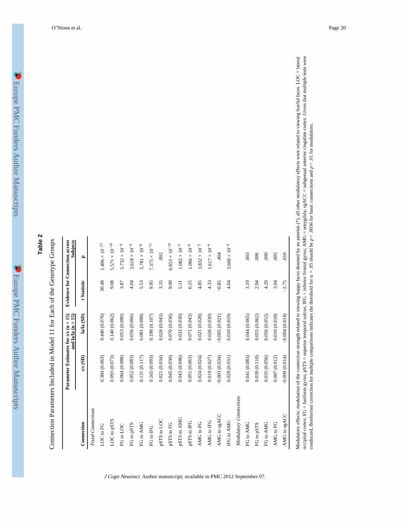
Model Estimation and Comparison—Our DCM specification procedure (see Methods)furnished 11 models, which were then compared using group Bayesian Model Comparison(Stephan et al., 2009), with random effects on models over subjects. Both the exceedanceand posterior probability indicated overwhelmingly that Model 11 was the best model(Figure 3B and C), even in comparison to extremely similar models (Model 10; Figure 3A).Images of the other nine models are displayed in Supplementary Figure 1. Conducting theBayesian Model Comparison procedure separately for each genotype group demonstratedthat Model 11 was the best model for both s/s and la/la subjects (Supplementary Table 5).All the fixed connections in the model were highly significant (save for one; see below),with significant modulatory effects of emotion identified on connections from the FG toamygdala, FG to pSTS, amygdala to FG, and amygdala to sgACC (Table 2). Note that thefixed connection from the amygdala to sgACC was nonsignificant, but this was retainedbecause the modulator on this connection was significant.
Having established that Model 11 was the best model (independent of genotype), wecompared the parameter estimates for the connections and modulators between the genotype
O’Nions et al. Page 7
J Cogn Neurosci. Author manuscript; available in PMC 2012 September 07.
Europe PM
C Funders A
uthor Manuscripts
Europe PM
C Funders A
uthor Manuscripts
groups (Table 2 and Figure 3B). The la/la subjects exhibited stronger fixed connections fromLOC to the FG [t(28) = 2.21, p = .035, 2-tailed) at a nominally significant threshold, withtrends toward nominal significance for LOC to the pSTS [t(28) = 1.90, p = .068, 2-tailed]and from the pSTS to the FG [t(28) = 2.03, p = .052, 2-tailed], whereas s/s subjects showeda nominal trend toward stronger connectivity from the IFG to the amygdala [t(28) = 2.035, p= .051, 2-tailed]. However, none of these results survived correction for multiplecomparisons. No differences in modulators between the genotype groups approached eventhe nominally significant level.
Model estimation was repeated with the sgACC node extracted using an 8-mm spherearound the peak voxel for the Genotype × Emotion interaction for the fear versus neutralcontrast from the categorical univariate analysis (i.e., instead of subject-specific peaks,which did not themselves show a significant Genotype × Emotion interaction for the fearversus neutral contrast; Figure 2C). This made very little difference to the results: Model 11remained the best model with an exceedance probability > 90%. In the majority of cases, thefixed and modulatory coupling parameters were almost identical. However, the modulationby fear of the amygdala–sgACC connection was no longer even nominally significant acrossall subjects, and the modulation by fear of the amygdala–FG connection no longer survivedcorrection for multiple comparisons. As in the previous analysis, there were no statisticallysignificant differences in the modulatory parameters between the genotype groups.
DISCUSSIONThis study assessed the impact of 5-HTTLPR genotype on neural responses to emotionalfaces using an incidentaltask. We identified an interaction between genotype and emotion insgACC, a region strongly implicated in depression and negative mood states (Harrison et al.,2009; Drevets, Savitz, & Trimble, 2008; Ongur, Drevets, & Price, 1998), which sharesreciprocal connections with the amygdala (Ongur & Price, 2000), although we were unableto replicate previous reports of amygdala hyperreactivity to emotional stimuli in 5-HTTLPRs/s carriers. Using DCM, we examined coupling across a network of regions implicated insocial processing, finding strong evidence for functional integration across this network andits modulation by emotion. However, in contrast with previous reports (e.g., Pezawas et al.,2005), there was only weak evidence in our dataset that connectivity in this network differedbetween the 5-HTTLPR genotype groups, with no results surviving correction for multiplecomparisons. Below, we consider separately the effects of genotype on regional responsesand functional coupling, and the findings of the DCM with regard to interplay during faceprocessing independent of genotype.
Effects of 5-HTTLPR on Functional Response in the Amygdala and sgACCConsistent with previous reports of enhanced sgACC response in s-carriers in the context ofaversive stimulation (Fortier et al., 2010; Fukudo et al., 2009), we observed an interactionbetween genotype and emotion in sgACC, with s/s subjects demonstrating increasedresponse to fearful versus neutral faces relative to la/la (Figure 2). Post hoc analysesrevealed sgACC deactivation to fearful versus neutral faces in la/la subjects, and increasedactivation for the same contrast in s/s subjects. Our findings differ from those recentlyreported by Shah et al. (2009), where no significant Genotype × Emotion interaction wasidentified in sgACC, using a similar incidental emotional processing task. However, s/s ands/l subjects were pooled into a single s-carrying group, and the rare A-to-G substitution in 5-HTTLPR was not taken into account, which may have decreased sensitivity in this analysis(Shah et al., 2009).
The absence of subgenual deactivation to fearful versus neutral faces in s/s subjects parallelsa recent finding in depressed subjects. Although subgenual deactivation was proportional to
O’Nions et al. Page 8
J Cogn Neurosci. Author manuscript; available in PMC 2012 September 07.
Europe PM
C Funders A
uthor Manuscripts
Europe PM
C Funders A
uthor Manuscripts
the subjective aversiveness of the stimuli in controls, there was no relationship betweenaversiveness and signal change in depressed subjects (Grimm et al., 2009). Several studieshave reported evidence for an association between the s-allele and susceptibility todepression, particularly in the context of stressful life events (Uher & McGuffin, 2010;Caspi et al., 2003; Lesch et al., 1996), consistent with greater sensitivity to aversiveemotional stimuli assessed cognitively (Strange et al., 2008). Taken together, these findingssuggest that subgenual reactivity may be important in emotion regulation, and could explainexaggerated sensitivity to aversive stimuli in short allele carriers (Thomason et al., 2010;Roiser et al., 2009; Strange et al., 2008).
Convergent evidence suggests that the subgenual cingulate may serve to dampen affectiveresponses to support cognitive functioning. Subgenual deactivation is typically observedduring cognitive tasks (e.g., inhibitory processing). However, whereas healthy controlsdisplay more subgenual deactivation for harder task levels, depressed patients show less(Matthews et al., 2009). This suggests a failure of inhibitory control, which could explainworse performance on cognitive tasks in depressed patients (Eshel & Roiser, 2010). In short,subgenual deactivation may curtail excessive emotional response for the benefit of higher-level cognitive functioning.
Consistent with this hypothesis, increased sgACC metabolism related to disturbed emotionregulation has been implicated in mood disorders (Drevets et al., 2008; Greicius et al., 2007;Botteron, Raichle, Drevets, Heath, & Todd, 2002; Drevets, 1999). Previous studies inhealthy subjects reported elevated sgACC responses associated with induced sadness andrecall of aversive memories (Kross, Davidson, Weber, & Ochsner, 2009; Drevets et al.,2008; Mayberg et al., 1999). In addition, a recent study observed increased sgACCresponses to emotional versus neutral faces as the result of a typhoid injection precipitatinghigh levels of inflammatory cytokines, which correlated with the extent of induceddepressive symptoms (Harrison et al., 2009). The decrease in sgACC response to fearfulversus neutral faces observed in la/la subjects may therefore reflect a compensatorymechanism to aversive stimuli that may confer resilience, and warrants further investigation.
The foregoing discussion argues that the 5-HTTLPR short variant renders individualssusceptible to cognitive and emotion processing biases associated with depression. This isdespite consistent lack of evidence for differential serotonin transporter expression related to5-HTTLPR genotype anywhere in the adult brain (Murthy et al., 2010), potentiallyindicating that structural and functional differences associated with this polymorphismreflect a developmental effect (e.g., Pacheco et al., 2009; Pezawas et al., 2005). Indeed,Dayan and Huys (2008) have proposed that the level of serotonin-mediated inhibition duringearly learning impacts long-term sensitivity to serotonergic stimulation, resulting indifferential sensitivity to serotonergic manipulations in adulthood. This hypothesis isconsistent with findings that s/s individuals are more sensitive to serotonin depletion(Roiser, Muller, Clark, & Sahakian, 2007; Roiser et al., 2005, 2006). Although the neuralbasis of this sensitivity has yet to be investigated using fMRI, our data are consistent withsuggestions that sgACC may play a key role in mediating this vulnerability (Neumeister etal., 2006).
We were unable to replicate previous reports of increased amygdala reactivity to fearfulfaces in s/s subjects (Dannlowski et al., 2008; Hariri et al., 2002, 2005). A potentialexplanation for our nonreplication is that we used an incidental facial emotion processingtask, which directed attention toward gender-related cues. In support of this hypothesis, arecent study reported greater amygdala response to neutral faces judged as “to be avoided”compared to those deemed “approachable,” but only found this effect in the context of asocial judgment task: There was no differential amygdala response to the same faces when
O’Nions et al. Page 9
J Cogn Neurosci. Author manuscript; available in PMC 2012 September 07.
Europe PM
C Funders A
uthor Manuscripts
Europe PM
C Funders A
uthor Manuscripts
judging gender (Blasi et al., 2009). Other recent studies that employed incidental tasks alsofailed to report an effect of 5-HTTLPR genotype on amygdala response to fear (e.g.,Thomason et al., 2010; Shah et al., 2009), and a meta-analysis has suggested that makingresponses of any kind can affect amygdala reactivity (Costafreda, Brammer, David, & Fu,2008). Nonetheless, it is possible that our failure to identify this difference represents a falsenegative, as even with 85% power, there remains a 15% chance of a false negative.
Effects of 5-HTTLPR on Amygdala–sgACC ConnectivityWe also examined changes in amygdala–sgACC connectivity during fearful face processing,and the effects of 5-HTTLPR genotype on coupling changes. In contrast with a previousreport, we found no evidence for differential amygdala–sgACC coupling between thegenotypes (Pezawas et al., 2005), perhaps reflecting differences in the task demands oranalytic methods employed. Notably, Pezawas et al. (2005) reported differential functionalcoupling across task duration (not specifically during fearful/angry face processing), and didnot specify the direction of coupling. Further studies will be required to disambiguatedrivers of the differential response during fearful face processing observed in sgACC relatedto genotype.
Nominally significant differences were observed between the genotype groups in the fixedconnections between other regions that participate in face processing, although nonesurvived correction for multiple comparisons. This is interesting given the evidence thatserotonin transporter expression facilitates plasticity across glutamatergic networks (Gasparet al., 2003; Bruning & Liangos, 1997). However, although these findings are of potentialimportance, they should be treated with caution until independently replicated.
Effective Connectivity during Face Processing: Fixed ConnectionsOur connectivity analysis sheds new light on interactions across regions implicated in socialprocessing associated with viewing faces and facial emotions. The Bayesian ModelSelection procedure identified a model with bidirectional fixed connectivity along the dorsaland ventral pathway (Figure 3B), in preference to models with only feedforward orfeedforward and lateral connections (e.g., FG–pSTS) (Models 1 and 7). This contrasts withFairhall and Ishai’s findings, where model identification indicated that including lateral andbackwards connections in the “core” network did not increase model fit. The connections incommon with the model identified by Fairhall and Ishai were stronger than the otherconnections in our model, suggesting this might be due to increased sensitivity of newimprovements to DCM to detect weak connections, as well as the subject-specific placementof nodes based on anatomical location. This is likely to have reduced noise related toanatomical heterogeneity, increasing the sensitivity of the analysis (Smith, Pillai, Chen, &Horwitz, 2010).
In support of Fairhall and Ishai’s findings, connectivity parameters indicated that responsesin the “extended” network, including the amygdala and the IFG, were driven predominantlyby the FG; although the selected model included connections from the pSTS to the extendednetwork, surpassing model evidence of a model with connections to the extended networkrouted solely via the FG (Model 8) (Fairhall & Ishai, 2007). The importance of the FG indriving responses may be attributable to the use of static facial stimuli, thought to exertlesser processing demands on regions in the dorsal stream compared to dynamic facialstimuli (James, Culham, Humphrey, Milner, & Goodale, 2003).
One of the strengths of quantifying interactions across neural systems using DCM is thesensitivity of this method (Stephan et al., 2008). This is evident in the large exceedanceprobability for Model 11 relative to Model 10, despite the similarity of the models (Figure
O’Nions et al. Page 10
J Cogn Neurosci. Author manuscript; available in PMC 2012 September 07.
Europe PM
C Funders A
uthor Manuscripts
Europe PM
C Funders A
uthor Manuscripts
3). It should be noted that the exceedance probability of Model 11 surpassed that of less aswell as more complex models. As such, we can conclude that the result justifies the sampledmodel space in terms of the numbers of parameters included. Modeling connectivity fromregions receiving visual input across the network reduced the likelihood that the parameterestimates for direct connections were contaminated by indirect effects. In addition, selectingthe position of the DCM nodes for each subject according to their own peak response greatlyincreased the sensitivity of the analysis. These factors improved our power to detect weakconnections and provided an extremely detailed picture of interactions across the faceprocessing network.
Effective Connectivity during Face Processing: Modulation by EmotionAcross all subjects, we observed increased bidirectional excitatory coupling between theamygdala and the FG during fearful face processing, providing a mechanistic explanationfor the enhanced amygdala response elicited by fearful relative to neutral faces. This findingis consistent with previous reports using neuroimaging (Surguladze et al., 2003; Morris etal., 1998) or a combination of neuroimaging and lesion models (Vuilleumier, Richardson,Armony, Driver, & Dolan, 2004). Viewing fearful faces also increased excitatory couplingbetween the FG and the pSTS to a degree similar to the enhancement of FG–amygdalacoupling. Using DCM, it is not possible to determine whether facilitation of functionalintegration between dorsal and ventral pathways was mediated by direct or indirectconnections (e.g., via attentional networks) (Vuilleumier & Driver, 2007). Viewing fearfulfaces also increased the inhibitory influence of the amygdala over sgACC, implying thatinteractions between these regions are important in the processing of threat-relevant stimuli.This finding is consistent with a previous report of negative connectivity between the rightamygdala and sgACC during passive viewing of fearful faces (Williams et al., 2006).
ConclusionWe used an incidental facial emotion processing task to explore the effects of 5-HTTLPR onfunctional response in the amygdala and sgACC, as well as differences in networkconnectivity and task-specific changes in coupling related to genotype. Although we wereunable to replicate differential amygdala response or amygdala–sgACC coupling related togenotype, we observed sgACC deactivation to fearful versus neutral faces in la/la subjects,which we speculate may represent an adaptive mechanism conferring resilience in thisgroup. Across all subjects, we found evidence for bidirectional connectivity across dorsaland ventral visual pathways, as well as increased functional integration between them in thecontext of fearful faces. We also identified increased excitatory bidirectional FG–amygdalacoupling and increased inhibitory amygdala–sgACC coupling related to the processing ofthreat-relevant stimuli. The absence of sgACC deactivation to fearful faces in s/s subjects isconsistent with evidence for their increased sensitivity to aversive stimuli and warrantsfurther investigation.
AcknowledgmentsThis work was supported by a Wellcome Trust Programme Grant (R. J. D). J. P. R. was supported by the RaymondWay Fund. We thank Ulrich Muller, Barbara Sahakian, and Trevor Robbins for assistance in recruiting participants.We also thank Geoff Tan, Nick Wood, David Rubinsztein, and Oana Sadiq for genotyping. We thank the BenSeymour, Dharshan Kumaran, and Benedetto de Martino, as well as the Wellcome Trust Centre for Neuroimagingsupport staff, for help with data collection. Finally, we thank Karl Friston for discussion of the manuscript.
O’Nions et al. Page 11
J Cogn Neurosci. Author manuscript; available in PMC 2012 September 07.
Europe PM
C Funders A
uthor Manuscripts
Europe PM
C Funders A
uthor Manuscripts
REFERENCESAdolphs R, Tranel D, Damasio H, Damasio A. Impaired recognition of emotion in facial expressions
following bilateral damage to the human amygdala. Nature. 1994; 372:669–672. [PubMed:7990957]
Baron-Cohen S, Ring HA, Wheelwright S, Bullmore ET, Brammer MJ, Simmons A, et al. Socialintelligence in the normal and autistic brain: An fMRI study. European Journal of Neuroscience.1999; 11:1891–1898. [PubMed: 10336657]
Bertolino A, Arciero G, Rubino V, Latorre V, De Candia M, Mazzola V, et al. Variation of humanamygdala response during threatening stimuli as a function of 5′HTTLPR genotype and personalitystyle. Biological Psychiatry. 2005; 57:1517–1525. [PubMed: 15953488]
Blasi G, Hariri AR, Alce G, Taurisano P, Sambataro F, Das S, et al. Preferential amygdala reactivity tothe negative assessment of neutral faces. Biological Psychiatry. 2009; 66:847–853. [PubMed:19709644]
Botteron KN, Raichle ME, Drevets WC, Heath AC, Todd RD. Volumetric reduction in left subgenualprefrontal cortex in early onset depression. Biological Psychiatry. 2002; 51:342–344. [PubMed:11958786]
Brothers L, Ring B, Kling A. Response of neurons in the macaque amygdala to complex social stimuli.Behavioural Brain Research. 1990; 41:199–213. [PubMed: 2288672]
Bruning G, Liangos O. Transient expression of the serotonin transporter in the developing mousethalamocortical system. Acta Histochemica. 1997; 99:117–121. [PubMed: 9150804]
Canli T, Qiu M, Omura K, Congdon E, Haas BW, Amin Z, et al. Neural correlates of epigenesis.Proceedings of the National Academy of Sciences, U.S.A. 2006; 103:16033–16038.
Caspi A, Sugden K, Moffitt TE, Taylor A, Craig IW, Harrington H, et al. Influence of life stress ondepression: Moderation by a polymorphism in the 5-HTT gene. Science. 2003; 301:386–389.[PubMed: 12869766]
Castelli F, Happe F, Frith U, Frith C. Movement and mind: A functional imaging study of perceptionand interpretation of complex intentional movement patterns. Neuroimage. 2000; 12:314–325.[PubMed: 10944414]
Catani M, Jones DK, Donato R, Ffytche DH. Occipito-temporal connections in the human brain.Brain. 2003; 126:2093–2107. [PubMed: 12821517]
Costafreda SG, Brammer MJ, David AS, Fu CH. Predictors of amygdala activation during theprocessing of emotional stimuli: A meta-analysis of 385 PET and fMRI studies. Brain ResearchReviews. 2008; 58:57–70. [PubMed: 18076995]
Croxson PL, Johansen-Berg H, Behrens TE, Robson MD, Pinsk MA, Gross CG, et al. Quantitativeinvestigation of connections of the prefrontal cortex in the human and macaque using probabilisticdiffusion tractography. Journal of Neuroscience. 2005; 25:8854–8866. [PubMed: 16192375]
Dannlowski U, Konrad C, Kugel H, Zwitserlood P, Domschke K, Schoning S, et al. Emotion specificmodulation of automatic amygdala responses by 5-HTTLPR genotype. Neuroimage. 2010;53:893–898. [PubMed: 19962442]
Dannlowski U, Ohrmann P, Bauer J, Deckert J, Hohoff C, Kugel H, et al. 5-HTTLPR biases amygdalaactivity in response to masked facial expressions in major depression. Neuropsychopharmacology.2008; 33:418–424. [PubMed: 17406646]
Dayan P, Huys QJ. Serotonin, inhibition, and negative mood. PLoS Computational Biology. 2008;4:e4. [PubMed: 18248087]
Dolan RJ. Emotion, cognition, and behavior. Science. 2002; 298:1191–1194. [PubMed: 12424363]
Drevets WC. Prefrontal cortical–amygdalar metabolism in major depression. Annals of the New YorkAcademy of Sciences. 1999; 877:614–637. [PubMed: 10415674]
Drevets WC, Price JL, Simpson JR Jr. Todd RD, Reich T, Vannier M, et al. Subgenual prefrontalcortex abnormalities in mood disorders. Nature. 1997; 386:824–827. [PubMed: 9126739]
Drevets WC, Savitz J, Trimble M. The subgenual anterior cingulate cortex in mood disorders. CNSSpectrums. 2008; 13:663–681. [PubMed: 18704022]
Eshel N, Roiser JP. Reward and punishment processing in depression. Biological Psychiatry. 2010;68:118–124. [PubMed: 20303067]
O’Nions et al. Page 12
J Cogn Neurosci. Author manuscript; available in PMC 2012 September 07.
Europe PM
C Funders A
uthor Manuscripts
Europe PM
C Funders A
uthor Manuscripts
Fairhall SL, Ishai A. Effective connectivity within the distributed cortical network for face perception.Cerebral Cortex. 2007; 17:2400–2406. [PubMed: 17190969]
Fortier E, Noreau A, Lepore F, Boivin M, Perusse D, Rouleau GA, et al. Early impact of 5-HTTLPRpolymorphism on the neural correlates of sadness. Neuroscience Letters. 2010; 485:261–265.[PubMed: 20851164]
Friston KJ, Harrison L, Penny W. Dynamic causal modelling. Neuroimage. 2003; 19:1273–1302.[PubMed: 12948688]
Fukudo S, Kanazawa M, Mizuno T, Hamaguchi T, Kano M, Watanabe S, et al. Impact of serotonintransporter gene polymorphism on brain activation by colorectal distention. Neuroimage. 2009;47:946–951. [PubMed: 19426812]
Garpenstrand H, Annas P, Ekblom J, Oreland L, Fredrikson M. Human fear conditioning is related todopaminergic and serotonergic biological markers. Behavioral Neuroscience. 2001; 115:358–364.[PubMed: 11345960]
Gaspar P, Cases O, Maroteaux L. The developmental role of serotonin: News from mouse moleculargenetics. Nature Reviews Neuroscience. 2003; 4:1002–1012.
George N, Driver J, Dolan RJ. Seen gaze-direction modulates fusiform activity and its coupling withother brain areas during face processing. Neuroimage. 2001; 13:1102–1112. [PubMed: 11352615]
Gotlib IH, Joormann J, Minor KL, Hallmayer J. HPA axis reactivity: A mechanism underlying theassociations among 5-HTTLPR, stress, and depression. Biological Psychiatry. 2008; 63:847–851.[PubMed: 18005940]
Greicius MD, Flores BH, Menon V, Glover GH, Solvason HB, Kenna H, et al. Resting-state functionalconnectivity in major depression: Abnormally increased contributions from subgenual cingulatecortex and thalamus. Biological Psychiatry. 2007; 62:429–437. [PubMed: 17210143]
Grimm S, Boesiger P, Beck J, Schuepbach D, Bermpohl F, Walter M, et al. Altered negative BOLDresponses in the default-mode network during emotion processing in depressed subjects.Neuropsychopharmacology. 2009; 34:932–943. [PubMed: 18536699]
Hariri AR, Drabant EM, Munoz KE, Kolachana BS, Mattay VS, Egan MF, et al. A susceptibility genefor affective disorders and the response of the human amygdala. Archives of General Psychiatry.2005; 62:146–152. [PubMed: 15699291]
Hariri AR, Mattay VS, Tessitore A, Kolachana B, Fera F, Goldman D, et al. Serotonin transportergenetic variation and the response of the human amygdala. Science. 2002; 297:400–403.[PubMed: 12130784]
Harrison NA, Brydon L, Walker C, Gray MA, Steptoe A, Critchley HD. Inflammation causes moodchanges through alterations in subgenual cingulate activity and mesolimbic connectivity.Biological Psychiatry. 2009; 66:407–414. [PubMed: 19423079]
Haxby JV, Hoffman EA, Gobbini MI. The distributed human neural system for face perception.Trends in Cognitive Sciences. 2000; 4:223–233. [PubMed: 10827445]
Hu XZ, Lipsky RH, Zhu G, Akhtar LA, Taubman J, Greenberg BD, et al. Serotonin transporterpromoter gain-of-function genotypes are linked to obsessive–compulsive disorder. AmericanJournal of Human Genetics. 2006; 78:815–826. [PubMed: 16642437]
Ishai A. Let’s face it: It’s a cortical network. Neuroimage. 2008; 40:415–419. [PubMed: 18063389]
James TW, Culham J, Humphrey GK, Milner AD, Goodale MA. Ventral occipital lesions impairobject recognition but not object-directed grasping: An fMRI study. Brain. 2003; 126:2463–2475.[PubMed: 14506065]
Kanwisher N, McDermott J, Chun MM. The fusiform face area: A module in human extrastriate cortexspecialized for face perception. Journal of Neuroscience. 1997; 17:4302–4311. [PubMed:9151747]
Kross E, Davidson M, Weber J, Ochsner K. Coping with emotions past: The neural bases of regulatingaffect associated with negative autobiographical memories. Biological Psychiatry. 2009; 65:361–366. [PubMed: 19058792]
Lesch KP, Bengel D, Heils A, Sabol SZ, Greenberg BD, Petri S, et al. Association of anxiety-relatedtraits with a polymorphism in the serotonin transporter gene regulatory region. Science. 1996;274:1527–1531. [PubMed: 8929413]
O’Nions et al. Page 13
J Cogn Neurosci. Author manuscript; available in PMC 2012 September 07.
Europe PM
C Funders A
uthor Manuscripts
Europe PM
C Funders A
uthor Manuscripts
Maldjian JA, Laurienti PJ, Kraft RA, Burdette JH. An automated method for neuroanatomic andcytoarchitectonic atlas-based interrogation of fMRI data sets. Neuroimage. 2003; 19:1233–1239.[PubMed: 12880848]
Matthews S, Simmons A, Strigo I, Gianaros P, Yang T, Paulus M. Inhibition-related activity insubgenual cingulate is associated with symptom severity in major depression. PsychiatryResearch. 2009; 172:1–6. [PubMed: 19239982]
Mayberg HS, Liotti M, Brannan SK, McGinnis S, Mahurin RK, Jerabek PA, et al. Reciprocal limbic–cortical function and negative mood: Converging PET findings in depression and normal sadness.American Journal of Psychiatry. 1999; 156:675–682. [PubMed: 10327898]
Molnar-Szakacs I, Iacoboni M, Koski L, Mazziotta JC. Functional segregation within pars opercularisof the inferior frontal gyrus: Evidence from fMRI studies of imitation and action observation.Cerebral Cortex. 2005; 15:986–994. [PubMed: 15513929]
Morris JS, Friston KJ, Buchel C, Frith CD, Young AW, Calder AJ, et al. A neuromodulatory role forthe human amygdala in processing emotional facial expressions. Brain. 1998; 121:47–57.[PubMed: 9549487]
Munafò MR, Brown SM, Hariri A. Erratum: Serotonin transporter (5-HTTLPR) genotype andamygdala activation: A meta-analysis. Biological Psychiatry. 2009; 66:302.
Murthy NV, Selvaraj S, Cowen PJ, Bhagwagar Z, Riedel WJ, Peers P, et al. Serotonin transporterpolymorphisms (SLC6A4 insertion/deletion and rs25531) do not affect the availability of 5-HTTto [11C] DASB binding in the living human brain. Neuroimage. 2010; 52:50–54. [PubMed:20406689]
Nakamura M, Ueno S, Sano A, Tanabe H. The human serotonin transporter gene linked polymorphism(5-HTTLPR) shows ten novel allelic variants. Molecular Psychiatry. 2000; 5:32–38. [PubMed:10673766]
Neumeister A, Hu XZ, Luckenbaugh DA, Schwarz M, Nugent AC, Bonne O, et al. Differential effectsof 5-HTTLPR genotypes on the behavioral and neural responses to tryptophan depletion inpatients with major depression and controls. Archives of General Psychiatry. 2006; 63:978–986.[PubMed: 16953000]
Ongur D, Drevets WC, Price JL. Glial reduction in the subgenual prefrontal cortex in mood disorders.Proceedings of the National Academy of Sciences, U.S.A. 1998; 95:13290–13295.
Ongur D, Price JL. The organization of networks within the orbital and medial prefrontal cortex ofrats, monkeys and humans. Cerebral Cortex. 2000; 10:206–219. [PubMed: 10731217]
Pacheco J, Beevers CG, Benavides C, McGeary J, Stice E, Schnyer DM. Frontal–limbic white matterpathway associations with the serotonin transporter gene promoter region (5-HTTLPR)polymorphism. Journal of Neuroscience. 2009; 29:6229–6233. [PubMed: 19439600]
Pelphrey KA, Morris JP, McCarthy G. Grasping the intentions of others: The perceived intentionalityof an action influences activity in the superior temporal sulcus during social perception. Journal ofCognitive Neuroscience. 2004; 16:1706–1716. [PubMed: 15701223]
Pezawas L, Meyer-Lindenberg A, Drabant EM, Verchinski BA, Munoz KE, Kolachana BS, et al. 5-HTTLPR polymorphism impacts human cingulate–amygdala interactions: A genetic susceptibilitymechanism for depression. Nature Neuroscience. 2005; 8:828–834.
Roiser JP, Blackwell AD, Cools R, Clark L, Rubinsztein DC, Robbins TW, et al. Serotonin transporterpolymorphism mediates vulnerability to loss of incentive motivation following acute tryptophandepletion. Neuropsychopharmacology. 2006; 31:2264–2272. [PubMed: 16541086]
Roiser JP, Cook LJ, Cooper JD, Rubinsztein DC, Sahakian BJ. Association of a functionalpolymorphism in the serotonin transporter gene with abnormal emotional processing in ecstasyusers. American Journal of Psychiatry. 2005; 162:609–612. [PubMed: 15741482]
Roiser JP, de Martino B, Tan GC, Kumaran D, Seymour B, Wood NW, et al. A genetically mediatedbias in decision making driven by failure of amygdala control. Journal of Neuroscience. 2009;29:5985–5991. [PubMed: 19420264]
Roiser JP, Muller U, Clark L, Sahakian BJ. The effects of acute tryptophan depletion and serotonintransporter polymorphism on emotional processing in memory and attention. International Journalof Neuropsychopharmacology. 2007; 10:449–461. [PubMed: 16893493]
O’Nions et al. Page 14
J Cogn Neurosci. Author manuscript; available in PMC 2012 September 07.
Europe PM
C Funders A
uthor Manuscripts
Europe PM
C Funders A
uthor Manuscripts
Schardt DM, Erk S, Nusser C, Nothen MM, Cichon S, Rietschel M, et al. Volition diminishesgenetically mediated amygdala hyperreactivity. Neuroimage. 2010; 53:943–951. [PubMed:19969089]
Schmahmann JD, Pandya DN, Wang R, Dai G, D’Arceuil HE, de Crespigny AJ, et al. Associationfibre pathways of the brain: Parallel observations from diffusion spectrum imaging andautoradiography. Brain. 2007; 130:630–653. [PubMed: 17293361]
Shah MP, Wang F, Kalmar JH, Chepenik LG, Tie K, Pittman B, et al. Role of variation in theserotonin transporter protein gene (SLC6A4) in trait disturbances in the ventral anterior cingulatein bipolar disorder. Neuropsychopharmacology. 2009; 34:1301–1310. [PubMed: 19037205]
Sheehan DV, Lecrubier Y, Sheehan KH, Amorim P, Janavs J, Weiller E, et al. The Mini-InternationalNeuropsychiatric Interview (M.I.N.I.): The development and validation of a structured diagnosticpsychiatric interview for DSM-IV and ICD-10. Journal of Clinical Psychiatry. 1998; 59(Suppl.20):22–33. quiz 34–57. [PubMed: 9881538]
Smith JF, Pillai A, Chen K, Horwitz B. Identification and validation of effective connectivity networksin functionalmagnetic resonance imaging using switching linear dynamic systems. Neuroimage.2010; 52:1027–1040. [PubMed: 19969092]
Stephan KE, Kasper L, Harrison LM, Daunizeau J, den Ouden HE, Breakspear M, et al. Nonlineardynamic causal models for fMRI. Neuroimage. 2008; 42:649–662. [PubMed: 18565765]
Stephan KE, Penny WD, Daunizeau J, Moran RJ, Friston KJ. Bayesian model selection for groupstudies. Neuroimage. 2009; 46:1004–1017. [PubMed: 19306932]
Strange BA, Kroes MC, Roiser JP, Tan GC, Dolan RJ. Emotion-induced retrograde amnesia isdetermined by a 5-HTT genetic polymorphism. Journal of Neuroscience. 2008; 28:7036–7039.[PubMed: 18614671]
Surguladze SA, Brammer MJ, Young AW, Andrew C, Travis MJ, Williams SC, et al. A preferentialincrease in the extrastriate response to signals of danger. Neuroimage. 2003; 19:1317–1328.[PubMed: 12948690]
Thomason ME, Henry ML, Paul Hamilton J, Joormann J, Pine DS, Ernst M, et al. Neural andbehavioral responses to threatening emotion faces in children as a function of the short allele of theserotonin transporter gene. Biological Psychology. 2010; 85:38–44. [PubMed: 20493234]
Uher R, McGuffin P. The moderation by the serotonin transporter gene of environmental adversity inthe etiology of depression: 2009 Update. Molecular Psychiatry. 2010; 15:18–22. [PubMed:20029411]
Vuilleumier P, Driver J. Modulation of visual processing by attention and emotion: Windows oncausal interactions between human brain regions. Philosophical Transactions of the Royal Societyof London, Series B, Biological Sciences. 2007; 362:837–855.
Vuilleumier P, Richardson MP, Armony JL, Driver J, Dolan RJ. Distant influences of amygdala lesionon visual cortical activation during emotional face processing. Nature Neuroscience. 2004;7:1271–1278.
Wechsler, D. Wechsler test of adult reading manual. Psychological Corporation; San Antonio, TX:2001.
Weiskopf N, Hutton C, Josephs O, Deichmann R. Optimal EPI parameters for reduction ofsusceptibility-induced BOLD sensitivity losses: A whole-brain analysis at 3 T and 1.5 T.Neuroimage. 2006; 33:493–504. [PubMed: 16959495]
Whalen PJ, Kagan J, Cook RG, Davis FC, Kim H, Polis S, et al. Human amygdala responsivity tomasked fearful eye whites. Science. 2004; 306:2061. [PubMed: 15604401]
Williams LM, Das P, Liddell BJ, Kemp AH, Rennie CJ, Gordon E. Mode of functional connectivity inamygdala pathways dissociates level of awareness for signals of fear. Journal of Neuroscience.2006; 26:9264–9271. [PubMed: 16957082]
O’Nions et al. Page 15
J Cogn Neurosci. Author manuscript; available in PMC 2012 September 07.
Europe PM
C Funders A
uthor Manuscripts
Europe PM
C Funders A
uthor Manuscripts
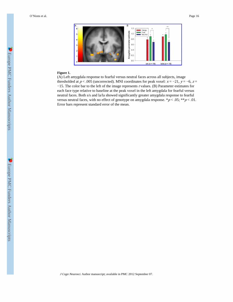
Figure 1.(A) Left amygdala response to fearful versus neutral faces across all subjects, imagethresholded at p < .005 (uncorrected). MNI coordinates for peak voxel: x = −21, y = −6, z =−15. The color bar to the left of the image represents t values. (B) Parameter estimates foreach face type relative to baseline at the peak voxel in the left amygdala for fearful versusneutral faces. Both s/s and la/la showed significantly greater amygdala response to fearfulversus neutral faces, with no effect of genotype on amygdala response. *p < .05; **p < .01.Error bars represent standard error of the mean.
O’Nions et al. Page 16
J Cogn Neurosci. Author manuscript; available in PMC 2012 September 07.
Europe PM
C Funders A
uthor Manuscripts
Europe PM
C Funders A
uthor Manuscripts
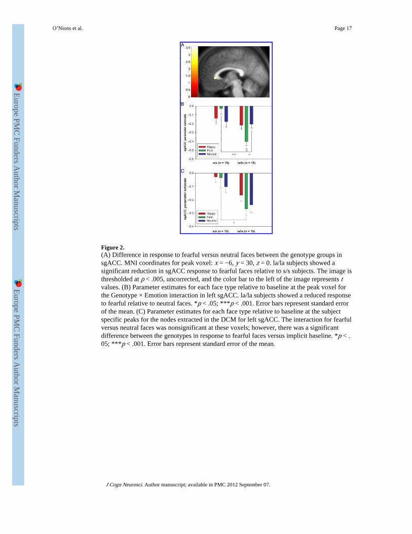
Figure 2.(A) Difference in response to fearful versus neutral faces between the genotype groups insgACC. MNI coordinates for peak voxel: x = −6, y = 30, z = 0. la/la subjects showed asignificant reduction in sgACC response to fearful faces relative to s/s subjects. The image isthresholded at p < .005, uncorrected, and the color bar to the left of the image represents tvalues. (B) Parameter estimates for each face type relative to baseline at the peak voxel forthe Genotype × Emotion interaction in left sgACC. la/la subjects showed a reduced responseto fearful relative to neutral faces. *p < .05; ***p < .001. Error bars represent standard errorof the mean. (C) Parameter estimates for each face type relative to baseline at the subjectspecific peaks for the nodes extracted in the DCM for left sgACC. The interaction for fearfulversus neutral faces was nonsignificant at these voxels; however, there was a significantdifference between the genotypes in response to fearful faces versus implicit baseline. *p < .05; ***p < .001. Error bars represent standard error of the mean.
O’Nions et al. Page 17
J Cogn Neurosci. Author manuscript; available in PMC 2012 September 07.
Europe PM
C Funders A
uthor Manuscripts
Europe PM
C Funders A
uthor Manuscripts

Figure 3.(A) Specification of the second best model (Model 10) and (B) best model (Model 11).AMG = amygdala; LOC = lateral occipital cortex; FG = fusiform gyrus; pSTS = posteriorsuperior temporal sulcus; IFG = inferior frontal gyrus; sgACC = subgenual cingulate cortex.Arrows with solid edge represent fixed connections modeled across the whole time seriessignificant at p < .005. Dashed edge arrows represent nonsignificant fixed connectionincluded in the model ( p > .005). Solid lines leading to arrows represent modulatorssignificant at p < .01, labeled F (fear) and H (happy). Dashed lines leading to arrowsrepresent nonsignificant modulators included in the model. * connection parameter strongerin la/la versus s/s subjects, nominally significant ( p < .05, 2-tailed, uncorrected). (C)Expected posterior probability and exceedance probability for the 11 models specified. Theexpected posterior probability is the probability that each model provides the best fit to themeasured data across subjects. The expected posterior probability was .3507 for the winningmodel, and the summed posterior probability was .6469 for the other models. Theexceedance probability is the probability that each model provides the best fit to the datarelative to other models. The exceedance probability was .9609 for the winning model, andthe summed exceedance probability was .0391 for the other models. Both statistics provideoverwhelming evidence that Model 11 was the best model.
O’Nions et al. Page 18
J Cogn Neurosci. Author manuscript; available in PMC 2012 September 07.
Europe PM
C Funders A
uthor Manuscripts
Europe PM
C Funders A
uthor Manuscripts
Europe PM
C Funders A
uthor Manuscripts
Europe PM
C Funders A
uthor Manuscripts
O’Nions et al. Page 19
Tabl
e 1
Coo
rdin
ates
of
the
Nod
es f
or th
e D
CM
Ana
lysi
s, a
nd C
orre
spon
ding
Eff
ects
of
Vis
ual S
timul
atio
n, E
mot
iona
l Val
ence
, or
Em
otio
n ×
Gen
otyp
eIn
tera
ctio
n: s
/s (
n =
15)
, la/
la (
n =
15)
Coo
rdin
ate
Reg
ion
xy
zC
ontr
ast
Z s
core
SV-F
WE
p V
alue
LO
C−
42−
78−
12al
l fac
es–b
asel
ine
6.25
<.0
01
FG−
42−
57−
21al
l fac
es–b
asel
ine
6.60
<.0
01
pST
S−
51−
5412
all f
aces
–bas
elin
e3.
37.0
23
IFG
−42
1512
all f
aces
–bas
elin
e3.
26.0
32
AM
G−
21−
6−
15fe
ar–n
eutr
al3.
42.0
20
sgA
CC
−6
300
fear
–neu
tral
(G
enot
ype
× E
mot
ion
inte
ract
ion)
3.22
.032
LO
C =
late
ral o
ccip
ital c
orte
x; F
G =
fus
ifor
m g
yrus
; pST
S =
sup
erio
r te
mpo
ral s
ulcu
s; I
FG =
infe
rior
fro
ntal
gyr
us; A
MG
= a
myg
dala
; sgA
CC
= s
ubge
nual
ant
erio
r ci
ngul
ate
cort
ex.
J Cogn Neurosci. Author manuscript; available in PMC 2012 September 07.
Europe PM
C Funders A
uthor Manuscripts
Europe PM
C Funders A
uthor Manuscripts
O’Nions et al. Page 20
Tabl
e 2
Con
nect
ion
Para
met
ers
Incl
uded
in M
odel
11
for
Eac
h of
the
Gen
otyp
e G
roup
s
Par
amet
er E
stim
ates
for
s/s
(n
= 15
)an
d la
/la (
n =
15)
Evi
denc
e fo
r C
onne
ctio
n ac
ross
Subj
ects
Con
nect
ion
s/s
(SD
)la
/la (
SD)
t St
atis
tic
p
Fixe
d C
onne
ctio
ns
LO
C to
FG
0.38
4 (0
.063
)0.
440
(0.0
76)
30.4
81.
406
× 1
0−23
LO
C to
pST
S0.
093
(0.0
73)
0.14
0 (0
.062
)9.
085.
571
× 1
0−10
FG to
LO
C0.
064
(0.0
88)
0.05
3 (0
.080
)3.
875.
732
× 1
0−4
FG to
pST
S0.
052
(0.0
83)
0.05
6 (0
.066
)4.
043.
618
× 1
0−4
FG to
AM
G0.
131
(0.1
17)
0.08
1 (0
.088
)5.
535.
781
× 1
0−6
FG to
IFG
0.16
5 (0
.093
)0.
198
(0.1
07)
9.95
7.37
5 ×
10−
11
pST
S to
LO
C0.
021
(0.0
34)
0.02
6 (0
.043
)3.
35.0
02
pST
S to
FG
0.04
5 (0
.030
)0.
070
(0.0
36)
9.00
6.85
3 ×
10−
10
pST
S to
AM
G0.
043
(0.0
46)
0.03
2 (0
.030
)5.
311.
082
× 1
0−5
pST
S to
IFG
0.05
1 (0
.063
)0.
071
(0.0
43)
6.15
1.06
6 ×
10−
6
AM
G to
FG
0.02
4 (0
.024
)0.
021
(0.0
28)
4.85
3.83
2 ×
10−
5
AM
G to
IFG
0.01
9 (0
.027
)0.
026
(0.0
30)
4.33
1.61
7 ×
10−
4
AM
G to
sgA
CC
−0.
003
(0.0
34)
−0.
005
(0.0
21)
−0.
85.4
04
IFG
to A
MG
0.02
9 (0
.031
)0.
010
(0.0
19)
4.04
3.60
0 ×
10−
4
Mod
ulat
ory
Con
nect
ions
FG to
AM
G0.
041
(0.0
83)
0.04
4 (0
.065
)3.
19.0
03
FG to
pST
S0.
039
(0.1
10)
0.05
5 (0
.062
)2.
94.0
06
FG to
AM
G0.
035
(0.0
56)
0.05
0 (0
.052
)4.
29.0
00
AM
G to
FG
0.00
7 (0
.012
)0.
010
(0.0
18)
3.04
.005
AM
G to
sgA
CC
−0.
009
(0.0
14)
−0.
004
(0.0
14)
−2.
75.0
10
Mod
ulat
ory
effe
cts:
mod
ulat
ion
of th
e co
nnec
tion
stre
ngth
rel
ated
to v
iew
ing
happ
y fa
ces
deno
ted
by a
n as
teri
sk (
*), a
ll ot
her
mod
ulat
ory
effe
cts
wer
e re
late
d to
vie
win
g fe
arfu
l fac
es. L
OC
= la
tera
loc
cipi
tal c
orte
x; F
G =
fus
ifor
m g
yrus
; pST
S =
sup
erio
r te
mpo
ral s
ulcu
s; I
FG =
infe
rior
fro
ntal
gyr
us; A
MG
= a
myg
dala
; sgA
CC
= s
ubge
nual
ant
erio
r ci
ngul
ate
cort
ex. G
iven
that
mul
tiple
test
s w
ere
cond
ucte
d, B
onfe
rron
i cor
rect
ion
for
mul
tiple
com
pari
sons
indi
cate
s th
e th
resh
old
for α
= .0
5 sh
ould
be
p <
.003
6 fo
r ba
sic
conn
ectio
ns a
nd p
< .0
1 fo
r m
odul
ator
s.
J Cogn Neurosci. Author manuscript; available in PMC 2012 September 07.