Serotonergic and noradrenergic lesions suppress the enhancing effect of maternal exercise during...
11
SEROTONERGIC AND NORADRENERGIC LESIONS SUPPRESS THE ENHANCING EFFECT OF MATERNAL EXERCISE DURING PREGNANCY ON LEARNING AND MEMORY IN RAT PUPS M. M. AKHAVAN, a * M. EMAMI-ABARGHOIE, a M. SAFARI, b B. SADIGHI-MOGHADDAM, c A. A. VAFAEI, d A. R. BANDEGI e AND A. RASHIDY-POUR d a Department of Pharmacology, School of Medicine, Semnan Univer- sity of Medical Sciences, Semnan, Iran b Department of Anatomy, School of Medicine, Semnan University of Medical Sciences, Semnan, Iran c Department of Immunology, School of Medicine, Semnan University of Medical Sciences, Semnan, Iran d Laboratory of Learning and Memory, Department and Research Cen- ter of Physiology, School of Medicine, Semnan University of Medical Sciences, Semnan, Iran e Department of Biochemistry, School of Medicine, Semnan University of Medical Sciences, Semnan, Iran Abstract—The beneficial effects of exercise on learning and memory are well documented but the effects of prenatal ex- posure to maternal exercise on offspring are not clear yet. Using a two-trial-per-day Morris water maze for five consec- utive days, succeeded by a probe trial 2 days later we showed that maternal voluntary exercise (wheel running) by pregnant rats increased the acquisition phase of the pups’ learning. Maternal forced swimming by pregnant rats increased both acquisition and retention phases of the pups’ learning. Also we found that the rat pups whose mother was submitted to forced-swimming during pregnancy had significantly higher brain, liver, heart and kidney weights compared with their sedentary counterparts. On the other hand we estimated the cell number of different regions of the hippocampus in the rat pups. We found that both exercise models during pregnancy increased the cell number in cornus ammonis subregion 1 (CA1) and dentate gyrus of the hippocampus in rat pups. To determine the role that noradrenergic and serotonergic neu- rotransmission and N-methyl-D-aspartate (NMDA) receptors hold in mediation of the maternal exercise in offspring, we used N-(2-chloroethyl)-N-ethyl-2-bromobenzylamine (DSP-4), p-chloroamphetamine (PCA) and MK-801 to eliminate or block the above systems, respectively. Blocking the NMDA receptors, significantly abolished learning and memory in rat pups from all three experimental groups. Elimination of nor- adrenergic or serotonergic input did not significantly attenu- ate the learning and memory in rat pups whose mothers were sedentary, while it significantly reversed the positive effects of maternal exercise during pregnancy on rat pups’ learning and memory. The presented results suggest that noradren- ergic and serotonergic systems in offspring brain seem to have a crucial specific role in mediating the effects of mater- nal physical activity during pregnancy on rat pups’ cognitive function in both models of voluntary and forced exercise. © 2008 IBRO. Published by Elsevier Ltd. All rights reserved. Key words: physical activity, hippocampus, offspring, DSP-4, PCA, MK-801. Several reports have studied the beneficial effects of phys- ical activity and exercise on brain function such as im- provement in learning and memory (Fordyce and Wehner, 1993; Kramer et al., 1999), cognitive function (Laurin et al., 2001), neurogenesis (van Praag et al., 1999) and recovery from brain injury (Grealy et al., 1999). It has been observed that exercise can improve the performance of experimental animals in tests of spatial learning (Vaynman et al., 2004; van Praag et al., 1999). On the other hand whether or not prenatal exposure to exercise (during pregnancy) has the same beneficial effects on fetus remained for the most parts unexplored. Recently it has been reported that ma- ternal forced exercise during pregnancy significantly en- hances the hippocampal brain-derived neurotrophic factor (BDNF) mRNA expression in neonatal rat pups and the hippocampal neurogenesis and spatial memory later in progeny (Kim et al., 2007; Lee et al., 2006; Parnpiansil et al., 2003). In these reports using multiple T maze and step-down avoidance tasks the positive effect of maternal physical activity during pregnancy on offspring spatial memory has been studied. Another aspect which remains to be explored is the mechanism(s) by which maternal exercise during pregnancy may affect the offspring learn- ing and memory. A number of mechanisms such as nor- adrenergic and serotonergic neurotransmission (Garcia et al., 2003; Ivy et al., 2003), BDNF receptor activation (Vaynman et al., 2003), N-methyl-D-aspartate (NMDA) re- ceptor activation (Vaynman et al., 2003), insulin like growth factor-I (IGF-I) receptor activation (Ding et al., 2006; Carro et al., 2001) and vascular endothelial growth factor (VEGF) receptor activation (Fabel et al., 2003) have been sug- gested as the mediators of exercise effects on brain. It has been shown that BDNF promotes the phosphorylation of synapsin I by activation of TrkB receptors in the presyn- aptic terminals, resulting in neurotransmitter release (Parn- piansil et al., 2003). NMDA receptors have a well-estab- lished interaction with BDNF (Suen et al., 1997). It has been proposed that BDNF interaction with other molecules such as NMDA receptors may significantly modulate its *Corresponding author. Tel: 98-912-3361296; fax: 98-231-3331551. E-mail address: [email protected] or maziarmohammad@ yahoo.com (M. M. Akhavan). Abbreviations: ANOVA, analysis of variance; BDNF, brain-derived neurotrophic factor; CA1, cornus ammonis subregion 1; DG, dentate gyrus; DSP-4, N-(2-chloroethyl)-N-ethyl-2-bromobenzylamine; F.swim- ming, forced swimming; HPA, hypothalamic–pituitary–adrenal; LTP, long-term potentiation; MWM, Morris water maze; NE, norepinephrine; NMDA, N-methyl-D-aspartate; PCA, p-chloroamphetamine; PND, postnatal day; S.E.M., standard error of the mean; V.exercise, volun- tary exercise. Neuroscience 151 (2008) 1173–1183 0306-4522/08$32.000.00 © 2008 IBRO. Published by Elsevier Ltd. All rights reserved. doi:10.1016/j.neuroscience.2007.10.051 1173
-
Upload
independent -
Category
Documents
-
view
0 -
download
0
Transcript of Serotonergic and noradrenergic lesions suppress the enhancing effect of maternal exercise during...

STP
MMAa
sb
Mc
od
tSe
o
AmpUutrMawfbscpi(drhupbrpaaso
*EyAngmlNpt
Neuroscience 151 (2008) 1173–1183
0d
EROTONERGIC AND NORADRENERGIC LESIONS SUPPRESSHE ENHANCING EFFECT OF MATERNAL EXERCISE DURING
REGNANCY ON LEARNING AND MEMORY IN RAT PUPSaehnf©
KP
Sip12ftavpspth(hpespmteiae(cfergbsaplb
. M. AKHAVAN,a* M. EMAMI-ABARGHOIE,a
. SAFARI,b B. SADIGHI-MOGHADDAM,c A. A. VAFAEI,d
. R. BANDEGIe AND A. RASHIDY-POURd
Department of Pharmacology, School of Medicine, Semnan Univer-ity of Medical Sciences, Semnan, Iran
Department of Anatomy, School of Medicine, Semnan University ofedical Sciences, Semnan, Iran
Department of Immunology, School of Medicine, Semnan Universityf Medical Sciences, Semnan, Iran
Laboratory of Learning and Memory, Department and Research Cen-er of Physiology, School of Medicine, Semnan University of Medicalciences, Semnan, Iran
Department of Biochemistry, School of Medicine, Semnan Universityf Medical Sciences, Semnan, Iran
bstract—The beneficial effects of exercise on learning andemory are well documented but the effects of prenatal ex-osure to maternal exercise on offspring are not clear yet.sing a two-trial-per-day Morris water maze for five consec-tive days, succeeded by a probe trial 2 days later we showedhat maternal voluntary exercise (wheel running) by pregnantats increased the acquisition phase of the pups’ learning.aternal forced swimming by pregnant rats increased bothcquisition and retention phases of the pups’ learning. Alsoe found that the rat pups whose mother was submitted to
orced-swimming during pregnancy had significantly higherrain, liver, heart and kidney weights compared with theiredentary counterparts. On the other hand we estimated theell number of different regions of the hippocampus in the ratups. We found that both exercise models during pregnancy
ncreased the cell number in cornus ammonis subregion 1CA1) and dentate gyrus of the hippocampus in rat pups. Toetermine the role that noradrenergic and serotonergic neu-otransmission and N-methyl-D-aspartate (NMDA) receptorsold in mediation of the maternal exercise in offspring, wesed N-(2-chloroethyl)-N-ethyl-2-bromobenzylamine (DSP-4),-chloroamphetamine (PCA) and MK-801 to eliminate orlock the above systems, respectively. Blocking the NMDAeceptors, significantly abolished learning and memory in ratups from all three experimental groups. Elimination of nor-drenergic or serotonergic input did not significantly attenu-te the learning and memory in rat pups whose mothers wereedentary, while it significantly reversed the positive effectsf maternal exercise during pregnancy on rat pups’ learning
Corresponding author. Tel: �98-912-3361296; fax: �98-231-3331551.-mail address: [email protected] or [email protected] (M. M. Akhavan).bbreviations: ANOVA, analysis of variance; BDNF, brain-derivedeurotrophic factor; CA1, cornus ammonis subregion 1; DG, dentateyrus; DSP-4, N-(2-chloroethyl)-N-ethyl-2-bromobenzylamine; F.swim-ing, forced swimming; HPA, hypothalamic–pituitary–adrenal; LTP,
ong-term potentiation; MWM, Morris water maze; NE, norepinephrine;MDA, N-methyl-D-aspartate; PCA, p-chloroamphetamine; PND,
sostnatal day; S.E.M., standard error of the mean; V.exercise, volun-ary exercise.
306-4522/08$32.00�0.00 © 2008 IBRO. Published by Elsevier Ltd. All rights reseroi:10.1016/j.neuroscience.2007.10.051
1173
nd memory. The presented results suggest that noradren-rgic and serotonergic systems in offspring brain seem toave a crucial specific role in mediating the effects of mater-al physical activity during pregnancy on rat pups’ cognitiveunction in both models of voluntary and forced exercise.
2008 IBRO. Published by Elsevier Ltd. All rights reserved.
ey words: physical activity, hippocampus, offspring, DSP-4,CA, MK-801.
everal reports have studied the beneficial effects of phys-cal activity and exercise on brain function such as im-rovement in learning and memory (Fordyce and Wehner,993; Kramer et al., 1999), cognitive function (Laurin et al.,001), neurogenesis (van Praag et al., 1999) and recovery
rom brain injury (Grealy et al., 1999). It has been observedhat exercise can improve the performance of experimentalnimals in tests of spatial learning (Vaynman et al., 2004;an Praag et al., 1999). On the other hand whether or notrenatal exposure to exercise (during pregnancy) has theame beneficial effects on fetus remained for the mostarts unexplored. Recently it has been reported that ma-ernal forced exercise during pregnancy significantly en-ances the hippocampal brain-derived neurotrophic factorBDNF) mRNA expression in neonatal rat pups and theippocampal neurogenesis and spatial memory later inrogeny (Kim et al., 2007; Lee et al., 2006; Parnpiansilt al., 2003). In these reports using multiple T maze andtep-down avoidance tasks the positive effect of maternalhysical activity during pregnancy on offspring spatialemory has been studied. Another aspect which remains
o be explored is the mechanism(s) by which maternalxercise during pregnancy may affect the offspring learn-
ng and memory. A number of mechanisms such as nor-drenergic and serotonergic neurotransmission (Garciat al., 2003; Ivy et al., 2003), BDNF receptor activationVaynman et al., 2003), N-methyl-D-aspartate (NMDA) re-eptor activation (Vaynman et al., 2003), insulin like growthactor-I (IGF-I) receptor activation (Ding et al., 2006; Carrot al., 2001) and vascular endothelial growth factor (VEGF)eceptor activation (Fabel et al., 2003) have been sug-ested as the mediators of exercise effects on brain. It haseen shown that BDNF promotes the phosphorylation ofynapsin I by activation of TrkB receptors in the presyn-ptic terminals, resulting in neurotransmitter release (Parn-iansil et al., 2003). NMDA receptors have a well-estab-
ished interaction with BDNF (Suen et al., 1997). It haseen proposed that BDNF interaction with other molecules
uch as NMDA receptors may significantly modulate itsved.

eAtr(t(atBsni15ertnoii(cembmtefr
aaeoaasepmLlre(2mlrtaompor
pm(eirtree
(mtapafiaw
A
Av((tncaillbocgmGetmapc
D
Osm(st2mePc1
M. M. Akhavan et al. / Neuroscience 151 (2008) 1173–11831174
ffects on hippocampal plasticity (Vaynman et al., 2003).ctivation of NMDA receptors generates long-term poten-
iation (LTP), whereas inhibition and deletion of NMDAeceptors impair LTP and spatial learning and memorySakimura et al., 1995; Tsien et al., 1996). Wheel andreadmill running were found to increase norepinephrineNE) level in several brain regions such as hippocampusnd propranolol (a beta receptor blocker) could suppresshe enhancing effect of physical exercise on hippocampalDNF mRNA (Ivy et al., 2003). The place that serotonergicystem takes in mediating the exercise effects on brain isot clearly understood albeit evidence exists that exercise
ncreases 5-HT release and metabolism (Meeusen et al.,996; Chaouloff, 1994). Furthermore it is reported that-HT2A/C blockade by ketanserin, could attenuate the ex-rcise induced increase in BDNF mRNA level in CA4egion of the hippocampus (Ivy et al., 2003). But adminis-ration of the 5-HT1A receptor antagonist WAY100635 didot attenuate exercise induced BDNF mRNA levels, andn the contrary enhanced the BDNF mRNA up-regulation
n the CA4 region (Ivy et al., 2003). This finding shows thenvolvement of at least some of the serotonergic receptorsalthough less evidently) in mediating the effects of exer-ise on brain function. However the effect of maternalxercise during pregnancy on a developing brain can beediated differently because in contrary to the mother’srain; the connection between the fetus and the exercisinguscles is only through the placental barrier. This can lead
o this suggestion that maternal mediators of exerciseffects should pass through the placenta and affect theetus, though such a hypothesis is not documented and theesponsible factor(s) is not identified yet.
Forced exercise (as treadmill running or swimming)nd voluntary exercise (V.exercise) (as wheel running) aremong the animal models which can be used to study theffect of exercise on brain. Forced exercise models arebligatory and consist of stressful experiences that mayctivate the hypothalamic–pituitary–adrenal (HPA) axisnd increase the levels of circulating adrenal steroids whilepontaneous running is not an intense stressor (Yanagitat al., 2007). Although several workers have indicated thatrenatal stressful events can be associated with an impair-ent of learning (Fujioka et al., 2006; Coe et al., 2003;ordi et al., 1997), it has been demonstrated that short
asting or mild prenatal stressors (such as short lastingestraining) could enhance neurogenesis, neuronal differ-ntiation in hippocampus and spatial learning in offspringFujioka et al., 2006; Cannizzaro et al., 2006; Fujioka et al.,001). The neurobiological mechanism underlying theodifying effect of maternal exposure to mild stress, on
earning performance in the offspring is proposed to be theise in maternal corticosterone which may exert a facilita-ive influence on the development and maturation of HPAxis and hippocampus (Cannizzaro et al., 2006). On thether hand the animals who are submitted to the forcedodels of exercise usually do the exercise only for shorteriods of time per day while V.exercise simulates aspectsf human behavior in which animals choose how much to
un themselves (Vaynman et al., 2004). It has been re- sorted that sub-lactate threshold treadmill running (15/min for 30 min) in rats does not increase c-Fos induction
a marker for neuronal activation) in hypothalamus (Soyat al., 2007). Nevertheless significant increase in c-Fos
nduction in various hypothalamic regions and plasma ad-enocorticotropic hormone was observed during supra-lac-ate threshold intensity (25 m/min for 30 min) treadmillunning (Soya et al., 2007). These reports indicate thatxercise intensity is another factor that should be consid-red in activation of HPA axis.
In the present study using a Morris water maze testMWM), the effects of maternal V.exercise and forced swim-ing (F.swimming) during pregnancy on the offspring cogni-
ive function were studied. Also the role of NMDA receptorsnd serotonergic and noradrenergic systems in the effects ofrenatal exposure to exercise on offspring spatial learningnd memory was evaluated. Additionally, the effect of per-
orming both exercise models in pregnant rats on cell numbern cornus ammonis subregion 1 (CA1), dentate gyrus (DG)nd subiculum of the newborn rat pup hippocampus and theeight of different organs were calculated.
EXPERIMENTAL PROCEDURES
nimals
ll animals were obtained from breeding colony of Semnan Uni-ersity of Medical Sciences, Semnan, Iran. Male Wistar rats210�10 g) were allowed to mate with female virgin Wistar rats210�10 g) during a 24 h period. Female rats were checked forhe presence of a vaginal plug twice at midnight and at 5 a.m. theext day. Once the vaginal plug was observed the animal wasonsidered as pregnant. The pregnant rats have been randomlyssigned to sedentary, F.swimming and V.exercise groups (N�36
n each group) and were housed individually in cages with a 12-hight/dark cycle at 22–24 °C temperature, with food and water adibitum. In each group the animals which have had the vaginal plugut were not pregnant were omitted from the study when their lackf pregnancy was confirmed. All experimental procedures werearried out in accordance with the National Institutes of Healthuide for the care and use of laboratory animals and all experi-ents conformed to Semnan University of Medical Sciencesuidelines on the ethical use of laboratory animals. Also in eachxperiment care was taken to minimize the animals’ suffering ando use the minimum number of the animals. From day 21 afterating female rats were checked twice daily for birth, at 9 a.m.nd 6 p.m. The day that pups were first observed was taken asostnatal day 0 (PND0) and each mother and its pups wereonsidered as a colony.
rugs and injections
n PND29, the rat pups were weaned and were randomly as-igned to different sub-groups based on the group that theirother had been submitted to, with eight rat pups in each group
four male and four female). The rat pups whose mother was fromedentary, F.swimming and V.exercise groups were assigned tohe following sub-groups: control (no treatment), NS (received00 �l of normal saline i.p. every day during the MWM trials 30in before the training), MK-801 (received 0.05 mg/kg MK-801 i.p.very day during MWM trial, 30 min before the training), NS-ND33 (received 200 �l of normal saline i.p. on PND33), p-hloroamphetamine (PCA) (received a single i.p. injection of PCA0 mg/kg on PND33), NS-PND29 (received 200 �l of normal
aline i.p. on PND29), N-(2-chloroethyl)-N-ethyl-2-bromoben-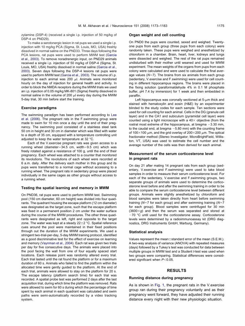
zD
idPerL2uihoan5
E
Temn5ta
rfpi6pria
T
OprwwdrzctsaaptlEdaeTrawsps
O
Oorcwueca(itbp
sbulcrtofta
Mi
Oesessagbtf(�ln
S
VA(mte
R
Agp
M. M. Akhavan et al. / Neuroscience 151 (2008) 1173–1183 1175
ylamine (DSP-4) (received a single i.p. injection of 50 mg/kg ofSP-4 on PND29).
To make a serotonergic lesion in rat pups we used a single i.p.njection with 10 mg/kg PCA (Sigma, St. Louis, MO, USA) freshlyissolved in normal saline on the PND33. Three days following theCA lesions, rat pups were used to perform MWM test (Garciat al., 2003). To remove noradrenergic input, on PND29 animalseceived a single i.p. injection of 50 mg/kg of DSP-4 (Sigma, St.ouis, MO, USA) freshly dissolved in normal saline (Garcia et al.,003). Seven days following the DSP-4 lesions rat pups weresed to perform MWM test (Garcia et al., 2003). The volume of i.p.
njection to each animal was 200 �l. Animals were monitoredourly on the day of injection for general health and activity. Inrder to block the NMDA receptors during the MWM trials we usedn i.p. injection of 0.05 mg/kg MK-801 (Sigma) freshly dissolved inormal saline in the volume of 200 �l every day during the MWM-day trial, 30 min before start the training.
xercise paradigms
he swimming paradigm has been performed according to Leet al. (2006). The pregnant rats in the F.swimming group wereade to swim for 10 min once a day until the end of their preg-ancy. The swimming pool consisted of a stainless steel tank0 cm in height and 30 cm in diameter which was filled with watero a depth of 35 cm, equipped with a temperature controlling unitdjusted to keep the water temperature at 32 °C.
Each of the V.exercise pregnant rats was given access to aunning wheel (diameter�34.5 cm, width�9.5 cm) which wasreely rotated against a resistance of 100 g, until the end of theirregnancy. Each wheel was attached to a counter that monitored
ts revolutions. The revolutions of each wheel were recorded ata.m. daily. After the delivery each mother in this group and its
ups were transferred to a normal cage without accessing to aunning wheel. The pregnant rats in sedentary group were placedndividually in the same cages as other groups without access to
running wheel.
esting the spatial learning and memory in MWM
n PND36, rat pups were used to perform MWM test. Swimmingool (150 cm diameter, 60 cm height) was divided into four quad-ants. The quadrant housing the escape platform (12 cm diameter)as designated as the target zone, such that the escape platformas fixed in a permanent position 2 cm under the water surfaceuring the course of the MWM procedures. The other three quad-ants were designated as left, right and opposite to the targetone. The water was kept at a steady 22�2 °C. Spatial referenceues around the pool were maintained in their fixed positionshrough out the duration of the MWM experiments. We used atringent two-trial-per-day, 5-day MWM training protocol, identifieds a good discriminative test for the effect of exercise on learningnd memory (Vaynman et al., 2004). Each rat was given two trialser day for five consecutive days. The animals were placed intohe pool facing the wall from one of four equally spaced startocations. Each release point was randomly altered every trial.ach trial lasted until the rat found the platform or for a maximumuration of 60 s. Animals who failed to find the platform within thellocated time were gently guided to the platform. At the end ofach trial, animals were allowed to stay on the platform for 20 s.he escape latency (platform search time) for each trial wasecorded. A spatial probe test was performed 2 days after the lastcquisition trial, during which time the platform was removed. Ratsere allowed to swim for 60 s during which the percentage of timepent by each animal in each quadrant was calculated and swimaths were semi-automatically recorded by a video tracking
ystem. drgan weight and cell counting
n PND0 the pups were counted, sexed and weighed. Twenty-ne pups from each group (three pups from each colony) wereandomly taken. These pups were weighed and anesthetized byhloroform in a chamber. Brain, heart, liver, kidneys and lungsere dissected and weighed. The rest of the rat pups remainedndisturbed with their mother until weaned and used for MWMxperiment. The mean weights of the organs from pups from eacholony were calculated and were used to calculate the final aver-ge values (N�7). The brains from six animals from each groupsedentary, V.exercise and F.swimming) were used for cell count-ng in different hippocampus regions. The brains were placed inhe fixing solution (paraformaldehyde 4% in 0.1 M phosphateuffer, pH 7.4 by immersion) for 1 week and then embedded inaraffin.
Left hippocampus was coronally sectioned at 5-�m thick andtained with hematoxylin and eosin (H&E) by an experimenterlinded to the study codes for each sample. Ten sections weresed for cell counting for each animal. Cells in the DG (granule cell
ayer) and in the CA1 and subiculum (pyramidal cell layer) wereounted using a light microscope with a 40� objective (from theostral most extreme of the hippocampus, at bregma �3.30 mm,o the caudal end, at bregma �5.80 mm) with the counting framef 100�100 �m, and the grid overlay of 200�200 �m. The opticalractionator method (Stereo Investigator; MicroBrightField, Willis-on, VT, USA) was used to estimate the cell number and theverage number of the cells was then derived for each animal.
easurement of the serum corticosterone leveln pregnant rats
n day 21 after mating 14 pregnant rats from each group (sed-ntary, V.exercise and F.swimming) were used to take bloodamples in order to measure their serum corticosterone level. Forach of the sedentary, V.exercise and F.swimming groups, twoeparate groups of animals were used to determine the cortico-terone level before and after the swimming training in order to beble to compare the serum corticosterone level between differentroups. Animals were slightly anesthetized by chloroform andlood samples were taken directly from heart before swimmingraining (N�7 for each group) and after swimming training (N�7or each group). Blood samples were centrifuged for 30 min3600�g) and then the serum was separated and stored at70 °C until used for the corticosterone assay. Corticosterone
evels were determined by a radioimmunoassay kit (DRG diag-ostics, DRG Instruments GmbH, Marburg, Germany).
tatistical analysis
alues represent the mean�standard error of the mean (S.E.M.).two-way analysis of variance (ANOVA) with repeated measures
days) followed by a Tukey’s test was conducted for data betweenultiple groups in MWM test and a Student t-test was used when
wo groups were comparing. Statistical differences were consid-red significant when P�0.05.
RESULTS
unning distance during pregnancy
s is shown in Fig. 1, the pregnant rats in the V.exerciseroup ran during their pregnancy voluntarily and as theirregnancy went forward, they have adjusted their running
istance every night with their new physiologic situation.
T
Tbmdebdwth
Sp
ObFwscft(tcFws(
tiVapRtrrfsspgsVadaPTgcsdam
Nrc
AlpVnpF5tDo(t
Fc
T
MNPPPPPP
**
M. M. Akhavan et al. / Neuroscience 151 (2008) 1173–11831176
otal body and organ weights
here was no significant difference in the body weightetween the rat pups from mothers in sedentary, F.swim-ing and V.exercise groups (Table 1). Also no significantifference was observed in the pups’ numbers and moth-rs’ weights. In addition, the mean weights of the pups’rain, liver, heart, lungs and kidneys were not significantlyifferent between the V.exercise and sedentary mothers,hereas mean weight of brain, liver, heart and kidneys of
he pups from F.swimming mothers was significantlyigher than that of the sedentary group (Table 1).
patial learning and memory enhanced in the ratups from exercised mothers
ur results showed that the escape latencies were similaretween all three groups (sedentary, V.exercise and.swimming) at the beginning of MWM training (Fig. 2A)hile rat pups from V.exercise and F.swimming groupshowed significantly lower latencies to locate the platformompared with sedentary group later. Specifically, this dif-erence was manifested as a decrease in latency to findhe platform on days 4, and 5 of training for V.exerciseP�0.01) and F.swimming (P�0.05 and P�0.01, respec-ively) groups compared with sedentary group (Fig. 2A). Toompare the data acquisition of the rat pups in sedentary,.swimming and V.exercise groups, a two-way ANOVAith repeated measures was performed which indicated aignificant effect of groups (F2, 21�3.84, P�0.05) and daysF4, 84�19, P�0.0001) and a nonsignificant interaction be-
ig. 1. Mean daily running distances by the pregnant rats in V.exer-ise group.
able 1. Comparison of parameters measured in sedentary, V.exerci
Sedentary group (n�7)
other’s weight (g) 231.2�11.71umber of the pups 11.29�1.38up’s weight (g) 6�0.17up’s brain weight (mg) 246.64�10.75up’s liver weight (mg) 221.63�19.24up’s heart weight (mg) 36.04�2.86up’s lung weight (mg) 102.17�11.14up’s kidney weight (mg) 55.38�4.51
P�0.05.
* P�0.01.ween groups and days (F8, 84�1.9, P�0.06). Tukey’s testndicated that there is a significant difference between.exercise or F.swimming and sedentary groups in days 4nd 5 of MWM trials. To evaluate the memory retention, weerformed a probe trial 2 days after the last training day.at pups were allowed to swim for 60 s in the pool in which
hey received their training, but with the escape platformemoved. The percentage of time spent in the probe quad-ant, which previously housed the platform, was calculatedor each animal. We found that the F.swimming grouphowed a clear preference for the platform quadrant overedentary, as they spent a significantly (P�0.01) greaterercentage of time in platform quadrant than sedentaryroup. No significant difference for the percentage of timepent in platform quadrant between the rat pups from.exercise and sedentary groups was observed (Fig. 2Bnd 2C). A two-way ANOVA with repeated measures in-icated a significant effect of groups (F2, 21�3.93, P�0.05)nd interaction between groups and quadrants (F6, 63�4.4,�0.001) but not the quadrants (F3, 63�1.1, P�0.34).ukey’s test indicated that only the rat pups in F.swimmingroup spent significantly more time in the target quadrantompared with their sedentary counterparts. There was noignificant difference in swimming velocity of the rat pupsuring probe test (data not shown). Also we did not detectny significant difference between the water maze perfor-ance of the male and female rat pups (data not shown).
oradrenergic and serotonergic systems and NMDAeceptors are involved in learning and memoryapability of the rat pups from exercised mothers
s is shown in Fig. 3A, DSP-4 could not increase theatency to platform in the sedentary[DSP-4] group com-ared with sedentary[NS-PND29], but the rat pups in the.exercise[DSP-4] or F.swimming[DSP-4] showed sig-ificantly increased latency to find the platform com-ared with their controls (V.exercise[NS-PND29] and.swimming[NS-PND29]) on days 3 (P�0.05), 4 (P�0.01),(P�0.01) and days 3 (P�0.01), and 5 (P�0.05) respec-
ively. Comparison between the groups for the effect ofSP-4 on data acquisition revealed a nonsignificant effectf groups (F5, 42�2.2, P�0.07), a significant effect of daysF4, 168�37.2, P�0.0001) and a significant interaction be-ween groups and days (F20, 168�2.9, P�0.0001).
.swimming groups (mean�S.E.M.)
V.exercise group (n�7) F.swimming group (n�7)
236.29�15.21 248.33�14.4510.88�2.23 10�2
6.11�0.62 6.49�0.70253.58�19.00 265�11.15**230.19�21.12 284.27�27**38.48�5.09 47.38�8.22**
107.66�10.1 120.18�21.3460.62�8.67 61.98�5.89*
se and F

mrgtaPnPoP
FaMlVtrtmeotpOstiqi(ANOVA, Tukey’s test, * P�0.05 and ** P�0.01).
Fmp(crsectcplbra(
M. M. Akhavan et al. / Neuroscience 151 (2008) 1173–1183 1177
Fig. 3B represents the effect of noradrenergic lesion onemory retention in different groups. Removal of norad-
energic input fully suppressed the preference for the tar-et quadrant in the F.swimming[DSP-4] group. Comparinghe memory retention between the groups mentionedbove showed a nonsignificant effect of groups (F5, 42�1.28,�0.29) and quadrants (F3, 126�0.91, P�0.44) but a sig-ificant interaction between both factors (F15, 126�2.9,�0.001). There was a significant difference in the percentf time spent in target quadrant between F.swimming[NS-ND29] and sedentary[NS-PND29] (P�0.01) or F.swim-
ig. 3. Effect of noradrenergic lesion on learning acquisition andemory retention as measured by MWM task. (A) DSP-4-treated ratups from both F.swimming (F.swimming[DSP-4]) and V.exerciseV.exercise[DSP-4]) mothers had significantly longer escape latenciesompared with their corresponding control groups while DSP-4-treatedat pups from sedentary mothers (sedentary[DSP-4]) did not showignificantly different escape latencies compared with the control (sed-ntary[NS-PND29]). § Represents comparison between the V.exer-ise[PND29] and V.exercise[DSP-4]. # Represents comparison be-ween the F.swimming[PND29] and F.swimming[DSP-4]. (B) DSP-4ould completely suppress the enhancing effect of F.swimming duringregnancy on memory retention in rat pups. O (opposite), AL (area
eft), T (target), AR (area right). * Represents the significant differenceetween F.swimming[NS-PND29] and sedentary[NS-PND29]. # Rep-esents the significant difference between F.swimming[NS-PND29]
nd F.swimming[DSP-4]. Data are expressed as the mean�S.E.M.ig. 2. Effect of F.swimming or V.exercise during pregnancy on learningnd memory compared with the sedentary control, as measured by theWM task. (A) Effect of F.swimming or V.exercise during pregnancy on
earning acquisition in offspring compared with the sedentary control..exercise[control] and F.swimming[control] groups took significantly less
ime to learn the location of the platform than sedentary[control]. § Rep-esents the significant difference between V.exercise[control] and seden-ary[control]. # Represents the significant difference between F.swim-ing[control] and sedentary[control]. (B) F.swimming during pregnancynhanced memory retention on the probe trial of the MWM task inffspring; animals in F.swimming[control] group spent significantly moreime in the target (T) quadrant than sedentary[control]. V.exercise duringregnancy had no significant effect on memory retention in offspring.(opposite), AL (area left), T (target), AR (area right). * Represents the
ignificant difference between F.swimming[control] and sedentary[con-rol]. (C) Representative sample of paths taken during the probe test,llustrating the marked preference by F.swimming[control] for the targetuadrant as compared with all other groups. Arrow represents the releas-
ng point of the animal. Data are expressed as the mean�S.E.M. (N�8)
N�8) (ANOVA, Tukey’s test, * P�0.05 and ** P�0.01).
metpgd
igt
so(cee3f
tmisrbwtsTtpscn
pVcc((baPsP
p5soaPcm8st(sfs
Fih
Am
Fofcppctctcplbra(
M. M. Akhavan et al. / Neuroscience 151 (2008) 1173–11831178
ing[DSP-4] (P�0.05). There was no significant differ-nce in swimming velocity of the rat pups during the probeest or the water maze performance of the male and femaleups (data not shown). This result shows that noradrener-ic lessoning specifically suppressed the effect of exerciseuring pregnancy on learning and memory in rat offspring.
As is shown in Fig. 4A, PCA could not significantlyncrease the latency to platform in the sedentary[PCA]roup compared with sedentary[NS-PND33], but PCA-reated rat pups in V.exercise[PCA] and F.swimming[PCA]
ig. 4. Effect of serotonergic lesion on learning acquisition and mem-ry retention as measured by MWM task. (A) PCA-treated rat pupsrom both F.swimming (F.swimming[PCA]) and V.exercise (V.exer-ise[PCA]) mothers had significantly longer escape latencies com-ared with their corresponding control groups while PCA-treated ratups from sedentary mothers (sedentary[PCA]) did not show signifi-antly different escape latencies compared with their control (seden-ary[NS-PND33]). § Represents comparison between the V.exer-ise[NS-PND33] and V.exercise[PCA]. # Represents comparison be-ween the F.swimming[NS-PND33] and F.swimming[PCA]. (B) PCAould completely suppress the enhancing effect of F.swimming duringregnancy on memory retention in rat pups. O (opposite), AL (area
eft), T (target), AR (area right). * Represents the significant differenceetween F.swimming[NS-PND33] and sedentary[NS-PND33]. # Rep-esents the significant difference between F.swimming[NS-PND33]
rnd F.swimming[PCA]. Data are expressed as the mean�S.E.M.N�8) (ANOVA, Tukey’s test, * P�0.05 and ** P�0.01).
howed significantly increased latency to find the platformn days 4 (P�0.05), 5 (P�0.01) and 4 (P�0.01), 5P�0.05) of MWM trials compared with the rat pups in theirontrol groups. Comparison between the groups for theffect of PCA on data acquisition revealed a significantffect of groups (F5, 42�2.6, P�0.05), of days (F4, 168�1.5, P�0.0001) and a significant interaction between bothactors (F20, 168�2, P�0.01).
On the other hand serotonergic lesion fully suppressedhe rat pups’ preference for the target quadrant in F.swim-ing[PCA] group during the probe test (Fig. 4B). Compar-
ng the memory retention between the groups showed aignificant effect of groups (F5, 42�3.8, P�0.01), of quad-ants (F3, 126�5.2, P�0.01) and a significant interactionetween both factors (F15, 126�2.2, P�0.05). Also thereas significant difference in the percent of time spent in
arget quadrant between F.swimming[NS-PND33] andedentary[NS-PND33] or F.swimming[PCA] (P�0.01).here was no significant difference in swimming velocity of
he rat pups during the probe test or the water mazeerformance of the male and female pups (data nothown). This result shows that serotonergic lessoning spe-ifically suppressed the effect of exercise during preg-ancy on learning and memory in rat offspring.
NMDA receptor blocking during the MWM trials sup-ressed the learning acquisition in sedentary[MK-801],.exercise[MK-801], and F.swimming[MK-801] groupsompared with their controls (sedentary[NS], V.exer-ise[NS], and F.swimming[NS]) on days 4 (P�0.05), 5P�0.01), on day 5 (P�0.01) and on days 3 (P�0.01), 4P�0.01), 5 (P�0.01) respectively (Fig. 5A). Comparisonetween the groups for the effect of MK-801 on datacquisition revealed a significant effect of group (F5, 42�9.3,�0.0001), of day (F4, 168�27.2, P�0.0001) and also aignificant interaction between both factors (F20, 168�2.8,�0.001).
MK-801 fully attenuated the memory retention in the ratups from F.swimming[MK-801] during the probe test (Fig.B). Comparing the memory retention between the groupshowed a significant effect of groups (F5, 42�4.2, P�0.01),f quadrant (F3, 126�2.8, P�0.05) and a significant inter-ction between groups and quadrants (F15, 126�2.2,�0.01). Also there was significant difference in the per-ent of time spent in target quadrant between F.swim-ing[NS] and sedentary[NS] (P�0.01) or F.swimming[MK-01] (P�0.05). There was no significant difference inwimming velocity of the rat pups during the probe test orhe water maze performance of the male and female pupsdata not shown). This result shows that MK-801 treatmentuppressed learning and memory not only in the rat pupsrom exercised mothers but also in the rat pups from theedentary group.
orced exercise and V.exercise during pregnancyncreased the cell number in some regions ofippocampus of the rat pups
s is presented in Fig. 7, maternal V.exercise and F.swim-ing increased the cell number in CA1, DG and subiculum
egions of the hippocampus (Fig. 6) of the rat pups. Rat

pemttpttFDg
ge(bTieb
Fo
Abtttlg(oPttbFseg
Itcdwwrtfs
Fmp(hsti#
ahi*sFt*
M. M. Akhavan et al. / Neuroscience 151 (2008) 1173–1183 1179
ups born from both voluntary and forced exercised moth-rs showed significantly more cell number in CA1 (pyra-idal cell layer) and DG (granular cell layer) regions of
heir hippocampus (P�0.01). In the subiculum region ofhe hippocampus of both V.exercise and F.swimming ratups we could not detect any increase in cell number. Onhe other hand significant difference was detected betweenhe cell numbers of CA1, and DG regions in V.exercise and.swimming groups. The number of the cells in CA1, andG areas of the hippocampus in rat pups from F.swimming
ig. 5. Effect of NMDA receptor blockade on learning acquisition andemory retention as measured by MWM task. (A) MK-801-treated ratups from sedentary control (sedentary[MK-801]), F.swimmingF.swimming[MK-801]) and V.exercise (V.exercise[MK-801]) mothersad significantly longer escape latencies compared with their corre-ponding control groups. * Represents the significant difference be-ween sedentary[MK-801] and sedentary[NS]. § Represents the signif-cant difference between V.exercise[MK-801] and V.exercise[NS].Represents the significant difference between F.swimming[MK-801]nd F.swimming[NS]. (B) MK-801 could completely suppress the en-ancing effect of F.swimming during pregnancy on memory retention
n rat pups. O (opposite), AL (area left), T (target), AR (area right).Represents the significant difference between F.swimming[NS] andedentary[NS]. # Represents the significant difference between.swimming[NS] and F.swimming[MK-801]. Data are expressed as
he mean�S.E.M. (N�8) (ANOVA, Tukey’s test, * P�0.05 and* P�0.01).
roup was significantly more than rat pups from V.exerciseFp
roup (P�0.01). A two-way ANOVA indicated a significantffect of region (F2, 46�320.37, P�0.0001), of groupF2, 46�184.99, P�0.0001) and a significant interactionetween region and group (F4, 46�45.1, P�0.0001).ukey’s test indicated that there are significant differences
n cell counts in CA1 and DG regions between both V.ex-rcise or F.swimming and sedentary groups (P�0.01 inoth cases).
.swimming increased the blood corticosterone levelf pregnant mothers
s is shown in Fig. 8, we have measured the amount oflood corticosterone in pregnant rats from all groups in twoime points (before and also 10 min after the swimmingraining on day 21 of their pregnancy). Our result showedhat after the F.swimming process blood corticosteroneevel significantly increased in pregnant rats in F.swimmingroup compared with sedentary and V.exercise groupsP�0.01). A two-way ANOVA showed a significant effectf group (F2, 35�6.48, P�0.004) and of time (F1, 35�19.7,�0.0001) and a significant interaction between both fac-
ors (F2, 35�9.12, P�0.0006). Tukey’s test indicated thathere are significant differences in corticosterone levelsetween F.swimming and sedentary (P�0.01) and.swimming and V.exercise groups (P�0.01) after thewimming training. There was not any significant differ-nce in blood corticosterone levels between the threeroups before swimming training cession (Fig. 8).
DISCUSSION
n the present study we provide novel evidences that ma-ernal voluntary and forced exercise during pregnancy in-reased spatial learning and memory in rat pups withifferent patterns. We revealed that maternal voluntaryheel running selectively enhanced the acquisition phase,hile maternal F.swimming enhanced both acquisition and
etention phases of spatial information. At least a part ofhis difference seems to be due to the stressful process oforced exercise as blood corticosterone levels increasedignificantly after the swimming in pregnant rats in F.swim-
ig. 6. Tissue sectioning in the coronal plane showing the hippocam-us areas such as DG, CA1, CA3, subiculum.

mgsceciih
To
WwwTrg(iwbwgcsscjtwnp(ewptedvte
Tir
OnrtmhmhheblhGtma
Tei
IithnrsqoOeeFeshe
FoDtbsg
FdDt
M. M. Akhavan et al. / Neuroscience 151 (2008) 1173–11831180
ing group compared with the V.exercise and sedentaryroups. Our findings also showed that offspring’s brainerotonergic and noradrenergic signaling may serve a spe-ific important role in mediating the effects of maternalxercise on pups’ cognitive function whereas NMDA re-eptors apparently had a non-selective role on pups’ learn-ng and memory. Finally both models of maternal exercisencreased the cell number of CA1 and DG regions of theippocampus of the pups.
he effect of maternal exercise in pregnancy onrgan weights in offspring
e found that the pups from swimming mothers were bornith significantly higher brain, liver, heart and kidneyeights compared with their sedentary counterparts (Table 1).his finding is in accordance with the suggestion thategular exercise during pregnancy can improve placentalrowth, enhance fetal growth and increase the birth weightLee et al., 2006). There is a controversy in results regard-ng the effect of exercise in pregnant animals on fetuseight and development and some of these studies areased on forced exercise training such as treadmill runninghich may consist of stressful events for the animal (Tur-ut et al., 2006; Houghton et al., 2000). Also there isontroversy in results regarding the effect of prenataltress on fetus weight. It has been reported that prenataltress (short-lasting or long-lasting) caused no apparenthanges in the weight of fetal whole body and brain (Fu-
ioka et al., 1999). On the other hand it has been reportedhat prenatal stress was associated with a reduction in birtheight (Cabrera et al., 1999). Therefore more studies areeeded to clarify the effect of stress during pregnancy onups’ total and organ weights. There is also another factoralthough not the only possibility) which should be consid-red in the effects of prenatal exercise on pups’ organeight at birth. It has been proposed that exercise duringregnancy may create a competition for glucose betweenhe exercising muscles and the developing fetus (Matsunot al., 1999). As V.exercise did not produce any significantifference in organ weights between the rat pups born fromoluntary running and sedentary mothers it is probable thathe different intensities of the exercises to which the moth-
ig. 7. Effect of maternal V.exercise or F.swimming during pregnancyn cell number in different regions of hippocampus of the rat pups.ata are expressed as the mean�S.E.M. (N�6) (ANOVA, Tukey’s
est, * P�0.05 and ** P�0.01). * Represents the significant differenceetween V.exercise or F.swimming and sedentary group. # Repre-ents the significant difference between V.exercise and F.swimmingroup.
rs were submitted may partially affect these parameters.bc
he effect of maternal voluntary and forced exercisen pregnancy on cell number in hippocampus ofat pups
ur results provide evidences that both models of mater-al exercise increase the cell number in CA1 and DGegions of the hippocampus of the rat pups compared withheir sedentary counterparts (Fig. 7). But maternal F.swim-ing enhances the cell number in different regions ofippocampus of the rat pups (CA1 and DG) significantlyore than maternal V.exercise (Fig. 7). Exercise-inducedippocampal neurogenesis has been suggested to en-ance learning ability and memory capability (Snydert al., 2005). Also it seems that a genetically determinedaseline level of adult hippocampal neurogenesis corre-
ates with parameters describing the acquisition phase of aippocampus-dependent learning task (Kempermann andage, 2002). It seems that increasing the cell number in
he hippocampus is one of the mechanisms by whichaternal exercise in pregnancy can enhance the learningnd memory performance of the rat pups.
he differential effects of voluntary and forcedxercise on pups’ learning and cell numbern hippocampus
n the present study F.swimming has been found to signif-cantly enhance both acquisition and retention phases ofhe learning and memory while voluntary wheel runningas increased only the acquisition phase of learning sig-ificantly. We did not detect any significant change in theetention phase in rat pups whose mothers have beenubmitted to voluntary wheel running (Fig. 2B). Conse-uently, the effects of prenatal exposure to exercise inffspring seem to be influenced by the exercise model.ne of the possible explanations for the observed differ-nces between the rat pups born from voluntary or forcedxercise mothers may be due to the stressful nature of the.swimming. Our results showed that on day 21 of thexercise protocol after the swimming training the level oferum corticosterone in pregnant rats was significantlyigher than their corresponding voluntary running or sed-ntary groups (Fig. 8). This explanation is in accordance
ig. 8. Measurement of serum corticosterone level in pregnant rats onay 21 of their pregnancy before and after 10 min swimming training.ata are expressed as the mean�S.E.M. (N�7) (ANOVA, Tukey’s
est, * P�0.05 and ** P�0.01). * Represents the significant difference#
etween F.swimming and sedentary group. Represents the signifi-ant difference between F.swimming and V.exercise group.

wletasdeadwbaspei
Tta
WwiicTsott2NagtadmsmB2
Tta
TtaaFath5dis
tesicselsdml6uP1otmpcbdepotl5hnabrippgsr
To
AtpeeiVrrs
On
M. M. Akhavan et al. / Neuroscience 151 (2008) 1173–1183 1181
ith the findings of other workers indicating that shortasting or mild prenatal stress could enhance neurogen-sis, neuronal differentiation in the hippocampus and spa-ial learning in offspring (Fujioka et al., 2006; Cannizzaro etl., 2006; Fujioka et al., 2001). On the other hand there areome other factors that may be of importance in part of theifferences observed between the different exercise mod-ls. It has been suggested that swimming minimizes thelterations of the utero-placental blood flow that are in-uced by exhaustive exercises (Lee et al., 2006). Non–eight bearing exercises (such as swimming) and weightearing ones (such as walking or jogging) promote weightnd body fat loss differently (Caldwell, 1988). Moreoverwimming differs from running in circulatory and core tem-erature responses and in muscle groups used (Matsunot al., 1999) which might be an important factor in mediat-
ng the exercise effects on fetus.
he role of the noradrenergic system in mediation ofhe effects of maternal exercise on pups’ learningnd memory
e found that a systemic lesion of the noradrenergic systemith DSP-4 eliminated the effect of maternal exercise on
ncreasing the acquisition and retention phases of spatialnformation in rat pups, whereas such lesions did not alterognitive function in rat pups from sedentary mothers (Fig. 3).his result may indicate a specific role for the noradrenergicystem in mediating the maternal exercise enhancing effectsn offspring cognitive function. Recent studies have revealedhe important role of the noradrenergic system in mediatinghe effects of exercise in brain (Garcia et al., 2003; Ivy et al.,003). Wheel and treadmill running was found to increase theE level in several brain regions such as hippocampus (Ivy etl., 2003). Also it has been shown that an intact noradrener-ic system may be crucial for the observed ability of exerciseo enhance hippocampal BDNF mRNA expression (Garcia etl., 2003). Our finding that lesion of the noradrenergic systemid not alter learning and memory in rat pups from sedentaryothers is in accordance with other reports indicating that in
edentary DSP-4-pretreated rats, the acquisition of wateraze task was not impaired (Sirvio et al., 1994) and alsoDNF mRNA level was not significantly altered (Garcia et al.,003).
he role of the serotonergic system in mediation ofhe effects of maternal exercise on pups’ learningnd memory
he place that the serotonergic system takes in mediatinghe exercise effects on brain is not clearly understoodlbeit evidence exists that exercise increases 5-HT releasend metabolism (Meeusen et al., 1996; Chaouloff, 1994).urthermore it is reported that 5-HT2A/C blockade by ket-nserin, could attenuate the exercise-induced increase inhe BDNF mRNA level only in the CA4 region of theippocampus (Ivy et al., 2003). This finding suggests that-HT activation is particularly influential for exercise-in-uced effects in this hippocampal region. On the contrary
t is reported that 5-HT receptor blockade has led to a
1Aignificantly higher level of BDNF mRNA in the CA4 region t
han did exercise alone (Ivy et al., 2003). The oppositeffects of different 5-HT receptor subtypes suggest moretudy about the involvement of these receptors in exercise-
nduced cognitive improvement. We have found that inontrast to the control group, serotonergic lesion by PCAignificantly suppressed the learning and memory in V.ex-rcise[PCA] and F.swimming[PCA] groups to the control
evel (Fig. 4). PCA is known for its ability to degenerateerotonergic axon terminals (Garcia et al., 2003). Differentoses of PCA have been used by different workers toake serotonergic lesions in rats from 2 mg/kg for a partial
esion (Harro et al., 2001) to a relatively high dose of0 mg/kg (Garcia et al., 2003). In rat pups PCA has beensed with doses such as two injections of 15 mg/kg s.c. onND3 and 4 (Tagliaferro et al., 2003; Faber and Haring,999) or 10 mg/kg i.p. on PND30 (Aragon et al., 2005). Inur study 10 mg/kg of PCA attenuated the effects of ma-ernal exercise in pregnancy on offspring learning andemory (Fig. 4). However Garcia et al. (2003) have re-orted that PCA-induced serotonergic lesions in adult ratsould not inhibit the BDNF up-regulating effects of exercisey the same animals. Such inconsistency may reflect theifferent mechanisms involved in mediating the effects ofxercise performed by the animal and maternal exercise inregnancy on offspring. Our current finding may be a signf a specific role for the serotonergic system for mediatinghe enhancing effects of maternal exercise on offspringearning and memory. It has been proposed that NE and-HT activation may be specifically responsible for en-anced transcription of BDNF mRNA due to exercise andot as vital for baseline expression (Ivy et al., 2003). Innother report it has been shown that 5-HT1B receptorlockade by NAS-181 failed to alter performance of theats in the MWM task (Ahlander-Luttgen et al., 2003). Alsot has been reported that 5-HT depletion using p-chloro-henylalanine (PCPA) has no effect on sedentary rats’erformance in the MWM (Beiko et al., 1997). These sug-estions are in agreement with our results showing thaterotonergic lesions did not alter learning and memory inat pups born from a sedentary mother.
he role of NMDA receptors in mediation of the effectsf maternal exercise on pups’ learning and memory
ccording to our results, NMDA blocking during MWMraining could suppress learning and memory in the ratups from both exercising (voluntary or forced) and sed-ntary mothers. This result is in accordance with Vaynmant al. (2003) who have shown that NMDA receptor blocking
n hippocampus by MK-801 attenuated the BDNF mRNA in.exercised as well as sedentary rats significantly. As a
esult it seems that NMDA receptors may have a generalole in mediating the effects of maternal exercise on off-pring learning and memory.
CONCLUSIONS
ur results indicated that maternal exercise during preg-ancy can increase the learning and memory abilities of
he pups. Also the exercise model based on the involve-

mvonmf
AhrM
A
A
B
C
C
C
C
C
C
D
F
F
F
F
F
F
G
G
H
I
K
K
K
L
L
L
M
M
P
S
S
S
S
M. M. Akhavan et al. / Neuroscience 151 (2008) 1173–11831182
ent of stress in the process of exercise (forced oroluntary) may contribute to the observed effects onffspring. On the other hand noradrenergic and seroto-ergic systems may play a specific role in the enhance-ent effect of maternal exercise (both voluntary and
orced) on pups’ cognitive performance in the MWM test.
cknowledgments—We wish to thank Mr. Hassan Sadeghi for hiselp in detection of vaginal plugs and sampling. Part of thisesearch was supported by a grant from Semnan University ofedical Sciences to Dr. M. M. Akhavan.
REFERENCES
hlander-Luttgen M, Madjid N, Schott PA, Sandin J, Ove Ogren S(2003) Analysis of the role of the 5-HT1B receptor in spatial andaversive learning in the rat. Neuropsychopharmacology 28:1642–1655.
ragon MA, Ayala ME, Marin M, Aviles A, Damian-Matsumura P,Dominguez R (2005) Serotonergic system blockage in the prepu-bertal rat inhibits spermatogenesis development. Reproduction129:717–727.
eiko J, Candusso L, Cain DP (1997) The effect of nonspatial watermaze pretraining in rats subjected to serotonin depletion and mus-carinic receptor antagonism: a detailed behavioural assessment ofspatial performance. Behav Brain Res 88:201–211.
abrera RJ, Rodriguez-Echandia EL, Jatuff AS, Foscolo M (1999)Effects of prenatal exposure to a mild chronic variable stress onbody weight, preweaning mortality and rat behavior. Braz J MedBiol Res 32:1229–1237.
aldwell J (1988) The energetics of swimming, running and walking inexercise prescriptions. Alaska Med 39:8–10.
annizzaro C, Plescia F, Martire M, Gagliano M, Cannizzaro G, Man-tia G, Cannizzaro E (2006) Single, intense prenatal stress de-creases emotionality and enhances learning performance in theadolescent rat offspring: Interaction with a brief, daily maternalseparation. Behav Brain Res 169:128–136.
arro E, Nunez A, Busiguina S, Torres-Aleman I (2001) Circulatinginsulin-like growth factor I mediates effects of exercise on the brain.J Neurosci 20:2926–2933.
haouloff F (1994) Influence of physical exercise on 5-HT1A receptorand anxiety-related behaviours. Neurosci Lett 176:226–230.
oe CL, Kramer M, Czeh B, Gould E, Reeves AJ, Kirschbaum C,Fuchs E (2003) Prenatal stress diminishes neurogenesis in thedentate gyrus of juvenile rhesus monkeys. Biol Psychiatry54:1025–1034.
ing Q, Vaynman S, Akhavan M, Ying Z, Gomez-Pinilla F (2006)Insulin-like growth factor I interfaces with brain-derived neuro-trophic factor-mediated synaptic plasticity to modulate aspectsof exercise-induced cognitive function. Neuroscience 140:823–833.
abel K, Tam B, Kaufer D, Baiker A, Simmons N, Kuo CJ, Palmer TD(2003) VEGF is necessary for exercise-induced adult hippocampalneurogenesis. Eur J Neurosci 18:2803–2812.
aber KM, Haring JH (1999) Synaptogenesis in the postnatal rat faciadentate is influenced by 5-HT1a receptor activation. Dev Brain Res114:245–252.
ordyce DE, Wehner JM (1993) Physical activity enhances spatiallearning performance with an associated alteration in hippocampalprotein kinase C activity in C57BL/6 and DBA/2 mice. Brain Res619:111–119.
ujioka A, Fujioka T, Ishida Y, Maekawa T, Nakamura S (2006)Differential effects of prenatal stress on the morphological matu-ration of hippocampal neurons. Neuroscience 141:907–915.
ujioka T, Fujioka A, Tan N, Chowdhury GM, Mouri H, Sakata Y, Naka-mura S (2001) Mild prenatal stress enhances learning performance in
the non-adopted rat offspring. Neuroscience 103:301–307.ujioka T, Sakata Y, Yamaguchi K, Shibasaki T, Kato H, Nakamura S(1999) The effects of prenatal stress on the development of hypo-thalamic paraventricular neurons in fetal rats. Neuroscience92:1079–1088.
arcia C, Chen MJ, Garza AA, Cotman CW, Russo-Neustadt A (2003)The influence of specific noradrenergic and serotonergic lesionson the expression of hippocampal brain-derived neurotrophic fac-tor transcripts following voluntary physical activity. Neuroscience119:721–732.
realy MA, Johnson DA, Rushton SK (1999) Improving cognitivefunction after brain injury: the use of exercise and virtual reality.Arch Phys Med Rehabil 80:661–667.
oughton PE, Mottola MF, Plust JH, Schachter CL (2000) Effect ofmaternal exercise on fetal and placental glycogen storage in themature rat. Can J Appl Physiol 25:443–452.
vy AS, Rodriguez FG, Garcia C, Chen MJ, Russo-Neustadt AA (2003)Noradrenergic and serotonergic blockade inhibits BDNF mRNAactivation following exercise and antidepressant. Pharmacol Bio-chem Behav 75:81–88.
empermann G, Gage FH (2002) Genetic determinants of adult hip-pocampal neurogenesis correlate with acquisition, but not probetrial performance in the water maze task. Eur J Neurosci 16:129–136.
im H, Lee SH, Kim SS, Yoo JH, Kim CJ (2007) The influence ofmaternal treadmill running during pregnancy on short-term mem-ory and hippocampal cell survival in rat pups. Int J Dev Neurosci25:243–249. Epub 2007 Mar 18.
ramer AF, Hahn S, Cohen NJ, Banich MT, McAuley E, HarrisonCR, Chason J, Vakil E, Bardell L, Boileau RA, Colombe A (1999)Aging, fitness, and neurocognitive function. Nature 400:418 –419.
aurin D, Verreault R, Lindsay J, MacPherson K, Rockwood K (2001)Physical activity and risk of cognitive impairment and dementia inelderly persons. Arch Neurol 58:498–504.
ee HH, Kim H, Lee JW, Kim YS, Yang HY, Chang HK, Lee TH, ShinMC, Lee MH, Shin MS, Park S, Baek S, Kim CJ (2006) Maternalswimming during pregnancy enhances short-term memory andneurogenesis in the hippocampus of rat pups. Brain Dev 28:147–154.
ordi B, Protais P, Melier D, Castor J (1997) Acute stress in pregnantrats on growth rate, learning, and memory capabilities of the off-spring. Physiol Behav 62:1087–1092.
atsuno AY, Esrey KL, Perrault H, Koski KG (1999) Low intensityexercise and varying proportions of dietary glucose and fat modifymilk and mammary gland compositions and pup growth. J Nutr129:1167–1175.
eeusen R, Thorre K, Chaouloff F, Sarre S, De Meirleir K, Ebinger G,Michotte Y (1996) Effects of tryptophan and/or acute running onextracellular 5-HT and 5- HIAA levels in the hippocampus of food-deprived rats. Brain Res 740:245–252.
arnpiansil P, Jutapakdeegul N, Chentanez T, Kotchabhakdi N (2003)Exercise during pregnancy increases hippocampal brain-derivedneurotrophic factor mRNA expression and spatial learning in neo-natal rat pup. Neurosci Lett 352:45–48.
akimura K, Kutsuwada T, Ito I, Manabe T, Takayama C, Kushiya E,Yagi T, Aizawa S, Inoue Y, Sugiyama H, et al. (1995) Reducedhippocampal LTP and spatial learning in mice lacking NMDA re-ceptor-1 subunit. Nature 373:151–155.
irvio J, Lahtinen H, Riekkinen P Jr, Riekkinen PJ (1994) Spatiallearning and noradrenaline content in the brain and periphery ofyoung and aged rats. Exp Neurol 125:312–315.
nyder JS, Hong NS, McDonald RJ, Wojtowicz JM (2005) A role foradult neurogenesis in spatial long-term memory. Neuroscience130:843–852.
oya H, Mukai A, Deocaris CC, Ohiwa N, Chang H, Nishijima T,
Fujikawa T, Togashi K, Saito T (2007) Threshold-like pattern of
S
T
T
T
v
V
V
Y
M. M. Akhavan et al. / Neuroscience 151 (2008) 1173–1183 1183
neuronal activation in the hypothalamus during treadmill running:establishment of a minimum running stress (MRS) rat model.Neurosci Res 58:341–348. Epub 2007 Apr 13.
uen P, Wu K, Levine ES, Mount HT, Xu JL, Lin SY, Black IB (1997)Brain-derived neurotrophic factor rapidly enhances phosphoryla-tion of the postsynaptic N-methyl-D-aspartate receptor subunit 1.Proc Natl Acad Sci U S A 94:8191–8195.
agliaferro P, Ramos AJ, Lopez-Costa JJ, Lopez EM, Brusco A (2003)Changes in the postnatal development on nitric oxide systeminduced by serotonin depletion. Dev Brain Res 146:39–49.
sien JZ, Huerta PT, Tonegawa S (1996) The essential role of hip-pocampal CA1 NMDA receptor-dependent synaptic plasticity inspatial memory. Cell 87:1327–1338.
urgut S, Kaptanoglu B, Emmungil G, Turgut G (2006) Increased
plasma levels of growth hormone, insulin-like growth factor (IGF-I)and IGF-binding protein 3 in pregnant rats with exercise. Tohoku JExp Med 208:75–81.
an Praag H, Kempermann G, Gage FH (1999) Running increases cellproliferation and neurogenesis in the adult mouse dentate gyrus.Nat Neurosci 2:266–270.
aynman S, Ying Z, Gomez-Pinilla F (2003) Interplay between brainderived neurotrophic factor and signal transduction modulators inthe regulation of the effects of exercise on synaptic-plasticity.Neuroscience 122:647–657.
aynman S, Ying Z, Gomez-Pinilla F (2004) Hippocampal BDNFmediates the efficacy of exercise on synaptic plasticity and cogni-tion. Eur J Neurosci 20:2580–2590.
anagita S, Amemiya S, Suzuki S, Kita I (2007) Effects of spontane-ous and forced running on activation of hypothalamic corticotro-
phin-releasing hormone in rats. Life Sci 80:356–363.(Accepted 13 January 2008)(Available online 17 November 2007)