An inactivated yellow fever 17DD vaccine cultivated in Vero cell cultures
Semaphorin-3B Is an Angiogenesis Inhibitor That Is Inactivated by
Furin-Like Pro-Protein Convertases
Asya Varshavsky,1Ofra Kessler,
1Sivan Abramovitch,
1Boaz Kigel,
1Shelly Zaffryar,
1
Gal Akiri,2and Gera Neufeld
1
1Cancer and Vascular Biology Research Center, Rappaport Research Institute in the Medical Sciences, The Bruce RappaportFaculty of Medicine, Technion, Israel Institute of Technology, Haifa, Israel and 2Department of Oncological Sciences,Mount Sinai School of Medicine, New York, New York
Abstract
Semaphorin-3B (sema3B) and semaphorin-3F (sema3F) aresecreted tumor suppressors of lung cancer. Sema3F functionsas an antiangiogenic factor that repels endothelial cells andcompromises their proliferation/survival. However, tumorcells expressing either endogenous or recombinant sema3Bfail to repel endothelial cells efficiently. Sema3B found in theconditioned medium of such cells is almost completely cleavedby furin-like pro-protein convertases, generating inactive 61-and 22-kDa fragments. We have generated a sema3B variantthat was point mutated at the cleavage site (sema3B-m),thereby conferring partial resistance to cleavage. Conditionedmedium from HEK293 cells expressing sema3b-m and condi-tioned medium of HEK293 cells expressing sema3B containedsimilar concentrations of semaphorin but sema3B-m wascleaved much less than sema3B. In contrast to HEK293 cellsexpressing native sema3B, cells expressing sema3b-m stronglyrepel endothelial cells. Conditioned medium from sema3B-m–expressing cells rapidly caused disassembly of focal adhesionsand a collapse of the actin cytoskeleton of endothelial cells,inhibited vascular endothelial growth factor–induced phos-phorylation of extracellular signal-regulated kinase 1/2,induced apoptosis of endothelial cells, and inhibited theformation of tubes from endothelial cells in an in vitroangiogenesis assay more potently than conditioned mediumfrom cells expressing sema3B. Furthermore, HEK293 cells ex-pressing sema3B-m inhibited basic fibroblast growth factor–induced angiogenesis in vivo much more potently than cellsexpressing sema3B. Repulsion of human umbilical vascularendothelial cells by sema3B-m was mediated primarily by theneuropilin-1 (np1) receptor but sema3B-m was also able totransduce signals via neuropilin-2 (np2). These results suggestthat up-regulation of furin-like pro-protein convertases inmalignant cells may enable tumors to evade the antiangio-genic effects of sema3B. [Cancer Res 2008;68(17):6922–31]
Introduction
The transition from a dormant nonangiogenic small tumor to anexpanding angiogenesis-promoting tumor is known as theangiogenic switch; it is a crucial step in tumor progression and isbelieved to be induced by up-regulation of proangiogenic factors orby down-regulation of angiogenic inhibitors (1).
Semaphorins are axon guidance factors that induce localizedcollapse of neuronal growth cones (2). The seven class-3semaphorins are the only secreted vertebrate semaphorins. Likeall semaphorins, they contain an NH2-terminal sema domainrequired for bioactivity and are distinguished by the presence of abasic domain at their COOH termini. Except for sema3E, class 3semaphorins bind to one or to both of the receptors belonging tothe neuropilin family. Neuropilins do not transduce semaphorinsignals on their own but associate with type A or type D plexins,which serve as the signal-transducing elements in neuropilin/plexin holoreceptors (3–5). The neuropilins also function asreceptors for several forms of the angiogenic factor vascularendothelial growth factor (VEGF) and are expressed in endothelialcells (6–8). In addition to their indispensable role in axon guidanceand central nervous system development, the neuropilins were alsofound to play important roles in developmental angiogenesis asrevealed by gene-targeting experiments (9, 10). Recently, it has beenfound that the class-3 semaphorins (i.e., sema3A, sema3F, andsema3E) display antiangiogenic properties (11–13). Experiments inwhich the native np1 receptor of mice was replaced by a np1variant that binds VEGF but not sema3A indicate that thecardiovascular developmental abnormalities observed in micelacking functional np1 receptors are probably caused by impairedVEGF signal transduction rather than by impaired semaphorinsignaling (14, 15).The genes encoding sema3B and sema3F were identified as
tumor suppressor genes that are deleted or inactivated in lungcancer (16, 17). Expression of sema3B in HEY ovarian carcinomacells inhibits tumor development from these cells (18). Recently, itwas observed that hypermethylation of the sema3B promoteroccurs in most cases of hepatocellular carcinomas and cholangio-carcinomas at a relatively early stage of tumor progression (19).Conditioned medium of recombinant sema3B-expressing cellsinhibited the proliferation of several breast and lung cancer–derived cell lines, indicating that secreted sema3B can directlyaffect the proliferation of cancer cells (20). Studies conducted usingMDA-MB-231 breast cancer cells that express primarily np1 suggestthat sema3B uses np1 as its receptor (20); however, recent studiessuggest that sema3B can also use np2 (21).Pro-protein convertases (PPC) constitute a family of nine
calcium-dependent serine endoproteases. The best studied mem-ber of the family is furin. PPCs cleave their substrates after theconsensus motif RXK/RR. The minimal recognition site for PPCs isRXXR but the presence of a basic amino acid (K/R) in the thirdposition greatly enhances processing (22). PPCs are often overex-pressed in cancer cells and are known to contribute totumorigenesis by proteolytic activation of precursors of protumori-genic factors such as insulin like growth factor-1 and its receptor,transforming growth factor-h; VEGF-C; and metalloproteases such
Requests for reprints: Gera Neufeld, Technion, Israel Institute of Technology,1 Efron Street, Haifa 31096, Israel. Phone: 972-4-8295430; Fax: 972-4-8523672;E-mail: [email protected].
I2008 American Association for Cancer Research.doi:10.1158/0008-5472.CAN-07-5408
Cancer Res 2008; 68: (17). September 1, 2008 6922 www.aacrjournals.org
Research Article
Research. on January 30, 2016. © 2008 American Association for Cancercancerres.aacrjournals.org Downloaded from
as MT1-MMP, which plays an important role in the induction oftumor invasiveness and tumor metastasis (22).All class-3 semaphorins contain PPC recognition sites. The
major PPC cleavage site is a KRRXRR sequence that is conserved inall the class 3 semaphorins. Cleavage at this site generates an NH2-terminal fragment off60 kDa. The cleaved NH2-terminal fragmentof sema3A is inactive (23) but the corresponding NH2-terminalcleavage product of sema3E displays potent prometastatic activity(24). Two more potential processing sites are located near theCOOH terminus of all class-3 semaphorins. Cleavage of sema3A atthese COOH-terminal sites increased its repulsive activity forneuronal growth cones, indicating that cleavage at these sites mayresult in the potentiation of class 3 semaphorin activity (23).We report that sema3B functions as a repellent of endothelial
cells. However, sema3B is highly susceptible to degradation byPPCs and is inactivated by PPC-mediated cleavage at the major siteof cleavage. We show that the conditioned medium of many typesof malignant cells contains cleaved inactivated sema3B. We alsoshow that a sema3B variant containing mutations at the major PPCcleavage site of sema3B displays increased resistance to cleavageand that cells expressing this mutated sema3B repel endothelialcells more effectively than cells expressing similar amounts ofnative sema3B. Furthermore, the mutated sema3B inhibits basicfibroblast growth factor (bFGF)–induced angiogenesis moreefficiently than native sema3B despite the limited protectionafforded by the mutation. Our results indicate that cleavage ofsema3B by up-regulated PPCs of cancer cells is a possiblemechanism by which tumors evade the antiangiogenic effects ofsema3B, thereby contributing to tumor progression.
Materials and Methods
Materials. Antibodies directed against h-actin and the FLAG epitopetag, as well as chemicals, were from Sigma. Mediums and sera for cellculture were from Biological-Industries, Inc. Oligofectamine was from
Invitrogen-Life Technologies. Antibodies directed against sema3B, Np1, Np2,
phosphorylated extracellular signal-regulated kinase 1/2 (ERK1/2), and
ERK2 (total ERK) were from Santa Cruz, Inc. The antibody against activecaspase-3 was from MBL. The antibody against vinculin was from
Chemicon International. The anti–VSV-G antibody was from ICL. The furin
inhibitor decanoyl-RVKR-CMK was from Alexis Biochemicals. The fluores-cent vital dye DiAsp and Alexa-conjugated phalloidin were from Molecular
Probes. The QuickChange site-directed mutagenesis kit and Strataclean
beads were from Stratagene. Cy2-conjugated donkey anti-mouse antibodies
were from Jackson Immunoresearch Laboratories. bFGF was produced aspreviously described (25). Matrigel was from BD Biosciences. The cDNA
encoding Sema3B was kindly donated by Dr. Susan Naylor (University of
Texas Health Science Center, San Antonio, TX). The NSPI-CMV-MCS-myc-
His lentiviral expression vector was constructed as follows: a linkercontaining the restriction enzymes NsiI-XbaI-BstBI-MluI-ClaI and SalI was
inserted between the ClaI and SalI sites of pRRL SIN cPPT PGK GFP. A
cassette containing SV40 promoter driving puromycin selection marker was
digested from pBabe-puro using AccI and ClaI and cloned into the BstBIsite. PGK-GFP cassette was then inserted into the ClaI and SalI sites to
generate NSPI-PGK-GFP. CMV promoter with multiple cloning sites (MCS)
was digested from pCDNA3.1+ Neo (Invitrogen) using MluI and XhoI,replacing the PGK-GFP to generate NSPI-CMV-MCS. Last, we replaced the
original CMV promoter with a CMV promoter containing the MCS and myc
His cassette from pCDNA3.1-myc/His (Invitrogen) using MluI and PmeI to
generate NSPI-CMV-MCS-myc-His.Expression plasmids. The VSV-G tag encoding sequence was inserted in
frame just before the stop codon of sema3B (COOH-terminal tag). The
modified sema3B cDNAs were cloned into the NSPI-CMV-MCS-myc-His
lentiviral expression vector. The modification in the furin-processing site
exchanging RFRR with KFKK was introduced into sema3B to generatesema3B-m using the QuikChange mutagenesis kit according to the
instructions of the vendor. The primers used were 5¶-CCAGTGCCAAGAG-GAAGTTCAAGAAGCAAGACGTAAGGAATGG-3¶ and the complementarilyreverse primer. The truncated sema3B forms were generated by PCRamplification of appropriate sema3B cDNA fragments and a FLAG epitope
tag was inserted in frame before the stop codon. The various cDNAs
encoding the sema3B variants were all subcloned into the NSPI-CMV-MCS-
myc-His lentiviral expression vector. Full-length sema3B-m containing theCOOH-terminal FLAG tag inserted in frame just before the stop codon
instead of the VSV-G tag was also cloned into the NSPI-CMV-MCS-myc-His
and served as a control in experiments using the truncated sema3B
variants.Generation of recombinant lentiviruses. HEK293-T cells were seeded
in 100-mm tissue culture dishes (2.5 � 106 cells per dish). A day after
seeding, the cells were cotransfected by the calcium phosphate method withthe lentiviral expression plasmid (15 Ag), packaging vector pCMVdR8.91(10 Ag), and a plasmid encoding the vesicular stomatitis virus coat envelopepMD2-VSVG (5 Ag). Conditioned medium containing infective lentiviral
particles was collected 48 and 72 h posttransfection.Cells. Human umbilical vascular endothelial (HUVEC), porcine aortic
endothelial (PAE), and HEK293 cells were cultured as previously described
(11). HUVEC were used between passages 3 to 7. DU145 prostate cancer
cells were cultured in RPMI supplemented with 10% FCS. WW94, YU-ZAZ6,and SW1416 melanoma cells were grown in medium containing a mixture
of F12 and low glucose DMEM supplemented with 10% FCS. All the
other cell lines were grown in high glucose DMEM supplemented with10% FCS.
Western blots. Cell lysates andWestern blots were done as described (26).Preparation of sema3B-containing conditioned medium. HEK293
cells expressing different forms of sema3B or control empty vector–infectedHEK293 cells were grown to 70% confluence and incubated subsequently
for 24 h with HUVEC growth medium (11) supplemented with 10% FCS ( for
ERK1/2 phosphorylation assays) or 20% FCS ( for all the other assays). The
proliferation rates of control, empty vector–infected HEK293 cells, and cellsexpressing either sema3B or sema3B-m were identical (data not shown).
Endothelial cell repulsion and proliferation/survival assays. Cellproliferation/survival assays were performed by counting surviving cells asdescribed (26). The results of these assays are influenced on the one hand by
effects on cell proliferation and on the other hand by proapoptotic effects
and represent an integration of the two mechanisms. Repulsion assays were
done as described (26, 27).Immunofluorescence. Detection of vinculin and actin in HUVEC was
performed essentially as described (26).
ERK1/2 phosphorylation. The effects of sema3B-m on VEGF121- and
VEGF165-induced ERK1/2 phosphorylation were determined essentially asdescribed (11).
Down-regulation of neuropilin expression in HUVEC using smallinterfering RNA. Inhibition of np1 and np2 expression in HUVEC wasperformed as previously described (26) using the np1-specific smallinterfering RNA (siRNA) r(AAGGAAACCUUGGUGGGAU)d(TT) and the
np2-specific siRNA r(CCAGAAGAUUGUCCUC AAC)d(TT) or a control
siRNA r(UUCUCCGAACGUGUCACGU)dTdT.Caspase-3 apoptosis assay. The assay was performed as previously
described (26).
Statistical analysis. Statistical analysis was performed using the upaireddata with unequal variance Student’s t test. Error bars represent the SE.Statistical significance is presented as *P < 0.05 and ***P < 0.001.
Results
Native as well as recombinant Sema3B produced by varioustypes of tumorigenic cells is cleaved by furin-like PPCs. Sema3Fis an inhibitor of endothelial cell proliferation, a repellent ofendothelial cells, and an inhibitor of tumor angiogenesis(11, 26, 27). To find out if sema3B also functions as a repellent of
Pro-Protein Convertases Inactivate Semaphorin-3B
www.aacrjournals.org 6923 Cancer Res 2008; 68: (17). September 1, 2008
Research. on January 30, 2016. © 2008 American Association for Cancercancerres.aacrjournals.org Downloaded from

endothelial cells, we expressed the native sema3B cDNA or asema3B cDNA containing an in-frame vesicular stomatitis virus(VSV-G) epitope tag at the COOH terminus (sema3B-VSV) inHEK293 cells using recombinant lentiviruses. However, althoughrecombinant sema3B was efficiently expressed, we could onlydetect weak repulsion of HUVEC compared with the repulsionobtained using cells expressing sema3A (Fig. 1A). When weexamined the conditioned medium of the cells using antibodiesdirected against the NH2 terminus of sema3B, we found that thesema3B was almost completely cleaved, yielding a single f60 kDaNH2-terminal fragment (Fig. 1B, a), leaving very little intactfull-length 83 kDa sema3B. A similar f60 kDa cleaved sema3Bfragment was revealed by the same antibody in conditionedmedium derived from HEK293 cells expressing native, untaggedsema3B (data not shown). The antibody used to detect the NH2terminus of sema3B does not detect the COOH-terminal cleavageproduct. This product was seen as a f20 kDa fragment in the
conditioned medium of the cells using antibodies directed againstthe COOH-terminal VSV epitope tag (Fig. 1B, b). A similarf60 kDacleavage product was also observed when transfection rather thanviral infection was used to express the recombinant sema3B (datanot shown). Throughout this work, we have mainly used theCOOH-terminal tagged sema3B. It did not differ in biologicalproperties from untagged sema3B and we therefore refer to it fromnow on as sema3B.All class-3 semaphorins contain a major conserved cleavage site
for furin-like PPCs. Cleavage at this site (Fig. 3B, site 1) generates anf60 kDa NH2-terminal fragment and anf20 kDa COOH-terminalfragment (23, 24), suggesting that the f60 kDa cleavage productobserved in the conditioned medium of sema3B-expressingHEK293 cells is also the product of PPC activity. When the activityof furin-like PPC of HEK293 cells expressing recombinant sema3Bwas inhibited with the furin inhibitor decanoyl-RVKR-CMK (24, 28),the cleavage of sema3B was partially inhibited, leading to the
Figure 1. Sema3B undergoes proteolytic cleavage by furin-like PPCs expressed in tumorigenic cells. A, HEK293 cells expressing sema3A (s3a ), sema3B (s3b ),or empty vector–infected HEK293 control cells (Cont. ) were incubated before seeding with 5 Ag/mL of the fluorescent vital dye DiAsp for 30 min. They were then seededon top of a monolayer of HUVEC and a repulsion assay was performed and photographed as previously described (26). Arrows, fluorescently labeled HEK293cells. B, a, conditioned medium (35 AL) from HEK293 cells expressing sema3B-VSV or conditioned medium from control cells (Cont. ) was separated on a 8%SDS-PAGE gel and probed with an anti-sema3B antibody. b, conditioned medium (35 AL) from HEK293 cells expressing sema3B-VSV (s3b-VSV ) or conditionedmedium from control cells was separated on a 10% SDS-PAGE gel and probed with an anti–VSV-G antibody. c, HEK293 cells expressing sema3B were incubated with(+) or without (�) 80 Amol/L decanoyl-RVKR-CMK for 24 h. Detection of sema3B in the conditioned medium by Western blot was done using an antibody directedagainst sema3B. C, a, conditioned medium (35 AL) from MDA-MB-231, HA188, and A549 cells expressing recombinant sema3B or conditioned medium from control,empty vector–infected cells (�) was separated on a 8% SDS-PAGE gel, blotted onto nitrocellulose, and probed with an anti-sema3B antibody. b, MDA-MB-231cells expressing sema3B were incubated with (+) or without (�) 135 Amol/L decanoyl-RVKR-CMK for 24 h. Conditioned medium (35 AL) from the cells was separated ona 8% SDS-PAGE gel, blotted onto nitrocellulose, and probed with an anti-sema3B antibody. D, a, SW1614, WW94, YU-ZAZ6, LoVo, HT29, SW480, and DU145cells were grown to confluence in 10-cm culture dishes and incubated for 48 h with 7 mL serum-free medium. The conditioned medium was collected and incubatedfor 2 h at 4jC with 30 AL of Strataclean beads. The beads were collected by centrifugation, boiled in 30 AL of SDS-PAGE loading buffer, and the released proteinswere separated on a 8% SDS-PAGE gel, blotted onto nitrocellulose, and probed with antibodies directed against sema3B. Nonconcentrated serum-free conditionedmedium (30 AL) derived from HEK293 cells expressing recombinant sema3B and sema3B-m was used as a positive control. b, YU-ZAZ6 cells were incubatedwith (+) or without (�) 200 Amol/L decanoyl-RVKR-CMK for 24 h. Conditioned medium (2 mL) from the cells was collected and incubated for 2 h at 4jC with 30 AL ofStrataclean beads. The beads were collected by centrifugation, boiled in 30 AL of SDS-PAGE loading buffer, and the released proteins were separated on a 8%SDS-PAGE gel, blotted onto nitrocellulose and probed with antibodies directed against sema3B.
Cancer Research
Cancer Res 2008; 68: (17). September 1, 2008 6924 www.aacrjournals.org
Research. on January 30, 2016. © 2008 American Association for Cancercancerres.aacrjournals.org Downloaded from
secretion of a mixture of full-length sema3B and cleaved sema3B(Fig. 1B, c). To find out if sema3B undergoes degradation inadditional types of cancer cells, we expressed recombinant sema3Bin several types of malignant cells that do not produce endogenoussema3B. These included MDA-MB-231 breast cancer cells, HA188lung cancer cells, A549 lung cancer cells (Fig. 1C, a), and H661 lungcancer cells (Fig. 2A, b). The conditioned medium of these cellscontained cleaved sema3B, and we could not detect full-lengthsema3B in them. To make sure that the cleavage was indeed causedby PPCs, we inhibited the PPC activity of the MDA-MB-231 cellsusing decanoyl-RVKR-CMK. The inhibitor was somewhat toxicto these cells and caused a decrease in the total amount ofrecombinant sema3B that was secreted into the medium; however,it can be seen that cleavage was inhibited and that, in the presenceof the inhibitor, full-length sema3B was secreted from the cells(Fig. 1C, b). To find out if natural sema3B is cleaved by PPCs ofcancer cells, we screened several types of malignant melanoma,colon cancer, and prostate cancer–derived cell lines. Several ofthese produced and secreted sema3B into their growth medium.However, the secreted sema3B was completely or almostcompletely cleaved in the conditioned medium of these cells(Fig. 1D, a). To make sure that the cleavage of native sema3B wascaused by furin-like enzymes, we inhibited the cleavage in one of
the melanomas using the inhibitor. Indeed, the inhibition partiallyprevented the cleavage (Fig. 1D, b).Tumorigenic cells expressing a sema3B mutant that is
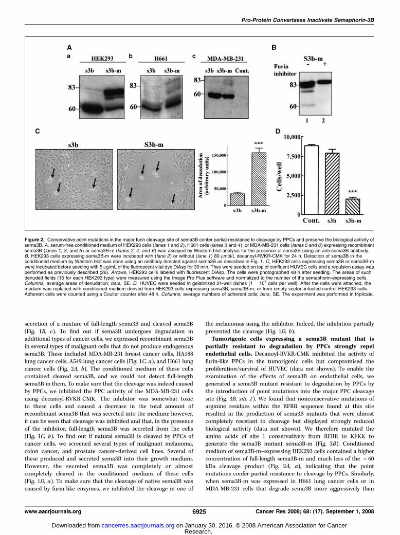
partially resistant to degradation by PPCs strongly repelendothelial cells. Decanoyl-RVKR-CMK inhibited the activity offurin-like PPCs in the tumorigenic cells but compromised theproliferation/survival of HUVEC (data not shown). To enable theexamination of the effects of sema3B on endothelial cells, wegenerated a sema3B mutant resistant to degradation by PPCs bythe introduction of point mutations into the major PPC cleavagesite (Fig. 3B, site 1). We found that nonconservative mutations ofarginine residues within the RFRR sequence found at this siteresulted in the production of sema3B mutants that were almostcompletely resistant to cleavage but displayed strongly reducedbiological activity (data not shown). We therefore mutated theamino acids of site 1 conservatively from RFRR to KFKK togenerate the sema3B mutant sema3B-m (Fig. 3B). Conditionedmedium of sema3B-m–expressing HEK293 cells contained a higherconcentration of full-length sema3B-m and much less of the f60kDa cleavage product (Fig. 2A, a), indicating that the pointmutations confer partial resistance to cleavage by PPCs. Similarly,when sema3B-m was expressed in H661 lung cancer cells or inMDA-MB-231 cells that degrade sema3B more aggressively than
Figure 2. Conservative point mutations in the major furin cleavage site of sema3B confer partial resistance to cleavage by PPCs and preserve the biological activity ofsema3B. A, serum-free conditioned medium of HEK293 cells (lanes 1 and 2), H661 cells (lanes 3 and 4), or MDA-MB-231 cells (lanes 5 and 6) expressing recombinantsema3B (lanes 1, 3 , and 5 ) or sema3B-m (lanes 2, 4 , and 6 ) was assayed by Western blot analysis for the presence of sema3B using an anti-sema3B antibody.B, HEK293 cells expressing sema3B-m were incubated with (lane 2) or without (lane 1 ) 80 Amol/L decanoyl-RVKR-CMK for 24 h. Detection of sema3B in theconditioned medium by Western blot was done using an antibody directed against sema3B as described in Fig. 1. C, HEK293 cells expressing sema3B or sema3B-mwere incubated before seeding with 5 Ag/mL of the fluorescent vital dye DiAsp for 30 min. They were seeded on top of confluent HUVEC cells and a repulsion assay wasperformed as previously described (26). Arrows, HEK293 cells labeled with fluorescent DiAsp. The cells were photographed 48 h after seeding. The areas of suchdenuded fields (15 for each HEK293 type) were measured using the Image Pro Plus software and normalized to the number of the semaphorin-expressing cells.Columns, average areas of denudation; bars, SE. D, HUVEC were seeded in gelatinized 24-well dishes (1 � 104 cells per well). After the cells were attached, themedium was replaced with conditioned medium derived from HEK293 cells expressing sema3B, sema3B-m, or from empty vector–infected control HEK293 cells.Adherent cells were counted using a Coulter counter after 48 h. Columns, average numbers of adherent cells; bars, SE. The experiment was performed in triplicate.
Pro-Protein Convertases Inactivate Semaphorin-3B
www.aacrjournals.org 6925 Cancer Res 2008; 68: (17). September 1, 2008
Research. on January 30, 2016. © 2008 American Association for Cancercancerres.aacrjournals.org Downloaded from

HEK293 cells, we found that their conditioned medium nowcontained full-length sema3B-m, although most of the sema3B-mwas still cleaved (Fig. 2A, b and c).The conditioned medium of the sema3B-m–expressing HEK293
cells also contained sema3B-derived fragments significantly largerthan f60 kDa but of lower molecular weight than the full-lengthsema3B-m (Fig. 2A, a). Similar fragments were also found in theconditioned medium of sema3B-m–expressing H661 lung cancercells (Fig. 2A, b). One of these fragments corresponds to the
f75 kDa degradation product also seen in conditioned medium ofsema3B-expressing HEK293 cells cultured in the presence ofdecanoyl-RVKR-CMK (Fig. 1B and C). These fragments are likelyto be generated by furin-like PPC-mediated cleavage at secondaryputative PPC consensus cleavage sites located closer to the COOHterminus of sema3B (Fig. 2B). To make sure that these fragments arenot the products of unrelated proteases, we inhibited the activity offurin-like PPCs in HEK293 cells expressing sema3B-m by usingdecanoyl-RVKR-CMK. Under these conditions, the conditioned
Figure 3. Characterization of the biological properties of sema3B fragments truncated at putative PPC cleavage sites. A, 35 AL of serum-free conditioned mediumderived from HEK293 cells expressing s3bt1, s3bt2, s3bt3, sema3B-m, or infected with empty vector were separated on a 8% SDS-PAGE gel and blotted ontonitrocellulose; the Western blot then was probed with an anti-sema3B antibody. B, schematic representation depicting the domain structure of sema3B and the locationof the six potential PPC cleavage sites. Potential cleavage sites are numbered starting from the most NH2-terminal site. The sequence of the potential PPC sites isshown and the points at which different sequences were truncated to generate the truncated sema3B-m forms [s3bt(1-3)] are marked out by arrows. The structuresof sema3B, sema3B-m, and the three truncated sema3B forms are depicted below. C, a HUVEC repulsion assay was performed as described using either emptyvector–infected HEK293 cells (Cont. ), HEK293 cells expressing full-length sema3B-m (s3b-m), HEK293 cells expressing the truncated sema3B-m forms [s3bt(1-3)].Arrows, DiAsp-labeled HEK293 cells. D, serum-free conditioned medium (CM ) derived from dishes containing equal concentrations of either control HEK293 cellsinfected with control lentivirus or HEK293 cells producing sema3B-m (s3b-m ), s3bt2, or s3bt3 were diluted with fresh growth medium to a final concentration of 75%,50%, 25%, 12.5%, or 6.25%, respectively. Equal aliquots were separated on a 8% SDS-PAGE gel, blotted onto nitrocellulose, and probed with antibodies directedagainst sema3B (top left ) or against the FLAG epitope tag used to label the various sema3B-m–derived forms at their COOH termini (bottom left ). The intensity oflabeled bands in the blots was quantified using a Fuji Film image reader LAS-3000 machine with the help of the Multi-Gauge program supplied with the reader.These data were used to quantify the relative concentration of the full-length forms present in the conditioned medium. To compare the biological activity of the variousforms, HUVEC were seeded in gelatinized 24-well dishes (2 � 104 cells per well). After the cell attachment, the medium was replaced with conditioned medium diluted tofinal concentrations of 75%, 50%, 25%, and 12.5% with fresh HUVEC growth medium. The conditioned media were derived from the same cells that were usedin the panels on the left. In all cases, the conditioning was done under equal conditions. Adherent HUVEC cells were trypsinized and counted after 48 h using aCoulter counter. The percentage of proliferation/survival inhibition relative to the same concentration of the control conditioned medium was calculated for each of thesema3B-m forms at each of the dilutions and normalized according to the relative concentration of the forms using arbitrary concentration units (right ). One arbitraryconcentration unit equaled the concentration of full-length sema3B-m in the conditioned medium of the producing cells diluted to the final concentration of 12.5%(determined by longer exposure of the Western blot). Points, average percentage of inhibition derived from two independent experiments; bars, SE. Each of theexperiments was performed in triplicate.
Cancer Research
Cancer Res 2008; 68: (17). September 1, 2008 6926 www.aacrjournals.org
Research. on January 30, 2016. © 2008 American Association for Cancercancerres.aacrjournals.org Downloaded from

medium of the HEK293 cells contained only the full-lengthsema3B-m, indicating that all the sema3B-m–derived fragmentsfound in the conditioned medium of the sema3B-m–producingHEK293 cells are generated by PPC-mediated cleavage (Fig. 2B).HEK293 cells expressing sema3B-m repelled HUVEC much more
effectively than HEK293 cells expressing sema3B (Fig. 2C), althoughthe total amounts of sema3B-m and native sema3B found in theconditioned medium of the producing cells were similar (Fig. 2A,a). We also compared the effect of conditioned medium derivedfrom HEK293 cells expressing sema3B-m, sema3B, or control cellsinfected with empty expression vector on the proliferation/survivalof HUVEC. Conditioned medium derived from sema3B-expressingHEK293 cells, which contains mainly the cleaved fragments, did notcompromise the proliferation/survival of HUVEC. In contrast,conditioned medium of HEK293 cells expressing sema3B-mstrongly inhibited the proliferation/survival of HUVEC (Fig. 2D).These experiments indicate that full-length sema3B is activewhereas the major PPC cleavage products of sema3B aresubstantially less active with respect to their effects on endothelialcells.Determination of the biological activity of sema3B-
m–derived NH2-terminal fragments generated by PPC cleav-age. To make sure that the f60 kDa fragment generated bycleavage at site 1 of sema3B (Fig. 1B, a) is indeed inactive, wegenerated a deletion mutant of sema3B in which we have insertedan in-frame FLAG epitope tag followed by a stop codon just beforethe site 1 cleavage site. This truncated sema3B fragment (s3bt1;Fig. 3B) was expressed and secreted by HEK293 cells (Fig. 3A,lane 1). S3bt1-expressing cells did not repel endothelial cells(Fig. 3C) and did not inhibit their proliferation/survival (data notshown), suggesting that the PPC-generated f60 kDa sema3Bproduct lacks biological activity.When examining conditioned medium obtained from cells
expressing sema3B-m, we noticed the presence of sema3B-mfragments larger than 60 kDa but smaller than full-length sema3B-m (Fig. 2A, a and b). These were apparently generated as a result ofcleavage at secondary PPC cleavage sites, which are apparentlyused more extensively in sema3B-m due to the presence of themutations at the main cleavage site. Some of these fragments arevery close in molecular weight to full-length sema3B and mayoriginate from cleavage at the cluster of potential sites locatedf2kDa upstream of the COOH-terminus (Fig. 3B, site 6), of which twoare conserved in the class 3 semaphorins (23, 24). To determine theactivity of NH2-terminal fragments generated by cleavage at theseclosely clustered sites, we produced a truncated sema3B-m variant(s3bt3) in which we inserted an in-frame FLAG epitope tagfollowed by a stop codon just before site 6 (Fig. 3B), therebyremoving all these putative cleavage sites as well as the whole basicbox of sema3B. Although the concentration of s3bt3 in theconditioned medium of producing HEK293 cells was higher thanthe concentration of sema3B-m produced by expressing HEK293cells (compare Fig. 3A, lanes 3 and 4), s3bt3-expressing cellsrepelled HUVEC very weakly if at all (Fig. 3C). The f75 kDafragment that is observed in the conditioned medium of sema3B-m–expressing cells (Fig. 2B) is likely to be produced as a result ofcleavage at site 4 of sema3B (Fig. 3B). We therefore introduced intothe sema3B-m cDNA an in-frame FLAG epitope tag followed by astop codon just before the start of the expected cleavage site of site4 (Fig. 3B) and expressed this cDNA in HEK293 cells to generatesb3t2. S3bt2 was devoid of biological activity in the repulsion assay(Fig. 3C) despite high expression levels (Fig. 3A, lane 2).
To compare the biological activities of the truncated sema3B-mvariants semiquantitatively, we compared the effects of condi-tioned medium containing sema3B-m or the three deletionmutants (Fig. 3B) on the proliferation/survival of HUVEC. Toassess the concentration of the deletion mutants in theconditioned medium of the producing HEK293 cells, we performedserial dilutions of serum-free conditioned medium and assessedthe concentration of the fragments by Western blot analysis usingantibodies directed against sema3B (Fig. 3D, top left) or against theFLAG epitope tag inserted in frame before the stop codon of themutants and the sema3B-m (Fig. 3D, bottom left). Sb3t1 (the f60kDa fragment) was devoid of bioactivity in this assay too (data notshown). The two other sema3B-m deletion mutants inhibited theproliferation/survival of HUVEC, but less effectively than sema3B-m (Fig. 3D, right ). The results of these experiments werenormalized by taking into account the relative concentrations ofthe secreted sema3B-m and sema3B-m deletion mutants asdetermined in the Western blots using the anti-sema3B antibodyand a Fuji Film LAS-3000 image reader to assess relative banddensities (Fig. 3D, top left). By these criteria, the relative EC50 ofs3bt3 was 9.2 F 2.8-fold higher than that of sema3B-m and therelative EC50 of s3bt2 was 57.5 F 7.5-fold higher than that ofsema3B-m (Fig. 3D, right). This estimate may be conservativebecause cleavage at the site 6 cluster (Fig. 3B) can generatesema3B-m fragments that the anti-sema3B antibody would not beable to distinguish from full-length sema3B-m because there is onlyan f2 kDa difference in mass. It is therefore possible that if suchtruncations take place, our estimate of the relative activity of full-length sema3B-m may be too low.For this reason, we also assessed the relative activities of the
truncated forms based on relative concentration measurementsobtained using the anti-FLAG epitope tag (Fig. 3D, bottom left).This antibody does not recognize cleaved sema3B-m or cleavedtruncated sema3B-m forms because the epitope tag is lost in theseforms. Using this antibody, we observed that the concentration offull-length sema3B-m is much lower in relation to the concen-trations of full-length s3bt2 and s3bt3 than the estimate obtainedwith the sema3B antibody. Using the values obtained with thisantibody, we estimated that s3bt3 isf40-fold less active than full-length sema3B-m, whereas s3bt2 is 1,200-fold less active (data notshown). However, regardless of the method used, we conclude thatin contrast to sema3A, in which truncations near the COOH-terminal result in enhanced activity (23), in sema3B even arelatively minimal COOH-terminal truncation already results insubstantial loss of biological activity.Sema3B signaling in HUVEC ismediated primarily by the np1
receptor. Previous work suggested that sema3B can signal usingboth np1 and np2 (20, 21). To determine the relative contributions ofnp1 and np2 in sema3B signal transduction in HUVEC, we selectivelyinhibited the expression of these receptors using specific siRNAs.Inhibition of np1 expression by f80% (Fig. 4A) resulted in asubstantial loss of responsiveness to sema3B-m in the endothelialcell repulsion assay (Fig. 4B, b). In contrast, cells in which theexpression of np2 was almost completely inhibited (Fig. 4A) werestill able to efficiently repulse HUVEC (Fig. 4B, c), indicating that inHUVEC, the sema3B repulsive signals are primarily mediated by np1.To find out if np2 is able to transduce sema3B signals in endothelialcells, we determined if cells expressing sema3B-m are able to repulsePAE cells engineered to coexpress np1 or np2 and plexin-A1 (Fig. 4C).The sema3B-m–expressing HEK293 cells were able to repel thenp2/plexin-A1–expressing PAE cells as efficiently as they repelled
Pro-Protein Convertases Inactivate Semaphorin-3B
www.aacrjournals.org 6927 Cancer Res 2008; 68: (17). September 1, 2008
Research. on January 30, 2016. © 2008 American Association for Cancercancerres.aacrjournals.org Downloaded from
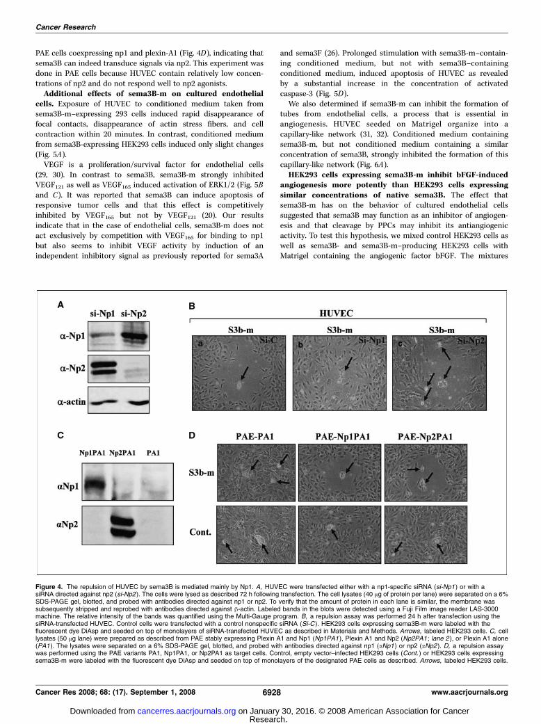
PAE cells coexpressing np1 and plexin-A1 (Fig. 4D), indicating thatsema3B can indeed transduce signals via np2. This experiment wasdone in PAE cells because HUVEC contain relatively low concen-trations of np2 and do not respond well to np2 agonists.Additional effects of sema3B-m on cultured endothelial
cells. Exposure of HUVEC to conditioned medium taken fromsema3B-m–expressing 293 cells induced rapid disappearance offocal contacts, disappearance of actin stress fibers, and cellcontraction within 20 minutes. In contrast, conditioned mediumfrom sema3B-expressing HEK293 cells induced only slight changes(Fig. 5A).VEGF is a proliferation/survival factor for endothelial cells
(29, 30). In contrast to sema3B, sema3B-m strongly inhibitedVEGF121 as well as VEGF165 induced activation of ERK1/2 (Fig. 5Band C). It was reported that sema3B can induce apoptosis ofresponsive tumor cells and that this effect is competitivelyinhibited by VEGF165 but not by VEGF121 (20). Our resultsindicate that in the case of endothelial cells, sema3B-m does notact exclusively by competition with VEGF165 for binding to np1but also seems to inhibit VEGF activity by induction of anindependent inhibitory signal as previously reported for sema3A
and sema3F (26). Prolonged stimulation with sema3B-m–contain-ing conditioned medium, but not with sema3B–containingconditioned medium, induced apoptosis of HUVEC as revealedby a substantial increase in the concentration of activatedcaspase-3 (Fig. 5D).We also determined if sema3B-m can inhibit the formation of
tubes from endothelial cells, a process that is essential inangiogenesis. HUVEC seeded on Matrigel organize into acapillary-like network (31, 32). Conditioned medium containingsema3B-m, but not conditioned medium containing a similarconcentration of sema3B, strongly inhibited the formation of thiscapillary-like network (Fig. 6A).HEK293 cells expressing sema3B-m inhibit bFGF-induced
angiogenesis more potently than HEK293 cells expressingsimilar concentrations of native sema3B. The effect thatsema3B-m has on the behavior of cultured endothelial cellssuggested that sema3B may function as an inhibitor of angiogen-esis and that cleavage by PPCs may inhibit its antiangiogenicactivity. To test this hypothesis, we mixed control HEK293 cells aswell as sema3B- and sema3B-m–producing HEK293 cells withMatrigel containing the angiogenic factor bFGF. The mixtures
Figure 4. The repulsion of HUVEC by sema3B is mediated mainly by Np1. A, HUVEC were transfected either with a np1-specific siRNA (si-Np1 ) or with asiRNA directed against np2 (si-Np2 ). The cells were lysed as described 72 h following transfection. The cell lysates (40 Ag of protein per lane) were separated on a 6%SDS-PAGE gel, blotted, and probed with antibodies directed against np1 or np2. To verify that the amount of protein in each lane is similar, the membrane wassubsequently stripped and reprobed with antibodies directed against h-actin. Labeled bands in the blots were detected using a Fuji Film image reader LAS-3000machine. The relative intensity of the bands was quantified using the Multi-Gauge program. B, a repulsion assay was performed 24 h after transfection using thesiRNA-transfected HUVEC. Control cells were transfected with a control nonspecific siRNA (Si-C ). HEK293 cells expressing sema3B-m were labeled with thefluorescent dye DiAsp and seeded on top of monolayers of siRNA-transfected HUVEC as described in Materials and Methods. Arrows, labeled HEK293 cells. C, celllysates (50 Ag lane) were prepared as described from PAE stably expressing Plexin A1 and Np1 (Np1PA1 ), Plexin A1 and Np2 (Np2PA1 ; lane 2 ), or Plexin A1 alone(PA1). The lysates were separated on a 6% SDS-PAGE gel, blotted, and probed with antibodies directed against np1 (aNp1 ) or np2 (aNp2 ). D, a repulsion assaywas performed using the PAE variants PA1, Np1PA1, or Np2PA1 as target cells. Control, empty vector–infected HEK293 cells (Cont. ) or HEK293 cells expressingsema3B-m were labeled with the fluorescent dye DiAsp and seeded on top of monolayers of the designated PAE cells as described. Arrows, labeled HEK293 cells.
Cancer Research
Cancer Res 2008; 68: (17). September 1, 2008 6928 www.aacrjournals.org
Research. on January 30, 2016. © 2008 American Association for Cancercancerres.aacrjournals.org Downloaded from

containing the different cell types were implanted s.c. in BALB/cnu/nu mice and the blood vessels that invaded the Matrigel plugswere quantified after 8 days. Although sema3B-expressing cellsreduced the number of blood vessels that invaded the plugs,sema3B-m–expressing cells had a significantly stronger effect(Fig. 6B). To compare the effect of sema3B and sema3B-m ontumor development, we also expressed sema3B and sema3B-m inMDA-MB-435 melanoma cells (Fig. 6C ; ref. 33). These cells expressthe sema3B receptor np2 (data not shown). The proliferation of thecells in cell culture was not affected by the expression of sema3B orsema3B-m (data not shown). However, both sema3B and sema3B-m inhibited the formation of these tumors relative to control MDA-MB-435 cells more or less similarly (Fig. 6D).
Discussion
Sema3B was identified, along with sema3F, as a tumorsuppressor that inhibits the development of small-cell lungcarcinoma (16, 17). Sema3F was subsequently characterized asa repellent of endothelial cells and as an antiangiogenic factor(11, 27). These findings suggested that part of the antitumorigenicbehavior of sema3B could perhaps be attributed to antiangiogenicactivity.Surprisingly, HEK293 cells infected with lentiviral vectors
directing high-level expression of sema3B did not repel endothelialcells efficiently. When we examined the conditioned medium ofthese cells, we found that sema3B was almost completely cleavedinto two fragments of f60 and f20 kDa, suggesting that these
Figure 5. Characterization of the biological activity of sema3B-m on cultured endothelial cells. A, HUVEC grown to 80% confluence were incubated for 20 min withconditioned medium from empty vector–infected HEK293 cells (Cont. ) or conditioned medium from HEK293 cells expressing either sema3B or sema3B-m. Focalcontacts were visualized using an antivinculin antibody as described in Materials and Methods (left three pictures ). The actin cytoskeleton was visualized usingAlexa-conjugated phalloidin (right three pictures ). The cells were photographed using a fluorescent microscope as described. The area of the cells stained withAlexa-conjugated phalloidin was quantified using the Image Pro Plus software. Columns, average areas derived from the analysis of between 20 and 30 cells fromdifferent microscopic fields; bars, SE. B, HUVEC were seeded in gelatinized six-well dishes (4.5 � 105 cells per well) in medium containing 10% FCS. After 16 h,the medium was replaced with conditioned medium derived from control HEK293 cells (lanes 1, 2 , and 5), from cells producing sema3B (lanes 3 and 6), or fromsema3B-m–producing cells (lanes 4 and 7). Following a 15-min preincubation at room temperature, either VEGF165 (3 ng/mL; lanes 2-4 ) or VEGF121 (25 ng/mL;lanes 5-7 ) was added to cells. The experiment was terminated after z10 min. VEGF-induced phosphorylation of ERK1/2 (pERK ) was measured using anti-pERKantibodies as described. The blot was stripped and total ERK1/2 was measured using an antibody directed against ERK2 (tERK ). The experiment was repeatedseveral times with similar results. C, the intensity of the bands in the blots shown in B was quantified using a Fuji Film image reader LAS-3000 machine with the helpof the Multi-Gauge program. The level of ERK1/2 phosphorylation was normalized to the total ERK1/2 protein (pERK/tERK) for each treatment. D, HUVEC cells(4.5 � 105 per well) were seeded in gelatinized six-well dishes. Following attachment, the medium was replaced with serum-free M199 medium (lane 1), withconditioned medium from control HEK293 cells (lane 2), with conditioned medium from cells expressing sema3B (lane 3), with conditioned medium from cellsexpressing sema3B-m (lane 4 ), or with M199 medium containing 20% serum (FCS) supplemented with 5 ng/mL bFGF (lane 5 ). Activation of caspase-3 was assayed asdescribed. To verify that the amount of protein in each lane was similar, the membrane was subsequently stripped and reprobed with antibodies directed against h-actin.
Pro-Protein Convertases Inactivate Semaphorin-3B
www.aacrjournals.org 6929 Cancer Res 2008; 68: (17). September 1, 2008
Research. on January 30, 2016. © 2008 American Association for Cancercancerres.aacrjournals.org Downloaded from

fragments are inactive. All the class-3 semaphorins contain aconserved consensus PPC cleavage site that, when used, leads tothe production of f60 and f20 kDa fragments. In the case ofsema3A, cleavage at this site leads to loss of activity (23), whereascleavage of sema3E at this site results in the generation of an activemetastasis promoting f60 kDa fragment (24). However, therecombinant f60 kDa NH2-terminal fragment of sema3B wasinactive. Furthermore, we have shown here that cleavage of sema3Bat secondary PPC cleavage sites also results in substantial loss ofactivity. Interestingly, even the removal of a relatively small COOH-terminal peptide of 2 kDa, which in the case of sema3A potentiatessema3A activity, lead to a substantial loss of sema3B bioactivity.Neither sema3A nor sema3F are efficiently cleaved by furin-like
PPCs of cancer cells and the cleaved semaphorins found in theconditioned medium of sema3A- and sema3F-producing cancercells usually represent in our experience no more than 10% to 20%of the total semaphorin found in the conditioned medium (11, 26,27). In contrast, sema3B seems to be much more susceptible to
cleavage by PPCs of cancer cells. We have found several melanoma-and colon cancer–derived cell lines that actively produce andsecrete sema3B; however, in all these cells, the secreted sema3Bwas almost completely cleaved. This susceptibility suggests that inthe case of sema3B, cleavage by up-regulated PPCs of malignantcells (22) may represent a major mechanism by which tumorsmay evade the antiangiogenic effects of sema3B in addition todown-regulation of expression and loss of heterozygosity (17,34, 35). Indeed, HEK293 cells expressing the sema3B point mutantsema3B-m, which confers partial resistance to cleavage by PPCs,contain higher concentrations of full-length mutated sema3B intheir conditioned medium. These cells repel endothelial cells morepotently than HEK293 cells expressing native sema3B using amechanism that depends on the presence of neuropilins.Furthermore, the full-length mutated sema3B found in theirconditioned medium induced the contraction of endothelial cells,inhibited their proliferation/survival, induced apoptosis, inhibitedVEGF-induced activation of ERK1/2, and inhibited tube formation
Figure 6. Tumorigenic cells expressing sema3B-m inhibit bFGF-induced angiogenesis more effectively than cells expressing unmodified sema3B. A, to assessthe effects of sema3B and sema3B-m on tube formation from HUVEC, HUVEC were seeded in 48-well tissue culture plates (2 � 104 cells per well) coated with 150 ALMatrigel in HUVEC growth medium conditioned by either control HEK293 cells or HEK293 cells producing sema3B or sema3B-m. The medium was diluted withfresh HUVEC growth medium to a final concentration of 50%. After 16 h of incubation, the wells were photographed using phase-contrast microscopy at lowmagnification. Columns, average number of bifurcation points in microscopic fields obtained from eight different wells in the case of the control and seven different wellsfor sema-3B or sema3B-m; bars, SE. The experiment was repeated thrice with similar results. Left, pictures from a representative experiment. B, HEK293 cellsexpressing either sema3B or sema3B-m or control HEK293 cells were mixed with Matrigel containing bFGF (300 ng/mL) to a final concentration of 2 � 106 cells/mL.The mixtures were injected s.c. into balb/c nu/nu mice (0.5 mL/mouse) as described (11). Matrigel plugs were excised 8 d later, embedded in optimum cuttingtemperature compound, and frozen in 2-methylbutane cooled by liquid nitrogen. They were then sectioned into 30-Am-thick sections using a cryostat. The sections wereblocked with cold acetone, reacted with an antibody directed against the endothelial marker CD-31, and counterstained with hematoxylin. The whole area of eachMatrigel section was subdivided into microscopic fields and photographed. The area of blood vessels in each microscopic field was quantified using the Image Pro Plussoftware. The average blood vessels density for each section was calculated as the overall area of the blood vessels in the section divided by the number of microscopicfields in the section, which represents the area of the section. Columns, average blood vessel density for eight different sections taken from four (for sema3B) orfive (for sema3B-m and control) different Matrigel plugs; bars, SE. C, conditioned medium (35 AL) from MDA-MB-435 cells expressing recombinant sema3B orsema3B-m or from control, empty vector–infected MDA-MB-435 cells was separated on a 8% SDS-PAGE gel; blotted onto nitrocellulose; and probed with ananti-sema3B antibody. D, MDA-MB-435 cells expressing either sema3B or sema3B-m or control MDA-MB-435 cells were implanted (7 � 106 per mouse) into themammary fat pads of 4- to 6-wk-old female balb/c nu/nu mice. The tumors were excised after 31 d and weighed. Columns, average weight of the tumors; bars, SE.
Cancer Research
Cancer Res 2008; 68: (17). September 1, 2008 6930 www.aacrjournals.org
Research. on January 30, 2016. © 2008 American Association for Cancercancerres.aacrjournals.org Downloaded from

by endothelial cells much more potently than conditioned mediumcontaining a similar concentration of native sema3B. Theproliferation/survival assays represent the balance between effectson cell proliferation and proapoptotic effects. It is possible thatthese effects are only secondary to the effects on the cytoskeletonand the adhesion and this will need to be examined in the future.Last, HEK293 cells secreting sema3B-m inhibited in vivo angio-genesis significantly better than HEK293 cells secreting similaramounts of native sema3B, suggesting that up-regulated furin-likePPCs produced in cancer cells (22) may contribute to tumorprogression through inactivation of sema3B.We also expressed sema3B and sema3B-m in MDA-MB-435
melanoma cells. In these cells, sema3B was cleaved by PPCs, butless efficiently than in the HEK293 cells. Expression of both sema3Band sema3B-m inhibited tumor formation similarly, althoughsema3B-m was marginally more efficient. The difference in theconcentrations of full-length sema3B and sema3B-m found in theconditioned medium of these cells is not as pronounced as inthe case of the HEK293 cells (compare Figs. 3A and 6C), which maybe the reason for the relatively efficient inhibition seen with nativesema3B. Furthermore, part of the inhibition may be due to a directin vivo effect on these np2-expressing tumor cells because in thetumor, the local concentrations of sema3B to which the tumor cellsare exposed may suffice to inhibit efficiently tumor development.
To conclude, our results suggest that sema3B is an inhibitor ofangiogenesis. Our results further suggest that furin-like PPCs,previously shown to contribute to tumor progression throughthe activation of tumor-promoting enzymes and growth factors(22), may also contribute to tumor progression through theinactivation of angiogenesis inhibitors such as sema3B. Ourresults further suggest that Sema3B variants displaying higherresistance to PPC-mediated cleavage may perhaps find use asantiangiogenic agents.
Disclosure of Potential Conflicts of Interest
No potential conflicts of interest were disclosed.
Acknowledgments
Received 9/15/2007; revised 6/17/2008; accepted 6/25/2008.Grant support: Israel Science Foundation, Komen Breast Cancer Foundation,
International Union against Cancer, and Rappaport Family Institute for Research inthe Medical Sciences of the Faculty of Medicine at the Technion, Israel Institute ofTechnology (G. Neufeld).The costs of publication of this article were defrayed in part by the payment of page
charges. This article must therefore be hereby marked advertisement in accordancewith 18 U.S.C. Section 1734 solely to indicate this fact.We thank Dr. Susan Naylor (Johns Hopkins University, Baltimore, MD) for the
sema3B cDNA and Dr. Stuart Aaronson (Mount Sinai School of Medicine, New York,NY) for helpful discussions and for the kind gift of lung cancer–derived cells andlentiviral expression vectors.
Pro-Protein Convertases Inactivate Semaphorin-3B
www.aacrjournals.org 6931 Cancer Res 2008; 68: (17). September 1, 2008
References
1. Hanahan D, Folkman J. Patterns and emergingmechanisms of the angiogenic switch during tumori-genesis. Cell 1996;86:353–64.2. Luo Y, Raible D, Raper JA. Collapsin: a protein in brainthat induces the collapse and paralysis of neuronalgrowth cones. Cell 1993;75:217–27.3. Tamagnone L, Artigiani S, Chen H, et al. Plexins are alarge family of receptors for transmembrane, secreted,and GPI-anchored semaphorins in vertebrates. Cell1999;99:71–80.4. Takahashi T, Fournier A, Nakamura F, et al. Plexin-neuropilin-1 complexes form functional semaphorin-3Areceptors. Cell 1999;99:59–69.5. Gitler AD, Lu MM, Epstein JA. PlexinD1 and sem-aphorin signaling are required in endothelial cells forcardiovascular development. Dev Cell 2004;7:107–16.6. Gitay-Goren H, Cohen T, Tessler S, et al. Selectivebinding of VEGF121 to one of the three VEGF receptorsof vascular endothelial cells. J Biol Chem 1996;271:5519–23.7. Soker S, Takashima S, Miao HQ, Neufeld G, KlagsbrunM. Neuropilin-1 is expressed by endothelial and tumorcells as an isoform specific receptor for vascularendothelial growth factor. Cell 1998;92:735–45.8. Gluzman-Poltorak Z, Cohen T, Herzog Y, Neufeld G.Neuropilin-2 and Neuropilin-1 are receptors for 165-amino acid long form of vascular endothelial growthfactor (VEGF) and of placenta growth factor-2, but onlyneuropilin-2 functions as a receptor for the 145 aminoacid form of VEGF. J Biol Chem 2000;275:18040–5.9. Kawasaki T, Kitsukawa T, Bekku Y, et al. A requirementfor neuropilin-1 in embryonic vessel formation. Devel-opment 1999;126:4895–902.10. Takashima S, Kitakaze M, Asakura M, et al.Targeting of both mouse neuropilin-1 and neuropilin-2 genes severely impairs developmental yolk sac andembryonic angiogenesis. Proc Natl Acad Sci U S A2002;99:3657–62.11. Kessler O, Shraga-Heled N, Lange T, et al. Sema-phorin-3F is an inhibitor of tumor angiogenesis. CancerRes 2004;64:1008–15.12. Bates D, Taylor GI, Minichiello J, et al. Neurovascularcongruence results from a shared patterning mecha-
nism that utilizes semaphorin3A and neuropilin-1. DevBiol 2003;255:77–98.13. Gu C, Yoshida Y, Livet J, et al. Semaphorin 3E andplexin-D1 control vascular pattern independently ofneuropilins. Science 2005;307:265–8.14. Gu C, Rodriguez ER, Reimert DV, et al. Neuropilin-1conveys semaphorin and VEGF signaling during neuraland cardiovascular development. Dev Cell 2003;5:45–57.15. Vieira JM, Schwarz Q, Ruhrberg C. Selective require-ments for NRP1 ligands during neurovascular pattern-ing. Development 2007;134:1833–43.16. Sekido Y, Bader S, Latif F, et al. Human semaphorinsA(V) and IV reside in the 3p21.3 small cell lung cancerdeletion region and demonstrate distinct expressionpatterns. Proc Natl Acad Sci U S A 1996;93:4120–5.17. Tomizawa Y, Sekido Y, Kondo M, et al. Inhibition oflung cancer cell growth and induction of apoptosis afterreexpression of 3p21.3 candidate tumor suppressor geneSEMA3B. Proc Natl Acad Sci U S A 2001;98:13954–9.18. Tse C, Xiang RH, Bracht T, Naylor SL. Humansemaphorin 3B (SEMA3B) located at chromosome3p21.3 suppresses tumor formation in an adenocarci-noma cell line. Cancer Res 2002;62:542–6.19. Tischoff I, Markwarth A, Witzigmann H, et al. Alleleloss and epigenetic inactivation of 3p21.3 in malignantliver tumors. Int J Cancer 2005;115:684–9.20. Castro-Rivera E, Ran S, Thorpe P, Minna JD.Semaphorin 3B (SEMA3B) induces apoptosis in lungand breast cancer, whereas VEGF165 antagonizes thiseffect. Proc Natl Acad Sci U S A 2004;101:11432–7.21. Julien F, Bechara A, Fiore R, et al. Dual functionalactivity of semaphorin 3B is required for positioning theanterior commissure. Neuron 2005;48:63–75.22. Bassi DE, Fu J, Lopez de CR, Klein-Szanto AJ.Proprotein convertases: ‘‘master switches’’ in the regu-lation of tumor growth and progression. Mol Carcinog2005;44:151–61.23. Adams RH, Lohrum M, Klostermann A, Betz H,Puschel AW. The chemorepulsive activity of secretedsemaphorins is regulated by furin-dependent proteolyticprocessing. EMBO J 1997;16:6077–86.24. Christensen C, Ambartsumian N, Gilestro G, et al.Proteolytic processing converts the repelling signalsema3e into an inducer of invasive growth and lungmetastasis. Cancer Res 2005;65:6167–77.
25. Tessler S, Neufeld G. Basic fibroblast growth factoraccumulates in the nuclei of various bFGF-producingcell types. J Cell Physiol 1990;145:310–7.26. Guttmann-Raviv N, Shraga-Heled N, Varshavsky A,Guimaraes-Sternberg C, Kessler O, Neufeld G. Sem-aphorin-3A and semaphorin-3F work together torepel endothelial cells and to inhibit their survivalby induction of apoptosis. J Biol Chem 2007;282:26294–305.27. Bielenberg DR, Hida Y, Shimizu A, et al. Semaphorin3F, a chemorepulsant for endothelial cells, induces apoorly vascularized, encapsulated, nonmetastatic tumorphenotype. J Clin Invest 2004;114:1260–71.28. Garcia AL, Han SK, Janssen WG, et al. A prohormoneconvertase cleavage site within a predicted a-helixmediates sorting of the neuronal and endocrinepolypeptide VGF into the regulated secretory pathway.J Biol Chem 2005;280:41595–608.29. Olsson AK, Dimberg A, Kreuger J, Claesson-Welsh L.VEGF receptor signalling—in control of vascularfunction. Nat Rev Mol Cell Biol 2006;7:359–71.30. Neufeld G, Cohen T, Gengrinovitch S, Poltorak Z.Vascular endothelial growth factor (VEGF) and itsreceptors. FASEB J 1999;13:9–22.31. Maheshwari RK, Srikantan V, Bhartiya D, KleinmanHK, Grant DS. Differential effects of interferon g and aon in vitro model of angiogenesis. J Cell Physiol 1991;146:164–9.32. Narazaki M, Tosato G. Ligand-induced internali-zation selects usage of common receptor Neuropilin-1 by VEGF165 and Semaphorin3A. Blood 2006;107:3892–901.33. Rae JM, Creighton CJ, Meck JM, Haddad BR, JohnsonMD. MDA-MB-435 cells are derived from M14 melano-ma cells—a loss for breast cancer, but a boon formelanoma research. Breast Cancer Res Treat 2007;104:13–9.34. Kuroki T, Trapasso F, Yendamuri S, et al. Allelic losson chromosome 3p21.3 and promoter hypermethylationof semaphorin 3B in non-small cell lung cancer. CancerRes 2003;63:3352–5.35. Malyukova A, Kilosanidze G, Loginov W, et al.Methylation of SEMA3B and new candidate TSGs inD3S2409-3S2456 in chromosome 3p21.31. Cancer Epi-demiol Biomarkers Prev 2002;11:1149S.
Research. on January 30, 2016. © 2008 American Association for Cancercancerres.aacrjournals.org Downloaded from

2008;68:6922-6931. Cancer Res Asya Varshavsky, Ofra Kessler, Sivan Abramovitch, et al. Inactivated by Furin-Like Pro-Protein ConvertasesSemaphorin-3B Is an Angiogenesis Inhibitor That Is
Updated version
http://cancerres.aacrjournals.org/content/68/17/6922
Access the most recent version of this article at:
Cited articles
http://cancerres.aacrjournals.org/content/68/17/6922.full.html#ref-list-1
This article cites 35 articles, 18 of which you can access for free at:
Citing articles
http://cancerres.aacrjournals.org/content/68/17/6922.full.html#related-urls
This article has been cited by 16 HighWire-hosted articles. Access the articles at:
E-mail alerts related to this article or journal.Sign up to receive free email-alerts
Subscriptions
Reprints and
To order reprints of this article or to subscribe to the journal, contact the AACR Publications
Permissions
To request permission to re-use all or part of this article, contact the AACR Publications
Research. on January 30, 2016. © 2008 American Association for Cancercancerres.aacrjournals.org Downloaded from
Copyright © 2022 FDOKUMEN