Semantic versus perceptual interactions in neural processing of speech-in-noise
10
Semantic versus perceptual interactions in neural processing of speech-in-noise Narly Golestani a, b, ⁎, Alexis Hervais-Adelman b , Jonas Obleser c , Sophie K. Scott a a Institute of Cognitive Neuroscience, University College London, 17 Queen Square, WC1N 3AR London, UK b University Medical School, 1, Rue Michel-Servet, CH-1211 Geneva, Switzerland c Max Planck Institute for Human Cognitive and Brain Sciences, Stephanstraße 1A, 04103 Leipzig, Germany abstract article info Article history: Accepted 16 April 2013 Available online 23 April 2013 Keywords: Native language fMRI Retroactive priming Semantics Speech-in-noise Native listeners make use of higher-level, context-driven semantic and linguistic information during the per- ception of speech-in-noise. In a recent behavioral study, using a new paradigm that isolated the semantic level of speech by using words, we showed that this native-language benefit is at least partly driven by se- mantic context (Golestani et al., 2009). Here, we used the same paradigm in a functional magnetic resonance imaging (fMRI) experiment to study the neural bases of speech intelligibility, as well as to study the neural bases of this semantic context effect in the native language. A forced-choice recognition task on the first of two auditorily presented semantically related or unrelated words was employed, where the first, ‘target’ word was embedded in different noise levels. Results showed that activation in components of the brain lan- guage network, including Broca's area and the left posterior superior temporal sulcus, as well as brain regions known to be functionally related to attention and task difficulty, was modulated by stimulus intelligibility. In line with several previous studies examining the role of linguistic context in the intelligibility of degraded speech at the sentence level, we found that activation in the angular gyrus of the left inferior parietal cortex was modulated by the presence of semantic context, and further, that this modulation depended on the in- telligibility of the speech stimuli. Our findings help to further elucidate neural mechanisms underlying the interaction of context-driven and signal-driven factors during the perception of degraded speech, and this specifically at the semantic level. © 2013 Elsevier Inc. All rights reserved. Introduction In studying the neural implementation of spoken language pro- cessing, it is important to consider the complexity of linguistic pro- cesses. For example, one can ask how higher-order, semantic versus lower-order, perceptual processes interact during the processing of noisy speech — a phenomenon that is ubiquitous in our daily lives, and how the brain supports the interaction of these complementary cognitive and perceptual dimensions (Mattys et al., 2009). It is known that in one's native language, speech comprehension is often successful even when hearing noisy, or degraded speech (Nabelek and Donahue, 1984; Takata and Nabelek, 1990; van Wijngaarden et al., 2002). Further, using the Speech Perception in Noise (SPIN) para- digm (Bilger et al., 1984; Kalikow et al., 1977), in which the predict- ability of the final word in sentences is manipulated, it has been shown that this native language advantage can arise from the use of higher-level linguistic, contextual information (Florentine, 1985a; Mayo et al., 1997). These original studies further showed that lower SNRs (or higher noise levels) are associated with a greater context benefit(Mayo et al., 1997). We are thus capable of making use of linguistic context to compensate for poor signal quality. Linguistic context includes semantic and syntactic information, as well as prag- matic and prosodic information. Recently, using a new paradigm that isolates the semantic level of speech (i.e., at the word rather than at the sentence level), we showed that this native language intelligibil- ity advantage for the perception of degraded speech is at least in part specifically driven by semantic context (Golestani et al., 2009). We used an auditory version of the retroactive word priming para- digm (Bernstein et al., 1989) in which we presented pairs of semanti- cally related or unrelated words, the first of which was embedded in different levels of noise. We found that performance was better on re- lated compared to unrelated trials during native language speech pro- cessing, and further, we showed that this benefit of context increases as SNR decreases. These results support the idea that semantic con- text, specifically, contributes to the native-language advantage for speech-in-noise processing. Here, we used the same paradigm in a functional magnetic reso- nance imaging (fMRI) experiment to study the neural bases of a) speech intelligibility at the word level, and of b) semantic context effects during degraded native language speech processing. Previous functional imag- ing studies have examined the neural bases of speech intelligibility at the sentence level during native language processing by comparing activation arising during the processing of distorted to clear speech. In many of these studies speech is distorted by noise-vocoding (Shannon NeuroImage 79 (2013) 52–61 ⁎ Corresponding author at: Brain and Language Lab, Department of Clinical Neuroscience, University of Geneva, 1 Rue Michel-Servet, CH-1211 Geneva, Switzerland. E-mail address: [email protected] (N. Golestani). 1053-8119/$ – see front matter © 2013 Elsevier Inc. All rights reserved. http://dx.doi.org/10.1016/j.neuroimage.2013.04.049 Contents lists available at SciVerse ScienceDirect NeuroImage journal homepage: www.elsevier.com/locate/ynimg
Transcript of Semantic versus perceptual interactions in neural processing of speech-in-noise
NeuroImage 79 (2013) 52–61
Contents lists available at SciVerse ScienceDirect
NeuroImage
j ourna l homepage: www.e lsev ie r .com/ locate /yn img
Semantic versus perceptual interactions in neural processing of speech-in-noise
Narly Golestani a,b,⁎, Alexis Hervais-Adelman b, Jonas Obleser c, Sophie K. Scott a
a Institute of Cognitive Neuroscience, University College London, 17 Queen Square, WC1N 3AR London, UKb University Medical School, 1, Rue Michel-Servet, CH-1211 Geneva, Switzerlandc Max Planck Institute for Human Cognitive and Brain Sciences, Stephanstraße 1A, 04103 Leipzig, Germany
⁎ Corresponding author at: Brain and Language Lab, DepaUniversity of Geneva, 1 Rue Michel-Servet, CH-1211 Genev
E-mail address: [email protected] (N. Golest
1053-8119/$ – see front matter © 2013 Elsevier Inc. Allhttp://dx.doi.org/10.1016/j.neuroimage.2013.04.049
a b s t r a c t
a r t i c l e i n f oArticle history:Accepted 16 April 2013Available online 23 April 2013
Keywords:Native languagefMRIRetroactive primingSemanticsSpeech-in-noise
Native listeners make use of higher-level, context-driven semantic and linguistic information during the per-ception of speech-in-noise. In a recent behavioral study, using a new paradigm that isolated the semanticlevel of speech by using words, we showed that this native-language benefit is at least partly driven by se-mantic context (Golestani et al., 2009). Here, we used the same paradigm in a functional magnetic resonanceimaging (fMRI) experiment to study the neural bases of speech intelligibility, as well as to study the neuralbases of this semantic context effect in the native language. A forced-choice recognition task on the first oftwo auditorily presented semantically related or unrelated words was employed, where the first, ‘target’word was embedded in different noise levels. Results showed that activation in components of the brain lan-guage network, including Broca's area and the left posterior superior temporal sulcus, as well as brain regionsknown to be functionally related to attention and task difficulty, was modulated by stimulus intelligibility. Inline with several previous studies examining the role of linguistic context in the intelligibility of degradedspeech at the sentence level, we found that activation in the angular gyrus of the left inferior parietal cortexwas modulated by the presence of semantic context, and further, that this modulation depended on the in-telligibility of the speech stimuli. Our findings help to further elucidate neural mechanisms underlying theinteraction of context-driven and signal-driven factors during the perception of degraded speech, and thisspecifically at the semantic level.
© 2013 Elsevier Inc. All rights reserved.
Introduction
In studying the neural implementation of spoken language pro-cessing, it is important to consider the complexity of linguistic pro-cesses. For example, one can ask how higher-order, semantic versuslower-order, perceptual processes interact during the processing ofnoisy speech — a phenomenon that is ubiquitous in our daily lives,and how the brain supports the interaction of these complementarycognitive and perceptual dimensions (Mattys et al., 2009). It isknown that in one's native language, speech comprehension is oftensuccessful even when hearing noisy, or degraded speech (Nabelekand Donahue, 1984; Takata and Nabelek, 1990; van Wijngaarden etal., 2002). Further, using the Speech Perception in Noise (SPIN) para-digm (Bilger et al., 1984; Kalikow et al., 1977), in which the predict-ability of the final word in sentences is manipulated, it has beenshown that this native language advantage can arise from the use ofhigher-level linguistic, contextual information (Florentine, 1985a;Mayo et al., 1997). These original studies further showed that lowerSNRs (or higher noise levels) are associated with a greater contextbenefit (Mayo et al., 1997). We are thus capable of making use of
rtment of Clinical Neuroscience,a, Switzerland.ani).
rights reserved.
linguistic context to compensate for poor signal quality. Linguisticcontext includes semantic and syntactic information, as well as prag-matic and prosodic information. Recently, using a new paradigm thatisolates the semantic level of speech (i.e., at the word rather than atthe sentence level), we showed that this native language intelligibil-ity advantage for the perception of degraded speech is at least inpart specifically driven by semantic context (Golestani et al., 2009).We used an auditory version of the retroactive word priming para-digm (Bernstein et al., 1989) in which we presented pairs of semanti-cally related or unrelated words, the first of which was embedded indifferent levels of noise. We found that performance was better on re-lated compared to unrelated trials during native language speech pro-cessing, and further, we showed that this benefit of context increasesas SNR decreases. These results support the idea that semantic con-text, specifically, contributes to the native-language advantage forspeech-in-noise processing.
Here, we used the same paradigm in a functional magnetic reso-nance imaging (fMRI) experiment to study the neural bases of a) speechintelligibility at theword level, and of b) semantic context effects duringdegraded native language speech processing. Previous functional imag-ing studies have examined the neural bases of speech intelligibilityat the sentence level during native language processing by comparingactivation arising during the processing of distorted to clear speech. Inmany of these studies speech is distorted by noise-vocoding (Shannon

53N. Golestani et al. / NeuroImage 79 (2013) 52–61
et al., 1995), or by parametrically modulating speech intelligibility bymanipulating the number of channels used to synthesize noise-vocoded speech (Davis and Johnsrude, 2003; Shannon et al., 1995).These studies have shown that the left inferior frontal gyrus (IFG), aswell as regions along the left anterior to posterior temporal lobe aremore activated when speech is degraded (challenging) but intelligiblecompared to when hearing unintelligible soundsmatched for complex-ity (Davis and Johnsrude, 2003; Narain et al., 2003; Obleser and Kotz,2010; Obleser et al., 2007a; Scott et al., 2000, 2006). Two studies explic-itly also examined activations during processing of clear speech, andfound the left IFG to be more strongly recruited if sentences were de-graded but intelligible compared to when they were unintelligible butalso compared to when they were clearly audible, suggesting that thisregion is recruited most strongly when successful but effortful speechprocessing is required (Davis and Johnsrude, 2003; Davis et al., 2011).Even in the absence of sentence-level context, it has been shownthat the LIFG responds significantly more to potentially-intelligiblenoise-vocoded words than to clear words (Hervais-Adelman et al.,2012). The left IFG's known role in phonetic, semantic and syntacticprocessing and also in verbal working memory makes it a likely candi-date in a compensatory process whereby such linguistic information(phonetic, semantic, and syntactic) as well as verbal working memoryare used to assist comprehension of degraded speech.
Left temporal cortex activations in the above studies range fromthe anterior to posterior superior temporal gyrus (STG), superiortemporal sulcus (STS), and middle temporal gyrus (MTG), withthe posterior activations extending to the classic temporo-parietalWernicke's area (Davis et al., 2011; Narain et al., 2003; Okada et al.,2010; Scott et al., 2000). A subset of the studies on speech intelligibil-ity have also found the left anterior hippocampus to be sensitive tospeech intelligibility, and this finding has been related to the role ofthis brain region in verbal memory and in processing meaning in ver-bal stimuli (Davis and Johnsrude, 2003; Davis et al., 2011). Together,this literature suggests that left frontal regions work in concert withleft anterior to posterior temporal regions involved in higher-levelbinding of meaning in connected speech to assist the effortful com-prehension of degraded speech (Davis and Johnsrude, 2003; Daviset al., 2011; Obleser et al., 2007a; Okada et al., 2010).
Several studies have been performed to explicitly assess the neuralprocesses involved when utilizing ‘top-down’ linguistic information, orlinguistic context to assist in comprehending degraded speech. Onesuch study by Obleser et al. (2007a) used noise-vocoded sentencesthat were subjected to different levels of degradation (using 2, 8, or32 filter bands), and that were either linguistically predictable or not(Obleser et al., 2007a). Consistent with the above studies on intelligibil-ity, it was shown that regions including bilateral temporal lobes aswell as the left IFG were overall more engaged when sentencesfrom the above-referenced SPIN set (Kalikow et al., 1977) were predict-able compared to when they were not, and when they were degradedbut intelligible. Behaviorally, it was shown that sentence predictability(i.e. linguistic context) assisted speech comprehension most whensentences were degraded at an intermediate (at 8-band noise-vocoding) level. Based on this behavioral finding, neural activation atthis level of degradation was assessed. Results revealed that regionsincluding the left angular gyrus, the left anterior temporal lobe andthe left ventral IFG (Brodmann's area 47) were involved only if speechcomprehension was likely to succeed despite adverse listening condi-tions, and under circumstances that allowed the use of higher-orderlinguistic information. In a related study, short, minimal sentenceswere used in which the ‘cloze’ probability differed: the pairs ofsentences all ended in the same keyword, but the preceding contextwas either predictable or not. The sentences were once again degradedusing noise vocoding. In this study also, the left angular gyrus wasshown to be involved when speech comprehension was successful,but this time either because of increased signal quality or because ofthe presence of linguistic context (Obleser and Kotz, 2010). The above
studies involved sentence-level stimuli, and thus do not allow us tospecify the level of speech (semantic, syntactic, prosodic) at which an-gular gyrus involvement is important in using context to extract mean-ing from degraded speech. A recent M/EEG study of noise-vocodedspeech (Sohoglu et al., 2012) showed that activity in the LIFG and leftSTSwas enhancedwhen listeners received congruent prior information(a transcription of the forthcoming degradedword) compared to eitherincongruent or neutral (a letter string) prior information. This paradigmexploited identity priming rather than semantic priming, and hints thatthe angular gyrus is specifically involved when semantic informationmay be used to help degraded speech perception.
The purpose of the present study was to isolate semantic influ-ences on top-down word comprehension during the processing ofspeech-in-noise, and to provide a better understanding of the neuralprocesses underlying such semantic context effects. We first predict-ed better behavioral performance at higher signal-to-noise ratios(SNRs) — that is, better performance at lower compared to highernoise levels. In line with our behavioral study, we also predictedthat we would find a semantic context effect (i.e.; benefit); that is,we predicted better behavioral performance during related comparedto unrelated trials. Lastly, we predicted that this behavioral benefit ofsemantic relatedness would be most apparent at lower compared tohigher SNRs. Examining brain regions whose activation is modulatedby SNR will provide further insights into the brain networks involvedin the effort of handling the challenge posed by acoustically degradedwords. We expected that activation in brain regions including the leftIFG and the left superior/middle temporal cortex would bemodulatedby speech intelligibility, and that activation in regions includingthe left angular gyrus would be further modulated by the presenceof semantic context. Thus, we expected greater involvement of higher-level components of the language network, such as the angular gyrus,duringmore context-driven processing (i.e.; during semantically relatedtrials). Conversely, we predicted greater involvement of lower-levelcomponents of the language network, such as the auditory cortex, dur-ing more stimulus-driven processing (i.e.; during semanticallyunrelated trials).
Materials and methods
Participants
Nine right-handed native French speakers (3 men) participated inthe study. None had previously taken part in an experiment using thisparadigm. Participants had a homogeneous language background; allhad learned a second language in school (English, German, or Spanish)from the ages of 10 to 18, and a third language (English, German, orSpanish) from the ages of 12 to 18. None spoke a second or thirdlanguage proficiently, and none had been regularly exposed to a lan-guage other than French before the age of 11, apart from sporadic andvariable exposure to English by watching TV and being exposed toother popular media (music etc.).
Participants were recruited in the larger framework of a bilingualstudy, and all had been living in an English-speaking environment(London, UK) for six months to 3 years prior to scanning. In the currentstudy, however, we only analyze and report responses to native-language French stimuli. All participants gavewritten informed consentto participate in the study, which was approved by the regional ethicalcommittee.
Stimulus materials
A forced-choice visual recognition task on the first of two auditorilypresented semantically related or unrelated words was employed,where the first, ‘target’ word was embedded in different noise levels(see below for task details). Details of stimulus parameters and synthe-sis have previously been reported (Golestani et al., 2009). While both

54 N. Golestani et al. / NeuroImage 79 (2013) 52–61
English and French stimuli were presented, only the French trials areanalyzed. Also, in the behavioral study (Golestani et al., 2009), we hadadditionally used an SNR of−4 dB, but we excluded this from the pres-ent study since in the previous study therewas nomain effect of context(semantic relatedness) in the native language at this SNR. Excludingthis SNR allowed us to include more trials per condition in the presentfMRI study.
French semantically related and unrelated word pairs were selectedfrom the database by Ferrand and Alario (1998). Two sets of stimuliwere generated (i.e. two sets of 520word pairs) such that in list 1, a spe-cific target was followed by a related prime, and that in list 2, it wasfollowed by an unrelated prime (see Table 1 for examples of stimulusitems). The lexical frequency of related and unrelated primes wasmatched across lists because more frequent (or common) words aremore likely to be recognized than less common ones (Bradlow andPisoni, 1999). The number of syllables of related and unrelated primeswas also matched because pilot testing revealed that longer words aremore likely to be recognized than shorter ones, perhaps because in lon-ger utterances there is usually more phonetic information that survivesthe noise. Word frequency information was taken from the ‘Lexique 3’database (New et al., 2004) (www.lexique.org). Half of the participantswere presented with one list (e.g. list 1 in Table 1) and the other halfwith the other list (e.g. list 2 in Table 1). This was done to ensure thatresults were not due to stimulus-specific effects but rather to the ma-nipulations of interest.
The visual foils used in the recognition phase of the task weresemantically matched to the target word (i.e. the degraded one, to berecognized). Visual foils were also matched with targets with respectto number of syllables; this was done in order to ensure that partici-pants did not use this information to recognize the target from the foil.
The stimuli were digitally recorded by amultilingual female speakerin an anechoic chamber using a sampling rate of 44.1 kHz with 16 bitquantization. Themicrophonewas positioned 30 cm from the speaker'smouth, at 15° to the mid-sagittal line. The final set of stimuli was creat-ed off-line by editing the words at zero crossing before and aftereach word. Recordings were normalized with respect to root-mean-squared amplitude and had an average duration of 1.1 s.
An equal proportion of targets from each list was embedded at thefollowing SNRs: −7 dB (highest level of noise), −6 dB, and −5 dB,and no noise. We used speech-shaped noise which approximatedthe average long term spectrum of the speech of adult males andfemales. SNR was varied by manipulating the ratio of a mixture of thenoise and the stimulus. The RMS amplitude for the final stimuli wasfixed, and the RMS amplitude of the noise and the word was scaledrelative to one-another to produce the desired SNRs, and the resultingcombined sound was scaled to the desired RMS. Thus, the RMS ampli-tude of the stimuli was equal over all SNR conditions. Signal processingwas carried out in Matlab (The Mathworks, Natick, MA.)
Experimental and scanning procedure
Before scanning, all participants performed approximately tenpractice trials with stimuli that were not used in the fMRI experimentoutside the scanner (at different SNRs), and the accuracy of theirresponses was verified in order to ensure that they understood the
Table 1Examples of stimulus and test word pairs.
List 1 (related prime) List 2 (unrelated prime)
Target Prime Target Foil Target Prime Target Foilcoqa poulea coq œuf a coq radioa coq œuf
Note: Words in italics represent auditorily presented words (stimulus words), andwords not in italics represent visually presented words (test words).
a English translations for French words: coq = rooster, poule = hen, œuf = egg,radio = radio.
task at hand. Participants were told that they would have 1.5 sand not longer to respond on every trial, and that they should alwaystry to respond within that time window. Imaging was performed ona 3.0-Tesla Philips (Best, The Netherlands) Intera scanner equippedwith a six element SENSE head coil for radiofrequency signal detection,fitted with a B0-dependent auditory stimulus delivery system(MR-Confon, Magdeburg, Germany). Two series of 162 gradient-echoimages of blood-oxygenation-level-dependent (BOLD) signal wereacquired (TR = 5 s, TA = 2 s, 2.5 × 2.5 × 3.25 mm voxels). Auditorywords were presented during the 3 second silent period betweenscans. High-resolution T1 weighted anatomical scans (0.94 mm ×0.94 mm × 1.2 mm voxels) were also obtained.
The following procedure was implemented for each participantduring the two scanning blocks in French. There was a one minuteperiod of rest between blocks. E-prime (Psychology Software Tools,Pittsburgh, PA) was used to present visual and auditory stimuli, andto collect responses. For each trial, participants heard a pair ofwords, half of which were semantically related and half of whichwere semantically unrelated. These stimuli were presented at a com-fortable listening level, that was unchanged from participant to par-ticipant. The first, ‘target’ word was degraded by being presented atdifferent SNRs (−7, −6, −5 dB, and no-noise), whereas the second,‘prime’ word was always clearly audible. Subsequently, participantssaw two visually presented words, one being the target and theother being a semantically related foil. They were required to decidewhich of the two visually presented words corresponded to the targetby making a button press response (left button for word on left side ofscreen, and right button for word on right side of screen).
The following conditions were included: semantic context (relatedand unrelated), SNR (−7, −6, −5 dB, and no noise), and silentoff-trials, resulting in a total of 9 conditions. There were 30 relatedand 30 unrelated word pairs at each of the four SNRs (i.e. 30 stimuliper condition), resulting in a total of 120 related and 120 unrelatedword pairs. Each participant therefore performed a total of 240 trials.The SNR was blocked into mini-blocks of 5 trials each, and ‘relatedness’(context) was mixed within mini-blocks in order to ensure that partic-ipants would not adopt different response strategies across relatednessconditions.
Data analysis
Data were preprocessed and analyzed using SPM5 (Wellcome De-partment of Imaging Neuroscience, London, UK). Preprocessing includedthe following steps: 1) rigid realignment of the first EPI volumes of eachsession to one-another, followed by realignment of all EPI volumes in asession to the first image of that session, 2) “unwarping” of images totake account of susceptibility-by-movement artifacts, 3) coregistrationof structural volumes to the first EPI image of the series, 4) normalizationof the structural scan to a standard structural template (SPM5's T1canonical template), 5) normalization of the EPI volumes by applicationof the parameters estimated in the normalization step, and 6) spatialsmoothing using a Gaussian kernel of 8 mm3.
For each participant a first level analysiswas carried out using a gen-eral linear model (GLM) in which every scan was coded for condition.Null events were left unmodeled and formed an implicit baseline, assuggested by Josephs and Henson (1999). Each run was modeled sepa-rately and the effect of block was coded as a separate regressor. Eachevent was modeled using the canonical hemodynamic response func-tion in SPM5. Six parameters were appended in each run to codefor the effects of movement (x, y and z translations and x, y and z rota-tions derived from the rigid realignment step of the pre-processing).A high-pass filter (cutoff 128 s) and correction for first-order serialautocorrelation were applied.
The analysis of group data was achieved by entering the parameterestimates for each of the 8 conditions fromeach participant into a singlerandom effect model. Using the contrast vectors described by Henson

Fig. 1. Mean reaction times of behavioral performance across the group. Error barsindicate standard error of the mean (SEM).
55N. Golestani et al. / NeuroImage 79 (2013) 52–61
and Penny (2005), an analysis of variance (ANOVA) with two factors(context: 2 levels; SNR: 4 levels) was carried out on the imaging datato test for the main effects of context and SNR, and any interaction be-tween the two. In order to test for regions that show a response that ismodulated by SNR, we carried out a correlational analysis in which wesought a linear effect of SNR. However, because the SNR = ∞ in theclear condition, SNR values were transformed into ameasure of the pro-portion of signal and noise in themixture.1 The transformed valueswerethen used as a contrast vector to find any regions showing a responsethat was reliably positively or negatively linearly correlated with SNR.
Unless otherwise stated, all results reported are significant at afamily-wise error (FWE) corrected level of p b 0.05. Coordinates ofpeak activations are in theMNI (Montreal Neurological Institute) space.
Results
Behavioral results
Fig. 1 presents the reaction times obtained during task perfor-mance in the scanner. A 2-way (SNR by context) repeated-measuresanalysis of variance was performed on the reaction times, excludingthe no noise condition. The no noise condition was excluded inorder to be able to examine the effect of SNR only in conditionswhere noise was present, so as to ensure that the effect of SNR isnot mainly driven by all conditions containing noise compared tothe no noise one. As predicted, results revealed a main effect of SNR(F (2, 16) = 3.76, p b 0.05), with faster reaction times at higherSNRs. The main effect of SNR was explained by a strong linear trend(F (1, 8) = 15.93, p b 0.01), demonstrating that higher SNRs wereassociated with proportionately faster performance. There was alsoa predicted main effect of context (F(1, 8) = 5.77, p b 0.05), withfaster reaction times on semantically related compared to unrelated tri-als. Last, there was a weak trend towards an SNR by context interaction(F(2, 16) = 2.32, p = 0.13). In Fig. 1 it can be seen that this is due tofaster reaction times at SNR = −7 on related compared to unrelatedtrials. Planned comparisons confirmed the prediction of a benefit ofcontext at the lowest SNRs (F(1, 16) = 6.62, p b 0.05). There was nobenefit of semantic context at any of the other SNRs (at SNR = −6:F (1, 16) = 0.00, p > 0.05, at SNR = −5: F (1, 16) = 0.00, p > 0.05,at SNR = no noise: F (1, 24) = 0.94, p > 0.05). Given the benefit ofcontext at this and not at other SNRs, these planned comparisonssuggest a differential effect of context at different SNRs. Specifically,they confirm the predicted benefit of semantic relatedness at the lowestSNR.
The accuracy measures obtained during scanning were not as sensi-tive as reaction times to the context effects. Accuracy values rangedfrom 77 to 86% correct across the six different conditions containingnoise (i.e. excluding the no noise conditions), with an overall mean of82% correct and a standard deviation of 2.8%. These results demonstratethat although the task was a difficult one, performance was abovechance (on a binomial distribution with p = 0.5, equivalent to randomresponding, the probability of scoring 77% correct by chance over 30 tri-als per condition = 0.002), and a relatively good proportion of itemswere likely comprehended by participants despite the items havingbeen embedded in noise. For the no noise conditions, accuracy valuesranged from 94 to 97% correct, with an overall mean of 96% and a stan-dard deviation of 1.3%. A 2-way (SNR by context) repeated-measuresanalysis of variance performed on the accuracy measures showed thepredicted main effect of SNR (F(3,24) = 20.5, p b 0.001), with betterperformance at higher compared to lower SNRs. The SNR effect wasexplained by strong linear (F(1,8) = 27.61, p b 0.001) and quadratic(F(1,8) = 73.34, p b 0.001) trends, demonstrating that higher SNRswere associated with proportionately better performance. Unlike the
1 Proportion of signal in stimulus: No noise = 1, SNR−5 = 0.3162, SNR−6 = 0.2512,and SNR−7 = 0.1995.
results of the previous behavioral study (Golestani et al., 2009), themain effect of context was not significant (F(1,8) = 0.17, p > 0.05),and there was no significant SNR by context interaction (F(3,24) =0.88, p > 0.05).
Imaging results
A positive correlation between brain activation and SNR (or alter-natively, a negative correlation between activation and noise, asshown on the red scale in Fig. 2) was observed in a network of regionsincluding portions of the left angular gyrus, and the posterior cingu-late gyrus and adjacent precuneus. At a lower statistical threshold ofp b 0.001 (uncorrected for multiple comparisons) the right angulargyrus, themedial orbitofrontal gyrus and the anterior cingulate cortexalso appeared, revealing a highly symmetrical network. As can be seenfrom the contrast estimate graphs showing the pattern of signalchange across conditions in Fig. 2, the correlationwith SNR in this net-work is driven by progressively greater relative deactivationwhen thestimuli are presented at lower SNRs (i.e. in greater amounts of noise).
A negative correlation between activation and SNR (or alternatively,a positive correlation between activation and noise, as shown on theblue scale in Fig. 2) was found in the anterior insula/frontal operculumbilaterally. At a lower statistical threshold of p b 0.001 (uncorrectedfor multiple comparisons), the left IFG (pars triangularis and parsopercularis), the lower bank of the left posterior superior temporal sul-cus (pSTS) extending into the MTG, and the right IFG (pars triangularis)also appear. It can been seen from the activation plots in Fig. 2 that theseregions are relatively more engaged when the stimuli are more maskedby noise. In other words, activation in this speech-related network isgreater when speech processing is made more challenging by noise.These results are summarized in Table 2.
The above results were uncovered by seeking a linear effect of SNRin our data. The main effect of SNR (i.e. not looking for a linear rela-tionship specifically) within a 2-way ANOVA on the imaging resultsshows activation in exactly the same regions as reported above, at athreshold of p b 0.001 uncorrected.
In parallel with the semantic context effect in the behavioralresults, the imaging results showed the predicted effect of context inthe angular gyrus of the left inferior parietal cortex, and at the lowerthreshold, in its right hemisphere homologue. These regions weremore active during the related compared to the unrelated conditions.There was also an effect of context in the left and right mid-STS, withthe left hemisphere result appearing at a more lenient threshold.In these regions, signal change was greater in unrelated compared torelated trials. Table 3 and Fig. 3 summarize these results.
In line with the behavioral data indicating a differential impact ofcontext at different SNRs, the imaging results revealed a significantSNR by context interaction in the angular gyrus of the left inferior

Fig. 2. Correlation with SNR. Positive correlations are displayed in red and reveal regions where activity is increased with increasing SNR. Negative correlations are displayed in blueand reveal regions whose activity increases with decreasing SNR. Bar plots represent signal change across conditions at locations indicated in Table 2. Note that for display purposes,images are presented at a threshold of p b 0.001, uncorrected. Error bars indicate SEM, corrected for within-subject comparisons (Loftus and Masson, 1994).
56 N. Golestani et al. / NeuroImage 79 (2013) 52–61
parietal cortex only, at the MNI coordinates X = −42, Y = −62,Z = 36. Table 4 and Fig. 4 summarize these results. Here, contrastestimate graphs show the pattern of signal change for the interactioneffect: there is a relatively greater recruitment of this region in therelated versus the unrelated conditions at higher compared to lowerSNRs. Higher SNRs therefore appear to result in a greater contexteffect in this region. Note that this is opposite to the pattern in thebehavioral findings, where the effect of context is greater at lowerSNRs.
Discussion
Here, we used a semantic retroactive priming paradigm with de-gradedwords to examine themodulation of brain activation by seman-tic context during effortful speech processing. Analysis of the behavioralresults obtained during scanning revealed that, as expected, perfor-mance was faster when words were presented at higher compared to
Table 2Weighted effect of SNR.
Structure MNI coordinates Z Direction ofcorrelationwith noise
Left inferior frontal gyrus (IFG) −46 20 16 4.99a PositiveRight IFG 50 22 24 4.17a PositiveLeft anterior insula/frontaloperculum (FO)
−30 22 −4 5.31 Positive
Right anterior insula/FO 34 24 −6 5.03 PositiveLeft angular gyrus −40 −78 28 4.89 NegativeRight angular gyrus 44 −70 38 4.23a NegativePosterior cingulate gyrus −2 −50 28 5.19 Negative
6 −50 18 4.84 NegativePrecuneus −2 −68 26 5.09 NegativeAnterior cingulate/medialorbitofrontal gyrus
8 56 −2 4.36a Negative
Left posterior STS/MTG −52 −46 8 4.48a Positive
Note: Results are presented at a family-wise error (FWE) corrected level of p b 0.05.a Indicates a result appearing at a lower voxel-wise threshold of 0.001, uncorrected.
lower SNRs. The imaging results showed that activation in componentsof the brain language network including Broca's area, extending to theleft anterior insula/frontal operculum medially, the left pSTS/MTG, aswell as brain regions known to be functionally related to attentionand task difficulty (components of the default-mode network, seebelow), was modulated by stimulus intelligibility.
The behavioral data also revealed an overall benefit of semantic con-text on performance, and they further showed that at the lowest SNR(−7 dB), performance was faster on semantically related comparedto unrelated trials. Thesefindings replicate a previously reported behav-ioral benefit of semantic context during the processing of nativelanguage speech in noise, shown there also to be particularly beneficialat relatively lower compared to higher SNRs (Golestani et al., 2009). Theimaging results revealed a differential response pattern as a function ofsemantic context. Specifically, there was an effect of context in the leftangular gyrus and in the mid-STS bilaterally. We also found that thestrength of the neural context effect was modulated by SNR in the leftangular gyrus.Wewill nowdiscuss the possible significance of these re-spective findings.
Signal-to-noise ratio (SNR) effects
Regions negatively correlated with SNR (or positively with noise)A number of previous functional imaging studies have examined
the neural bases of speech intelligibility at the sentence level (Davis
Table 3Main effect of context.
Structure MNI coordinates Z
Left angular gyrus −42 −64 36 4.93Right angular gyrus 56 −54 40 4.00a
Right mid-STS 62 −10 −10 5.2258 −16 −8 4.91
Left mid-STS −52 −12 −10 4.21a
Note: Results are presented at a family-wise error (FWE) corrected level of p b 0.05.a Indicates a result appearing at a lower voxel-wise threshold of p b 0.001, uncorrected.

Fig. 3. Main effect of semantic context. Bar plots represent signal change across condi-tions at locations indicated in Table 3. Error bars indicate SEM, corrected forwithin-subject comparisons.
57N. Golestani et al. / NeuroImage 79 (2013) 52–61
and Johnsrude, 2003; Davis et al., 2011; Giraud et al., 2004; Narainet al., 2003; Obleser et al., 2007a; Okada et al., 2010; Scott et al.,2000, 2006). Consistent with these studies, our results show thatactivation in a fronto-temporal network including the left IFG and themedially adjacent anterior insula, as well as left posterior STS/MTG ismodulated by speech intelligibility.
We find greater engagement of the left IFG at lower SNRs (i.e. whenthere is more noise) compared to when the target words are heardmore clearly, suggesting that more effortful processing is required atlower SNRs. Further, when the words are presented in no noise, leftIFG involvement is minimal (see Fig. 1). This is consistent with theresults of two studies by Davis and Johnsrude (2003) and Davis et al.(2011) using sentences thatwere distorted to different extents, rangingfrom being clear, to being degraded but intelligible, to being degradedbut unintelligible. In both studies, inverted ‘U’-shaped signal changepatternswere shown in the left IFG as a function of the degree of degra-dation: activation was found to be strongest when sentences weredegraded but intelligible compared to the two other conditions (Davisand Johnsrude, 2003; Davis et al., 2011). These findings suggest thatgreatest involvement of this region is observed when speech can becomprehended, but effortfully (Peelle et al., 2010). Similarly, in anelegant study by Giraud et al. (2004), neural responses to naturalsentences, to broad-band speech envelope sentences (intelligible buteffortfully after training) and to narrow-band speech envelopesentences (unintelligible, even after training) were compared. It wasfound that after training, Broca's area responded to natural speech andto broadband speech envelope noises, but that the response to naturalspeech dropped rapidly whereas the response to broadband speech
Table 4SNR × context interaction.
Structure MNI coordinates Z
Left angular gyrus −42 −62 36 5.13(2nd cluster) −48 −60 22 4.14a
Note: Results are presented at a family-wise error (FWE) corrected level of p b 0.05.a Indicates a result appearing at a lower voxel-wise threshold of p b 0.001, uncorrected
for multiple comparisons.
envelope noises remained sustained throughout stimulus presentation(Giraud et al., 2004). This finding together with ours and with those ofDavis and Johnsrude (2003) and Davis et al. (2011) suggests that theleft IFG is recruited during the processing of degraded speech, veryprobably to support comprehension. Obleser et al. (2007a) showed anincrease in left IFG involvement with increasing intelligibility, butin this study responses to clear speech were not evaluated (Obleseret al., 2007a).
The left IFG has been shown to play a role in linguistic (phonetic,semantic, syntactic, and prosodic) processing, in verbal workingmemory, and in semantic and contextual integration (Baumgaertneret al., 2002; Braver et al., 1997; Buckner et al., 1995; Dapretto andBookheimer, 1999; Gold and Buckner, 2002; Gough et al., 2005;Keller et al., 2001; Paulesu et al., 1993; Rodd et al., 2005). These areprocesses which can be expected to be more strongly recruitedduring sentence compared toword processing. Nonetheless, we still ob-serve involvement of the LIFG during the low SNR condition, demon-strating the role of this region during effortful speech processing evenwhen there are lesser demands on higher-level linguistic processingduring our word-level task. This finding is consistent with the resultsof Hervais-Adelman et al. (2012) who demonstrated involvement ofthe LIFG in response to listening to potentially-comprehensible degrad-edwords, as compared to clear words and to incomprehensible degrad-edwords. Left IFG involvement during degradedword processing is alsoconsistentwith the results of another study, wherewordswere degrad-ed by being interrupted by white noise bursts centered around the fric-atives and affricatives. It was found that the left IFG is involved in the‘repair’ process, whereby participants had the illusion of hearing naturalwords when in fact they were hearing degraded words (Shahin et al.,2009). Alsowork on processing sentences containing semantic ambigu-ity has shown the involvement of regions including the left IFG duringthe disambiguation of high-ambiguity sentences, in support of therole of this region in selecting contextually appropriate word meaningsduring processing that is more demanding (i.e. that requires disambig-uation) (Rodd et al., 2005). A recent study has shown that response biascan modulate the speech motor system during speech processing(Venezia et al., 2012). Our findings in the left IFG are unlikely to bedriven by response bias, however, because the present 2-alternativeforced-choice task was unlikely to become systematically biased(i.e., participants had to choose one of two words on each trial, andthe pairs of words differed from trial to trial).
We find evidence for greater activation in the left posterior STS/MTGwith increasing degradation levels. The direction of this SNR effect is thesame as that in the left IFG. Previous studies using sentences haveshown greater involvement of regions along the temporal lobe, span-ning from the anterior STS to the middle/lateral temporal gyrus (nearHeschl's gyrus) to the posterior STG/STS/MTG during the processing ofdegraded but intelligible speech (Davis and Johnsrude, 2003; Daviset al., 2011; Narain et al., 2003; Scott et al., 2000, 2006). In one studywhere neural responses to degraded speech were explicitly comparedto those of clear speech, andwhere the response profile in the posteriorSTG is shown, it appears as though similar to the left IFG, the response inthis region is maximal when speech is degraded but intelligible, andthat the response diminishes somewhat during listening to clear speech(Davis et al., 2011). This is in line with our finding that the response inthis region is diminished when speech is effortlessly perceived. In thepresent study we do not find evidence for modulation by SNR in theleft anterior STG/STS. This may due to methodological issues related tofMRI in the anterior temporal lobe, which is known to be more suscep-tible to signal loss due to magnetic susceptibility artifacts (Devlin et al.,2000).
The left posterior MTG has previously been implicated in semanticprocessing (Chao et al., 1999; Moore and Price, 1999; Mummery et al.,1998; Perani et al., 1999), including modality independent semanticprocessing (e.g.; in response to pictures and words) (Vandenbergheet al., 1996). More recently, it has been proposed that this region is
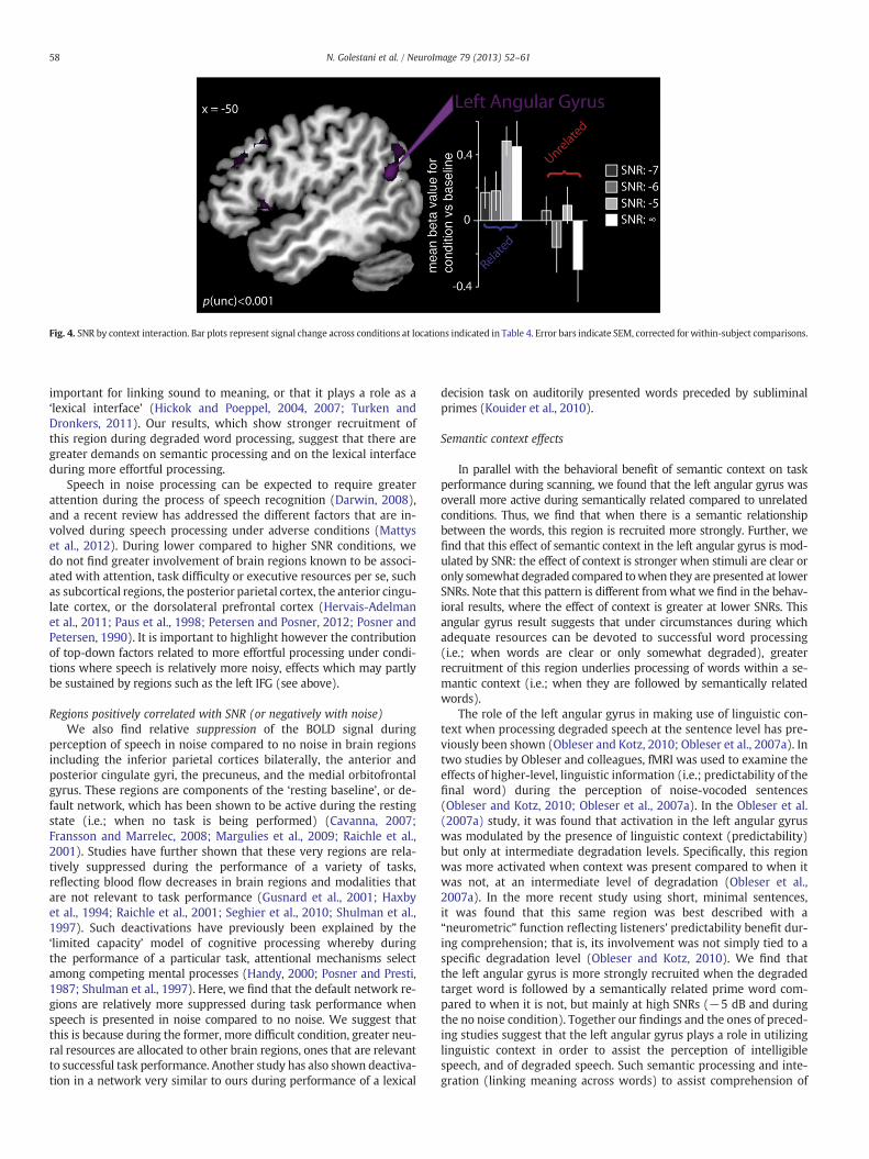
Fig. 4. SNR by context interaction. Bar plots represent signal change across conditions at locations indicated in Table 4. Error bars indicate SEM, corrected for within-subject comparisons.
58 N. Golestani et al. / NeuroImage 79 (2013) 52–61
important for linking sound to meaning, or that it plays a role as a‘lexical interface’ (Hickok and Poeppel, 2004, 2007; Turken andDronkers, 2011). Our results, which show stronger recruitment ofthis region during degraded word processing, suggest that there aregreater demands on semantic processing and on the lexical interfaceduring more effortful processing.
Speech in noise processing can be expected to require greaterattention during the process of speech recognition (Darwin, 2008),and a recent review has addressed the different factors that are in-volved during speech processing under adverse conditions (Mattyset al., 2012). During lower compared to higher SNR conditions, wedo not find greater involvement of brain regions known to be associ-ated with attention, task difficulty or executive resources per se, suchas subcortical regions, the posterior parietal cortex, the anterior cingu-late cortex, or the dorsolateral prefrontal cortex (Hervais-Adelmanet al., 2011; Paus et al., 1998; Petersen and Posner, 2012; Posner andPetersen, 1990). It is important to highlight however the contributionof top-down factors related to more effortful processing under condi-tions where speech is relatively more noisy, effects which may partlybe sustained by regions such as the left IFG (see above).
Regions positively correlated with SNR (or negatively with noise)We also find relative suppression of the BOLD signal during
perception of speech in noise compared to no noise in brain regionsincluding the inferior parietal cortices bilaterally, the anterior andposterior cingulate gyri, the precuneus, and the medial orbitofrontalgyrus. These regions are components of the ‘resting baseline’, or de-fault network, which has been shown to be active during the restingstate (i.e.; when no task is being performed) (Cavanna, 2007;Fransson and Marrelec, 2008; Margulies et al., 2009; Raichle et al.,2001). Studies have further shown that these very regions are rela-tively suppressed during the performance of a variety of tasks,reflecting blood flow decreases in brain regions and modalities thatare not relevant to task performance (Gusnard et al., 2001; Haxbyet al., 1994; Raichle et al., 2001; Seghier et al., 2010; Shulman et al.,1997). Such deactivations have previously been explained by the‘limited capacity’ model of cognitive processing whereby duringthe performance of a particular task, attentional mechanisms selectamong competing mental processes (Handy, 2000; Posner and Presti,1987; Shulman et al., 1997). Here, we find that the default network re-gions are relatively more suppressed during task performance whenspeech is presented in noise compared to no noise. We suggest thatthis is because during the former, more difficult condition, greater neu-ral resources are allocated to other brain regions, ones that are relevantto successful task performance. Another study has also shown deactiva-tion in a network very similar to ours during performance of a lexical
decision task on auditorily presented words preceded by subliminalprimes (Kouider et al., 2010).
Semantic context effects
In parallel with the behavioral benefit of semantic context on taskperformance during scanning, we found that the left angular gyrus wasoverall more active during semantically related compared to unrelatedconditions. Thus, we find that when there is a semantic relationshipbetween the words, this region is recruited more strongly. Further, wefind that this effect of semantic context in the left angular gyrus is mod-ulated by SNR: the effect of context is stronger when stimuli are clear oronly somewhat degraded compared towhen they are presented at lowerSNRs. Note that this pattern is different fromwhat we find in the behav-ioral results, where the effect of context is greater at lower SNRs. Thisangular gyrus result suggests that under circumstances during whichadequate resources can be devoted to successful word processing(i.e.; when words are clear or only somewhat degraded), greaterrecruitment of this region underlies processing of words within a se-mantic context (i.e.; when they are followed by semantically relatedwords).
The role of the left angular gyrus in making use of linguistic con-text when processing degraded speech at the sentence level has pre-viously been shown (Obleser and Kotz, 2010; Obleser et al., 2007a). Intwo studies by Obleser and colleagues, fMRI was used to examine theeffects of higher-level, linguistic information (i.e.; predictability of thefinal word) during the perception of noise-vocoded sentences(Obleser and Kotz, 2010; Obleser et al., 2007a). In the Obleser et al.(2007a) study, it was found that activation in the left angular gyruswas modulated by the presence of linguistic context (predictability)but only at intermediate degradation levels. Specifically, this regionwas more activated when context was present compared to when itwas not, at an intermediate level of degradation (Obleser et al.,2007a). In the more recent study using short, minimal sentences,it was found that this same region was best described with a“neurometric” function reflecting listeners' predictability benefit dur-ing comprehension; that is, its involvement was not simply tied to aspecific degradation level (Obleser and Kotz, 2010). We find thatthe left angular gyrus is more strongly recruited when the degradedtarget word is followed by a semantically related prime word com-pared to when it is not, but mainly at high SNRs (−5 dB and duringthe no noise condition). Together our findings and the ones of preced-ing studies suggest that the left angular gyrus plays a role in utilizinglinguistic context in order to assist the perception of intelligiblespeech, and of degraded speech. Such semantic processing and inte-gration (linking meaning across words) to assist comprehension of

59N. Golestani et al. / NeuroImage 79 (2013) 52–61
degraded speech can fail if the signal quality is too poor (when thereis too much noise), and thus less angular gyrus involvement can beobserved (Obleser and Kotz, 2010).
In the two above-described studies by Obleser and Kotz (2010)and Obleser et al. (2007a), sentence-level stimuli were used. Assuch, higher-level information other than semantics such as syntaxand prosody also contained in the sentences likely contributed tothe observed context effects. Our results, obtained with word-levelstimuli, extend the above and show that the left angular gyrus playsa role in making use specifically of semantic information containedat the word level (Raettig and Kotz, 2008). A related, non-exclusiveexplanation for our effects is the possibility that we are observingretroactive semantic priming effects (i.e. when word pairs are relatedbut not when they are unrelated), but mainly when the stimuli areperceived with relative ease.
Consistent with these interpretations, the left angular gyrus has pre-viously been implicated in semantic processing (Binder et al., 1997,2003; Chee et al., 1999; Price et al., 1999; Pugh et al., 1996; Seghieret al., 2010; Sharp et al., 2004), and has been shown to be part of a leftperisylvian network that is activated during lexical decisions on audito-rily presentedwords (Kouider et al., 2010). Consistentwith the possiblerole of the left angular gyrus in semantic priming is the finding that inchildren and adolescents, there is a greater activation of the left inferiorparietal lobule during semantic relatedness judgments when wordpairs are strongly semantically related compared to when they are notsemantically related (Chou et al., 2006). It has been suggested thatthis region is involved in feature integration and in semantic categoriza-tion, allowing semantic relationships between words to be determined(Grossman et al., 2003).
We also find that the left and right mid-STS were differentiallyactivated during related compared to unrelated trials. Unlike theabove results, these regions were more active in the unrelated com-pared to the related conditions. The left and right STS are known toplay an important role in speech processing. Using fMRI, it has beenshown that regions centered around the STS bilaterally respond moreto speech stimuli than to tones, but also that these regions respond towords, pseudowords, and reversed speech, suggesting that these re-gions respond to lower-level, acoustic aspects of speech processing(Binder et al., 2000). Further, also using fMRI, it has been shown thatdorsal temporal areas closer to primary auditory cortex in the STG re-spond bilaterally and equally to phonemic and nonphonemic soundsmatched for complexity (Liebenthal et al., 2005). When speech soundsare perceived phonemically however, activation is observed in the leftmiddle and anterior STSmore thanwhen sounds are not perceived pho-nemically, suggesting that these regions play a role in phonetic percep-tion (Liebenthal et al., 2005; Obleser et al., 2007b). Our results thussuggest the existence of a complimentary aspect of the expected neuralcontext effect. We find that lower-level, auditory regions responsiblefor acoustic/phonetic processing are more involved when semanticcontext is lacking and hence a more in-depth analysis at these lowerprocessing levels is needed, compared to when a semantic context ispresent. In other words, we find that low-level auditory regions arerelatively more involved during conditions that favor more perceptual,stimulus-driven processing.
General discussion and conclusions
It is of interest that some of our results regarding the effects ofnoise and of semantic context are convergent with those of previousstudies even though in many of these, speech was degraded by noise-vocoding (Davis and Johnsrude, 2003; Davis et al., 2011;Hervais-Adelman et al., 2012; Obleser et al., 2007a; Okada et al., 2010;Scott et al., 2000, 2006) and not by embedding stimuli in noise (Daviset al., 2011; Giraud et al., 2004; Zekveld et al., 2012) as we have done.Also, most of these previous studies have used sentence-level stimuliwhereas we have used word pairs. The convergence of our results
with those of previous studies suggests the implication of similar net-works underlying speech intelligibility (LIFG and left STS) and semanticcontext (left angular gyrus) for noise-vocoding versus speech in noise,and for sentence-level versus word-level stimuli.
Our results have important implications for understanding theneural bases of language and communication that are generalizableto real life situations, where speech and communication often takeplace in noisy external and internal environments, as well as for co-chlear implant users who hear speech in a manner that is distortedrelative to normal-hearing individuals. Our results also speak to themore general question of how sensory (lower-level) and cognitive(higher-level) processes and resources interact in the brain: duringthe processing of speech in noise, the presence of semantic contextengages a higher-level component of the language network, namelythe left angular gyrus relatively more. Moreover, the lack of suchsemantic context engages lower-level, auditory brain regions, namelythe bilateral STS, relatively more. Such a complementary interplay ofperceptual and cognitive resources during linguistic processing likelyextends to domains other than speech, and to modalities other thanaudition. For example, it may apply to the processing of writtenlanguage, where poor input (e.g.; misspelledwords) can be compensat-ed for by expectancies (Rodd, 2004). Similarmechanisms likely operatein vision (Bar, 2004) and in memory (Jenkins and Ranganath, 2010;Montaldi and Mayes, 2010). More generally, there may be individualdifferences in the relative use of stimulus-driven versus context-driven processing, and changes throughout the lifespan might resultin a relative shift from the former to the latter.
The exact contributions of different brain regions in using contextto comprehend degraded speech can be further elucidated by seekingconvergent evidence regarding the respective roles of these regions inthe various required subfunctions. For example, a very recent studyhas shown the roles of the left anterior temporal lobe and of the leftangular gyrus in basic linguistic combinatorials during both listeningand reading (Bemis and Pylkkänen, 2012), demonstrating the modal-ity independent contributions of these regions in the type of integra-tion that is required when utilizing linguistic context to comprehenddegraded speech. Our findings have extended this and other previouswork in regard to the role of the left angular gyrus in showing thatits involvement in combining meaning, or in using context to assistspeech and language processing under demanding circumstances,can be demonstrated even at the level of word processing.
Acknowledgments
We wish to thank Richard J.S. Wise and the Imperial College foraccess to scanning time, to Jane Warren and Alex Dresner for helpwith scanning, and to anonymous reviewers for their helpful com-ments. This work was supported by a Marie Curie International Incom-ing Fellowship under the European Commission's FP6 frameworkto N.G, by a Swiss National Science Foundation grant awarded to N.G.and by a Wellcome Trust Grant to S.K.S.
Conflict of interest
The authors declare no conflict of interest.
References
Bar, M., 2004. Visual objects in context. Nat. Rev. Neurosci. 5, 617–629.Baumgaertner, A., Weiller, C., Buchel, C., 2002. Event-related fMRI reveals cortical sites
involved in contextual sentence integration. Neuroimage 16, 736–745.Bemis, D.K., Pylkkänen, L., 2012. Basic linguistic composition recruits the left anterior
temporal lobe and left angular gyrus during both listening and reading. Cereb.Cortex. http://dx.doi.org/10.1093/cercor/bhs170 (Epub ahead of print).
Bernstein, I.H., Bissonnette, V., Vyas, A., Barclay, P., 1989. Semantic priming: subliminalperception or context? Percept. Psychophys. 45, 153–161.
Bilger, R.C., Nuetzel, J.M., Rabinowitz, W.M., Rzeczkowski, C., 1984. Standardization of atest of speech perception in noise. J. Speech Hear. Res. 27, 32–48.

60 N. Golestani et al. / NeuroImage 79 (2013) 52–61
Binder, J.R., Frost, J.A., Hammeke, T.A., Cox, R.W., Rao, S.M., Prieto, T., 1997. Human brainlanguage areas identified by functional magnetic resonance imaging. J. Neurosci. 17,353–362.
Binder, J.R., Frost, J.A., Hammeke, T.A., Bellgowan, P.S., Springer, J.A., Kaufman, J.N.,Possing, E.T., 2000. Human temporal lobe activation by speech and nonspeechsounds. Cereb. Cortex 10, 512–528.
Binder, J.R., McKiernan, K.A., Parsons, M.E., Westbury, C.F., Possing, E.T., Kaufman, J.N.,Buchanan, L., 2003. Neural correlates of lexical access during visual word recognition.J. Cogn. Neurosci. 15, 372–393.
Bradlow, A.R., Pisoni, D.B., 1999. Recognition of spoken words by native and non-native lis-teners: talker-, listener-, and item-related factors. J. Acoust. Soc. Am. 106, 2074–2085.
Braver, T.S., Cohen, J.D., Nystrom, L.E., Jonides, J., Smith, E.E., Noll, D.C., 1997. A parametricstudy of prefrontal cortex involvement in human working memory. Neuroimage 5,49–62.
Buckner, R.L., Raichle, M.E., Petersen, S.E., 1995. Dissociation of human prefrontal corticalareas across different speech production tasks and gender groups. J. Neurophysiol. 74,2163–2173.
Cavanna, A.E., 2007. The precuneus and consciousness. CNS Spectr. 12, 545–552.Chao, L.L., Haxby, J.V., Martin, A., 1999. Attribute-based neural substrates in temporal
cortex for perceiving and knowing about objects. Nat. Neurosci. 2, 913–919.Chee, M.W., O'Craven, K.M., Bergida, R., Rosen, B.R., Savoy, R.L., 1999. Auditory and
visual word processing studied with fMRI. Hum. Brain Mapp. 7, 15–28.Chou, T.L., Booth, J.R., Bitan, T., Burman, D.D., Bigio, J.D., Cone, N.E., Lu, D., Cao, F., 2006.
Developmental and skill effects on the neural correlates of semantic processing tovisually presented words. Hum. Brain Mapp. 27, 915–924.
Dapretto, M., Bookheimer, S.Y., 1999. Form and content: dissociating syntax andsemantics in sentence comprehension. Neuron 24, 427–432.
Darwin, C.J., 2008. Listening to speech in the presence of other sounds. Philos. Trans. R.Soc. Lond. B Biol. Sci. 363, 1011–1021.
Davis, M.H., Johnsrude, I.S., 2003. Hierarchical processing in spoken language compre-hension. J. Neurosci. 23, 3423–3431.
Davis, M.H., Ford, M.A., Kherif, F., Johnsrude, I.S., 2011. Does semantic context benefitspeech understanding through “top-down” processes? Evidence from time-resolvedsparse fMRI. J. Cogn. Neurosci. 23, 3914–3932.
Devlin, J.T., Russell, R.P., Davis, M.H., Price, C.J., Wilson, J., Moss, H.E., Matthews, P.M.,Tyler, L.K., 2000. Susceptibility-induced loss of signal: comparing PET and fMRIon a semantic task. Neuroimage 11, 589–600.
Ferrand, L., Alario, X., 1998. Normes d'associations verbales pour 366 noms d'objetsconcrets. Annee Psychol. 98, 689–739.
Florentine, M., 1985. Speech perception in noise by fluent, non-native listeners.Proceedings of the Acoustical Society of Japan, p. H-85-16.
Fransson, P., Marrelec, G., 2008. The precuneus/posterior cingulate cortex plays apivotal role in the default mode network: evidence from a partial correlationnetwork analysis. Neuroimage 42, 1178–1184.
Giraud, A.L., Kell, C., Thierfelder, C., Sterzer, P., Russ, M.O., Preibisch, C., Kleinschmidt, A.,2004. Contributions of sensory input, auditory search and verbal comprehension tocortical activity during speech processing. Cereb. Cortex 14, 247–255.
Gold, B.T., Buckner, R.L., 2002. Common prefrontal regions coactivate with dissociableposterior regions during controlled semantic and phonological tasks. Neuron 35,803–812.
Golestani, N., Rosen, S., Scott, S.K., 2009. Native-language benefit for understandingspeech-in-noise: the contribution of semantics. Biling.-Lang. Cogn. 12, 385–392.
Gough, P.M., Nobre, A.C., Devlin, J.T., 2005. Dissociating linguistic processes in the left infe-rior frontal cortexwith transcranial magnetic stimulation. J. Neurosci. 25, 8010–8016.
Grossman, M., Koenig, P., DeVita, C., Glosser, G., Moore, P., Gee, J., Detre, J., Alsop, D.,2003. Neural basis for verb processing in Alzheimer's disease: an fMRI study.Neuropsychology 17, 658–674.
Gusnard, D.A., Raichle, M.E., Raichle, M.E., 2001. Searching for a baseline: functionalimaging and the resting human brain. Nat. Rev. Neurosci. 2, 685–694.
Handy, T.C., 2000. Capacity theory as a model of cortical behavior. J. Cogn. Neurosci. 12,1066–1069.
Haxby, J.V., Horwitz, B., Ungerleider, L.G., Maisog, J.M., Pietrini, P., Grady, C.L., 1994. Thefunctional organization of human extrastriate cortex: a PET-rCBF study of selectiveattention to faces and locations. J. Neurosci. 14, 6336–6353.
Henson, R.N., Penny, W.D., 2005. ANOVAs and SPM. Institute of Cognitive Neuroscience:Wellcome Department of Imaging Neuroscience, London.
Hervais-Adelman, A.G., Moser-Mercer, B., Golestani, N., 2011. Executive control oflanguage in the bilingual brain: integrating the evidence from neuroimaging toneuropsychology. Front. Psychol. 2, 234.
Hervais-Adelman, A.G., Carlyon, R.P., Johnsrude, I.S., Davis, M.H., 2012. Brain regionsrecruited for the effortful comprehension of noise-vocoded words. Lang. Cognit.Process. 27, 1145–1166.
Hickok, G., Poeppel, D., 2004. Dorsal and ventral streams: a framework for understandingaspects of the functional anatomy of language. Cognition 92, 67–99.
Hickok, G., Poeppel, D., 2007. Opinion — the cortical organization of speech processing.Nat. Rev. Neurosci. 8, 393–402.
Jenkins, L.J., Ranganath, C., 2010. Prefrontal and medial temporal lobe activity atencoding predicts temporal context memory. J. Neurosci. 30, 15558–15565.
Josephs, O., Henson, R.N., 1999. Event-related functional magnetic resonance imaging:modelling, inference and optimization. Philos. Trans. R. Soc. Lond. B Biol. Sci. 354,1215–1228.
Kalikow, D.N., Stevens, K.N., Elliott, L.L., 1977. Development of a test of speech intelligi-bility in noise using sentence materials with controlled word predictability.J. Acoust. Soc. Am. 61, 1337–1351.
Keller, T.A., Carpenter, P.A., Just, M.A., 2001. The neural bases of sentence comprehension:a fMRI examination of syntactic and lexical processing. Cereb. Cortex 11, 223–237.
Kouider, S., de Gardelle, V., Dehaene, S., Dupoux, E., Pallier, C., 2010. Cerebral bases ofsubliminal speech priming. Neuroimage 49, 922–929.
Liebenthal, E., Binder, J.R., Spitzer, S.M., Possing, E.T., Medler, D.A., 2005. Neuralsubstrates of phonemic perception. Cereb. Cortex 15, 1621–1631.
Loftus, G.R., Masson, M.E.J., 1994. Using confidence-intervals in within-subject designs.Psychon. Bull. Rev. 1, 476–490.
Margulies, D.S., Vincent, J.L., Kelly, C., Lohmann, G., Uddin, L.Q., Biswal, B.B., Villringer, A.,Castellanos, F.X., Milham, M.P., Petrides, M., 2009. Precuneus shares intrinsic functionalarchitecture in humans and monkeys. Proc. Natl. Acad. Sci. U. S. A. 106, 20069–20074.
Mattys, S.L., Brooks, J., Cooke, M., 2009. Recognizing speech under a processing load:dissociating energetic from informational factors. Cogn. Psychol. 59, 203–243.
Mattys, S.L., Davis, M., Bradlow, A.R., Scott, S.K., 2012. Speech recognition in adverseconditions: a review. Lang. Cognit. Process. 27, 953–978.
Mayo, L.H., Florentine, M., Buus, S., 1997. Age of second-language acquisition andperception of speech in noise. J. Speech Lang. Hear. Res. 40, 686–693.
Montaldi, D., Mayes, A.R., 2010. The role of recollection and familiarity in the functionaldifferentiation of the medial temporal lobes. Hippocampus 20, 1291–1314.
Moore, C.J., Price, C.J., 1999. A functional neuroimaging study of the variables thatgenerate category-specific object processing differences. Brain 122, 943–962.
Mummery, C.J., Patterson, K., Hodges, J.R., Price, C.J., 1998. Functional neuroanatomy ofthe semantic system: divisible by what? J. Cogn. Neurosci. 10, 766–777.
Nabelek, A.K., Donahue, A.M., 1984. Perception of consonants in reverberation bynative and non-native listeners. J. Acoust. Soc. Am. 75, 632–634.
Narain, C., Scott, S.K., Wise, R.J., Rosen, S., Leff, A., Iversen, S.D., Matthews, P.M., 2003.Defining a left-lateralized response specific to intelligible speech using fMRI.Cereb. Cortex 13, 1362–1368.
New, B., Pallier, C., Brysbaert, M., Ferrand, L., 2004. Lexique 2: a new French lexicaldatabase. Behav. Res. Methods Instrum. Comput. 36, 516–524.
Obleser, J., Kotz, S.A., 2010. Expectancy constraints in degraded speech modulate thelanguage comprehension network. Cereb. Cortex 20, 633–640.
Obleser, J., Wise, R.J., Alex Dresner, M., Scott, S.K., 2007a. Functional integration acrossbrain regions improves speech perception under adverse listening conditions.J. Neurosci. 27, 2283–2289.
Obleser, J., Zimmermann, J., Van Meter, J., Rauschecker, J.P., 2007b. Multiple stagesof auditory speech perception reflected in event-related FMRI. Cereb. Cortex 17,2251–2257.
Okada, K., Rong, F., Venezia, J., Matchin, W., Hsieh, I.H., Saberi, K., Serences, J.T.,Hickok, G., 2010. Hierarchical organization of human auditory cortex: evidencefrom acoustic invariance in the response to intelligible speech. Cereb. Cortex 20,2486–2495.
Paulesu, E., Frith, C.D., Frackowiak, R.S., 1993. The neural correlates of the verbalcomponent of working memory. Nature 362, 342–345.
Paus, T., Koski, L., Caramanos, Z., Westbury, C., 1998. Regional differences in the effectsof task difficulty and motor output on blood flow response in the human anteriorcingulate cortex: a review of 107 PET activation studies. Neuroreport 9, R37–R47.
Peelle, J.E., Johnsrude, I.S., Davis, M.H., 2010. Hierarchical processing for speech inhuman auditory cortex and beyond. Front. Hum. Neurosci. 4.
Perani, D., Schnur, T., Tettamanti, C., Gorno-Tempini, M., Cappa, S.F., Fazio, F., 1999. Wordand picturematching: a PET study of semantic category effects. Neuropsychologia 37,293–306.
Petersen, S.E., Posner, M.I., 2012. The attention system of the human brain: 20 yearsafter. Annu. Rev. Neurosci. 35, 73–89.
Posner, M.I., Petersen, S.E., 1990. The attention system of the human brain. Annu. Rev.Neurosci. 13, 25–42.
Posner, M.I., Presti, D.E., 1987. Selective attention and cognitive control. Trends Neurosci.10, 12–17.
Price, C.J., Mummery, C.J., Moore, C.J., Frakowiak, R.S., Friston, K.J., 1999. Delineatingnecessary and sufficient neural systems with functional imaging studies of neuro-psychological patients. J. Cogn. Neurosci. 11, 371–382.
Pugh, K.R., Shaywitz, B.A., Shaywitz, S.E., Constable, R.T., Skudlarski, P., Fulbright, R.K.,Bronen, R.A., Shankweiler, D.P., Katz, L., Fletcher, J.M., Gore, J.C., 1996. Cerebralorganization of component processes in reading. Brain 119 (Pt 4), 1221–1238.
Raettig, T., Kotz, S.A., 2008. Auditory processing of different types of pseudo-words:an event-related fMRI study. Neuroimage 39, 1420–1428.
Raichle, M.E., MacLeod, A.M., Snyder, A.Z., Powers, W.J., Gusnard, D.A., Shulman, G.L.,2001. A default mode of brain function. Proc. Natl. Acad. Sci. U. S. A. 98, 676–682.
Rodd, J.M., 2004. When do leotards get their spots? Semantic activation of lexicalneighbors in visual word recognition. Psychon. Bull. Rev. 11, 434–439.
Rodd, J.M., Davis, M.H., Johnsrude, I.S., 2005. The neural mechanisms of speech compre-hension: fMRI studies of semantic ambiguity. Cereb. Cortex 15, 1261–1269.
Scott, S.K., Blank, C.C., Rosen, S., Wise, R.J., 2000. Identification of a pathway for intelli-gible speech in the left temporal lobe. Brain 123 (Pt 12), 2400–2406.
Scott, S.K., Rosen, S., Lang, H., Wise, R.J., 2006. Neural correlates of intelligibility inspeech investigated with noise vocoded speech — a positron emission tomographystudy. J. Acoust. Soc. Am. 120, 1075–1083.
Seghier, M.L., Fagan, E., Price, C.J., 2010. Functional subdivisions in the left angulargyrus where the semantic system meets and diverges from the default network.J. Neurosci. 30, 16809–16817.
Shahin, A.J., Bishop, C.W., Miller, L.M., 2009. Neural mechanisms for illusory filling-in ofdegraded speech. Neuroimage 44, 1133–1143.
Shannon, R.V., Zeng, F.G., Kamath, V., Wygonski, J., Ekelid, M., 1995. Speech recognitionwith primarily temporal cues. Science 270, 303–304.
Sharp, D.J., Scott, S.K., Wise, R.J., 2004. Monitoring and the controlled processing ofmeaning: distinct prefrontal systems. Cereb. Cortex 14, 1–10.
Shulman, G.L., Corbetta, M., Buckner, R.L., Raichle, M.E., Fiez, J.A., Miezin, F.M., Petersen,S.E., 1997. Top-down modulation of early sensory cortex. Cereb. Cortex 7, 193–206.

61N. Golestani et al. / NeuroImage 79 (2013) 52–61
Sohoglu, E., Peelle, J.E., Carlyon, R.P., Davis, M.H., 2012. Predictive top-down integrationof prior knowledge during speech perception. J. Neurosci. 32, 8443–8453.
Takata, Y., Nabelek, A.K., 1990. English consonant recognition in noise and in reverber-ation by Japanese and American listeners. J. Acoust. Soc. Am. 88, 663–666.
Turken, A.U., Dronkers, N.F., 2011. The neural architecture of the language comprehension net-work: convergingevidence from lesion andconnectivity analyses. Front. Syst.Neurosci. 5, 1.
van Wijngaarden, S.J., Steeneken, H.J., Houtgast, T., 2002. Quantifying the intelligibilityof speech in noise for non-native talkers. J. Acoust. Soc. Am. 112, 3004–3013.
Vandenberghe, R., Price, C., Wise, R., Josephs, O., Frackowiak, R.S.J., 1996. Functional anat-omy of a common semantic system for words and pictures. Nature 383, 254–256.
Venezia, J.H., Saberi, K., Chubb, C., Hickok, G., 2012. Response bias modulatesthe speech motor system during syllable discrimination. Front. Psychol. 3,157.
Zekveld, A.A., Rudner, M., Johnsrude, I.S., Heslenfeld, D.J., Rönnberg, J., 2012. Behavioraland fMRI evidence that cognitive ability modulates the effect of semantic contexton speech intelligibility. Brain Lang. 122 (2), 103–113.