Selective neuronal staining in tardigrades and onychophorans provides insights into the evolution of...
16
Selective neuronal staining in tardigrades and onychophorans provides insights into the evolution of segmental ganglia in panarthropods Mayer et al. Mayer et al. BMC Evolutionary Biology 2013, 13:230 http://www.biomedcentral.com/1471-2148/13/230
-
Upload
uni-kassel -
Category
Documents
-
view
3 -
download
0
Transcript of Selective neuronal staining in tardigrades and onychophorans provides insights into the evolution of...
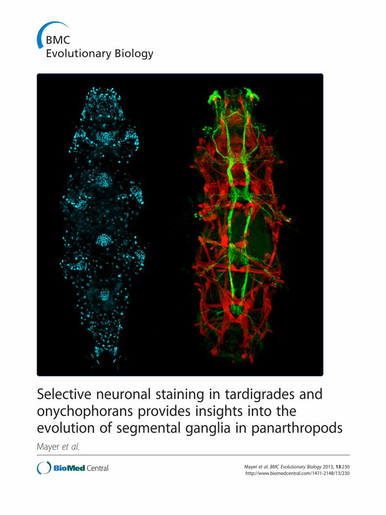
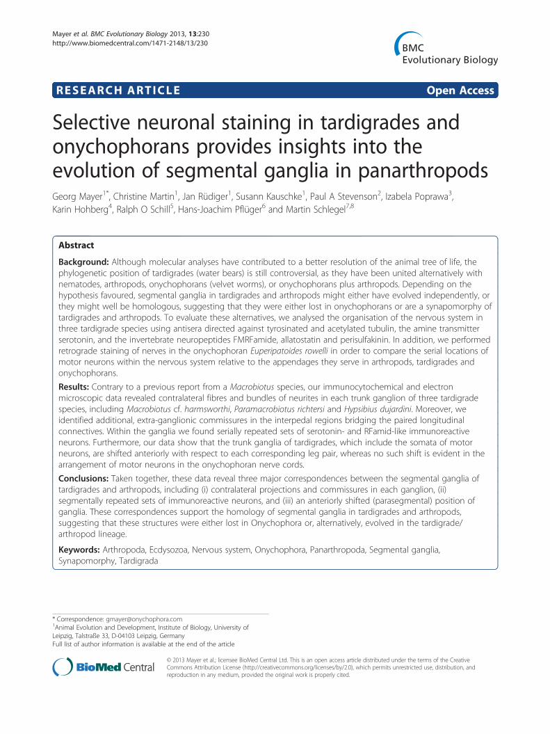
Selective neuronal staining in tardigrades andonychophorans provides insights into theevolution of segmental ganglia in panarthropodsMayer et al.
Mayer et al. BMC Evolutionary Biology 2013, 13:230http://www.biomedcentral.com/1471-2148/13/230
Mayer et al. BMC Evolutionary Biology 2013, 13:230http://www.biomedcentral.com/1471-2148/13/230
RESEARCH ARTICLE Open Access
Selective neuronal staining in tardigrades andonychophorans provides insights into theevolution of segmental ganglia in panarthropodsGeorg Mayer1*, Christine Martin1, Jan Rüdiger1, Susann Kauschke1, Paul A Stevenson2, Izabela Poprawa3,Karin Hohberg4, Ralph O Schill5, Hans-Joachim Pflüger6 and Martin Schlegel7,8
Abstract
Background: Although molecular analyses have contributed to a better resolution of the animal tree of life, thephylogenetic position of tardigrades (water bears) is still controversial, as they have been united alternatively withnematodes, arthropods, onychophorans (velvet worms), or onychophorans plus arthropods. Depending on thehypothesis favoured, segmental ganglia in tardigrades and arthropods might either have evolved independently, orthey might well be homologous, suggesting that they were either lost in onychophorans or are a synapomorphy oftardigrades and arthropods. To evaluate these alternatives, we analysed the organisation of the nervous system inthree tardigrade species using antisera directed against tyrosinated and acetylated tubulin, the amine transmitterserotonin, and the invertebrate neuropeptides FMRFamide, allatostatin and perisulfakinin. In addition, we performedretrograde staining of nerves in the onychophoran Euperipatoides rowelli in order to compare the serial locations ofmotor neurons within the nervous system relative to the appendages they serve in arthropods, tardigrades andonychophorans.
Results: Contrary to a previous report from a Macrobiotus species, our immunocytochemical and electronmicroscopic data revealed contralateral fibres and bundles of neurites in each trunk ganglion of three tardigradespecies, including Macrobiotus cf. harmsworthi, Paramacrobiotus richtersi and Hypsibius dujardini. Moreover, weidentified additional, extra-ganglionic commissures in the interpedal regions bridging the paired longitudinalconnectives. Within the ganglia we found serially repeated sets of serotonin- and RFamid-like immunoreactiveneurons. Furthermore, our data show that the trunk ganglia of tardigrades, which include the somata of motorneurons, are shifted anteriorly with respect to each corresponding leg pair, whereas no such shift is evident in thearrangement of motor neurons in the onychophoran nerve cords.
Conclusions: Taken together, these data reveal three major correspondences between the segmental ganglia oftardigrades and arthropods, including (i) contralateral projections and commissures in each ganglion, (ii)segmentally repeated sets of immunoreactive neurons, and (iii) an anteriorly shifted (parasegmental) position ofganglia. These correspondences support the homology of segmental ganglia in tardigrades and arthropods,suggesting that these structures were either lost in Onychophora or, alternatively, evolved in the tardigrade/arthropod lineage.
Keywords: Arthropoda, Ecdysozoa, Nervous system, Onychophora, Panarthropoda, Segmental ganglia,Synapomorphy, Tardigrada
* Correspondence: [email protected] Evolution and Development, Institute of Biology, University ofLeipzig, Talstraße 33, D-04103 Leipzig, GermanyFull list of author information is available at the end of the article
© 2013 Mayer et al.; licensee BioMed Central Ltd. This is an open access article distributed under the terms of the CreativeCommons Attribution License (http://creativecommons.org/licenses/by/2.0), which permits unrestricted use, distribution, andreproduction in any medium, provided the original work is properly cited.
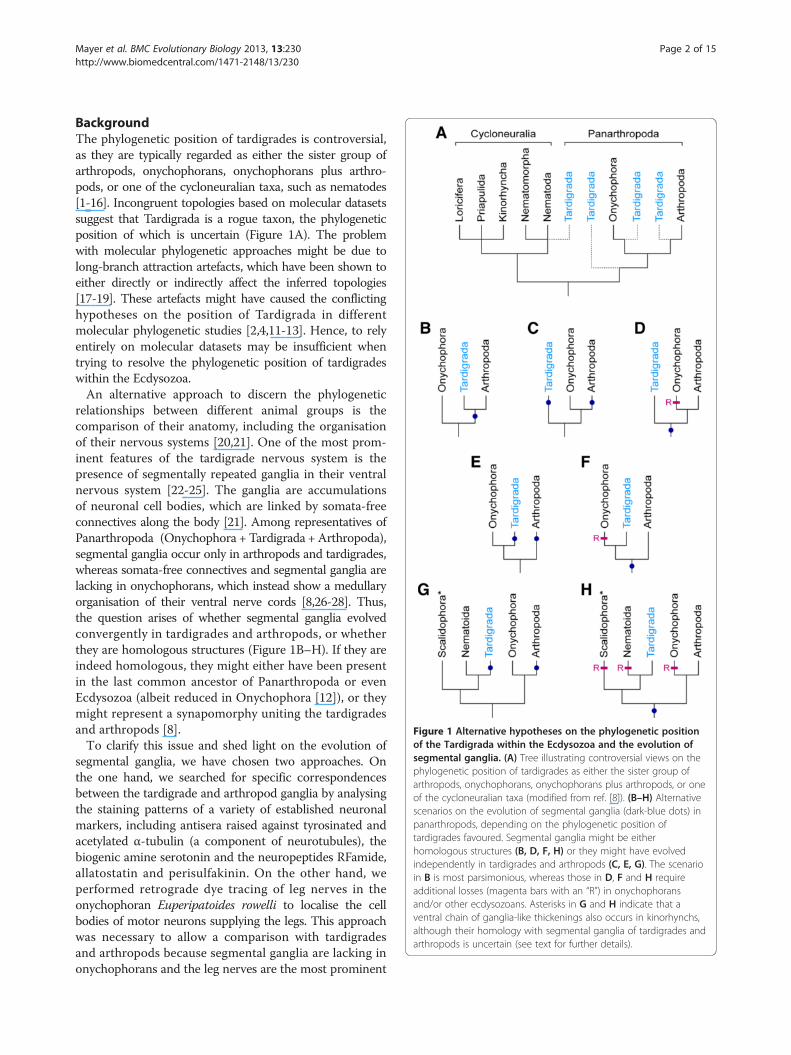
Figure 1 Alternative hypotheses on the phylogenetic positionof the Tardigrada within the Ecdysozoa and the evolution ofsegmental ganglia. (A) Tree illustrating controversial views on thephylogenetic position of tardigrades as either the sister group ofarthropods, onychophorans, onychophorans plus arthropods, or oneof the cycloneuralian taxa (modified from ref. [8]). (B–H) Alternativescenarios on the evolution of segmental ganglia (dark-blue dots) inpanarthropods, depending on the phylogenetic position oftardigrades favoured. Segmental ganglia might be eitherhomologous structures (B, D, F, H) or they might have evolvedindependently in tardigrades and arthropods (C, E, G). The scenarioin B is most parsimonious, whereas those in D, F and H requireadditional losses (magenta bars with an “R”) in onychophoransand/or other ecdysozoans. Asterisks in G and H indicate that aventral chain of ganglia-like thickenings also occurs in kinorhynchs,although their homology with segmental ganglia of tardigrades andarthropods is uncertain (see text for further details).
Mayer et al. BMC Evolutionary Biology 2013, 13:230 Page 2 of 15http://www.biomedcentral.com/1471-2148/13/230
BackgroundThe phylogenetic position of tardigrades is controversial,as they are typically regarded as either the sister group ofarthropods, onychophorans, onychophorans plus arthro-pods, or one of the cycloneuralian taxa, such as nematodes[1-16]. Incongruent topologies based on molecular datasetssuggest that Tardigrada is a rogue taxon, the phylogeneticposition of which is uncertain (Figure 1A). The problemwith molecular phylogenetic approaches might be due tolong-branch attraction artefacts, which have been shown toeither directly or indirectly affect the inferred topologies[17-19]. These artefacts might have caused the conflictinghypotheses on the position of Tardigrada in differentmolecular phylogenetic studies [2,4,11-13]. Hence, to relyentirely on molecular datasets may be insufficient whentrying to resolve the phylogenetic position of tardigradeswithin the Ecdysozoa.An alternative approach to discern the phylogenetic
relationships between different animal groups is thecomparison of their anatomy, including the organisationof their nervous systems [20,21]. One of the most prom-inent features of the tardigrade nervous system is thepresence of segmentally repeated ganglia in their ventralnervous system [22-25]. The ganglia are accumulationsof neuronal cell bodies, which are linked by somata-freeconnectives along the body [21]. Among representatives ofPanarthropoda (Onychophora + Tardigrada + Arthropoda),segmental ganglia occur only in arthropods and tardigrades,whereas somata-free connectives and segmental ganglia arelacking in onychophorans, which instead show a medullaryorganisation of their ventral nerve cords [8,26-28]. Thus,the question arises of whether segmental ganglia evolvedconvergently in tardigrades and arthropods, or whetherthey are homologous structures (Figure 1B–H). If they areindeed homologous, they might either have been presentin the last common ancestor of Panarthropoda or evenEcdysozoa (albeit reduced in Onychophora [12]), or theymight represent a synapomorphy uniting the tardigradesand arthropods [8].To clarify this issue and shed light on the evolution of
segmental ganglia, we have chosen two approaches. Onthe one hand, we searched for specific correspondencesbetween the tardigrade and arthropod ganglia by analysingthe staining patterns of a variety of established neuronalmarkers, including antisera raised against tyrosinated andacetylated α-tubulin (a component of neurotubules), thebiogenic amine serotonin and the neuropeptides RFamide,allatostatin and perisulfakinin. On the other hand, weperformed retrograde dye tracing of leg nerves in theonychophoran Euperipatoides rowelli to localise the cellbodies of motor neurons supplying the legs. This approachwas necessary to allow a comparison with tardigradesand arthropods because segmental ganglia are lacking inonychophorans and the leg nerves are the most prominent
Mayer et al. BMC Evolutionary Biology 2013, 13:230 Page 3 of 15http://www.biomedcentral.com/1471-2148/13/230
segmental structures associated with nerve cords in theseanimals [8,26-28].
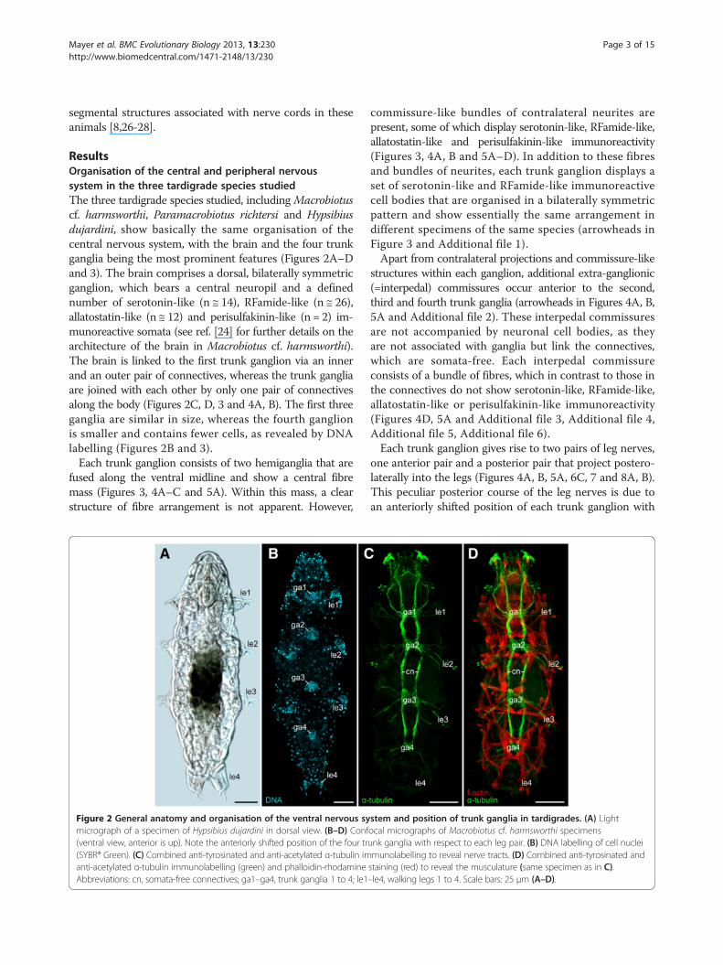
ResultsOrganisation of the central and peripheral nervoussystem in the three tardigrade species studiedThe three tardigrade species studied, includingMacrobiotuscf. harmsworthi, Paramacrobiotus richtersi and Hypsibiusdujardini, show basically the same organisation of thecentral nervous system, with the brain and the four trunkganglia being the most prominent features (Figures 2A–Dand 3). The brain comprises a dorsal, bilaterally symmetricganglion, which bears a central neuropil and a definednumber of serotonin-like (n ≅ 14), RFamide-like (n ≅ 26),allatostatin-like (n ≅ 12) and perisulfakinin-like (n = 2) im-munoreactive somata (see ref. [24] for further details on thearchitecture of the brain in Macrobiotus cf. harmsworthi).The brain is linked to the first trunk ganglion via an innerand an outer pair of connectives, whereas the trunk gangliaare joined with each other by only one pair of connectivesalong the body (Figures 2C, D, 3 and 4A, B). The first threeganglia are similar in size, whereas the fourth ganglionis smaller and contains fewer cells, as revealed by DNAlabelling (Figures 2B and 3).Each trunk ganglion consists of two hemiganglia that are
fused along the ventral midline and show a central fibremass (Figures 3, 4A–C and 5A). Within this mass, a clearstructure of fibre arrangement is not apparent. However,
Figure 2 General anatomy and organisation of the ventral nervous symicrograph of a specimen of Hypsibius dujardini in dorsal view. (B–D) Conf(ventral view, anterior is up). Note the anteriorly shifted position of the four tr(SYBR® Green). (C) Combined anti-tyrosinated and anti-acetylated α-tubulin imanti-acetylated α-tubulin immunolabelling (green) and phalloidin-rhodamineAbbreviations: cn, somata-free connectives; ga1–ga4, trunk ganglia 1 to 4; le1
commissure-like bundles of contralateral neurites arepresent, some of which display serotonin-like, RFamide-like,allatostatin-like and perisulfakinin-like immunoreactivity(Figures 3, 4A, B and 5A–D). In addition to these fibresand bundles of neurites, each trunk ganglion displays aset of serotonin-like and RFamide-like immunoreactivecell bodies that are organised in a bilaterally symmetricpattern and show essentially the same arrangement indifferent specimens of the same species (arrowheads inFigure 3 and Additional file 1).Apart from contralateral projections and commissure-like
structures within each ganglion, additional extra-ganglionic(=interpedal) commissures occur anterior to the second,third and fourth trunk ganglia (arrowheads in Figures 4A, B,5A and Additional file 2). These interpedal commissuresare not accompanied by neuronal cell bodies, as theyare not associated with ganglia but link the connectives,which are somata-free. Each interpedal commissureconsists of a bundle of fibres, which in contrast to those inthe connectives do not show serotonin-like, RFamide-like,allatostatin-like or perisulfakinin-like immunoreactivity(Figures 4D, 5A and Additional file 3, Additional file 4,Additional file 5, Additional file 6).Each trunk ganglion gives rise to two pairs of leg nerves,
one anterior pair and a posterior pair that project postero-laterally into the legs (Figures 4A, B, 5A, 6C, 7 and 8A, B).This peculiar posterior course of the leg nerves is due toan anteriorly shifted position of each trunk ganglion with
stem and position of trunk ganglia in tardigrades. (A) Lightocal micrographs of Macrobiotus cf. harmsworthi specimensunk ganglia with respect to each leg pair. (B) DNA labelling of cell nucleimunolabelling to reveal nerve tracts. (D) Combined anti-tyrosinated andstaining (red) to reveal the musculature (same specimen as in C).–le4, walking legs 1 to 4. Scale bars: 25 μm (A–D).
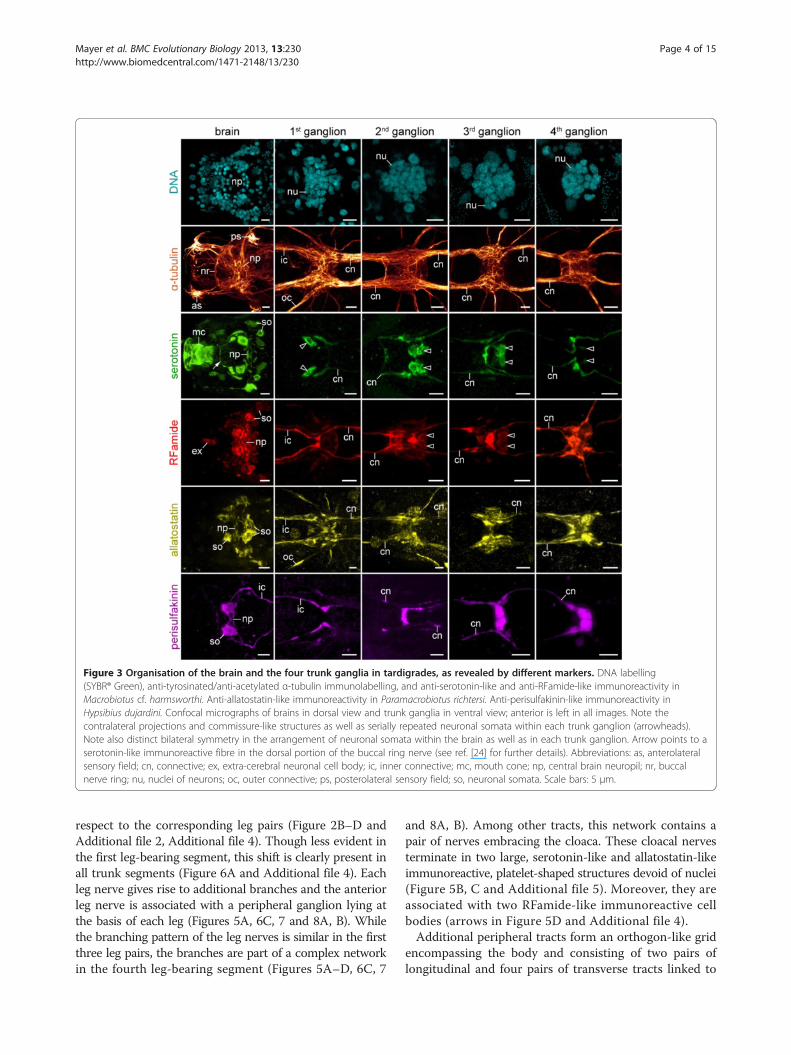
Figure 3 Organisation of the brain and the four trunk ganglia in tardigrades, as revealed by different markers. DNA labelling(SYBR® Green), anti-tyrosinated/anti-acetylated α-tubulin immunolabelling, and anti-serotonin-like and anti-RFamide-like immunoreactivity inMacrobiotus cf. harmsworthi. Anti-allatostatin-like immunoreactivity in Paramacrobiotus richtersi. Anti-perisulfakinin-like immunoreactivity inHypsibius dujardini. Confocal micrographs of brains in dorsal view and trunk ganglia in ventral view; anterior is left in all images. Note thecontralateral projections and commissure-like structures as well as serially repeated neuronal somata within each trunk ganglion (arrowheads).Note also distinct bilateral symmetry in the arrangement of neuronal somata within the brain as well as in each trunk ganglion. Arrow points to aserotonin-like immunoreactive fibre in the dorsal portion of the buccal ring nerve (see ref. [24] for further details). Abbreviations: as, anterolateralsensory field; cn, connective; ex, extra-cerebral neuronal cell body; ic, inner connective; mc, mouth cone; np, central brain neuropil; nr, buccalnerve ring; nu, nuclei of neurons; oc, outer connective; ps, posterolateral sensory field; so, neuronal somata. Scale bars: 5 μm.
Mayer et al. BMC Evolutionary Biology 2013, 13:230 Page 4 of 15http://www.biomedcentral.com/1471-2148/13/230
respect to the corresponding leg pairs (Figure 2B–D andAdditional file 2, Additional file 4). Though less evident inthe first leg-bearing segment, this shift is clearly present inall trunk segments (Figure 6A and Additional file 4). Eachleg nerve gives rise to additional branches and the anteriorleg nerve is associated with a peripheral ganglion lying atthe basis of each leg (Figures 5A, 6C, 7 and 8A, B). Whilethe branching pattern of the leg nerves is similar in the firstthree leg pairs, the branches are part of a complex networkin the fourth leg-bearing segment (Figures 5A–D, 6C, 7
and 8A, B). Among other tracts, this network contains apair of nerves embracing the cloaca. These cloacal nervesterminate in two large, serotonin-like and allatostatin-likeimmunoreactive, platelet-shaped structures devoid of nuclei(Figure 5B, C and Additional file 5). Moreover, they areassociated with two RFamide-like immunoreactive cellbodies (arrows in Figure 5D and Additional file 4).Additional peripheral tracts form an orthogon-like grid
encompassing the body and consisting of two pairs oflongitudinal and four pairs of transverse tracts linked to
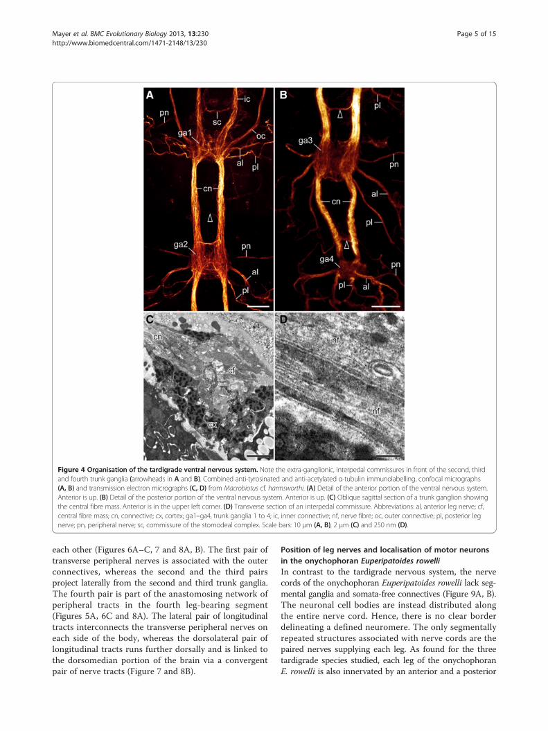
Figure 4 Organisation of the tardigrade ventral nervous system. Note the extra-ganglionic, interpedal commissures in front of the second, thirdand fourth trunk ganglia (arrowheads in A and B). Combined anti-tyrosinated and anti-acetylated α-tubulin immunolabelling, confocal micrographs(A, B) and transmission electron micrographs (C, D) from Macrobiotus cf. harmsworthi. (A) Detail of the anterior portion of the ventral nervous system.Anterior is up. (B) Detail of the posterior portion of the ventral nervous system. Anterior is up. (C) Oblique sagittal section of a trunk ganglion showingthe central fibre mass. Anterior is in the upper left corner. (D) Transverse section of an interpedal commissure. Abbreviations: al, anterior leg nerve; cf,central fibre mass; cn, connective; cx, cortex; ga1–ga4, trunk ganglia 1 to 4; ic, inner connective; nf, nerve fibre; oc, outer connective; pl, posterior legnerve; pn, peripheral nerve; sc, commissure of the stomodeal complex. Scale bars: 10 μm (A, B), 2 μm (C) and 250 nm (D).
Mayer et al. BMC Evolutionary Biology 2013, 13:230 Page 5 of 15http://www.biomedcentral.com/1471-2148/13/230
each other (Figures 6A–C, 7 and 8A, B). The first pair oftransverse peripheral nerves is associated with the outerconnectives, whereas the second and the third pairsproject laterally from the second and third trunk ganglia.The fourth pair is part of the anastomosing network ofperipheral tracts in the fourth leg-bearing segment(Figures 5A, 6C and 8A). The lateral pair of longitudinaltracts interconnects the transverse peripheral nerves oneach side of the body, whereas the dorsolateral pair oflongitudinal tracts runs further dorsally and is linked tothe dorsomedian portion of the brain via a convergentpair of nerve tracts (Figure 7 and 8B).
Position of leg nerves and localisation of motor neuronsin the onychophoran Euperipatoides rowelliIn contrast to the tardigrade nervous system, the nervecords of the onychophoran Euperipatoides rowelli lack seg-mental ganglia and somata-free connectives (Figure 9A, B).The neuronal cell bodies are instead distributed alongthe entire nerve cord. Hence, there is no clear borderdelineating a defined neuromere. The only segmentallyrepeated structures associated with nerve cords are thepaired nerves supplying each leg. As found for the threetardigrade species studied, each leg of the onychophoranE. rowelli is also innervated by an anterior and a posterior
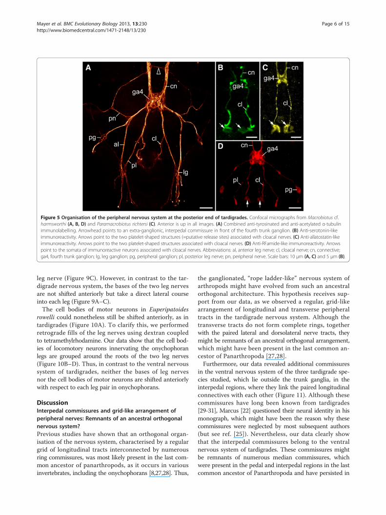
Figure 5 Organisation of the peripheral nervous system at the posterior end of tardigrades. Confocal micrographs from Macrobiotus cf.harmsworthi (A, B, D) and Paramacrobiotus richtersi (C). Anterior is up in all images. (A) Combined anti-tyrosinated and anti-acetylated α-tubulinimmunolabelling. Arrowhead points to an extra-ganglionic, interpedal commissure in front of the fourth trunk ganglion. (B) Anti-serotonin-likeimmunoreactivity. Arrows point to the two platelet-shaped structures (=putative release sites) associated with cloacal nerves. (C) Anti-allatostatin-likeimmunoreactivity. Arrows point to the two platelet-shaped structures associated with cloacal nerves. (D) Anti-RFamide-like immunoreactivity. Arrowspoint to the somata of immunoreactive neurons associated with cloacal nerves. Abbreviations: al, anterior leg nerve; cl, cloacal nerve; cn, connective;ga4, fourth trunk ganglion; lg, leg ganglion; pg, peripheral ganglion; pl, posterior leg nerve; pn, peripheral nerve. Scale bars: 10 μm (A, C) and 5 μm (B).
Mayer et al. BMC Evolutionary Biology 2013, 13:230 Page 6 of 15http://www.biomedcentral.com/1471-2148/13/230
leg nerve (Figure 9C). However, in contrast to the tar-digrade nervous system, the bases of the two leg nervesare not shifted anteriorly but take a direct lateral courseinto each leg (Figure 9A–C).The cell bodies of motor neurons in Euperipatoides
rowelli could nonetheless still be shifted anteriorly, as intardigrades (Figure 10A). To clarify this, we performedretrograde fills of the leg nerves using dextran coupledto tetramethylrhodamine. Our data show that the cell bod-ies of locomotory neurons innervating the onychophoranlegs are grouped around the roots of the two leg nerves(Figure 10B–D). Thus, in contrast to the ventral nervoussystem of tardigrades, neither the bases of leg nervesnor the cell bodies of motor neurons are shifted anteriorlywith respect to each leg pair in onychophorans.
DiscussionInterpedal commissures and grid-like arrangement ofperipheral nerves: Remnants of an ancestral orthogonalnervous system?Previous studies have shown that an orthogonal organ-isation of the nervous system, characterised by a regulargrid of longitudinal tracts interconnected by numerousring commissures, was most likely present in the last com-mon ancestor of panarthropods, as it occurs in variousinvertebrates, including the onychophorans [8,27,28]. Thus,
the ganglionated, “rope ladder-like” nervous system ofarthropods might have evolved from such an ancestralorthogonal architecture. This hypothesis receives sup-port from our data, as we observed a regular, grid-likearrangement of longitudinal and transverse peripheraltracts in the tardigrade nervous system. Although thetransverse tracts do not form complete rings, togetherwith the paired lateral and dorsolateral nerve tracts, theymight be remnants of an ancestral orthogonal arrangement,which might have been present in the last common an-cestor of Panarthropoda [27,28].Furthermore, our data revealed additional commissures
in the ventral nervous system of the three tardigrade spe-cies studied, which lie outside the trunk ganglia, in theinterpedal regions, where they link the paired longitudinalconnectives with each other (Figure 11). Although thesecommissures have long been known from tardigrades[29-31], Marcus [22] questioned their neural identity in hismonograph, which might have been the reason why thesecommissures were neglected by most subsequent authors(but see ref. [25]). Nevertheless, our data clearly showthat the interpedal commissures belong to the ventralnervous system of tardigrades. These commissures mightbe remnants of numerous median commissures, whichwere present in the pedal and interpedal regions in the lastcommon ancestor of Panarthropoda and have persisted in
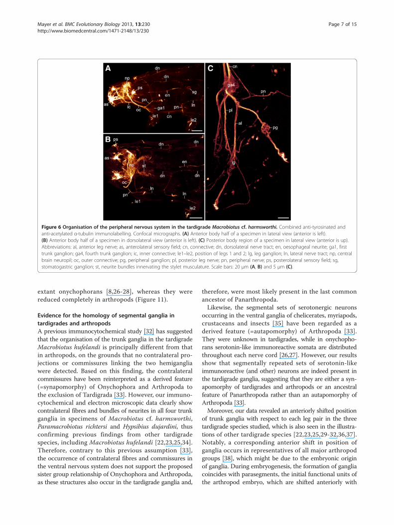
Figure 6 Organisation of the peripheral nervous system in the tardigrade Macrobiotus cf. harmsworthi. Combined anti-tyrosinated andanti-acetylated α-tubulin immunolabelling. Confocal micrographs. (A) Anterior body half of a specimen in lateral view (anterior is left).(B) Anterior body half of a specimen in dorsolateral view (anterior is left). (C) Posterior body region of a specimen in lateral view (anterior is up).Abbreviations: al, anterior leg nerve; as, anterolateral sensory field; cn, connective; dn, dorsolateral nerve tract; en, oesophageal neurite; ga1, firsttrunk ganglion; ga4, fourth trunk ganglion; ic, inner connective; le1–le2, position of legs 1 and 2; lg, leg ganglion; ln, lateral nerve tract; np, centralbrain neuropil; oc, outer connective; pg, peripheral ganglion; pl, posterior leg nerve; pn, peripheral nerve; ps, posterolateral sensory field; sg,stomatogastric ganglion; st, neurite bundles innervating the stylet musculature. Scale bars: 20 μm (A, B) and 5 μm (C).
Mayer et al. BMC Evolutionary Biology 2013, 13:230 Page 7 of 15http://www.biomedcentral.com/1471-2148/13/230
extant onychophorans [8,26-28], whereas they werereduced completely in arthropods (Figure 11).
Evidence for the homology of segmental ganglia intardigrades and arthropodsA previous immunocytochemical study [32] has suggestedthat the organisation of the trunk ganglia in the tardigradeMacrobiotus hufelandi is principally different from thatin arthropods, on the grounds that no contralateral pro-jections or commissures linking the two hemigangliawere detected. Based on this finding, the contralateralcommissures have been reinterpreted as a derived feature(=synapomorphy) of Onychophora and Arthropoda tothe exclusion of Tardigrada [33]. However, our immuno-cytochemical and electron microscopic data clearly showcontralateral fibres and bundles of neurites in all four trunkganglia in specimens of Macrobiotus cf. harmsworthi,Paramacrobiotus richtersi and Hypsibius dujardini, thusconfirming previous findings from other tardigradespecies, including Macrobiotus hufelandi [22,23,25,34].Therefore, contrary to this previous assumption [33],the occurrence of contralateral fibres and commissures inthe ventral nervous system does not support the proposedsister group relationship of Onychophora and Arthropoda,as these structures also occur in the tardigrade ganglia and,
therefore, were most likely present in the last commonancestor of Panarthropoda.Likewise, the segmental sets of serotonergic neurons
occurring in the ventral ganglia of chelicerates, myriapods,crustaceans and insects [35] have been regarded as aderived feature (=autapomorphy) of Arthropoda [33].They were unknown in tardigrades, while in onychopho-rans serotonin-like immunoreactive somata are distributedthroughout each nerve cord [26,27]. However, our resultsshow that segmentally repeated sets of serotonin-likeimmunoreactive (and other) neurons are indeed present inthe tardigrade ganglia, suggesting that they are either a syn-apomorphy of tardigrades and arthropods or an ancestralfeature of Panarthropoda rather than an autapomorphy ofArthropoda [33].Moreover, our data revealed an anteriorly shifted position
of trunk ganglia with respect to each leg pair in the threetardigrade species studied, which is also seen in the illustra-tions of other tardigrade species [22,23,25,29-32,36,37].Notably, a corresponding anterior shift in position ofganglia occurs in representatives of all major arthropodgroups [38], which might be due to the embryonic originof ganglia. During embryogenesis, the formation of gangliacoincides with parasegments, the initial functional units ofthe arthropod embryo, which are shifted anteriorly with
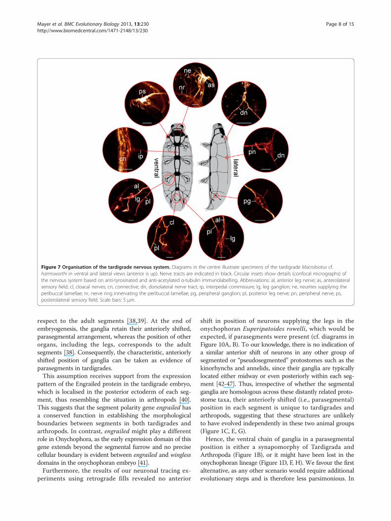
Figure 7 Organisation of the tardigrade nervous system. Diagrams in the centre illustrate specimens of the tardigrade Macrobiotus cf.harmsworthi in ventral and lateral views (anterior is up). Nerve tracts are indicated in black. Circular insets show details (confocal micrographs) ofthe nervous system based on anti-tyrosinated and anti-acetylated α-tubulin immunolabelling. Abbreviations: al, anterior leg nerve; as, anterolateralsensory field; cl, cloacal nerves; cn, connective; dn, dorsolateral nerve tract; ip, interpedal commissure; lg, leg ganglion; ne, neurites supplying theperibuccal lamellae; nr, nerve ring innervating the peribuccal lamellae; pg, peripheral ganglion; pl, posterior leg nerve; pn, peripheral nerve; ps,posterolateral sensory field. Scale bars: 5 μm.
Mayer et al. BMC Evolutionary Biology 2013, 13:230 Page 8 of 15http://www.biomedcentral.com/1471-2148/13/230
respect to the adult segments [38,39]. At the end ofembryogenesis, the ganglia retain their anteriorly shifted,parasegmental arrangement, whereas the position of otherorgans, including the legs, corresponds to the adultsegments [38]. Consequently, the characteristic, anteriorlyshifted position of ganglia can be taken as evidence ofparasegments in tardigrades.This assumption receives support from the expression
pattern of the Engrailed protein in the tardigrade embryo,which is localised in the posterior ectoderm of each seg-ment, thus resembling the situation in arthropods [40].This suggests that the segment polarity gene engrailed hasa conserved function in establishing the morphologicalboundaries between segments in both tardigrades andarthropods. In contrast, engrailed might play a differentrole in Onychophora, as the early expression domain of thisgene extends beyond the segmental furrow and no precisecellular boundary is evident between engrailed and winglessdomains in the onychophoran embryo [41].Furthermore, the results of our neuronal tracing ex-
periments using retrograde fills revealed no anterior
shift in position of neurons supplying the legs in theonychophoran Euperipatoides rowelli, which would beexpected, if parasegments were present (cf. diagrams inFigure 10A, B). To our knowledge, there is no indication ofa similar anterior shift of neurons in any other group ofsegmented or “pseudosegmented” protostomes such as thekinorhynchs and annelids, since their ganglia are typicallylocated either midway or even posteriorly within each seg-ment [42-47]. Thus, irrespective of whether the segmentalganglia are homologous across these distantly related proto-stome taxa, their anteriorly shifted (i.e., parasegmental)position in each segment is unique to tardigrades andarthropods, suggesting that these structures are unlikelyto have evolved independently in these two animal groups(Figure 1C, E, G).Hence, the ventral chain of ganglia in a parasegmental
position is either a synapomorphy of Tardigrada andArthropoda (Figure 1B), or it might have been lost in theonychophoran lineage (Figure 1D, F, H). We favour the firstalternative, as any other scenario would require additionalevolutionary steps and is therefore less parsimonious. In
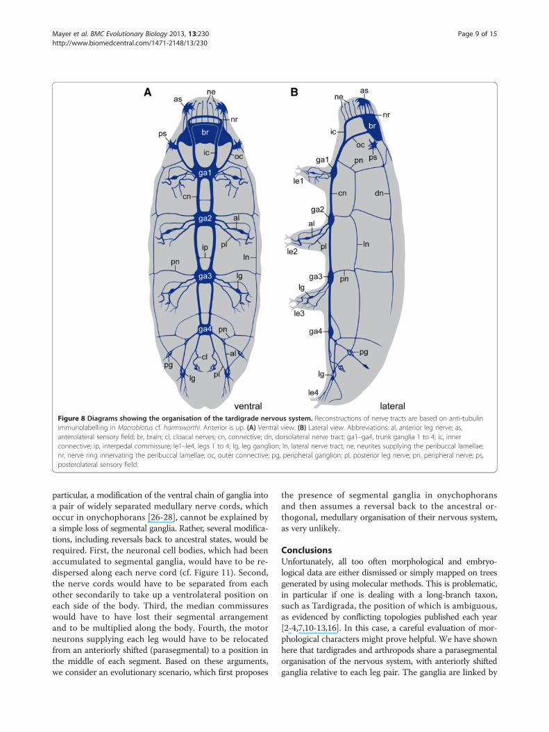
Figure 8 Diagrams showing the organisation of the tardigrade nervous system. Reconstructions of nerve tracts are based on anti-tubulinimmunolabelling in Macrobiotus cf. harmsworthi. Anterior is up. (A) Ventral view. (B) Lateral view. Abbreviations: al, anterior leg nerve; as,anterolateral sensory field; br, brain; cl, cloacal nerves; cn, connective; dn, dorsolateral nerve tract; ga1–ga4, trunk ganglia 1 to 4; ic, innerconnective; ip, interpedal commissure; le1–le4, legs 1 to 4; lg, leg ganglion; ln, lateral nerve tract; ne, neurites supplying the peribuccal lamellae;nr, nerve ring innervating the peribuccal lamellae; oc, outer connective; pg, peripheral ganglion; pl, posterior leg nerve; pn, peripheral nerve; ps,posterolateral sensory field.
Mayer et al. BMC Evolutionary Biology 2013, 13:230 Page 9 of 15http://www.biomedcentral.com/1471-2148/13/230
particular, a modification of the ventral chain of ganglia intoa pair of widely separated medullary nerve cords, whichoccur in onychophorans [26-28], cannot be explained bya simple loss of segmental ganglia. Rather, several modifica-tions, including reversals back to ancestral states, would berequired. First, the neuronal cell bodies, which had beenaccumulated to segmental ganglia, would have to be re-dispersed along each nerve cord (cf. Figure 11). Second,the nerve cords would have to be separated from eachother secondarily to take up a ventrolateral position oneach side of the body. Third, the median commissureswould have to have lost their segmental arrangementand to be multiplied along the body. Fourth, the motorneurons supplying each leg would have to be relocatedfrom an anteriorly shifted (parasegmental) to a position inthe middle of each segment. Based on these arguments,we consider an evolutionary scenario, which first proposes
the presence of segmental ganglia in onychophoransand then assumes a reversal back to the ancestral or-thogonal, medullary organisation of their nervous system,as very unlikely.
ConclusionsUnfortunately, all too often morphological and embryo-logical data are either dismissed or simply mapped on treesgenerated by using molecular methods. This is problematic,in particular if one is dealing with a long-branch taxon,such as Tardigrada, the position of which is ambiguous,as evidenced by conflicting topologies published each year[2-4,7,10-13,16]. In this case, a careful evaluation of mor-phological characters might prove helpful. We have shownhere that tardigrades and arthropods share a parasegmentalorganisation of the nervous system, with anteriorly shiftedganglia relative to each leg pair. The ganglia are linked by
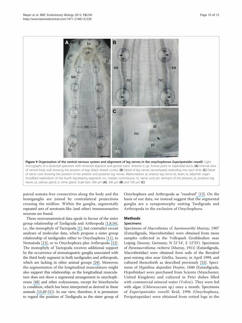
Figure 9 Organisation of the ventral nervous system and alignment of leg nerves in the onychophoran Euperipatoides rowelli. Lightmicrographs of a dissected specimen with removed digestive and genital tracts. Anterior is up. Arrows point to nephridial ducts. (A) Internal viewof ventral body wall showing the position of legs (black dotted circles). (B) Detail of leg nerves (arrowheads) extending into each limb. (C) Detailof nerve cord showing the position of the anterior and posterior leg nerves. Abbreviations: al, anterior leg nerve; br, brain; lo, labyrinth organ(modified nephridium of the fourth leg-bearing segment); mc, median commissure; nc, nerve cord; ph, remnant of the pharynx; pl, posterior legnerve; sa, salivary gland; sl, slime gland. Scale bars: 500 μm (A), 300 μm (B) and 100 μm (C).
Mayer et al. BMC Evolutionary Biology 2013, 13:230 Page 10 of 15http://www.biomedcentral.com/1471-2148/13/230
paired somata-free connectives along the body and thehemiganglia are joined by contralateral projectionscrossing the midline. Within the ganglia, segmentallyrepeated sets of serotonin-like (and other) immunoreactiveneurons are found.These neuroanatomical data speak in favour of the sister
group relationship of Tardigrada and Arthropoda [1,8,16],i.e., the monophyly of Tactopoda [1], but contradict recentanalyses of molecular data, which propose a sister grouprelationship of tardigrades either to Onychophora [11], toNematoda [13], or to Onychophora plus Arthropoda [12].The monophyly of Tactopoda receives additional supportby the occurrence of stomatogastric ganglia associated withthe third body segment in both tardigrades and arthropods,which are lacking in other animal groups [24]. Moreover,the segmentation of the longitudinal musculature mightalso support this relationship, as the longitudinal muscula-ture does not show a segmental arrangement in onychoph-orans [48] and other ecdysozoans, except for kinorhynchs(a condition, which has been interpreted as derived in theseanimals [15,49-51]). In our view, therefore, it is prematureto regard the position of Tardigrada as the sister group of
Onychophora and Arthropoda as “resolved” [12]. On thebasis of our data, we instead suggest that the segmentalganglia are a synapomorphy uniting Tardigrada andArthropoda to the exclusion of Onychophora.
MethodsSpecimensSpecimens of Macrobiotus cf. harmsworthi Murray, 1907(Eutardigrada, Macrobiotidae) were obtained from mosssamples collected in the Volkspark Großdeuben nearLeipzig (Saxony, Germany; N 51°14’, E 12°23’). Specimensof Paramacrobiotus richtersi (Murray, 1911) (Eutardigrada,Macrobiotidae) were obtained from soils of the Berzdorfpost-mining sites near Görlitz, Saxony, in April 1999, andcultured thenceforth as described previously [52]. Speci-mens of Hypsibius dujardini Doyère, 1840 (Eutardigrada,Hypsibiidae) were purchased from Sciento (Manchester,United Kingdom) and cultured in Petri dishes filledwith commercial mineral water (Volvic). They were fedwith algae (Chlorococcum sp.) once a month. Specimensof Euperipatoides rowelli Reid, 1996 (Onychophora,Peripatopsidae) were obtained from rotted logs in the
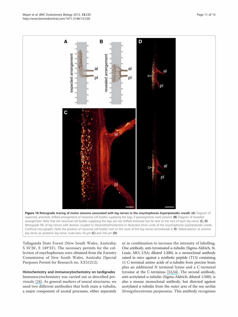
Figure 10 Retrograde tracing of motor neurons associated with leg nerves in the onychophoran Euperipatoides rowelli. (A) Diagram ofexpected, anteriorly shifted arrangement of neuronal cell bodies supplying the legs, if parasegments were present. (B) Diagram of revealedarrangement. Note that the neuronal cell bodies supplying the legs are not shifted anteriorly but lie next to the root of each leg nerve. (C, D)Retrograde fills of leg nerves with dextran coupled to tetramethylrhodamine in dissected nerve cords of the onychophoran Euperipatoides rowelli.Confocal micrographs. Note the position of neuronal cell bodies next to the roots of the leg nerves (arrowheads in D). Abbreviations: al, anteriorleg nerve; pl, posterior leg nerve. Scale bars: 50 μm (C) and 250 μm (D).
Mayer et al. BMC Evolutionary Biology 2013, 13:230 Page 11 of 15http://www.biomedcentral.com/1471-2148/13/230
Tallaganda State Forest (New South Wales, Australia;S 35°26', E 149°33'). The necessary permits for the col-lection of onychophorans were obtained from the ForestryCommission of New South Wales, Australia (SpecialPurposes Permit for Research no. XX51212).
Histochemistry and immunocytochemistry on tardigradesImmunocytochemistry was carried out as described pre-viously [24]. As general markers of neural structures, weused two different antibodies that both stain α-tubulin,a major component of axonal processes, either separately
or in combination to increase the intensity of labelling.One antibody, anti-tyrosinated α-tubulin (Sigma-Aldrich, St.Louis, MO, USA; diluted 1:200), is a monoclonal antibodyraised in mice against a synthetic peptide (T13) containing11 C-terminal amino acids of α-tubulin from porcine brainplus an additional N terminal lysine and a C-terminaltyrosine at the C-terminus [53,54]. The second antibody,anti-acetylated α-tubulin (Sigma-Aldrich; diluted 1:500), isalso a mouse monoclonal antibody, but directed againstacetylated α-tubulin from the outer arm of the sea urchinStrongylocentrotus purpuratus. This antibody recognises
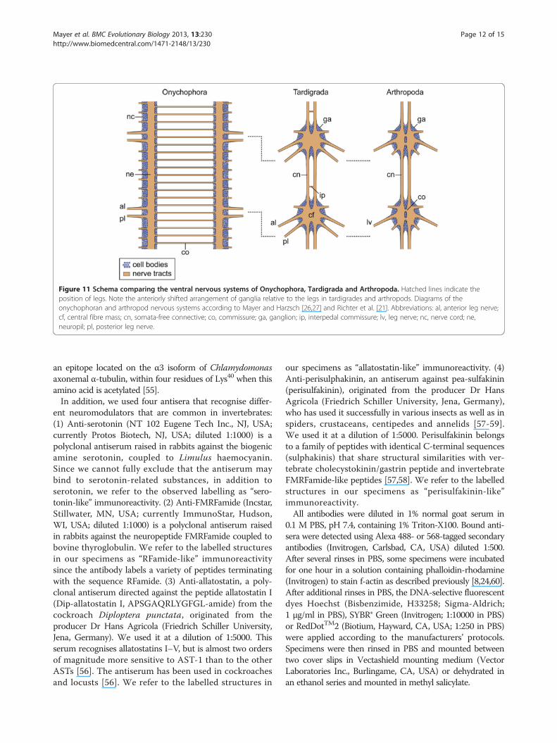
Figure 11 Schema comparing the ventral nervous systems of Onychophora, Tardigrada and Arthropoda. Hatched lines indicate theposition of legs. Note the anteriorly shifted arrangement of ganglia relative to the legs in tardigrades and arthropods. Diagrams of theonychophoran and arthropod nervous systems according to Mayer and Harzsch [26,27] and Richter et al. [21]. Abbreviations: al, anterior leg nerve;cf, central fibre mass; cn, somata-free connective; co, commissure; ga, ganglion; ip, interpedal commissure; lv, leg nerve; nc, nerve cord; ne,neuropil; pl, posterior leg nerve.
Mayer et al. BMC Evolutionary Biology 2013, 13:230 Page 12 of 15http://www.biomedcentral.com/1471-2148/13/230
an epitope located on the α3 isoform of Chlamydomonasaxonemal α-tubulin, within four residues of Lys40 when thisamino acid is acetylated [55].In addition, we used four antisera that recognise differ-
ent neuromodulators that are common in invertebrates:(1) Anti-serotonin (NT 102 Eugene Tech Inc., NJ, USA;currently Protos Biotech, NJ, USA; diluted 1:1000) is apolyclonal antiserum raised in rabbits against the biogenicamine serotonin, coupled to Limulus haemocyanin.Since we cannot fully exclude that the antiserum maybind to serotonin-related substances, in addition toserotonin, we refer to the observed labelling as “sero-tonin-like” immunoreactivity. (2) Anti-FMRFamide (Incstar,Stillwater, MN, USA; currently ImmunoStar, Hudson,WI, USA; diluted 1:1000) is a polyclonal antiserum raisedin rabbits against the neuropeptide FMRFamide coupled tobovine thyroglobulin. We refer to the labelled structuresin our specimens as “RFamide-like” immunoreactivitysince the antibody labels a variety of peptides terminatingwith the sequence RFamide. (3) Anti-allatostatin, a poly-clonal antiserum directed against the peptide allatostatin I(Dip-allatostatin I, APSGAQRLYGFGL-amide) from thecockroach Diploptera punctata, originated from theproducer Dr Hans Agricola (Friedrich Schiller University,Jena, Germany). We used it at a dilution of 1:5000. Thisserum recognises allatostatins I–V, but is almost two ordersof magnitude more sensitive to AST-1 than to the otherASTs [56]. The antiserum has been used in cockroachesand locusts [56]. We refer to the labelled structures in
our specimens as “allatostatin-like” immunoreactivity. (4)Anti-perisulphakinin, an antiserum against pea-sulfakinin(perisulfakinin), originated from the producer Dr HansAgricola (Friedrich Schiller University, Jena, Germany),who has used it successfully in various insects as well as inspiders, crustaceans, centipedes and annelids [57-59].We used it at a dilution of 1:5000. Perisulfakinin belongsto a family of peptides with identical C-terminal sequences(sulphakinis) that share structural similarities with ver-tebrate cholecystokinin/gastrin peptide and invertebrateFMRFamide-like peptides [57,58]. We refer to the labelledstructures in our specimens as “perisulfakinin-like”immunoreactivity.All antibodies were diluted in 1% normal goat serum in
0.1 M PBS, pH 7.4, containing 1% Triton-X100. Bound anti-sera were detected using Alexa 488- or 568-tagged secondaryantibodies (Invitrogen, Carlsbad, CA, USA) diluted 1:500.After several rinses in PBS, some specimens were incubatedfor one hour in a solution containing phalloidin-rhodamine(Invitrogen) to stain f-actin as described previously [8,24,60].After additional rinses in PBS, the DNA-selective fluorescentdyes Hoechst (Bisbenzimide, H33258; Sigma-Aldrich;1 μg/ml in PBS), SYBR® Green (Invitrogen; 1:10000 in PBS)or RedDotTM2 (Biotium, Hayward, CA, USA; 1:250 in PBS)were applied according to the manufacturers’ protocols.Specimens were then rinsed in PBS and mounted betweentwo cover slips in Vectashield mounting medium (VectorLaboratories Inc., Burlingame, CA, USA) or dehydrated inan ethanol series and mounted in methyl salicylate.
Mayer et al. BMC Evolutionary Biology 2013, 13:230 Page 13 of 15http://www.biomedcentral.com/1471-2148/13/230
Retrograde staining of onychophoran motor nervesFor neuronal tracing, the onychophoran nerve cords weredissected in physiological saline [61]. Retrograde fills of legnerves and ring commissures were carried out with dextrancoupled to tetramethylrhodamine as described previously[62,63]. Stained nerve cords were dehydrated through amethanol series and mounted between two cover slipsin methyl salicylate.
Transmission electron microscopy on tardigradesFor ultrastructural studies, specimens of the tardigradeMacrobiotus cf. harmsworthi were prepared using stand-ard methods as described previously [64] and embeddedin Epoxy Embedding Medium Kit (Sigma, St. Louis, MO).Ultra-thin sections were cut on a Leica Ultracut UCT25ultramicrotome (Leica Microsystems, Wetzlar, Germany),stained with uranyl acetate and lead citrate and examinedusing a Hitachi H500 transmission electron microscope(Hitachi Ltd, Tokyo, Japan) at 75 kV.
Confocal microscopy, light microscopy and image processingSpecimens were analysed with the confocal laser-scanning microscopes Zeiss LSM 510 META (Carl ZeissMicroImaging GmbH, Jena, Germany) and Leica TCSSTED (Leica Microsystems, Wetzlar, Germany). Confocalimage stacks were processed with Zeiss LSM IMAGEBROWSER v4.0.0.241 (Carl Zeiss MicroImaging GmbH)and Leica AS AF v2.3.5 (Leica Microsystems). The overallstructure of the onychophoran nervous system and theposition of leg nerves were analysed with a stereomicro-scope (Leica Wild M10) equipped with a colour digitalcamera (PCO AG SensiCam, Kelheim, Germany). Theoverall anatomy of tardigrades was analysed under atransmitted light microscope (Leica Leitz DMR; LeicaMicrosystems), equipped with a colour digital camera(PCO AG SensiCam). Several micrographs were taken fromeach specimen at different focal planes and merged to asingle projection using the Auto-Blend Layers function inAdobe (San Jose, CA, USA) Photoshop CS4. Final panelsand diagrams were produced using Adobe Illustrator CS4and exported to Tagged Image File Format files.
Animal ethicsThe experiments in this study did not require an approvalby an ethical committee. All procedures in this investiga-tion complied with international and institutional guide-lines, including the guidelines for animal welfare as laiddown by the German Research Foundation (DFG).
Additional files
Additional file 1: Organisation of the brain and the four trunkganglia in two specimens of the tardigrade Macrobiotus cf.harmsworthi. Anti-RFamide-like immunoreactivity. Confocal micrographs
of brains in dorsal view and trunk ganglia in ventral view; anterior is upin all images. Note the similar organisation of the brain and trunk gangliain the two specimens. Arrowheads point to the repeated somata ofRFamide-like immunoreactive neurons within the second and third trunkganglia. Abbreviations: cn, connective; ex, extra-cerebral neuronal cellbody; ic, inner connective; np, central brain neuropil; so, neuronal somata.Scale bars: 5 μm.
Additional file 2: Organisation of the ventral nervous system in thetardigrade Hypsibius dujardini. Note the extra-ganglionic, interpedalcommissures in front of the second, third and fourth trunk ganglia(arrowheads). Combined anti-tyrosinated and anti-acetylated α-tubulinimmunolabelling. Confocal micrographs; anterior is up. (A) Specimen inlateral view. (B) Specimen in ventral view. Abbreviations: al, anterior legnerve; cn, connective; ga2–ga4, trunk ganglia 2 to 4; lg, leg ganglion; pl,posterior leg nerve; pn, peripheral nerve. Scale bars: 10 μm.
Additional file 3: Organisation of the ventral nervous system in thetardigrade Macrobiotus cf. harmsworthi. Anti-RFamide-likeimmunoreactivity; confocal micrographs; anterior is up in all images. Notethe lack of signal in the interpedal commissures (asterisks). Arrowheads(in A and B) point to the repeated somata of RFamide-like immunoreactiveneurons within the second and third trunk ganglia. (A) Detail of the first andsecond trunk ganglia. (B) Detail of the third trunk ganglion. (C) Detail of thefourth trunk ganglion. Abbreviations: cn, connective; ga1–ga4, trunk ganglia1 to 4. Scale bars: 5 μm.
Additional file 4: Organisation of the ventral nervous system in thetardigrade Macrobiotus cf. harmsworthi. RFamide-likeimmunoreactivity (glow mode) and DNA labelling (light blue); confocalmicrographs; anterior is up in all images. (A) Entire specimen in ventralview. Asterisks indicate the position of the interpedal commissures. (B–D)Details of the posterior end showing the position of two RFamide-likeimmunoreactive somata associated with cloacal nerves (arrows).Abbreviations: cl, cloacal nerve; cn, connectives; ga1–ga4, trunk ganglia 1to 4; ic, inner connective; le1–le4, walking legs 1 to 4; pg, peripheralganglion; pn, peripheral nerve. Scale bars: 20 μm (A) and 10 μm (B–D).
Additional file 5: Organisation of the ventral nervous system at theposterior end in the tardigrade Macrobiotus cf. harmsworthi.Serotonin-like immunoreactivity (glow mode) and DNA labelling(light blue); confocal micrographs. Arrowheads point to the somata ofserotonin-like immunoreactive neurons. (A) Specimen in ventral view; anterioris up. Note the absence of signal in the interpedal commissure (asterisk). (B–D)Details of the posterior end showing the position of two platelet-shaped,serotonin-like immunoreactive structures (=putative release cites) associatedwith cloacal nerves (arrows). Abbreviations: cl, cloacal nerve; cn, connectives;ga3–ga4, trunk ganglia 3 to 4. Scale bars: 10 μm (A–D).
Additional file 6: Anti-allatostatin immunolabelling in thetardigrade Hypsibius dujardini. Confocal micrographs; anterior is up inall images. Note the lack of signal in the interpedal commissures(asterisks; cf. Additional file 2). Arrowheads (in C) indicated the position ofthe two platelet-shaped structures associated with cloacal nerves. (A)Details of the brain in dorsal view. (B) Detail of the first and second trunkganglia in ventral view. (C) Detail of the third and fourth trunk ganglia.Abbreviations: cn, connective; ga1–ga4, trunk ganglia 1 to 4; ic, innerconnective; so, somata of allatostatin-like immunoreactive neurons. Scalebars: 10 μm.
Competing interestsThe authors declare that they have no competing interests.
Authors’ contributionsGM conceived and designed the experiments, analysed the data and wrotethe first draft of the manuscript. CM, JR, SK and IP performed theexperiments. CM, JR, SK, PAS, IP, KH, ROS, HJP and MS participated in dataanalysis and helped to draft the manuscript. All authors read, madecomments on and approved the final manuscript.
AcknowledgementsWe are thankful to Andreas Hejnol for providing details on the anti-tyrosinatedα-tubulin antibody and to Noel Tait for his help with the collecting permits. Thestaff members of the State Forests NSW (New South Wales, Australia) are
Mayer et al. BMC Evolutionary Biology 2013, 13:230 Page 14 of 15http://www.biomedcentral.com/1471-2148/13/230
gratefully acknowledged for providing the collecting permits. This work wassupported by a grant from the German Research Foundation (DFG; grant Ma4147/3-1) to GM, who is a Research Group Leader supported by the EmmyNoether Programme of the DFG.
Author details1Animal Evolution and Development, Institute of Biology, University ofLeipzig, Talstraße 33, D-04103 Leipzig, Germany. 2Physiology of Animals andBehavior, Institute of Biology, University of Leipzig, Talstraße 33, D-04103Leipzig, Germany. 3Department of Animal Histology and Embryology,University of Silesia, Bankowa 9, 40-007 Katowice, Poland. 4SenckenbergMuseum of Natural History Görlitz, Am Museum 1, D-02826 Görlitz, Germany.5Biological Institute, Zoology, University of Stuttgart, Pfaffenwaldring 57,D-70569 Stuttgart, Germany. 6Neurobiology, Institute of Biology, FreieUniversität Berlin, Königin-Luise-Str. 28-30, D-14195 Berlin, Germany.7Molecular Evolution & Animal Systematics, Institute of Biology, University ofLeipzig, Talstraße 33, D-04103 Leipzig, Germany. 8German Centre forIntegrative Biodiversity Research (iDiv), Halle-Jena-Leipzig, Deutscher Platz 5e,D-04103 Leipzig, Germany.
Received: 14 August 2013 Accepted: 16 October 2013Published: 24 October 2013
References1. Budd GE: Tardigrades as ’stem-group arthropods’: the evidence from the
Cambrian fauna. Zool Anz 2001, 240:265–279.2. Mallatt J, Giribet G: Further use of nearly complete 28S and 18S rRNA
genes to classify Ecdysozoa: 37 more arthropods and a kinorhynch.Mol Biol Evol 2006, 40:772–794.
3. Roeding F, Hagner-Holler S, Ruhberg H, Ebersberger I, von Haeseler A, KubeM, Reinhardt R, Burmester T: EST sequencing of Onychophora andphylogenomic analysis of Metazoa. Mol Phylogenet Evol 2007, 45:942–951.
4. Dunn CW, Hejnol A, Matus DQ, Pang K, Browne WE, Smith SA, Seaver E,Rouse GW, Obst M, Edgecombe GD, et al: Broad phylogenomic samplingimproves resolution of the animal tree of life. Nature 2008, 452:745–749.
5. Lartillot N, Philippe H: Improvement of molecular phylogenetic inference andthe phylogeny of Bilateria. Philos Trans R Soc B Biol Sci 2008, 363:1463–1472.
6. Telford MJ, Bourlat SJ, Economou A, Papillon D, Rota-Stabelli O: The evolutionof the Ecdysozoa. Philos Trans R Soc B Biol Sci 2008, 363:1529–1537.
7. Hejnol A, Obst M, Stamatakis A, Ott M, Rouse GW, Edgecombe GD, MartinezP, Baguñà J, Bailly X, Jondelius U, et al: Assessing the root of bilateriananimals with scalable phylogenomic methods. Proc R Soc B Biol Sci 2009,276:4261–4270.
8. Mayer G, Whitington PM: Neural development in Onychophora (velvet worms)suggests a step-wise evolution of segmentation in the nervous system ofPanarthropoda. Dev Biol 2009, 335:263–275.
9. Roeding F, Borner J, Kube M, Klages S, Reinhardt R, Burmester T: A 454sequencing approach for large scale phylogenomic analysis of thecommon emperor scorpion (Pandinus imperator). Mol Phylogenet Evol2009, 53:826–834.
10. Meusemann K, von Reumont BM, Simon S, Roeding F, Strauss S, Kück P,Ebersberger I, Walzl M, Pass G, Breuers S, et al: A phylogenomic approachto resolve the arthropod tree of life. Mol Biol Evol 2010, 27:2451–2464.
11. Rota-Stabelli O, Kayal E, Gleeson D, Daub J, Boore J, Telford M, Pisani D, Blaxter M,Lavrov D: Ecdysozoan mitogenomics: evidence for a common origin of thelegged invertebrates, the Panarthropoda. Genome Biol Evol 2010, 2:425–440.
12. Campbell LI, Rota-Stabelli O, Edgecombe GD, Marchioroc T, Longhorna SJ,Telford MJ, Philippe H, Rebecchi L, Peterson KJ, Pisani D: MicroRNAs andphylogenomics resolve the relationships of Tardigrada and suggest thatvelvet worms are the sister group of Arthropoda. Proc Natl Acad Sci U S A2011, 108:15920–15924.
13. Rehm P, Borner J, Meusemann K, von Reumont BM, Simon S, Hadrys H,Misof B, Burmester T: Dating the arthropod tree based on large-scaletranscriptome data. Mol Phylogenet Evol 2011, 61:880–887.
14. Giribet G, Edgecombe G: Reevaluating the arthropod tree of life. Annu RevEntomol 2012, 57:167–186.
15. Nielsen C: Animal Evolution: Interrelationships of the Living Phyla. Oxford:Oxford University Press; 2012.
16. Arango CP, Wheeler WC: Phylogeny of the sea spiders (Arthropoda,Pycnogonida) based on direct optimization of six loci andmorphology. Cladistics 2007, 23:1–39.
17. Bergsten J: A review of long-branch attraction. Cladistics 2005, 21:163–193.18. Wägele JW, Mayer C: Visualizing differences in phylogenetic information
content of alignments and distinction of three classes of long-brancheffects. BMC Evol Biol 2007, 7:147.
19. Kück P, Mayer C, Wägele J-W, Misof B: Long branch effects distort maximumlikelihood phylogenies in simulations despite selection of the correctmodel. PLoS ONE 2012, 7(5):e36593.
20. Harzsch S: Neurophylogeny: architecture of the nervous system and afresh view on arthropod phyologeny. Integr Comp Biol 2006, 46:162–194.
21. Richter S, Loesel R, Purschke G, Schmidt-Rhaesa A, Scholtz G, Stach T, VogtL, Wanninger A, Brenneis G, Döring C, et al: Invertebrate neurophylogeny:suggested terms and definitions for a neuroanatomical glossary.Front Zool 2010, 7:29.
22. Marcus E: Tardigrada. In Dr H G Bronns Klassen und Ordnungen des Tier-Reichswissenschaftlich dargestellt in Wort und Bild, Volume 5. Leipzig: AkademischeVerlagsgesellschaft; 1929:1–609.
23. Persson DK, Halberg KA, Jørgensen A, Møbjerg N, Kristensen RM: Neuroanatomyof Halobiotus crispae (Eutardigrada: Hypsibiidae): tardigrade brain structuresupports the clade Panarthropoda. J Morphol 2012, 273:1227–1245.
24. Mayer G, Kauschke S, Rüdiger J, Stevenson PA: Neural markers reveal aone-segmented head in tardigrades (water bears). PLoS ONE 2013,8(3):e59090.
25. Schulze C, Schmidt-Rhaesa A: The architecture of the nervous system ofEchiniscus testudo (Echiniscoidea, Heterotardigrada). J Limnol 2013, 72:44–53.
26. Mayer G, Harzsch S: Immunolocalization of serotonin in Onychophoraargues against segmental ganglia being an ancestral feature ofarthropods. BMC Evol Biol 2007, 7:118.
27. Mayer G, Harzsch S: Distribution of serotonin in the trunk of Metaperipatusblainvillei (Onychophora, Peripatopsidae): implications for the evolution ofthe nervous system in Arthropoda. J Comp Neurol 2008, 507:1196–1208.
28. Whitington PM, Mayer G: The origins of the arthropod nervous system:insights from the Onychophora. Arthropod Struct Dev 2011, 40:193–209.
29. Doyère M: Mémoire sur les Tardigrades. Ann Sci Nat (Paris), Zoo 1840,14(2):269–361.
30. Greeff R: Ueber das Nervensystem der Bärthierchen, Arctiscoida C.A.S.Schultze (Tardigraden Doyère) mit besonderer Berücksichtigung derMuskelnerven und deren Endigungen. Arch Mikrosk Anat 1865, 1:101–123.
31. Plate L: Beiträge zur Naturgeschichte der Tardigraden. Zool Jahrb AbtAnat Ontog Tiere 1889, 3:487–550.
32. Zantke J, Wolff C, Scholtz G: Three-dimensional reconstruction of thecentral nervous system of Macrobiotus hufelandi (Eutardigrada,Parachela): implications for the phylogenetic position of Tardigrada.Zoomorphology 2008, 127:21–36.
33. Edgecombe GD: Palaeontological and molecular evidence linking arthropods,onychophorans, and other Ecdysozoa. Evo Edu Outreach 2009, 2:178–190.
34. Greven H, Kuhlmann D: Die Struktur des Nervengewebes von Macrobiotushufelandi C.A.S. Schultze (Tardigrada). Z Zellforsch 1972, 132:131–146.
35. Harzsch S: Phylogenetic comparison of serotonin-immunoreactiveneurons in representatives of the Chilopoda, Diplopoda, and Chelicerata:implications for arthropod relationships. J Morphol 2004, 259:198–213.
36. Kristensen RM, Higgins RP: A new family of Arthrotardigrada (Tardigrada:Heterotardigrada) from the Atlantic Coast of Florida. U S A Trans AmMicrosc Soc 1984, 103:295–311.
37. Kristensen RM, Higgins RP: Revision of Styraconyx (Tardigrada: Halechiniscidae)with descriptions of two new species from Disko Bay, West Greenland.Smithson Contrib Zool 1984, 391:1–40.
38. Deutsch JS: Segments and parasegments in arthropods: a functionalperspective. BioEssays 2004, 26:1117–1125.
39. Damen WGM: Evolutionary conservation and divergence of thesegmentation process in arthropods. Dev Dyn 2007, 236:1379–1391.
40. Gabriel WN, Goldstein B: Segmental expression of Pax3/7 and Engrailedhomologs in tardigrade development. Dev Genes Evol 2007, 217:421–433.
41. Eriksson BJ, Tait NN, Budd GE, Akam M: The involvement of engrailed andwingless during segmentation in the onychophoran Euperipatoideskanangrensis (Peripatopsidae: Onychophora) (Reid 1996). Dev Genes Evol2009, 219:249–264.
42. Bullock TH: Annelida. In Structure and Function in the Nervous Systems ofInvertebrates, Volume I. Edited by Bullock TH, Horridge GA. San Francisco,California: W.H. Freeman Company; 1965:661–789.
43. Bullock TH: Pseudocoelomate phyla: Acanthocephala, Rotifera,Gastrotricha, Kinorhyncha, Nematoda, Nematomorpha, and Entoprocta.
Mayer et al. BMC Evolutionary Biology 2013, 13:230 Page 15 of 15http://www.biomedcentral.com/1471-2148/13/230
In Structure and Function in the Nervous Systems of Invertebrates, Volume I.Edited by Bullock TH, Horridge GA. San Francisco, California: W.H. FreemanCompany; 1965:597–629.
44. Kristensen RM, Higgins RP: Kinorhyncha. In Microscopic Anatomy ofInvertebrates. Volume 4. Aschelminthes. Edited by Harrison FW, Ruppert EE.New York: Wiley-Liss; 1991:377–404.
45. Nebelsick M: Introvert, mouth cone, and nervous system of Echinoderescapitatus (Kinorhyncha, Cyclorhagida) and implications for the phylogeneticrelationships of Kinorhyncha. Zoomorphology 1993, 113:211–232.
46. Scholtz G: The Articulata hypothesis — or what is a segment? Org DiversEvol 2002, 2:197–215.
47. Orrhage L, Müller MCM: Morphology of the nervous system of Polychaeta(Annelida). Hydrobiologia 2005, 535/536:79–111.
48. Hoyle G, Williams M: The musculature of Peripatus and its innervation.Philos Trans R Soc B Biol Sci 1980, 288:481–510.
49. Müller MCM, Schmidt-Rhaesa A: Reconstruction of the muscle system inAntygomonas sp. (Kinorhyncha, Cyclorhagida) by means of phalloidinlabeling and cLSM. J Morphol 2003, 256:103–110.
50. Rothe BH, Schmidt-Rhaesa A: Probable development from continuous tosegmental longitudinal musculature in Pycnophyes kielensis(Kinorhyncha, Homalorhagida). Meiofauna Mar 2004, 13:21–28.
51. Schmidt-Rhaesa A, Rothe H: Postembryonic development of dorsoventraland longitudinal musculature in Pycnophyes kielensis (Kinorhyncha,Homalorhagida). Integr Comp Biol 2006, 46:144–150.
52. Hohberg K: Tardigrade species composition in young soils and someaspects on life history of Macrobiotus richtersi J. Murray, 1911.Pedobiologia 2006, 50:267–274.
53. Kreis TE: Microtubules containing detyrosinated tubulin are less dynamic.EMBO J 1987, 6:2597–2606.
54. Siddiqui SS, Aamodt E, Rastinejad F, Culotti J: Anti-tubulin monoclonalantibodies that bind to specific neurons in Caenorhabditis elegans.J Neurosci 1989, 9:2963–2972.
55. LeDizet M, Piperno G: Identification of an acetylation site ofChlamydomonas α-tubulin. Proc Natl Acad Sci U S A 1987, 84:5720–5724.
56. Vitzthum H, Homberg U, Agricola H: Distribution of Dip-allatostatin I-likeImmunoreactivity in the brain of the locust Schistocerca gregaria withdetailed analysis of immunostaining in the central complex.J Comp Neurol 1996, 369:419–437.
57. Agricola HJ, Bräunig P: Comparative aspects of peptidergic signalingpathways in the nervous systems of arthropods. In The Nervous Systems ofInvertebrates: An Evolutionary and Comparative Approach. Edited byBreidbach O, Kutsch W. Basel: Birkhäuser Verlag; 1995:303–327.
58. Utting M, Agricola H-J, Sandeman R, Sandeman D: Central complex in thebrain of crayfish and its possible homology with that of insects.J Comp Neurol 2000, 416:245–261.
59. Loesel R, Seyfarth E-A, Bräunig P, Agricola H-J: Neuroarchitecture of the arcuatebody in the brain of the spider Cupiennius salei (Araneae, Chelicerata)revealed by allatostatin-, proctolin-, and CCAP-immunocytochemistry and itsevolutionary implications. Arthropod Struct Dev 2011, 40:210–220.
60. Mayer G, Whitington PM: Velvet worm development links myriapods withchelicerates. Proc R Soc B Biol Sci 2009, 276:3571–3579.
61. Robson EA, Lockwood APM, Ralph R: Composition of the blood inOnychophora. Nature 1966, 209:533.
62. Mayer G, Whitington PM, Sunnucks P, Pflüger H-J: A revision of braincomposition in Onychophora (velvet worms) suggests that thetritocerebrum evolved in arthropods. BMC Evol Biol 2010, 10:255.
63. Pflüger HJ, Field LH: A locust chordotonal organ coding forproprioceptive and acoustic stimuli. J Comp Physiol A 1999, 184:169–183.
64. Poprawa I: Ultrastructural studies of the formation of the egg capsule inthe hermaphroditic species, Isohypsibius granulifer granulifer Thulin, 1928(Eutardigrada: Hypsibiidae). Zool Sci 2011, 28:37–40.
doi:10.1186/1471-2148-13-230Cite this article as: Mayer et al.: Selective neuronal staining intardigrades and onychophorans provides insights into the evolution ofsegmental ganglia in panarthropods. BMC Evolutionary Biology2013 13:230.
Submit your next manuscript to BioMed Centraland take full advantage of:
• Convenient online submission
• Thorough peer review
• No space constraints or color figure charges
• Immediate publication on acceptance
• Inclusion in PubMed, CAS, Scopus and Google Scholar
• Research which is freely available for redistribution
Submit your manuscript at www.biomedcentral.com/submit