The new mutation, E46K, of ?-synuclein causes parkinson and Lewy body dementia
Upload
independentCategory
view
4download
0
Selective Insolubility of �-Synuclein in Human LewyBody Diseases Is Recapitulated in a TransgenicMouse Model
Philipp J. Kahle,* Manuela Neumann,†
Laurence Ozmen,‡ Veronika Muller,*Sabine Odoy,* Noriko Okamoto,*Helmut Jacobsen,‡ Takeshi Iwatsubo,§
John Q. Trojanowski,¶ Hitoshi Takahashi,�
Koichi Wakabayashi,** Nenad Bogdanovic,††
Peter Riederer,‡‡ Hans A. Kretzschmar,† andChristian Haass*From the Laboratory for Alzheimer’s and Parkinson’s Disease
Research,* the Department of Biochemistry and the Department
of Neuropathology,† Ludwig Maximilians University, Munich,
Germany; Pharma Research Genomics,‡ F. Hoffmann–La Roche
Ltd., Basel, Switzerland; the Department of Neuropathology and
Neuroscience,§ Graduate School of Pharmaceutical Sciences,
University of Tokyo, Tokyo, Japan; the Department of Pathology
and Laboratory Medicine,¶ Center for Neurodegenerative Disease
Research, University of Pennsylvania, School of Medicine,
Philadelphia, Pennsylvania; the Department of Pathology,� Brain
Research Institute, Niigata University, Niigata, Japan; the
Department of Neuropathology,** Institute of Brain Science,
Hirosaki University School of Medicine, Hirosaki, Japan; the
Department of Clinical Neuroscience,†† Geriatric Section,
Huddinge Brain Bank, Huddinge, Sweden; and the Department
of Psychiatry,‡‡ Clinical Neurochemistry and The National
Parkinson Foundation Center of Excellence Research
Laboratories, Julius Maximilians University, Wurzburg, Germany
�-Synuclein (�-SYN) is deposited in intraneuronal cy-toplasmic inclusions (Lewy bodies, LBs) characteris-tic for Parkinson’s disease (PD) and LB dementias.�-SYN forms LB-like fibrils in vitro , in contrast to itshomologue �-SYN. Here we have investigated the sol-ubility of SYNs in human LB diseases and in trans-genic mice expressing human wild-type and PD-asso-ciated mutant [A30P]�-SYN driven by the brainneuron-specific promoter, Thy1. Distinct �-SYN spe-cies were detected in the detergent-insoluble fractionsfrom brains of patients with PD, dementia with LBs,and neurodegeneration with brain iron accumulationtype 1 (formerly known as Hallervorden-Spatz dis-ease). Using the same extraction method, detergent-insolubility of human �-SYN was observed in brainsof transgenic mice. In contrast, neither endogenousmouse �-SYN nor �-SYN were detected in detergent-insoluble fractions from transgenic mouse brains.
The nonamyloidogenic �-SYN was incapable of form-ing insoluble fibrils because amino acids 73 to 83 inthe central region of �-SYN are absent in �-SYN. Inconclusion, the specific accumulation of detergent-insoluble �-SYN in transgenic mice recapitulates apivotal feature of human LB diseases. (Am J Pathol2001, 159:2215–2225)
�-Synuclein (�-SYN) has been identified as the precursorprotein of a nonamyloid �-protein component (NAC) iso-lated from Alzheimer’s disease plaques.1 �-SYN was de-tected immunohistochemically in Lewy bodies (LBs) andLewy neurites that characterize Parkinson’s disease (PD),LB dementia (DLB), LB variant Alzheimer’s disease,2–7
and neurodegeneration with brain iron accumulation type1 (NBIA1).8–11 Antibodies directed against both N-termi-nal and C-terminal epitopes recognized LB fila-ments,12,13 and the presence of full-length �-SYN wasbiochemically proven on Western blots of isolated LBs.6
Moreover, full-length �-SYN is the major fibrillar compo-nent of glial cytoplasmic inclusions in multiple systematrophy.14
The formation of LB-like fibrils is an intrinsic property of�-SYN. Purified recombinant �-SYN, but not �-SYN, ag-gregated in vitro to amyloid fibrils resembling those ex-tracted from LBs.15–19 PD risk factors, namely �-SYNmutations20–22 and oxidative stress,23 accelerated�-SYN aggregation. The causal relationship between�-SYN fibrillization and PD are therefore subject to in-tense research.24
Transgenic animals expressing human wild-type [wt]-as well as PD-associated mutant [A53T]�-SYN25 and[A30P]�-SYN26 were recently presented. Wild-type andmutant �-SYN assembled into LB-like fibrils in transgenicDrosophila, and a locomotor deficit became apparentwith increasing age.27 Somal and neuritic accumulationsof wt and mutant �-SYN were observed in transgenicmouse brain.28–30 Ubiquitination was occasionally de-
Supported by grants from the Deutsche Forschungsgemeinschaft (HA1737/4-1) and the Bavaria California Technology Center (to C. H.).
Accepted for publication September 10, 2001.
Address reprint requests to Philipp Kahle or Christian Haass, Laboratoryfor Alzheimer’s and Parkinson’s Disease Research, Department of Biochem-istry, Ludwig Maximilians University of Munich, Schillerstrasse 44, D-80336Munich, Germany. E-mail: [email protected] or [email protected].
American Journal of Pathology, Vol. 159, No. 6, December 2001
Copyright © American Society for Investigative Pathology
2215

tected, but the �-SYN accumulations did not meet ultra-structural criteria of LBs.28,29 Masliah and colleagues28
reported a modest reduction of locomotor performanceand van der Putten and colleagues29 found that age-dependent degeneration of neuromuscular junctionscaused a severe locomotor deficit and premature deathin their mice.
�-SYN and �-SYN have both been found in the synap-tosomal fractions of rodent and human brain.30–34 Syn-aptosomal �-SYN was released into the soluble fractionof human brain biopsies.30 We now report that synapto-somal �-SYN was recovered from the particulate fractionin the case of frozen post mortem brain samples whereas�-SYN was released into the soluble synaptosomal frac-tion even from archived brain samples. To directly mea-sure SYN solubility, differential detergent extractionswere performed. Most of the �-SYN was highly soluble inaqueous buffer and the remainder easily extractable withsodium dodecyl sulfate (SDS). However, detergent-insol-uble �-SYN monomers and aggregates were detected inurea extracts from LB disease patient brains, but not incontrols. Likewise, some of the human �-SYN was deter-gent-insoluble in transgenic mouse brains, in sharp con-trast to the endogenous mouse �-SYN and �-SYN. Thenonamyloidogenic �-SYN failed to form aggregates invitro because of the lack of amino acids 73 to 83 in theNAC domain. In conclusion, transgenic expression ofhuman �-SYN in mouse brain neurons recapitulates animportant aspect of human LB diseases, namely the ac-cumulation of insoluble �-SYN.
Materials and Methods
Antibodies
Rat monoclonal anti-�-SYN 15G711,30 and mouse mono-clonal anti-synaptophysin SY38 hybridoma supernatantswere used as described previously.30 Mouse monoclonalanti-�-SYN LB509 and Syn102 were described before.6
The mouse monoclonal anti-�-SYN MC42 (working dilu-
tion, 1:1000) was purchased from Transduction Labora-tories (Lexington, KY), and the rabbit polyclonal anti-�-SYN antiserum 3400 (working dilution, 1:20,000) fromAffiniti (Mamhead, UK). Mouse-specific anti-�-SYN anti-serum 7544 and anti-�-SYN antiserum 6485 have beendescribed previously.30 The rabbit polyclonal anti-NACantiserum7 was used at a working dilution of 1:1000.Mouse monoclonal anti-ubiquitin Ubi-1 (working dilution,1 �g/ml) was purchased from Zymed (South San Fran-cisco, CA). Goat anti-rat IgG peroxidase conjugate(working dilution, 1:1000) was purchased from SantaCruz Biotechnology (Santa Cruz, CA), and peroxidase-conjugated anti-mouse IgG and anti-rabbit IgG (workingdilution, 1:5000) from Sigma (St. Louis, MO).
Brain Fractionation and Western Blotting
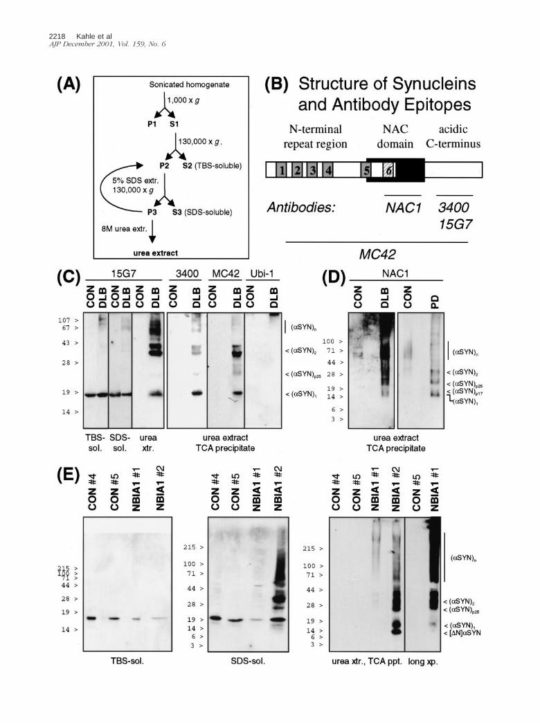
Subcellular fractionation of archived human cerebral cor-tex samples was performed as previously described forfresh tissue.30 The detergent extraction method of Culve-nor and colleagues35 was applied to human and mousebrain with slight modifications. Approximately 0.5 g ofbrain tissue was homogenized in 10 volumes of TBS�(Tris-buffered saline plus Complete protease inhibitorcocktail; Roche Diagnostics, Mannheim, Germany) andsonicated. After 5 minutes of centrifugation at 1000 � g,the supernatants were ultracentrifuged for 1 hour at130,000 � g. The resulting supernatants represented thebuffer-soluble fractions. The pellets were rinsed twicewith TBS� and extracted with 500 �l of 5% SDS in TBS�.All subsequent steps were performed at 24°C. After ul-tracentrifugation for 30 minutes at 130,000 � g the pelletswere re-extracted twice with 5% SDS, and the detergent-soluble supernatants were collected. The bicucullinicacid (BCA) protein assay (Pierce, Rockford, IL) revealedconcentrations �1 mg/ml in the first two SDS superna-tants that were pooled. The extensively washed deter-gent-insoluble pellets were squashed in 100 �l of 8 mol/Lurea/5% SDS in TBS� and incubated for at least 10minutes at room temperature. Then, 80 �l of the resultingsuspension were mixed with 20 �l of trichloroacetic acid(TCA) (100%) and allowed to precipitate overnight at 4°C.Protein precipitates were collected by centrifugation,washed with acetone, and resuspended in protein gel-loading buffer containing 6 mol/L urea.
Denaturing polyacrylamide gel electrophoresis(PAGE), Western blotting, and probing were done asdescribed previously.30 Equal loading was verified byCoomassie blue staining of the gels after transfer. En-hanced chemiluminescence was generated with Super-Signal (Pierce) or ECLplus (Amersham Pharmacia, LittleChalfont, UK). Human-specific 15G7 band intensities in25-�g mouse brain cytosol samples were determinedrelative to 5 ng, 10 ng, 20 ng, and 40 ng recombinanthuman �-SYN (see below) on the same blot. Mouse-specific 7544 band intensities in 100-�g mouse braincytosol samples were determined relative to four stan-dards of 75- to 500-ng recombinant mouse �-SYN (seebelow) on the same blot. Band intensities from densito-metric scans were quantified using NIH Image v1.62
Table 1. The Amount of Insoluble �-SYN Correlates withSeverity of LB Diseases
Brain sample Insoluble �-SYN LB count
Temporal cortexCON no. 1 � �CON no. 2 � �CON no. 3 � �DLB no. 1 �� 22.8/mm2
DLB no. 2 � 6.0/mm2
DLB no. 3 � 6.6/mm2
Parietal cortexCON no. 4 � �CON no. 5 � �NBIA1 no. 1 � 5.3/mm2
NBIA1 no. 2 �� 13.5/mm2
The amount of insoluble �-SYN was estimated from the bandstrength on urea extract blots (see Figure 2). Band intensity: ��,strong; �, weak; �, absent. To determine LB density, vibratomesections of 50 � thickness from the NBIA1 brains were immunostainedwith Syn102, and 5-�-thick sections of DLB brains with 15G7. Fivedifferent visual fields (each 0.98 mm2) were examined.
2216 Kahle et alAJP December 2001, Vol. 159, No. 6
freeware (developed at the U.S. National Institutes ofHealth and available on the Internet at http://rsb.info.nih.gov/nih-image). Linear regression of the �-SYN standardband intensities revealed correlation coefficients be-tween 0.9 and 1.0.
Generation and Characterization of TransgenicMice
The amplified human [wt]�-SYN-coding sequence wassubcloned into the XhoI site of the Thy1 cassette ofpTSC21k, and the NotI-linearized DNA was used to gen-erate transgenic C57BL/6 mice as described for [A30P]�-SYN.30 Five founders stably transmitted the transgene,as determined by tail biopsy polymerase chain reaction.
Transgene copy number was determined by Southernblotting using as references known amounts of transgenefragment mixed to genomic DNA isolated from nontrans-genic littermates. Ten �g of genomic XbaI-KpnI restrictionfragments were fractionated by gel electrophoresis andblotted onto Nylon membranes (Roche Molecular Bio-sciences). A 1.6-kb DNA probe (HindIII-EcoRV fragmentof the transgene) was labeled with [33P]dCTP by therandom primer method using the Ready-to-Go DNAlabeling kit (Pharmacia Biotech). Hybridization wasperformed overnight at 65°C in 6� standard salinecitrate, 10% dextran sulfate, 0.5% SDS. Blots werewashed in 2� standard saline citrate (�0.1% SDS) at65°C for 20 minutes followed by a second wash for 20minutes at 65°C in 0.2� standard saline citrate (�0.1%SDS). The intensity of the bands was quantified using a
phosphorimager scanner. Northern blotting using oli-gonucleotide probes specific for mRNA of the human�-SYN transgene and mouse �-actin was performed asdescribed previously.30
Fresh mouse brains were fixed in phosphate-buffered4% paraformaldehyde and embedded in paraffin. Immu-nocytochemical detection of SYNs was performed asdescribed previously.30
Expression and in Vitro Aggregation ofRecombinant SYNs
The �-SYN expression vector has been described byJakes and colleagues.36 The mouse �-SYN37 codingregion was amplified from whole brain RNA (High PureRNA Isolation Kit; Boehringer Mannheim, Mannheim,Germany) by reverse transcriptase-polymerase chainreaction using outer mouse primers (5�-GGAATTC-CATATGGATGTGTTCATGAAAGG-3� and 5�-GGAAT-TCCATATGTTAGGCTTCAGGCTCAT-3�). The codingregion of human �-SYN38 was amplified by polymerasechain reaction with outer human primers (5�-TTCAT-TACATATGGATGTATTCATGAAAGG-3� and 5�-GGAA-TTCCATATGTTAGGCTTCAGGTTCGTAG-3�). Codons73 to 83 of �-SYN were deleted by 4-primer polymer-ase chain reaction using outer human primers andinner mutagenesis primers 5�-GGAGGAGCAGTGGT-GACGGGAGCAGGGAGC-3� and 5�-GCTCCCTGCTC-CCGTCACCACTGCTCCTCC-3�. Amplimers were sub-cloned into the NdeI site of pET-5a (Promega, Madison,WI), and constructs used to transform Escherichia coli
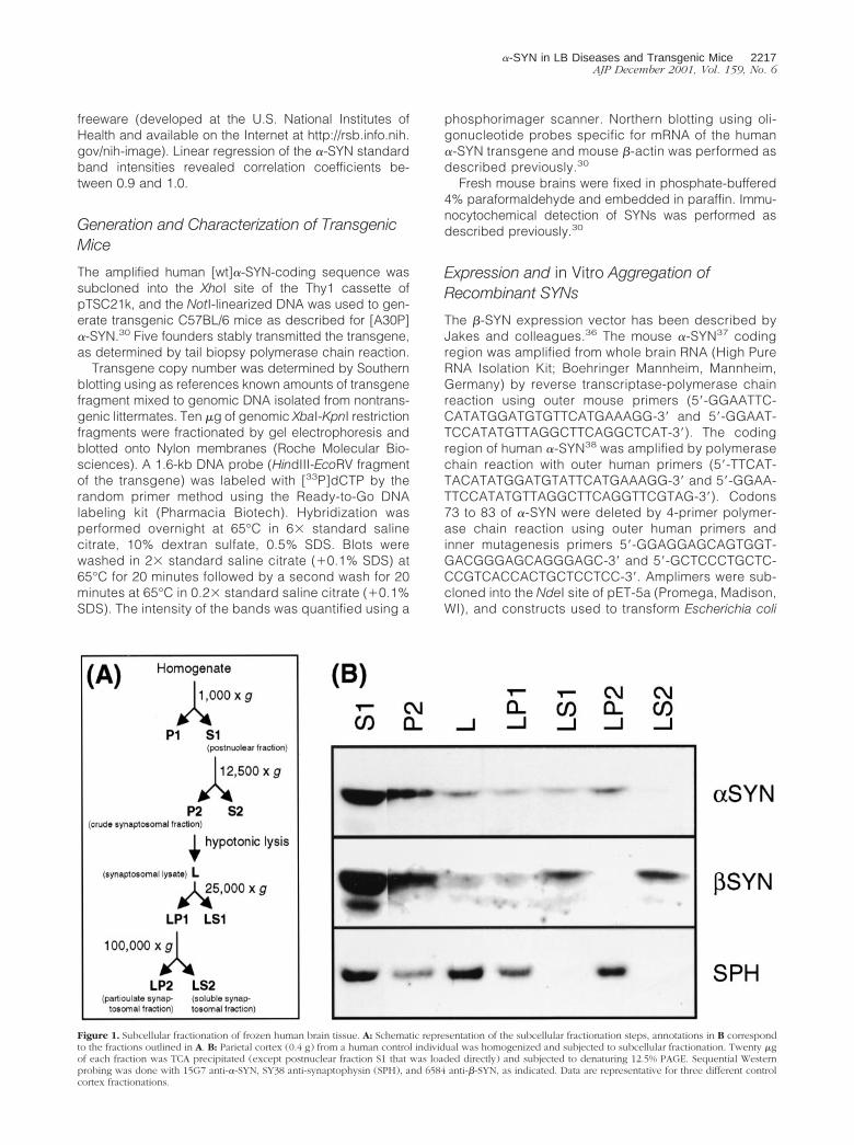
Figure 1. Subcellular fractionation of frozen human brain tissue. A: Schematic representation of the subcellular fractionation steps, annotations in B correspondto the fractions outlined in A. B: Parietal cortex (0.4 g) from a human control individual was homogenized and subjected to subcellular fractionation. Twenty �gof each fraction was TCA precipitated (except postnuclear fraction S1 that was loaded directly) and subjected to denaturing 12.5% PAGE. Sequential Westernprobing was done with 15G7 anti-�-SYN, SY38 anti-synaptophysin (SPH), and 6584 anti-�-SYN, as indicated. Data are representative for three different controlcortex fractionations.
�-SYN in LB Diseases and Transgenic Mice 2217AJP December 2001, Vol. 159, No. 6
2218 Kahle et alAJP December 2001, Vol. 159, No. 6

BL21(DE3) pLys. All constructs were sequenced (Me-digenomix, Martinsried, Germany).
Bacterial cultures were induced with isopropyl-�-D-thiogalactoside for 4 hours, and lysed by freeze/thaw andsonication. After 15 minutes of boiling, the heat-stable17,000 � g supernatant was loaded onto Q-Sepharose(Pharmacia, Uppsala, Sweden) and eluted with a 25-mmol/L to 500-mmol/L salt gradient. The pooled SYNpeak fractions were desalted by Sephacryl S-200 (Phar-macia) gel filtration.
Characteristic electron-dense fibrils (data not shown)were formed after 7 days of incubation of 2 mg/ml ofpurified recombinant SYN proteins in 50 mmol/L HEPESor phosphate (pH 6.9) at 37°C under constant agitation.Aggregates were collected by 100,000 � g centrifuga-tion, and subjected to the detergent extraction protocoldescribed above.
Results
Recovery of �-SYN from the ParticulateSynaptosomal Lysate Fraction Post Mortem
Previous subcellular fractionation experiments with hu-man brain have demonstrated the presence of �-SYN insynaptosomes.30,34 In accord with results from rapidlyprocessed rodent brain,30,33 �-SYN was released into thesoluble fraction on hypotonic lysis of the synaptosomesprepared from human biopsy brain.30 Using archivedcortical tissue, Irizarry and colleagues34 have found asignificant portion of �-SYN in the particulate fraction oflysed synaptosomes. Indeed, when subcellular fraction-ations (Figure 1) were performed with frozen tissue, re-covery of �-SYN from the soluble synaptosomal fraction(LS2) was decreased and instead a significant portion of�-SYN was detected in the particulate synaptosomalfraction (LP2). Interestingly, the subcellular fractionationprofiles of �-SYN as well as of synaptophysin were thesame as previously reported for rapidly processed hu-man biopsy samples.30 Thus, there seems to be a shift of�-SYN but not �-SYN into the pelletable synaptosomalfraction post mortem. This effect could be either becauseof altered membrane affinity and/or decreased solubilityof �-SYN.
Detergent-Insoluble �-SYN in LB Disease Brain
Sequential detergent extraction methods have been suc-cessfully used to detect �-SYN in brains of patients with
�-synucleinopathies.6,10,35,39 We have adapted themethod of Culvenor and colleagues35 (Figure 2A) to de-tect insoluble �-SYN molecules in human LB diseasebrain and in transgenic mice expressing human �-SYN.
Buffer- and detergent-soluble monomeric �-SYN wasdetected in brains from human controls as well as fromLB disease patients (Figure 2). In contrast to control brainurea extracts that were virtually devoid of �-SYN, strongimmunoreactivity was found in urea extracts from PD andDLB patients (Figure 2, C and D). The detergent-insolu-ble �-SYN was characterized using four different antibod-ies raised against distinct epitopes (Figure 2B). Mono-meric �-SYN migrated as a 16- to 19-kd band [(�-SYN)1].Anti-NAC, but not C-terminal antibodies detected a pre-viously unrecognized �-SYN species with slightly re-tarded electrophoretic motility (�-SYN)p17) (Figure 2D).This band was unlikely to be cross-reactive �-SYN, be-cause specific anti-�-SYN did not reveal any signal inurea extracts (data not shown). In addition, all four anti-�-SYN antibodies recognized �40- to 45-kd doublebands [(�-SYN)2; consistent with �-SYN dimers and/or arecently described membrane-bound form of �-SYN40],multiple bands in the 60- to 80-kd range [(�-SYN)n; pu-tative �-SYN oligomers), and higher molecular weightaggregates (Figure 2). Interestingly, an �25-kd band[(�-SYN)p25] was consistently observed in urea extractsfrom LB disease patients (Figure 2). This band was rem-iniscent of an O-glycosylated form of �-SYN recentlydescribed by Shimura and colleagues.41 Consistent withthis notion was the absence of ubiquitin immunoreactivityof (�-SYN)p25 (Figure 2C), indicating that this species didnot correspond to mono-ubiquitinated �-SYN.
A similar differential extraction pattern for �-SYN wasfound for another LB disease. NBIA1 is a neurodegen-erative synucleinopathy formerly known as Hallervorden-Spatz disease that is characterized by �-SYN inclusionssimilar to LBs as well as by axonal spheroids that containimmunoreactive �-, �-, and �-SYN.8–11 We have ana-lyzed brain samples from two NBIA1 patients describedelsewhere (case no. 1,42 case no. 210). Monomeric�-SYN was detected in the buffer-soluble and detergent-soluble fractions. The decrease in the relative amount ofbuffer-soluble �-SYN in both NBIA1 samples (Figure 2E)may be indicative of defective synaptic integrity in thesepatients.10 Alternatively, the soluble pool of �-SYN mightbe depleted as �-SYN monomers and aggregates accu-mulated in the detergent-insoluble fraction (Figure 2E).Limited N-terminal degradation to a 14-kd band ([�N]�-SYN) was occasionally noted, but the bulk of insoluble�-SYN was full-length protein. Higher molecular weight
Figure 2. SDS-insoluble �-SYN species in LB diseases. A: Schematic representation of the differential extraction steps. See Materials and Methods for details. B:Structure of �-SYN and epitopes recognized by anti-�-SYN antibodies. The imperfect KTKEGV repeats are numbered, the sixth repeat missing in �-SYN (seebelow) is stippled. C: Extracts from temporal cortex of control and DLB brain were prepared, and 10 �g of TBS-soluble material, 10 �g of SDS-soluble material,and 10 �l of urea extracts or TCA precipitates from 50-�l urea extracts (as indicated at the bottom) were subjected to denaturing 12.5% PAGE. Western blots weresequentially probed with three different antibodies against �-SYN (15G7, 3400, MC42) and anti-ubiquitin (Ubi-1), as indicated on the top. Immunoreactivity wasvisualized with SuperSignal (for 15G7) or ECLplus. D: TCA precipitates from 50-�l urea extracts from temporal cortex of control and DLB brain (left) and fromparietal cortex of control and PD brain (right) were loaded on 10 to 20% Tris-tricine gels. The corresponding Western blots were probed with anti-NAC anddeveloped with ECLplus. E: Parietal cortex samples from two controls and two NBIA1 patients were extracted in parallel. TBS-soluble material (10 �g, left),SDS-soluble material (25 �g, middle), and urea extracts (80 �l, right) were separated by SDS-PAGE (TBS-soluble, 15%; SDS and urea extracts, 4 to 20% gradient).MC42 and ECLplus were used for Western detection of �-SYN. Note that sample NBIA1 no. 2 with higher LB density than NBIA1 no. 1 had also much stronger�-SYN immunoreactivity in the urea extract. Nevertheless, the �-SYN immunoreactive band pattern of NBIA1 no. 1 was qualitatively the same as of NBIA1 no.2, as evidenced by a longer exposure of the blot to the far right. Each experiment is representative for two to three independent extractions. Positions ofprestained molecular weight standards are indicated to the left. See text for description of �-SYN species denoted to the right.
�-SYN in LB Diseases and Transgenic Mice 2219AJP December 2001, Vol. 159, No. 6

�-SYN bands were observed even in the SDS fractionsfrom brain of NBIA1 case no. 2, who also had much moreintense �-SYN immunoreactivity in the urea extracts thanNBIA1 case no. 1 (Figure 2E). In fact, there was a positivecorrelation between the �-SYN immunoreactivity in ureaextracts and LB density in the two NBIA1 patients and thethree DLB patients (Table 1).
Expression Pattern of �-SYN in Transgenic Mice
To generate a rodent model of �-synucleinopathy, wehave generated transgenic mice expressing human wtand PD-associated mutant [A30P]�-SYN under the con-trol of a brain-specific pan-neuronal promoter, Thy1. The(Thy1)-[A30P]�-SYN mice described previously30 andnewly generated strains of (Thy1)-[wt]�-SYN mice wereof identical genetic background.
As expected for the Thy1 cassette,43 expression oftransgenic �-SYN was undetectable in the first postnatalweek, then increased sharply to reach a plateau around1 month (Figure 3). The developmental onset of trans-gene expression paralleled that of endogenous �-SYN,except for the low but significant early postnatal expres-
sion of endogenous �-SYN (Figure 3). The putative em-bryonic/perinatal function of �-SYN in mouse brain37,44
remains to be elucidated.In adult animals, expression levels of transgenic mRNA
and protein generally correlated with each other (exceptin the [A30P]�-SYN-expressing mouse line 8, which hadvery high mRNA levels but intermediate protein amounts)(Table 2). In contrast, the transgene copy number was nopredictor of expression levels (Table 2). Approximately0.1% (w/w) of total adult transgenic mouse brain cytosolicprotein was �-SYN, as determined by quantitative West-ern blot analysis (Table 2). Individual quantification ofmouse and human �-SYN revealed up to threefold over-expression levels of the transgenic protein.
At these rather moderate overexpression levels, aber-rant subcellular localization of human �-SYN was appar-ent in transgenic mouse brain sections. The human(transgene)-specific antibody showed somal and neuriticaccumulations in both [wt]�-SYN and [A30P]�-SYN mice(Figure 4, A and D), in addition to neuropil (presumablysynaptic) staining. Compact LBs were not observed intransgenic mice. Nevertheless, swollen �-SYN-positiveneurites as observed in [wt]�-SYN and [A30P]�-SYNtransgenic mice (Figure 4, A and D, inserts) were aprominent feature in brain sections from LB disease pa-tients.2,9,30 In sharp contrast, endogenous mouse �-SYNshowed only the normal neuropil staining pattern (Figure4, C and F). Likewise, our �-SYN antibody detected onlya normal synaptic staining pattern in sections from trans-genic mouse brain (Figure 4, B and E).
Transgenic Human �-SYN But Not EndogenousMouse SYNs in Detergent-Insoluble Fractions
As the presence of SDS-insoluble �-SYN seemed to be adiagnostic criterion for LB diseases in human brain, weapplied the above method to transgenic mouse brains
Figure 3. Developmental expression of �-SYN. Mouse brains were collectedfrom mice at the indicated age, and Western blots prepared from 25-�gcytosolic extracts. Wild-type mouse blots were probed with MC42 (top),representative [(Thy1)-h[A30P]�-SYN line 18] transgenic mouse blots probedwith 3400 (bottom), and developed with ECLplus.
Table 2. �-SYN Expression Levels in Transgenic Mice
Transgenic construct Line
�-SYNtransgene
copy number
Transgenic�-SYN mRNA
[% actin]
Transgenic�-SYN protein
[ng/�g]
Endogenous�-SYN protein
[ng/�g]
(Thy1)-h[wt]�-SYN 6 7 175 1.93 � 0.31 1.35 � 0.10(Thy1)-h[wt]�-SYN 10 85 65 0.80 � 0.31 1.61 � 0.14(Thy1)-h[wt]�-SYN 14 13 190 2.02 � 0.11 0.65 � 0.06(Thy1)-h[wt]�-SYN 23 2 130 1.32 � 0.17 0.43 � 0.21(Thy1)-h[wt]�-SYN 38 5 130 1.58 � 0.60 0.81 � 0.28Nontransgenic N/A† N/A N/A 1.77 � 0.55(Thy1)-h[A30P]�-SYN 8 15–25 370 1.20 � 0.60 1.42 � 0.99(Thy1)-h[A30P]�-SYN 9 5 30 0.39 � 0.05 1.64 � 0.05(Thy1)-h[A30P]�-SYN 14 10 105 1.2* n.d.(Thy1)-h[A30P]�-SYN 18 10–15 170 1.43 � 0.18 1.59 � 0.00(Thy1)-h[A30P]�-SYN 31 15–20 155 1.69 � 0.31 1.26 � 0.01
Five lines each of [wt]�-SYN and [A30P]�-SYN-expressing transgenic mice stably transmitted the transgene. The integrated transgene copynumber was determined by quantitative Southern blotting, transgenic �-SYN mRNA expression levels relative to �-actin by quantitative Northernblotting, and the amount of transgenic and endogenous �-SYN protein by quantitative Western blotting. �-SYN protein was measured in three differentmouse whole-brain cytosol samples on the same blot. Results are expressed as mean � SD.
*Determined indirectly; expression levels relative to the other lines.†N/A: not applicable.
2220 Kahle et alAJP December 2001, Vol. 159, No. 6

expressing human �-SYN in brain neurons. Although theoverexpression levels of transgenic �-SYN were rathermoderate, a portion of human �-SYN was specificallydetected in urea extracts of detergent-insoluble fractionsfrom transgenic mouse brains (Figure 5). Both transgenic
[A30P] �-SYN and [wt] �-SYN were found in SDS-insolu-ble fractions (Figure 5A). Insoluble transgenic �-SYN be-came detectable parallel to the onset of transgene ex-pression (Figure 3) and persisted for at least 1 year(Figure 5B). The onset of expression of insoluble trans-
Figure 4. Immunostainings of brain slices (motor cortex) show specific accumulation of �-SYN but not of �-SYN in transgenic mice. Animals expressing either[wt]�-SYN (A–C) or [A30P]�-SYN (D–F) showed a strong cytosolic labeling of neuronal cells with the human-specific �-SYN antibody 15G7 (A and D). A sectionfrom a 1-month-old [wt]�-SYN-expressing mouse is shown in A to demonstrate the early onset of accumulation of transgenic protein. In contrast, immunostainingswith the �-SYN-specific antiserum (6485) and the murine-specific �-SYN antiserum (7544) only revealed a synaptic staining pattern. Neither accumulation ofendogenous murine �-SYN (C and F) nor �-SYN (B and E) was detectable in neuronal cell bodies. Scale bar in A, 100 �m.
�-SYN in LB Diseases and Transgenic Mice 2221AJP December 2001, Vol. 159, No. 6

genic �-SYN was concomitant with the appearance ofcytosolic accumulations (Figure 4A).
In sharp contrast, endogenous mouse �-SYN as well as�-SYN were entirely soluble in buffer and detergent. In asemiquantitative manner, Western blots were sequentiallyprobed with human (transgene)-specific anti-�-SYN, mouse(endogenous)-specific anti-�-SYN, and anti-�-SYN, and thesignal of a control lane on the same blot (10 �g transgenicmouse brain cytosol) was used for normalization. Exposuretimes yielding comparable signals from the control lanedemonstrated that SDS-insoluble endogenous �-SYN or�-SYN in transgenic �-SYN-positive urea extracts were un-detectable (Figure 5).
Taken together, cytoplasmic accumulation and deter-gent-insolubility of transgenic human �-SYN representspecific pathological alterations in transgenic mousebrain that are reminiscent of human LB diseases.
Lack of an Aggregation-Promoting Stretch ofAmino Acids in the NAC Domain of �-SYN
To determine whether the detergent insolubility of �-SYNin LB disease brain and transgenic mice was a conse-
Figure 5. Detergent-insoluble �-SYN in transgenic mouse brains. Wholebrains of transgenic mice (A: 3- to 4-month-old [wt]�-SYN lines 14 and 23,and [A30P]�-SYN lines 18 and 31; B: 1-month-old and 1-year-old [A30P]�-SYN lines 18 and 31; as indicated at the bottom) and age-matched nontrans-genic littermates (lm) were differentially extracted. Buffer- and detergent-soluble proteins (10 �g for transgenic human �-SYN, 50 �g for endogenousmouse SYNs), and TCA precipitate of urea extracts were subjected to dena-turing 15% PAGE. Western blots were probed with human (transgene)-specific anti-h�-SYN 15G7, endogenous mouse-specific anti-m�-SYN 7544,and anti-�-SYN 6485, as indicated to the left. 15G7-immunoreactive bandswere developed with SuperSignal, polyclonal antibody immunoreactivitywith ECLplus. A control lane on the urea extract blots contained 10 �g ofcytosol from a transgenic [A30P]�-SYN mouse. The positions of prestainedmolecular weight markers are indicated to the right. Individual variance oftransgenic �-SYN expression levels may account for the apparently higheramount of urea extractable mutant h[A30P]�-SYN compared to h[wt]�-SYN inone experiment (A, exp. 2), but not in two additional experiments (one ofthem shown as A, exp. 1), and for the apparent increase with age ofSDS-soluble �-SYN (B) that was not seen in an additional experiment.
Figure 6. �-SYN, but not �-SYN aggregates in vitro because of a criticaldeterminant in the NAC domain. A: Recombinant human �-SYN aggregateswere collected by 100,000 � g centrifugation and redissolved in 15 volumesof TBS�. The buffer-insoluble material was extracted like the brain samplesabove. All fractions were TCA precipitated and separated by denaturing 15%PAGE. Western blots were probed with 3400 anti-�-SYN and developed withSuperSignal. This experiment was repeated three times with the same result.B: Solutions (2 mg/ml) of [wt]�-SYN (left), [�73–83]�-SYN (middle), and[wt]�-SYN (right) were aggregated for 7 days. After ultracentrifugation, the100,000 � g pellets (pel) were subjected to denaturing 12.5% PAGE. �-SYNimmunoblots were probed with MC42 and �-SYN immunoblots with 6485,and developed with SuperSignal until the band intensities of 1-�g freshlydissolved, nonaggregated protein (sol) were comparable. Note the typicalretarded electrophoretic motility of recombinant �-SYN.36,38 Positions ofprestained molecular weight markers are indicated to the right.
2222 Kahle et alAJP December 2001, Vol. 159, No. 6
quence of aggregation, in vitro formed aggregates of�-SYN were subjected to a similar differential extractionprocedure used for brain tissue (see above). �-SYN ag-gregated at a concentration of 2 mg/ml was partiallysolubilized by resuspension in 15 volumes of TBS�.However, 5% SDS was required for complete solubiliza-tion (Figure 6A). In contrast, �-SYN failed to form insolu-ble aggregates in vitro (Figure 6B).
�-SYN lacks amino acids homologous to residues 73to 83 of �-SYN. Codons 73 to 83 were specifically deletedby site-directed mutagenesis yielding [�73–83]�-SYN. Inparallel in vitro aggregation assays, [�73–83]�-SYN be-haved like �-SYN in that it practically lost its capability toform 100,000 � g pellets after 1 week incubation at 37°C(Figure 6B). Thus, amino acids 73 to 83 in the N-terminalhalf of NAC are critical for �-SYN aggregation, and theirabsence in �-SYN accounts for the loss of aggregationcapacity of the nonamyloidogenic �-SYN.
Discussion
PD, DLB, and NBIA1 are characterized immunohisto-chemically by �-SYN-immunoreactive intraneuronal in-clusions (LBs) and dystrophic neurites. Biochemically,detergent-insoluble �-SYN was found to be diagnostic forthese diseases. We have performed differential detergentextractions to evaluate the potential development of�-synucleinopathy in transgenic mice expressing human�-SYN in brain neurons. Like in human LB diseases,detergent-insoluble human �-SYN was detected in trans-genic mouse brain. In striking contrast, endogenousmouse �-SYN and �-SYN were not found in the ureaextracts. These results demonstrate that a transgenicmouse model recapitulates some specific features of�-synucleinopathies.
The source of detergent-insoluble �-SYN may not onlybe solid LBs. It is noteworthy that the NBIA1 case withmuch urea-extractable �-SYN had abundant dystrophicneurites (not shown). Moreover, diffuse accumulation of�-SYN in neuronal cell bodies was occasionally reportedfor human �-synucleinopathies.45,46 The accumulationsof human �-SYN in transgenic mouse brain neurons didnot meet ultrastructural criteria of LBs. Nevertheless, de-tergent-insoluble transgenic �-SYN was specifically de-tectable in these mice. Thus, a portion of transgenic�-SYN is converted to a less soluble form that mightrepresent an early form of �-synucleinopathy. It remainsto be shown if the decreased solubility of transgenic�-SYN is a mere consequence of overexpression, or ifthere is some secondary processing that is peculiar tohuman transgenic �-SYN in mouse brain.
Both PD-associated [A30P]�-SYN and human [wt]�-SYN were detected in detergent-insoluble fractions. Thisis of note because the overwhelming majority of PD pa-tients have no mutation in the �-SYN gene.47 The faster invitro aggregation rate of concentrated solutions of mutant�-SYN22 was apparently not reflected by greater pathol-
ogy of human mutant �-SYN compared to [wt]�-SYN intransgenic mice.29,30 The rather moderate expressionlevels of transgenic �-SYN (Table 2) make it unlikely thatthe critical concentration required for recombinant �-SYNaggregation in vitro (28 �mol/L)48 was reached in neuro-nal cytosol. Perhaps the differences in aggregation kinet-ics between wt and mutant �-SYN are not evident atconcentrations reached in transgenic mouse neurons.Because �-SYN expression is not elevated enough in PDpatients to allow spontaneous aggregation, additionalrisk factors are likely to exist that favor the aggregation atsubcritical �-SYN concentrations. Similar risk factors mayact in rodents. For example, the mitochondrial complex Iinhibitor, rotenone, was recently shown to elicit PD-likealterations in rat brain, including the formation of �-SYNinclusions and selective loss of striatonigral dopaminer-gic neurons.49
Potentially aggregation-promoting posttranslationalmodifications of �-SYN include phosphorylation,38 nitra-tion,50 and glycation.51 Moreover, perturbation of proteo-somal degradation should be considered. LBs containubiquitinated �-SYN. It was reported that in human brain,�-SYN needs to be converted to a slower migrating spe-cies (termed “�Sp22”) to become a substrate for theubiquitin ligase, parkin,41 We have found an �-SYN spe-cies with similar retarded electrophoretic motility runningat apparent 25-kd [(�-SYN)p25] in the detergent-insolublefractions from LB disease brain (Figure 2), but not intransgenic mice (Figure 5). It is possible that posttrans-lational modifications characteristic for human LB dis-eases have to occur in transgenic mice to allow true LBformation in an animal model.
In contrast to the amyloidogenic �-SYN, the close ho-mologue �-SYN was absent from amyloid deposits.�-SYN has no intrinsic capacity to form amyloid fibrils.�-SYN lacks the sequence GVTAVAQKTVE correspond-ing to amino acids 73 to 83 of �-SYN in the NAC domain.Indeed, the deletion mutant protein [�73–83]�-SYN wasaggregation-deficient. Similar findings were recently re-ported by Giasson and colleagues52 for a slightly shifteddeletion mutant, namely [�71–82]�-SYN. Interestingly,the region critical for �-SYN aggregation, which is missingin �-SYN, overlaps with one of the imperfectly conservedKTKEGV repeats. Such repeats are characteristic for thelipid-binding N-terminal half of �-SYN that has a predictedamphipathic helix structure comparable with apolipopro-tein A.53,54 It will be interesting to determine whether theaggregation-promoting region of �-SYN acquires an am-phipathic �-strand conformation upon fibrillization.
In vitro aggregates of �-SYN clearly required harshersolubilization methods than �-SYN. Nevertheless, hardlyany SDS-insoluble protein was found in urea extracts of invitro aggregated recombinant �-SYN. Thus, the massiveamounts of �-SYN in urea extracts from LB disease andtransgenic mouse brain represent a pathological form of�-SYN that may not be fully reproduced by simple aggre-gation in vitro. Thus, �-SYN transgenic mice provide an invivo model that exhibits an important aspect of human�-synucleinopathy, namely selective insolubility of �-SYN.
�-SYN in LB Diseases and Transgenic Mice 2223AJP December 2001, Vol. 159, No. 6

Acknowledgments
We thank R. Jakes and M. Goedert for the gift of �-SYNexpression plasmid, K. Beyreuther and T. Hartmann forproviding anti-NAC, W. Franke and R. Leube for the dona-tion of anti-synaptophysin, and H. Schubert for animal care.
References
1. Ueda K, Fukushima H, Masliah E, Xia Y, Iwai A, Yoshimoto M, OteroDA, Kondo J, Ihara Y, Saitoh T: Molecular cloning of cDNA encodingan unrecognized component of amyloid in Alzheimer disease. ProcNatl Acad Sci USA 1993, 90:11282–11286
2. Spillantini MG, Schmidt ML, Lee VM-Y, Trojanowski JQ, Jakes R,Goedert M: �-Synuclein in Lewy bodies. Nature 1997, 388:839–840
3. Wakabayashi K, Matsumoto K, Takayama K, Yoshimoto M, TakahashiH: NACP, a presynaptic protein, immunoreactivity in Lewy bodies inParkinson’s disease. Neurosci Lett 1997, 239:45–48
4. Takeda A, Mallory M, Sundsmo M, Honer W, Hansen L, Masliah E:Abnormal accumulation of NACP/�-synuclein in neurodegenerativedisorders. Am J Pathol 1998, 152:367–372
5. Irizarry MC, Growdon W, Gomez-Isla T, Newell K, George JM, ClaytonDF, Hyman BT: Nigral and cortical Lewy bodies and dystrophic nigralneurites in Parkinson’s disease and cortical Lewy body disease con-tain �-synuclein immunoreactivity. J Neuropathol Exp Neurol 1998,57:334–337
6. Baba M, Nakajo S, Tu P-H, Tomita T, Nakaya K, Lee VM-Y, Tro-janowski JQ, Iwatsubo T: Aggregation of �-synuclein in Lewy bodiesof sporadic Parkinson’s disease and dementia with Lewy bodies.Am J Pathol 1998, 152:879–884
7. Bayer TA, Jakala P, Hartmann T, Havas L, McLean C, Culvenor JG, LiQX, Masters CL, Falkai P, Beyreuther K: �-Synuclein accumulates inLewy bodies in Parkinson’s disease and dementia with Lewy bodiesbut not in Alzheimer’s disease �-amyloid plaque cores. Neurosci Lett1999, 266:213–216
8. Arawaka S, Saito Y, Murayama S, Mori H: Lewy body in neurodegen-eration with brain iron accumulation type 1 is immunoreactive for�-synuclein. Neurology 1998, 51:887–889
9. Wakabayashi K, Yoshimoto M, Fukushima T, Koide R, Horikawa Y,Morita T, Takahashi H: Widespread occurrence of �-synuclein/NACP-immunoreactive neuronal inclusions in juvenile and adult-onset Hall-ervorden-Spatz disease with Lewy bodies. Neuropathol Appl Neuro-biol 1999, 25:363–368
10. Galvin JE, Giasson B, Hurtig HI, Lee VM-Y, Trojanowski JQ: Neuro-degeneration with brain iron accumulation, type 1 is characterized by�-, �-, and �-synuclein neuropathology. Am J Pathol 2000, 157:361–368
11. Neumann M, Adler S, Schluter O, Kremmer E, Benecke R,Kretzschmar HA: �-Synuclein accumulation in a case of neurodegen-eration with brain iron accumulation type 1 (NBIA-1, formerly Haller-vorden-Spatz syndrome) with widespread cortical and brainstem-type Lewy bodies. Acta Neuropathol 2000, 100:568–574
12. Spillantini MG, Crowther RA, Jakes R, Hasegawa M, Goedert M:�-Synuclein in filamentous inclusions of Lewy bodies from Parkin-son’s disease and dementia with Lewy bodies. Proc Natl Acad SciUSA 1998, 95:6469–6473
13. Arima K, Ueda K, Sunohara N, Hirai S, Izumiyama Y, Tonozuka-Uehara H, Kawai M: Immunoelectron-microscopic demonstration ofNACP/�-synuclein-epitopes on the filamentous component of Lewybodies in Parkinson’s disease and in dementia with Lewy bodies.Brain Res 1998, 808:93–100
14. Dickson DW, Lin W, Liu WK, Yen SH: Multiple system atrophy: asporadic synucleinopathy. Brain Pathol 1999, 9:721–732
15. Hashimoto M, Hsu LJ, Sisk A, Xia Y, Takeda A, Sundsmo M, MasliahE: Human recombinant NACP/�-synuclein is aggregated and fibril-lated in vitro: relevance for Lewy body disease. Brain Res 1998,799:301–306
16. Giasson BI, Uryu K, Trojanowski JQ, Lee VM-Y: Mutant and wild typehuman �-synucleins assemble into elongated filaments with distinctmorphologies in vitro. J Biol Chem 1999, 274:7619–7622
17. Conway KA, Harper JD, Lansbury Jr PT: Fibrils formed in vitro from
�-synuclein and two mutant forms linked to Parkinson’s disease aretypical amyloid. Biochemistry 2000, 39:2552–2563
18. Serpell LC, Berriman J, Jakes R, Goedert M, Crowther RA: Fiberdiffraction of synthetic �-synuclein filaments shows amyloid-likecross-� conformation. Proc Natl Acad Sci USA 2000, 97:4897–4902
19. Biere AL, Wood SJ, Wypych J, Steavenson S, Jiang Y, Anafi D,Jacobsen FW, Jarosinski MA, Wu G-M, Louis J-C, Martin F, Narhi LO,Citron M: Parkinson’s disease-associated �-synuclein is more fibril-logenic than �- and �-synuclein and cannot cross-seed its homologs.J Biol Chem 2000, 275:34574–34579
20. Conway KA, Harper JD, Lansbury PT: Accelerated in vitro fibril for-mation by a mutant �-synuclein linked to early-onset Parkinson dis-ease. Nat Med 1998, 4:1318–1320
21. El-Agnaf OMA, Jakes R, Curran MD, Wallace A: Effects of the muta-tions Ala30 to Pro and Ala53 to Thr on the physical and morphologicalproperties of �-synuclein protein implicated in Parkinson’s disease.FEBS Lett 1998, 440:67–70
22. Narhi L, Wood SJ, Steavenson S, Jiang Y, Wu GM, Anafi D, KaufmanSA, Martin F, Sitney K, Denis P, Louis J-C, Wypych J, Biere AL, CitronM: Both familial Parkinson’s disease mutations accelerate �-synucleinaggregation. J Biol Chem 1999, 274:9843–9846
23. Hashimoto M, Hsu LJ, Xia Y, Takeda A, Sisk A, Sundsmo M, MasliahE: Oxidative stress induces amyloid-like aggregate formation ofNACP/�-synuclein in vitro. NeuroReport 1999, 10:717–721
24. Goldberg MS, Lansbury Jr PT: Is there a cause-and-effect relation-ship between �-synuclein fibrillization and Parkinson’s disease? NatCell Biol 2000, 2:E115–E119
25. Polymeropoulos MH, Lavedan C, Leroy E, Ide SE, Dehejia A, Dutra A,Pike B, Root H, Rubenstein J, Boyer R, Stenroos ES, Chan-drasekharappa S, Athanassiadou A, Papapetropoulos T, JohnsonWG, Lazzarini AM, Duvoisin RC, Di Iorio G, Golbe LI, Nussbaum RL:Mutation in the �-synuclein gene identified in families with Parkinson’sdisease. Science 1997, 276:2045–2047
26. Kruger R, Kuhn W, Muller T, Woitalla D, Graeber M, Kosel S, PrzuntekH, Epplen JT, Schols L, Riess O: Ala30Pro mutation in the geneencoding �-synuclein in Parkinson’s disease. Nat Genet 1998, 18:106–108
27. Feany MB, Bender WW: A Drosophila model of Parkinson’s disease.Nature 2000, 404:394–398
28. Masliah E, Rockenstein E, Veinbergs I, Mallory M, Hashimoto M,Takeda A, Sagara Y, Sisk A, Mucke L: Dopaminergic loss and inclu-sion body formation in �-synuclein mice: implications for neurode-generative disorders. Science 2000, 287:1265–1269
29. van der Putten H, Wiederhold K-H, Probst A, Barbieri S, Mistl C,Danner S, Kauffmann S, Hofele K, Spooren WPJM, Ruegg MA, Lin S,Caroni P, Sommer B, Tolnay M, Bilbe G: Neuropathology in miceexpressing human �-synuclein. J Neurosci 2000, 20:6021–6029
30. Kahle PJ, Neumann M, Ozmen L, Muller V, Jacobsen H, SchindzielorzA, Okochi M, Leimer U, van der Putten H, Probst A, Kremmer E,Kretzschmar HA, Haass C: Subcellular localization of wild-type andParkinson’s disease-associated mutant �-synuclein in human andtransgenic mouse brain. J Neurosci 2000, 20:6365–6373
31. Maroteaux L, Scheller RH: The rat brain synucleins; family of proteinstransiently associated with neuronal membrane. Mol Brain Res 1991,11:335–343
32. Shibayama-Imazu T, Okahashi I, Omata K, Nakajo S, Ochiai H, NakaiY, Hama T, Nakamura Y, Nakaya K: Cell and tissue distribution anddevelopmental change of neuron specific 14 kDa protein (phospho-neuroprotein 14). Brain Res 1993, 622:17–25
33. George JM, Jin H, Woods WS, Clayton DF: Characterization of a novelprotein regulated during the critical period for song learning in thezebra finch. Neuron 1995, 15:361–372
34. Irizarry MC, Kim T-W, McNamara M, Tanzi RE, George JM, ClaytonDF, Hyman BT: Characterization of the precursor protein of thenon-A� component of senile plaques (NACP) in the human centralnervous system. J Neuropathol Exp Neurol 1996, 55:889–895
35. Culvenor JG, McLean CA, Cutt S, Campbell BCV, Maher F, Jakala P,Hartmann T, Beyreuther K, Masters CL, Li Q-X: Non-A� componentof Alzheimer’s disease amyloid (NAC) revisited: NAC and �-synucleinare not associated with A� amyloid. Am J Pathol 1999, 155:1173–1181
36. Jakes R, Spillantini MG, Goedert M: Identification of two distinctsynucleins from human brain. FEBS Lett 1994, 345:27–32
2224 Kahle et alAJP December 2001, Vol. 159, No. 6
37. Hong L, Ko HW, Gwag BJ, Joe E, Lee S, Kim Y-T, Suh Y-H: The cDNAcloning and ontogeny of mouse �-synuclein. NeuroReport 1998,9:1239–1243
38. Okochi M, Walter J, Koyama A, Nakajo S, Baba M, Iwatsubo T, MeijerL, Kahle PJ, Haass C: Constitutive phosphorylation of the Parkinson’sdisease associated �-synuclein. J Biol Chem 2000, 275:390–397
39. Dickson DW, Liu W-K, Hardy J, Farrer M, Mehta N, Uitti R, Mark M,Zimmerman T, Golbe L, Sage J, Sima A, D’Amato C, Albin R, GilmanS, Yen S-H: Widespread alterations of �-synuclein in multiple systematrophy. Am J Pathol 1999, 155:1241–1251
40. Leng Y, Chase TN, Bennett MC: Muscarinic receptor stimulationinduces translocation of an �-synuclein oligomer from plasma mem-brane to a light vesicle fraction in cytoplasm. J Biol Chem 2001,276:28212–28218
41. Shimura H, Schlossmacher MG, Hattori N, Frosch MP, Trocken-bacher A, Schneider R, Mizuno Y, Kosik KS, Selkoe DJ: Ubiquitinationof a new form of �-synuclein by parkin from human brain: implicationsfor Parkinson’s disease. Science 2001, 293:263–269
42. Wakabayashi K, Fukushima T, Koide R, Horikawa Y, Hasegawa M,Watanabe Y, Noda T, Eguchi I, Morita T, Yoshimoto M, Iwatsubo T,Takahashi H: Juvenile-onset generalized neuroaxonal dystrophy(Hallervorden-Spatz disease) with diffuse neurofibrillary and Lewybody pathology. Acta Neuropathol 2000, 99:331–336
43. Kollias G, Spanopoulou E, Grosveld F, Ritter M, Beech J, Morris R:Differential regulation of a Thy-1 gene in transgenic mice. Proc NatlAcad Sci USA 1987, 84:1492–1496
44. Hsu LJ, Mallory M, Xia Y, Veinbergs I, Hashimoto M, Yoshimoto M,Thal LJ, Saitoh T, Masliah E: Expression pattern of synucleins (non-A�
component of Alzheimer’s disease amyloid precursor protein/�-synuclein) during murine brain development. J Neurochem 1998,71:338–344
45. Gomez-Tortosa E, Newell K, Irizarry MC, Sanders JL, Hyman BT:�-Synuclein immunoreactivity in dementia with Lewy bodies: morpho-
logical staging and comparison with ubiquitin immunostaining. ActaNeuropathol 2000, 99:352–357
46. Gai WP, Yuan HX, Li XQ, Power JTH, Blumbergs PC, Jensen PH: Insitu and in vitro study of colocalization and segregation of�-synuclein, ubiquitin, and lipids in Lewy bodies. Exp Neurol 2000,166:324–333
47. El-Agnaf OMA, Curran MD, Wallace A, Middleton D, Murgatroyd C,Curtis A, Perry R, Jaros E: Mutation screening in exons 3 and 4 of�-synuclein in sporadic Parkinson’s and sporadic and familial de-mentia with Lewy bodies cases. NeuroReport 1998, 9:3925–3927
48. Wood SJ, Wypych J, Steavenson S, Louis J-C, Citron M, Biere AL:�-Synuclein fibrillogenesis is nucleation-dependent. Implications forthe pathogenesis of Parkinson’s disease. J Biol Chem 1999, 274:19509–19512
49. Betarbet R, Sherer TB, MacKenzie G, Garcia-Osuna M, Panov AV,Greenamyre JT: Chronic systemic pesticide exposure reproducesfeatures of Parkinson’s disease. Nat Neurosci 2000, 3:1301–1306
50. Giasson BI, Duda JE, Murray IVJ, Chen Q, Souza JM, Hurtig HI,Ischiropoulos H, Trojanowski JQ, Lee VM-Y: Oxidative damage linkedto neurodegeneration by selective �-synuclein nitration in synucle-inopathy lesions. Science 2000, 290:985–989
51. Munch G, Luth HJ, Wong A, Arendt T, Hirsch E, Ravid R, Riederer P:Crosslinking of �-synuclein by advanced glycation endproducts—anearly pathophysiological step in Lewy body formation? J Chem Neu-roanat 2000, 20:253–257
52. Giasson BI, Murray IVJ, Trojanowski JQ, Lee VM-Y: A hydrophobicstretch of 12 amino acid residues in the middle of �-synuclein isessential for filament assembly. J Biol Chem 2001, 276:2380–2386
53. Davidson WS, Jonas A, Clayton DF, George JM: Stabilization of�-synuclein secondary structure upon binding to synthetic mem-branes. J Biol Chem 1998, 273:9443–9449
54. Jo E, McLaurin J, Yip CM, St. George-Hyslop P, Fraser PE:�-synuclein membrane interactions and lipid specificity. J Biol Chem2000, 275:34328–34334
�-SYN in LB Diseases and Transgenic Mice 2225AJP December 2001, Vol. 159, No. 6
Copyright © 2022 FDOKUMEN