Seed coat development in Leucospermum cordifolium (Knight) Fourcade (Proteaceae) and a clarification...
10
Botanical Journal of the Linnean Society (19931, 112: 139-148. With 11 figures Seed coat development in Leucospermum cordifoZi~rn (Knight)Fourcade (Proteaceae) and a clarification of the seed covering structures in Proteaceae JOHN C. MANNING National Botanical Institute, Private Bag X7, Claremont 7735, South Africa AND GERT J. BRITS Fynbos Research Unit, Elsenburg Agricultural Research Institute, Private Bag, Elsenburg, 7607, South Africa Received April 1992, accepted for publication December 1992 MANNING, J. C. & BRITS, G. J., 1993. Seed coat development in Leucosperrnum cordifoium (Knight) Fourcade (Proteaceae) and a clarification of the seed covering structures in Proteaceae. The development of the seed coat and pericarp is studied in Leucospermum cordZfolium from ovule to mature seed. The ovule and seed are characterized by a tegmic pachychalaza. The pericarp is adnate to the integuments from anthesis and remains unthickened to maturity. The outer integument forms the seed coat and the seed is endotestal: the outer epidermis becomes tanniniferous and the inner epidermis develops into a crystalliferous palisade. The inner integument degenerates at an early stage. Examination of the literature reveals that the crystal palisade layer of the outer integument has been erroneously assumed to constitute an endocarp. This finding indicates that a re-interpretation of all published information on the seed coat in indehiscent Proteaceae is necessary before any speculations on the phylogenetic significance of the seed coat can be entertained. ADDITIONAL KEY WORDS:-Anatomy - endocarp - pericarp - seed structure - testa. Introduction . . . . Material and methods . Results . . . . . Mature ovule , . Seed coat development Discussion . . . . Acknowledgements . . References . . . . CONTENTS . . . . . . . . . . . . . . . 139 . . . . . . . . . . . . . . . 140 . . . . . . . . . . . . . . . 140 . . . . . . . . . . . . . . . 140 . . . . . . . . . . . . . . . 141 . . . . . . . . . . . . . . . 144 . . . . . . . . . . . . . . . 147 . . . . . . . . . . . . . . . 147 INTRODUCTION Despite the horticultural popularity of the Proteaceae, detailed anatomical investigations of the seeds are relatively uncommon (Corner, 1976; Venkata 139 0024-4074/93/060139+ 10 008.00/0 0 1993 The Linnean Society of London
-
Upload
independent -
Category
Documents
-
view
1 -
download
0
Transcript of Seed coat development in Leucospermum cordifolium (Knight) Fourcade (Proteaceae) and a clarification...
Botanical Journal of the Linnean Society (19931, 112: 139-148. With 11 figures
Seed coat development in Leucospermum cordifoZi~rn (Knight) Fourcade (Proteaceae) and a clarification of the seed covering structures in Proteaceae
JOHN C. MANNING
National Botanical Institute, Private Bag X7, Claremont 7735, South Africa
AND
GERT J. BRITS
Fynbos Research Unit, Elsenburg Agricultural Research Institute, Private Bag, Elsenburg, 7607, South Africa
Received April 1992, accepted f o r publication December 1992
MANNING, J. C. & BRITS, G. J., 1993. Seed coat development in Leucosperrnum cordifoium (Knight) Fourcade (Proteaceae) and a clarification of the seed covering structures in Proteaceae. The development of the seed coat and pericarp is studied in Leucospermum cordZfolium from ovule to mature seed. The ovule and seed are characterized by a tegmic pachychalaza. The pericarp is adnate to the integuments from anthesis and remains unthickened to maturity. The outer integument forms the seed coat and the seed is endotestal: the outer epidermis becomes tanniniferous and the inner epidermis develops into a crystalliferous palisade. The inner integument degenerates at a n early stage. Examination of the literature reveals that the crystal palisade layer of the outer integument has been erroneously assumed to constitute an endocarp. This finding indicates that a re-interpretation of all published information on the seed coat in indehiscent Proteaceae is necessary before any speculations on the phylogenetic significance of the seed coat can be entertained.
ADDITIONAL KEY WORDS:-Anatomy - endocarp - pericarp - seed structure - testa.
Introduction . . . . Material and methods . Results . . . . .
Mature ovule , . Seed coat development
Discussion . . . . Acknowledgements . . References . . . .
CONTENTS
. . . . . . . . . . . . . . . 139
. . . . . . . . . . . . . . . 140
. . . . . . . . . . . . . . . 140
. . . . . . . . . . . . . . . 140
. . . . . . . . . . . . . . . 141
. . . . . . . . . . . . . . . 144
. . . . . . . . . . . . . . . 147
. . . . . . . . . . . . . . . 147
INTRODUCTION
Despite the horticultural popularity of the Proteaceae, detailed anatomical investigations of the seeds are relatively uncommon (Corner, 1976; Venkata
139 0024-4074/93/060139+ 10 008.00/0 0 1993 The Linnean Society of London
140 J. C. MANNING AND G. J. BRITS
Rao, 1971). Furthermore, it appears that even in those species examined some considerable confusion exists in the interpretation of various layers of the seed covering structures. Seed structure can prove very useful in reflecting phylogenetic relationships between taxa (Corner, 1976), and it is essential that the homologies of the layers be correctly established.
Schwarzbart (1905) was the first to study, and misinterpret, the structure of the seed of Proteaceae. He examined mature fruits, as have many subsequent authors, and erroneously considered the thickened outer integument to comprise the endocarp. Although Jordaan (1945) commented on this error and correctly interpreted the covering structures of the seed in Leucospermum conocarpodendron (L.) Buek (as L. conocarpum R. Br.), Leucadendron salignum Berg. (as L. adscendens), L. lanigerum Meisn. and L. rubrum Burm. f. (as L. plumosum R. Br.), his work has apparently been almost entirely overlooked by virtue of the language and journal in which it was published. Much of the subsequent studies on seed morphology in Proteaceae were conducted by Venkato Rao (1971 and references therein), who, following Schwarzbart in his terminology, ensured that this misinterpretation continued thence uninterrupted into the literature. Midgely (1987) recognized and commented briefly on this problem in Leucadendron but did not concern himself further with it.
In view of the extent to which this misinterpretation has persisted in subsequent studies and the resultant ramifications in attempts at phylogenetic analyses, it is essential to clarify the nature of the seed covering structures in Proteaceae.
MATERIAL AND METHODS
Seed of Leucospermum cordiflium (Knight) Fourc. at all stages of development was collected directly into FPA from plants growing at Tygerhoek Experimental Farm, Riviersonderend, South Africa, dehydrated in ethanol and embedded in wax following established procedures. Sections were stained with Alcian Blue and Safranin. Material for SEM examination was freeze-fractured in liquid nitrogen, mounted on to aluminium stubs and sputter coated with gold- palladium for viewing in a Cambridge S200 at 10 kV.
In the text the following standard abbreviations are used: oi = outer integument; ii = inner integument; oe = outer epidermis; ie = inner epidermis.
RESULTS
Mature ovule
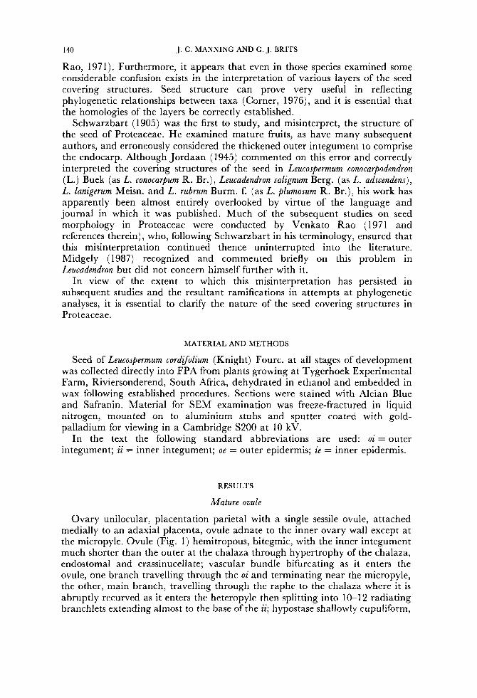
Ovary unilocular, placentation parietal with a single sessile ovule, attached medially to an adaxial placenta, ovule adnate to the inner ovary wall except at the micropyle. Ovule (Fig. 1) hemitropous, bitegmic, with the inner integument much shorter than the outer at the chalaza through hypertrophy of the chalaza, endostomal and crassinucellate; vascular bundle bifurcating as it enters the ovule, one branch travelling through the oi and terminating near the micropyle, the other, main branch, travelling through the raphe to the chalaza where it is abruptly recurved as it enters the heteropyle then splitting into 10-12 radiating branchlets extending almost to the base of the ii; hypostase shallowly cupuliform,
SEED COAT IN LEUCOSPERMUM 141
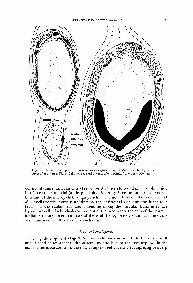
Figures 1-3. Seed development in Leucospermum cordijolium. Fig. 1. Mature ovule. Fig. 2. Seed 3 weeks after anthesis. Fig. 3. Fully formed seed 5 weeks after anthesis. Scale bar = 500 pm.
densely staining. Integuments (Fig. 4): oi 8-10 seriate on adaxial (raphal) side but 2-seriate on abaxial (antiraphal) side; ii mostly 3-seriate but 4-seriate at the base and at the micropyle through periclinal division of the middle layer; cells of oi c. isodiametric, densely staining on the antiraphal side and the inner four layers on the raphal side and extending along the vascular bundles to the hypostase; cells of ii brick-shaped except at the base where the cells of the oe are c . isodiametric and resemble those of the ie of the oe, densely-staining. The ovary wall consists of c. 10 rows of parenchyma.
Seed coat development
During development (Figs 2, 3) the ovule remains adnate to the ovary wall and is shed as an achene: the oi remains attached to the pericarp, while the embryo sac separates from the now complex seed covering (comprising pericarp
142 J. C. MANNING AND G. J. BRITS
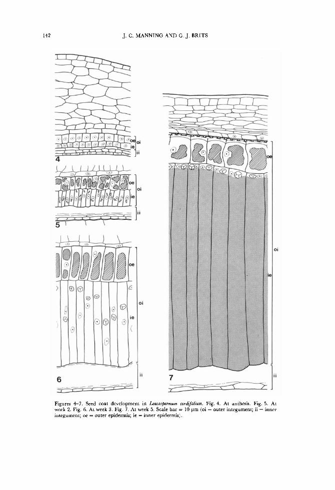
Figures 4-7. Seed coat development in Leucospermum cordijiolium. Fig. 4. At anthesis. Fig. 5. At week 2. Fig. 6. At week 3. Fig. 7. At week 5. Scale bar = 10 pm (oi = outer integument; ii = inner integument; oe = outer epidermis; ie = inner epidermis).
SEED COAT IN LEUCOSPERMUM 143
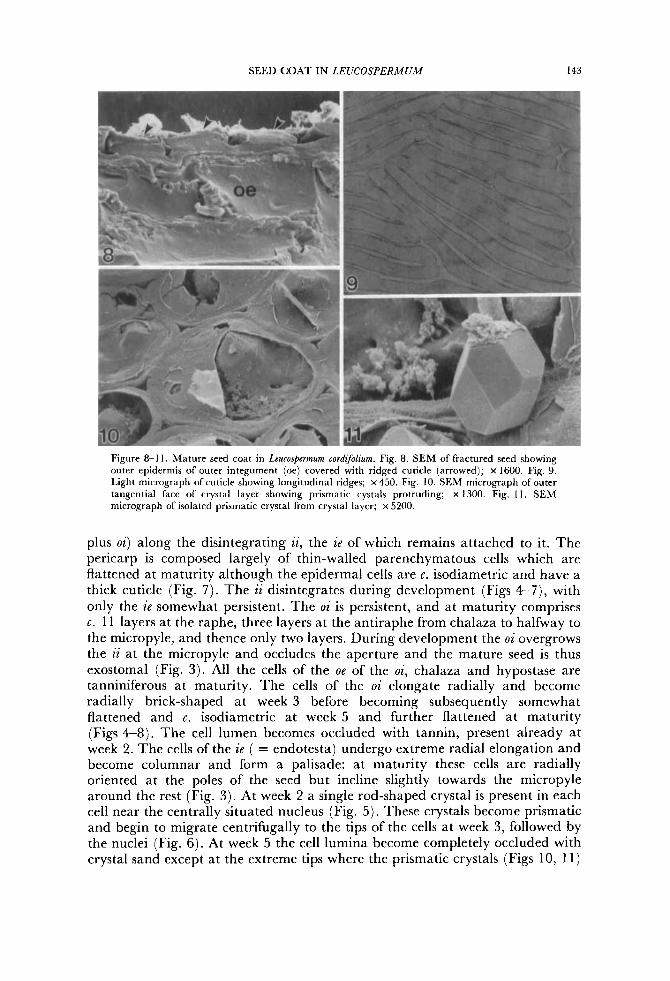
Figure 8-1 I . Mature seed coat in Lcucospermum cordifohm. Fig. 8. SEM of fractured seed showing outer epidermis of outer integument (oe) covered with ridged cuticle (arrowed); x 1600. Fig. 9. Light micrograph of cuticle showing longitudinal ridges; x 450. Fig. 10. SEM micrograph of outer tangential face of crystal layer showing prismatic cystals protruding; x 1300. Fig. I I . SEM micrograph of isolated prismatic crystal from crystal layer; x 5200.
plus oi) along the disintegrating ii, the ie of which remains attached to it. The pericarp is composed largely of thin-walled parenchymatous cells which are flattened at maturity although the epidermal cells are c. isodiametric and have a thick cuticle (Fig. 7). The ii disintegrates during development (Figs 4-7), with only the ie somewhat persistent. The oi is persistent, and at maturity comprises c. 11 layers at the raphe, three layers at the antiraphe from chalaza to halfway to the micropyle, and thence only two layers. During development the oi overgrows the ii at the micropyle and occludes the aperture and the mature seed is thus exostomal (Fig. 3) . All the cells of the oe of the oi, chalaza and hypostase are tanniniferous at maturity. The cells of the oi elongate radially and become radially brick-shaped at week 3 before becoming subsequently somewhat flattened and c. isodiametric at week 5 and further flattened a t maturity (Figs 4-8). The cell lumen becomes occluded with tannin, present already at week 2. The cells of the ze ( = endotesta) undergo extreme radial elongation and become columnar and form a palisade: at maturity these cells are radially oriented at the poles of the seed but incline slightly towards the micropyle around the rest (Fig. 3) . At week 2 a single rod-shaped crystal is present in each cell near the centrally situated nucleus (Fig. 5). These crystals become prismatic and begin to migrate centrifugally to the tips of the cells at week 3, followed by the nuclei (Fig. 6). At week 5 the cell lumina become completely occluded with crystal sand except a t the extreme tips where the prismatic crystals (Figs 10, 11)
144 J. C. MANNING AND G. J. BRITS
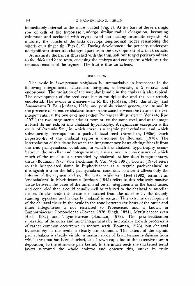
immediately internal to the ie are located (Fig. 7). At the base of the oi a single row of cells of the hypostase undergo similar radial elongation, becoming columnar and occluded with crystal sand but lacking prismatic crystals. At maturity the cuticle of the testa develops longitudinal ridges resembling the whorls on a finger tip (Figs 8, 9). During development the pericarp undergoes no significant structural changes apart from the development of a thick cuticle.
At maturity the fruit is thus shed with the thin, soft but turgid pericarp adnate to the thick and hard testa, enclosing the embryo and endosperm which bear the tenuous remains of the tegmen. The fruit is thus an achene.
DISCUSSION
The ovule in Leucospermum cordifolium is unremarkable in Proteaceae in the following integumental characters: bitegmic, o i biseriate, ii 3 seriate, and endostomal. The radiation of the vascular bundle in the chalaza is also typical. The development of the seed coat is non-multiplicative and the coat strictly endotestal. The ovules in Leucospermum R. Br. Uordaan, 1945; this study) and Leucadendron R. Br. Uordaan, 1945), and possibly related genera, are unusual in the presence of extensive chalazal tissue in the zone between the bases of the two integuments. In the ovules of most other Proteaceae illustrated in Venkato Rao (1971) the two integuments arise at more or less the same level, and at this stage at least do not exhibit this chalazal hypertrophy. A significant exception is the ovule of Persoonia Sm., in which there is a tegmic pachychalaza, and which subsequently develops into a pachychalazal seed (Stroschen, 1986~) . Such hypertrophy of the chalazal region is discussed by Bouman (1978). The interpolation of this tissue between the integumentary bases distinguishes it from the true pachychalazal condition, in which the chalazal hypertrophy occurs between the nucellar and integumentary tissues, and in consequence of which much of the nucellus is surrounded by chalazal, rather than integumentary, tissue (Bouman, 1978; Von Teichman & Van Wyk 1991). Corner (1976) refers to this interpolated tissue in Euphorbiaceae as a ‘tegmic pachychalaza’ to distinguish it from the fully pachychalazal condition because it affects only the interior of the tegmen and not the testa, while van Heel (1982) terms it an ‘endochalaza’ in Myristicaceae. Jordaan ( 1945) refers to this relatively massive tissue between the bases of the inner and outer integuments as the basal tissue, and concluded that it could equally well be referred to the chalazal or nucellar tissues. In the ovule this tissue is separated from the nucellus by the densely staining hypostase and is clearly chalazal in nature. This extreme development of the chalazal tissue in the ovule in the zone between the bases of the outer and inner integuments is not restricted to Proteaceae, and is known in Euphorbiaceae: Crotonoideae (Corner, 1976; Singh, 1854), Myristicaceae (van Heel, 1982) and Thymeleaceae (Bouman, 1978). The post-fertilization separation of the outer and inner integuments by intercalary growth processes is of rather common occurrence in mature seeds (Bouman, 1978), but chalazal hypertrophy in the ovule is clearly less common. The extent of the tegmic pachychalaza is readily visible in mature seeds of Leucospermum cordifolium from which the testa has been shucked, as a brown cap (due to the extensive tannin deposition) to the otherwise pale kernel. In the intact seeds the thickened testa1 layers surround the whole embryo and obscure this, unlike in truly
SEED COAT IN LEUCOSPERMUM
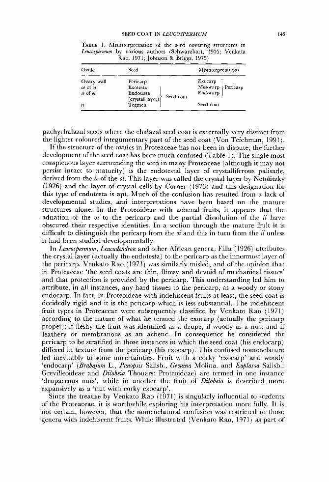
TABLE 1. Misinterpretation of the seed covering structures in Leucosberrnurn by various authors (Schwarzbart, 1905; Venkata
Rao, 1971; Johnson & Briggs, 1975)
145
Ovule Seed Misinterpretations
Mesocarp Pericarp Endocarp 1 Ovary wall Pericarp Exocarp
oe of oi Exotesta ie of oi Endotesta
(crystal layer) Seed coat Tegmen 1 1 I Seed coat
~ ~ ~~~ ~
pachychalazal seeds where the chalaza1 seed coat is externally very distinct from the lighter coloured integumentary part of the seed coat (Von Teichman, 1991).
If the structure of the ovules in Proteaceae has not been in dispute, the further development of the seed coat has been much confused (Table 1). The single most conspicuous layer surrounding the seed in many Proteaceae (although it may not persist intact to maturity) is the endotestal layer of crystalliferous palisade, derived from the i e of the oi. This layer was called the crystal layer by Netolitzky (1926) and the layer of crystal cells by Corner (1976) and this designation for this type of endotesta is apt. Much of the confusion has resulted from a lack of developmental studies, and interpretations have been based on the mature structures alone. In the Proteoideae with achenal fruits, it appears that the adnation of the oi to the pericarp and the partial dissolution of the ii have obscured their respective identities. In a section through the mature fruit it is difficult to distinguish the pericarp from the o i and this in turn from the ii unless it had been studied developmentally.
In Leucospermum, Leucadendron and other African genera, Filla ( 1926) attributes the crystal layer (actually the endotesta) to the pericarp as the innermost layer of the pericarp. Venkato Rao (1971) was similarly misled, and of the opinion that in Proteaceae ‘the seed coats are thin, flimsy and devoid of mechanical tissues’ and that protection is provided by the pericarp. This understanding led him to attribute, in all instances, any hard tissues to the pericarp, as a woody or stony endocarp. In fact, in Proteoideae with indehiscent fruits at least, the seed coat is decidedly rigid and it is the pericarp which is less substantial. The indehiscent fruit types in Proteaceae were subsequently classified by Venkato Rao (1971) according to the nature of what he termed the exocarp (actually the pericarp proper); if fleshy the fruit was identified as a drupe, if woody as a nut, and if leathery or membranous as an achene. In consequence he considered the pericarp to be stratified in those instances in which the seed coat (his endocarp) differed in texture from the pericarp (his exocarp). This confused nomenclature led inevitably to some uncertainties. Fruit with a corky ‘exocarp’ and woody ‘endocarp’ (Brabajum L., Panopsis Salisb., Gevuina Molina. and Euplassa Salisb.: Grevilleoideae and Dilobeia Thouars: Proteoideae) are termed in one instance ‘drupaceous nuts’, while in another the fruit of Dilobeia is described more expansively as a ‘nut with corky exocarp’.
Since the treatise by Venkato Rao (1971) is singularly influential to students of the Proteaceae, i t is worthwhile exploring his interpretation more fully. I t is not certain, however, that the nomenclatural confusion was restricted to those genera with indehiscent fruits. While illustrated (Venkato Rao, 1971) as part of
146 J. C. MANNING AND G. J. BRITS
the ‘pericarp’ in Orothamnus Hook., Adenanthos Labill., Petrophila R. Br., Aulax Berg. and Leucadendron (Proteoideae) , the distinctive palisade layer containing prismatic crystals is also (this time correctly) attributed to the testa in Grevillea J. Knight and Hakea Schrader (Grevillioideae) but to the tegmen in Embothrium Forest & Forster f. (Grevillioideae). In the grevillioid genera this layer is evidently crushed at maturity and so apparently does not develop the crystal sand found in Leucospermum and other proteoid genera. Despite this difference in ultimate fate, it is necessary to establish absolutely whether or not the palisade layer is homologous in all genera in which it occurs. Corroboration of the homology of the crystalliferous palisade thoughou t the Proteaceae comes from the studies on fruit development in Persoonia (Persoonioideae) , Hickbeachia F. Muell. and Macadamia (Grevillioideae) (Stroschen, 1986a-c). In all these genera the crystalliferous layer is identified as endotestal, and together with the present study indicates its homology throughout the family. In view of its occurrence in members of all three subfamilies, it appears that a crystalliferous endotesta is characteristic of the Proteaceae and plesiomorphic, and its absence should be viewed as secondary and an apomorphy.
Macadamia F. Muell. (Grevillioideae) has been singled out as the only genus in the family in which the seeds exhibit a hard seed coat, but this was incorrectly interpreted (Venkato Rao, 1971) as of perichalazal origin and thus not homologous with the seed coat in other Proteaceae. I n fact, although the seed in Macadamia is pachychalazal, a significant portion of the seed coat is integumental and this portion is endotestal with a crystalliferous inner epidermis (Joubert, 1986; Strohschen, 1986a). The chalaza1 part of the seed coat develops a similar crystalliferous layer and is externally indistinguishable, and the seed coat is uniformly woody. The structure of the integumental part of the seed coat in Macadamia is thus homologous with the seed coat of other Proteaceae, and far from being exceptional in the family by virtue of its hard seed coat, Macadamia is perfectly normal. The pachychalazal nature of the mature seed is also apparent, although not stated, in Hicksbeachia and Persoonia (Stroschen 1986b, c).
Thus the fruit wall in Leucospermum and Leucadendron at least, and probably throughout the family, has erroneously been construed to comprise the pericarp proper plus the adnate outer integument, which separates from the inner integument as the latter disintegrates, and the seed coat to comprise merely those scant remnant layers of the ii which adhere to the embryo sac. This interpretation results in the view that the pericarp proper in Leucospermum is an exocarp while the outer integument layers (the testa proper) constitute a hard endocarp. The fruit of Leucospermum has accordingly been misinterpreted as a nut, supposedly derived from a drupe (Venkato Rao, 1971), whereas it is actually an achene, and the drupes actually achenes with fleshy pericarp. The mature seeds of Leucospermum cordgolium are dispersed by ants (Slingsby & Bond, 1981) and the pericarp functions as an elaisosome. When the seed is shed the pericarp is turgid and gelatinous. As the fruit wall is thus not strictly dry, the fruit may be considered as an unusual, somewhat fleshy, achene.
The immediate result of the misinterpretation has been complete confusion as to the identity of the fruit and seed layers, particularly in taxa with indehiscent fruits. Accordingly, Johnson & Briggs (1975), in their treatise on phylogeny in Proteaceae, following this interpretation, defined the crystal layer as the endocarp, and reported its presence in other species of Leucospermum examined by
SEED COAT IN L E U C O S P E R M U M 147
themselves. They thus interpreted the fruits of Leucospermum and Leucadendron (amongst others) also as nuts (type 6 fruits: 132) and were forced to conclude that ‘types of fruit, and particularly of endocarp development.. . only partly correspond to groupings which are supported by other. . . evidence’. Whether this will be the conclusion when seeds of the Proteaceae are correctly interpreted remains to be seen. Reassessment of the identity of the crystalliferous palisade leads to the suspicion that the distinction between types 3, 5, and 6 fruits recognized by Johnson & Briggs (1975) is scarcely tenable. The ramifications of this misapprehension in phylogenetic speculations are extreme, and all further discussion relating to crystalliferous endocarps in that publication must be viewed with caution until it is established whether any of the genera referred to actually have such an endocarp. Naturally, were the crystal layers found to be invariably testal, then their homology would be unimpeachable, and some of the phylogenetic speculation justified, albeit that the tissues are misnamed. A survey of the distribution of a persistent crystalliferous endotestal palisade in Proteaceae is more likely to yield a character of phylogenetic significance than is the distinction between fleshy, woody or leathery pericarps, uncertain as it sometimes is.
It is clear that a complete re-evaluation of published accounts of the nature of the fruit and seed covering structures in Proteaceae is required before any meaningful phylogenetic discussions can be entertained.
ACKNOWLEDGEMENTS
This paper is dedicated to the memory of Professor Piet Jordaan, who lectured to one of us (GB) and was the first author to interpret correctly the covering structures in the proteaceous seed. Unfortunately linguistic parochialism determined that his work was overlooked, and his contribution to the field unappreciated. We thank the Electron Microscope Unit, University of Cape Town and especially M r Dane Geericke, for technical assistance. Comments on the manuscript by the referees are much valued.
REFERENCES
Bouman F. 1978. Development ofovule and seed coat structure in Angiosperms. Vistas in Plant Science, 5: 1-73. Comer EJH. 1976. The seeds of dicotyledons. Cambridge: Cambridge University Press. Filla F. 1926. Das Perikarp der Proteaceae. Floras 120: 99-142. Johnson LAS, & Briggs BG. 1975. O n the Proteaceae-the evolution and classification of a southern Family.
Botanical Journal of the Linnean Society 70: 83-182. Jordaan PG. 1945. (a) Die saadknop en ernbriologie van Leucadendron (b) Die saadknop en ernbriologie van
Leucospermum conocarpum R. Br. Annale van die Universitieit van Stellenbosch 23: 1-45. Joubert AJ. 1986. ‘ n Vergehkende vrugmorfoologenetise studie van Macadamia integrifolia (Maiden @ Betche) en
Faurea speciosa ( Welw.) (Proteaceae). Unpublished D.Sc. thesis, University of Pretoria, South Africa. Midgley JJ. 1987. The deviation, utility and implications of a divergence index for the fynbos genus
Leucadendron (Proteaceae). Botanical Journal of the Linnean SocieQ 95: 137-1 52. Netolitzky F. 1926. Anatomie der Angiospermen-Samen. In Linsbsauer, K. ed, Handbuch der Pflanzenanatomie
10: 364. Berlin: Gehruder Borntraeger. Schwarzbart J. 1905. Anatomische Untersuchungen von Proteaceen-Fruchten und Samen. Beihefle botanisches
~en t ra lb la t t 18: 1-27. Singh RP. 1954. Structure and development of seeds in Euhorhiaceae: Ricinus communis L. Phytomorpholou 4:
Slingsby P, Bond W. 1981. Ants-friends of the fynbos. Veld and Flora 67: 39-45. 118-123.
148 J. C. MANNING AND G. J. BRITS
Stroschen B. 1986a. Contributions to the biology of useful plants 4. Anatomical studies of fruit development and fruit classification of the Macadamia nut (Macadamia integrifolia Maiden and Betche). Angewandte Botanik 60: 239-247.
Stroschen B. 198613. Contributions to the biology of useful plants 5. Anatomical studies of fruit development and fruit classification of the Monkey nut (Hicksbeachia pinnatiflia F. Muell.). Angewandte Botanik 60:
Stroschen B. 1986c. Contributions to the biology of useful plants 6 . Anatomical studies of fruit development and fruit classification of Persoonia pinifl ia R. Br. Angewandte Botanik 60: 257-265.
Venkata Rao C. 1971. Proteaceae. Botanical Monograph No. 6. New Delhi: Council of Scientific and Industrial Research .
Von Teichman I. 1991. Ontogeny of the seed-coat of Rhus lanceu L. f., and pachychalazy in the Anacardiaceae. Botanical Journal of the Linnean Sociely 107: 35-47.
Von Teichman I, Van Wyk AE. 1991. Trends in the evolution of dicotyledonous seeds based on character associations, with special reference to pachychalazy and recalcitrance. Botanical Journal IJJ the Linnean .So&&
249-256.
105: 21 1-237.