Synthetic viability genomic screening defines Sae2 function in DNA repair
Upload
khangminh22Category
view
1download
0
Screening of Marine Natural Products and Their Synthetic Derivatives for Antimicrobial and Antiproliferative Properties
CENTRE FOR DRUG RESEARCHDIVISION OF PHARMACEUTICAL BIOSCIENCESFACULTY OF PHARMACYDOCTORAL PROGRAMME IN DRUG RESEARCHUNIVERSITY OF HELSINKI
SOFIA MONTALVÃO
dissertationes scholae doctoralis ad sanitatem investigandam universitatis helsinkiensis 16/2016
16/2016
Helsinki 2016 ISSN 2342-3161 ISBN 978-951-51-1939-1
SO
FIA
MO
NT
AL
VÃ
O S
creenin
g of Marin
e Natu
ral Prod
ucts an
d T
heir S
ynth
etic Derivatives for A
ntim
icrobial and
An
tiproliferative P
roperties
Recent Publications in this Series
103/2015 Elina ReponenPreoperative Risk-Assessment Methods, Short-Term Outcome, and Patient Satisfaction in Elective Cranial Neurosurgery104/2015 Riina KandolinCardiac Sarcoidosis in Giant Cell Myocarditis in Finland106/2015 Karmen KappPolyphenolic and Essential Oil Composition of Mentha and Their Antimicrobial Effect107/2015 Dina PopovaNeurophysiological mechanisms of Plasticity Induced in Adult Brain1/2016 Pauliina SaurusRegulation of Podocyte Apoptosis in Diabetic Kidney Disease – Role of SHIP2, PDK1 and CDK22/2016 Sanna ToivonenDerivation of Hepatocyte Like Cells from Human Pluripotent Stem Cells3/2016 Marjaana PeltolaAMIGO-Kv2.1 Potassium Channel Complex: Identification and Association with Schizophrenia-Related Phenotypes4/2016 Niko-Petteri NykänenCellular Physiology and Cell-to-Cell Propagation of Tau in Neurodegeneration: The Impact of Late-Onset Alzheimer’s Disease Susceptibility Genes5/2016 Liisa KorkaloHidden Hunger in Adolescent Mozambican Girls: Dietary Assessment, Micronutrient Status, and Associations between Dietary Diversity and Selected Biomarkers6/2016 Teija OjalaLactobacillus crispatus and Propionibacterium freudenreichii: A Genomic and Transcriptomic View7/2016 César AraujoProstatic Acid Phosphatase as a Regulator of Endo/Exocytosis and Lysosomal Degradation8/2016 Jens VerbeerenRegulation of the Minor Spliceosome through Alternative Splicing and Nuclear Retention of the U11/U12-65K mRNA9/2016 Xiang Zhao HMGB1 (Amphoterin) and AMIGO1 in Brain Development10/2016 Tarja Pääkkönen (Jokinen)Benign Familial Juvenile Epilepsy in Lagotto Romagnolo Dogs11/2016 Nora HiivalaPatient Safety Incidents, Their Contributing and Mitigating Factors in Dentistry12/2016 Juho HeinonenIntravenous Lipid Emulsion for Treatment of Local Anaesthetic and Tricyclic Antidepressant Toxicity13/2016 Riikka JokinenGenetic Studies of Tissue-Specific Mitochondrial DNA Segregation in Mammals14/2016 Sanna MäkeläActivation of Innate Immune Responses by Toll-like Receptors and Influenza Viruses15/2016 Mari HirvinenImmunological Boosting and Personalization of Oncolytic Virotherapies for Cancer Treatment
Centre for Drug Research Division of Pharmaceutical Biosciences
Faculty of Pharmacy University of Helsinki
FINLAND
Screening of Marine Natural Products and Their Synthetic Derivatives for Antimicrobial and Antiproliferative Properties
Sofia Isabel Gonçalves Hernâni Martins Montalvão
ACADEMIC DISSERTATION
To be presented, with the permission of the Faculty of Pharmacy of
the University of Helsinki, for public examination in Auditorium 2 at Viikki Infocenter Korona (Viikinkaari 11) on February 26th, at 12 o’clock noon.
Helsinki 2016

�2
Supervisors Docent Päivi Tammela, Ph.D.
Centre for Drug Research Division of Pharmaceutical Biosciences
Faculty of Pharmacy University of Helsinki
FINLAND
Professor Heikki Vuorela, Ph.D. Division of Pharmaceutical Biosciences
Faculty of Pharmacy University of Helsinki
FINLAND
Reviewers Docent Heiko Rischer, Ph.D.
VTT – Technical Research Centre of Finland Ltd.
Espoo FINLAND
Professor Kemal Hüsnü Can Başer, Ph.D.
Division of Pharmacognosy Faculty of Pharmacy
Anadolu University
TURKEY
Opponent Professor Maria Valeria D’Auria, Ph.D.
Department of Pharmacy University of Naples Federico II
ITALY © Sofia Montalvão 2016 ISBN 978-951-51-1939-1 (paperback) ISBN 978-951-51-1940-7 (PDF) ISSN 2342-3161 (print) and ISSN 2342-317X (online) Cover image by Vilen Looga (adapted from “Art of a Diatom algae” by Ernst Haeckel). Hansaprint Helsinki 2016

�3
Table of contents
ABSTRACT 5
RESUMO 7
ACKNOWLEDGEMENTS 9
LIST OF ORIGINAL PUBLICATIONS (I, II, III AND IV) 11
AUTHOR’S CONTRIBUTION 12
LIST OF FIGURES 13
LIST OF TABLES 14
ABBREVIATIONS 15
1. INTRODUCTION 17
2. REVIEW OF LITERATURE 18 2.1. SOURCES OF MNPS: OVERVIEW OF THE MARINE-DERIVED SOURCES USED IN THE SEARCH OF NEW BIOACTIVE COMPOUNDS 18 2.1.1. MAIN CLASSES OF MARINE BIOACTIVE COMPOUNDS 23 2.1.1.1. Terpenes 25 2.1.1.2. Peptides 25 2.1.1.3. Alkaloids 26 2.1.1.4. Polyketides 27 2.1.1.5. Polysaccharides 27 2.2. MNPS AS SCAFFOLDS 28 2.3. EXTRACTION OF BIOACTIVE COMPOUNDS: OVERVIEW OF SAMPLE PREPARATION AND EXTRACTION CONDITIONS 30 2.4. IMPORTANCE AND APPROACHES FOR SCREENING BIOACTIVE COMPOUNDS: GENERAL OVERVIEW OF BIOACTIVE SCREENING 32 2.5. MARINE DRUG DISCOVERY AND DEVELOPMENT 33 2.5.1. COMMERCIAL USES AND APPLICATIONS OF MNPS 36
3. AIMS OF THE STUDY 42
4. MATERIAL AND METHODS 43 4.1. MICROBIAL STRAINS AND GROWTH CONDITIONS 43 4.2. CELL LINES AND CULTURE CONDITIONS 45 4.3. REFERENCE COMPOUNDS 45 4.4. MARINE TEST MATERIAL 45 4.5. SCREENING FOR BIOACTIVITY 46 4.5.1. ANTIMICROBIAL SCREENING 46
�4
4.5.2. IN VITRO ANTIPROLIFERATIVE AND CYTOTOXICITY SCREENING 47 4.6. SELECTIVITY INDEX 49 4.7. DATA ANALYSIS 49 4.8. QUALITY PARAMETERS 49
5. SUMMARY OF RESULTS AND DISCUSSION 51 5.1. OROIDIN AND CLATHRODIN AS PARENT STRUCTURES FOR SYNTHESIS OF NEW COMPOUNDS (I), (II) 51 5.1.1. ANTIMICROBIAL STUDIES (I), (II) 51 5.1.2. ANTIPROLIFERATIVE (I), (II) AND ANTIVIRAL STUDIES (II) 55 5.1.3. SELECTIVITY STUDIES 58 5.2. OKADAIC ACID AS INSPIRATION FOR MACROCYCLIC SYNTHETIC COMPOUNDS (III) 59 5.2.1. ANTIMICROBIAL STUDIES 60 5.2.2. ANTIPROLIFERATIVE STUDIES 61 5.3. BIOPROSPECTING FOCUSED ON THE AEGEAN SEA (IV) 64 5.3.1. ANTIMICROBIAL AND ANTIFOULING STUDIES 65 5.3.2. ANTIPROLIFERATIVE STUDIES 65 5.3.3. ANTI-INFLAMMATORY STUDIES 66
6. CONCLUSIONS AND FUTURE CONSIDERATIONS 68 6.1. FUTURE RESEARCH 69
REFERENCES 71
���������������������

�5
Abstract �Marine environment is prolific in organisms with unique properties. Seas and oceans contain a wide diversity of species with biologically active metabolites that represent a valuable source with great potential for the development of novel therapeutic agents. This dissertation is focused on the biological study of synthetic compounds based on marine scaffolds as well as on marine natural product extracts originating from the Aegean Sea. Furthermore, it offers an introduction on the importance of marine natural products in the search of new bioactive compounds, the use of natural products as scaffolds for the synthesis of new drugs, and a general overview on bioactivity screening and the current status of marine-derived bioactive compounds as therapeutic agents. The potential of oroidin and clathrodin as parent structures for synthesis of novel compounds was explored. Antimicrobial and antiproliferative studies were conducted and it was concluded that 4-phenyl-2-aminoimidazoles 6g(I) and 6h(I) showed the best antimicrobial effect against Gram-positive bacteria (Enterococcus faecalis and Staphylococcus aureus), while compound 6j(I) showed the most interesting IC50 in antiproliferative studies. Compound 7(II), a synthetic derivative of 2-aminobenzothiazole, showed IC50 of 16 μM and 71 μM against a cancer cell line and a normal cell line, respectively. The selectivity index showed selectivity towards cancerous cells. In addition, okadaic acid was used as inspiration for the synthesis of crown ether acyl compounds. Compound (1,4,7,10,13,16-hexaoxacyclooctadecan-2-yl)methyl 3-(pyren-1-yl)propanoate) 1o(III) was found to be the most active in antimicrobial studies against Gram-positive Staphylococcus aureus with a MIC50 of 7.2 μM. The importance of bioprospecting the rich marine biodiversity in the Aegean Sea was also studied in this thesis. Biological activities of extracts from cyanobacteria, micro- and macroalgae were evaluated, and microalgae

�6
extracts (Amphora cf capitellata and Nitzschia communis) showed the most interesting antimicrobial results against Staphylococcus aureus and fungus Candida albicans. The results of the biological studies conducted in this thesis demonstrated antimicrobial and antiproliferative activity of several marine natural products and their synthetic derivatives. Further studies and structural optimization should be done to fully explore their potential for the development of therapeutic agents.
�
�
�7
Resumo O ambiente marinho é promissor na variedade de organismos com propriedades únicas. Dos mares e oceanos, existe uma diversidade imensa de metabólitos bioactivos que representam uma fonte de enorme valor e com grante potencial para o desenvolvimento de novos agentes terapêuticos. Esta tese mostra o estudo biológico de compostos sintéticos com base em “scaffolds” marinhos e extractos do Mar Egeu. Além disso, esta tese oferece uma introdução sobre a importância de produtos naturais marinhos e a procura de novos compostos bioactivos, no uso de produtos naturais como “scaffolds” para a síntese de novos fármacos e uma visão geral sobre avaliação da capacidade biológica de compostos marinhos e o status de compostos bioactivos como agentes terapêuticos O potential do composto clathrodin como modelo para a síntese de novos compostos foi explorado. Estudos antimicrobianos e antiproliferativos foram realizados e concluiu-se que 4-phenyl-2-aminoimidazoles 6g(I) e 6h(I) teve os melhores resultados antimicrobianos quando testados contra bactérias Gram-positivas (Enterococcus faecalis and Staphylococcus aureus) enquanto que o composto 6j(I) mostrou o IC50 mais interessante nos estudos antiproliferativos. O composto 7(II), um derivado sintético de 2-aminobenzotiazole mostrou IC50 de 16 μM e 71 μM quando testado contra uma linha celular cancerígena e uma linha celular não-cancerígena, respectivamente. O índice de selectividade mostrou uma selecção para células cancerígenas. O ácido ocadáico foi usado como inspiração para a síntese de compostos macrocíclicos. Composto (1,4,7,10,13,16-hexaoxacyclooctadecan-2-yl)methyl 3-(pyren-1-yl)propanoate) 1o(III) foi o mais activo em estudos antimicrobianos quando testado contra a bactéria Gram-positiva Staphylococcus aureus, com MIC50 igual a 7.2 μM. A importância de bioprospecção da enorme biodiversidade marinha no Mar Egeu, foi também estudado nesta tese. Estudos biológicos foram feitos em
�8
extratos de cyanobacteria, micro- e macroalgas e as microalgas marinhas (Amphora cf capitellata e Nitzschia communis) mostraram a melhor actividade antimicrobial, quando testados contra a bacteria Staphylococcus aureus e o fungo Candida albicans. Os resultados preliminares de estudos biológicos em compostos marinhos, presentes nesta tese, mostraram o potencial destes em terapias antimicrobianas e anticancerígenas. Num futuro próximo, uma optimização na estrutura química destes compostos poderá ser realizada, para explorar todo o potencial como agentes terapêuticos. ��������������������������������
�9
Acknowledgements Undertaking this PhD has been a truly life-changing experience for me and it would not have been possible to do without the support that I received from many people. First and foremost, I want to thank my first supervisor, Docent Päivi Tammela Tammela for her contribution of time, ideas and superb scientific knowledge. Thanks to her, I gained excellent insights into marine pharmacology and developed essential skills for my future career. Many thanks to my second supervisor, Professor Heikki Vuorela, for his suggestions and sense of humor often needed in stressful situations. I gratefully acknowledge Prof. Kemal Hüsnü�Can Başer and Docent Heiko Rischer for pre-examining the thesis and for their valuable suggestions, and Prof. Maria Valeria D’Auria for accepting the invitation to act as the official opponent. I am thankful to all the co-authors that helped in the experimental work and writing of the publications. The members of the Bioactivity Screening group have contributed immensely to my personal and professional time at the Centre for Drug Research (CDR). I am especially grateful to Andreas Helfenstein, Katja-Emilia Lillsunde, Päivi Järvinen, Shafiul Haque and Susanna Nybond. My sincere thanks to the Cancer Immunovirotherapy group, especially to Cristian Capasso, Lukasz Kuryk and Mariangela Garofalo for their great help in the Cell lab – I am going to miss the happy times and the great work environment we had there! Also, I would like to thank colleagues and former colleagues from the Faculty of Pharmacy for their skill in spreading happiness, even on those scientifically dark days: Alma, Dominique, Elina, Enass, Erja, Leena, Liisa, Marco, Mari, Otto, Olli, Pia F., Tarja, Tiina and all the others! Many thanks to Prof. Jari Yli-Kauhaluoma, Dr. Paula Kiuru and Dr. Hélder Santos from the Division of Pharmaceutical Chemistry, for their knowledge and help in professional and non-professional issues.

�10
I also take this opportunity to acknowledge the funding bodies: EU FP-7 MAREX (grant no. 245137, within KBBE-2009-3-2-01), the Academy of Finland (grant no. 2770011) and the Finnish Cultural Foundation. A very special thank you to friends who were always there for me, during the laughter and the tears, always reminding me that I am strong and I can do it! Aliki, Andreia, Cristina, Diana, Elisabete(s) Lídia, Michal, Oleg, Shinjini, Tatiana, Yanming – you are awesome and you know it! And to many others! Also, being in contact with friends from my home country always cheered me up and gave me confidence! A special thanks to Navi, my little cutie pie! You always make me happy every time I arrive home and I love you for always being by my side! Last and the most important, I would like to say a heartfelt thank you to my parents, my sister and Vilen – I dedicate this thesis to you. Father and Mother, MUITO OBRIGADA E AMO-VOS MUITO! For always believing in me, being always at my side, for all the conversations even being far away from you (and I miss you so much!!!) and for the encouragement to follow my dreams. You are perfect! Patrícia, MUITO OBRIGADA! For all the moments we have had as sisters and for your support and help whenever I needed it. You are an amazing and extremely intelligent woman and I am very proud of being your sister. And Vilen, my loving, smart, supportive, encouraging and patient fiancé, to whom I am going to get married this year! You have been by my side throughout this Ph.D., giving me strength every single minute of it. Without your belief and encouragement I would not have had enough faith in myself. I feel that we have both learned a lot about life, strengthening our commitment and determination for each other and to live life to the fullest. ��
Helsinki, 10 February 2016

�11
List of original publications (I, II, III and IV) This thesis is based on the following publications and manuscripts:
I� Nace Zidar†, Sofia Montalvão†, Žiga Hodnik, Dorota A. Nawrot, Aleš Žula,
Janez Ilaš, Danijel Kikelj, Päivi Tammela, Lucija Peterlin Mašič. Antimicrobial Activity of the Agelas Marine Alkaloids Clathrodin and Oroidin, and Their
Synthetic Analogues. Marine Drugs 2013. 12(2), 940-963; doi:10.3390/md12020940. † These authors contributed equally to this work.
II� Sofia Montalvão, Teppo O. Leino, Paula S. Kiuru, Katja-Emilia Lillsundea,
Jari Yli-Kauhaluoma, Päivi Tammela. Synthesis and Biological Evaluation of 2-Aminobenzothiazole and Benzimidazole Analogs Based on Clathrodin
Structure. Archiv der Pharmazie 2016. 349(2), 137-149; doi: 10.1002/ardp.201500365.
III� Martin Febles†, Sofia Montalvão†, Guillermo D. Crespín, Martin Norte, José
M. Padrón, Päivi Tammela, José J. Férnandez, António H. Daranas. Synthesis and Biological Evaluation of Crown Ethers Acyl Derivatives
(manuscript) † These authors contributed equally to this work.
IV� Sofia Montalvão, Zeliha Demirel, Prabha Devi, Valter Lombardi, Vesa Hongisto, Merja Perälä, Johannes Hattara, Esra Imamoglu, Supriya Shet
Tilvi, Gamze Turan, Meltem Conk Dalay, Päivi Tammela. Large-scale Bioprospecting of Cyanobacteria, Micro- and Macroalgae from the Aegean
Sea. New Biotechnology 2016, accepted.
The publications are referred to in the text by their Roman numerals. The articles are
reprinted with permission from the publishers. The supporting information of the
original publications is not included in this thesis. The material is available from the author or online.
�12
Author’s contribution ��� Sofia Montalvão participated in the design of the study and executed
the antimicrobial testing and collaborated with the other authors in writing the manuscript. Sofia Montalvão and Nace Zidar contributed equally.�
���� Sofia Montalvão participated in the design of the study and
experimental work. She analyzed the results and wrote the manuscript in collaboration with the other authors.�
���� Sofia Montalvão participated in the design of the study, in the
experimental work, in interpreting the results and writing the manuscript. Sofia Montalvão and Martín Febles contributed equally.�
���� Sofia Montalvão participated in the design of the study and executed
part of the experimental work. She analyzed the results and wrote the manuscript in collaboration with the other authors.�
� �
�� ������������
�13
�List of figures �FIGURE 1: TREND IN THE NUMBER OF MARINE NATURAL PRODUCTS FROM 1985 TO
2012 [177]. 18 FIGURE 2: OVERVIEW OF MARINE NATURAL PRODUCTS IN THE DISCOVERY OF
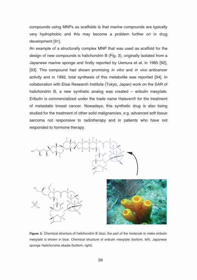
NOVEL BIOACTIVE COMPOUNDS, FROM 2010 TO 2013 (ADAPTED FROM: [178]).19 FIGURE 3: CHEMICAL STRUCTURE OF HALICHONDRIN B (TOP): THE PART OF THE
MOLECULE TO MAKE ERIBULIN MESYLATE IS SHOWN IN BLUE. CHEMICAL STRUCTURE OF ERIBULIN MESYLATE (BOTTOM, LEFT). JAPANESE SPONGE HALICHONDRIA OKADAI (BOTTOM, RIGHT). 29
FIGURE 4: THE PROCESS OF DRUG DISCOVERY AND DEVELOPMENT – “THE PIPELINE” (ADAPTED FROM: [108]). AN INVESTIGATIONAL NEW DRUG APPLICATION (INDA) IS SUBMITTED BEFORE THE CLINICAL TRIALS AND A NEW DRUG APPLICATION IS A SPONSOR’S REQUEST FOR APPROVAL OF A DRUG FOR COMMERCIALIZATION. 35
FIGURE 5: DIFFERENT STAGES OF CLINICAL TRIALS (ADAPTED FROM: [179]). IN PHASE IV, ACTIVE POST MARKETING SURVEILLANCE OF DRUG SIDE EFFECTS IS ESSENTIAL. 36
FIGURE 6: CHEMICAL STRUCTURES OF PYRROLE-IMIDAZOLE ALKALOIDS, CLATHRODIN (R = H) AND OROIDIN (R = BR). 52
FIGURE 7: A) DOSE-RESPONSE CURVES OF COMPOUND 7(II) AGAINST HUMAN MELANOMA CELL LINE A-375, BY MTT ASSAY. RESULTS ARE EXPRESSED AS PERCENTAGE OF CYTOTOXICITY. B) LDH LEAKAGE IN A-375 HUMAN MELANOMA CELLS AND BALB/C 3T3 CLONE A31 FIBROBLAST CELLS AFTER COMPOUND 7(II) EXPOSURE. RESULTS ARE EXPRESSED AS PERCENTAGE OF INTRACELLULAR LACTATE DEHYDROGENASE LEAKAGE. DATA ARE THE MEANS FROM THREE INDEPENDENT EXPERIMENTS. 57
FIGURE 8: EXAMPLES OF CROWN ETHERS, WITH INCREASING RING SIZE (FROM LEFT TO RIGHT): 12-CROWN-4, 15-CROWN-5 AND 18-CROWN-6 ETHER. 60
FIGURE 9: DOSE-RESPONSE CURVES OF COMPOUND 1O(III) AGAINST S. AUREUS (SQUARE) AND C. ALBICANS (TRIANGLE). RESULTS WERE EXPRESSED AS % OF
GROWTH INHIBITION. 61 FIGURE 10: PROCEDURES PERFORMED FOR THE DISCOVERY OF BIOACTIVE
SPECIES. CYANOBACTERIA AND MICROALGAE SPECIES WERE CULTIVATED IN BATCH CULTURES. 64
�

�14
List of Tables
TABLE 1: BIOACTIVE COMPOUNDS FROM CYANOBACTERIA. 21 TABLE 2: SELECTED EXAMPLES OF BIOACTIVE MNP STRUCTURES BELONGING TO
THE FOLLOWING CHEMICAL CLASSES: TERPENES, PEPTIDES, ALKALOIDS, POLYKETIDES AND POLYSACCHARIDES. 23
TABLE 3: SOLVENTS COMMONLY USED IN EXTRACTION OF BIOACTIVE COMPOUNDS (ADAPTED FROM: [100]). 31
TABLE 4: MARINE-DERIVED COMPOUNDS IN DRUG DISCOVERY AND DEVELOPMENT (ADAPTED FROM: [122]). NP = NATURAL PRODUCT. 39
TABLE 5: BIOLOGICAL ASSAYS USED IN STUDIES (I)-(IV). 43 TABLE 6: CHARACTERISTICS OF PATHOGENIC BACTERIA AND FUNGUS USED IN
THIS WORK [124]. 44 TABLE 7: ANTIMICROBIAL MINIMUM INHIBITORY CONCENTRATION ACTIVITY (MIC90
AND MIC50) OF ANALOGS 6G(I) AND 6H(I) AGAINST ENTEROCOCCUS FAECALIS ATCC 29212, STAPHYLOCOCCUS AUREUS ATCC 25923, ESCHERICHIA COLI ATCC 25922 AND CANDIDA ALBICANS ATCC 90028. ALL RESULTS ARE SHOWN IN μM. 54
TABLE 8: CHEMICAL STRUCTURES OF SELECTED CROWN ETHER DERIVATIVES AND THEIR BIOLOGICAL ACTIVITIES. FOR ANTIMICROBIAL STUDIES, CIPROFLOXACIN (BACTERIA) AND AMPHOTERICIN B (C. ALBICANS) WERE USED AS POSITIVE CONTROLS. FOR ANTIPROLIFERATIVE STUDIES, STANDARD ANTICANCER DRUGS CISPLATIN AND ETOPOSIDE WERE USED AS POSITIVE CONTROLS. ALL RESULTS WERE TESTED IN TRIPLICATE (EXCEPT 1L(III), ANTIPROLIFERATIVE ASSAY) AND VALUES ARE SHOWN IN μM. 63
TABLE 9: NUMBER OF ACTIVE EXTRACTS (% INHIBITION > 50) IN EACH PHYLA. 64
�������������
�15
Abbreviations �ATP Adenosine Triphosphate CFU Colony-forming Unit CLSI Clinical and Laboratory Standards Institute Ctrl Control DMEM Dulbecco’s Modified Eagle Medium DSP Diarrhetic Shellfish Poisoning ECACC European Collection of Cell Cultures EMA European Medicines Agency FBS Fetal Bovine Serum FDA Food and Drug Administration HCV Hepatitis C Virus HPV Human Papillomavirus HRS High-Resolution Screening HTS High-Throughput Screening IC Inhibitory Concentration INDA Investigational New Drug Application kDA kilodalton LDH Lactate Dehydrogenase MAX Maximum MHA Mueller-Hinton Agar MHB Mueller-Hinton Broth MIC Minimum Inhibitory Concentration MIN Minimum MNP Marine Natural Product MTT 3-(4,5-Dimethylthiazol-2-yl)-2,5-Diphenyltetrazolium
Bromide NCS Newborn Calf Serum NP Natural Product NRPS Nonribosomal Peptide Synthetase OA Okadaic Acid
�16
P.C. Positive Control PBS Phosphate Buffered Saline PIA Pyrrole-imidazole Alkaloid PMX Polymyxin B Sulphate PS Polysaccharides PUFA Polyunsaturated Fatty Acid S/B Signal-to-background S/N Signal-to-noise SAR Structure-activity Relationship SD Standard Deviation SDA Sabourad Dextrose Agar SI Selectivity Index TI Therapeutic Index UV Ultraviolet Z’ Z-factor
�

�17
1.� Introduction �Yuri Gagarin once said, “The Earth is blue. How wonderful. It is amazing.” These were the words of the first human astronaut to travel into space and orbit our planet. Approximately three quarters, i.e. 77%, of planet Earth is occupied by water and because of that, some scientists believe that the word “Earth” should have been replaced by “Water”. As a consequence of the amount of water on the planet, the marine biodiversity is enormous ‒ from shallow coastal waters to deep oceans ‒ and is being considered a promising source of unique secondary metabolites for research and drug discovery [1]. Secondary metabolites – compounds not directly involved in the normal growth, development or reproduction [2], and with a molecular weight (MW) below 2 kDa – are normally produced as an answer to ecological pressures, such as competition for space, deterrence of predation, reproduction, among others [3]. Bergmann et al. reported the first discovery of a biologically active marine natural compound in the late 1950s – arabino and ribo-pentosyol nucleosides extracted from Cryptotethia crypta sponge, which was the first demonstration that naturally occurring nucleosides could contain sugars other than ribose and desoxyribose [4]–[6]. Chemical synthesis allowed the development of two derivatives, cytarabine and vidarabine, nucleosides with significant anticancer and antiviral activities [7]. However, only after the discovery of several prostaglandins, isolated from octocoral Plexaura homomalla [8], interest in marine-related research has increased. Due to their broad panel of biological activities, such as their antibacterial, antifungal, anticancer, antiviral, anti-inflammatory, anticoagulant, antiprotozoal, antituberculosis, antidiabetic, antimalarial and antioxidant effects [9]–[12], marine natural products (MNPs) are remarkably interesting, high-value ingredients for applications not only in the pharmaceutical industry, but also in the cosmetic, nutraceutical and agrochemical industries [13]–[15]. The following review provides a comprehensive overview of the importance of marine compounds with a focus on the pharmaceutical field.

�18
2.� Review of literature 2.1.� Sources of MNPs: overview of the marine-derived sources used in
the search of new bioactive compounds �The extraordinary biological, biochemical and biosynthetic potential of marine organisms show that oceans and seas are promising hotspots in the research of secondary metabolites [9], [16]–[21]. Comparing the taxonomic levels, 76 phyla are recognized by the “Catalogue of Life” (online database of the world’s known species [22]), of which 60 include marine representatives, while 40 phyla are represented by terrestrial ones. Such a high number of representatives show great biological diversity, corroborating the fact that oceans and seas are the greatest source for the discovery of new therapeutic agents [23]. From mid-1980s until 2012, there has been an outstanding increase in discovered MNPs, mainly due to several advances in technology, and more than 22 000 compounds have been isolated from marine organisms (Figure 1). Figure 2 shows data on marine-derived sources used in the discovery of new bioactive compounds from 2010 to 2013. More than 30% of new bioactive
Figure 1: Trend in the number of marine natural products from 1985 to 2012 [178].

�19
compounds reported from marine sources are derived from microorganisms such as cyanobacteria and microalgae.
Microalgae, one of the first photosynthetic microorganisms have been highlighted as the new source of marine bioactive compounds due to their interesting biological activities [24]. Their enormous biodiversity results from the capability to adapt to different environments, some of them extreme. To tolerate extreme adverse environments, microalgae develop defense strategies, resulting in a high diversity of compounds from different metabolic pathways. As cyanobacteria, some species of microalgae can produce value-added compounds such as antioxidants, polyunsaturated fatty acids (PUFAs), polysaccharides and sterols [25]. Other microalgae also produce high quantities of hydrocarbons – convertible into biodiesel or hydrogen used as alternative energy sources. In structure and function, microalgae can be prokaryotic or eukaryotic: the prokaryotic ones belong to the Cyanophyta
Microorganisms; 38%
Sponges; 20%
Algae; 14%
Others; 16%
Coelenterates; 4%
Mollusks; 5% Echinoderms; 1%
Bryozoans; 2%
Figure 2: Overview of marine natural products in the discovery of novel bioactivecompounds, from 2010 to 2013 (Adapted from: [179]).

�20
group (cyanobacteria) � ; the eukaryotic ones are included mainly in Bacilariophyceae (diatoms), Dinophyceae (dinoflagellates), Prymnesiophycae (coccolithophores), Cryptophyceae (cryptomonas), Prasinophyceae and Chlorophyceae. It is estimated that until now, around 30 000 microalgae have been studied and analyzed [26]. Chlorella sp. are eukaryotic green unicellular microalgae from the Chlorophyta group that has been extensively studied due to antitumoral, antibacterial, anticoagulant and antioxidant effects [27], [28]. These microalgae, reported to possess antimicrobial activity originally in 1945 [29], are rich in chlorophyll, polysaccharides, vitamins, minerals and essential amino acids [30]. Other species widely studied due to their tolerance of extreme habitat conditions and physiological aspects are green unicellular halotolerant microalgae that belong to the Chlorophyceae group: Dunaliella sp. These microalgae contain carotenoids, glycerol, lipids, enzymes and vitamins [31], and are known for their antioxidant, antibacterial and analgesic properties [32]. Cyanobacteria (or blue-green algae) are prokaryotic organisms and studies confirm that these organisms have existed approximately for 3.5 billion years [33], [34]. They are considered one of the main organisms responsible for the creation of the current Earth’s atmosphere [35]. All cyanobacteria are photoautotrophs, but there are some groups that can show also heterotrophic metabolism, described as mixotrophics. Endowed with the capacity to adapt to a wide range of environmental conditions, cyanobacteria colonize terrestrial, aquatic and extreme ecosystems. However, the majority of these organisms live in water. They have been studied broadly due to their capacity of producing bioactive compounds, such as toxins (microcystins), siderophores and antibiotics. The use of cyanobacteria in medicine and pharmacology dates back to 1500 B.C., when strains of Nostoc were used to treat gout, fistula and several forms of cancer [36]. Between 1950 and 1970, studies on prospecting, isolation and characterization of cyanobacteria started to be carried out regularly [37], [38]. After 1990, research on these ��������������������������������������������������������� In some studies, cyanobacteria are considered members of the Bacteria domain.

�21
prokaryotics received more emphasis and the pioneering work led by Richard Moore and William Gerwick showed the value of cyanobacteria for biomedical research [20]. The interest in studying compounds produced by marine cyanobacteria is also related to the fact that some of the isolated compounds show enormous structural variety, making them interesting as scaffolds for the synthesis of new drugs. Compounds derived from filamentous genera such as Lyngbya, Leptolyngbya and Symploca have shown anticancer activity [39]–[41]. In a review conducted by Tan [42], in which 128 compounds isolated from marine cyanobacteria were studied, more than 35% of the compounds showed activity in tumoral cell lines and 10% of compounds exhibited activity in normal cell lines (non-tumoral). Some of the compounds showed antimicrobial and anti-inflammatory activities. Table 1 shows examples of bioactive compounds from marine cyanobacteria, their chemical class, species (source) and biological activity. Table 1: Bioactive compounds from cyanobacteria.�
Compound Chemical class Source Biological
activity Reference
Ambiguine Alkaloid Fischerella ambigua Antimicrobial [43], [44]
Aulosirazole Aromatic Aulosira fertilissima Anticancer [45]
Cryptophycin Lipopeptide Nostoc sp. Cytotoxic 46], [47]
Didemnin Lipopeptide Synechocystis
trididemni Anticancer,
antiviral [48]
Dolastatin Lipopeptide Lyngbya sp.
Symploca sp. Anticancer [49]
Hapalindole Alkaloid Fischerella sp. Cytotoxic,
antibacterial [44]
Hassallidin Nonribosomal peptide
synthetase (NRPS) Anabaena sp. Antifungal [50]
Macroalgae, macroscopic and multicellular seaweeds, existing in abundance, are potential renewable resources that have been explored as novel and sustainable sources of compounds for pharmaceutical and nutraceutical applications. These marine algae can be classified according to the presence

�22
of specific pigments into three main groups: Chlorophyta (green algae), Phaeophyta (brown algae) and Rhodophyta (red algae). The color of green algae is caused by the presence of green pigments called chlorophylls (a and b). The presence of fucoxanthin, chlorophyll (a and c) gives Phaeophyta a greenish brown color. The red color of Rhodophyta is attributed to the presence of phycobilins (phycoerythrin and phycocyanin). Several species of macroalgae have the capacity to produce a diverse array of secondary metabolites of potential medicinal values as antimicrobial [51]–[53] and antiproliferative agents [54]. Vairappan et al. showed antibacterial activities of eight halogenated compounds, isolated from five species of red algae Laurencia sp., against a range of Gram-positive bacteria, including antibiotic-resistant bacteria [55]. From the red alga Odonthalia corymbifera, bromophenol compounds have been isolated and synthesized, and antimicrobial activity tested against Gram-positive and Gram-negative bacteria and fungi [56]. The most active isolated compound was 2,2’,3,3’-tetrabromo-4,4’,5,5’-tetrahydroxydiphenylmethane, displaying activity against Candida albicans, Aspergillus fumigatus and Trichophyton sp. Two synthetic analogs from the same alga showed potent antibacterial effect against Staphylococcus aureus, Bacillus subtilis, Proteus vulgaris, Micrococcus luteus and Salmonella typhimurium. The antibacterial activity of two species of brown algae Ecklonia (Ecklonia kurome and Ecklonia stolonifera) have been evaluated against S. aureus, Escherichia coli, Pseudomonas aeruginosa and Bacillus cereus [57] and significant activity was observed against all bacteria except Gram-negative E. coli. Another important marine resource are sponges (Porifera) – sessile aquatic animals that live in rivers or deep oceanic waters. These organisms constitute a mark in the evolutionary history of planet Earth, being one of the oldest existing lineages of metazoans. It is estimated that sponges appeared in Neoproterozoic Era more than 635 million years ago [58]. Recent studies indicate that Porifera played an important role in the oxygenation of the

�23
oceans [59]. Since sponges are benthic (fixed to the seabed during adult life) and with a simple morphology, they were able to develop, among others, an adaptive chemical strategy as a way of defense and competition. Many of the secondary metabolites from sponges present novel chemical structures, showing interesting biological activities [60]–[62]. Antimicrobial studies of secondary metabolites isolated from Lotrochota purpurea – i.e. halogenated alkaloids purpuroines A-J – have been reported to demonstrate inhibitory activity against fungi and bacteria [63]. The extract of an Indonesian marine sponge, Haliclona sp., have been shown to display potential cytotoxic activity against human solid cancer cell lines [MCF-7 (breast), LNCap (prostate), Caco-2 (colon) and HCT-15 (colon) cells] [64]. Among approved MNPs derived from sponges (Sub-chapter 2.5.1), there are some that have recently entered into clinical trials for cancer treatment, such as the synthetic tripeptide hemiasterlin, first identified from the marine sponge Cymbastela sp. [65]. 2.1.1.�Main classes of marine bioactive compounds �The following sub-chapters describe the most relevant chemical classes of marine natural compounds (terpenes, peptides, alkaloids, polyketides and polysaccharides) followed by a brief description on their properties. In Table 2, some selected examples of bioactive MNP structures that belong to these classes are presented. Table 2: Selected examples of bioactive MNP structures belonging to the following chemical classes: terpenes, peptides, alkaloids, polyketides and polysaccharides.
Chemical Classes
Structure and Compound name Reference
Terpenes
Secomanoalide
[66] CHO
OH
OHO
O

�24
Peptides
Didemnin B
[67]
Alkaloids
Nagelamide J
[68]
Polyketides
Spongistatin 1
[69], [70]
O
O
O
O
O
O
OO
O
O
O
O
OO
O
N
N
N
N N
N
N
H
H
HH
H
H
H
HN
Br
Br
NH
O
HNN
NH2
N
HN
OCH3
HHN NH2HO
HN
Br
Br
O
O
O
O
O
O
O
HO
O
O
OOH
Cl
OHOH
OHH
OH
O
O
HOO
O
�25
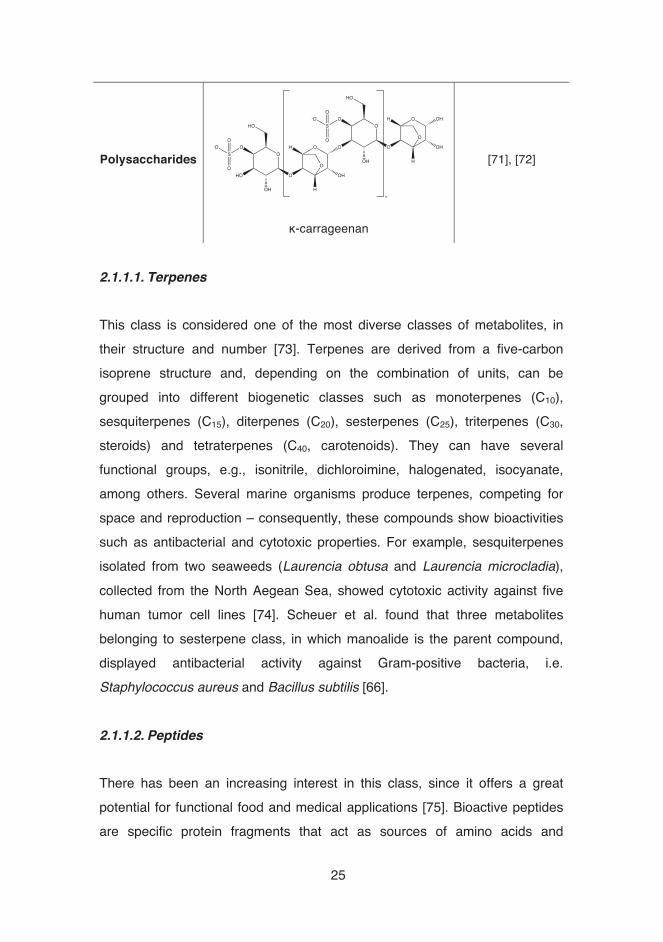
Polysaccharides
κ-carrageenan
[71], [72]
2.1.1.1.�Terpenes �This class is considered one of the most diverse classes of metabolites, in their structure and number [73]. Terpenes are derived from a five-carbon isoprene structure and, depending on the combination of units, can be grouped into different biogenetic classes such as monoterpenes (C10), sesquiterpenes (C15), diterpenes (C20), sesterpenes (C25), triterpenes (C30, steroids) and tetraterpenes (C40, carotenoids). They can have several functional groups, e.g., isonitrile, dichloroimine, halogenated, isocyanate, among others. Several marine organisms produce terpenes, competing for space and reproduction – consequently, these compounds show bioactivities such as antibacterial and cytotoxic properties. For example, sesquiterpenes isolated from two seaweeds (Laurencia obtusa and Laurencia microcladia), collected from the North Aegean Sea, showed cytotoxic activity against five human tumor cell lines [74]. Scheuer et al. found that three metabolites belonging to sesterpene class, in which manoalide is the parent compound, displayed antibacterial activity against Gram-positive bacteria, i.e. Staphylococcus aureus and Bacillus subtilis [66]. 2.1.1.2.�Peptides �There has been an increasing interest in this class, since it offers a great potential for functional food and medical applications [75]. Bioactive peptides are specific protein fragments that act as sources of amino acids and
OHO
O
OH H
H
HO
O
O
O OHO
O
OH H
H OHO
O
HO
HO
O
O
-OS
O O
SO-O
O
n

�26
nitrogen, and have numerous potential physiological functions within the body [76], [77]. The sizes of bioactive peptides range from 2 to 20 amino acid residues in length and are encrypted within the sequence of parent protein. These peptides are inactive or latent in the parent, but are released in an active form by enzymatic hydrolysis. Marine bioactive peptides have shown diverse biological activities. For example, the depsipeptide didemnin B, first isolated from Caribbean tunicate Trididemnum solidum show significant antitumor activity and antiproliferative activity against human prostatic cancer cell lines [67]. Didemnin B is also the first MNP evaluated in human clinical trials. Matsunaga et al. [78] reported that a peptide discodermin A, isolated from marine sponge Discodermia kiiensis, showed antimicrobial activity against a range of Gram-positive and Gram-negative bacteria and fungi. A well-known compound belonging to this class is ziconotide, originally derived from a tropical marine cone snail and commercialized under the trade name Prialt® for the treatment of chronic pain in spinal cord injury (see Sub-chapter 2.5.1 for further details). 2.1.1.3.�Alkaloids �Alkaloids are well known by their wide spectrum of biological activities [79], [80]. Pelletier et al. defined this class as “cyclic organic compounds containing nitrogen in a negative oxidation state which is of limited distribution among living organisms” [81]. They can be divided into seven subclasses: indole alkaloids, pyrrole alkaloids, pyridoacrine alkaloids, isoquinoline alkaloids, gadinine alkaloids, aminoimidazole alkaloids and sterol alkaloids [82]. Pyrrole alkaloids, especially bromopyrroles, are well known for being present in sponges, e.g. Agelas, Axinella, Acanthella, Hymeniacidon and Pseudoaxinyssa. Oroidin is a secondary metabolite from this subclass that exists in more abundance, being also the first one to be isolated� [83], and it is
��������������������������������������������������������2 A revised structure was established in 1973 and proven by total synthesis in 1986 [177]. �

�27
considered to be the biogenetic precursor of all the other bromopyrrole alkaloids. Nagelamide J, isolated from sponge Agelas sp. and the first bromopyrrole alkaloid possessing a cyclopentane ring fused to an aminoimidazole ring, showed antimicrobial activity against Staphylococcus aureus and yeast [68]. Nagelamide A, isolated as well from the same family, inhibited not only the growth of two Gram-positive bacteria but also Gram-negative Escherichia coli [84]. Tunicate-derived trabectedin, a secondary metabolite also belonging to the alkaloid class, is one of the marine-derived drugs that has passed cancer clinical trials [85]. More than 40 years after its discovery, it became the first marine anticancer drug to be approved in the European Union. 2.1.1.4.�Polyketides �Polyketides are low molecular weight compounds assembled via sequential condensations of small carboxylic acids. Their sub-classes comprise polyethers, polyenes, polyphenols, macrolides and polyols, being mainly derived from the simplest (or one of the simplest) building blocks available in nature, i.e. acetic acid. From a pharmaceutical point of view, polyketides are an important source of novel drugs. Eribulin, a synthetic derivative of halichondrin B (natural polyketide) is one example of marine-derived anticancer drugs, approved by Food and Drug Administration (FDA) and European Medicines Agency (EMA). Spongistatin 1, a macrolide initially isolated from sponges Spirastrella spinispirulifera and Hyrtios [69], [70], display significant growth inhibition against a wide variety of cancer cell lines [86]. Macrolides (+)-brefeldin A, (+)-brefeldin C and 7-oxobrefeldin A, isolated from marine-derived fungus Penicillium sp. PSU-F44, have been described to show antimicrobial activity [87]. 2.1.1.5.�Polysaccharides �

�28
The majority of polysaccharides (PS) produced by marine organisms are heteropolysaccharides, i.e. complex carbohydrates composed of repeating units of several types of monosaccharides connected by glycosidic bonds. Mainly micro- and macroalgae contain PS, such as agar, alginates, agarose, carrageenans and fucoidans. There are several applications for PS, such as drug or nutraceutical carriers in pharmaceutical industry, thickeners and gelling agents in food industry, in soil and water treatments [88]. Review conducted by Raposo et al. [72], showed that crude PS and their derivatives can show anticoagulant, antitumor and anticancer activities and can also be potent antibiotic, antioxidant and anti-inflammatory agents. For example, carrageenan, a family of PS obtained by extraction from certain species of red seadweeds (Rhodophyta), have shown potential antiviral properties – Buck and coworkers demonstrated that by testing it against a range of sexually transmitted human papillomaviruses (HPVs) [71]. 2.2.� MNPs as scaffolds �Nowadays, drug discovery is based on three major sources: NPs (natural products), semi-synthetic derivatives of NPs and synthetic compounds derived from combinatorial chemistry [89]. NPs can provide physicochemical properties such as specific interactions with multiple biological targets (barely found in molecules derived from combinatorial synthesis) and contain more chiral centers and a larger number of rings. However, the process from discovery to commercialization is complex, time-consuming and expensive, causing pharmaceutical industries to direct their drug discovery programs from NP-based research into synthetic pathways [90]. Since NPs generally have poor pharmacokinetics (but excellent bioactivity), using NPs as scaffolds for synthesis of new compounds can be pursued to improve the properties. Development of novel compounds by chemical synthesis includes modification/removal of functional groups and introduction of novel groups and stereocenters into the molecule. Sometimes, more radical changes in the scaffold are possible. One thing to keep in mind in the synthesis of new
�29
compounds using MNPs as scaffolds is that marine compounds are typically very hydrophobic and this may become a problem further on in drug development [91]. An example of a structurally complex MNP that was used as scaffold for the design of new compounds is halichondrin B (Fig. 3), originally isolated from a Japanese marine sponge and firstly reported by Uemura et al. in 1985 [92], [93]. This compound had shown promising in vitro and in vivo anticancer activity and in 1992, total synthesis of this metabolite was reported [94]. In collaboration with Eisai Research Institute (Tokyo, Japan) work on the SAR of halichondrin B, a new synthetic analog was created – eribulin mesylate. Eribulin is commercialized under the trade name Halaven® for the treatment of metastatic breast cancer. Nowadays, this synthetic drug is also being studied for the treatment of other solid malignancies, e.g. advanced soft tissue sarcoma not responsive to radiotherapy and in patients who have not responded to hormone therapy.
O
OHO
OH OH
H
H
H
O
O
O
O
O
O
O
O
O
O
O
H
H
H
H
H
H
H
H
O
H
OH
O
H
H H H
H
H
H
H
H
O
O
O
O
O
H
H
O
H
OH
O
H
H H H
H
H
H
H
O
OH2N OH
HOHS
O O
Figure 3: Chemical structure of halichondrin B (top): the part of the molecule to make eribulin
mesylate is shown in blue. Chemical structure of eribulin mesylate (bottom, left). Japanesesponge Halichondria okadai (bottom, right).

�30
Another example of a lead compound used for the synthesis of new derivatives is bryostatin 1. This macrocycle, isolated from a bryozoan (aquatic invertebrate animal) ‒ Bulgula neritina [95] ‒ is known to present remarkable in vitro and in vivo activities related to cancer treatment, i.e., restoration of apoptotic function, reversal of multidrug resistance, among others [96]. Since economic and environmental factors limited further development of this source organism and its production, new functional derivatives (known as the bryologs) were made. These analogs exhibited greater in vitro and in vivo potency than the parent compound and have been tuned for clinical trials [96]. �2.3.� Extraction of bioactive compounds: overview of sample
preparation and extraction conditions �Sample preparation and the following extraction are two of the most critical steps in the isolation of marine compounds, necessary for extracting the desired chemical components from the material for further separation and characterization. Firstly, marine organisms are collected from the selected environment. However, intrinsic (as genetic) and extrinsic (as environment) collection method, sampling (life cycle, season, day/night and preservation) should be taken into account. The most serious risks that can occur when sampling all type of organisms are contamination and cross-contamination. Once samples are collected, they should be quickly frozen with dry ice and stored at -20 ºC until processing in the shortest gap of time possible [97]. When in a laboratory, compounds should be isolated from marine organisms, following a procedure with several steps. Before starting any screening process, some objectives should be kept in mind when preparing extracts (crude or fractions) [98]: •� steps should be taken to provide chemical stability of compounds in the
extract; •� efforts need to be made to minimize material losses; •� sample preparation costs need to be minimized.
�31
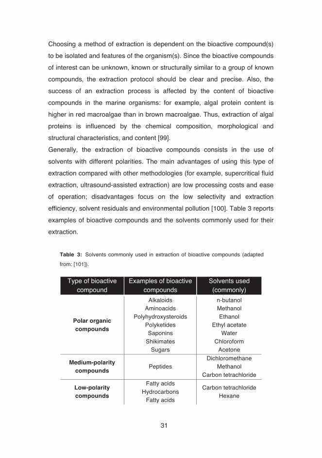
Choosing a method of extraction is dependent on the bioactive compound(s) to be isolated and features of the organism(s). Since the bioactive compounds of interest can be unknown, known or structurally similar to a group of known compounds, the extraction protocol should be clear and precise. Also, the success of an extraction process is affected by the content of bioactive compounds in the marine organisms: for example, algal protein content is higher in red macroalgae than in brown macroalgae. Thus, extraction of algal proteins is influenced by the chemical composition, morphological and structural characteristics, and content [99]. Generally, the extraction of bioactive compounds consists in the use of solvents with different polarities. The main advantages of using this type of extraction compared with other methodologies (for example, supercritical fluid extraction, ultrasound-assisted extraction) are low processing costs and ease of operation; disadvantages focus on the low selectivity and extraction efficiency, solvent residuals and environmental pollution [100]. Table 3 reports examples of bioactive compounds and the solvents commonly used for their extraction.
Table 3: Solvents commonly used in extraction of bioactive compounds (adapted from: [101]).
Type of bioactive compound
Examples of bioactive compounds
Solvents used (commonly)
Polar organic compounds
Alkaloids Aminoacids
Polyhydroxysteroids Polyketides Saponins
Shikimates Sugars
n-butanol Methanol Ethanol
Ethyl acetate Water
Chloroform Acetone
Medium-polarity compounds Peptides
Dichloromethane Methanol
Carbon tetrachloride
Low-polarity compounds
Fatty acids Hydrocarbons
Fatty acids
Carbon tetrachloride Hexane

�32
Extraction by solvents can follow the principle of either liquid-liquid or solid-liquid extractions. Kupchan’s extraction method [102] is probably the most popular liquid-liquid fractionation scheme. This method separates compounds based on their relative solubility in two different immiscible liquids. The selective partitioning of components of interest into one of the immiscible phases results from the choice of the most adequate extraction solvent. However, during the recent years, solid-liquid extractions have emerged as an alternative. There the material is placed in contact with a solvent, which diffuses into the cells, solubilizing the metabolites. After that, the metabolites are diffused out of the cells into the solvent. This extraction method has been commonly used for obtaining marine compounds, using a marine organism directly as a solid matrix [99]. �2.4.� Importance and approaches for screening bioactive compounds:
general overview of bioactive screening �Due to the great biodiversity in marine environment, the use of appropriate methodologies for biological screening of marine sources is of great importance. Basically, the selection of bioactivity screening assay depends mostly upon the target disease as well as the available information about the target marine source/organism. For example, if a marine source has some pharmacological history of use for a specific disease, we would rationally use a particular bioassay, which can show the known therapeutic activity, in order to isolate the active compound, which is accountable for that bioactivity. Whichever assay format is selected, the following factors need to be considered [103]: •� Tolerance to several impurities available in crude extracts; •� Sensitivity and capability to detect the presence of potentially active
substances present in low concentrations (limit of 0.0001% of an active compound, based on the dry weight of extract);
•� Reproducibility and reliability;

�33
•� High throughput; •� Tolerance to DMSO (commonly used solvent for solubilization of
samples). During the primary biological screening, multiple samples are screened to evaluate whether any desired bioactivity is present. These types of assays are capable of providing results fast, not always quantitative, but economic. If a positive hit is observed in the primary screening, follow-up assays should be performed in order to eliminate false positives. Generally, a hit rate of ≤1% is considered reasonable from a primary screening for progressing to follow-up studies [104]. Secondary evaluation/confirmatory assays involve more exhaustive and comprehensive testing of active compounds. As a part of this process, consideration should be given to the properties of each compound, such as, if there is a SAR between the molecular structure and biological activity of the compounds. 2.5.� Marine drug discovery and development �MNP discovery is a complex and multidisciplinary effort requiring work and interaction between marine biologists, chemists, microbiologists and pharmacologists. As it is known, the marine realm contains a rich variety of organisms, many of them under-investigated. Bioprospecting is a tool for biodiversity conservation, defined as the collection of biological material and analysis of its properties and/or its molecular, biochemical or genetic content for the purpose of developing a commercial product [105]. However, attention must be paid to ethical issues and conservation policies, since the loss of biodiversity is often associated with overexploitation and habitat degradation. In the last decades, discovery of new MNPs has exponentially increased, but the number of marine-derived molecules that are commercialized is still very low – eight approved drugs by FDA and/or EMA. Despite that, more than 20

�34
marine products are in clinical trials and more than 1000 are in pre-clinical trials [106]–[108]. The process of R&D of new drugs is neither easy nor always successful; it is statistically shown [109] that of 5000 compounds that are discovered, only 5 progress to clinical studies and only 1 leads to an approved and commercialized drug (Fig. 4). A clinical trial is, per se, the testing carried out on human beings for determining its value for treatment or for prevention of diseases, involving a number of parameters. These parameters [110] include: the patient population to be studied (patients should meet specific criteria to ensure that they are as similar as possible to each other so that the results of treatments effect can be associated as much as possible with the drug treatment), use of controls [placebo (e.g., medically ineffectual treatment) or standard treatment (e.g., in wide use and considered effective at the time that the trial is designed)], endpoints (existence of a primary endpoint that usually assesses the treatment efficacy) and methods by which the trial will be conducted (randomly allocated to receive one or other of the alternative treatments being studied and/or partitioned by a factor other than the treatment, to ensure that equal number of patients with a characteristic thought to response to the intervention will be allocated to each comparison group).

�35
Between Phase I and III, the potential of failing and withdrawals of drug development is high, mainly due to lack of efficacy and drug toxicity [111]. Upon authorization by the EMA/FDA, the therapies that have proven safety, efficacy and quality in the clinical trials may be made available to the general population. Still, EMA/FDA requires continued evaluation after release to evaluate safety signs that can affect the benefit-risk ratio [112], [113].
Discovery
Pre-clinical trials
Clinical trials
3-6 years
6-7 years
Approval for commercialization 2 years
5000 compounds
In vitro assays, Pharmacodynamics,
Pharmacokinetics, Toxicology ~250 compounds
Phase I ~5 compounds
Phase II
Phase III
Approved drug
Figure 4: The process of drug discovery and development – “The pipeline” (Adapted from: [109]). An Investigational New Drug Application (INDA) is submitted before the clinical trials and a New Drug Application is a sponsor’s request for approval of a drug for commercialization.

�36
� � 2.5.1.� Commercial uses and applications of MNPs �A large number of bioactive compounds found in marine organisms have proceeded into clinical trials and until 2014, eight FDA or EMA approved drugs originating from marine microbes are currently on the market [114]. For example, companies such as PharmaMar (Spain), AquaPharm Biodiscovery Ltd. (United Kingdom) or Nereus Pharmaceutical (United States of America) have several compounds in clinical trials, preclinical trials or even on the market. However, until now, only three compounds (Prialt®, Yondelis® and Carragelose®) have become drugs without modification of the original natural molecule, i.e. without any type of synthesis and optimization in the structure. Selected marine bioactive compounds in trials or commercialized from 2004 until now are highlighted in Table 4.
Phase I - Checking for
safety
Sample: 10-20 healthy volunteers
Side effects may occur
Phase II - Checking for
efficacy Sample:
~200 patients
High hypothesis of
fail since product can be not so effective
as expected
Phase III - Confirm
findings in large patient population
Sample: more than
1000 people
Likelihood to detect rare side effects
Phase IV - Testing long-term safety in
diverse patient
population
Sample: "real life patients"
Previously untested
groups can show adverse
reactions
Figure 5: Different stages of clinical trials (Adapted from: [180]). In Phase IV, active post-marketing surveillance of drug side effects is essential.

�37
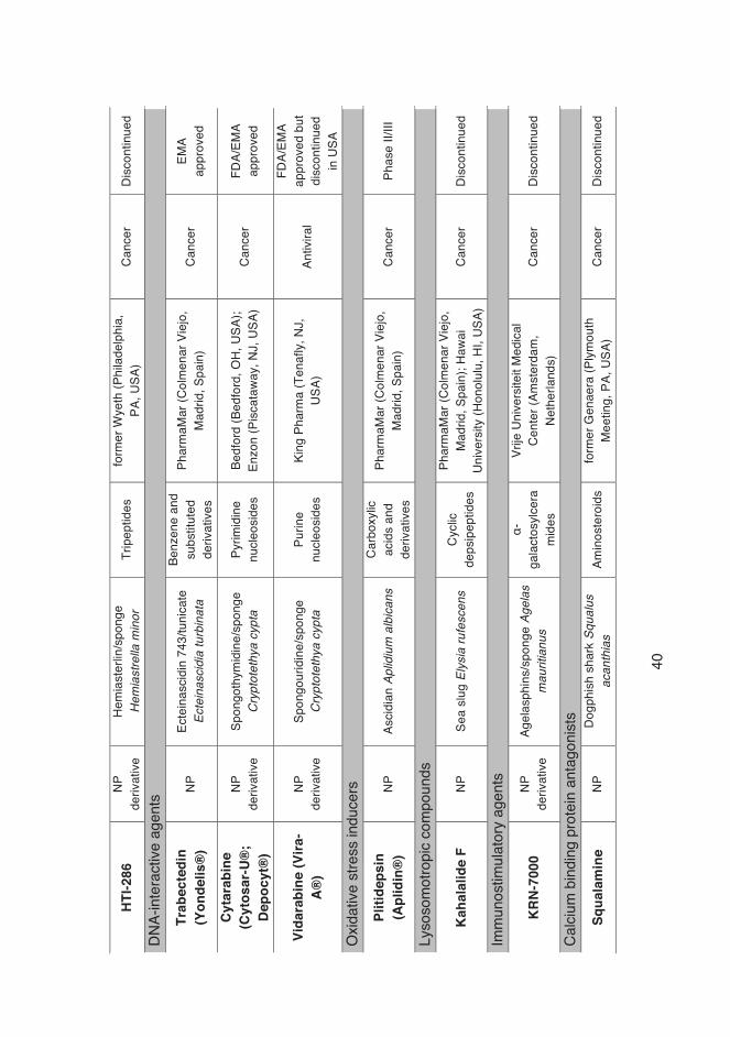
Cytarabine and vidarabine were the first marine-derived drugs approved by the FDA, as anticancer (1969) and antiviral (1976) drugs [115], respectively. Cytarabine (Cytosar-U®) is used for the treatment of two types of leukemia (myeloid and meningeal) and non-Hodgkin’s lymphoma. However, this drug has a short plasma half-life, low stability and limited bioavailability. Vidarabine (Vira-A®), has been used as an antiviral drug for the treatment of keratoconjunctivitis, an epithelial keratitis caused by the herpes simplex virus, but it has been discontinued by FDA in the US market because of its side effects such as fever, sore or other signs of infection. Another compound included in the same group as cytarabine and vidarabine based on their mechanism of action (DNA interaction), is trabectedin (Yondelis®). PharmaMar markets Yondelis® in Europe and Japan and new clinical trials have been carried out for breast and prostate cancer. The active ingredient was originally isolated from a Caribbean sea squirt called Ecteinascidia turbinata and it shows potent antitumor [116], [117] and antiproliferative [118] activities towards cancer cells. Prialt®, a ziconotide (synthetic derivative of ω-Conotoxin MVIIA) is being used for the treatment of chronic pain in the spinal medulla [11]. This compound was isolated from the marine snail Conus magnus in 1979. However, the complete synthesis was only finalized in 1987 [11]. Recently, a synthetic derivative of halichondrin B, eribulin mesylate (Halaven®) gained FDA’s approval for the treatment of metastatic breast cancer (Sub-chapter 2.2). An Austrian company, Marinomed Biotechnologie GmbH, has developed an antiviral nasal spray containing Iota-carrageenan (Carragelose®). This copolymer derives from carrageenan, high molecular weight sulphated polysaccharide extracted from red edible seaweeds, such as Rhodphyceae sp. This substance is clinically effective against early symptoms of the cold and is marketed as an OTC (over-the-counter) product.

�38
Lovaza®, an anti-hypertriglyceridemia drug, is commercialized by GlaxoSmithKline and consists of a mixture of eicosapentaenoic acid (EPA) and docosahexaenoic acid (DHA). The approach to this drug discovery was based on studies stating that some ethnic populations, such as Native Alaskans, had much lower mortality rates from cardiovascular diseases, assumingly due to their diets that were high in polyunsaturated fatty acids [119]. Other studies confirmed also that ingesting omega-3 fatty acids was associated with a reduced rate of premature cardiovascular deaths [120], [121]. The latest marine-derived drug to enter the market until now was brentuximab vedotin (AdcetrisTM), consisting of an antibody brentuximab and monomethyl auristatin (vedotin), a synthetic analog of dolastatin 10. Dolastatin 10 is a compound originally isolated from the marine mollusk Dolabella auricularia [122]. Of the antibody-drug conjugate, the chimeric monoclonal antibody brentuximab targets the CD30 antigen on the surface of malignant cells, leading to the uptake of vedotin (an antimitotic agent) into the targeted cells. Brentuximab vedotin is used for treating Hodgkin’s lymphoma and systemic anaplastic large-cell lymphoma.

�39
Tabl
e 4:
Mar
ine-
deriv
ed c
ompo
unds
in d
rug
disc
over
y an
d de
velo
pmen
t (Ad
apte
d fro
m: [
123]
). NP
= n
atur
al p
rodu
ct.
Prod
uct
NP o
r de
rivat
ive
Sour
ce o
rgan
ism
Chem
ical
class
Co
mpa
ny
Ther
apeu
tic
area
Cu
rrent
st
atus
Com
poun
ds ta
rget
ing
ion
chan
nels
Zico
notid
e (P
rialt®
) NP
ω
-Con
otox
in/ m
arin
e co
ne
snai
l Con
us m
agus
Pe
ptid
es
Elan
Cor
pora
tion
(Dub
lin,
Irela
nd)
Chro
nic
(neu
roph
atic)
pa
in
FDA/
EMA
appr
oved
DMXB
A (G
TS-2
1)
NP
deriv
ative
An
abes
eine
/wor
m
Para
nem
erte
s pe
regr
ina
Pyrid
ines
and
de
rivat
ives
Com
entis
(San
Fra
ncisc
o, C
A,
USA)
Alzh
eim
er’s
dise
ase
and
schi
zoph
reni
a Ph
ase
II
Com
poun
ds ta
rget
ing
enzy
mes
Bryi
osta
tin-1
NP
Br
yozo
an B
ugul
a ne
ritin
a Po
lyket
ides
NC
I (Be
thes
da, M
D, U
SA)
Canc
er a
nd
Alzh
eim
er’s
dise
ase
Phas
e I/I
I
Erib
ulin
mes
ylat
e (H
alav
en®
) NP
de
rivat
ive
Halic
hond
rin B
/spo
nge
Hal
icho
ndria
oka
dai
Furo
pyra
ns
Eisa
i (To
kyo,
Jap
an)
Canc
er
FDA/
EMA
appr
oved
M
icrot
ubul
e-in
terfe
ring
agen
ts
Bret
uxim
ab
vedo
tin (S
GN-
35)
(Adc
etris
TM)
NP
deriv
ative
Do
last
atin
10/
sea
har
e D
olab
ella
aur
icul
aria
Carb
oxyli
c ac
ids
and
deriv
ative
s
Seat
tle G
enet
ics (B
othe
ll, W
A,
USA)
; Tak
eda
GRD
C (O
saka
, Ja
pan)
Ca
ncer
FD
A/EM
A ap
prov
ed
Tasi
dotin
(ILX
-65
1)
NP
deriv
ative
Do
last
atin
15/
sea
har
e D
olab
ella
aur
icul
aria
Pe
ptid
es
Gen
zym
e Co
rpor
atio
n (C
ambr
idge
, MA,
USA
) Ca
ncer
Di
scon
tinue
d
Disc
oder
mol
ide
NP
Spon
ge D
isco
derm
ia
diss
outa
Po
lyket
ides
No
varti
s (B
asel
, Swi
tzer
land
); Ha
rbor
Bra
nch
(For
t Pie
rce,
FL
, USA
) Ca
ncer
Di
scon
tinue
d
�40
HTI-2
86
NP
deriv
ative
He
mia
ster
lin/s
pong
e H
emia
stre
lla m
inor
Tr
ipep
tides
fo
rmer
Wye
th (P
hila
delp
hia,
PA
, USA
) Ca
ncer
Di
scon
tinue
d
DNA-
inte
ract
ive a
gent
s Tr
abec
tedi
n (Y
onde
lis®
) NP
Ec
tein
ascid
in 7
43/tu
nica
te
Ecte
inas
cidi
a tu
rbin
ata
Benz
ene
and
subs
titut
ed
deriv
ative
s
Phar
maM
ar (C
olm
enar
Vie
jo,
Mad
rid, S
pain
) Ca
ncer
EM
A ap
prov
ed
Cyta
rabi
ne
(Cyt
osar
-U®
; De
pocy
t®)
NP
deriv
ative
Sp
ongo
thym
idin
e/sp
onge
C
rypt
otet
hya
cypt
a Py
rimid
ine
nucl
eosi
des
Bedf
ord
(Bed
ford
, OH,
USA
); En
zon
(Pis
cata
way,
NJ,
USA
) Ca
ncer
FD
A/EM
A ap
prov
ed
Vida
rabi
ne (V
ira-
A®)
NP
deriv
ative
Sp
ongo
urid
ine/
spon
ge
Cry
ptot
ethy
a cy
pta
Purin
e nu
cleo
side
s Ki
ng P
harm
a (T
enaf
ly, N
J,
USA)
An
tivira
l
FDA/
EMA
appr
oved
but
di
scon
tinue
d in
USA
O
xidat
ive s
tress
indu
cers
Pl
itide
psin
(A
plid
in®
) NP
As
cidi
an A
plid
ium
alb
ican
s Ca
rbox
ylic
acid
s an
d de
rivat
ives
Phar
maM
ar (C
olm
enar
Vie
jo,
Mad
rid, S
pain
) Ca
ncer
Ph
ase
II/III
Lyso
som
otro
pic
com
poun
ds
Kaha
lalid
e F
NP
Sea
slug
Ely
sia
rufe
scen
s Cy
clic
deps
ipep
tides
Phar
maM
ar (C
olm
enar
Vie
jo,
Mad
rid, S
pain
); Ha
wai
Unive
rsity
(Hon
olul
u, H
I, US
A)
Canc
er
Disc
ontin
ued
Imm
unos
timul
ator
y ag
ents
KRN-
7000
NP
de
rivat
ive
Agel
asph
ins/
spon
ge A
gela
s m
aurit
ianu
s
α-ga
lact
osylc
era
mid
es
Vrije
Uni
vers
iteit
Med
ical
Cent
er (A
mst
erda
m,
Neth
erla
nds)
Ca
ncer
Di
scon
tinue
d
Calci
um b
indi
ng p
rote
in a
ntag
onist
s Sq
uala
min
e NP
Do
gphi
sh s
hark
Squ
alus
ac
anth
ias
Amin
oste
roid
s fo
rmer
Gen
aera
(Plym
outh
M
eetin
g, P
A, U
SA)
Canc
er
Disc
ontin
ued

�41
Com
poun
ds w
ith a
ntivi
ral a
ctivi
ty
Iota
-car
rage
enan
(C
arra
gelo
se®
) NP
Io
ta-c
arra
geen
an/re
d al
gae
Euch
eum
a/C
nond
us
Carb
ohyd
rate
s M
arin
omed
(Vie
nna,
Aus
tria)
; Bo
ehrin
ger I
ngel
heim
(In
gelh
eim
, Ger
man
y)
Antiv
iral
Ove
r-the
-co
unte
r dru
g (O
TC)
Com
poun
ds w
ith o
ther
or u
nkno
wn m
echa
nism
of a
ctio
n IP
L-57
6092
and
de
rivat
ives
NP
de
rivat
ive
Cont
igna
ster
ol/s
pong
e Pe
trosi
a co
ntig
nata
St
eroi
ds
form
er A
vent
is (S
trasb
ourg
, Fr
ance
) An
ti-as
thm
atic
Di
scon
tinue
d
Om
ega-
3-ac
id
ethy
l est
ers
(Lov
aza®
)
NP
deriv
ative
O
meg
a-3-
fatty
acid
s/fis
h Es
ters
G
laxo
Smith
Klin
e (B
rent
ford
, UK
) Hy
pertr
iglyc
erid
emia
FD
A/EM
A ap
prov
ed
Iota
-car
rage
enan
(C
arra
gelo
se®
) NP
Io
ta-c
arra
geen
an/re
d al
gae
Euch
eum
a/C
nond
us
Carb
ohyd
rate
s M
arin
omed
(Vie
nna,
Aus
tria)
; Bo
ehrin
ger I
ngel
heim
(In
gelh
eim
, Ger
man
y)
Antiv
iral
Ove
r-the
-co
unte
r dru
g (O
TC)
�42
3.� Aims of the study The aim of this study was to evaluate, using in vitro assays, the biological
activity of extracts and synthetic compounds derived from marine organisms.
NPs, especially those from terrestrial plants and microbes, have been the
most successful sources of potential drug leads. MNPs have only become a
“hot topic” in the last decades, as a consequence of their proven potential as
antimicrobial, anticancer, antiviral and anti-inflammatory agents, among other
properties. The studies included in this thesis focused on the assessment of
biological potential of marine-derived substances that can have a significant
impact in the pharmacological field during the coming years.
The specific aims of this doctoral thesis were:
•� to study the biological properties of synthetic derivatives designed by
using marine alkaloids oroidin and clathrodin as parent structures (I, II);
•� to determine the biological activities of synthetic crown acyl ether
derivatives, developed by using okadaic acid structure as inspiration (III);
•� to evaluate the bioactive potential of cyanobacteria, micro- and
macroalgae extracts originating from the Aegean Sea (IV).
�43
4.� Material and Methods Microbial strains and cancer cell lines used in this study are listed in sub-
chapters 4.1 and 4.2. Types of biological assays used are listed below.
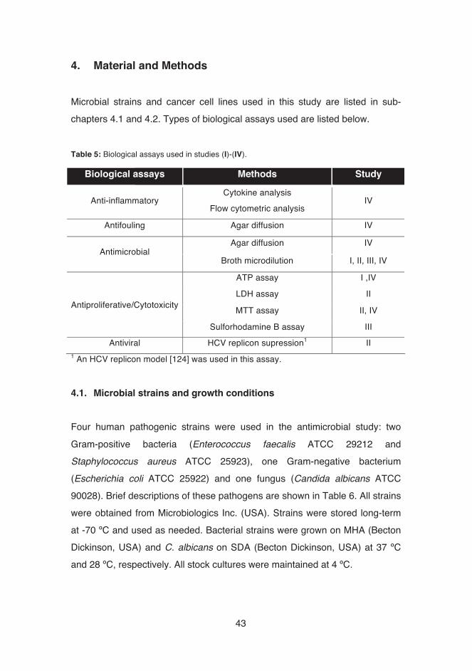
Table 5: Biological assays used in studies (I)-(IV).
Biological assays Methods Study
Anti-inflammatory Cytokine analysis
Flow cytometric analysis IV
Antifouling Agar diffusion IV
Antimicrobial Agar diffusion IV
Broth microdilution I, II, III, IV
Antiproliferative/Cytotoxicity
ATP assay I ,IV
LDH assay II
MTT assay II, IV
Sulforhodamine B assay III
Antiviral HCV replicon supression1 II 1 An HCV replicon model [124] was used in this assay.
4.1.� Microbial strains and growth conditions �Four human pathogenic strains were used in the antimicrobial study: two
Gram-positive bacteria (Enterococcus faecalis ATCC 29212 and
Staphylococcus aureus ATCC 25923), one Gram-negative bacterium
(Escherichia coli ATCC 25922) and one fungus (Candida albicans ATCC
90028). Brief descriptions of these pathogens are shown in Table 6. All strains
were obtained from Microbiologics Inc. (USA). Strains were stored long-term
at -70 ºC and used as needed. Bacterial strains were grown on MHA (Becton
Dickinson, USA) and C. albicans on SDA (Becton Dickinson, USA) at 37 ºC
and 28 ºC, respectively. All stock cultures were maintained at 4 ºC.�

�44
� Tabl
e 6:
Cha
ract
eris
tics
of p
atho
geni
c ba
cter
ia a
nd fu
ngus
use
d in
this
wor
k [1
25].
Path
ogen
D
isea
se
Mai
n sy
mpt
oms
Mod
e of
tran
smis
sion
Bact
eria
Ente
roco
ccus
faec
alis
Urin
ary
tract
, wou
nd a
nd s
oft-
tissu
e in
fect
ions
, end
ocar
ditis
Burn
ing
pain
with
urin
atio
n, u
rinar
y ur
genc
y,
feve
r, ba
ck/fl
ank
pain
, nig
ht s
wea
ts, f
atig
ue
Nos
ocom
ial a
nd p
erso
n-to
-per
son
trans
mis
sion
, tra
nsm
issi
on b
y fo
od
prod
ucts
Stap
hylo
cocc
us
aure
us
Scal
ded
skin
syn
drom
e, li
fe-
thre
aten
ing
dise
ases
(e.g
.,
pneu
mon
ia)
Nau
sea,
vom
iting
, abd
omin
al c
ram
ps, r
etch
ing,
pros
tratio
n
Inge
stio
n of
food
con
tain
ing
ente
roto
xins
(spe
cific
toxi
n fo
r cel
ls
of in
test
inal
muc
osa)
Esch
eric
hia
coli
Urin
ary
and
bilia
ry d
uct
infe
ctio
ns, g
astro
ente
ritis
,
pneu
mon
ia, m
enin
gitis
, sep
sis
Hem
orrh
agic
col
itis,
dia
rrhea
, dys
ente
ry, f
ever
,
abdo
min
al p
ains
Inge
stio
n of
con
tam
inat
ed
food
/wat
er, f
ecal
-ora
l tra
nsm
issi
on,
pers
on-to
-per
son
trans
mis
sion
Fung
i Can
dida
albi
cans
C
andi
dias
is
Whi
te c
oate
d to
ngue
, bod
y ra
sh, p
ainf
ul
urin
atio
n an
d di
scha
rge,
feve
r, ge
nera
l pai
n
Hum
an o
wn
natu
ral f
lora
, ext
erna
l
sour
ce (e
ven
if ra
re)

�45
4.2.� Cell lines and culture conditions �For antiproliferative and cytotoxicity studies, the following cell lines were used: A-375 (human malignant melanoma) and BALB/c 3T3 clone A31 (embryonic mouse fibroblast). A-375 was kindly provided by Prof. Marikki Laiho (University of Helsinki, FI) and BALB/c 3T3 clone A31 was obtained from the European Collection of Cell Cultures (ECACC, UK). Melanoma cells were grown in Glutamax DMEM with 4.5 g/L D-glucose (Gibco, USA), supplemented with 10% FBS (Gibco, USA), 100 IU/mL penicillin (Gibco, USA) and 100 μg/mL streptomycin (Gibco, USA). BALB/c 3T3 cell line was cultured in DMEM (Sigma-Aldrich, USA), supplemented with 5% FBS (Gibco, USA), 5% NCS (Gibco, USA), 292 μg/mL L-glutamine, 100 IU/mL penicillin and 100 μg/mL streptomycin. Methods related to human breast epithelial (HBL-100), human cervical cancer (HeLa), alveolar carcinoma (SW1573) and colon adenocarcinoma (WiDr) cell lines are described in detail in Publication (III). To prostate cancer (LNCa, PC-3), breast cancer (MCF-7) and non-tumorigenic epithelial (MCF-10A) cell lines, methods are described in Publication (IV). 4.3.� Reference compounds �Stock solutions of amphotericin B (Sigma-Aldrich, USA) and ciprofloxacin (MP Biomedicals, USA) were made in DMSO and milliQ water, respectively. The reference antibiotics were used for assay validation and as positive controls in the antimicrobial assays.� Stock solutions of polymyxin B sulphate (PMX, Sigma-Aldrich, DK) were diluted in milliQ water and assays were validated. PMX was used as positive control in antiproliferative assays.��4.4.� Marine test material �A set of marine natural products and marine-derived compounds were used in experiments and evaluated for their antimicrobial and antiproliferative
�46
properties. In publications I, II and III, the following sets of marine-derived synthetic compounds were studied: •� (I): set of thirty-four novel analogs, using oroidin as scaffold; •� (II): set of fourteen new clathrodin-inspired 2-aminobenzothiazole and
benzimidazole derivatives and their synthetic intermediates; •� (III): set of twenty-two macrocyclic compounds, employing secondary metabolite okadaic acid (OA) as inspiration.
In the last publication (Study (IV)), ninety-eight crude extracts (solvent used in extraction: ethanol) of cyanobacteria, micro- and macroalgae originating from the Aegean Sea were studied. Methods related to the synthesis and chromatographic analyses of the compounds are described in detail in Publications (I), (II) and (III). �4.5.� Screening for bioactivity 4.5.1.� Antimicrobial screening �Broth microdilution
Broth microdilution method was used in all studies (I, II, III, IV). Together with agar dilution, it is the standard susceptibility testing method approved by EUCAST [126] and CLSI [127]. Briefly, the bacterial or fungal strain to be used in the assay was inoculated the day before on MHB or SDA, respectively, and grown for 16-24 h. For the experiment, CFU/mL was calculated and suspensions prepared, based on the absorbance values at 620 nm, previously calibrated against plate counts. Compounds were diluted into the assay media, and dispensed into clear 96-well microplates (Thermo Fisher Scientific, FI) and the bacterial/fungal suspension added to the plates. The assay plates also included the positive control antibiotics: amphotericin B for C. albicans and ciprofloxacin for bacterial strains (MICs are mentioned in the articles). The plates were incubated for 24 h at 37 ºC for bacteria and 48 h at 28 ºC for Candida. Synthetic compounds and extracts were initially screened at the final concentration of 50 μM and 100 μg/mL, respectively.
�47
Samples that demonstrated potent activity (growth inhibition >50%) were selected for confirmatory testing (dose-response) and MIC determination. �4.5.2.� In vitro antiproliferative and cytotoxicity screening �The general procedures for routine subculturing and exposure of cells to compounds are found in publications (I) and (II). In Study (I), Huh-7 (human hepatocellular carcinoma) cell line was used for selectivity studies. In Study (II), two cell lines were used: A-375 (human malignant melanoma, epithelial, cancer) and BALB/c 3T3 clone 31 (embryonic mouse fibroblast, non-cancer). Melanoma is a cancer that involves the melanocytes (specialized pigment cells) in the basal layer of epidermis and is one of the most common and aggressive forms of skin cancer and related deaths. This “fatal black tumor” [128] displays a growth of incidence in most populations of Caucasian origin [129]; in Europe, the highest incidence rates of melanoma are reported in Scandinavian countries [130]. The main environmental factor, UV light exposure, can cause damage in keratinocytes and melanocytes synthesize melanin (protective pigment). If exposure to UV is extreme, keratinocyte population can die but melanocytes survive – due to efficient DNA damage repair response ‒, having the chance of developing mutations [131], [132]. BALB/c 3T3 non-cancerous cell line was used in publication II as a model for selectivity studies in comparison with melanoma cell line. ATP assay (I) Adenosine triphosphate (ATP) is indicative of metabolically active cells, as it is responsible for intracellular energy transfer [133]. Non-viable cells are not metabolically active, and therefore do not produce ATP. CellTiter-Glo® Luminescent Cell Viability Assay (Promega Corporation, USA) was used to study the effect of the most promising compounds on the metabolic activity of Huh-7 cells. This assay is based on luciferase reaction that produces light in the presence of ATP, and is thus used for measuring the intracellular ATP content. After the exposure of cells to compounds, cells were washed with phosphate buffered saline (PBS) and 50 μL of assay media and 50 μL of

�48
CellTiter-Glo reagent were added. After plate shaking for 2 min and incubation at room temperature (rt) for 10 min, the luminescent signal was determined using a Varioskan Flash Plate reader (Thermo Fisher Scientific, FI). Cytotoxicity (%) was calculated by normalizing to vehicle-treated control values. MTT assay (II) MTT assay [134], [135], based on MTT dye [3-(4,5-dimethyl thiazol-2-yl)-2,5-diphenyl tetrazolium bromide], was used to estimate the cytotoxic effects of compounds. This assay is based on the conversion of MTT to formazan crystals by reduction, catalyzed by intracellular dehydrogenases and it is widely used for measuring in vitro cytotoxicity. Briefly, after the adhesion of cells to 96-well microplates (Thermo Fisher Scientific, FI) and treatment with compounds (duration of 24 h), 10 μL of 5 mg/mL MTT stock solution (Sigma-Aldrich, USA) in sterile PBS was added to the cells. Following 3 h incubation in a humidified atmosphere (37 ºC, 5% CO2), the medium was removed and the blue formazan crystals trapped in the cells were dissolved in 200 μL of DMSO. The plates were agitated until formazan had completely dissolved. The absorbance at 550nm/655nm was measured with Multiskan® GO UV/Vis spectrophotometer (Thermo Fisher Scientific, FI). Cytotoxicity (%) was calculated by normalizing to vehicle-treated control values. LDH assay (II)�Leakage of intracellular lactate dehydrogenase into culture medium can be measured by LDH leakage assay. The activity is measured by monitoring the rate at which the substrate (pyruvate) is reduced to lactate. Its evaluation is based on the decrease of membrane integrity to retain the enzyme in the cytoplasm when cells are exposed to toxic agents [136], [137]. The more damaged a cell membrane is, the more leakage of LDH occurs. The CytoTox-ONE-Homogeneous Integrity Assay Kit (Promega, USA) was used for measuring LDH leakage. After exposure of cells to compounds, 50 μL samples from culture supernatants were transferred into sterile, white and

�49
clear-bottom 96-well plates (PerkinElmer Inc., USA). After 20-30 min of incubation at room temperature (rt) in dark, 50 μL of CytoTox-ONE reaction mixture was added to the plates and shaken for 10 min. The enzyme reaction was stopped with 25 μL CytoTox-ONE stop solution. After plate shaking for 10s, fluorescent signal was measured at a wavelength of 560 nm (excitation) and 655 nm (emission) with a Varioskan Flash Plate reader (Thermo Fisher Scientific, FI). The % of LDH leakage was calculated by normalizing to vehicle-treated control values. 4.6.� Selectivity index �Selectivity index (SI), also known as therapeutic index (TI), is an important criterion that determines which compounds will advance further in laboratory and clinical studies. It is considered a quantitative relationship between their efficacy (pharmacology) and safety (toxicology). In Study (I), the selectivity was based on the effects between eukaryotic (IC50) and prokaryotic (MIC50) cells. In Study (II), the ratio between a normal cell line (IC50) and a cancer cell line (IC50) was used to measure this index. 4.7.� Data analysis �All data presented are means ± SD. Data were analyzed using Microsoft Office Excel 2010 and the IC50 values were determined using Origin Graphing and Analysis, version 8.6 (OriginLab, USA). All experiments were performed at least in triplicate. 4.8.� Quality parameters �Three types of quality parameters were used for evaluating the quality of the results: Z’, S/B and S/N. The data quality of the assays can be estimated by the following formulas [138], [139]

�50
� � � � ����� � � ��������� � � ����
� � � � ����
� � � � � � ������ �� � �����
, where � � and �� � are the mean and standard deviation for the high reference control samples (maximum) and ��� and ���� are the mean and standard deviation for the low reference control samples (i.e. background). Z’ factor is used as a screening quality assurance parameter and it is the most practical factor to use when evaluating assay quality. Z’ close to 1 is considered an ideal assay and values between 0.5 and 1 are considered of good quality. In general, if Z’ value is lower than 0.5, the assay is considered of low quality and should be repeated (for cell-based assays, threshold 0.3 or 0.4 is sometimes considered acceptable). The S/B (signal-to-background) and S/N (signal-to-noise) ratios should also be taken into account since they reflect the assay signal strength. The S/B ratio evaluates the strength of signal in comparison with background measurements (S/B�5, good quality) and S/N ratio signifies the strength of signal in relation to the noise (high S/N desired if S/B is low) [140].

�51
5.� Summary of Results and Discussion
This chapter of the thesis summarizes the main results of the work. More details can be found in the original publications (I-IV). Articles (I), (II) and (III) focus on biological studies of synthetic derivatives. The innovations and advances in marine research have contributed to the perception that MNPs can be considered as privileged structures or biologically validated stating points for the design of new compounds. Article (IV) shows results from a large-scale screening programme of bioprospecting of species from the Aegean Sea, where several crude extracts from cyanobacteria, micro- and macroalgae were evaluated for biological activities. Marine ecosystems and rarely explored places such as the Aegean Sea that contains a rich biodiversity of marine organisms can be particularly suited for bioprospecting, a process that aims to identify and isolate bioactive natural compounds. 5.1.� Oroidin and clathrodin as parent structures for synthesis of new
compounds (I), (II) 5.1.1.�Antimicrobial studies (I), (II) �Clathrodin is a secondary metabolite isolated from Caribbean sponge Agelas
sp. that belongs to a class of alkaloids, pyrrole-imidazole alkaloids (PIAs). These metabolites contain 2-aminoimidazole, pyrrole amide structures, and are rarely found in terrestrial flora and fauna. Oroidin, an achiral and monomeric product also used in Study (I), is generally considered the biogenetic precursor of other alkaloids from the same family. Within the PIA’s, oroidin and clathrodin (Figure 6) are believed to be one of the basic biosynthetic units for the construction of more complex and larger analogs. Oroidin, for example, due to its low molecular mass and simple structure, is believed to offer several possibilities for chemical optimization, with the introduction of additional side chains/functional groups.
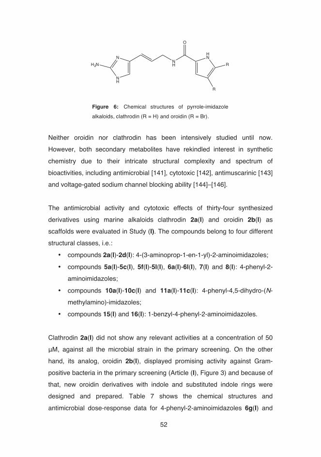
�52
Neither oroidin nor clathrodin has been intensively studied until now. However, both secondary metabolites have rekindled interest in synthetic chemistry due to their intricate structural complexity and spectrum of bioactivities, including antimicrobial [141], cytotoxic [142], antimuscarinic [143] and voltage-gated sodium channel blocking ability [144]–[146]. The antimicrobial activity and cytotoxic effects of thirty-four synthesized derivatives using marine alkaloids clathrodin 2a(I) and oroidin 2b(I) as scaffolds were evaluated in Study (I). The compounds belong to four different structural classes, i.e.:
•� compounds 2a(I)-2d(I): 4-(3-aminoprop-1-en-1-yl)-2-aminoimidazoles; •� compounds 5a(I)-5c(I), 5f(I)-5l(I), 6a(I)-6l(I), 7(I) and 8(I): 4-phenyl-2-
aminoimidazoles; •� compounds 10a(I)-10c(I) and 11a(I)-11c(I): 4-phenyl-4,5-dihydro-(N-
methylamino)-imidazoles; •� compounds 15(I) and 16(I): 1-benzyl-4-phenyl-2-aminoimidazoles.
Clathrodin 2a(I) did not show any relevant activities at a concentration of 50 μM, against all the microbial strain in the primary screening. On the other hand, its analog, oroidin 2b(I), displayed promising activity against Gram-positive bacteria in the primary screening (Article (I), Figure 3) and because of that, new oroidin derivatives with indole and substituted indole rings were designed and prepared. Table 7 shows the chemical structures and antimicrobial dose-response data for 4-phenyl-2-aminoimidazoles 6g(I) and
Figure 6: Chemical structures of pyrrole-imidazolealkaloids, clathrodin (R = H) and oroidin (R = Br).
N
NH
H2NNH
HN
R
R
O

�53
6h(I) that were active (>80% inhibition of growth) in the primary screening. Both obtained the best results against Gram-positive bacteria; compound 6h(I) showed MIC50 of 8.0 and 7.3 μM with Enterococcus faecalis and Staphylococcus aureus, respectively. In a study conducted by Zidar et al., synthetic compounds belonging to the same structural class acted as state-dependent voltage-gated sodium channel modulators and have shown interesting activities with moderate isoform selectivities [147]. Hammami et al. have shown that oroidin display antifungal activity against Alternaria solani, a plant pathogen [148]. In a study conducted by Scala et al, two analogs of oroidin (dispacamide B and spongiacidin) have displayed antimalarian activity (IC50 of 1.34 and 1.09 μg/mL, respectively) against a protozoa belonging to the genus Plasmodium [149]. In the same study, a dimeric derivative of the same parent compound (dibromopalaumine) has been identified as a trypanocidal agent, showing an IC50 of 0.46 μg/mL [149]. In another recent article, oroidin and clathrodin were found to be weakly active when tested for apoptosis-inducing activities against the human hepatocellular carcinoma (HepG2) cell line; however, some of the indole-based analogs showed promising activities, with EC50 values between 13 and 42 μM [150]. The importance of an imidazole ring in the chemical structure of a compound for antimicrobial activity was also found in this study. For example, compounds 10a(I)-c(I) and 11a(I)-c(I) did not show any antimicrobial activity; these compounds have in their chemical structure a reduced imidazole � ���bond. The incorporation of imidazole ring can result in exhibition of a wide

�54
Tabl
e 7:
Ant
imic
robi
al m
inim
um in
hibi
tory
con
cent
ratio
n ac
tivity
(MIC
90 a
nd M
IC50
) of a
nalo
gs 6
g(I)
and
6h(I)
aga
inst
Ent
eroc
occu
s fa
ecal
is A
TCC
292
12,
Stap
hylo
cocc
us a
ureu
s AT
CC
259
23, E
sche
richi
a co
li AT
CC
259
22 a
nd C
andi
da a
lbic
ans
ATC
C 9
0028
. All
resu
lts a
re s
how
n in
μM
.
E.
faec
alis
S.
aur
eus
E. c
oli
C. a
lbic
ans
Cpd
C
hem
ical
stru
ctur
e M
IC90
M
IC50
M
IC90
M
IC50
M
IC90
M
IC50
M
IC90
M
IC50
6g(I)
�
25
11.4
25
9.
0 25
17
.7
50
30.5
6h(I)
�
12.5
8.
0 12
.5
7.3
50
30.4
n.
d.
n.d.�
�55
spectrum of biological activities [151]–[154]; its unique structural feature is valuable for derivatives to bind with receptors in biological systems. In Study (II), clathrodin was used as an inspirational structure for the synthesis of fourteen new benzimidazole and 2-aminobenzothiazole analogs. Benzimidazole, a heterocyclic aromatic compound that consists of the fusion of benzene and imidazole, has been used as nucleus structure of various drugs and is known for its biological activities [155]–[157]. Benzothiazole is considered a privileged bicyclic ring system due to its potent and significant biological activities of pharmaceutical importance [158], [159]. A derivative of 2-aminobenzothiazole, compound 7(II), showed 56% growth inhibition against Gram-positive E. faecalis in the primary screening. In dose-response assay, the inhibition did not reach MIC90 level (even at the highest concentration tested), concluding that an optimization of the compound structure should be made in order to improve its properties. �5.1.2.�Antiproliferative (I), (II) and antiviral studies (II) �In Study (I), the metabolic activity of Huh-7 hepatocytes after exposure to the most promising antimicrobial compounds was evaluated by intracellular ATP quantitation. This test is based on a luciferase reaction to measure the amount of ATP in cells, correlating directly with the number of cells and their viability because cells lose the ability to synthesize ATP directly after e.g. loss of membrane integrity or other cytotoxic event. Most of the derivatives that were active in antimicrobial assays, showed also an effect on eukaryotic cells. 4-phenyl-2-aminoimidazole 6j(I)� showed the most interesting IC50 (19.8 μM) while the compounds that had the best antibacterial results, i.e. 6g(I) and 6h(I), showed only moderate IC50 of 31 and 21.2 μM, respectively, against eukaryotic cells. In Study (II), the antiproliferative activities of compounds were studied against a human cancer cell line and a normal cell line: A-375 (melanoma) and
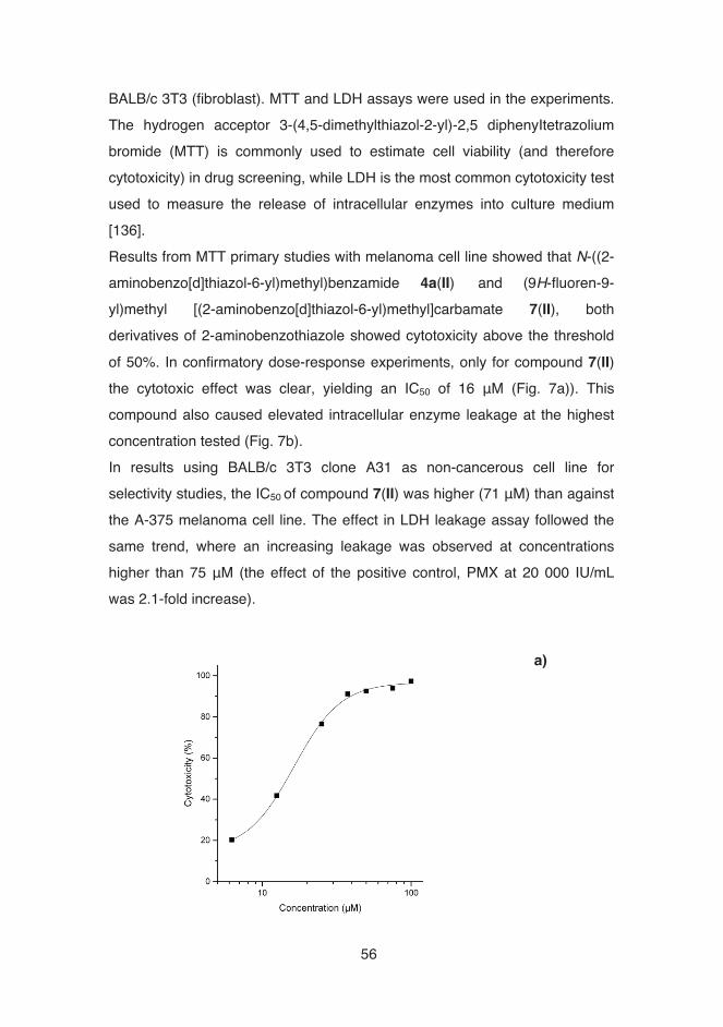
�56
BALB/c 3T3 (fibroblast). MTT and LDH assays were used in the experiments. The hydrogen acceptor 3-(4,5-dimethylthiazol-2-yl)-2,5 diphenyItetrazolium bromide (MTT) is commonly used to estimate cell viability (and therefore cytotoxicity) in drug screening, while LDH is the most common cytotoxicity test used to measure the release of intracellular enzymes into culture medium [136]. Results from MTT primary studies with melanoma cell line showed that N-((2-aminobenzo[d]thiazol-6-yl)methyl)benzamide 4a(II) and (9H-fluoren-9-yl)methyl [(2-aminobenzo[d]thiazol-6-yl)methyl]carbamate 7(II), both derivatives of 2-aminobenzothiazole showed cytotoxicity above the threshold of 50%. In confirmatory dose-response experiments, only for compound 7(II) the cytotoxic effect was clear, yielding an IC50 of 16 μM (Fig. 7a)). This compound also caused elevated intracellular enzyme leakage at the highest concentration tested (Fig. 7b). In results using BALB/c 3T3 clone A31 as non-cancerous cell line for selectivity studies, the IC50 of compound 7(II) was higher (71 μM) than against the A-375 melanoma cell line. The effect in LDH leakage assay followed the same trend, where an increasing leakage was observed at concentrations higher than 75 μM (the effect of the positive control, PMX at 20 000 IU/mL was 2.1-fold increase).
a)

�57
When comparing both tests used for cytotoxicity assessment, MTT appears to be an earlier indicator of cytotoxicity compared with LDH test. Also, the most active compound not only in antiproliferative screening against melanoma and fibroblast cell lines, but as well in antimicrobial screening was compound 7(II), as stated previously. This compound is an intermediate and not a final amide, suggesting that the length of the linker chain is not optimal. When comparing with Study (I), where bulky 4-phenyl-2-aminoimidazoles were the most active, is possible to say that probably bulkier lipophilic groups than pyrrole are preferred on the right side of the molecule. To study the antiviral properties of compounds 2b(II)-18(II) against hepatitis C virus (HCV) replication, a replicon cell line in which a HCV replicon is co-expressed in a hepatocyte derived cellular carcinoma (Huh-7) cell line with firefly luciferase (marker protein) was used. Luciferase expression was measured using a Firefly Luciferase Assay Kit (Promega, USA) and
Figure 7: a) Dose-response curves of compound 7(II) against human melanoma cell line A-
375, by MTT assay. Results are expressed as percentage of cytotoxicity. b) LDH leakage in A-375 human melanoma cells and BALB/c 3T3 clone A31 fibroblast cells after compound
7(II) exposure. Results are expressed as percentage of intracellular lactate dehydrogenase
leakage. Data are the means from three independent experiments.
�
�
�
�
��
� �
��� �� � ��� �
���
� ���
����
���
�#"��"&%�&�#"��(��
�#!$#'"�� �����#%����� �
�#!$#'"�� �����#%������������
b)

�58
cytotoxicity effect was determined using a CellTiter GLO® Luminescent Cell Viability Assay Kit (Promega, USA). The methods are explained in detail in Article (II). Compounds 4d(II) and 7(II) showed HCV replicon inhibition of 73% and 86%, respectively. However, when measuring the viability of cells after treatment with compounds, it was shown that both had cytotoxic effects on the HCV replicon host cells; therefore, the antiviral properties were not studied further. �5.1.3.� Selectivity studies �To determine the selectivity of a compound between different cell types, a selectivity index (or therapeutic index) was used in both studies: •� Study (I): eukaryotic cells vs prokaryotic cells; •� Study (II): embryonic mouse fibroblast cell line (BALB/c 3T3 clone A31)
vs human malignant melanoma cell line (A-375). In drug discovery, this parameter is useful for selecting candidate compounds for further development or in the characterization of lead compounds. The most active synthetic analogs from Study (I) in antimicrobial studies showed in general nonselectivity towards prokaryotic cells: the majority of SI values were under 5. However, 5-chloro-indole derivative 5i(I) and 4-phenyl-2-aminoimidazole derivative 7(I) displayed selectivity towards prokaryotic cells (i.e. S. aureus), with SI values higher than 8, close to minimum threshold to be selected for further testing [160]. With further structural optimisation of the analogs that showed higher antimicrobial selectivity against Gram-positive bacteria, these compounds could be useful for further development. When estimating the SI of compound 7(II), results exhibited a moderate level of selectivity, around 4. In a study performed by Delgado et al. [161] , two amiquinones structurally related to marine isoquinoline quinones, showed SI between 1.3 and 3.7 and were selected as promising lead compounds. According to Suffness et al. [162], the selectivity index for anticancer effects is
�59
interesting even at values equal or greater than two. Therefore, (9H-fluoren-9-yl)methyl [(2-aminobenzo[d]thiazol-6-yl)methyl]carbamate 7(II) should be considered promising for further development towards new anticancer agents. 5.2.� Okadaic acid as inspiration for macrocyclic synthetic compounds
(III) �Following the line of Study (I) and (II), Study (III) originated from the preparation of compounds having protein-okadaic acid (OA) structure as an initial inspiration. OA binds adopting a structure of a macrocycle that interacts with the metal atom of the protein and a hydrophobic tail. Additionally, OA binds potassium ions – their presence seems to be a requirement for biological activity [163]–[165]. Aiming to find a simple way to synthesize molecules mimicking the properties of OA, crown ethers were used. Before any biological studies, computational methods showed promising results between the crown ether acyl derivatives created and the receptor protein. Since Pedersen’s discovery of crown ethers in 1967 [166], the study of crown ethers have attracted attention thanks to their powerful noncovalent interaction properties, not only in chemistry but also in the fields of pharmacology [167], [168] and medical imaging [169], [170]. Crown ethers are macrocyclic compounds constituted by organic rings, normally with more than 12 atoms in the cycle, and having oxygen, nitrogen, sulphur or other heteroatoms. They are named as x-crown-y where “x” denotes the total number of atoms in the cyclic backbone and “y” denotes the number of oxygen atoms. The classic ones contain between 3 and 20 oxygen atoms, separated from each other by two or more carbon atoms. There are studies that show the use of crown ethers as systems to mimic biological processes based on molecular recognition, since these macrocycles behave very similarly to natural ionophores (such as gramicidin) and exhibit an exceptional versatility in selective binding to a variety of ions.

�60
This capacity of molecular recognition and mimic behaviour enhances the application of crown ethers in the biomedical field [167].
The twenty-two synthesized compounds in Study (III), were macrocyclic, i.e., molecules with ring architecture of 12 or more atoms. Since macrocyclic compounds are more stable thermodynamically and kinetically, they have captured the attention of synthetic chemists for the development of new compounds. In this study, the antimicrobial and antiproliferative effects of the crown ether acyl derivatives, based on an 18-crown-6 moiety (Study (III), Figure 1), were determined. 5.2.1.� Antimicrobial studies Based on antimicrobial data, (1,4,7,10,13,16-hexaoxacyclooctadecan-2-yl)methyl 3-(pyren-1-yl)propanoate 1o(III) was the most active against Gram-positive S. aureus and fungus C. albicans, presenting a full inhibition for both strains in the primary screening and MIC50 of 7.2 μM and 39 μM, respectively (Figure 9 and Table 8). Against Gram-negative bacterium, no significant activity was shown, possibly due to the molecular composition and presence of an outer membrane. Only scarce data on antimicrobial properties of crown ether derivatives have been previously reported. However, Leevy et al. [171] showed that antibacterial activity against E. coli is altered by the spacer length.
Figure 8: Examples of crown ethers, with increasing ring size (from left toright): 12-crown-4, 15-crown-5 and 18-crown-6 ether.
O
O
O
O
O O
O
OO
O
O
O
O
O
O

�61
��������5.2.2.�Antiproliferative studies �To determine the in vitro antiproliferative activity of the derivatives, the following human cell lines were used: human breast epithelial (HBL-100), human cervical cancer (HeLa), alveolar carcinoma (SW1573) and colon adenocarcinoma (WiDr). The determination of GI50 (proliferation inhibition by 50%) values by using the SRB assay, showed that (1,4,7,10,13,16-hexaoxacyclooctadecan-2-yl)methyl anthracene-9-carboxylate 1p(III) displayed promising activity, with values in the range of 3.7-5.6 μM (Study (III), Table 1). However, in antimicrobial studies, this compound showed a moderate MIC50 of 20 μM against S. aureus. Further, the most active compound against S. aureus ((1,4,7,10,13,16-hexaoxacyclooctadecan-2-yl)methyl 3-(pyren-1-yl)propanoate 1o(III) showed only moderate GI50 against all cancer cell lines.��
Figure 9: Dose-response curves of compound 1o(III)against S. aureus (square) and C. albicans (triangle).Results were expressed as % of growth inhibition.

�62
In a study conducted by Supek et al. [172], it was shown that the lipophilicity coefficient (logP) of 18-crown-6 ethers is considered an important molecular descriptor in determining biological potency. In Study (III), calculated logP values for four compounds showed values higher than 3 (Article (III), Table 1). However, the value was not directly correlated with the observed biological activity. �Nevertheless, the results obtained may enhance the understanding of biological effects of crown ether derivatives studied and thus provide information for synthetic optimization that can lead to novel antimicrobial and antiproliferative agents. �

�63
Tabl
e 8:�C
hem
ical
stru
ctur
es o
f sel
ecte
d cr
own
ethe
r der
ivat
ives
and
thei
r bio
logi
cal a
ctiv
ities
. For
ant
imic
robi
al s
tudi
es, c
ipro
floxa
cin
(bac
teria
)
and
amph
oter
icin
B (C
. alb
ican
s) w
ere
used
as
posi
tive
cont
rols
. For
ant
ipro
lifer
ativ
e st
udie
s, s
tand
ard
antic
ance
r dru
gs c
ispl
atin
and
eto
posi
de
wer
e us
ed a
s po
sitiv
e co
ntro
ls. A
ll re
sults
wer
e te
sted
in tr
iplic
ate
(exc
ept 1
l(III)
, ant
ipro
lifer
ativ
e as
say)
and
val
ues
are
show
n in
μM
.�
C
. al
bica
ns
S.
aure
us
HBL
-100
(b
reas
t) H
eLa
(cer
vix)
SW
1573
(lu
ng)
WiD
r (c
olon
) C
pd
Che
mic
al s
truct
ure
(R=)
M
IC50
G
I 50
1g(II
I) �
n.d.
n.
d.
20
41
24
66
1l(II
I)
�n.
d.
15
6.7
15
5.1
11
1o(II
I)
�39
7.
2 21
17
18
19
1p(II
I)
�n.
d.
20
5.0
3.7
3.8
5.6
n.d.
: not
det
erm
ined
.
*O
10
Br
3

�64
5.3.� Bioprospecting focused on the Aegean Sea (IV) �While the Mediterranean Sea is considered a marine biodiversity hotspot [173], the Aegean Sea is still an untapped and mostly unstudied source of potentially bioactive species. Marine bioprospecting is, per se, a targeted and systematic search for components, bioactive compounds or genes within marine organisms. From the business perspective, the purpose of bioprospecting is to find compounds that may be included as components in products or processes. In this study, five different steps were employed, as illustrated below:
�A large-scale biological screening of 98 specimens collected from the Aegean Sea located between the mainlands of Greece and Turkey was conducted in Study (IV). Samples were evaluated for antimicrobial, anticancer, antifouling and anti-inflammatory properties altogether in 32 different assays. The crude extracts originated from organisms such as cyanobacteria, micro- and macroalgae. Cyanobacteria, micro- and macroalgae are known for their potential to produce biologically active components (described in detail in Chapter 2). The number of active extracts (threshold = 50%) per phyla is presented in Table 9. Table 9: Number of active extracts (% inhibition > 50) in each phyla.
Phyla % extracts Nr. active extracts
Cyanobacteria 7 0
Chlorophyta 27 3
Collection of specimens Isolation Batch
cultivationPreparation of extracts
Biological screening
Figure 10: Procedures performed for the discovery of bioactive species. Cyanobacteria andmicroalgae species were cultivated in batch cultures.

�65
Bacillariophyta 7 3
Haptophyta 1 0
Ochrophyta 40 9
Rhodophyta 16 5
Tracheophyta 2 0
5.3.1.� Antimicrobial and antifouling studies Extracts derived from microalgae showed inhibition of microbial growth higher than 50%: Amphora cf capitellata 10S014 and Nitzschia communis 10S152 fully inhibited fungus Candida albicans and bacterium Staphylococcus aureus, respectively, at a concentration of 100 μg/ml. Also, the first microalgae previously stated had an effect of inhibition against Gram-positive bacteria Enterococcus faecalis (68%) and S. aureus (83%). In antifouling studies, 2 macroalgae species belonging to a same genus – Cystoseira barbata 10S022-1(IV) and Cystoseira crinita 10S027(IV) – showed moderate antifouling activity equal or higher than 57% against two marine bacteria, Alcanivorax sp. and Pseudoalteromonas sp. These preliminary results summarize the importance of algal extracts, which may have potential as antimicrobial agents and should be considered to study further, to isolate and identify bioactive compounds from the extracts. 5.3.2.� Antiproliferative studies �Two prostate cancer (LNCa, PC-3), a breast cancer (MCF-7) and non-tumorigenic epithelial (MCF-10A) cell lines were used in this work for evaluation of antiproliferative activity. While extracts of macroalgae extracts Dictyopteris membranacea 10S039(IV) and Hypnea musciformis (10S049(IV) and 10S053(IV)) showed growth inhibition higher than 60% for all the cell lines at 50 μg/ml, Cystoseira sp. (10S035(IV), 10S037(IV), 10S038(IV)), microalgae Nitzschia thermalis 10S151, and red algae Laurencia papillosa (10157(IV)) and 10S041(IV)) only
�66
showed significant cytotoxic effects against cancer cell lines (Study (IV), Fig. 1). It has been reported that Nitzschia sp. can induce leukemia cell death (apoptosis) [174] and that polysaccharides extracted from D. membranacea have been demonstrated to be effective antitumor agents (IC50 of 9.83 μg/dL, HeLa cell line) [175]. Mhadhebi et al. [54] reported similar antiproliferative effects for aqueous extracts from Cystoseira sp., with different cancer and normal cell lines, exhibiting an IC50 between 17.9 and 29.5 μg/mL against MCF-7. These results suggest that the extracts that showed promising, selective results against cancer cell lines should be studied further. 5.3.3.� Anti-inflammatory studies �Anti-inflammatory properties of the extracts were studied by using primary cell cultures and the following assays: •� MTT reduction assay: used for evaluating the effects on neuronal cells
and astrocytes. Data are presented as percentage of survival/activation of treated versus untreated cells;
•� MHC class-I and MHC class-II antigen expression: used for flow cytometric analysis, using a FACScan flow cytometer (Becton Dickinson, USA);
•� Determination of cytokine concentration: supernatants obtained from
microglial cell cultures were analyzed for TNF-�, IL-1�, IL-4, IL-6, IL-8,
IL-10, and IL-18 cytokines. Concentration was calculated via a standard curve of the corresponding recombinant cytokine.
Results summarized in Article (IV), Fig. 1 showed that four extracts belonging to macroalgae Caulerpa racemosa, Chaetomorpha aerea, Cystoseira crinita (reproductive) and Dilophus fasciola, exhibited the most promising anti-inflammatory activity for both astrocyte and Class-II microglial activation. It has been shown in similar studies that C. racemosa possess anti-inflammatory activity [176].

�67
These results encourage the continuation of anti-inflammatory studies of these extracts, to define whether they might provide new agents for the treatment of inflammatory diseases.

�68
6.� Conclusions and Future Considerations �
Marine natural products possess a tremendous chemical and pharmacological potential for drug discovery and development. Marine organisms have existed for more than 3.5 billion years. Throughout human history, they have been mainly seen as a source of food by the populations that were living near seas, rivers or lakes. However, with the advent of modern science, their significant research potential have been realized. Nowadays, marine organisms have been studied in several fields such as ecology, biology, chemistry and pharmacology. The aim of this thesis was to investigate the biological activity, i.e. antimicrobial and antiproliferative activities of extracts and synthetic compounds derived from marine resources. Study (I) and (II) showed that alkaloids clathrodin and oroidin could be considered as important scaffolds for the design of new drug candidates. In Study (I), where pyrrole-2-aminoimidazole alkaloids clathrodin and oroidin, and synthetic analogs were prepared, it was possible to conclude that two indole-based derivatives showed promising results with MIC50 between 7 μM and 8 μM against Gram-positive bacteria (Enterococcus faecalis and Staphylococcus aureus). However, these same derivatives presented low IC50, when tested against Huh-7 hepatocyte-derived cellular carcinoma cell line. Selectivity index, an important and widely used parameter, was also applied to evaluate the compounds as antimicrobial agents (eukaryotic vs prokaryotic cells). A 5-chloro-indole derivative and a 4-phenyl-2-aminoimidazole derivative displayed values higher than 8, showing a greater specificity for prokaryotic cells. In Study (II), where clathrodin was used as an inspirational structure for the synthesis of new benzimidazole and 2-aminobenzothiazole analogs, a derivative of 2-aminobenzothiazole was found to display the best activity with an IC50 of 16 μM against an human melanoma cell line and IC50 of 71 μM against a non-cancerous, embryonic mouse fibroblast cell line, showing a moderate selectivity towards cancerous cells (SI = 4).

�69
Study (III) showed the importance of a marine natural product scaffold for the design of new molecules. In this case, okadaic acid was used as an inspiration for the synthesis of crown ether acyl derivatives. A macrocycle derivative demonstrated to be the most active with MIC50 of 7.2 μM and 38.5 μM against bacteria S. aureus and fungus C. albicans. Finally, Study (IV) showed the importance of marine bioprospecting in the Aegean Sea. Cyanobacteria, micro- and macroalgae specimens were collected and several bioassays were carried out. Extracts derived from a diatom, Amphora cf capitellata, showed antimicrobial effects at 100 μg/mL, i.e. full growth inhibition of Candida albicans, and inhibitions in the order of 68% and 83% of Gram-positive E. faecalis and S. aureus. With the results of the existing studies, a lot of future work can be developed: optimizing the existing synthetic compound structures, fractionating the active extracts studied and performing new bioactivity assays and structure-activity relationship analyses. 6.1.� Future research �The results of this thesis showed that some of the samples studied have potential, in the future, to become candidates for generating new pharmaceuticals, nutraceuticals or cosmeceuticals. As future research, a number of ideas can be considered: •� screening with more resistant strains: in the past decades, microbes,
especially bacteria, have developed cross-resistance to a series of structurally and functionally unrelated drugs, making multidrug resistance one of the most alarming threats to human health. The combination of a marine-derived compound and other drugs could provide a novel insight to combat multidrug resistance;
•� on-line combination of bioassays with chemical and structural characterization: although challenging, this strategy could allow for a
�70
rapid and simultaneous screening, and identification of bioactive compounds with several biological activities (for example, the ones presented in Study (IV)) in matrices with no prior purification steps. However, it would require significant time and effort to establish an assay amenable for this type of samples;
•� study of the mode of action: this task would be challenging as well, but critical for understanding the principles of the bioactive compound uptake, metabolism of a cell exposed to a compound and identifying the molecular target sites of the compound. The goal of this study would be to understand the action of compounds in its entirety.
���������

�71
References
[1] T. O. Larsen, J. Smedsgaard, K. F. Nielsen, M. E. Hansen, and J. C. Frisvad, “Phenotypic taxonomy and metabolite profiling in microbial drug discovery”, Nat. Prod. Rep., vol. 22, no. 6, p. 672, Dec. 2005.
[2] A. Kossel, Ueber die chemische Zusammensetzung der Zelle [The chemical composition of the cell]. Archiv für Physiologie, 1891.
[3] G. M. König, A. D. Wright, O. Sticher, C. K. Angerhofer, and J. M. Pezzuto, “Biological activities of selected marine natural products”, Planta Med., vol. 60, no. 6, pp. 532–7, Dec. 1994.
[4] W. Bergmann and R. J. Feeney, “Contributions to the study of marine products. XXXII. The nucleosides of sponges”, J. Org. Chem., vol. 16, pp. 981–987, 1951.
[5] W. Bergmann and D. C. Burke, “Contributions to the Study of Marine Products. XL. The Nucleosides of Sponges. 1 IV. Spongosine 2”, J. Org. Chem., vol. 21, no. 2, pp. 226–228, Feb. 1956.
[6] W. Bergmann and M. F. Stempien, “Contributions to the Study of Marine Products. XLIII. The Nucleosides of Sponges. V. The Synthesis of Spongosine 1”, J. Org. Chem., vol. 22, no. 12, pp. 1575–1577, Dec. 1957.
[7] L. Zhang, R. An, J. Wang, N. Sun, S. Zhang, J. Hu, and J. Kuai, “Exploring novel bioactive compounds from marine microbes”, Curr. Opin. Microbiol., vol. 8, no. 3, pp. 276–81, Jun. 2005.
[8] A. J. Weinheimer and R. L. Spraggins, “The occurrence of two new prostaglandin derivatives (15-epi-PGA2 and its acetate, methyl ester) in the Gorgonian Chemistry of Coelenterates. XV”, Tetrahedron Lett., vol. 10, no. 59, pp. 5185–5188, Jan. 1969.
[9] A. M. S. Mayer, A. D. Rodríguez, O. Taglialatela-Scafati, and N. Fusetani, “Marine pharmacology in 2009-2011: marine compounds with antibacterial, antidiabetic, antifungal, anti-inflammatory, antiprotozoal, antituberculosis, and antiviral activities; affecting the immune and nervous systems, and other miscellaneous mechanisms of action“, Mar. Drugs, vol. 11, no. 7, pp. 2510–73, Jul. 2013.
[10] B. B. Mishra and V. K. Tiwari, “Natural products: an evolving role in future drug discovery”, Eur. J. Med. Chem., vol. 46, no. 10, pp. 4769–807, Oct. 2011.
[11] T. F. Molinski, D. S. Dalisay, S. L. Lievens, and J. P. Saludes, “Drug development from marine natural products”, Nat. Rev. Drug Discov., vol. 8, no. 1, pp. 69–85, Jan. 2009.

�72
[12] M. Schumacher, M. Kelkel, M. Dicato, and M. Diederich, “Gold from the sea: marine compounds as inhibitors of the hallmarks of cancer”, Biotechnol. Adv., vol. 29, no. 5, pp. 531–47, Jan. 2011.
[13] A. Kijjoa and P. Sawangwong, “Drugs and Cosmetics from the Sea”, Mar. Drugs, vol. 2, no. 2, pp. 73–82, May 2004.
[14] S.-K. Lin, “Marine Nutraceuticals: Prospects and Perspectives”. By Se-Kwon Kim, CRC Press, 2013; 464 Pages. Price £108.00, ISBN 978-1-4665-1351-8”, Mar. Drugs, vol. 11, no. 4, pp. 1300–1303, Apr. 2013.
[15] J. Peng, X. Shen, K. A. El Sayed, D. C. Dunbar, T. L. Perry, S. P. Wilkins, M. T. Hamann, S. Bobzin, J. Huesing, R. Camp, M. Prinsen, D. Krupa, and M. A. Wideman, “Marine Natural Products as Prototype Agrochemical Agents”, J. Agric. Food Chem., vol. 51, no. 8, pp. 2246–2252, Apr. 2003.
[16] J. W. Blunt, B. R. Copp, M. H. G. Munro, P. T. Northcote, and M. R. Prinsep, “Marine natural products”, Nat. Prod. Rep., vol. 28, no. 2, pp. 196–268, Mar. 2011.
[17] J. W. Blunt, B. R. Copp, R. A. Keyzers, M. H. G. Munro, and M. R. Prinsep, “Marine natural products”, Nat. Prod. Rep., vol. 31, no. 2, pp. 160–258, Jan. 2014.
[18] J. W. Blunt, B. R. Copp, R. A. Keyzers, M. H. G. Munro, and M. R. Prinsep, “Marine natural products”, Nat. Prod. Rep., vol. 30, no. 2, pp. 237–323, Feb. 2013.
[19] J. W. Blunt, B. R. Copp, R. A. Keyzers, M. H. G. Munro, and M. R. Prinsep, “Marine natural products”, Nat. Prod. Rep., vol. 32, no. 2, pp. 116–211, Jan. 2015.
[20] W. H. Gerwick and B. S. Moore, “Lessons from the past and charting the future of marine natural products drug discovery and chemical biology”, Chem. Biol., vol. 19, no. 1, pp. 85–98, Jan. 2012.
[21] R. T. Hill and W. Fenical, “Pharmaceuticals from marine natural products: surge or
ebb?”, Curr. Opin. Biotechnol., vol. 21, no. 6, pp. 777–9, Dec. 2010.
[22] E. Roskov Y., Abucay L., Orrell T., Nicolson D., Kunze T., Flann C., Bailly N., Kirk P., Bourgoin T., DeWalt R.E., Decock W., De Wever A., “Species 2000 & ITIS Catalogue of Life”, Naturalis, Leiden, the Netherlands. [Online]. Available: http://www.catalogueoflife.org/. [Accessed: 17-Oct-2015].
[23] J. M. Arrieta, S. Arnaud-Haond, and C. M. Duarte, “What lies underneath: conserving the oceans’ genetic resources”, Proc. Natl. Acad. Sci. U. S. A., vol. 107, no. 43, pp. 18318–24, Oct. 2010.
[24] M. F. de Jesus Raposo, R. M. S. C. de Morais, and A. M. M. B. de Morais, “Health applications of bioactive compounds from marine microalgae”, Life Sci., vol. 93, no.

�73
15, pp. 479–86, Oct. 2013.
[25] T. M. Mata, A. A. Martins, and N. S. Caetano, “Microalgae for biodiesel production and other applications: A review”, Renew. Sustain. Energy Rev., vol. 14, no. 1, pp. 217–232, Jan. 2010.
[26] R. Contreras, J. Paniagua-Michel, and J. Olmos, “Chapter 3 - Identification of Microalgae Producers of Commercially Important Compounds”, in Marine Algae Extracts: Processes, Products, and Applications, Volume 2, First., S.-K. Kim and K. Chojnacka, Eds. Wiley-VCH, 2015, pp. 41–58.
[27] M. Plaza, M. Herrero, A. Cifuentes, and E. Ibáñez, “Innovative natural functional ingredients from microalgae”, J. Agric. Food Chem., vol. 57, no. 16, pp. 7159–70, Aug. 2009.
[28] K. H. Cha, S. W. Kang, C. Y. Kim, B. H. Um, Y. R. Na, and C.-H. Pan, “Effect of pressurized liquids on extraction of antioxidants from Chlorella vulgaris”, J. Agric. Food Chem., vol. 58, no. 8, pp. 4756–61, Apr. 2010.
[29] R. Pratt, J. F. Oneto, and J. Pratt, “Studies on Chlorella vulgaris. X. Influence of the Age of the Culture on the Accumulation of Chlorellin”, Am. J. Bot., vol. 32, no. 7, pp.
405–408, Jul. 1945.
[30] J. V. Costa and M. G. de Morais, “Microalgae for Food Production”, in Fermentation Processes Engineering in the Food Industry, C. R. Soccol, A. Pandey, and C. Larroche, Eds. CRC Press, 2013, pp. 405–428.
[31] A. Hosseini Tafreshi and M. Shariati, “Dunaliella biotechnology: methods and applications”, J. Appl. Microbiol., vol. 107, no. 1, pp. 14–35, Jul. 2009.
[32] K. N. Chidambara Murthy, A. Vanitha, J. Rajesha, M. Mahadeva Swamy, P. R. Sowmya, and G. A. Ravishankar, “In vivo antioxidant activity of carotenoids from Dunaliella salina ‒ a green microalga”, Life Sci., vol. 76, no. 12, pp. 1381–90, Feb. 2005.
[33] J. W. Schopf, “Microfossils of the Early Archean Apex Chert: New Evidence of the Antiquity of Life”, Science, vol. 260, no. 5108, pp. 640–646, Apr. 1993.
[34] W. Altermann and J. Kazmierczak, “Archean microfossils: a reappraisal of early life on Earth”, Res. Microbiol., vol. 154, no. 9, pp. 611–7, Nov. 2003.
[35] B. Rasmussen, I. R. Fletcher, J. J. Brocks, and M. R. Kilburn, “Reassessing the first appearance of eukaryotes and cyanobacteria”, Nature, vol. 455, no. 7216, pp. 1101–4, Oct. 2008.
[36] A. M. Burja, B. Banaigs, E. Abou-Mansour, J. Grant Burgess, and P. C. Wright,

�74
“Marine cyanobacteria—a prolific source of natural products”, Tetrahedron, vol. 57, no. 46, pp. 9347–9377, Nov. 2001.
[37] M. T. Runnegar, S. M. Kong, Y. Z. Zhong, J. L. Ge, and S. C. Lu, “The role of glutathione in the toxicity of a novel cyanobacterial alkaloid cylindrospermopsin in cultured rat hepatocytes”, Biochem. Biophys. Res. Commun., vol. 201, no. 1, pp. 235–41, May 1994.
[38] M. L. Saker and G. K. Eaglesham, “The accumulation of cylindrospermopsin from the cyanobacterium Cylindrospermopsis raciborskii in tissues of the Redclaw crayfish Cherax quadricarinatus”, Toxicon, vol. 37, no. 7, pp. 1065–77, Jul. 1999.
[39] M. Gutiérrez, T. L. Suyama, N. Engene, J. S. Wingerd, T. Matainaho, and W. H. Gerwick, “Apratoxin D, a Potent Cytotoxic Cyclodepsipeptide from Papua New Guinea Collections of the Marine Cyanobacteria Lyngbya majuscula and Lyngbya sordida”, J. Nat. Prod., vol. 71, no. 6, pp. 1099–1103, Jun. 2008.
[40] F. D. Horgen, W. Y. Yoshida, and P. J. Scheuer, “Malevamides A-C, new depsipeptides from the marine cyanobacterium Symploca laeteviridis”, J. Nat. Prod., vol. 63, no. 4, pp. 461–7, Apr. 2000.
[41] R. A. Medina, D. E. Goeger, P. Hills, S. L. Mooberry, N. Huang, L. I. Romero, E. Ortega-Barría, W. H. Gerwick, and K. L. McPhail, "Coibamide A, a Potent Antiproliferative Cyclic Depsipeptide from the Panamanian Marine Cyanobacterium Leptolyngbya sp”, J. Am. Chem. Soc., vol. 130, no. 20, pp. 6324–6325, May 2008.
[42] L. T. Tan, “Bioactive natural products from marine cyanobacteria for drug discovery”, Phytochemistry, vol. 68, no. 7, pp. 954–79, Apr. 2007.
[43] M. L. Hillwig, Q. Zhu, and X. Liu, “Biosynthesis of Ambiguine Indole Alkaloids in Cyanobacterium Fischerella ambigua”, ACS Chem. Biol., vol. 9, no. 2, pp. 372–377, Feb. 2014.
[44] M. L. Micallef, D. Sharma, B. M. Bunn, L. Gerwick, R. Viswanathan, and M. C. Moffitt, “Comparative analysis of hapalindole, ambiguine and welwitindolinone gene clusters and reconstitution of indole-isonitrile biosynthesis from cyanobacteria”, BMC
Microbiol., vol. 14, no. 1, p. 213, Jan. 2014.
[45] K. Stratmann, J. Belli, C. M. Jensen, R. E. Moore, and G. M. L. Patterson, “Aulosirazole, a novel solid tumor selective cytotoxin from the blue-green alga Aulosira fertilissima”, J. Org. Chem., vol. 59, no. 21, pp. 6279–6281, Oct. 1994.
[46] G. V Subbaraju, T. Golakoti, G. M. Patterson, and R. E. Moore, “Three new cryptophycins from Nostoc sp. GSV 224”, J. Nat. Prod., vol. 60, no. 3, pp. 302–5, Mar. 1997.
�75
[47] G. Trimurtulu, I. Ohtani, G. M. L. Patterson, R. E. Moore, T. H. Corbett, F. A. Valeriote, and L. Demchik, “Total Structures of Cryptophycins, Potent Antitumor Depsipeptides from the Blue-Green Alga Nostoc sp. Strain GSV 224”, J. Am. Chem. Soc., vol. 116, no. 11, pp. 4729–4737, Jun. 1994.
[48] K. L. Rinehart, “Antitumor compounds from tunicates”, Med. Res. Rev., vol. 20, no. 1, pp. 1–27, Jan. 2000.
[49] H. Sone, H. Kigoshi, and K. Yamada, “Isolation and stereostructure of dolastatin I, a cytotoxic cyclic hexapeptide from the Japanese sea hare Dolabella auricularia”, Tetrahedron, vol. 53, no. 24, pp. 8149–8154, Jun. 1997.
[50] H. Wang, K. Sivonen, L. Rouhiainen, D. P. Fewer, C. Lyra, A. Rantala-Ylinen, J. Vestola, J. Jokela, K. Rantasärkkä, Z. Li, and B. Liu, “Genome-derived insights into the biology of the hepatotoxic bloom-forming cyanobacterium Anabaena sp. strain 90”, BMC Genomics, vol. 13, no. 1, p. 613, 2012.
[51] M. Kadhasamy and K. D. Arunachalam, “Evaluation of in vitro antibacterial property of seaweeds of southeast coast of India”, African Journal of Biotechnology, vol. 7, no. 12. Academic Journals, pp. 1958–1961, 17-Jun-2008.
[52] G. Pierre, V. Sopena, C. Juin, A. Mastouri, M. Graber, and T. Maugard, “Antibacterial activity of a sulfated galactan extracted from the marine alga Chaetomorpha aerea against Staphylococcus aureus”, Biotechnol. Bioprocess Eng., vol. 16, no. 5, pp. 937–945, Sep. 2011.
[53] A. González del Val, G. Platas, A. Basilio, A. Cabello, J. Gorrochategui, I. Suay, F. Vicente, E. Portillo, M. Jiménez del Río, G. G. Reina, and F. Peláez, “Screening of antimicrobial activities in red, green and brown macroalgae from Gran Canaria (Canary Islands, Spain)”, Int. Microbiol., vol. 4, no. 1, pp. 35–40, Mar. 2001.
[54] L. Mhadhebi, A. Mhadhebi, J. Robert, and A. Bouraoui, “Antioxidant, Anti-inflammatory and Antiproliferative Effects of Aqueous Extracts of Three Mediterranean Brown Seaweeds of the Genus Cystoseira”, Iran. J. Pharm. Res. IJPR, vol. 13, no. 1, pp. 207–20, Jan. 2014.
[55] C. S. Vairappan, T. Kawamoto, H. Miwa, and M. Suzuki, “Potent antibacterial activity of halogenated compounds against antibiotic-resistant bacteria”, Planta Med., vol. 70, no. 11, pp. 1087–90, Nov. 2004.
[56] K.-B. Oh, J. H. Lee, S.-C. Chung, J. Shin, H. J. Shin, H.-K. Kim, and H.-S. Lee, “Antimicrobial activities of the bromophenols from the red alga Odonthalia corymbifera and some synthetic derivatives”, Bioorg. Med. Chem. Lett., vol. 18, no. 1, pp. 104–8, Jan. 2008.

�76
[57] T. Kuda, T. Kunii, H. Goto, T. Suzuki, and T. Yano, “Varieties of antioxidant and antibacterial properties of Ecklonia stolonifera and Ecklonia kurome products harvested and processed in the Noto peninsula, Japan”, Food Chem., vol. 103, no. 3, pp. 900–905, Jan. 2007.
[58] G. D. Love, E. Grosjean, C. Stalvies, D. A. Fike, J. P. Grotzinger, A. S. Bradley, A. E. Kelly, M. Bhatia, W. Meredith, C. E. Snape, S. A. Bowring, D. J. Condon, and R. E. Summons, “Fossil steroids record the appearance of Demospongiae during the Cryogenian period”, Nature, vol. 457, no. 7230, pp. 718–21, Feb. 2009.
[59] D. B. Mills, L. M. Ward, C. Jones, B. Sweeten, M. Forth, A. H. Treusch, and D. E. Canfield, “Oxygen requirements of the earliest animals”, Proc. Natl. Acad. Sci. U. S. A., vol. 111, no. 11, pp. 4168–72, Mar. 2014.
[60] E. H. Belarbi, A. Contreras Gómez, Y. Chisti, F. García Camacho, and E. Molina Grima, “Producing drugs from marine sponges”, Biotechnol. Adv., vol. 21, no. 7, pp. 585–98, Oct. 2003.
[61] B. Haefner, “Drugs from the deep: Marine natural products as drug candidates”, Drug Discovery Today, vol. 8. pp. 536–544, 2003.
[62] D. Sipkema, M. C. R. Franssen, R. Osinga, J. Tramper, and R. H. Wijffels, “Marine sponges as pharmacy”, Mar. Biotechnol. (NY)., vol. 7, no. 3, pp. 142–62, Jan. 2005.
[63] S. Shen, D. Liu, C. Wei, P. Proksch, and W. Lin, “Purpuroines A–J, halogenated alkaloids from the sponge Iotrochota purpurea with antibiotic activity and regulation of tyrosine kinases”, Bioorg. Med. Chem., vol. 20, no. 24, pp. 6924–6928, Dec. 2012.
[64] H. Yamazaki, D. S. Wewengkang, S.-I. Kanno, M. Ishikawa, H. Rotinsulu, R. E. P. Mangindaan, and M. Namikoshi, “Papuamine and haliclonadiamine, obtained from an Indonesian sponge Haliclona sp., inhibited cell proliferation of human cancer cell lines”, Nat. Prod. Res., vol. 27, no. 11, pp. 1012–5, Jan. 2013.
[65] A. L. Waters, R. T. Hill, A. R. Place, and M. T. Hamann, “The expanding role of marine microbes in pharmaceutical development”, Curr. Opin. Biotechnol., vol. 21, no. 6, pp. 780–786, Dec. 2010.
[66] S. S. Ebada, W. Lin, and P. Proksch, “Bioactive Sesterterpenes and Triterpenes from Marine Sponges: Occurrence and Pharmacological Significance”, Mar. Drugs, vol. 8, no. 2, pp. 313–346, Feb. 2010.
[67] G.-M. Suarez-Jimenez, A. Burgos-Hernandez, and J.-M. Ezquerra-Brauer, “Bioactive Peptides and Depsipeptides with Anticancer Potential: Sources from Marine Animals”, Mar. Drugs, vol. 10, no. 12, pp. 963–986, Apr. 2012.

�77
[68] A. Araki, M. Tsuda, T. Kubota, Y. Mikami, J. Fromont, and J. Kobayashi, “Nagelamide J, a Novel Dimeric Bromopyrrole Alkaloid from a Sponge Agelas Species”, Org. Lett., vol. 9, no. 12, pp. 2369–2371, Jun. 2007.
[69] G. R. Pettit, Z. A. Chicacz, F. Gao, C. L. Herald, M. R. Boyd, J. M. Schmidt, and J. N. A. Hooper, “Antineoplastic agents. 257. Isolation and structure of spongistatin 1”, J. Org. Chem., vol. 58, no. 6, pp. 1302–1304, Mar. 1993.
[70] K. von Schwarzenberg and A. M. Vollmar, “Targeting apoptosis pathways by natural compounds in cancer: Marine compounds as lead structures and chemical tools for cancer therapy”, Cancer Lett., vol. 332, no. 2, pp. 295–303, May 2013.
[71] C. B. Buck, C. D. Thompson, J. N. Roberts, M. Müller, D. R. Lowy, and J. T. Schiller, “Carrageenan is a potent inhibitor of papillomavirus infection”, PLoS Pathog., vol. 2, no. 7, p. e69, Jul. 2006.
[72] M. F. de Jesus Raposo, A. M. B. de Morais, and R. M. S. C. de Morais, “Marine Polysaccharides from Algae with Potential Biomedical Applications”, Mar. Drugs, vol. 13, no. 5, pp. 2967–3028, Jan. 2015.
[73] R. K. Devappa, H. P. S. Makkar, and K. Becker, “Jatropha Diterpenes: a Review”, J.
Am. Oil Chem. Soc., vol. 88, no. 3, pp. 301–322, Dec. 2010.
[74] M. Kladi, H. Xenaki, C. Vagias, P. Papazafiri, and V. Roussis, “New cytotoxic sesquiterpenes from the red algae Laurencia obtusa and Laurencia microcladia”, Tetrahedron, vol. 62, no. 1, pp. 182–189, Jan. 2006.
[75] S.-K. Kim and I. Wijesekara, “Development and biological activities of marine-derived bioactive peptides: A review”, J. Funct. Foods, vol. 2, no. 1, pp. 1–9, Jan. 2010.
[76] P. A. Harnedy and R. J. FitzGerald, “Bioactive proteins, peptides, and amino acids from macroalgae”, J. Phycol., vol. 47, no. 2, pp. 218–232, Apr. 2011.
[77] D.-H. Ngo, T.-S. Vo, D.-N. Ngo, I. Wijesekara, and S.-K. Kim, “Biological activities and potential health benefits of bioactive peptides derived from marine organisms”, Int. J. Biol. Macromol., vol. 51, no. 4, pp. 378–83, Nov. 2012.
[78] S. Matsunaga, N. Fusetani, and S. Konosu, “Bioactive Marine Metabolites, IV. Isolation and the Amino Acid Composition of Discodermin A, an Antimicrobial Peptide, from the Marine Sponge Discodermia kiiensis”, J. Nat. Prod., vol. 48, no. 2, pp. 236–241, Mar. 1985.
[79] K. C. Güven, A. Percot, and E. Sezik, “Alkaloids in marine algae”, Mar. Drugs, vol. 8, no. 2, pp. 269–84, Jan. 2010.
[80] N. Netz and T. Opatz, “Marine Indole Alkaloids”, Mar. Drugs, vol. 13, no. 8, pp. 4814–

�78
4915, Aug. 2015.
[81] S. W. Pelletier, The nature and definition of an alkaloid, Alkaloids: New York, NY, USA: John Wiley & Sons, 1983.
[82] D. Kumar and D. S. Rawat, “Opportunity, Challenge and Scope of Natural Products in
Medicinal Chemistry”, V. K. Tiwari and B. B. Mishra, Eds. New Delhi, 2011, pp. 213–268.
[83] S. Forenza, L. Minale, R. Riccio, and E. Fattorusso, “New bromo-pyrrole derivatives from the sponge Agelas oroides”, J. Chem. Soc. D Chem. Commun., no. 18, p. 1129, Jan. 1971.
[84] T. Endo, M. Tsuda, T. Okada, S. Mitsuhashi, H. Shima, K. Kikuchi, Y. Mikami, J. Fromont, and J. Kobayashi, “Nagelamides A-H, new dimeric bromopyrrole alkaloids from marine sponge Agelas species”, J. Nat. Prod., vol. 67, no. 8, pp. 1262–7, Aug. 2004.
[85] C. Cuevas and A. Francesch, “Development of Yondelis (trabectedin, ET-743). A semisynthetic process solves the supply problem”, Nat. Prod. Rep., vol. 26, no. 3, pp. 322–37, Mar. 2009.
[86] Q. Xu, K.-C. Huang, K. Tendyke, J. Marsh, J. Liu, D. Qiu, B. A. Littlefield, K. Nomoto, O. Atasoylu, C. A. Risatti, J. B. Sperry, and A. B. Smith, “In vitro and in vivo anticancer activity of (+)-spongistatin 1”, Anticancer Res., vol. 31, no. 9, pp. 2773–9, Sep. 2011.
[87] K. Trisuwan, V. Rukachaisirikul, Y. Sukpondma, S. Phongpaichit, S. Preedanon, and J. Sakayaroj, “Lactone derivatives from the marine-derived fungus Penicillium sp. PSU-F44”, Chem. Pharm. Bull. (Tokyo)., vol. 57, no. 10, pp. 1100–2, Oct. 2009.
[88] A. Mishra, K. Kavita, and B. Jha, “Characterization of extracellular polymeric substances produced by microalgae Dunaliella salina”, Carbohydr. Polym., vol. 83, no. 2, pp. 852–857, Jan. 2011.
[89] D. J. Newman and G. M. Cragg, “Natural products as sources of new drugs over the 30 years from 1981 to 2010”, J. Nat. Prod., vol. 75, no. 3, pp. 311–35, Mar. 2012.
[90] F. von Nussbaum, M. Brands, B. Hinzen, S. Weigand, and D. Häbich, “Antibacterial natural products in medicinal chemistry ‒ exodus or revival?”, Angew. Chem. Int. Ed. Engl., vol. 45, no. 31, pp. 5072–129, Aug. 2006.
[91] D.-X. Kong, Y.-Y. Jiang, and H.-Y. Zhang, “Marine natural products as sources of novel scaffolds: achievement and concern”, Drug Discov. Today, vol. 15, no. 21–22, pp. 884–886, Nov. 2010.
[92] D. Uemura, K. Takahashi, T. Yamamoto, C. Katayama, J. Tanaka, Y. Okumura, and
�79
Y. Hirata, “Norhalichondrin A: an antitumor polyether macrolide from a marine sponge”, J. Am. Chem. Soc., vol. 107, no. 16, pp. 4796–4798, Aug. 1985.
[93] Y. Hirata and D. Uemura, “Halichondrins - antitumor polyether macrolides from a marine sponge”, Pure Appl. Chem., vol. 58, no. 5, pp. 701–710, Jan. 1986.
[94] T. D. Aicher, K. R. Buszek, F. G. Fang, C. J. Forsyth, S. H. Jung, Y. Kishi, M. C. Matelich, P. M. Scola, D. M. Spero, and S. K. Yoon, “Total synthesis of halichondrin B and norhalichondrin B”, J. Am. Chem. Soc., vol. 114, no. 8, pp. 3162–3164, Apr. 1992.
[95] Y.-W. Chin, M. J. Balunas, H. B. Chai, and A. D. Kinghorn, “Drug discovery from natural sources”, AAPS J., vol. 8, no. 2, pp. E239–53, Jan. 2006.
[96] P. A. Wender, B. A. Dechristopher, and A. J. Schrier, “Efficient synthetic access to a new family of highly potent bryostatin analogues via a Prins-driven macrocyclization strategy”, J. Am. Chem. Soc., vol. 130, no. 21, pp. 6658–9, May 2008.
[97] B. Dhorajiya, M. Malani, and B. Dholakiya, “Extraction and Preservation Protocol of Anti-Cancer Agents from Marine World”, Chem. Sci. J., vol. 2, no. 32, 2012.
[98] M. D. Hilton, “Natural products: Discovery and screening”, in Handbook of Industrial Cell Cultures, V. A. Vinci and S. R. Parekh, Eds. Humana, Totowa, NK, 2003, pp. 107–136.
[99] C. I. L. Justino, K. Duarte, and A. C. Freitas, “Chapter 3 - Classical Methodologies for Preparation of Extracts and Fractions”, in Analysis of Marine Samples in Search of Bioactive Compounds, First., vol. 65, T. Rocha-Santos and A. C. Duarte, Eds. Elsevier, 2014, pp. 35–57.
[100] L. Najafian and A. S. Babji, “A review of fish-derived antioxidant and antimicrobial peptides: their production, assessment, and applications”, Peptides, vol. 33, no. 1, pp. 178–85, Jan. 2012.
[101] K. Duarte, T. A. P. Rocha-Santos, A. C. Freitas, and A. C. Duarte, “Analytical techniques for discovery of bioactive compounds from marine fungi”, TrAC Trends Anal. Chem., vol. 34, pp. 97–110, Apr. 2012.
[102] S. M. Kupchan, R. W. Britton, J. A. Lacadie, M. F. Ziegler, and C. W. Sigel, “Tumor inhibitors. 100. Isolation and structural elucidation of bruceantin and bruceantinol, new potent antileukemic quassinoids from Brucea antidysenterica”, J. Org. Chem., vol. 40, no. 5, pp. 648–654, Mar. 1975.
[103] G. S. Sittampalam, N. P. Coussens, H. Nelson, M. Arkin, D. Auld, C. Austin, B. Bejcek, M. Glicksman, J. Inglese, P. W. Iversen, Z. Li, J. McGee, O. McManus, L. Minor, A. Napper, J. M. Peltier, T. Riss, J. O. Joseph Trask, and J. Weidner, “Assay Guidance

�80
Manual.” Eli Lilly & Company and the National Center for Advancing Translational Sciences, 2004.
[104] S. I. G. H. M. Montalvão, V. Singh, and S. Haque, “Chapter 5 - Bioassays for bioactivity screening”, in Analysis of Marine Samples in Search of Bioactive Compounds, First., vol. 65, T. Rocha-Santos and A. C. Duarte, Eds. Elsevier, 2014, pp. 79–114.
[105] S. Leston, M. Nunes, J. Rosa, M. F. L. Lemos, F. Ramos, and M. A. Pardal, “Chapter 2 - Prospection, Collection, and Preservation of Marine Samples”, in Analysis of Marine Samples in Search of Bioactive Compounds, First., vol. 65, T. Rocha-Santos and A. C. Duarte, Eds. Elsevier, 2014, pp. 15–34.
[106] A. Martins, H. Vieira, H. Gaspar, and S. Santos, “Marketed marine natural products in the pharmaceutical and cosmeceutical industries: tips for success”, Mar. Drugs, vol. 12, no. 2, pp. 1066–101, Mar. 2014.
[107] EMA, “EMA - European Medicines Agency.” [Online]. Available: http://www.ema.europa.eu/. [Accessed: 19-Nov-2015].
[108] FDA, “FDA - U.S. Food and Drug Administration.” [Online]. Available:
http://www.fda.gov. [Accessed: 19-Nov-2015].
[109] American Association for Cancer Research, “AACR Cancer progress report”, Clin Cancer Res, vol. 18 (suppl. 1), 2012.
[110] R. Chin and B. Y. Lee, Principles and Practice of Clinical Trial Medicine. Elsevier, 2008.
[111] I. Kola and J. Landis, “Can the pharmaceutical industry reduce attrition rates?”, Nat. Rev. Drug Discov., vol. 3, no. 8, pp. 711–5, Aug. 2004.
[112] Institute of Medicine. "Ethical and Scientific Issues in Studying the Safety of Approved
Drugs", The National Academies Press, 2012.
[113] M. M. Mello, S. N. Goodman, and R. R. Faden, “Ethical considerations in studying drug safety - The Institute of Medicine report”, N. Engl. J. Med., vol. 367, no. 10, pp. 959–64, Sep. 2012.
[114] C. . Meyer, “Marine pharmacology”, The Global Marine Pharmaceuticals Pipeline. [Online]. Available: http://marinepharmacology.midwestern.edu/. [Accessed: 08-Oct-2015].
[115] R. Montaser and H. Luesch, “Marine natural products: a new wave of drugs?”, Future Med. Chem., vol. 3, no. 12, pp. 1475–89, Sep. 2011.
[116] H. R. Hendriks, H. H. Fiebig, R. Giavazzi, S. P. Langdon, J. M. Jimeno, and G. T.

�81
Faircloth, “High antitumour activity of ET743 against human tumour xenografts from melanoma, non-small-cell lung and ovarian cancer”, Ann. Oncol., vol. 10, no. 10, pp. 1233–40, Oct. 1999.
[117] R. Garcia-Carbonero, J. G. Supko, J. Manola, M. V Seiden, D. Harmon, D. P. Ryan, M. T. Quigley, P. Merriam, J. Canniff, G. Goss, U. Matulonis, R. G. Maki, T. Lopez, T. A. Puchalski, M. A. Sancho, J. Gomez, C. Guzman, J. Jimeno, and G. D. Demetri, “Phase II and pharmacokinetic study of ecteinascidin 743 in patients with progressive sarcomas of soft tissues refractory to chemotherapy”, 2004.
[118] Y. Takebayashi, P. Pourquier, D. B. Zimonjic, K. Nakayama, S. Emmert, T. Ueda, Y. Urasaki, A. Kanzaki, S. I. Akiyama, N. Popescu, K. H. Kraemer, and Y. Pommier, “Antiproliferative activity of ecteinascidin 743 is dependent upon transcription-coupled nucleotide-excision repair”, Nat. Med., vol. 7, pp. 961–966, 2001.
[119] P. M. Kris-Etherton, “Fish Consumption, Fish Oil, Omega-3 Fatty Acids, and Cardiovascular Disease”, Circulation, vol. 106, no. 21, pp. 2747–2757, Nov. 2002.
[120] K. He, “Accumulated Evidence on Fish Consumption and Coronary Heart Disease Mortality: A Meta-Analysis of Cohort Studies”, Circulation, vol. 109, no. 22, pp. 2705–2711, Jun. 2004.
[121] S. P. Whelton, J. He, P. K. Whelton, and P. Muntner, “Meta-analysis of observational studies on fish intake and coronary heart disease”, Am. J. Cardiol., vol. 93, no. 9, pp. 1119–23, May 2004.
[122] G. R. Pettit, Y. Kamano, C. L. Herald, Y. Fujii, H. Kizu, M. R. Boyd, F. E. Boettner, D. L. Doubek, J. M. Schmidt, J.-C. Chapuis, and C. Michel, “Isolation of dolastatins 10–15 from the marine mollusc Dolabella auricularia”, Tetrahedron, vol. 49, no. 41, pp. 9151–9170, Jan. 1993.
[123] A. M. S. Mayer, “Marine Pharmacology Clinical Development”, 2015. [Online]. Available: http://marinepharmacology.midwestern.edu/clinPipeline.htm. [Accessed: 01-Oct-2015].
[124] J. M. Vrolijk, A. Kaul, B. E. Hansen, V. Lohmann, B. L. Haagmans, S. W. Schalm, and
R. Bartenschlager, “A replicon-based bioassay for the measurement of interferons in patients with chronic hepatitis C”, J. Virol. Methods, vol. 110, no. 2, pp. 201–9, Jun. 2003.
[125] R. W. Bauman, Microbiology with Diseases by Taxonomy, Third Edit. Pearson, 2010.
[126] G. Kahlmeter, D. F. J. Brown, F. W. Goldstein, A. P. MacGowan, J. W. Mouton, I. Odenholt, A. Rodloff, C.-J. Soussy, M. Steinbakk, F. Soriano, and O. Stetsiouk, “European Committee on Antimicrobial Susceptibility Testing (EUCAST). Technical

�82
Notes on antimicrobial susceptibility testing”, Clin. Microbiol. Infect., vol. 12, pp. 501–503, 2006.
[127] M. A. Wikler, F. R. Cockerill, W. A. Craig, M. N. Dudley, G. M. Eliopoulos, D. W. Hecht, J. F. Hindler, M. J. Ferraro, J. M. Swenson, D. E. Low, D. J. Sheehan, F. C. Tenover, J. D. Turnidge, M. P. Weinstein, and B. L. Zimmer, M02-A12: Performance Standards for Antimicrobial Disk Susceptibility Tests; Approved Standard—Twelfth Edition, vol. M0–A12. 2015.
[128] V. W. Rebecca, V. K. Sondak, and K. S. M. Smalley, “A brief history of melanoma: from mummies to mutations”, Melanoma Res., vol. 22, no. 2, pp. 114–22, Apr. 2012.
[129] W. H. Organization, “GLOBOCAN 2012: Estimated Cancer Incidence, Mortality and Prevalence Worldwide in 2012”, 2012. [Online]. Available: http://globocan.iarc.fr/. [Accessed: 12-Jul-2015].
[130] C. Garbe and U. Leiter, “Melanoma epidemiology and trends”, Clin. Dermatol., vol. 27, no. 1, pp. 3–9, Jan. 2009.
[131] T. Regad, “Molecular and cellular pathogenesis of melanoma initiation and progression”, Cell. Mol. Life Sci., vol. 70, no. 21, pp. 4055–65, Nov. 2013.
[132] M. R. Zaidi, C.-P. Day, and G. Merlino, “From UVs to metastases: modeling melanoma initiation and progression in the mouse”, J. Invest. Dermatol., vol. 128, no. 10, pp. 2381–91, Oct. 2008.
[133] S. P. M. Crouch, R. Kozlowski, K. J. Slater, and J. Fletcher, “The use of ATP bioluminescence as a measure of cell proliferation and cytotoxicity”, J. Immunol. Methods, vol. 160, no. 1, pp. 81–88, Jan. 1993.
[134] T. Mosmann, “Rapid colorimetric assay for cellular growth and survival: application to proliferation and cytotoxicity assays”, J. Immunol. Methods, vol. 65, no. 1–2, pp. 55–63, Dec. 1983.
[135] D. I. Bounous, R. P. Campagnoli, and J. Brown, “Comparison of MTT colorimetric assay and tritiated thymidine uptake for lymphocyte proliferation assays using chicken splenocytes”, Avian Dis., vol. 36, no. 4, pp. 1022–7, Jan. 1992.
[136] C. Korzeniewski and D. M. Callewaert, “An enzyme-release assay for natural cytotoxicity”, J. Immunol. Methods, vol. 64, no. 3, pp. 313–320, Nov. 1983.
[137] D. B. Mitchell, K. S. Santone, and D. Acosta, “Evaluation of cytotoxicity in cultured cells by enzyme leakage”, J. Tissue Cult. Methods, vol. 6, no. 3–4, pp. 113–116, Sep. 1980.
[138] J. Zhang, T. Chung, and K. Oldenburg, “A Simple Statistical Parameter for Use in

�83
Evaluation and Validation of High Throughput Screening Assays”, J. Biomol. Screen., vol. 4, no. 2, pp. 67–73, Jan. 1999.
[139] S. Bollini, J. J. Herbst, G. T. Gaughan, T. A. Verdoorn, J. Ditta, G. M. Dubowchik, and A. Vinitsky, “High-throughput fluorescence polarization method for identification of FKBP12 ligands”, J. Biomol. Screen., vol. 7, no. 6, pp. 526–30, Dec. 2002.
[140] E. A. Sergienko and S. Heynen-Genel, Diversity-Oriented Synthesis: Basics and Applications in Organic Synthesis, Drug Discovery, and Chemical Biology. John Wiley & Sons, 2013.
[141] J. J. Richards, T. E. Ballard, and C. Melander, “Inhibition and dispersion of Pseudomonas aeruginosa biofilms with reverse amide 2-aminoimidazole oroidin analogues”, Org. Biomol. Chem., vol. 6, no. 8, pp. 1356–63, Apr. 2008.
[142] J. J. Morales and A. D. Rodríguez, “The Structure of Clathrodin, a Novel Alkaloid Isolated from the Caribbean Sea Sponge Agelas clathrodes”, J. Nat. Prod., vol. 54, no. 2, pp. 629–631, Mar. 1991.
[143] R. Rosa, W. Silva, G. Escalona de Motta, A. D. Rodríguez, J. J. Morales, and M. Ortiz, “Anti-muscarinic activity of a family of C11N5 compounds isolated from Agelas
sponges”, Experientia, vol. 48, no. 9, pp. 885–7, Sep. 1992.
[144] A. Al-Sabi, J. McArthur, V. Ostroumov, and R. J. French, “Marine Toxins That Target Voltage-gated Sodium Channels”, Mar. Drugs, vol. 4, no. 3, pp. 157–192, Apr. 2006.
[145] S. Peigneur, A. Zula, N. Zidar, F. Chan-Porter, R. Kirby, D. Madge, J. Ilaš, D. Kikelj, and J. Tytgat, “Action of clathrodin and analogues on voltage-gated sodium channels”, Mar. Drugs, vol. 12, no. 4, pp. 2132–43, Apr. 2014.
[146] A. L. Rivera Rentas, R. Rosa, A. D. Rodríguez, and G. E. De Motta, “Effect of alkaloid toxins from tropical marine sponges on membrane sodium currents”, Toxicon, vol. 33, no. 4, pp. 491–497, Apr. 1995.
[147] N. Zidar, Ž. Jakopin, D. J. Madge, F. Chan, J. Tytgat, S. Peigneur, M. S. Dolenc, T. Tomašić, J. Ilaš, L. P. Mašič, and D. Kikelj, “Substituted 4-phenyl-2-aminoimidazoles and 4-phenyl-4,5-dihydro-2-aminoimidazoles as voltage-gated sodium channel modulators”, Eur. J. Med. Chem., vol. 74, pp. 23–30, Mar. 2014.
[148] S. Hammami, A. Bergaoui, N. Boughalleb, A. Romdhane, I. Khoja, M. Ben Halima Kamel, and Z. Mighri, “Antifungal effects of secondary metabolites isolated from marine organisms collected from the Tunisian coast”, Comptes Rendus Chim., vol. 13, no. 11, pp. 1397–1400, Nov. 2010.
[149] F. Scala, E. Fattorusso, M. Menna, O. Taglialatela-Scafati, M. Tierney, M. Kaiser, and

�84
D. Tasdemir, “Bromopyrrole alkaloids as lead compounds against protozoan parasites”, Mar. Drugs, vol. 8, no. 7, pp. 2162–74, Jan. 2010.
[150] T. Tomašič, D. Nabergoj, S. Vrbek, N. Zidar, Ž. Jakopin, A. Žula, Ž. Hodnik, M. Jukič, M. Anderluh, J. Ilaš, M. S. Dolenc, J. Peluso, G. Ubeaud-Séquier, C. D. Muller, L. P. Mašič, and D. Kikelj, “Analogues of the marine alkaloids oroidin, clathrodin, and hymenidin induce apoptosis in human HepG2 and THP-1 cancer cells”, Med. Chem. Commun., vol. 6, no. 1, pp. 105–110, Jan. 2015.
[151] R. V Shingalapur, K. M. Hosamani, and R. S. Keri, “Synthesis and evaluation of in vitro antimicrobial and antitubercular activity of 2-styryl benzimidazoles”, Eur. J. Med. Chem., vol. 44, no. 10, pp. 4244–8, Oct. 2009.
[152] D. Zampieri, M. G. Mamolo, L. Vio, E. Banfi, G. Scialino, M. Fermeglia, M. Ferrone, and S. Pricl, “Synthesis, antifungal and antimycobacterial activities of new bis-imidazole derivatives, and prediction of their binding to P45014DM by molecular docking and MM/PBSA method”, Bioorg. Med. Chem., vol. 15, no. 23, pp. 7444–58, Dec. 2007.
[153] D. Olender, J. Zwawiak, V. Lukianchuk, R. Lesyk, A. Kropacz, A. Fojutowski, and L. Zaprutko, “Synthesis of some N-substituted nitroimidazole derivatives as potential antioxidant and antifungal agents”, Eur. J. Med. Chem., vol. 44, no. 2, pp. 645–52, Feb. 2009.
[154] A. Puratchikody and M. Doble, “Antinociceptive and antiinflammatory activities and QSAR studies on 2-substituted-4,5-diphenyl-1H-imidazoles”, Bioorg. Med. Chem., vol. 15, no. 2, pp. 1083–90, Jan. 2007.
[155] J. T. Leonard, O. S. Rajesh, L. Jeyaseeli, K. Murugesh, R. Sivakumar, and V. G. Gunasekaran, “Synthesis, Anti-inflammatory and Antibacterial Activities of Substituted Phenyl Benzimidazoles”, Asian J. Chem., vol. 19, no. 1, pp. 116–120, 2007.
[156] R. V. Devivar, E. Kawashima, G. R. Revankar, J. M. Breitenbach, E. D. Kreske, J. C. Drach, and L. B. Townsend, “Benzimidazole Ribonucleosides: Design, Synthesis, and Antiviral Activity of Certain 2-(Alkylthio)- and 2-(Benzylthio)-5,6-dichloro-1-(β-D-ribofuranosyl)benzimidazoles”, J. Med. Chem., vol. 37, no. 18, pp. 2942–2949, Sep. 1994.
[157] A. Gellis, H. Kovacic, N. Boufatah, and P. Vanelle, “Synthesis and cytotoxicity evaluation of some benzimidazole-4,7-diones as bioreductive anticancer agents”, Eur. J. Med. Chem., vol. 43, no. 9, pp. 1858–64, Sep. 2008.
[158] Y. A. Al-Soud, H. Al-Sa’doni, H. A. S. Amajaour, and N. A. Al-Masoudib, “Nitroimidazoles, Part 3. Synthesis and anti-HIV Activity of New N-Alkyl-4-nitroimidazoles Bearing Benzothiazole and Benzoxazole Backbones”, Zeitschrift für

�85
Naturforsch. B, vol. 62, no. 4, pp. 523–528, Jan. 2007.
[159] R. S. Keri, M. R. Patil, S. A. Patil, and S. Budagumpi, “A comprehensive review in current developments of benzothiazole-based molecules in medicinal chemistry”, Eur. J. Med. Chem., vol. 89, pp. 207–51, Jan. 2015.
[160] E. Vicente, S. Pérez-Silanes, L. M. Lima, S. Ancizu, A. Burguete, B. Solano, R. Villar, I. Aldana, and A. Monge, “Selective activity against Mycobacterium tuberculosis of new quinoxaline 1,4-di-N-oxides”, Bioorg. Med. Chem., vol. 17, no. 1, pp. 385–9, Jan. 2009.
[161] V. Delgado, A. Ibacache, C. Theoduloz, and J. A. Valderrama, “Synthesis and in vitro cytotoxic evaluation of aminoquinones structurally related to marine isoquinolinequinones”, Molecules, vol. 17, no. 6, pp. 7042–56, Jan. 2012.
[162] M. Suffness and J. M. Pezzuto, “Assays related to cancer drug discovery”, in Methods in Plant Biochemistry: Assays for Bioactivity, K. Hostettmann, Ed. London: Academic Press, 1990, pp. 71–133.
[163] A. H. Daranas, J. J. Fernández, E. Q. Morales, M. Norte, and J. A. Gavín, “Self-association of okadaic acid upon complexation with potassium ion”, J. Med. Chem.,
vol. 47, no. 1, pp. 10–3, Jan. 2004.
[164] A. H. Daranas, P. G. Cruz, A. H. Creus, M. Norte, and J. J. Fernández, “Self-assembly of okadaic acid as a pathway to the cell”, Org. Lett., vol. 9, no. 21, pp. 4191–4, Oct. 2007.
[165] M. L. Candenas, M. Norte, R. González, E. Arteche, J. J. Fernández, R. Borges, J. Boada, C. Advenier, and J. D. Martín, “Inhibitory and contractile effects of okadaic acid on rat uterine muscle”, Eur. J. Pharmacol., vol. 219, no. 3, pp. 473–6, Sep. 1992.
[166] C. J. Pedersen, “Cyclic polyethers and their complexes with metal salts”, J. Am. Chem. Soc., vol. 89, no. 10, pp. 2495–2496, May 1967.
[167] M. Kralj, L. Tusek-Bozić, and L. Frkanec, “Biomedical potentials of crown ethers: prospective antitumor agents”, ChemMedChem, vol. 3, no. 10, pp. 1478–92, Oct. 2008.
[168] K. Brandt, R. Kruszynski, T. J. Bartczak, and I. Porwolik-Czomperlik, “AIDS-related lymphoma screen results and molecular structure determination of a new crown ether bearing aziridinylcyclophosphazene, potentially capable of ion-regulated DNA cleavage action”, Inorganica Chim. Acta, vol. 322, no. 1–2, pp. 138–144, Oct. 2001.
[169] S. Liu, “Ether and crown ether-containing cationic 99mTc complexes useful as radiopharmaceuticals for heart imaging”, Dalton Trans., no. 12, pp. 1183–93, Mar.
�86
2007.
[170] F. K. Schweighardt and J. A. Rubertone, “Perfluoro-crown ethers in fluorine magnetic resonance imaging”, EP0307863 (B1)1991.
[171] W. M. Leevy, G. M. Donato, R. Ferdani, W. E. Goldman, P. H. Schlesinger, and G. W.
Gokel, “Synthetic hydraphile channels of appropriate length kill Escherichia coli”, J. Am. Chem. Soc., vol. 124, no. 31, pp. 9022–3, Aug. 2002.
[172] F. Supek, T. Š. Ramljak, M. Marjanović, M. Buljubašić, G. Kragol, N. Ilić, T. Smuc, D. Zahradka, K. Mlinarić-Majerski, and M. Kralj, “Could LogP be a principal determinant of biological activity in 18-crown-6 ethers? Synthesis of biologically active adamantane-substituted diaza-crowns”, Eur. J. Med. Chem., vol. 46, no. 8, pp. 3444–54, Aug. 2011.
[173] M. Coll, C. Piroddi, J. Steenbeek, K. Kaschner, F. Ben Rais Lasram, J. Aguzzi, E. Ballesteros, C. N. Bianchi, J. Corbera, T. Dailianis, R. Danovaro, M. Estrada, C. Froglia, B. S. Galil, J. M. Gasol, R. Gertwagen, J. Gil, F. Guilhaumon, K. Kesner-Reyes, M.-S. Kitsos, A. Koukouras, N. Lampadariou, E. Laxamana, C. M. López-Fé de la Cuadra, H. K. Lotze, D. Martin, D. Mouillot, D. Oro, S. Raicevich, J. Rius-Barile, J. I. Saiz-Salinas, C. San Vicente, S. Somot, J. Templado, X. Turon, D. Vafidis, R. Villanueva, and E. Voultsiadou, “The biodiversity of the Mediterranean Sea: estimates, patterns, and threats”, PLoS One, vol. 5, no. 8, p. e11842, Jan. 2010.
[174] S. K. Prestegard, L. Oftedal, R. T. Coyne, G. Nygaard, K. H. Skjaerven, G. Knutsen, S. O. Døskeland, and L. Herfindal, “Marine benthic diatoms contain compounds able to induce leukemia cell death and modulate blood platelet activity”, Mar. Drugs, vol. 7, no. 4, pp. 605–23, Jan. 2009.
[175] A. H. Abou Zeid, E. A. Aboutabl, A. A. Sleem, and H. M. El-Rafie, “Water soluble polysaccharides extracted from Pterocladia capillacea and Dictyopteris membranacea and their biological activities”, Carbohydr. Polym., vol. 113, pp. 62–6, Nov. 2014.
[176] N. A. Ribeiro, T. M. Abreu, H. V. Chaves, M. M. Bezerra, H. S. A. Monteiro, R. J. B. Jorge, and N. M. B. Benevides, “Sulfated polysaccharides isolated from the green seaweed Caulerpa racemosa plays antinociceptive and anti-inflammatory activities in a way dependent on HO-1 pathway activation”, Inflamm. Res., vol. 63, no. 7, pp. 569–80, Jul. 2014.
[177] E. Hao, J. Fromont, D. Jardine, and P. Karuso, “Natural Products From Sponges of the Genus Agelas ‒ on the Trail of a [2+2]-Photoaddition Enzyme”, Molecules, vol. 6, no. 2, pp. 130–141, Jan. 2001.
[178] Y. Hu, J. Chen, G. Hu, J. Yu, X. Zhu, Y. Lin, S. Chen, and J. Yuan, “Statistical
�87
research on the bioactivity of new marine natural products discovered during the 28 years from 1985 to 2012”, Mar. Drugs, vol. 13, no. 1, pp. 202–21, Jan. 2015.
[179] T. Rocha-Santos and A. C. Duarte, “Chapter 1 – Introduction to the Analysis of Bioactive Compounds in Marine Samples”, in Analysis of Marine Samples in Search of Bioactive Compounds, Comprehens., vol. 65, T. Rocha-Santos and A. C. Duarte, Eds. Elsevier, 2014, pp. 1–13.
[180] B. K. Sagitova, “Watching clinical trials liability”, Allianz Global Corporate & Specialty, 2009. [Online]. Available: http://www.agcs.allianz.com/insights/expert-risk-articles/watching-clinical-trials-liability/. [Accessed: 01-Oct-2015].

Screening of Marine Natural Products and Their Synthetic Derivatives for Antimicrobial and Antiproliferative Properties
CENTRE FOR DRUG RESEARCHDIVISION OF PHARMACEUTICAL BIOSCIENCESFACULTY OF PHARMACYDOCTORAL PROGRAMME IN DRUG RESEARCHUNIVERSITY OF HELSINKI
SOFIA MONTALVÃO
dissertationes scholae doctoralis ad sanitatem investigandam universitatis helsinkiensis 16/2016
16/2016
Helsinki 2016 ISSN 2342-3161 ISBN 978-951-51-1939-1
SO
FIA
MO
NT
AL
VÃ
O S
creenin
g of Marin
e Natu
ral Prod
ucts an
d T
heir S
ynth
etic Derivatives for A
ntim
icrobial and
An
tiproliferative P
roperties
Recent Publications in this Series
103/2015 Elina ReponenPreoperative Risk-Assessment Methods, Short-Term Outcome, and Patient Satisfaction in Elective Cranial Neurosurgery104/2015 Riina KandolinCardiac Sarcoidosis in Giant Cell Myocarditis in Finland106/2015 Karmen KappPolyphenolic and Essential Oil Composition of Mentha and Their Antimicrobial Effect107/2015 Dina PopovaNeurophysiological mechanisms of Plasticity Induced in Adult Brain1/2016 Pauliina SaurusRegulation of Podocyte Apoptosis in Diabetic Kidney Disease – Role of SHIP2, PDK1 and CDK22/2016 Sanna ToivonenDerivation of Hepatocyte Like Cells from Human Pluripotent Stem Cells3/2016 Marjaana PeltolaAMIGO-Kv2.1 Potassium Channel Complex: Identification and Association with Schizophrenia-Related Phenotypes4/2016 Niko-Petteri NykänenCellular Physiology and Cell-to-Cell Propagation of Tau in Neurodegeneration: The Impact of Late-Onset Alzheimer’s Disease Susceptibility Genes5/2016 Liisa KorkaloHidden Hunger in Adolescent Mozambican Girls: Dietary Assessment, Micronutrient Status, and Associations between Dietary Diversity and Selected Biomarkers6/2016 Teija OjalaLactobacillus crispatus and Propionibacterium freudenreichii: A Genomic and Transcriptomic View7/2016 César AraujoProstatic Acid Phosphatase as a Regulator of Endo/Exocytosis and Lysosomal Degradation8/2016 Jens VerbeerenRegulation of the Minor Spliceosome through Alternative Splicing and Nuclear Retention of the U11/U12-65K mRNA9/2016 Xiang Zhao HMGB1 (Amphoterin) and AMIGO1 in Brain Development10/2016 Tarja Pääkkönen (Jokinen)Benign Familial Juvenile Epilepsy in Lagotto Romagnolo Dogs11/2016 Nora HiivalaPatient Safety Incidents, Their Contributing and Mitigating Factors in Dentistry12/2016 Juho HeinonenIntravenous Lipid Emulsion for Treatment of Local Anaesthetic and Tricyclic Antidepressant Toxicity13/2016 Riikka JokinenGenetic Studies of Tissue-Specific Mitochondrial DNA Segregation in Mammals14/2016 Sanna MäkeläActivation of Innate Immune Responses by Toll-like Receptors and Influenza Viruses15/2016 Mari HirvinenImmunological Boosting and Personalization of Oncolytic Virotherapies for Cancer Treatment
Copyright © 2022 FDOKUMEN