Scientific Examination of Art: Modern Techniques in ...
235
Scientific Examination of Art: Modern Techniques in Conservation and Analysis Copyright National Academy of Sciences. All rights reserved. MODERN TECHNIQUES IN CONSERVATION AND ANALYSIS Scientific Examination of Art Washington, D.C. March 19–21,2003
-
Upload
khangminh22 -
Category
Documents
-
view
0 -
download
0
Transcript of Scientific Examination of Art: Modern Techniques in ...

Scientific Examination of Art: Modern Techniques in Conservation and Analysis
Copyright National Academy of Sciences. All rights reserved.
MODERN TECHNIQUES IN CONSERVATION AND ANALYSIS
Scientific Examination of Art
Washington, D.C.March 19–21,2003
Scientific Examination of Art: Modern Techniques in Conservation and Analysis
Copyright National Academy of Sciences. All rights reserved.
THE NATIONAL ACADEMIES PRESS 500 Fifth Street, N.W. Washington, D.C. 20001
This work includes articles from the Arthur M. Sackler Colloquium on the ScientificExamination of Art: Modern Techniques in Conservation and Analysis held at the NationalAcademy of Sciences Building in Washington, D.C., March 19-21, 2003. The articlesappearing in these pages were contributed by speakers and attendees at the colloquium andwere anonymously reviewed, but they have not been independently reviewed by theAcademy. Any opinions, findings, conclusions, or recommendations expressed in thiswork are those of the authors and do not necessarily reflect the views of the NationalAcademy of Sciences.
The National Academy of Sciences is a private, nonprofit, self-perpetuating society ofdistinguished scholars engaged in scientific and engineering research, dedicated to thefurtherance of science and technology and to their use for the general welfare. Upon theauthority of the charter granted to it by the U.S. Congress in 1863, the Academy has amandate that requires it to advise the federal government on scientific and technicalmatters.
International Standard Book Number: 0-309-09625-1 (Book)
Copies of this report are available from the National Academies Press, 500 Fifth Street,N.W., Lockbox 285, Washington, D.C. 20055; (800) 624-6242 or (202) 334-3313 in theWashington metropolitan area; Internet, http: // www.nap.edu.
Copyright 2005 by the National Academy of Sciences. All rights reserved.Printed in the United States of America
Cover: “Corner of the Studio” by Antonio Ciocci. Courtesy of Catherine and WayneReynolds

Scientific Examination of Art: Modern Techniques in Conservation and Analysis
Copyright National Academy of Sciences. All rights reserved.
The National Academy of Sciences is a private, nonprofit, self-perpetuating society ofdistinguished scholars engaged in scientific and engineering research, dedicated to thefurtherance of science and technology and to their use for the general welfare. Upon theauthority of the charter granted to it by the Congress in 1863, the Academy has a mandatethat requires it to advise the federal government on scientific and technical matters. Dr.Bruce M. Alberts is president of the National Academy of Sciences.
The National Academy of Engineering was established in 1964, under the charter of theNational Academy of Sciences, as a parallel organization of outstanding engineers. It isautonomous in its administration and in the selection of its members, sharing with theNational Academy of Sciences the responsibility for advising the federal government.The National Academy of Engineering also sponsors engineering programs aimed atmeeting national needs, encourages education and research, and recognizes the superiorachievements of engineers. Dr. Wm. A. Wulf is president of the National Academy ofEngineering.
The Institute of Medicine was established in 1970 by the National Academy of Sciences tosecure the services of eminent members of appropriate professions in the examination ofpolicy matters pertaining to the health of the public. The Institute acts under the respon-sibility given to the National Academy of Sciences by its congressional charter to be anadviser to the federal government and, upon its own initiative, to identify issues of medicalcare, research, and education. Dr. Harvey V. Fineberg is president of the Institute ofMedicine.
The National Research Council was organized by the National Academy of Sciences in1916 to associate the broad community of science and technology with the Academy’spurposes of furthering knowledge and advising the federal government. Functioning inaccordance with general policies determined by the Academy, the Council has become theprincipal operating agency of both the National Academy of Sciences and the NationalAcademy of Engineering in providing services to the government, the public, and thescientific and engineering communities. The Council is administered jointly by bothAcademies and the Institute of Medicine. Dr. Bruce M. Alberts and Dr. Wm. A. Wulf arechair and vice chair, respectively, of the National Research Council.
www.national-academies.org

Scientific Examination of Art: Modern Techniques in Conservation and Analysis
Copyright National Academy of Sciences. All rights reserved.

Scientific Examination of Art: Modern Techniques in Conservation and Analysis
Copyright National Academy of Sciences. All rights reserved.
v
Born in Brooklyn, New York, Arthur M. Sackler waseducated in the arts, sciences, and humanities at NewYork University. These interests remained the focus ofhis life, as he became widely known as a scientist, artcollector, and philanthropist, endowing institutions oflearning and culture throughout the world.
He felt that his fundamental role was as a doctor,a vocation he decided upon at the age of four. Aftercompleting his internship and service as house physi-cian at Lincoln Hospital in New York City, he becamea resident in psychiatry at Creed-moor State Hospital.There, in the 1940s, he started research that resulted inmore than 150 papers in neuroendocrinology, psychiatry, and experimental medi-cine. He considered his scientific research in the metabolic basis of schizophreniahis most significant contribution to science and served as editor of the Journal ofClinical and Experimental Psychobiology from 1950 to 1962. In 1960 he startedpublication of Medical Tribune, a weekly medical newspaper that reached overone million readers in 20 countries. He established the Laboratories for Thera-peutic Research in 1938, a facility in New York for basic research that he directeduntil 1983.
As a generous benefactor to the causes of medicine and basic science, ArthurSackler built and contributed to a wide range of scientific institutions: the SacklerSchool of Medicine established in 1972 at Tel Aviv University, Tel Aviv, Israel;the Sackler Institute of Graduate Biomedical Science at New York University,founded in 1980; the Arthur M. Sackler Science Center dedicated in 1985 atClark University, Worcester, Massachusetts; and the Sackler School of GraduateBiomedical Sciences, established in 1980, and the Arthur M. Sackler Center forHealth Communications, established in 1986, both at Tufts University, Boston,Massachusetts.
His pre-eminence in the art world is already legendary. According to his wifeJillian, one of his favorite relaxations was to visit museums and art galleries andpick out great pieces others had overlooked. His interest in art is reflected in his
Arthur M. Sackler, M.D.1913-1987

Scientific Examination of Art: Modern Techniques in Conservation and Analysis
Copyright National Academy of Sciences. All rights reserved.
philanthropy; he endowed galleries at the Metropolitan Museum of Art andPrinceton University, a museum at Harvard University, and the Arthur M. SacklerGallery of Asian Art in Washington, D.C. True to his oft-stated determination tocreate bridges between peoples, he offered to build a teaching museum in China,which Jillian made possible after his death, and in 1993 opened the Arthur M.Sackler Museum of Art and Archaeology at Peking University in Beijing.
In a world that often sees science and art as two separate cultures, ArthurSackler saw them as inextricably related. In a speech given at the State Universityof New York at Stony Brook, Some reflections on the arts, sciences and humanities,a year before his death, he observed: ‘‘Communication is, for me, the primummovens of all culture. In the arts. . . I find the emotional component most moving.In science, it is the intellectual content. Both are deeply interlinked in the hu-manities.’’ The Arthur M. Sackler Colloquia at the National Academy of Sciencespay tribute to this faith in communication as the prime mover of knowledge andculture.
vi

Scientific Examination of Art: Modern Techniques in Conservation and Analysis
Copyright National Academy of Sciences. All rights reserved.
ORGANIZING COMMITTEE
BARBARA BERRIE, Senior Conservation Scientist, National Gallery of Art,Washington, D.C.
E. RENÉ DE LA RIE, Head of Scientific Research, National Gallery of Art,Washington, D.C.
ROALD HOFFMANN (NAS) (Chair), Frank H. T. Rhodes Professor ofHumane Letters, Cornell University
JANIS TOMLINSON (NAS), Director of University Museums at the Universityof Delaware
TORSTEN WIESEL (NAS) (Chair), President Emeritus, The RockefellerUniversity
JOHN WINTER, Conservation Scientist, Freer Gallery of Art and Arthur M.Sackler Gallery, Washington, D.C.
Staff
KENNETH R. FULTON, Executive DirectorALYSSA CRUZ, Program Administrator (from October 2005)MIRIAM GLASER HESTON, Program Officer (until October 2005)
vii

Scientific Examination of Art: Modern Techniques in Conservation and Analysis
Copyright National Academy of Sciences. All rights reserved.

Scientific Examination of Art: Modern Techniques in Conservation and Analysis
Copyright National Academy of Sciences. All rights reserved.
Preface
The study of works of art using scientific methods dates back to the late 18thcentury but expanded exponentially in the late 20th century. The Sackler confer-ence held March 19-21, 2003, assembled a group of leading conservators andconservation scientists to present and assess recent initiatives providing a uniqueoverview of this important field. Six of the following fourteen papers begin witha key material for cultural artifacts (Venetian pigments, works of art on paper,photographs, stone sculpture, modern paints, and early Chinese jade) and enu-merate various means of identification and analysis. Four of the papers start withan advanced analytical method and discuss its applications: infraredreflectography, multi-spectral imaging, Raman microspectroscopy, and quantita-tive gas chromatography-mass spectrometry. Two papers focus on mechanismsof deterioration—biodeterioration of outdoor stone and disruptions in the sur-faces of aged paint films. The breadth of the discourse is well illustrated by thetopics listed above and by three summary papers: an overview of the concept ofconservation science, a brief history of the evolution of practical conservationtechniques and attitudes in the 20th century, and a discussion of the impact ofcollaborative research among conservators, scientists, and art historians. Thesefourteen contributions exemplify the wide variety of art materials that challengethe investigative scientist and the increasing sophistication of an array of scientifictools that now aid in the decision making for the important task of the preserva-tion of works of art and cultural heritage.
Dr. Joyce Hill Stoner, Professor and Paintings ConservatorWinterthur/University of Delaware Program in Art Conservation
ix

Scientific Examination of Art: Modern Techniques in Conservation and Analysis
Copyright National Academy of Sciences. All rights reserved.

Scientific Examination of Art: Modern Techniques in Conservation and Analysis
Copyright National Academy of Sciences. All rights reserved.
Contents
xi
THE STATE OF THE FIELD
Overview 3John Winter
Material Innovation and Artistic Invention: New Materials andNew Colors in Renaissance Venetian Paintings 12
Barbara H. Berrie and Louisa C. Matthew
The Scientific Examination of Works of Art on Paper 27Paul M. Whitmore
Changing Approaches in Art Conservation: 1925 to the Present 40Joyce Hill Stoner
An Overview of Current Scientific Research on Stone Sculpture 58Richard Newman
Biodeterioration of Stone 72Thomas D. Perry IV, Christopher J. McNamara, and Ralph Mitchell

Scientific Examination of Art: Modern Techniques in Conservation and Analysis
Copyright National Academy of Sciences. All rights reserved.
xii CONTENTS
TECHNIQUES AND APPLICATIONS
Analytical Capabilities of Infrared Reflectography:An Art Historian’s Perspective 87
Molly Faries
Color-Accurate Image Archives Using Spectral Imaging 105Roy S. Berns
Multi-Spectral Imaging of Paintings in the Infrared to Detect andMap Blue Pigments 120
John K. Delaney, Elizabeth Walmsley, Barbara H. Berrie,and Colin F. Fletcher
Modern Paints 137Tom Learner
Material and Method in Modern Art: A Collaborative Challenge 152Carol Mancusi-Ungaro
Raman Microscopy in the Identification of Pigments on Manuscriptsand Other Artwork 162
Robin J. H. Clark
Paint Media Analysis 186Michael R. Schilling
A Review of Some Recent Research on Early Chinese Jades 206Janet G. Douglas
APPENDIXES
A Contributors 217B Program 225C Participants 228

Scientific Examination of Art: Modern Techniques in Conservation and Analysis
Copyright National Academy of Sciences. All rights reserved.
The State of the Field

Scientific Examination of Art: Modern Techniques in Conservation and Analysis
Copyright National Academy of Sciences. All rights reserved.

Scientific Examination of Art: Modern Techniques in Conservation and Analysis
Copyright National Academy of Sciences. All rights reserved.
3
This paper introduced a colloquium whose theme was the study of works of art byscientific methods. To present a brief overview of a field where all kinds of worksmight be studied by any applicable kind of scientific technique is hardly a practi-cal possibility. Rather, I would like to try to give a little more depth to all of this,in terms of both the history and the diversity to be found in studies of these types.
One basic problem lies in the conceptual magnitude and diversity of such afield. A “work of art” can mean a human artifact designated as such and madefrom an enormous variety of materials. Implicitly we are attempting to bringtogether objects made from rocks and minerals, metals of all kinds, ceramics,organic materials derived from plants and animals, or synthetically created—thelist goes on. An artifact may be a complex, partially ordered system with compo-nents of diverse chemical nature, as is true of most paintings and many otherthings, or it may comprise only one type of component. The scale can vary fromthumbnail size to that of architecture and monuments. Even the word “art” doesnot help much, since any familiarity with the field reveals people working withwhat is usually termed self-conscious art, with decorative art, or with functionalobjects regarded for the purpose as art. For the most part, scientists who choose todo this kind of research do not seem to trouble themselves overmuch with howartistic the art is. The field overlaps that of archaeological science, which studiesarchaeological, usually excavated artifacts, although much archaeological scienceis not concerned with artifacts at all. All these things might be examined using anymethod from any branch of science that holds the promise of yielding some kindof result. This colloquium will be covering large segments of this whole area,
Overview
John WinterFreer Gallery of Art and Arthur M. Sackler Gallery
Smithsonian InstitutionWashington, D.C.

Scientific Examination of Art: Modern Techniques in Conservation and Analysis
Copyright National Academy of Sciences. All rights reserved.
4 SCIENTIFIC EXAMINATION OF ART
though it would be optimistic to suppose that all possible types of work and kindsof artifacts could possibly be covered in two days.
PEOPLE
A word should be entered concerning the scientists who choose to do this kind ofwork and where they do it. The field can scarcely be said to be overpopulated bypractitioners, at least in relation to its overall conceptual scale. Tennent (1997)saw the organizational structure as being in four parts: laboratories in museums,those university departments that take an interest, research institutes (often na-tional research institutes) that have departments established for this purpose, andto a lesser extent the private sector. Many people in the field nowadays are profes-sional research scientists fully committed to this branch of research in the samesense that other scientists will consider themselves fully committed to a particularbranch of science. These tend to be found working in the research institutes andin departments of the larger museums, occasionally in universities. The majorityof them are scientists who started out in some branch of the mainstream sciences,typically a branch of chemistry or physics or materials science, before moving intothe present field. There are now a few, though only a few, who were able to dograduate studies in the field itself. A smaller group of research scientists have theirprimary interests elsewhere but also take part in cultural properties studies. Theytend to be in academic institutions and may work on projects of interest for ashort or extended period and then move out of the field again. Then there is a lesseasily defined group of scholars and professionals who are trained in fields otherthan the sciences but who perform and apply research to problems in their ownfield: art historians, conservators, and archaeologists may fall into this category.
Most major branches of physical science have much higher numbers of re-searchers than is the case with us, and modern science has as a consequence aconsiderable social structure, for want of a better term. Leading scientists formgroups and schools of research that interact with one another, perhaps in collabo-ration, perhaps in competition. This can be on a relatively large scale and maysometimes last for extended periods. It includes direct, informal contact as well asmore formalized kinds. In our field this intensity of interaction, which dependson a kind of critical mass of people, is much less. The number of practicingresearchers is too small in relation to the number of kinds of things that theymight be doing, that is the number and variety of research topics that exist. Sinceit is at least arguable that the immense success of the twentieth-century scientificendeavor in general was to some extent a result of such social structuring, prob-lems are implied for our own comparatively diluted areas for which it might bedifficult to find answers.

Scientific Examination of Art: Modern Techniques in Conservation and Analysis
Copyright National Academy of Sciences. All rights reserved.
OVERVIEW 5
TECHNIQUES AND TERMINOLOGY
The scientific methods that we use deserve some comment, though it is difficultto generalize. They have usually been methods of study—of analysis, imaging,accelerated testing, and so forth—taken quite directly from other areas of scienceand technology. They tend, as a result, to have been optimized for work withinsome other field. With a few important exceptions, such as one or two datingmethods, most techniques were not developed specifically within our own field.This state of affairs, of a conceptually large research field populated by relativelysmall numbers of researchers using techniques borrowed from elsewhere, led onecolleague, Irwin Scollar, (actually with reference to archaeological science) tosuggest that this was equivalent to conducting guerrilla warfare using capturedweapons (Olin, 1982, p. 102).
One of the consequences of the rather complex situation that I have justsketched is terminological: There is no general agreement on what to call thisfield of study, taken as a whole. There is not even total agreement on whatgeneral term to use for the objects of study. Since they may or may not bearchaeological, may or may not be historical, and may or may not always beartistic (according to somebody’s definition), such phrases as “cultural heri-tage,” “cultural property,” and “cultural assets” have come into use but areclumsy when an extension of the terms into studies using scientific methods isrequired. For the field of study itself we have on the archaeological side,“archaeometry,” “archaeological science,” and “science in archaeology,” whichhave all been used, and sometimes criticized. These terms are not usually ex-tended to research on works of artistic or historical importance unrelated toarchaeology. Here “conservation science” has become prevalent, especially in theUnited States, though the work may or may not be related to efforts to conservethe objects concerned. “Technical studies of works of art” was in use in the 1930sbut is seldom found now. “Technical art history” has appeared, and the parallelto archaeological science would appear to be “art historical science.” All theseterms, however, seem to imply subsets of the field as a whole, which awaits itsdefinitive title and therefore perhaps its precise definition.
HISTORY
It might help give some depth to the discussions to look briefly at the history ofthe field. Even an extended look would be partial, since to the best of my knowl-edge no definitive account is available: Much of the historical spadework remainsto be done. We do know that scientific study of antiquities and works of art goesback to the late eighteenth century. Earle Caley (1951) located almost 100 publi-cations dated before 1875 (of which the earliest was late eighteenth century)mostly concerned with archaeological materials, and especially with the analysisof metals. Through the nineteenth century, work on this kind of material was

Scientific Examination of Art: Modern Techniques in Conservation and Analysis
Copyright National Academy of Sciences. All rights reserved.
6 SCIENTIFIC EXAMINATION OF ART
sporadic and mostly conducted by a few individuals concerned with identifyingand analyzing archaeological and similar material on the side in laboratories pri-marily devoted to other purposes. Thus were the origins of one kind of researchthat continues to the present day: the study of artifacts that we consider archaeo-logical, whether or not systematically excavated. It is now regarded as one seg-ment of archaeological science, the segment concerned with artifacts. Much ofthis research seems to be done in academic institutions. The driving force islargely archaeological, and although the objects concerned may also be classifiedas fine art, this is largely coincidental. There is typically freedom to take samplesnecessary for analysis, and conservation of the objects has not usually been anissue. We might regard this as the archaeological tributary of the research effortsthat developed during the twentieth century.
The examination of paintings and sculpture appears to go back over a similartime period. Since this paper was delivered, Nadolny (2003) has published ahistorical study of early analytical work on paintings, which appears to date fromca 1780. We know of analyses of pigments in mural painting by Haslam in 1800and Humphrey Davy in 1815 (Rees-Jones, 1990), and of work in Munich on easelpaintings from 1825 (Miller, 1998). It can be regarded as forming another line ofdevelopment leading to where we are now. Two of the better-known practitionerswere A. H. Church in the late nineteenth century and A. P. Laurie in the earlierpart of the twentieth century; both served as professors at the Royal Academy ofArts in London. Much of the motivation for this type of work seems to have beenhistorical interest, with reference being made also to various historical texts. Bothconnoisseurship and a desire to encourage contemporary artists to use appropri-ate and durable materials may also have played a part. This kind of research seemsmostly to have taken place in the larger museums and in research institutes set upto work with them, occasionally in academic departments. Here conservation ofthe object is much more of an issue; the taking of samples is more restricted,especially in recent times, and may be forbidden outright. Consequentlynoninvasive methods have become important.
Scientific research devoted to making conservation itself more rational andeffective came along a little later than the preceding two tributaries of develop-ment, though it can also be traced back to the nineteenth century. The NationalGallery in London commissioned reports on the condition of its paintings in the1850s (Brommelle, 1956), and the British Museum consulted outside scientists onconservation problems well before setting up its own facilities (Watkins, 1997). In1888 Friedrich Rathgen’s laboratory was set up in the Königlichen Museen inBerlin (Plenderleith, 1998). The years following the First World War saw thefounding of conservation departments in a number of places: the British Museumand the National Gallery in London, Le Louvre in Paris, the Fogg Museum atHarvard University, among others. This kind of research has come to overlapextensively the research in the preceding category, the historical investigation of

Scientific Examination of Art: Modern Techniques in Conservation and Analysis
Copyright National Academy of Sciences. All rights reserved.
OVERVIEW 7
the fine arts. It tends to be done in similar places and often by the same people,and similar restrictions on methods of investigating an object usually apply.
There is much complexity in the ways that these historical streams haveflowed down to contribute to the present state of affairs. There is overlap of majorcategories, both conceptually and in the sense that the same people may conductkinds of research that might be looked upon as logically different. Different classesof cultural property also impose their own characteristics on any studies that areconducted on them. Research on large-scale entities (for example, buildings,monuments, and sites) is probably driven very largely by conservation needs,including protection and restoration, but its practitioners might see little in com-mon with the conservation of museum objects.
AESTHETIC CONSIDERATIONS
Given this complexity in the study of anything held to be of cultural significance,using many techniques from the sciences, with a number of reasons and motiva-tions driving us, what are the common threads? What kind of conceptual frame-work is it possible to discern in all this? Before making any attempt to answer wemust refer to yet another aspect of the situation. When we say we want to studyworks of art using the methods of science, we imply that these works have signifi-cance quite outside any scientific considerations, and that this significance is thereason for finding them important enough to study. This aspect cannot be ig-nored. Obviously the practicing conservator can never ignore it, but I suggest thatthe scientist doing research on works of art cannot ignore it either, even when theresearch appears to consist entirely of, say, solving problems of analysis and to bequite matter of fact in nature. The distinction to be seen here has been drawnbefore, perhaps many times.
Anything that we call a work of art is being seen by definition from at leasttwo points of view. One point of view sees it as a physical object, the other looksat whatever properties the object has that lead us to say that it is a work of art, andto attach value to it on this basis. Joseph Margolis (1980) defined a work of art asa token embodied in a physical object. Referring to a work as an image conveysmuch the same idea. When we speak of such aspects of the work as expressiveness,style, symbolism, the meaning of the whole work or parts of it, any emotionalfeelings (positive or negative) that may be aroused, we are adopting the token orimage point of view. Seeing the work as a physical object is, I believe, self-evidentin meaning, and doing so is not confined to the research scientist or conservator;however, to study a work of art using scientific methods means scrutinizing it asa physical object to a greater depth and from more points of view than would bedone with any other approach. The specification of what should be studied springsfrom other parts of human culture.
Traditional art history adopts the token or image point of view largely, though

Scientific Examination of Art: Modern Techniques in Conservation and Analysis
Copyright National Academy of Sciences. All rights reserved.
8 SCIENTIFIC EXAMINATION OF ART
not entirely. Around the late nineteenth to early twentieth century we see ex-amples of art historians, such as Konrad Fiedler, who saw the final form and thestyle of a work as the product of the interaction of artists with their materials, andGottfried Semper, who appeared to see art as the byproduct of handicraft (Hauser,1985). Although this kind of thing does not represent very much that has enduredin art historical concepts, the physical object that embodies the art as a token hasmeant something in traditional art history. For example, art historians have fromtime to time taken an interest in workshop organization and procedures in theproduction of paintings (e.g., Phillips, 2000; Shimizu, 1981). For all this, theconceptual framework of art history has been established very largely in aestheticsand similar considerations. It is reasonable to ask how far this can affect our owninterest in the same objects of study and how far there can be intersections in theframes of reference.
ART AND TIME
Apart from the fact that we study works of art rather intensively as physicalobjects, what other commonality can be discerned to make us think that thescientific study of this huge mass of disparate cultural assets can form a coherentsubject? One way of looking at it is to say that we study those products of human-kind, defined as cultural assets—or art—along each object’s time axis. Such aconcept can be divided into three phases. At one end of the time axis we look atthe materials the creator (or creators) of an artifact used and how they used them.Then we can consider what changes have occurred in the product. Finally weassess what is the situation for the artifact in question now and how we canpredict and influence its life into the future.
We start with the production of the work of art. Art historians talk about theinspiration of the artist, that artist’s vision, the influence of other artists or schools,and so on that results in the creation of the particular thing that we now admireand discuss. The fact remains that no painting or sculpture or anything elsesprings from somebody’s mind in the fashion of a “thinks” bubble in a cartoonstrip. It has to be fashioned from whatever materials were available, using what-ever techniques were in use, and these aspects are among the things we are tryingto discover about that object. The identity of the artist may or may not be known,and commonly more than one person was involved. We could look on this asinvestigating the ethnology of the creation of a surviving work. We take accountof the historical context and the cultural context in which this process occurred,both of which inevitably had their influences on what was created, which rawmaterials were used, and on how it all happened. We have a link with humanbeings who lived in the past—perhaps the recent past, perhaps a more remotepast—not just in the sense of the aesthetic concepts or visions they possessed(important as these were) but also in the sense of how they got their hands dirty tomake something; ultimately we are investigating not just interesting assemblies of
Scientific Examination of Art: Modern Techniques in Conservation and Analysis
Copyright National Academy of Sciences. All rights reserved.
OVERVIEW 9
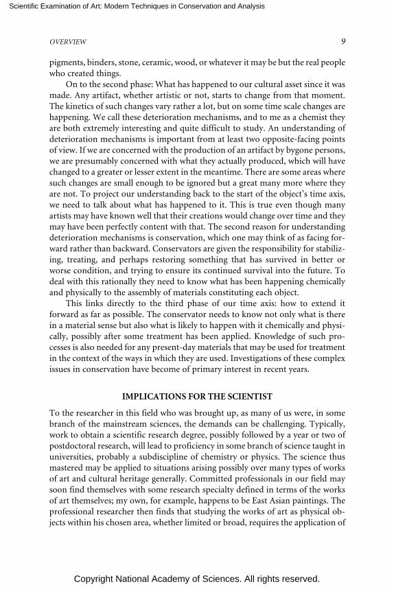
pigments, binders, stone, ceramic, wood, or whatever it may be but the real peoplewho created things.
On to the second phase: What has happened to our cultural asset since it wasmade. Any artifact, whether artistic or not, starts to change from that moment.The kinetics of such changes vary rather a lot, but on some time scale changes arehappening. We call these deterioration mechanisms, and to me as a chemist theyare both extremely interesting and quite difficult to study. An understanding ofdeterioration mechanisms is important from at least two opposite-facing pointsof view. If we are concerned with the production of an artifact by bygone persons,we are presumably concerned with what they actually produced, which will havechanged to a greater or lesser extent in the meantime. There are some areas wheresuch changes are small enough to be ignored but a great many more where theyare not. To project our understanding back to the start of the object’s time axis,we need to talk about what has happened to it. This is true even though manyartists may have known well that their creations would change over time and theymay have been perfectly content with that. The second reason for understandingdeterioration mechanisms is conservation, which one may think of as facing for-ward rather than backward. Conservators are given the responsibility for stabiliz-ing, treating, and perhaps restoring something that has survived in better orworse condition, and trying to ensure its continued survival into the future. Todeal with this rationally they need to know what has been happening chemicallyand physically to the assembly of materials constituting each object.
This links directly to the third phase of our time axis: how to extend itforward as far as possible. The conservator needs to know not only what is therein a material sense but also what is likely to happen with it chemically and physi-cally, possibly after some treatment has been applied. Knowledge of such pro-cesses is also needed for any present-day materials that may be used for treatmentin the context of the ways in which they are used. Investigations of these complexissues in conservation have become of primary interest in recent years.
IMPLICATIONS FOR THE SCIENTIST
To the researcher in this field who was brought up, as many of us were, in somebranch of the mainstream sciences, the demands can be challenging. Typically,work to obtain a scientific research degree, possibly followed by a year or two ofpostdoctoral research, will lead to proficiency in some branch of science taught inuniversities, probably a subdiscipline of chemistry or physics. The science thusmastered may be applied to situations arising possibly over many types of worksof art and cultural heritage generally. Committed professionals in our field maysoon find themselves with some research specialty defined in terms of the worksof art themselves; my own, for example, happens to be East Asian paintings. Theprofessional researcher then finds that studying the works of art as physical ob-jects within his chosen area, whether limited or broad, requires the application of

Scientific Examination of Art: Modern Techniques in Conservation and Analysis
Copyright National Academy of Sciences. All rights reserved.
10 SCIENTIFIC EXAMINATION OF ART
scientific knowledge and understanding from a number of scientific disciplines,which may be removed from his original area of proficiency. There has been akind of orthogonal transposition of concepts; rather than specializing in a singlescientific discipline in depth, the researcher needs to take a range of basic disci-plines and apply them to a class of objects that will themselves be studied indepth. No doubt this happens in other fields of research too, and it is certainlyintellectually stimulating. It can also be alarming. Most scientists, I think, aresensitive to the implications of specialization, to the probability of wandering intoerror when they venture into branches of science other than their own. Thephysicist John Ziman published a book (1987) some years ago dealing with ques-tions of mobility and career change in the sciences, including the reasons whymost scientists tend to be reluctant to change areas of research in which theywork. The problem of how to apply selected, specialized areas of science to afurther understanding of things that ultimately have to be understood on theirown terms is also an intellectual challenge of the field.
CONCLUSION
I conclude with a few words about the colloquium that followed. For reasons thatI mentioned earlier, describing all aspects—or all important aspects—of the sci-entific examination of art is impractical. We hope to have organized a fair sam-pling of what the field is about, in all its variety and complexity. This first day wasintended to give fairly broad reviews of progress in at least some of the majorareas of work. The second day saw accounts of significant progress in more spe-cific topics. This was intended to give us some realistic perspectives on what hasbeen achieved and what has not been achieved in research, particularly that of thepast few years. I think that most of the presentations will fit on the time axis of anobject that I suggested as describing the kinds of work done. Some may look atquestions of the materials and methods used by the creators of artifacts that wechoose to call “art,” some at research on deterioration mechanisms, and others atquestions of an object’s present status and the prognostications we may have forits future.
In this introductory paper, rather than discussing modern techniques orrecent progress, which others will discuss later, I have tried to give some sugges-tion of depth, even (in a sketchy kind of way) historical depth to the subject. Iwould like to be able to give it some coherence, but I fear that would be claimingaltogether too much. Do we really have just one field here, or several smaller fieldsthat happen to overlap here and there? What are the connections between scien-tific studies and considerations of aesthetics, the original intent behind creatingsomething, and the connections to questions of intended use? This colloquiumwas never intended to cast light on problems of this nature, but if we have aserious intellectual discipline underpinning what we do, the more fundamentalquestions implied by its pursuit should at least be recognized to exist.

Scientific Examination of Art: Modern Techniques in Conservation and Analysis
Copyright National Academy of Sciences. All rights reserved.
OVERVIEW 11
REFERENCES
Brommelle, N. 1956. Studies in Conservation 2:176-187.Caley, E. R. 1951. Journal of Chemical Education 28:64-66.Hauser, A. 1985. The Philosophy of Art History. Evanston, Ill.: Northwestern University Press. English
version of Philosophie der Kunstgeschichte, Oscar Beck, Munich, 1958, pp. 216, 232-234.Margolis, J. 1980. Art and Philosophy: Conceptual Issues in Aesthetics. Brighton, Sussex: Harvester
Press.Miller, B. F. 1998. In Painting Techniques. History, Materials and Studio Practice. Contributions to the
Dublin Congress 7-11 September 1998, eds. A. Roy and P. Smith, pp. 246-248. London: Interna-tional Institute for Conservation of Historic and Artistic Works.
Nadolny, J. 2003. Reviews in Conservation 4:39-51.Olin, J. S., ed. 1982. Future Directions in Archaeometry. A Round Table. Washington, D.C.:
Smithsonian Institution.Phillips, Q. E. 2000. The Practices of Painting in Japan, 1475-1500. Stanford, Calif.: Stanford Univer-
sity Press.Plenderleith, H. J. 1998. Studies in Conservation 43:129-143.Rees-Jones, S. 1990. Studies in Conservation 35:93-101.Shimizu, Y. 1981. Archives of Asian Art 34:20-47.Tennent, N. 1997. In British Museum Occasional Papers, 116: The Interface between Science and Con-
servation, ed. S. Bradley, pp. 15-23. London: The British Museum.Watkins, S. C. 1997. In British Museum Occasional Papers, 116: The Interface between Science and
Conservation, ed. S. Bradley, pp. 221-226. London: The British Museum.Ziman, J. 1987. Knowing Everything about Nothing. Specialization and Change in Scientific Careers.
Cambridge: Cambridge University Press.

Scientific Examination of Art: Modern Techniques in Conservation and Analysis
Copyright National Academy of Sciences. All rights reserved.
12
Material Innovation and Artistic Invention:New Materials and New Colors in
Renaissance Venetian Paintings
Barbara H. BerrieNational Gallery of Art, Washington, D.C.
andLouisa C. Matthew
Department of Visual Arts, Union College, Schenectady, N.Y
Sixteenth-century Venetian painters have been regarded as “colorists” since theirown time. The phrase “Venetian palette” is used today by art historians to de-scribe the colors used by Renaissance painters of Venice, among whom Titian,Giovanni Bellini, and Tintoretto are the most famous. There is in fact little writ-ten consensus about how to define this so-called Venetian palette, but our knowl-edge is continually expanding thanks to scientific research on these artists’ paint-ings. One color has always been mentioned as being particularly Venetian: a richdeep orange, used generously by Venetian painters from about 1490. These artistsused the arsenical sulfides yellow orpiment (As2S3) and orange realgar (As4S4) toachieve this color. Until the end of the fifteenth century this pair of minerals hadbeen largely confined to the miniaturists’ palette, but they became so popular insixteenth century Venetian painting that G. P. Lomazzo remarked in his 1584treatise “burnt orpiment is the color of gold and it is the alchemy of the Venetianpainters” [1]. Artists such as Giovanni Bellini used it abundantly in their paint-ings; for example, Bellini used it for Silenus’ robe in The Feast of the Gods (1514;reworked by Titian, 1524) (Figure 1). The analytical data we discuss here, whilestill fragmentary, points to a richness of materials and their innovative use byVenetian artists that is greater than imagined heretofore, and much more thansimply the addition of the arsenical minerals.
Recently discovered evidence has established that professional color-sellersplied their trade in Venice from the end of the fifteenth century. It appears thatthey existed here as much as a century earlier than in any other Italian city. Thesecolor-sellers were neither apothecaries (“speziali”) nor general grocers from whomartists had purchased their painting supplies throughout the middle ages and

Scientific Examination of Art: Modern Techniques in Conservation and Analysis
Copyright National Academy of Sciences. All rights reserved.
MATERIAL INNOVATION AND ARTISTIC INVENTION 13
FIGURE 1 The Feast of the Gods, Giovanni Bellini and Titian, 1514/1529, oil on canvas,(National Gallery of Art, Washington, D.C. 1942.9.1).
early Renaissance. They were sources who specialized in materials used in the artsand trades that dealt with color and color manufacturing. Some of the mostinteresting and useful evidence for the existence of professional color-sellers takesthe form of inventories of the contents of their shops. The earliest found so fardates to 1534 [2]. Another, longer inventory of a color-seller’s shop dated 1596has been found and published [3]. Examination of the materials in the 1534inventory and investigation of their uses, particularly in glass-making and ceram-ics, coupled with our new analyses, reveal relationships that encompass bothtradition and innovation. There is evidence for more cross-fertilization of tech-nological know-how and taste among artisan industries than previously sup-posed. In this paper we will show how the information from the inventoriescombined with new analytical data has been used to expand our knowledge andunderstanding of the materials used by painters in Venice and add to the com-plexity of the definition of the Venetian Renaissance palette.

Scientific Examination of Art: Modern Techniques in Conservation and Analysis
Copyright National Academy of Sciences. All rights reserved.
14 SCIENTIFIC EXAMINATION OF ART
The 1534 inventory lists 102 items; weights or amounts are given but nomonetary values. Many of the materials on the inventory have an establishedconnection with the easel painters’ art, including, for example, the pigments azur-ite, vermilion, lead white, and orpiment. Kermes and brazilwood, organic extractswhich were used to make red dyes as well as red paints, are listed. Other items inthe “vendecolore” shop that relate to the dyers’ craft include alum for mordantingdyes, galls (for making black dyes), and various resins.
The first printed book on dyeing on a commercial scale was published inVenice in 1548, titled The Plichto of Gioanventura Rosetti [4]. It was written notby a dyer but by a technologist, Gioanventura Rosetti, whose intention was toprovide information on what might be termed “best practices” to benefit theVenetian Republic. The recipes in the Plichto contain many of the items on boththe 1534 and the 1596 inventories, including some usually considered by histori-ans as pigments, including orpiment, vermilion and azurite, which are describedin one recipe as mineral dyes (Figure 2). The overlap between painters’ anddyers’ colorants continues to become more apparent.
FIGURE 2 Extract from “The Plictho of Gioanventura Rossetti” first published in Venicein 1548. Translated by Sidney M. Edelstein and Hector C. Borghetty, The MIT Press (1969).

Scientific Examination of Art: Modern Techniques in Conservation and Analysis
Copyright National Academy of Sciences. All rights reserved.
MATERIAL INNOVATION AND ARTISTIC INVENTION 15
The Venetian glass industry, centered on Murano, one of the islands in theVenetian lagoon, was burgeoning in the late fifteenth century. By this time theglassmakers had produced a clear glass called “cristallo” after the rock crystal thathad inspired its invention. Large quantities of clear and colored glass were pro-duced for making a wide variety of objects, including tableware, goblets, glasses,and mosaic tesserae. Recipes for richly colored glass, both single-toned and multi-colored to imitate opal and chalcedony, were developed. Special, deeply-coloredglass was produced for making false rubies, sapphires, and emeralds that were asintensely and beautifully colored as the real gems. In the first decades of thesixteenth century recipes for glassmaking were being compiled [5]. The Darduinmanuscript provides important information on Renaissance glassmaking, andthe work of the Florentine, Antonio Neri (died 1614), who wrote L’Arte Vetraria(1612), a compilation of recipes including many of sixteenth-century origin, is aninvaluable source. [This recipe book was translated into English by ChristopherMerrett in 1662.] For our knowledge of the Venetian glassmaking industry wealso owe much to the work of the Muranese, Luigi Zecchin [6].
Materials necessary for glassmaking are found on the 1534 inventory. Recipesfor glass indicate that tin and lead were required in large quantities; both of theseare on the inventory. Other ingredients include tartar, mercuric chloride, borax,alum, salt, and “tuzia” (zinc oxide), as well as orpiment. These materials are alsoused by dyers and some by painters.
The wide range of materials available at the color-seller’s shop suggests thatartisans from many trades that used color went there to obtain their raw materi-als. The variety available in this one place prompted us to consider whether therewas more cross-fertilization among artisans than previously assumed and if wemight find some evidence for this in the painting practice of the Venetian artists.
We reanalyzed samples from paintings in this light, looking for materials notpreviously recognized. Samples from several paintings by Venetian Renaissanceartists were available from prior studies. They are preserved as cross-sections ofthe paintings mounted in bioplastic polyester/acrylate resin. For optical micros-copy, a Leica DMRX polarizing light (PL) microscope was used with PL fluotarobjectives. For fluorescence microscopy the light source was a mercury lamp(100W) and the D and I3 filter packs. Scanning electron microscopy (SEM) wasundertaken using a JEOL 6300 equipped with an Oxford Instruments Tetra back-scatter detector. For energy dispersive spectrometry (EDS) the SEM was fittedwith an Oxford Si(Li) ATW detector (capable of detecting low-energy x-rays)with a resolution at the Mn kα line greater than 130 eV. The cross-sections wereusually carbon-coated, but sometimes gold-palladium coatings were used. X-raypowder diffraction patterns were obtained using Philips XRG 3100 x-ray genera-tor with a copper tube. Data were collected on photographic film in a Gandolficamera (radius 57.3 mm). Line spacings were measured against a calibrated ruleand relative intensities estimated by eye.

Scientific Examination of Art: Modern Techniques in Conservation and Analysis
Copyright National Academy of Sciences. All rights reserved.
16 SCIENTIFIC EXAMINATION OF ART
Samples from paintings by the Venetians Lorenzo Lotto (1480-1556) andJacopo Tintoretto (1519-1594) were among the first to be re-examined. Althoughthe samples are limited in number they already show that the range of materialsused to make paint is wider than previously known.
Lotto was “rediscovered” in the late nineteenth century, but it took most ofthe twentieth century for him to become acknowledged as a Venetian painter.Recent research on his painting technique and color palette has helped define hisplace in the Renaissance [7, 8]. There is little documentary information on Lotto’searly career as an artist, but it is believed that he trained in Venice and spent hisfirst years as an independent artist there. Later, he painted in Bergamo and theMarches. He traveled a good deal, usually within the economic and political orbitof the Venetian Republic, and he returned to the city itself for several periods. Ourknowledge of Lotto’s working methods is augmented by the survival of one of hisaccount books in which he documented commissions and expenditures duringthe years 1538 to 1556 [9]. One particularly valuable section of the account books isan appendix of spese per l’arte (expenditures for art), where he recorded the purchaseof painting supplies, among which are notes on pigments he purchased in Venice.
Among Lotto’s paintings at the National Gallery of Art in Washington, D.C.is St. Catherine, signed and dated 1522 (Figure 3). St. Catherine’s dress is a glori-ous red, perhaps reminiscent of the color of expensive red cloth worn by someVenetian brides at this time. A cross-section from the sleeve (Figure 4) shows thecomplicated layering Lotto used to create this color. In the cross-section, we see,from the bottom, the preparatory layer of gesso (CaSO4.2H2O in glue), used toprovide a smooth surface for painting, over which many layers of paint wereapplied. The first layers of paint are pinks prepared from a mixture of vermilionand lead white. Lying over these are layers of transparent red paint. From fluores-cence microscopy (Figure 5) it can be discerned that what appears to be a thickhomogeneous paint film is in fact many layers of thin glazes of paint; there appearto be at least six layers. The same painting technique was found in two versions ofanother composition painted by Lotto in the same year, The Virgin and Child withSaints Jerome and Nicholas of Tolentino [8]. It was shown, using high-perfor-mance liquid chromatography, that for the version at the National Gallery, Lon-don, Lotto used both madder and insect lakes. The fluorescence of the lakes in St.Catherine’s dress implies that he used two different lakes here also.
Digital dot maps of the distribution of the elements in a sample from St.Catherine obtained using SEM-EDS are shown in Figure 6. The lowest layer ofpaint contains mercury, confirming that Lotto used vermilion for mixing thelight red underpaint. Aluminum is present throughout most of the upper layersof transparent paint glazes. This strongly suggests that the pigment is a dye lakedon alumina, the traditional way to prepare insoluble pigments from dyes madefrom lakes. Unexpectedly, several of the layers of transparent paint contain small,rounded particles, ca. 4-8 microns in diameter. These particles appear to be verypure silica. It is difficult to obtain information on individual particles embedded

Scientific Examination of Art: Modern Techniques in Conservation and Analysis
Copyright National Academy of Sciences. All rights reserved.
MATERIAL INNOVATION AND ARTISTIC INVENTION 17
FIGURE 3 St. Catherine, Lorenzo Lotto, oil on panel, Samuel H. Kress Collection,1939.1.117.
in paint, owing to the comparatively large interaction volume (the volume beinganalyzed) in a low-density matrix such as paint made using lake pigments. EDSspectra were obtained at 20 kV and 15 kV accelerating voltage; lowering thevoltage was designed to decrease the analysis volume. The spectra (Figure 7)indicate a (rather) pure form of silica; only aluminum is present, and its origin islikely the surrounding particles of red lake. Only silicon and oxygen are signifi-cant elements in line scans through the particles. Elements that would indicatethis material is a glass, for example, the fluxes sodium and potassium or thestabilizers, calcium and lead, are below detectable limits. Venetian glassmakingrequired pure silica, which was, in this period, provided by quartzite pebbles fromthe Ticino River.

Scientific Examination of Art: Modern Techniques in Conservation and Analysis
Copyright National Academy of Sciences. All rights reserved.
18 SCIENTIFIC EXAMINATION OF ART
FIGURE 4 Cross section from a dark fold in the sleeve of St. Catherine (Figure 3) near thebottom edge, photographed in reflected light.
FIGURE 5 The cross-section illustrated in Figure 4, observed using fluorescence micros-copy (filter cube: Leitz I3).

Scientific Examination of Art: Modern Techniques in Conservation and Analysis
Copyright National Academy of Sciences. All rights reserved.
MATERIAL INNOVATION AND ARTISTIC INVENTION 19
Fifteenth and sixteenth century treatises suggested using crushed marble orcrushed travertine as additives to give body to paints [10]. Glass has been de-scribed as a drier for paint in Renaissance treatises and has been found in someartists’ red lake paint [11]. However, the presence of silica is unexpected, and thisoccurrence appears to be the first finding of this material used by Italian Renais-sance painters as an extender or an agent to give body in red lake paints. Themajor ingredient in Antonio Neri’s recipe for “cristallo” is pebbles “poundedsmall, serced as fine as flower” [12] (serce is probably a variant of sarce, to sievethrough a cloth). This description corresponds to the material in Lotto’s redpaint, which was a ground silica.
FIGURE 6 Digital dot maps of the cross-section shown in Figures 4 and 5.

Scientific Examination of Art: Modern Techniques in Conservation and Analysis
Copyright National Academy of Sciences. All rights reserved.
20 SCIENTIFIC EXAMINATION OF ART
FIGURE 7 Energy dispersive spectrum of small rounded particles in the translucent redpaint; obtained at 20 kV.
The artist Jacopo Robusti, called Tintoretto, worked in Venice a few decadeslater than Lorenzo Lotto. Tintoretto was born in that city in 1519; his father wasa member of the “cittadini” class, involved in the dyeing profession. Tintorettolived and worked in the city throughout his career, and rarely traveled. He estab-lished a family workshop that outlived him, and he worked for a wide variety ofVenetian patrons. Arguably his most famous surviving work is a series of paint-ings executed for the Scuola Grande di San Rocco over several decades [13].
The painting Christ at the Sea of Galilee (Figure 8) is attributed to Tintorettoand dated to 1575/80. This picture presents complicated issues in understandingits structure and the artist’s painting technique since the canvas support wasassembled from several pieces of fabric that had been used for painting imagesdifferent from the one we see now. The infrared reflectogram of the painting

Scientific Examination of Art: Modern Techniques in Conservation and Analysis
Copyright National Academy of Sciences. All rights reserved.
MATERIAL INNOVATION AND ARTISTIC INVENTION 21
reveals that at some point the largest, central piece of canvas had been used tobegin a portrait. The portrait had been sketched out using a wash of dark paint,clearly imaged in the infrared. The x-radiograph reveals that the canvas had alsobeen used for a landscape that is of a different scale from both the portrait and thecurrent image.
Tintoretto’s painting techniques have been well studied [14, 15]. An investi-gation into the materials used for the Gonzaga cycle (1577-1578) showed that theartist employed a diverse palette [16].
Here we restrict the discussion to two pigments found in Christ at the Sea ofGalilee that have special relevance to the use of glassy materials for pigments. Across-section obtained from the sea at the right-hand side of the boat is shown inFigure 9. The bottom layer of the section appears to relate to the landscapeobservable in the x-radiograph. The pigment is a green, transparent, glassy-appearing pigment. The particle shape and size is similar to that of the blue glasspigment smalt (a potassium silicate colored by small amounts of cobalt). Al-though the term “smalt” is used in English today to describe only a blue glasspigment, reading the contemporary documents shows that artists of the six-teenth century used this term to describe not only blue but also numerous other
FIGURE 8 Christ at the Sea of Galilee, Jacopo Tintoretto 1575/1580, oil on canvas, SamuelH. Kress Collection,1952.5.27.

Scientific Examination of Art: Modern Techniques in Conservation and Analysis
Copyright National Academy of Sciences. All rights reserved.
22 SCIENTIFIC EXAMINATION OF ART
FIGURE 9 Cross section from the sea near the right hand side edge of the boat. Thebottom layer contains a green glassy pigment.
colored glasses, including yellow, white, and green, at least some of which mayhave been used by painters [17, 18].
The backscatter image of this section is shown in Figure 10. The greenishpigment in the bottom layer appears dark gray, and therefore we can infer it is oflow atomic weight. The EDS spectrum of the pigment shows that it has a compo-sition very similar to blue smalt (Figure 11). An anonymous Venetian glassmaker’srecipe book dating to early-mid sixteenth century has recipes for green glass thathave the same general composition as blue smalts: “Per fare smalto verdebellissimo. Prendi della zaffera e un po’ di manganese, pestati sottili e ben lavati edi questi prendi 2 libbre, aggiungi 3,5 libbre di pani cristallini e fa fondere inforno.” [To make a beautiful green glass. Take some zaffre (an impure cobaltore), grind it fine and wash well and of this take 2 lbs, add 3.5 lbs of crystal frit (apotash glass) and melt in the furnace” [5].
This green smalt in Christ at the Sea of Galilee contains an impurity of bis-muth. Bismuth has been found in late-fifteenth and early-sixteenth Venetianenamels and in fifteenth century cobalt blue enamels and smalt in a south Ger-man painting [19]. Bismuth is an impurity in the cobalt ore from Germany, andits presence in this pigment suggests that the source of the raw cobalt-containingmaterial, “zaffera,” used for making this glass was from north of the Alps. Thespectrum shows that the glass contains iron. Iron can give rise to a yellow glass.Therefore the green color of this pigment might arise from a mixture at themicroscopic level of blue and yellow glasses.

Scientific Examination of Art: Modern Techniques in Conservation and Analysis
Copyright National Academy of Sciences. All rights reserved.
MATERIAL INNOVATION AND ARTISTIC INVENTION 23
FIGURE 10 Backscatter electron image of the sample in Figure 9.
FIGURE 11 Energy dispersive spectrum of the green pigment in the bottom layer of thesection illustrated in Figure 9.

Scientific Examination of Art: Modern Techniques in Conservation and Analysis
Copyright National Academy of Sciences. All rights reserved.
24 SCIENTIFIC EXAMINATION OF ART
A yellow pigment is used widely in Christ at the Sea of Galilee. In a cross-section from Christ’s drapery it can be seen mixed with green earth for the seapainted under Christ’s red robe and as an intense yellow layer under the greenishpaint of the sea. It was also used, well mixed with green earth and azurite, for thehills in the background. At first glance the pigment appears to be lead tin yellowtype II (Pb(Sn,Si)O3). SEM-EDS clearly indicates that the colorant is an opaqueyellow glass composed of particles of lead tin oxide suspended in a glassy matrix.X-ray powder diffraction (XRD) reveals that the yellow opacifier is similar butnot identical to the material usually characterized in paintings. The XRD patternof the pigment is given in Table 1. Although the pattern is very close to thatpublished for PbSnO3, there are some subtle differences and additional lines notattributable to expected impurities. The compendia of recipes for making glassgive several variations for the yellow colorant, which likely cause different hues. Itwould be interesting to compare the XRD pattern of the colorant and the compo-sition of the glassy matrix of the pigment in this painting with those of enamels onmetals and glazes on majolica and relate the results to the contemporary recipes.By comparing the details of these materials we may be able to shed further lighton the variety of yellows that was available for the ceramic decorators and used byeasel painters to increase the range of their palette. A recent paper differentiatesbetween the production of lead tin yellow pigment and the “raw” material for theproduction of yellow glass [20]. This difference might be found among the mate-rials used by Venetian artists and craftsmen. Thus the glassy matrix might beimportant, and this and other differences between glasses and pigments might bethe source for the variety of materials and colors that painters used.
Many of the materials we find on the 1534 (and the 1596) inventory arematerials used by dyers, glassmakers, and glass and maiolica painters. Some ofthese, including vermilion, kermes, brazilwood, orpiment, and lead white, areexpected in paintings by Bellini, Giorgione, and Titian. The re-analysis of samplesfrom pictures by these and other Venetian artists has begun to indicate that thepalette they used was enriched by materials that until then had only been used byartisans and artists working in other media. Venetian painters (and others influ-enced by them) boldly incorporated into their work, to vivid effect, colorants notspecifically designed for use in oil paint. We see that artists were using glassymaterials and/or “smalti” more often and in greater diversity than we previouslythought. Among these materials there appear to be frits and colorants designedfor glass-painters and majolica decorators, in addition to the powdered glass, bluesmalt and lead tin yellow type II, which have been identified previously.
The presence of the professional color-seller in Venice might have been thecatalyst and the conduit for the transfer of materials among the arts and contrib-uted to the emergence of the Venetian palette, a palette that cannot be preciselydefined, but is characterized by its complexity and diversity of colorants.

Scientific Examination of Art: Modern Techniques in Conservation and Analysis
Copyright National Academy of Sciences. All rights reserved.
MATERIAL INNOVATION AND ARTISTIC INVENTION 25
TABLE 1 d-Spacings and Estimated Intensities of Lines in the DiffractionPattern of the Glassy Yellow Pigment in Tintoretto’s Christ at the Sea of Galileeand patterns for PbSnO3 and SnO.
Yellow PbSnO3 SnOPigment ICDD 17-607 ICDD 24-1342
d I/Imax d I/Imax d I/ImaxAngstroms
6.17 18
4.654.5*4.324.20*3.933.63*3.50 w3.30*3.25 3.22 123.10 100 3.09 1002.98 20 2.9 802.852.77 20 2.78 802.69 802.61* 50 2.63 1002.462.45 2.45 122.30*2.21 10 2.24 102.10 5 2.12 102.05 2.06 61.95 10 1.95 301.90 80 1.89 751.864 65 1.83 251.61 80 1.61 80 1.61 201.54 1.52 161.23 1.227 291.195 1.196 16
*These lines can be attributed to lead white (International Committee for Diffraction Data 13-131).
ACKNOWLEDGEMENTS
We are grateful to the Center for Advanced Study in the Visual Arts (NationalGallery of Art, Washington, D.C.) where we held a Samuel H. Kress Paired Fel-lowship. We benefited from discussions with members of the scientific researchdepartment of The National Gallery, London, and particularly acknowledgestimulating discussions with Jo Kirby-Atkinson.

Scientific Examination of Art: Modern Techniques in Conservation and Analysis
Copyright National Academy of Sciences. All rights reserved.
26 SCIENTIFIC EXAMINATION OF ART
REFERENCES
1. Lomazzo, G.P., Trattato dell’arte della pittura. 1590, Milan: Paolo Gottardo Ponto.2. Matthew, L.C., ‘Vendecolori a Venezia’: the reconstruction of a profession. The Burlington Maga-
zine, 2002. CXLIV (1196): pp. 680-686.3. Krischel, R., Zur Geschichte des Venezianischen Pigmenthandels - Das Sortiment des Jacobus de
Benedictus a Coloribus, in Sonderuch aus dem Wallraf - Richartz - Jahrbuch Band LXIII 2002. 2002,Cologne: Dumont Literatur und Kunst Verlag. pp. 93-158.
4. Rosetti, G., Plictho de l’arte de tentori. 1548. Translated by Sidney M. Edelstein and Hector C.Borghetty, 1969. Cambridge, Massachusetts: The M.I.T. Press.
5. Moretti, C. and T. Toninato, Ricette vetrarie del Rinascimento: Trascrizione da un manoscrittoanonimo veneziano. 2001, Venice: Marsilio.
6. Zecchin, L., Vetro e Vetrai di Murano. Vol. 1-3. 1987-1989, Venice: Arsenale.7. Lazzarini, L., et al., Pittura veneziana: materiali, techniche, restauri. Bollettino d’Arte, 1983. 5:
pp. 133-166.8. Dunkerton, J., N. Penny, and A. Roy, Two paintings by Lorenzo Lotto at the National Gallery.
National Gallery Technical Bulletin, 1998. 19: pp. 52-63.9. Lotto, L., (Libro di spese diverse [1538-1556] con aggiunta di lettere e d’altri documenti.), P.
Zampetti, editor. 1969, Venice, Rome. See also Bensi, P., Studi di storia dell’arte, 5 1983-1985, 63.10. Merrifield, M.P., Medieval and Renaissance Treatises on the Arts of Painting. 1999, Mineola,
NY: Dover. p. clii.11. The Painting Technique of Pietro Vanucci, Called Il Perugino Editors. B. G. Brunetti, C.
Seccaroni, A. Sgamellotti, Nardini Editore, 2003. Papers from the conference, 14-15 April, 2003.12. Merrett, C., The World’s Most Famous Book on Glassmaking ‘The Art of Glass’ by Antonio Neri,
M. Cable, editor. 1662, Sheffield: The Society of Glass Technology reprint 2003. (Neri’s book hadbeen first published in Italian in 1612.)
13. Krischel, R., Jacopo Tintoretto. 2000, Cologne: Könemann.14. Plesters, J. and L. Lazzarini. Preliminary Observations of the Technique and Materials of
Tintoretto in Conservation of Paintings and the Graphic Arts. 1972, Lisbon Congress: InternationalInstitute for Conservation.
15. Plesters, J. and L. Lazzarini. I materiali e la tecnica dei Tintoretto della scuola di San Rocco, inJacopo Tintoretto nel quarto centenario della morte. 1994, Venice: Il Polygrafo.
16. Burmester, A. and C. Krekel, “Azurri oltramarini, lacche et altri colori fini”: the quest for the lostcolours, in Tintoretto: The Gonzaga Cycle, C. Syre, editor. 2000, Munich: Hatje Cantz Publishers. pp.193-211.
17. Venturi, A., I due Dossi documenti - prima serie. Archivio Storico dell’Arte Nuovi Documenti,1892. Anno 5 (Fase VI): pp. 440-443.
18. S. Pezzella, Il trattato di Antonio da Pisa sulla fabricazione delle vetrate artitiche, 1976. Perguia:Umbria Editrice.
19. Darrah, J.A. Connections and Coincidences: Three Pigments. in Historical Painting Techniques,Materials, and Studio Practice. 1995, University of Leiden, the Netherlands: The Getty ConservationInstitute.
20. Heck, M., T. Rehren, and P. Hoffmann, The Production of Lead-Tin Yellow at MerovingianSchleitheim (Switzerland). Archaeometry, 2003. 45(1): pp. 33-44.

Scientific Examination of Art: Modern Techniques in Conservation and Analysis
Copyright National Academy of Sciences. All rights reserved.
27
The Scientific Examination ofWorks of Art on Paper
Paul M. WhitmoreResearch Center on the Materials of the Artist and Conservator
Carnegie Mellon UniversityPittsburgh, Pennsylvania
ABSTRACT
The scientific examination of works of art on paper utilizes tools from thevery simple to state-of-the-art analytical instrumentation, depending inlarge part on the question that is the objective of the investigation. Identify-ing pigments or paper fibers is straightforward, constrained only by the sizeof the samples that can be removed for destructive analysis. Inks are moredifficult because of the lack of pronounced chemical differentiation betweenthe ink types and because of possible interferences in the analyses from thepaper substrate. Paper can be characterized easily to an extent, in identify-ing a watermark or the risk of deterioration from a high acid content, butthe monitoring of the condition and degradation of paper remains an ex-tremely difficult challenge. The assessment of light sensitivity, which is noteasy to determine by merely identifying material composition, has beenmade straightforward by the development of a device that allows rapid,essentially nondestructive fading tests. Those tests are now being exploitedto survey groups of objects to determine whether one may make generaliza-tions about their exhibition needs. The further adaptation of nondestructiveor micro-scale destructive analytical tools in the study of works of art onpaper promises to allow even more extensive investigations of the creationand preservation of these objects.

Scientific Examination of Art: Modern Techniques in Conservation and Analysis
Copyright National Academy of Sciences. All rights reserved.
28 SCIENTIFIC EXAMINATION OF ART
INTRODUCTION
The scientific study of works of art on paper shares common objectives with thetechnical studies of any work of art. Artifacts are examined in order to answer arthistorical questions about the origin of a work, namely, where, when, and bywhom a work was created. The scientific examinations seeking to answer thesequestions generally require identification of the materials and working methodsused to craft the object. Other studies seek to answer basic questions about thecare of the artifact: its physical and chemical condition, causes for deterioration,and vulnerability to storage or exhibition conditions.
Technical studies of paper-based artifacts tend to resemble the study ofpaintings, because many paper objects actually are paintings that just happen tobe executed on a paper support. Manuscript illuminations, watercolors, litho-graphic prints—these objects could easily be viewed as paintings, amenable toanalyses of the colorants, paint media, or layer structure of paints observable incross-sections. Apart from the occasional thinness of the paint layer itself, as inwatercolor paintings, or binder-poor paint layers, such as in pastels, these paper-based paintings can often be studied as one would study any other painting.
Despite this similarity, many works of art on paper present special circum-stances that constrain analyses or warrant unusual examination techniques. Paperartifacts tend to be small: The sheets were traditionally made in molds that couldbe manipulated by people, and these sheets were then cut down for use. Thus,books, prints, watercolors, and other paper-based art are relatively small, meantfor close-up viewing within an arm’s length. For this reason, analytical methodsthat require removal of paint samples are often not feasible, for the damage to theartifact can sometimes be visible upon close inspection. Nondestructive tools,particularly optical spectroscopic or imaging techniques, are more widely used tostudy these objects.
Another distinction between paper-based objects and traditional paintings isthe use of the paper substrate as part of the image itself. Particularly with suchgraphic art as drawings and prints but also with printed text or even thinly paintedwatercolors, the paper substrate is exposed and is part of the image. Thus, thecolor of the paper and its surface texture are important contributors to the ap-pearance and visual appeal of the object, and study of the paper and its preserva-tion is of great importance. (Occasionally in historical times and more frequentlyin the twentieth century, paintings too have been created with unpainted canvasas part of the image. For these objects the concern about the appearance andstability of the canvas is of course shared.) A complication in studying objects inwhich the paper is so intimately associated with the drawing media is the dis-crimination between the two, so that many analyses must have very small spatialor depth resolution, or contributions to the detected signal from the paper mustbe subtracted.
Paper-based collections in museums are known to pose some of the most

Scientific Examination of Art: Modern Techniques in Conservation and Analysis
Copyright National Academy of Sciences. All rights reserved.
THE SCIENTIFIC EXAMINATION OF WORKS OF ART ON PAPER 29
common preservation problems because many of the artifacts that are now prizedwere not created as lasting works of art but as more utilitarian objects. Becausepaper was inexpensive and widely available through much of history, it has seenuse for many purposes, a primary one being for communication and recording ofinformation. Some of these artifacts, such as books, were meant to last for a longtime, but others, such as newspapers, announcements, or letters, were often notcreated with posterity in mind. Thus, it is not uncommon for museums andarchives to have paper artifacts that are delicate or deteriorating because of theircreation with impermanent materials or techniques. Preservation problems arecommon, particularly with those objects that were not made as art objects.
This review will survey the examination techniques of paper-based objectsthat are used both for art historical investigations as well as for preservationstudies. Some of those techniques are routine and can be found in many well-equipped museum laboratories; others are less widely available and have notfound widespread use. This survey will conclude with a description of a relativelynew tool developed to detect a particular vulnerability, the susceptibility of col-ored materials to fade from light exposure, and illustrate its use for the study ofJapanese woodblock prints.
SURVEY OF EXAMINATION AND MATERIALIDENTIFICATION TECHNIQUES
The most common technical investigation for paintings or colored prints onpaper involves identification of the pigments in the paint. For this, the routineanalytical tools of polarizing-light microscopy, X-ray diffraction, and elementalanalyses by X-ray fluorescence are commonly employed, usually on samples ofthe paint that have been removed from the artifact. Descriptions of these tools canbe found in accounts of painting examinations, or in reference books devoted topigment identification (Feller, 1986; Roy, 1993; FitzHugh, 1997). Nondestructivetechniques can also sometimes be used to identify pigments on paper objects.Open-air X-ray fluorescence is used for elemental analyses of pigments, andRaman spectroscopy and Raman microscopy have been found useful for examin-ing both pigments in paints and dyes in colored paper (Bell et al., 2000; Best et al.,1995). Some pigments have distinctive features in the visible spectrum (Schweppeand Roosen-Runge, 1986; Leona and Winter, 2001), while others, like Indianyellow, can be detected by their peculiar fluorescence observable under ultravioletlight illumination (Baer et al., 1986).
Drawing materials can also be studied, although they present some difficul-ties. Early drawings were created using metal tools or wires as drawing imple-ments (thus the name “metalpoints” for these drawings), and they can be ana-lyzed by measuring the elemental composition of the metals in the lines (by X-rayfluorescence, typically). Inks are more problematic, with the exception of iron gallinks, which can be distinguished by the presence of iron in X-ray fluorescence or

Scientific Examination of Art: Modern Techniques in Conservation and Analysis
Copyright National Academy of Sciences. All rights reserved.
30 SCIENTIFIC EXAMINATION OF ART
in more unusual techniques such as Mössbauer spectroscopy (Rusanov et al.,2002) or PIXE (Budnar et al., 2001). Inks can also be analyzed for the traceelements they contain, introduced in the ink ingredients or as residues from theprinting process. Inks in early books (such as a Gutenberg Bible) have been exam-ined for these trace elements by synchrotron-excited X-ray fluorescence in thehope of distinguishing books produced in the early German printing shops(Mommsem et al., 1996). Other organic inks, such as sepia (cuttlefish ink), bistre(from soot), or such black drawing media as charcoal, bone black, lamp black,ivory black, or graphite cannot usually be distinguished by their elemental com-position (although bone black is often detected by the presence of phosphorus),nor do the infrared spectra of these inks usually present characteristic featuresuseful for their identification. Polarizing-light microscopy remains a commontool to discriminate between inks on the basis of their particle morphologies. Themedia used as pigment binders for drawing and painting materials can be identi-fied by analyzing the organic composition of micro-samples. Of the various meth-ods available the most useful are the gas chromatography/mass spectroscopyanalyses that have been developed for oils and resins used as paint binders (Millsand White, 2000; Schilling and Khanjian, 1996) and more recently adapted for thestudy of gums used in watercolors or gouaches (Vallance et al., 1998).
In addition to the study of the image-forming materials, the examination ofthe paper itself is also often a clue to the artifact’s origin. Paper is usually differen-tiated by its fiber composition, its physical characteristics, and its manufacturingmethod. The fibers can be studied with optical microscopy, and the plant originof the component fibers can be determined by appearance or by reaction tocertain stains, such as Hertzberg or Graff C stains. The fiber type, length, andheterogeneity can all be distinctive, as can such physical dimensions as sheetthickness. The evidence of manufacture is most easily detected in the pattern leftby the papermaking mold, typically a pattern of lines called chain and laid lines inso-called “laid” paper. Watermarks, the decorative patterns often woven into thewire molds or embossed on the cast sheets, are also the most obvious characteris-tic patterns of the paper manufacture. The evidence of chain and laid lines andwatermarks can be captured in any of a number of ways, with transmitted lightphotography or with beta or soft X-ray radiography, and various image process-ing tools have been applied to enhance such records (erasing interferences fromthe printing, for example) and making them more useful for indexing and re-trieval for comparison in a reference database (Brown and Mulholland, 2002).The presence of sizing (a water-resistant finish on the surface of the paper) can bedetermined by infrared spectroscopy or colorimetric methods (Barrett andMosier, 1995), and fillers (typically finely ground minerals or clays added forincreased opacity of the sheet) can be identified by optical or electron microscopies(Browning, 1969). The fiber, finish, or watermark, along with other historicalevidence, can assist in tracing a paper’s origin (Hunter, 1978), yet many chal-lenges remain (Slavin et al., 2001).

Scientific Examination of Art: Modern Techniques in Conservation and Analysis
Copyright National Academy of Sciences. All rights reserved.
THE SCIENTIFIC EXAMINATION OF WORKS OF ART ON PAPER 31
EXAMINATIONS TO ASSESS CONDITIONAND PRESERVATION PROBLEMS
As with all works of art, preserving works of art on paper focuses on main-taining both the physical integrity of the artifact and its appearance. The physicalintegrity is derived mainly from the paper sheet itself, and preservation of thesheet’s cohesive strength is of paramount importance. (For books and archivalmaterials that are handled by users, other sheet properties, such as flexibility, arealso important, but for works of art that are usually mounted in a frame, thephysical stresses are usually merely the tensile stresses from the paper’s weightand from its reaction to temperature and humidity.)
The cohesive strength of a sheet of paper is derived from the strength of itsconstituent fibers and of the bonds between the fibers. Aging tends to reduce thefiber strength, and old weak papers are usually seen to fail from broken fibersrather than by unraveling from weakened interfiber bonds. The reduction of fiberstrength is in turn a result of the breakdown of cellulose, the natural polymer ofglucose that composes plant fibers. Chemical degradation breaks cellulose chains,which reduces the average molecular weight but more importantly also breaks theconnections between the highly crystalline cellulose zones. This progressive rup-ture of the tie chains, the amorphous cellulose chains connecting the crystallitesand imparting the cohesive strength to the fiber, is the underlying aging chemistryleading to physical failure of the paper sheet.
Unfortunately, there are no analytical tools that can allow detection of suchdeterioration in a paper artifact without destructive analysis of unacceptably largeportions of paper. Typically for degrading polymers, nondestructive tools such asinfrared spectroscopy do not have the sensitivity to detect the production of thevery small concentrations of new chain ends in the degrading cellulose. Recentstudies suggest that production of glucose or xylose residues (Erhardt andMecklenberg, 1995) or low molecular weight acids (Shahani and Harrison, 2002)may be easier to track as some measure of cellulose reaction, but these techniqueshave not yet been applied to artifacts. Other efforts to develop micro-scale mo-lecular weight analyses for cellulose have reduced the amount of paper needed(Rohrling et al., 2002), but a recent molecular weight analysis of cellulose in asingle paper fiber, while successful, also suggests that such small sample sizes maynot be typical of the other fibers or representative of the average molecular weightof larger samples (Stol et al., 2002). Thus, even if the analytical procedure can beadapted, the slow deterioration of the cellulose may not be easily tracked bysuccessive measurements of individual fibers over time.
While the deterioration of paper artifacts may be difficult to detect directly,many years of investigation of cellulose degradation have clearly indicated thatthere are other materials that may be reliable indicators of instability in the paper.Acidity is well established as a catalyst for the hydrolytic breakdown of cellulose,the most important of the known degradation chemistries. Lignin is primarily

Scientific Examination of Art: Modern Techniques in Conservation and Analysis
Copyright National Academy of Sciences. All rights reserved.
32 SCIENTIFIC EXAMINATION OF ART
responsible for the discoloration of groundwood papers, and iron and copperimpurities can also act both as acid catalysts for hydrolysis and as catalysts foroxidative breakdown of cellulose. It is much easier to determine the presence ofthese sensitizing agents in paper than to track the slow deterioration of the cellu-lose, so the study of artifact materials often does not go beyond a pH measure-ment, the detection of lignin with phloroglucinol stain or an infrared spectrum,or the analysis for iron or copper impurities by a technique such as electron spinresonance spectroscopy (Attanasio et al., 1995). Iron present not as a paper impu-rity but as an ink component is also a well-known and easily identifiable riskfactor for the preservation of manuscript and print materials.
The maintenance of the image-forming materials, particularly the coloredpaints and inks used to create the image, is another objective of preservationstrategies. Light exposure is the most common hazard to the pigments and dyesused on art objects. In contrast to preserving the paper support, in which thedeterioration is difficult to monitor and easier to predict by detecting the pres-ence of destabilizing components, the loss of color is easy to monitor by periodiccolor measurements but difficult to predict. The light stability of a pigment de-pends not only on the material but also on its preparation, particle size, and priorfading history. None of these is easy to determine from study of the pigments, anduntil recently the only means to detect light sensitivity was to monitor the damageinflicted by light exposure.
Recently a new device has been developed to determine the risk of futurefading from light exposure (Whitmore et al., 1999). That device operates as areflectance spectrophotometer using a very intense focused beam from a xenonlamp as the illumination for the measurement (see Figure 1). By making rapidrepeated spectral measurements while the material is illuminated by the intenselight, very slight degrees of fading can be detected in light-sensitive materials inonly a few minutes (see Figure 2). Because of the high precision of the spectrumacquisition, extremely small amounts of fading are easily recorded, and the testcan be stopped before perceptible changes to the art object have been produced.All of the different color areas on a work of art can be tested, and the overallsensitivity of the object can be judged by the fading rate of the most light-sensitivecolor (see Figure 3). These tests can be used to develop exhibition requirementsthat are tailored to the needs of the object, with the very light-sensitive objectsreceiving greater care (less frequent exhibition at lower light levels) so that theydo not suffer from fading damage caused by inappropriate display. These sametests, done with filtered illumination, can also be used to test the effectiveness ofdifferent lighting in reducing fading rates. By performing the tests in air or underan inert gas, the efficacy of oxygen-free housings for slowing the fading of worksof art can also be assessed (see Figure 4).
It has been found that this fading test can also be used to identify pigments,not by their elemental or chemical constitution but rather on the basis of theirphotochemical reaction. Prussian blue, a ferric ferrocyanide complex used in art

Scientific Examination of Art: Modern Techniques in Conservation and Analysis
Copyright National Academy of Sciences. All rights reserved.
THE SCIENTIFIC EXAMINATION OF WORKS OF ART ON PAPER 33
FIGURE 1 Schematic of fading tester. Reprinted from the Journal of the American Institutefor Conservation, vol. 38, no. 3, with the permission of the American Institute for Conser-vation of Historic and Artistic Works, 1717 K St., NW, Suite 200, Washington, D.C.20006.
FIGURE 2 Fading test results for selected Winsor & Newton gouache paints. “Blue Wool1” designates fading test results for the ISO Blue Wool no. 1, the most light-sensitive ofthe standard cloths. Reprinted from the Journal of the American Institute for Conservation,vol. 38, no. 3, with the permission of the American Institute for Conservation of Historicand Artistic Works, 1717 K St., NW, Suite 200, Washington, D.C. 20006.
0
5
10
15
20
25
0 1 2 3 4 5
Time (minutes)
CIE
Col
or D
iffer
ence
, ∆
E
Rose Tyrien
Bengal Rose
Magenta
Rose Malmaison
Geranium
Blue Wool 1

Scientific Examination of Art: Modern Techniques in Conservation and Analysis
Copyright National Academy of Sciences. All rights reserved.
34 SCIENTIFIC EXAMINATION OF ART
since the early eighteenth century, is known to fade reversibly during light expo-sure, with the blue color being recovered in a subsequent dark reaction (Ware,1999). The fading tests of Prussian blue using the tester described above demon-strated this peculiar reversible fading behavior on a cyanotype, an early photo-graphic process used to create blueprints (see Figure 5) (Whitmore et al., 2000).
In addition to these fading tests designed to evaluate individual artifacts,current studies are measuring the fading rates of particular colorants in Japanesewoodblock prints from different eras, printed at different depths of color, and ofvarying degrees of prior fading. Results of such a population study will revealwhether there is general consistency or a wide variation in light sensitivity forparticular materials. If there is widely varying behavior, fading tests must beperformed on each object in order to determine the sensitivity and light exhibi-tion needs. If there are very similar fading rates among the different applicationsof a pigment, one need not test every object and can instead safely use a rule ofthumb to make such judgments of the object’s required care. This formulation ofnew rules of thumb, based on actual fading sensitivities observed in a large popu-lation of objects, will bring a new level of intuition about how to preserve objects.The results of tests on a large number of Japanese woodblock prints indicate thatthe fading of the colorant dayflower blue (aobana) is very regular, and its sensitiv-ity can probably be safely estimated without individual fading tests (see Figure6a). By contrast, yellow passages on the Japanese prints vary greatly in their light
0
1
2
3
4
5
0 1 2 3 4 5
Time (minutes)
CIE
Col
or D
iffer
ence
, ∆E
BW2
BW3
FIGURE 3 Fading test results for all the different color areas on a Japanese woodblockprint (Yoshitoshi, Carnegie Museum of Art No. 89.28.1516). “BW2” and “BW3” desig-nate the degree of color difference produced after five minutes in fading tests of ISO BlueWool fading standards nos. 2 and 3.

Scientific Examination of Art: Modern Techniques in Conservation and Analysis
Copyright National Academy of Sciences. All rights reserved.
THE SCIENTIFIC EXAMINATION OF WORKS OF ART ON PAPER 35
0
2
4
6
8
10
12
14
16
18
0 2 4 6 8 10
Time (minutes)
CIE
Col
or D
iffer
ence
, E ∆
0
1
2
3
4
5
6
0 2 4 6 8 10
Time (minutes)
CIE
Col
or D
iffer
ence
, E ∆
FIGURE 4 Fading test results in air (solid lines) and under nitrogen (dashed lines). (a)Results for a gouache paint (Winsor & Newton Rose Bengal), showing slower fading inanoxic environment. (b) Results for ISO Blue Wool cloth no. 1, showing no difference infading rate in anoxic environment. Reprinted from the Journal of the American Institutefor Conservation, vol. 38, no. 3, with the permission of the American Institute for Conser-vation of Historic and Artistic Works, 1717 K St., NW, Suite 200, Washington, D.C.20006.
(a)
(b)

Scientific Examination of Art: Modern Techniques in Conservation and Analysis
Copyright National Academy of Sciences. All rights reserved.
36 SCIENTIFIC EXAMINATION OF ART
0
2
4
6
8
10
12
0.0 0.2 0.4 0.6 0.8 1.0
Cumulative Dose (Million Lux-hours)
CIE
Col
or D
iffer
ence
, E ∆
FIGURE 5 Reversible fading of Prussian blue under exposure in fading tester. Solid linesare fading measured during light exposure; dashed lines represent period of recovery, andreturn of blue color (smaller color difference) in the dark. Reprinted from Tradition andInnovation: Advances in Conservation, eds. A. Roy and P. Smith, with the permission of theInternational Institute for Conservation of Historic and Artistic Works, 6 Buckingham St.,London WC2N 6BA, UK.
sensitivity, probably because many different kinds of natural colorants were usedin the printing (see Figure 6b). These materials will require individual testing inorder to assess their fading risks.
CONCLUSION
The scientific examination of works of art on paper utilizes tools from the verysimple to state-of-the-art analytical instrumentation, depending in large part onthe question that is the objective of the investigation. Identifying pigments orpaper fibers is relatively easy, while inks are more challenging because of the lackof pronounced chemical differentiation between the ink types and because ofpossible interferences in the analyses from the paper substrate. Paper can becharacterized easily to an extent, in identifying a watermark or the risk of deterio-ration from a high acid content, but the monitoring of the condition and degra-dation of paper—or for that matter, any polymeric material—remains an ex-

Scientific Examination of Art: Modern Techniques in Conservation and Analysis
Copyright National Academy of Sciences. All rights reserved.
THE SCIENTIFIC EXAMINATION OF WORKS OF ART ON PAPER 37
0
1
2
3
4
5
0 1 2 3 4 5
Time (minutes)
CIE
Col
or D
iffer
ence
, E
BW2
BW3
∆
0
1
2
3
4
5
0 1 2 3 4 5
Time (minutes)
CIE
Col
or D
iffer
ence
, E
BW2
BW3
∆
FIGURE 6 Fading test results for (a) 36 dayflower blue (aobana) passages on 25 differentJapanese woodblock prints in the collection of the Carnegie Museum of Art; and (b) 55yellow passages on 48 prints from that collection. “BW2” and “BW3” denote the colorchange produced in ISO Blue Wool fading standards nos. 2 and 3, respectively, after afive-minute exposure in the fading tester.
(a)
(b)

Scientific Examination of Art: Modern Techniques in Conservation and Analysis
Copyright National Academy of Sciences. All rights reserved.
38 SCIENTIFIC EXAMINATION OF ART
tremely difficult challenge. The assessment of light sensitivity, which is not easy todetermine by merely identifying material composition, has been made straight-forward by the development of a device that allows rapid, essentially nondestruc-tive fading tests. Those tests are now being exploited to survey groups of objects todetermine whether one may make generalizations about their exhibition needs.The further adaptation of nondestructive or micro-scale destructive analyticaltools in the study of works of art on paper promises to allow even more extensiveinvestigations of the creation and preservation of these objects.
REFERENCES
Attanasio, D., D. Capitani, C. Federici, and A. L. Segre. 1995. Archaeometry 37:377-384.Baer, N. S., A. Joel, R. L. Feller, and N. Indictor. 1986. In Artists’ Pigments, vol. 1, ed. R. L. Feller, pp.
17-36. Cambridge, U.K.: Cambridge University Press.Barrett, T., and C. Mosier. 1995. Journal of the American Institute for Conservation 34:173-186.Bell, S. E. J., E. S. O. Bourguignon, A. C. Dennis, J. A. Fields, J. J. McGarvey, and K. R. Seddon. 2000.
Analytical Chemistry 72:234-239.Best, S. P., R. J. H. Clark, M. A. M. Daniels, C. A. Porter, and R. Withnall. 1995. Studies in Conserva-
tion 40:31-40.Brown, A. J. E., and R. Mulholland. 2002. In Works of Art on Paper, Books, Documents, and Photo-
graphs: Techniques and Conservation, eds. V. Daniels, A. Donnithorne, and P. Smith, pp. 21-26.London: International Institute for Conservation.
Browning, B. L. 1969. Analysis of Paper. New York: Marcel Dekker.Budnar, M., J. Vodopivec, P. A. Mando, F. L. G. Casu, and O. Signorini. 2001. Restaurator 22:228-
241.Erhardt, D., and M. F. Mecklenberg. 1995. In Materials Issues in Art and Archaeology IV, eds. P. B.
Vandiver, J. R. Druzik, J. L. G. Madrid, I. C. Freestone, and G. S. Wheeler, pp. 247-270. Pitts-burgh: Materials Research Society.
Feller, R. L., ed. 1986. Artists’ Pigments, vol. 1. Cambridge, U. K.: Cambridge University Press.FitzHugh, E. W., ed. 1997. Artists’ Pigments, vol. 3. Washington, D.C.: National Gallery of Art.Hunter, D. 1978. Papermaking: The History and Technique of an Ancient Craft. New York: Dover.Leona, M., and J. Winter. 2001. Studies in Conservation 46:153-162.Mills. J. S., and R. White. 2000. The Organic Chemistry of Museum Objects, 2nd ed. London:
Butterworth-Heinemann.Mommsem, H., T. Beier, H. Dittmann, D. Heimermann, A. Hein, A. Rosenberg, M. Boghardt, E.-M.
Hanebutt-Benz, and H. Halbey. 1996. Archaeometry 38:347-357.Rohrling, J., A. Potthast, T. Rosenau, T. Lange, G. Ebner, H. Sixta, and P. Kosma. 2002.
Biomacromolecules 3:959-968.Roy, A., ed. 1993. Artists’ Pigments, vol. 2. Washington, D.C.: National Gallery of Art.Rusanov, V., K. Chakarova, and T. Madolev. 2002. Applied Spectroscopy 56:1228-1236.Schilling, M. R., and H. P. Khanjian. 1996. In Preprints of the 11th Triennial Meeting of the ICOM
(International Council of Museums) Committee for Conservation, ed. J. Bridgland, pp. 220-227.London: James & James Ltd.
Schweppe, H., and H. Roosen-Runge. 1986. In Artists’ Pigments, vol. 1, ed. R. L. Feller, pp. 17-36.Cambridge, U.K.: Cambridge University Press.
Shahani, C. J., and G. Harrison. 2002. In Works of Art on Paper, Books, Documents, and Photographs:Techniques and Conservation, eds. V. Daniels, A. Donnithorne, and P. Smith, pp. 189-192. Lon-don: International Institute for Conservation.

Scientific Examination of Art: Modern Techniques in Conservation and Analysis
Copyright National Academy of Sciences. All rights reserved.
THE SCIENTIFIC EXAMINATION OF WORKS OF ART ON PAPER 39
Slavin, J., L. Sutherland, J. O’Neill, M. Haupt, and J. Cowan, eds. 2001. Looking at Paper: Evidence andInterpretation. Ottawa: Canadian Conservation Institute.
Stol, R., J. L. Pedersoli, Jr., H. Poppe, and W. T. Kok. 2002. Analytical Chemistry 74:2314-2320.Vallance, S. L., B. W. Singer, S. M. Hitchen, and J. H. Townsend. 1998. Journal of the American
Institute for Conservation 37:294-311.Ware, M. 1999. Cyanotype: The History, Science, and Art of Photographic Printing in Prussian Blue.
London: Science Museum; Bradford, West Yorkshire, U.K.: National Museum of Photography,Film, and Television.
Whitmore, P. M., C. Bailie, and S. Connors. 2000. In Tradition and Innovation: Advances in Conserva-tion, eds. A. Roy and P. Smith, pp. 200-205. London: International Institute for Conservation.
Whitmore, P. M., X. Pan, and C. Bailie. 1999. Journal of the American Institute for Conservation38:395-409.

Scientific Examination of Art: Modern Techniques in Conservation and Analysis
Copyright National Academy of Sciences. All rights reserved.
40
Changing Approaches in Art Conservation:1925 to the Present
Joyce Hill StonerProfessor, Winterthur/University of Delaware
Program in Art ConservationWinterthur Museum
Winterthur, Delaware
ABSTRACT
The years between 1925 and 1975 in the United States marked a period ofpioneering progress and expansion in the field of art conservation: museumsestablished conservation departments and analytical laboratories; the firstart technical journals were published; and professional societies and train-ing programs were established. From 1975 to the present, processes wererefined, choices multiplied, and procedures that had once seemed black andwhite became gray and variable. There was also a hands-off or minimalistmovement, increased attention to preventive conservation, and a new rolefor the conservator as a high-level collaborator.
The twenty-first-century conservator should work with museum scien-tists to understand the strengths and limitations of a vast array of possibili-ties for instrumental analysis, should collaborate with curators, archivists,archaeologists, architects, and artists, and should understand a vocabularyof technology and connoisseurship that may range from the contents of ashipwreck to Indian miniature paintings. Today’s conservator should un-derstand integrated pest management, light levels, heating, ventilation, andair-conditioning systems, and should be able to speak articulately about thefield to audiences ranging from grade school groups to museum and univer-sity trustees. Rules are flexing with regard to use and handling of NativeAmerican materials in museums, removal of ceremonial substances, andcollaboration with living artists. Conservators, who were once lonely advo-cates for the physical materials of art works and their long-term survival,

Scientific Examination of Art: Modern Techniques in Conservation and Analysis
Copyright National Academy of Sciences. All rights reserved.
CHANGING APPROACHES IN ART CONSERVATION 41
now must look at preservation in a much larger arena, including the cul-tures of origin and economic survival in the twenty-first century.
EVOLUTION OF THE FIELD OF CONSERVATION FROM 1925 TO 1975
The nineteenth century witnessed a growing collaboration between the fields ofart and science. Michael Faraday made analytical and deterioration studies for theNational Gallery in London, motivated by an official inquiry into the methods ofcleaning paintings. He demonstrated the damaging effect on works of art of sul-phur compounds liberated by coal smoke and gas lighting and showed that dete-rioration increased during London fogs and high humidity. Louis Pasteur carriedout analytical studies of paint in the 1870s. A scientific department was estab-lished at the Staatliche Museen in Berlin in 1888, and the British Museum fol-lowed suit in 1921.
The years between 1925 and 1975 in the United States marked a period ofpioneering progress and expansion in the field of art conservation. A specialclimate of cooperation among scientists, art historians, and restorers developed atthe Fogg Art Museum in the late 1920s. One pivotal figure was Edward WaldoForbes, the director of the Fogg from 1909 to 1944. He realized how misleadingthe contemporary practice of wholesale retouching of paintings could be. Heencouraged technical investigation and X radiography. He was the chairman ofthe Advisory Committee for the first technical journal, Technical Studies in theField of the Fine Arts, published by the Fogg from 1932 to 1942. Forbes hadapproached Francis P. Garvin, the president of the Chemical Foundation, for adonation to finance the publication of Technical Studies.
Two significant “Fogg founding fathers” of art conservation were RutherfordJohn Gettens, the first chemist in the United States to be permanently employedby an art museum, and George L. Stout, the founder and first editor of TechnicalStudies. Gettens and Stout coauthored Painting Materials: A Short Encyclopaedia,first published in 1942 and reprinted in 1966. This useful compendium is stillcited regularly, up to and including our most recent University of Delaware Ph.D.dissertation of 2002 on paint analysis in historic buildings by Susan Buck. Only afew dates and descriptions in the little Gettens and Stout book are now outdated.
In 1974 Gettens presented a paper suggesting that we begin a conservationhistory initiative, and Stout helped launch (after Gettens’s sudden death) ourFoundation of the American Institute for Conservation (FAIC) oral historyproject. We now have more than 150 transcribed interviews with pioneer conser-vation professionals. Stout lectured about the history of the field from 1925 to1975 at our American Institute for Conservation meeting in Dearborn, Michigan,in 1976, illustrating his remarks with his own watercolors presenting the evolvingprofession (see Figure 1).
Stout noted that before the Fogg launched its technical laboratory in 1928featuring the collaboration of art historians, scientists, and practicing conserva-

Scientific Examination of Art: Modern Techniques in Conservation and Analysis
Copyright National Academy of Sciences. All rights reserved.
42 SCIENTIFIC EXAMINATION OF ART
FIGURE 1 Watercolor by George L. Stout illustrating the state of the field of art conserva-tion and restoration in 1925.

Scientific Examination of Art: Modern Techniques in Conservation and Analysis
Copyright National Academy of Sciences. All rights reserved.
CHANGING APPROACHES IN ART CONSERVATION 43
tors that he dubbed “the three-legged stool,” there had been lone “technologicalinvestigators” in some museums (see Figure 2). Alan Burroughs (1897-1965) ofthe Fogg traveled to major museums in Europe with an old Picker X-ray machinein 1926, making landmark X radiographs of Old Master paintings. Burroughspublished his findings in 1938 as Art Criticism from a Laboratory. X radiographsrepresented the paragon of technical investigation for paintings and sculpture forthe first half of the twentieth century, accompanied by examination with ultravio-let light. X rays had been discovered in 1895 by Roentgen, and paintings were firstx-rayed within a year. Christian Wolters, one of the pioneer conservators of Ger-many, wrote his dissertation on the importance of radiography for art history in1936. One issue of the Philadelphia Museum Bulletin of 1940 featured a solemn,worshipful photograph of an X-ray unit as its cover image. By 1931 James JosephRorimer (1905-1966) of the Metropolitan Museum of Art had published Ultra-violet Rays and Their Use in the Examination of Works of Art.
Stout described the 1920s as
the great days of Berenson. There was contempt for concern about condition.That was as naughty as to inquire about the digestive system of an opera singer.You didn’t look into those things—it wasn’t proper. And that was very good forthe trade. When a dealer sold a picture, he didn’t like to have anyone considerfor a moment that the condition of that picture had anything to do with thecase. This was something of beauty and your sensitivities for the quality of thebeauty were the important matter. Whether it was about to buckle up and beginto give you hell in another couple of years was something never to be considered(see Figure 3).1
There was no luxury of specialization during the early days of the three-legged-stool collaboration at the Fogg. Everyone worked on everything; the con-servators and scientists brainstormed, took notes, documented, collected pig-ments, and painted out samples of paints on the walls. No one was full time; theyeven did hit-and-run paint chip analysis for the Harvard police. Forbes and laterStout taught young art historians about historical artists’ techniques, insistingthat they paint in egg tempera and fresco themselves; these students went off tobecome curators and directors in museums throughout the United States, bring-ing with them a unique concern for connoisseurship and the physical presence ofworks of art.
In the early 1950s members of the original Fogg team of conservators andconservation scientists were dispersed, largely because of funding issues and theattitude of the administration at that time, according to Richard Buck.2 Gettensnext founded the technical laboratory at the Freer Gallery of Art at theSmithsonian Institution in Washington, D.C., in a humble space once used forpacking crates, and Richard Buck helped to design the first Regional Conserva-tion Laboratory, the Intermuseum Conservation Association (ICA) at the AllenArt Museum in Oberlin, Ohio, which opened in 1953. The ICA was founded bysix major Midwest museums to provide professional and cost-effective art con-
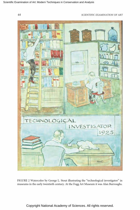
Scientific Examination of Art: Modern Techniques in Conservation and Analysis
Copyright National Academy of Sciences. All rights reserved.
44 SCIENTIFIC EXAMINATION OF ART
FIGURE 2 Watercolor by George L. Stout illustrating the “technological investigator” inmuseums in the early twentieth century. At the Fogg Art Museum it was Alan Burroughs.

Scientific Examination of Art: Modern Techniques in Conservation and Analysis
Copyright National Academy of Sciences. All rights reserved.
CHANGING APPROACHES IN ART CONSERVATION 45
FIGURE 3 Watercolor by George L. Stout illustrating the “Berensonian” dealer/merchantfigure of the 1920s.

Scientific Examination of Art: Modern Techniques in Conservation and Analysis
Copyright National Academy of Sciences. All rights reserved.
46 SCIENTIFIC EXAMINATION OF ART
servation services. There are now 12 regional centers, and in 1997 this groupbegan a consortium known as RAP (Regional Alliance for Preservation) with awebsite (http://www.rap-arcc.org).
Major professional societies and training programs also appeared between1925 and 1975. The International Institute for Conservation of Historic and Ar-tistic Works (IIC) was incorporated under British law in 1950 as “a permanentorganization to co-ordinate and improve the knowledge, methods, and workingstandards needed to protect and preserve precious materials of all kinds.” Since1967 when the first meeting on climate control was held in London (see Figure 4),triennial or biennial congresses have been held in international locations on spe-cial topics ranging from archaeological conservation (in Stockholm, Sweden, in1975, with attention to the recovery of the warship Wasa) to library and archiveconservation (Baltimore, Maryland, in 2002, with accompanying visits to thepaper conservation department of the Library of Congress). Harold Plenderleith(1898-1997), founding member and author of what was once considered theconservation “Bible,” The Conservation of Antiquities and Works of Art: Treat-ment, Repair, and Restoration (1956) noted, “We never envisaged more than 50
FIGURE 4 The first triennial meeting of the International Institute for Conservation, infront of Albert Hall, London, 1967.

Scientific Examination of Art: Modern Techniques in Conservation and Analysis
Copyright National Academy of Sciences. All rights reserved.
CHANGING APPROACHES IN ART CONSERVATION 47
fellows because there weren’t 50 fellows in existence who shared our ideas or wereadequately experienced in scientific conservation.” As of June 2002 there werealmost 2,400 individual members of this international body, including 301 fel-lows. (This figure does not fairly represent the international population of con-servators, as many now elect to join only their regional groups because of eco-nomic concerns and ready availability of key professional information on theWorld Wide Web; more information about IIC can be found at http://www.iiconservation.org/.)
IIC began publication of its quarterly journal Studies in Conservation in 1952and sponsored an international abstracting periodical IIC Abstracts in 1955. Tech-nical Studies published by the Fogg from 1932-1942 had also contained abstractsof the international literature, and Gettens compiled Abstracts of Technical Studiesin Art and Archaeology (published by the Freer in 1955), a fairly slender volumecovering literature published between 1943 and 1952. (Stout and other conserva-tors had been active elsewhere during World War II as officers in the Arts andMonuments initiative to identify and protect cultural heritage.) IIC Abstractsmoved to the New York University (NYU) Conservation Center in 1966, and thename of the publication was changed to Art and Archaeology Technical Abstracts(AATA). AATA moved to the Getty Conservation Institute in 1985 (see Figure 5),and became available online in 2002 (http://aata.getty.edu/NPS). The conserva-tion literature vastly expanded between 1975 and 1990. When this author was a
FIGURE 5 The Editorial Board of Art and Archaeology Technical Abstracts, semiannualmeeting in 1988, held at the Canadian Conservation Institute.

Scientific Examination of Art: Modern Techniques in Conservation and Analysis
Copyright National Academy of Sciences. All rights reserved.
48 SCIENTIFIC EXAMINATION OF ART
graduate conservation student at the NYU Conservation Center in the 1960s, veryfew books were available on the topic of art conservation. Current conservationgraduate students can readily spend our new Gutmann Foundation grants of$3,000 on books in their specialties; I doubt I could have spent $300 in 1968.
The American Group of the IIC began in 1960 (see Figure 6) and became theAmerican Institute for Conservation (AIC) in 1972, publishing a journal and anewsletter and establishing a 501(c)(3) foundation (FAIC). The AIC now hasabout 3,000 members, including 860 fellows or professional associates (http://aic.stanford.edu). The National Conservation Advisory Council (NCAC) was or-ganized in 1973, funded by the National Museum Act of the Smithsonian Institu-tion. The NCAC surveyed needs of the field and published useful and colorfulbooklets and became the National Institute for the Conservation of Cultural Prop-erty (NIC) in 1982. Whereas individual conservators are members of the AIC, theNIC is composed of conservation associations that meet to discuss overarchingissues, such as preservation of outdoor sculpture throughout the United Statesand historic houses and their contents. In 1997 the NIC changed its name toHeritage Preservation and now has 156 institutional members (http://www.heritagepreservation.org/).
FIGURE 6 The first meeting of the International Institute for Conservation-AmericanGroup at the Isabella Stewart Gardner Museum in 1960.

Scientific Examination of Art: Modern Techniques in Conservation and Analysis
Copyright National Academy of Sciences. All rights reserved.
CHANGING APPROACHES IN ART CONSERVATION 49
Major conservation research laboratories were also founded during this pe-riod. William J. Young sponsored seminars in 1958, 1965, and 1970 on “TheApplication of Science in Examination of Works of Art” at the Museum of FineArts, Boston; the laboratory there had begun in 1930. In 1950 the National Gal-lery of Art in Washington, D.C. established a fellowship at the Mellon Institute inPittsburgh. Robert Feller focused on natural and synthetic picture varnishes, color,and the damaging effects of light exposure. In 1976 the research project wasreorganized as the Research Center on the Materials of the Artist and Conservatorat Carnegie Mellon University. In 1988 Feller retired and Paul Whitmore came tothe center to become the director. Whitmore edited an excellent compilation ofFeller’s research, Contributions to Conservation Science, published in 2002. Origi-nally established in 1963, principally to provide technical support to theSmithsonian museums in the analysis and conservation needs of the collections,the Conservation Analytical Laboratory (CAL) moved in 1983 to the MuseumSupport Center in Suitland, Maryland (see Figure 7). In January 1998 the Boardof Regents of the Smithsonian Institution voted to rename CAL the SmithsonianCenter for Materials Research and Education (SCMRE). The Getty Trust and itsvarious branches is our most significant recent addition; major activities weredefined in 1982. In the summer of 1985 the Getty Conservation Institute took up
FIGURE 7 The Conservation Analytical Laboratory building of the Smithsonian Institu-tion (now known as SCMRE, Smithsonian Center for Materials Research and Education).

Scientific Examination of Art: Modern Techniques in Conservation and Analysis
Copyright National Academy of Sciences. All rights reserved.
50 SCIENTIFIC EXAMINATION OF ART
quarters in rented warehouse space at the Marina del Rey, and in 1997 joinedother Getty branches at the Getty Center in California, an acropolis in travertineon the hills of Brentwood, representing the apotheosis of the architecture withdedicated space for conservation.
Conservation training evolved from apprenticeship to formal education andtraining programs in London in 1934, Vienna in 1936, Munich in 1938, Rome in1943, and New York in 1960. The other two U.S. fine arts graduate programsopened in 1970 and 1974. As of 1994 there were at least 50 programs in 30countries in the fine arts and another 50 in archaeological materials, books, deco-rative arts, and musical instruments according to a directory co-published by theGetty Conservation Institute. The Conservation Center at the Institute of FineArts of New York University accepted its first four graduate students in 1960; westudents studied in the basements of the Duke mansion at One East 78th Street. In1983 the program moved across the street to a converted brownstone with 11floors of conservation laboratories, classrooms, and libraries; eight students arenow accepted each year. Ten students were accepted annually into theCooperstown Graduate Program in the conservation of historic and artistic worksbeginning in 1970. This program relocated to Buffalo State College in 1987. In1974 a third graduate program was sponsored jointly by the University of Dela-ware and the Winterthur Museum. Both the University of Delaware and BuffaloState College have awarded space to their art conservation departments in theflagship buildings of their institutions, another encouraging sign of the architec-tural status now granted our profession. George Stout summarized the state ofconservation in 1975 with another collage of a fence (see Figure 8). The Conserva-tion Education Program at Columbia University accepted students in 1981 fortraining in library and archives conservation; this program moved to the Univer-sity of Texas in 1992. The conservation training programs also have an associa-tion, ANAGPIC (Association of North American Graduate Programs in Conser-vation), including the Master of Art Conservation Program at Queen’s Universityin Kingston, Ontario, and the Center for Conservation and Technical Studies atHarvard. The students from the programs convene annually and present papers.3
CHANGING STYLES OF CONSERVATION: 1975 TO THE PRESENT
The Fogg conservators noted in their interviews that they rarely specialized; thereare now nine specialty groups listed by material in each AIC Newsletter: architec-ture, book and paper, electronic media, objects, paintings, photographic materi-als, RATS (research and technical studies), textiles, and wooden artifacts. Anothergroup represents the particular concerns of conservators in private practice. Thereis also an independent group, the Society of the Preservation of Natural HistoryCollections (SPNHC). The International Council of Museums-Committee forConservation (ICOM-CC) (http://www.icom-cc.org) has at least 18 workinggroups, including an active group on preventive conservation for conservators
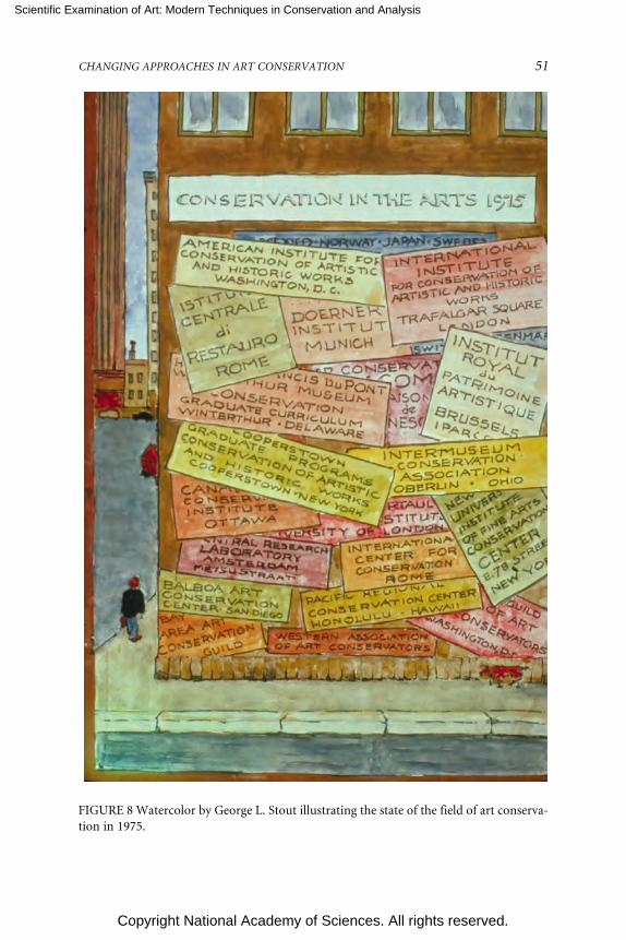
Scientific Examination of Art: Modern Techniques in Conservation and Analysis
Copyright National Academy of Sciences. All rights reserved.
CHANGING APPROACHES IN ART CONSERVATION 51
FIGURE 8 Watercolor by George L. Stout illustrating the state of the field of art conserva-tion in 1975.

Scientific Examination of Art: Modern Techniques in Conservation and Analysis
Copyright National Academy of Sciences. All rights reserved.
52 SCIENTIFIC EXAMINATION OF ART
who specialize in climate and pest control, changing exhibition, loans, and ship-ment. All these spin-off groups are publishing, conducting meetings and work-shops, and collaborating with scientists or curators, architects, librarians, andartists.
Methods of analysis have become far more sophisticated than just X radiog-raphy or examination with ultraviolet light; the Sackler conference of 2003 at theNational Academy of Sciences elaborated on many of these excellent resources. Iwill note that for my own specialty, a professional paper on paintings conserva-tion is now rarely without cross-sections indicating the layered buildup of paintfilms, dirt, and varnishes shown in normal and in ultraviolet illumination, oftenwith supporting Fourier transform infra red (FTIR) or x-ray diffraction (XRF)data. The landmark article launching this work was Joyce Plesters’s article forStudies in Conservation, “Cross-sections and Chemical Analysis of Paint Samples.”Ashok Roy of the National Gallery, London, noted in his Forbes lecture for theIIC that this 1956 paper is the single most cited reference in the whole of theliterature of conservation.
By the middle 1970s a hands-off, minimalist, or “less is more” approachappeared, subsequent to our initial excitement regarding many new tools andtechniques. For example, in paintings conservation the heat table was first intro-duced in 1948 in Germany and the United Kingdom, and vacuum pressure wasadded in 1955 (see Figure 9). About 20 years later, in 1974, a lining moratoriumwas suggested at a conference in Greenwich, England, reinforced at ICOM-CC
FIGURE 9 The use of a vacuum heat table, a typical treatment of the late 1960s and 1970s(Michael Heslip, paintings conservator at Winterthur Museum in the late 1970s).

Scientific Examination of Art: Modern Techniques in Conservation and Analysis
Copyright National Academy of Sciences. All rights reserved.
CHANGING APPROACHES IN ART CONSERVATION 53
meetings in Venice in 1975 and again at a special 1976 meeting in Ottawa. Theproblems caused by old and new lining technologies were reexamined. In 1992 Isurveyed practicing paintings conservators in the United States; conservators whoonce said they had formerly lined nearly every painting they treated now reportedlining only about 10 percent of their current treatments.4 Teaching treatmentsbecame more difficult, as there was now a complex menu of choices—loose lin-ings, drop linings, edge linings, hand linings, humidity treatments, suction tables,local suction platforms, and more highly controlled vacuum tables—and lining isonly one of many types of treatments used for paintings. We also have a complexmenu of new adhesives and new electronic tools; we have “pharmacists of conser-vation” who specialize in supplying new spatulas, sampling kits, retouching sup-plies, and cleaning gel ingredients. There are many new approaches to removingvarnishes—new enzymes, gels, and aqueous materials—less toxic and more spe-cific in what they remove, thanks to changes made by conservation scientists orscience-friendly conservators such as Richard Wolbers.5 My 1992 survey alsorevealed that many paintings conservators who used to remove varnishes entirelynow may simply thin or reduce them instead, another more conservative ap-proach.
A minimally interventive philosophy has now been adopted by most conser-vators, especially as we revisit our own treatments from 25 years ago and as wewatch our materials along with ourselves and our attitudes age. Marjorie Cohn,formerly a conservator of prints and drawings and now a curator and director atthe Fogg, noted in the 2001 history issue of the Journal of the Institute of PaperConservation, “Think first of the high value, both aesthetic and commercial, nowattached to the unvarnished or unlined canvas. But ‘unwashed’ is now also a salespoint in the catalogue descriptions of old master prints.”6 Our Art ConservationProgram faculty members all generally adhere to this minimally interventive phi-losophy. In furniture conservation new approaches aim to preserve every bit oforiginal material.7
We are now more aware of health hazards for the conservator and for thegood health of the environment. Marjorie Cohn continued in her paper with asuccinct summary of some other changes:
One by one over the past three decades options for fumigation have beendeemed too hazardous, until now there are no facilities remaining at my institu-tion, Harvard University, and good housekeeping rather than extermination isthe mode. Likewise, carcinogenic solvents have been minimized in the conserva-tors’ repertory, and ventilation and disposal are now the first engineering prior-ity in the design of facilities.8
More interdisciplinary research and publications have appeared in recentyears. Cohn was asked by Agnes Mongan of the Fogg to write what she now noteswas an “unprecedented essay” for an Ingres catalogue in 1967 and continued thatsince then more art historians have come to appreciate the potential of technical

Scientific Examination of Art: Modern Techniques in Conservation and Analysis
Copyright National Academy of Sciences. All rights reserved.
54 SCIENTIFIC EXAMINATION OF ART
examination and scientific analysis, and “conservators themselves have realizedthe essential importance of historical evidence on and in the works of art them-selves.”9 George Stout’s three-legged-stool approach is embodied in an increasingnumber of publications authored by teams of conservators with art historians andscientists, such as Art and Autoradiography: Insights into the Genesis of Paintings byRembrandt, Van Dyck, and Vermeer from the Metropolitan Museum of Art in1982 and Examining Velasquez in 1988. There are now paired fellowships spon-sored by the National Gallery and the Getty encouraging similar teams to createfuture publications. Textile conservators may now embrace a “connoisseurshipbias” while choosing treatment options.10 Objects conservators may now elect toretain ritual substances such as yak butter that were applied to Tibetan sculptures.We have become more aware of respecting and retaining age value, historicalvalue, and commemorative value, such as the proverbial blood on Lincoln’s shirt.Susan Heald, a textile conservator of the National Museum of the AmericanIndian, gave a memorable talk for our graduate students. Heald worked with theSiletz regalia makers to stabilize pieces before the dance and get them ready fordancing during the dance house dedication ceremony. She noted, “For me thiswas a career altering experience to see these pieces danced on the night of thedance house dedication—it was very emotional for me and many others.” Shedescribed the smoky night atmosphere of the night of the dance she was allowedto observe (see Figure 10).
FIGURE 10 Susan Heald with members of the Siletz community at the National Museumof the American Indian, Smithsonian Institution.

Scientific Examination of Art: Modern Techniques in Conservation and Analysis
Copyright National Academy of Sciences. All rights reserved.
CHANGING APPROACHES IN ART CONSERVATION 55
Other issues and concerns that would probably have never occurred to con-servators in the 1950s and 1960s emerged following NAGPRA, the Native Ameri-can Graves Protection and Repatriation Act of 1990, which was discussed exten-sively by Miriam Clavir, senior anthropological conservator at the University ofBritish Columbia, in her book Preserving What Is Valued: Museums, Conservation,and First Nations (UBC Press, 2002). People from First Nations may be reclaim-ing their sacred materials and reburying them; however, these materials may nowhave indelible museum registration numbers that must be removed from fragilesurfaces or, even more important, be dangerously full of toxic pesticides that werestate-of-the-art treatments in museums several decades ago.
Glenn Wharton, objects conservator who wrote his dissertation for the Uni-versity College London on “Heritage Conservation as Social Intervention,” re-ported in the International Council of Museums–Committee for ConservationPreprints on the case of the royal Hawaiian monument Kamehameha the First.The state of Hawaii asked Wharton to remove the brightly colored paints that hadbeen applied to the surface, returning the gilded bronze monument to its originalstate as part of the NIC’s Save Outdoor Sculpture. He soon learned that the localpeople intentionally paint Kamehameha in lifelike colors and hold annual cel-ebrations with chants and parades. He convinced the state to support a commu-nity-based, two-and-one-half-year participatory conservation project that essen-tially gave the community a seat at the table in deciding how to conserve themonument. Wharton brought current theory from material culture studies, eth-nography, and a reflexive stance to the study. He noted, “The recognition ofcultural relativism and contested meanings embedded in material objects hasbegun to enter conservation literature.”11
In a landmark conservation conference in 1980 at the National Gallery ofCanada, conservators, artists, scientists, and curators discussed issues relevant tothe conservation of contemporary art. Several conservators spoke about theircollaborations with living artists and the importance of interviewing artists toascertain their views about materials and addressing damages to their pieces.Consulting and working with artists or collaborating with native Americans havebeen categorized together as acknowledging the cultures of origin, yet anotherimportant new direction for conservation.
CONSERVATION SCIENCE IN THE LAST DECADE
The number of conservation scientists and scientific research and analytical labo-ratories in the United States has increased substantially since the early 1990s. Theinclusion of sophisticated scientific data in scholarly conservation conferencepapers and publications has become standard, enhanced by the students andgraduates of active U.S. and U.K. doctoral programs. Conservation methods havebeen improved by jointly sponsored conservation science research, demonstrat-ing such factors as the impact of solvents on paint films, the absorption of dirt by

Scientific Examination of Art: Modern Techniques in Conservation and Analysis
Copyright National Academy of Sciences. All rights reserved.
56 SCIENTIFIC EXAMINATION OF ART
acrylic paint, and the impact of temperature and humidity changes, with teammembers from the Getty Conservation Institute, the National Gallery of Art, theCanadian Conservation Institute, and the Tate in Britain.
Since the year 2000, Angelica Rudenstine and the Mellon Foundation havetaken conservation science another quantum leap forward. In the mid-1990s Mrs.Rudenstine interviewed conservation leaders to determine areas of need withinthe profession. She identified both conservation of photographic materials andconservation science and has made carefully considered grants available for train-ing, workshops, and internships in those areas. Mrs. Rudenstine embodies aunique case of highly directed and hands-on improvement by the leader of agranting organization. She has funded and thereby created new conservationscience positions in graduate training programs and major museum conservationlaboratories. She has also imaginatively provided underwriting for talentedyounger conservation professionals, such as Philip Klausmeyer, who has returnedto graduate study in order to enhance the scientific capabilities and instrumenta-tion at his home institution, the Worcester Art Museum.
The field of conservation science would not have been able to gainfully ab-sorb the Mellon’s munificence in 1975. Mrs. Rudenstine scrupulously investigatesthe staff and capacity of an institution before making a grant. The integration ofscientific understanding and conservation practice within well-trained individu-als or by cooperating teams is a phenomenon of the last decade, and her programshave taken advantage of this moment in conservation history.
As I mentioned earlier, the multiplication of approaches, sophistication ofscientific research, and explosion of information has not made it easy to be aprofessor of art conservation. There are no old and outdated methods that wecan now delete from the curriculum because someone in the past may have usedthem on the work we must treat. We now believe that the conservators of thetwenty-first century that we are now training must thoroughly know their spe-cialties, including current philosophy, history, literature, ethics, and the materialproperties and methods of analysis (subjects might range from underwater can-nonballs to ivory miniatures); collaborate with scientists and be able to under-stand scientific terms and methods; cooperate with allied professionals, includ-ing archaeologists, art historians, and the various cultures of origin; understandproper light levels, indoor pollutants, insect life cycles, climate control, emer-gency preparedness, and toxicity; be articulate advocates who write papers, givepresentations, and in this time of economic cutbacks, be able to charm politi-cians, foundation heads, and reporters from “Sixty Minutes” if necessary. Per-haps we can now say that conservation has evolved from George Stout’s three-legged stool of the early days of the Fogg Art Museum to a twenty-first-centuryversion of a ten-legged settee.

Scientific Examination of Art: Modern Techniques in Conservation and Analysis
Copyright National Academy of Sciences. All rights reserved.
CHANGING APPROACHES IN ART CONSERVATION 57
NOTES
1. FAIC oral history interview with George Leslie Stout, Richard Buck, and Katherine Gettens byW. Thomas Chase and Joyce Hill Stoner, September 4, 1975. (The FAIC oral history archive is housedat the Winterthur Museum Library, Winterthur, Del.).
2. Ibid.3. ANAGPIC (Association of North American Graduate Programs in Conservation). North
American Graduate Programs in the Conservation of Cultural Property: Histories. Buffalo, N.Y.:2000.
4. J. H. Stoner. The impact of research on the lining and cleaning of easel paintings. Journal ofthe American Institute for Conservation 33(1994):131-40.
5. R. C. Wolbers. Cleaning Painted Surfaces: Aqueous Methods. London: Archetype Books, 2000.6. M. B. Cohn. Change, we hope for the better. The Paper Conservator 25(2000):101-105.7. M. J. Anderson and M. S. Podmaniczky. Preserving the artifact: Minimally intrusive conserva-
tion treatment at the Winterthur Museum. In Wooden Artifacts Group, pp. 1-10. Richmond: Ameri-can Institute for Conservation, 1990.
8. Cohn, op. cit., p. 102.9. Cohn, op. cit., p. 103.
10. P. Orlofsky and D. L. Trupin. The role of connoisseurship in determining the textileconservator’s treatment options. Journal of the American Institute for Conservation 32(1993):109-118.
11. G. Wharton. Conserving the Kamehameha I monument in Hawai’i: A case study in publicconservation. 13th Triennial Meeting, Rio de Janeiro, 22-27 September 2002, ICOM Committee forConservation I(2003): 203-210.

Scientific Examination of Art: Modern Techniques in Conservation and Analysis
Copyright National Academy of Sciences. All rights reserved.
58
An Overview of Current Scientific Researchon Stone Sculpture
Richard NewmanScientific Research Lab,
Department of Conservation and Collections ManagementMuseum of Fine ArtsBoston, Massachusetts
ABSTRACT
Scientific research on stone sculpture is focused on three major categories:determining sources of raw materials, developing methods of authenticat-ing stone artifacts, and preservation. This paper reviews research in thefirst two categories. The goal of source determination is identification of thequarry for the stone used in a sculpture. In situ analytical techniques areoccasionally applied, but most research involves samples. There are manyapproaches, ranging from petrography (study of thin sections and quanti-tative analysis of mineral compositions, usually on the thin sections) toelemental and isotopic analyses on drilled samples. Elemental analysescarried out by instrumental neutron activation, X-ray fluorescence, induc-tively coupled plasma (ICP) techniques, and others often provide the mostuseful information.
Authentication of stone sculpture can focus on materials, techniques bywhich the materials were worked, and weathering layers. The alteration of asculptural surface and the buildup of alteration products on that surface ina burial environment can be useful as an indication of age, and as such isoften studied in stone sculpture authentication projects.
INTRODUCTION
Stone was among the earliest naturally occurring materials to be used to createartifacts because it was readily available and required little processing. The steps
Scientific Examination of Art: Modern Techniques in Conservation and Analysis
Copyright National Academy of Sciences. All rights reserved.
AN OVERVIEW OF CURRENT SCIENTIFIC RESEARCH ON STONE SCULPTURE 59
from procurement to desired final product are few and can be very simple. Thecurrent areas of research on stone artifacts to which science has made and contin-ues to make a major impact fall into a few broad categories: determining sourcesof raw materials; developing methods of authenticating stone artifacts; and pres-ervation. This paper will focus only on the first two categories because of limita-tions of space. “Rock” is the correct geological term for the raw material. Accord-ing to a definition used by some, “stone” refers to rock that has been intentionallyshaped into an artifact. For the remainder of this paper all the material underdiscussion will be called rock.
DETERMINING SOURCES OF RAW MATERIALS
The geographical origin of the rock used to make an artifact is a crucial piece ofinformation in fully understanding the artifact. “Quarry” can be defined as theprecise location from which the rock originated. In most cases the rock used in asculpture is taken from an outcrop or exposure of rock, and the location of thatoutcrop would be the ultimate desired goal of the source determination. Someoutcrops could be quite small, such as a pit in the ground from which some blocksto make small sculptures could be taken. In other cases an outcrop could consistof a very large exposure extending hundreds of yards or more. In the case of a verylarge quarry, localization of the source to some smaller area within the larger areamay be desired.
It is possible that a boulder or smaller chunks of rock could serve as sourcematerial, and these may have been collected from areas that are distant from theactual geographical location of the quarry. Examples include boulders moved byglaciers to pebbles moved down streambeds. One well-known example isStonehenge, which some argue was constructed from glacial boulders that werefar removed from their original source site.
What is the relationship between the quarry location and the location atwhich the rock was shaped into an artifact? Some artifacts could have been shapedat the quarry, but blocks of rock could also be transported from a quarry toworkshops, which could be at distant locations. Pebbles or small rocks could, ofcourse, be easily transported long distances from the points at which they werecollected. This is the case, for example, with some of the jade used in ancientChina to produce small objects. A very widely traded material in many ancientcultures was obsidian—volcanic glass—to which considerable scientific researchon sources has been devoted. We know in the case of marble sarcophagi in Ro-man times that blocks were sometimes partially shaped at the quarry and thenshipped to various workshops where the final carving was completed. Determin-ing workshop locations is an aspect of research on rock sculptures that can drawon information about quarry sources.

Scientific Examination of Art: Modern Techniques in Conservation and Analysis
Copyright National Academy of Sciences. All rights reserved.
60 SCIENTIFIC EXAMINATION OF ART
Approaches to Determining Sources
Nondestructive methodologies, carried out directly on artifacts without sam-pling, are particularly valuable in the case of monuments or sculptures on monu-ments. One technique that has been utilized in recent studies is magnetic suscep-tibility, which gives a signal based to a great extent on the presence of the mineralmagnetite. Sources for Roman-period gray-granite columns in the Rome regionhave been studied (Williams-Thorpe and Thorpe, 1993), and sources for the grayto yellow-brown sandstone blocks used in different phases of construction of theeighth- to thirteenth-century complexes at Angkor in Cambodia have also beenidentified (Uchida et al., 2003).
For the most part, sourcing of projects involves analysis of samples, whichfall into two broad kinds: solid chips and powder. The appropriate type and sizeof sample depend on many factors related to the rock type, the geology of thepotential quarry source or sources, and constraints on sampling of the sculpture(s)being studied.
A rock-sourcing project could involve a single artifact or a group of artifacts.Sourcing projects usually begin with a group of artifacts made from a particulartype of rock material. The questions to be answered are typically: Do the artifactsall come from one quarry source? What is that quarry source?
One phase of the research involves characterizing the material used in thegroup of artifacts. The goal is to acquire types of data that can characterize therock material in sufficient detail to determine whether the rock from the variousobjects in the group being studied can reasonably be concluded to have comefrom the same source, or is likely to have come from more than one source. Ifmore than one specific source is likely, another aspect of this phase of the researchis to group objects that are likely to have come from the different sources sug-gested by the data.
As in sourcing studies of other types of materials used in artifacts (such asceramics), it is common practice today to use multivariate statistical analysis toevaluate analytical data, thereby establishing potentially related groups of objectson the basis of this kind of evaluation. There are different approaches to utilizingstatistics for data evaluation, and some may be more appropriate in certain cir-cumstances than others. Although beyond the scope of this paper, the examplesdiscussed in this paper include most of the current statistical procedures.
Evaluation of data from artifacts can establish hypothetical distinct sourcesor quarries, but at this point in a study it is uncertain whether the groups estab-lished is this manner actually represent distinctly separate sources, or whether theartifacts within individual groups actually all originated from a single source.
A second phase of research involves characterizing potential quarry sourcesand then comparing these with results from the artifacts. The goal is to determinethe quarries from which the artifact rock came. Assuming that potential quarrysources can be located, systematic sampling of these is crucial in order to fully

Scientific Examination of Art: Modern Techniques in Conservation and Analysis
Copyright National Academy of Sciences. All rights reserved.
AN OVERVIEW OF CURRENT SCIENTIFIC RESEARCH ON STONE SCULPTURE 61
characterize them. In advance it is difficult to establish what would constitutesystematic sampling. The nature of the rock being sampled is one factor thatneeds to be considered, and whether the rock type is present in more than onehorizon (or layer) that was formed during distinctly different geological events. Asingle rock type usually displays compositional variation, particularly if the out-crop is quite large. For multivariate statistical analyses, it is usually stated that thenumber of source samples should be at least equal to the number of variables(usually elements) being analyzed in each sample. For rock quarries far moreextensive sampling than this is usually an absolute necessity unless the outcrop isextremely small.
Another aspect that needs to be determined in analytical studies of artifacts(and quarries) is the appropriate sample size. A representative sample of an arti-fact can be defined as one whose composition reflects the composition of theartifact as a whole. In the case of a block of rock, samples of 3 to 4 milligramstaken from several areas may not have identical compositions, while samples of 1or 2 grams may. What constitutes the minimum acceptable sample size to trulyrepresent the whole will often vary according to which properties are being ana-lyzed. In studies that involve elemental analysis, certain elements are often foundto be good discriminators with very small samples, others may be useful but onlyif larger samples are studied, and still others may be so unevenly distributed thatthey have little utility. Research on stable carbon and oxygen isotope ratios incarbonate rocks (discussed in more detail below) has shown that a sample of a fewmilligrams can be considered to be representative of a large block of the rock.
Solid Chip Samples (Petrography)
Solid chip samples are utilized mainly for petrographic analysis, the well-established tool used by geologists to characterize and classify rocks. Geologiststypically prepare thin sections that cover most of the surface of a standard petro-graphic slide (in the United States, 27 x 46 millimeters), but that would require asample far larger than could be taken from the vast majority of sculptures. Samplesthat can provide a thin section about 1 centimeter across, taken with sculptors’chisels or hollow core drills, are often more than adequate for characterizing arock, unless the rock is relatively coarse grained (with numerous individual min-eral grains exceeding about a millimeter in size, for example). The thin sectionsprovide information on the mineral content of the rock, the grain sizes and shapes,and the interrelationships of the different mineral grains (the texture).
Geologists have long utilized the electron beam microprobe to quantitativelyanalyze individual mineral grains in thin sections of rocks. The microprobe canprovide excellent quantitative analyses of the major and minor elements in min-eral grains that are at least a few micrometers across. This quantitative informa-tion further characterizes the mineral in the specific rock being studied.
One application of the microprobe focused on a silvery gray schist used in the

Scientific Examination of Art: Modern Techniques in Conservation and Analysis
Copyright National Academy of Sciences. All rights reserved.
62 SCIENTIFIC EXAMINATION OF ART
ancient Gandharan region (present-day northern Pakistan) from about the firstto third centuries AD. The specific find sites are unknown for virtually allGandharan sculptures made from this easily recognized rock. Some small localsources of this type of rock have been identified, but there are undoubtedly manymore than are currently known. In a pilot project (Newman, 1992) 6 to 8 grains ofchloritoid, a characteristic mineral of the rock, were analyzed in thin sectionsfrom 16 sculptures. After considering relative amounts of magnesium, iron, andmanganese in the mineral, chloritoid in seven of the sculptures was found to havevirtually identical compositions. Separate samples from the top and bottom ofone of these sculptures, a 1.5-meter-tall figure, were analyzed to give some senseof the variation in chloritoid compositions in a fairly large block of rock. Thespread in the compositions of these grains in the one block was about the same asthe spread shown by grains in individual thin sections. As a working hypothesis, itis possible that the rock used in the sculptures with virtually identical chloritoidcompositions came from the same quarry, while the other nine sculptures weremade from rock taken from other quarries. Although more sculptures could bestudied by this method and more extensive chloritoid analyses carried out in eachthin section, the conclusions will remain tentative until possible quarry sourcescan be identified and studied. The research points out a possible relationshipbetween the seven sculptures, but the precise significance of the relationship isuncertain. Were the sculptures produced in the same workshop within a narrowtime frame, or produced in a general area in proximity to a certain quarry but atdifferent times, or was material exported to several sites from one quarry? Or arethese sculptures even from a single quarry?
Another application of microprobe mineral analysis was applied to smallscrapings (0.1-1.0 milligram) from predynastic Egyptian basalt vessels (Mallory etal., 1999). It has been estimated that nearly one-quarter of stone vessels from thisperiod were made from basalt. Grains of two minerals were quantitatively ana-lyzed for major and minor elements: plagioclase feldspar and pyroxene. Aftercomparing the results with some analyses of samples from major Egyptian basaltsources, it was concluded that the predynastic sculptural material was all north-ern Egyptian basalt. Finished objects, or pieces of rock that were shaped in localworkshops, were apparently shipped all over Egypt from the source quarry orquarries.
A more recently developed tool for individual mineral grain analysis in thinsections is laser ablation microprobe ICP mass spectrometry. Currently, the mini-mum spot size for this technique is about 20-50 micrometers, meaning that itrequires larger grains than does the microprobe, but the technique has the advan-tage of being able to analyze trace elements more readily than does the micro-probe. A recent application focused on basalts in ancient Egyptian sculpture(Mallory-Greenough et al., 1999), using thick polished thin sections. The mineralanalyzed, augite (a type of pyroxene), is a major constituent of most basalts. It wasfound that some trace elements, which can easily be analyzed by the laser probe

Scientific Examination of Art: Modern Techniques in Conservation and Analysis
Copyright National Academy of Sciences. All rights reserved.
AN OVERVIEW OF CURRENT SCIENTIFIC RESEARCH ON STONE SCULPTURE 63
procedure, are far more useful for discriminating between augites in basalts fromdifferent quarry sources than are major and minor elements. Samples studiedwere basalt temper from pottery. A conclusion was that some of the basalt sourcesutilized during the pharaonic period in ancient Egypt have yet to be identified,since the augite compositions in samples from some artifacts did not correlatewell with augite from basalts from the known sources.
Both of the techniques just discussed are adjuncts to traditional petrography.There are many rocks, however, that cannot be adequately distinguished by pe-trography, or that contain minerals that do not show sufficient variations inmajor, minor, or trace element compositions in order to be suitable for micro-probe or laser ablation ICP mass spectrometry.
Other analytical techniques that can be applied to solid chip samples, whetherprepared as thin sections or not, include Fourier transform infrared spectroscopyand Raman spectroscopy. The latter shows particular promise as an analyticaltool, for example, in a project that was concerned with Mesoamerican jadeitesources in Guatemala (Gendron et al., 2002).
Powdered Samples
There is a full battery of analytical techniques that are applied to whole-rocksamples, that is, drilled (powder) samples that include all of the minerals found ina rock. Most of the analytical techniques currently applied to drilled samplesdetermine elements (in some cases, isotopes), with the exception of X-ray diffrac-tion (XRD).
XRD is useful for general mineral identification and, by extension, rock iden-tification in some cases. In sourcing research projects XRD can provide a quanti-tative estimate of the amounts of different major and minor minerals in a rock.Lacking textural information, XRD is less useful than thin section analysis forspecific rock identification and classification. XRD is probably most useful instudying certain monomineralic or nearly monomineralic rocks. Many mineralshave variable compositions, and variations in composition can give rise to dis-tinctive XRD patterns. One interesting project studied chert and chalcedony, twomicrocrystalline varieties of quartz used in New England archaeological sites(Pretola, 2001). XRD patterns were used to identify minor minerals in some ofthe source materials. In addition, calculated diffraction patterns from crystal struc-ture parameters were fit by computer methods to observed diffraction patterns inorder to determine the ratios of quartz to a silica polymorph called moganite inthe samples.
Powder samples are most commonly used for elemental analysis. The fullbattery of modern elemental analysis techniques has been applied to rock analy-sis. Current widely applied techniques include X-ray fluorescence spectrometry,instrumental neutron activation analysis (INAA), ICP optical emission spectros-copy, and ICP mass spectrometry. Some of these techniques are more appropriate

Scientific Examination of Art: Modern Techniques in Conservation and Analysis
Copyright National Academy of Sciences. All rights reserved.
64 SCIENTIFIC EXAMINATION OF ART
for certain elements, or abundances of elements, than others. A given rock, ofcourse, will contain dozens of elements, which are present from major to very lowtrace levels (parts per billion and less). Not all these elements will be useful in asourcing project. Which elements are the most useful is not usually known at theoutset of a project unless previous work on similar rocks has been carried out.
Usually the application of a particular technique or techniques in a discreteresearch project can easily produce data that is reliable. More difficult sometimesis comparing data acquired by other analytical techniques, or even other researchgroups using some of the same instrumentation. Use of common standards andfrequent equipment calibrations can help to overcome this kind of difficulty, butit can prove to be a limitation in comparing data from different research groupsor projects.
Both the suite of elements utilized in a sourcing study and the methods bywhich the data is evaluated are obviously crucial to the conclusions.
Limestone in Medieval France
One of the most outstanding research projects yet to be published on sculp-tural rock sources involves the limestone utilized in French medieval sculptures.The beginning of this massive undertaking goes back to a modest self-contained1985 project published on a small group of sculptures (French et al., 1985). NineRomanesque sculptures in four American museum collections that shared certainstylistic features were considered by some scholars to have originated from onemonument in southern France. The goal of the project was to determine whetherthis was the case and the location of the rock source(s). Petrographic examinationand neutron activation analysis indicated that the nine were probably made ofrock from the same quarry. As a part of this phase of the project the representativesample size for the chosen analytical technique and elements being analyzed wasdetermined, and the fact that a single sample could be considered representativeof an entire block of rock (of the size used in the sculptures) confirmed. Possiblequarry sites in the general region of origin were selected for sampling and analysison the basis of the petrographic features of the sculpture samples. Initial neutronactivation analysis of a few samples from several different geological formationsnarrowed down the possibility to one area. From this area over 100 samples of thesame geological formation were analyzed, three old quarries in particular beingextensively sampled. The neutron activation analyses coupled with petrographicdata narrowed down the likely source to two quarries that were about 2.5 kilome-ters apart. Multivariate statistical analysis was carried out on the elemental data.
Since that study, the same research group has focused on the characterizationof some of the major limestone quarries in the Paris region, a study that to datehas involved some 2,300 samples from 300 quarries (Blanc et al., 2002). Theresearch group concluded that 10 to 15 elements were required to best character-ize the French limestones, as were many samples from each compositional group

Scientific Examination of Art: Modern Techniques in Conservation and Analysis
Copyright National Academy of Sciences. All rights reserved.
AN OVERVIEW OF CURRENT SCIENTIFIC RESEARCH ON STONE SCULPTURE 65
(quarry), a number that actually ranged from 8 to 40 for the major Paris basinquarries. The extensive database now makes it possible to determine quarrysources for museum artifacts made from limestone that came from this generalregion of northern France. The database includes samples from some monu-ments, and this data has enabled the identification of the monument that sculp-tures in museum collections originally embellished. Examples are the head of anangel, the head of a Virtue, and a choir screen from three separate collections. Thetrace element patterns of these three objects closely matched the trace elementpatterns of a group of samples from 25 sculptures on Notre Dame in Paris. Thestone on the cathedral and in the three sculptures may have come from one of theancient quarry sites that the research group has extensively studied, Charenton.This is one of a number of quarries in the Paris region from which one particulartype of fine-grained limestone (Upper Lutetian limestone) was taken. Trace ele-ment analyses permit rock from many of the different quarries to be distin-guished, although all are indistinguishable by petrography. About 1 gram of rockis required for this analysis.
White Marble in Classical Antiquity
The most extensively studied general rock type used by any culture or groupof cultures in a region are the white marbles of the Mediterranean. Bronze Agesculptors in the Cycladic islands of the Aegean used white marble extensively inGreek and Roman times and well beyond. There are less than a dozen majorquarries that supplied rock that was widely exported at different periods. In addi-tion, there were dozens of quarries of mainly regional importance.
The majority of the white marbles consist entirely or almost entirely of cal-cite. Their textures, grain sizes, and minor and accessory mineral compositionsprovide some useful properties for discrimination, but petrographic propertiesalone have not been adequate to clearly distinguish most of these marbles.
An important breakthrough came in 1972 with a publication by marinegeochemists who applied a tool of their trade, stable isotope analysis, to a fewsamples from several of the major ancient Mediterranean marble quarries (Craigand Craig, 1972). The pilot project showed that simple plots of the ratios of thestable oxygen isotopes (18O and 16O) versus the stable carbon isotopes (13C and12C), calculated with reference to a standard international reference material, thecarbonate fossil Pee Dee belemnite, separated the marble from different quarries.Stable isotope analysis, which is comparatively inexpensive to carry out and re-quires a sample of only a few milligrams, is now perhaps the most frequentlyapplied technique in sourcing studies of marble sculptures from this part of theworld, but time has clouded the picture considerably. A recent publication showedhow the isotope fields for the quarries have expanded since the 1972 publication(Gorgoni et al., 2002). It is possible to discriminate between certain quarries withstable isotope data, but many cannot be distinguished from this data alone. Quarry

Scientific Examination of Art: Modern Techniques in Conservation and Analysis
Copyright National Academy of Sciences. All rights reserved.
66 SCIENTIFIC EXAMINATION OF ART
fields were initially defined by drawing an outline around all data points fromanalyzed samples taken from a quarry, but statistical analysis is now more com-monly utilized.
In studies of ancient quarries it is not always certain that samples taken fromthe quarry as it is known today entirely represent the quarry as it may have beenknown during a much earlier period. For example, it is possible that parts of aquarry may no longer be accessible, or even may have been depleted. In this case,analyses of sculptures that can reasonably be assumed to have been made frommaterial originating in that quarry can be used to define the compositional field,instead of relying solely on newly taken quarry samples. This has recently beendone for some of the major Mediterranean marble quarries. Incorporation ofsculptural data has in fact somewhat expanded some of the fields.
Many other analytical procedures have been applied to the study of thesewhite marbles. Some have been championed by particular research groups andhave yet to become widely applied, in spite of their promise. A brief survey ofthese techniques serves as an example of how there are diverse, often equallyuseful approaches to a sourcing problem, and more important, how data frommore than one technique may be crucial in solving a particular problem.
Although petrography was probably not considered a particularly valuabletechnique by most researchers for many years, within the last few years one par-ticular property that is best determined with a thin section has been shown to bevery useful: maximum grain size. Although a large thin section is required toaccurately determine this, this parameter does enable some quarries whose iso-tope fields overlap to be distinguished. In a recent publication (Gorgoni et al.,2002), in fact, isotope maps for fine-grained and coarse-grained marbles wereshown (a maximum grain size of 2 millimeters was the dividing point).
Elemental analysis has been applied by a number of research groups, whichhave proposed different elements. Reliable comprehensive databases have yet tobe established for many quarries, and elemental analysis is probably best appliedin small-scale projects. Certain elements have been shown to be useful in certaineither-or questions, where the possible quarries have been narrowed downto two.
Two other techniques that have been employed only by a few researchers todate are quantitative cathodoluminescence and electron paramagnetic resonance(EPR). Qualitative cathodoluminescence carried out with a suitably equippedoptical microscope and recorded on color film distinguishes certain marbles onthe basis of the color they show when bombarded with an electron beam. Quan-titative cathodoluminescence carried out in a scanning electron microscope withan attached spectrophotometer is potentially more valuable. In the ultraviolet/visible region of the electromagnetic spectrum most marbles show only one ortwo major peaks: a peak at 620 nanometers, due to manganese (2+) substitutingfor calcium in the crystalline matrix and a pair of peaks at about 350 and 377nanometers due to cerium (3+). Ratios of the two can distinguish many marbles

Scientific Examination of Art: Modern Techniques in Conservation and Analysis
Copyright National Academy of Sciences. All rights reserved.
AN OVERVIEW OF CURRENT SCIENTIFIC RESEARCH ON STONE SCULPTURE 67
from one another. The orange color shown by some marbles is related mainly totrace amounts of manganese in the rock.
EPR or electron spin resonance (ESR) has been applied for many years by asmall number of researchers but in the last few years has come into its own as aviable technique. One recent article evaluated a number of features of the EPRspectra of marbles, features in part attributable to the presence of manganese andiron in the crystalline lattice of calcite (Polikreti and Maniatis, 2002).
The study of Mediterranean white marbles has reached such a level of matu-rity that many important quarries have been well characterized and the strengthsand limitations of a range of analytical techniques that can be focused on aspecific sourcing problem are fairly well understood. In the near future morework will continue to center on characterizing some of the smaller quarries.Multivariate statistical analyses incorporating properties or features from two ormore analytical techniques have become common practice in marble provenancestudies.
DEVELOPING METHODS OF AUTHENTICATING ROCK ARTIFACTS
Authentication of stone artifacts from a scientific point of view usually focuses onone of two lines of evidence: the original materials and manufacturing proce-dures, and weathering or alteration of the surface after carving or other finishing.
Materials and Working Procedures
In this category would obviously be the nature of the rock itself. If a suffi-ciently large database of information on the rocks that presumably should havebeen utilized were available, detailed rock analysis may help to build a case for oragainst the authenticity of a problematic artifact. More often than not, the resultof such an analysis even when a large database is available may lead to the conclu-sion that the material used in an artifact is consistent with the purported periodand place of origin, which does not imply that the object is definitely authentic,only that it could be.
Working of the rock can also be important. Certain tools or manners of usinga tool may be characteristic of a type of stone artifact. Some researchers havecarefully compared drill marks produced by known ancient drilling procedureswith marks produced by modern drills, and this information could obviously beused in certain authentication studies. Tool marks on ancient gemstones havealso been extensively studied (Rosenfeld et al., 2003), with the purpose of deter-mining the tools that were utilized and the way the tools were used. Applicationsof this type of information to weathered sculptural surfaces is much more diffi-cult since many details have been worn away.
There may be residues of tools that can be identified, for example, elementsfrom metal chisels or bits of abrasives used in polishing operations. A study of

Scientific Examination of Art: Modern Techniques in Conservation and Analysis
Copyright National Academy of Sciences. All rights reserved.
68 SCIENTIFIC EXAMINATION OF ART
Roman gemstones found residues of lead and tin metal as well as barite, all ofwhich were probably associated with abrasive polishing procedures (Rosenfeld etal., 2003).
It is difficult to quantify the appearance of tool marks on a complex sculpturein such a manner that would allow the data to be readily utilized by other re-searchers. The use of tool marks in authentication studies may fall more into therealm of technical connoisseurship, where interpretation of significance dependson the experienced eye of a researcher who has carefully examined many toolmarks on many artifacts. One example is in a paper published 13 years ago by asculptor who has studied tool marks on Roman marble sculptures (Rockwell,1990). He argued that although a skilled modern forger could use the same toolsas an ancient Roman sculptor, utilizing them in exactly the same manner wouldbe very difficult, and that the differing manners of use could be distinguished bycareful examination of tool marks, when the appropriate marks are still presenton a sculpture. A very interesting concluding remark of his was, “In readingliterature on fakes and faking I find that no one questions that the faker can, if hewants to, reproduce the technique of the period being faked. It is taken as a giventhat a good technician can reproduce the technique of any period. I think that thisis an assumption about techniques, based on the ignorance of nontechnicians,that deserves serious questioning.”
Surface Alteration
One final approach is to look closely at surface alteration. Changes that takeplace on a newly worked rock surface over time can involve physical as well aschemical factors. Sharp details can become rounded or abraded. Minerals at thesurface can chemically break down due to interaction with the atmosphere orburial environment. The surface of an ancient rock sculpture may contain mate-rials that in part arise from interaction of the rock surface with its environmentover some period of time. “Weathering layer (layers)” will be used to refer to allsuch materials.
Many ancient rock artifacts will have been buried for some extended periodof time, but few if any details are usually known about the burial environment.While it is quite possible that there is some correlation between thickness of aweathering layer and amount of burial time for a given type of rock and givenenvironment, all the information that would be required to establish this is neveravailable.
The composition and microstructure of a weathering layer are both useful.Possible variation in both composition and structure from place to place on thesurface of an artifact make it prudent to examine multiple samples. Small chipsthat can be prepared as cross-sections are the single most useful type of sample,since compositional and structural features can both be studied on the same

Scientific Examination of Art: Modern Techniques in Conservation and Analysis
Copyright National Academy of Sciences. All rights reserved.
AN OVERVIEW OF CURRENT SCIENTIFIC RESEARCH ON STONE SCULPTURE 69
sample utilizing such techniques as scanning electron microscopy with attachedX-ray spectrometers.
On rocks that contain more than one mineral the microscopic structure andcomposition of a weathering layer will not be identical on exposed surfaces of thedifferent minerals. Some minerals, such as quartz, are very resistant to weather-ing, while others are much less resistant. The processes by which different miner-als break down and the products of those breakdown processes vary from onemineral type to another. Even on monomineralic rocks the nature of the weather-ing layer will not necessarily be uniform over the entire surface of a sculpture.
To have confidence in the application of weathering layer data to an authen-tication question, the weathering that takes place on the type of rock in questionin the (burial) environment in which it is likely to have undergone most of itsweathering should be characterized. Geological samples if appropriate or artifacts(or architectural blocks) if appropriate should be examined. A range of deteriora-tion, on a single artifact and on different artifacts, can probably be expected.Rarely can such a database be built, however. Another option is to look for weath-ering features and by-products that are known to result from the deterioration ofcertain minerals or types of rocks under general conditions similar to those of asculpture made from the same material.
If the question involves the possible faking of weathering layers, specificanalyses that can distinguish the real phenomenon from an artificially producedemulation need to be carried out. Some researchers search for organic materialsthat might have served as binders used to adhere an artificial patina onto thesurface of an artifact. Some organic materials will undoubtedly be present inauthentic weathering layers, but the nature of these and their relative abundancecan reasonably be expected to be quite different. A wide range of very sensitivetechniques for organic analysis are available, such as gas chromatography/massspectrometry, which can be applied to characterization of organic residues in rockweathering layers.
The study of weathering layers on an artifact may require analyses of a num-ber of different kinds. To date, the weathering layers that arise on stone sculpturesthrough long-term burial have not been very extensively studied for many classesof rocks or rock artifacts. Probably the rock to which the most work has beenapplied is again the white marbles of the Mediterranean region. Marble is sensi-tive to acidic groundwater. Weathering in a burial environment usually involvessolution at the surface, and along grain boundaries, coupled with reprecipitationof calcium carbonate on the sculpture surface. The reprecipitated calcite oftenincorporates bits of rock from the surface and organic and inorganic materialsfrom the soil. In the case of marble, stable isotope analysis has also been carriedout, since interaction between rock and groundwater will result in a change inisotope ratios. This technique can sometimes detect extremely thin weatheringlayers.

Scientific Examination of Art: Modern Techniques in Conservation and Analysis
Copyright National Academy of Sciences. All rights reserved.
70 SCIENTIFIC EXAMINATION OF ART
Marble made of dolomite instead of the much more common calcite canundergo a very different kind of weathering. Dedolomitization is a phenomenonin which calcite replaces dolomite through interaction with groundwater in somespecific circumstances (Doehne et al., 1992). To date, dedolomitization has notbeen able to be induced in a laboratory setting to more than a minor extent. Thepresence of a reasonably thick layer of this kind would provide some proof that anartifact has been buried for an extended period of time, at least given our presentunderstanding of the phenomenon. In a study of a dolomite sculpture attributedto the Archaic Greek period, researchers observed a weathering layer that veryclosely resembled in structure and composition the layers seen on buried dolo-mite marble that had undergone weathering for nearly two millennia (Newmanand Herrmann, 1995).
Most rocks are not as simple in mineralogy as marble, nor as reactive togroundwater. The weathering layers on these can be more complex to analyze andinterpret. One recent example involves the volcanic tuff utilized in eastern Javaduring the Majapahit period (1293 to about 1520). In a study of eight statuettesresearchers concluded that certain minerals form in small depressions on thesculpture surface during long-term burial (Duboscq, 1989-1990). The depres-sions are sites where certain minerals that made up the original rock were located(the depressions were formed in geological time, not during burial). The researchidentified several minerals coating the surfaces in these depressions. It was con-cluded that these minerals (zeolites, clay, and monazite) should be present insimilar depressions on authentic artifacts made from this type of rock. Scanningelectron microscopy with energy-dispersive X-ray fluorescence was used to char-acterize the weathering products, using either cross-sections or, more typically,scrapings from the insides of depressions on the surface of a sculpture. Althoughthe conclusions regarding the value of this type of evidence were too stridentlystated given the scope of the project, the research is an example of the use ofdetailed compositional and structural information associated with weathering.
Weathering layers are certainly one of the best pieces of evidence in authen-tication studies of rock artifacts, but the application of this type of evidence can besaid to be in its infancy for the vast majority of rock types. More extensive re-search will be required to add certainty to conclusions based on this approach.
The study of marble weathering layers can be taken as an example of theincreasing sophistication applied to the problem as time goes by. A little over 30years ago, when the use of weathering layers on marble as an authentication toolwas first suggested, thin sections examined under a polarizing-light microscopewere the only tool applied to their study.
Another type of evidence that for many decades has been taken to indicate anancient weathered surface on a marble artifact are root marks, bits of plant rootsthat become cemented to the surfaces of sculptures that have been buried. Inrecent years the very important role of biodeterioration in the weathering of rockshas been the subject of much research, most of it focused on monuments and

Scientific Examination of Art: Modern Techniques in Conservation and Analysis
Copyright National Academy of Sciences. All rights reserved.
AN OVERVIEW OF CURRENT SCIENTIFIC RESEARCH ON STONE SCULPTURE 71
architecture. Some of the molecular characterization techniques that have beenapplied to the study of biodeterioration in aboveground settings may also be ofvalue in characterizing weathering in burial environments, where biological agentsmay also be at work.
CONCLUSIONS: THE FUTURE
The major categories of research noted in this paper have long been areas ofresearch and will continue to be very important in the future. As examples in thispaper have shown, sourcing projects that can be expected to produce worthwhileresults are often time consuming. There are many sourcing projects on rocksculptures that remain to be undertaken, projects that can potentially be of greatimportance to art historians and archaeologists. Authentication of rock sculp-tures has also long been an active area of research, in which advances are continu-ously made. The future will hold more of the same.
REFERENCES
Blanc, A., L. L. Holmes, and G. Harbottle. 2002. In Interdisciplinary Studies in Ancient Stone, eds. J. L.Herrmann, N. Herz, and R. Newman, pp. 103-109. London: Archetype Publications.
Craig, H., and V. Craig. 1972. Science 176:401-403.Doehne, E., J. Podany, and W. Showers. 1992. In Ancient Stones: Quarrying, Trade and Provenance,
eds. M. Waelkens, N. Herz, and L. Moens, pp. 179-190. Leuven, Belgium: Leuven UniversityPress.
Duboscq, B. 1989-1990. In Majapahit. Paris: Beurdeley & Cie (no pagination).French, J. M., E. V. Sayre, and L. van Zelst. 1985. In Application of Science in Examination of Works of
Art, eds. P. A. England and L. van Zelst, pp. 132-141. Boston: The Research Laboratory, Mu-seum of Fine Arts.
Gendron, F., D. C. Smith, and A. Gendron-Badou. 2002. Journal of Archaeological Science 29:837-851.Gorgoni, C., L. Lazzarini, P. Pallante, and B. Turi. 2002. In Interdisciplinary Studies in Ancient Stone,
eds. J. L. Herrmann, N. Herz, and R. Newman, pp. 115-131. London: Archetype Publications.Mallory, L. M., J. D. Greenough, and J. V. Owen. 1999. Journal of Archaeological Science 26:1261-
1272.Mallory-Greenough, L.M., J. D. Greenough, G. Dobosi, and J. V. Owen. 1999. Archaeometry 41:227-
238.Newman, R. 1992. Archaeometry 34:163-174.Newman, R., and J. Herrmann. 1995. In The Study of Marble and Other Stones Used in Antiquity, eds.
Y. Maniatis, N. Herz, and Y. Basiakos, pp.103-112. London: Archetype Publications.Polikreti, K., and Y. Maniatis. 2002. Archaeometry 44:1-21.Pretola, J. P. 2001. Journal of Archaeological Science 28:721-739.Rockwell, P. 1990. In Marble: Art Historical and Scientific Perspectives on Ancient Sculpture, pp. 207-
222. Malibu, Calif.: J. Paul Getty Museum.Rosenfeld, A., M. Dvorachek, and S. Amorai-Stark. 2003. Journal of Archaeological Science 30:227-
238.Uchida, E., O. Cunin, I. Shimoda, C. Suda, and T. Nakagawa. 2003. Archaeometry 48:221-232.Williams-Thorpe, O., and R. S. Thorpe. 1993. Archaeometry 35:185-195.

Scientific Examination of Art: Modern Techniques in Conservation and Analysis
Copyright National Academy of Sciences. All rights reserved.
72
Biodeterioration of Stone
Thomas D. Perry IV, Christopher J. McNamara,and Ralph Mitchell
Division of Engineering and Applied SciencesLaboratory of Applied Microbiology
Harvard UniversityCambridge, Massachusetts
ABSTRACT
Stone cultural heritage materials are at risk of biodeterioration caused bydiverse populations of microorganisms living in biofilms. The microbialmetabolites of these biofilms are responsible for the deterioration of theunderlying substratum and may lead to physical weakening and discolora-tion of stone. Air pollutants in urban environments accelerate biodeterio-ration by serving as an additional nutrient source for the microorganisms.Current strategies to reduce biodeterioration and repair damage that hasalready occurred are discussed. Current techniques for assessing microbialpopulations and their effects are evaluated. Additionally, we describe twonew techniques for quantification of these interactions: microcomputer-assisted tomography (microCT) and atomic force microscopy (AFM). Thestudy of biodeterioration of stone cultural heritage materials is as diverse asthe sites studied. This review also attempts to address some of the issuesfacing conservation scientists, including methodology and application.
INTRODUCTION
Many cultural heritage materials are at risk of biodeterioration by microorgan-isms, including metals (e.g., Berk et al., 2001), glass (e.g., Schabereiter-Gurtneret al., 2001), ceramics (e.g., Sand and Bock, 1991), paper (e.g., Fabbri et al.,1997), paintings (e.g., Rubio and Bolivar, 1997), wood (e.g., Bjordal et al., 1999),coatings (e.g., Flemming, 1998), synthetic polymers (e.g., Gu et al., 1998a), and

Scientific Examination of Art: Modern Techniques in Conservation and Analysis
Copyright National Academy of Sciences. All rights reserved.
BIODETERIORATION OF STONE 73
FIGURE 1 Photograph of a room in the Quadrangle of the Nuns, Uxmal, Yucatán, Mex-ico, showing visible microbial growth and staining.
mummified bodies (e.g., Arya et al., 2001). Biodeterioration presents conserva-tion challenges that vary as widely as the types of materials themselves, anddiscussion of the processes causing degradation of these varied historic materialsis not possible here. Consequently, the scope of this paper is limited to discus-sion of the mechanisms, analytical techniques, and conservation strategies ofstone biodeterioration.
Biodeterioration plays an important role in the degradation of stone in his-toric buildings, monuments, and archaeological sites (e.g., Saiz-Jimenez, 1999).Microorganisms that have been demonstrated as the causative agents in deterio-ration of stone include bacteria (Urzi et al., 1991), Archaea (Rölleke et al., 1998),cyanobacteria, algae (Tomaselli et al., 2000), fungi (Gorbushina et al., 1993), andlichens (Garcia-Rowe and Saiz-Jimenez, 1991). Additionally, stone objects maysupport novel communities of microorganisms (e.g., alkaliphiles, halophiles, andendoliths) that function in the biodeterioration process (Saiz-Jimenez and Laiz,2000). Our work focuses on Maya archaeological ruins in the Yucatán, Mexico,which are heavily colonized by microorganisms (see Figure 1). We have isolatedcopiotrophic (capable of growth on a medium containing a high concentration oforganic carbon), alkaliphilic (capable of growing at high pH), oligotrophic (ca-pable of growth on media containing a low concentration of organic carbon), andhalophilic (salt-tolerant) bacteria, as well as phototrophic microorganisms and

Scientific Examination of Art: Modern Techniques in Conservation and Analysis
Copyright National Academy of Sciences. All rights reserved.
74 SCIENTIFIC EXAMINATION OF ART
FIGURE 2 Scanning electron micro-graph (30,000X) of an unidentified mi-croorganism with an unusual morphol-ogy collected from the surface of aninner room in a temple in the Maya siteat Uxmal, Yucatán, Mexico.
fungi from the ruins. Unique organisms that have to date eluded our identifica-tion efforts have also been observed (see Figure 2).
Microbial biodeterioration of stone occurs as a result of the formation ofbiofilms (see Figure 3). Biofilms are collections of bacterial cells on surfaces thatare maintained by electrostatic forces and/or adhering exopolymers. Biofilm for-mation begins with the initial adhesion of microorganisms to a surface. Divisionof attached cells produces microcolonies containing large amounts of exopolymerseparated by patchy areas relatively devoid of growth. Production of exopolymerand other exudates is stimulated in response to cellular density by cell-cell signal-ing. The exopolymer matrix is composed mainly of polysaccharides and serves avariety of functions such as providing protection from desiccation, radiation,erosion, and disinfectants, as well as storage of organic carbon and nutrients(Flemming and Wingender, 2001; Costerton et al., 1995). The exopolymer matrixlimits the rate of diffusion in microcolonies, resulting in the formation of mi-croenvironments due to gradients in pH, O2, nutrients, and organic carbon(Rittman et al., 1999).
EFFECTS OF MICROBIAL METABOLITES
Effects of Acids
Microbial biodeterioration of stone is widely thought to occur through theaction of organic and inorganic acids produced as metabolic by-products (Gu et

Scientific Examination of Art: Modern Techniques in Conservation and Analysis
Copyright National Academy of Sciences. All rights reserved.
BIODETERIORATION OF STONE 75
FIGURE 3 Diagram of a microbial biofilm growing on a stone surface, which highlightsthe environmental heterogeneity present in attached microbial communities.
al., 2000). Bacteria isolated from the Maya site of Ek’ Balam, Yucatán, Mexico, arecapable of producing calcium carbonate-dissolving exudates (Perry et al., 2003).However, not all organic acids produced by microorganisms cause immediatedissolution of stone. For example, oxalic acid may have a protective role by theformation of calcium oxalate on stone surfaces (Di Bonaventura et al., 1999).
Effects of Exopolymers
In addition to metabolic acids, biofilm exopolymers may play an importantrole in deterioration of stone because of their proximity to the stone substratum.Bacteria produce the polymers as biofilm growth is initiated, and adhere directlyto the stone (see Figure 4). Bacterial exopolymers are large macromolecules con-sisting of varied sugar molecules exhibiting several kinds of functional groups(Ford et al., 1991), including acidic carbonyls. These functional groups are oftencapable of binding cations in solution (Smidsrød and Haug, 1965). For example,negatively charged carboxylic and hydroxyl groups of exopolymeric materials,such as alginic acid, form complexes with the mineral surface and may leachcalcium from limestone matrices (Perry et al., 2004). Other polysaccharides withdifferent chemistry may inhibit dissolution (Welch and Vandevivere, 1994). Theactivity of these molecules and their functional groups in chelation and dissolu-tion of stone is still not understood because of their variability and complexity.

Scientific Examination of Art: Modern Techniques in Conservation and Analysis
Copyright National Academy of Sciences. All rights reserved.
76 SCIENTIFIC EXAMINATION OF ART
FIGURE 4 Scanning electron micrograph (30,000X) showing bacteria growing on a mar-ble surface and producing exopolymer attached to the stone substratum.
Discoloration
Microbial pigments frequently cause discoloration of stone. While these me-tabolites may not cause physical damage, they can cause aesthetic problems.Monuments are particularly susceptible to this form of discoloration.
A typical example is the red stain observed on the U.S. Naval Academy’sTripoli Monument in Annapolis, Maryland (see Figure 5a). Two pigment-producing fungi were isolated from the monument: Epicoccum nigrum andDrechslera sp. Interestingly, in a growth medium of low salt concentration, E.nigrum appeared milky white in color, while in a high calcium concentrationmedium, the fungus produced a red pigment (see Figure 5b). The red stain mayhave been an exudate produced to protect the fungus from the stresses of itshabitat on the stone, such as ultraviolet radiation or ionic strength (Wheeler andBell, 1988).
INTERACTION OF MICROORGANISMS WITH AIR POLLUTANTS
One of the most serious causes of stone deterioration is urban air pollution caused

Scientific Examination of Art: Modern Techniques in Conservation and Analysis
Copyright National Academy of Sciences. All rights reserved.
BIODETERIORATION OF STONE 77
FIGURE 5 (a) Red pigmentation on the Tripoli Monument in Annapolis, Maryland. (b)Production of pigment by the fungus Epicoccum nigrum in a growth medium containingmarble. The fungus was colorless when grown on the same medium without addition ofmarble.
by fossil fuel combustion. In addition to chemical weathering of stone, pollutantsmay stimulate microbial biodeterioration. Urban air pollutants are rich in bothnitrogen dioxide (NO2) and sulfur dioxide (SO2). NO2 and SO2 are mainly de-rived from fossil fuel combustion and are transported by wind or water to stonesurfaces (Saiz-Jimenez, 1993). Some chemoautotrophic bacteria obtain energy byoxidizing sulfur and nitrogen compounds to sulfuric and nitric acids. For ex-ample, Thiobacillus colonizes weathered surfaces of marble in a polluted area(Mitchell and Gu, 2000). These bacteria, through production of sulfuric acid,cause degradation of acid-sensitive materials, including limestones and concrete(Gu et al., 2000; Gugliandolo and Maugeri, 1990; Sand, 1994). The reaction ofstone carbonate with sulfuric acid also causes the formation of gypsum. Thecontribution of microorganisms to gypsum formation is unknown. Gypsum crys-tals combine with dust, aerosols, and other atmospheric particles to form black orbrown sulfated crusts, which can tarnish the monument’s aesthetic appearance.The composition of these crusts varies and is dependent on the particular air-borne pollutants in individual areas (Saiz-Jimenez, 1993). Furthermore, effects ofgypsum formation are not limited to aesthetic problems. While gypsum maytemporarily passivate the limestone, the crust may ultimately exfoliate, causingextensive deterioration (Gauri and Holdren, 1981).
Nitrifying chemoautotrophic bacteria are able to derive energy by oxidizingnitrogen-containing inorganic substrates (Mansch and Bock, 1996), ultimatelyresulting in the formation of nitric acid. The effect of biogenic nitric acid attackon stone was investigated and was compared to the effects of a smoggy atmo-sphere (Mansch and Bock, 1996). Mansch and Bock (1996) indicated that micro-

Scientific Examination of Art: Modern Techniques in Conservation and Analysis
Copyright National Academy of Sciences. All rights reserved.
78 SCIENTIFIC EXAMINATION OF ART
biologically formed nitric acid corrosion was eight times more harmful than thecorrosion caused by smog.
The relative significance of chemical and biological processes in most loca-tions is unknown. It is probable that there is synergism between the two pro-cesses. There is extensive evidence that in urban environments, a wide range ofhydrocarbons is adsorbed to historic buildings. The growth of hydrocarbon-utilizing bacteria on these buildings may increase the deterioration rate of thestone (Mitchell and Gu, 2000).
ANALYSIS OF MICROBIAL POPULATIONS AND PROCESSES
Microbial growth on stone has traditionally been analyzed using methods thatrely on the ability of microorganisms to grow on culture media. In recent years,however, molecular techniques that exploit natural variation in DNA sequenceshave been developed to enumerate and identify microorganisms. Culture-basedand molecular methods each have advantages and disadvantages. For example,despite the simplicity and ease of use of culture-based techniques, they routinelydetect less than 1 percent of microorganisms present in environmental samples(Pace, 1997). Very often the physiological state observed in culture does notrepresent the organism’s activity in situ (Bonin et al., 2001). Molecular tech-niques avoid the selective bias of culturing but require significant expertise andmay introduce other sources of error especially in natural samples containingmultiple species’ templates (Thompson et al., 2002). The inability to detect themajority of organisms limits the usefulness of culture-based techniques for stud-ies in which the goal is an accurate description of the microbial community.However, in studies where the goal is a comparison between treatments, analysisof a fraction of the community may be sufficient (Lemke et al., 1997). Further-more, molecular analyses yield little information about the function of organ-isms. Molecular methods are insufficient for investigating the production of meta-bolic products and for investigating the effects of these products on mineraldissolution. These data are required if we are to understand the underlying pro-cesses of deterioration.
In addition to phylogenetic descriptions, the deterioration of stone has beenexamined using a variety of techniques, which have some utility in the quantifica-tion of biodeterioration. These methods include depth measurements using cali-pers, use of reference surfaces, macro-stereophotogrammetry, ion measurementsof water runoff, or acid extraction (Winkler, 1986). Scanning electron micros-copy (SEM), which is used to observe surface degradation, fails to detect changesbelow the surface. Nuclear magnetic resonance (NMR) measures changes in poresize distribution within stone (Alesiani et al., 2000) but gives no informationabout the microorganisms. Both SEM and NMR require destruction of the sample.Acoustic wave velocity provides information about subsurface discontinuities in

Scientific Examination of Art: Modern Techniques in Conservation and Analysis
Copyright National Academy of Sciences. All rights reserved.
BIODETERIORATION OF STONE 79
stone (Papida et al., 2000). However, results generally must be correlated withother measurements, such as change in stone mass.
X-ray computed tomography (CT) has been used extensively for the nonde-structive visualization of objects in medical research (Berland, 1987), paleontol-ogy (Conroy and Vannier, 1984), soil, and sediments (Phogat and Aylmore, 1989).Recently attempts have been made to adapt CT for use with stone samples frombuilding materials (Jacobs et al., 1995). These standard CT machines are large andhave poor resolution. A recently developed small desktop micro computed X-raytomography (MicroCT) was used to analyze stone samples at high resolution(Ruegsegger et al., 1996). In addition to being nondestructive, MicroCT providedimages of the interior and exterior of stone objects, as well as three-dimensionalreconstructions of the samples. Marble blocks were exposed to 1 mM sulfuricacid, which is typically associated with acid rain, and the effect was assessed byMicroCT according to McNamara et al. (2002). Exposure to 1mM sulfuric acidcaused little change in mass, surface area, or solution pH value. An 18 percentincrease in surface area was observed using MicroCT, possibly indicating theformation of a gypsum crust protecting the stone from further dissolution. Athree-dimensional reconstruction of horizontal scans can be seen in Figure 6.MicroCT is a useful technique for quantifying the effects of microorganisms andtheir exudates. MicroCT is also of use for long-term studies, because it is nonde-structive and allows for repeated scans on the same sample. Quantitative densitymeasurements can be recorded over time.
Atomic force microscopy (AFM) has previously been used to study the kinet-ics of calcite dissolution when exposed to water and simple acids (e.g., Shiraki etal., 2000). Using a time series of AFM micrographs (see Figure 7), a geometricanalysis of surface topographical changes can be used to calculate a microscopicdissolution rate. The AFM chamber can be concurrently used as a flow-throughreactor to determine macroscopic dissolution rates (Shiraki et al., 2000) with
FIGURE 6 A three-dimensional reconstruction of microCT planar scans of a marble sam-ple before (a) and after (b) treatment for four days with 1 mM sulfuric acid. The surfaceshows formation of a gypsum layer.

Scientific Examination of Art: Modern Techniques in Conservation and Analysis
Copyright National Academy of Sciences. All rights reserved.
80 SCIENTIFIC EXAMINATION OF ART
FIGURE 7 An atomic force micrograph of a pit forming in a calcite surface. By monitor-ing the step velocity of the pit expansion a dissolution rate can be calculated.
methods described in Duckworth and Martin (2003). The AFM may prove usefulfor quantifying the effect of bacterially produced acids and polymers on stonedissolution (Perry et al., 2004).
STRATEGIES FOR CONTROL OF BIODETERIORATION
Environmental Control
Microorganisms can persist in dry environments. Active metabolism, how-ever, requires appropriate levels of relative humidity and temperature. A combi-nation of low humidity and low temperature is the simplest way to control micro-bial growth, but this treatment may be less effective for control of fungi (Gu et al.,1998b) and is impractical in outdoor situations. Regular cleaning may be themost effective treatment for preventing biofilm formation and subsequentbiodeterioration of materials in historic buildings and monuments (Krumbein etal., 1992).

Scientific Examination of Art: Modern Techniques in Conservation and Analysis
Copyright National Academy of Sciences. All rights reserved.
BIODETERIORATION OF STONE 81
Biocides
The application of biocides has become a routine practice in the conservationof cultural heritage materials. However, environmental issues have severely lim-ited the number of available effective biocidal chemicals for use in conservation(Bingaman and Willingham, 1994). Biofilm bacteria respond differently to bio-cides and are generally more resistant than unattached cells (McFeters et al.,1995). Because microorganisms are capable of rapidly acquiring chemical resis-tance, no one chemical can be relied on for long-term use; frequently severalchemicals need to be combined in order to achieve effective eradication of biofilmpopulations. Biocides are a difficult tool for preservation, because many are toocaustic for environmental use, they are not strong enough to discourage micro-bial growth, or the microorganisms ultimately develop resistance.
Consolidants
Consolidants have been used for some time to conserve archaeological stonefrom biological and chemical weathering (Selwitz, 1992). Consolidation is a meansof generating structural strength in disintegrating material and is an artificialmeans of repairing the damage caused by natural processes (Crafts Council, 1992).The efficacy of consolidants on outdoor stone is controversial, because they canupset the natural saturation and evaporation of moisture from within the stone,often resulting in exfoliation and cracking of stone surfaces (Boyes, 1997). Appli-cation of consolidants is not easily reversible, which is a serious drawback whendealing with ancient monuments. Some consolidants may also discolor as theydegrade because of aging, photochemical processes, and oxidation (Biscontin etal., 1976; Gonzalez, 2000).
Two of the most common types of consolidants used for monuments andarchaeological stone are ethoxysilanes and acrylic resins. Commercial consolidantsare susceptible to biodegradation (Koestler et al., 1994). A wide range ofconsolidants has been tested for effectiveness to protect Maya limestone in Belize(Kumar and Ginell, 1995), and most of the polymers tested proved to be suscep-tible to microbial degradation under local environmental conditions of theYucatán. However, guidelines are needed for systematic evaluation of candidatepolymers and their suitability in specific applications. Physical conditions forbiodeterioration may be particularly favorable in tropical and subtropical regionsbecause of high temperatures and humidity.
While problems associated with the use of consolidants for the protection ofarchaeological stone are numerous, they are only one of the means of preventingthe disintegration of stone grains due to exposure and weathering. The additionof biocides to consolidants would help to prevent microbial degradation, increas-ing the longevity of the treatment. Commercially available and environmentallyacceptable biocides could be used as additives in consolidants.

Scientific Examination of Art: Modern Techniques in Conservation and Analysis
Copyright National Academy of Sciences. All rights reserved.
82 SCIENTIFIC EXAMINATION OF ART
CONCLUSIONS
Stone cultural heritage materials are constantly at risk of deterioration by micro-organisms. This risk is augmented in urban environments, where deposition ofpollutants enhances the rate of deterioration. Microbiologists are actively pursu-ing the microorganisms responsible for deterioration and attempting to quantifytheir effects. The effects of metabolic products, the deterioration processes, andthe responsible microorganisms are all being elucidated. This effort, however,requires a multidisciplinary examination of the geology, chemistry, and conserva-tion of these sites.
ACKNOWLEDGEMENTS
The authors would like to thank M. Breuker (National Park Service, Lowell,Mass.), M. Muilenberg (School of Public Health, Harvard University, Boston,Mass.), R. Muller (Beth Israel Deaconess Medical Center, Boston, Mass.), and J.Sembrat (Conservation Solutions, District Heights, Maryland.) for their collabo-ration on aspects of this manuscript. This work was supported in part by a grantfrom the National Science Foundation (BES-9906337). T. D. Perry is supportedby a Samuel H. Kress Fellowhip.
REFERENCES
Alesiani, M., S. Capuani, F. Curzi, and B. Maraviglia. 2000. In International Congress on Deteriorationand Conservation of Stone, ed. V. Fassina, pp. 579-585. Venice, Italy: Elsevier.
Arya, A., A. R. Shah, and S. Sadasivan. 2001. Current Science 81:793-799.Berk, S. G., R. Mitchell, R. J. Bobbie, J. S. Nickels, and D. C. White. 2001. International Biodeterioration
and Biodegradation 48:167-175.Berland, L. L. 1987. Practical CT: Technology and Techniques. New York: Raven Press.Bingaman, W. W., and G. L. Willingham. 1994. International Biodeterioration and Biodegradation
34:387-399.Biscontin, G., S. Frascati, and L. Marchesini. 1976. In The Conservation of Stone, pp. 731-747. Bolo-
gna: Centro per la Conservazione delle Sculture all’Aperto.Bjordal, C. G., T. Nilsson, and G. Daniel. 1999. International Biodeterioration and Biodegradation
43:63-71.Bonin, P., J.-F. Rontani, and L. Bordenave. 2001. FEMS Microbiology Letters 194:111-119.Boyes, N. 1997. In Aspects of Stone Weathering, Decay, and Conservation: Stone Weathering and Atmo-
spheric Pollution Network Conference (SWAPNET), eds. M. S. Jones and R. Wakefield, pp. 170-178. London: Imperial College Press.
Conroy, G. C., and M. W. Vannier. 1984. Science 226:456-458.Costerton, J. W., Z. Lewandowski, D. E. Caldwell, D. R. Korber, and H. M. Lappinscott. 1995. Annual
Review of Microbiology 49:711-745.Crafts Council. 1992. In Adhesives and Coatings, pp. 123-131. London: Conservation Unit of the
Museums and Galleries Commission in conjunction with Routledge.Di Bonaventura, M. P., M. D. Gallo, P. Cacchio, C. Ercole, and A. Lepidi. 1999. Geomicrobiology
Journal 16:55-64.Duckworth, O. W., and S. T. Martin. 2003.Geochimica et Cosmochimica Acta 67:1787-1801.

Scientific Examination of Art: Modern Techniques in Conservation and Analysis
Copyright National Academy of Sciences. All rights reserved.
BIODETERIORATION OF STONE 83
Fabbri, A. A., A. Ricelli, S. Brasini, and C. Fanelli. 1997. International Biodeterioration and Biodegra-dation 39:61-65.
Flemming, H. C. 1998. Polymer Degradation and Stability 59:309-315.Flemming, H. C., and J. Wingender. 2001. Water Science and Technology 43:1-8.Ford, T., J. Black, T. Kelley, R. Goodacre, R. C. W. Berkeley, and R. Mitchell. 1991. Applied Environ-
mental Microbiology 57:1595-1601.Garcia-Rowe, J., and C. Saiz-Jimenez. 1991. International Biodeterioration and Biodegradation 28:151-
163.Gauri, K. L., and G. C. Holdren, Jr. 1981. Environmental Science and Technology 15:386-390.Gonzalez, R. F. 2000. In International Congress on Deterioration and Conservation of Stone, vol. 2, ed.
V. Fassina, pp. 235-243. Venice, Italy: Elsevier.Gorbushina, A. A., W. E. Krumbein, C. H. Hamman, L. Panina, S. Soukharjevski, and U. Wollenzien.
1993. Geomicrobiology Journal 11:205-221.Gu, J.-D., T. E. Ford, and R. Mitchell. 2000. In Uhlig’s Corrosion Handbook, 2nd ed., ed. R. W. Revie,
pp. 477-491. New York: John Wiley.Gu, J.-D., D. B. Mitton, T. E. Ford, and R. Mitchell. 1998a. Biodegradation 9:39-45.Gu, J.-D., M. Roman, T. Eesselman, and R. Mitchell. 1998b. International Biodeterioration and Bio-
degradation 41:25-33.Gugliandolo, C., and T. L. Maugeri. 1990. In The Conservation of Monuments in the Mediterranean
Basin, ed. F. Zezza, pp. 221-224. Brescia, Italy: Grafo.Jacobs, P., E. Sevens, and M. Kunnen. 1995. Science of the Total Environment 167:161-170.Koestler, R. J., E. Santoro, L. Koepp, M. Derrick, J. Druzik, F. Preusser, A. Rodarte, and N. Valentin.
1994. In Research Abstracts of the Scientific Program: GCI Scientific Program Report, ed. J. R.Druzik, pp. 132-133. Marina del Rey, Calif.: Getty Conservation Institute.
Krumbein, W., J. Braams, G. Grote, M. Gross, K. Petersen, V. Schostak, and T. Warscheid. 1992. InThe International Conference, ed. R. G. M. Webster, pp. 237-238. Edinburgh: Donhead.
Kumar, R., and W. S. Ginell. 1995. In Methods of Evaluating Products for the Conservation of PorousBuilding Materials in Monuments, pp. 163-178. Rome, Italy: ICCROM.
Lemke, M. J., B. J. Brown, and L. G. Leff. 1997. Microbial Ecology 34:224-231.Mansch, R., and E. Bock. 1996. In Microbially Influenced Corrosion of Materials, eds. E. Heitz, H.-C.
Flemming, and W. Sand, pp. 167-186. Berlin: Springer.McFeters, G. A., F. P. Yu, B. H. Pyle, and P. S. Stewart. 1995. Journal of Industrial Microbiology
15:333-338.McNamara, C. J., T. D. Perry, M. Breuker, and R. Mitchell. 2002. In Microbially Influenced Corrosion,
Corrosion—2002, ed. B. Little, pp. 113-121. Houston: NACE International.Mitchell, R., and J. D. Gu. 2000. International Biodeterioration and Biodegradation 46:299-303.Pace, N. R. 1997. Science 276:734-740.Papida, S., W. Murphy, and E. May. 2000. In 9th International Congress on Deterioration and Conser-
vation of Stone, ed. V. Fassina, pp. 609-617. Venice, Italy: Elsevier.Perry IV, T. D., O. W. Duckworth, C. J. McNamara, S. T. Martin, R. Mitchell. 2004. Environmental
Science and Technology 38:3040-3046.Perry, T. D., C. J. McNamara, G. Hernandez-Duqué, and R. Mitchell. 2003. In Molecular Biology and
Cultural Heritage, ed. C. Saiz-Jimenez, pp. 137-140. Lisse: A. A. Balkema.Phogat, V. K., and L. A. G. Aylmore. 1989. Australian Journal of Soil Research 27:313-323.Rittman, B. E., M. Pettis, H. W. Reeves, and D. A. Stahl. 1999. Water Science and Technology 39:99-
105.Rölleke, S., A. Witte, G. Wanner, and W. Lubitz. 1998. International Biodeterioration and Biodegrada-
tion 41:85-92.Rubio, R. F., and F. C. Bolivar. 1997. International Biodeterioration and Biodegradation 40:161-169.Ruegsegger, P., B. Koller, and R. Muller. 1996. Calcified Tissue International 58:24-29.Saiz-Jimenez, C. 1993. Atmospheric Environment, Part B—Urban Atmosphere 27:77-85.

Scientific Examination of Art: Modern Techniques in Conservation and Analysis
Copyright National Academy of Sciences. All rights reserved.
84 SCIENTIFIC EXAMINATION OF ART
Saiz-Jimenez, C. 1999. Geomicrobiology Journal 16:27-37.Saiz-Jimenez, C., and L. Laiz. 2000. International Biodeterioration and Biodegradation 46:319-326.Sand, W. 1994. Werkstoffe und Korrosion 45:10-16.Sand, W., and E. Bock. 1991. International Biodeterioration and Biodegradation 27:175-183.Schabereiter-Gurtner, C., G. Pinar, W. Lubitz, and S. Rolleke. 2001. Journal of Microbiological Meth-
ods 47:345-354.Selwitz, C. M. 1992. In Materials Research Society Symposium, vol. 267, pp. 925-941. Los Angeles:
Getty Conservation Institute.Shiraki, R., P. A. Rock, and W. H. Casey. 2000. Aquatic Geochemistry 6:87-108.Smidsrød, O., and A. Haug. 1965. Acta Chemica Scandinavica 19:329.Thompson, J. R., L. A. Marcelino, and M. F. Polz. 2002. Nucleic Acids Research 30:2083-2088.Tomaselli, L., G. Lamenti, M. Bosco, and P. Tiano. 2000. International Biodeterioration and Biodegra-
dation 46:251-258.Urzi, C., C. Lisi, G. Criseo, and A. Pernice. 1991. Geomicrobiology Journal 9:81-90.Welch, S. A., and P. Vandevivere. 1994. Geomicrobiology Journal 12:227-238.Wheeler, M. H., and A. A. Bell. 1988. In Current Topics in Medical Mycology, vol. 2, ed. M. R.
McGinnis, pp. 338-387. New York: Springer-Verlag.Winkler, E. M. 1986. APT Bulletin 18:65-70.

Scientific Examination of Art: Modern Techniques in Conservation and Analysis
Copyright National Academy of Sciences. All rights reserved.
Techniques and Applications

Scientific Examination of Art: Modern Techniques in Conservation and Analysis
Copyright National Academy of Sciences. All rights reserved.

Scientific Examination of Art: Modern Techniques in Conservation and Analysis
Copyright National Academy of Sciences. All rights reserved.
87
Analytical Capabilities ofInfrared Reflectography:
An Art Historian’s Perspective
Molly FariesInstituut voor Kunst-en Architectuurgeschiedenis
Rijksuniversiteit GroningenGroningen, The Netherlands
Department of the History of ArtIndiana University
Bloomington, Indiana
ABSTRACT
The technique traditionally known as infrared reflectography (IRR) is to-day only one of many different possibilities for imaging in the infrared.Some cameras developed recently are focal plane arrays based on materialsthat were formerly classified, and they offer the option of using filters forbroad- or narrowband imaging. This paper discusses the application ofboth traditional and newer cameras to the imaging and interpretation ofthe layered, pictorial stages of paintings. In many cases infrared can “seethrough” paint layers to the underdrawing, the layout drawing an artistmakes before the application of color; a painting can then be understood interms of compositional evolution from the layout to the surface. Layers thatare opaque to infrared can also register, and may provide equally valuableinformation about the painting process. The paintings and drawings men-tioned in this text derive from different art historical periods, from earlyNetherlandish paintings of the fifteenth and sixteenth centuries to works bylater artists such as Rembrandt and Vincent Van Gogh. Researchers in thisfield now need to recognize the expanding capabilities of infrared imagingand at the same time define more precisely the appropriate parameters ofits use.
As an imaging technique, infrared reflectography (IRR) is a method thatallows sophisticated types of visual analysis. It accesses various stages in awork of art, particularly those that lie beneath the paint surface. Although itis also possible to obtain different responses of colors at different wave-

Scientific Examination of Art: Modern Techniques in Conservation and Analysis
Copyright National Academy of Sciences. All rights reserved.
88 SCIENTIFIC EXAMINATION OF ART
lengths in the infrared, the provisional identification of pigments that thisphenomenon might allow will not be discussed here. Instead, this paperemphasizes infrared’s capacity to “see through” paint, as well as the oppo-site: infrared’s ability to detect paint layers that remain impervious to infra-red light. The interpretation of the infrared imaging of paintings thereforedepends on an informed reading of induced opacities and transparencies ofpaint.
The infrared vidicon is the device that has been used traditionally forIRR. This is hardly an emerging technology since it has been in continuoususe in art history and conservation for nearly three decades. The methodwas developed in the late 1960s by the Dutch physicist, J. R. J. van Asperende Boer, to improve upon the results of infrared photography (van Asperende Boer, 1970). The advantages of IRR were immediately obvious at thetime: The blues and greens that had remained opaque in infrared photog-raphy were rendered much more transparent, and underdrawings wererevealed to a much greater extent. To date, almost all the results producedby this technique have been interpreted in the field of art history. In thisapplication the IRR equipment rarely stays in the lab, but it is taken intothe field (i.e., to museums, churches, and private collections). The infraredvidicon, housed in a high-resolution television camera, is portable, as is thetelevision monitor, where the infrared image called a reflectogram can beviewed and documented by photography or captured to computer. Theonly additional equipment required is a source of illumination and a sturdytripod. As a result, what has come to be known as the IRR expedition hasbeen essential in gathering basic information about the working proceduresof related artists or artistic groups.
INFRARED IMAGING DEVICES
In addition to the vidicon used in IRR, there are now perhaps as many as 15 otherpossibilities for imaging in the infrared. These range from inexpensive CCD(charge-coupled device) cameras with sensitivity close to that of visible light,some of which have been recommended for use in conservation (Meyer andRaquet, 2002), to more advanced focal plane arrays based on materials that weredeclassified in the 1990s, such as indium gallium arsenide or platinum silicide.Any of these imaging devices can be used with filters, but since the high-end focalplane arrays do not usually require as much increase in illumination when usingfilters, they can more easily be adapted to study works of art. If required, imagingcan concentrate in narrow-wavelength bands, such as a range near 1100 nanom-eters (nm) for the visualization of brownish inks that quickly become transparentin infrared, or around 2 microns for the penetration of particularly opaque paints,such as malachite green. In practice multispectral imaging already exists, althoughit is not clear whether all users of infrared imaging sufficiently distinguish one

Scientific Examination of Art: Modern Techniques in Conservation and Analysis
Copyright National Academy of Sciences. All rights reserved.
ANALYTICAL CAPABILITIES OF INFRARED REFLECTOGRAPHY 89
range of infrared from another, or exploit different ranges of infrared in the mostappropriate manner. This is hardly surprising, since some of the cameras are sonew that no established patterns of use have been developed.
This paper makes reference to infrared results from three different types ofimaging devices. One is a camera that is sensitive closer to the range of infraredphotography or the very near infrared: the MuSIS CCD camera, sensitive toaround 1150 nm. (The MuSIS camera has a CCD sensor with 1024 x 768 resolu-tion and a spectral response from 320 to 1150 nm, including two selectable rangesin the near infrared, from 700 to 950 nm and from 950 to 1150 nm.) The secondis the “classic” IRR vidicon. (The IRR equipment used in this study is a Grundig70 H television camera set at 875 lines and outfitted with a Hamamatsu N 214infrared vidicon that works in a range from 900 to around 1600/2000 nm, a TVMacromar 1:2.8/36 mm lens, and Kodak 87 A filter, with a Grundig BG 12 moni-tor. Documentation is done with a Canon A-1 35 mm camera, a 50 mmMacrolens, and Kodak Plus X film. Currently the reflectogram negatives are digi-tized and assembled into composites using VIPs and/or Adobe Photoshop.) Thethird type of camera is the platinum silicide focal plane array: both the MitsubishiM700 thermal imager and the European AEG Infrarot Module camera. (Bothcameras are Stirling-cooled 640 × 480 platinum silicide focal plane arrays withdigital and video output, outfitted with Nikon f 3.5, 55 mm lenses, so that thebroadband spectral response ranges from 1.1 to 2.5-2.6 µm. For the Mitsubishicamera belonging to the Rijksuniversiteit Groningen, images are captured withArte software and assembled with Panavue.) More information on the AEG cam-era has been published elsewhere (van der Weerd et al., 2001).
The vidicon and focal plane arrays have a spectral response much further inthe infrared, and they can thus be categorized as devices that meet the specifica-tions for infrared reflectography. These cameras all work in the region of infraredthat has been determined by theoretical and experimental research to be essentialfor the optimum transparency of traditional nonsynthetic pigments. The work ofJ. R. J. van Asperen de Boer, as well as that done later by researchers at theNational Gallery of Art in Washington, D.C., has determined that devices must besensitive in the range around 2 µm—and more generally from 1.5 to 2 µm—foroptimum penetration of paint and the visibility of most underdrawings (vanAsperen de Boer, 1970; Walmsley et al., 1994).
EXAMPLES
The greatest penetration of paint has always been a prime requirement of IRR,along with high resolution of the imaging device. This was the case when thetechnique was developed with the objective of disclosing underdrawings, and thisremains the case today. Although it is to a degree self-evident that these featureswould facilitate visual interpretation, several examples can further illustrate thetypes of reasoning that become possible when IRR imaging is optimized.

Scientific Examination of Art: Modern Techniques in Conservation and Analysis
Copyright National Academy of Sciences. All rights reserved.
90 SCIENTIFIC EXAMINATION OF ART
The IRR vidicon was used recently to study the painted wings of an earlysixteenth-century altarpiece by Joos van Cleve (now in Warsaw, MuseumNaradowe). (The painting was studied in the context of a RijksuniversiteitGroningen research project, Painting in Antwerp Before Iconoclasm: A Socio-Economic Approach, funded by the Netherlands Organization for Scientific Re-search.) In this case IRR obtains what can be considered almost ideal results (seeFigures 1 and 2). The different colors and color mixtures of the painted scenesbecome almost completely and uniformly transparent. This is true not only ofblues and greens, which sometimes maintain more opacity, but also of whites,which if painted thickly, can block the penetration of infrared light. The infrareddocuments give one the sense of looking directly at the underdrawing withoutany interference from the paint. The only paint that continues to register has beenmixed with dark or black pigments. The underdrawing of the altarpiece registersvery clearly, since the underdrawing material, a dark black ink, is able to absorbinfrared radiation strongly through the paint layers, as opposed to the whiteground, which is highly reflective. The other panels in the altarpiece responded toinfrared in a similar way, so that in this case IRR obtained both a complete andconsistent record of the underdrawing in all 11 panels of the entire work. The typeof underdrawing revealed can be considered representative of the master’s work-ing method.
Many color notations were discovered in the underdrawing and since nocolored area was obscured by paint, all the notations present were made visible,both for single colors and for color mixtures. These give an insight into the artist’sshop practice. They may have been directives to assistants and/or used to estimatehow much of a certain color had to be ground and prepared for painting.
Because the overall underdrawing was revealed so clearly in terms of its typeand function, it was possible to identify it more as an indicator of workshoproutine than of a given artist’s personal drawing style. In this instance the masterof the shop took over Albrecht Dürer’s woodcuts as models, probably for thewoodcut’s more diagrammatic method of rendering form. This woodcut look hasalso been found in the underdrawings of other early sixteenth-century Antwerpmasters, so that scholars in the field now recognize it as a production method thatcuts across different shops. Such a carefully crafted drawing must represent aconsiderable investment of time and effort, whether or not it represents the workof the master of the shop or an assistant, who would have to be trained to reach ahigh degree of proficiency. This layout method must have been found useful as away of facilitating the painting process, and would fit with the streamlining ofworkshop procedure that we know was occurring in Antwerp at the time.
A second example can demonstrate how high-resolution imaging can bringout the character of an underdrawing. Platinum silicide focal plane arrays arecapable of very sharp images, equal to and sometimes better than the resolutionof the best IRR vidicons (van der Weerd et al., 2001). A camera of this type wasused to distinguish the very different underdrawings in two identical Madonna

Scientific Examination of Art: Modern Techniques in Conservation and Analysis
Copyright National Academy of Sciences. All rights reserved.
ANALYTICAL CAPABILITIES OF INFRARED REFLECTOGRAPHY 91
and Child compositions from the workshop of the sixteenth-century Dutchpainter, Jan van Scorel (Faries et al., 2000).
In the digital composites of one of the Madonnas a researcher can obtain agood visual sense of the material of the underdrawing, a dry black chalk, and theartist’s handling of it, which is remarkably free. The second Madonna displays anunderdrawing that is entirely different: It seems to have been executed in ahesitant, painstaking manner, and it includes features that are based on fullyrealized forms of a preexisting model. When the underdrawings of both paint-ings are compared in their entirety, it becomes evident that there is a compli-cated compositional change in the first Madonna that is not repeated in thesecond. The pose of the Christ child was changed significantly between the un-derdrawing and paint stage of the freely underdrawn Madonna, while the Christchild’s pose in the second Madonna follows the paint surface of the first. Thesepaintings are both products of the same shop. Nonetheless, the infrared com-parison provides information about the sequence in which these paintings weremade; the first of the Madonnas exhibits a more creative and idiosyncratic man-ner of working, while the execution of the second version adheres more tightly topredetermined motifs.
Many more examples of the interpretation of underdrawings can be foundin the several surveys that have been published recently covering the extensiveresearch that IRR has made possible in the last few decades (Faries, 2001). WhenIRR can be shown to provide clearly legible and reliable results, it givesbasic validity to various types of studies. These can range from investigations ofindividual painters to more general studies of artistic production and paintingtechnology.
Infrared results are not always as straightforward as the examples just men-tioned, even if the imaging device has optimum resolution and penetration. Thecondition of a painting can always influence infrared results, and can greatlycomplicate the reading of infrared documents. (Infrared imaging, in fact, usuallyprovides a great deal of information about the condition of paintings, the types offills, and the extent of retouching.) Other factors can also have an effect, such ascolored grounds and different underdrawing and painting materials. During aninfrared examination, underdrawings that are very faint and/or executed inbrownish pigments or other colors can become transparent along with the paint.This is a fairly well-known phenomenon, and can occur with any one of theinfrared imaging devices currently available. Recent infrared study of a Nativity ofChrist from the workshop of Jacob Cornelisz van Oostsanen (Utrecht, CentraalMuseum) showed that while the underdrawing could be made visible by thevidicon IRR camera, it was rendered transparent by the broadband imaging of theMitsubishi focal plane array (see Figures 3 and 4). Obtaining complete and repre-sentative material in this case would require a combination or cameras or the useof filters. If the camera allows the use of a variety of filters, one can work frombroadband imaging back through ranges that come closer and closer to visible

Scientific Examination of Art: Modern Techniques in Conservation and Analysis
Copyright National Academy of Sciences. All rights reserved.
92 SCIENTIFIC EXAMINATION OF ART
FIGURE 1 Joos van Cleve and workshop. Detail of Saint John the Evangelist in an altar-piece wing, Warsaw, Museum Naradowe (Photo: Micha Leeflang).
light until the underdrawing can be imaged. The drawback, however, is that thecloser the imaging approaches visible light, the more opaque are greens, blues,and white, and the more restricted is the view of the underdrawing. Most infraredimaging, in fact, falls between the two extremes mentioned above and the casedescribed here. Ideally IRR will render paint completely transparent and allow the

Scientific Examination of Art: Modern Techniques in Conservation and Analysis
Copyright National Academy of Sciences. All rights reserved.
ANALYTICAL CAPABILITIES OF INFRARED REFLECTOGRAPHY 93
FIGURE 2 Same detail as Figure 1. Digital composite of IRR reflectograms at 0.9-1.6/2µm, showing elaborately worked out underdrawing (IRR and digital composite: MollyFaries).

Scientific Examination of Art: Modern Techniques in Conservation and Analysis
Copyright National Academy of Sciences. All rights reserved.
94 SCIENTIFIC EXAMINATION OF ART
FIGURE 3 Workshop of Jacob Cornelisz van Oostsanen. Detail of Joseph in a Nativityscene (Utrecht, Centraal Museum). Digital composite of IRR reflectograms at 0.9-1.6/2µm in which underdrawing appears in the sleeves and robe of Joseph (IRR and digitalcomposite: Molly Faries).

Scientific Examination of Art: Modern Techniques in Conservation and Analysis
Copyright National Academy of Sciences. All rights reserved.
ANALYTICAL CAPABILITIES OF INFRARED REFLECTOGRAPHY 95
FIGURE 4 Same detail as Figure 3. Digital composite of IRR reflectograms at 1.1-2.5 µmin which the underdrawing becomes partially or totally transparent (IRR and digital com-posite: Molly Faries).

Scientific Examination of Art: Modern Techniques in Conservation and Analysis
Copyright National Academy of Sciences. All rights reserved.
96 SCIENTIFIC EXAMINATION OF ART
underdrawing to register strongly, while in the least responsive cases most of thepaint will remain opaque and it will not be possible to visualize the underdrawing,if indeed it is present at all. Researchers must therefore always keep in mind thatvisibility of the underdrawing depends on a number of variable factors: the mate-rials and technique of the underdrawing, the overlying pigment, and the spectralresponse of the detector (van Asperen de Boer, 1970; Walmsley et al., 1994).
Although imaging close to the range of visible light has its obvious draw-backs, there is one recent application that exploits the possibilities of the 700-1150 nm range. This concerns the infrared study of drawings on paper (and couldapply equally well to archival documents and perhaps to illuminated manuscriptsas well). The development of this examination method has been led by HansScholten of the Dutch company, Art Innovation, and should shortly appear inprint (Havermans et al., 2003). In this type of study the MuSIS CCD cameracaptures a high-resolution color image in visible light and an image in the nearinfrared. These are then used to generate a false color infrared image using thecolor channels of Adobe Photoshop. Inks such as iron gall inks become transpar-ent in this range of infrared light, but in false color infrared the differences indrawing materials and changes in condition can still be detected. Carbon blackinks can be distinguished from iron gall ink; retouches can often be made visibleand in some cases the presence of a corrosion product formed by iron gall ink canbe detected. This is a promising area for further research, especially since thesensitivity of the MuSIS camera allows the low levels of illumination required forthe study of works on paper or parchment. It must be emphasized, however, thatthis is an application for drawings that are not covered by paint.
There seems to be a prevailing notion that infrared reflectography is onlyuseful for revealing underdrawings in paintings. If a researcher suspects that itwill not be possible to make underdrawing visible, then the technique is simplyignored. Because infrared can see into the layers making up a painting, it canfrequently reveal other equally valuable information about the painting process.
Changes are routinely made during the painting process; they can occurduring the realization of the original image, or they may reflect changes made tothe work after its completion. Many can be detected using such traditional meth-ods as X-radiography, but many are easier to visualize in infrared. In these casesinfrared imaging can work in exactly the same way it does when revealingunderdrawings: The addition of black to the dark and grey portions of an under-lying layer or form will absorb infrared and register. In this way unsuspectedmodifications and/or particular aspects of painting technique can be revealed.Ironically an imaging device that is optimized for the penetration of paint is stillrequired. A researcher must be certain that infrared has revealed a “true” opacityand not one that is the result of using a camera whose sensitivity is too close tovisible light.
Selected examples from Faries’s own research can illustrate some of the pos-sibilities of this type of study. One concerns a small Crucifixion panel by an

Scientific Examination of Art: Modern Techniques in Conservation and Analysis
Copyright National Academy of Sciences. All rights reserved.
ANALYTICAL CAPABILITIES OF INFRARED REFLECTOGRAPHY 97
anonymous fifteenth-century German master (Houston, Museum of Fine Arts).In the left background of the painting, colors of an underlying painted form showthrough the paint of the sky (see Figure 5). Further examination of this workrevealed that the painting process was surprisingly complex, with some formsunderdrawn that were later painted out, and then painted in again. Although X-radiography might seem the method of choice to elucidate these overlappingpaint stages, infrared reflectography actually imaged some of the underlying formsin more detail. In X-ray the shape under the background sky could hardly bediscerned (either because it was painted extremely thinly or with little densepaint, such as lead white), but in infrared the shape could be identified as a smallcastle painted on the horizon (see Figures 6 and 7). Because sufficient dark orblack pigment was used in its execution, the gate, castle walls, and castle buildingand tower with crenellations, windows, and varying rooflines could all be distin-guished (Faries, 1991).
Overpaintings can change not only the appearance of a painting but also itsfunction, and sometimes occur when a new portrait is painted in over the imageof an original donor. Other cases indicate a slightly more complicated situation: Awork could be supplied with the intention of adding in portraits or coats of armsat a later date. We know now that altarpieces were occasionally ordered or boughton the art market with the middle panel completed but with the wings left blank,painted only with a dark, base color to protect the ground and wood. Infrared, ofcourse, can detect this difference in ground color. In a triptych in theCatharijneconvent Museum in Utrecht, infrared study revealed that the image inthe middle panel was built up from a light, reflective ground, while the portraitswere added in over a dark underlying layer. The work was completed in twostages, and in its final form the altarpiece represents the work of two unrelatedmasters: an artist from Antwerp who painted the central scene and supplied thepanels of the altarpiece and an Amsterdam master, Dirck Jacobszoon, who ex-ecuted the portraits that were added to the wings (Faries, 2001).
In the same way, infrared can disclose other opacities that are part of devel-oping painting techniques in the fifteenth and sixteenth centuries. In the six-teenth century, painters began to use dark pigments more and more frequentlyfor shading and/or undermodeling. The painter Herri met de Bles is known tohave used a gray layer as undermodeling for some of his blues. The infrared studyof a landscape by this artist in the Cincinnati Museum of Art clearly singled outsome blue drapery as an opaque dark form, while surrounding reds and greensbecame completely transparent. Further study clarified the infrared results: Across section showed that the blue pigment, smalt, had been applied over a layerof black particles in a white matrix (Faries and Bonadies, 1998). Blue smalt on itsown is known to become transparent in infrared. Developments of this natureeventually lead to the methods of seventeenth-century masters, such as Rubens,Jordaens, and others, and their use of an undermodeling paint stage that is oftendescribed as “dead coloring.”

Scientific Examination of Art: Modern Techniques in Conservation and Analysis
Copyright National Academy of Sciences. All rights reserved.
98 SCIENTIFIC EXAMINATION OF ART
FIGURE 5 Anonymous German Master, Crucifixion, Houston, Museum of Fine Arts(Photo: Molly Faries).

Scientific Examination of Art: Modern Techniques in Conservation and Analysis
Copyright National Academy of Sciences. All rights reserved.
ANALYTICAL CAPABILITIES OF INFRARED REFLECTOGRAPHY 99
FIGURE 6 X-radiograph detail of Figure 5 (Photo: Molly Faries).
Infrared has not been thought useful for the study of Rembrandt, but in thelast few years it has become apparent that we need to look at Rembrandt’s paint-ings again. The recent IRR study of the painting known as Self-Portrait withGorget (in the Mauritshuis in The Hague) revealed traces of underdrawing alongwith undermodeling. The underdrawn shapes and undermodeling have been in-terpreted by scholars as the result of compositional transfer, that is, a replicationof the surface of another closely related portrait of Rembrandt that is inNuremberg. This unexpected discovery has prompted a lively debate about theattribution of The Hague painting. Some scholars still believe both paintings areby Rembrandt, but others now consider the Nuremberg painting to be the au-thentic Rembrandt, and The Hague painting the work of a studio assistant orother anonymous seventeenth-century master (see articles by Edwin Buijsen,Jørgen Wadum, and Eric Jan Sluijter in the 2/4 issue of volume 114 [2000] of thejournal Oud Holland). Still, at this point in time, little is known about comparablepainting practices. The disclosure of the shaped layout of The Hague portrait

Scientific Examination of Art: Modern Techniques in Conservation and Analysis
Copyright National Academy of Sciences. All rights reserved.
100 SCIENTIFIC EXAMINATION OF ART
FIGURE 7 Same detail as Figure 5. IRR reflectogram assembly at .9-1.6/2 µm (IRR andassembly: Molly Faries).
suggests that infrared must be used more systematically in future technical studiesof seventeenth-century paintings, and that the undermodeling stage must be takenmore seriously.
The last examples derive from research in progress (fieldwork done withgraduate students at the Rijksuniversiteit Groningen). They concern several paint-ings by Vincent Van Gogh, although the current findings would in theory applyto any painting with lighter colors on the surface and darker colors in underlyingforms. It is now known that nearly a third of Van Gogh’s works were painted ontop of previous compositions. Infrared reflectography, however, has rarely beenapplied to study this phenomenon, since it has generally been assumed Van Gogh’spaint would be too thick to reveal anything except the surface.
In one of the paintings studied, Interior of a Restaurant (Otterlo, Kröller-Müller Museum), it was not possible to image any of the underlying composition,but underdrawing was revealed. This took the form of a rectangular border withcrossed diagonals, part of the perspective frame that Van Gogh is known to haveused to lay out his paintings and drawings. This painting was studied using both

Scientific Examination of Art: Modern Techniques in Conservation and Analysis
Copyright National Academy of Sciences. All rights reserved.
ANALYTICAL CAPABILITIES OF INFRARED REFLECTOGRAPHY 101
the MuSIS CCD camera and the Mitsubishi focal plane array. It was found thatthe underdrawing registered in infrared light accessed by both of the cameras, butthe surface colors reflected infrared radiation in a completely different way.
Results were different in the second work studied, A Patch of Grass (Otterlo,Kröller-Müller Museum) (see Figure 8). Infrared reflectography was able to im-age underdrawing as well as dark painted forms at different levels in the painting(see Figure 9). The painted composition on the surface was found to be based ona perspective frame and was easily made visible with the Mitsubishi camera. Thiscamera also disclosed some broad horizontal bands of paint near the center of thepainting that have yet to be explained—as well as the darker and lighter shapes ofan underlying form. Once the canvas was turned into an upright position, it waseasy to recognize the form as a portrait of a woman, similar in appearance toportraits from the period of Van Gogh’s well-known Potato Eaters. The underly-ing portrait also registers in X-radiography. Both X-ray and infrared reveal thebasic shape of the portrait, but the infrared image provides additional informa-tion about dark modeling strokes around the mouth and eyes and in the hair. In
FIGURE 8 Vincent Van Gogh, A Patch of Grass, Otterlo, Kröller-Müller Museum. (Photo:Molly Faries).
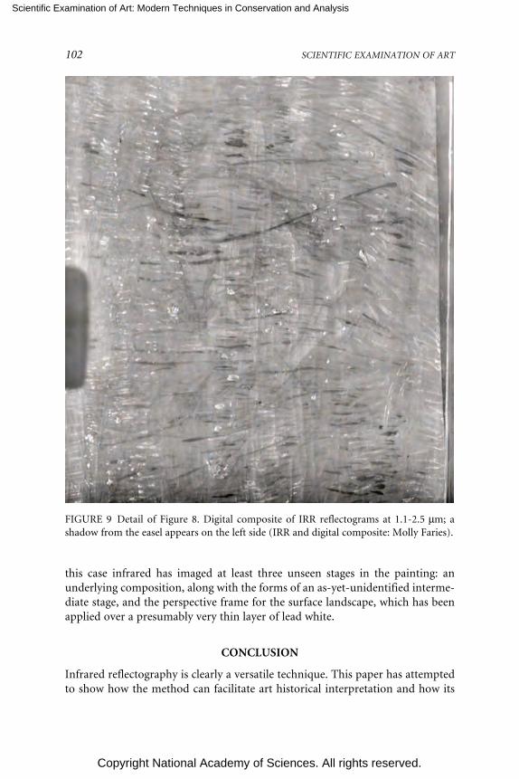
Scientific Examination of Art: Modern Techniques in Conservation and Analysis
Copyright National Academy of Sciences. All rights reserved.
102 SCIENTIFIC EXAMINATION OF ART
FIGURE 9 Detail of Figure 8. Digital composite of IRR reflectograms at 1.1-2.5 µm; ashadow from the easel appears on the left side (IRR and digital composite: Molly Faries).
this case infrared has imaged at least three unseen stages in the painting: anunderlying composition, along with the forms of an as-yet-unidentified interme-diate stage, and the perspective frame for the surface landscape, which has beenapplied over a presumably very thin layer of lead white.
CONCLUSION
Infrared reflectography is clearly a versatile technique. This paper has attemptedto show how the method can facilitate art historical interpretation and how its

Scientific Examination of Art: Modern Techniques in Conservation and Analysis
Copyright National Academy of Sciences. All rights reserved.
ANALYTICAL CAPABILITIES OF INFRARED REFLECTOGRAPHY 103
application may be extended for study beyond the disclosure of underdrawings.The research discussed here done with both the “classic” infrared vidicon and thenewer focal plane arrays would indicate that there is reason to maintain the term“infrared reflectography” as a concept in infrared imaging. For the optimumpenetration of paint and visibility of most underdrawings, devices that work be-yond 1.5 µm and include the region around 2 µm are required. There are, none-theless, appropriate applications for devices that are only slightly sensitive in theinfrared up to 1.1 µm.
Infrared reflectography has always had the advantage of being a portable,noninvasive scanning technique, and with new developments in the computer-ized assembly of reflectograms, it has become easier to document paintings intheir entirety. If in coming years X-radiography were also developed as a portablescanning technique, the usefulness of both methods would be enhanced whenapplied concurrently. With such combined information about the overall evolu-tion of a painting at hand, researchers would be in a better position to refine thequestions and selection of methods required for further study, and for any sam-pling that might be required. One can also envision linking IRR and X-ray resultswith those from other portable methods that analyze the components of paint,such as X-ray fluorescence. Researchers would then be able to use integratedscanning systems to obtain a dimensional image of the stages of execution coupledwith information from materials analysis of the paint surface.
The parameters of use for infrared imaging can be determined only by acombination of theoretical study and experiential research. The theoretical workthat has already been done gives us a basic guide for infrared applications (vanAsperen de Boer, 1970; Walmsley et al., 1994). Such work is essential in modelingthe interrelationship of wavelength, pigments, grounds, and underdrawing mate-rials. Nonetheless, reconstructions and test planks cannot replicate all the com-plexities of historical painting techniques in actual works. Nor can they approxi-mate the effects of aging or the varied thicknesses and mixtures of paint. Asindicated by the examples discussed in this text, infrared imaging can produceextremely varied results. It is up to the researcher to determine how to adapt andoptimize the possibilities of infrared imaging as the examination unfolds. In addi-tion, wavelength response itself is not always an absolute factor. Tests done withsome indium gallium arsenide focal plane arrays have shown that the high quan-tum efficiency of these cameras can improve their performance (van Asperen deBoer, 2003). Systematic comparisons of the same paintings using different infra-red imaging devices at different wavelength ranges should therefore be pursued,and this author has already begun to carry out such work. This research couldproduce a set of reference images that would help in establishing reasonableexpectations for researchers and complement the already existing theoreticalguidelines for infrared studies.
Infrared reflectography is becoming both more sophisticated and more tech-nologically complex. Our current knowledge about the painting techniques of

Scientific Examination of Art: Modern Techniques in Conservation and Analysis
Copyright National Academy of Sciences. All rights reserved.
104 SCIENTIFIC EXAMINATION OF ART
different periods and different artists will undoubtedly need to be expanded.Analytical interpretation in this field depends on the expertise of the researcher,and can even be described as connoisseurship of technical documentation. Thereis thus every reason to pursue the systematic cataloguing of painting collections aswell as larger comparative studies using infrared, and to encourage interdiscipli-nary research that combines the expertise of technical art historians with that ofresearch-oriented painting conservators and scientists who have a nuanced un-derstanding of art.
This text is based on the paper given at the Arthur M. Sackler colloquium,Scientific Examination of Art: Modern Techniques in Conservation and Analysis,held at the National Academy of Sciences, Washington, D.C., March 19-21, 2003.Molly Faries recently (September 2004) began a related research project: “Infra-red Reflectography: Evaluative Studies,” part of the De Mayerne ResearchProgramme on Molecular Studies in Conservation and Technical Studies in ArtHistory funded by the Netherlands Organization for Scientific Research (NWO).
REFERENCES
Faries, M. 1991. Studying Underdrawings: Notes for the Cologne Workshop. Bloomington, Ind., § 1.212.(This reader, under copyright, is used by many who work in the field of technical studies in arthistory.)
Faries, M. 2001. Reshaping the field: The contribution of technical studies. In Early NetherlandishPainting at the Crossroads: A Critical Look at Current Methodologies, ed. M. W. Ainsworth, pp.70-105. New York: Metropolitan Museum of Art.
Faries, M., and S. Bonadies. 1998. The Cincinnati Landscape with the Offering of Isaac by Herri met deBles: Imagery and artistic strategies. In Herri met de Bles, Studies and Explorations of the WorldLandscape Tradition, eds. N. E. Muller, B. J. Rosasco, and J. H. Marrow, pp. 73-84. Turnhout:Art Museum of Princeton University in collaboration with Brepols Publishers.
Faries, M., L. Helmus, with contributions by J. R. J. van Asperen de Boer. 2000. The Madonnas of Janvan Scorel, Serial Production of a Cherished Motif (exhibition catalog). Utrecht: Centraal Mu-seum.
Havermans, J., H. Abdul Aziz, and H. Scholten. Non-destructive detection of iron gall inks by meansof multispectral imaging. Restaurator 24(2003) no. 1, pp. 55-60 and no. 2, pp. 88-94.
Meyer, M., and M. Raquet. 2002. Digitalfotografie für die Restaurierung Restauro 5:350-355.van Asperen de Boer, J. R. J. 1970. Infrared Reflectography: A Contribution to the Examination of
Earlier European Paintings, Ph.D. thesis, University of Amsterdam.van Asperen de Boer, J. R. J. 2003. Slowly towards improved infrared reflectography equipment. In
Recent Developments in the Technical Examination of Early Netherlandish Painting: Methodology,Limitations, and Perspectives (M. Victor Leventritt Symposium), ed. M. Faries and R. Spronk,pp. 57-64. Turnhout: Harvard University in collaboration with Brepols Publishers.
van der Weerd, J., R. M. A. Heeren, and J. R. J. van Asperen de Boer. 2001. A European 640 x 486 PtSicamera for infrared reflectography. In Colloque XIII pour l’étude du dessin sous-jacent et de latechnologie dans la peinture: la peinture et le laboratoire: la peinture et le laboratoire, ed. R. VanSchoute and H. Verougstraete, pp. 231-243. Leuven: Uitgeverij Peeters.
Walmsley, E., C. Metzger, J. K. Delaney, and C. Fletcher. 1994. Improved visualization ofunderdrawings with solid-state detectors operating in the infrared. Studies in Conservation39:217-231.
Scientific Examination of Art: Modern Techniques in Conservation and Analysis
Copyright National Academy of Sciences. All rights reserved.
105
Color-Accurate Image Archives UsingSpectral Imaging
Roy S. BernsMunsell Color Science Laboratory
Chester F. Carlson Center for Imaging ScienceRochester Institute of Technology
Rochester, New York
ABSTRACT
Digital imaging that includes spectral estimation can overcome limitationsof typical digital photography, such as limited color accuracy and con-straints to a predefined viewing condition or a specific output device. Anexample includes the use of ICC color management to generate an archiveof images rendered for a specific display or for a specific printing technol-ogy. A spectral image offers enhanced opportunities for image analysis, artconservation science, lighting design, and an archive that can be used torelate back to an object’s physical properties. The Munsell Color ScienceLaboratory at Rochester Institute of Technology is involved in a joint re-search program with the National Gallery of Art in Washington, D.C., andthe Museum of Modern Art in New York to develop a spectral-imagingsystem optimized for artwork imaging, archiving, and reproduction.Progress is being documented at the website www.art-si.org. This papersummarizes the scientific approach.
INTRODUCTION
Imaging is an important technique in the scientific examination of art. Its mainuse has been for visual documentation. Photographs have long been used todocument condition before and after transit, microscopic examinations, conser-vation treatments, and so on. They are used to enable color reproductions inbooks and from the Internet. Images using materials with spectral sensitivities in

Scientific Examination of Art: Modern Techniques in Conservation and Analysis
Copyright National Academy of Sciences. All rights reserved.
106 SCIENTIFIC EXAMINATION OF ART
such non-visible regions of the electromagnetic spectrum as infrared and X rayare equally important to the visible spectrum. Although images are used to recordscientific examinations, they are used infrequently as an analytical tool, that is, theamount of colorant in a photographic material would be used to relate to physicalproperties of the art. In contrast, astronomy, remote sensing, and medicine haveexploited this capability for many years.
The advent of digital imaging offers increased opportunities to exploit im-ages for the scientific examination of art. A research program is underway atRochester Institute of Technology to develop an image-acquisition system thatrecords reflection information as a function of wavelength. The system initially islimited to the visible region.
This publication will summarize our methodologies and give some perfor-mance examples. Full results, documentation, and demonstrations can be down-loaded and viewed at www.art-si.org. At the end of this paper are relevant publi-cations written by students, faculty, and staff of the Munsell Color ScienceLaboratory.
TECHNICAL APPROACH
Complete Sampling—Spectral Measurement
A spectrophotometer records spectral reflectance or transmittance for a spe-cific circular aperture; a single color is measured. By analogy a spectral-imagingsystem records spectral reflectance or transmittance for a projected scene at aspecific spatial resolution; many colors are measured. One can envision a numberof techniques to disperse light onto a detector plane. The technique we have takenis to couple a monochrome, area-array chaged-couple device detector with aliquid-crystal tunable filter. Successive images are captured, each image centeredat a specific wavelength. Typically we capture 31 bands corresponding to 400-700nm at 10 nm increments.
As a measurement device, calibration is necessary. For each band, imagesare taken of a dark field (to remove fixed-pattern noise), several neutral diffusepapers (to compensate for lighting non-uniformity and optical flare), a pressedpolytetrafluoroethylene tablet (to determine optimal exposure time), and a colortarget made from a number of colorants (to compensate for wavelength andgeometry bandwidth). These targets are crucial to achieve acceptable perfor-mance. In general, spectroradiometry and imaging have greater uncertainty thancontact spectrophotometry. Thus, it is necessary to derive transformations thatminimize these uncertainties. A typical transformation is shown in Figure 1. TheGretagMacbeth ColorChecker DC and a custom target of blue pigments mixedwith titanium white were used to develop the transformation. This figure is avisualization of the matrix transformation from spatially corrected 31-band im-ages to spectral reflectance factor images. The matrix contains 961 coefficients

Scientific Examination of Art: Modern Techniques in Conservation and Analysis
Copyright National Academy of Sciences. All rights reserved.
COLOR-ACCURATE IMAGE ARCHIVES USING SPECTRAL IMAGING 107
400
475
550
625
700
400
475
550
625
700
0
0.02
0.04
Spectral reflectance factor (nm)
0
0.005
0.01
0.015
0.02
0.025
Coe
ffici
ent
FIGURE 1 Visualization of calibration matrix for 31-band image-acquisition system.
(31 × 31). Ideally the matrix should have dominant diagonal and small off-diagonal coefficients. Figure 2 is an image of the well-known color target, theGretagMacbeth ColorChecker Color Rendition Chart. This independent evalua-tion target provides a method to benchmark color and spectral accuracy. Typicalperformance is shown in Figure 3 for these colors. The spectral accuracy was 1.4percent root-mean-square (RMS) reflectance and an average color accuracy of1.5∆E00 under daylight (D65) and viewed by the 1931 CIE standard observer.
Subsampling—Spectral Estimation
The system described in the previous section performs spectral measure-ment; there are the same numbers of image bands as wavelengths. The majority ofnatural and synthesized colorants have large-bandwidth absorption spectra in thevisible region. Furthermore, there are not many sharp transitions from high tolow reflectance (and vice versa). From a dimensionality reduction perspective itmay not be necessary to collect images every 10 nm, that is, sub-sampling may notresult in a loss of accuracy. For example, during the 1970s, many spectrophotom-

Scientific Examination of Art: Modern Techniques in Conservation and Analysis
Copyright National Academy of Sciences. All rights reserved.
108 SCIENTIFIC EXAMINATION OF ART
FIGURE 2 GretagMacbeth ColorChecker Color Rendition Chart.
400 700 400 700 400 700 400 700 400 700 400 700
1
0
1
0
1
0
1
0
Wavelength (nm)
Ref
lect
ance
fact
or
FIGURE 3 Typical spectral-measurement accuracy for the ColorChecker using a 31-bandimage-acquisition system (blue lines) compared with a small-aperture contact spectro-photometer (red lines).

Scientific Examination of Art: Modern Techniques in Conservation and Analysis
Copyright National Academy of Sciences. All rights reserved.
COLOR-ACCURATE IMAGE ARCHIVES USING SPECTRAL IMAGING 109
eters used in color technology sampled the visible spectrum in 20-nm-wavelengthincrements and bandwidth. As the number of direct measurements reduces weare performing spectral estimation rather than spectral measurement.
Suppose a painting was created from a single chromatic colorant and white(or paper in the case of a watercolor). Because the concentration of one colorantis being varied, one can measure the light reflection at a single wavelength, usuallythe wavelength of maximum absorption (minimum reflectance ignoring thewhite). A single image is captured; differences in gray level relate to differences incolorant concentration. At this wavelength, changes in concentration will resultin the greatest change in reflectance (i.e., the greatest image contrast). If we mea-sure the spectral absorption properties of the colorant using a spectrophotometerand determine the relationship between camera signals and concentration andbetween concentration and spectral reflectance (e.g., Kubelka-Munk theory,Beer’s law), the single image can be used to estimate a 31-band spectral image.This estimation process has also enabled significant data reduction. We need toarchive only the single-band image. The spectral reflectances of the colorant andwhite, the transformation from camera signals to concentration, and betweenconcentration and spectral reflectance are stored in the image tag. This is analo-gous to an ICC input profile except in this case, the profile performs spectral colormanagement.
This idea is extended to paintings created with many colorants. Principalcomponent analysis (PCA) is used to define a set of statistical colorants. Becauseof the spectral properties of colorants in the visible region, the number of statisti-cal colorants (eigenvectors) can vary between 5 and 16. The specific numberdepends on spectral accuracy requirements and the samples analyzed statistically.In general the imaging system captures the same number of images as the numberof statistical colorants. A relationship is determined between the camera signalsand statistical colorant amounts (principal components). Concatenating thesevarious steps results in a transformation that relates camera signals to spectralreflectance.
Principal component analysis can be interpreted as constraining the spectraloutcome of the mathematical transformation, a type of spectral interpolation.With a large enough number of samples, we can eliminate the use of PCA. In itsplace we derive a direct transformation from camera signals to spectral reflec-tance. This method uses a singular-value-decomposition-based pseudo-inversecalculation in which several hundred thousand samples are used to estimate sev-eral-hundred-transformation coefficients. These many samples are acquired byconsidering each pixel of an image an individual data point. We have found thatthese two methods yield similar spectral accuracy.
Both techniques are constrained in two ways. The first has to do with thecamera. Performance depends on the spectral sensitivities of each camera chan-nel. Optimal filter design has been studied for many years; unfortunately thesefilters, designed by simulation, cannot be fabricated. The practical solution is to

Scientific Examination of Art: Modern Techniques in Conservation and Analysis
Copyright National Academy of Sciences. All rights reserved.
110 SCIENTIFIC EXAMINATION OF ART
select the best filters from those produced commercially. We have taken thisapproach. We have also used commercial cameras with color filter area-arraysensors. With additional filtration using colored absorption filters, sets of colorimages are recorded. Three, six, or nine image planes (each triplet is the usualred, green, blue image) are related to three, six, or nine statistical colorants ordirectly to spectral reflectance. The second constraint is the dependence on acolor target. The target is used to derive the mathematical transformation. Ide-ally, the target should have a number of colored patches sampling thoroughlythe color gamut of materials to be imaged. The patches should be made fromcolorants with unique spectral properties. The gloss properties should be consis-tent. In essence there is an assumption that the color target has spectral proper-ties that encompass those of the art to be imaged. Most commercial targets donot have these ideal properties.
Despite these constraints, the method has proven to be nearly equivalent to31-band spectral imaging. Using a color-filter-array camera and two absorptionfilters, the average performance for the ColorChecker was 1.6 percent RMS and1.2 ∆E00, plotted in Figure 4. The transformation matrix is plotted in Figure 5,derived using the ColorChecker DC. This transformation relates six camera sig-
400 700 400 700 400 700 400 700 400 700 400 700
1
0
1
0
1
0
1
0
Wavelength (nm)
Ref
lect
ance
fact
or
FIGURE 4 Typical spectral-measurement accuracy for the ColorChecker using a two-filter color-filter-array image-acquisition system (blue lines) compared with a small-aper-ture contact spectrophotometer (red lines).

Scientific Examination of Art: Modern Techniques in Conservation and Analysis
Copyright National Academy of Sciences. All rights reserved.
COLOR-ACCURATE IMAGE ARCHIVES USING SPECTRAL IMAGING 111
nals to 36 wavelengths, totaling 216 matrix coefficients. At each wavelength, thereshould be at least one peak or valley.
Spectral Advantage
A spectral image archive has a number of advantages over many currentimage archives. Sometimes, an archive is created by digitizing photographs. Inother cases direct digitization is used with scanbacks, using repurposed flatbedscanning sensors. Film and scanbacks have spectral sensitivities quite differentfrom the human visual system. As a result these archives require significant visualediting as part of the workflow. Thus, the archive is connected to a particulardisplay, viewing condition, and observer. Color accuracy is limited. Color man-agement principles can be used to reduce the reliance on visual editing. Even so,color accuracy can still be limited. The spectral archive is not subject to theseconstraints; the result is excellent color accuracy, eliminating the need for visualediting.
A non-spectral archive stores three image planes per object, such as RGB
12
34
56
380430
480530
580630
680730 –40
–20
0
20
40
ChannelWavelength, nm
Coe
ffici
ent
–30
–20
–10
0
10
20
30
FIGURE 5 Visualization of calibration matrix for six-band image-acquisition system,achieved using two absorption colored glass filters and a color-filter area-array image-acquisition system.

Scientific Examination of Art: Modern Techniques in Conservation and Analysis
Copyright National Academy of Sciences. All rights reserved.
112 SCIENTIFIC EXAMINATION OF ART
TIFF (tagged image file format). For color-managed images tags are used to relatethe digital signals to standardized viewing and illuminating conditions (sourceprofiles). Using ICC color management, this includes CIE illuminant D50 and theCIE 1931 standard observer. Thus, the archive is limited to a single observer andilluminant. The spectral archive can be used to relate the digital signals to anyobserver, viewing, and illuminating condition. This provides tremendous oppor-tunities by enabling an object to be rendered under multiple conditions withoutre-imaging. Using vision models that account for chromatic adaptation, one cancompare an object’s appearance with changes in lighting, providing lighting de-signers with a unique and powerful tool.
Many colorants have unique spectral properties within the visible spectrum.Thus, the spectral archive can be used to analyze the colorants used in a work ofart. The spectral information can aid conservators in selecting colorants forinpainting (retouching) that result in minimal metamerism. We expect that acombination of spectral imaging and direct small-aperture spectrophotometrycan be used to create colorant maps.
Printed reproductions are quite useful for scholarly endeavors and duringconservation treatments. Color-managed prints are designed to match under CIEilluminant D50 and to be viewed by the 1931 standard observer. By definition theprints are metameric and will only match for this single condition. However,prints are viewed under a variety of conditions. Spectral data can be used toproduce prints that better match original objects for these many conditions.
Finally, a visible-spectrum archive can be combined with other wavelengthregions such as infrared and X ray, aiding in a more complete record on a work ofart’s physical properties.
CONCLUSIONS
A spectral image archive results in high color accuracy and facilitates the scien-tific examination of art in the visible region of the electromagnetic spectrum.Two methods of image acquisition have been described: (1) complete spectralsampling and (2) spectral sub-sampling combined with estimation. Each methodhas advantages and disadvantages. Issues include spectral accuracy, colorimetricaccuracy, hardware complexity and cost, software complexity, image capturetime, data storage, ease of use, maintenance, and system duplication complexity.One of the research goals is to describe these trade-offs in order to providemuseums, archives, and libraries with information to assist them in makingpractical decisions regarding the incorporation of spectral imaging into theirimaging practices.

Scientific Examination of Art: Modern Techniques in Conservation and Analysis
Copyright National Academy of Sciences. All rights reserved.
COLOR-ACCURATE IMAGE ARCHIVES USING SPECTRAL IMAGING 113
ACKNOWLEDGEMENTS
This research is supported by the Andrew W. Mellon Foundation; RochesterInstitute of Technology; the National Gallery of Art, Washington, D.C.; and theMuseum of Modern Art, New York, and would not have been possible withoutthe participation of the students, faculty, and staff of the Munsell Color ScienceLaboratory.
RELEVANT MUNSELL COLOR SCIENCE LABORATORY (MCSL)PUBLICATIONS
Publications
1994
Vent. D. S. Multichannel analysis of object-color spectra. M.S. Thesis, RochesterInstitute of Technology, Rochester, N.Y.
1996
Burns, P. D. and R. S. Berns. Analysis of multispectral image capture. In Proceed-ings of the IS&T/SID Fourth Color Imaging Conference Color Science, Systems,and Applications, pp. 19-22. Springfield, Va.: Society for Imaging Science andTechnology.
1997
Burns, P. D. Analysis of image noise in multi-spectral color acquisition. Ph.D.Dissertation, Rochester Institute of Technology, Rochester, N.Y.
Burns, P. D. and R. S. Berns. Error propagation in color signal transformations.Color Research and Application 22:280-289.
Burns, P. D., and R. S. Berns. Modeling colorimetric error in electronic imageacquisition, Proceedings of the Optical Society of America Annual Meeting, pp.147-149. Washington, DC: Optical Society of America.
1998
Berns, R. S., F. H. Imai, P. D. Burns, and D. Tzeng. Multispectral-based colorreproduction research at the Munsell Color Science Laboratory. In Proceed-ings of the International Society for Optical Engineering, vol. 3409, ed. J. Bares,pp. 14-25. Bellingham, Wash.: International Society for Optical Engineering.
Imai, F. H. and R. S. Berns. High-resolution multi-spectral image capture for finearts preservation. In Proceedings of the Fourth Argentina Color Conference, pp.21-22. Buenos Aires, Argentina: Grupo Argentino del Color.
Scientific Examination of Art: Modern Techniques in Conservation and Analysis
Copyright National Academy of Sciences. All rights reserved.
114 SCIENTIFIC EXAMINATION OF ART
Imai, F. H. and R. S. Berns. High-resolution multi-spectral image archives: ahybrid approach. In Proceedings of the IS&T/SID Sixth Color Imaging Confer-ence Color Science, Systems, and Applications, pp. 224-227. Springfield, Va.:Society for Imaging Science and Technology.
1999
Berns, R. S. Challenges for colour science in multimedia imaging systems. InColour Imaging: Vision and Technology, eds. L. MacDonald and R. Luo, pp.99-127. Chichester: Wiley.
Burns, P. D. and R. S. Berns. Quantization in multispectral color image acquisi-tion. In Proceedings of the IS&T/SID Seventh Color Imaging Conference: ColorScience, Systems, and Applications, pp. 32-35. Springfield, Va.: Society forImaging Science and Technology.
Imai, F. H. and R. S. Berns. A comparative analysis of spectral reflectance recon-struction in various spaces using a trichromatic camera system. In Proceed-ings of the IS&T/SID Seventh Color Imaging Conference: Color Science, Sys-tems, and Applications, pp. 21-25. Springfield, Va.: Society for Imaging Scienceand Technology.
Imai, F. H. and R. S. Berns. Spectral estimation using trichromatic digital cam-eras. In Proceedings of the International Symposium on Multispectral Imagingand Color Reproduction for Digital Archives, pp. 42-49. Chiba, Japan: ChibaUniversity, Miyake Laboratory .
Rosen, M. R. and X. Jiang. Lippmann 2000: A spectral image database underconstruction. In Proceedings of the International Symposium on MultispectralImaging and Color Reproduction for Digital Archives, pp. 117-122. Chiba,Japan: Chiba University, Miyake Laboratory.
2000
Berns, R. S. Billmeyer and Saltzman’s Principles of Color Technology, 3rd ed. NewYork:Wiley.
Imai, F. H., R. S. Berns, and D. Tzeng. A comparative analysis of spectral reflec-tance estimation in various spaces using a trichromatic camera system. Jour-nal of Imaging Science and Technology 44:280-287.
Imai, F. H., M. R. Rosen, and R. S. Berns. Comparison of spectrally narrow-bandcapture versus wide-band with a priori sample analysis for spectral reflec-tance estimation. In Proceedings of the Eighth Color Imaging Conference: ColorScience and Engineering, Systems, Technologies and Applications, pp. 234-241.Springfield, Va.: Society for Imaging Science and Technology.
Imai, F. H., M. R. Rosen, R. S. Berns, N. Ohta, and N. Matsushiro. Preliminarystudy on spectral image compression. In Proceedings of Color Forum Japan2000, pp. 67-70. Tokyo: Japanese Optics Society, Japanese Illumination Soci-ety, Japanese Color Society, and Japanese Photographic Society.

Scientific Examination of Art: Modern Techniques in Conservation and Analysis
Copyright National Academy of Sciences. All rights reserved.
COLOR-ACCURATE IMAGE ARCHIVES USING SPECTRAL IMAGING 115
Quan, S. and N. Ohta. Optimization of camera spectral sensitivities. In Proceed-ings of the Eighth Color Imaging Conference: Color Science and Engineering,Systems, Technologies and Applications, pp. 273-278. Springfield, Va.: Societyfor Imaging Science and Technology.
Rosen, M. R., M. D. Fairchild, G. M. Johnson, and D. R. Wyble. Color manage-ment within a spectral image visualization tool. In Proceedings of the EighthColor Imaging Conference: Color Science and Engineering, Systems, Technolo-gies and Applications, pp.75-80. Springfield, Va.: Society for Imaging Scienceand Technology.
2001
Berns, R. S. The science of digitizing paintings for color-accurate image archives:A review. Journal of Imaging Science and Technology 45:305-325.
Imai, F. H., M. R. Rosen, and R. S. Berns. Multi-spectral imaging of a van Gogh’sself-portrait at the National Gallery of Art, Washington, D.C. In Proceedingsof the IS&T PICS Conference, pp. 185-189. Springfield, Va.: Society for Imag-ing Science and Technology.
Imai, F. H., S. Quan, M. R. Rosen, and R. S. Berns. Digital camera filter design forcolorimetric and spectral accuracy. In Proceedings of the Third InternationalConference on Multispectral Color Science, eds. M. Hauta-Kasari, J. Hiltunen,and J. Vanhanen, pp. 13-16. Joensuu, Finland: University of Joensuu Depart-ment of Computer Science.
Imai, F. H., M. R. Rosen, D. R. Wyble, R. S. Berns, and D. Tzeng. Spectral repro-duction from scene to hardcopy. I: Input and Output. In Proceedings of theInternational Society for Optical Engineering, vol. 4306, eds. M. M. Blouke, J.Canosa, and N. Sampat, pp. 346-357.
Matsushiro, N., F. H. Imai, and N. Ohta. Principal component analysis of spectralimages based on the independence of color matching function vectors. InProceedings of the Third International Conference on Multispectral Color Sci-ence, eds. M. Hauta-Kasari, J. Hiltunen, and J. Vanhanen, pp. 77-80. Joensuu,Finland: University of Joensuu Department of Computer Science.
Rosen, M. R., F. H. Imai, X. Jiang, and N. Ohta. Spectral reproduction from sceneto hardcopy II: Image processing. In Proceedings of the International Societyfor Optical Engineering, vol. 4300, eds. R. Eschbach and G. G. Marcu, pp. 33-41.
2002
Berns, R. S. and F. H. Imai. The use of multi-channel visible spectrum imaging forpigment identification. In Proceedings of the 13th Triennial ICOM-CC Meet-ing, pp. 217-222. London: James & James Ltd..
Berns, R. S. and R. Merrill. Color science and painting. American Artist, 68-70, 72(January, 2002).
Scientific Examination of Art: Modern Techniques in Conservation and Analysis
Copyright National Academy of Sciences. All rights reserved.
116 SCIENTIFIC EXAMINATION OF ART
Berns, R. S. Visible-spectrum imaging techniques: An Overview. In Proceedings ofthe 9th Congress of the International Colour Association, Rochester, N.Y. pp.475-480. SPIE vol. 4421. Bellingham, Wash.: The International Society forOptical Engineering.
Imai, F. H. and R. S. Berns. Spectral estimation of oil paints using multi-filtertrichromatic imaging. In Proceedings of the 9th Congress of the InternationalColour Association, Rochester, N.Y. pp. 504-507. SPIE vol. 4421. Bellingham,Wash.: The International Society for Optical Engineering.
Imai, F. H., M. R. Rosen, and R. S. Berns. Comparative study of metrics forspectral match quality. In Proceedings of the First European Conference onColor in Graphics, CGIV 2002, Imaging and Vision, pp. 492-496. Springfield,Va.: Society for Imaging Science and Technology.
Quan, S. and N. Ohta. Evaluating hypothetical spectral sensitivities with qualityfactors. Journal of Imaging Science and Technology 46:8-14.
Rosen, M. R., F. H. Imai, M. D. Fairchild, and N. Ohta. Data-efficient methodsapplied to spectral image capture. In Proceedings of the International Congressof Imaging Science, Tokyo, ICIS’02, pp. 389-390. Tokyo: The Society of Photo-graphic Science and Technology of Japan and The Imaging Society of Japan.
Rosen, M. R., F. H. Imai, M. D. Fairchild, and N. Ohta. Data-efficient methodsapplied to spectral image capture. Journal of the Society of Photographic Sci-ence and Technology of Japan 65:353-362.
Rosen, M. R., M. D. Fairchild, and N. Ohta. An introduction to data-efficientspectral imaging. In Proceedings of the First European Conference on Color inGraphics, CGIV’2002, Imaging and Vision, pp. 497-502. Springfield, Va.: So-ciety for Imaging Science and Technology.
2003
Berns, R. S., L. A. Taplin, F. H. Imai, E. A. Day, D. C. Day. Spectral imaging ofMatisse’s Pot of Geraniums: A case study. In Proceedings of the IS&T/SIDEleventh Color Imaging Conference: Color Science and Engineering, pp. 149-153. Springfield, Va.: Society for Imaging Science and Technology.
Day, D. C. Filter selection for spectral estimation using a trichromatic camera.M.S. Thesis, Rochester Institute of Technology, Rochester, N.Y.
Day, E. A. The effects of multi-channel spectrum imaging on perceived spatialimage quality and color reproduction accuracy. M.S. Thesis, Rochester Insti-tute of Technology, Rochester, N.Y.
Day, E. A., R. S. Berns, L. A. Taplin, and F. H. Imai. A psychophysical experimentevaluating the color accuracy of several multispectral image capture tech-niques. In Proceedings of the IS&T 2003 PICS conference, pp.199-204. Spring-field, Va.: Society for Imaging Science and Technology.
Imai, F. H., D. R. Wyble, R. S. Berns, and D. Tzeng. A feasibility study of spectralcolor reproduction. Journal of Imaging Science and Technology 47: 543-553.

Scientific Examination of Art: Modern Techniques in Conservation and Analysis
Copyright National Academy of Sciences. All rights reserved.
COLOR-ACCURATE IMAGE ARCHIVES USING SPECTRAL IMAGING 117
Quan, S., N. Ohta, R. S. Berns, and N. Katoh. Heirarchical approach to the opti-mal design of camera spectral sensitivities for colorimetric and spectral per-formance, pp. 159-170. SPIE 5008. Bellingham, Wash.: The InternationalSociety for Optical Engineering.
Quan, S. Evaluation and optimal design of spectral sensitivities for digital colorimaging. Ph.D. Dissertation, Rochester Institute of Technology, Rochester,N.Y.
Rosen, M. R. Navigating the roadblocks to spectral color reproduction: Data-efficient multi-channel imaging and spectral color management. Ph.D. Dis-sertation, Rochester Institute of Technology, Rochester, N.Y.
Sun, Q. Spectral imaging of human portraits and image quality. Ph.D. Disserta-tion, Rochester Institute of Technology, Rochester, N.Y.
2004
Day, E. A., R. S. Berns, L. A. Taplin, and F. H. Imai. A psychophysical experimentevaluating the color and spatial-image quality of several multi-spectral imagecapture techniques. Journal of Imaging Science and Technology 48:99-110.
Mohammadi, M., M. Nezamabadi, R. S. Berns, and L. A. Taplin. Spectral imagingtarget development based on hierarchical cluster analysis. In Proceedings ofthe IS&T/SID Twelfth Color Imaging Conference: Color Science and Engineer-ing: Systems, Technologies, Applications, pp. 59-64. Springfield, Va.: Societyfor Imaging Science and Technology.
2005
Berns, R. S., L. A. Taplin, M. Nezamabadi, Y. Zhao, and Y. Okumura. High-accuracy digital imaging of cultural heritage without visual editing. In Pro-ceedings IS&T Second Image Archiving Conference, in press. Springfield, Va.:Society for Imaging Science and Technology.
Berns, R. S., L. A. Taplin, M. Nezamabadi, and M. Mohammadi. Spectral imagingusing a commercial color-filter array digital camera. In Proceedings 14thTriennial Meeting The Hague, ICOM Committee for Conservation, in press.
Mohammadi, M., M. Nezamabadi, R. S. Berns, and L. A. Taplin, Pigment selec-tion for multispectral imaging. In Proceedings 10th Congress of the Interna-tional Colour Association, in press.
Murphy, E. P. A testing procedure to characterize color and spatial quality ofdigital cameras used to image cultural heritage. M.S. Thesis, Rochester Insti-tute of Technology, Rochester, N.Y.
Rosen, M.R., and F.S. Frey. RIT American museums survey on digital imagingfor direct capture of artwork. In Proceedings IS&T Second Image ArchivingConference, in press. Springfield, Va.: Society for Imaging Science andTechnology.
Scientific Examination of Art: Modern Techniques in Conservation and Analysis
Copyright National Academy of Sciences. All rights reserved.
118 SCIENTIFIC EXAMINATION OF ART
Smoyer, E. P. M., L. A. Taplin, and R. S. Berns. Experimental evaluation of mu-seum case study digital camera systems. In Proceedings IS&T Second ImageArchiving Conference, in press. Springfield, Va.: Society for Imaging Scienceand Technology.
Zhao, Y., L. A. Taplin, M. Nezamabadi, and R. S. Berns. Using matrix R methodin the multispectral image archives. In Proceedings 10th Congress of the Inter-national Colour Association, in press.
Technical Reports
These reports can be downloaded from www.art-si.org and www.cis.rit.edu/mcsl/research/reports.shtml.
1998
Imai, F. H. Multi-spectral image acquisition and spectral reconstruction using atrichromatic digital camera system associated with absorption filters, parts I-VIII. MCSL Technical Report, August.
2000
Berns, R. S. Direct digital imaging of Vincent van Gogh’s self-portrait—A per-sonal view. MCSL Technical Report, May.
Berns, R. S. The science of digitizing two-dimensional works of art for color-accurate image archives—Concepts through practice. MCSL Technical Re-port, May.
Imai, F. H. Spectral reproduction from scene to hardcopy: Multi-spectral acquisi-tion and spectral estimation using a trichromatic digital camera system asso-ciated with absorption filters. Parts I and II. MCSL Technical Report, October.
2002
Berns, R. S. Phase I final report to the National Gallery of Art, Washington, Art-SI project update. MCSL Technical Report, October.
Day, E. A. Colorimetric characterization of a computer-controlled (SGI) CRTdisplay. MCSL Technical Report, April.
Day, E. A., F. H. Imai, L. A. Taplin, and S. Quan. Characterization of a RoperScientific Quantix monochrome camera. MCSL Technical Report, March.
Imai, F. H. Simulation of spectral estimation of an oil-paint target under differentilluminants. MCSL Technical Report, January.
Imai, F. H., L. A. Taplin, and E. A. Day. Comparison of the accuracy of varioustransformations from multi-band images to reflectance spectra. MCSL Tech-nical Report, Summer.
Imai, F. H., L. A. Taplin, D. C. Day, E. A. Day, and R. S. Berns. Imaging at theNational Gallery of Art, Washington, D.C. MCSL Technical Report, Decem-ber.

Scientific Examination of Art: Modern Techniques in Conservation and Analysis
Copyright National Academy of Sciences. All rights reserved.
COLOR-ACCURATE IMAGE ARCHIVES USING SPECTRAL IMAGING 119
2003
Imai, F. H., L.A., Taplin, and E. A. Day. Comparative study of spectral reflectanceestimation based on broad-band imaging systems. MCSL Technical Report,April.
Day, D.C. Spectral sensitivies of the Sinarback 54 camera. MCSL Technical Report,February.
Day, D.C. Evaluation of optical flare and its effects on spectral estimation accu-racy. MCSL Technical Report, February.
2004
Berns, R. S., L.A. Taplin, M. Nezamabadi, and Y. Zhao. Modifications of aSinarback 54 digital camera for spectral and high-accuracy colorimetric im-aging: Simulations and experiments. MCSL Technical Report, June.
Mohammadi, M. and R. S. Berns. Verification of the Kubelka-Munk turbid me-dia theory for artist acrylic paint. MCSL Technical Report, June.
Mohammadi, M., M. Nezamabadi, L. A. Taplin, and R. S. Berns. Pigment selec-tion using Kubelka-Munk turbid media theory and non-negative least squarestechnique. MCSL Technical Report, June.
Zhao, Y., L. A. Taplin, M. Nezamabadi, and R. S. Berns, Methods of SpectralReflectance Reconstruction for A Sinarback 54 Digital Camera. MCSL Tech-nical Report, December.

Scientific Examination of Art: Modern Techniques in Conservation and Analysis
Copyright National Academy of Sciences. All rights reserved.
120
Multispectral Imaging of Paintings in theInfrared to Detect and Map Blue Pigments
John K. Delaney,1 Elizabeth Walmsley,1
Barbara H. Berrie,1 and Colin F. Fletcher2
SUMMARY
Spectral imaging for conservators offers the promise of providing a non-destruc-tive tool for the identification of artists’ materials in situ, as well as determiningtheir spatial distribution in an artwork. In this paper spectral imaging in thereflective infrared (IR) spectral region (0.7 to 2.5 microns) is examined for itspotential to discriminate and identify blue pigments in paintings. The blue pig-ments considered are azurite, indigo, Prussian blue, lapis lazuli, cobalt blue,ultramarine, and thalo blue. Toward this end, visible to shortwave infrared dif-fuse reflection spectra of the blue pigments in both powder and paint forms werecollected to determine the optimal spectral region to discriminate among thesepigments. The measured spectra show that these blue pigments have large andvaried reflectance in the near infrared (NIR, 0.7 to 1.0 microns) to shortwaveinfrared (SWIR, 1 to 2.5 microns) as compared to the visible spectral region. Thelarge reflectance variation suggests the ability of broadband multispectral imag-ing (MSI) (< ~15 spectral bands, bandwidths of a few 100 to a few 10s of nm) toseparate and support identification of these blue pigments in situ and to maptheir distribution. To test this, visible and infrared cameras were equipped withspectral band filters to allow collection of multispectral images of test panels and
1Conservation Division, National Gallery of Art, 2000-B S Club Drive, Landover, MD 207852 The Genomics Institute of the Novartis Research Foundation, 10675 John Jay Hopkins Drive, San
Diego, CA 92121

Scientific Examination of Art: Modern Techniques in Conservation and Analysis
Copyright National Academy of Sciences. All rights reserved.
MULTISPECTRAL IMAGING OF PAINTINGS 121
two paintings known to contain a subset of the blue pigments listed above wereimaged.
The reflectance spectra of the test panels obtained using cameras were foundto correlate to those obtained using the benchtop spectrometers. Multispectralimaging of two paintings by Vincent van Gogh provided reflectance spectra con-sistent with the presence of the blue pigments Prussian blue, cobalt blue andultramarine and gave information on the distribution of the pigments in theworks by utilizing spectral band ratio images and false color composites. Theidentification of the pigments was confirmed using air-path X-ray fluorescencespectroscopy and energy dispersive spectrometry. The results show that multi-spectral imaging, either in numerous spectral bands from the visible to SWIR, orin a judicious selection of bands, can be a powerful tool to aid in pigment identi-fication and distribution in paintings especially when combined with sample-based pigment identification methods such as X-ray fluorescence spectroscopy oranalysis of cross-sections.
INTRODUCTION
Spectral imaging (or Imaging Spectrometry), the collection of images in separatespectral bands in order to obtain reflection spectra, has been shown to be a pow-erful tool for geophysical remote sensing (1). The method also provides the spa-tial distribution (maps) of materials, such as minerals and agricultural crops,across the imaged region.
The scientific examination of paintings often begins with the identification ofthe base set of pigments used by an artist. When identifying and determining thespatial distribution of pigments in a work of art, conservators begin with a visualinspection of the artwork. Subsequently, analytical methods, both destructive andnon-destructive, are used in the classification and identification of pigments.Most of these techniques are applied to small areas of the painting either becausethey are destructive (that is, involve removing a sample from the work for analysisusing polarized light microscopy or other techniques) or, if non-destructive, aretoo cumbersome and expensive to apply across the painting (e.g., air-path X-rayfluorescence spectroscopy, Raman spectroscopy). Although powerful, these tech-niques provide only localized information, and sampling sites are limited andmay be biased. A less precise analytical methodology is used to extrapolate theresults of “point analysis” to the rest of the artwork. The identification of pig-ments in un-sampled regions is often made through visual association. Successoften depends on the ability to identify various pigments by their color. Thishinders the ability to form general conclusions about the distribution of materialsthroughout the artwork. This can lead not only to misidentifications but also thepossibility that some materials may not be discovered. Consequently, there hasbeen growing interest in the development of methods that do not require sam-pling and provide information on spatial distribution.

Scientific Examination of Art: Modern Techniques in Conservation and Analysis
Copyright National Academy of Sciences. All rights reserved.
122 SCIENTIFIC EXAMINATION OF ART
One such method is visible reflectance spectroscopy using high-resolutionspectra acquired from a limited number of sites in an artwork (2, 3, 4). Thismethodology is useful for helping to accurately define the color and has been usedfor pigment identification utilizing spectral reference databases (2, 5). It is espe-cially useful for pigment identification when it is extended to include the infraredregion (6, 7, 8). However, the technique suffers from the same limitation of theother non-destructive methods, i.e., small sample sizes and the lack of an analyti-cal methodology to sample the entire painting. Simply extending the number ofsample sites over the entire painting would allow, in principle, the determinationof the possible pigment composition at each site in the painting. Extending high-resolution visible spectral analysis to large areas of a painting is problematic. Theability to acquire high fidelity visible reflectance spectra (<5 nm band pass) ofentire paintings is beyond the resources of conservation laboratories. Using vis-ible spectroscopy alone to identify pigments having similar colors is challengingbecause of the small inherent reflectance differences among them, which are fur-ther reduced and confounded by the presence of scattered light associated withvarying pigment particle size, and the collection/illumination geometries.
In this paper, we investigate the use of infrared reflectance spectroscopy toaid in separating similar-appearing pigments and provide sufficient spectral datato be an adjunct to pigment identification. One motivation comes from the suc-cess of remote sensing reflectance spectral imaging, with its well-developed meth-odology for the identification of mineral deposits, and because numerous artists’paints consist of ground minerals. Some success in applying this technique toworks of art has already been achieved (6,); Casini et al. have differentiated be-tween two yellow pigments in a painting by Pontormo (7).
A well-designed reflectance spectral imaging system requires a compromisebetween the desired spectral and spatial resolutions. While the goal is to acquirehigh-quality reflectance spectra with the smallest sample size over the target areaof interest (here the entire painting), the signal-to-noise requirements, instru-ment and data handling complexity force compromise. Increasing spectral reso-lution requires a reduction in spatial resolution and vice versa. Knowledge of theexpected range of artists’ materials to be encountered, their spectral properties,and their particle size can be used to select the spectral and spatial resolutionsrequired for identification and mapping of the pigments.
The pigments typically encountered in paintings are either mineral or or-ganic and as such their visible reflectance spectra are determined by broad elec-tronic transitions that account for their color. There are overtones and combina-tions of vibrational transitions in the near infrared (NIR, 0.7 to 1.0 microns) toshortwave infrared (SWIR, 1 to 2.5 microns). In solids, the vibrational absorptionbands have widths ca. 20 nm or wider which determines a limiting spectral reso-lution required for acquisition of spectra. While measurement of the position ofvibrational overtone bands may be useful for unique identification of materials,designing instruments at this level of resolution (i.e., 20 nm) may be unnecessary.

Scientific Examination of Art: Modern Techniques in Conservation and Analysis
Copyright National Academy of Sciences. All rights reserved.
MULTISPECTRAL IMAGING OF PAINTINGS 123
Many minerals have slowly changing, large amplitude reflectance variations, largerthan 0.1, in the NIR to SWIR (9), which can be sampled with > 0.1 micronbandwidths. Thus collection of broadband multispectral images (> 0.1 micronbandwidth) appears to be sufficient for material separation and to support pig-ment identification.
The focus of this work was to optimize spatial sampling and utilize broaderspectral resolution compared to benchtop instrumentation, that is to use multi-spectral reflectance imaging. This was applied over a larger spectral region thanthe visible with the expectation of providing a more robust discrimination toolfor pigment identification. This methodology is suitable for conservators who areused to collecting broadband images of paintings in the ultraviolet, visible andinfrared (10).
EXPERIMENTAL
In this study the reflectance spectra and multispectral images of six blue pigmentsare measured and analyzed. The pigments include mineral ores (azurite, lapislazuli); colors manufactured by industrial processes (Prussian blue, thalo blue,cobalt blue, and synthetic ultramarine), and organic materials (indigo). Thecolorant in lapis lazuli and synthetic ultramarine is chemically the same and istreated as one pigment here.
A three-part experimental approach was employed. First, the collection ofhigh-resolution diffuse reflectance spectra of six blue pigments in both pressedpowder and paint (linseed oil binder) at expected thickness on a chalk ground.Second, the collection of broadband spectral images of the paint test panels toinvestigate the correlation between spectral images and high-resolution spectra.Third, the collection of spectral images of paintings to determine the ability ofmultispectral imaging to separate and identify blue pigments in situ.
Test Panels
Test panels of paint were constructed as previously described (10). The pan-els consist of paintouts of pigments hand-ground in linseed oil for azurite, lapislazuli, Prussian blue, and indigo. Commercial oil paint was used for cobalt blue(Charbonnel) and thalo blue (Fezandie & Sperrle). The azurite, lapis lazuli, Prus-sian blue, indigo, and thalo blue paints were applied to a white chalk ground andthe cobalt blue paint on a gesso ground.
Diffuse Reflection Spectra of Pigments and Paints
Diffuse reflectance spectra of seven dark blue pigments, in pressed powderform and from the test panels, were collected using either a Nicolet System 510
Scientific Examination of Art: Modern Techniques in Conservation and Analysis
Copyright National Academy of Sciences. All rights reserved.
124 SCIENTIFIC EXAMINATION OF ART
interferometer spectrometer or a Beckman Instruments UV 5240 dual beam grat-ing interferometer, both equipped with an integrating sphere.
Multispectral Imaging Instrumentation and Procedures
The multispectral images were obtained in the visible, near infrared, andshortwave infrared spectral regions using cameras fitted with band pass filters. ASony XC-77 camera fitted with narrow band pass filters was used to acquire thevisible/NIR (0.45-1.0 microns) image sets. The silicon monochrome video charge-coupled device (CCD) camera had a 25 mm f/1.6 lens. The filters (Corion Inc.)were 0.40 micron bandwidth filters in 0.50 micron increments over the range0.45-1.0 microns. A Mitsubishi M600 PtSi or a Kodak PtSi 310-21X thermalimager was used to collect the SWIR (1.0 to 2.0 microns) image sets. Each camerawas fitted with a 55 mm f/1.2 Nikon lens. Images of La Mousmé were collectedusing 1.2 (0.09 microns FWHH) and 1.6 (0.5 microns FWHH) micron broad-band filters. Infrared images of details of the two paintings, the Self-Portrait andLa Mousmé, were captured using three broadband filters: 1.1-1.4 microns (As-tronomy J, Barr Associates), 1.5-1.8 microns (Astronomy H, Barr Associates),and 2.0-2.4 microns (Astronomy K, Barr Associates). Diffuse illumination wasused to light the scene, typically using a pair of Lowell Tota lights with FDNQ5000T3/4 quartz halogen lamps (500 W, 3200 K) and photographers’ umbrel-las. Black and white Spectralon (Labsphere) diffuse reflection targets (1 inch di-ameter) were used for in-scene calibration of the camera to reflectance. The lowstandard was 2-3 percent reflective over the range 0.45-2.4 microns, and the highstandard was 98-99 percent reflective over 0.45-2.4 microns. Non-uniformity ofthe detector response was corrected using a gray card. For each test panel orpainting, a set of 8 to 15 images was captured. In the multispectral image sets ofthe paintings, the paint test panels and Spectralon reflection standards were in-cluded in each image. The Spectralon standards were used to convert the digitalcounts of the image sets to reflectance units. Each multi-spectral experiment wasrepeated several times.
A Macintosh computer with a Perceptics Pixelbuffer card or a Scion AG5 PCIcard was used to capture the images. A variety of image processing programs(Scanalytics IP Lab Spectrum, Adobe Photoshop, NIH Image, Research SystemsInc. ENVI 3.4) was used in the analysis, non-uniformity, and calibration of thedata sets. Registration and warping of the visible-NIR images with the SWIRimage set was performed using ENVI. Spectral image data cubes were constructedand analyzed in ENVI.
X-ray Fluorescence Spectroscopy
Non-destructive X-ray fluorescence spectroscopy (XRF) was performed us-ing a Kevex 0750A spectrometer. The air-path instrument was equipped with a
Scientific Examination of Art: Modern Techniques in Conservation and Analysis
Copyright National Academy of Sciences. All rights reserved.
MULTISPECTRAL IMAGING OF PAINTINGS 125
secondary target made from pressed barium chloride. This allowed simulta-neous measurement of X-rays from ca. 2 eV to 45 eV. Pigments composedof light elements, for example, ultramarine, cannot be detected using thisinstrumentation.
Optical Microscopy and Other Analysis of Cross Sections
Pigments from the top layer of paint were obtained by gently scraping thesurface of the painting using a surgeon’s scalpel. A Leica MPX microscope wasused to examine the particles mounted in Cargille MeltMount (refractive index1.66) on glass slides using polarized light microscopy. The same particles or oth-ers could be mounted on a carbon planchet (stub) for examination by scanningelectron microscopy (SEM) and energy dispersive spectrometry (EDS). A double-sided sticky carbon tab was used to adhere the particle to the planchet. If theparticles could not be examined owing to charging they were coated with carbon.A JEOL 6300 scanning electron microscope with an Oxford Tetra backscatteredelectron detector was used for SEM. For EDS measurements an Oxford Inca 300system was used with an Oxford Super ATW Si(Li) detector.
Cross-sections were obtained from cracks or areas of loss. The fragmentswere mounted in Bioplastic® then cut to expose the layer structure of the paint.The sections were polished on SiC grit papers (Micromesh) and examined usingoptical microscopy and SEM-EDS.
RESULTS AND DISCUSSION
Diffuse Spectra of the Blue Pigments and Paints
The six blue pigments have similar reflectance spectra in the visible regionwith few features. However, the spectra demonstrate larger and more variedchanges in reflectance in the reflective infrared (0.7-2.5 microns) (Figure 1). Re-flectance peaks for all the pigments are centered near 0.44 microns, and all displaya strong absorption in the red. The mineral samples, azurite and lapis lazuli, havea transition to increased reflectance in the NIR (0.7 to 1 microns), azurite laggingbehind lapis lazuli. In the SWIR (1 to 2.5 microns) both pigments have near-constant reflectance. In contrast, the pigments indigo and thalo blue show a rapidrise to high reflectance, peaking at ca. 1.5 microns and decreasing beyond that.Prussian blue’s transition to higher reflectance occurs at about 1.35 microns.Cobalt blue has the most variable reflectance in the IR, becoming highly reflectivein the NIR and weakly reflective in the high-energy side of the SWIR, between 1.3and 1.6 microns, and moderately reflective at wavelengths > 1.6 microns.
For all of the pigments the variations in reflectance amplitude through the IRregion are large, > 0.1, and occur over spectral regions wider than 0.1 microns.These large, but slowly varying, changes in reflectance are suitable to be followed

Scientific Examination of Art: Modern Techniques in Conservation and Analysis
Copyright National Academy of Sciences. All rights reserved.
126 SCIENTIFIC EXAMINATION OF ART
FIGURE 1 Diffuse reflectance spectra of six blue pigments in powdered form (dark line)and in oil-bound paint (blue line). The powder samples were optically thick. Syntheticultramarine in powdered form was used instead of powdered lapis lazuli (natural ultrama-rine). The paint layers were generally 15-25 microns, a thickness often encountered inpaintings. The reflectance spectrum of the chalk ground is given in the Prussian blue plot(top solid black curve). Reflectance values derived from multispectral images (solid cir-cles) of the blue paint test panels (Figure 2) were obtained using spectral band pass filters.
using broadband multispectral imaging. However, increased spectral resolutionand sampling (~10 nm) would allow the collection of vibrational lines such as the-O—H stretch in azurite that could be useful to characterize certain pigments.
In Old Master paintings pigments are bound in organic binders (e.g., dryingoils, egg tempera) and applied in thin layers over preparatory ground layers con-taining calcium carbonate (chalk) or calcium sulfate (gesso). As a result the re-flectance spectra of the pigment and corresponding paint made from it may differfor several reasons. To test the influence of the binders on the reflectance spectra

Scientific Examination of Art: Modern Techniques in Conservation and Analysis
Copyright National Academy of Sciences. All rights reserved.
MULTISPECTRAL IMAGING OF PAINTINGS 127
in the IR, diffuse reflectance spectra of the blue paints were collected (Figure 1).The possible effects of the binders on the reflectance spectra are (1) decrease inthe amount of first surface reflectance (the intensity of reflectance spectra arereduced), (2) alteration of the depth of “color” from absorption (some scaling),(3) filtering of the pigment reflection by absorption properties of the binder(added spectral lines or shape changes). The spectra of the test panels comparedto the powdered pigments show these effects occur. However, the general shape ofthe reflectance spectra of the pigments in paint is preserved with an offset (whitelight scatter) and scaling. Additionally, owing to the increasing transparency ofthe pigments in the SWIR, vibration bands from the preparatory layer may beobserved in some cases.
Linseed oil as a binder acts to reduce the difference in refractive index at thefirst interface with air and thus reduces the intensity of the first surface reflec-tance. For particles larger than the wavelength of light (e.g., azurite) this results ina decrease in the ‘white light’ reflectance, hence the reflectance curves shift down-ward. Since the paints on the test panels do not represent infinitely thick layers,there is also a change in the total absorption and hence a scaling change betweenpowder and paint arises. The binder can act as a spectral filter owing to its ownabsorption. Linseed oil has little absorption in the visible to infrared, but some ofthe commercial paint films (e.g., cobalt blue) appear to have additional bands inthe IR. High reflectance correlates with low absorption, thus the underlying pre-paratory ground or underpaint layers may contribute to the reflectance spectra.The contribution of the ground to the reflectance spectrum becomes significantwhen the paint becomes transparent owing to decreased absorption and scatter-ing in the SWIR. It is dependent on the thickness of the paint film. The additionof absorption features from the ground to the spectrum of Prussian blue at wave-lengths beyond 1.6 microns may be noted. The general reflectance signatures are,however, maintained between bulk pressed powder and oil-bound pigments. Inthe samples painted on a chalk or gesso ground, which itself has a high reflectancein the visible to the SWIR, the paints appear to mimic the scattering of the opti-cally thick powder samples.
Multispectral Imaging of Paint Samples
To examine the extent to which diffuse reflectance spectra of pigments can bemodeled by using imaging cameras available to conservators, multispectral imagesof the paint swatches were collected (Figure 2). Two cameras, one sensitive in thevisible/NIR and one in the SWIR, each outfitted with bandpass filters, were usedto sample the visible/NIR and SWIR spectral regions. For in-scene calibration, thereflectance of the ground and carbon black were used as standards.
The reflectance values derived from the multispectral images of the test pan-els show good agreement with the reflectance spectra measured using the bench-top reflectance spectrometer (Figure 1), demonstrating that the image collection

Scientific Examination of Art: Modern Techniques in Conservation and Analysis
Copyright National Academy of Sciences. All rights reserved.
128 SCIENTIFIC EXAMINATION OF ART
FIGURE 2 Multispectral images of test panels with blue paint swatches. By column, fromleft to right: Lapis lazuli (natural ultramarine), azurite, Prussian blue, indigo, and thaloblue. Center wavelength of spectral band pass filter, by row, from top to bottom: 0.56,0.90, 1.2, 1.4, and 1.6 microns. The images show varying changes in the reflectance of thedifferent pigments in the infrared and their increased transparency.
method is adequate. This shows that these blue pigments can be distinguishedfrom each other using only the multispectral images. This ability exists despite thefact that the measurements on the powdered pigment samples were performedusing an integrating sphere, whereas the imaging of the panels and the works of artwere performed with a narrow collection solid angle. Given the near-Lambertiannature of the samples’ reflectance, and the near-diffuse illumination, the close tosuperimposition of the spectral data should not be surprising. These results dem-onstrate that by utilizing multispectral imaging techniques the reflectance of theblue pigments can be characterized adequately to distinguish among them andideally to identify them.
In geophysical remote sensing applications, materials of interest are encoun-tered in optically thick, granular form, like the powdered pigment samples. How-ever, as noted earlier, in works of art the pigments are in thin paint layers. Inregions of high reflectance, an increase in the transparency of the paint layer canbe observed (compare Figures 1 and 2). The preservation of the spectral reflec-tance properties between paint and powder pigments and the maintenance ofhigh reflectance of the bound pigments appear to be due to the high reflectance ofthe ground. A light-absorbing ground or pigment underneath would howeveralter the reflectance spectra. Thus the effect of layering of thin paint films thatoccurs in artwork needs to be considered. Since almost all pigments becometransparent by 1.5 microns (10), collection of spectral images from 1.5 to 2.5

Scientific Examination of Art: Modern Techniques in Conservation and Analysis
Copyright National Academy of Sciences. All rights reserved.
MULTISPECTRAL IMAGING OF PAINTINGS 129
microns seems to be of little utility for their identification. It is the property ofincreasing transparency in this region that allows the detection of underdrawingsin paintings using infrared reflectography (10).
Multispectral Analysis of Paintings to Characterize Blue Pigments
To demonstrate the ability of multi-spectral imaging to discriminate andidentify blue pigments in situ, two paintings by Vincent van Gogh in the collec-tion of the National Gallery of Art, Washington, D.C., were examined using thetechnique. The paintings were selected because they have large regions of brightto dark blue paints. Moreover, results from site-specific analysis techniques suchas XRF, polarized light microscopy and SEM-EDS were available, and the paint-ings contain the pigments Prussian blue, cobalt blue, and ultramarine.
The first painting imaged was La Mousmé, by Vincent van Gogh (1888,Chester Dale Collection, 1963.10.151) (Figure 3, visible light image; see alsowww.nga.gov/search/index.shtm). Van Gogh describes this painting in his ownwords (11):
The portrait of the girl is against a background of white strongly tinged withemerald green, her bodice is striped blood red and violet, the skirt is royalblue, with large yellow-orange dots. The mat flesh tones are yellowish-grey;the hair tinged with violet; the eyebrows and eyelashes are black; the eyes,orange with Prussian blue. A branch of oleander in her fingers for the twohands are showing.
Prussian blue has a large change in reflectance at ~1.35 microns where thepigment becomes less absorbing (Figure 1). Other blue pigments do not have aslarge an absorbance change over this interval. To identify areas of the paintingwhere Prussian blue occurs, a ratio image was created by dividing an image ac-quired at 1.6 microns by one acquired at 1.2 microns. In the ratio image, passageswhere Prussian blue is present have a ratio of absorbance >1 in comparison topassages where ultramarine, cobalt blue, or indigo are present. The ratio image ofLa Mousmé (Figure 3) shows bright (high reflectance ratio) features having higherreflectance at 1.6 than at 1.2 microns, which can be assigned to Prussian blue. Thespectral ratio image in the IR indicates that, in the visible image, the dark lines ofthe chair, the dark lines outlining the flowers, the outline of the girl’s hair and toa lesser extent some daubs of paint on her skirt and some of the stripes on herblouse are painted using Prussian blue. The lack of large reflectance changes insome of the darker and brighter blue regions of the visible image suggests the useof a different blue pigment there.
To more confidently assign the regions identified in the band ratio image ofLa Mousmé, multispectral images in 11 spectral bands from the visible to theSWIR were collected from an 8 × 10-inch section of the painting. Each spectralband image was collected using a visible/NIR or SWIR camera fitted with bandpass

Scientific Examination of Art: Modern Techniques in Conservation and Analysis
Copyright National Academy of Sciences. All rights reserved.
130 SCIENTIFIC EXAMINATION OF ART
FIGURE 3 Vincent van Gogh’s La Mousmé (1888). (Left) Visible light image. (Right)Shortwave infrared spectral band ratio image creaed from two infrared composite imagescaptured at 1.2 microns and at 1.6 microns. The bright areas are regions where the reflec-tance of the painting is higher at 1.6 microns than at 1.2 microns, and thus indicate boththe probable presence of Prussian blue and where it occurs in high concentration withinthe painting. Each IRR composite comprises a mosaic of 24 flat-field-corrected images.
filters in an arrangement similar to that typically used for collecting infraredreflectograms. The image sets were converted to reflectance units using the in-scene reflectance standards. A multispectral image cube (a 3-D image where z isthe spectral band and x,y are spatial locations) was then constructed and theimages registered using tie points. The visible color image in Figure 4, generatedfrom an MSI cube using the 0.45, 0.55, and 0.65 micron images, shows a detail ofLa Mousmé captured with the reference standards and blue test panels.
Verification of the reflectance calibration of the MSI cube was determined bycomparing the image-derived spectra of “in-scene” blue test panels with the priorhigh-resolution spectra obtained with the benchtop spectrometer (Figure 4A).Qualitatively these two match (compare Figure 1 with Figure 4A). The imagecube spectra derived from the blue test panels were a reasonable fit to the spectraacquired using benchtop instrumentation after scaling and a small amount oftranslation. Specifically only a small translation for Prussian blue (0.04 offset) andscaling for lapis lazuli (0.87×, 0.03 offset) and cobalt blue (0.75×) (see Figure 4A)were required to get a good match. Since the illumination of the test panels alone,versus in the scene of the painting, are different some transitional and scalingchanges are not unexpected.
Reflectance spectra derived from the multispectral image cube of the paint-ing itself support the assignment of Prussian blue to bright areas of the ratio

Scientific Examination of Art: Modern Techniques in Conservation and Analysis
Copyright National Academy of Sciences. All rights reserved.
MULTISPECTRAL IMAGING OF PAINTINGS 131
FIGURE 4 Multispectral image analysis of a detail of La Mousmé showing the presence oftwo different blue pigments. The multispectral image cube consists of 11 spectral bandsfrom the visible through the SWIR. (Left) Visible color composite of spectral images ob-tained at 0.65, 0.55, 0.45 microns. Along the bottom, from left to right, are black andwhite reflectance standards (2 and 98 percent Spectralon standards) and blue pigment testpanels: cobalt (two swatches), Prussian blue, and lapis lazuli. (Right) A. Plots of reflec-tance, derived from the multispectral images of the reference panels: high reflectance 98percent Spectralon (open circles), cobalt blue swatch (solid blue diamonds), Prussian blue(solid black circles), and lapis lazuli (blue squares). The blue and black lines are scaleddiffuse spectra. B. Plots of reflectance derived from the multispectral images of the sites 1to 4 in the detail image. Sites 1 and 2 (black circles and triangles) are in the chair. Sites 3and 4 are in the blue stripes on the blouse.
image (Figure 3) of La Mousmé. The image-derived spectra of the bright regionsin the ratio image that map to the dark blue areas of the visible image most closelyresemble the reference spectrum of Prussian blue paint test panel and powderedpigment (Figure 4B). Spectra from two sites on the chair rail (Figure 4B, sites 1, 2)are reasonably well described by the reference spectrum of Prussian blue scaled0.53. Based on this spectral sensitivity, these areas can be determined to containPrussian blue. Not all the blue stripes on the blouse are bright in the 1.6/1.2micron ratio image, suggesting the presence of another pigment (Figure 3 andFigure 4, sites 3, 4). The image-based reflectance spectra show high reflectance inthe NIR to SWIR (0.8 to 1.6 microns, Figure 4B, sites 3, 4) that is consistent withthe reflectance behavior of indigo or ultramarine and not Prussian or cobalt blue.A scaled diffuse reference spectrum of ultramarine powdered pigment (0.39×,

Scientific Examination of Art: Modern Techniques in Conservation and Analysis
Copyright National Academy of Sciences. All rights reserved.
132 SCIENTIFIC EXAMINATION OF ART
FIGURE 5 Multispectral image analysis of a detail of La Mousmé showing the use of twodifferent dark blue pigments for outlining. The multispectral image cube consists of 11spectral bands from the visible through the SWIR. (Top left) Visible color composite ofspectral images obtained at 0.65, 0.55, 0.45 microns. (Top right) Infrared reflectogram ofthe detail area obtained at 1.2 microns and (Bottom right) 1.6 microns. Infrared imageobtained at 1.6 microns shows the change in reflectance from 1.2 microns for the darkoutline of the girl’s flowers, but not the buttonholes. (Bottom left) Plots of reflectancederived from the images of the dark lines outlining the button (blue diamonds andsquares) and flower (black circles and triangles). The solid blue and black lines are diffusereflectance spectra of the paints using the ultramarine and Prussian blue test panels (afterscaling).
0.17 offset) matches the measured spectra reasonably well. The reflectance spectrathus show that at least two blue pigments were used in this painting.
The painting has other passages of the dark blue, for example, the outlines ofthe buttonholes on the girl’s blouse and the top of the flowers she is holding.Visual inspection of the painting alone might suggest that these could be Prussianblue as well. However, the ratio image does not show the strong reflectancechanges associated with Prussian blue. A detail from images obtained at 1.2 and1.6 microns shows the dark blue outlining the flowers is dark at 1.2 microns andlight at 1.6 microns (Figure 5), whereas the dark blue outlining the buttonholes isnot apparent in either the image obtained at 1.2 or at 1.6 microns. The reflectancespectra derived from the multispectral image cube show the dark lines of theflowers are similar to Prussian blue (0.53 scaling of reference spectra), but thebuttonholes are not (Figure 5). The reflectance spectra of the dark blue outlining

Scientific Examination of Art: Modern Techniques in Conservation and Analysis
Copyright National Academy of Sciences. All rights reserved.
MULTISPECTRAL IMAGING OF PAINTINGS 133
the buttonhole shows an earlier rise in reflectance than Prussian blue, more likesome of the blue stripes on the blouse. A scaled and offset reference spectrum ofultramarine (0.42×, 0.02 offset) does pass though the majority of data in the redand infrared suggesting this blue pigment is present. Confirmation of ultramarineat these sites was provided by SEM/EDS analysis, which also showed the presenceof zinc oxide (ZnO). Both the spectra and SEM/EDS are consistent with thepigment in this area being ultramarine.
Thus the blue pigments in the La Mousmé painting have been discriminatedinto Prussian blue and ultramarine blue. The ratio image of the area in combina-tion with the MSI cube suggest that Prussian blue was used in the chair, theoutline of the flowers in her hand, and in strokes of the girl’s hair, eyebrows, lipsand some stripes of her jacket. The reflectance spectra derived from the imagecube suggest that the dark outline of the buttonholes and the blue outlines aroundthe spots on the girl’s skirt were painted using ultramarine. Cobalt blue, a pig-ment that van Gogh used often, does not appear to have been used for painting LaMousmé.
X-ray fluorescence spectroscopy indicated the presence of cobalt in the back-ground of Vincent van Gogh’s Self-Portrait (1889, Collection of Mr. and Mrs.John Hay Whitney, 1998.74.5) (Figure 6, visible light image; see also www.nga.gov/search/index.shtm). A spectral image cube of a region of the paintingshows that it can be used to find the locus of cobalt blue. The multispectral imagecube of Self-Portrait gives reflectance spectra from the background that are simi-lar to that of the blue cobalt blue test panel (Figure 6). A false color image, madeby assigning the infrared image to the red channel shows the distribution of cobaltblue in the painting (Figure 6). Cobalt blue is extensively used in the backgroundand also in the jacket, although spectra from the jacket are complex, indicating amixture of pigments here.
CONCLUSIONS
This report contributes to the body of work that demonstrates the utility ofspectral imaging not only for discrimination among pigments but also to deter-mine their spatial distribution within a work of art. While reflectance spectros-copy is a relatively less specific analytical tool than other analytical chemicaltools, it is non-destructive and can be readily applied to the entire artwork. Itspower lies in helping to define the set of pigments used in the work, and identi-fying regions of high concentration and thus directing site-specific, more power-ful analytical tools such cross-section analysis, XRF, and SEM/EDS for morethorough chemical analysis. Moreoever, in the case of Prussian blue, the reflec-tance spectra in the infrared may be a more definitive assignment method thanXRF given the high tinting strength of Prussian blue. The results here demon-strate that extending the multispectral imaging method to include the infrared

Scientific Examination of Art: Modern Techniques in Conservation and Analysis
Copyright National Academy of Sciences. All rights reserved.
134 SCIENTIFIC EXAMINATION OF ART
can improve the success of spectral imaging in pigment identification and dis-crimination given the large and varied reflectance changes in these regions, inparticular when the visible spectra of pigments are similar. Large and slowlyvarying reflectance changes in the infrared allow the utilization of broadbandMSI techniques, simplifying the methodology.
This study of two paintings demonstrates that multispectral imaging in thevisible and NIR regions can be employed as a useful tool in the scientific examina-tion of paintings. We have demonstrated that the optical properties of pigmentsin the infrared display diagnostic features which can be employed to assign andmap pigments, and that these features can be detected using conventional imag-ing techniques, including modeling of reflectance spectra and ratioing images
FIGURE 6 Multispectral image analysis of a detail of van Gogh’s Self-Portrait (1889). Thedistribution of cobalt blue is indicated in the false color image by the color red. The multi-spectral image cube consists of 13 spectral bands from the visible through the SWIR.(Left) Visible light image. (Bottom right) Plots of reflectance derived from the image ofthe background (solid diamonds). The solid blue and black lines are scaled diffuse reflec-tance spectra of the cobalt blue paint (blue) and powder (black). (Top right) False colorinfrared composite of spectral images obtained at 0.90, 0.55, 0.45 microns. This imagerenders the cobalt blue to appear “red” thus showing the cobalt is present in a largeproportion in the background and a smaller proportion in the jacket.

Scientific Examination of Art: Modern Techniques in Conservation and Analysis
Copyright National Academy of Sciences. All rights reserved.
MULTISPECTRAL IMAGING OF PAINTINGS 135
obtained at different wavelengths. It is difficult to infer this information fromconventional techniques, which rely on a limited number of micro-samples. Theadvantages of the technique are tempered by the increased complexity of thereflectance spectra owing to particle size variation and increasing transparency inthe infrared.
Conservators and conservation scientists utilizing imaging systems currentlyavailable can apply the power of visible and infrared multispectral imaging totheir work.
ACKNOWLEDGEMENTS
This research forms part of an ongoing project at the National Gallery of Art onapplications of infrared imaging. Over the past ten years, we have benefited fromdiscussions and assistance from many people, including Dr. Jack Salisbury, whogave us access in 1994-95 to his laboratory at Johns Hopkins University in orderto collect the diffuse reflectance spectra of the samples, and Mr. Dana D’Aria, whoassisted us in the collection of the spectra; Raymond Rehberg, David L Clark, andRollo E Black, of Eastman Kodak, who provided generous assistance using theKodak thermal imager; Elizabeth Freeman, Kristi Dahm, Lucy Bisognano, andLaura Rivers, who helped with the image captures; Dr. Lisha Glinsman, whoprovided the results of XRF data on the Self-Portrait; and Dr. René de la Rie andMr. Ross Merrill, Chief of Conservation, for their continued interest. An earlyphase of this research was supported by the Circle of the National Gallery of Art.
REFERENCES
1. A. F. H. Goetz, G. Vane, J. E. Solomon, and B. N. Rock, “Imaging Spectrometry for EarthFemote Sensing,” Science, 228, 1147-1153.
2. Guillaume Dupuis, Mady Elias, and Lionel Simonet, “Pigment Identification by Fiber-OpticsDiffuse Reflectance Spectroscopy,” Applied Spectroscopy, 56, (2002), 1329-1336.
3. Otto Hahn, Doris Oltrogge, and H. Bevers, “Coloured Prints of the 16th Century: Non-Destructive Analyses on Coloured Engravings from Albrecht Dürer and Contemporary Artists,”Archaeometry, 46, (2004), 273-282.
4. Mauro Bacci and Marcello Picollo, “Non-Destructive Spectroscopic Detection of Cobalt(II)in Paintings and Glass,” Studies in Conservation, 41(3), (1996), 129-135.
5. Roy S. Berns, Jay Kreuger, and Michael Swicklik, “Multiple Pigment Selection for InpaintingUsing Visible Reflectance Spectrophotometry,” Studies in Conservation, 47, (2002), 46-61.
6. Elizabeth Walmsley, John K. Delaney, Barbara H. Berrie, Dana D’Aria, Colin Fletcher, andJack Salisbury, “Pigment Identification in Artworks by MultiSpectral Imaging in the Near Infrared[abstract],” Final Program of the 34th Annual Eastern Analytical Symposium ’95, Somerset, NJ;section “Imaging for Conservation,” 66.
7. Andrea Casini, Franco Lotti, Marcello Picollo, Lorenzo Stefani, and Ezio Buzzegoli, “ImageSpectroscopy Mapping Technique for Non-Invasive Analysis of Paintings, Studies in Conservation, 44(1999), 39-48.

Scientific Examination of Art: Modern Techniques in Conservation and Analysis
Copyright National Academy of Sciences. All rights reserved.
136 SCIENTIFIC EXAMINATION OF ART
8. Applied Spectroscopy Laboratory of the Institute of Applied Physics “Nello Carrara” of theItalian National Research Council and the Restoration Laboratory of the Opificio delle Pietre Dure,Fiber Optics Reflectance Spectra (FORS) of Pictorial Materials in the 350-1000 nm range database, http://fors.ifac.cnr.it/index.php.
9. Dana D’Aria and Jack Salisbury, Johns Hopkins University Spectral Library, http://speclib.jpl.nasa.gov.
10. Elizabeth Walmsley, Catherine Metzger, John K. Delaney and Colin Fletcher, “Improved Vi-sualization of Underdrawings with Solid-State Detectors Operating in the Infrared,” Studies in Con-servation, 39, (1994), 217-231.
11. The Complete Letters of Vincent van Gogh, 3 vols. Boston (1958). Letter 518.

Scientific Examination of Art: Modern Techniques in Conservation and Analysis
Copyright National Academy of Sciences. All rights reserved.
137
Modern Paints
Tom LearnerSenior Conservation Scientist
TateLondon
ABSTRACT
Few would argue that oil paint has been the most important type of paintover the last 500 years. The use of oil as the film-forming component ofpaint—the binding medium—was well established by the start of the fif-teenth century, and for many artists oil paints still remain the preferredchoice today. However, throughout the twentieth century a wide and variedrange of synthetic polymers have been developed, many of which have beenused as binding media in modern paints. The introduction of these syntheticbinders, most notably acrylic, alkyd, and polyvinyl acetate, has undoubtedlyenabled great advances to be made in paint technology, in terms of reducedyellowing, greater flexibility, faster drying times, and in the case of emulsionformulations, the elimination of organic solvents as thinners and diluents.Many artists have utilized these modern paint types, including those thatwere never intended specifically for artists’ use, and have explored andexploited their distinct handling and optical properties.
Establishing the constituents of paint is frequently necessary prior to anykind of conservation treatment and for developing long-term preventiveconservation strategies, as well as for technical art historical studies andissues surrounding authenticity. The identification of binding media is par-ticularly important, as this component appears to have the largest influenceon many of the properties of the resulting dried paint film. Althoughnoninvasive/nondestructive techniques would clearly be favorable, at presentthe most useful analysis is obtained from high-sensitivity techniques that

Scientific Examination of Art: Modern Techniques in Conservation and Analysis
Copyright National Academy of Sciences. All rights reserved.
138 SCIENTIFIC EXAMINATION OF ART
require the removal of submilligram paint samples. Two analytical tech-niques—pyrolysis-gas chromatography-mass spectrometry (PyGCMS) andFourier transform infrared spectroscopy (FTIR)—are now routinely used atTate to identify and characterize modern paints from works of art. Thispaper will summarize the three principal classes of synthetic binder and howPyGCMS and FTIR have been utilized to analyze them.
INTRODUCTION
Despite the great variety of modern paint formulations (see Figure 1), there arethree principal classes of synthetic binder that have been widely used by artists:acrylic, alkyd, and polyvinyl acetate (PVA) (Crook and Learner, 2000; Learner,2000). The main binder used in the artists’ paint market has been acrylic, al-though there are two quite distinct forms: acrylic solution, where the acrylic
FIGURE 1 Selection of modern paints.

Scientific Examination of Art: Modern Techniques in Conservation and Analysis
Copyright National Academy of Sciences. All rights reserved.
MODERN PAINTS 139
polymer is dissolved in a mineral spirit or turpentine, and acrylic dispersion (i.e.,emulsion), where the acrylic polymer is dispersed in water (with the aid of asurfactant and other additives). The solution form consists of a poly (n-butylmethacrylate) homopolymer, which was developed in the late 1940s, whereas theemulsion form consists of an acrylic copolymer, typically between methyl meth-acrylate (MMA) and either ethyl acrylate (EA) or n-butyl acrylate (nBA), and onlybecame available in the late 1950s. The two types have quite distinct mechanicalproperties and exhibit very different sensitivities to organic solvents and water. Itis important therefore to be able to distinguish between them analytically.
Acrylic binders are also used in the house paint market, but two other im-portant types of synthetic binder—alkyd and PVA—are also widely utilized.Alkyd paints are oil-modified polyester paints, introduced in the late 1930s,although they did not make a significant impact on the paint industry until thelate 1950s in Europe and slightly earlier in the United States. Since then, the vastmajority of oil-based house paints have incorporated an alkyd resin as the prin-cipal binder. Perhaps somewhat surprisingly, they have received only limited useby artists’ colormen. Alkyd resins are produced from three main components: apolyhydric alcohol, a polybasic carboxylic acid, and a source of monobasic fattyacid, which is often added in the form of a drying oil. The polyhydric alcohol(also called “polyol”) and polybasic acid constituents in the vast majority ofalkyd house paints are actually limited to just three compounds: glycerol and/orpentaerythritol as the polyol and phthalic anhydride—the dehydrated version ofortho-phthalic acid (1,2-benzenedicarboxylic acid)—as the polybasic acid.
PVA has also been used in waterborne polymer emulsions, although it re-quires some slight modification to lower its glass transition temperature, either bythe addition of a plasticizer (common in early formulations) or by copolymeriza-tion with a softer monomer (the preferred option since the 1960s). A number ofdifferent plasticizing methods have been used for this since the introduction ofPVA paints, and PyGCMS is able to differentiate between them. In early emulsionformulations an external plasticizer, such as dibutyl phthalate (DBP), was addedand often in appreciable quantities (up to 20 percent by weight) (Martens, 1981,p. 81). The problems caused by these plasticizers migrating out of the paint filmwere overcome during the 1960s by the copolymerization of PVA with softermonomers, often called internal plasticization. This has been achieved with avariety of other vinyl monomers, including some of the softer acrylates but alsocommonly achieved with vinyl versatates or VeoVa monomers, which are com-mercial mixtures of highly branched C9 and C10 vinyl esters manufactured byShell (Slinckx and Scholten, 1994).
The choice of which binder is used in a household emulsion formulationappears to be dependent on such factors as cost (acrylic is more expensive),durability (acrylic is considered more durable and therefore often used for exte-rior paints), surface finish (acrylic has a superior binding power and is therefore

Scientific Examination of Art: Modern Techniques in Conservation and Analysis
Copyright National Academy of Sciences. All rights reserved.
140 SCIENTIFIC EXAMINATION OF ART
sometimes used for matt paints where less binder is present), and age (PVAemulsions were developed in the 1940s, earlier that the acrylics).
The ability to identify the binding medium in paints is often essential forconservation reasons. Since different paint types will respond differently to clean-ing solvents and reagents, paint characterization is often needed prior to treat-ment. It is also necessary when examining the aging properties of paints. Reac-tions such as oxidation, cross-linking or chain-scission all affect the physical andchemical properties of a paint; so understanding the likely reactions is an impor-tant consideration. It is, after all, better to prevent deterioration than try to re-verse it. Much effort is currently being put into the general understanding ofartists’ materials and techniques, in other words, what did an artist use and how?Analysis can also play an important role in authentication issues.
Many of the techniques used for traditional medium analysis, such as gas andliquid chromatography, are not totally suited to all these modern paint binders,largely because of their high molecular weights (i.e., they are nonvolatile andfrequently insoluble in solvents) and the inability to extract diagnostic compo-nents from the polymer matrix. Nevertheless, these polymeric materials can beeffectively broken down into volatile fragments through pyrolysis (i.e., heat in theabsence of oxygen), and these fragments can consequently be separated and iden-tified by gas chromatography (GC). This technique—pyrolysis-gas chromatogra-phy (Py-GC)—has been used since the 1960s by forensic scientists for the identi-fication of synthetic binders in house paints, car paints, and various industrialcoatings (Jain et al., 1965; Challinor, 1983; Wheals, 1985) but was not properlyassessed by the conservation profession for its capability to identify the syntheticbinders used in artists’ painting materials until the 1990s (Sonoda and Rioux,1990; Stringari and Pratt, 1991; Sonoda, 1998). More recently a wider range ofpaint binders has been investigated with the many advantages of using a massspectrometer as a detector (i.e., with PyGCMS) (Learner, 1995a, 2001).
Another technique frequently adopted for the analysis of traditional bindingmedia is Fourier transform infrared spectroscopy (FTIR). FTIR is normally usedas a comparative technique with the spectra of each unknown material beingmatched either visually with a library of known standards or through a computersearch. Although this technique requires no instrumental modifications to enablethe analysis of synthetic polymers, an entire set of new reference standards has tobe generated. The technique is also semiquantitative; so it is normally possible toassess the relative proportions of two components in a mixture if the spectra ofeach are available and adequately different.
At Tate, FTIR has been widely used as a nondestructive analytical method(see FTIR section below for details) to be carried out prior to PyGCMS (Learner,1996). More recently it has also been used in attenuated total reflectance (ATR)mode to examine the migration of surfactants to the surface of acrylic emulsionfilms as part of an ongoing study into the effects of surface cleaning (Learner et al.,

Scientific Examination of Art: Modern Techniques in Conservation and Analysis
Copyright National Academy of Sciences. All rights reserved.
MODERN PAINTS 141
2002a,b). The use of PyGCMS and FTIR in the analysis of modern paints will nowbe outlined.
PYROLYSIS-GAS CHROMATOGRAPHY-MASS SPECTROMETRY
Acrylic Solutions
Artists’ acrylic solution paints, bound with a poly n-butyl methacrylate(pnBMA) homopolymer resin, produce extremely simple pyrograms consistingof a single peak of nBMA monomer. The acrylic binder undergoes completedepolymerization (a mechanism common to all polymethacrylates [Irwin, 1979])on pyrolysis.
.
O OO OO O
C4H
9C
4H
9C
4H
9
O
C4
H9
OO
O
C4H9
C H3
CH3
C H3CH3 CH3
Figure 2 shows a pyrogram of Paraloid F-10 (Rohm and Haas)—the acrylicbinder used in acrylic solution paints—with the mass spectrum of the single peakidentified as nBMA. The two most intense ions in the mass spectrum are those ofm/z = 69 (loss of the n-butoxy side group) and m/z = 41 (loss of a further C=O),
nBMA
FIGURE 2 Pyrogram of Paraloid F-10 (pnBMA acrylic resin) with mass spectrum ofnBMA.

Scientific Examination of Art: Modern Techniques in Conservation and Analysis
Copyright National Academy of Sciences. All rights reserved.
142 SCIENTIFIC EXAMINATION OF ART
and these are both seen with all methacrylate monomers. Strong peaks at m/z = 87(from the protonation of methacrylic acid) and m/z = 56 (from butene) are alsoseen. The molecular ion of nBMA (m/z = 142) is extremely weak and often notobserved.
Acrylic Emulsions
Figure 3 shows the overall pyrogram from Plextol B-500 (Röhm), a p(EA/MMA) emulsion that has been used in artists’ acrylic paint formulations, andfrom Spectrum polymer medium (Spectrum), a p(nBA/MMA) acrylic copolymerartists’ product. Also shown in the pyrogram of Plextol B-500 is the mass spec-trum from the most intense peak, MMA. The mass spectrum of MMA shows asimilar fragmentation pattern to nBMA seen with acrylic solution paints, with
Plextol B-500(Rohm)
trimers
dimers
trimers
sesquimers
MMA
sesquimers anddimers
MMA
nBA
EA
FIGURE 3 Pyrogram of Plextol B-500, a p(EA/MMA) emulsion with mass spectrum ofMMA (top), and Spectrum polymer medium, a p(nBA/MMA) emulsion (bottom).
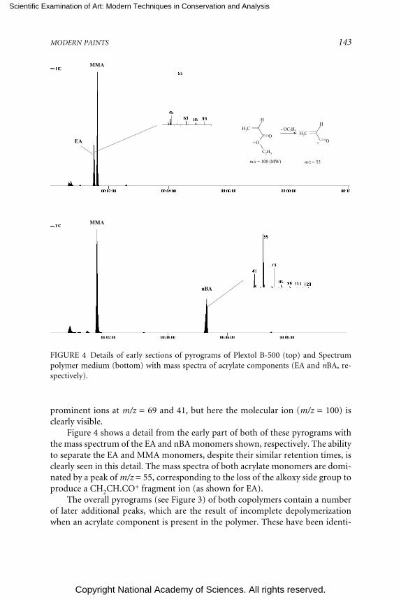
Scientific Examination of Art: Modern Techniques in Conservation and Analysis
Copyright National Academy of Sciences. All rights reserved.
MODERN PAINTS 143
prominent ions at m/z = 69 and 41, but here the molecular ion (m/z = 100) isclearly visible.
Figure 4 shows a detail from the early part of both of these pyrograms withthe mass spectrum of the EA and nBA monomers shown, respectively. The abilityto separate the EA and MMA monomers, despite their similar retention times, isclearly seen in this detail. The mass spectra of both acrylate monomers are domi-nated by a peak of m/z = 55, corresponding to the loss of the alkoxy side group toproduce a CH2CH.CO+ fragment ion (as shown for EA).
The overall pyrograms (see Figure 3) of both copolymers contain a numberof later additional peaks, which are the result of incomplete depolymerizationwhen an acrylate component is present in the polymer. These have been identi-
- OC2H5
++
m/z = 55m/z = 100 (MW)
CH2
H
O
O
C2H5
CH2
H
O
MMA
MMA
nBA
EA
FIGURE 4 Details of early sections of pyrograms of Plextol B-500 (top) and Spectrumpolymer medium (bottom) with mass spectra of acrylate components (EA and nBA, re-spectively).

Scientific Examination of Art: Modern Techniques in Conservation and Analysis
Copyright National Academy of Sciences. All rights reserved.
144 SCIENTIFIC EXAMINATION OF ART
fied as a series of sesquimers, dimers, and trimers of acrylate or acrylate-MMAcombinations. Sesquimer is a term meaning 1.5 monomer units (i.e., a moleculeconsisting of a three-carbon atom backbone with an acrylate/methacrylate groupat either end).
Some materials labeled acrylic emulsions can actually be copolymers withother monomers, such as styrene (to create styrene-acrylics) or vinyl acetate (tocreate vinyl-acrylics). Although not shown here, both are readily distinguished bypyrolysis-gas chromatography-mass spectrometry (PyGCMS), by the detection ofstyrene monomer and acetic acid (see below), respectively.
Polyvinyl Acetate (PVA) Emulsions
On pyrolysis, PVA emulsion paints produce principally ethanoic (acetic)acid and benzene by a side group elimination mechanism. Figure 5 shows theoverall pyrogram observed from Emultex VV536 (Harco), with the mass spec-trum from the intense peak at the start of the pyrogram. This spectrum is mainlythat of ethanoic acid (with a molecular ion of m/z = 60, and strong fragment ionsat m/z = 43, 45), although the peak of m/z = 78 corresponds to the molecular ionof benzene.
It is usually possible to confirm the presence of an emulsion form of PVA (asopposed to a solution form) by the detection of a plasticizer, since pure PVA isslightly too hard to form a continuous film from an emulsion.
This particular emulsion, Emultex VV536 (Harco), actually contains both avinyl versatate resin and a phthalate plasticizer (in the majority of PVA emulsionsonly one kind is used). The sharp peak at the far right of the pyrogram is identi-fied as DBP, whose mass spectrum (also shown) has a very intense fragment ionof m/z = 149, which is the characteristic fragment ion of all dialkyl phthalates. Inthe center of the pyrogram is the band of rather broad peaks produced by theVeoVa plasticizer. Although these peaks are not fully resolved, the overall peakpattern does conform to a very distinctive profile.
Alkyds
For alkyd paints based on ortho-phthalic acid, phthalic anhydride is the prin-cipal peak detected on pyrolysis and therefore used as the diagnostic peak. Figure5 shows the pyrogram of 75045 alkyd resin (Croda), a typical ortho-phthalic alkydresin, with the mass spectrum of the very dominant phthalic anhydride peak onthe left. The molecular ion (m/z = 148) is clearly seen, with the most intense peakat m/z = 104 produced from the loss of CO2. The mass spectrum on the right isfrom palmitic acid (with a molecular ion clearly visible at m/z = 256), normallythe most intense fatty acid observed from a dried oil component. The suspectedmechanism of phthalic anhydride liberation from the alkyd’s polyester structureis as follows:

Scientific Examination of Art: Modern Techniques in Conservation and Analysis
Copyright National Academy of Sciences. All rights reserved.
MODERN PAINTS 145
acetic acid+ benzene
DBPVeoVa
phthalicanhydride
palmitic acid
FIGURE 5 Top: Pyrogram of Emultex VV536, a PVA emulsion with mass spectrum ofethanoic acid/benzene (left) and dibutylphthalate plasticizer (right). Bottom: Pyrogram ofCroda 75045 alkyd resin with mass spectrum of phthalic anhydride (left) and palmiticacid (right).
+
O
O
O
O O
O
OO
More recently work has been carried out to assess the advantages of carryingout an in situ methylation step at the time of pyrolysis, which appears to give aquantitative method of analysis (Cappitelli et al., 2002). This is being investigatedto see whether oil type can be obtained reliably.

Scientific Examination of Art: Modern Techniques in Conservation and Analysis
Copyright National Academy of Sciences. All rights reserved.
146 SCIENTIFIC EXAMINATION OF ART
FOURIER TRANSFORM INFRARED SPECTROSCOPY
There are many ways of introducing a sample to a Fourier transform infraredspectroscopy (FTIR) instrument. The main technique currently employed at Tateis to compress the sample in a diamond cell and make the measurement throughan infrared microscope, although a beam condenser seems to give equally goodresults (Learner, 1995b).
Although spectra obtained from a diamond cell are arguably inferior to thosefrom a KBr disc, the use of the diamond cell has three major advantages. First, thetechnique is nondestructive, which permits the sample to be retrieved and thenreanalyzed by a complementary technique. Second, there is no sample prepara-tion necessary. The diamond cell simply compresses the sample to a sufficientlyreduced thickness for reliable transmission spectra to be obtained. Use of a dia-mond cell can be problematic with hard and brittle materials, but fortunately themajority of twentieth-century paints are fairly soft materials, which permit easycompression in the cell. Third, this soft nature of most synthetic polymers used inpaints (in particular, the acrylics) makes grinding them into KBr powder verydifficult. The main disadvantage of the diamond cell is the possibility of pressureeffects on the spectrum, although the actual pressures used in the diamond cellare not thought to be particularly high.
FTIR is an excellent way of obtaining information quickly about the basicchemical class of a binding material. For homogeneous samples, such as certainsynthetic varnishes, this is relatively straightforward. Figure 6 shows the FTIR
FIGURE 6 FTIR spectra of unpigmented media. From top to bottom: acrylic solution(pink line), pEA/MMA-type acrylic emulsion (black line), PVA emulsion (blue line), alkydresin (green line), and nitro-cellulose (red line).
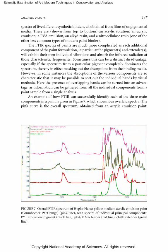
Scientific Examination of Art: Modern Techniques in Conservation and Analysis
Copyright National Academy of Sciences. All rights reserved.
MODERN PAINTS 147
spectra of five different synthetic binders, all obtained from films of unpigmentedmedia. These are (shown from top to bottom) an acrylic solution, an acrylicemulsion, a PVA emulsion, an alkyd resin, and a nitrocellulose resin (one of theother less common types of modern paint binder).
The FTIR spectra of paints are much more complicated as each additionalcomponent of the paint formulation, in particular the pigment(s) and extender(s),will exhibit their own individual vibrations and absorb the infrared radiation atthose characteristic frequencies. Sometimes this can be a distinct disadvantage,especially if the spectrum from a particular pigment completely dominates thespectrum, thereby in effect masking out the absorptions from the binding media.However, in some instances the absorptions of the various components are socharacteristic that it may be possible to sort out the individual bands by visualmethods. Here the presence of overlapping bands can be turned into an advan-tage, as information can be gathered from all the individual components from apaint sample from a single analysis.
An example of how FTIR can successfully identify each of the three maincomponents in a paint is given in Figure 7, which shows four overlaid spectra. Thepink curve is the overall spectrum, obtained from an acrylic emulsion paint:
FIGURE 7 Overall FTIR spectrum of Hyplar Hansa yellow medium acrylic emulsion paint(Grumbacher 1994 range) (pink line), with spectra of individual principal components:PY1 azo yellow pigment (black line), pEA/MMA binder (red line), chalk extender (greenline).

Scientific Examination of Art: Modern Techniques in Conservation and Analysis
Copyright National Academy of Sciences. All rights reserved.
148 SCIENTIFIC EXAMINATION OF ART
Hansa yellow medium artists’ acrylic color (Grumbacher). The other three curvesare reference curves taken from each individual constituents.
• Red curve: The frequencies of the C-H stretching bands at 2986 cm–1 and2955 cm–1, the overall profile of the C-H stretching region, C=O stretching at1732 cm–1, and skeletal vibrations at 1179 cm–1 are all indicative of a p(EA/MMA)acrylic emulsion.
• Grey curve: The sharp peaks seen at 1667 cm–1, 1602 cm–1, 1562 cm–1,1508 cm–1, 1296 cm–1, 1140 cm–1, 953 cm–1, and 774 cm–1 are all present as strongabsorptions in the spectrum of the pigment PY1, one of the common organicmonoazo yellow pigments. The profile of absorptions in the region between 3000cm–1 and 3300 cm–1, with peaks at 3098 cm–1, 3145 cm–1, 3181 cm–1, and 3243cm–1 is also highly diagnostic of pigment PY1.
• Green curve: the two absorptions at 2520 cm–1 and 1799 cm–1 are immedi-ately indicative of the presence of chalk (calcium carbonate). Although relativelyweak absorptions, these two wave numbers are normally found to be completelyseparated from the absorptions from all the binding media, pigments, and otherextenders. The two very sharp peaks at 877 cm–1 and 713 cm–1 confirm the pres-ence of chalk as extender and the strong and very broad absorption between 1400cm–1 and 1500 cm–1 is also clearly visible.
Recently, attenuated total reflectance (ATR), a reflective mode of FTIR, hasproved useful at identifying the differences at the surface of a paint film, com-pared with its bulk properties (as measured in transmission mode). This mode isshowing great potential for following chemical surface changes on a paint filmwith age and after certain conservation treatments, such as cleaning. Figure 8shows three stacked spectra measured with ATR. The top spectrum is from theupper surface of an unpigmented acrylic medium (Golden) that has been cast ona glass slide and been left for approximately five years. The middle spectrum isfrom the lower surface of the same sample, after removal from the slide. There areclear differences between the two. The peak assignments on each of these spectrathat are placed to the left of the relevant peak are characteristic of a p(EA/MMA)acrylic emulsion. All peak labels that are placed to the right of their peaks areindicative of polyethylene glycol, a common class of surfactant. In this examplethe PEG has gathered at the upper surface of the paint film, a phenomenon thatcould have significant ramifications for a painting’s appearance and its changewith age and/or cleaning.
CONCLUSIONS AND LOOKING AHEAD
It is possible to identify, characterize, and differentiate the principal classes ofsynthetic binders used in modern paints with a combination of pyrolysis-gaschromatography-mass spectrometry (PyGCMS) and Fourier transform infrared

Scientific Examination of Art: Modern Techniques in Conservation and Analysis
Copyright National Academy of Sciences. All rights reserved.
MODERN PAINTS 149
1107
1149
1343 2952
1149
1725
2952
961
1107
1342
acrylic medium frontacrylic medium rearpoly(ethylene glycol) standard
-1.4
-1.2
-1.0
-0.8
-0.6
-0.4
-0.2
0.0
0.2
0.4
0.6
0.8
Abs
orba
nce
1000 2000 2000 3000 4000Wavenumbers (cm-1)
1725
961
2890
2891
FIGURE 8 FTIR-ATR spectrum of unpigmented pEA/MMA acrylic medium (Golden1993 range): upper surface (red line), lower surface (after removal from glass slide sup-port) (black line), and polyethylene glycol (PEG) reference spectrum (blue line).
spectroscopy (FTIR). However, there still remain a great many analytical needsfor modern paints, including
• improved quantitative methods of medium analysis;• analytical techniques for organic pigments and the additives added to
paint formulations;• surface analysis methods for chemical, physical, and optical changes on
aging and conservation treatments; and• high spatial resolution techniques to analyze individual layers from lay-
ered paint structures.
REFERENCES
Cappitelli, F., T. Learner, and O. Chiantore. 2002. An initial assessment of thermally assisted hydroly-sis and methylation—gas chromatography/mass spectrometry for the identification of oils fromdried paint films. Journal of Analytical and Applied Pyrolysis 63:339-348.
Challinor, J. 1983. Forensic applications of pyrolysis gas chromatography. Forensic Science Interna-tional 21:269-285.
Crook, J., and T. Learner. 2000. The Impact of Modern Paints. London: Tate Gallery Publishing.Irwin, W. 1979. Analytical pyrolysis—an overview. Journal of Analytical and Applied Pyrolysis 1:3-25.
Scientific Examination of Art: Modern Techniques in Conservation and Analysis
Copyright National Academy of Sciences. All rights reserved.
150 SCIENTIFIC EXAMINATION OF ART
Jain, N., C. Fontain, and P. Kirk. 1965. Identification of paints by pyrolysis gas chromatography.Journal of the Forensic Science Society 5:102-109.
Learner, T. 1995a. The analysis of synthetic resins found in twentieth century paint media. In ResinsAncient and Modern, eds. M. Wright and J. Townsend, pp. 76-84. Edinburgh: Scottish Societyfor Conservation and Restoration.
Learner, T. 1995b. The use of a diamond cell for the FTIR characterisation of paints and varnishesavailable to twentieth century artists. Postprints: IRUG2 Meeting, pp. 7-20, available at http://www.irug.org/documents/1Learner.pdf .
Learner, T. 1996. The use of FTIR in the conservation of twentieth century paintings. SpectroscopyEurope 8(4):14-19.
Learner, T. 2000. A review of synthetic binding media in twentieth century paints. The Conservator24:96-103.
Learner, T. 2001. The analysis of synthetic paints by pyrolysis-gas chromatography-mass spectrom-etry (PyGCMS). Studies in Conservation 46:225-241.
Learner, T., O. Chiantore, and D. Scalarone. 2002a. Ageing studies of acrylic emulsion paints. Pre-prints of the 13th Triennial meeting of the ICOM Committee for Conservation, Rio de Janeiro, pp.911-919. London: James and James.
Learner, T., M. Schilling, and R. de la Rie. 2002b. Modern paints: A new collaborative researchproject. Conservation. The Getty Conservation Institute Newsletter 17(3):18-20, available at http://www.getty.edu/conservation/resources/newsletter/17_3/news_in_cons1.html.
Martens, C. 1981. Waterborne Coatings. New York: Van Nostrand Reinhold.Slinckx, M., and H. Scholten. 1994. Veova9/(meth)acrylates, a new class of emulsion copolymers.
Journal of the Oil and Colour Chemists’ Association 77:107-112.Sonoda, N. 1998. Application des méthodes chromatographiques a la caractérisation des peintures
alkydes pour artistes. Techne 8:33-43.Sonoda, N., and J.-P. Rioux. 1990. Identification des matériaux synthétiques dans les peintures
modernes. 1. Vernis et liants polymères. Studies in Conservation 35:189-204.Stringari, C., and E. Pratt. 1991. The identification and characterization of acrylic emulsion paint
media. In Saving the 20th Century: The Conservation of Modern Materials, ed. D. Grattan, pp.411-439. Ottawa: Canadian Conservation Institute.
Wheals, B. 1985. The practical application of pyrolytic methods in forensic science during the lastdecade. Journal of Analytical and Applied Pyrolysis 8:503-514.
APPENDIXEXPERIMENTAL CONDITIONS
Pyrolysis-Gas Chromatography-Mass Spectrometry
FOM-4LX Curie point pyrolysis unit mounted directly onto the injectionport of a Hewlett-Packard 5890 gas chromatograph and interfaced to a FinniganMAT Incos 50 quadrupole mass spectrometer. Pyrolysis conditions: 610°C foreight seconds. Pyrolysis chamber kept at 200°C. GC injection port kept at 180°C.BPX-5 (SGE) nonpolar column: 25 meters long, 0.32 µm internal diameter and0.1 µm film thickness. Temperature program: 40°C held for two minutes, thenramped at 10°C/min–1 to 350°C and held for two minutes. The transfer line waskept at 250°C. Incos 50 utilized EI ionization at 70 eV, and scanned from mass 35-500 every second.

Scientific Examination of Art: Modern Techniques in Conservation and Analysis
Copyright National Academy of Sciences. All rights reserved.
MODERN PAINTS 151
Fourier Transform Infrared Spectroscopy
Transmission work carried out on a Nicolet Avatar 360 instrument withSpectraTech IR Plan microscope. Sample held in a diamond cell and 128 scanswere averaged at 4 cm–1 resolution. ATR work carried out on a Nicolet Magna IR560 instrument and Nicolet Nic Plan IR microscope with a Spectra-Tech ATRobjective with zinc selenide crystal and purged with dry air. Two hundred scanswere averaged at 4 cm–1 resolution.
ACKNOWLEDGEMENTS
This work was made possible by the support of the Tate Gallery and theLeverhulme Trust, and the generosity of the FOM Institute (which loaned thePyGCMS instrument). The FTIR was purchased with a grant from theClothworkers’ Foundation in London. Herant Khanjian at the Getty Conserva-tion Institute carried out the ATR measurements during the author’s guest schol-arship there in 2001. The author is extremely grateful to all those involved.

Scientific Examination of Art: Modern Techniques in Conservation and Analysis
Copyright National Academy of Sciences. All rights reserved.
152
Material and Method in Modern Art:A Collaborative Challenge
Carol Mancusi-UngaroAssociate Director of Conservation and Research
Whitney Museum of American ArtDirector, Center for the Technical Study of Modern Art
Harvard Univesity Art Museums
Recently I reacquainted myself with an illuminating interview of Jasper Johns byone of the more important interviewers of American artists in our time, DavidSylvester. I knew the interview well. However, this particular rereading occurredas I was reexamining in a more thoughtful way Johns’s encaustic works in theWhitney Museum of American Art, including Three Flags (1958), White Target(1957), and Double White Map (1965). Despite my familiarity with the discussion,there was something that the artist said in the 1965 interview that gave me pausein 2003 and forced me to reconsider the nature of our collective professionalcharge to elucidate and care for works of art.
In addressing the now famous paintings of flags and letters, Sylvester askedJohns about the objects with which he begins. Johns, seeking clarification, asked,“The empty canvas?” “No,” replied Sylvester, “Not only the empty canvas: well,the motif, if you like, such as the letters, the Flag and so on, or whatever it maybe.” Johns said soberly, “I think it’s just a way of beginning.” Clearly surprised,Sylvester persisted, “In other words the painting is not about the elements withwhich you have begun.” Johns explained, “No more than it is about the elementswhich enter it at any moment. Say, the painting of a flag is always about a flag, butit is no more about a flag than it is about a brush-stroke or about a colour orabout the physicality of the paint, I think.” Struck by the candor of this artist, whohad obviously given enormous thought to the role of materiality in his art, Ieagerly awaited clarification of his view, which came a few sentences later. Heexplained, “What I think this means is that, say in a painting, the processes in-volved in the painting are of greater certainty and of, I believe, greater meaningthan the referential aspects of the painting. I think the processes involved in the

Scientific Examination of Art: Modern Techniques in Conservation and Analysis
Copyright National Academy of Sciences. All rights reserved.
MATERIAL AND METHOD IN MODERN ART 153
painting in themselves mean as much or more than any reference value that thepainting has.” “And what would their meaning be?” asks Sylvester. “Visual, intel-lectual activity, perhaps recreation,” answers Johns.1
Is Johns saying that the making of the work of art is its most relevant aspect?If so, does that mean that we can understand the painting only if we elucidate theprocess? Is the artist by implication suggesting that a conservator and a museumscientist must readily be at hand with explanations in order for a viewer to com-prehend fully a work of art? As tantalizing as we may find that proposition, Ibelieve it oversimplifies what the artist had in mind. Indeed, the cited passage mayelicit diverse interpretations of Johns’s view, and his attitude may not necessarilybe shared by other artists. In our context Johns’s comments focus attention on arelevant distinction that shapes the way we think about art. Clarifying histhoughts, the artist explained further, “And I think the experience of looking at apainting is different from the experience of planning a painting or of painting apainting. And I think the statements one makes about finished work are differentfrom the statements one can make about the experience of making it.”2
Conservators, museum scientists, and art historians usually come upon thefinished work of art. Although some of us may occasionally be a part of themaking, generally our roles crystallize once the work of art is complete. AfterPollock puts down his stick, Rothko retires his brush, Newman takes off hispainting hat, and Johns exhausts his interest, the work of art moves away from themaker and into a realm of the viewer. As researchers interested in how substanceand process affect the visual statement, conservators seek to enrich the aestheticexperience through elucidation of the process, while art historians consideringprimarily what is seen may posit and assess that information in a cultural context.The third collaborative component is the museum scientist who may not onlyaffirm the nature of materials present but through analytic review may alsoreconfigure historical perspective. From different points of reference that shapedifferent types of statement, as predicted by Johns, each inevitably seeks to resolvethe visual and intellectual activity of the process insofar as it affects the meaningof the work of art.
The conservation of the Rothko Chapel paintings, which engaged over 20years of my professional life, provides a personal case in point. Created between1964 and 1967 by Mark Rothko for a chapel he designed in Houston, Texas, thepredominantly black and plum, so-called black-form, paintings had begun todevelop a whitening on their surfaces less than five years after their installation in1971. Over time the whitish films developed into crystals that gathered into dis-
1D. Sylvester. Interview. Jasper Johns Drawings (London: Arts Council of Great Britain, 1974), pp.13-14.
2Ibid., p. 18.

Scientific Examination of Art: Modern Techniques in Conservation and Analysis
Copyright National Academy of Sciences. All rights reserved.
154 SCIENTIFIC EXAMINATION OF ART
tinct patterns on the surfaces of the paintings. The patterns interfered with theunified, monochromatic nature of the paintings and certainly bore no relation tothe final scheme for the chapel as determined by art scholars and as presumablyintended by Rothko. For this reason conservators and conservation scientistswere called upon to treat what was widely considered an inexplicable conditionproblem.
In the literature, art historians and critics had focused on the dark paletteand sharp contours of the chapel paintings as opposed to the bright amorphouscoloration of his earlier paintings and even his earlier commissions, namely theSeagram murals of 1958-1959 and the Harvard panels of 1962. Although a reso-nance certainly existed among the seven black-form paintings and the sevenplum paintings that comprised the whole of the chapel, no mention was made inthe literature of the facture of the paint or the particular physical properties thatshaped it. Rothko, who died before the chapel was consecrated, had been inter-viewed throughout his career, but none of the discussion focused on the materi-ality of these enigmatic paintings.
My investigation began with the customary conservator’s question of howthe paintings were made. To that end we sought and located one of Rothko’sassistants for the project, and he and I painted out simulations, using the samematerials and processes that the artist had employed. Although our simulationscertainly did not recall the originals, the material effect was close enough to con-firm the ingredients of the mixture as whole eggs, tube oil paint, damar resin, andturpentine. Through analysis of the whitening conducted by local scientists at theShell Oil Company, we were able to attribute the exudate to the migration of fattyacids from the paint, and ultimately we devised a treatment that enabled its re-moval. The strange patterns of rectangles formed by the exudate were explainedby differential amounts of egg in a day’s mixture or by the buildup of media inconsecutively layered forms. Ultimately, working drawings provided by theRothko Foundation offered an astonishing correspondence to the patterns ofwhitening and thereby confirmed the relationship between the development ofthe condition and the unfolding of the creative act.
What had begun as a conservator’s customary question of “what” were welooking at ended up providing insight into Rothko’s creative process and anexplanation of “why” he chose to employ the particular materials that he did. Insum, the technical study offered information that had much broader ramifica-tions for the history of the art. For example, a thoughtful appraisal of the draw-ings in graphite on black paper indicated that they were more than recordings ofprocess. Throughout, what distinguished the line was not color but reflection.Indeed, in certain light the drawing was hardly visible. Rothko could have usedwhite chalk instead of graphite on the black paper, but he did not. Rather, by hischoice of materials the artist acknowledged that differences in reflectivity could beas legible as differences in contrast. Thought of in this way the studies fortifiedour developing notion of Rothko’s keen regard for nuances of surface as docu-

Scientific Examination of Art: Modern Techniques in Conservation and Analysis
Copyright National Academy of Sciences. All rights reserved.
MATERIAL AND METHOD IN MODERN ART 155
mented also by the variable reflectance of the plum borders and black forms.3
Reference to his earlier work confirmed that the artist had been engaged withthese issues throughout his career, but had brought them to fruition in part byeliminating vivid color in the chapel’s paintings. The technical analysis had in-formed the process that in turn directed the treatment and affected our regard forRothko’s later work.
Although statements about the experience of making art differ from state-ments about the finished work, they should inform each other. In the world ofmodern art this discourse could begin with closer scrutiny of its most commondescriptive rubric, namely, “mixed media.” This term, which abounds not onlyon museum label copy but also in catalogues, is as familiar to postwar art scholarsas “oil on canvas” is to those who study old-master painting. Walter Hopps,founding director of the Menil Collection, predicted the emergence one day of, inhis terms, a “mixed media morass.”4 That era has arrived, but in some ways it isnot a new phenomenon, considering that “oil on canvas” is the official descrip-tion of Sir Joshua Reynolds’s Captain Robert Orne (1756), as well as Willem deKooning’s Door to the River (1960). Given the visual range of these works of artand the investigative capability of our technological age, one overriding descrip-tive term seems woefully inadequate for both old-master painting and modernart. In a recent interview Wayne Thiebaud mentioned that he added Zec, a brandname for a substance that added girth to his oil paint, in order to create thecreamy “icings” on his cakes. Willem de Kooning apparently did not add Zec, butwe know from technical investigations that he did add vegetable oils to his mediain order to achieve carefully sought-after working properties and effects that wevalue in his paintings.5
It is encouraging that recent studies of paintings by Pollock, de Kooning,Jacob Lawrence and Mark Rothko, among others, have identified materials byscientific analysis in the context of technique and have thereby broadened ourunderstanding. Such analysis has also debunked prevailing myths, such as deKooning’s alleged use of mayonnaise in his paint, and undoubtedly will substan-tiate others. These exemplary studies have also offered insight into the intellectualactivity of the artist and that contribution about the making has broadened ourunderstanding of the seen. Once that information becomes an integral part ofhow a work of art is discussed in the literature at large, we may begin to confront
3C. Mancusi-Ungaro. Nuances of surface in the Rothko chapel paintings. Mark Rothko: The ChapelCommission (Houston: The Menil Collection, 1996), pp. 27-28.
4Private conversation with the author. June 1988.5S. Lake. The challenge of preserving modern art: a technical investigation of paints used in se-
lected works by Willem de Kooning and Jackson Pollock. MRS Bulletin, January 2001, Volume 26.pp. 56-60.

Scientific Examination of Art: Modern Techniques in Conservation and Analysis
Copyright National Academy of Sciences. All rights reserved.
156 SCIENTIFIC EXAMINATION OF ART
the disorder of the “mixed media morass” that confounds our scholarship. Theimportance of analytic review in this process cannot be overestimated.
Generally, technical questions about modern art cannot be framed in thecontext of what we know about practice from artists’ treatises or the precepts of aguild system. Rather, they are often shaped by anecdotal information provided byartists or their assistants. Inevitably we have had to rely on art historical precedentand our eyes to assess the credibility of the information. That is not a bad combi-nation, but it is not enough at a time when analytic confirmation is possible.Precise technical information may surface, but depending upon the source andcontext, its certainty may not be assured.
In 1982 I wrote a short technical note about Yves Klein’s materials in acatalogue for a posthumous retrospective exhibition of the artist’s work. I basedmy information on interviews conducted in Paris with Klein’s former associatesand on a patent that the artist had secured in 1960 for “International Klein Blue,”his preferred painting medium. The mixture consisted of dry pigments in polyvi-nyl acetate and industrial solvents, formulated by Rhône-Poulenc. Klein’s de-scription of the medium in the patent actually differed from that provided by thecompany and seemed incompatible with the working properties necessary for itssundry applications. Nonetheless, the reason for my note was not to draw atten-tion to a possible error in Klein’s application for a patent but rather to try todescribe how his choice of material permitted widely diverse processes. Despitemy conclusion that “though quantifiable, this quintessence of unencumberedcolor owes its vitality and beauty to the magic of the artistic endeavor—a factorthat can never be measured or duplicated”—there arose a concern that I haddemystified Klein’s art by describing its making.6
Considering that allegation with regard to our work, I am reminded of anesteemed engineer, Peter Rice, who once spoke about the role of Iago in Othello.He said, “Iago, if you remember, destroys the love of Othello and Desdemona byrational argument, by applying reason all the way through to every act which,particularly, Desdemona undertakes. And in the eyes of many, the Iago role is therole given to the engineer in modern life and in modern architecture of actuallyreducing by reason, to destroy or to undermine the kind of unreasonable andsoaring ideas that architects may have.”7 I suspect the same could be said ofmuseum scientists who decipher the material ambiguity of works of art. Admit-tedly the danger is there when the scientist is given a chip of paint in isolation andis asked to identify it. Out of context, devoid of its visual significance not tomention its role in artistic creation, the sample may be reduced to a fact that maynot undermine the “soaring ideas” but certainly does little to enhance them.
6C. C. Mancusi-Ungaro. A technical note on IKB. Yves Klein (Houston: Institute for the Arts, RiceUniversity, 1982), pp. 258-259.
7P. Rice. RIBA Royal Gold Medal speech. Arup Journal (winter 1992/1993), p. 20.

Scientific Examination of Art: Modern Techniques in Conservation and Analysis
Copyright National Academy of Sciences. All rights reserved.
MATERIAL AND METHOD IN MODERN ART 157
Providing the data is one thing but explaining it in context is quite another.Cross-sections certainly help explicate the process in specific areas, and simula-tions can inform the overall technique; however, anyone who has observed anartist looking at a cross-section or tried to make a simulation of a work of artknows that deconstruction of substance and process is informative but far fromart. There is the intangible element of the artist’s intent in manipulating tangiblematerial that must be considered. Although associates or even studio assistants ofan artist may not comprehend this factor, artists invariably do, because theyappreciate the complexity of their undertaking.
In part because we share a natural affinity for how substance and processaffect the visual statement, I began interviewing artists in front of their work over12 years ago. I intentionally chose an open interview style that was captured onfilm because in the presence of the works of art, artists invariably reveal throughapproach and reaction their relationships to the materials. Alternatively, muse-ums often ask artists to complete questionnaires about technique when a work ofart is acquired. If returned, these forms can impart important information. Whenasked about the materials she used in For the Light (1978-1979), for instance,Susan Rothenberg carefully listed the various media she had employed: Liquitexgesso-ground, Liquitex Matte Medium, and LeFranc and Bourgeois Flashe (vinylpaint made in France). She further noted, “All 3 used in conjunction with mattemedium for both gesso + flashe.”8 When asked on the following page about thesubject of the work, the ideas expressed and the circumstances under which it wasexecuted, she replied with an emphatically drawn explanation point and questionmark. The point is, of course, as Johns postulated years earlier, there is greatercertainty about the processes than about the referential aspects. It is not so mucha question of relative importance as it is of relative surety.
From the outset it was clear to me that my questions would inevitably reflectthe concerns of my own time and might therefore not provide answers to theproblems that might confront future conservators. What I had hoped to docu-ment was not merely a discussion of materials and technique but, more than that,a solid sense of the artists’ concerns about what they were looking at and its futurepreservation. Naturally, artists’ relationships to their materials and thoughts aboutthe future care of the art are as varied as their personalities. For instance, JamesRosenquist may be concerned about the sinking in of his oil medium over time,while Brice Marden worries more about the proper treatment of a localized dam-age to one of his monochromatic works. The artists’ concerns may be narrow orbroad in scope. Yet, inevitably their involvement adds another dimension to theinvestigation by posing questions unimagined by researchers and thereby enrich-ing the pursuit in unexpected ways.
8Questionnaire statement by S. Rothenberg. Whitney Museum of American Art archive. July 1,1979.

Scientific Examination of Art: Modern Techniques in Conservation and Analysis
Copyright National Academy of Sciences. All rights reserved.
158 SCIENTIFIC EXAMINATION OF ART
After the Menil Collection acquired Ed Kienholz’s John Doe (1959), theartist came to the museum to discuss the piece. Having carefully surveyed thesurface of the sculpture, he opened the drawer and removed a portion of flutepipe from within. I had noted powdery debris in the bottom of the drawer, but Ihad not initially associated it with the industrial ductwork (Figure 1). EventuallyKienholz explained that the flute pipe represented the mannequin’s male privatepart and that the dust in the drawer was what remained of its head that had beenfashioned from a rubber mask. Evidently the original Halloween mask had to-tally disintegrated over 30 years, but without the artist’s intervention I doubt wewould have known about the change. Archival photographs of John Doe hadunfortunately only documented the sculpture with the contents of the drawerlocked within. Obligingly Kienholz took the appendage with him to Los Angelesand returned months later with a completed part fashioned out of a new rubbermask (Figure 2).
This albeit extreme example raises two issues concerning preservation. First,instances of disintegrating industrial materials unfortunately comprise many im-portant works of modern art, among them John Chamberlain’s foam sculpturesof the 1970s. The challenge to preserve the physicality of these objects is enor-mous. Since the unstable material is central to the works of art and the sculpturescannot be properly viewed encased, it seems the only reasonable course is torestrict periods of exhibition as well as to require proactive storage containment.In this scenario more rigorous research might focus on the object in storage sothat storage rooms become de facto laboratories wherein technical solutions areexecuted without regard for exhibition parameters or other customary restric-tions. This approach to a limited degree has been adopted in some institutions,but it should become standard practice.
The second issue concerns the broader philosophical question of what to dowith the sculpture in the future when the current replacement disintegrates. Themore expedient approach, of course, would be to replace the mask yet again, asdid Kienholz. Once the artist has died, however, it is unlikely that anyone wouldbe eager to refashion a new part without the artist’s hand. A conservator and ascientist’s approach might be to make a mold of the current form and then cast itin a more permanent material. An art historian could rightly object to the idea ofa cast form replacing a found object because it counters Kienholz’s notion ofmateriality. One wonders if replacement parts are ever appropriate? I had toconfront that question once when I made a new white wedding dress to replacethe discolored and irreparably stained original on Kienholz’s Jane Doe (1960). Iwas asked to treat the work in this way by a curator because the aged dress offereda tawdry view of marriage that countered the deceased artist’s expressed intent.These two replacements, undertaken as treatments, were couched in terms thatreflect the central importance of the artist’s intent—an elusive concept that defiesquantification yet rests at the heart of our collective pursuit.

Scientific Examination of Art: Modern Techniques in Conservation and Analysis
Copyright National Academy of Sciences. All rights reserved.
MATERIAL AND METHOD IN MODERN ART 159
FIGURE 1. Detail of John Doe, showing powdery remnants of original mask. Source: EdKienholz, John Doe, 1959, The Menil Collection. Photograph taken by Carol Mancusi-Ungaro. Permission for photograph granted by Nancy Reddin Kienholz.

Scientific Examination of Art: Modern Techniques in Conservation and Analysis
Copyright National Academy of Sciences. All rights reserved.
160 SCIENTIFIC EXAMINATION OF ART
FIGURE 2 Detail of restored John Doe showing mask fashioned as male anatomy. Source:Ed Kienholz, John Doe, 1959, The Menil Collection. Photograph taken by Carol Mancusi-Ungaro. Permission for photograph granted by Nancy Reddin Kienholz.

Scientific Examination of Art: Modern Techniques in Conservation and Analysis
Copyright National Academy of Sciences. All rights reserved.
MATERIAL AND METHOD IN MODERN ART 161
When the artist is still alive, the complicated but key questions regardingintent are often more readily identified. A recent exchange involving two sculp-tures of a man and a woman by Kiki Smith at the Whitney Museum of AmericanArt comes to mind. Dated 1990, they had been intermittently on exhibition forover a decade but had also spent a fair amount of time in storage. Recent observa-tion revealed patterns of white crystals in the beeswax that seemed to mimic thebattens that held the pieces secure in their crates. Upon close examination thedisfigurement and its probable cause were obvious to the scientist who sampledthe material and the conservator who had begun to consider treatment options.In an impromptu interview the artist offered a totally unexpected assessment ofthe objects’ physical state. It seems what disturbed her most was not the exudatethat commanded our attention, which she summarily dismissed, but rather areddened pallor that had overcome the male figure. In her view the red wax thatunderlay the uppermost visible layer and had been used to offer skin tone hadsomehow become dominant. That condition problem, which had eluded us, faroutweighed any other in terms of importance to her. By affirming the certainty ofprocess, which Johns had observed, Smith not only left us with a better under-standing of the nature of the problem but also of the work of art itself. Withouther intervention would the fundamental alteration in the material have evenelicited a question from us?
Thoughts about the artist’s intent affect what conservators do with the factsthat museum scientists uncover. A discussion of how the material is used and towhat artistic end is as important as, if not more important than, what the materialis. Analytic investigation is crucial, as are other types of review that take intoaccount art history, criticism, and connoisseurship. All play a part in affirmingartistic intent, especially after the artist has died. From their particular perspectivescientists offer valuable insight in this debate by evaluating statements about thefinished work in light of statements about the experience of making. Beyondnaming the material and thinking logically in terms of questions and answers,they bring to the discussion diverse patterns of thinking. That contribution affectsthe tensions between “reason and intuition, certainty and uncertainty, delibera-tion and spontaneity,” the precise qualities that shape our reasoned comprehen-sion of the illogical artifacts of human expression in our care.9
It is only through intense collaboration among the distinct but related disci-plines that consider works of art that we can attempt to frame and pose therelevant technical questions. By digesting the experience of looking as well as theexperience of making, we can assign meaning to the chips of paint that are ana-lyzed, offer definition to the “mixed media morass,” and discern the artist’s intentas it relates to materiality. Only in collaboration can we begin to offer the indeter-minate work of art the rigorous yet insightful review it deserves.
9A. Lightman. Art that transfigures science. New York Times, March 15, 2003, p. B9-10.

Scientific Examination of Art: Modern Techniques in Conservation and Analysis
Copyright National Academy of Sciences. All rights reserved.
162
Raman Microscopy in the Identification ofPigments on Manuscripts and
Other Artwork
Robin J. H. ClarkChristopher Ingold Laboratories
University College LondonLondon
The identification of pigments on manuscripts, paintings, enamels, ceramics,icons, polychromes, and papyri is critical in finding solutions to problems ofrestoration, conservation, dating, and authentication in artwork, and many tech-niques (molecular and elemental) have been used for this purpose. Raman mi-croscopy has emerged, thanks to recent advances in optics and detectors, as per-haps the most suitable of these techniques on account of its high spatial (≤ 1 µm)and spectral (≤ 1 cm–1) resolution, its specificity, its excellent sensitivity by way ofcharge coupled device (CCD) detectors, and the fact that many artifacts may beanalyzed in situ. Long-needed links between the arts and the sciences in this areaare now rapidly being developed.
The Raman effect was first detected in 1928 and rapidly became, and thenremained for the following 20 years, the basis of the key technique for providingvibrational information on molecules in all states of matter, and of ions andlattice structures (Raman and Krishnan, 1928). However, the technique was thenvery slow, as it involved the use of a mercury arc as radiation source and photo-graphic plates for detection. With the introduction in the 1960s of lasers as mono-chromatic polarized light beams of high irradiance and of semiconductors (e.g.,GaAs) as detectors, the ability to detect weak Raman signals from materials of allkinds increased markedly. The technique came to be applied to a wide variety ofchemical problems and increasingly to those amenable to analysis with amicrosampling configuration. In the past 25 years, and especially in the last de-cade, the technique has been increasingly applied with great effect to the analysisof micrometre-sized particles using optical microscopes interfaced with existingspectrometer systems. These developments have revolutionized the usage of the

Scientific Examination of Art: Modern Techniques in Conservation and Analysis
Copyright National Academy of Sciences. All rights reserved.
RAMAN MICROSCOPY IN THE IDENTIFICATION OF PIGMENTS 163
technique, which is seen to have many advantages over infrared (IR) microscopyand other techniques for phase identification.
The intrinsic weakness of the Raman effect (Long, 2002) in the absence ofresonance effects (Clark and Dines, 1986), and the moderate sensitivity of evensemiconductors or multichannel intensified diode arrays as detectors were stillnot ideal features of Raman microscopy for many applications. However, theintroduction of CCD detectors over the past decade has made it feasible to detectand identify even poor Raman scatterers of micrometre dimensions and, bychange of excitation line, even many fluorescent materials. These technical ad-vances have opened up many new areas to which Raman microscopy could makea major contribution, pigment identification being one of these. It is now recog-nized that the technique combines the attributes of high reproducibility and highsensitivity with that of being nondestructive. Moreover, the technique can beapplied in situ, an important consideration for manuscript study. It also has highspatial resolution (≤ 1 µm) and high spectral resolution (≤ 1 cm–1), features thatare of obvious value for establishing the composition of pigment mixtures andeven of binders on works of art.
Conservators and restorers need to be concerned with pigment identificationfor at least four reasons.
1. To decide whether (a) all restoration should be carried out with theoriginal pigment and not with alternatives of similar hue; this is important sincesome alternatives might be liable to react with contiguous pigments with delete-rious visual effects or (b) restoration with a different pigment might be desirableowing to instability of the original one or because it is more desirable to restorewith carefully documented and easily identifiable nonoriginal pigment, possiblymodern.
2. To identify any degradation products of pigments and to suggest possibletreatments whereby degradation processes may be prevented, arrested or reversed.
3. It is increasingly likely that auction houses will be required to assess scien-tifically any works of art that they intend to offer for sale. One way of checking forobvious forgeries is to establish the palette and to check that no pigments ofinappropriate dates of first manufacture or usage are present. Such analysis isextremely important in view of the immense prices—often > $1 million—forwhich medieval manuscripts and codices can currently be sold, usually withoutany scientific validation of the pigments present.
4. It is essential that conclusions on artwork as to the date, school, artist, etc.,be drawn not only on paleographical, philological, and stylistic grounds but alsoon scientific grounds based upon an experimentally established palette. The artscommunity has been in general slow to accept this point.
This article begins with a brief comment on the range of pigments tradition-ally used to illuminate artifacts throughout time, and then moves on to a discus-

Scientific Examination of Art: Modern Techniques in Conservation and Analysis
Copyright National Academy of Sciences. All rights reserved.
164 SCIENTIFIC EXAMINATION OF ART
sion of various case histories in which knowledge of pigments present and of theirdegradation products has proved to be of interest to both the arts and the sciencecommunities.
THE PIGMENTS
The colors of inorganic pigments arise in most cases from ligand field, chargetransfer or intervalence charge transfer transitions, and in the case of metals—commonly for silver or gold—from specular reflectance (Clark, 1995, 2002;Jorgensen, 1962). The depth of color of inorganic pigments is related both to themolar decadic absorption coefficients of electronic transitions in the visible re-gion of the spectrum and to the dimensions of the pigment particles. These di-mensions affect the balance between diffuse reflectance, which is controlled bythe absorption coefficients and bandwidths, and specular reflectance, which iscontrolled by complementary factors (Clark, 1964). Early artists were well awareof these effects in practice, and with a restricted palette could often achieve a widerange of hues by selective control of particle size.
Listings of standard inorganic pigments are given in reviews (Clark, 1995,2002), books (Mayer, 1972; Feller, 1986; Roy, 1993; FitzHugh, 1997; Thompson,1956; Gettens and Stout, 1966; Wehlte, 1975), and proprietary literature. By wayof illustration, the commonly used blue inorganic pigments are listed in Table 1,together with their chemical names, formulas, provenance, and an indication asto the nature of the electronic transitions principally responsible for the bluecolor in each case. Of course, the identification of blue pigments alone would onlybe of restricted value for dating purposes, and so extensive studies of all thepigments on a work of art are essential in order for it to be possible to estimate thedate of production of any particular piece.
Many different organic dyes have been extracted from plants through theages for use on illuminations of all sorts, notably indigo from woad for blue,alizarin from madder for red, weld from the weld plant (related to mignonette)for yellow, crocetin from saffron, and gamboge from gum resin—both also foryellow. In addition, organic dyes were extracted from marine life (e.g., Tyrianpurple [6,6'-dibromoindigo] from mollusks and sepia from cuttlefish); from ani-mal life (e.g., Indian yellow from cow urine); from insects (e.g., carmine fromcochineal or kermes beetles); and others from lichens (Clark, 1995). Since theisolation by W. H. Perkin in 1856 of mauveine, the first synthetic dye, manyhundreds of other organic dyes have been synthesized, and this has greatly ex-tended the nature of the palette to which artists have had access. The main scien-tific concerns in the examination of a work of art are
• to identify each pigment, its crystal structure and, if possible, its place oforigin and to identify the pigment medium;
• to assess whether it is feasible to restore any degradation to the paintwork

Scientific Examination of Art: Modern Techniques in Conservation and Analysis
Copyright National Academy of Sciences. All rights reserved.
RAMAN MICROSCOPY IN THE IDENTIFICATION OF PIGMENTS 165
TABLE 1 Commonly Used Blue Inorganic Pigments
Pigment Chemical Name Formula Datea Transitionb
Azurite Basic copper(II) 2CuCO3. min. LFcarbonate Cu(OH)2
Cerulean blue Cobalt(II) stannate CoO.nSnO2 1821 LF
Chinese blue Barium BaCuSi4O10 ca 480 BC LFcopper(II) silicate
Cobalt blue Cobalt(II)-doped CoO.nAl2O3 ca 1550 LFalumina glass Ming dynasty
Egyptian blue Calcium copper(II) CaCuSi4O10 ca 3100 BC LFsilicate
Fluorite (and Calcium fluoride CaF2 min. Trappedantonozite) (purple) electrons?
Lazurite (from Sodalite + sulfur Na8[Al6Si6O24]Sn min. 1828 CTlapis lazuli) radical anions S3
-, S2-
Manganese blue Barium manganate(V) Ba(MnO4)2 + BaSO4 1907 LFsulfate
Maya blue Palygorskite/ Mg5(Si,Al)8O20 Mayan Mieindigo/nano- (OH)2.8H2O, etc. scatteringc
material
Phthalocyanine Copper(II) Cu(C32H16N8) 1936 π-π*d
blue /Winsor phthalocyanineblue
Posnjakite Basic copper(II) CuSO4. min. LFsulfate 3Cu(OH)2.H2O
Prussian blue Iron(III) Fe4[Fe(CN)6]3. 1704 IVCThexa-cyanoferrate 14-16H2O Fe(II)/
Fe(III)
Smalt Cobalt(II) silicate CoO.nSiO2 Earlier than 1500 LF
Vanadium blue Vanadium(IV)-doped ZrSiO4(V(IV)) 1950? LFzircon
Verdigris Basic copper(II) 2Cu(O2CCH3)2. Corrosion LFacetate Cu(OH)2 product
Vivianite Iron(II,III)- phosphate Fe3P2O8.8H2O min. IVCTFe(II)/Fe(III)
aThe pigment is specified to be a mineral (min.) and/or the date of its first manufacture is listed.bLF = ligand field; CT = charge transfer; IVCT = intervalence charge transfer transition.cThe origin of the color is uncertain. (José-Yacamán M., L. Rendon, J. Arenas, and M. C. Serra
Puche. 1996. Science 273:223-227.)dπ-π* = electric-dipole-allowed charge transfer transition of the phthalocyanine ring system.

Scientific Examination of Art: Modern Techniques in Conservation and Analysis
Copyright National Academy of Sciences. All rights reserved.
166 SCIENTIFIC EXAMINATION OF ART
or fabric, given the nature of the chemical processes likely to be involved;• to consider whether knowledge of the identity of the pigments present on
a work of art would give an indication as to date of production and hence theauthenticity, school, and/or artist; and
• to identify the correct measures needed to preserve a work of art from theeffects of heat, light, and gaseous pollutants, and from contiguous or underlyingpigments, dyes, or inks.
THE RAMAN MICROSCOPE
In a Raman microscope the incident laser beam is brought to a focus by theobjective onto each different pigment grain in turn on the manuscript understudy (see Figure 1). The Raman scattering is collected by the same objective andthen directed by a beamsplitter in the optical path to the monochromator and thedetector. Although the use of a beamsplitter reduces the overall efficiency of thesystem, as does the use of a pinhole as a spatial filter, both devices restrict theamount of unwanted scattered light collected from outside the focus of the laserbeam. The benefit of a pinhole is that it ensures a good confocal arrangement,thereby providing spatial resolution as a function of sample depth.
The overall efficiency of detection of the Raman signal from the older Ramanspectrometers was still relatively poor in the 1980s, because it was based on double
FIGURE 1 Schematic representation of a Raman microscope (Renishaw RM 1000), whichemploys notch filter assemblies and a CCD detector.

Scientific Examination of Art: Modern Techniques in Conservation and Analysis
Copyright National Academy of Sciences. All rights reserved.
RAMAN MICROSCOPY IN THE IDENTIFICATION OF PIGMENTS 167
or triple spectrometers with a large number of optical surfaces and on multichan-nel intensified diode arrays, which are relatively insensitive detectors, albeit betterthan photographic plates. However, two devices have improved matters greatly:two-dimensional CCDs with up to 80 percent quantum efficiency of detectionand holographic notch filters, which are wavelength specific and have the prop-erty of blocking out the unwanted Rayleigh scattering. These filters possess acutoff that readily permits approach to within 50 cm–1 or less of the excitationline. This has the consequence that monochromators with the high dispersionpreviously required to filter out the Rayleigh line can be replaced by a spec-trograph system with a single grating only, leading to much greater efficiency ofthroughput. The introduction of notch filters eliminates the need for separatepinholes in the optical path. The benefits of CCD detector/notch filter spectro-graphs are that (1) low-powered air-cooled lasers can be used, which both reducethe costs of purchase and operation and make the entire system portable (sinceelaborate and fixed water-cooling systems for the lasers are not required); (2) thespectrographs are much lighter and much easier to realign than earlier double ortriple grating systems; and (3) the time required to acquire significant data isgreatly reduced, in some cases even to seconds. Such spectrographs can also beused for remote Raman microscopy in which a probe head assembly both deliversthe excitation beam to the sample and collects the scattered radiation from thesample by means of fiber optics. They may be appropriate for the study of heavyitems that need support from a specially designed cradle, notably large codicesthat cannot fit safely onto the microscope stage, or for archaeological studies ofmurals, cave paintings, etc.
It is now also possible to collect data taken from many different samplepoints on an inhomogeneous surface to produce a Raman spectral map. Themethod involves direct two-dimensional imaging of an inhomogeneous surfaceby analyzing one or more of the Raman bands characteristic of a given compo-nent on the surface. It offers intriguing further opportunities for the study of verysmall (approximately 1 mm2) areas of artwork on stamps, maps, and writing (soas to be able to follow iron gall ink diffusion, paper damage, etc.), and in manyother fields (e.g., that of identification of the precise whereabouts and propor-tions of active ingredients in pharmaceutical tablets).
It is usually desirable to have a wide range of excitation lines (frequency ν0)available in order to search for the most enhanced Raman spectrum, bearing inmind the often opposing effects on the scattering intensity of ν4 (the fourth powerof the frequency of the scattered light), absorption, resonance, and possible pho-tochemical and/or thermal degradation of the sample. Fluorescence from certainmaterials is often best avoided by use of a Nd/YAG (yttrium aluminum garnet)laser operating at 1064 nm, albeit with significant loss in spatial resolution andscattering intensity. By use of equipment such as that described above, libraries ofRaman spectra of common inorganic, mineral and earth pigments, and organicpigments and dyes have been compiled (Griffith, 1987; Bell et al., 1997; Burgio
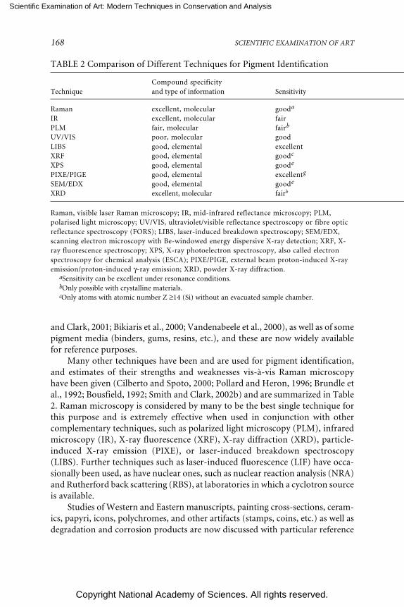
Scientific Examination of Art: Modern Techniques in Conservation and Analysis
Copyright National Academy of Sciences. All rights reserved.
168 SCIENTIFIC EXAMINATION OF ART
TABLE 2 Comparison of Different Techniques for Pigment Identification
Compound specificityTechnique and type of information Sensitivity
Raman excellent, molecular gooda
IR excellent, molecular fairPLM fair, molecular fairb
UV/VIS poor, molecular goodLIBS good, elemental excellentXRF good, elemental goodc
XPS good, elemental goode
PIXE/PIGE good, elemental excellentg
SEM/EDX good, elemental goode
XRD excellent, molecular fairb
Raman, visible laser Raman microscopy; IR, mid-infrared reflectance microscopy; PLM,polarised light microscopy; UV/VIS, ultraviolet/visible reflectance spectroscopy or fibre opticreflectance spectroscopy (FORS); LIBS, laser-induced breakdown spectroscopy; SEM/EDX,scanning electron microscopy with Be-windowed energy dispersive X-ray detection; XRF, X-ray fluorescence spectroscopy; XPS, X-ray photoelectron spectroscopy, also called electronspectroscopy for chemical analysis (ESCA); PIXE/PIGE, external beam proton-induced X-rayemission/proton-induced γ-ray emission; XRD, powder X-ray diffraction.
aSensitivity can be excellent under resonance conditions.bOnly possible with crystalline materials.cOnly atoms with atomic number Z ≥14 (Si) without an evacuated sample chamber.
and Clark, 2001; Bikiaris et al., 2000; Vandenabeele et al., 2000), as well as of somepigment media (binders, gums, resins, etc.), and these are now widely availablefor reference purposes.
Many other techniques have been and are used for pigment identification,and estimates of their strengths and weaknesses vis-à-vis Raman microscopyhave been given (Cilberto and Spoto, 2000; Pollard and Heron, 1996; Brundle etal., 1992; Bousfield, 1992; Smith and Clark, 2002b) and are summarized in Table2. Raman microscopy is considered by many to be the best single technique forthis purpose and is extremely effective when used in conjunction with othercomplementary techniques, such as polarized light microscopy (PLM), infraredmicroscopy (IR), X-ray fluorescence (XRF), X-ray diffraction (XRD), particle-induced X-ray emission (PIXE), or laser-induced breakdown spectroscopy(LIBS). Further techniques such as laser-induced fluorescence (LIF) have occa-sionally been used, as have nuclear ones, such as nuclear reaction analysis (NRA)and Rutherford back scattering (RBS), at laboratories in which a cyclotron sourceis available.
Studies of Western and Eastern manuscripts, painting cross-sections, ceram-ics, papyri, icons, polychromes, and other artifacts (stamps, coins, etc.) as well asdegradation and corrosion products are now discussed with particular reference

Scientific Examination of Art: Modern Techniques in Conservation and Analysis
Copyright National Academy of Sciences. All rights reserved.
RAMAN MICROSCOPY IN THE IDENTIFICATION OF PIGMENTS 169
dX-ray escape depth varies from nm to mm dimensions depending on the material and the ele-ment being detected. Can lead to loss of spatial resolution and to interference from sub-surfacelayers.
eAll atoms with Z ≥ 5 (B).fDepth of sample is normally confined to several nanometres.gAll atoms with Z ≥ 11(Na). A proton beam (via PIXE) generates less bremsstrahlung background
radiation than other X-ray techniques, providing greater sensitivity. PIXE can also be modified withdifferent detectors to perform nuclear reaction analysis (NRA) and Rutherford back-scattering (RBS)studies.
hEnvironmental SEM can analyse small, intact artefacts.
to those carried out in London. Raman microscopy also has wide application tomany other fields of study, as discussed by Corset et al. (1989) and Turrell andCorset (1996).
WESTERN AND EASTERN MANUSCRIPTS
The Anglo-Saxon manuscripts in the British Library, one of the world’s foremostcollections, have been well studied from a paleographical standpoint but onlyslightly in respect of the materials used and the methods followed in their con-struction. Conclusive identification allows the correct materials to be used for theconservation of any object. Most of the pigments present on a large number ofWestern and Eastern manuscripts have now been identified at University CollegeLondon, the more important items being cited below.
Western Manuscripts
Early Raman studies of a Paris Bible of ca 1275 in Latin rapidly revealed theease with which most inorganic pigments may be distinguished, even with Ramanmicroscope systems of a 1980s design (Best et al., 1992, 1993). Spectra could
Immunity tointerference Spatial resolution In situ analysis Portable
good excellent (< 1 µm) yes yespoor good (~ 20 µm) yes yesexcellent good (~ 10 µm) no yesfair good (~ 10 µm) yes yesgood good (~ 20 µm) yes nogood faird (~ 1 mm) yes yesgood fairf (~ 1 mm) yes nogood fair (< 1 mm) yes nogood excellent (< 1 µm) noh nogood fair (< 0.1 mm) no no

Scientific Examination of Art: Modern Techniques in Conservation and Analysis
Copyright National Academy of Sciences. All rights reserved.
170 SCIENTIFIC EXAMINATION OF ART
FIGURE 2 Magnified (100 times) portion of a dark grey-black part of an illuminatedletter R on a German choir book (sixteenth century) showing grains of different pigments,all identifiable by Raman microscopy (Best et al., 1992). Reproduced with permission,Elsevier.
readily be obtained from a pigment grain of 1-2 µm diameter, even when adjacentto other grains of different composition (see Figure 2), this is not an uncommonsituation in artwork, since many artists choose to mix pigments in order to obtainshades of color not otherwise available. Other studies established the palettes ofLatin manuscripts (Burgio et al., 1997a), a German choir book (Burgio et al.,1997b), German manuscripts (Burgio et al., 1997b), and illuminated plates fromthe Flora Danica (Burgio et al., 1999a). Of particular interest is the Skard copy ofthe Icelandic Book of Law, ca 1360, which was shown to have been richly illumi-nated, albeit not with either of the lead pigments commonly used in Europe atthat time (i.e., white lead [2PbCO3.Pb(OH)2] and red lead [Pb3O4] [Best et al.,1995]) but with bone white (Ca3PO4) and vermilion (HgS)/red ochre (Fe2O3),respectively, possibly owing to the lack of lead ores in Iceland. Extensive studieshave now been carried out on many manuscripts and codices in the British Li-brary, the Victoria and Albert Museum, the Museum of London, the Beinecke

Scientific Examination of Art: Modern Techniques in Conservation and Analysis
Copyright National Academy of Sciences. All rights reserved.
RAMAN MICROSCOPY IN THE IDENTIFICATION OF PIGMENTS 171
Library, and elsewhere, the more recent work having the benefit of detection byCCD of the Raman scattered light from the pigments.
One of the most notable of the recent studies is that in which the palettes ofseven different Gutenberg Bibles were established (i.e., the King George III in theBritish Library, the ones at Eton College [Windsor] and Lambeth Palace [Lon-don], and two in France and two in Germany [Chaplin et al., 2002a, 2005]).These are brilliantly illuminated codices, the red, green, blue, white, and blackpigments on the King George III Bible consisting of vermilion (HgS), lead tinyellow Type I (Pb2SnO4), carbon black, azurite (2CuCO3.Cu(OH)2), malachite(CuCO3. Cu(OH)2), verdigris (approximately 2Cu(CH3CO2)2.Cu(OH)2), chalk(CaCO3), gypsum (CaSO4.2H2O), and lead white (2PbCO3.Pb(OH)2), in agree-ment with instructions given in the accompanying model book (a situation thatis far from being always the case). The illuminations on the British Library and
FIGURE 3 Illumination on the prologue page of the King George III version of the Guten-berg Bible held in the British Library (Chaplin et al., 2002a, 2005). Reproduced withpermission, Wiley and the American Chemical Society.

Scientific Examination of Art: Modern Techniques in Conservation and Analysis
Copyright National Academy of Sciences. All rights reserved.
172 SCIENTIFIC EXAMINATION OF ART
Eton College Bibles are similar to one another, consisting of interwoven floraand fauna around the columns of printed text (see Figure 3). Those on theLambeth Palace Bible differ for the major illuminations, which consist of geo-metric patterns in blue, white, and gold but are similar for the minor ones.
Also recently completed has been a detailed Raman study (Brown and Clark,2004a) of the Lindisfarne Gospels (valued at perhaps $40 million), which repre-sent to many the pinnacle of artistic achievement of manuscript illumination.Considered to have been created around 715 by Eadfrith in honor of St. Cuthbert,who was bishop of Lindisfarne in Northumbria during the period 685-687, themajor pages display fantastic complexity of zoomorphic and interlace ornament,by contrast to the simplicity of the evangelist portraits. The most important resultof the early pigment analyses of the Gospels by light microscopy was thought tobe the apparent identification (Roosen-Runge and Werner, 1960) of lazurite(along with some indigo) on the evangelist portraits of St. Mark, f. 93v (folio page93, verso) and St. Luke, f. 139, a result which would indicate the earliest knownusage of lazurite on an Anglo-Saxon manuscript. Since the presence of non-indigenous materials on a work of art of known origin is regarded as indicatingthat a trade route between the source of the material and the place of constructionof the work existed at its date of construction, the existence is implied at that veryearly date of a trade route to Northumbria from the Badakshan mines in Afghani-stan, then the only source of lazurite. The Gospels were recognised even in ca 715to be very prestigious, and so there is little doubt that the most impressive andexpensive blue pigment would have been used thereon, if available. However,Raman studies (Brown and Clark, 2004a) led to the identification of indigo alone,both on f. 93v as well as on f. 139 (see Figure 4), there being no evidence at all forlazurite. This suggests that trade in lazurite to Northumbria was most unlikely tohave begun by the early eighth century. Several other substantial studies of Anglo-Saxon (Brown and Clark, 2004b,c) and Carolingian (Clark and van der Weerd,2004) manuscripts have also recently been published.
Eastern Manuscripts
Studies quickly revealed that the palette of Eastern manuscripts was not ingeneral greatly different from that of Western ones, except in respect tosome plant and animal extracts (e.g., Indian yellow [euxanthic acid,MgC19H16O11.5H2O, from cow urine]). Included in early studies were ones onvarious Persian manuscripts: “Anatomy of the Body,” a nineteenth-century copyof an earlier manuscript, and “Poetry in Praise,” sixteenth century (Ciomartanand Clark, 1996); three very rare sixteenth-century copies of the Qazwini manu-script “Wonders of Creation and Oddities of Existence,” a late thirteenth-cen-tury encyclopedic work in Arabic but of Indian style (Clark and Gibbs, 1998a); aQu’ran section, Iran or Central Asia, thirteenth century, eastern Kufic script(Clark and Huxley, 1996); a Byzantine/Syriac Gospel lectionary, Iraq, thirteenth

Scientific Examination of Art: Modern Techniques in Conservation and Analysis
Copyright National Academy of Sciences. All rights reserved.
RAMAN MICROSCOPY IN THE IDENTIFICATION OF PIGMENTS 173
FIGURE 4 Initial page of the Gospel of St. Luke, f. 139, in the Lindisfarne gospels, ca 715(Brown and Clark, 2004a). Reproduced with permission, Wiley.

Scientific Examination of Art: Modern Techniques in Conservation and Analysis
Copyright National Academy of Sciences. All rights reserved.
174 SCIENTIFIC EXAMINATION OF ART
century (Clark and Gibbs, 1998b); manuscript and textile fragments fromDunhuang, northwest China, tenth century (Clark et al., 1997a); Thai, Javanese,Korean, Chinese, and Uighur manuscripts (Burgio et al, 1999b); and a precioussixteenth-century Turkish manuscript (Hurev U Sirin) (Jurado-Lopez et al.,2004).
The serious and widespread blackening of the white areas on the valuablelectionary referred to above (Clark and Gibbs, 1998b) is considered to arise fromthe conversion of white lead, either alone or in admixture with colored pigments,into black lead(II) sulfide by hydrogen sulfide or other sulfur-containing species.Hydrogen sulfide could arise from atmospheric pollutants, from bacteria, fromdegradation of adjacent pigments, or from pigments on the reverse side of thepage containing the illumination in question. This blackening is a widespreadproblem in artwork, the reversal of which has posed as many questions as an-swers. Treatment of lead(II) sulfide with alkaline hydrogen peroxide generateslead(II) sulfate, which is white, and this superficially at least, appears to solve theproblem. Whether the resulting pigment will remain permanently attached to themanuscript has yet to be established.
The photochemical conversion of the red pigment realgar (As4S4) to yellowpararealgar (also As4S4) by light optimally in the wavelength range 530-560 nm isof great interest. The conversion occurs naturally in sunlight and was apparentlyrecognized in Mesopotamia even as early as 1220, when pararealgar was appliedto the above lectionary as a yellow pigment in addition to, and in distinctly differ-ent places from, the much more common yellow pigment orpiment, As2S3 (Clarkand Gibbs, 1998b).
PAINTINGS
It may be possible to remove samples from watercolor or oil paintings in such away that the lacunae are not discernible to the naked eye, a procedure that israrely if ever permitted for manuscripts. Alternatively, the pigment might be ableto be sampled from under the frame of a painting or, in the case of codices, fromoffsets transferred to the opposite page. A further possibility is to sample pigmentcross-sections, a procedure with the advantages of requiring neither the removalof the work from its permanent location nor, equally importantly, the removal ofdelicate and valuable scientific equipment from a laboratory to a library. Studiesof cross-sections are of considerable importance for paintings and icons whendepth profile analyses are required.
Many Raman studies on pigments removed from paintings have now beenmade in order to complement those made by infrared spectroscopy and othertechniques. For example, studies of Titian and Veronese paintings at the NationalGallery in London have allowed the characterization of two distinct types of leadtin yellow, Type I, Pb2SnO4, and Type II, PbSn0.76Si0.24O3, which was shown tohave a defect pyrochlore structure (Clark et al., 1995). These two pigments, in use

Scientific Examination of Art: Modern Techniques in Conservation and Analysis
Copyright National Academy of Sciences. All rights reserved.
RAMAN MICROSCOPY IN THE IDENTIFICATION OF PIGMENTS 175
FIGURE 5 Painting “Young Woman Seated at a Virginal” under study by Raman micros-copy to identify the pigments present, and leading to evidence consistent with its attribu-tion to Vermeer, ca 1670 (Burgio et al., 2005). Reprinted with permission, AmericanChemical Society.
at different periods of history, have very similar colors, but they are readily distin-guishable by their Raman spectra.
Some paintings are sufficiently small that they may be examined in situ undera Raman microscope. Figure 5 shows a painting—considered by art historians tobe from the late 17th century and Dutch —of a young woman with red ribbons inher hair, a pearl necklace, and a cream-coloured skirt beneath a yellow shawl. Thepigments (vermilion, lazurite, and lead tin yellow Type I, particularly the last two)identified by Raman microscopy and other techniques are consistent with theattribution of this painting to Vermeer. In consequence of these identificationsand other considerations, when it was auctioned at Sotheby’s on July 8, 2004, itrealised US$30 million (Burgio et al., 2005).

Scientific Examination of Art: Modern Techniques in Conservation and Analysis
Copyright National Academy of Sciences. All rights reserved.
176 SCIENTIFIC EXAMINATION OF ART
CERAMICS
The effective use of Raman microscopy in the study of ceramics was demon-strated in 1997 on the glaze from buried fragments of medieval faience/majolicaexcavated from the abandoned village of Castel Fiorentino in southern Italy. Thestudies showed for the first time that lapis lazuli is stable at the firing temperatureof the glaze and that it had been used as a pigment in Italian glaze; the brown/black pigment used was manganese(IV) dioxide (the latter identified at the timeby photoelectron spectroscopy) (Clark et al., 1997b,c). Red-brown shards of me-dieval pottery from many sites in Italy have, not surprisingly, been identified to bepigmented with iron oxides and yellow shards with hydrated iron(III) oxides(Clark and Curri, 1998). Raman studies of many different ochres used in wallpaintings have also been carried out (Bikiaris et al., 2000).
A Raman study (Clark and Gibbs, 1997) of the pigments on Egyptian faienceof the XVIIIth dynasty (ca 1350 BC) recovered in 1891 from the Nile Valley by SirWilliam Flinders Petrie and held at the Petrie Museum, University College Lon-don, has revealed that the red shards are pigmented with red ochre/red earth andthe brilliant yellow shards with lead antimony yellow (Pb2Sb2O7), a very earlysynthetic pigment dating back to ca 1500 BC (see Figure 6). The latter gives a veryclear and distinctive Raman spectrum that is identical to that of a contemporarysample of this pigment. Similar studies of shards from an ancient (4300-2800 BC)
FIGURE 6 Lotus leaf shard from El Amarna; the pigment used is lead antimony yellow,Pb2Sb2O7 (Clark and Gibbs, 1997). Reproduced with permission, Wiley.

Scientific Examination of Art: Modern Techniques in Conservation and Analysis
Copyright National Academy of Sciences. All rights reserved.
RAMAN MICROSCOPY IN THE IDENTIFICATION OF PIGMENTS 177
site in Xishan, Henan, China, indicate that anatase, hematite, and magnetite wereamong the pigments used to decorate pottery found at that site (Zuo et al., 1999).
Raman studies have been made by Colomban et al. (2001) of the palettescharacteristic of the Sèvres Factory, one relating to colored glazes/enamels onbisque (1050 °C) and the other on moufle (850 °C) painting colors. Other recentstudies by the same group relate to Vietnamese porcelain and celadon glazes(Liem et al., 2002; Faurel et al., 2003). It is now clear that Raman microscopy is avery valuable technique for compositional and provenance studies on ceramicsfrom sites of archaeological interest (van der Weerd et al., 2004b), this area havingrecently been reviewed by Smith and Clark (2004).
PAPYRI
Egyptian papyri supposedly dating from the thirteenth to the first centuries BCand brought to London for auction were recently shown to be illuminated notonly with mineral pigments but also with the modern pigments phthalocyanineblue (1935) and green (1936), a Hansa yellow (ca 1950), ultramarine blue (1828)—the synthetic form of lazurite, red organic lakes (probably β-napthols, ca 1939),and synthetic anatase (1923) (Burgio and Clark, 2000). Moreover, the pigmentshad been painted directly onto the papyri, with no intervening ground layer ofmineral pigments. The papyri are clearly modern. An authentic papyrus from thePetrie Museum at University College London had no modern pigments on itsilluminations, only carbon, orpiment (As2S3), malachite, and Egyptian blue(CaCuSi4O10), the earliest synthetic pigment (ca 3000 BC). Such discoveries high-light the urgent need for proper scientific evaluation of items offered for sale orauction; indeed purchasers increasingly expect this prior to purchase.
ICONS AND POLYCHROMES
The combined application of Raman spectroscopy and LIBS has proved to be veryeffective for the identification of pigments at different depths below the surfacesof icons and polychromes. LIBS is an atomic emission technique in which anintense nanosecond laser pulse onto the surface of the sample results in the for-mation of plasma which, upon being allowed to cool, emits radiation characteris-tic of the elements present. The technique has high sensitivity and selectivity, andonly a minute amount of material is consumed during each pulse (Anglos et al.,1997). Successive pulses probe deeper into the artwork, so that depth profilingbecomes possible. A continuous-wave laser beam can be used as a Raman probeat each depth and so complementary molecular information can also be obtained.
Recent combined LIBS and Raman studies of a nineteenth-century Russianicon (see Figure 7) have revealed the identities of the pigments found in the upperlayers to be white lead, zinc oxide (ZnO, for repair purposes), vermilion, and redearth, etc., all above a silver foil. Below this was found the white ground consisting

Scientific Examination of Art: Modern Techniques in Conservation and Analysis
Copyright National Academy of Sciences. All rights reserved.
178 SCIENTIFIC EXAMINATION OF ART
FIGURE 7 Nineteenth-century Russian icon of St. Nicholas held in Greece on whichstudies by LIBS and Raman microscopy enabled depth profile analyses of the pigments tobe made (Burgio et al., 2000). Reproduced with permission, Society for Applied Spectros-copy.
mainly of gypsum immediately above the wood (Burgio et al., 2000). Similarstudies of a rococo polychrome, a fragment from a gilded altarpiece in a church inEscatrón in Spain, has been carried out (Castillejo et al., 2000), and these revealthe power of the combined application of the two techniques for stratigraphicanalyses of the pigmentation on artwork. An extensive study of cross-sectionsfrom two post-Byzantine icons from Chalkidiki, Greece, have revealed the cur-rent state of preservation of these icons, any damage thereto, and details of thepigments and materials used in the original paintings and in their overpaintings(Daniilia et al., 2002). Similar details are revealed in cross-sections from a monas-tic habit on the icon of St. Athanasios the Athonite (see Figure 8).

Scientific Examination of Art: Modern Techniques in Conservation and Analysis
Copyright National Academy of Sciences. All rights reserved.
RAMAN MICROSCOPY IN THE IDENTIFICATION OF PIGMENTS 179
FIGURE 8 A. Cross-section of a monastic habit painted on a Greek icon; photographywith a microscope in reflected light. B. Spectra of pigments taken with a Raman micro-scope. Identification: (a) underlayer: caput mortuum, lead white, azurite, red lake, andyellow ochre; (b) highlight: lead white and grains of caput mortuum; (c) varnish; (d)overpainting: ultramarine blue, minium, lithopone, and carbon black (Daniilia et al.,2002). Reproduced with permission, Wiley.
PHILATELY
Raman microscopy has recently been shown to have potential for establishingwhether postage stamps are authentic or forgeries by way of offering an effective,rapid, and nondestructive way of identifying the pigments and dyes used in theinks, paper, and cancel marks. Thus the rare and valuable Hawaiian Missionarystamps (1851) from the Tapling Collection at the British Library were shown tohave been printed using Prussian blue, Fe4[Fe(CN)6]3.14-16H2O, as the blue pig-

Scientific Examination of Art: Modern Techniques in Conservation and Analysis
Copyright National Academy of Sciences. All rights reserved.
180 SCIENTIFIC EXAMINATION OF ART
ment (see Figure 9). In addition, the paper fibers of the stamps were shown tohave ultramarine blue particles interspersed between them, to act as an opticalbrightener. Distinctions between genuine and forged or reproduction stamps canbe drawn on the basis of the pigments used (Chaplin et al., 2002b).
Similar studies of the earliest Mauritian stamps (1847) have been carried out,notably on an extremely rare one penny (1d, orange-red, used) stamp, a rare 2d(blue, unused) stamp, as well as on a reproduction stamp (1905), early forgeries,and Britannia-type stamps (1858-1862), in order to identify the pigments used.For the Britannia-type stamps the pigments used were red lead (Pb3O4) on the 1dstamp, Prussian blue on the 2d stamp, chrome green—a mixture of Prussian blueand chrome yellow (PbCrO4)—on the 4d stamp, and vermilion (HgS) on the 6d(orange) stamp. The technique has great potential for expertising, i.e. distinguish-ing between, genuine and forged or reproduction, stamps (Chaplin et al., 2004).
WALL PAINTINGS
Many studies have now been carried out on the pigments used to illuminatecolored frescos, whose palettes are much more restricted than those of manu-scripts and paintings. By contrast with scriptoria, libraries, and museum collec-tions, where pigment degradation has usually only been slight, wall paintings andfrescos often show obvious signs of overexposure to environmental extremes oftemperature and humidity. Moreover, the effects of microbial, fungal, and lichencolonization on exposed frescos can be severe (Perez et al., 1999). Many moreRaman studies of the pigments and pigment degradation products on frescos andon wall paintings in caves are likely to be made with the effective development ofmobile Raman systems (Clark and Gibbs, 1998a).
FIGURE 9 Hawaiian Missionary stamp (left) of 1851 showing (right) the blue printing(Prussian blue, Fe4[Fe(CN)6]3.14-16H2O) on the surface of the stamp (top left) and parti-cles of ultramarine blue (bottom right) within the paper fibers (Chaplin et al., 2002b).Reproduced with permission, Wiley.

Scientific Examination of Art: Modern Techniques in Conservation and Analysis
Copyright National Academy of Sciences. All rights reserved.
RAMAN MICROSCOPY IN THE IDENTIFICATION OF PIGMENTS 181
PIGMENT DEGRADATION PROBLEMS
Manuscripts are subject to problems arising from the degradation of pigments,particularly those that are copper-, arsenic-, or lead-based. Degradation productsof pigments can be identified by Raman microscopy, enabling each such processto be revealed and its progress monitored.
Verdigris is an umbrella term used for any green or blue corrosion productresulting from the action of atmospheric agents on copper, in particular aceticacid and sometimes formic acid. It can be regarded as a disfiguring product of thecorrosion of objects made of copper alloys or as a pigment made deliberately bycorrosion of copper or by the conversion of a copper compound. Moreover, itdissolves in many oils and resins to form copper resinates that can themselves bedissolved in size or gelatin to form copper proteinates (Scott et al., 2001). Ramanmicroscopy has recently been applied to the problem of characterizing thesechemically very similar compounds.
Many black minerals are used, or may have been used, as pigments at differ-ent periods of time, notably carbon black, chromite (FeCr2O4), covellite (CuS),galena (PbS), ilmenite (FeTiO3), magnetite (Fe3O4), plattnerite (PbO2), pyro-lusite (MnO2), tenorite (CuO), and silver glance (Ag2S). Both galena andplattnerite may develop on artwork as degradation products of other lead-containing pigments, notably white lead, the net result being a gross disfigure-ment of the manuscript or painting. The identification may now be carried outusing modern Raman microscopes and excitation lines of low power, both di-rectly and by way of the recognition of their oxidation products (i.e., PbO.PbSO4,3PbO.PbSO4, and 4PbO.PbSO4) (Giovannoni et al., 1990; Burgio et al., 2001).Reversal of black PbS to white PbSO4 is one option sometimes used for restora-tion of the intended effects of the artist, but this procedure is by no means fullyaccepted by conservators. Detailed Raman studies have also been carried out onmillimetre-sized single crystals of PbS, a model material for quantum dot re-search, under resonance Raman conditions, and the phonon modes identifiedand assigned (Smith et al., 2002b).
In Figure 10 a detail of the illumination (a set of dividers) in volume 1, f. 33v,of the Jamnitzer Manuscript reveals that severe degradation of lead white to leadsulfide can readily be demonstrated in situ by Raman microscopy, most particu-larly in the highlights (now black) (Smith et al., 2002a). Copper pigments, such asazurite, likewise rapidly degrade to covellite in the presence of gaseous H2S (Smithand Clark, 2002a), as is also easily shown by Raman microscopy (see Figure 11).
Raman microscopy may also be applied to the study of the corrosion ofmetals (Martens et al., 2003), including the main encrustments on ancient bronzesderived from copper, tin, and lead. Thus bronze artifacts from Chinese tombs ofthe Eastern Han dynasty (25-220) have been shown to include among their corro-sion products Cu2O (cuprite), CuCO3.Cu(OH)2, PbO, PbCO3, and PbSO4(McCann et al., 1999). Two-dimensional mapping provided information on the
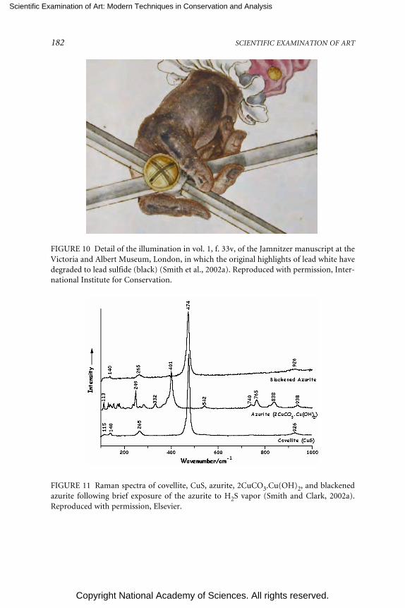
Scientific Examination of Art: Modern Techniques in Conservation and Analysis
Copyright National Academy of Sciences. All rights reserved.
182 SCIENTIFIC EXAMINATION OF ART
FIGURE 10 Detail of the illumination in vol. 1, f. 33v, of the Jamnitzer manuscript at theVictoria and Albert Museum, London, in which the original highlights of lead white havedegraded to lead sulfide (black) (Smith et al., 2002a). Reproduced with permission, Inter-national Institute for Conservation.
FIGURE 11 Raman spectra of covellite, CuS, azurite, 2CuCO3.Cu(OH)2, and blackenedazurite following brief exposure of the azurite to H2S vapor (Smith and Clark, 2002a).Reproduced with permission, Elsevier.

Scientific Examination of Art: Modern Techniques in Conservation and Analysis
Copyright National Academy of Sciences. All rights reserved.
RAMAN MICROSCOPY IN THE IDENTIFICATION OF PIGMENTS 183
spatial extent of each corrosion product. Many extensions to this work are beingplanned with the vast collections of degraded metals in museums in order tounderstand the nature and causes of the degradation. The most recent studies ofthis sort have led to the Raman-based identification of iron oxide impurities(magnetite, iron-deficient magnetite, and hematite) in early industrial-scale pro-cessed platinum (e.g., in the platinum metal constituting the three-ruble Russiancoin of 1837) (van der Weerd et al., 2004a).
Modern Raman spectrometers may now be capable of identifying irongallotannate inks on manuscripts, a notoriously difficult task. Recent studies onthe Vinland Map (Beinecke Library, Yale University) have shown that the frag-mented black ink lines that define the map consist of carbon black rather that irongallotannate and that the yellow-brown background to the lines, but not else-where, contains nearly pure anatase (TiO2) (Brown and Clark, 2002). This mate-rial had earlier been shown to have a particle size (approximately 0.15 µm) andparticle size distribution that is characteristic of the synthetic ca 1920 product(McCrone, 1988). The Raman results thus confirm that the map dates from thetwentieth not the early fifteenth century, and thus that it is not pre-Columbian;this is in complete agreement with the analysis of Towe (1990).
CONCLUSION
Raman microscopy is now established to be a key technique for the identificationof pigments on works of art, largely because of its high spatial and spectral resolu-tion, excellent sensitivity and specificity, and because it can be applied to anobject in situ. Difficulties may arise on occasions with certain organic pigments,supports, and binders that fluoresce, that are photosensitive, or that fail to yield aRaman spectrum owing to their small particle size, high dilution, or poor scatter-ing efficiency. The use of other techniques in conjunction with Raman micros-copy then becomes essential in order to effect full pigment characterization. Re-mote laser Raman microscopy (Clark and Gibbs, 1998a) will increasingly be usedfor the study of objects unable to be moved from their place of exhibition. Thewhole area is one in which the arts and the sciences can coordinate with greateffect.
ACKNOWLEDGEMENTS
The author is most grateful to the members of his group in this field, most re-cently Drs. K. L. Brown, L. Burgio, T. D. Chaplin, S. Firth, A. Jurado-Lopez, G. D.Smith, and J. van der Weerd, and to Renishaw PLC, the Engineering and PhysicalSciences Research Council, the European Union, and the British Library for theirsupport of this research.

Scientific Examination of Art: Modern Techniques in Conservation and Analysis
Copyright National Academy of Sciences. All rights reserved.
184 SCIENTIFIC EXAMINATION OF ART
REFERENCES
Anglos, D., S. Couris, and C. Fotakis. 1997. Applied Spectroscopy 51:1025-1030.Bell, I. M., R. J. H. Clark, and P. J. Gibbs. 1997. Spectrochimica Acta Part A 53:2159-2179.Best, S. P., R. J. H. Clark, and R. Withnall. 1992. Endeavour, New Series 16:66-73.Best, S. P., R. J. H. Clark, M. A. M. Daniels, and R. Withnall. 1993. Chemistry in Britain 118-122.Best, S. P., R. J. H. Clark, M. A. M. Daniels, C. A. Porter, and R. Withnall. 1995. Studies in Conserva-
tion 40:31-40.Bikiaris, D., Sister Daniilia, S. Sotiropoulou, O. Katsimbiri, E. Pavlidou, A. P. Moutsatsou, and Y.
Chryssoulakis. 2000. Spectrochimica Acta Part A: Molecular and Biomolecular Spectroscopy56:3-18.
Bousfield, B. 1992. Surface Preparation and Microscopy of Materials. Chichester, U.K.: Wiley.Brown, K. L., and R. J. H. Clark. 2002. Analytical Chemistry 74:3658-3661.Brown, K. L., and R. J. H. Clark. 2004a. Journal of Raman Spectroscopy 35:4-12.Brown, K. L., and R. J. H. Clark. 2004b. Journal of Raman Spectroscopy 35:181-189.Brown, K. L., and R. J. H. Clark. 2004c. Journal of Raman Spectroscopy 35:217-223.Brundle, C. R., C. A. Evans, and S. Wilson. 1992. Encyclopaedia of Materials Characterization. Boston:
Butterworth-Heinemann.Burgio, L., and R. J. H. Clark. 2000. Journal of Raman Spectroscopy 31:395-401.Burgio, L., and R. J. H. Clark. 2001. Spectrochimica Acta Part A 57:1491-1521.Burgio, L., D. Ciomartan, and R. J. H. Clark. 1997a. Journal of Raman Spectroscopy 28:79-83.Burgio, L., D. Ciomartan, and R. J. H. Clark. 1997b. Journal of Molecular Structure 405:1-11.Burgio, L., R. J. H. Clark, and H. Toftlund. 1999a. Acta Chemica Scandinavica 53:181-187.Burgio, L., R. J. H. Clark, and P. J. Gibbs. 1999b. Journal of Raman Spectroscopy 30:181-184.Burgio, L., R. J. H. Clark, T. Stratoudaki, D. Anglos, and M. Doulgeridis. 2000. Applied Spectroscopy
54:463-470.Burgio, L., R. J. H. Clark, and S. Firth. 2001. Analyst 126:222-227.Burgio, L., R. J. H. Clark, L. Sheldon, and G. D. Smith, 2005. Analytical Chemistry 77:1261-1267.Castillejo, M., M. Martin, D. Silva, T. Stratoudaki, D. Anglos, L. Burgio, and R. J. H. Clark. 2000.
Journal of Molecular Structure 550:191-198.Chaplin, T. D., R. J. H. Clark, D. Jacobs, K. Jensen, and G. D. Smith. 2002a. In Raman Spectroscopy,
eds. J. Mink, G. Jalovszky, and G. Kereszbury, pp. 823-824. Chichester, U.K.: Wiley.Chaplin, T. D., R. J. H. Clark, D. Jacobs, K. Jensen, and G. D. Smith. 2005. Analytical Chemistry 77.Chaplin, T. D., R. J. H. Clark, and D. R. Beech. 2002b. Journal of Raman Spectroscopy 33:424-428.Chaplin, T. D., A. Jurado-Lopez, R. J. H. Clark, and D. R. Beech. 2004. Journal of Raman Spectroscopy
35:600-604.Cilberto, E., and G. Spoto, eds. 2000. Modern Analytical Methods in Art and Archaeology. New York:
Wiley.Ciomartan, D., and R. J. H. Clark. 1996. Journal of the Brazilian Chemical Society 7:395-402.Clark, R. J. H. 1964. Journal of Chemical Education 41:488-492.Clark, R. J. H. 1995. Chemical Society Reviews 24:187-196.Clark, R. J. H. 2002. In Handbook of Vibrational Spectroscopy, eds. J. M. Chalmers and P. R. Griffiths,
pp. 2977-2992. Chichester, U.K.: Wiley.Clark, R. J. H., and M. L. Curri. 1998. Journal of Molecular Structure 440:105-111.Clark, R. J. H., and T. J. Dines. 1986. Angewandte Chemie-International Edition in English 25:131-158.Clark, R. J. H., and P. J. Gibbs. 1997. Journal of Raman Spectroscopy 28:99-103.Clark, R. J. H., and P. J. Gibbs. 1998a. Journal of Archaeological Science 25:621-629.Clark, R. J. H., and P. J. Gibbs. 1998b. Analytical Chemistry 70:99A-104A.Clark, R. J. H, and K. Huxley. 1996. Science and Technology for Cultural Heritage 5:95-101.Clark, R. J. H., and J. van der Weerd. 2004. Journal of Raman Spectroscopy 35:279-283.Clark, R. J. H., L. Cridland, B. M. Kariuki, K. D. M. Harris, and R. Withnall. 1995. Journal of the
Chemical Society, Dalton Transactions 2577-2582.

Scientific Examination of Art: Modern Techniques in Conservation and Analysis
Copyright National Academy of Sciences. All rights reserved.
RAMAN MICROSCOPY IN THE IDENTIFICATION OF PIGMENTS 185
Clark, R. J. H., P. J. Gibbs, K. R. Seddon, N. M. Brovenko, and Y. A. Petrosyan. 1997a. Journal ofRaman Spectroscopy 28:91-94.
Clark, R. J. H., M. L. Curri, and C. Laganara, 1997b. Spectrochimica Acta Part A 53:597-603.Clark, R. J. H., M. L. Curri, G. S. Henshaw, and C. Laganara. 1997c. Journal of Raman Spectroscopy
28:105-109.Colomban, P., G. Sagon, and X. Faurel. 2001. Journal of Raman Spectroscopy 32:351-360.Corset, J., P. Dhamelincourt, and J. Barbillat. 1989. Chemistry in Britain 612-616.Daniilia, Sister, D. Bikiaris, P. Gavala, R. J. H. Clark, and Y. Chryssoulakis. 2002. Journal of Raman
Spectroscopy 33:807-814.Faurel, X., A. Vanderperre, and P. Colomban. 2003. Journal of Raman Spectroscopy 34:290-294.Feller, R. L., ed. 1986. Artists’ Pigments, vol. 1. Cambridge: Cambridge University Press.FitzHugh, E. W., ed. 1997. Artists’ Pigments, vol. 3. Oxford: Oxford University Press.Gettens, R. J., and G. L. Stout. 1966. Painting Materials. New York: Dover.Giovannoni, S., M. Matteini, and S. Moles. 1990. Studies in Conservation 35:21-25.Griffith, W. P. 1987. In Advances in Spectroscopy, vol. 14, eds. R. J. H. Clark and R. E. Hester, pp. 119-
186. Chichester, U.K.: Wiley.Jorgensen, C. K. 1962. Absorption Spectra and Chemical Bonding. Oxford: Pergamon.Jurado-Lopez, A., O. Demko, R. J. H. Clark, and D. Jacobs. 2004. Journal of Raman Spectroscopy
35:119-124.Liem, N. Q., N. T. Thanh, and P. Colomban. 2002. Journal of Raman Spectroscopy 33:287-294.Long, D. A. 2002. The Raman Effect. Chichester, U.K.: Wiley.Martens, W., R. L. Frost, J. T. Kloprogge, and P. A. Williams. 2003. Journal of Raman Spectroscopy
34:145-151.Mayer, R. 1972. The Artist’s Handbook. London: Faber and Faber.McCann, L. I., K. Trentleman, T. Possley, and B. Golding. 1999. Journal of Raman Spectroscopy
30:121-132.McCrone, W. C. 1988. Analytical Chemistry 60:1009-1018.Perez, F. R., H. G. M. Edwards, A. Rivas, and L. Drummond. 1999. Journal of Raman Spectroscopy
30:301-305.Pollard, A. M., and C. Heron. 1996. Archaeological Chemistry. Cambridge: Royal Society of Chemistry.Raman, C. V., and K. S. Krishnan. 1928. Nature 121:501.Roosen-Runge, H., and A. E. A. Werner. 1960. In Evangeliorum Quattuor Codex Lindisfarnensis, vol.
2, ed. T. D. Kendrick, pp. 263-272. Lausanne: Urs Gras.Roy, A., ed. 1993. Artists’ Pigments, vol. 2. Oxford: Oxford University Press.Scott, D. A., Y. Taniguchi, and E. Koseto. 2001. Reviews in Conservation 2:73-91.Smith, G. D., and R. J. H. Clark. 2002a. Journal of Cultural Heritage 3:101-105.Smith, G. D., and R. J. H. Clark. 2002b. Reviews in Conservation 2:92-106.Smith, G. D., and R. J. H. Clark. 2004. Journal of Archaeological Science 31:1137-1160.Smith, G. D., A. Derbyshire, and R. J. H. Clark. 2002a. Studies in Conservation 47:250-257.Smith, G. D., S. Firth, R. J. H. Clark, and M. Cardona. 2002b. Journal of Applied Physics 92:4375-4380.Thompson, D. V. 1956. The Materials and Techniques of Painting. New York: Dover.Towe, K. M. 1990. Accounts of Chemical Research 23:84-87.Turrell, G., and J. Corset, eds. 1996. Raman Microscopy: Developments and Applications. London:
Academic Press.van der Weerd, J., T. Rehren, S. Firth, and R. J. H. Clark. 2004a. Materials Characterisation 53: 63-70.van der Weerd, J., G. D. Smith, S. Firth, and R. J. H. Clark. 2004b. Journal of Archaeological Science
31:1429-1437.Vandenabeele, P., L. Moens, H. G. M. Edwards, and R. Dams. 2000. Journal of Raman Spectroscopy
31:509-517.Wehlte, K. 1975. The Materials and Techniques of Painting. New York: Van Nostrand Reinhold.Zuo, J., C. Xu, C. Wang, and Z. Yushi. 1999. Journal of Raman Spectroscopy 30:1053-1055.

Scientific Examination of Art: Modern Techniques in Conservation and Analysis
Copyright National Academy of Sciences. All rights reserved.
186
Paint Media Analysis
Michael R. SchillingSenior Scientist
The Getty Conservation Institute
Pigments and organic binding media are the two principle components of paint.Whereas pigments impart color to paint, it is the role of the organic bindingmedia to bind together the grains of pigment and adhere them to the work of art.Synthetic polymers are the binding media of choice for most of today’s commer-cial paints. Nevertheless, the continued use by contemporary artists of such natu-ral products as egg, milk, animal hides, vegetable oils, plant gums, waxes, andnatural resins (which were the only binding media available from antiquitythrough the end of the nineteenth century [Kühn, 1986]) attests to their durabil-ity, versatility, and working properties that artists value.
Conservation scientists are often called upon to analyze organic binding me-dia and pigments in painted works of art. Knowledge obtained from the study ofartists’ materials and techniques enriches our understanding of the history of art,informs the decisions of conservators who must develop appropriate conserva-tion treatments, and reveals compositional changes in artists’ materials broughtabout by age, weathering, and environmental factors. Although many instrumen-tal analysis techniques now exist for identifying organic substances, several keyfactors limit the actual number of techniques that are suitable for identifyingorganic binding media. To begin, typical samples removed from paintings weighin the range of 1 to 50 micrograms; in many instances the medium simply may bepresent below instrumental detection limits. Mixtures of organic binding mediamay present problems of overlapping signals. Physical aging and pigment inter-ferences may complicate data interpretation by changing the original composi-tion. Moreover, it is extremely difficult, if not impossible, to resolubilize someorganic binding media (such as egg tempera) once they have become dried into

Scientific Examination of Art: Modern Techniques in Conservation and Analysis
Copyright National Academy of Sciences. All rights reserved.
PAINT MEDIA ANALYSIS 187
paint films. For those instrumental techniques that are capable of detecting or-ganic binding media, application of simple qualitative analysis limits the extent towhich the analytical test results may be interpreted.
Quantitative gas chromatography-mass spectrometry (GC-MS) is one of thefew analytical techniques capable of overcoming the myriad of problems associ-ated with identification of natural organic binding media in painted works of art.In research at the Getty Conservation Institute, quantitative GC-MS procedureswere developed for identifying organic binding media based on proteins, oils, andplant gums. The procedures were validated on test paints that were subjected totwo types of artificial aging (six weeks at 80°C and 500 hours in a Weather-O-Meter light exposure chamber at 50 percent RH and 50°C). The present studyillustrates the utility of quantitative GC-MS in the study of paintings by twoprominent American artists.
A TECHNICAL STUDY OF PAINTINGS BY JACOB LAWRENCE
Jacob Lawrence was known for his simplified, brilliant graphic forms that depictAfrican American history and experience (Steele, 2000). Throughout his longcareer he favored working in various water-based organic binding media com-monly referred to as tempera: casein, egg, plant gums, and animal glue (Mayer,1940). It is known that Lawrence mixed some of his own tempera paints fromartists’ recipe books, whereas in many of his later works he used commerciallyavailable tube colors. Manufacturers often add materials to tube colors, in addi-tion to the binding media, to modify the working properties of the paints andstabilize the mixtures. These additives include glycerol, seed oils (such as linseed,poppy, and walnut), natural resins (dammar, rosin), phthalate plasticizers, andsugar. From these lists it is quite clear that Lawrence’s paint media may be com-plicated mixtures of many substances (Steele and Halpine, 1993). It should alsobe noted that it is nearly impossible to differentiate these tempera media basedsolely on the appearance of the painted surface, yet because this is sometimes theonly means available to museum registrars when cataloguing their collections,these records are sometimes erroneous.
Recently a technical study of samples from a number of Lawrence’s paintings(see Table 1) was undertaken to learn more about his painting technique, checkthe accuracy of museum archival records, and contribute to a catalogue raisonnéof Lawrence’s paintings (Schilling et al., 2000). Pigments were identified in thepaint samples using polarized-light microscopy. Some samples were tested usingFourier transform infrared microspectrometry (FTIR) to identity the paint com-ponents.
To test for proteinaceous media in paint samples, amino acids were liberatedby acid hydrolysis and analyzed by quantitative GC-MS in the form of (tert-butyl-dimethylsilyl) derivatives (see Appendix A for experimental details) (Simek et al.,1994; Columbini et al., 1998; Schilling and Khanjian, 1996a). The quantitative

Scientific Examination of Art: Modern Techniques in Conservation and Analysis
Copyright National Academy of Sciences. All rights reserved.
188 SCIENTIFIC EXAMINATION OF ART
TABLE 1 Jacob Lawrence Paintings Analyzed in This Study
The Metropolitan Museum of Art, New YorkBlind Beggars, 1938
National Museum of American Art, Washington, D.C.Painting the Bilges, 1944New Jersey, 1946Men Exist for the Sake of One Another, 1958Library, 1960
Hirshhorn Museum and Sculpture Garden, Washington, D.C.African Gold Miners, 1946Vaudeville, 1951The Cue and the Ball, 1956Magic Man, 1958Playing Card (Joker) or (King), 1962Harriet and the Promised Land No.10, 1967In a Free Government, 1976
Worcester Art MuseumThe Checker Players, 1947
The Museum of Modern Art, New YorkSedation, 1950
Private CollectionStruggle Series No.11: Informers Coded Message, 1955Ordeal of Alice, 1963
National Gallery of Art, Washington, D.C.Street to Mbari, 1964Daybreak-A Time to Rest, 1967
Merril C. Berman CollectionStudents with Books, 1966
Jacob and Gwen Knight Lawrence CollectionOther Rooms, 1975
results for the alkyl- and imino-substituted amino acids (the so-called “stable”amino acids) were normalized to 100 mole percent. The quantitative yields for theother amino acids are often unreliable due to pigment interferences in the hy-drolysis and/or derivatization procedures, or due to aging (Halpine, 1992; Ronca,1994; Schilling and Khanjian, 1996b); these amino acids were excluded from thefinal dataset.
Samples tested for plant gum media were hydrolyzed in trifluoroacetic acid,and the monosaccharides were analyzed as O-methyloxime acetate derivatives(see Appendix B for details) (Murphy and Pennock, 1972; Neeser and Schweizer,1983). For comparative purposes the monosaccharide dataset, excluding glucoseand fructose, were normalized to 100 weight percent.

Scientific Examination of Art: Modern Techniques in Conservation and Analysis
Copyright National Academy of Sciences. All rights reserved.
PAINT MEDIA ANALYSIS 189
Test Results for Jacob Lawrence Paintings
Table 2 lists the quantitative stable amino acid test results for the Lawrencepaint samples with those of several common proteinaceous and plant-gum-binding media included for reference (the reference data originated from in-house tests and from published sources [Schilling et al., 1996]). Carbohydratecompositions for selected Lawrence samples and various reference materials arelisted in Table 3.
Using the method of correlation coefficients, the quantitative stable aminoacid composition for each sample was compared to those of the common bindingmedia in order to find the closest match, as listed in Table 4 (Anderson, 1987); thesame method was employed for identification of plant gums (see Table 3). Mostsamples correlated very closely either to glue, egg, casein, or gum arabic. In twopaint samples, however, there were indications in the test data that two proteina-ceous media were present. This situation may arise either because the artist inten-tionally mixed two binding media together in the paint, or because the paintsample was contaminated with medium from a second paint layer (this last situ-ation occurs frequently in samples from egg tempera paintings that have groundlayers mixed with glue). And so, for the two Lawrence samples, simple algebraicequations were used to find the most likely pair of proteinaceous media that gavethe closest correlations to those in the paint samples (Schilling and Khanjian,1996c). Thus, the red from Blind Beggars had a 0.99 correlation to a mixture (1:2)of casein and glue, whereas the green from Sedation had a 0.95 correlation to amixture (1:3) of casein and glue.
In general, good agreement was evident between the analytical findings andthe medium attributions in the museum archives. One notable exception was thedetection of glue as the medium of Playing Card, which had been previouslymisidentified in the archives as plant gum. Another exception was Street to Mbari,which had been assessed visually as having a gouache medium.
GC-MS was useful for detecting components in the paints that were unre-lated to the protein or plant gum media. Some samples revealed the presence ofcommercial paint additives, such as Struggle Series Number 11. The brown paintcontained egg medium plus high amounts of glycerol, gallic acid, and rosin; thisformulation is consistent with an artists’ tube color (Steele, 2000; Steele andHalpine, 1993; Schilling et al., 2000). Moreover, a few samples showed evidenceof biodeterioration of the paint medium. For instance, oxalic acid (a common by-product of microbial activity [Matteini, 1998]) was detected in the dark paintfrom The Checker Players. It may be that the relatively poor quality of the correla-tion for the medium in this paint to the reference materials (0.91 to casein, 0.74 toegg) may be due in part to the effects of biodeterioration.

Scientific Examination of Art: Modern Techniques in Conservation and Analysis
Copyright National Academy of Sciences. All rights reserved.
190
TA
BL
E 2
Sta
ble
Am
ino
Aci
d C
ompo
siti
ons
of J
acob
Law
ren
ce P
ain
t Sa
mpl
es
Con
cen
trat
ion
(m
ole
perc
ent)
Pai
nti
ng
Sam
ple
AL
AV
AL
ILE
LE
UG
LY
PR
OH
YP
% P
rote
in
Bli
nd
Beg
gars
Red
13.5
10.8
7.8
13.7
28.8
20.9
4.6
4C
heck
er P
laye
rsP
ain
t &
gro
un
d16
.114
.911
.322
.113
.122
.30.
27
Day
brea
kY
ello
w22
.216
.312
.321
.917
.210
.20.
04
Lib
rary
Och
re20
.618
.316
.425
.716
.32.
70.
02
Bro
wn
22.4
20.8
15.7
25.8
9.5
5.3
0.5
6M
agic
Man
Lig
ht
bla
ck24
.319
.714
.223
.315
.92.
30.
39
Men
Exi
stB
lue
& g
rou
nd
21.5
16.3
13.5
24.0
16.4
8.3
0.0
3O
rdea
l of
Ali
ceB
row
n27
.219
.514
.121
.713
.33.
70.
42
Par
ade
Blu
e20
.516
.112
.922
.721
.36.
40.
19
Pla
yin
g C
ard
(Jok
er)
Blu
e &
pap
er14
.93.
52.
14.
351
.613
.110
.412
Pla
yin
g C
ard
(Kin
g)R
ed &
fib
ers
14.2
2.7
1.6
3.2
55.8
12.3
10.2
18B
lack
& p
a per
17.1
5.5
3.6
6.9
52.6
10.8
3.5
11Se
dati
o nG
reen
& p
a per
14.0
13.1
9.0
16.8
25.6
21.0
0.5
5St
reet
to
M’B
ari
Yel
low
& b
lue
18.7
3.7
2.0
3.5
52.4
12.6
7.2
14B
lue
17.8
3.1
1.7
3.2
51.2
13.2
9.7
13St
rugg
le S
erie
s #1
1B
row
n22
.216
.212
.424
.114
.410
.80.
012
Stu
den
ts a
nd
Bo o
k sY
ello
w25
.017
.813
.123
.510
.48.
71.
516
Bro
wn
& g
rou
nd
23.1
18.3
13.2
22.4
15.4
6.7
0.9
6Y
ello
w22
.015
.112
.222
.320
.67.
80.
08
Vau
devi
lle
Blu
e24
.017
.012
.926
.117
.92.
10.
06
Gro
un
d12
.416
.811
.121
.88.
828
.90.
23

Scientific Examination of Art: Modern Techniques in Conservation and Analysis
Copyright National Academy of Sciences. All rights reserved.
PAINT MEDIA ANALYSIS 191
A STUDY OF WILLEM DE KOONING’S PAINTINGS FROM THE 1960S AND 1970S
Willem de Kooning was born in the Netherlands in 1904, and immigrated to theUnited States in 1926. He trained in guild and craft traditions of wood graining,gilding, marbleizing, lettering, and sign painting; he also spent time as a housepainter and a commercial artist. These experiences gave him a thorough masteryof materials and a craftsman’s skills (Lake et al., 1999).
De Kooning routinely exploited unconventional materials for his pictures.Historical and anecdotal records report that he mixed house paint, safflowercooking oil, water, egg, and even mayonnaise into his artists’ paints to achieve thedesired appearance and texture. During the course of creating a painting, hescraped the painted canvas at day’s end, and repeatedly reworked it; for thisprocess to be successful, soft, slow-drying paints were required. Such paints wereabundant in the 1940s and 1950s, when oil was the typical medium in housepaints. Unfortunately for de Kooning, alkyd paint formulations became popularin the retail trade industry in the 1960s and 1970s; their fast-drying propertieswere incompatible with his chosen technique. In paintings from this period,sources document his use of Bellini Bocour artists’ tube colors, which containedheat-bodied linseed oil, to which he occasionally added safflower oil and water(Lake et al., 1999).
Although his methods and materials have been well documented, it was notclear what his actual practices were at specific times in his career. Moreover, therewas concern that his unusual paint formulations could negatively affect the long-term stability of his paintings. The paintings executed during the 1960s and 1970s,in particular, are problematic for conservators, with passages that remain soft andsticky. Such paint surfaces are easily deformed when touched and they readilypick up surface dust. To learn more about de Kooning’s materials and techniques,a study was undertaken to analyze the binding media and pigments of a selectionof his paintings from the period of 1960-1977 (Lake et al., 1999). Table 5 providesa complete list of the paintings that were sampled.
Chemistry of Oil Paints
The chemistry of oil paint is very complex, and even with modern analyticalequipment, it is difficult to understand the precise details of the interactionsbetween the polymerized oil media and pigments. Nonetheless, a substantial bodyof knowledge has been developed that sheds some light on the drying and subse-quent aging of oil paints (van den Berg, 2002). Essentially, seed oils differ in termsof their fatty acid distribution on the triglyceride molecules. The so-called dryingoils that are favored by artists (e.g., linseed, walnut, poppy seed) have a highproportion of multiple unsaturated fatty acids, whereas the semidrying and non-drying oils (such as castor, safflower, sunflower) do not. All of the aforemen-

Scientific Examination of Art: Modern Techniques in Conservation and Analysis
Copyright National Academy of Sciences. All rights reserved.
192 SCIENTIFIC EXAMINATION OF ART
TABLE 3 Gum Sugar Compositions of Jacob Lawrence Paint Samples andPlant Gum Standards, with Correlation Coefficient Data
Normalized Weight % of Gum SugarsWeight %
Sample Gum Sugars Rhamnose Fucose Arabinose
African Gold Miners: blue paint 10.4 13.0 0.1 36.3
African Gold Miners: black paint 6.3 10.2 0.1 29.1
Blind Beggars: blue paint 3.8 17.0 0.0 35.5
New Jersey: red paint 11.8 12.9 0.1 32.8
Painting the Bilges: blue paint 1.9 15.4 0.0 37.7
Gum Arabic standard 57.0 16.0 0.0 36.0
Cherry gum standard 95.7 0.6 0.0 46.6
Gum tragacanth standard 46.4 3.3 8.8 48.4
tioned seed oils contain approximately 10 percent by weight of glycerol, plussmall amounts of saturated fatty acids (the two most important being hexade-canoic acid and octadecanoic acid, more commonly known as palmitic acid andstearic acid, respectively) (Mills, 1966).
As oil paints dry, unsaturated fatty acids react with oxygen to form a poly-merized oil matrix; triglycerides and diglycerides provide additional cross-links tothe polymerized oil matrix via their glycerol backbones. The saturated fatty acids,being less reactive, neither oxidize nor cross-link, and so remain as marker com-pounds in oil paint. During the drying and aging processes, chain scission prod-ucts such as dicarboxylic fatty acids are formed in oil paint. The most importantdicarboxylic fatty acid marker compound is nonanedioic acid (azelaic acid), butother straight-chain dicarboxylic fatty acids (that contain from two to ten carbonatoms) also form (Mills, 1966).
Other reactions also occur during aging that further alter the fatty acid distri-bution of oil paints. For example, free fatty acids are produced by hydrolysis ofglycerides (e.g., glycerol esters of fatty acids). As hydrolysis progresses there is areduction in residual triglycerides and diglycerides, and formation of free fattyacids. Complete hydrolysis of an oil paint would eventually yield three moles offree fatty acids per every mole of glycerol (van den Berg, 2002).
Reaction of the carboxylate groups of free fatty acids with pigments thatcontain coordinating metal cations (e.g., lead, copper, cobalt) produces metalsoaps. Evidence has shown that aged oil paints are essentially ionomeric polymersof fatty acids coordinated with pigments (van den Berg et al., 1999).
Figure 1 illustrates the results from a Monte Carlo simulation of the hydroly-sis of a model triglyceride, in which the extent of hydrolysis is estimated by the

Scientific Examination of Art: Modern Techniques in Conservation and Analysis
Copyright National Academy of Sciences. All rights reserved.
PAINT MEDIA ANALYSIS 193
percent of free fatty acids. This figure shows that nontrivial amounts of triglycer-ides and diglycerides remain even after significant hydrolysis of the model com-pound. Undoubtedly this model is extremely simple, and does not account fordiffering rates of hydrolysis of the middle ester group on a glyceride compared tothe end positions. Nonetheless, it does suggest that residual triglycerides anddiglycerides may remain even in extremely hydrolyzed oil paints, which can addi-tionally stabilize oil paint ionomeric polymers.
Additional alteration of the fatty acid composition of oil paints occurs byevaporation of free fatty acids. So-called ”ghost images” that develop on the glassnext to framed and glazed oil paintings provide clear evidence of fatty acid evapo-ration (Williams, 1989). Thermogravimetric analysis indicated that palmitic acidevaporated approximately twice as rapidly as stearic acid or azelaic acid (Schillinget al., 1999). Oil paints made with lead white pigment produced no visible ghostimage, which is consistent with the fact that lead pigments readily coordinate freefatty acids.
Analysis of Oil Paints
Two GC-MS procedures were employed to analyze the de Kooning paintsamples. A quantitative procedure for fatty acid and glycerol analysis of food oils(Mason et al., 1964) was modified to work on oil paint samples (Schilling andKhanjian, 1996d). In this procedure FAMEs and isopropylidene glycerol (IPG, avolatile glycerol derivative) were produced quantitatively by overnight treatmentwith sodium methoxide in 2,2-dimethoxypropane, followed by addition ofmethanolic hydrochloric acid. The FAMEs and IPG were separated in a single
Correlation Coefficient to Plant Gum Standards
Xylose Mannose Galactose Gum Arabic Cherry Gum Gum Tragacanth
0.7 0.4 49.4 1.00 0.90 0.39
0.7 0.5 59.3 0.97 0.84 0.24
0.5 0.5 46.5 1.00 0.88 0.37
0.5 0.2 53.6 0.99 0.87 0.31
0.6 0.0 46.3 1.00 0.90 0.42
0.0 0.0 48.0
10.4 2.2 40.2
27.5 0.0 12.1

Scientific Examination of Art: Modern Techniques in Conservation and Analysis
Copyright National Academy of Sciences. All rights reserved.
194T
AB
LE
4 C
orre
lati
on B
etw
een
Jac
ob L
awre
nce
Pai
nt
Sam
ple
Dat
a (T
able
2)
and
Ref
eren
ce M
ater
ial D
ata
(Tab
le 8
)
Cor
rela
tion
Coe
ffic
ien
t
Pai
nti
ng
Pai
nt
Sam
ple
Col
lage
nE
gg Y
olk
Cas
ein
Oth
er
Bli
nd
Beg
gars
Red
0.82
0.29
0.33
0.99
(1
par
t gl
ue,
2 p
arts
cas
ein
)C
heck
er P
laye
rsP
ain
t &
gro
un
d–0
.06
0.79
0.91
Day
brea
kY
ello
w0.
060.
980.
39L
ibra
ryO
chre
0.13
0.90
0.19
0.99
(yo
lk &
ult
ram
arin
e, l
igh
t ag
ed)
Bro
wn
–0.4
00.
910.
320.
96 (
yolk
, ag
ed a
t 80
deg
C)
Mag
ic M
anL
igh
t b
lack
–0.1
00.
900.
121.
00 (
Ro
wn
ey c
adm
ium
red
)M
en E
xist
Blu
e &
gro
un
d–0
.03
0.98
0.36
Ord
eal
of A
lice
Bro
wn
–0.1
70.
890.
140.
96 (
yolk
, li
ght
aged
)P
arad
eB
lue
0.19
0.90
0.21
0.96
(yo
lk &
lea
d w
hit
e, l
igh
t ag
ed)
Pla
yin
g C
ard
(Jok
er)
Blu
e &
pap
er0.
99–0
.06
–0.2
7P
layi
ng
Car
d (K
ing)
Red
& f
iber
s0.
99–0
.07
–0.2
8B
lack
& p
aper
0.96
0.13
–0.1
9Se
dati
onG
reen
& p
aper
0.61
0.55
0.56
0.95
(1
par
t gl
ue,
3 p
arts
cas
ein
)St
reet
to
M’B
ari
Yel
low
& b
lue
0.98
0.02
–0.2
5B
lue
0.99
–0.0
3–0
.28
Stru
ggle
Ser
ies
#11
Bro
wn
–0.0
91.
000.
47St
ude
nts
an
d B
o ok s
Yel
low
–0.2
80.
950.
36B
row
n &
gro
un
d–0
.08
0.95
0.26
Yel
low
0.20
0.92
0.25
0.96
(y o
lk,
lig h
t a g
ed)
Vau
devi
lle
Blu
e–0
.01
0.90
0.00
0.99
(R
ow
ney
ca d
miu
m r
ed)
Gro
un
d–0
.21
0.55
1.00

Scientific Examination of Art: Modern Techniques in Conservation and Analysis
Copyright National Academy of Sciences. All rights reserved.
PAINT MEDIA ANALYSIS 195
chromatogram using an HP-INNOWAX capillary column. This procedurederivatizes all three forms of fatty acids in oil paints (free fatty acids, pigmentsoaps, and glycerides). The second quantitative procedure used hexamethylenedisilazane with trichloromethyl silane catalyst (HMDS/TMCS) in pyridine to formtrimethyl silyl esters of the free fatty acids and soaps, and separation of the deriva-tives on a DB-5MS column (Pierce, 1968).
In evaluating the quantitative test data several key parameters are calculatedfor identification and diagnostic purposes. For instance, the minimum content ofoil in the paint sample can be approximated from the glycerol content, althoughthe accuracy of this estimation is limited by loss of glycerol due to aging andsolvent extraction. Second, the molar ratio of palmitic acid to stearic acid (P/S) isuseful for identifying the type of oil present (Mills, 1966). Third, the extent ofhydrolysis may be estimated by comparing the content of free azelaic acid and itssoap to the total azelaic acid content. To probe the extent of alteration of the driedoil matrix, the two most diagnostic ratios are palmitic to glycerol (P/G), and theratio of dicarboxylic fatty acids to glycerol (D/G, where D is the sum of all dicar-boxylic fatty acids from C3 to C8 plus C10). Reduction in P/G from its originalvalue in the fresh oil is caused by loss of palmitic acid due to evaporation ormigration into the canvas and ground layer. Photo-oxidative reactions are re-sponsible for increases in D/G (Schilling and Khanjian, 1996d; Schilling et al.,1997).
TABLE 5 Willem de Kooning Paintings Analyzed in This Study
Door to the River, 1960, Whitney Museum of American Art, New York
Spike’s Folly II, 1960, Robert and Jane Meyerhoff, Phoenix, MD
Rosy-Fingered Dawn at Louse Point, 1963, Stedelijk Museum, Amsterdam
Pastorale, 1963, private collection, New Orleans
Woman, Sag Harbor, 1964, Hirshhorn Museum and Sculpture Garden, Washington, D.C.
Woman, 1965, Hirshhorn Museum and Sculpture Garden, Washington, D.C.
The Visit, 1966-67, Tate Gallery, London
Two Figures in a Landscape, 1967, Stedelijk Museum, Amsterdam
Amityville, 1971, private collection
. . . Whose Name Was Writ in Water, 1975, Solomon R. Guggenheim Museum, New York
Untitled I, 1977, Adriana and Robert Mnuchin
Untitled V, 1977, Albright-Knox Art Gallery, Buffalo, New York
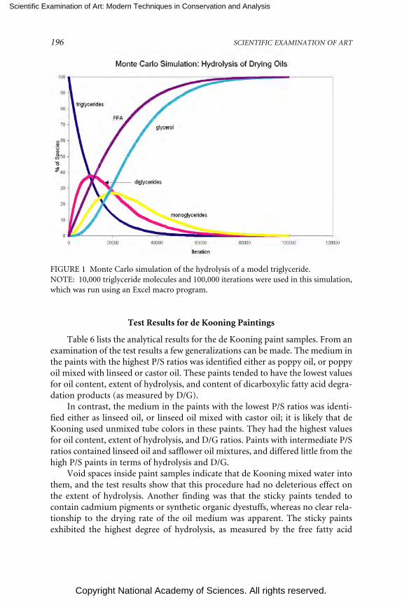
Scientific Examination of Art: Modern Techniques in Conservation and Analysis
Copyright National Academy of Sciences. All rights reserved.
196 SCIENTIFIC EXAMINATION OF ART
FIGURE 1 Monte Carlo simulation of the hydrolysis of a model triglyceride.NOTE: 10,000 triglyceride molecules and 100,000 iterations were used in this simulation,which was run using an Excel macro program.
Test Results for de Kooning Paintings
Table 6 lists the analytical results for the de Kooning paint samples. From anexamination of the test results a few generalizations can be made. The medium inthe paints with the highest P/S ratios was identified either as poppy oil, or poppyoil mixed with linseed or castor oil. These paints tended to have the lowest valuesfor oil content, extent of hydrolysis, and content of dicarboxylic fatty acid degra-dation products (as measured by D/G).
In contrast, the medium in the paints with the lowest P/S ratios was identi-fied either as linseed oil, or linseed oil mixed with castor oil; it is likely that deKooning used unmixed tube colors in these paints. They had the highest valuesfor oil content, extent of hydrolysis, and D/G ratios. Paints with intermediate P/Sratios contained linseed oil and safflower oil mixtures, and differed little from thehigh P/S paints in terms of hydrolysis and D/G.
Void spaces inside paint samples indicate that de Kooning mixed water intothem, and the test results show that this procedure had no deleterious effect onthe extent of hydrolysis. Another finding was that the sticky paints tended tocontain cadmium pigments or synthetic organic dyestuffs, whereas no clear rela-tionship to the drying rate of the oil medium was apparent. The sticky paintsexhibited the highest degree of hydrolysis, as measured by the free fatty acid

Scientific Examination of Art: Modern Techniques in Conservation and Analysis
Copyright National Academy of Sciences. All rights reserved.
197T
AB
LE
6 T
est
Res
ult
s fo
r W
illem
de
Koo
nin
g P
ain
t Sa
mpl
es
%%
Pai
nti
ng,
Dat
eSa
mpl
e D
escr
ipti
onP
/SP
/GD
/GH
ydro
lyze
dO
ilO
il(s)
Do
or.
.., 1
960
Pin
k, v
oid
s, m
atte
3.5
0.26
0.32
3225
Po
pp
y/li
nse
edW
hit
e, v
oid
s, m
atte
, b
ritt
le4.
10.
260.
3619
29P
op
py
Spik
e’s.
.., 1
960
Yel
low
, so
ft,
mat
te0.
90.
040.
6640
52C
asto
rB
lue,
bri
ttle
, m
atte
3.7
0.24
0.34
1223
Po
pp
y/ca
sto
rP
asto
rale
, 19
63L
igh
t b
lue,
vo
ids,
bri
ttle
4.8
0.2
0.36
3248
Po
pp
yR
osy
...,
1963
Bri
ght
yell
ow
, so
ft1.
90.
070.
5748
50C
asto
r/li
nse
edW
om
an...
, 19
64P
ink,
so
ft3.
40.
10.
2329
18P
op
py/
lin
seed
Wo
man
, 19
65A
mb
er d
rip
2.6
0.14
0.65
1752
Lin
seed
/saf
flo
wer
Vis
it,
1966
Ora
nge
, so
ft2.
60.
120.
533
24L
inse
ed/s
affl
ow
erW
rin
kled
gra
y-gr
een
, so
ft2.
60.
150.
2334
31L
inse
ed/s
affl
ow
erT
wo
...,
1967
Ora
nge
, so
ft,
soft
2.1
0.17
0.32
2931
Lin
seed
/saf
flo
wer
Pin
k, w
rin
kled
, so
ft2.
70.
160.
332
33Sa
fflo
wer
Am
ber
gel
, w
rin
kled
fle
sh2.
80.
090.
3522
59Sa
fflo
wer
Am
ityv
ille
, 19
71Y
ello
w,
wri
nkl
ed,
soft
3.9
0.21
0.28
2823
Po
pp
y/li
nse
edW
hit
e, s
oft
3.7
0.24
0.25
1538
Po
pp
y/li
nse
edW
ho
se...
, 19
75A
mb
er d
rip
, st
icky
2.6
0.09
0.28
2529
Lin
seed
/sa f
flo
wer
Red
, so
ft,
stic
ky2.
70.
140.
2626
67Sa
fflo
wer
Ma r
oo
n,
soft
, st
rin
g y3.
80.
130.
3426
24P
op
py /
cast
or
Un
titl
ed I
, 19
77W
hit
e, w
rin
kled
1.3
0.05
0.36
2359
Lin
seed
Pin
k-w
hit
e, w
rin
kled
1.4
0.04
0.34
3767
Lin
seed
Un
titl
ed V
, 19
77G
ray ,
wri
nkl
ed,
v oid
s1.
80.
070.
3117
40L
inse
edB
lack
, w
rin
kled
0.91
0.03
0.72
6648
Ca s
tor
Dar
k bl
ue,
wri
nkl
ed, s
oft
2.0
0.07
0.48
3759
Lin
seed

Scientific Examination of Art: Modern Techniques in Conservation and Analysis
Copyright National Academy of Sciences. All rights reserved.
198 SCIENTIFIC EXAMINATION OF ART
content. In conclusion, the results supported the anecdotal evidence that deKooning did occasionally add semidrying oils to his paints, which would haveretarded their drying rate.
CONCLUSIONS
Quantitative GC-MS is an important analytical technique for characterizing natu-ral products that have been used by artists as organic binding media. In the studyof modern paintings the technique provides valuable information that enhancesour understanding of artists’ materials and techniques, permits changes in mate-rial composition to be monitored, and contributes to the development of appro-priate conservation strategies.
ACKNOWLEDGEMENTS
The following colleagues from the Getty Conservation Institute made invaluablecontributions to the analytical research presented in this paper: Herant Khanjian,Joy Keeney, David Carson, Narayan Khandekar, Andrew Parker, and Luiz Souza.Jim Druzik was the creative influence behind the Monte Carlo simulation study.I am especially grateful to Dusan Stulik, who developed the organic binding me-dium research project and who was its director for many years. Our principalcollaborator in the Jacob Lawrence study was Elizabeth Steele, paintings conser-vator at the Phillips Collection, whose extensive knowledge of Lawrence’s tech-nique and materials greatly enhanced the interpretation of our research. I am alsothankful for the enthusiastic support of Peter Nesbett, director of the JacobLawrence Catalogue Raisonné project, and Michelle DuBois, associate director.In the Willem de Kooning study we collaborated with Susan Lake, chief conserva-tor at the Hirshhorn Museum and Sculpture Garden, who contributed in count-less ways to the success of the research. Suzanne Quillen-Lomax, organic chemistat the National Gallery of Art, Washington, D.C., studied a number of de Kooningpaintings and was an important partner in the collaboration.
REFERENCES
Anderson, R. L. 1987. In Practical Statistics for Analytical Chemists. New York: Van Nostrand Reinhold.Columbini, M. P., R. Fuoco, A. Giacomelli, and B. Muscatello. 1998. Characterization of proteina-
ceous binders in wall painting samples by microwave-assisted acid hydrolysis and GC-MS de-termination of amino acids. Studies in Conservation 43:33-41.
Halpine, S. 1992. Amino acid analysis of proteinaceous media from Cosimo Tura’s The Annunciationwith Saint Francis and Saint Louis of Toulouse. Studies in Conservation 37:22-38.
Kühn, H. 1986. In Conservation and Restoration of Works of Art and Antiquities, vol. 1, pp. 157-167.London: Butterworths.

Scientific Examination of Art: Modern Techniques in Conservation and Analysis
Copyright National Academy of Sciences. All rights reserved.
PAINT MEDIA ANALYSIS 199
Lake, S., S. Lomax, and M. Schilling. 1999. A technical investigation of Willem de Kooning’s paint-ings from the 1960s and 1970s. In ICOM Committee for Conservation Preprints, 12th TriennialMeeting, Lyon, France 29 August-3 September 1999, ed. J. Bridgland, pp. 381-385. London: Jamesand James.
Mason, M. E., M. E. Eager, and G. R. Waller. 1964. A procedure for the simultaneous quantitativedetermination of glycerol and fatty acid contents of fats and oils. Analytical Chemistry 36(3):597-590.
Matteini, M. 1998. Different integrated analytical methods for the study of the pictorial techniques inthe Vasari and Zuccari wall paintings of Florence Cathedral: Comparison and discussion. Sci-ence and Technology for Cultural Heritage 7(1):83-94.
Mayer, R. 1940. In The Artists Handbook of Materials and Techniques, p. 223. New York: Viking Press.Mills, J. S. 1966. The gas chromatographic examination of paint media. Part I. Fatty acid composition
and identification of dried oil films. Studies in Conservation 11:92-107.Murphy, D., and C. A. Pennock. 1972. Gas chromatographic measurement of blood and urine glu-
cose and other monosaccharides. Clinica Chimica Acta 42:67-75.Neeser, J. R., and T. F. Schweizer. 1983. A quantitative determination by capillary gas-liquid chroma-
tography of neutral and amino sugar (as O-methyloxime acetates). Analytical Biochemistry142:58-67.
Pierce, A. E. 1968. In Silylation of Organic Compounds, pp. 160-162. Rockford, Ill.: Pierce ChemicalCo.
Ronca, F. 1994. Protein determination in polychromed stone sculptures, stuccoes and gesso grounds.Studies in Conservation 39:107-120.
Schilling, M., and H. Khanjian. 1996a. Gas chromatographic investigations of organic materials in artobjects: New insights from a traditional technique. In Innovation et Technologie au Service duPatrimoine de l’Humanite, pp. 137-143. Paris: UNESCO/Admitech.
Schilling, M., and H. Khanjian. 1996b. Gas chromatographic analysis of amino acids as ethylchloroformate derivatives. II. Effects of pigments and accelerated aging on the identification ofproteinaceous binding media. Journal of the American Institute of Conservation 35:123-144.
Schilling, M., and H. Khanjian. 1996c. Gas chromatographic analysis of amino acids as ethylchloroformate derivatives. III. Identification of proteinaceous binding media by interpretationof amino acid composition data. In ICOM Committee for Conservation Preprints, 11th TriennialMeeting, Edinburgh, Scotland 1-6 September 1996, ed. J. Bridgland, pp. 220-227. London: Jamesand James.
Schilling, M., and H. Khanjian. 1996d. Gas chromatographic determination of the fatty acid andglycerol content of lipids. I. The effects of pigments and aging on the composition of oil paints.In ICOM Committee for Conservation Preprints, 11th Triennial Meeting, Edinburgh, Scotland 1-6September 1996, ed. J. Bridgland, pp. 220-227. London: James and James.
Schilling, M., D. Carson, and H. Khanjian. 1999. Gas chromatographic determination of the fattyacid and glycerol content of lipids. IV. Evaporation of fatty acids and the formation of ghostimages by framed oil paintings. In ICOM Committee for Conservation Preprints, 12th TriennialMeeting, Lyon, France 29 August-3 September 1999, ed. J. Bridgland, pp. 242-247. London: Jamesand James.
Schilling, M., H. Khanjian, and D. Carson. 1997. Fatty acid and glycerol content of lipids; effects ofageing and solvent extraction on the composition of oil paints. Techné 5:71-78.
Schilling, M., H. Khanjian, and L. Souza. 1996. Gas chromatographic analysis of amino acids as ethylchloroformate derivatives. I. Composition of proteins associated with objects of art and monu-ments. Journal of the American Institute of Conservation 35:45-59.
Schilling, M., N. Khandekar, J. Keeney, and H. Khanjian. 2000. Identification of binding media andpigments in the paintings of Jacob Lawrence. In Over the Line: The Art and Life of Jacob Lawrence,eds. P. T. Nesbett and M. DuBois, pp. 266-269. Seattle: University of Washington Press.

Scientific Examination of Art: Modern Techniques in Conservation and Analysis
Copyright National Academy of Sciences. All rights reserved.
200 SCIENTIFIC EXAMINATION OF ART
Simek, P., A. Heydová, and A. Jegorov. 1994. High resolution capillary gas chromatography and gaschromatography-mass spectrometry of protein and non-protein amino acids, amino alcohols,and hydroxycarboxylic acids as their tert-butyldimethylsilyl derivatives. Journal of High Resolu-tion Chromatography 17:145-152.
Steele, E. 2000. The materials and techniques of Jacob Lawrence. In Over the Line: The Art and Life ofJacob Lawrence, eds. P. T. Nesbett and M. DuBois, pp. 247-265. Seattle: University of Washing-ton Press.
Steele, E., and S. M. Halpine. 1993. Precision and spontaneity: Jacob Lawrence’s materials and tech-niques. In Jacob Lawrence: The Migration Series. Washington, D.C.: Rappahannock Press inassociation with the Phillips Collection.
Van den Berg, J. D. J. 2002. In Analytical Chemical Studies on Traditional Linseed Oil Paints. Ph.D.thesis, pp. 45-52. University of Amsterdam.
Van den Berg, J. D. J., K. J. van den Berg, and J. Boon. 1999. Chemical change in curing and aging oilpaints. In ICOM Committee for Conservation Preprints, 12th Triennial Meeting, Lyon, France 29August-3 September 1999, ed. J. Bridgland, pp. 248-253. London: James and James.
Williams, S. R. 1989. Blooms, blushes, transferred images and mouldy surfaces: What are these dis-tracting accretions on art works? In Proceedings of the Fourteenth Annual IIC-CG Conference, pp.65-84. Ottawa: IIC-Canadian Group.
APPENDIX APROCEDURE FOR QUANTITATIVE GC-MS ANALYSIS OF AMINO ACIDS,
FATTY ACIDS, AND GLYCEROLAS (TERT -BUTYL-DIMETHYLSILYL) DERIVATIVES
All analytical standards were obtained from Aldrich Chemical Company. Weighpaint sample on an ultra-microbalance and then transfer to a 0.1 ml conical vial.Add norleucine internal standard to give a final concentration of 20 ppm in thefinal injection volume. Add 100 µl of 6.0N hydrochloric acid (Pierce, sequanal-grade) to the sample vial and close the vial with a screw-top lid and PTFE septum.Heat the vial at 105°C for 24 hours in an oven. Remove vial from oven, let standuntil cool, and centrifuge.
Evaporate the contents to dryness under a stream of nitrogen gas while warm-ing the vial to 60°C. Add 40 µl of HPLC-grade water (VWR Scientific), replace lid,stir on a vortex mixer, centrifuge, and evaporate the contents to dryness. Add 40µl of absolute ethanol (Spectrum Chemical), replace lid, stir with a vortex mixer,centrifuge, and evaporate the contents to dryness.
Prepare a solution of 40 mg of pyridine hydrochloride (Aldrich) to 1 ml ofsilylation-grade pyridine (Pierce Chemical). The silylating reagent consists of 300µl of 99 percent MTBSTFA/1 percent TBDMCS mixture (Pierce Chemical) in 700µl of pyridine hydrochloride solution.
Add silylating reagent to the vial and replace lid (Note: Use 1 µl of reagent per2 µg of sample for typical paint samples; use 300 µl per 50 to 100 µg of pure

Scientific Examination of Art: Modern Techniques in Conservation and Analysis
Copyright National Academy of Sciences. All rights reserved.
PAINT MEDIA ANALYSIS 201
proteinaceous reference materials. Use a minimum of 20 µl of reagent if an ovenis used for heating and 50 µl if a heating block is used).
Stir with a vortex mixer, warm the vial at 60°C for 30 minutes on a hotplate,and heat in an oven at 105°C for five hours. Remove vial from heat, let stand untilcool, centrifuge, and transfer solution to an injection vial.
GC-MS Conditions for 30 M × 0.25 mm × 1 µm DB-5MS Column: Heliumcarrier set to a linear velocity of 45 cm/sec; splitless injector at 300°C with a 60 secpurge off time; MS transfer line set to 300°C. GC oven temperature program:80°C for one minute; 75°C/m to 180°C; 10°C/m to 320°C; isothermal for threeminutes; solvent delay of seven minutes. MSD source temperature is approxi-mately 200°C. Figure 2 shows the GC-MS result for a standard mixture of aminoacids, fatty acids, and glycerol.
Calibration parameters: See Table 7 for the list of quantitation ions for theTBDMS derivatives. Using a quadratic curve fit forcing through the origin givescorrelation coefficients of 0.995 or better for most analytes over the calibrationrange of 2 to 50 ppm. Stable amino acid compositions for various referencematerials are listed in Table 8.
FIGURE 2 GC-MS analysis of (tert-butyl dimethylsilyl) derivatives of amino acid, fattyacid, and glycerol standards.

Scientific Examination of Art: Modern Techniques in Conservation and Analysis
Copyright National Academy of Sciences. All rights reserved.
202 SCIENTIFIC EXAMINATION OF ART
TABLE 7 Quantitation Ions for TBDMS Derivatives
Analyte m/z Analyte m/z
Norleucine 200.1 Glycerol 189.1Alanine 260.1 Decanoic acid 229.1Glycine 246.1 Lauric acid 257.1Valine 186.1 Myristic acid 285.1Leucine 200.1 Pentadecanoic acid 299.2Isoleucine 200.1 Palmitic acid 313.2Proline 184.1 Heptadecanoic acid 327.2Methionine 218.1 Oleic acid 339.2Serine 390.2 Stearic acid 341.2Threonine 303.2 Nonadecanoic acid 355.2Phenylalanine 234.1 Eicosanoic acid 369.2Aspartic acid 302.2 Oxalic acid 261.1Hydroxyproline 416.2 Malonic acid 115.1Glutamic acid 432.2 Succinic acid 289.1Lysine 488.2 Glutaric acid 303.2Arginine 460.2 Adipic acid 317.2Histidine 196.1 Pimelic acid 331.2Tyrosine 302.2 Subaric acid 345.2
Azelaic acid 359.2Sebacic acid 373.2
APPENDIX BPROCEDURE FOR QUANTITATIVE GC-MS ANALYSIS OF SUGARS IN
PLANT GUMS AS O-METHYLOXIME ACETATE DERIVATIVES
All analytical standards were obtained from Aldrich Chemical Company. Weighsample on an ultra-microbalance, transfer to a conical reaction vial, and addallose internal standard to give a final concentration of 20 ppm in the injectionvolume. Add 100 µl of 1.2N trifluoroacetic acid (Pierce Chemical), purge vial withnitrogen for 30 seconds, and cap. Heat the vial for one hour at 125°C, removefrom heat, let stand until cool, and centrifuge.
Transfer contents to a 2 ml autosampler vial, rinse conical vial with 40 µl ofwater, and combine in the 2 ml vial. Evaporate the contents to dryness using anitrogen stream while warming the vial to 50°C. Rinse with 40 µl of absoluteethanol (Spectrum Chemical), and evaporate to dryness.
Add 80 µl of a 1 percent solution of methoxyamine hydrochloride (Sigma) inpyridine (Pierce Chemical), replace cap, and heat for 10 minutes at 70°C. Removefrom heat, let stand until cool, and add 40 µl of acetic anhydride (Supelco).Replace the cap, and heat the vial for 10 minutes at 70°C. Remove vial from heat,let stand until cool, and centrifuge.

Scientific Examination of Art: Modern Techniques in Conservation and Analysis
Copyright National Academy of Sciences. All rights reserved.
203
TA
BL
E 8
Sta
ble
Am
ino
Aci
d C
ompo
siti
ons
of V
ario
us
Ref
eren
ce M
ater
ials
Con
cen
trat
ion
(m
ole
perc
ent)
Ref
eren
ce M
ater
ial
AL
AV
AL
ILE
LE
UG
LY
PR
OH
YP
Cas
ein
(m
ean
)10
.916
.912
.822
.08.
628
.80.
0
Co
llag
en &
Gel
atin
e (m
ean
)15
.73.
01.
83.
746
.716
.712
.4
Egg
Yo
lk (
mea
n)
20.9
16.6
13.3
23.2
14.1
11.9
0.0
Yo
lk &
ult
ram
arin
e, l
igh
t ag
ed23
.515
.713
.122
.315
.89.
50.
0
Yo
lk,
aged
at
80°C
21.0
19.2
14.6
21.9
13.6
9.8
0.0
Ro
wn
ey e
gg t
emp
era
wit
h c
adm
ium
red
23.6
18.7
14.4
24.5
16.1
2.7
0.0
Yo
lk,
ligh
t ag
ed23
.516
.013
.422
.715
.58.
90.
0
Yo
lk &
lea
d w
hit
e, l
igh
t a g
ed23
.316
.014
.021
.018
.57.
10.
0
1 p
a rt
g lu
e, 2
pa r
ts c
a sei
n13
.310
.17.
313
.027
.322
.96.
1
1 p
a rt
g lu
e, 3
pa r
ts c
a sei
n12
.711
.68.
515
.023
.224
.24.
8
NO
TE
: A
ll da
ta t
aken
fro
m (
M.
Sch
illin
g , H
. K
han
jian
, an
d L
. So
uza
, 19
96),
exc
ept
for
the
mix
ture
s of
glu
e an
d ca
sein
(w
hic
h a
re h
ypot
het
ical
mix
ture
sca
lcu
late
d fr
om t
he
equ
atio
ns
in M
. Sch
illin
g an
d H
. Kh
anjia
n, 1
996)
, an
d th
e R
own
ey e
g g t
empe
ra w
ith
cad
miu
m r
ed (
wh
ich
was
tes
ted
for
this
stu
dy).
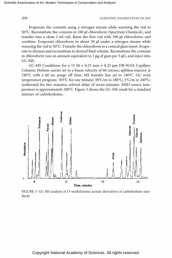
Scientific Examination of Art: Modern Techniques in Conservation and Analysis
Copyright National Academy of Sciences. All rights reserved.
204 SCIENTIFIC EXAMINATION OF ART
FIGURE 3 GC-MS analysis of O-methyloxime acetate derivatives of carbohydrate stan-dards.
Evaporate the contents using a nitrogen stream while warming the vial to50°C. Reconstitute the contents in 100 µl chloroform (Spectrum Chemical), andtransfer into a clean 2 ml vial. Rinse the first vial with 100 µl chloroform, andcombine. Evaporate chloroform to about 50 µl under a nitrogen stream whilewarming the vial to 50°C. Transfer the chloroform to a conical glass insert. Evapo-rate to dryness and reconstitute to desired final volume. Reconstitute the contentsin chloroform (use an amount equivalent to 1 µg of gum per 5 µl), and inject intoGC-MS.
GC-MS Conditions for a 15 M × 0.25 mm × 0.25 µm DB-WAX CapillaryColumn: Helium carrier set to a linear velocity of 60 cm/sec; splitless injector at240°C with a 60 sec purge off time; MS transfer line set to 240°C. GC oventemperature program: 105°C for one minute; 30°C/m to 180°C; 5°C/m to 240°C;isothermal for two minutes; solvent delay of seven minutes. MSD source tem-perature is approximately 200°C. Figure 3 shows the GC-MS result for a standardmixture of carbohydrates.

Scientific Examination of Art: Modern Techniques in Conservation and Analysis
Copyright National Academy of Sciences. All rights reserved.
PAINT MEDIA ANALYSIS 205
TABLE 9 Quantitation Ions for MOA Derivatives
Analyte m/z Analyte m/z
Allose 115.1 Xylose 115.1Rhamnose 129.2 Mannose 131.2Fucose 129.2 Fructose 203.2Ribose 115.1 Glucose 89.2Arabinose 115.1 Galactose 131.2
Calibration parameters: See Table 9 for the list of quantitation ions for theMOA derivatives. Using a linear curve fit forcing through the origin gives correla-tion coefficients of 0.995 or better for most analytes over the calibration range of2 to 50 ppm.

Scientific Examination of Art: Modern Techniques in Conservation and Analysis
Copyright National Academy of Sciences. All rights reserved.
206
A Review of Some Recent Researchon Early Chinese Jades
Janet G. DouglasDepartment of Conservation and Scientific Research
Freer Gallery of Art/Arthur M. Sackler GalleryWashington, D.C.
ABSTRACT
Chinese jades produced in the earliest periods of China, during the Neolithicperiod (5000 to 1700 BCE) to the Han dynasty (206 BCE to 220 CE), weretypically fashioned by abrasive techniques using fine mineral powders with-out the advantage of metal tools. Most of these jades are composed of neph-rite, a fine-grained variety of the tremolite-actinolite series of amphiboles,although other stone materials were used as well. The study of early Chinesejades using scientific techniques is a relatively narrow field aimed at devel-oping the cultural and archaeological contexts of these materials. The pri-mary areas of investigation include mineralogical identification, geologicalsource of jade, early jade working methods, detection of heating in jade,burial alteration ,and surface accretions. Research in this field is particu-larly exciting given the large number of excavations in China during thepast few decades.
INTRODUCTION
In early China most jade manufacturing involved abrasion, using fine mineralpowders, without the advantage of metal tools. The material of choice was neph-rite, a fine-grained variety of the tremolite-actinolite series of amphiboles, al-though other stone materials were used as well. Nephrite is a calcium magnesiumhydroxyl silicate that occurs in a massive form consisting of interlocking fibrouscrystals (Hurlbut and Switzer, 1979). Another jade material, jadeite, was not

Scientific Examination of Art: Modern Techniques in Conservation and Analysis
Copyright National Academy of Sciences. All rights reserved.
A REVIEW OF SOME RECENT RESEARCH ON EARLY CHINESE JADES 207
known in China until the eighteenth century, when it was imported fromMyanmar (Burma) for working by Chinese artisans.
Scientific study of Chinese jades produced in the earliest periods of China,during the Neolithic period (5000 to 1700 BCE) to the Han dynasty (206 BCE to220 CE), is leading to a richer understanding of these early cultures and their useof jade. Study of well-documented, preferably excavated Chinese jades is helpingto place into context those jades of uncertain origin and address issues ofauthentication.
MINERALOGICAL IDENTIFICATION
During the last few decades, analysis of early Chinese jades has focused on theidentification of the mineral content of jade materials. In China over 500 exca-vated jades from a wide variety of sites dating from the Neolithic period to theHan dynasty have been analyzed for their mineral content at the Chinese Acad-emy of Geological Sciences (Wen, 1996, 1997, 1998; Wen and Jing, 1992). Manyof the over 800 early jades at the Freer and Sackler galleries have been analyzedfor mineral content, thus making this collection one of the most extensivelystudied in the West. In the last decade minimally invasive analytical methodssuch as X-ray diffraction (XRD) and Fourier transform infrared spectroscopy(FTIR) have been routinely used for identification. Most of these early Chinesejades have been found to be composed of nephrite, a fine grained, massive vari-ety of tremolite-actinolite. Other materials identified include serpentine, marble,olivine, and corundum. Examples of FTIR spectra from three of these jades aregiven in Figure 1. Similar findings appear throughout a wide range of Chinesearchaeological reports.
In addition to the study of individual jades, some composite works havebeen studied in detail, such as the Freer Gallery’s jade and gold pectoral from theJincun site in Henan province, dating to about the third century BCE (Douglasand Chase, 2001). The pectoral consists of 10 jades attached to a gold chain, andwas examined to determine whether its configuration was correct. The jadeswere found to be similar in material and workmanship and consistent with otherjades from the site. The pectoral, however, was found to be a pastiche where thejades were attached to the gold chain with modern gold wires and cut links fromthe chain.
GEOLOGICAL SOURCE OF JADE
Both nephrite and jadeite are known to occur in geological environments throughmetasomatic processes in a variety of worldwide localities (Harlow and Sorensen,2001). Two major types of geologic occurrences of Chinese nephrite are known:nephrite associated with metamorphosed dolomitic marbles and nephrite associ-ated with serpentinites.

Scientific Examination of Art: Modern Techniques in Conservation and Analysis
Copyright National Academy of Sciences. All rights reserved.
208 SCIENTIFIC EXAMINATION OF ART
The geological sources of nephrite used by early cultures in China are notcurrently known. Such sources may have been depleted in antiquity, as nephritecan occur in small localized deposits. Research involving scientific methods onearly Chinese jades has been addressing issues related to the geological origin ofnephrite in early China, as well as jade production and use (Douglas, 2003).Analysis of early Chinese jades at the Freer and Sackler galleries using X-rayfluorescence spectroscopy (XRF) indicates that the geological sources of the ma-terial used for these jades are most likely associated with dolomitic marbles. The145 jades analyzed by XRF were found to be consistently low in Cr2O3 (< 0.08percent by wt.) and NiO (< 0.01 percent by wt.), characteristic of nephrites asso-ciated with dolomitic marbles. Future work on geological sourcing of nephriteshould concentrate on these types of deposits in China.
Of particular interest are the FeO and MnO contents, which have been deter-mined by XRF to point to simple source patterns for the nephrite used by theNeolithic cultures of Hongshan, Liangzhu, and Longshan, possibly involving oneor more related geologic sources for each culture. Longshan jades were found tohave unusually high FeO (0.35-17.95 percent by wt.) and MnO (0.02-0.89 percentby wt.) contents, suggesting a source particularly rich in iron and manganese.Jades of the Shang and Western Zhou dynasties show a wide range of composi-tions, suggesting multiple nephrite sources for these objects. XRF is a simplenoninvasive tool for determining minor elemental oxide concentrations, but fur-ther work on jade sources will need to involve an expanded suite of analyticalmethods on a wider range of jade objects and geological samples from China.
FIGURE 1 FTIR spectra of some early Chinese jades.

Scientific Examination of Art: Modern Techniques in Conservation and Analysis
Copyright National Academy of Sciences. All rights reserved.
A REVIEW OF SOME RECENT RESEARCH ON EARLY CHINESE JADES 209
EARLY JADE WORKING METHODS
Jade working methods have been investigated by a variety of researchers, and weare beginning to understand how early jades were worked. This type of study caninclude examination of tool marks on finished and unfinished jades as well asremains from jade working (Wu, 1994). It is particularly important to understandthe working methods used on jades of the Neolithic Hongshan culture because ofthe large numbers of forgeries that have been produced (So and Douglas, 1998;Forsythe, 1990).
The remains of one jade workshop were discovered in 1997 north ofDingshadi near Nanjing in the proximity of the remains of the Neolithic culturesof Majiaban, Songze, and Liangzhu (Lu and Tao, 2001). The Nanjing MuseumInstitute of Archaeology and the Institute of Geological Research of Huadong arecurrently excavating this area. To date, the workshop has yielded a variety of stonetools that may have been used to work jade through cutting, drilling, surfaceabrading, polishing, and incising. Raw jade pebbles can still be found along anearby river’s bank that may have been a source of jade for craftsmen during theNeolithic period.
The Lingjiatan site in Anhui province was discovered in 1987, and jadesyielded from the site are being studied with the aid of stereomicroscopy (Zhanget al., 2002). The Lingjiatan site is thought to be the location of the earliestagriculture-based city in China, dating to 4000 BCE or earlier. A proficient jade-producing culture inhabited the area as evidenced by the approximately 1,200jades that have been unearthed there. This work is showing the presence ofhighly developed working methods, and evidence of the earliest use of the “tuo,”a small rotary disk tool to create fine incised decoration. A cutting edge of thetuo would be similar to the flat head of a nail, although other shapes could havebeen used for different purposes. In most cases the technique for drilling holesthrough jade was typically done from both sides of the object. The high level ofcraftsmanship is exemplified by the glossy polish on these jades, which has leftlittle or no surface striation visible under the stereomicroscope.
Tool marks preserved on jades from the collection at the British Museumhave begun to be studied using detailed impressions with silicone dental resin(Michaelson and Sax, 2003), which follows from previous work on Mesopotamianseals composed of several quartz varieties. Impressions of small tool marks fromjades are imaged using scanning electron microscopy (SEM), which greatly facili-tates examination and documentation of these marks for comparison amongjades. One Eastern Zhou jade plaque mentioned in this work was worked withseveral different handheld tools.
Polishing techniques used in early China has been a largely unexplored areaof research, but quartz sand and related fine-grained materials are generally ac-cepted as the abrasives employed. After a Liangzhu corundum-rich axe was stud-ied at the Freer Gallery of Art in 1998, a fragment from a similar axe was studied

Scientific Examination of Art: Modern Techniques in Conservation and Analysis
Copyright National Academy of Sciences. All rights reserved.
210 SCIENTIFIC EXAMINATION OF ART
in a polishing replication experiment using commercially available abrasives thatapproximate natural diamond and corundum (Lu et al., 2005). The resultingpolished surfaces were compared with the original surface polish produced byLiangzhu jade workers in antiquity. The polished surfaces were examined usingoptical and electron microscopes and characterized using atomic force micros-copy. Three abrasives were used to polish the corundum-rich stone, and thediamond-polished surface most closely matched the surface polished in antiquity.The data suggest that extremely hard mineral abrasives composed of diamondmay have been used to polish jades to the high gloss observed on these jadestoday. Likewise, corundum would have been another likely hard abrasive used.
Ornamental jade rings from the Spring and Autumn period (771 to 475 BCE)are decorated with spiral grooves that were created through a mechanical methodinvolving the use of a precision compound machine (Lu, 2004). Such rings musthave had their spiral design drafted or directly carved through precisely linkedrotational and linear motion of the type that has been demonstrated in recentexperiments. These findings imply greater mechanical sophistication than haspreviously been assumed for this period in ancient China.
DETECTION OF HEATING
Some physical and chemical changes that occur with the heating of nephrite areknown from studies of the amphibole group minerals, tremolite-actinolite(Whittels, 1951; Vermaas, 1952). The dehydration of actinolite occurs in threestages, including the loss of adsorbed water, the loss of structural water, and a verysmall quantity of absorbed water. Studies using differential thermal analysis(DTA) show that an exothermic reaction takes place between 815oC and 824oC,and is associated with the oxidation of the small amounts of ferrous iron presentin the mineral. This oxidation is not associated with any structural change in thecrystal structure. Structural water is liberated at temperatures between 930oC and988oC, and at lower temperatures with increasing iron in the mineral structure.This change occurs through a solid-state reaction:
Ca2Mg5Si8O22(OH)2 + heat → 2 CaSiO3.5MgSiO3 + SiO2 + H2O [1]
nephrite (tremolite) pyroxene (diopside) cristobalite water
Detection of heating in jades using minimally invasive analytical methods is ofinterest because some jades may have been heated in antiquity prior to working orduring burial rituals involving burning. Heat treatment may also be used in theproduction of modern-day forgeries to make jade appear older due to naturalweathering or alteration. At the Freer and Sacker galleries, XRD and FTIR havebeen used to detect heating in jade, but these techniques have been found to besuccessful only if the object has been heated to at least 900oC (Douglas, 2001).
In this study a nephrite pebble was sliced and heated in 100oC increments

Scientific Examination of Art: Modern Techniques in Conservation and Analysis
Copyright National Academy of Sciences. All rights reserved.
A REVIEW OF SOME RECENT RESEARCH ON EARLY CHINESE JADES 211
from 500oC to 1100oC to observe visual changes and to investigate XRD and FTIRas methods to identify heating in jade. This heating series is shown in Figure 2,along with an example of a heated jade bracelet dating to the Neolithic or Shangdynasty. The heating series samples became more white and opaque with heating.Vickers hardness measurements on the heated samples showed that nephrite be-comes slightly harder, rather than softer up to 800oC. After this temperature thematerial becomes brittle and tends to fracture more easily. In addition, blackareas developed in the nephrite upon heating, which then became brown at 900oC.This black coloration may be due either to carbonization of small amounts oforganic material trapped in crevices or oxidation of iron in the nephrite.
The applicability of noninvasive Raman spectroscopy to the detection ofheating in jade is also being investigated on the same nephrite heating series(unpublished information from P. P. Knops-Gerrits). Some of the XRD, FTIR,and microRaman data that can be used to identify heating in jade composed ofnephrite are summarized in Table 1.
BURIAL ALTERATION AND SURFACE ACCRETIONS
Burial alteration is a particular type of alteration known to occur on early Chinesejades composed of nephrite. Such alteration usually appears as opaque, white,chalky areas on otherwise translucent, polished jades. In many cases these patchy
FIGURE 2 (a) Heating series of low-iron (tremolitic) nephrite heating from Hetian (Kho-tan), Xinjiang province (nephrite slice length approximately 3 cm). (b) Heated bracelet(F1917.43) dating to the Neolithic period or Shang dynasty (bracelet diameter 6.0 cm).

Scientific Examination of Art: Modern Techniques in Conservation and Analysis
Copyright National Academy of Sciences. All rights reserved.
212T
AB
LE
1 S
ome
XR
D, F
TIR
, an
d M
icro
Ram
an D
ata
Th
at C
an B
e U
sed
to I
den
tify
Hea
tin
g in
Jad
e B
ased
on
a N
eph
rite
Hea
tin
g E
xper
imen
t
XR
Da
FT
IRb
Pea
ks
Hea
tin
g Se
ries
Dif
fuse
8.38
ÅR
el %
TSa
mpl
ed-
spac
ings
d-sp
acin
g36
70 c
m–1
1075
cm
–1(4
75 a
nd
512
cm1 )
Mic
roR
aman
c Pea
ks
Un
hea
ted
_+
+_
475
cm–1
393,
368
, 34
8, 1
058,
102
9, 9
30 c
m–1
500o
C_
++
_47
5 cm
–139
3, 3
69,
349,
105
8, 1
028,
930
cm
–1
600o
C_
++
_47
5 cm
–139
1, 3
65,
349,
105
8, 1
027,
930
cm
–1
700o
C_
++
_47
5 cm
–139
1, 3
65,
1058
, 10
26,
929
cm–1
800o
C_
++
_47
5 cm
–139
1, 3
67,
1058
, 10
25,
927
cm–1
900o
C+
++
+51
2 cm
–139
1, 3
23,
1340
, 10
10 c
m–1
1000
oC
+_
_+
512
cm–1
392,
327
, 13
41,
1011
cm
–1
1100
oC
+_
_+
512
cm–1
392,
327
, 13
41,
1013
cm
–1
NO
TE
: _ a
bsen
t; +
pre
sen
t.a P
hili
ps R
G-2
600
X-r
ay d
iffr
acto
met
er w
ith
Gan
dofi
cam
era.
b Mat
tson
Alp
ha
Cen
tau
ri F
ouri
er t
ran
sfor
m in
frar
ed s
pect
rom
eter
.c R
enis
haw
R10
00 m
icro
Ram
an s
pect
rom
eter
, 785
nm
exc
itat
ion
.

Scientific Examination of Art: Modern Techniques in Conservation and Analysis
Copyright National Academy of Sciences. All rights reserved.
A REVIEW OF SOME RECENT RESEARCH ON EARLY CHINESE JADES 213
areas of alteration are softer than the unaltered areas of the jade. This type ofalteration consists of a selective dissolution or leaching on a microscopic scalealong mineral grain boundaries by solutions of high pH (pH > 9) rather than amineralogical change (Gaines and Handy, 1975). This type of high-pH environ-ment can occur during decay of the corpse(s) with which the jades were buried.Experiments to produce burial alteration on jade indicate that it is likely that thistype of alteration occurs during the months immediately after the burial when acorpse decomposes (Aerts et al., 1995).
Optical coherence tomography (OCT) is a noninvasive technique that isbeing used to study the subsurface morphologies of jade objects to determinewhether surface whitening is due to burning or natural alteration (Yang et al.,2004). Tomography images are used to show the refractive index or dielectricconstant variations in jades, which reflect their internal structures. To date, OTChas been applied to a relatively small number of early jades but may prove to beuseful in the future to answer questions relating to the authenticity of jade objects.
Surface accretions remain an unexplored area of focused research, probablybecause it can be difficult to determine the significance and relative age of suchdeposits. Many jades are heavily cleaned and waxed, which often obliterates anyaccretions on their surfaces. Other accretions may unintentionally find their wayto the surface of a jade but are typically not related to its early history. Earthyencrustations typically include calcareous deposits and soil. Occasionally lacquerand other organic remains can be seen.
CONCLUSIONS AND FUTURE DIRECTIONS
Research using scientific techniques is helping us to understand the mineral com-position and early history of well-documented and excavated jades. Similar workon unknown jades is helping to solve questions of authenticity (Douglas, 2000).Such study is most fruitful if it can include thorough visual examination using astereomicroscope along with comparison to similar, preferably excavated jades.No direct methods of dating jade materials exist.
Some future areas for research include
1. identification and distribution of surface accretions, weathering, and al-teration;
2. continued study of jade working methods, with particular emphasis onthe study of large groups of related jades from individual sites and cultural areas;
3. study of jade working remains, including tools and jade debris;4. analytical and technical methods of dating jade workmanship; and5. study of early jades from areas neighboring China, such as Korea, Taiwan,
Siberia, and Southeast Asia, as all of these areas had jade-producing cultures.
Scientific Examination of Art: Modern Techniques in Conservation and Analysis
Copyright National Academy of Sciences. All rights reserved.
214 SCIENTIFIC EXAMINATION OF ART
REFERENCES
Aerts, A., K. Janssens, and F. Adams. 1995. Orientations Nov.:79-80.Douglas, J. G. 2000. Orientations Feb.:86.Douglas, J. G. 2001. Proceedings of the Conference on Archaic Jades across the Taiwan Straits. Taipei:
Guo li Taiwan da xue li xue yuan di zhi ke xue xi yin xing and Guo li Taiwan da xue chu ban weiyuan hui. pp. 543-554.
Douglas, J. G. 2003. In Scientific Research in the Field of Asian Art, Proceedings of the first ForbesSymposium at the Freer Gallery of Art, ed. P. Jett, with J. G. Douglas, B. McCarthy, and J. Winter,pp. 192-199. London: Archetype Publications in association with the Freer Gallery of Art,Smithsonian Institution, Washington, D.C.
Douglas, J. G., and W. T. Chase. 2001. Studies in Conservation 46:35-48.Forsythe, A. 1990. Orientations May:54-63.Gaines, A. M., and J. L. Handy. 1975. Nature 253:433-434.Harlow, G., and S. Sorensen. 2001. Australian Gemmologist 21:7-10.Hurlbut, C. S., and G. S. Switzer. 1979. Gemology. 243 pp., Canada: John Wiley.Lu, J., and H.Tao. 2001. In Enduring Art of Jade Age China, vol. 2, ed. E. Childs-Johnson, pp. 31-42.
New York: Throckmorton Fine Art.Lu, P. 2004. Science 304:38.Lu, P. J., N. Yao, J. F. So, G. E. Harlow, J. F. Lu, G. F. Wang, and P. M. Chaikin. February 2005.
Archaeometry 47:1-12.Michaelson, C., and M. Sax. 2003. APOLLO Nov.:3-8.So, J. F., and J. G. Douglas. 1998. In East Asian Jades: Symbol of Excellence, vol. 1, ed. C. Tang, pp.148-
163: Hong Kong: Chinese University of Hong Kong.Vermaas, F. H. S. 1952. Transactions of the Geological Society of South Africa 55:1.Wen, G. 1996. Acta Geological Taiwanica 32:55-83.Wen, G. 1997. Chinese Jades-Colloquies on Art & Archaeology in Asia 18:105-122.Wen, G. 1998. In East Asian Jades: Symbol of Excellence, vol. 2, ed. C. Tang, pp. 217-221. Hong Kong:
Chinese University of Hong Kong.Wen, G., and Z. Jing. 1992. Geoarchaeology 7:251-255.Whittels, M. 1951. American Mineralogist 36:851.Wu, T. 1994. Renshi Guyu: Gudai yuqi zhizuo yu xingzhi. Taiwan: Zhonghua ziran wenhua xuehui.Yang, M. L., C. W. Lu, I. J. Hsu, and C. C. Yang. 2004. Archaeometry 46(2):171-182.Zhang J., Z. Yang, and Q. Cheng. 2002. Dong nan wen hua 5:17-27.

Scientific Examination of Art: Modern Techniques in Conservation and Analysis
Copyright National Academy of Sciences. All rights reserved.
APPENDIXES

Scientific Examination of Art: Modern Techniques in Conservation and Analysis
Copyright National Academy of Sciences. All rights reserved.
217
APPENDIX
A
Contributors
Roy S. Berns is the Richard S. Hunter Professor in Color Science, Appearance,and Technology at the Munsell Color Science Laboratory and Graduate Coordi-nator of the Color Science master’s degree program within the Center for ImagingScience at Rochester Institute of Technology. He received B.S. and M.S. degrees intextile science from the University of California at Davis and a Ph.D. degree inchemistry with an emphasis in color science from Rensselaer Polytechnic Insti-tute. His research includes spectral-based imaging, archiving, and reproductionof cultural heritage; algorithm development for multi-ink printing; the use ofcolor and imaging sciences for art conservation science; and colorimetry. He isactive in the International Commission on Illumination, the Council for OpticalRadiation Measurements, the Inter-Society Color Council, and the Society forImaging Science and Technology. He has authored over 150 publications includ-ing the third edition of Billmeyer and Saltzman’s Principles of Color Technology.During the 1999-2000 academic year, he was on sabbatical at the National Galleryof Art, Washington, D.C., as a Senior Fellow in Conservation Science. During2000, Dr. Berns was invited to participate in the Technical Advisory Group of theStar-Spangled Banner Preservation Project. He is currently involved in a jointresearch program in museum imaging with the National Gallery of Art, Washing-ton, D.C., and the Museum of Modern Art, New York. He is also collaboratingwith the Art Institute of Chicago and the Van Gogh Museum in digitally rejuve-nating paintings that have undergone undesirable color changes.
Barbara H. Berrie is senior conservation scientist at the National Gallery of Art,Washington, D.C. She received her B.Sc.(Hons) in chemistry from St. Andrews

Scientific Examination of Art: Modern Techniques in Conservation and Analysis
Copyright National Academy of Sciences. All rights reserved.
218 APPENDIX A
University, Scotland, and her Ph.D. on electron transfer reactions fromGeorgetown University. She was awarded a National Research CouncilPostdoctoral Fellowship at the Naval Research Laboratory where she investi-gated the reaction of carbon dioxide with low-valent palladium compounds. Shehas worked at the National Gallery since 1984. Dr. Berrie has always been inter-ested in the alchemy of turning base materials into art; now she is involved instudying the materials and painting methods of artists and analysis of materialsin works of art in order to understand the artist’s original intention, and addressissues of authenticity and preservation. She has used chemical analysis in thestudy of over 300 works of art in all media, including works on paper, easelpaintings, and sculpture. She has published on paintings by Dosso Dossi, GerardDavid, and Orazio Gentileschi among others and on the watercolors of WinslowHomer. Berrie is a Fellow of the International Institute for Conservation. She isthe editor of the forthcoming volume of Artists’ Pigments that will be publishedby the National Gallery of Art.
Robin J. H. Clark is the Sir William Ramsay Professor of Chemistry and formerDean of Science at University College London. His research on physical inorganicchemistry and spectroscopy is concerned with synthesis, characterisation, andstructure, focusing mainly on the electronic and vibrational spectroscopy of inor-ganic compounds, on matrix isolation infrared spectroscopy of photochemicallygenerated species, and on infrared-based spectroelectrochemistry of redox-activespecies. In particular, he has made seminal contributions to virtually all aspects ofRaman spectroscopy, notably to the characterisation of deeply coloured materials(e.g., TiI4) and to metal-metal bonded (e.g., M2X8
n-) and linear-chain species, togas-phase Raman band contour analysis, to Raman band intensities and the na-ture of the chemical bond, to the theory and practice of resonance Raman spec-troscopy (including its application to the determination of excited state geom-etries), to nanostructures and thin, photoactive oxide/sulfide films on glass and,at the Arts/Science interface, to the application of Raman microscopy to thecharacterisation of pigments on medieval manuscripts, paintings, icons, papyri,sherds, and other artefacts. His research is embodied in nearly 500 scientific pa-pers, 3 books, and 36 edited books. He has acted as Visiting Professor in 13universities and has lectured in over 350 universities and institutions in 33 coun-tries throughout the world. He was elected Hon FRSNZ (1989), FRS (1990),FRSA (1992), FUCL (1992), Hon DSc(Cant 2001), HonFRI (2004), and a Com-panion of the New Zealand Order of Merit (CNZM, 2004).
John K. Delaney has been a scientific consultant to the Conservation Division ofthe National Gallery of Art, Washington, D.C., since 1990, and has consulted oninfrared imaging applications for other major American museums. He has pub-lished over 20 peer-reviewed papers on spectroscopy and several papers on infra-red imaging in art conservation. He received his Ph.D. in Biophysics from The

Scientific Examination of Art: Modern Techniques in Conservation and Analysis
Copyright National Academy of Sciences. All rights reserved.
APPENDIX A 219
Rockefeller University. He completed post-doctoral studies in the spectroscopy ofbiomolecules at the University of Arizona’s Department of Chemistry and theDepartment of Biological Chemistry at John Hopkins University’s School of Medi-cine. He is currently Chief Scientist for the Business Unit of Surveillance andReconnaissance Systems, which is a part of Optical and Space Systems of GoodrichCorporation.
Janet G. Douglas is a Conservation Scientist in the Arthur M. Sackler Gallery andFreer Gallery of Art’s Department of Conservation and Scientific Research at theSmithsonian Institution. Her area of research involves the analysis of a variety ofinorganic materials relating to Asian art such as jade, stone, pigments, metals, andcorrosion products. She holds an M.A. in Geology (Metamorphic petrology) fromBryn Mawr College, awarded in 1980. She was a mineralogist at the U.S. Bureau ofMines for 5 years, involved in asbestos research. At the Freer and Sackler Galler-ies, her work involves research on Asian art and archaeological materials to an-swer questions relating to their authenticity, cultural context, and method ofmanufacture. Recent projects involve the mineralogical study of early Chinesejades, characterization of glass and stone gokok beads from Korea, and petro-graphic analysis of stone sculpture from Cambodia.
Molly Faries received her Ph.D. from Bryn Mawr College in 1972. During heryears at Indiana University/Bloomington from 1975 on, she directed two long-term infrared reflectography (IRR) research projects: one a National Endowmentfor the Humanities Basic Research Grant (1984-1987) and the second, a SamuelH. Kress Foundation Grant for Art Historical Study Using Infrared Reflectography(1990-1997). Since 1998, she has also held a chair in Technical Studies in ArtHistory at the University of Groningen in the Netherlands. Currently, she is in-volved in research for the catalogue of the fifteenth- and sixteenth-century north-ern collection of the Centraal Museum, Utrecht (funded by the Mondrian Foun-dation), and she is CPI for a project entitled, “Infrared Reflectography: EvaluativeStudies,” in the interdisciplinary De Mayerne Program funded by the Dutch Or-ganization for Scientific Research (NWO), linking the exact sciences, conserva-tion, and art history. In 1995, for her many publications in the field of northernEuropean painting, she was awarded the College Art Association/National Insti-tute for Conservation Joint Award for Distinction in Scholarship and Conserva-tion, and in 2001, she was awarded the American Institute for ConservationCaroline and Sheldon Keck Award for Excellence in Education. Recent publica-tions include The Madonnas of Jan van Scorel, Serial Production of a CherishedMotif (2000) and Recent Developments in the Technical Examination of Early Neth-erlandish Painting: Methodology, Limitations & Perspectives (2003).
Colin F. Fletcher is a Program Manager of Mouse Genetics at the GenomicsInstitute of the Novartis Research Foundation (GNF). His area of research is the

Scientific Examination of Art: Modern Techniques in Conservation and Analysis
Copyright National Academy of Sciences. All rights reserved.
220 APPENDIX A
genetic analysis of mouse models of human disease, specifically neurologicalmutants that display ataxia. While at GNF he was a scientific co-founder ofPhenomix Corp. Prior to joining GNF, he was a staff scientist in the MammalianGenetics Laboratory at the National Cancer Institute. In the course of his researchDr. Fletcher employs a variety of imaging modalities, including confocal micros-copy, magnetic resonance, X-ray, and low-light luciferase imaging with cryogeni-cally cooled CCDs. A long-standing interest in the scientific examination of worksof art has lead to the previous publication of two reports in Studies in Conserva-tion. Dr. Fletcher received his Ph.D. from The Rockefeller Institute in Biochemis-try and Molecular Biology and his A.B. from Dartmouth College in Biochemistry.
Joyce Hill Stoner has taught for the Winterthur/University of Delaware (UD)Program in Art Conservation for 29 years and served as its director for 15 years(1982-1997). She graduated Phi Beta Kappa summa cum laude from the Collegeof William and Mary in 1968. She received her Master’s degree in Art History atthe Institute of Fine Arts of New York University (1970), her diploma in conser-vation at the NYU Conservation Center (1973), and a Ph.D. in Art History (1995,UD). She has been a Visiting Scholar in Painting Conservation at the Metropoli-tan Museum and at the J. Paul Getty Museum. In 1976, she founded the oralhistory project for the Foundation of the American Institute for Conservationand has interviewed more than 45 major art conservation professionals interna-tionally. Both an art historian and a practicing paintings conservator, Stoner hastreated paintings for many museums and private collectors and was senior con-servator of the team for the five-year project of examination and treatment ofWhistler’s Peacock Room at the Freer Gallery of Art in Washington, D.C. Stonerhas authored more than 60 book chapters and articles, and has recently beenstudying the paintings of the Wyeth family. She is currently serving as a VicePresident of the Board of Directors of the College Art Association and a VicePresident of the Council of the International Institute for Conservation. In June2003 she received the AIC “Lifetime Achievement Award” sponsored by Univer-sity Products.
Tom Learner is a Senior Conservation Scientist at Tate in London, the UK’snational collection of British and international 20th/21st century art. He receiveda Master’s degree in Chemistry from Oxford University in 1988 and a Diploma inthe Conservation of Easel Paintings from the Courtauld Institute of Art, Londonin 1991. He spent a year as a Getty Intern in the Painting Conservation andScientific Research Departments at the National Gallery of Art (NGA), Washing-ton, D.C., and then joined the Conservation Department of the Tate Gallery in1992, where he established appropriate analytical protocols for the identificationand characterisation of twentieth-century painting materials with Fourier Trans-form infrared spectroscopy (FTIR) and pyrolysis-gas chromatography-mass spec-trometry (PyGCMS). During this time he received his Ph.D. in Chemistry on The
Scientific Examination of Art: Modern Techniques in Conservation and Analysis
Copyright National Academy of Sciences. All rights reserved.
APPENDIX A 221
Characterisation of Acrylic Painting Materials and Implications for their Use, Con-servation and Stability from Birkbeck College, University of London in 1997. Hewas a guest scholar at the Getty Conservation Institute (GCI) in 2001, assessinganalytical techniques to follow changes in artists’ acrylic emulsion paints withaccelerated light aging and with water immersion. He has written two books: TheImpact of Modern Paints, co-authored with Jo Crook and published in 2000, andThe Analysis of Modern Paints, published in 2004. He is currently coordinating acollaborative research venture into modern paints between the GCI, the NGA,and Tate, in which three initial areas of focus are improving methods for chemicalanalysis, studying their physical properties and assessing cleaning treatments.
Carol Mancusi-Ungaro serves as Associate Director for Conservation and Re-search at the Whitney Museum of American Art and Founding Director of theCenter for the Technical Study of Modern Art at Harvard University Art Muse-ums. She graduated with a Bachelor of Arts degree from Connecticut College in1968 and a Master of Arts degree from the Institute of Fine Arts, New YorkUniversity in 1970. She trained and worked in conservation at the Yale UniversityArt Gallery until she assumed a position of Conservator of Paintings at the BritishArt Center at Yale. Subsequent positions included Conservator of Paintings at theJ. Paul Getty Museum in Malibu, California, and at the Intermuseum Conserva-tion Association in Oberlin, Ohio. For 19 years she served as Chief Conservator ofThe Menil Collection in Houston, Texas, and during that time she also consultedon the conservation of twentieth-century paintings at the National Gallery of Artin Washington, D.C. She has lectured widely on the conservation of modern artand written for retrospective catalogues on Mark Rothko and Jackson Pollockand most recently for the catalogue raisonné of Barnett Newman. In 2004 shereceived the College Art Association/Heritage Preservation Award for Distinctionin Scholarship and Conservation. In her joint position, she teaches undergraduateand graduate students at Harvard University and continues to engage in researchdocumenting the materials and techniques of living artists as well as other issuespertaining to the conservation of modern art.
Louisa C. Matthew received her Ph.D. in Italian Renaissance Art History fromPrinceton University with a thesis on Venetian painting. She received fellowshipsfrom the Delmas Foundation, the Harvard Center for Italian Renaissance Studiesat Villa I Tatti in Florence, Italy, and recently a paired Kress fellowship togetherwith Dr. Barbara Berrie at the Center for Advanced Studies in the Visual Arts atthe National Gallery of Art in Washington, D.C. Currently, Dr. Matthew is anAssociate Professor of Art History at Union College in Schenectady, N.Y.
Christopher J. McNamara received his Ph.D. in Aquatic Ecology in 2001 fromthe Department of Biological Sciences at Kent State University. Since receiving hisdoctorate, he has worked in the Division of Engineering and Applied Sciences at

Scientific Examination of Art: Modern Techniques in Conservation and Analysis
Copyright National Academy of Sciences. All rights reserved.
222 APPENDIX A
Harvard University, first as a Postdoctoral Fellow and currently as a ResearchAssociate. His research focuses on the ecology of biofilm bacteria and he hasstudied biofilms in diverse systems such as streams and aircraft fuel tanks. He hasalso studied the role of biofilms in deterioration of cultural heritage materialssuch as limestone from Maya ruins, protective coatings for bronze statues, syn-thetic cloth in Apollo spacesuits, and wax sculptures by Edward Degas.
Ralph Mitchell is the Gordon McKay Professor of Applied Biology in the Divi-sion of Engineering and Applied Sciences at Harvard. His Laboratory of AppliedMicrobiology has as its focus the microbiology of surfaces. The laboratory inves-tigates the basic processes involved in the formation of biofilms on surfaces. Hisresearch group emphasizes the effects of biofilms on degradation of stone, metals,and artificial polymers. Current research in his laboratory involves the role ofmicroorganisms in the biodeterioration of Maya sites in Mexico and in microbialprocesses resulting in corrosion of metals in the U.S.S. Arizona memorial.
Richard Newman is Head of Scientific Research at the Museum of Fine Arts,Boston, where he has worked since 1986. His lab oversees scientific research onthe Museum’s collections carried out in collaboration with curatorial and conser-vation activities. One of his research interests is scientific methods for establishingthe provenance of stone sculptures and the application of alteration layers inhelping to resolve questions of authenticity. He is particularly interested in inter-disciplinary research projects on works of art involving scientists, conservators,and art historians, and subjects he has studied range from qero cups produced inthe Inka and colonial periods in Peru, to stone sculptures from the Indian sub-continent, to painting materials used in ancient Egypt. He collaborated with anart historian, conservator, and technical photographer in a 1988 book, ExaminingVelazquez, which received the 1991 Award for Distinction in Scholarship andConservation from the College Art Association and National Institute for Con-servation.
Thomas D. Perry IV is the Sandia National Laboratories Campus ExecutiveGraduate Fellow at Harvard University. His research involves understanding theprocesses of deterioration of materials, including stone, aluminum, and artificialpolymers, by microorganisms living in biofilms. Biofilms are thin films of micro-organisms living on surfaces that are capable of changing their surrounding envi-ronment through production of metabolites resulting in affected material dete-rioration. He is particularly interested in the specific mineral binding andmineralization caused by microbially produced polymers.
Michael R. Schilling earned his B.S. and M.S. degrees in chemistry from TheCalifornia State Polytechnic University, Pomona. He has worked at The GettyConservation Institute (GCI) since 1983 and presently holds the position of Se-

Scientific Examination of Art: Modern Techniques in Conservation and Analysis
Copyright National Academy of Sciences. All rights reserved.
APPENDIX A 223
nior Scientist in charge of the Analytical Research Section. Michael oversees andcoordinates a wide variety of projects in his section: applied research in materialsanalysis, scientific support to GCI’s field conservation projects, study of museumcollections, evaluation of the air quality in museums, assessment of safe levels oflighting in museum galleries, and characterization of building materials. Oneresearch area in which Analytical Research scientists have developed considerableexpertise is the characterization and analysis of organic materials. In this project,several gas chromatography-mass spectrometry procedures were developed forqualitative and quantitative analysis of natural organic binding media in paints.He and other scientists in the Analytical Research Section have conducted numer-ous workshops that were developed to inform conservation professionals aboutthese GC-MS procedures. Since 1997, Michael and his staff have been studyingthe materials and techniques of modern and contemporary artists. Much of thiswork has involved the analysis of modern synthetic binding media and syntheticorganic pigments. Michael has participated in collaborative projects to study andpreserve wall paintings in the tomb of Nefertari, located in Luxor, Egypt, and alsoin the Mogao Grottoes, which is near the city of Dunhuang in the Gansu Provinceof China. He was also a member of a GCI research team that studied the Dead SeaScrolls.
Elizabeth Walmsley is a painting conservator at the National Gallery of Art(NGA), Washington, D.C. She has also worked on the NGA’s systematic cata-logue project from which has stemmed her interests in the technical examinationof Old Master paintings using digital imaging, infrared reflectography, and x-radiography, and in the history of conservation. She graduated with an AB fromDartmouth College and received an M.A. in Art History with a Certificate in ArtConservation from Buffalo State College.
Paul M. Whitmore was trained as a chemist, getting a B.S. from Caltech and aPh.D. from the University of California at Berkeley. He has worked in art conser-vation science for his entire professional career, starting at the EnvironmentalQuality Laboratory at Caltech, working with Professor Glen Cass studying theeffects of air pollution on works of art. From there, he went to the Fogg ArtMuseum at Harvard University, where he worked as a scientist in what is now theStraus Center for Conservation. Since 1988 he has been at Carnegie Mellon Uni-versity, directing the Research Center on the Materials of the Artist and Conserva-tor. His current research interests are in material degradation chemistries, intrin-sic and environmental risk factors for those processes, and chemical sensors formaterial aging processes and risk factors. He has published on paper deteriora-tion, its treatment, and damage induced by humidity changes; acrylic paint mediastability and the physical damage to acrylic coatings from shrinkage stresses dur-ing drying; fading of colorants from air pollutant exposure; fading of transparentpaint glazes from light exposure and the relationship between photochemical

Scientific Examination of Art: Modern Techniques in Conservation and Analysis
Copyright National Academy of Sciences. All rights reserved.
224 APPENDIX A
degradation and color changes; and projects utilizing a new nondestructive probeof light stability for colored artifact materials. He has edited a book, Contributionsto Conservation Science, a compilation of research papers published by the firstdirector of the Center, Robert Feller. He is currently senior editor of the Journal ofthe American Institute for Conservation.
John Winter is a chemist (B.A., Cambridge University, Ph.D., University ofManchester) who, after a period of academic and industrial research, moved intothe field of archaeological science and then into research on works of art usingscientific methods. He holds the position of Conservation Scientist on the staff ofthe Department of Conservation and Scientific Research, Freer Gallery of Art/Arthur M. Sackler Gallery, Smithsonian Institution. These museums hold thenational collections of Asian art, which form the chief focus of research in thedepartment. Dr. Winter’s own studies have mainly centered around East Asianpaintings, their components, structural aspects, and the influence of microstruc-ture and macrostructure on deterioration processes. He has published work onChinese ink (the ubiquitous black design component across China, Japan, andKorea), lead-based white pigments, methods for the identification of organiccolorants used as design components or as support dyes, painting techniquesincluding those based on the use of precious metals, deterioration processes inEast Asian paintings, and on other aspects of paintings as physical objects. Dr.Winter is a past President of the International Institute for Conservation of His-toric and Artistic Works, and has received research support from the Andrew W.Mellon Foundation as well as from the Smithsonian Institution itself.