SBAs for the Final FRCR 2A - Perpustakaan ATRO Bali
150
-
Upload
khangminh22 -
Category
Documents
-
view
0 -
download
0
Transcript of SBAs for the Final FRCR 2A - Perpustakaan ATRO Bali


SBAs for the Final FRCR 2A
RWK Lindsay MB BCh BAO MRCS FRCRClinical Fellow in Interventional Radiology, McGill University Hospital, Montreal, Canada
JSJ Gillespie MB BCh BAO (Hons) MRCP FRCRConsultant Radiologist, Royal Victoria Hospital, Belfast, Northern Ireland
RM Kelly, MB BCh BAO (Hons) MRCP FRCRConsultant Radiologist, Antrim Area Hospital, Antrim, Northern Ireland
R Sathyanarayana, MB BS MRCS FRCRClinical Fellow in Interventional Radiology, Auckland City Hospital, Auckland, New Zealand
PA Burns BSc MB BCh BAO (Hons) MRCP FRCRSpecialty Registrar, Royal Victoria Hospital, Belfast, Northern Ireland
1

1Great Clarendon Street, Oxford, OX2 6DP,
United Kingdom
Oxford University Press is a department of the University of Oxford.It furthers the University’s objective of excellence in research, scholarship,
and education by publishing worldwide. Oxford is a registered trade mark ofOxford University Press in the UK and in certain other countries
© Oxford University Press 2012
The moral rights of the authors have been asserted
First Edition published in 2012
Impression: 1
All rights reserved. No part of this publication may be reproduced, stored ina retrieval system, or transmitted, in any form or by any means, without the
prior permission in writing of Oxford University Press, or as expressly permittedby law, by licence or under terms agreed with the appropriate reprographics
rights organization. Enquiries concerning reproduction outside the scope of theabove should be sent to the Rights Department, Oxford University Press, at the
address above
You must not circulate this work in any other formand you must impose this same condition on any acquirer
British Library Cataloguing in Publication Data
Data available
Library of Congress Cataloging in Publication Data
Library of Congress Control Number: 2012934790
ISBN 978–0–19–960776–1
Printed and bound byCPI Group (UK) Ltd, Croydon, CR0 4YY
Oxford University Press makes no representation, express or implied, that the drug dosages in this book are correct. Readers must therefore always check the product information and clinical procedures with the most up-to-date
published product information and data sheets provided by the manufacturers and the most recent codes of conduct and safety regulations. The authors and the publishers do not accept responsibility or legal liability for any errors in the text or for the misuse or misapplication of material in this work. Except where otherwise stated, drug dosages and recommendations are for the non-pregnant
adult who is not breast-feeding
Links to third party websites are provided by Oxford in good faith andfor information only. Oxford disclaims any responsibility for the materials
contained in any third party website referenced in this work.

To my wife Maura and my children Peter, Charlotte, and Alexander
RWKL
To my wife Janice and my children Katie, Harry, and Johnny
JSJG
To my wife Jane
RMK
To my wife Bharathi and my children Riya and Keshav
RS
To my wife Ciara and my son Oisin
PAB

In view of the recent changes in the FRCR 2A examination format from multiple true/false to single best answer (SBA), we decided to write SBAs for the Final FRCR 2A, appreciating that success very much depends on acquisition and repeated testing of knowledge. We feel well placed to write such a book due to our own recent examination success. In addition, included in our authors are a previous FRCR 2A gold medallist and a past chairman of a Regional MCQ panel for the FRCR part 2 examination.
The questions have been written in the format of the new FRCR 2A examination and are extensively referenced from current literature and leading radiology texts. As the book is divided into system modules, each chapter can be taken as a practice examination if desired. When preparing for the exam we found that those questions which were of most benefi t were those which were set at a level of diffi culty slightly above that of the actual exam and also provided detailed answers and explanations. There is little to be gleaned from undertaking questions the answers to which one already knows. Therefore do not be disheartened if some of the questions seem overly diffi cult. We hope that this book will not only provide a challenging test of knowledge but also, through the detailed answers, give a broad overview of radiology in general. Finally, we wish you all the best in your forthcoming exam!
RWKLJSJG
RMKRS
PAB
PREFACE
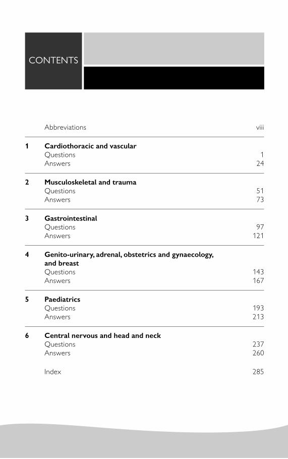
CONTENTS
Abbreviations viii
1 Cardiothoracic and vascular Questions 1Answers 24
2 Musculoskeletal and traumaQuestions 51Answers 73
3 Gastrointestinal Questions 97Answers 121
4 Genito-urinary, adrenal, obstetrics and gynaecology, and breast Questions 143Answers 167
5 PaediatricsQuestions 193Answers 213
6 Central nervous and head and neckQuestions 237Answers 260
Index 285

A&E accident and emergencyAAA abdominal aortic aneurysm AAST American Association for the Surgery of Trauma ABC aneurysmal bone cyst ABPA allergic bronchopulmonary aspergillosisACC agenesis of the corpus callosumACL anterior cruciate ligament ACTH adrenocorticotropic hormoneADC apparent diffusion coeffi cientADEM acute disseminated encephalomyelitisADI atlanto-dens interval AF atrial fi brillation AFP alphafetoprotein AH autoimmune hypophysitisALCAPA anomalous origin of the coronary artery from the pulmonary artery AP antero-posteriorAPACHE II Acute Physiology and Chronic Health Evaluation IIAPW absolute percentage washout ARDS acute respiratory distress syndromeARPKD autosomal recessive polycystic kidney disease ASD atrial septal defectASO arterial switch operationATN acute tubular necrosisAV atrioventricularAVN avascular necrosisAVM arteriovenous malformation AXR abdominal x-rayBAE bronchial artery embolizationBAL bronchoalveolar lavageBHL bilateral hilar lymphadenopathy BMI body mass index BOOP bronchiolitis obliterans organizing pneumoniaBPD bronchopulmonary dysplasia bpm beats per minuteCAD coronary artery disease CAH congenital adrenal hyperplasiaCBD common bile ductCBF cerebral blood fl ow CBV cerebral blood volume CCA common carotid artery
ABBREVIATIONS

ABBREVIATIONS ix
CCAM congenital cystic adenomatoid malformationsCDH congenital diaphragmatic herniaCEA carcino-embryonic antigenCF cystic fi brosisCHD common hepatic ductCho/Cr choline/creatineCMC carpometacarpalCMV cytomegalovirus CNS central nervous systemCOP cryptogenic organizing pneumoniaCOPD chronic obstructive pulmonary diseaseCPA cerebellopontine angleCPPD calcium pyrophosphate depositionCRMO chronic recurrent multifocal osteomyelitis CRP C-reactive proteinCSF cerebrospinal fl uidCSP cavum septum pellucidum CT computed tomographyCTA CT angiogram CTP CT perfusionCTPA CT pulmonary angiographyCV cavum vergaeCVI cavum vellum interpositum CXR chest X-rayDAD diffuse alveolar damage DAI diffuse axonal injury DAVF dural arteriovenous fi stulaDCIS ductal carcinoma in situ DIG desmoplastic infantile gangliogliomaDIP desquamative interstitial pneumoniaDNET dysembryoplastic neuroepithelial tumour DRE digital rectal examination DRUJ distal radio-ulnar jointDSA digital subtraction angiographyDVA developmental venous anomalyDWI diffusion-weighted imagingECA external carotid arteryECG electrocardiogramEDH extradural haematoma EEA extrinsic allergic alveolitisEEG electroencephalogrameGRF estimated glomerular fi ltration rate ENT ear, nose, and throatERCP endoscopic retrograde cholangiopancreatographyESR erythrocyte sedimentation rateESWL extracorporeal shock wave lithotripsy ET endotrachealEUS endoscopic ultrasoundFAP familial adenomatous polyposis FDG fl uorodeoxyglucose

x ABBREVIATIONS
FESS functional endoscopic sinus surgery FLAIR fl uid attenuated inversion recoveryFNA fi ne needle aspirationFFNH focal nodular hyperplasia FSE fast spin echoGBM glioblastoma multiforme GCS Glasgow Coma ScaleGCT giant cell tumour GDA gastroduodenal arteryGFR glomerular fi ltration rateGGO ground-glass opacifi cation GI gastrointestinalGIST gastrointestinal stromal tumourGP general practitionerGSF greater sciatic foramenHAART highly active antiretroviral therapyHb haemoglobin HCC hepatocellular carcinoma -HCG beta human chorionic gonadotrophin
HCM hypertrophic cardiomyopathy HHT hereditary haemorrhagic telangiectasiaHIDA hepatobiliary iminodiacetic acidHIFU high-intensity focused ultrasoundHIV human immunodefi ciency virus HLA human leucocyte antigenHMPAO hexamethylpropylene amine oximeHPOA hypertrophic pulmonary osteoarthropathyHPV human papilloma virusHRCT high-resolution CTHSG hysterosalpingogram HSV herpes simplex virus HU Hounsfi eld unitIAC internal auditory canalIAM internal auditory meatusICA internal carotid arteryICU intensive care unitIEE idiopathic eosinophilic oesophagitis ILC invasive lobular carcinoma IP interphalangealIUCD intrauterine contraceptive device IV intravenousIVC inferior vena cavaIVU intravenous urogramJGC juxtaglomerular cell JRA juvenile rheumatoid arthritis JVP jugular venous pressureKUB kidneys, ureters, and bladderLAD left anterior descendingLAO left anterior oblique LBBB left bundle branch block
ABBREVIATIONS xi
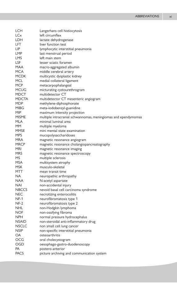
LCH Langerhans cell histiocytosis LCx left circumfl exLDH lactate dehydrogenaseLFT liver function testLIP lymphocytic interstitial pneumoniaLMP last menstrual period LMS left main stemLSF lesser sciatic foramenMAA macro-aggregated albumin MCA middle cerebral arteryMCDK multicystic dysplastic kidney MCL medial collateral ligament MCP metacarpophalangeal MCUG micturating cystourethrogram MDCT multidetector CTMDCTA multidetector CT mesenteric angiogramMDP methylene-diphosphonateMIBG meta-iodobenzyl-guanidine MIP maximum intensity projectionMISME multiple intracranial schwannomas, meningiomas and ependymomasMLA minimal luminal area MM multiple myelomaMMSE mini mental state examinationMPS mucopolysaccharidoses MRA magnetic resonance angiogram MRCP magnetic resonance cholangiopancreatographyMRI magnetic resonance imagingMRS magnetic resonance spectroscopy MS multiple sclerosisMSA multisystem atrophyMSK musculo-skeletalMTT mean transit timeNA neuropathic arthropathy NAA N-acetyl aspartateNAI non-accidental injury NBCCS nevoid basal cell carcinoma syndrome NEC necrotizing enterocolitis NF-1 neurofi bromatosis type 1 NF-2 neurofi bromatosis type 2NHL non-Hodgkin lymphoma NOF non-ossifying fi broma NPH normal pressure hydrocephalusNSAID non-steroidal anti-infl ammatory drug NSCLC non small cell lung cancerNSIP non-specifi c interstitial pneumoniaOA osteoarthritis OCG oral cholecystogram OGD oesophago-gastro-duodenoscopyPA postero-anteriorPACS picture archiving and communication system

xii ABBREVIATIONS
PAES popliteal artery entrapment syndrome PaO2 partial pressure of arterial O2 PCI percutaneous coronary intervention PCL posterior cruciate ligamentPCOS polycystic ovarian syndrome PCP P carinii pneumoniaPDA patent ductus arteriosusPIE pulmonary interstitial emphysemaPIOPED Prospective Investigation of Pulmonary Embolism DiagnosisPE Pulmonary embolismPET positron emission tomographyPET-CT positron emission tomography-computed tomographyPHPV persistent hyperplastic primary vitreousPI pulsatility index PML progressive multifocal leucoencephalopathy POEMS polyneuropathy, organomegally, endocrinopathy, monoclonal gammopathy and
skin changes PPH primary (idiopathic) pulmonary hypertensionPR per rectumPRES posterior reversible encephalopathy syndrome PSA prostate specifi c antigenPSP progressive supranuclear palsy PTC percutaneous transhepatic cholangiography PTH parathyroid hormonePVNS pigmented villonodular synovitisPXA pleomorphic xanthoastrocytomaRA rheumatoid arthritis RAO right anterior oblique RAS renal artery stenosis RBILD respiratory bronchiolitis interstitial lung disease RCA right coronary arteryrCBV relative cerebral blood volume RCC renal cell carcinoma RCJ radiocarpal joint RFA radiofrequency ablation RI resistive index RIF right iliac fossa RPF retroperitoneal fi brosis RPW relative percentage washout RTA road traffi c accidentRUQ right upper quadrantSAH subarachnoid haemorrhageSAPHO synovitis, acne, pustulosis, hyperostosis, and osteitisSBA single best answerSCBU Special Care Baby Unit SDH subdural haematomaSE spin echoSI sacroiliac SLE systemic lupus erythematosisSMA superior mesenteric artery

ABBREVIATIONS xiii
SPECT single photon emission computed tomographySPIO super paramagnetic iron oxide SPN solitary pulmonary noduleSSPE subacute sclerosing panencephalitis STIR short tau inversion recovery SUFE slipped upper femoral epiphysisSUV standard uptake valueSUVmax maximum standard uptake valueSVC superior vena cavaT2WI T2 weighted imagingTAPVR total anomalous pulmonary venous returnTASC trans-Atlantic intersociety consensus TB tuberculosisTCC transitional cell carcinoma TE time-to-echoTFCC triangular fi brocartilage complex TGA transposition of the great arteries THAD transient hepatic attenuation differenceTHID transient hepatic intensity difference TIA transient ischaemic attackTNM tumour, nodes, metastasesTOL tracheo-oesophageal lineTRUC trans-rectal ultrasound TS tuberous sclerosis TTN transient tachypnoea of the newbornTVUS transvaginal ultrasoundUAC umbilical arterial line UES undifferentiated embryonal sarcomaUIP usual interstitial pneumoniaUSS ultrasound scanUTI urinary tract infectionUVC umbilical venous catheterVCUG voiding cystourethrogram VHL von Hippel–LindauVSD ventricular septal defectVZV varicella zoster virusWCC white cell countWHO World Health OrganizationXGC xanthogranulomatous cholecystitis

This page intentionally left blank

1. A 33-year-old driver is severely injured in a motor vehicle accident. He develops increasing dyspnoea and hypoxia, and requires intubation. A chest x-ray (CXR) was normal on admission and his pulmonary capillary wedge pressure is normal. A repeat CXR performed at over 24 hours after the trauma is not normal. He is re-imaged during his intensive care unit (ICU) stay and at one point undergoes a computed tomography pulmonary angiography (CTPA), which is negative for pulmonary embolism (PE). The clinical team suspect acute respiratory distress syndrome (ARDS). Which of the following radiographic features is inconsistent with this diagnosis?
A. Bronchial dilatation on computed tomography (CT).B. Bilateral heterogenous air-space opacities.C. Diffuse reticular changes.D. Pneumothorax.E. Bilateral pleural effusions.
2. A previously well 42-year-old man is admitted with acute left-sided pleuritic chest pain. His SaO2 is recorded as 92%. D-Dimer assay is elevated. His mother had died suddenly at the age of 58 years. He is further investigated via CTPA, which is negative for PE. Based on his presenting symptoms, the referring consultant continues to be concerned that the patient has a PE. What advice do you offer regarding this patient’s management?
A. Refer for V/Q scanning.B. Refer for catheter pulmonary angiography.C. Commence anticoagulation for 3 months given clinical suspicion.D. Commence anticoagulation for 6 months given clinical suspicion.E. No further investigation or anticoagulation required.F. Repeat CTPA.
QUESTIONS
CARDIOTHORACIC AND VASCULARchapter
1

CARDIOTHORACIC AND VASCULAR | QUESTIONS2
3. A 45-year-old male smoker has a 6-month history of gradually increasing shortness of breath and cough. CXR shows a mild increase in interstitial markings in the mid and upper zones. An high-resolution CT (HRCT) of chest is requested for clarifi cation and this demonstrates ill-defi ned centrilobular ground-glass nodules, more pronounced in the mid and upper zones. There is no traction bronchiectasis or honeycombing. What is the most likely diagnosis?
A. Desquamative interstitial pneumonia (DIP).B. Usual interstitial pneumonia (UIP).C. Respiratory bronchiolitis interstitial lung disease (RBILD).D. Non-specifi c interstitial pneumonia (NSIP).E. Cryptogenic organizing pneumonia (COP).
4. A 25-year-old with a history of cystic fi brosis presents with massive haemoptysis. Bronchial artery embolization is requested. Which of the following statements regarding bronchial artery embolization is false?
A. A descending thoracic aortogram is performed prior to selective bronchial angiography.B. Bronchial angiography is performed with manual injection of contrast medium.C. The abnormal bronchial artery is embolized at its origin.D. Polyvinyl alcohol particles (diameter of 350–500 μm) may be used as the embolic
material.E. Chest pain is the most common complication.
5. A 73-year-old patient is involved in a road traffi c accident (RTA) and sustains a head injury. He is intubated at the scene due to a low Glasgow Coma Scale (GCS). The patient is transferred for a CT chest as he is hypoxic. On reviewing the CT scan you note widespread emphysema, consistent with the history of smoking. He has a narrowing of the trachea, immediately inferior to the distal margin of the endotracheal (ET) tube. This narrowing is caused by an endoluminal mass associated with a circumferential area of soft tissue that extends into the paratracheal space. There is no pneumomediastinum and no other lung injury is seen. What is the most likely cause?
A. Post-intubation stenosis.B. Tracheal papilloma.C. Non small cell lung cancer.D. Adenoid cystic carcinoma.E. Squamous cell carcinoma.

CARDIOTHORACIC AND VASCULAR | QUESTIONS 3
6. A patient is admitted with a comminuted femoral fracture. Initially he is quite well, but goes to theatre for internal fi xation of the fracture. His clinical condition deteriorates after 24 hours and he develops fever, hypoxia, and confusion. The clinical team have noted a rash and at the same time as requesting a CT chest, request a CT brain ‘?meningitis secondary to epidural’. The CT chest reveals widespread peripheral areas of ground-glass opacifi cation (GGO) and air-space consolidation. There are no septal lines or pleural effusions. A follow-up radiograph 10 days later reveals complete resolution. What is the most likely diagnosis?
A. Multiple pulmonary contusions.B. Pulmonary oedema secondary to anaesthetic medication.C. Fat embolism.D. ARDS.E. Pneumococcal meningitis.
7. A 50-year-old woman presents with gradually increasing shortness of breath. A CXR and HRCT of chest show subpleural reticulation, more marked in the lower zones. Which of the following further fi ndings on HRCT is most likely to support the diagnosis of NSIP?
A. Centrilobular nodules.B. Air-trapping.C. GGO.D. Cystic changes.E. Pleural effusions.
8. A 60-year-old man presents with a history of headache, vertigo, ataxia, and intermittent pain and weakness in his left arm initiated by using the left arm for daily activities. On examination, the left radial pulse is weak and the systolic blood pressure on the left side is reduced by 30 mmHg. Doppler ultrasound reveals reversal of fl ow in the left vertebral artery. What is the likely underlying pathology?
A. Critical stenosis of right middle cerebral artery (MCA).B. Critical stenosis of left MCA.C. Critical stenosis of third part of left subclavian artery.D. Critical stenosis of left vertebral artery.E. Critical stenosis of the origin of left subclavian artery.

CARDIOTHORACIC AND VASCULAR | QUESTIONS4
9. You are carrying out a CT chest scan on a patient who is under the joint care of the respiratory physicians and the rheumatologists. The patient reports slowly progressing stridor. The patient has already been assessed by ear, nose and throat (ENT) due to collapse of the nasal turbinates, but this is felt to be unconnected to his stridor. His infl ammatory markers are elevated. A nasal biopsy showed an infl ammatory infi ltrate in the cartilage causing dissolution, but no granuloma formation or vasculitis. The CT shows smooth thickening of the anterior trachea, with early calcium deposition, with relative sparing of the posterior trachea. This pattern is most marked in the subglottic region. There is narrowing of the airway. This pattern is unaffected on the expiratory scan as compared to the inspiratory scan. The transverse diameter of the trachea is 60% of the sagittal diameter. What is the likely cause?
A. Wegener’s granulomatosis.B. Amyloidosis.C. Relapsing polychondritis.D. Mounier–Kuhn disease.E. Tracheobronchomalacia.
10. A 56-year-old man is admitted via the accident and emergency (A&E) department. He has a past medical history of mitral valve disease. He is complaining of shortness of breath and the clinical team believe he has pulmonary oedema, but ask for your opinion on his CXR to rule out infection. The presence of which of the follow features could not be attributed to cardiac failure and would make you doubt the diagnosis?
A. Perihilar alveolar opacities.B. Sparing of the lung periphery.C. A unilateral pleural effusion.D. Unilateral regional oligaemia.E. Right upper lobe opacifi cation.
11. A 62-year-old man undergoes lung scintigraphy for investigation of PE. There is no prior history of PE. Which of the following scan patterns would be in keeping with a low probability for PE?
A. Triple matched defect in the lower lung zone.B. Single moderate matched V/Q defect with a normal CXR.C. Perfusion defect with a rim of surrounding normally perfused lung.D. No defects present on perfusion scan.E. Four moderate segmental defects.

CARDIOTHORACIC AND VASCULAR | QUESTIONS 5
12. A 65-year-old man has kept pigeons for over 20 years. He is complaining of gradually worsening shortness of breath. A CXR shows increased interstitial markings, with reduction in lung volumes. A subsequent HRCT of chest shows quite marked pulmonary fi brosis with areas of honeycomb formation. Which part of the lung is likely to be relatively spared by the fi brotic process?
A. Upper zones.B. Mid zones.C. Posterior costophrenic sulci.D. Central peribronchovascular regions.E. Subpleural lung.
13. A 70-year-old male undergoes endovascular stent graft repair of an infra-renal abdominal aortic aneurysm. A follow-up CT at 1 year demonstrates increasing aneurysm sac diameter without any evidence of endoleak. What is the diagnosis?
A. Type I endoleak.B. Type II endoleak.C. Type III endoleak.D. Type IV endoleak.E. Type V endoleak.
14. A patient is referred to radiology with a diagnosis of a mass in the lung which is adjacent to, but not overtly invading, the pleura. The clinical team need a tissue type to decide on treatment. There is a history of colorectal carcinoma. You are undecided as to whether to carry out a core biopsy with a coaxial system or a fi ne needle aspiration (FNA). Which of these factors should have the greatest infl uence on your decision?
A. Pneumothorax risk.B. Availability of a cytopathologist.C. Tumour seeding risk.D. Suspected cell type of the lesion.E. Risk of air embolism.

CARDIOTHORACIC AND VASCULAR | QUESTIONS6
15. A 28-year-old man is being investigated for haemoptysis. He has a history of sinusitis. Full blood picture is normal. He is referred for a CT of chest during which intravenous (IV) contrast was withheld by the radiographer due to a reduction in estimated glomerular fi ltration rate (eGFR). It reveals bilateral nodules in a peribronchovascular distribution, some of which show cavitation. There are peripheral wedge-shaped areas of consolidation. There are also areas of bronchial stenosis and thickening. No mediastinal or hilar adenopathy is present. What is the most likely diagnosis?
A. Goodpasture’s syndrome.B. Sarcoidosis.C. Churg–Strauss syndrome.D. Wegener’s granulomatosis.E. Pulmonary tuberculosis.
16. A 50-year-old woman presents with progressive exertional dyspnoea, fatigue and atypical chest pain. Her jugular venous pressure (JVP) is elevated on examination. Her CXR reveals prominence of the right side of the heart with asymmetric enlargement of the central pulmonary arteries. Patchy oligaemic vascularity is also evident. What is the most likely diagnosis?
A. Atrial septal defect.B. Primary pulmonary hypertension.C. Chronic thromboembolic pulmonary hypertension.D. Cardiopulmonary schistosomiasis.E. Pulmonary veno-occlusive disease.
17. A 34-year-old woman presents with a 4-month history of gradually increasing dyspnoea and cough. A CXR and subsequent CT scan show multiple cavitating lung lesions. On the CT scan, some of these lesions are noted to have surrounding ground-glass attenuation. No other abnormality is seen. Which of the following diagnoses are the fi ndings most compatible with?
A. Rheumatoid lung.B. Lung abscesses.C. Eosinophilic granuloma.D. Churg–Strauss syndrome. E. Melanoma metastases.

CARDIOTHORACIC AND VASCULAR | QUESTIONS 7
18. A 45-year-old woman presents with signifi cant ongoing melaena, tachycardia and hypotension. Multidetector CT mesenteric angiography (MDCTA) is requested prior to consideration of mesenteric embolization. Which of the following statements regarding gastrointestinal (GI) bleeding and MDCTA is true?
A. Oral contrast should be administered to identify the causative lesion.B. Scans are usually performed in the arterial phase only from diaphragm to ischial
tuberosity.C. Acute GI bleeding can be intermittent. Failure to demonstrate active bleeding does not
prove cessation of bleeding.D. Suture material, dense foreign bodies or faecolith can be easily distinguished from
contrast extravasation.E. The lowest detectable bleeding rate with MDCTA is 2 ml/min.
19. You are taking the respiratory multidisciplinary team meeting. A respiratory physician has asked you to present two patients, both with incidentally detected solitary pulmonary nodules. Patient A is a 64-year-old male patient. He is a non-smoker. The lesion is 7 mm in diameter and smooth. Patient B is also a 64-year-old male, who smokes 30 cigarettes per day. His lesion is 5 mm in diameter. What follow-up would you recommend for these patients?
A. Urgent positron emission tomography (PET) scan for both. Reassess with result. B. CT within 6 months for Patient A. If unchanged repeat within 12 months. PET scan for
Patient B and reassess with result.C. CT scan within 12 months for Patient A. If unchanged further CT within a further 12
months. Serial 6 monthly CT scans for Patient B for 2 years.D. CT scan within 12 months for both. If unchanged, both need a follow-up CT within a
further 12 months.E. Follow-up CT at 12 months for both. If unchanged, no further follow-up.
20. A 64-year-old smoker is referred by his GP for persisting consolidation which has failed to resolve despite multiple antibiotic therapies. Of note he has been apyrexic and infl ammatory markers have not been particularly raised. The respiratory team request a CT of chest, which shows GGO and consolidation of almost the entire left lower lobe, delineated by the major fi ssure, which is not displaced. Air bronchograms are present, but there is no signifi cant loss of volume or expansion of the lobe and no mediastinal or hilar adenopathy. No mass obstructing the left lower lobe bronchus (either endoluminal or extrinsic) is demonstrated and the bronchoscopy fi ndings corroborate this (results from washings not yet available). A PET-CT is normal. What is the most likely pathology?
A. Carcinoid tumour. B. Bronchioloalveolar carcinoma. C. Small cell carcinoma. D. Tuberculosis (TB). E. Klebsiella pneumonia.

CARDIOTHORACIC AND VASCULAR | QUESTIONS8
21. A 34-year-old woman with a preceding history of chronic cough, weight loss and intermittent chest tightness presents with acute shortness of breath. CTPA reveals a large fi lling defect within the left pulmonary artery. Which radiological feature would most suggest a diagnosis of pulmonary artery sarcoma as opposed to pulmonary embolism?
A. Mosaic lung perfusion.B. Peripheral fi lling defect forming acute angle with arterial wall.C. Peripheral fi lling defect forming obtuse angle with arterial wall.D. Low attenuation fi lling defect occupying and expanding the entire luminal diameter.E. Partial fi lling defect surrounded by areas of intravascular contrast enhancement.
22. A 45-year-old male smoker has a history of fatigue and mild shortness of breath. He also keeps pigeons. A CXR shows mildly increased interstitial markings in the upper zones. An HRCT of chest demonstrates multiple small pulmonary nodules and reticulation, more marked in the upper lungs. What location of the nodularity is more likely to suggest a diagnosis of subacute extrinsic alveolitis or respiratory bronchiolitis interstitial lung disease as opposed to sarcoidosis?
A. Bronchovascular bundle.B. Centrilobular region.C. Fissural.D. Subpleural region.E. Interlobular septa.
23. A 40-year-old male presents with a history of severe epigastric pain and raised amylase. CT demonstrates acute pancreatitis complicated by a 2.5-cm pseudoaneurysm of the gastroduodenal artery (GDA). Embolization of the GDA is requested. What is the accepted method of embolization?
A. Coil embolization proximal to the pseudoaneurysm.B. Coil embolization distal and proximal to the pseudoaneurysm.C. Embolization with polyvinyl alcohol (PVA) particles.D. Amplatzer plug occlusion of common hepatic artery.E. Embolization with gelfoam.

CARDIOTHORACIC AND VASCULAR | QUESTIONS 9
24. A 72-year-old former ship builder has presented with increasing shortness of breath to the respiratory physicians. A CXR reveals a pleural mass. You carry out a CT scan, which shows a 1cm diameter area of pleural thickening extending along the lateral chest wall inferiorly to the diaphragm. On coronal reconstructions the diaphragm appears smooth. There are a number of >1-cm nodes noted in the ipsilateral hilum as well as a solitary 1.2-cm node noted in the contralateral hilum. Following discussion with thoracic surgery a core biopsy is done, which confi rms the diagnosis of malignant mesothelioma. A magnetic resonance imaging (MRI) scan is carried out. The lesion is increased signal on T2 weighted imaging (T2WI). The enlarged nodes are also identifi ed. On post-gadolinium coronal fat saturation sequences a focus of high signal is noted to extend from the parietal pleura through the diaphragm to involve the peritoneum. A single focus of chest wall invasion is also noted. PET-CT shows high uptake in the lesion with a standard uptake value maximum (SUV max) of 25. All nodes with the exception of the contralateral node demonstrate uptake. Which of these factors means this tumour is inoperable?
A. The contralateral enlarged node noted on CT and MRI.B. The tissue diagnosis of malignant mesothelioma.C. The high SUV max.D. The chest wall disease noted on MRI.E. The diaphragmatic disease noted on MRI.
25. A 61-year-old man has a history of chronic myeloid leukaemia. He presents with mild dyspnoea and dry cough. A CXR shows symmetrical, perihilar reticulo-nodular opacities with relative sparing of the apices and costophrenic angles, without cardiomegaly or pleural effusion. An HRCT reveals smoothly thickened septal lines with intervening GGO and sharply marginated areas of geographic sparing. Bronchoalveolar lavage (BAL) is negative for organisms. What is the most likely diagnosis?
A. Pulmonary haemorrhage.B. Left ventricular failure.C. Pulmonary alveolar proteinosis.D. Pneumocystis pneumonia.E. Radiation fi brosis.
26. A 28-year-old Asian male immigrant presents with low-grade fever, weight loss and productive cough. There is no history of immunosuppression. Which of the following CXR fi ndings is most in keeping with post-primary TB?
A. Unilateral hilar lymphadenopathy.B. Cavitating parenchymal opacity.C. Pleural effusion.D. Multiple bilateral non-calcifi ed nodules <3 mm diameter.E. Right lower lobe atelectasis.

CARDIOTHORACIC AND VASCULAR | QUESTIONS10
27. A 65-year-old man has a history of liver cirrhosis. He presents with increasing dyspnoea. A CXR shows some basal reticulo-nodular opacities. An HRCT of chest demonstrates distal vascular dilatation and subpleural telangiectasia. You suspect he may have hepato-pulmonary syndrome. Which of the following nuclear medicine techniques is most likely to prove the presence of right-to-left shunting?
A. 99mTc-labelled sulphur colloid scan.B. 99mTc-labelled red blood cell scan.C. 99mTc-labelled pertechnetate scan.D. 99mTc-labelled HMPAO scan.E. 99mTc-labelled macro-aggregated albumin scan.
28. A 55-year-old man with a recent diagnosis of multifocal hepatocellular carcinoma is referred for transarterial chemoembolisation. Which of the following statements regarding hepatic arterial anatomy is true?
A. The classic hepatic arterial anatomy, with the proper and hepatic artery dividing into the right and left hepatic arteries, is seen in approximately 80% of the population.
B. Accessory left hepatic artery from left gastric artery is seen in 25% of cases.C. Replaced right hepatic artery commonly arises from the gastroduodenal artery.D. Replaced left hepatic artery commonly arises from the left gastric artery.E. The common hepatic artery is a branch of the superior mesenteric artery.
29. A 72-year-old female patient presents with a diagnosis lung malignancy obtained from bronchial washings. The CT shows a 4cm lesion in the right upper lobe with ipsilateral hilar and mediastinal lymphadenopathy in the 4R station. There is no chest wall invasion and the lung lesion is surrounded on all sides by lung parenchyma. There is currently no evidence of infradiaphragmatic disease. What is the TNM stage of this small cell lung cancer?
A. T2a N1 M0.B. T2b N1 M0.C. T1b N2 M0.D. T2a N2 M0.E. None of these.
30. A 43-year-old patient presents with cough, shortness of breath and fever which has lasted a month. An HRCT reveals bilateral areas of consolidation, predominantly in a peripheral distribution. There are also areas of GGO, predominantly in the middle and upper zones, with band-like subpleural attenuation. The plain fi lm fi ndings have remained unchanged for days. What is the most likely diagnosis?
A. Chronic eosinophilic pneumonia.B. Allergic bronchopulmonary aspergillosis (ABPA).C. Acute eosinophilic pneumonia.D. Löffl er’s syndrome.E. Eosinophilic granuloma.

CARDIOTHORACIC AND VASCULAR | QUESTIONS 11
31. A 30-year-old caucasian man, recently treated with bone marrow transplantation for acute myeloid leukaemia, presents with fever and cough. HRCT chest demonstrates multiple, small centrilobular nodules of soft tissue attenuation connected to linear branching opacities. What is the most likely cause of this fi nding?
A. Endobronchial tuberculosis.B. Primary pulmonary lymphoma.C. Invasive aspergillosis.D. Obliterative bronchiolitis.E. Diffuse panbronchiolitis.
32. A 56-year-old woman with a history of Sjogren’s syndrome complains of gradually increasing shortness of breath. A CXR has identifi ed a mild generalized interstitial pattern, with maintained lung volumes. A subsequent HRCT of chest demonstrates a few scattered well-defi ned, regular lung cysts. Within the lung parenchyma there is also noted patchy ground-glass change and mild centrilobular nodularity. Mild mediastinal and hilar lymphadenopathy is present. What is the most likely diagnosis?
A. Langerhans cell histiocytosis.B. Desquamative interstitial pneumonia.C. Lymphangioleiomyomatosis.D. Lymphocytic interstitial pneumonia.E. Birt–Hogg–Dube syndrome.
33. A 70-year-old man undergoes a trans-femoral angiogram as a day procedure. Haemostasis is achieved by manual compression to the puncture site for 15 minutes. The next day he returns to A&E with a history of pain and swelling in the groin. On examination a tender, pulsatile swelling is noted in the groin at the site of femoral puncture. Doppler ultrasound confi rms a femoral artery pseudo-aneurysm. Which of the following statements regarding iatrogenic femoral artery pseudoaneurysm is false?
A. It is contained only by the haematoma and surrounding tissues.B. Patients undergoing haemodialysis are at increased risk of developing pseudoaneurysm.C. Low femoral puncture is associated with a higher risk of developing pseudoaneurysm.D. Ultrasound is the diagnostic method of choice.E. Ultrasound-guided compression is the treatment of choice.

CARDIOTHORACIC AND VASCULAR | QUESTIONS12
34. A 68-year-old patient has a CXR carried out due to a recurrent chest infection. The patient is a smoker. The CXR shows a solitary pulmonary nodule. A CT is carried out which demonstrates a 2.8-cm lesion in the right lower lobe as noted on CXR. This lesion is spiculated. There is a second lesion noted in the right lower lobe that is 1.2 cm in size and was not visible on the CXR. There is a 0.8-cm ipsilateral peribronchial lymph node identifi ed. There are no evident metastases. A PET-CT is carried out which shows an SUV max of 8 in both pulmonary lesions. There is no uptake in the lymph node. No metastases are identifi ed. A biopsy confi rms non-small cell lung cancer. Based on the available imaging, what is the stage of this lesion?
A. Stage 1A.B. Stage 1B.C. Stage 2A.D. Stage 2B.E. Stage 3A.
35. A specialty trainee from the medical ward shows you a CXR of a breathless patient. You observe splaying of the carina and a ’double right heart border’. What is the most likely underlying diagnosis?
A. Mitral stenosis.B. Aortic stenosis.C. Tricuspid incompetence.D. Left ventricular aneurysm.E. Coarctation of the aorta.
36. A 64-year-old man with a history of alcoholism presents with acute onset fever and productive cough. What feature on his admission CXR would be in keeping with Klebsiella pneumonia as opposed to pneumococcal pneumonia?
A. Lobar consolidation.B. Parapneumonic effusion.C. Reticulonodular opacity.D. Bulging interlobar fi ssure.E. Spherical opacity.
37. A 67-year-old man who was previously a manual worker presents with chest pain, which subsequently turns out to be due to myocardial ischaemia. He has a CXR performed which shows numerous small nodular densities and you suspect he has an occupational lung disease, as these densities are unchanged from previous radiographs. A subsequent HRCT of chest shows no evidence of linear interstitial change or fi brosis. Pulmonary function tests are normal. Which of the following possible causes is least likely to result in functional lung impairment?
A. Coal workers’ pneumoconiosis.B. Silicosis.C. Berylliosis.D. Siderosis.E. Asbestosis.

CARDIOTHORACIC AND VASCULAR | QUESTIONS 13
38. A 35-year-old male smoker presents with a history of progressive dyspnoea and rapidly deteriorating lung function. CXR shows hyperinfl ated lungs and decreased pulmonary vascular markings. High-resolution CT of chest shows well-defi ned foci of reduced lung attenuation without defi nable wall, decreased pulmonary vascular markings and bullae with basilar predominance. What is the likely diagnosis?
A. Centrilobular emphysema.B. Paraseptal emphysema.C. Alpha-1 antitrypsin defi ciency.D. Congenital lobar emphysema.E. Chronic obstructive pulmonary disease (COPD).
39. You are attending a lecture on lung cancer, but unfortunately you arrive late so you have missed the introduction. The lecturer is describing a subtype of lung cancer. The description is of a tumour that comprises 30% of all lung cancers. It typically occurs peripherally, but can be central. This tumour can cavitate, but this occurs in only 4% of cases. Hilar and/or mediastinal involvement is seen in over half of cases on plain fi lm radiography. What subtype of lung cancer is being described?
A. Adenocarcinoma.B. Bronchoalveolar carcinoma.C. Squamous cell carcinoma.D. Small cell carcinoma.E. Giant cell carcinoma.
40. A 63-year-old male has a complex past medical history including testicular carcinoma, cardiac disease, rheumatoid arthritis (RA) and diabetes mellitus. He presents with shortness of breath and is referred for a CT chest. This reveals multiple areas of ground-glass attenuation, crazy-paving and consolidation in both lungs. You also notice that the spleen and liver are of increased attenuation. What is the most likely explanation for these fi ndings?
A. RA-related lung disease.B. Cardiac failure.C. Amiodarone.D. Bleomycin.E. Methotrexate.
41. A 28-year-old HIV-positive IV drug user presents with progressive exertional dyspnoea, fever and non-productive cough. CXR demonstrates bilateral parahilar fi ne reticular opacities. There is no appreciable lymphadenopathy. What is the most likely diagnosis?
A. Mycobacterium avium-intracellulare.B. Pneumocystis jirovecii (formerly P. carinii).C. Toxoplasmosis.D. Coccidioidomycosis.E. Candidiasis.

CARDIOTHORACIC AND VASCULAR | QUESTIONS14
42. A 50-year-old man has developed graft v host disease following a bone marrow transplant. He develops some breathlessness and has pulmonary function tests showing irreversible obstruction. Constrictive (obliterative) bronchiolitis is suspected. Which of the following fi ndings on HRCT is likely to be most helpful in making this diagnosis?
A. ‘Tree in bud’ opacities.B. Bronchiolectasis.C. Air-trapping.D. Centrilobular nodules.E. Cystic change.
43. A 50-year-old female is found to have a solitary pulmonary nodule on imaging. Which of the following features suggests that it is benign?
A. Irregular, spiculated margin.B. Central ‘popcorn’ calcifi cation.C. Doubling time of 180 days.D. Contrast enhancement of 25 Hounsfi eld units(HU).E. SUV of 8 on PET-CT.
44. A 47-year-old male patient is referred to the respiratory physicians with a 1-year history of wheeze. He is a non-smoker. A CXR reveals subtle narrowing of the bronchus intermedius. A CT scan reveals a lesion with an endobronchial component, which narrows the airway signifi cantly. The lesion also has an extraluminal component, which is 2 cm in diameter and has smooth margins. The lesion displays stippled calcifi cation and no cavitation. Following contrast enhancement, the lesion enhances avidly in the arterial phase. You formulate a differential diagnosis based on these imaging features. The patient is not keen for intervention. Based on your suspicions, what would be the least invasive means of follow-up imaging to help achieve a diagnosis?
A. PET-CT.B. Bronchoscopy and biopsy.C. Indium-111 octreotide single photon emission computed tomography (SPECT) CT.D. MRI using T2WI and short tau inversion recovery (STIR) coronal imaging.E. Bronchial angiography.

CARDIOTHORACIC AND VASCULAR | QUESTIONS 15
45. A 45-year-old male presents with a 3-month history of a non-productive cough and dyspnoea, which was preceded by a fl u-like illness. Pulmonary function tests reveal a restrictive pattern and a CXR shows multifocal bilateral consolidation. HRCT of chest reveals bilateral peripheral subpleural well-defi ned areas of consolidation, some of which are surrounded by ground-glass opacity and some of which show an air bronchogram. There is also a focal area of GGO, which is surrounded by a smooth-walled ring of consolidation. Which of the following is the most likely diagnosis?
A. Sarcoidosis.B. TB.C. Cryptococcosis.D. Obliterative bronchiolitis.E. Cryptogenic organizing pneumonia.
46. A 24-year-old serviceman presents with insidious onset of fever, headache and worsening non-productive cough. His white cell count and erythrocyte sedimentation rate (ESR) are elevated and serum cold agglutination is positive. He had failed to improve with initial antibiotic therapy. HRCT of chest demonstrates areas of ground-glass opacity, air-space consolidation, centrilobular nodules and thickening of bronchovascular bundles. What is the most likely diagnosis?
A. Chlamydia pneumonia.B. Mycoplasma pneumonia.C. Pneumococcal pneumonia.D. Legionella pneumonia.E. Staphylococcal pneumonia.
47. A 51-year-old woman has a past history of a prolonged ICU admission following a subarachnoid haemorrhage 2 years previously. Despite the stormy course in ICU, she made a good neurological recovery, but has had persistent breathlessness on exertion since discharge. Her imaging shows interstitial fi brosis. Which part of the lung is likely to be relatively spared by the interstitial process?
A. Posterior aspect of the lungs.B. Anterior aspect of the lungs.C. Periphery of the lungs.D. Lower zones of the lungs.E. Mid-zones of the lungs.

CARDIOTHORACIC AND VASCULAR | QUESTIONS16
48. A 30-year-old male smoker presents with a history of acute dyspnoea. CXR shows bilateral reticulo-nodular interstitial changes, predominantly in the upper and mid zones, with preservation of lung volume. There is a right-sided apical pneumothorax and a small right pleural effusion. HRCT of chest shows complex thin- and thick-walled cysts and irregular centrilobular nodules in a similar distribution with sparing of the bases. The intervening lung appears normal. What is the diagnosis?
A. Lymphangioleiomyomatosis.B. Bronchiectasis.C. Metastases.D. Pulmonary Langerhans cell histiocytosis.E. Idiopathic pulmonary fi brosis.
49. A 55-year-old male patient with a history of dilated cardiomyopathy has undergone a cardiac transplantation. Now 3 months post-op, the patient presents to his cardiologists with acute lethargy, dyspnoea and productive cough. A CXR is carried out, which shows a diffuse right-sided airspace infi ltrate, with an ill-defi ned density noted in the right upper lobe. A CT scan is carried out, which shows patchy areas of air-space consolidation with surrounding ground-glass change in the right hemi thorax. There is an area of cavitation in the right upper lobe that has a surrounding halo of ground-glass change. The interstitial markings are not thickened. There is low attenuation noted around the heart, which has an attenuation value of –10 HU. There is a calibre change between the donor and recipient aorta. Based on the most likely pathology, as indicated by these features, what is the most appropriate fi rst-line treatment?
A. Systemic amphotericin.B. Frusemide infusion.C. Systemic ganciclovir.D. High-dose steroid and OKT3.E. CT guided biopsy of lesion.
50. A 42-year-old male presents with chest pain, dyspnoea and palpitations. He undergoes cardiac MRI, which reveals extensive scattered delayed enhancement in the anterior, lateral and inferior wall and apex of the left ventricle. This enhancement occurs in the midwall with relative sparing of the subendocardial region. T2WI is unremarkable. What is the most likely diagnosis?
A. Acute myocardial infarction.B. Sarcoidosis.C. Myocarditis.D. Hypertrophic cardiomyopathy.E. Amyloidosis.

CARDIOTHORACIC AND VASCULAR | QUESTIONS 17
51. A 28-year-old woman presents with fever, myalgia and cough. Due to a current community outbreak, the clinical team suspect that she has H1N1 infl uenza (swine fl u). Which fi nding on her admission CXR is most strongly predictive of an adverse outcome?
A. Upper lobe consolidation.B. Bilateral central opacity.C. Multizonal peripheral opacity.D. Air bronchogram.E. Pleural effusion.
52. A 56-year-old man presents with shortness of breath. He subsequently has an HRCT of chest performed. This shows a mosaic attenuation pattern throughout the lung parenchyma, but you are having some diffi culty determining if the more lucent areas are normal or abnormal. Which of the following fi ndings is most likely to be helpful in confi rming that the lucent areas are the abnormal areas and you are not dealing with multifocal GGO?
A. Increased calibre of vessels in denser areas.B. Decreased calibre of vessels in denser areas.C. Increased calibre of vessels in lucent areas.D. Decreased caliber of vessels in lucent areas.E. Calibre of vessels is unhelpful and expiratory scans must be used.
53. A 60-year-old male awaiting cardiac bypass surgery undergoes Doppler assessment of leg veins to check suitability for a vein graft. On ultrasound, incidental note is made of 1.8-cm popliteal artery aneurysm with mural thrombus. Which of the following statements regarding popliteal artery aneurysm is false?
A. It is bilateral in 50–70% of cases.B. It is associated with abdominal aortic aneurysm in 30–50% of cases.C. Symptomatic patients present with effects of distal embolization.D. It may be missed on conventional angiography.E. It should be treated only when symptomatic.

CARDIOTHORACIC AND VASCULAR | QUESTIONS18
54. A 55-year-old female patient presents to the neurology service with features of myasthenia gravis. As part of the routine work-up a CXR is requested which demonstrates an anterior mediastinal mass. A CT scan is requested. This reveals a 5cm mass located centrally within the anterior mediastinum. This mass has poorly defi ned margins, resulting in obliteration of the mediastinal fat plane. There are areas of low attenuation within this lesion which have an attenuation value of 3 HU. There are stippled areas of calcifi cation noted. There is also a right-sided pleural effusion. There is no evidence of disease elsewhere in the mediastinum, or invasion of the great vessels. You plan to carry out a CT guided biopsy, but at this stage what is the most likely diagnosis?
A. Benign thymoma.B. Atypical thymoma.C. Thymic carcinoma.D. Thymic lymphoma.E. Malignant thymic germ cell tumour.
55. A 34-year-old man presents with chest pain and palpitations. An electrocardiogram (ECG) reveals a ventricular tachycardia with left bundle branch block (LBBB). A T1WI sequence shows transmural high signal and thinning of the myocardium of the right ventricle, with dilatation of the right ventricle and right ventricular outfl ow tract. What is the most likely diagnosis?
A. Tricuspid stenosis.B. Uhl’s anomaly.C. Pericardial effusion.D. Arrhythmogenic right ventricular dysplasia.E. Melanoma metastasis.
56. An 18-year-old woman with Poland syndrome is being assessed by plastic surgery for reconstruction. As part of her pre-operative work-up a CT chest is requested. What is the classic fi nding in this disorder?
A. Absence of the sternal head of pectoralis major.B. Hypoplastic clavicles.C. Anterior protrusion of the ribs.D. Bilateral breast aplasia.E. Anterior protrusion of the sternum.
57. A 65-year-old man presents to the A&E department with acute shortness of breath. He has a CXR performed and this demonstrates a ‘bat-wing’ pattern of pulmonary oedema. Which of the following is the most likely cause?
A. Fat embolism.B. Diffuse alveolar damage.C. Adult respiratory distress syndrome.D. Acute mitral valve insuffi ciency.E. Left ventricular failure.

CARDIOTHORACIC AND VASCULAR | QUESTIONS 19
58. A 25-year-old baseball player presents with a history of worsening pain, diffuse oedema and discolouration of the right upper limb following a game. Doppler ultrasound demonstrates occlusion of the axillary and subclavian veins. He undergoes catheter-directed thrombolysis successfully. Check venogram demonstrates external compression from scalenus muscle. What is the diagnosis?
A. May–Thurner syndrome.B. Nutcracker syndrome.C. Paget–Schroetter syndrome.D. Trousseau syndrome.E. Virchow syndrome.
59. A 45-year-old male patient is referred by his GP for a CXR due to a history of dyspnoea and cough. The CXR shows a convex appearance to the hila with a right paratracheal stripe that measures 1.5 cm. You are arranging follow-up and the respiratory team ask you for your top differential. What do you say?
A. TB.B. Lymphoma.C. Sarcoid.D. Castleman’s disease.E. Silicosis.
60. A 58-year-old smoker with a history of hypertension and diabetes presents with chest pain typical of angina and is referred for CT coronary angiography. This reveals soft plaque in the proximal left anterior descending (LAD) artery, which is causing a 40% stenosis by area and calcifi ed plaque in the right coronary artery (RCA), which is causing a 50% stenosis by area. Which of the following is the most appropriate next step?
A. Percutaneous coronary intervention (PCI) to the LAD.B. PCI to the RCA.C. PCI to both vessels.D. Exercise stress testing.E. Lifestyle and risk factor modifi cation.
61. A junior doctor requests your opinion on a postero-anterior (PA) CXR of a 21-year-old man admitted with chest pain. She suspects that the patient has right middle lobe consolidation. What feature on the patient’s radiograph allows you to reassure her that the imaging appearances are secondary to pectus excavatum?
A. Rightward displacement of the heart.B. ‘Sevens’ appearance to ribs.C. Indistinct right heart border.D. Bilateral hilar enlargement.E. Steeply angulated posterior ribs.

CARDIOTHORACIC AND VASCULAR | QUESTIONS20
62. A 30-year-old male mechanic presents with digital ischaemia. Catheter angiogram demonstrates occlusion of the distal ulnar artery and abrupt occlusion of some of the digital arteries. The radial artery is patent and there is fi lling of the superfi cial palmar arch via deep palmar arch collaterals. What is the diagnosis?
A. Hypothenar hammer syndrome.B. Peripheral embolic disease.C. Raynaud’s disease.D. Thoracic outlet syndrome.E. Takayasu arteritis.
63. A 57-year-old patient has a CXR carried out. This shows a mass in the left apex, adjacent to the spine. Numerous soft tissue densities are noted projected across the lungs. A lateral radiograph is carried out which projects the mass over the vertebral bodies and indicates that the smaller densities are cutaneous. You request additional information from the referring clinician. This patient has a complex history. He has type 1 neurofi bromatosis, but has also recently been diagnosed with myelofi brosis. You perform a CT scan, which shows widening of the neural foramen on the left side, which is in continuity with the left apex mass. This mass measures 2 cm in diameter and has an attenuation value of 4 HU. What is the most likely diagnosis?
A. Neuroblastoma.B. Neurofi broma.C. Extramedullary haematopoesis.D. Lateral meningocele.E. Neuro-enteric cyst.
64. A 45-year-old woman presents with chest pain, typical of angina. Her ECG and troponin are normal. She is a non-smoker and does not have hypertension or diabetes. Her resting heart rate is 56 beats per minute (bpm). To best assess her further, what do you decide to perform next?
A. CT calcium score.B. Exercise stress testing.C. Contrast enhanced cardiac MRI.D. Retrospectively ECG-gated CT coronary angiography.E. Prospectively ECG-gated CT coronary angiography.

CARDIOTHORACIC AND VASCULAR | QUESTIONS 21
65. A 64-year-old woman presents with a 3-week history of dry cough. A CXR is performed and shows multifocal bilateral peripheral areas of consolidation. An HRCT of chest is recommended and this demonstrates bilateral peripheral areas of consolidation and GGO. There is no fi brosis. There is peripheral eosinophilia detected on routine blood tests. A careful drug history is obtained. Which of the following medications is the patient most likely to be on?
A. Nitrofurantoin.B. Amiodarone.C. Methotrexate.D. Bleomycin.E. Cyclophosphamide.
66. A 50-year-old chronic alcoholic and smoker presents with chronic cough. CXR shows bilateral upper lobe consolidation with nodular opacities and cavitation. These changes are slowly progressive over serial x-rays. A bronchoscopy is arranged and washouts from the upper lobes are negative for mycobacterial infection. Aspergillus titres are positive. How is the disease process best described?
A. Allergic bronchopulmonary aspergillosis.B. Bilateral aspergillomas with background COPD.C. Semi-invasive aspergillosis.D. Invasive aspergillosis.E. Chronic aspiration pneumonia (aspergillus titres irrelevant).
67. A 70-year-old man presents with a 6-month history of cramping pain in the left calf brought on by walking and settling with rest. For the past 3 weeks he has been experiencing pain at rest, which is relieved by dependency of the foot. On examination, the popliteal and tibial pulses are absent. There is no ulceration or gangrene. What is the diagnosis?
A. Intermittent claudication.B. Critical limb ischaemia.C. Acute limb ischaemia.D. Nerve root compression.E. Diabetic neuropathy.
68. A 4-year-old with a history of asthma is admitted with an acute exacerbation. A post-admission CXR shows evidence of pneumomediastinum. Which one of the following is a recognized sign of pneumomediastinum?
A. Spinnaker/thymic sail sign.B. Air-crescent sign.C. Deep sulcus sign.D. Inverted V sign.E. Outline of the medial diaphragm inferior to the cardiac silhouette.

CARDIOTHORACIC AND VASCULAR | QUESTIONS22
69. A 25-year-old man presents with chest pain on exertion. He is referred for CT coronary angiography. Which of the following fi ndings is most signifi cant?
A. The RCA arises from the left coronary sinus and passes between the aorta and pulmonary artery.
B. Separate ostia of the LAD and left circumfl ex (LCx) coronary arteries arise from the left coronary sinus.
C. The left main stem (LMS) arises from the right coronary cusp and passes anterior to the pulmonary artery.
D. The RCA arises from the right coronary cusp and passes into the right atrioventricular (AV) groove.
E. The LMS arises from the left coronary cusp and trifurcates into an LAD, LCx and ramus intermedius branch.
70. A patient is being investigated by his GP due to a history of dysphagia and occasional stridor. A CXR has been requested, which is reported as showing possible tracheal abnormality. A lateral CXR is requested and this shows an abnormality in the retro-tracheal space (Raider triangle). Using your knowledge of the anatomy of this space and the diseases that may affect it, which of the following statements correctly describes an abnormality in this area and the effect it will have radiologically on the retro-tracheal space?
A. A thickened tracheo-oesophageal stripe of 11mm will displace the trachea posteriorly.B. An enlarged aorta bulges into the inferior aspect of the retro-tracheal space.C. A subclavian artery aneurysm will be noted posterior to the tracheo-oesophageal stripe
and will displace this anteriorly.D. Mediastinal extension of a retropharyngeal abscess will widen the tracheo-oesophageal
stripe superiorly.E. A thyroid goitre extending retrosternally will displace the trachea posteriorly.
71. A 50-year-old male smoker presents with a history of right-sided calf claudication. The symptoms are not settling with best medical therapy. The magnetic resonance angiogram (MRA) shows occlusion of the right common and external iliac arteries. He is otherwise well and has normal calf veins. What is the recommended treatment for this type of lesion?
A. Symptomatic relief.B. Continue modifi cation of risk factors and exercise therapy.C. Percutaneous angioplasty.D. Percutaneous stenting.E. Surgical bypass.
CARDIOTHORACIC AND VASCULAR | QUESTIONS 23
72. You notice a mass within the heart on a CT thorax of a 45-year-old patient. There is contrast enhancement of the mass. Without further assessment, what is this mass most likely to be?
A. Myxoma.B. Angiosarcoma.C. Metastasis.D. Rhabdomyosarcoma.E. Primary cardiac lymphoma.
73. A 28-year-old patient is admitted from the dermatology clinic where she is being treated for basal cell carcinoma. She suffered an episode of ventricular tachycardia and imaging is requested secondary to the results of echocardiography. CXR reveals bifi d ribs. Cardiac MRI reveals a well-circumscribed abnormality, which is low signal on both T1WI and T2WI and shows delayed enhancement, within the myocardium of the left ventricular free wall. CT reveals a soft-tissue attenuation mass with calcifi cation. What is the most likely diagnosis?
A. Myxoma.B. Paraganglioma.C. Fibroma.D. Fibroelastoma.E. Lipoma.
74. A patient is referred for cardiac MRI. Which of the following is a defi nite contraindication?
A. Cardiac pacemaker.B. Loop recorder.C. Coronary artery bypass grafting 2 months ago.D. Cardiac stenting 2 months ago.E. None of the above.
75. A 24-year-old woman who is 28 weeks pregnant is admitted with suspected pulmonary embolism. As the on-call radiologist, her obstetrician contacts you seeking advice regarding further management. An admission CXR is normal. What investigation do you advise initially?
A. Venous ultrasound.B. Low-dose CTPA.C. Reduced dose lung scintigraphy.D. MRA.E. Catheter pulmonary angiography.

ANSWERS
chapter
1CARDIOTHORACIC AND VASCULAR
1. E. Bilateral pleural effusions.
The underlying diagnosis is ARDS. The causes may be direct lung injury (e.g. pneumonia, toxic gas inhalation, aspiration) or indirect lung injury (e.g. trauma, sepsis, pancreatitis). Diagnostic guidelines require a partial pressure of arterial O2/fraction of inspired O2 (PaO2/FiO2) < 200 mmHg and no evidence of left heart failure, and thus the presence of a pleural effusion casts doubt on the diagnosis; the appearance of ARDS can otherwise mimic pulmonary oedema.
Bronchial dilatation is frequently seen on CT. The alveolar changes are heterogenous, showing a density gradient in both the cranio-caudal and antero-posterior directions (the dorsal/dependent and lower lobes are denser than the ventral/non-dependent and upper lobes). Radiographic changes tend to be absent for the fi rst 24 hours (with the exception of direct lung injury), then increase to remain static for days or weeks, and then begin to resolve. Pneumothorax can occur secondary to ventilation. Reticular changes, with a predilection for non-dependent lung, may be secondary to the underlying process or to barotrauma (seen in 85% of survivors in one study; the mortality of ARDS is approximately 50%).
Desai SR. Acute respiratory distress syndrome: imaging of the injured lung. Clinical Radiology
2002; 57: 8–17.
2. E. No further investigation or anticoagulation required.
In the setting of a low probability clinical assessment and positive D-dimer assay, a negative CTPA has a negative predictive value of 96% and further investigation and treatment are therefore not warranted. A repeat CTPA may be indicated if the images are of poor quality. The Prospective Investigation of Pulmonary Embolism Diagnosis (PIOPED) II investigators recommend that in the setting of a high pre-test probability, a negative CTPA should be followed with either venous ultrasound or MR venography.
Stein PD, Woodard PK, Weg JG, Wakefi eld TW, Tapson VF, Sostman HD et al. Diagnostic
pathways in acute pulmonary embolism: recommendations of the PIOPED II investigators.
Radiology 2007; 242: 15–21.
3. C. Respiratory bronchiolitis interstitial lung disease (RBILD).
RBILD is a disease of smokers and the centrilobular nodules refl ect chronic infl ammation in the respiratory bronchioles. GGO may occur and is typically multifocal and upper lobe predominant. Reticulation is uncommon and fi brosis is not a typical feature.
The typical features of UIP are reticulation, honeycombing, and traction bronchiectasis, with a basal and subpleural predominance. NSIP has overlapping features with UIP, but GGO is more common and honeycombing less common. There is a similar basal and subpleural predominance on HRCT.
DIP is a rare disease and, like RBILD, associated with smoking. The cardinal feature is GGO, with a basal and peripheral predominance. Sometimes small cystic areas can be seen within the areas of GGO. Progression to fi brosis/honeycombing is rare.
CARDIOTHORACIC AND VASCULAR | ANSWERS 25
COP was formerly known as bronchiolitis obliterans organizing pneumonia (BOOP). The main fi nding on HRCT is consolidation, which is usually multifocal and bilateral. It typically has a lower zone and peripheral predominance, but can affect any lobe. Dilated airways are often seen on HRCT with air bronchograms mimicking acute pneumonia. There may also be GGO and reticulation, but lung volumes are usually maintained.
Dixon S and Benamore R. The idiopathic interstitial pneumonias: understanding key radiological
features. Clinical Radiology 2010; 65: 823–831.
4. C. The abnormal bronchial artery is embolized at its origin.
Bronchial artery embolization (BAE) is an established procedure in the management of massive haemoptysis. Knowledge of the bronchial artery anatomy and its variations is essential in carrying out the procedure safely. A preliminary descending thoracic aortogram is performed to identify the number and site of origin of the bronchial arteries. Abnormal bronchial arteries are visualized on the preliminary thoracic aortogram in the majority of affected patients. Selective bronchial angiography is performed with manual injection of contrast. Selective bronchial artery catheterization and safe positioning distal to the origin of spinal cord branches is essential to avoid spinal cord ischaemia/infarction. Polyvinyl alcohol particles (350–500 μm diameter) are the most frequently used embolic agent. Smaller particles can freely fl ow via the intrapulmonary shunts, causing pulmonary or systemic infarcts. Chest pain is the most common complication (24–91%). Other complications include dysphagia (due to embolization of oesophageal branches), dissection of the bronchial artery or aorta (usually self-limited), and spinal cord ischaemia.
Yoon W, Kim JK, Kim YH, Chung TW, and Kang HK. Bronchial and nonbronchial systemic artery
embolisation for life-threatening hemoptysis: a comprehensive review. RadioGraphics 2002; 22:
1395–1409.
5. E. Squamous cell carcinoma.
Tracheal malignancies make up 1–2% of all adult intrathoracic tumours and as such are uncommon. Malignant lesions make up 90% of all tracheal malignancies. Of these, squamous cell carcinomas are the most common, presenting in elderly patients with a history of smoking. Adenoid cystic carcinoma is the next most common, presenting in a younger age group and associated with a better prognosis. Benign lesions account for less than 10%. Non small cell lung cancer (NSCLC) would be the leading differential diagnosis if this lesion was found endobronchially, but not in the trachea. NSCLC can cause tracheal narrowing, but as an extrinsic lesion. The history is too brief for post-intubation stenosis to be considered and this is not associated with a soft tissue mass.
Marom E, Goodman P, and McAdams H. Focal abnormalities of the trachea and main bronchi.
American Journal of Roentgenology 2001; 176(3): 707–711.
Adam A and Dixon A. Grainger and Allison’s Diagnostic Radiology. A textbook of medical imaging,
Vol 1, 5th edn, Churchill Livingstone, 2008. Ch 16.
6. C. Fat embolism.
This is an infrequent complication of long bone fracture, occurring in 1–3% of patients with simple tibial or femoral fractures, but in up to 20% of those with more severe trauma. Less commonly it can be caused by major burns, pancreatitis, haemoglobinopathy, tumours and liposuction. A complication of pulmonary, cerebral, and cutaneous symptoms (petechiae secondary to coagulopathy), it typically occurs within 12–24 hours after the traumatic event. The time lapse between the traumatic event and the radiographic abnormalities is usually 1–2 days, which allows differentiation from traumatic contusion. The radiographic fi ndings resemble ARDS, although a peripheral distribution of consolidation is described. V/Q scanning will reveal multiple

CARDIOTHORACIC AND VASCULAR | ANSWERS26
peripheral perfusion defects. In practice it is the clinical features such as the rash, confusion, and coagulopathy, as well as the presence of a fracture, which raise the suspicion of fat embolism.
Han D, Lee KS, Franquet T, Müller NL, Kim TS, Kim H et al. Thrombotic and nonthrombotic
pulmonary arterial embolism: spectrum of imaging fi ndings. RadioGraphics 2003; 23: 1521–1539.
7. C. GGO.
This is a salient feature of NSIP and is seen in almost all cases. Diffuse nodules are very infrequent in NSIP. If centrilobular nodules are present, one should think of other forms of diffuse lung disease such as respiratory bronchiolitis interstitial lung disease (RBILD) or hypersensitivity pneumonitis.
If multiple cysts are present, again other diffuse lung disease should be considered, such as lymphocytic interstitial pneumonia (LIP), DIP, lymphangioleiomyomatosis, and pulmonary Langerhans cell histiocytosis.
Areas of air-trapping would be a more typical fi nding in hypersensitivity pneumonitis, rather than NSIP. Pleural effusions are not a typical fi nding in NSIP.
Kligerman SJ, Groshong S, Brown K, and Lynch DA. Non-specifi c interstitial pneumonia:
radiological, clinical and pathologic considerations. RadioGraphics 2009; 29: 73–87.
8. E. Critical stenosis of the origin of the left subclavian artery.
The vertebral artery arises from the fi rst part of the subclavian artery. Stenosis/obstruction of the subclavian artery at its origin (i.e. proximal to the origin of the vertebral artery) leads to reversal of fl ow in the vertebral artery to maintain circulation to the ipsilateral upper limb. This effectively ‘steals’ blood from the posterior cerebral circulation, resulting in symptoms of vertebro-basilar insuffi ciency. Exercising the ipsilateral extremity triggers the symptoms of vertebro-basilar and/or brachial insuffi ciency. Atherosclerosis is the most common (94%) acquired cause of subclavian steal syndrome. It is more common in males and on the left side, with a ratio of 3:1. Additional lesions of extracranial arteries are often seen (up to 81%) when symptoms of vertebro-basilar insuffi ciency are present. However, the underlying pathology for this syndrome is stenosis/occlusion of the origin of the subclavian artery.
Reversal of vertebral artery fl ow can be demonstrated by colour Doppler imaging, augmented by arm exercise/blood pressure cuff infl ation above systolic pressure for 5 minutes. Aortic arch angiography/MR angiography may be performed to demonstrate the subclavian stenosis/occlusion. Percutaneous angioplasty and surgical bypass are the treatment options.
Huang BY and Castillo M. Radiological reasoning: extracranial causes of unilateral decreased
brain perfusion. American Journal of Roentgenology 2007; 189: S49–S54.
9. C. Relapsing polychondritis.
Wegener’s granulomatosis and amyloidosis can both give a similar appearance to that described. Amyloid can occur as an isolated condition or as a part of systemic amyloidosis. It gives smooth narrowing, but can also give multifocal stenoses or plaques, and is frequently associated with calcifi cation. Wegener’s commonly affects the subglottic region, giving an identical appearance, although it can cause a more irregular pattern of thickening and ulcer formation. Similarly Wegener’s commonly affects the cartilage in the nose. However, histologically, Wegener’s causes vasculitis and granuloma formation. Mounier–Kuhn disease is also known as tracheobronchomegaly. It can be associated with tracheobronchomalacia, which may give a similar CT appearance to that described. The key difference is that this condition is characterized by a reduction in calibre of >50% of the airway lumen during expiration, as compared to inspiration. Relapsing polychondritis is a systemic condition also affecting the cartilage of the nose, ears, and joints.
Adam A and Dixon A. Grainger and Allison’s Diagnostic Radiology. A textbook of medical imaging,
Vol 1, 5th edn, Churchill and Livingstone, 2008. Ch 16.

CARDIOTHORACIC AND VASCULAR | ANSWERS 27
10. D. Unilateral regional oligaemia.
This represents Westermark’s sign and is associated with PE, not pulmonary oedema. The other features are consistent with cardiac failure. In particular, focal right upper lobe oedema is associated with mitral regurgitation, where the regurgitant jet produces locally increased pressures in the right upper lobe pulmonary veins with a focal increase in oedema in that region. This can mimic consolidation on plain fi lm, but will be seen to resolve after diuresis. Pleural effusions may be unilateral in cardiac failure.
Miller WT. Diagnostic Thoracic Imaging. McGraw Hill, 2006. pp. 3–6.
11. C. Perfusion defect with a rim of surrounding normally perfused lung.
Multiple bilateral perfusion defects with a normal ventilation scan are the classic diagnostic fi ndings in PE. Occluding pulmonary emboli produce segmental perfusion defects that extend to the pleural surface. As other conditions may also produce perfusion defects, the ventilation scan improves specifi city. Non-embolic lung disease will typically have both perfusion and ventilation abnormalities, resulting in matched defects. V/Q scans are categorized as normal, low, intermediate, or high probability. A perfusion defect that matches ventilation and CXR abnormalities in size and location is a triple matched defect. A triple matched defect in the middle or upper lung zones is in keeping with low probability, but rises to intermediate probability when in the lower zones. A single moderate matched V/Q defect, but with a normal CXR, is also of intermediate probability. No perfusion defect is in keeping with a normal scan and four moderate segmental defects is a high probability scan. A perfusion defect with a rim of surrounding normally perfused lung is known as the stripe sign and corresponds to low probability for PE, as PE perfusion defects should extend to the pleural surface and have no overlying stripe of perfused lung.
Brant WE and Helms CA. Fundamentals of Diagnostic Radiology, 3rd edn, Lippincott Williams &
Wilkins, 2007. pp. 1376–1383.
12. C. Posterior costophrenic sulci.
The question is leading to chronic hypersensitivity pneumonitis as the most likely underlying diagnosis. The fi brotic process, in advanced stages, affects both the subpleural lung and the peribronchovascular interstitium. There may be honeycomb formation at the lung bases, but unlike usual interstitial pneumonia/idiopathic pulmonary fi brosis, the honeycombing typically spares the most extreme posterior costophrenic sulci. Classically, the fi brotic process is more pronounced in the mid and upper lung zones.
Hirschmann JV, Pipavath SNJ, and Godwin JD. Hypersensitivity pneumonitis: a historical, clinical
and radiologic review. RadioGraphics 2009; 29: 1921–1938.
13. E. Type V endoleak.
The main aim of endovascular or surgical treatment of abdominal aortic aneurysm (AAA) is exclusion of the aneurysm sac from the systemic high-pressure circulation. Ongoing leakage of blood into the excluded aneurysm sac after endovascular repair is termed ‘endoleak’. Identifi cation of the type of endoleak and its effect on the aneurysm sac is important for further management. Type I endoleak: Contrast/blood leak at the proximal or distal landing zones of the stent graft is
described as type I endoleak. This is due to poor proximal or distal graft apposition, exposing the sac to systemic pressures with signifi cant risk of aneurysm rupture. This type is further sub-divided into type IA (proximal aortic attachment) and type IB (distal iliac attachment). These are most commonly seen at the time of the procedure or may develop subsequently due to graft migration. They require urgent treatment.

CARDIOTHORACIC AND VASCULAR | ANSWERS28
Type II endoleak: This is due to retrograde fl ow into the aneurysm sac via the inferior mesenteric artery (type IIA) or lumbar arteries (type IIB). Many of these close spontaneously and are managed expectantly. Further treatment is indicated if the sac enlarges or the patient develops symptoms of sac pressurization.
Type III endoleak: Leakage of blood through the body of the stent graft due to either poor apposition of graft components or a tear in the graft material is type III endoleak. This requires urgent management due to sac pressurization.
Type IV endoleak: Aneurysm sac opacifi cation without an identifi able source intraprocedurally is described as type IV endoleak. These are transient and usually resolve after withdrawal of anticoagulation.
Type V endoleak: Continued growth of the sac without radiological evidence of a leak is termed type V endoleak or endotension. Continued growth of the aneurysm sac will require surgical repair due to risk of rupture.
Bashir MR, Ferral H, Jacobs C, McCarthy W, and Goldin M. Endoleaks after endovascular
abdominal aortic aneurysm repair: management strategies according to CT fi ndings. American
Journal of Roentgenology 2009; 192: W178–W186.
14. B. Availability of a cytopathologist.
Contrary to expectations, core biopsy is not associated with a higher rate of pneumothorax as compared to FNA, especially when used to sample peripheral lesions. Overall complication rates for the two procedures are similar. The chance of tumour seeding is low and is postulated to be lower when coaxial systems are used. Air embolism is also rare, although it is more common in core biopsies and when a central lesion is being sampled. The main drawback of FNA is inadequate sampling, which frequently occurs, requiring a repeat procedure. Thus the availability of a cytopathologist to review the sample at the time, to ensure an adequate number of cells have been obtained, is essential to this being a cost-effective procedure.
Tomiyama N and Yasuhara Y, Nakajima Y, Adachi S, Arai Y, Kusumoto M et al. CT-guided
needle biopsy of lung lesions: a survey of severe complications based on 9783 biopsies in Japan.
European Journal of Radiology 2006; 59(1): 60–64.
Anderson J, Murchison J, and Patel D. CT guided lung biopsy: factors infl uencing diagnostic yield
and complication rate. Clinical Radiology 2003; 58: 791–797.
15. D. Wegener’s granulomatosis.
The history of sinusitis, pulmonary haemorrhage, and renal involvement (reduced eGFR) point towards Wegener’s granulomatosis. This is a systemic autoimmune disease characterized by granulomatous vasculitis of the upper and lower respiratory tracts, glomerulonephritis, and small vessel vasculitis. It predominantly affects male patients. There is pulmonary involvement in most patients. The most common radiographic appearance is lung nodules or irregularly marginated masses with no zonal predilection. The nodules are solitary in up to 25% and cavitating in 50% of cases. The cavities usually have a thick, irregular wall. With treatment they may resolve or result in a scar. Peripheral areas of wedge-shaped consolidation representing infarction may occur. Pleural effusions occur in less than 10% and mediastinal/hilar adenopathy is uncommon. Tracheal and bronchial thickening can be smooth or nodular. Haemorrhage can result in focal areas of dense consolidation, patchy bilateral areas of consolidation, or diffuse consolidation (these may be diffi cult to distinguish from infection).
The main differential diagnosis would be Churg–Strauss syndrome, which involves asthma, eosinophilia (which would be noticed in the full blood picture), and less severe renal and sinus disease. Patchy transient consolidation, which may be peripheral, is the norm and nodules may occur, although cavitation is rare. Wegener’s granulomatosis is associated with c-ANCA and Churg–Strauss with p-ANCA. Goodpasture’s syndrome is associated with glomerulonephritis and

CARDIOTHORACIC AND VASCULAR | ANSWERS 29
pulmonary haemorrhage, but the fi ndings are initially extensive perihilar and basal consolidation with sparing of the apices, which is subsequently replaced by an interstitial pattern. Cavitating nodules are not a feature. Haemoptysis is uncommon in sarcoidosis and cavitating nodules rare; one would typically expect perilymphatic nodules and in classic cases adenopathy. TB can produce cavitating nodules and haemoptysis, but the other features are more consistent with Wegener’s granulomatosis.
Mayberry JP, Primack SL, and Müller NL. Thoracic manifestations of systemic autoimmune
diseases: radiographic and high resolution CT fi ndings. RadioGraphics 2000; 20: 1623–1635.
16. C. Chronic thromboembolic pulmonary hypertension.
Pulmonary hypertension is the haemodynamic consequence of vascular changes within the precapillary (arterial) or postcapillary (venous) pulmonary circulation. The diagnosis of primary (idiopathic) pulmonary hypertension (PPH) can only be made after exclusion of known secondary causes. The classical fi ndings in advanced disease include prominent central pulmonary arteries with sharply tapering peripheral vessels and right ventricular enlargement. It typically affects younger women of childbearing age. The radiographic features of pulmonary hypertension caused by chronic shunting are similar to PPH, although a normal sized cardiac silhouette may refl ect diminished shunting due to a markedly elevated pulmonary vascular resistance. Most congenital cardiac lesions that may eventually cause pulmonary hypertension are now repaired at an early age. Chronic thromboembolic pulmonary hypertension may mimic PPH clinically, making diagnosis diffi cult. Radiographic fi ndings are more likely to be asymmetrical. A triangular opacity representing pulmonary infarction may also been seen. Schistosomiasis will demonstrate the radiographic features of pulmonary hypertension and may also exhibit tiny nodular granulomas. It is endemic in the Middle East, Africa, and the Atlantic coast of South America. Pulmonary veno-occlusive disease is the post-capillary counterpart of PPH. It is suggested radiographically when the features of pulmonary arterial hypertension are accompanied by evidence of diffuse pulmonary oedema and a normal sized left atrium.
Frazier AA, Galvin JR, Franks TJ, and Christenson ML. Pulmonary vasculature: hypertension and
infarction. RadioGraphics 2000; 20: 491–524.
17. E. Melanoma metastases.
The GGO surrounding a nodule is known as the ‘halo’ sign and represents perilesional haemorrhage. The differential diagnosis given rests on the presence of this feature and cavitation. Melanoma metastases can both cavitate and produce perilesional haemorrhage. The halo sign may also be seen in other conditions with perilesional haemorrhage or cellular infi ltration and is usually best seen on HRCT. These diagnoses include bronchoalveolar carcinoma, haemorrhagic metastases, Wegener’s granulomatosis, and angio-invasive infections, such as invasive aspergillosis. Alternative correct answers would be Wegener’s granulomatosis, lymphoma, bronchoalveolar carcinoma, and squamous cell carcinoma as these can produce both cavitating nodules and the halo sign. Rheumatoid lung, eosinophilic granuloma, and lung abscesses are associated with cavitating nodules but not the halo sign. Churg–Strauss syndrome is not associated with the halo sign and cavitation is rare.
LeeYR, Choi YW, Lee KJ, Jeon SC, Park CK, and Heo J-N. CT halo sign: the spectrum of
pulmonary diseases. British Journal of Radiology 2005; 78: 862–865.
18. C. Acute GI bleeding can be intermittent and failure to demonstrate active bleeding does not prove cessation of bleeding.
Acute GI bleeding is a medical emergency associated with high mortality and morbidity, especially in those presenting with haemodynamic instability. The majority (75%) of GI bleeds cease spontaneously, but it can recur in 25% of cases. Accurate early diagnosis of the bleeding source is

CARDIOTHORACIC AND VASCULAR | ANSWERS30
crucial. Endoscopy is commonly used to identify and treat the source of bleeding. However, it is of limited value in massive haemorrhage (due to diffi culty in visualizing the bleeding point) and in assessing the distal duodenum and most of the small bowel.
MDCTA is a rapid and accurate diagnostic method used to identify the site and in some cases the cause of active GI bleeding. However, acute GI bleeds (even when massive) are intermittent and failure to demonstrate active bleeding does not imply cessation of bleeding. The reported lowest detectable rate of bleeding with MDCTA, in animal and in vitro studies, is 0.35ml/min.
Suture material, clips, foreign bodies, and faecoliths may lead to false–positive results. An unenhanced scan is therefore essential. A portal venous phase scan is useful in identifying venous bleeds, therefore the protocol should include unenhanced and post-intravenous contrast arterial and portal venous phase scans.
Geffroy Y, Rodallec MH, Boulay-Coletta I, Julles MC, Ridereau-Zins C, and Zins M. Multidetector
CT angiography in acute gastrointestinal bleeding: why, when and how. RadioGraphics 2011; 31:
E1–E12.
19. D. CT scan within 12 months for both. If unchanged, both need a follow-up CT within a further 12 months.
Due to the rapid rise in the detection of solitary pulmonary nodules (SPNs) on CT, the Fleischner Society released guidelines on the follow-up of SPNs. For SPNs below 1 cm in diameter, PET is less reliable due to the small volume of tissue being examined. These guidelines simply use the size of the lesion and knowledge of the patient’s relative risk (smoker versus non-smoker) to guide follow-up. Whilst Patient B’s lesion is smaller, it requires the same follow-up as the larger lesion found in Patient A because of his increased risk. Lesions less than 4mm in low-risk patients do not require any follow-up; these lesions require a single CT within 12 months in high-risk patients.
MacMahon H, Austin J, Gamsu G, Herold C, Jett J, Naidich D, Edward F, Patz E, and Swensen S.
Guidelines for Management of Small Pulmonary Nodules Detected on CT Scans: a statement
from the Fleischner Society. Radiology 2005; 237: 395–400.
20. B. Bronchioloalveolar carcinoma.
In this disease, the tumour spreads along the alveolar septa without invading alveolar walls. The air in the alveoli is replaced by tumour cells, producing consolidation and GGO. Mediastinal lymphadenopathy is rare; pleural effusion is common. Diagnosis is made by sputum/bronchial washing cytology or lung biopsy. It mimics other causes of air-space opacifi cation such as pneumonia, haemorrhage, oedema etc. Disseminated adenocarcinoma, choriocarcinoma, or lymphoma might produce identical CT fi ndings.
PET-CT is often negative in the case of both bronchioloalveolar carcinoma and carcinoid tumours of the lung. Carcinoid is in the form of a focal mass, not diffuse consolidation. Klebsiella pneumonia classically produces enlargement of the involved lobe, bulging of the fi ssures with the propensity for cavitation, and abscess formation. One would expect pyrexia and raised infl ammatory markers, and consolidation usually produces some abnormality on PET-CT. Small cell carcinoma is usually positive on PET-CT. TB is usually PET positive and has a predilection for the upper lobes, or apical segments of the lower lobes if the latter are involved.
Verschakelen JA and De Wever W. Computed Tomography of the Lung: A Pattern Approach,
Springer, 2007. pp. 112–116.
Davies S (ed.). Aids to Radiological Differential Diagnosis, 5th edn, Saunders, 2009. p. 73.

CARDIOTHORACIC AND VASCULAR | ANSWERS 31
21. D. Low attenuation fi lling defect occupying and expanding the whole luminal diameter.
Pulmonary artery sarcoma is a rare malignancy arising from the intima of the pulmonary artery. It is frequently misdiagnosed as PE, although there are features that help differentiation. Findings that favour pulmonary artery sarcoma include a low attenuation fi lling defect occupying the entire luminal diameter of the proximal or main pulmonary artery, expansion of the involved arteries and extraluminal tumour extension. A fi lling defect forming an acute angle with the arterial wall is seen in acute PE, whereas a fi lling defect forming an obtuse angle indicates organizing thrombus in chronic PE. A partial fi lling defect surrounded by areas of contrast enhancement is a feature of embolus fl oating freely within the lumen.
Yi CA, Lee KS, Choe YH, Han D, Kwon OJ, and Kim S. Computed tomography in pulmonary
artery sarcoma: distinguishing features from pulmonary embolic disease. Journal of Computer
Assisted Tomography 2004; 28: 34–39.
22. B. Centrilobular region.
In sarcoid, the granulomatous nodules are typically distributed along the lymphatics and are therefore seen along the bronchovascular bundles, interlobular septa, major fi ssures, and subpleural regions. The centrilobular region of the secondary pulmonary lobule contains the bronchiole and therefore conditions that cause peribronchiolar infl ammation, such as subacute extrinsic allergic alveolitis (EAA) and respiratory bronchiolitis interstitial lung disease (RBILD), more often cause centrilobular nodules. These are often ground glass in attenuation and ill-defi ned, unlike the more solid appearing granulomatous nodules of sarcoid. Rarely sarcoid can cause centrilobular nodules because of the presence of peribronchiolar granulomas, but the other mentioned locations are much more typical.
Hawtin KE, Roddie ME, Mauri FA, and Copley SJ. Pulmonary sarcoidosis: the ‘Great Pretender’.
Clinical Radiology 2010; 65: 642–650.
23. B. Coil embolization distal and proximal to the pseudoaneurysm.
Coil occlusion is the commonly used method for embolization of GDA. GDA territory is a classic example for the concept of occluding ‘front door’ and ‘back door’. If only proximal occlusion is carried out then retrograde fl ow via the pancreatico-duodenal arcade will re-perfuse the pseudoaneurysm. It is therefore essential to embolize GDA proximal (infl ow) and distal (outfl ow) to the pseudoaneurysm.
Gelfoam is a temporary embolic agent and is not used in GDA embolization. Particles are not used in this territory. For aneurysms of smaller arteries, branch vessel occlusion may be carried out with particles, followed by coil embolization of the proximal parent artery.
Nosher JL, Chung J, Brevetti LS, Graham AM, and Siegel RL. Visceral and renal artery aneurysms:
a pictorial essay on endovascular therapy. RadioGraphics 2006; 26: 1687–1704.
Kessel DO and Ray CE. Transcatheter embolisation and therapy, Springer-Verlag, London, 2010.
pp. 5–6.
24. E. The diaphragmatic disease noted on MRI.
The fi nding of peritoneal disease upstages this tumour to Stage 4, which is inoperable. CT is the main imaging modality in malignant mesothelioma, which is suffi cient in a lot of cases that are obviously inoperable on this modality alone. MRI has been shown to be superior to CT in the detection of local invasion, hence the normal CT appearance in this case, where peritoneal disease was noted on MRI. PET or PET-CT are better at detecting nodal disease than either CT or MRI. As such, in some cases all three modalities are necessary to stage disease. As MM does not respond well to radiotherapy or chemotherapy, the key therapeutic decision is whether or not the lesion is suitable for surgery. Even then, surgery alone has poor results and combination
CARDIOTHORACIC AND VASCULAR | ANSWERS32
surgery, radiotherapy, and chemotherapy are required. T3 tumours and lower are all surgically resectable. Nodal disease extending to the contralateral mediastinum or internal mammary regions, or any supraclavicular nodal disease, denotes N3 disease. N3 disease is also unresectable, as are distant metastases.
Wang Z, Reddy G, Gotway M, Higgins C, Jablons D, Ramaswamy M, Hawkins R, and Webb W.
Malignant pleural mesothelioma: evaluation with CT, PET and MRI. RadioGraphics 2004; 24: 105–119.
25. C. Pulmonary alveolar proteinosis.
This is rare, but most commonly develops in a primary idiopathic form (90%), chiefl y in middle-aged smokers, with a male predominance. It can also occur secondary to industrial dust exposure, immunodefi ciency disorders (HIV and iatrogenic causes), and, as in the question, secondary to underlying haematological malignancies. There is also a congenital form.
The typical plain fi lm appearance is that described in the question, but less commonly there can be multifocal asymmetric opacities or extensive diffuse consolidation. The radiological fi ndings are often out of proportion with the disease severity. The HRCT fi ndings described are those of ‘crazy-paving’. There is a wide differential diagnosis for this, including pulmonary oedema, pneumocystis pneumonia, haemorrhage, bronchoalveolar carcinoma, ARDS, lymphangitis carcinomatosis, radiation or drug-induced pneumonitis, hypersensitivity pneumonitis, and pulmonary veno-occlusive disease. The distinguishing factor in our case is the history of leukaemia; this would also predispose to infection, but the BAL has ruled pneumocystis out. In alveolar proteinosis, BAL or lung biopsy reveals intra-alveolar deposits of proteinaceous material, dissolved cholesterol, or eosinophilic globules. Symptomatic treatment includes whole lung lavage and multiple procedures may be required.
Frazier AA, Franks TJ, Cooke EO, Mohammed TH, Pugatch RD, and Galvin JR. From the archives
of the AFIP: pulmonary alveolar proteinosis. RadioGraphics 2008; 28: 883–899.
26. B. Cavitating parenchymal opacity.
In persons with normal immune function, radiologic manifestations can be categorized into the two distinct forms of primary and post-primary disease that develop in individuals without and with prior exposure and acquired specifi c immunity. Lymphadenopathy is the radiologic hallmark of primary TB, although the prevalence decreases with increasing age. Parenchymal involvement in primary TB commonly appears as an area of homogenous consolidation. Obstructive atelectasis may occur from compression by adjacent enlarged lymph nodes. Pleural effusion occurs in approximately 30% of adults with primary TB. The characteristic manifestation of post-primary disease is an apical parenchymal opacity associated with cavitation. Other manifestations of post-primary TB are ill-defi ned opacities and tuberculomas. Lymphadenopathy is uncommon and pleural effusion is seen more frequently with primary disease. Multiple non-calcifi ed nodules <3mm in diameter are characteristic of military TB.
Leung AN. Pulmonary tuberculosis: the essentials. Radiology 1999; 210: 307–322.
27. E. 99mTc-labelled macro-aggregated albumin scan.
Whole-body imaging obtained after intravenous injection of 99mTc-labelled macro-aggregated albumin (MAA) shows activity in organs other than the lungs, for example the brain, liver, and spleen, fi ndings that are consistent with an intrapulmonary right-to-left shunt. Normally only about 3% of activity is seen outside the lungs, as the MAA particles are usually effi ciently trapped by the pulmonary capillaries, unless there is an intrapulmonary right-to-left shunt.
Other imaging fi ndings suggesting hepato-pulmonary syndrome may be seen on conventional and CT angiography. These fi ndings include multiple slightly dilated subpleural vessels that do not taper normally and thus extend to the pleural surface (subpleural telangiectasia). Alternatively,

CARDIOTHORACIC AND VASCULAR | ANSWERS 33
there may be the presence of individual arteriovenous malformations on angiograms and nodular dilatation of peripheral pulmonary vessels on CT scans.99mTc-labelled sulphur colloid is usually used in splenic imaging. 99mTc-labelled red blood cells are typically used to try and identify the location of GI bleeding. 99mTc-labelled pertechnetate is used in thyroid imaging and in the identifi cation of a Meckel’s diverticulum (due to the presence within the diverticulum of ectopic gastric mucosa). 99mTc-labelled hexamethylpropylene amine oxime (HMPAO) is used in brain imaging in an attempt to differentiate different causal pathologies in dementia.
Kim YK, Kim Y, and Shim SS. Thoracic complications of liver cirrhosis: radiological fi ndings.
RadioGraphics 2009; 29: 825–837.
28. D. Replaced left hepatic artery commonly arises from the left gastric artery.
The classic hepatic arterial anatomy, with the proper and hepatic artery dividing into the right and left hepatic arteries, is seen in approximately 55% of the population. Variations in hepatic arterial anatomy are common. A replaced right hepatic artery from the superior mesenteric artery is seen in 11%. A replaced left hepatic artery arising from the left gastric artery is seen in 10%. The entire hepatic trunk may be replaced, which may arise from the superior mesenteric artery (4.5%) or left gastric artery (0.5%). The common hepatic artery is a branch of the coeliac axis.
Catalan OA, Singh AH, Uppot RN, Hahn PF, Ferrone CR, and Sahani DV. Vascular and biliary
variants in the liver: implications for liver surgery. RadioGraphics 2008; 28: 359–378.
29. E. None of these.
The TNM staging of lung cancer is not commonly used for staging small cell lung cancer. This cell type is particularly aggressive and often has occult metastases at the time of malignancy. The mainstay of treatment is with chemoradiotherapy, with imaging only used to stage disease as intrathoracic (limited or extensive) or extrathoracic.
Adam A and Dixon A. Grainger and Allison’s Diagnostic Radiology. A textbook of medical imaging,
Vol 1, 5th edn, Churchill Livingstone, 2008. Ch 18.
30. A. Chronic eosinophilic pneumonia.
This has been described as the radiological ‘photographic negative’ of pulmonary oedema. It is a disease of middle age and affects females more commonly than males. The history is that described, with a common history of atopy. The predominant histologic fi nding is fi lling of the alveolar airspaces with an infl ammatory infi ltrate containing a high proportion of eosinophils. There is usually also a cellular infi ltration of the interstitium and peripheral blood eosinophilia. There is a dramatic response to steroid therapy within days.
ABPA and acute eosinophilic pneumonia do produce blood eosinophilia, but the former is characterized by bronchiectasis and mucus plugging with the possibility of mosaic perfusion in addition to peripheral consolidation, while the latter is characterized by diffuse GGO, defi ned nodules, smooth interlobular septal thickening, and often the presence of pleural effusion. Löffl er’s syndrome (simple pulmonary eosinophilia) refers to predominantly peripheral transient parenchymal consolidation accompanied by eosinophilia. There are minimal or no pulmonary symptoms, the plain fi lm appearances change within one to several days and spontaneous resolution occurs within one month. Eosinophilic granuloma is nodular/cystic, a pulmonary form of Langerhan’s cell histiocytosis, and should not be confused with the eosinophilic pneumonias.
Johkoh T, Müller NL, Akira M, Ichikado K, Suga M, Ando M et al. Eosinophilic lung diseases:
diagnostic accuracy of thin-section CT in 111 patients. Radiology 2000; 216: 773–780.

CARDIOTHORACIC AND VASCULAR | ANSWERS34
31. A. Endobronchial tuberculosis.
The CT fi ndings describe the ‘tree-in-bud’ pattern, which results from centrilobular bronchiolar dilatation and fi lling by mucus, pus, or fl uid that resembles a budding tree. It is usually most pronounced in the lung periphery. All of the options provided are differentials for ‘tree-in-bud’, although infective causes are most common, classically endobronchial spread of active TB. The patient in this case is also at risk of invasive aspergillosis, although typically the ‘tree-in-bud’ pattern occurs in combination with consolidation accompanied by a halo of GGO. Obliterative bronchiolitis occurs in bone marrow transplantation in the setting of chronic graft-versus-host disease. The most sensitive CT fi nding in this condition is air-trapping on expiratory CT. Diffuse panbronchiolitis is of unknown cause, but occurs almost exclusively in Eastern Asia. Primary pulmonary lymphoma is also a rare cause of ‘tree-in-bud’. Other potential differentials of this pattern include cytomegalovirus infection, cystic fi brosis, aspiration, connective tissue disease, and tumour emboli.
Gosset N, Bankier AA, and Eisenberg RL. Tree-in-bud pattern. American Journal of Roentgenology
2009; 193: W472–W477.
32. D. Lymphocytic interstitial pneumonia.
The key to this question, in addition to the described imaging features, is the history of Sjogren’s syndrome, which has an association with LIP.
LIP is a benign lymphoproliferative disorder that is also associated with AIDS, autoimmune thyroid disease, and Castleman’s syndrome. Lymphomas may arise in some cases. On HRCT, thin-walled cysts are seen in two-thirds and are randomly distributed, occupying less than 10% of the lung parenchyma. Other features include GGO, centrilobular/subpleural nodules, and septal thickening. Eventually larger nodules (>2 cm), consolidation, and architectural distortion may develop. The presence of mediastinal and hilar lymphadenopathy (two-thirds) and septal thickening help distinguish from Langerhans cell histiocytosis. The presence of centrilobular nodules assist in the differentiation from lymphangioleiomyomatosis.
Birt–Hogg–Dube syndrome is a rare autosomal dominant condition characterized by facial fi brofolliculomas, malignant renal tumours, and the development of thin-walled pulmonary cysts and spontaneous pneumothorax.
Grant LA, Babar J, and Griffi n N. Pictorial review. Cysts, cavities and honeycombing in
multisystem disorder: differential diagnosis and fi ndings on thin section CT. Clinical Radiology
2009; 64: 439–448.
33. E. Ultrasound-guided compression is the treatment of choice.
Femoral artery pseudoaneurysm has been reported to occur in 0.2% of diagnostic and 8% of interventional procedures. Femoral pseudoaneursym is contained only by the haematoma and the pressure of the surrounding tissues. It is therefore at a high risk of rupture.
A number of risk factors for pseudoaneurysm formation have been identifi ed. Patient factors include obesity, anticoagulation, haemodialysis, and calcifi ed arteries. Procedural factors include low femoral punctures, superfi cial femoral or profunda punctures, and inadequate compression post procedure. Doppler ultrasound is the diagnostic method of choice. A fl uid collection adjacent to femoral artery puncture site with ‘yin yang’ internal fl ow is diagnostic. Ultrasound guided thrombin injection is the treatment method of choice.
Ahmad, F, Turner SA, Torrie P, and Gibson M. Iatrogenic femoral artery pseudoaneurysms—A
review of current methods of diagnosis and treatment. Clinical Radiology 2008; 63: 1310–1316.

CARDIOTHORACIC AND VASCULAR | ANSWERS 35
34. D. Stage 2B.
The TNM defi nitions, on which the staging system is based, were recently updated in 2010. In this update, the nodal classifi cations were not changed, but the T staging was updated, as was M staging. Sub-classifi cations were added to T1, with lesions <2 cm being T1a and lesions between 2 and 3 cm being T1b. Lesions between 3 and 5 cm are T2a and between 5 and 7 cm are T2b. Lesions over 7 cm are T3, as are synchronous lesions within the primary lobe, as in this case. Multiple lesions within the primary lobe were formerly T4. In this question, the lymph node is not enlarged by size criteria and is not FDG avid on PET. While this may yet still be involved, on imaging fi ndings alone, this lesion should be classed as N0. The absence of metastases is obviously M0. Thus this patient is T3 N0 M0, which corresponds to stage 2b. T2b N1 tumours are also in this stage. Stage 2a lesions are T2a N1 or T2b N0. Stage 3a lesions are T3 or less with N2 disease or T3 N1 lesions.
Goldstraw P, Crowley J, Chansky K, Giroux DJ, Groome PA, Rami-Porta R et al. The IASLC lung
cancer staging project: proposals for the revision of the TNM stage groupings in the forthcoming
(seventh) edition of the TNM classifi cation of malignant tumours. Journal of Thoracic Oncology
2007; 2(8): 706–714.
35. A. Mitral stenosis.
The fi ndings describe left atrial enlargement, which is caused by mitral valve disease (stenosis or incompetence), venticular septal defect (VSD), patent ductus arteriosus (PDA), atrial septal defect (ASD) with shunt reversal, and left atrial myxoma. Aortic stenosis produces left ventricular hypertrophy and eventually dilatation, the latter producing a prominent left heart border with inferior displacement of the cardiac apex. A left ventricular aneurysm produces a prominent bulge of the left heart border. Tricuspid incompetence produces an enlarged right atrium and thus a prominent right heart border on plain fi lm. Coarctation produces left ventricular enlargement and inferior rib notching of the fourth to eighth ribs bilaterally if conventional and a ‘reverse fi gure 3’ sign: a prominent ascending aorta/arch and a small descending aorta, with an intervening notch.
Chapman S and Nakielny R. Aids to Radiological Differential Diagnosis. 4th edn, Elsevier, 2003.
pp. 53, 197–200.
36. D. Bulging interlobar fi ssure.
Klebsiella (Gram-negative) pneumonia occurs predominantly in older alcoholic men and debilitated hospitalized patients. On the CXR it appears as a lobar opacifi cation with air bronchograms. A bulging interlobar fi ssure is secondary to infl ammatory exudate, increasing the volume of the involved lobe. This sign, however, is not specifi c and is also seen with Haemophilus infl uenzae and Staphylococcus aureus. Pneumococcal (Streptococcus) pneumonia typically presents as lobar consolidation. Parapneumonic effusions are seen in up to 50%. Reticulonodular opacity is a recognized atypical presentation. In children it typically presents as a spherical opacity (round pneumonia).
Brant WE and Helms CA. Fundamentals of Diagnostic Radiology, 3rd edn, Lippincott Williams &
Wilkins, 2007. pp. 461–463.
37. D. Siderosis.
Siderosis is due to the inhalation of iron oxide particles and usually occurs in welders. It causes mutiple small centrilobular nodules, but is not usually associated with any symptoms or fi brosis. If combined with silica dust it can cause silicosiderosis, which can be associated with fi brosis.
Silicosis and coal workers pneumoconiosis, secondary to inhalation of silica dust and washed coal dust, respectively, show similar features on CT. This is usually the presence of 2–5 mm nodules,

CARDIOTHORACIC AND VASCULAR | ANSWERS36
mainly involving the upper and posterior lung zones. Large opacities (>1 cm) indicate progressive massive fi brosis. Calcifi cation in lymph nodes can occur and egg shell calcifi cation is more typical in silicosis.
Berylliosis is a chronic granulomatous lung disease caused by exposure to beryllium dust or fumes. CT fi ndings are similar to other granulomatous lung diseases, such as sarcoid. Fibrosis may therefore occur.
Asbestosis is pulmonary fi brosis secondary to inhalation of asbestos fi bres.
Chong S, Lee KS, Chung MJ, Han J, Kwon OJ, and Kim TS. Pneumoconiosis: comparison of
imaging and pathologic fi ndings. RadioGraphics 2006; 26: 59–77.
38. C. Alpha-1 antitrypsin defi ciency.
This is a rare autosomal recessive disorder. Alpha-1 antitrypsin is a glycoprotein synthesized in the hepatocytes, which acts as a proteolytic inhibitor. In the absence of alpha-1 antitrypsin, the enzyme elastase released by neutrophils and alveolar macrophages acts unopposed and digests the basement membrane. There is rapid progressive deterioration of lung function due to severe pan-acinar emphysema, which shows basilar predominance (due to gravitational distribution of pulmonary blood fl ow). The alveolar destruction is accelerated in smokers. Cirrhosis of the liver is a complication.
Meyer CA, White CS, and Sherman, KE. Disease of the hepato-pulmonary axis. RadioGraphics
2000; 20: 687–698.
39. A. Adenocarcinoma.
These are all classical features of adenocarcinoma. Bronchoalveolar carcinoma is a subtype of adenocarcinoma. This comprises 2–10% of lung cancers. There are three subtypes: a solitary nodule (41%), multifocal nodules (36%), and peripheral consolidation (23%). Squamous cell carcinoma is only slightly less prevalent than adenocarcinoma. It cavitates in 86% of cases and typically occurs centrally. Small cell carcinoma comprises 18% of lung cancers. It usually presents on plain fi lm radiography as hilar and/or mediastinal adenopathy. CT often detects lung opacities. Giant cell carcinoma is a poorly differentiated subtype of NSCLC that is capable of rapid growth and early metastasis.
Hollings N and Shaw P. Diagnostic imaging of lung cancer. European Respiratory Journal 2002; 19:
722–742.
40. C. Amiodarone.
The pulmonary fi ndings could equally be caused by NSIP and cardiac failure, among other causes. However, the high attenuation of the liver and spleen is due to deposition of amiodarone, which contains iodine.
Amiodarone, methotrexate, and bleomycin all may cause pulmonary toxicity. Pulmonary toxicity occurs in 5–10% of patients on amiodarone, usually within months of starting therapy. The prognosis is good, with most patients improving after discontinuation of therapy. NSIP is the most common manifestation of amiodarone-induced lung disease. Pleural infl ammation is an accompanying feature and can manifest as pleural effusion. COP is less common and typically occurs in association with NSIP. A distinctive feature of amiodarone toxicity is the occurrence of focal, homogenous pulmonary opacities. They are typically peripheral in location and of high attenuation at CT, due to the incorporation of amiodarone into the type II pneumocytes. The combination of high attenuation within the lung, liver, and spleen is characteristic of amiodarone toxicity.
RA can produce many pulmonary sequelae: pulmonary nodules, pleural effusion, fi brosis, obliterative bronciolitis, and COP. Methotrexate can be used to treat RA and psoriasis, and as chemotherapy for various cancers. NSIP is most common and COP is seen less frequently.
CARDIOTHORACIC AND VASCULAR | ANSWERS 37
Bleomycin is a chemotherapy agent used in the treatment of testicular carcinoma, among others. Diffuse alveolar damage (DAD) is its most common manifestation, with NSIP and COP being less common. The prognosis is poor, with most patients dying of respiratory failure within 3 months of the onset of symptoms.
Rossi SE, Erasmus JJ, McAdams HP, Sporn T, and Goodman P. Pulmonary drug toxicity: radiologic
and pathologic manifestations. RadioGraphics 2000; 20: 1249–1259.
41. B. Pneumocystis jiroveci (formerly P. carinii).
This is most common in AIDS patients, usually when CD4 <200 cells/mm3. Despite highly active antiretroviral therapy (HAART) and prophylaxis, it remains the most common AIDS-defi ning opportunistic infection. The CXR may be normal initially, but eventually a fi ne parahilar reticular or ground-glass pattern develops. Pleural effusions and lymphadenopathy are uncommon. Mycobacterium avium-intracellulare primarily affects the GI tract, but chest involvement in disseminated disease typically manifests as lymphadenopathy. Diffuse reticular opacities and hilar lymphadenopathy are a feature of toxoplasmosis. Diffuse miliary nodules are seen in coccidioidomycosis. Candida pneumonia demonstrates diffuse, bilateral nonsegmental airspace opacities.
Brant WE and Helms CA. Fundamentals of Diagnostic Radiology, 3rd edn, Lippincott Williams &
Wilkins, 2007. pp. 472–477.
42. C. Air-trapping.
All of the answers, apart from cystic change, are recognized HRCT features of small airways disease. Air-trapping is an indirect fi nding of small airway narrowing/obliteration and is the most common and identifying imaging feature of constrictive bronchiolitis. Air-trapping is accentuated on expiratory scans.
Constrictive or obliterative bronchiolitis is a category of disorders recognized by a pattern of peribronchiolar fi brosis resulting in complete cicatrization of the bronchiolar lumen. Although most commonly idiopathic, other known causes include infections, toxic fume inhalation (oxides of nitrogen, chlorine), autoimmune disorders, including RA, graft-versus-host disease, lung transplantation, infl ammatory bowel disease, and drug reactions, e.g. D-penicillamine.
Pipavath SNJ and Stern EJ. Imaging of small airway disease. Radiological Clinics of North America
2009; 47: 307–316.
43. B. Central ’popcorn’ calcifi cation.
Irregular or spiculated margin, eccentric or stippled calcifi cation, doubling time of 20–400 days, contrast enhancement of more than 15 HU, and high uptake (SUV > 2.5) on PET-CT are all features associated with a malignant lesion.
Diffuse, central nodular, and popcorn-like calcifi cation, doubling time of more than 400 days, contrast enhancement of less than 15 HU, and low uptake (SUV < 2.5) on PET-CT are associated with benign lesions.
Girvin F and Ko JP. Pulmonary nodules: detection, assessment, and CAD. American Journal of
Roentgenology 2008; 191: 1057–1069.
44. C. Indium-111 octreotide SPECT–CT.
The imaging features allude to the diagnosis of bronchial carcinoid. This tends to occur in younger patients than bronchogenic carcinoma does, and is not associated with smoking. Whilst carcinoid tumours can present as a peripheral nodule, they are more typically central, hilar, perihilar, or endobronchial, as in this case. Classical features are of a smooth nodule, narrowing

CARDIOTHORACIC AND VASCULAR | ANSWERS38
or compressing the bronchus, or of an endobronchial lesion. Both commonly have stippled calcifi cation and demonstrate avid enhancement. Carcinoid tumours in this location are classed as either typical or atypical based on pathology. All the imaging options are valid. The degree of enhancement can simulate a pulmonary vascular malformation, which can be assessed with angiography, although MR angiography would be less invasive in this case. The defi nitive tissue diagnosis is commonly reached with bronchoscopy, but this can be associated with massive haemorrhage because of the vascular nature of this tumour. As such radiolabelled imaging, which is sensitive in 86% of patients, is a less invasive option. FDG PET is often negative in cases of carcinoid because of the low metabolic activity of this tumour. MRI characteristics of bronchial carcinoid have been well described, but will be unlikely to signifi cantly progress the diagnostic pathway in this case.
Jeung M, Gasser B, Gangi A, Charneau D, Ducroq X, Kessler R, Quoix E, and Roy C. Bronchial
carcinoid tumors of the thorax: spectrum of radiologic fi ndings. RadioGraphics 2002; 22: 351–365.
45. E. Cryptogenic organizing pneumonia.
An area of GGO surrounded by a ring of consolidation describes the ‘ring halo’ sign. This is not specifi c to COP, but has been described in tuberculosis, active sarcoidosis, cryptococcosis, and blastomycosis. However it has been found that granulomatous infectious diseases and sarcoidosis, which cause the ‘ring halo’ sign, result in a nodular ring, whereas COP results in a smooth-walled ring. COP is rapidly responsive to steroids, but the latter can have deleterious effects in infectious diseases. Thus it has been suggest that a nodular ring can be used as a discriminator.
COP (also known as idiopathic BOOP) is a patchy organizing pneumonia caused by bronchiolar obstruction by plugs of loose organizing connective tissue that may wax and wane. The main fi nding of COP is consolidation, seen in 90% and usually multifocal and bilateral. In 50% the consolidation is subpleural or peribronchovascular. It has a lower zone predominance, but can affect any lobe. Dilated airways are often seen on CT and air bronchograms give the appearance of acute pneumonia. GGO is a prominent pattern in 60%. Linear opacities occur in isolation or in association with multifocal areas of consolidation. Nodules are seen in 30–50% and lung volumes are preserved in 75%. COP is a clinical–pathological entity and is diagnosed when the correct clinical picture and radiological fi ndings are present. If these features are uncertain, lung biopsy is required.
Organizing pneumonia can be idiopathic (COP) or secondary to viral infection, toxic fume inhalation, RA, systemic lupus erythematosis (SLE), organ transplantation, drug reaction, or chronic aspiration.
The hallmark of obliterative bronchiolitis is air-trapping and hyperinfl ation. It has many causes, including RA and other connective tissue diseases.
Marchiori E, Zanetti G, Hochhegger B, and Irion KL. Re: Reversed halo sign: nodular wall as
criterion for differentiation between cryptogenic organising pneumonia and active granulomatous
diseases. Clinical Radiology 2010; 65: 770–771.
Dixon S and Benamore R. The idiopathic interstitial pneumonias: understanding key radiological
features. Clinical Radiology 2010; 65: 823–831.
46. B. Mycoplasma pneumonia.
The given clinical history is classical of mycoplasma pneumonia, which usually affects younger adults in closed populations such as prisons or the military. It is one of the most common causes of community acquired pneumonia in otherwise healthy individuals. Serum cold agglutination is positive in up to 70%. On HRCT areas of ground-glass attenuation tend to be around areas of consolidation. Centrilobular nodules and peribronchovascular thickening are common associated fi ndings.
Reittner P, Muller NL, Heyneman L, Johkoh T, Park JS, Lee KS et al. Mycoplasma pneumoniae
pneumonia. American Journal of Roentgenology 2000; 174: 37–41.

CARDIOTHORACIC AND VASCULAR | ANSWERS 39
47. A. Posterior aspect of the lungs.
The stem of the question is pointing towards ARDS during the ICU admission, resulting in pulmonary fi brosis. Classically HRCT shows relative sparing of the posterior aspect of the lungs. This pattern of sparing is unusual in other causes of peripheral fi brosis and is an important clue to the aetiology. During the acute and subacute phases of ARDS in the supine patient, the dependent portions of the lungs usually demonstrate extensive consolidation and atelectasis. It is postulated that these areas may be protected from the long-term effects of barotrauma and high oxygen exposure as they are essentially non-aerated during the acute and subacute phases.
Murray K, Anderson M, Berger G, and Bachman E. Distribution of lung disease. Seminars in
ultrasound, CT and MRI 2002; 23(4): 352–377.
48. D. Pulmonary Langerhans cell histiocytosis.
This is a rare isolated form of Langerhans cell histiocytosis that primarily affects young adult smokers. Most patients are symptomatic and the most frequent symptoms are non-productive cough (50–70% of cases) and dyspnoea (35–87%). Less common symptoms include fatigue, weight loss, pleuritic chest pain, and fever. The most common fi nding on CXR is small irregular nodules, usually bilaterally symmetric, with upper lobe predominance and sparing of the costo-phrenic angles. Coarse reticular and reticulo-nodular pattern is seen in later stages. Pneumothorax occurs in up to 25% (may be recurrent). Pleural effusion is uncommon, but may occur with pneumothorax. Lung volumes are normal or increased in most patients. HRCT of chest demonstrates the cysts and nodules in a characteristic distribution with normal intervening lung. Interstitial fi brosis and honeycombing are seen in advanced stages. Treatment consists of smoking cessation; steroids may be useful in selected patients. Chemotherapeutic agents and lung transplantation may be offered in advanced disease. The prognosis is variable. Stable disease is seen in up to 50%. Spontaneous regression is reported in up to 25%. A variably progressive, deteriorating course is seen in up to 25%.
Abbott GF, Rosado-de-Christenson ML, Franks TJ, Frazier AA, and Galvin JR. Pulmonary
Langerhans cell histiocytosis. RadioGraphics 2004; 24: 821–841.
49. A. Systemic amphotericin.
There are two parts to this question. Firstly, identifying the pathology based on imaging features. Secondly, knowledge of the treatment required. There are numerous complications that can occur post transplantation. These can be broken down into complications of the surgery, complications of monitoring, and post-operative non-surgical complications. This question obviously involves the latter group—non-surgical complications. These are largely related to immunosuppression post transplantation and include infections, rejection, accelerated atherosclerosis of the graft, and post-transplant malignancy. In the latter group, the most commonly noted malignancies are skin carcinoma, adenocarcinoma of the lung or GI tract, Kaposi sarcoma, leukaemia, and lymphoma. Post-transplant lymphoproliferative disorder occurs in 6%. The most common cause of death, however, is infection, particularly respiratory infection. One of the most common infections, and the infection with the highest mortality, is aspergillus infection. Invasive aspergillosis is demonstrated in this case with the hallmark features of cavitary necrosis and a surrounding halo of ground-glass change representing haemorrhage. This requires urgent antifungal therapy, but still has a mortality rate of over 50%. Cytomegalovirus (CMV) pneumonitis is also common, but usually displays diffuse pulmonary air-space change. This responds better to therapy, with a mortality rate of 14%. Bacterial pneumonias tend to occur earlier in the post-operative course. Acute allograft rejection gives a radiographic appearance not dissimilar to pulmonary oedema. It is usually diagnosed on endomyocardial biopsy.
Knisely B, Mastey L, Collins J, and Kuhlman J. Imaging of cardiac transplantation complications.
RadioGraphics 1999; 19: 321–341.

CARDIOTHORACIC AND VASCULAR | ANSWERS40
50. C. Myocarditis.
This is defi ned as infl ammation of the heart muscle. A large variety of infections, systemic diseases, drugs, and toxins have been associated with this condition. The diagnosis is based on a combination of clinical and imaging features. The presence of focal delayed enhancement on cardiac MRI in a non-coronary artery distribution, together with wall motion abnormalities, correlates strongly with myocarditis in the correct clinical setting. Many patients present with a non-specifi c illness characterized by fatigue, dyspnoea, and myalgia. An antecedent viral syndrome is present in more than 50% of patients. Myocarditis lesions occur typically in the lateral free wall and originate from the epicardial quartile of the ventricular wall. The subendocardial area is spared, a pattern that is otherwise typical for myocardial infarction (in the latter case the lesion would also correspond to a coronary artery territory). In myocarditis the enhancement pattern has been described as becoming less intense and more diffuse over weeks and months.
In acute myocardial sarcoidosis, increased focal signal intensity can be observed on T2WI (secondary to oedema due to infl ammation) and both early and delayed post-contrast T1 weighted imaging (T1WI). Focal myocardial thickening is often seen due to the oedema and can mimic hypertrophic cardiomyopathy (HCM).
HCM will reveal marked hypertrophy of the interventricular septum and left ventricular wall, with associated transmural delayed enhancement in the hypertrophied areas. The latter fi nding corresponds to the scattered fi brosis present and the amount of enhancement will inversely correlate with regional contractivity.
Cardiac amyloidosis leads to a restrictive cardimyopathy. MR imaging shows functional impairment, biventricular hypertrophy, and non-specifi c inhomogenous gadolinium enhancement.
Vogel-Claussen J, Rochitte CE, Wu KC, Kamel IR, Foo TK, Lima JA et al. Delayed enhancement
MR imaging: utility in myocardial assessment. RadioGraphics 2006; 26: 795–810.
51. C. Multizonal peripheral opacity.
The majority of H1N1 infl uenza cases have been mild, but the 2009 strain can cause severe illness, including in young previously healthy persons. Radiological fi ndings in four or more lung zones distributed bilaterally and peripherally, are signifi cantly more often seen on the CXR obtained at admission in patients with poor outcome (requiring mechanical ventilation) compared to those with good clinical outcome. Central GGO is the most common radiographic abnormality, but is not signifi cantly associated with poor outcome. Pleural effusions are uncommon, although bilateral effusions are an independent predictor of short-term mortality in community acquired pneumonia. It should be noted that an initial normal CXR does predict against a poor outcome.
Aviram G, Bar-Shai A, Sosna J, Rogowski O, Rosen G, Weinstein I et al. H1N1 Infl uenza: initial
chest radiographic fi ndings in helping predict patient outcome. Radiology 2010; 255: 252–259.
52. D. Decreased calibre of vessels in lucent areas.
When mosaic lung attenuation is observed, it often is in an extensive, but patchy, distribution and it is important to determine if it is the lucent or denser areas of lung that are abnormal. If the blood vessels in the lucent areas are smaller, then the lucent areas are probably abnormal. The paucity of vessels in these regions may be secondary to focal air-trapping or poor ventilation and subsequent refl ex vasoconstriction. If areas of lucency are exaggerated on expiratory scans (air-trapping), then this is the hallmark of small airways disease.
Alternatively, if the areas of lucency do not become more prominent on the expiratory scans, small airways disease is not the likely cause. In this situation, the inhomogeneous lung attenuation is probably secondary to changes in vessel calibre, and secondary to pulmonary hypertension, including chronic PE, emphysema, or infl ammatory vasculopathies.

CARDIOTHORACIC AND VASCULAR | ANSWERS 41
If the blood vessels in the regions of relative lucency are equal in size to vessels in surrounding areas, the regions of relative opacity are most likely abnormal, e.g. areas of GGO.
Murray K, Anderson M, Berger G, and Bachman E. Distribution of lung disease. Seminars in
ultrasound, CT and MRI 2002; 23(4): 352–377.
53. E. It should be treated only when symptomatic.
Popliteal artery aneurysm (>0.7 cm in diameter) is the most common peripheral artery aneurysm. It is commonly associated with aneurysms in other locations—abdominal aortic aneurysm in 30–50% and contralateral popliteal aneurysm in 50–70% of cases.
They are more common in men (10:1 to 30:1) in their sixth and seventh decades. It is important to diagnose popliteal artery aneurysms due to signifi cant associated risk of limb-threatening thrombo-embolic complications. Due to the high risk of complications, it is recommended that popliteal aneurysms should be repaired regardless of the symptoms or size, unless the patient is high risk for surgery due to associated co-morbidity.
Wright LB, Matchett WJ, Cruz CP, James CA, Culp WC, Eidt JF et al. Popliteal artery disease:
diagnosis and treatment. RadioGraphics 2004; 24: 467–479.
54. B. Atypical thymoma.
Whilst the ultimate differentiation between these lesions is pathological, there are a number of clinical and imaging features that can help limit the differential if present. Whilst benign thymoma, atypical thymoma, and thymic carcinoma can all present as focal mass lesions in the thymus, benign thymoma would not demonstrate the locally aggressive features found on this patient’s scan. Atypical thymoma is a locally aggressive lesion with benign features on pathology; it has a better prognosis than thymic carcinoma. Atypical thymoma, thymic carcinoma, thymic lymphoma, and malignant thymic germ cell tumours can all be locally aggressive. The presence of mediastinal lymphadenopathy, invasion of the great vessels, or distant metastases are uncommon for atypical thymoma, but are features of the other three tumours; none of these were present in this case. The fi nal key differentiating feature in this case to indicate atypical thymoma over the other differentials is the presence of myasthenia gravis. As thymic lymphoma and malignant germ cell tumours are not of thymic origin, they would not cause this. It is rarely a feature of thymic carcinoma.
Jung K, Lee K, Han J, Kim J, Kim T, and Kim E. Malignant thymic epithelial tumours: CT-pathologic
correlation. American Journal of Roentgenology 2001; 176(2): 433–439.
55. D. Arrhythmogenic right ventricular dysplasia.
This is part of the group of cardiomyopathies and is characterized by fi bro-fatty replacement of the right ventricular myocardium and clinically by right ventricular arrhythmias of the LBBB pattern. It has a variety of clinical presentations, including mechanical dysfunctions and ventricular arrhythmia, and is a cause of sudden cardiac death in young adults. Pathogenesis is not yet understood. Diagnosis is based on structural, histologic, electrocardiographic, and genetic factors. There are major and minor criteria for diagnosis. Angiography and echocardiography lack sensitivity and specifi city. MRI provides the most important morphological, anatomic, and functional criteria for the diagnosis of ARVD with one investigation.
Findings can include fatty or fi bro-fatty replacement of the right ventricular free wall myocardium (hence the high T1WI signal), dilatation of the right ventricle or right ventricular outfl ow tract, right ventricular aneurysms, and segmental hypokinesia. Positive MRI fi ndings should be used as important additional criteria in the diagnosis of ARVD, but the absence of MRI fi ndings does not exclude the diagnosis.

CARDIOTHORACIC AND VASCULAR | ANSWERS42
Uhl’s anomaly is very rare (less than 100 reported cases in the 20th century) and consists of a paper-thin right ventricle, with complete absence of any musculature. It can be distinguished from ARVD as it has no gender predisposition or familial occurrence (ARVD is more common in males).
Tricuspid stenosis produces dilatation of the right atrium. A pericardial effusion will result in high T2WI and low T1WI signal (unless the effusion is proteinaceous) within the pericardium. Melanoma metastases may well be high signal on T1WI, but they are rare to the heart and would not be expected to cause myocardial thinning.
Kayser HW, van der Wall EE, Sivananthan MU, Plein S, Bloomer TN, and de Roos A. Diagnosis
of arrhythmogenic right ventricular dysplasia: a review. RadioGraphics 2002; 22: 639–648.
56. A. Absence of the sternal head of pectoralis major.
Poland syndrome is an uncommon congenital unilateral chest wall deformity characterized by partial or total absence of the greater pectoral muscle and ipsilateral syndactyly. Associated anomalies include ipsilateral breast aplasia and atrophy of the second to fi fth ribs. Hypoplastic clavicles are a feature of cleidocranial dysostosis. Anterior protrusion of the ribs gives rise to pectus excavatum, whereas anterior protrusion of the sternum is seen in pectus carinatum.
Jeung MY, Gangi A, Gasser B, Vasilescu C, Massard G, and Wihlm JM. Imaging of chest wall
disorders. RadioGraphics 1999; 19: 617–637.
57. D. Acute mitral valve insuffi ciency.
‘Bat-wing’ oedema refers to a central, non-gravitational alveolar oedema, which is seen in less than 10% of cases of pulmonary oedema. It generally occurs with rapidly developing severe cardiac failure, such as that seen with acute mitral valve insuffi ciency or renal failure. It develops so rapidly that it is initially observed as an alveolar infi ltrate and the preceding interstitial phase of pulmonary oedema goes undetected radiologically.
ARDS and diffuse alveolar damage may overlap pathophysiologically, and along with fat embolism show radiographic changes of a non-cardiogenic pulmonary oedema. These are similar to the standard radiographic features of cardiogenic pulmonary oedema affecting the lung parenchyma, except that GGO tends to be more confl uent and consolidative, the changes tend to be less dependent, and subfi ssural thickening/septal lines are uncommon.
Nowers K, Rasband JD, Berges G, and Gosselin M. Approach to ground glass opacifi cation of the
lung. Seminars in Ultrasound, CT and MRI 2002; 23(4): 302–323.
Gluecker T, Capasso P, Schnyder P, Gudinchet F, Schaller MD, Revelly JP, Chiotero R, Vock P,
and Wicky S. Clinical and radiologic features of pulmonary oedema. RadioGraphics 1999; 19:
1507–1531.
58. C. Paget–Schroetter syndrome.
This is also known as ‘effort’ syndrome and is the name given to thrombosis of the axillary and subclavian veins usually due to anatomic compression in the costoclavicular space of the thoracic outlet. It is commonly seen in young healthy adults who are involved in activities with repetitive shoulder–arm movements.
Doppler ultrasound and MR venogram are useful in diagnosis. Pharmaco-mechanical or catheter-directed thrombolysis, followed by surgical decompression of the thoracic outlet is recommended for optimal treatment.
Stepansky F, Hecht EM, Rivera R, Hirsch LE, Taouli B, Kaur M et al. Dynamic MR angiography of
upper extremity vascular disease: pictorial review. RadioGraphics 2008; 28: e28.

CARDIOTHORACIC AND VASCULAR | ANSWERS 43
59. C. Sarcoid.
There are numerous causes of bilateral hilar lymphadenopathy (BHL) and in the absence of further clinical information, sarcoid would always feature high in the list of differentials. However, the fi nding of BHL in association with paratracheal adenopathy is classical for this disease.
Adam A and Dixon A. Grainger and Allison’s Diagnostic Radiology. A textbook of medical imaging,
Vol 1, 5th edn, Churchill Livingstone, 2008. Ch 17.
Millar W. Diagnostic Thoracic Imaging, McGraw-Hill, 2006. Ch 12.
60. E. Lifestyle and risk factor modifi cation.
A stenosis is suitable for PCI when it is greater than 75% and may be suitable when it lies between 50 and 75%, depending on the minimal luminal area (MLA) and functional signifi cance. Those stenoses less than 50% are not generally suitable for intervention and require medical management only (however, the minimal luminal area of the LMS and proximal main vessels are also taken into account by some authors). Unfortunately the blooming artefact caused by calcium will cause overestimation of the size of a calcifi ed plaque on CT coronary angiography and this remains a great limitation of the technique. In some cases this will make a vessel appear completely occluded by calcium when it is not and an alternative investigation, e.g. catheter angiography, is required.
In this question, the calcifi ed plaque will appear to cause a 50% stenosis, when in fact the stenosis is much smaller, certainly less than 50%. The LAD stenosis is too small to require treatment (assuming the vessel is of adequate size). Exercise stress testing is not of any particular use, as we have shown no need for revascularization and it would not alter management, which should be aggressive treatment of hypertension, hyperlipidaemia, diabetes, and lifestyle modifi cation.
Nakanishi T, Kayashima Y, Inoue R, Sumii K, and Gomyo Y. Pitfalls in 16-detector row CT of the
coronary arteries. RadioGraphics 2005; 25: 425–440.
Budoff MJ and Shinbane JS (eds). Handbook of Cardiovascular CT: Essentials for Clinical Practice,
Springer, 2008. pp. 39–44.
61. B. ‘Sevens’ appearance to ribs.
Pectus excavatum is a relatively common thoracic skeletal anomaly. The majority of cases are isolated, although it is associated with Marfan’s syndrome and congenital heart disease. The majority of patients are asymptomatic. On the PA CXR, the heart is shifted to the left. The right heart border is indistinct (suggesting right middle lobe consolidation). The posterior ribs appear horizontal and the anterior ribs are angulated steeply, giving rise to the ‘sevens’ appearance.
Planner A, Uthappa M, and Misra R. A–Z of Chest Radiology, Cambridge University Press, 2007.
pp. 152–153.
62. A. Hypothenar hammer syndrome.
This is a post-traumatic vascular insuffi ciency of the hand. Any form of repetitive blunt trauma to the hypothenar eminence may result in intimal injury to the terminal ulnar artery or proximal superfi cial palmar arch, leading to thrombotic occlusion, aneurysms, and distal thrombo-embolism.
The symptoms may be similar to Raynaud phenomenon or other causes of embolic occlusion, but the location of the abnormality on imaging is specifi c for hypothenar hammer syndrome. Angiography may demonstrate a ‘corkscrew’ appearance in the symptomatic and in the asymptomatic contralateral hand. Distal occlusion due to embolic phenomenon is seen in 50% of cases. Takayasu arteritis affects the proximal aortic branch vessels.
Stepansky F, Hecht EM, Rivera R Hirsch LE, Taouli B, Kaur M et al. Dynamic MR angiography of
upper extremity vascular disease: pictorial review. RadioGraphics 2008; 28: e28.

CARDIOTHORACIC AND VASCULAR | ANSWERS44
63. D. Lateral meningocele.
Neuroblastoma is a tumour of childhood and would be extremely rare in a 57-year-old. Whilst the history of neurofi bromatosis type 1 (NF-1) would raise the possibility of this lesion being a neurofi broma, the CT fi ndings of a cyst discount this. Extramedullary haematopoesis is a rare feature of myelofi brosis and commonly gives bilateral soft tissue masses. Lateral meningoceles are herniations of CSF through a dilated neural foramen, most commonly in patients with a history of NF-1. As they contain cerebrospinal fl uid (CSF), the attenuation value would be comparable to water. A neuro-enteric cyst can have a similar appearance. These are often symptomatic and detected in childhood, unlike lateral meningoceles, which are asymptomatic. They are associated with congenital spinal abnormalities rather than widening of the neural exit foramen as described in this case.
Millar W. Diagnostic Thoracic Imaging, McGraw-Hill, 2006. Ch 13.
64. A. CT calcium score.
The NICE guidelines on chest pain of recent onset were published in March 2010. Given the history typical of angina, but lack of risk factors and her age, this lady will fall into the 10–29% estimated likelihood of coronary artery disease (CAD) category (i.e. 10–29% pretest probability of CAD). NICE recommends that these patients should fi rst undergo CT calcium scoring. If the calcium score is 0, other causes of chest pain should be investigated. If the score lies between 1 and 400, they should proceed to CT coronary angiography, in which case, because of her low resting heart rate, a prospective study is possible and would provide a signifi cantly smaller radiation dose than a retrospective one. If the score is greater than 400, she should be treated as for 61–90% CAD risk: catheter angiography if revascularization is appropriate. It remains to be seen in practice whether clinicians will proceed to catheter angiography on the basis of the calcium score if CT coronary angiography is available to select those who will actually require intervention.
For those with uncertain results from invasive or CT coronary angiography, or a pretest probability of CAD of 30–59%, functional imaging (e.g. myocardial perfusion imaging with SPECT, stress echo, MR perfusion, or MRI to assess for stress-induced wall motion abnormalities) is advised. If the pre-test probability is >90% with typical features of angina, treatment as angina is recommended (with regard to the NICE guidelines on stable angina, to be published in 2011). If the pre-test probability is <10%, alternative causes of pain should initially be explored. The guidelines only mention standard exercise stress testing with respect to patients with known CAD, where it is uncertain if the pain is caused by myocardial ischaemia.
NICE. Chest pain of recent onset: assessment and diagnosis of recent onset chest pain or discomfort of
suspected cardiac origin. NICE guideline 95, National Institute of Clinical Excellence, 2010. Available
online at: http://guidance.nice.org.uk/CG95.
65. A. Nitrofurantoin.
The fi ndings described on the CXR and HRCT scan are those of pulmonary eosinophilia. Drugs known to cause pulmonary eosinophilia include nitrofurantoin, penicillamine, sulphasalazine, non-steroidal anti-infl ammatory drugs, and para-aminosalicylic acid.
Bleomycin and cyclophosphamide are more commonly associated with DAD. Other drugs that can cause this type of lung injury include busulphan, carmustine, gold salts, and mitomycin. In early DAD, HRCT typically shows scattered or diffuse areas of GGO. Fibrosis typically develops within 1 week and if progressive can cause marked architectural distortion and honeycombing.
Amiodarone and methotrexate are associated with an NSIP pattern on HRCT. Carmustine and chlorambucil can also cause this appearance. With early disease, HRCT scans may show

CARDIOTHORACIC AND VASCULAR | ANSWERS 45
only scattered or diffuse areas of GGO. Later, fi ndings of fi brosis (traction bronchiectasis, honeycombing) predominate in a basal distribution.
Other recognized patterns of drug-induced lung injury include a COP pattern and diffuse pulmonary haemorrhage.
Rossi SE, Erasmus JJ, McAdams HP, Sporn TA, and Goodman PC. Pulmonary drug toxicity:
radiologic and pathologic manifestations. RadioGraphics 2000; 20: 1245–1259.
66. C. Semi-invasive aspergillosis.
This is also known as chronic necrotising aspergillosis and typically runs a more indolent, but slowly progressive, course than angio-invasive aspergillosis and occurs in patients with mildly impaired immunity (e.g. chronic alcoholism). The radiographic fi ndings consist of upper lobe consolidation, multiple nodules, and cavitatory disease. The gold standard for diagnosis of semi-invasive aspergillosis is the histological demonstration of tissue invasion by the fungus and growth of aspergillus on culture. However, in practice this is diffi cult to achieve and therefore the combination of the characteristic clinical and radiological features and either positive serological results for aspergillus or the isolation of aspergillus from respiratory samples is highly indicative of semi-invasive aspergillosis.
Angio-invasive aspergillosis occurs in more severely immuno-compromised patients (e.g. AIDS patients). Rapidly progressive nodular opacities occur to form single or multiple homogeneous consolidations. The lesions show a characteristic halo sign on CT, refl ecting an area of alveolar haemorrhage around a central nodule.
ABPA is a hypersensitivity reaction to aspergillus fumigatus that occurs in patients with asthma or cystic fi brosis. It is characterized by inspissated mucus plugs containing aspergillus organisms and eosinophils, resulting in chronic infl ammation in the airway and bronchial ectasia. The most common CT fi nding is central bronchiectasis with upper lobe predominance and mucus impaction.
Aspergilloma is a mycetoma (fungus ball), which typically occurs in ectatic airways or parenchymal cavities (e.g. old TB, chronic sarcoid). CT characteristically shows an intracavitatory mass with a surrounding air crescent, more commonly in the upper lobes.
Al-Alawi A, Ryan CF, Flint JD, and Muller N. Aspergillus-related lung disease. Canadian Respiratory
Journal 2005; 12(7): 377–387.
67. B. Critical limb ischaemia.
Muscle pain or discomfort in the lower limb brought on by exercise and relieved by rest within 10 minutes is termed intermittent claudication. This is secondary to reduced perfusion, which may be enough during periods of rest, but on exercise is insuffi cient to meet the metabolic demand of the muscles.
With progression of disease, these symptoms may be noticed at rest. Typical ischaemic rest pain is relieved by dependency of the foot secondary to gravity-aided blood fl ow.
Critical limb ischaemia is the presence of rest pain or tissue loss with ulcers or gangrene. This should be distinguished from acute limb ischaemia. The term ‘critical limb ischaemia’ implies chronicity with symptoms being present for at least 2 weeks.
Diabetic neuropathy is associated with burning or shooting pain in the feet, which may sometimes be diffi cult to differentiate from atypical ischaemic rest pain. Distinguishing features include bilateral symmetric distribution, cutaneous hypersensitivity, and failure to relieve by dependency of the foot. Reduced vibration sensation and refl exes are also seen in diabetic neuropathy.

CARDIOTHORACIC AND VASCULAR | ANSWERS46
Nerve root compression may sometimes result in continuous pain but it has a dermatomal distribution and is associated with backache.
Nogren L, Hiatt WR, Dormandy JA, Nehler MR, Harris KA, and Fowkes FG. Inter-society
consensus for the management of peripheral arterial disease. Journal of Vascular Surgery 2007;
45 (1): S5–S67.
68. A. Spinnaker/thymic sail sign.
This is due to mediastinal air outlining the thymus in children. Other features of pneumomediastinum are streaky lucencies in the thoracic inlet, air outlining the major arteries (tubular artery sign), and the continuous diaphragm sign, where air in the posterior mediastinum outlines the diaphragm. This is different to air outlining the medial diaphragm under the cardiac silhouette, which is the earliest sign of a pneumothorax in a supine patient. The deep sulcus sign is also a sign of a pneumothorax in a supine patient. The air-crescent sign is a feature to note in cavitating lung lesions and mycetomas. The inverted V sign is an indicator of pneumoperitoneum.
Dahnert W. Radiology Review Manual, 5th edn, Lippincott Williams & Wilkins, 2003. p. 429.
69. A. The RCA arises from the left coronary sinus and passes between the aorta and pulmonary artery.
Coronary artery anomalies are rare, but can be a cause of chest pain and sudden cardiac death. Diagnosis can be diffi cult via conventional catheter angiography due to both diffi culty in locating the abnormal ostia and correct interpretation of the vessel course. Cardiac CT is superior in this regard.
The anomalies can be malignant or non-malignant depending on the site of origin and course. Option D gives the normal path of the RCA and option E is a common normal variant of the LMS; a bifurcation into LAD and LCx being more usual. In option A the RCA has an anomalous origin from the left coronary cusp and takes a malignant, ‘interarterial’ course, passing between the aorta and pulmonary artery. It is thought that when dilatation of the aorta occurs during exercise, the abnormal slit-like ostium of the RCA becomes narrower, reducing RCA perfusion and causing myocardial infarction. This variant can be associated with sudden cardiac death in 30% of patients.
In option C the LMS has an anomalous origin and path, but it is benign as it passes anterior to the pulmonary artery. The multiple ostia in option B are benign and may be benefi cial, as disease in one vessel proximally would not compromise the other, as would normally occur in LMS disease.
A further malignant coronary anomaly is anomalous origin of the coronary artery from the pulmonary artery (ALCAPA), which is usually symptomatic in childhood. Myocardial bridging, in which a length of coronary artery (usually mid LAD) takes an intramyocardial course and may cause ischaemia, infarction, arrhythmia, and even death, commonly causes no symptoms.
Kim SY, Seo JB, Do KH, Heo JN, Lee JS, Song JW et al. Coronary artery anomalies: classifi cation
and ECG-gated multi-detector row CT fi ndings with angiographic correlation. RadioGraphics
2006; 26: 317–334.
70. B. An enlarged aorta bulges into the inferior aspect of the retro-tracheal space.
Whilst lateral CXRs are seldom requested, when they are requested it is often the retro-tracheal space that requires assessment and thus knowledge of its borders and pathological conditions is relevant. The retro-tracheal space is bounded anteriorly by the posterior border of the trachea and posteriorly by the vertebrae. The inferior margin is the aortic arch. The space is of low density, being created by the lung posterior to the trachea. The posterior tracheal line is usually 2.5 mm thick, but can be 5.5 mm thick if the anterior wall of the oesophagus lies adjacent to the posterior wall of the trachea (the tracheo-oesophageal line (TOL)). Thus an enlarged aorta

CARDIOTHORACIC AND VASCULAR | ANSWERS 47
would be noted inferiorly. Extension of retro-pharyngeal abscesses usually occurs along the prevertebral space, posteriorly in the retro-tracheal space, thus not affecting the TOL. A normal retro-sternal goitre extending anterior to the trachea is not located in the retro-tracheal space. A normal subclavian artery is not present in the retro-tracheal space, but an aberrant left or right subclavian artery may be identifi ed in the position described.
Franquet T, Erasmus J, Gimenez A, Rossi S, and Prats R. The retro-tracheal space: normal
anatomic and pathologic appearances. RadioGraphics 2002; 22(Special): S231–S246.
71. E. Surgical bypass.
According to the trans-Atlantic intersociety consensus (TASC) for management of peripheral arterial disease, aorto-iliac lesions are classifi ed into four groups: A, B, C, and D. For full details of TASC classifi cation please refer to the article below.
The MR angiography fi ndings in this case fall into group D. For TASC A lesions (generally short stenosis) endovascular therapy is the treatment of choice and for TASC D lesions (complex/long occlusions) surgical bypass is the treatment of choice. For TASC B and C lesions, the patient’s co-morbidities, patient preference, and local operator’s long-term success rates should be considered. TASC recommends endovascular treatment for type B lesions and surgery for good-risk patients with type C lesions.
Nogren L, Hiatt WR, Dormandy JA, Nehler MR, , Harris KA, and Fowkes FG. Inter-society
consensus for the management of peripheral arterial disease. Journal of Vascular Surgery 2007;
45(1): S5–S67.
72. C. Metastasis.
With the exception of thrombus, metastasis is the most common cardiac mass, being 100–1000 times more common than primary tumour. Melanoma has the highest propensity for cardiac involvement. Other tumours which commonly metastasize to the heart are sarcomas, lymphoma, and bronchogenic and breast carcinoma. Primary cardiac tumours are rare and 75% are benign. The most common benign primary cardiac tumour is myxoma. The most common malignant primary cardiac tumour is angiosarcoma, followed by rhabdomyosarcoma/primary lymphoma. Primary cardiac lymphoma is much less frequent than secondary involvement and most commonly occurs in immunocompromised patients.
Thrombus is by far the most common cardiac mass and most frequent mimic of a cardiac tumour, but it does not enhance. Most thrombi occur in predictable locations, e.g. within the left atrial appendage in the setting of atrial fi brillation (AF), within the left ventricle underlying a dyskinetic segment, or in the right atrium adjacent to central venous lines.
Hoey ETD, Mankad K, Puppala S, Gopalan D, and Sivanathan MU. MRI and CT appearances of
cardiac tumours in adults. Clinical Radiology 2009; 64: 1214–1230.
73. C. Fibroma.
This patient has Gorlin’s syndrome (nevoid basal cell carcinoma syndrome, NBCCS). This may result in abnormalities of the skin (basal cell carcinoma), skeletal (jaw odontogenic keratocysts, bifi d, fused, or markedly splayed ribs), and genitourinary (ovarian fi bromas) systems, as well as cardiac fi broma (relatively rare) and calcifi cation of the falx. Medulloblastoma is a relatively less common manifestation.
The imaging characteristics of cardiac fi bromas refl ect their fi brous nature: low signal on T1WI and T2WI with delayed enhancement on MRI. Most are well circumscribed with a surrounding rim of compressed myocardium. On CT they manifest as mildly enhancing soft tissue attenuation masses. Foci of calcifi cation are present in up to 50% of cases. Although benign they may cause

CARDIOTHORACIC AND VASCULAR | ANSWERS48
ventricular arrhythmias and even sudden death secondary to interference with conduction pathways.
Atrial myxomas are of mixed signal on T1WI and T2WI sequences. They are most commonly found within the left atrium (80%), with 15% in the right atrium. On CT a low attenuation intracavitary mass with a smooth or slightly villous surface is seen.
Cardiac paragangliomas are well encapsulated, hypervascular (intensely enhancing), and 3–8cm in size. They are isointense to myocardium on T1WI and markedly hyperintense on T2WI. Presentation is with symptoms of catecholamine excess. They are found in the posterior wall of the left atrium, atrioventricular groove, and root of the great vessels.
Fibroelastomas arise from endocardial surfaces, most commonly the aortic and mitral valves. They are a recognized cause of sudden death and immediate resection is warranted. Trans-oesophageal echocardiography is the optimal means of visualization due to their small size and highly mobile nature. Cardiac lipomas have characteristic imaging features consistent with fat on MRI and CT.
Hoey ETD, Mankad K, Puppala S, Gopalan D, and Sivanathan MU. MRI and CT appearances of
cardiac tumours in adults. Clinical Radiology 2009; 64: 1214–1230.
74. E. None of the above.
The presence of cardiac pacemakers used to be an absolute contraindication to MRI scanning. However, with the introduction of MR-conditional pacemakers, scanning is safe under certain conditions. Among these conditions is that only a 1.5 T (Tesla) magnet can be used, and that the appropriate pacing leads for the MRI-conditional pacemaker must be in situ. The pacemaker and leads even have a distinctive ‘wiggly’ line which is projected on chest radiographs and can be used for their recognition.
Modern loop recorders are MRI compatible, although they should be interrogated in advance of the scan or information may potentially be lost. Modern metallic grafts, stents etc. are non-ferromagnetic and are generally regarded as having undergone adequate fi brosis/neo-intimal hyperplasia to become fi xed within the body 6 weeks after the date of insertion. MRI is generally contraindicated in cases of cochlear implants (although there may be exceptions where the internal magnet has been surgically removed or is easily removed, or at fi eld strengths of 0.2 T). The compatibility of any device can be verifi ed by consulting www.mrisafety.com, but it is another matter whether the presence of a compatible device will still result in such artefact as to render the images non-diagnostic.
Wilkoff BL, Bello D, Taborsky M, Vymazal J, Kanal E, Heuer H et al. Magnetic resonance imaging
in patients with a pacemaker system designed for the MR environment. Heart Rhythm 2011; 8(1):
65–73.
75. A. Venous ultrasound.
For pregnant patients, venous ultrasound is recommended before imaging tests with ionizing radiation are performed. Up to 29% of pregnant patients with PE will have a positive venous ultrasound, obviating the need for further imaging. The majority of the PIOPED II investigators currently recommend V/Q scanning over CTPA in the evaluation of PE in pregnant patients. The foetal dose with V/Q is similar to that with CTPA, although the effective dose per breast is much greater with CTPA. MRI requires further evaluation and gadolinium-based contrast agents have not been proven to be safe in pregnancy. The role of catheter angiography is probably limited to those patients requiring mechanical thrombectomy. It should be noted that even a combination of CXR, lung scintigraphy, CTPA, and pulmonary angiography exposes the foetus

CARDIOTHORACIC AND VASCULAR | ANSWERS 49
to approximately 1.5mGy of radiation, which is well below the accepted limit of 50 mGy for the induction of deterministic effects in the foetus.
Stein PD, Woodard PK, Weg JG, Wakefi eld TW, Tapson VF, Sostman HD et al. Diagnostic
pathways in acute pulmonary embolism: recommendations of the PIOPED II investigators.
Radiology 2007; 242: 15–21.
Pahade JK, Litmanovich D, Pedrosa I, Romero J, Bankier AA, and Boiselle PM. Imaging pregnant
patients with suspected pulmonary embolism: what the radiologist needs to know. RadioGraphics
2009; 29: 639–654.

This page intentionally left blank

1. A 50-year-old man who has been previously well presents with low back pain. Plain fi lm reveals an osteolytic midline lesion in the lower sacrum containing secondary bone sclerosis in the periphery, as well as amorphous peripheral calcifi cations. A lateral fi lm shows anterior displacement of the bladder and rectum. He subsequently develops faecal incontinence. No additional lesions were discovered after imaging of the whole spine. What is the most likely diagnosis?
A. Osteomyelitis.B. Ewing’s sarcoma.C. Chordoma.D. Myeloma.E. Sacrococcygeal teratoma.
2. A 28-year-old male presents with soft tissue swelling, pain, and reduction of motion in the small joints of his hands. Plain fi lms of the hands show erosions at the metacarpophalangeal (MCP) joints and distal interphalangeal joints with periosteal reaction and enthesophytes. What is the most likely diagnosis?
A. Psoriatic arthropathy.B. RA.C. SLE.D. Haemochromatosis.E. Calcium pyrophosphate dihydrate crystal deposition disease.
3. A 25-year-old male presents with a history of dislocation and spontaneous relocation of the patella while playing football. An MRI of the knee is requested. Which of the following fi ndings is consistent with the clinical history of patellar dislocation?
A. Bone oedema involving medial facet of patella and medial femoral condyle.B. Bone oedema involving posterior patella and anterior aspect of the tibial plateau.C. Bone oedema involving the lateral facet of patella and lateral femoral condyle.D. Bone oedema involving the lateral facet of patella and medial femoral condyle.E. Bone oedema involving the medial facet of patella and lateral femoral condyle.
QUESTIONS
MUSCULOSKELETAL AND TRAUMAchapter
2

MUSCULOSKELETAL AND TRAUMA | QUESTIONS52
4. A 63-year-old female is being worked up for a left total hip replacement. She has a history of RA. As part of the routine pre-operative assessment in your hospital a cervical spine radiograph is requested. This demonstrates that there is widening of the pre-dental space, with the anterior arch of C1 located anterior to the lower part of the body of C2. The dens is not clearly visible. This appearance is constant on the fl exion view. The patient is asymptomatic. What do you think these fi ndings represent?
A. Degenerative change.B. Pannus erosion of dens.C. Atlanto-axial subluxation.D. Erosion of the occipital condyles.E. Atlanto-axial impaction.
5. A 25-year-old man presents with a 4-month history of increasing dull lower back ache. He is otherwise systemically well. He has no neurological signs. An x-ray of the lumbar spine demonstrates a slight scoliosis, with an enlarged sclerotic left pedicle of L3. A subsequent CT scan shows a 3-cm lucent focus within the left pedicle of L3, which has expanded the bone. There is surrounding sclerosis. What is the most likely underlying diagnosis?
A. Osteoid osteoma. B. Enostosis. C. Osteoblastoma. D. Osteomyelitis. E. Intracortical haemangioma.
6. A 35-year-male presents with pain in the thigh. A plain radiograph reveals an eccentric expansile lucent lesion without a sclerotic margin but with a narrow zone of transition in the distal femoral metaphysis and epiphysis, which extends to the joint surface. What is the most likely diagnosis?
A. Osteosarcoma.B. Giant cell tumour (GCT).C. Metastasis.D. Aneurysmal bone cyst.E. Fibrous dysplasia.
7. A 34-year-old female presents to the A&E department after falling on an outstretched hand. Examination reveals tenderness at the anatomic snuff box. A scaphoid radiograph series confi rms scaphoid fracture. Which of the following features is most associated with a poor prognosis?
A. Fracture of the distal third.B. Fracture of the middle third.C. Fracture of the proximal third.D. Horizontal oblique fracture orientation.E. Displacement of the scaphoid fat stripe.
MUSCULOSKELETAL AND TRAUMA | QUESTIONS 53
8. A 44-year-old female patient presents to the rheumatologists with a history of multiple painful joints for 2 years. She has synovitis clinically, confi rmed on ultrasound, which involves the MCP joints bilaterally. PA and Norgaard views of the hands are requested and show small erosions in the distal radio-ulnar joint and the piso-triquetral joint, but no erosions at the MCP joints. There is widening of the scapholunate interval on the right side. There is anklyosis of the capitate to the hamate on the left. There is periarticular osteoporosis. Which of these features is atypical of RA?
A. Symmetrical disease.B. Synovitis on ultrasound but no erosions radiographically.C. Erosions noted in the radio-ulnar joint and radio-carpal joint preceding MCP erosions.D. Bony ankylosis of the carpal bones.E. Periarticular osteoporosis.
9. A 26-year-old woman presents with a 2-year history of an enlarging soft tissue mass in her left thumb adjacent to the interphalangeal joint. An x-ray of the left thumb shows a soft tissue swelling with a large well-defi ned erosion seen affecting the distal metaphysis of the proximal phalanx. There is no soft tissue calcifi cation or evidence of arthropathy at the interphalangeal joint. A subsequent MRI scan shows a 3.5-cm well-defi ned soft-tissue mass, which is low signal on T1WI and enhances post administration of gadolinium. The lesion is low signal on T2WI and gradient echo (GE) imaging. What is the most likely diagnosis?
A. Ganglion cyst. B. Peripheral nerve sheath tumour. C. Lipoma. D. GCT of the tendon sheath. E. Soft tissue haemangioma.
10. A 24-year-old male presents to the A&E department with pain and swelling of his right thumb after landing against his ski pole while practising at the local dry ski-slope. An avulsion fracture at the base of the proximal phalanx is noted on a radiograph of the thumb. What underlying soft tissue structure has been injured to result in this fracture?
A. Ulnar collateral ligament.B. Radial collateral ligament.C. Joint capsule.D. Flexor pollicis longus tendon.E. Extensor pollicis longus tendon.

MUSCULOSKELETAL AND TRAUMA | QUESTIONS54
11. A 75-year-old male with a history of backache undergoes plain radiographs of the lumbar spine, which demonstrate diffuse bone sclerosis. An MRI demonstrates diffuse low signal intensity of bone marrow on all sequences with no architectural distortion. The MRI planning sequence demonstrates splenomegaly. What is the diagnosis?
A. Sickle-cell anemia.B. Lymphoma.C. Osteoblastic metastasis.D. Myelofi brosis.E. Myeloma.
12. You are carrying out an MRI on a patient with a known history of RA. The patient has minimal erosions on plain fi lm, but severe arthralgia. She is being considered for biologic therapy. The clinicians have requested an MRI of her hands. This reveals symmetrical disease in both hands with areas of high signal on T2WI and low signal on T1WI around the triangular fi brocartilage complex (TFCC), the radio-carpal joint (RCJ), and the distal radio-ulnar joint (DRUJ). The abnormal areas at the TFCC and RCJ enhance following administration of gadolinium, the DRUJ does not. A delayed T1WI sequence displays uniform enhancement in all joints. What do these fi ndings indicate?
A. Hypervascular pannus at the TFCC and RUJ, with fi brous pannus at DRUJ.B. Fibrous pannus at the TFCC and RUJ, with joint effusion at DRUJ.C. Hypervascular pannus at the TFCC and RUJ, with joint effusion at DRUJ.D. Fibrous pannus at the TFCC and RUJ showing differential enhancement.E. Fibrous pannus at the TFCC and RUJ with hypervascular pannus at DRUJ.
13. A 56-year-old man has a 6-week history of dull discomfort just above his right ankle. A plain ankle radiograph is performed and this demonstrates a relatively ill-defi ned area of lucency in the distal tibial metaphysis. An underlying aggressive lesion is suspected and the patient is referred for an MRI of the distal right leg. This shows a rather serpiginous-shaped lesion in the distal right tibia. A parallel rim of hypo- and hyperintensity is seen on one of the imaging sequences, which is very helpful in confi rming that the lesion is secondary to metadiaphyseal osteonecrosis rather than a neoplasm. On which imaging sequence is this parallel rim most likely to be seen?
A. GE T2*WI. B. Fast spin echo (SE) T1WI.C. Fast SE T2WI. D. Fast SE STIR. E. Fast SE T1WI post gadolinium.
MUSCULOSKELETAL AND TRAUMA | QUESTIONS 55
14. A 60-year-old man presents to the A&E department with acute onset lower back pain following a relatively minor fall. A plain fi lm reveals a collapse of the L4 vertebral body against a background of osteopenia. He has a history of renal cell carcinoma and the clinical team request an MRI to ‘rule out metastatic disease’. Which of the following features would most suggest a malignant rather than a benign cause for a vertebral compression fracture?
A. Isointense signal to adjacent vertebral bodies on T2WI.B. A band-like area of low signal adjacent to the fractured end-plate on T1WI.C. High signal intensity adjacent to the vertebral endplate on STIR imaging.D. Retropulsion of a posterior fragment into the spinal canal.E. A convex bulge involving the whole of the posterior cortex of the vertebral body.
15. A 39-year-old male presents with tenderness and decreased range of movement of the right elbow after falling on an outstretched arm while playing indoor football. A radial head fracture is noted on his radiographs, but the A&E doctor asks for your opinion, suspecting an additional injury. What is the most common associated fracture with this injury?
A. Olecranon fracture.B. Coronoid process fracture.C. Scaphoid fracture.D. Proximal ulna fracture.E. Capitellum fracture.
16. A 35-year-old female with a history of fl ushing, pruritis, and diarrhoea is referred for a small bowel series. A barium study demonstrates irregular diffuse thickening of small bowel folds. There is also diffuse osteosclerosis. Laboratory tests reveal elevated serum tryptase level. What is the diagnosis?
A. Mastocytosis.B. Intestinal lymphangectasia.C. Amyloidosis.D. Waldenstrom’s macroglobulinaemia.E. Whipple’s disease.
17. You are looking at an MRI of the knees of a 16-year-old male. There is widening of the distal femoral metaphyses, with a widened intercondylar notch bilaterally. There is mild loss of joint space height in the medial tibio-femoral compartment, with subchondral cyst formation on the left, with preserved joint space but subchondral erosions on the right. The ligaments are intact. GE sequences reveal blooming artefact. Synovial enhancement causing joint erosion is noted on the enhanced T1WI sequence. What is the likely diagnosis?
A. Juvenile arthritis.B. Pigmented villonodular synovitis (PVNS).C. Amyloid.D. Haemophilia.E. Tuberculous arthritis.

MUSCULOSKELETAL AND TRAUMA | QUESTIONS56
18. A 75-year-old man has a cemented right total hip replacement. On routine follow-up imaging he is noted to have a progressive well-delineated, rounded, focal area of lucency at the cement bone interface adjacent to the tip of the femoral stem. Which of the following given reasons is the most appropriate for this progressive lucency?
A. Aggressive granulomatous disease.B. Primary loosening. C. Cement fracture. D. Normal fi nding. E. Metal bead shedding.
19. A patient attends A&E following an RTA in which she was the driver of car involved in a head-on collision. She complains of pain in both knees. Plain radiographs of the knees are unremarkable. Which of the following fi ndings on MRI is most likely?
A. Bruising in the posterior aspect of the lateral tibial plateau and middle portion of the lateral femoral condyle.
B. Bruising at the anterior aspect of the tibia.C. Kissing contusions in the anterior aspect of the distal femur and proximal tibia.D. Bruising in the lateral femoral condyle with a second smaller area in the medial femoral
condyle.E. Bruising in the inferior medial patella and the anterior aspect of the lateral femoral
condyle.
20. An 18-year-old motorcyclist is involved in an RTA in which he was dragged by the colliding car. He is noted to have pain in his right shoulder and neck with associated paraesthesia. An MRI is requested, suspecting brachial plexus injury. What fi nding is most suggestive of nerve root avulsion?
A. Pseudomeningocoele.B. Intradural nerve root enhancement.C. Spinal cord T2WI hyperintensity.D. T2WI hyperintensity within the paraspinal muscles.E. Thickening of the brachial plexus.
21. A 54-year-old man presents with a swelling in his right popliteal fossa. A Baker’s cyst is suspected clinically and an ultrasound scan is arranged. This confi rms a complex cystic structure with debris. To help confi rm this is a Baker’s cyst, you look for a communication of this cyst with fl uid at the posterior aspect of the knee joint between which two tendons?
A. Semitendinosis and lateral head of gastrocnemius.B. Semitendinosis and medial head of gastrocnemius.C. Semitendinosis and semimembranosis.D. Medial and lateral heads of gastrocnemius.E. Lateral head of gastrocnemius and semimembranosis.F. Medial head of gastrocnemius and semimembranosis.

MUSCULOSKELETAL AND TRAUMA | QUESTIONS 57
22. An 18-year-old male patient presents to the rheumatologists with a history of proximal right tibial pain and sternal pain. The patient has a history of psoriasis and is also being seen by the dermatologists with palmoplantar pustulosis. Plain fi lms of the sternum indicate sclerosis of the manubrium and erosive disease in the sternoclavicular joint. Plain fi lms of the tibia show a lucent lesion in the proximal tibial metaphysis with associated periosteal reaction. An MRI shows high signal on STIR in the proximal tibial metaphysis, with a cortical defect. This area enhances on the T1 post gadolinium images, as does the periosteal region. A bone biopsy of the region is negative except for infl ammatory cells. What is the most likely diagnosis?
A. Psoriatic arthropathy.B. Synovitis, acne, pustulosis, hyperostosis, and osteitis (SAPHO) syndrome.C. Chronic recurrent multifocal osteomyelitis (CRMO).D. Chronic osteomyelitis.E. Aseptic necrosis.
23. A 16-year-old boy fell playing football and hurt his left knee. He has some diffi culty weight-bearing and presents to the A&E department. An x-ray of his left knee is performed. This demonstrates a small joint effusion, but no fracture is seen. An approximately 3-cm diameter, well-defi ned lucent bony lesion, with a thin sclerotic margin, is identifi ed within the proximal epiphysis of the tibia. No internal calcifi cation is evident on plain x-ray. What is the most likely diagnosis for this abnormality?
A. Chondromyxoid fi broma. B. Enchondroma. C. GCT. D. Chondroblastoma. E. Chondrosarcoma.
24. A 75-year-old man presents with bone pain. Investigations reveal anaemia, renal impairment, hypercalcaemia, proteinuria, and a monoclonal gammopathy. He undergoes radiological investigation. Which of the following is most correct in relation to the radiological features of the disease process?
A. 25% bone destruction must occur on plain fi lm before a lesion will be apparent on plain fi lm.
B. 75% of patients will have positive radiographic fi ndings on plain fi lm.C. MRI typically reveals general hypointensity of bone marrow on T2WI sequences.D. MRI typically reveals general hyperintensity of bone marrow on T1WI sequences.E. The use of PET is inappropriate for imaging recurrent disease.

MUSCULOSKELETAL AND TRAUMA | QUESTIONS58
25. A 17-year-old female is admitted with multiple penetrating injuries to her arms after shielding her face from a nearby bomb blast while walking in the city centre. For which type of penetrating foreign body is ultrasound most superior for detection?
A. Gravel.B. Wood.C. Plastic.D. Windshield glass.E. Bottle glass.
26. A 30-year-old female runner presents with a history of pain in the legs on running. Plain radiographs are unremarkable. An isotope bone scan reveals subtle, longitudinal, linear uptake on the delayed bone scan images, with normal angiogram and blood pool images. What is the diagnosis?
A. Stress fracture.B. Shin splints.C. Osteoid osteoma.D. Osteomyelitis.E. Hypertrophic osteoarthropathy.
27. You are reporting an MRI knee on a patient with moderately severe osteoarthritis (OA), as diagnosed on plain fi lm radiography. The patient describes signifi cant knee pain. Which of the following statements best describes the relationship between symptoms, plain fi lm fi ndings, and MRI fi ndings?
A. The MRI fi ndings correlate well with the severity of fi ndings on plain fi lm radiography.B. MRI fi ndings correlate well with the patient’s symptoms.C. Plain fi lm fi ndings correlate well with the patient’s symptoms, unlike MRI.D. Plain fi lm and MRI both correlate well with the severity of the patient’s symptoms.E. Symptoms, plain fi lm fi ndings, and MRI fi ndings do not have a signifi cant association with
each other.
28. A 35-year-old man sprains his right ankle and attends the A&E department. An x-ray of the right ankle is performed. This does not show any evidence of a fracture, but the lateral view does demonstrate a well-defi ned radiolucent lesion with a faint sclerotic margin in the mid calcaneus. There is some central calcifi cation within the lesion. What is the most likely diagnosis?
A. Simple bone cyst. B. Normal variant. C. Enchondroma. D. Intraosseus lipoma. E. Bone infarct.

MUSCULOSKELETAL AND TRAUMA | QUESTIONS 59
29. A 25-year-old man presents with a painful knee. A plain fi lm reveals a lucent area with a wide zone of transition in the distal femoral metaphysis. MRI reveals fl uid–fl uid levels. What is the most likely diagnosis?
A. Aneurysmal bone cyst.B. GCT.C. Osteosarcoma.D. Chondroblastoma.E. Osteoblastoma.
30. A 21-year-old rugby player presents to the A&E department with right shoulder pain and decreased range of movement following a tackle. There is obvious contour deformity on examination. Plain radiographs confi rm anterior dislocation. Which additional radiographic fi nding is in keeping with a Hill–Sachs deformity?
A. Intra-articular loose body.B. Greater tuberosity fracture.C. Anterior glenoid rim fracture.D. Anterior humeral head indentation.E. Posterior humeral head indentation.
31. A 35-year-old male presents with a history of backache. Plain radiograph demonstrates reduction in the lumbar L2-3 disc space with mild endplate irregularity. An MRI of lumbar spine is carried out for further assessment. What feature on MRI is useful in differentiating discitis from modic type I endplate change?
A. Reduction in the disc height.B. Low signal change in the endplate on T1WI.C. High signal change in the endplate on T2WI.D. Mild irregularity of the endplates.E. High signal within the disc on T2WI.
32. You are discussing OA with a rheumatologist. He/she is curious to know what radiological fi ndings seen in early disease are associated with progressive, as opposed to stable, arthritis. All of these are associated with OA, but which is least likely to indicate progressive disease?
A. Increased uptake on isotope bone scan.B. Grade 1 osteophytosis in the knee.C. Osteochondral defect.D. A focal area of high signal on T2WI and STIR in the subchondral bone. E. A serpiginous subchondral line that is low signal on T2WI and T1WI, with an adjacent
T2WI high signal line.
MUSCULOSKELETAL AND TRAUMA | QUESTIONS60
33. You are asked to image the pelvis with MRI for someone who has a hip arthroplasty. Which of the following measures can be used to decrease the magnetic susceptibility artifact from the joint prosthesis?
A. Use fast spin echo (FSE) imaging rather than GE imaging. B. Choose a higher fi eld strength magnet, e.g. 3 T rather than 1.5 T. C. Position the long axis of the prosthesis perpendicular to the main magnetic fi eld strength
(β0) direction if possible. D. If fat saturation is to be employed, use spectral fat suppression rather than STIR imaging. E. Increase the volume of the voxels (decrease spatial resolution).
34. A patient who is HIV positive presents with knee and ankle pain and swelling. Clinical examination is otherwise unremarkable. Initial radiographs reveal only a joint effusion. The complaint resolves after 4 weeks. What is the most likely diagnosis?
A. Septic arthritis.B. Psoriatric arthritis.C. HIV-associated arthritis.D. Acute symmetric polyarthritis.E. Hypertrophic pulmonary osteoarthropathy (HPOA).
35. A 26-year-old man presents to the A&E department with wrist pain and swelling after falling from a ladder on an outstretched hand. The lateral radiograph demonstrates posterior dislocation of the capitate relative to the lunate. What is the most commonly associated fracture with this injury?
A. Capitate.B. Lunate.C. Triquetral.D. Scaphoid.E. Radius.
36. A 55-year-old female with a history of pulmonary sarcoidosis presents with pain in both hands. Which of the following fi ndings on plain radiograph of the hands is atypical of skeletal sarcoidosis?
A. Cyst-like radiolucencies.B. Joint space narrowing.C. Bone erosions.D. Subcutaneous soft tissue nodules/mass.E. Lace-like pattern of bone destruction.

MUSCULOSKELETAL AND TRAUMA | QUESTIONS 61
37. A 55-year-old female presents to the rheumatologists with a history of episodic swollen red joints over the previous 2 years. She also complains of left hip pain. The patient’s rheumatoid factor is not known at the time of requesting the radiographs. There is no other past medical history. The rheumatologists have requested bilateral hand and pelvis x-rays. The hand x-rays show bilateral asymmetric disease affecting the distal and proximal interphalangeal (IP) joints. In the affected distal IP joints there are central erosions, adjacent sclerosis, and marginal osteophytes. The fi rst carpometacarpal joint in the left hand shows loss of joint space with osteophyte formation. The scaphoid-trapezium joint in the right hand also shows loss of joint space and adjacent sclerosis. In the left hip there is non-uniform loss of joint space, with associated subchondral cyst formation. What is the main differential?
A. RA.B. Psoriatic arthritis.C. Erosive OA.D. Calcium pyrophosphate deposition (CPPD) arthropathy.E. Ankylosing spondylitis.
38. A 39-year-old man presents with a gradually enlarging swelling in the upper lateral aspect of the right calf. He is also experiencing some numbness affecting the dorsum of his right foot. An ultrasound scan and subsequently an MRI scan demonstrate a well-defi ned, thinly septated cystic lesion intimately related to the proximal tibio-fi bular joint and extending into the adjacent soft tissues. It measures approximately 4cm in maximum diameter. There is no enhancement of the soft tissue component post injection of gadolinium. What is the most likely diagnosis?
A. Parameniscal cyst. B. Bursitis. C. Focal tenosynovitis. D. Ganglion cyst. E. Chronic seroma.
39. A 30-year-old man presents with backache and morning stiffness. Examination reveals loss of spinal movement, uveitis, and upper zone end inspiratory fi ne crepitations on auscultation. Which of the following statements is most correct in relation to the radiological features of the underlying condition?
A. Romanus lesions (anterior or posterior spondylitis) are a late feature.B. Syndesmophytes are better depicted on MRI than plain fi lm.C. Ankylosis involves the vertebral edges or centre.D. Sacroiliac joint widening is not a feature.E. Enthesitis appears as low signal within the ligaments on STIR imaging.

MUSCULOSKELETAL AND TRAUMA | QUESTIONS62
40. The case of a 22-year-old male with typical clinical and radiographic features of osteoid osteoma is discussed at the musculoskeletal multidisciplinary team meeting regarding treatment planning. A decision is made to offer radiofrequency ablation (RFA). You have been asked to consent the patient for the procedure. Which of the following statements is true?
A. Up to six repeat procedures may be required.B. It is performed under local anaesthetic.C. Biopsy is necessary to confi rm diagnosis prior to treatment.D. Complete symptom relief is seen in 90% after initial therapy.E. RFA treatment of vertebral osteoid osteomas is contraindicated.
41. A 15-year-old boy presents with a history of knee pain. Plain radiographs demonstrate calcifi cation at the patellar tendon attachment to the inferior pole of the patella. MRI of the knee demonstrates oedema at the patellar attachment of the patellar tendon. What is the diagnosis?
A. Osgood–Schlatter disease.B. Patellar sleeve avulsion.C. Sinding–Larsen–Johansson syndrome.D. Complete rupture of patellar tendon.E. Partial tear of quadriceps tendon.
42. An 81-year-old male diabetic is referred from the endocrinology team for an MRI of foot. This patient was seeing a podiatrist, who became concerned that the foot had become increasingly deformed and was acutely red and swollen around the tarso-metatarsal joints. The patient is asymptomatic as he has peripheral neuropathy. The clinical query is whether this patient has osteomyelitis/septic arthritis in this region, or neuropathic arthropathy. Which of these MRI features would be more typically associated with osteomyelitis than acute neuropathic arthropathy?
A. Focal involvement.B. Predominant midfoot involvement.C. Associated bony debris.D. High T2WI and STIR, low T1WI. Enhancement present.E. Bony changes are in a periarticular and subchondral location.
43. A 16-year-old boy presents with a slowly enlarging, painful swelling in his left lateral chest wall. A CXR shows an expansile lucent lesion arising from the lateral aspect of the left seventh rib. An MRI scan is performed for further evaluation and this demonstrates a lobulated, thin-walled multiseptated lesion with fl uid–fl uid levels, the dependent layer of which are hyperintense on T1WI. What is the most likely diagnosis?
A. Fibrous dysplasia. B. Aneursymal bone cyst. C. Enchondroma. D. Chondroblastoma. E. Cystic angiomatosis.

MUSCULOSKELETAL AND TRAUMA | QUESTIONS 63
44. A 70-year-old man undergoes an x-ray of his right hand following trauma. There is no evidence of fracture, but incidental resorption of the middle portion of the distal phalanges is demonstrated. Which of the following would be the most likely underlying cause?
A. Scleroderma.B. Frostbite.C. Leprosy.D. Polyvinyl chloride.E. Psoriatic arthropathy.
45. A 56-year-old woman is referred for MR arthrography of her right shoulder for query rotator cuff tear. You are asked to explain the procedure to a group of medical students attached to the department. What is the advantage of using a fat-suppressed T1WI sequence?
A. Differentiating partial from full thickness tear.B. Identify bursal fl uid collections.C. Differentiating inadvertent air injection from intra-articular loose body.D. Diagnosing capsular laxity.E. Detecting incidental bone marrow lesions.
46. A 40-year-old female presents with a small lump in her foot. An MRI of the foot demonstrates a small soft tissue mass, which has homogenous low signal on T1WI and T2WI. The mass enhances with gadolinium. What is the most likely diagnosis?
A. Morton’s neuroma.B. Lipoma.C. Ganglion cyst.D. Plantar fi bromatosis.E. Hemangioma.
47. A patient presents to their GP with a complex history of acute episodes of severe tender infl amed joints, in particular around the knee. At present the patient has joint stiffness which is most pronounced in the evenings and mild joint pain. The patient has a past medical history of hypothyroidism. A plain fi lm is requested which shows chondrocalcinosis and moderate degenerative change in the lateral tibiofemoral compartment and the patellofemoral compartment. Regarding CPPD disease, which of the following statements is the most appropriate?
A. The presence of chondrocalcinosis indicates a radiological diagnosis of pseudogout.B. Pseudogout syndrome is the most common means of presentation for this disease.C. Disproportionate involvement of the patellofemoral joint is the most frequently seen
radiographic fi nding.D. The presence of crystals displaying positive birefringence at polarized light microscopy
allows for the defi nitive diagnosis of pyrophosphate arthropathy. E. The presence of hypothyroidism is associated with the diagnosis.

MUSCULOSKELETAL AND TRAUMA | QUESTIONS64
48. A 34-year-old woman has chronic right wrist pain, with no documented history of previous trauma. An x-ray of the right wrist shows sclerosis and irregularity of the scaphoid with early bony fragmentation. What is the most likely eponymous disease that has resulted in this abnormality?
A. Sever disease. B. Freiberg disease. C. Kohler disease. D. Iselin disease. E. Preiser disease.
49. A 76-year-old man presents with hip and pelvic pain. He has a past history of renal cell carcinoma treated by radiofrequency ablation, and has been treated on multiple occasions with heparin for thromboembolic disease. Plain fi lms are non-contributory but a 99mTc bone scan reveals increased thoracic kyphosis and increased uptake in the body and bilateral alae of the sacrum in an H confi guration. What is the most likely diagnosis?
A. Brown tumour.B. Multiple myeloma.C. Metastasis from renal cell carcinoma.D. Chordoma.E. Insuffi ciency fractures.
50. A 47-year-old man presents with a progressive history of pain, swelling, and reduced range of movement affecting his right knee. Symptoms have been ongoing for 2–3 years. Locking is noted on examination. Radiography of his knee reveals multiple intra-articular calcifi cations. A supra-patellar joint effusion is also present. The joint space is maintained. What is the most likely diagnosis?
A. Neuropathic arthropathy.B. Osteochondritis dissecans.C. Osteochondral fracture.D. PVNS.E. Synovial osteochondromatosis.
51. A 45-year-old female undergoes aggressive chemotherapy for bone metastases followed by bone marrow transplantation. Which of the following fi ndings on MRI indicates recurrent metastatic disease instead of rebound hematopoietic marrow?
A. Intermediate signal on T1WI.B. High signal on T2WI.C. Loss of signal on out-of-phase GE images.D. High signal on STIR images.E. Increased conspicuity on prolonged time-to-echo (TE) images.

MUSCULOSKELETAL AND TRAUMA | QUESTIONS 65
52. A 24-year-old male patient is referred from the rheumatologists with a history of back pain and hip pain. Plain fi lms are carried out. These show bilateral sacroiliitis with erosive change on the iliac side on the left, but sacral and iliac erosions on the right. The imaging of the spine reveals large non-marginal syndesmophytes in the thoracolumbar spine with a relatively normal lower lumbar spine. The patient also complains of foot pain and plain fi lms reveal evidence of a retrocalcaneal bursitis with erosion of the calcaneus. Hand x-rays reveal small erosions asymmetrically in the distal IP joints in both hands. What is the most likely diagnosis?
A. Ankylosing spondylitis.B. Reactive arthritis.C. Psoriatic arthritis.D. Erosive OA.E. Adult Stills disease.
53. A 34-year-old man has an MRI of the lumbar spine for lower back pain. This is normal apart from a focal lesion present in the L4 vertebral body. This is reported as a vertebral haemangioma. Which of the following MRI characteristics does this lesion most likely have?
A. ↓T1 ↓T2 ↓STIRB. ↓T1 ↓T2 ↑STIRC. ↓T1 ↑T2 ↓STIRD. ↑T1 ↓T2 ↓STIR E. ↑T1 ↓T2 ↑STIRF. ↑T1 ↑T2 ↓STIRG. ↑T1 ↑T2 ↑STIR
54. An 18-year-old male with fi ngernail dysplasia and a family history of renal failure is investigated for possible nail-patella syndrome. Which of the following radiographic fi ndings is considered pathognomonic for this disorder?
A. Patellar hypoplasia.B. Lateral elbow hypoplasia.C. Posterior iliac horns.D. Calcaneo-valgus feet.E. Madelung deformity.

MUSCULOSKELETAL AND TRAUMA | QUESTIONS66
55. A 10-year-old male involved in an RTA is brought to the A&E department with a history of severe right thigh pain. Plain radiograph demonstrates a transverse fracture in the mid-diaphysis of the femur. Incidental note is made of bone osteopenia and undertubulation of the femur with metaphyseal fl aring producing Erlenmeyer fl ask deformity and coxa magna related to previous avascular necrosis of the femoral head. What is the underlying bone disease?
A. Pyle’s disease.B. Osteopetrosis.C. Gaucher’s disease.D. Fibrous dysplasia.E. Ollier’s disease.
56. A 45-year-old female is being investigated. She has a history of connective tissue disease. You are reviewing her imaging and trying to decide which connective tissue disease she has. Her hand x-rays reveal distal tuft resorption with cutaneous calcifi cation. She also has erosion of the distal IP joints in the hands and the fi rst carpometacarpal (CMC) joint. She has ulnar deviation deformity to the MCP joints in both hands on the Norgaard views, which corrects on the antero-posterior (AP) views. She also has an MRI scan of the pelvis which shows uniform high T2WI signal in the gluteal muscles bilaterally. From the list of connective tissues diseases below, select the one paired with a feature that is atypical for the disease but present in this patient?
A. Systemic sclerosis and high signal changes in musclesB. SLE and deforming arthropathyC. Systemic sclerosis and acro-osteolysisD. Polymyositis and erosive arthropathyE. Polymyositis and soft tissue calcifi cation
57. A 34-year-old man has a 3-month history of right knee pain. There is a remote history of previous right leg trauma. He has an x-ray of the right knee performed, which demonstrates a densely ossifi ed mass immediately adjacent to the posterior cortex of the distal femur. You determine that the differential diagnosis is between post-traumatic myositis ossifi cans or a parosteal osteosarcoma. Which of the following features on plain x-ray is likely to be most helpful in distinguishing between these diagnoses?
A. Periosteal reaction in the adjacent bone. B. Presence of lucent areas in the lesion. C. Pattern of ossifi cation in the lesion. D. Size of the lesion. E. Presence of lucent cleft between the lesion and adjacent bone.

MUSCULOSKELETAL AND TRAUMA | QUESTIONS 67
58. You are reviewing a plain fi lm of pelvis of a 70-year-old woman with recent hip pain. She has a past medical history of bronchial carcinoid. You notice thick, coarsened trabeculae of the left iliac bone, but in comparison to a previous fi lm there is an area of cortical destruction with ‘ring-and-arc’ calcifi cation. There is no adjacent periosteal reaction. Which of the following is the most signifi cant pathology present?
A. Paget’s disease.B. Chondrosarcoma.C. Osteosarcoma.D. Chondroblastoma.E. Lung metastasis.
59. A 34-year-old man is admitted with sudden onset chest pain described as tearing in nature. Clinical examination reveals a diastolic murmur consistent with aortic regurgitation. Subsequent chest CT confi rms ascending aortic dissection. He has a past medical history of spontaneous pneumothorax. Despite a negative family history, an underlying diagnosis of Marfan syndrome is suspected. Which of the following musculoskeletal manifestations is required for this diagnosis to be made?
A. Joint hypermobility.B. Pectus excavatum of moderate severity.C. Reduced upper-to-lower segment ratio.D. High arched palate.E. Malar hypoplasia.
60. A 25-year-old marathon runner presents with a history of right calf pain during exercise. Popliteal artery entrapment is suspected clinically. Which of the following statements regarding imaging of popliteal artery entrapment syndrome (PAES) is true?
A. In PAES, the popliteal artery is compressed with the ankle in the neutral position.B. A normal Doppler ultrasound of the popliteal artery excludes the diagnosis.C. In the normal popliteal fossa, the popliteal artery and vein pass lateral to the medial
head of the gastrocnemius and are surrounded by fat.D. The anatomical abnormality is invariably unilateral.E. Catheter angiography is the gold standard for the diagnosis.

MUSCULOSKELETAL AND TRAUMA | QUESTIONS68
61. A patient is referred from the dialysis unit with a history of joint and muscular pain. In particular they complain of bilateral hand pain and hip pain. The plain fi lms of both hands show a loss of distinction of the radial aspect of the phalanges of the index and middle fi ngers. There is an area of para-articular soft tissue calcifi cation noted adjacent to the middle fi nger metacarpal of the right hand. The pelvic x-ray is distinctly abnormal. There is a large expansile lucent lesion in the right iliac bone, which has a narrow zone of transition and no evidence of internal matrix. The bony defi nition of the rest of the pelvic bone reveals a coarsened trabecular pattern, but no evidence of expansion of the bones. There are multiple small linear lucencies noted along the medial aspect of the femurs bilaterally, which demonstrate a periosteal reaction. What condition do you think this patient has?
A. Primary hyperparathyroidism.B. Secondary hyperparathyroidism.C. Paget’s disease.D. Osteomalacia.E. Renal osteodystrophy.
62. A 55-year-old man is noted on a plain x-ray of pelvis to have a right hip prosthesis. There is a cemented acetabular component present with an uncemented stem. Of the following hip arthroplasties, which is the most likely procedure that he has undergone?
A. Unipolar hemiarthroplasty. B. Bipolar hemiarthroplasty. C. Hip resurfacing. D. Hybrid total hip replacement. E. Reverse hybrid total hip replacement.
63. A 62-year-old male with a known diagnosis of bronchogenic carcinoma presents with pain and swelling of his wrists. What radiographic features are consistent with hypertrophic pulmonary osteoarthropathy?
A. Metaphyseal lamellar periosteal reaction.B. Irregular epiphyseal periosteal proliferation.C. Asymmetrical, thick ‘feathery’ periosteal reaction.D. Cortical thickening and trabecular coarsening.E. Symmetrical, solid periosteal new bone formation.

MUSCULOSKELETAL AND TRAUMA | QUESTIONS 69
64. A 57-year-old female patient with a history of multiple myeloma is referred for imaging due to a history of arthralgia primarily affecting the hands. The patient describes early morning stiffness that eases through the day. The clinicians report a fi nding of synovitis clinically. Blood results have revealed a raised ESR. Hand x-rays are carried out which reveal sharply defi ned intra-articular marginal erosions at the MCP joints of the index and middle fi ngers bilaterally. The joint spaces are well preserved. There are also well-marginated subchondral cysts noted in the carpal bones, again with joint space preservation. Soft tissue nodules are noted around the wrist joints, which are not calcifi ed. There is no evidence of juxta-articular osteopenia. No osteophytes are noted. What diagnosis is most strongly suggested by these fi ndings?
A. Gout.B. CPPD.C. RA.D. Amyloidosis.E. Wilson’s disease.
65. A 24-year-old man undergoes acute trauma to his right knee playing football. He is unable to weight bear. An x-ray of the right knee is performed and this demonstrates a large joint effusion and a small, avulsed elliptical fragment of bone at the medial aspect of the proximal tibia at the joint margin. Which knee structure is likely to be deranged in association with this injury at a subsequent MRI?
A. Anterior cruciate ligament. B. Posterior cruciate ligament. C. Lateral collateral ligament. D. Patellar tendon. E. Lateral meniscus.
66. A 5-year-old boy presents with a history of walking diffi culty. On examination he is noted to have an antalgic gait and lower limb length discrepancy, with the right limb being shorter than the left. Plain radiographs of the right leg show lobular ossifi c masses arising from the distal femoral epiphysis and the talus, which resemble osteochondromas. What is the most likely underlying diagnosis?
A. Dysplasia epiphysealis hemimelica (Trevor disease). B. Multiple epiphyseal dysplasia. C. Diaphyseal aclasis. D. Dyschondrosteosis (Leri–Weil disease). E. Klippel–Trenaunay–Weber syndrome.

MUSCULOSKELETAL AND TRAUMA | QUESTIONS70
67. A 22-year-old patient presents to casualty with a reduced GCS and hypotension. He is visiting the UK from abroad and fellow backpackers in a local youth hostel state that he was complaining of abdominal pain earlier that day. A CT abdomen reveals sclerosis in both femoral heads and H-shaped vertebrae. The spleen is small and calcifi ed. What is the patient’s most likely underlying diagnosis?
A. Scheuermann’s disease.B. Hereditary spherocytosis.C. Gaucher disease.D. Sickle-cell disease.E. Primary bone lymphoma.
68. A 41-year-old male presents to the A&E department with knee pain following a fall at work. Plain radiography does not demonstrate any fracture, but note is made of continuous, irregular cortical hyperostosis along the lateral margin of the femur. What is the most likely diagnosis?
A. Osteopoikilosis.B. Fibrous dysplasia.C. Engelmann disease.D. Melorheostosis.E. Osteopathia striata.
69. A 30-year-old male presents with a history of painful heels after a fall from a height. Plain radiograph demonstrates calcaneal fractures. Which of the following statements regarding calcaneal fractures is true?
A. Extraarticular fractures represent 75% of all calcaneal fractures.B. Calcaneal fracture classifi cation is based on fracture line location at the posterior facet.C. Bilateral fractures are present in 30% of cases.D. The fl exor hallucis longus tendon passes inferior to the sustentaculum tali on the lateral
aspect of the calcaneus.E. Normal Boehler’s angle is less than 20°.
70. An orthopaedic surgeon in your hospital comes to your offi ce to ask your advice on a 15-year-old girl he is about to see at his clinic. Although limited clinical information is available, he was able to fi nd out that the patient has a congenital condition, which has resulted in her being confi ned to a wheelchair. As she was complaining of a sore knee, an x-ray was carried out. There is a long gracile femur and tibia, indicating undertubulation of the bone. What is the most likely cause for this appearance?
A. Dwarfi sm.B. Gaucher’s disease.C. Cerebral palsy.D. Arthrogryposis multiplex congenital.E. Juvenile RA (JRA).

MUSCULOSKELETAL AND TRAUMA | QUESTIONS 71
71. A patient is being assessed for a possible congenital foot deformity. Both AP and lateral weight-bearing views of the hind and fore foot have been taken. You are lucky to have an experienced radiographer working with you and she has carried out the standard measurements in assessing for foot deformities. The tibiocalcaneal angle measures 60° on the lateral foot image. On the AP hindfoot image, the talocalcaneal angle measures 30°. On the lateral foot image the metatarsals are superimposed, with the fi fth metatarsal being in the most plantar position. What sort of deformity does this patient have?
A. None.B. Clubfoot.C. Rocker bottom foot.D. Flexible fl at foot deformity.E. Pes cavus.
72. A 21-year-old patient attends the A&E department following a minor injury with a suspected fracture. The request form states that the patient has osteogenesis imperfecta. It is noted that the patient is of reduced stature and does not display any evidence of blue sclera, but that the colouration of his sclera has faded over time. He has normal hearing. What subtype of osteogenesis imperfecta does he likely have?
A. Type I.B. Type II.C. Type III.D. Type IV.E. Type V.
73. A radiologist is reporting a 99mTc bone scan and describes it as a ‘superscan’. He can say this because of reduced uptake in the:
A. brainB. skeletonC. kidneysD. bowelE. myocardium.

MUSCULOSKELETAL AND TRAUMA | QUESTIONS72
74. You are reviewing the x-rays of a child that are stored in your department’s museum. Sequential radiographs have been taken as the child has aged and the appearances have become more pronounced with time. The child has a form of dwarfi sm. On the CXR you notice ‘oar-shaped’ ribs. The metacarpals are short and wide, but narrow proximally, giving a fan-like appearance. The patient has a J-shaped sella turcica. The iliac wings are wide, but the iliac bones narrow inferiorly. On the lateral lumbar spine, the vertebra have central anterior beaks. A clinical vignette mentions that the patient was not intellectually impaired. What condition does the patient probably have?
A. Campomelic dysplasia.B. Niemann–Pick disease.C. Morquio syndrome.D. Achondroplasia.E. Hurler’s syndrome.
75. A 35-year-old man presents with pain, swelling, and reduced movement of his knee. A plain fi lm reveals a joint effusion, well-defi ned erosions with preservation of joint space, and normal bone mineralization. An MRI reveals, in addition, a mass in the region of the femoro-tibial joint space with low signal on T1WI and T2WI, and blooming artefact on GE imaging. What is the most likely diagnosis?
A. Synovial cell sarcoma.B. Regional migratory osteoporosis.C. Gout. D. Synovial chondromatosis.E. PVNS.

chapter
2ANSWERS
MUSCULOSKELETAL AND TRAUMA
1. C. Chordoma.
Plain fi lm is very insensitive for detecting sacral lesions. Metastatic disease is much more common in the sacrum than primary malignancy. Chordoma is the most common primary sacral lesion. It is derived from the embryonic remnants of the notochord and is thus almost always found in the midline or a paramedian location with respect to the spine. It is most commonly found in the sacrum (50%), clivus (35%), and vertebrae (15%). A chordoma manifests as a destructive, lytic lesion, commonly with internal calcifi cations, at both plain radiography and CT. A large presacral soft-tissue component is usually present, as are soft-tissue components within the sacrum and sacral canal. Symptoms can include pain, sciatica, and rectal bleeding as well as other bowel and bladder symptoms, refl ecting compromise of sacral nerves. The tumours can extend across the adjacent disc space and sacroiliac joint.
On MRI, chordomas demonstrate low to intermediate signal intensity on T1WI and prominent increased signal intensity on T2WI. Enhancement of the soft-tissue components is variable, yet often moderate, on both CT and MR images. Chordomas demonstrate a prominent vascular stain at angiography. They are locally aggressive and develop in locations that do not permit easy surgical cure. There is an almost 100% recurrence rate; tumour seeding along biopsy tracts and surgical incisions can lead to multicentric local recurrences. Metastasis occurs in 5–43% to liver, lung, regional lymph nodes, peritoneum, skin, and heart. The 5-year survival rate is 66% for adults.
Osteomyelitis in the sacrum is most often due to contiguous spread from a suppurative focus and we are told this patient was previously well. Ewing sarcoma would occur at a younger age (peaking at 15 years, 90% manifest between the ages of 5 and 30). In the case of myeloma it would be atypical for the rest of the spine to be uninvolved. The sacrococcygeal region is the most common location of teratomas discovered in infancy. It is only rarely discovered in adulthood. Teratomas are composed of a mixture of cystic and solid components.
The other lesions to be included in the differential diagnosis of such a sacral mass are metastasis, sarcomas, GCT, chondrosarcoma, and ependymoma.
Diel J, Ortiz O, Losada RA, Price DB, Hayt MW, and Katz DS. The sacrum: pathologic spectrum,
multimodality imaging, and subspecialty approach. RadioGraphics 2001; 21: 83–104.
Dähnert W. Radiology Review Manual, 5th edn, Lippincott Williams & Wilkins, 2003. p. 198.
Manaster BJ, May DA, and Disler DG. Musculoskeletal Imaging: The Requisites, 3rd edn, Mosby,
2007. p. 526.
2. A. Psoriatic arthropathy.
Bone involvement before skin changes is evident in up to 20% of cases. Nail pitting or discolouration is common and correlated with the severity of the arthropathy. Five distinct manifestations have been described: oligoarthritis, polyarthritis (predominately distal IP joints), symmetric type (resembling RA), arthritis mutilans, and spondyloarthropathy. The characteristic distribution involves the small joints of the hands and feet, with or without spondyloarthropathy. Involvement in the hands tends to include distal IP as well as MCP or PIP joints, with early tuft

MUSCULOSKELETAL AND TRAUMA | ANSWERS74
resorption and distal IP erosive disease. The erosions become so severe that a ‘pencil-in-cup’ deformity and telescoping of the joint may occur. Bone density can be normal and the joint distribution is asymmetric. Similarly, sacroiliitis is asymmetric, unlike ankylosing spondylitis and syndesmophtes, which are non-marginal and asymmetric; in ankylosing spondylitis they are marginal and asymmetric. The spondyloarthropathy of psoriatric arthropathy is indistinguishable from that of reactive arthritis, the clinical scenario (rash vs uveitis/urethritis) providing the diagnosis.
The arthropathies of CPPD disease and haemochromatosis are essentially identical radiographically. Chondrocalcinosis is commonly seen in the wrist (triangular fi brocartilage) and knee (menisci). The joints most affected are the knee, wrist and second and third MCPs of the hand: the IP joints tend to be spared. Early disease shows erosive change. More advanced disease demonstrates sclerosis, osteochondral fragments, and osteophytes. Subchondral cysts are common and large. RA is rarely found in the distal IP joints and periosteal reaction is not a feature. SLE is usually non-erosive and affects the MCP joints.
Manaster BJ, May DA, and Disler DG. Musculoskeletal Imaging: The Requisites, 3rd edn, Mosby,
2007. p. 321.
3. E. Bone oedema involving the medial facet of patella and lateral femoral condyle.
Transient dislocation of the patella typically occurs laterally as a result of a twisting injury in a fi xed and fl exed knee. The medial facet of the dislocated patella impacts against the lateral femoral condyle, resulting in the classic bone contusion pattern. Rarely oedema may also be seen in the adductor tubercle of the medial femoral condyle due to avulsion of the medial patello-femoral ligament.
Sanders TG, Medynski MA, Feller JF, and Lawhorn KW. Bone contusion patterns of the knee at
MR imaging: footprint of the mechanism of injury. RadioGraphics 2003; 20: S135–S151.
4. E. Atlanto-axial impaction.
This is a more severe form of atlanto-axial subluxation where the C1-2 facets collapse and there is invagination of the dens of C2 into the foramen magnum. As such, the dens is not visible on the lateral radiograph. The key feature, apart from widening of the pre-dental space (which can also be caused by pannus eroding the dens or more commonly atlanto-axial subluxation), is that the anterior arch of C1 lies in front of the lower portion of C2, whereas it normally lies anterior to the dens.
Manaster B, May D, and Disler D. Musculoskeletal Radiology: The Requisites, 3rd edn, Mosby, 2007.
pp. 296–298.
5. C. Osteoblastoma.
Osteoblastoma is similar both clinically and histologically to osteoid osteoma, but there are some differences that aid in distinguishing these two entities. Clinically osteoblastoma is typically less painful than osteoid osteoma and does not respond as well to salicylates. An osteoblastoma in the neural arch of the spine is more likely to cause neurological signs, as these lesions are typically larger and more expansile than osteoid osteoma. The lucent nidus seen in osteoid osteoma is usually less than 1.5–2 cm in size, whereas the nidus in osteoblastoma is usually larger than 2 cm at diagnosis and has less surrounding sclerosis. The nidus may or may not have a calcifi c focus within, in both these diagnoses. The appearance described in the question is the subgroup of osteoblastoma that has similar features to osteoid osteoma. Other appearances on imaging of osteoblastoma include an expansile lesion with multiple small calcifi cations and a peripheral sclerotic rim or, more rarely, an aggressive appearance with osseous expansion, bone destruction, infi ltrating soft tissue, and intermixed matrix calcifi cation.
An enostosis (or bone island) may be giant (greater than 2 cm), but should be well defi ned and densely sclerotic. It is possible a bone abscess could cause a lytic lesion with surrounding

MUSCULOSKELETAL AND TRAUMA | ANSWERS 75
sclerosis, but the patient is systemically well, making infection less likely. A bone abscess is also unlikely to be as expansile as the lesion described. Intracortical haemangioma is a very rare diagnosis, usually within the cortex of a long bone, such as the tibia. On CT there is a hypoattenuating lesion, with spotty internal calcifi cation or a ‘wire-netting’ appearance.
Chai JW, Hong SH, Choi J-Y, Koh YH, Lee JW, Choi J-A et al. Radiologic diagnosis of osteoid
osteoma: from simple to challenging fi ndings. RadioGraphics 2010; 30: 737–749.
6. B. Giant cell tumour (GCT).
This is the classical description and location of a GCT. They occur age 20–40 years, in the long bones and occasionally the sacrum and pelvis. They are lucent, eccentric, and expansile but do not usually produce sclerosis and produce a periosteal reaction in less than a third of patients. They may have a multiloculated appearance. They originate in the metaphysis but extend to the subchondral surface in the skeletally mature. MRI often reveals fl uid–fl uid levels and some low signal on T2WI due to haemosiderin or collagen deposition. The major differential diagnosis is an aneurysmal bone cyst (ABC), but this classically has a sclerotic margin and usually occurs under 30 years of age (75% occur before the age of 20 years). Fibrous dysplasia would usually present at a younger age in the metaphysis with extension into the diaphysis; a trabeculated/ground-glass appearance is typical with a thick sclerotic margin and endosteal scalloping. Metastasis would be relatively rare at this age and there is no mention of a primary tumour. Osteosarcoma would have a more aggressive appearance with a wide zone of transition, periosteal reaction, cortical destruction, and soft tissue extension.
Dähnert W. Radiology Review Manual, 5th edn, Lippincott Williams & Wilkins, 2003. p. 92.
Chapman S and Nakielny R. Aids to Radiological Differential Diagnosis, 4th edn, Elsevier, 2003.
p. 552 (GCT), 534 (ABC).
Manaster BJ, May DA, and Disler DG. Musculoskeletal Imaging: The Requisites, 3rd edn, Mosby,
2007. p. 509.
7. C. Fracture of the proximal third.
Scaphoid fracture is the most common of all carpal bone fractures and also potentially serious due to the high rate of avascular necrosis. This fracture can be diffi cult to detect on initial radiographs. Wrist casting and repeat radiography after 1 week are typically advised if there is ongoing suspicion. Fractures of the proximal third account for 20% of injuries, but are associated with failure to unite in 90%. Middle third fractures make up the majority (70%), with up to 30% failing to reunite. Distal third fractures usually reunite. A vertical oblique fracture is more unstable than a horizontal oblique fracture. Fracture displacement of greater than 1mm is also a poor prognostic feature.
Dahnert W. Radiology Review Manual, 6th edn, Lippincott Williams & Wilkins, 2007. p. 86.
8. D. Bony ankylosis of the carpal bones.
Whilst fi brous ankylosis of the carpal and tarsal bones does occur, bony ankylosis is extremely rare in RA. It is, however, common in JRA. There are a number of unusual fi ndings, which if present should indicate a diagnosis other than RA. Productive bone change (e.g. periostitis or enthesopathy) is extremely unusual. Osteophytes are also uncommon in the absence of advanced associated osteoarthritic change. The exception to this is the distal ulna, a feature known as ulnar capping. The other features are all typical of RA.
Sommer O, Kladosek A, Weiler V, Czembirek H, Boeck M, Stiskal M et al. Rheumatoid arthritis:
a practical guide to state-of-the-art imaging, image interpretation and clinical implications.
RadioGraphics 2005; 25: 381–398.
Manaster B, May D, and Disler D. Musculoskeletal Radiology: The Requisites, 3rd edn, Mosby, 2007.
pp. 290–292.

MUSCULOSKELETAL AND TRAUMA | ANSWERS76
9. D. GCT of the tendon sheath.
A GCT of the tendon sheath is a nodular form of PVNS. These tumours are intimately associated with a tendon sheath and are most commonly located in the hand. They usually manifest as a small slow-growing mass, with or without pain. Radiographs may show no abnormality or non-aggressive remodelling of the adjacent bone. These lesions are typically hypo- or iso-intense to muscle on T1WI and T2WI, owing to abundant collagen and haemosiderin, often with enhancement. This is similar to the fi ndings of diffuse intra-articular PVNS, when the extent of haemosiderin deposition may cause hypointense nodules on T2WI and blooming artifact on gradient echo (GE) sequences. It must be stated that the degree of haemosiderin content may not always be enough to cause marked hypointensity on T2WI in GCT of the tendon sheath.
A ganglion cyst could occur in this location and be related to a tendon sheath, but on MRI it is typically hyperintense on T2WI secondary to its fl uid component. There may be thin rim enhancement of the wall post administration of gadolinium. Peripheral nerve sheath tumours are typically hyperintense on T2WI with variable contrast enhancement. Lipomas are similar in signal characteristic to subcutaneous fat on MRI, i.e. hyperintense on both T1WI and T2WI. A soft-tissue haemangioma may contain phleboliths on plain radiographic imaging. On MRI, haemangiomas may be well circumscribed or have poorly defi ned margins, with varying amounts of increased T1WI signal owing to either reactive fat overgrowth or haemorrhage. Areas of slow fl ow are typically hyperintense on T2WI, while rapid fl ow can demonstrate a signal void on images obtained with a non-fl ow-sensitive sequence.
Wu JS and Hochman MG. Soft-tissue tumors and tumorlike lesions: a systematic imaging
approach. Radiology 2009; 253: 297–316.
10. A. Ulnar collateral ligament.
The history and radiographic fi ndings are typical of gamekeeper’s thumb, which is an injury to the ulnar collateral ligament at its insertion site into the proximal phalanx of the thumb. This injury usually requires internal fi xation to secure the ligament. Radial collateral ligament injuries of the thumb lead to painful deformity and articular degeneration. Rupture of fl exor pollicis longus results in loss of active fl exion of the thumb. The thumb remains in fl exion with rupture of extensor pollicis longus. Thumb tendon injuries are typically seen in RA due to their susceptibility to synovitis.
Brant WE and Helms CA. Fundamentals of Diagnostic Radiology, 3rd edn, Lippincott Williams &
Wilkins, 2007. p. 1113.
11. D. Myelofi brosis.
This is associated with collagen proliferation in the marrow, which may be primary or secondary/reactive following other myeloproliferative disorders. The highly structured collagen matrix has tightly bound protons that do not resonate or produce signifi cant signal. As a result the marrow is of diffuse low signal intensity on all sequences. There is also no architectural distortion.
In contrast, myeloma, metastases, and lymphoma are generally associated with focal or multifocal signal abnormalities. Sickle-cell anaemia causes bone sclerosis secondary to bone infarcts, which have an irregular appearance.
In myelofi brosis, splenomegaly is secondary to extramedullary hematopoiesis. The presence of splenomegaly is helpful in limiting the differential diagnoses. Sickle-cell anaemia is associated with a small, sometimes calcifi ed, spleen due to autoinfarction.
Cloran F and Banks KP. Diffuse osteosclerosis with hepatosplenomegaly. American Journal of
Roentgenology 2007; 188: S18–S20.
Long SS, Yablon CM, and Eisenberg RL. Bone marrow signal alteration in the spine and sacrum.
American Journal of Roentgenology 2010; 195: W178–W200.

MUSCULOSKELETAL AND TRAUMA | ANSWERS 77
12. C. Hypervascular pannus at the TFCC and RUJ, with joint effusion at DRUJ.
Hypervascular pannus is intermediate to high signal on T2WI and low signal on T1WI. It also enhances, retaining enhancement on delayed imaging. Joint effusions can be diffi cult to differentiate from hypervascular pannus on pre-contrast imaging. Following enhancement they show only delayed enhancement. Fibrous pannus is low signal on all sequences.
Narvaez JA, Narvaez J, Roca Y, and Aguilera C. MR imaging assessment of clinical problems in
rheumatoid arthritis. European Radiology 2002; 12: 1819–1828.
13.C. Fast SE T2WI.
The parallel rim of hypo- and hyperintensity seen on T2WI refers to the ‘double line’ sign, which is almost pathognomic of osteonecrosis. It is most commonly associated with avascular necrosis of the femoral head, but can be seen in osteonecrosis at other sites on MRI. The ‘double line’ sign constitutes a hyperintense inner border (infl ammatory response of bone with granulation tissue), with a hypointense periphery (reactive bone interface).
The characteristic plain radiographic pattern of metadiaphyseal osteonecrosis is that of a serpentine ring-like band of sclerosis that separates a central necrotic zone of variable lucency from surrounding normal marrow, although this pattern is a relatively late manifestation of osteonecrosis. Earlier in the course of the disease, osteonecrosis may result in a poorly defi ned region of lucency within the medullary space, a feature that may be indistinguishable from a lytic neoplastic process on x-ray. MRI is then very useful in these cases by showing the serpentine low signal rim of the lesion on T1WI. On T2WI, the rim of the lesion may have low signal, high signal or both (the ‘double line’ sign), the latter being the most specifi c sign for osteonecrosis.
Stacy GS and Dixon LB. Pitfalls in MR image interpretation prompting referrals to an orthopedic
oncology clinic. RadioGraphics 2007; 27: 805–826.
14. E. A convex bulge involving the whole of the posterior cortex of the vertebral body.
The others are more in keeping with benign compression fractures. Retropulsion of a posterior fragment into the spinal canal is a highly specifi c (100%) fi nding of benign compression fracture, but has a sensitivity of only 16%. Other features in keeping with malignant compression fractures are complete replacement of normal marrow with low signal on T1WI, involvement of the pedicles, and the presence of an epidural and/or paraspinal soft-tissue mass. The presence of an epidural mass is said to have 80% sensitivity and 100% specifi city for malignant fractures. Convex bulging of the posterior cortex of the vertebra and involvement of the pedicle have respective sensitivities and specifi cities of 70% and 94%, and 80% and 94%. Beware that compression fractures due to multiple myeloma only rarely show MRI features of malignant fracture and this diagnosis should be included in the differential of a non-traumatic, benign-appearing vertebral compression fracture.
Uetani M, Hashimi R, and Hayashi K. Malignant and benign compression fractures: differentiation
and diagnostic pitfalls on MRI. Clinical Radiology 2004; 59: 124–131.
15. B. Coronoid process fracture.
Radial head fractures are common, accounting for approximately one-third of all elbow fractures and up to 5% of all fractures in adults. A recent retrospective study found that associated fracture of the upper extremity was seen in 10.2% of patients, with fractures of the coronoid process the most common (4.1%). Radial head fracture, coronoid fracture, and medial collateral ligament tear form the ‘terrible triad’ of the elbow, which requires operative fi xation.
Kaas L, van Riet RP, Vroemen JPAM, and Eygendaal D. The incidence of associated fractures of
the upper limb in fractures of the radial head. Strategies in Trauma and Limb Reconstruction 2008; 3:
71–74.

MUSCULOSKELETAL AND TRAUMA | ANSWERS78
16. A. Mastocytosis.
This is a rare disorder characterized by proliferation of mast cells in the skin, bone marrow, liver, spleen, lymph nodes, and small bowel. Histamine released from mast cells is responsible for the associated symptoms of episodic fl ushing, pruritis, hypotension, and diarrhoea. The serum tryptase level is also elevated in mastocytosis.
Whilst the other conditions mentioned could also cause diffuse thickening of small bowel folds, the associated clinical laboratory fi ndings and osteosclerosis are diagnostic of mastocytosis.
Levine MS, Rubesin SE, and Laufer I. Pattern approach for diseases of mesenteric small bowel on
barium studies. Radiology 2008; 249: 445–460.
17. D. Haemophilia.
There are three salient features to this question. Firstly, widening of the intercondylar notch—this is typically caused by haemophilia and JRA, but can also be caused by tuberculous arthritis. Secondly, causes of arthritis with variable loss of joint space. This is seen in tuberculous arthritis, amyloid, and PVNS, and may be present in haemophilia, although this can also cause severe arthropathy. Thirdly, causes of blooming artefact on GE sequences. This is usually caused by haemosiderin and is found in PVNS and haemophilia. Thus, when the whole picture is considered, the diagnosis is haemophilia.
Manaster B, May D, and Disler D. Musculoskeletal Radiology: The Requisites, 3rd edn, Mosby, 2007.
pp. 562–566.
Chapman S and Nakielny R. Aids to Radiological Differential Diagnosis, 4th edn, Saunders, 2003. p. 107.
18. A. Aggressive granulomatous disease.
Well-delineated, rounded, focal areas of lucency at the cement bone interface, which are progressive, are suggestive of either infection or aggressive granulomatous disease. It can occur with both cemented and non-cemented components. Its origin is thought to be multifactorial. Metal, cement, or polyethylene fragments may penetrate the cement bone interface and induce a focal infl ammatory foreign-body reaction, leading to osteolysis.
Primary loosening usually manifests as a wide (>2 mm) radiolucent zone at the cement–bone or metal–bone interface or a progressive radiolucent zone at the metal–cement interface. The radiolucent zones are not typically rounded.
Cement fractures are thin linear lucent areas within the cement. They may be asymptomatic, but are important to identify as they may lead to component failure.
Metal bead shedding is defi ned as opaque microfragments separated from the porous-coated femoral stem. Metal beads can be seen on immediate postoperative radiographs, as a consequence of the stem insertion. Bead shedding might later occur with loose non-cemented components, refl ecting micro-motion of the stem. These metal beads are seen in the soft tissue adjacent to the hip replacement and their increase in number on follow-up indicates loosening.
Pluot E, Davis ET, Revell M, Davies AM, and James SLJ. Hip arthroplasty. Part 2: Normal and
abnormal radiographic fi ndings. Clinical Radiology 2009; 64: 961–971.
19. B. Bruising at the anterior aspect of the tibia.
Such bruising occurs in a dashboard injury when a posteriorly directed force is applied to the anterior aspect of the proximal tibia with the knee in fl exion, such as occurs in an RTA. Bruising is also occasionally found in the posterior patella in this situation. Associated soft-tissue injuries are disruption of the posterior capsule and posterior cruciate ligament (PCL).
The pattern of injury in option A is caused by the pivot shift injury (valgus load in fl exion combined with external rotation of the tibia or internal rotation of the femur). This will result in

MUSCULOSKELETAL AND TRAUMA | ANSWERS 79
anterior cruciate ligament (ACL) disruption and the resultant anterior subluxation of the tibia causes impaction of the lateral femoral condyle against the posterolateral margin of the lateral tibial plateau. Soft-tissue injuries that may occur are tears of the posterior capsule, the posterior horn of the lateral or medial meniscus, and the medial collateral ligament (MCL). The kissing contusions in option C are as a result of hyperextension injury; resulting injuries may be to the ACL, PCL, or menisci. Option D describes the pattern found in clip injury, which involves a pure valgus stress while the knee is in mild fl exion. The second area of bruising in the medial femoral condyle in this situation is due to avulsive stress to the MCL. The fi ndings in option E are in keeping with transient lateral patellar dislocation, as discussed elsewhere in this chapter.
Mandalia V, Fogg AJ, Chari R, Murray J, Beale A, and Henson JH. Bone bruising of the knee.
Clinical Radiology 2005; 60: 627–636.
Sanders TG, Medynski MA, Feller JF, and Lawhorn KW. Bone contusion patterns of the knee at
MR imaging: footprint of the mechanism of injury. RadioGraphics 2000; 20: S130–S151.
20. A. Pseudomeningocoele.
Imaging in brachial plexus injury via CT myelography and/or MRI helps to determine whether the injury is pre- or postganglionic, which has therapeutic implications. Signal intensity changes in the spinal cord are seen in only 20% of preganglionic injuries and lack specifi city. Intradural nerve root enhancement suggests functional impairment of the nerve roots, despite morphological continuity. This is not a common fi nding. T2WI signal intensity changes within the paraspinal muscles are observed in nerve root avulsion, but this is less accurate than enhancement on T1WI post contrast. Abnormal enhancement within the multifi dus muscle is the most accurate of all paraspinal muscle fi ndings since it is innervated by a single nerve root. Thickening of the brachial plexus, secondary to oedema and fi brosis, is seen in postganglionic injury. Traumatic pseudomeningocoele, although not pathognomonic, is the most valuable sign of a preganglionic lesion.
Yoshikawa T, Hayashi N, Yamamoto S, Tajiri, Y, Yoshioka N, Masumoto T et al. Brachial plexus
injury: clinical manifestations, conventional imaging fi ndings and the latest imaging techniques.
RadioGraphics 2006; 26: S133–S143.
21. F. Medial head of gastrocnemius and semimembranosis.
Identifi cation of anechoic cysts communicating with fl uid between the semimembranosis and gastrocnemius tendons confi rms the diagnosis of Baker’s cyst. It is important to perform further imaging if the mass in the posterior compartment lacks signs of communication with fl uid between the semimembranosis and medial gastrocnemius tendons. If this is the case, there are other possibilities for the lesion, including meniscal cyst or even a myxoid sarcoma.
Paczesney L and Kruczynski J. Ultrasound of the knee. Seminars in Ultrasound, CT and MRI 2011;
32: 114–124.
22. B. SAPHO syndrome.
The most common presentation for this condition is acne and palmoplantarpustulosis with changes in the sternocostoclavicular region where hyperostosis is seen, often with some erosive change. The condition can also cause a number of manifestations in the spine, ranging from focal hyperostosis of one or more lumbar vertebrae, to a syndrome strikingly similar to psoriatic spondylarthropathy. In the appendicular skeleton hyperostosis is commonly seen. SAPHO can also give rise to manifestations identical to osteomyelitis, but with no causative organism. In this respect there is overlap with CRMO syndrome. Patients are frequently human leucocyte antigen (HLA) B27 positive and psoriasis frequently coexists, leading some authors to suggest that SAPHO is a variant of psoriatic arthropathy, although this is not widely accepted.
Cotton A, Flipo R, Mentre A, Delaporte E, Duquesnoy B, and Chastanet P. SAPHO syndrome.
RadioGraphics 1995; 15: 1147–1154.

MUSCULOSKELETAL AND TRAUMA | ANSWERS80
23. D. Chondroblastoma.
These are radiolucent lesions that typically occupy the epiphysis of long bones in younger people, usually before skeletal maturity. They tend to be less than 4 cm in size, with approximately three-quarters having a sclerotic border and one-third a calcifi ed matrix seen on plain radiographs.
Chondromyxoid fi bromas are rare benign tumours occurring in predominantly the second and third decades of life. They characteristically have sclerotic margins and appear lobulated or ‘bubbly’. They usually arise in the metaphysis of long bones with occasional diaphyseal extension.
GCTs tend to occur in young adulthood following skeletal maturity. Patients usually present with pain. The lesion is purely lytic, typically with well-defi ned, but non-sclerotic, margins. When present in long bones, the lesions are typically metaphyseal, extending across a fused epiphysis to a subarticular location. Periosteal reaction is atypical, but expansile remodelling, cortical penetration, and soft-tissue extension may be seen.
Stacy GS, Peabody TD, and Dixon LB. Mimics on radiography of giant cell tumor of bone.
American Journal of Roentogenology 2003; 181: 1583–1589.
24. B. 75% of patients will have positive radiographic fi ndings on plain fi lm.
The clinical vignette alludes to a diagnosis of multiple myeloma (MM) and 50% bone destruction must occur before a myelomatous lesion will be visible on plain fi lm. Four distinct forms of bony involvement have been described in MM: (1) plasmacytoma, a solitary lytic lesion that predominately affects the spine, pelvis, skull, ribs, sternum, and proximal appendages, (2) diffuse skeletal involvement (myelomatosis), which classically manifests as osteolytic lesions with discrete margins and uniform size, (3) diffuse skeletal osteopenia, without well-defi ned lytic lesions, which predominately involves the spine (multiple compression fractures may be seen with this pattern), (4) sclerosing myeloma, which results in sclerotic bony lesions, and is associated with POEMS syndrome (polyneuropathy, organomegally, endocrinopathy, monoclonal gammopathy and skin changes).
As well as typically producing general hyperintensity on T2WI and hypointensity on T1WI in diffuse myelomatous involvement, plasmacytomas may produce focal areas of hypointensity on T1WI/hyperintensity on T2WI. STIR imaging allows better assessment of marrow involvement; fat-suppressed post contrast studies can demonstrate enhancement in focal or diffuse disease. These fi ndings are not specifi c for MM and may be seen in spinal metastases. However, MM is suspected whenever MR images depict an expansile focal mass, multiple focal masses in the axial skeleton, diffuse marrow involvement, particularly at known sites of normal hematopoiesis, or multiple compression fractures in a patient with no known primary malignancy. 99mTc-based bone scanning under-appreciates the extent of disease. FDG-PET has been used to study relapsing patients in whom recurrent disease is not easily detectable with routine imaging. PET, in this instance, has been found to aid in the detection of unsuspected sites of medullary and extramedullary disease.
Angtuaco EJ, Fassas AB, Walker R, Sethi R, and Barlogie B. Multiple myeloma: clinical review and
diagnostic imaging. Radiology 2004; 231: 11–23.
25. B. Wood.
Radiography is highly sensitive for foreign bodies considered radio-opaque. All glass material is radio-opaque to some degree on radiographs and does not need to contain lead. The role for ultrasound is limited to those foreign bodies that are radiolucent, such as wood and plastic. Wood appears hyperechoic with marked posterior acoustic shadowing. Ultrasound can detect wooden foreign bodies as small as 2.5 mm in length with 87% sensitivity and 97% specifi city. Plastic is also radiolucent, but less echogenic than other foreign bodies on ultrasound.
Horton LK, Jacobson JA, Powell A, Fessell DP, and Hayes CW. Sonography and radiography of
soft tissue foreign bodies. American Journal of Roentgenology 2001; 176: 1155–1159.
MUSCULOSKELETAL AND TRAUMA | ANSWERS 81
26. B. Shin splints.
Excessive exertion of tibialis and soleus muscles of the legs causes periostitis along the muscular attachments. This results in longitudinal linear uptake on delayed bone scan images. The angiogram and blood pool images are usually normal compared to stress fracture, which is associated with hyperperfusion and hyperaemia. On delayed images focal fusiform uptake is seen with stress fracture.
Infection is associated with hyperperfusion, hyperaemia, and focal increased uptake.
Osteoid osteoma demonstrates hyperperfusion, hyperaemia, and focal double density due to nidus and reactive osteosclerosis.
Paget’s disease is associated with increased uptake in an enlarged and deformed bone. Age and clinical presentation are also against this diagnosis.
Hypertrophic osteoarthropathy is associated with irregular cortical uptake producing the ‘tramline’ sign.
Love C, Din AS, Tomas MB, Kalapparambath TP and Palestro CJ. Radionuclide bone imaging: an
illustrative review. RadioGraphics 2003; 23: 341–358.
27. A. The MRI fi ndings correlate well with the severity of fi ndings on plain fi lm radiography.
MRI has been shown to correlate well with the severity of OA as depicted on plain fi lm radiography. Neither MRI nor plain fi lm appearances are signifi cantly associated with the patient’s symptoms.
Link T, Steinbach L, Ghosh S, Ries M, Lu Y, Lane N et al. Osteoarthritis: MR imaging fi ndings in
different stages of disease and correlation with clinical fi ndings. Radiology 2003; 226: 373–381.
28. D. Intraosseus lipoma.
This is usually asymptomatic and discovered as an incidental fi nding in adults between 30 and 60 years. The calcaneus is the most common site for intraosseus lipoma, accounting for approximately 32% of cases. The key radiographic features are as described. The central dystrophic calcifi cation seen in approximately 62% of cases is considered pathognomic.
The major differential diagnosis of this lesion is a simple bone cyst, although this would not contain central calcifi cation. Unlike bone cysts elsewhere, it is seen at this site into adulthood. Also within the differential diagnosis is a pseudolesion within the calcaneus, caused by a relative paucity of trabecular bone at the same location. Again, central calcifi cation would not be a feature in this phenomenon.
Enchondroma is typically a well-defi ned osteolytic lesion with central calcifi cation, but it usually has a predilection for tubular bones and would be exceedingly rare in the calcaneus. The described appearances are not typical for bone infarct.
Foo LF and Raby N. Tumours and tumour-like lesions in the foot and ankle. Clinical Radiology
2005; 60: 308–332.
29. C. Osteosarcoma.
The telangiectatic variety of osteosarcoma does show fl uid–fl uid levels, as does malignant fi brous histiocytoma or any necrotic bone tumour. Telangiectatic osteosarcoma is highly vascular and contains necrotic tissue and blood, with tumour located only along the periphery and septa. MRI will thus reveal enhancing nodularity in the latter locations; this fi nding will be absent in the case of ABC or GCT. In addition to those mentioned in the stem, the plain fi lm fi ndings include bone expansion and cortical breakthrough.
Unlike the other lesions, osteoblastoma does not demonstrate fl uid–fl uid levels on MRI; it is more common in the posterior elements of the spine than in the long bones. ABC, GCT,

MUSCULOSKELETAL AND TRAUMA | ANSWERS82
and chondroblastoma have a narrower zone of transition on plain fi lm than telangiectatic osteosarcoma. GCT is subarticular. Chondroblastoma is epiphyseal.
Other benign causes of fl uid–fl uid levels include simple bone cysts and fi brous dysplasia.
Chapman S and Nakielny R. Aids to Radiological Differential Diagnosis, 4th edn, Elsevier, 2003. p. 62.
Manaster BJ, May DA, and Disler DG. Musculoskeletal Imaging: The Requisites, 3rd edn, Mosby,
2007. p. 429.
30. E. Posterior humeral head indentation.
Anterior dislocation occurs when the arm is forcibly externally rotated and abducted. Radiographically, the humeral head lies inferior and medial to the glenoid on the AP view. The Hill–Sachs deformity is an indentation on the posterosuperior portion of the humeral head and indicates a greater likelihood of recurrence. A Bankart deformity is a bony fragment off the inferior glenoid. Anterior humeral head indentation is a ‘reverse Hill–Sachs’ deformity seen in posterior dislocations.
Brant WE and Helms CA. Fundamentals of Diagnostic Radiology, 3rd edn, Lippincott Williams &
Wilkins, 2007. pp. 1119–1123.
31. E. High signal within the disc on T2WI.
Degenerative disc disease with associated degenerative modic type 1 endplate change (endplate oedema) can mimic discitis. All the mentioned changes are seen in both conditions except high signal within the disc on T2WI, which is seen in infection/discitis. In contrast, the degenerated disc is of low signal due to loss of hydration.
In addition, disc enhancement and paravertebral infl ammatory tissue, soft-tissue mass, and fl uid collection are associated with infection.
Long SS, Yablon CM, and Eisenberg RL. Bone marrow signal alteration in the spine and sacrum.
American Journal of Roentgenology 2010; 195: W178–W200.
32. B. Grade 1 osteophytosis in the knee.
While the Kellegren and Lawrence grading of OA is the most widely used scale for grading OA on plain fi lms, early osteophyte formation is not defi nitively ‘arthritic’ change. Studies have shown that patients with this type of early change infrequently progress to developing more severe disease. Recent research in OA is focused on the impact of the subchondral bone on the disease, rather than hyaline cartilage. This is evidenced by increased uptake on bone scan being closely linked to progressive OA, even in patients with relatively normal joints on plain fi lm. The foci of high signal on T2WI and STIR are bone marrow lesions that if persistent can indicate pathology in the subchondral bone, which can lead to arthritic change. The serpiginous line describes the classical fi nding of avascular necrosis. Both this and osteochondral defects lead to progressive OA change.
Watt I. Osteoarthritis revisited—again! Skeletal Radiology 2009; 38: 419–423.
33. A. Use fast spin echo (FSE) imaging rather than GE imaging.
Magnetization of the implant affects the local fi eld gradient, proton dephasing, and spin frequency, resulting in signal void, spatial distortion, and spurious high signal. The lack of a 180° rephasing pulse in GE sequences, as opposed to SE/FSE sequences, means that T2* effects are not reversed and a greater dephasing of spins occurs in GE than in SE/FSE techniques. Thus GE techniques have a greater sensitivity to magnetic susceptibility effects. This is detrimental when imaging patients with metal prostheses, but can be used to advantage in certain clinical situations, e.g. when identifying subtle haemorrhage due to the magnetic susceptibility effects of iron in haemosiderin (a blood breakdown product).

MUSCULOSKELETAL AND TRAUMA | ANSWERS 83
Magnetic susceptibility artefacts are increased with higher fi eld strengths. Positioning the long axis of the prosthesis parallel to the main magnetic fi eld direction (β0) reduces susceptibility artefact.
Spectral fat suppression is particularly susceptible to metallic artefact and should be avoided in favour of STIR, where some of the dephasing of proton spins, due to magnetic fi eld inhomogeneity, is refocused by the 180° inversion pulse.
Reducing the number of voxels (increasing spatial resolution) reduces diffusion-related signal intensity loss. It also reduces the spatial defi nition of the signal void and therefore leads to a reduction in the apparent size of the void.
Cahir JG, Toms AP, Marshall TJ, Wimhurst J, and Nolan J. CT and MRI of hip arthroplasty. Clinical
Radiology 2007; 62: 1163–1171.
34. C. HIV-associated arthritis.
This is oligoarticular, asymmetric and peripheral. It primarily affects the knees and ankles. It has a short duration of 1–6 weeks; radiography may reveal a joint effusion. Acute symmetric polyarthritis also occurs in HIV. It behaves clinically like RA, but patients are negative for rheumatoid factor. Features that help differentiate it from RA are periostitis and proliferative new bone formation. Occasionally an erosive variety with little or no proliferative bone formation occurs. Psoriatric arthritis has a higher prevalence among AIDS patients than in the general population. HPOA is associated with P carinii pneumonia (PCP) in AIDS. Plain fi lms reveal periosteal reaction.
Kaposi’s sarcoma uncommonly affects the bone, but does so most commonly in Africa. Non-Hodgkin lymphoma (NHL) is the second most common tumour in HIV infection. It can produce lytic, sclerotic, or mixed lesions with a wide zone of transition; they are usually lytic. Other musculoskeletal complications in AIDS include infections (cellulitis, osteomyelitis, septic arthritis, pyomyositis, necrotising fasciitis), Reiter’s syndrome, undifferentiated spondyloarthropathy, polymyositis, osteonecrosis (especially of the femoral head), osteoporosis, rhabdomyolysis, and anaemia.
Restrepo CS, Lemos DF, Gordillo H, Odero R, Varghese T, Tiemann W et al. Imaging fi ndings in
musculoskeletal complications of AIDS. RadioGraphics 2004; 24: 1029–1049.
35. D. Scaphoid.
The fi ndings describe perilunate dislocation, which is the most common carpal dislocation. It can occur without fracture (lesser arc injury) or with fracture (greater arc injury). Greater arc injuries are twice as frequent as lesser arc injuries. When describing these injuries the fracture is named fi rst with the prefi x ‘trans’ followed by the dislocation. Trans-scaphoid perilunate dislocation is the most common type of perilunate injury. Fractures of the trapezium, capitate, hamate, and triquetrum are also part of the greater arc injuries. Other radiographic signs of this injury include disruption of the carpal (Gilula) arcs and a triangular lunate on the AP view. An early sign is widening of the scaphoid–lunate space (Terry-Thomas sign), which suggests scapholunate dissociation. Lunate dislocation is the fi nal stage of perilunate injuries, and is associated with the highest degree of instability.
Kaewlai R, Avery LL, Asrani AV, Abujudeh HH, Sacknoff R, and Novelline RA. Multidetector CT of
carpal injuries: anatomy, fractures and fracture-dislocations. RadioGraphics 2008; 28: 1771–1784.
36. B. Joint space narrowing.
Around 5–10% of patients with sarcoidosis demonstrate skeletal involvement. The phalanges in the hands and feet are most commonly affected.
Joint space narrowing is unusual in sarcoidosis, unless neuropathic changes develop. Typical radiographic changes include cyst-like radiolucencies, a ‘lace-like’ pattern of bone destruction,

MUSCULOSKELETAL AND TRAUMA | ANSWERS84
bone erosions, and subcutaneous soft-tissue mass. These changes occurring in combination are diagnostic of sarcoidosis.
Koyama T, Ueda H, Togashi K, Umeoka S, Kataoka M, Nagai S et al. Radiologic manifestations of
sarcoidosis in various organs. RadioGraphics 2004; 24: 87–104.
37. C. Erosive OA.
If the presence of erosions is ignored the pattern of disease is typical of OA. Erosive, or infl ammatory, OA shows typical OA distribution, but with central erosions in the affected joint spaces in the distal IP, and less commonly proximal IP, joints. These central erosions in combination with marginal osteophytes give the classical gull-wing appearance to affected joints. Whilst individual joint appearance can be identical to psoriatic arthropathy, which can precede the development of the skin disease, the overall pattern is atypical, making it less likely. Also, while the distribution of CPPD arthropathy is often identical to OA, it would not cause erosions, and would usually be associated with chondrocalcinosis.
Manaster B, May D, and Disler D. Musculoskeletal Radiology: The Requisites, 3rd edn, Mosby, 2007.
pp. 304–311 and 325–326.
38. D. Ganglion cyst.
What is being described is a ganglion cyst adjacent to the proximal tibiofi bular joint that is causing a common peroneal nerve palsy. This is a well-recognized entity. Ganglion cysts can be uni- or multilocular. They occur predominately in peri-articular locations and may arise from tendon sheaths, joint capsules, bursae, or ligaments.
Although parameniscal cysts can extend inferiorly from the lateral knee joint margin, they typically show a communication with a meniscal tear. This is not described in the radiological fi ndings and they are not typically centred at the level of the proximal tibiofi bular joint.
Bursal distension can cause a multiloculated fl uid collection. It can occur in typical locations around the knee joint, but not usually adjacent to the proximal tibiofi bular joint. Examples include pes anserine bursitis, semi-membranosis-tibial collateral ligament bursitis, and pre-, supra-, and infrapatellar bursitis.
Focal tenosynovitis and chronic seroma do not particularly fi t with the clinical and radiological fi ndings.
Crundwell N, O’Donnell P, and Saifuddin A. Non-neoplastic conditions presenting as soft tissue
tumours. Clinical Radiology 2007; 62: 18–27.
39. C. Ankylosis involves the vertebral edges or centre.
The question refers to ankylosing spondyitis. Ankylosis involves the vertebral edges or centre, with bony extension through the disc. The former is thought to be secondary to a Romanus lesion, the latter an Andersson lesion. Romanus lesions are irregularities and erosions involving the anterior and posterior edges of the vertebral endplates and are the earliest changes of spondylitis depicted on conventional radiographs. On MRI an Andersson lesion is depicted as disc-related signal-intensity abnormalities of one or both vertebral halves of a discovertebral unit. They are often hemispherically shaped. MRI is better than conventional radiography at depicting Romanus lesions, Andersson lesions (spondylodiscitis), and most other abnormalities, although ankylosis is equally well detected by both modalities.
Syndesmophtyes are diffi cult to detect on MRI. Plain radiography is superior in this respect because of its superior spatial resolution; syndesmophtyes are seen as bony outgrowths of the anterior vertebral edges. They occur in 15% of the vertebrae of patients. Apical pulmonary fi brosis affects 1% of patients. Sacroiliac (SI) joint erosion and widening is an early feature, and this may initially be more prominent on the iliac side of the joint, as the cartilage on that

MUSCULOSKELETAL AND TRAUMA | ANSWERS 85
side is normally thinner. Later in the disease, sclerosis and ankylosis occur and the SI joints become symmetrically fused. Enthesitis is most prominently seen when the interspinal ligaments, those that extend between the spinous processes, and the supraspinal ligaments are affected. Ligamentous involvement is characterized by an increased signal intensity on either STIR images or contrast-enhanced T1WI fat saturated sequences. It may be associated with osteitis of adjacent bone marrow in the spinous processes. Arthritis of the synovial joints (e.g. facet joints) and insuffi ciency fractures (often spontaneous or after minor trauma) are also features of the seronegative spondylarthritides.
Hermann KA, Althoff CE, Schneider U, Zühlsdorf S, Lembcke A, Hamm B et al. Spinal changes
in patients with spondyloarthritis: comparison of MR imaging and radiographic appearances.
RadioGraphics 2005; 25: 559–570.
40. D. Complete symptom relief is seen in 90% after initial therapy.
Osteoid osteoma is a benign, but painful, bone tumour typically found in the lower limbs of children and young adults. The use of CT-guided RFA is considered to be a safe and effective technique and is considered to be the treatment of choice over open surgical approaches. Complete relief of symptoms is observed in approximately 90% after initial therapy and is reported up to 100% for secondary procedures. The procedure is performed under general or spinal anaesthesia. A typical clinical history of night pain relieved by non-steroidal analgesia and radiographic features of the central nidus are considered suffi ciently diagnostic to proceed with RFA. The procedure should not be performed if there are doubts regarding diagnosis. Osteomas within the spine are potentially treated by RFA if the nidus is >1 cm from the dura/neural structures. Since most spinal osteomas are located within the posterior elements, however, RFA is generally unsuitable.
Linder NJ, Ozaki T, Roedl R, Gosheger G, Winkelmann W, and Wortler K. Percutaneous
radiofrequency ablation in osteoid osteoma. Journal of Bone & Joint Surgery 2001; 83: 391–396.
Rosenthal DI, Hornicek FJ, Torriani M, Gebhardt MC, and Mankin HJ. Osteoid osteoma:
percutaneous treatment with radiofrequency energy. Radiology 2003; 229: 171–175.
41. C. Sinding–Larsen–Johansson syndrome.
This is a traction tendonitis occurring at the attachment of the patellar tendon to the inferior pole of the patella. Repetitive stress/microtrauma at the tendinous attachment results in calcifi cation or ossifi cation of the tendon on the plain fi lm. MRI demonstrates oedema within the tendon and at the inferior pole of the patella.
Similar changes occurring at the tibial attachment of the patellar tendon is called Osgood–Schlatter disease.
Patellar sleeve fracture is a unique paediatric injury in which the cartilage at the inferior pole of the patella is avulsed along with a small bone fragment.
The quadriceps tendon inserts into the superior pole of the patella, therefore a partial tear produces oedema at the superior pole of the patella.
Dupuis CS, Westra SJ, Makris J, and Wallace EC. Injuries and conditions of the extensor
mechanism of the pediatric knee. RadioGraphics 2009; 29: 877–886.
42. A. Focal involvement.
Whilst differentiating these conditions can be diffi cult and they frequently overlap, there are certain features that can be of value. Neuropathic arthropathy (NA) seldom affects a single bone/joint in the foot, and is most common in the midfoot region. As such a more focal abnormality, or abnormality affecting the metatarsal heads, or other points of pressure, should indicate osteomyelitis. Whilst high T2WI/STIR, low T1WI and enhancement are seen in osteomyelitis,

MUSCULOSKELETAL AND TRAUMA | ANSWERS86
it is also seen in acute NA and as such is not a good differentiating factor. The converse is not true, where low signal on T1WI and T2WI, typical of chronic NA, would make the presence of osteomyelitis unlikely.
Tan P and The J. MRI of the diabetic foot: differentiation of infection from neuropathic change.
British Journal of Radiology 2007; 80: 939–948.
43. B. Aneurysmal bone cyst.
ABC accounts for approximately 5% of primary rib lesions, excluding myeloma. The radiological fi ndings and age described in the question are classical for this lesion. Approximately 75% of patients are <20 years of age. The key fi ndings on MRI are the fl uid–fl uid levels due to the settling of degraded blood products within the cysts. Fluid–fl uid levels may also be a feature of other lesions, including GCT and chondroblastoma, but the thin, well-defi ned margins of an ABC should help to distinguish it from other lesions, particularly in this young age group.
Fibrous dysplasia is the most common benign rib lesion. The radiographic appearances are variable, but may show unilateral fusiform enlargement and deformity with cortical thickening and increased trabeculation of one or more ribs. The matrix may appear lytic, may demonstrate a ground-glass appearance, or rarely be sclerotic. Amorphous or irregular calcifi cations may be seen within the lesion on CT, and MRI shows low to intermediate signal on T1WI and variable T2WI signal.
Enchondromas more typically arise in the anterior cartilaginous portion of the rib. Radiographs reveal a lobulated, well-demarcated osteolytic lesion that demonstrates mild expansion and well-defi ned, sclerotic margins. There is typically matrix calcifi cation and CT is more sensitive at detecting this when the calcifi cation is subtle. MRI shows T2WI hyperintense foci that appear to coalesce with one another and refl ect the high fl uid content of hyaline cartilage.
Chondroblastoma of a rib is reported in the literature, but would be exceedingly rare and typically occurs in the epiphyses of long bones. Cystic haemangiomatosis is a rare disease of disseminated multifocal haemangiomatous or lymphangiomatous lesions in the skeleton and is usually an incidental asymptomatic fi nding.
Hughes EK, James SLJ, Butt S, Davies AM, and Saifuddin A. Benign primary tumours of the ribs.
Clinical Radiology 2006; 61: 314–322.
44. D. Polyvinyl chloride.
This results in resorption of the middle portion of the terminal phalanx. The other answers cause resorption of the terminal tufts of the distal phalanges. Other causes of resorption of the terminal tuft include Raynaud’s, diabetes, syringomyelia, burns, trauma, epidermolysis bullosa, congenital phenytoin toxicity (in infants of epileptic mothers), and snake and scorpion venom. Hyperparathyroidism can cause tuft, mid-portion, and periarticular resorption; psoriatic arthropathy can cause tuft and periarticular resorption.
Chapman S and Nakielny R. Aids to Radiological Differential Diagnosis, 4th edn, Elsevier, 2003. p. 60.
45. A. Differentiating partial from full thickness tear.
MR arthrography is most helpful for outlining labral-ligamentous abnormalities in the shoulder and distinguishing partial thickness from full thickness tears in the rotator cuff. The technique involves injection of diluted gadolinium mixed with iodinated contrast, which allows fl uoroscopic confi rmation of intra-articular needle placement. Partial and full thickness tears may not be distinguishable on standard T1WI because fat and gadolinium have similar signal intensities. This is especially the case when cuff tendons show contrast solution extending to the bursal surface but not defi nitively through it. This problem can be overcome with use of fat suppression. MR arthrography should include a T2WI sequence to identify bursal fl uid collections and tears.

MUSCULOSKELETAL AND TRAUMA | ANSWERS 87
T2WI is also helpful in characterizing incidental bone marrow lesions. Inadvertent injection of gas may lead to a false-positive diagnosis of intra-articular loose bodies, but gas bubbles will rise to non-dependent regions, whereas loose bodies will gravitate to dependent locations. No accurate MR imaging criteria are recognized in the diagnosis of capsular laxity.
Steinbach LS, Palmer WE, and Schweitzer ME. Special Focus Session: MR arthrography.
RadioGraphics 2002; 22: 1223–1246.
46. D. Plantar fi bromatosis.
Fibrous masses containing mature collagen are homogenously low in signal on T1WI and T2WI sequences, and demonstrate enhancement with gadolinium. Common fi brous masses in the foot are plantar fi bromatosis and fi broma of the tendon sheath.
Morton’s neuroma is typically intermediate in signal on T1WI and low on T2WI with variable contrast enhancement. Lipomas follow fat signal intensity. They are high on T1WI and T2WI, and low on fat-suppressed sequences. A ganglion cyst follows fl uid signal. Ganglion cysts are low on T1WI and high on T2WI with rim enhancement. Haemangiomas are of mixed signal on T1WI and T2WI due to the presence of vessels, fat, and fi brous tissue. The vascular portions of hemangiomas enhance homogenously.
Roberts CC, Morrison WB, and Liu PT. Imaging evaluation of foot and ankle pathology: self-
assessment module. American Journal of Roentgenology 2008; 190: S18–S22.
47. E. The presence of hypothyroidism is associated with the diagnosis.
Disorders associated with CPPD are the four Hs: hyperparathyroidism, haemochromatosis, hypothyroidism, and hypomagnesaemia. Use of nomenclature in this disorder is confused. Pseudogout is the clinical presentation of an acutely infl amed joint due to calcium pyrophosphate crystal deposition, and as such is not a radiological diagnosis. Pseudogout syndrome is the dominant feature in only 10–20% of cases of CPPD. Another 10–20% of cases are asymptomatic, whilst most present with symptoms identical to OA, with occasional fl ares. Disproportionate involvement of the patellofemoral joint is a characteristic feature, but is only occasionally seen. Pyrophosphate arthropathy is a description of the pattern of disease present due to crystal deposition, and as such is not a diagnosis made by analysis of joint aspirate.
Steinbach L and Resnick D. Calcium pyrophosphate dehydrate crystal deposition disease
revisited. Radiology 1996; 200: 1–9.
48. E. Preiser disease.
The x-ray appearances are typical for osteonecrosis within the scaphoid. This is usually post-traumatic in aetiology, but when idiopathic it is known as Preiser disease. Postulated mechanisms for the osteonecrosis are repetitive minor trauma or secondary to drug treatment (e.g. steroids).
The remaining wrong answers refer to osteochondroses affecting the foot. Freiberg disease affects the head of the second metatarsal, Kohler disease the tarsal navicular, Iselin disease the base of the fi fth metatarsal and Sever disease the calcaneal apophysis.
Resnick D. Diagnosis of Bone and Joint Disorders, 3rd edn, Saunders, 1995. p. 3604.
49. E. Insuffi ciency fractures.
This patient has developed osteoporosis due to heparin administration (and with age). This has resulted in thoracic kyphosis and the ‘H’ sign of increased uptake within the sacral body and alae, which is classical of insuffi ciency fractures of the sacrum. Often there will have been relatively minor trauma that will not be reported by the patient, and there may be associated pubic rami fractures. Radiotherapy to the area (e.g. in gynaecological malignancy) is another predisposing factor.

MUSCULOSKELETAL AND TRAUMA | ANSWERS88
Multiple myeloma, metastases from renal cell carcinoma, and chordoma are typically osteolytic and result in osteopenia at isotope bone scan (IBS), although the investigation has poor sensitivity for myeloma. Brown tumours do cause increased uptake on IBS, but we are not given a history of renal failure or hyperparathyroidism to explain their presence. There is no history given to suggest infection.
Bone metastases which cause an increased uptake on IBS are breast, prostate, lymphoma, pulmonary carcinoid, mucinous GI, and bladder tumours. Renal cell carcinoma, thyroid, and melanoma typically cause photopoenia.
Love C, Din A, Tomas M, Kalapparambath T, and Palestro C. Radionuclide bone imaging: an
illustrative review. RadioGraphics 2003; 23: 341–358.
50. E. Synovial osteochondromatosis.
The primary form of this represents an uncommon benign neoplastic process with hyaline cartilage nodules in the subsynovial tissue of a joint, tendon sheath, or bursa. Secondary synovial chondromatosis is associated with joint abnormalities, such as mechanical or arthritic conditions, that cause intraarticular chondral bodies. The primary form of the disease predominantly affects men in the third to fi fth decades. The knee is the most common site, but it is also seen in the hip, shoulder, elbow, and ankle; less commonly the MCP, IP, distal radioulnar, and acromioclavicular joints are involved. It can rarely involve extra-articular sites, the synovium about the tendons, or bursa. Clinical symptoms typically include pain, swelling, and restriction of the range of motion of the joint. Radiologic fi ndings are frequently pathognomonic. Radiographs reveal multiple intraarticular calcifi cations (70–95% of cases) of similar size and shape distributed throughout the joint, with typical ‘ring-and-arc’ chondroid mineralization. Extrinsic erosion of bone is seen in 20–50% of cases. Juxtaarticular osteopenia is not typically apparent in synovial chondromatosis unless it is the result of disuse. CT is often diagnostic if the bodies are adequately mineralized and is particularly helpful for identifying characteristic ring-and-arc or punctate mineralization and the multiplicity of nodules in cases for which radiographic fi ndings are normal or equivocal. Lack of mineralization of the bodies does occur in which cases MRI is very helpful to distinguish from, for example, PVNS or amyloid. The most common pattern on MRI (77% of cases) reveals low to intermediate signal intensity with T1WI and very high signal intensity with T2WI with hypointense calcifi cations.
Murphey MD, Vidal JA, Fanburg-Smith JC, and Gajewski DA. Imaging of synovial chondromatosis
with radiologic–pathologic correlation. RadioGraphics 2007; 27: 1465–1488.
Manaster BJ, May DA, and Disler DG. Musculoskeletal Imaging: The Requisites, 3rd edn, Mosby,
2007. p. 355.
51. E. Increased conspicuity on prolonged TE images.
Distinguishing recurrent metastases from rebound hematopoietic marrow is diffi cult on standard T1WI and T2WI sequences because both have intermediate signal intensity on T1WI and high signal intensity on T2WI. They may also occur in same anatomic regions.
Out-of-phase GE imaging is useful in differentiating the two. Most neoplastic processes replace the marrow elements such as fat, osseous trabeculae, and hematopoietic elements, but hyperplastic red marrow does not. Out-of-phase imaging allows detection of intralesional fat by demonstrating a drop in signal intensity compared to in-phase images.
Lengthening of echo results in loss of signal of rebound red marrow due to T2* dephasing, while the water-laden metastatic foci become more conspicuous.
Andrews CL. Evaluation of the marrow space in the adult hip. RadioGraphics 2000; 20: S27–S42.

MUSCULOSKELETAL AND TRAUMA | ANSWERS 89
52. C. Psoriatic arthritis.
Ankylosing spondylitis causes a symmetrical sacroiliitis. The syndesmophytes associated with this are marginal and fi ne. It also typically progresses superiorly from the lumbar spine. Both reactive arthritis and psoriatic arthritis cause an asymmetric sacroiliitis and the syndesmophytes are usually centred on the thoracolumbar spine and are non-marginal and bulky. Retrocalcaneal bursitis and erosions, whilst more common in reactive arthritis, can occur in psoriatic arthritis, and reactive arthritis would uncommonly affect the hands. Also, with all other factors being equal, psoriatic arthritis is much more common than reactive arthritis, even without the skin manifestations, which are absent in up to 20% at presentation.
Manaster B, May D, and Disler D. Musculoskeletal Radiology: The Requisites, 3rd edn, Mosby, 2007.
pp. 318–327.
53. F. ↑T1 ↑T2 ↓STIR
The variable proportions of vascular and fatty soft-tissue elements infl uence the MRI appearance of haemangiomas. Lesions with a predominantly fatty matrix show high signal intensity on T1WI, intermediate to high signal intensity on T2WI, and loss of signal on STIR or fat-suppressed T2WI. If the vascular elements predominate, the lesions appear hypointense on T1WI and extremely hyperintense on STIR and T2WI. If MRI is inconclusive, CT may be helpful in identifying the typical pattern of haemangiomatous bone replacement, such as the honeycomb, ‘soap bubble’ or ‘sunburst’ appearance.
Woertler K. Benign bone tumours and tumour-like lesions: value of cross-sectional imaging.
European Radiology 2003;13: 1820–1835.
54. C. Posterior iliac horns.
Nail-patella syndrome (hereditary onycho-osteodysplasia) is an autosomal dominant condition characterized by nail dysplasia, patella hypoplasia, elbow hypoplasia, and iliac horns. Iliac horns are present in over 80% of patients and are considered pathognomonic. They arise at the site of gluteus medius and project posterolaterally. Patella hypoplasia results in chronic knee pain and recurrent dislocations. Elbow hypoplasia is typically towards the lateral side of the joint. Madelung deformity and calcaneo-valgus feet are other features described in nail-patella syndrome. The most important non-orthopaedic condition is an immune complex nephropathy, which can result in end-stage renal failure. These patients are also at risk of open-angle glaucoma.
Jones C, Diamond D, Amirfeyz R, and Gargan M. Nail-patella syndrome. Orthopaedics & Trauma
2009; 23: 362–364.
55. C. Gaucher’s disease.
All these conditions cause Erlenmeyer fl ask deformity and are associated with pathological fractures. However, the history of previous avascular necrosis of femoral head suggests Gaucher’s disease. Sickle-cell disease (SCD) may also cause all the above bone changes.
Gaucher’s disease is a rare familial metabolic disorder caused by defi ciency of the enzyme -glucocerebrosidase. This leads to accumulation of glucocerebroside in reticuloendothelial cells
(macrophages) of the liver, spleen, and bone marrow.
The imaging fi ndings include delayed growth, osteopenia, Erlenmeyer fl ask deformity, metaphyseal notching of humeri, bone infarction/avascular necrosis, and pathological fractures. Diffuse marrow replacement with low signal on T1WI is noted on MRI. Visceral manifestations include hepato-splenomegaly and reticulonodular interstitial lung disease.
McHugh K, Olsen OE, and Vellodi A. Gaucher disease in children: radiology of non-central
nervous system manifestations. Clinical Radiology 2004; 59: 117–123.

MUSCULOSKELETAL AND TRAUMA | ANSWERS90
56. D. Polymyositis and erosive arthropathy.
There is a lot of overlap in the features of connective tissue diseases, with a number of patients labelled as mixed-connective tissue disease due to this. There are features of these conditions that can help differentiate them. SLE is a great mimic and can manifest in a myriad of ways. Erosive disease is not typically seen in SLE, but SLE does classically give a reducible deforming arthropathy of the hands, which is most pronounced on Norgaard views, but can appear completely normal on PA hand views. Avascular necrosis (AVN) and deforming arthropathy are not typically seen in systemic sclerosis. Acro-osteolysis and soft-tissue calcifi cations, especially in the fi ngertip pulps, are classical features of this disease. Polymyositis classically gives high T2WI signal in muscles due to myositis. It does not typically give erosions.
Boutry N, Hachulla E, Zanetti-Musielak E, Morel M, Demondion X, Cotten A et al. Imaging
features of musculoskeletal involvement in systemic sclerosis. European Radiology 2007; 17:
1172–1180.
Manaster B, May D, and Disler D. Musculoskeletal Radiology: The Requisites, 3rd edn, Mosby, 2007.
pp. 328–334.
57. C. Pattern of ossifi cation in the lesion.
The pattern of ossifi cation is likely to be the most helpful. In post-traumatic myositis ossifi cans, the ossifi cation occurs classically fi rst at the periphery, whereas in parosteal osteosarcoma, the ossifi cation is diffuse, but predominantly central. Periosteal reaction is typically absent in both these lesions and both lesions may contain lucent areas on plain radiograph.
A lucent cleft between the mass and the bony cortex, representing periosteum, is characteristic in parosteal osteosarcoma, but frequently is not seen as the tumour envelops bone. A thick lucent zone separating myositis ossifi cans from an adjacent bony cortex is typical, but may not be seen on plain radiograph if the lesion is immediately juxta-cortical.
O’Donnell P. Evaluation of focal bone lesions: basic principles and clinical scenarios. Imaging
2003; 15: 298–323.
58. B. Chondrosarcoma.
The fi ndings describe development of chondrosarcoma in an area of Paget’s disease. While osteosarcoma is more common than either malignant fi brous histiocytoma or chondrosarcoma in Paget’s disease, the ‘ring-and-arc’ calcifi cation in the vignette indicates chondroid rather than osteoid calcifi cation. Sarcomatous transformation in Paget’s is rare, occurring in approximately 1% of cases, but should be suspected if there is new focal pain or swelling. Such lesions, even osteosarcomas, are usually lucent. Periosteal reaction is often absent due to the rapidity of bone destruction.
Other complications of Paget’s disease include those related to osseous weakening (deformity and fracture), arthritis, neurological entrapment, and both benign and malignant GCT.
Chondroblastoma may have internal chondroid calcifi cation (60%) but is a well-defi ned, benign, lucent lesion with a sclerotic rim occurring in the epiphyses of children and young adults. Bronchial carcinoid metastases are usually purely osteoblastic (i.e. sclerotic, not lucent).
Smith S, Murphey M, Motamedi K, Mulligan M, Resnik C, and Gannon F. Radiologic spectrum of
Paget disease of bone and its complications with pathological correlation. RadioGraphics 2002; 22:
1191–1216.
59. C. Reduced upper-to-lower segment ratio.
Marfan syndrome is an autosomal dominant multisystem connective tissue disorder, but approximately 25% are sporadic mutations. Mutation of the fi brillin-1 gene is the underlying genetic abnormality. There is a broad phenotype expression, although diagnosis can be made

MUSCULOSKELETAL AND TRAUMA | ANSWERS 91
clinically based on the presence of major and minor features as per the Ghent classifi cation system. In the absence of a family history, the presence of two major criteria in two different organ systems and a minor criterion in a third system supports a diagnosis of Marfan syndrome.
In this case, dissection of the ascending aorta is a major cardiovascular criterion and spontaneous pneumothorax a minor pulmonary system criterion. Of the musculoskeletal manifestations, reduced upper-to-lower segment ratio is a major criterion, the remaining options are all minor criteria. Other musculoskeletal system major criteria include scoliosis with a curvature greater than 20o, pectus carinatum, pectus excavatum requiring surgery, acetabular protrusion, and medial displacement of the medial malleolus causing pes planus.
Ha HI, Seo JB, Lee SH, Kang J-W, Goo HW, Lim T-H et al. Imaging of Marfan syndrome:
multisystemic manifestations. RadioGraphics 2007; 27: 989–1004.
60. C. In the normal popliteal fossa, the popliteal artery and vein pass lateral to the medial head of the gastrocnemius and are surrounded by fat.
Popliteal artery entrapment syndrome is a developmental abnormality resulting from an abnormal relationship between the popliteal artery and neighbouring muscles. It is commonly seen in healthy young adults and can present with symptoms of intermittent claudication or thromboembolism. The anatomical abnormality occurs bilaterally in 27–67%.
The popliteal vessels normally pass lateral to the medial head of gastrocnemius. An anomalous origin of the medial head or an anomalous course of the popliteal artery may result in extrinsic compression of the artery.
Doppler and digital subtraction angiography (DSA) fi ndings may be non-specifi c with a wide spectrum of fi ndings. A normal Doppler or DSA with neutral ankle position does not exclude the diagnosis. Provocative measures with ankle dorsifl exion and plantar fl exion may be useful in confi rming the diagnosis on Doppler and DSA, but they do not demonstrate the underlying anatomical cause. Non-invasive assessment with CT angiogram or MR angiogram is preferred as they also demonstrate the anatomical abnormality.
Hai Z, Guangrui S, Yuan Z, Zhoudong X Cheng L, Jingmin L et al. CT angiography and MRI in
patients with popliteal artery entrapment syndrome. American Journal of Roentgenology 2008; 191:
1760–1766.
Chew FS and Bui-Mansfi eld LT. Imaging popliteal artery disease in young adults with claudication:
self-assessment module. American Journal of Roentgenology 2007; 189: S13–S16.
61. E. Renal osteodystrophy.
The fi rst important observation to note is the referral route. A patient from the dialysis unit is going to have renal failure, thereby excluding tertiary hyperparathyroidism and making primary less likely. This leaves Paget’s disease, osteomalacia, and renal osteodystrophy. The patient clearly has features of both hyperparathyroidism (subperiosteal resorption, brown tumour in iliac bone) and osteomalacia (Looser’s zones), giving the diagnosis of renal osteodystrophy, which has features of both. Another feature of renal osteodystrophy is the soft-tissue calcifi cation noted in the hand. The Looser’s zones are small stress fractures on the load-bearing aspect of a bone, caused by osteomalacia. This contrasts with the stress fractures seen on the tensile aspect of bones (i.e. lateral aspect of femur) seen in Paget’s disease and fi brous dysplasia. Whilst Brown tumours are more closely associated with primary hyperparathyroidism, the majority actually occur in secondary hyperparathyroidism as this disease is much more prevalent.
Manaster B, May D, and Disler D. Musculoskeletal Radiology: The Requisites, 3rd edn, Mosby, 2007.
pp. 373–385.
MUSCULOSKELETAL AND TRAUMA | ANSWERS92
62. E. Reverse hybrid total hip replacement.
A combination of a cemented acetabular cup and an uncemented femoral stem is known as a reverse hybrid hip total hip replacement. A hybrid total hip replacement is a combination of a cemented femoral stem and an uncemented acetabular cup.
A unipolar hemiarthroplasty comprises a combination of a femoral component articulating directly with the native cartilage surface of the acetabulum.
A bipolar hemiarthroplasty comprises a combination of a femoral component articulating with a cup inserted into the native acetabulum without fi xation. This cup is usually made of polyethylene with a metal backing and can normally move within the native acetabular cavity as a result of the absence of fi xation.
Hip resurfacing consists of replacing the surface of the femoral head by a metallic ‘cap’ without removing the femoral neck or instrumenting the femoral diaphysis. The cap used on the femoral head is virtually the same size as the natural head and articulates with an acetabular prosthetic cup, usually made of metal. This type of procedure is favoured in younger active patients and may allow for easier revision to a total hip replacement in later years.
Pluot E, Davis ET, Revell M, Davies AM, and James SLJ. Hip arthroplasty. Part 1: Prosthesis
terminology and classifi cation. Clinical Radiology 2009; 64: 954–960.
63. A. Metaphyseal lamellar periosteal reaction.
Hypertrophic pulmonary osteoarthropathy is a paraneoplastic syndrome secondary to the release of vasodilators. It typically causes burning pain and swelling, with the ankles and wrists being most commonly affected. Pulmonary causes include bronchogenic carcinoma, mesothelioma, and pleural fi broma. Radiographs demonstrate cortical thickening and lamellar periosteal proliferation in a diametaphyseal location. Bone scintigraphy will demonstrate patchy linear increased uptake along the cortical margins. Option B describes pachydermoperiostosis, a self-limited condition in adolescents. Option C is typical of thyroid acropachy. Cortical thickening and trabecular coarsening is a feature of Paget’s disease and symmetrical, solid periosteal new bone formation is described in hypervitaminosis A.
Dahnert W. Radiology Review Manual, 6th edn, Lippincott Williams & Wilkins, 2007. p. 106.
64. D. Amyloidosis.
There are a lot of conditions that are capable of mimicking RA. In these cases a few key features can help reach a diagnosis. The classic fi nding in gout is of non-marginal erosions, as opposed to those described. Nevertheless, marginal erosions can occur with gout. An RA-type picture in the presence of non-marginal erosions or calcifi ed soft-tissue nodules (tophi) should suggest this diagnosis. CPPD gives a more productive pattern of arthritis, such as seen with OA, affecting the radio-carpal joint. Thus, it is often suspected when the appearance is of OA with a ‘funny distribution’. Amyloidosis is suggested fi rst by the history of MM. Involvement of the hands is more commonly seen in amyloid secondary to prolonged dialysis, but can be seen when the amyloid is secondary to MM, when the wrists are often affected. Amyloid can closely resemble RA in its distribution and the pattern of erosions. However, three important features can help differentiate: amyloidosis classically preserves the joint space, is not usually associated with periarticular osteopenia, and amyloidosis causes well-demarcated subchondral cyst formation in excess to that expected from the degree of joint disease.
Manaster B, May D, and Disler D. Musculoskeletal Radiology: The Requisites, 3rd edn, Mosby, 2007.
pp. 335–345.

MUSCULOSKELETAL AND TRAUMA | ANSWERS 93
65. B. Posterior cruciate ligament.
The avulsion injury described is a reverse Segond fracture. This injury is known to be associated with both mid-substance tears of the posterior cruciate ligament and avulsions of the PCL from the posterior tibial plateau. They can also be associated with medial meniscus injuries. They are not to be confused with a Segond fracture, which is a small elliptical fragment of bone avulsed from the lateral tibial plateau at the lateral joint margin, best seen on the AP view of the knee. They have a strong association with tears of the anterior cruciate ligament and also meniscal tears.
Gottsegen CJ, Eyer BJ, White EA, Learch TJ, and Forrester D. Avulsion fractures of the knee:
imaging fi ndings and clinical signifi cance. RadioGraphics 2008; 28: 1755–1770.
66. A. Dysplasia epihysealis hemimelica (Trevor disease).
This is an uncommon developmental disorder relating to the formation of an osteochondroma-type lesion at the epiphyses of usually a single lower extremity. The epiphyses most commonly involved are those on either side of the knee or ankle. Typically it is only the medial or lateral side of the epiphyses affected (medial:lateral 2:1). The disease is usually recognized at a young age because of an antalgic gait, palpable mass, varus or valgus deformity, or limb length discrepancy.
Murphey MD, Choi JJ, Kransdorf MJ, Flemming DJ, and Gannon FH. Imaging of osteochondromas:
variants and complications with radiologic-pathologic correlation. RadioGraphics 2000; 20:
1407–1434.
67. D. Sickle-cell disease.
Gaucher disease and SCD can both cause H-shaped vertebrae and avascular necrosis of the humeral heads, but Gaucher disease causes splenomegaly, whereas by adulthood SCD will usually have caused splenic infarction, resulting in a small, calcifi ed spleen. Films illustrating the complications of these diseases are beloved by examiners in the 2B exam: remember to look for the mediastinal mass (extramedullary haematopoiesis) and AVN of the proximal humeri on the chest fi lm.
Other musculoskeletal manifestations of SCD include osteomyelitis (particularly salmonella species), septic arthritis, and medullary bone infarcts. Infarction in SCD is common throughout the body and is responsible for the acute pain crisis. Infarction can occur in the liver, spleen, and kidneys, and can result in stroke.
Scheuermann’s disease is osteochondrosis of the apophyses of the thoracic vertebrae and results in end-plate irregularity, Schmorl’s nodes, loss of disc space, and kyphosis. True H-shaped vertebrae are not a feature.
Hereditary spherocytosis is an autosomal dominant condition. It produces splenomegaly and, as with other haematological conditions, can result in widening of the diploic spaces of the skull.
Primary bone lymphoma typically appears as a solitary focal lesion with an aggressive appearance.
Lonergan GJ, Cline DB, and Abbondanzo SL. Sickle cell anemia. RadioGraphics 2001; 21: 971–994.
68. D. Melorheostosis.
The radiographic fi ndings describe the ‘fl owing candle-wax’ sign, which indicates melorheostosis, a non-hereditary sclerosing bone dysplasia of unknown aetiology. Patients are often asymptomatic, being discovered incidentally. It is most common in the long bones. The disease can overlap with other sclerosing bone dysplasias such as osteopoikilosis (multiple ovoid bone islands) and osteopathia striata (metaphyseal longitudinal striations). Engelmann disease presents in childhood with neuromuscular dystrophy. Diaphyseal fusiform enlargement with cortical thickening is seen in the long bones. Fibrous dysplasia causes bone thinning.
Bansal A. The dripping candle wax sign. Radiology 2008; 246: 638–640.

MUSCULOSKELETAL AND TRAUMA | ANSWERS94
69. B. Calcaneal fracture classifi cation is based on fracture location at the posterior facet.
Calcaneal fractures represent 60% of fractures involving the tarsal bones. Axial loading resulting from a fall from a height is the most common cause followed by motor vehicle accidents. Treatment is based on accurate evaluation and classifi cation of calcaneal fractures using multidetector CT reformats.
Calcaneal fractures are classifi ed into intra-articular and extra-articular based on the involvement of the posterior facet of the subtalar joint. Intra-articular fractures, accounting for 75% of all calcaneal fractures, are further classifi ed into four types depending on the number of fracture lines and fragments. Extra-articular fractures are classifi ed into three types depending on whether the fracture involves the anterior, middle, or posterior aspect of calcaneus.
Bilateral fractures are seen in less than 10% of cases. Approximately 10% of calcaneal fractures are associated with compression injuries of the spine, commonly at the thoracolumbar junction.
Boehler’s angle is formed by the intersection of (a) a line from the highest point of the posterior calcaneal tuberosity to the highest point of the posterior facet and (b) a line from the latter point to the highest of the anterior process. Normal Boehler’s angle is 20–40°. An angle less than 20° indicates collapse of the posterior facet.
The sustentaculum tali is an eminence on the medial aspect of the calcaneus bearing the middle facet of subtalar joint.
Badillo K, Pacheco J, Padua S, Gomez A, Colon E, and Vidal JA. Multidetector CT evaluation of
calcaneal fractures. RadioGraphics 2011; 31: 81–92.
70. C. Cerebral palsy.
As a radiologist you would obviously have been able to correct your orthopaedic colleague, that long gracile bones are examples of overtubulation, not undertubulation. As such options A, B, and E are not diagnostic considerations as these result in undertubulation and may cause an Erlenmeyer fl ask abnormality. This phenomenon is further described elsewhere in this chapter, but causes of Erlenmeyer fl ask abnormality include anaemias (thalassaemia, SCD), storage disorders (Gaucher’s, Niemann–Pick), and skeletal dysplasias (Pyle’s disease, craniometaphyseal dysplasia, Melnick–Needles syndrome).
The most common cause of over-tubulation is in patients with diminished weight bearing (cerebral palsy, myelomeningocele, arthrogryposis), with cerebral palsy being the most common of these. JRA and Marfan syndrome can also cause this appearance.
Manaster B, May D, and Disler D. Musculoskeletal Radiology: The Requisites, 3rd edn, Mosby, 2007.
pp. 577–581.
Chapman S and Nakielny R. Aids to Radiological Differential Diagnosis, 4th edn, Saunders, 2003. p. 49.
71. A. None.
This is a complicated question, but one that is frequently posed to musculoskeletal radiologists. A full description is beyond the remit of this section and the reader is referred to the excellent synopsis referenced below. Basically the foot is divided into the hind foot and fore foot when assessing for congenital abnormalities. The tibio-calcaneal angle (angle made by a line drawn along the length of both bones) should be between 60° and 90°. Less than this is due to abnormal dorsifl exion (e.g. as seen with congenital vertical talus, or rocker bottom foot) and more is due to equinus deformity. Secondly, degree of hindfoot varus or valgus is assessed. The talo-calcaneal angle (on an frontal hindfoot x-ray this is the angle between the lines drawn along the length of each bone) should be between 15° and 40°; less is varus deformity and more is valgus. On the lateral view, the fore foot can be roughly assessed with two observations. Normally the metatarsals overlap. However, if the fi rst metatarsal is the most plantar, then forefoot valgus is

MUSCULOSKELETAL AND TRAUMA | ANSWERS 95
present (as the foot is too fl at). If the metatarsals are not superimposed, then varus is present. The fi nal step is to remember the features of the common conditions. These are clubfoot (hindfoot equinus and varus), pes planovalgus (hind foot and fore foot valgus, not equinus), and pes cavus (hindfoot valgus).
Manaster B, May D, and Disler D. Musculoskeletal Radiology: The Requisites, 3rd edn, Mosby, 2007.
pp. 610–621.
72. D. Type IV.
Osteogenesis imperfecta in an adult is almost always type I or IV. Type I is the most common. Patients have can have normal stature and the characteristic blue sclera are seen in 90%. Patients also often have hearing impairment. Type IV has variable bone fragility, from mild to severe. Hearing impairment is less common, as is reduced stature. Blue sclera are present in children, but are often absent after adolescence. Type II is universally fatal in the neonatal period. Type III is also severe and often associated with reduced lifespan. Stature is signifi cantly reduced. In patients who survive to adolescence the blue sclera are also often absent. Type V is not universally recognized, but is similar to type IV.
Manaster B, May D, and Disler D. Musculoskeletal Radiology: The Requisites, 3rd edn, Mosby, 2007.
pp. 622–625.
Chapman S and Nakielny R. Aids to Radiological Differential Diagnosis, 4th edn, Saunders, 2003. pp.
585–586.
73. C. Kidneys
A superscan refers to a 99mTc-labelled technetium IBS where there is diffuse increased osseous uptake with apparent reduced renal and soft tissue uptake. The appearance is commonly due to widespread osteoblastic bony metastases (e.g. prostate or breast carcinoma), but is also caused by non-malignant disease (e.g. renal osteodystrophy, hyperparathyroidism, osteomalacia, myelofi brosis, Paget’s disease). In metastatic disease there is usually higher uptake in the axial than the appendicular skeleton.
In IBS uptake is normally seen in bone, kidneys, and bladder, soft tissues (low levels), breasts (particularly in young women), and epiphyses (skeletally immature patients). Uptake is seen in the myocardium (high), brain (high), and bowel (moderate) in FDG-PET scanning, not IBS; however myocardial uptake on IBS can be seen in cases of recent myocardial infarction and amyloidosis. Note that poor renal function can often demonstrate reduced or absent renal visualization producing an appearance similar to a superscan (false positive), whereas urinary tract obstruction in prostatic carcinoma can increase renal activity and lead to false negative scans.
Love C, Din A, Tomas M, Kalapparambath T, and Palestro C. Radionuclide bone imaging: an
illustrative review. RadioGraphics 2003; 23: 341–358.
74. C. Morquio syndrome.
The constellation of skeletal manifestations describes the characteristic appearance of dyostosis multiplex. This pattern of skeletal abnormalities is seen with the mucopolysaccharidoses (MPS), although it can also be seen with other storage disorders. With the exception of Hurler’s syndrome, where the manifestations are present at 1 year of age, the skeletal manifestations progress as the patients get older. Hurler’s and Morquio’s are the most common of the MPS conditions. Amongst the MPS conditions, Morquio’s stands out as a favourite for single best answer (SBA) and viva questions as it is the only MPS where the patient is not intellectually impaired. It also displays a central anterior vertebral body beak, whereas the other conditions have an anterior beak in the lower third of the vertebral body.

MUSCULOSKELETAL AND TRAUMA | ANSWERS96
The differences with achondroplasia are the progression with age and the pelvic shape. The pelvis in achondroplasia has widened iliac wings, with horizontal acetabular roofs and a narrow inlet, giving the classic ‘champagne glass’ appearance. The anterior beak in achondroplasia is also in the lower third of the vertebral body.
Manaster B, May D, and Disler D. Musculoskeletal Radiology: The Requisites, 3rd edn, Mosby, 2007.
pp. 629–642.
Chapman S and Nakielny R. Aids to Radiological Differential Diagnosis, 4th edn, Saunders, 2003,
p, 86.
75. E. PVNS.
This is a monoarticular tumour-like proliferation of synovium that occurs in joints, bursae, and tendon sheaths. It may be focal or diffuse. It occurs most frequently in the knee (80% of cases), then the hip, ankle, shoulder, and elbow. The abnormal synovium is prone to haemorrhage, thus producing blooming artefact on GE sequences, secondary to haemosiderin deposition. In general the classic MRI appearance is variable low signal intensity on all sequences (T2WI signal being more variable due to fat, oedema, and blood products). Early changes involve a focal mass and joint effusion. Subsequently large erosions, synovial hypertrophy, and subchondral cysts may occur. Joint space is preserved until advanced disease is present and bone density is normal. After IV contrast at CT, PVNS shows variable enhancement, which can be striking. The differential diagnosis includes diseases causing recurrent haemarthroses, e.g. haemophilia and haemochromatosis (PVNS is monoarticular) as well as gout, amyloid, synovial chondromatosis, and tuberculosis.
Some 90% of synovial cell sarcomas do not originate from a joint. They are usually isointense to muscle on T1WI, with heterogeneous high-signal intensity on T2WI. Regional migratory osteoporosis would obviously involve loss of bone mineralization, as well as marrow oedema. Gout demonstrates typically ‘rat’s bite’ para-articular erosions and soft-tissue calcifi cation; when it involves the knee it tends to affect the patello-femoral compartment.
Al-Nakshabandi NA, Ryan AG, Choudur H, Torreggiani W, Nicoloau S, Munk PL et al.
Pigmented villonodular synovitis. Clinical Radiology 2004; 59: 414–420.
Manaster BJ, May DA, and Disler DG. Musculoskeletal Imaging: The Requisites, 3rd edn, Mosby,
2007. p. 50.
1. A 63-year-old man is day 7 post operative following a Billroth II partial gastrectomy for a gastric carcinoma. The initial post-operative phase was uncomplicated, but the patient has begun complaining of increasing abdominal pain. Infl ammatory markers have increased with white cell count (WCC), rising from 12 to 42, and CRP increased from 8 to 56. A CT scan carried out with oral and intravenous contrast demonstrates no evidence of contrast leakage into the peritoneum. A skiff of free air is noted in the abdomen. A fl uid collection is noted in the right subhepatic space, which extends toward the peripancreatic area. What is the most likely diagnosis?
A. Leakage from the gastroduodenal anastomosis site.B. Leakage from the duodenal stump.C. Post-operative pancreatitis.D. Tumour recurrence.E. Pseudocyst formation following post-operative pancreatitis.
2. A 32-year-old female patient attends for a barium swallow with a history of a sensation of food sticking in her throat. The barium swallow reveals uniform horizontally orientated folds in the lower oesophagus. There is a change in the texture of the mucosa 1 cm above the hiatus, which is sited 25 cm from the origin of the oesophagus. There is a slight smooth narrowing noted 2 cm above the hiatus, beyond which there is a slight dilatation of the oesophagus prior to it joining the stomach. Which of the following is an unusual fi nding?
A. The appearance of the oesophageal folds.B. The change in mucosal appearance 1 cm above the hiatus.C. The distance of the hiatus from the origin of the oesophagus.D. The slight narrowing 2 cm above the hiatus.E. The distal bulge just before the stomach.
QUESTIONS
GASTROINTESTINALchapter
3

GASTROINTESTINAL | QUESTIONS98
3. A patient with a history of alcohol abuse presents to A&E with epigastric pain. His haemoglobin is 8 g/dl on admission. An oesophago-gastro-duodenoscopy (OGD) and ultrasound of abdomen are requested. The ultrasound of abdomen shows multiple hyperechoic lesions in the liver. There is no evidence of gallstones. Prior to the OGD the patient becomes acutely unwell and a CT scan is requested. This shows evidence of air and fl uid in the subhepatic space. It also reveals a focal enhancing lesion causing prominence of the head of the pancreas. What is the most likely diagnosis?
A. Pancreatitis with associated peripancreatic abcscess.B. Pancreatic carcinoma and liver metastases.C. Pancreatitis and peptic ulcer perforation in an alcoholic patient.D. Islet cell tumour with liver metastases.E. Cholangiocarcinoma with liver metastases.
4. Which of the following is correct regarding carcinoid of the GI tract?
A. A minority are asyptomatic when discovered.B. The appendix is the most common site of occurrence, representing 33% of all carcinoids.C. Over 50% are multiple.D. Although malignant change is uncommon in appendiceal carcinoid, as this is the most
common site, it accounts for the majority of malignant carcinoids.E. The size of the tumour at diagnosis is related to the risk of metastatic spread.
5. A 45-year-old male patient presents to A&E with an 8-hour history of epigastric pain. There is no history of alcohol intake. On examination he is tender in the epigastrium. The initial blood tests reveal that his amylase is 1024. His WCC is slightly elevated at 15. His glucose, calcium, PaO2, liver function tests (LFTs), lactate dehydrogenase (LDH) and serum electrolytes are all normal. Following an erect CXR, what is the next most appropriate radiological investigation?
A. Urgent CT scan to assess the pancreas.B. CT within 24 hours.C. Ultrasound scan within 24 hours.D. Endoscopic retrograde cholangiopancreatography (ERCP) to look for ductal calculi.E. MRCP on this admission to assess for ductal stones.
6. A patient presents to an outpatient barium meal list with a history of epigastric discomfort and weight loss of 8 kg over 6 months. The barium meal reveals an ulcer on the greater curve of the stomach, near the pylorus. This ulcer has a surrounding mound. It is demonstrated to project slightly beyond the lumen of the stomach. There is a thin line noted which crosses the base of the ulcer and a degree of retraction of the greater curve around the ulcer. What type of ulcer is this likely to be?
A. Benign due to the line noted crossing the base of the ulcer.B. Benign due to the ulcer projecting beyond the lumen of the stomach.C. Benign due to the surrounding mound. D. Malignant due to the fi nding of scar retraction of the greater curve.E. Malignant due to being found on the greater curve.

GASTROINTESTINAL | QUESTIONS 99
7. A 24-year-old male patient is brought into A&E following a high-speed RTA. His blood pressure was 90/60 mmHg and his heart rate was 112 on admission, but these observations respond well to intravenous fl uids and the patient has remained stable since. He complains of left-sided abdominal pain. A pneumothorax is noted on CXR, with associated left-sided rib fractures. An urgent CT scan of chest and abdomen is carried out. This reveals fl uid in the abdomen. A cresenteric area of low attenuation is noted around the spleen. There is a further area of hypoattenuation passing 4 cm into the splenic parenchyma, adjacent to the hilum. The rest of the splenic parenchyma is of uniform attenuation. The CT also shows a fl ail segment of chest and an area of lung contusion at the left base. Which of the following statements with regard to the spleen is true?
A. The appearances described represent subcapsular haematomas.B. The appearances described represent a haematoma and a parenchymal laceration. The
presence of free fl uid represents acute haemorrhage and a laparotomy is indicated.C. The appearances are consistent with a shattered spleen as the laceration extends to the
hilum.D. The appearances are consistent with a subcapsular haematoma and a splenic laceration.
Conservative management is appropriate with serial CT scans. E. Whilst the appearances are consistent with a laceration and subcapsular haematoma,
radiological fi ndings are not reliable in determining the need for a laparotomy.
8. A small bowel series is requested for a patient who has a history of systemic sclerosis. Which of the following is a feature of small bowel systemic sclerosis?
A. Stacked coin appearance due to infi ltration of small bowel loops.B. Pseudo-diverticula affecting the anti-mesenteric side of the bowel.C. Decreased intestinal transit time.D. Small bowel systemic sclerosis is only seen in 10% of patients with systemic sclerosis, but
the disease is rapidly progressive when it is present.E. Pneumatosis intestinalis.

GASTROINTESTINAL | QUESTIONS100
9. A 26-year-old female presents with a 1-day history of right iliac fossa (RIF) pain. She is mid-cycle and prone to mittleschmerz-type pain, but reports that this pain is more severe than previously. Serum infl ammatory markers are elevated. Clinical examination reveals tenderness in the RIF, but no rebound. Due to the compounding gynaecological history, a CT is requested. This reveals a thickened caecum and thickened appendix, which appears to have a defect in the wall on the multiplanar reformatted images. There is a calcifi ed density present in the orifi ce of the appendix. There is a loculated fl uid collection adjacent to the appendix, which has air bubbles within it. There is also fl uid in the pelvis. A perforated appendix is removed at surgery. Which of the CT fi ndings is most specifi c for detecting a perforated appendix?
A. Presence of a faecolith.B. Identifi cation of a wall defect.C. Fluid in the pelvis.D. Adjacent abscess formation.E. Enlarged regional lymph nodes.
10. A 65-year-old man presents with weight loss and obstructive jaundice. An ultrasound reveals dilatation of the intra- and extrahepatic biliary system. MRCP reveals a stricture in the distal common bile duct (CBD). The patient becomes septic and biliary drainage is required. Which is the most appropriate method for this?
A. Percutaneous transhepatic cholangiography (PTC) and external drainage.B. PTC with internal/external drainage.C. ERCP with plastic stent insertion.D. ERCP with metal stent insertion.E. PTC/ERCP rendezvous procedure.
11. A lesion is noted in the liver on CT and ultrasound. It is inferior, anterior, and to the left of the right hepatic vein, but to the right of the middle hepatic vein. It is inferior of the confl uence of the right and left portal veins. According to the Couinaud system, what segment of the liver is the lesion in?
A. Segment 4b.B. Segment 5.C. Segment 6.D. Segment 7.E. Segment 8.

GASTROINTESTINAL | QUESTIONS 101
12. A 56-year-old male patient is referred for an ultrasound of abdomen prior to undergoing an anterior resection for a proximal rectal carcinoma. The ultrasound reveals a 2cm lesion in the right lobe of the liver, which is hyperechoic centrally with a hypoechoic rim. Which one of the following cannot be considered in the differential for this lesion?
A. Metastases.B. Haemangioma.C. Sarcoid.D. Candidiasis.E. Lymphoma.
13. A patient is undergoing a barium meal. What is the best position to place the patient in to see an en face view of the lesser curve?
A. Left lateral.B. Left anterior oblique (LAO).C. Supine.D. Right anterior oblique (RAO). E. Right lateral.
14. Which one of the following is false regarding peritoneal and mesenteric structures?
A. The lesser sac communicates with the rest of the abdominal cavity through the foramen of Winslow.
B. The left paracolic gutter communicates with the left subphrenic space.C. The falciform ligament connects to the left coronary lignament.D. Part of the duodenum is suspended in the lesser omentum.E. The right paracolic space communicates with the pouch of Douglas.
15. A patient is being worked up for a pancreatic neoplasm to assess potential resectability. Which one of the following does not rule out surgery?
A. Extension of the tumour beyond the margins of the pancreas into duodenum.B. Tumour involvement of adjacent organs.C. Enlarged peripancreatic lymph nodes (>15 mm).D. Encasement or obstruction of superior mesenteric vessels.E. Peritoneal carcinomatosis.

GASTROINTESTINAL | QUESTIONS102
16. A 68-year-old male patient has a 20-year history of RA. During a recent fl are he was commenced on steroid therapy, although this has now been discontinued. The patient is now complaining of mild abdominal discomfort, diarrhoea, and mild weight loss. A barium meal is performed, but is suboptimal, as the patient is poorly mobile. Within the limitations of the study, there is reduced peristalsis in the oesophagus and mild refl ux. The antrum of the stomach is felt to be mildly narrowed and rigid. Thickened rugal folds are noted. A subsequent small bowel series is carried out. The jejunal folds measure 4 mm and the ileal folds appear more plentiful and measure 3 mm. Contrast is present in the caecum at 4 hours. Spot screening of the terminal ileum reveals the same fi ndings as those described above. What is the most likely diagnosis?
A. Gastric erosions. B. Whipple’s disease.C. Mastocytosis.D. Amyloidosis.E. Crohn’s disease.
17. A 72-year-old male patient presents to the surgical team with a 3-week history of increasing painless jaundice. He has a past medical history of gallstones, prostatic carcinoma, and ischaemic heart disease. There is no history of alcohol abuse. The LFTs are abnormal. Serum bilirubin is 346. He is referred for an ultrasound scan of the abdomen, which identifi es grossly dilated intrahepatic bile ducts, but no evidence of a dilated CBD. The common hepatic duct (CHD) is not clearly visible due to an isoechoic mass in the region of the porta hepatis at the ductal confl uence. A triple phase CT scan of the liver is carried out. The lesion is iso- to hypo-attenuating. There is limited arterial enhancement, with some portal venous enhancement peripherally. On delayed images the lesion displays enhancement with mild peripheral washout. What is the most likely pathology?
A. Cholangiocarcinoma.B. Portal metastasis.C. Hepatocellular carcinoma.D. Benign biliary stricture.E. Cavernous haemangioma.

GASTROINTESTINAL | QUESTIONS 103
18. A patient is admitted with right upper quadrant (RUQ) pain to the surgical team and is referred for ultrasound. On the ultrasound there is a curvilinear echogenic line at the margin of the gallbladder and posterior acoustic shadowing in the gallbladder fossa. There is no evidence of peristalsis and the shadowing does not change on patient positioning. The sonographer states that the patient’s pain has settled and they are otherwise well. What is the most likely cause of this appearance?
A. Bowel in the gallbladder fossa.B. Porcelain gallbladder.C. Gallstones.D. Emphysematous cholecystitis.E. Post-ERCP.
19. You are left in charge of a barium meal list. Due to an acute staff shortage there is only a student radiographer with you, who wants to know about which barium to use and why. Which one of the following statements regarding barium contrast media is correct?
A. Simethicone is added to reduce fl occulation.B. The weight/volume ratio of barium for barium meals is 150%.C. The same weight/volume ratio is used for barium meals and follow-through
examinations.D. Uniform particle size improves mucosal coating.E. Gastrografi n can be added to improve transit time.
20. A patient presents to the surgical team with central abdominal pain and vomiting associated with abdominal distension. The abdominal x-ray (AXR) reveals numerous dilated loops of small bowel. A CT scan is carried out. Which of the following statements with regard to CT imaging in small bowel obstruction is accurate?
A. Small bowel mural hyperdensity is a feature and is due to vasodilatation seen in early ischaemia.
B. Oral contrast is mandatory for the investigation of small bowel obstruction.C. Small bowel mural thickening is due to increased venous pressure.D. Absence of small bowel mural enhancement is a feature of ischaemic gut secondary to
emboli rather than small bowel obstruction.E. Lack of small bowel pneumatosis excludes ischaemia of the gut.

GASTROINTESTINAL | QUESTIONS104
21. A patient with a history of infl ammatory bowel disease, treated with colonic resection and J pouch anastomosis, presents to the surgical team in your hospital. The operation was 3 months ago and the initial post-operative period was unremarkable. His post-operative pouchogram was reported as normal and he underwent a reversal of his defunctioning ileostomy 6 weeks ago. He now presents with central and lower abdominal pain associated with nausea and vomiting, but no diarrhoea. The surgeons request a pouchogram, which shows a small blind ending lumen at the superior aspect of the pouch. A follow-up CT scan shows dilated small bowel with a transition point in the ileum, beyond which the bowel is non-distended. The J pouch has mild infl ammatory change in the surrounding fat. There is also a small amount of free fl uid in the pelvis. The wall of the pouch is not thickened. What is the most likely diagnosis?
A. Small bowel obstruction, as can occur in up to 30% of these patients.B. Pouchitis.C. Pouch fi stula.D. Recurrence of Crohn’s in the pouch and affected segment of bowel.E. Pouch leak.
22. A 35-year-old male patient from the Indian subcontinent presents with a 2-month history of lower abdominal pain, per rectum (PR) bleeding, and weight loss. His haemoglobin is 9.4 and C-reactive protein (CRP) is 123. The patient is tender in the RIF. A CT scan is performed due the suspicion of appendiceal pathology, but unusual history. This shows bowel wall thickening of the terminal ileum with mild proximal bowel dilatation. The inner bowel wall is hypodense with enhancement of the outer bowel wall. There is stranding in the fat, which causes mass effect displacing other loops of bowel. Mild regional adenopathy is noted. The appendix is not visualized, but the caecum appears normal. There is a similar area of bowel wall thickening in the sigmoid colon. What is the most likely diagnosis?
A. Yersinia.B. Tuberculosis.C. Lymphoma.D. Crohn’s disease.E. Carcinoid.
23. A 40-year-old female undergoes MRI of the liver, which demonstrates a 5-cm lesion that is isointense to liver on T1WI and slightly hyperintense on T2WI. It has a central scar that is hypointense on T1WI and hyperintense on T2WI. On contrast-enhanced dynamic MRI, the lesion is hyperintense in the arterial phase, and isointense to liver in the portal venous phase with delayed fi lling in of the central scar. What is the diagnosis?
A. Hepatic adenoma.B. Fibrolamellar hepatoma.C. Hypervascular metastasis.D. Focal nodular hyperplasia (FNH).E. Giant haemangioma.

GASTROINTESTINAL | QUESTIONS 105
24. A 50-year-old female patient is referred for an outpatient CT after an ultrasound carried out to look for gallstones revealed a cystic lesion within the pancreas. The CT shows a number of large cysts of over 2 cm in diameter, containing fl uid measuring 3 HU in the head and body of the pancreas. These cysts have thin enhancing walls. The pancreatic duct is not signifi cantly distended. On further questioning the patient denies a history of previous pancreatitis. An MRI does not extend the diagnostic process. An FNA reveals fl uid low in amylase, but with high carcino-embryonic antigen (CEA) content. What is the most likely diagnosis?
A. Pancreatic pseudocyst.B. Mucinous cystic neoplasm.C. Microcystic pancreatic tumour.D. Intraductal papillary mucinous tumour (main duct type).E. Lymphangioma.
25. A 45-year-old female is suspected to have focal areas of fat infi ltration on ultrasound of the liver. An MRI of the liver is requested for further assessment. What sequences are most useful in confi rming the diagnosis of focal fat infi ltration?
A. T1WI pre and post gadolinium.B. T1WI and T2WI.C. T1WI and fat-saturated T2WI. D. Dual GE T1WI in phase and out of phase.E. MR spectroscopy.
26. A 45-year-old male presents with a history of jaundice and RUQ pain. An ultrasound of the abdomen demonstrates an impacted calculus in the gallbladder neck with dilatation of the intrahepatic ducts. An MRCP is requested to exclude Mirizzi syndrome. What additional features on MRCP confi rm the diagnosis of Mirizzi syndrome?
A. Dilated common hepatic duct.B. Dilated common hepatic and common bile ducts.C. Dilated common hepatic duct with normal common bile duct.D. Double duct sign.E. Normal ducts.
27. A 60-year-old diabetic male presents with a history of fever and right upper quadrant pain. Ultrasound of the abdomen demonstrates curvilinear high-amplitude echoes in the gallbladder wall with reverberation artefact and multiple high-amplitude echoes in the gallbladder lumen. What is the diagnosis?
A. Acute cholecystitis.B. Emphysematous cholecystitis.C. Adenomyomatosis.D. Chronic cholecystitis.E. Cholesterosis.

GASTROINTESTINAL | QUESTIONS106
28. A 65-year-old male with a pancreatic head mass and obstructive jaundice undergoes percutaneous cholangiogram and external biliary drain insertion via the right lobe of the liver. The patient returns for a biliary stent insertion. On removing the external drain there is signifi cant arterial bleed from the puncture site. A selective coeliac axis angiogram does not reveal any abnormality, but pulsatile bleeding persists. What would you do next?
A. Selective left gastric angiogram.B. Selective superior mesenteric angiogram.C. Selective inferior mesenteric angiogram.D. Selective gastroduodenal artery angiogram.E. Embolise coeliac axis.
29. A 55-year-old female with cirrhosis undergoes MRI of the liver, which demonstrates multiple small nodules that are hypointense on T2WI and enhance following administration of gadolinium in the arterial and portal venous phase. The nodules demonstrate uptake of hepatocellular agent and super paramagnetic iron oxide (SPIO) particles. What is your diagnosis?
A. Multifocal hepatocellular carcinoma (HCC).B. Siderotic nodules.C. Dysplastic nodules.D. Regenerative nodules.E. Multiple arterio-venous shunts.
30. A 62-year-old male with acute myocardial infarction develops abdominal discomfort and deranged liver function tests. A CT scan of the abdomen demonstrates heterogeneous liver enhancement, poor enhancement of the hepatic veins and inferior vena cava (IVC), ascites and bibasal pleural effusions. What additional feature would favour a diagnosis of passive hepatic congestion instead of acute Budd–Chiari syndrome?
A. Flip-fl op enhancement pattern of the liver.B. Absent fl ow in hepatic veins.C. Dilated hepatic veins and IVC.D. Enlarged caudate lobe.E. Hepatomegaly.
31. A 47-year-old male patient undergoes an MRI examination for further characterization of an adrenal lesion. Axial gradient T1 in- and out-of-phase sequences confi rm the benign nature of the adrenal lesion. Incidentally, the liver and pancreas demonstrate a signal drop on the in-phase images compared to out-of-phase images. What is your diagnosis and what additional sequence would confi rm the diagnosis?
A. Diffuse fatty infi ltration. GE T2WI.B. Diffuse fatty infi ltration. SE T2WI.C. Haemochromatosis. SE T2WI.D. Haemochromatosis. GE T2WI.E. Haemosiderosis. GE T2WI.F. Haemosiderosis. SE T2WI.

GASTROINTESTINAL | QUESTIONS 107
32. A 35-year-old female undergoes an MRI of abdomen that shows multiple cystic lesions in the pancreas. Each lesion consists of a cluster of small cysts with central scar. Multiple cysts and solid lesions are also noted in both kidneys. What further investigation/s would you recommend?
A. Ophthalmology referral.B. MRI of the brain.C. MRI of the spine.D. Molecular genetic testing and genetic counselling.E. All of the above.
33. A 55-year-old man presents with dysphagia. He gives no history of weight loss and investigations reveal a normal full blood picture. He is referred for a barium swallow, which reveals a long stricture (several centimetres) in the mid to distal oesophagus with a fi ne reticular pattern adjacent to the distal aspect of the stricture and distal oesophageal widening. What is the most likely diagnosis?
A. Refl ux oesophagitis.B. Candidiasis.C. Barrett’s oesophagus.D. Oesophageal adenocarcinoma.E. Hiatus hernia.
34. A patient presents to A&E with severe upper abdominal pain 4 days following a barium enema. There is no free air under the diaphragm on the erect CXR. There is mild elevation of the infl ammatory markers, but the surgeon is concerned with the degree of peritonism and requests a CT scan of abdomen. On this, the small bowel is dilated to 5 cm, but is not thick walled. The vascular structures enhance normally. There is infl ammatory change noted around the duodenum. Linear areas of low attenuation are noted extending from the porta hepatis into the liver parenchyma. These do not extend to the margin of the liver and are in general central in their location. The Hounsfi eld attenuation value of these areas is approximately –1500 HU. Barium in the rectum obscures the images of the pelvis. What is the most likely pathology?
A. Cholecystoduodenal fi stula.B. Mesenteric infarction.C. Acute bowel obstruction.D. Perforated duodenal ulcer.E. Complication of barium enema.

GASTROINTESTINAL | QUESTIONS108
35. A 74-year-old female patient undergoes a barium swallow and meal as part of investigation of anaemia, as she refuses endoscopy. She denies any weight loss, dysphagia, or odynophagia. The swallow reveals multiple rounded plaques and nodules in the mid oesophagus. What is the most likely diagnosis?
A. Oesophageal candidiasis.B. Herpes oesophagitis.C. HIV oesophagitis.D. Glycogenic acanthosis.E. Cytomegalovirus oesophagitis.
36. A 75-year-old woman presents with severe chest pain radiating to her back and some haematemesis. The surgical team have considered a differential diagnosis of aortic dissection or aorto-enteric fi stula and requested a CT scan to assess the aorta. No aortic dissection is seen, but there is a long eccentric fi lling defect identifi ed within the oesophageal wall, extending from the level of the carina to the gastro-oesophageal junction. This area did not enhance after contrast administration but did measure 75 HU on a pre-contrast scan. Barium swallow revealed a longitudinal impression on the oesophagus, which had resolved on a repeat swallow 6 weeks later. What is the most likely diagnosis?
A. Aorto-oesophageal fi stula.B. Mallory–Weiss tear.C. Boerhaave syndrome.D. Oesophageal varices.E. Intramural haematoma of the oesophagus.
37. A 50-year-old male undergoes MRI of the liver for further characterization of a suspected haemangioma on ultrasound. In addition to the haemangioma, a peripheral wedge-shaped area of enhancement is seen in the arterial phase but no abnormality is seen in the corresponding area in the non-contrast or portal venous phases. What is the diagnosis?
A. Hepatocellular carcinoma.B. Hepatic infarct.C. Transient hepatic intensity difference (THID).D. Hypervascular metastasis.E. Haemangioma.

GASTROINTESTINAL | QUESTIONS 109
38. A patient with recently diagnosed oesophageal carcinoma is referred for endoscopic ultrasound (EUS) staging. This shows a hypoechoic area at 36 cm involving the mucosa extending into the submucosa, muscularis propria, and adventitia. It lies close to the aorta but there is no obvious invasion. There is a further hyperechoic lesion noted centrally within this area that only involves the mucosal layer. A subsequent staging CT scan shows an area of oesophageal thickening, which is in contact with 60% of the aorta and there is loss of fat plane between it and the pericardium. There is a lymph node noted adjacent to the oesophagus which measures 15 mm in diameter. Three 12-mm nodes are noted in the para-aortic region in the abdomen. There is a hypoattenuating lesion in segment six of the liver, which demonstrates nodular peripheral enhancement. A delayed scan shows that this lesion has fi lled in completely. For completion of staging, a PET-CT scan is performed and this shows increased uptake in the primary lesion. The lymph nodes in the abdomen have an SUV maximum of 3, and the para-oesophageal node has an SUV maximum of 13. There is mottled uptake in the liver. What is the radiological staging of this lesion?
A. T4, N1, M0.B. T2, N1, M0.C. T3, N1, M1.D. T4, N2, M1.E. T3, N1, M0.
39. A 45-year-old woman is referred by her GP for a barium swallow for investigation of dysphagia. Gastro-oesophageal refl ux into the lower third of the oesophagus is demonstrated and delicate transverse striations in the lower oesophagus are observed as a transient phenomenon. What is the next appropriate action appropriate for the radiologist?
A. Recommend a staging CT of chest and abdomen.B. Recommend oesophagoscopy and biopsy of the affected area.C. Recommend to the GP that the study was unremarkable but for mild refl ux.D. Recommend referral for manometry.E. Recommend endoscopic ultrasound.
40. A 50-year-old woman presents with dysphagia. At barium swallow, contrast passes sluggishly into the oropharynx. No peristaltic waves are seen in the upper oesophagus. After swallowing, the lumen of the hypopharynx and upper oesophagus remain patent and distended. The lower oesophagus outlines normally. What is the most likely diagnosis?
A. Achalasia.B. Scleroderma.C. Polymyositis.D. Chagas disease.E. SLE.

GASTROINTESTINAL | QUESTIONS110
41. A 63-year-old male patient is admitted with acute pancreatitis. During his admission survey he is noted to have a Ranson score of 7 and he is transferred to the ICU. A CT scan is carried out prior to ICU admission and shows a homogeneously enhancing enlarged pancreas with a fl uid collection in the tail. There are gallstones in the gallbladder, with a dilated duct. A further CT is carried out on day 3 and this shows two further fl uid collections in the tail of the pancreas, with an area of poorly enhancing pancreas that involves over half of the gland. There is no evidence of abscess. Which of the following options is most useful in detecting the severity of this patient’s pancreatitis?
A. Ranson score to indicate severity of pancreatitis. CT is of value in detecting complications.
B. CT within 24 hours to show presence of gland swelling and/or necrosis.C. CT scan within 24 hours to indicate absence of complications and evidence that the
causative factor has passed.D. CT after 3 days showing necrosis and fl uid collections.E. CT scan after 3 days showing absence of abscess formation.
42. A 45-year-old man presents with acute abdominal pain. He has pyrexia and his infl ammatory markers are raised. The surgical team request a CT scan of abdomen for ‘?perforation’. The CT reveals infl ammatory change in the anterior pararenal space. Which of the following is least likely to be the underlying cause for the CT fi nding?
A. Acute pancreatitis.B. Gastric ulceration.C. Diverticulitis of the descending colon.D. Duodenal perforation.E. Perforation of ascending colon due to neoplasm.
43. A 73-year-old woman is referred from surgical outpatients for a barium enema. She has a 3-month history of weight loss and a microcytic anaemia. The procedure is unremarkable, and you leave the screening room to go and continue some plain fi lm reporting. Ten minutes later you are contacted by one of the radiographers who was helping during the enema. She is distressed and tells you that she found the patient collapsed in the bathroom having what appeared to be a seizure. You immediately attend and assess the patient. She is drowsy, but heart rate, blood pressure, and SaO2 are normal. What is the most likely complication to have caused her acute illness?
A. Cardiac arrhythmia secondary to rectal distension.B. Venous intravasation.C. Water intoxication.D. Intramural barium.E. Side-effect of hyoscine butyl bromide (Buscopan®).

GASTROINTESTINAL | QUESTIONS 111
44. A 67-year-old man presents to A&E with abdominal pain. Infl ammatory markers are raised, but serum electrolytes, amylase, haemoglobin, and coagulation are normal. He takes no regular medication. On examination, there is a palpable mass in the right lower quadrant. CT reveals a lobulated, hypoattenuating mass with thick walls, septa, and curvilinear calcifi cations. It is located in the RIF, and displaces and distorts the adjacent psoas muscle. What is the most likely diagnosis?
A. Pancreatic pseudocyst.B. Pseudomyxoma retroperitonei.C. Urinoma.D. Haematoma.E. Retroperitoneal liposarcoma.
45. A 30-year-old man undergoes CT of the abdomen following a high-velocity collision during an RTA. The scan reveals peripancreatic fat stranding and a superfi cial laceration in the tail of the pancreas, which extends to less than 50% of the pancreatic thickness. What is the next most appropriate step?
A. Laparotomy.B. ERCP.C. Supportive therapy.D. Ultrasound to assess the pancreatic duct.E. Diagnostic peritoneal lavage.
46. An adolescent complains of chest and abdominal pain after suffering a handlebar injury whilst out riding his bicycle. He undergoes a CT scan of abdomen, as the surgical team fear he may have suffered a liver or splenic injury. In the recent past he has been complaining of loose motions and his mother has noticed he has failed to thrive. He has a long history of respiratory disease, which has been diagnosed as asthma. The CT scan shows low attenuation (–90 to –120 HU) in the region of the pancreas and air-trapping and cystic bronchiectasis in the upper lobes of both lungs. Which of the following is the most likely underlying pathological process explaining the appearance of the pancreas?
A. Chronic pancreatitis.B. Congenital absence of the pancreas.C. Lipomatous pseudohypertrophy of the pancreas.D. Gluten enteropathy.E. Shwachman–Diamond syndrome.

GASTROINTESTINAL | QUESTIONS112
47. A 54-year-old man presents with persistent abdominal pain and fever. His amylase has been normal, and colonoscopy and small bowel series were unremarkable during previous investigation. He has a past medical history of thyroid disease. A CT of abdomen reveals ill-defi ned rounded areas in the root of the mesentery, with adjacent mild lymphadenopathy. There is some central calcifi cation. A rim of preserved fat is seen surrounding the adjacent vessels. What is the most likely diagnosis?
A. Sclerosing mesenteritis.B. Desmoid tumour.C. Carcinoid tumour.D. Lymphoma.E. Metastatic disease.
48. A 58-year-old man with a history of alcohol abuse and diabetes presents with painless jaundice. Liver function tests reveal an obstructive picture and he undergoes an ultrasound of abdomen, which reveals dilatation of the CBD and a hypoechoic region in the head of the pancreas. He has a history of iodine allergy and undergoes MRI with dynamic gadolinium enhancement, as an alternative to contrast-enhanced CT. Which fi nding in the pancreatic head is most in keeping with the diagnosis of pancreatic adenocarcinoma?
A. Hypointensity on T1WI. B. Hyperintensity on T2WI.C. Hyperintensity on a STIR sequence.D. Hypointensity during arterial phase enhancement.E. Hypointensity during portal venous phase enhancement.
49. A 42-year-old man is referred for a CT scan by an upper GI surgeon. He has a long history of recurrent upper abdominal pain, with more recent episodic vomiting. CT shows excess soft-tissue thickening between the head of pancreas and duodenum. Small cystic lesions are seen along the medial wall of the duodenum. There is also mild dilatation of the common bile duct and distension of the stomach and proximal duodenum. What is the most likely diagnosis?
A. Autoimmune pancreatitis.B. Groove pancreatitis.C. Pancreatitis related to ectopic or heterotopic pancreatic tissue.D. Hereditary pancreatitis.E. Pancreas divisum associated pancreatitis.

GASTROINTESTINAL | QUESTIONS 113
50. An overweight 42-year-old man decides to join a gym as a New Year’s resolution. During a vigorous work-out, he develops acute left lower quadrant pain and tenderness. An initial ultrasound demonstrates a small, 2-cm solid hyperechoic, non-compressible oval mass at the site of maximal tenderness. Further investigation via CT shows a pericolic pedunculated mass with fat attenuation and a hyperattenuating peripheral rim with adjacent fat-stranding abutting the anterior sigmoid colon. What is the most likely diagnosis?
A. Diverticulitis.B. Appendicitis.C. Epiploic appendagitis.D. Omental infarction.E. Sclerosing mesenteritis.
51. A 50-year-old male patient is admitted with congestive cardiac failure and undergoes a CT scan of the abdomen, which shows tortuous and prominent intrahepatic and extrahepatic arterial branches with early fi lling of dilated hepatic veins and IVC. The arterial phase scan shows mosaic perfusion with multiple enhancing foci. In the portal venous phase there is homogenous enhancement of the liver, with the prominent hepatic veins and IVC noted. What is the diagnosis?
A. Passive hepatic congestion.B. Budd–Chiari syndrome.C. Osler–Weber–Rendu syndrome.D. Multifocal transient hepatic attenuation differences.E. Von Meyerburg complex.
52. A 65-year-old man, being investigated for iron defi ciency anaemia, altered bowel habit, and weight loss, is diagnosed with colon cancer. A staging CT demonstrates irregularity to the outer bowel wall at the site of tumour, a cluster of three lymph nodes with the largest individual node measuring 0.9cm, and no evidence of distant metastases. What is the most likely TNM stage?
A. T2, N0, M0.B. T2, N1, M0.C. T3, N0, M0.D. T3, N1, M0.E. T4, N1, M0.

GASTROINTESTINAL | QUESTIONS114
53. A 41-year-old female with a background of arthralgia, chronic abdominal pain, and diarrhoea is investigated via a small bowel series. Findings include a prolonged transit time, and dilated loops of small bowel with normal appearing valvulae and pseudodiverticula. What is the most likely diagnosis?
A. GI scleroderma.B. Behcet’s disease.C. Whipple disease.D. Small bowel lymphoma.E. Coeliac disease.
54. A 55-year-old man with a previous history of liver transplantation presents with a 1-week history of abdominal pain and distension. An AXR shows some distended small bowel loops centrally within the abdomen. You are asked to perform a CT scan of abdomen for further evaluation. This shows a cluster of non-encapsulated dilated small bowel loops adjacent to the anterior abdominal wall on the right side. There are adjacent crowded mesenteric vessels. What is the most likely diagnosis?
A. Small bowel adhesions.B. Left paraduodenal hernia.C. Right paraduodenal hernia.D. Foramen of Winslow hernia.E. Transmesenteric hernia.
55. A 64-year-old man presents to A&E with onset of severe watery diarrhoea and abdominal pain. An AXR is performed which shows dilated large bowel and nodular haustral fold thickening. The patient has a CT scan with oral and intravenous contrast. The CT scan shows large bowel dilatation with diffuse bowel wall thickening. Some of the oral contrast given has become trapped between the oedematous haustral folds, causing alternating bands of high and low attenuation. What is the most likely underlying diagnosis?
A. Ulcerative colitis.B. Crohn’s colitis.C. Ischaemic colitis.D. Pseudomembranous colitis.E. Bacillary dysentery.

GASTROINTESTINAL | QUESTIONS 115
56. A 30-year-old man with a long history of dysphagia presents with food impaction. He has a past medical history of allergies but nothing else of note. The food bolus passes spontaneously, and a water-soluble followed by a barium swallow are requested prior to endoscopy, to ensure there has been no perforation due to chicken/fi sh bones. The barium study reveals a moderately long stricture in the lower oesophagus, with multiple distinct ring-like indentations. What is the most likely diagnosis?
A. Idiopathic eosinophilic oesophagitis (IEE).B. Crohn’s disease.C. Oesophageal carcinoma.D. Oesophageal perforation.E. Peptic stricture.
57. A patient has an ultrasound scan carried out on a radiographer’s ultrasound list. The radiographer notices an unusual fi nding and asks you to check the images. The liver, kidneys, and spleen appear unremarkable. There are gallstones in the gallbladder, but also in the fundus of the gallbladder, and there is a reverberation artefact that gives a comet tail appearance. This fi nding is pathogonomic of a condition. Which of the following statements is true regarding this condition?
A. Adenomyomatosis is caused by abnormal deposits of cholesterol esters in foam cells in the lamina propria.
B. Cholesterolosis is caused by the rupture of Rokitansky–Aschoff sinuses with subsequent intramural leak of bile causing an infl ammatory reaction.
C. Xanthogranulomatous cholecystitis is characterized by an increase in the number and height of glandular elements in the gallbladder.
D. Xanthogranulomatous cholecystitis is associated with gallbladder carcinoma in around 10% of cases.
E. Adenomyomatosis is associated with cholesterolosis in up to a third of patients.
58. A 45-year-old man, with a history of AIDS, has a 3-month history of abdominal pain and weight loss. A CT scan of abdomen is performed which shows ascites with peritoneal thickening, several areas of mural thickening in the small bowel, and multiple low attenuation lymph nodes. Which one of the following infections is most likely?
A. CMV infection.B. TB.C. Cryptosporidiosis.D. Amoebiasis.E. Campylobacter.

GASTROINTESTINAL | QUESTIONS116
59. A 44-year-old man presents with a vague history of central abdominal pain and mild weight loss. On further questioning, there are other features in the history suggestive of malabsorption. Amongst other investigations, a CT scan of abdomen is requested. This shows dilated fl uid-fi lled small bowel loops and multiple enlarged mesenteric lymph nodes, encasing the mesenteric vessels. The lymph nodes are of homogeneous soft tissue density. What is the most likely cause of the CT fi ndings?
A. Whipples disease.B. Coeliac disease complicated by lymphoma.C. Cavitating mesenteric lymph node syndrome.D. Abdominal tuberculosis.E. Castleman disease.
60. With regard to the use of glucagon in barium enema examinations, which of the following statements is correct?
A. 0.1mg of glucagon is an appropriate dose.B. Diabetes is a contraindication to the use of glucagon.C. Insulinoma is a contraindication to the use of glucagon.D. Glucagon can be safely used in patients with phaeochromocytoma.E. Smooth muscle relaxation is optimal at 5 minutes and lasts approximately 1 hour.
61. A 39-year-old male complains of severe, colicky left lower abdominal pain and rectal bleeding. He has experienced intermittent abdominal pain for the last 3–4 months. There is no previous history of medical problems. On examination he has left lower abdominal tenderness without signs of peritonism. A CT examination is performed which reveals a focal intraluminal abnormality, with the appearance of a mass within the sigmoid colon. There are concentric rings of soft tissue and fatty attenuation giving a ‘target’ like appearance. Mesenteric vessels are seen to course into the lesion. At the most distal point of the abnormality there is a more discrete low attenuation mass measuring approximately 3 cm in size of fatty attenuation. The large bowel distal to the sigmoid lesion is collapsed and proximal to the lesion there are multiple loops of small bowel and a dilated colon. What is the most likely underlying pathology for this condition?
A. Benign tumour.B. Malignant tumour.C. Inverted diverticulum.D. Idiopathic.E. Infl ammatory bowel disease.
GASTROINTESTINAL | QUESTIONS 117
62. A patient with a known history of malignancy undergoes a CT scan of the chest, abdomen, and pelvis for staging purposes. This examination identifi es a solitary hypodense lesion in the spleen measuring 4 cm in diameter, but no other evidence of metastatic disease. A PET-CT is considered as a possible mechanism for determining whether or not this is a metastasis, but is considered not likely to be helpful. Which malignancy is the patient most likely to have?
A. Melanoma.B. Lung carcinoma.C. Lymphoma.D. Renal cell carcinoma.E. Colon carcinoma.
63. A 54-year-old man has a CT scan of renal tracts for suspected right renal colic. The right renal tract is normal, but an incidental 6-cm well-defi ned cyst is noted within the spleen. There is no past medical history of note. What is the most likely aetiology of the splenic cyst?
A. Previous trauma.B. Echinococcal infection.C. Congenital cyst.D. Liquefi ed infarct.E. Unilocular lymphangioma.
64. A patient presents to the surgeons with a known history of gallstones, for which she underwent an ERCP 2 years earlier. She has had recurring pain and mildly elevated liver function tests. She underwent an MRCP/MRI liver prior to consideration for surgery. This showed a number of 8-mm fi lling defects in the CBD. Which of the following MRI sequences is likely to be the most helpful in trying to determine if these fi lling defects are due to pneumobilia, as opposed to retained calculi?
A. Axial T2 steady-state GE.B. Coronal thick slab MRCP. C. Three-dimensional volume coronal MRCP. D. Two-dimensional coronal oblique thin (4mm) MRCP. E. Axial T1 in phase and out of phase GE.
65. You have been asked to give a presentation on MRI of the liver to your radiological colleagues. One of the audience asks if any of the contrast agents used in MRI of the liver works best with any sequence other than a T1WI sequence. What do you respond?
A. Yes, gadopentate dimeglumine.B. Yes, mangafodipir trisodium.C. Yes, gadobenate dimeglumine (hepatocyte specifi c).D. Yes, SPIO.E. No, all liver contrast agents work best with T1WI.

GASTROINTESTINAL | QUESTIONS118
66. A 55-year-old woman is admitted to hospital after several episodes of melaena. She has an upper GI endoscopy performed, which is normal. A CT scan of abdomen is requested and this demonstrates a large exophytic mass arising from the jejunum in the left upper quadrant. It is heterogeneous in density, and has some peripheral enhancement and central necrosis. There is no calcifi cation, intestinal obstruction, or evidence of aneurysmal dilatation of the affected segment of jejunum. There is no adjacent lymphadenopathy or ascites. What is the most likely diagnosis?
A. Adenocarcinoma.B. Lymphoma.C. Carcinoid tumour.D. Metastasis.E. Gastrointestinal stromal tumour (GIST).
67. A 70-year-old woman presents with a history of high dysphagia. Barium swallow reveals a barium-fi lled sac extending postero-inferior from the C5/6 level to the left of the upper oesophagus. What is the most likely diagnosis?
A. Pulsion diverticulum.B. Traction diverticulum.C. Zenker diverticulum.D. Early intramural diverticulosis.E. Oesophageal perforation.
68. A 26-year-old man, with a previous history of a panprocto-colectomy for Gardner’s syndrome, presents with vague abdominal discomfort and a CT scan is requested to ascertain the cause. He is found to have a well-defi ned mass of homogenous density, which you suspect may be a desmoid tumour, given the previous clinical history. Where in the abdomen is this most likely to be located?
A. Abdominal wall.B. Retroperitoneum.C. Small bowel mesentery.D. Pelvis.E. Duodenal wall.
69. A 75-year-old man is undergoing a CT colonography examination for investigation of a change in bowel habit. He has diffi culty retaining the CO2 for adequate bowel distension. Which of the following segments of colon is likely to be better distended on the prone scan?
A. Caecum.B. Transverse colon.C. Rectosigmoid.D. Ascending colon.E. Hepatic fl exure.

GASTROINTESTINAL | QUESTIONS 119
70. A 55-year-old male liver transplant recipient undergoes Doppler ultrasound assessment at 1 year for deranged liver function tests. Colour Doppler imaging demonstrates a stenosis in the hepatic artery, at the presumed anastomosis. Which of the following statements with regard to the associated pulsed Doppler fi ndings is likely to be false?
A. Tardus-parvus arterial waveform distal to the stenosis.B. Resistive index of 0.9 distal to the stenosis.C. Spectral broadening in the immediate post-stenotic portion.D. Elevated peak systolic velocity at the stenosis.E. Elevated end diastolic velocity at the stenosis.
71. A 50-year-old male with a 2.5-cm hepatocellular carcinoma undergoes RFA. Which of the following fi ndings is uncommon in the immediate post-ablation period?
A. Transient peri-ablational hyperaemia.B. Small number of tiny intra-lesional air bubbles.C. Arterio-portal shunting.D. Ablation zone larger than the primary lesion.E. ‘Mural nodule in cyst’ pattern.
72. A 45-year-old man has a long history of intermittent diarrhoea, abdominal bloating, and cramps, but has neglected to seek medical advice until now. His GP is worried about undiagnosed Crohn’s disease and sends him for a small bowel series. This shows some dilatation of the proximal small bowel, with segmentation and fl occulation of the barium and an increased number of normal thickness folds seen in the ileum. There is no evidence of stricture formation or ulceration. What is the most likely underlying diagnosis?
A. Amyloidosis.B. Chronic ischaemic enteritis.C. Whipple’s disease.D. Coeliac disease.E. Lymphoma.
73. A 40-year-old male with a 22-year history of Crohn’s disease presents with abdominal pain, diarrhoea, and low-grade fever. To attempt to limit his lifetime radiation exposure he is investigated via MR enterography. Which of the following MRI fi ndings is considered to be the earliest in active infl ammation?
A. Increased mesenteric vascularity.B. Small bowel wall thickening.C. Mucosal hyperenhancement.D. Perienteric infl ammation.E. Reactive adenopathy.

GASTROINTESTINAL | QUESTIONS120
74. A 20-year-old male with a recent history of medulloblastoma now presents with vague abdominal pain, PR bleeding, and weight loss. Innumerable colonic polyps are demonstrated on colonoscopy. What is the most likely unifying diagnosis?
A. Familial adenomatous polyposis.B. Turcot syndrome.C. Gardner syndrome.D. Lynch syndrome.E. Chronic infl ammatory bowel disease.
75. A 50-year–old male is admitted under the surgical team having presented with upper abdominal pain and raised infl ammatory markers. Suspecting acute cholecystitis, an ultrasound is requested, but due to large body habitus there is poor visualization of his gallbladder. To further evaluate hepatobiliary scintigraphy using 99mTc-labelled iminodiacetic acid is arranged. Which of the following fi ndings are consistent with acute cholecystitis?
A. Non-visualization of the gallbladder at 1 and 4 hours.B. Non-visualization of the gallbladder at 1 hour but seen at 4 hours.C. Visualization of the gallbladder at 1 hour.D. Visualization of the gallbladder at 30 minutes after morphine administration.E. Hepatobiliary scintigraphy is not appropriate for investigation of acute cholecystitis.

ANSWERS
chapter
3GASTROINTESTINAL
1. B. Leakage from the duodenal stump.
There is no gastro-duodenal anastomosis in a Billroth II procedure. The amylase is not suffi ciently elevated for pancreatitis in most cases and there is no described abnormality in the pancreas. It is too early for pseudocyst formation and tumour recurrence.
Kim KW, Choi BI, Han JK, Kim TK, Kim AY, Lee HJ et al. Postoperative anastomotic and
pathologic fi ndings at CT following gastrectomy. RadioGraphics 2002; 22: 323–336.
2. A. The appearance of the oesophageal folds.
The oesophageal folds are normally longitudinally orientated. Horizontally orientated folds are described as feline oesophagus. The change in mucosal appearance is the normal Z line – the squamo-columnar junction. The narrowing described is the A line at the origin of the vestibule of the distal oesophagus. The position of the hiatus is normally stated as being 40 cm. This is the distance from the teeth at gastroscopy – the distance from the origin of the oesophagus is 25 cm.
Grainger RG, Allison DJ, and Dixon AK. Grainger & Allison’s Diagnostic Radiology, 4th edn, Churchill
Livingstone, 2001.
3. D. Islet cell tumour with liver metastases.
Whilst this tumour is rare, the CT fi ndings indicate a focal mass lesion in the head of the pancreas. Hyperechoic metastases in the liver are suggestive of islet cell tumour, rather than pancreatic carcinoma metastases, even though pancreatic carcinoma is more common. The islet cell tumour could be a gastrinoma, which is most commonly found in the head of the pancreas and is malignant in 60%. It is also associated with peptic ulcer disease and the fi nding of air and fl uid in the subhepatic space suggests a perforated duodenal ulcer.
King C, Reznek R, Dacie J, and Wass J. Imaging islet cell tumours. Clinical Radiology 1994; 49:
295–303.
4. E. The size of tumour at diagnosis is related to the risk of metastatic spread.
Carcinoid is the 33% tumour, as 33% occur in the small bowel, 33% are multiple, 33% are malignant, and 33% are associated with a second malignancy. Appendiceal carcinoid accounts for 50% of all carcinoids and 67% are asymptomatic at presentation. Appendiceal carcinoid accounts for only 7% of metastatic disease, with small bowel carcinoid causing 75%. The size of the tumour at diagnosis is related to the risk of metastatic spread, which is 2% if the lesion is <1 cm, but 85% if the lesion is over 2 cm in size.
Dahnert W. Radiology Review Manual, 6th edn, Lippincott Williams & Wilkins, 2007. pp. 811–812.
5. C. Ultrasound scan within 24 hours.
CT is only indicated as an investigation in cases of pancreatitis with severe prognostic indicators. This patient’s Ranson score is 1, which indicates a mild episode of pancreatitis and therefore CT is not indicated. ERCP was formerly contraindicated in pancreatitis, but is now recognized

GASTROINTESTINAL | ANSWERS122
as a treatment for obstructing stones in the ampulla that are causing the pancreatitis. Ultimately further investigations can be directed based on the ultrasound fi ndings.
Balthazar EJ. Acute pancreatitis: assessment of severity with clinical and CT evaluation. Radiology
2002; 223(3): 603–613.
6. A. Benign due to the line noted crossing the base of the ulcer.
This line—Hampton’s line—represents undermining of the mucosa by the more vulnerable submucosa. It is not commonly seen, but is taken to be virtually diagnostic of a benign ulcer when present. Projection beyond the lumen and a symmetrical mound are features of a benign ulcer along with smooth radiating mucosal folds. Scar retraction can be seen with benign ulcers. Both benign and malignant ulcers are more commonly seen on the lesser curve.
Grainger RG, Allison DJ, and Dixon AK. Grainger & Allison’s Diagnostic Radiology, 4th edn, Churchill
Livingstone, 2001.
7. E. Whilst the appearances are consistent with a laceration and subcapsular haematoma, radiological fi ndings are not reliable in determining the need for a laparotomy.
Splenic injuries can be graded 1–5 (American Association of Trauma Surgeons). Grade 1 is a subcapsular haematoma that involves <25% of the splenic surface or a laceration <1cm deep. Grade 2 is a haematoma that involves 25–50% of the surface or a laceration up to 3cm deep. Grade 3 is a haematoma involving >50% of splenic surface or 10 cm in length or a laceration greater than 3 cm into the parenchyma. Grade 4 is a laceration extending into the hilum that devascularizes up to 25% of the spleen. Grade 5 is a shattered spleen, with multiple lacerations or a spleen avulsed from its vascular bed. Radiological fi ndings do not correlate well with requirement for laparotomy in the more minor splenic injuries. Clinical assessment is of more value, with surgery only indicated in unstable patients. The value of radiology is in detecting other injuries and in quantifying the amount of the haematoma due to the risk of delayed splenic rupture in more severe injuries.
Dahnert W. Radiology Review Manual, 6th edn, Lippincott Williams & Wilkins, 2007. pp. 807–808.
8. E. Pneumatosis intestinalis.
The stacked coin appearance is seen secondary to intramural haemorrhage—the appearances of systemic sclerosis are of tightly packed folds of normal thickness in a dilated portion of bowel, which has been given the title ‘accordion’ or ‘hidebound’ bowel. The pseudo-diverticula (10–40%) are seen on the mesenteric side of the bowel, unlike colonic diverticula. The transit time is prolonged, as there is reduced intestinal motility. Another classical feature is of a markedly dilated duodenum, due to the loss of the enteric innervations—mega duodenum. This classically terminates abruptly at the level of the superior mesenteric artery (SMA). Pneumatosis cystoides can occur in systemic sclerosis of the small bowel. Small bowel disease is seen in up to 40% of patients with systemic sclerosis and indicates rapidly progressing disease.
Dahnert W. Radiology Review Manual, 6th edn, Lippincott Williams & Wilkins, 2007. pp. 863–864.
9. D. Adjacent abscess formation.
Abscess formation has been found to be the most specifi c fi nding in appendiceal perforation, along with extraluminal gas and small bowel ileus. Abscess formation is also one of the least sensitive fi ndings. Regional mesenteric lymph nodes are the most sensitive, but are reasonably non-specifi c. A focal wall defect, if seen, is reasonably sensitive and specifi c. Appendicolith is only found in 50% on CT and has a specifi city of 70%.
Bixby SD, Lucey BC, Soto JA, Theysohn JM, Ozonoff A, and Varghese JC. Perforated versus
nonperforated acute appendicitis: accuracy of multidetector CT detection. Radiology 2007;
243(1): 302.

GASTROINTESTINAL | ANSWERS 123
10. C. ERCP with plastic stent insertion.
PTC is an appropriate approach, but biliary sepsis can cause bacteraemia during PTC and thus ERCP is preferable in this case, if possible. Contraindications to PTC include prothrombin time greater than 2 seconds higher than control, platelet count less than 100,000, ascites, hydatid disease, and lack of access to surgical facilities. If an ERCP were to fail, the other options would be viable alternatives. Metal stent insertion at the fi rst instance is inappropriate unless it is known that the biliary dilatation is due to inoperable malignancy. This is because metal stents cannot be removed, whereas the plastic variety can be removed, if necessary.
Chapman S and Nakielny R. A Guide to Radiological Procedures. Saunders, 2001. p. 115.
11. B. Segment 5.
For a review of the segmental anatomy of the liver, please see the reference below.
Dahnert W. Radiology Review Manual, 6th edn, Lippincott Williams & Wilkins, 2007. p. 684.
12. B. Haemangioma.
The ultrasound fi ndings describe a target lesion or bull’s eye lesion. Cavernous haemangiomas can have unusual appearances, but a small lesion such as described will normally have a uniform hyperechoic appearance. Of all the other lesions described, metastasis would be top of the differential, although this appearance is not the most classical for a colonic metastasis.
Chapman S and Nakielny R. Aids to Radiological Differential Diagnosis, 4th edn, Saunders, 2003. pp.
288–289.
13. B. LAO.
The right lateral position is not routinely used. The RAO shows the body and antrum of the stomach. Supine positioning shows the greater curve and the antrum of the stomach. Left lateral position shows the fundus of the stomach.
Chapman S, Nakielny R. A Guide to Radiological Procedures, 4th edn, Saunders, 2001.
14. B. The left paracolic gutter communicates with the left subphrenic space.
There is no direct connection between these two spaces due to the phrenico-colic ligament. The rest of the statements are correct.
Brant WE and Helms CA. Fundamentals of Diagnostic Radiology, 3rd edn, Lippincott Williams &
Wilkins, 2007. pp. 733–736.
15. C. Enlarged peripancreatic lymph nodes (>15mm).
Enlarged regional nodes are a sign of unresectability, but nodes adjacent to the pancreas are resected as part of Whipple’s procedure. The other factors all indicate that a pancreatic lesion is unresectable. Other features of unresectable pancreatic carcinoma are liver metastases. Only 10–15% of pancreatic neoplasms are resectable at presentation.
Brant WE and Helms CA. Fundamentals of Diagnostic Radiology, 3rd edn, Lippincott Williams &
Wilkins, 2007. pp. 786–787.
16. D. Amyloidosis.
This patient probably has amyloidosis secondary to prolonged RA. GI involvement is more common in primary (70%) than secondary (13%) amyloidosis. Nevertheless, the small bowel is involved in 74% of cases of GI amyloidosis and secondary amyloidosis is the most common type of amyloid disease. Amyloidosis is secondary to the deposition of insoluble amlyoid protein in soft tissues and organs. In primary amyloidosis the heart (90%), followed by the small bowel and the lungs (70%), are the most commonly affected organs. The kidneys are affected in 90% of cases

GASTROINTESTINAL | ANSWERS124
of secondary amyloidosis. Amyloidosis classically causes a diffuse thickening of bowel folds. It may cause dilated bowel folds, if the myenteric plexus is involved. The main differential for amyloid is Whipple’s disease and intestinal lymphangiectasia. Whipple’s disease does not cause bowel dilatation or rigidity, as described in the antrum in this patient. Crohn’s disease can also present with thickened folds, but it is more commonly focal with the most pronounced abnormality in the terminal ileum. Ulceration is also commonly seen in Crohn’s, but 68 years old would be a late fi rst presentation for Crohn’s. Whilst option A is true, this is not what the question asked. Patients with mastocytosis most commonly present in infancy. Dahnert W. Radiology Review Manual, 6th edn, Lippincott Williams & Wilkins, 2007. pp. 801–802.
Kobayashi H, Tada S, Fuchigami T, Okuda Y, Takasagi K, Matsumoto T et al. Secondary
amyloidosis in patients with rheumatoid arthritis: diagnostic and prognostic value of
gastroduodenal biopsy. British Journal of Rheumatology 1996; 35(1): 44–49.
17. A. Cholangiocarcinoma.
Specifi cally a Klatskin tumour, as it occurs at the porta hepatis. Cholangiocarcinoma can be iso- to hyperechoic on ultrasound. On CT and MRI, it shows delayed enhancement in 74%. Many conditions predispose to cholangiocarcinoma and gallstones are identifi ed in 20–50% of patients with cholangiocarcinoma. Hepatocellular carcinoma would be the next most likely diagnosis. It can have a variable ultrasonographic appearance. Hepatocellular carcinoma usually demonstrates arterial phase enhancement (80%). Prostate does not commonly metastasize to the liver and would again demonstrate arterial phase enhancement classically. Whilst haemangiomas are classically hyperechoic on ultrasound, larger lesions can appear heterogeneously hypoechoic (40%).
Dahnert W. Radiology Review Manual, 6th edn, Lippincott Williams & Wilkins, 2007. pp. 696–698.
18. B. Porcelain gallbladder.
It can sometimes be diffi cult to see a cause for this appearance, and a number of the given options could result in it. However, peristalsis should be seen in a healthy patient if the abnormality is due to bowel. With gallstones, the appearance should change on positioning. There is no mention of an ERCP in the history, and whilst air in the biliary tree is common after sphincterotomy, it is often only seen in the gallbladder immediately after the procedure. Patients with emphysematous cholecystitis are usually clinically unwell and are unlikely to be asymptomatic. Porcelain gallbladder is associated with gallstones in 90% of cases. It is a relevant fi nding to make as 10–20% of patients develop carcinoma of the gallbladder.
Dahnert W. Radiology Review Manual, 6th edn, Lippincott Williams & Wilkins, 2007. p. 743.
19. E. Gastrografi n can be added to improve transit time.
Simethicone is an antifoaming agent. While a uniform particle size helps reduce fl occulation, a heterogeneous particle size improves mucosal coating. The barium densities used for different examinations are barium swallow 150%, barium meal 250%, barium follow-through 50%, barium small bowel enteroclysis 18%, double-contrast barium enema 125%, and single-contrast barium enema 70%.
Francis IS, Aviv RI, Dick EA, and Watkins AF. Fundamental Aspects of Radiology, Remedica
Publishing, 1999.
20. A. Small bowel mural hyperdensity is a feature and is due to vasodilatation seen in early ischaemia.
Multi-detector CT (MDCT) has been found to correlate with pathological processes in small bowel obstruction. The earliest appearance is increased mural density due to hyperaemia. Wall thickening is due to increasing capillary permeability, which causes submucosal oedema. Dilatation is secondary to oedema that limits peristalsis. Lack of enhancement occurs when the
GASTROINTESTINAL | ANSWERS 125
bowel dilates and compresses the capillary bed. Pneumatosis is secondary to mucosal ischaemic change, which allows luminal air to track into the wall. Lack of enhancement is also seen in embolic ischaemia, but is not a specifi c sign of this process. Whilst oral contrast is preferred in many centres, as it can help defi ne if complete obstruction is present, it is not mandatory. Some centres prefer the negative contrast provided by the fl uid in the bowel lumen. Patients with small bowel obstruction are also often unable to tolerate oral contrast due to vomiting.
Qalbani A, Paushter D, and Dachman AH. Multidetector row CT of small bowel obstruction.
Radiological Clinics of North America 2007; 45(3): 499–512.
21. A. Small bowel obstruction, as can occur in up to 30% of these patients.
Total colonic resection with J pouch anastomosis is carried out in patients with ulcerative colitis or familial adenomatous polyposis (FAP) in order to resect the entire colon, but retain anal defaecation. One of the main contraindications to total colectomy and J pouch formation is Crohn’s disease and such a procedure is uncommonly carried out in this situation due to the high recurrence rate. Pouchitis and small bowel obstruction both occur in up to 30% of patients who undergo this procedure. The radiological features of pouchitis are non-specifi c, but include wall thickening and increased enhancement of the bowel wall, with peripouch fat-stranding. Fat-stranding on its own is common as a result of both the surgery and possible infl ammatory change due to previous proctitis. Small bowel obstruction most commonly occurs in the region of the ileostomy. Free fl uid is often seen in small bowel obstruction. The absence of fl uid cavities or air pockets is against a leak from the pouch anastomosis and the clinical features are not consistent with fi stula. The blind ending lumen described is the normal appearance seen on a pouchogram.
Crema MD, Richarme D, Azizi L, Hoeffel CC, Tubiana JM, and Arrive L. Pouchography, CT
and MRI features of ileal J pouch-anal anastomosis. American Journal of Roentgenology 2006; 187:
W594–W603.
22. D. Crohn’s disease.
The fi ndings described are classical for Crohn’s disease and lymphadenopathy is seen in up to 30% of cases. Tuberculosis more typically involves the caecum. Lymphoma usually causes a nodular appearance to the bowel. It is not associated with stricturing of the affected segment and is more classically associated with dilatation of the affected segment due to destruction of the myenteric plexus.
Dahnert W. Radiology Review Manual, 6th edn, Lippincott Williams & Wilkins, 2007. pp. 818–820.
23. D. Focal nodular hyperplasia (FNH).
This is the second most common benign liver tumour. It is thought to represent a hyperplastic response of hepatocytes to an underlying vascular malformation. It is most common in young adult females and is usually an asymptomatic solitary lesion. On histology, FNH consists of hyperplastic hepatocytes and small bile ductules around a central scar. The bile ductules of FNH do not communicate with the adjacent biliary tree. At ultrasound, FNH is isoechoic or hypoechoic. Colour Doppler may show prominent central vascularity. At CT, FNH is typically slightly hyperattenuating or isoattenuating to surrounding liver on precontrast images. On post contrast images, FNH is hyperattenuating in the arterial phase and isoattenuating in the portal venous phase with hypoattenuating central scar. The scar shows delayed enhancement. At MRI, FNH is iso- to hypointense on T1WI and slightly hyper- to isointense on T2WI. The central scar is hypointense on T1WI and hyperintense on T2WI. The enhancement pattern is similar to that on CT. If the appearances are atypical, MRI with hepatocyte-specifi c contrast agent (gadobenate dimeglumine) may be useful in confi rming the hepatocellular origin of the mass. With gadobenate dimeglumine, FNH is iso- to hyperintense on the 1–3 hour delayed images in over 96% of cases.

GASTROINTESTINAL | ANSWERS126
Silva AC, Evans JM, McCullough AE, Jatoi MA, Vargas HE, and Hara AK. MR imaging of
hypervascular liver masses: a review of current techniques. RadioGraphics 2009; 29: 385–402.
Anderson SW, Kruskal JB, and Kane RA. Benign hepatic tumors and iatrogenic pseudotumors.
RadioGraphics 2009; 29: 211–229.
24. B. Mucinous cystic neoplasm.
Mucin is detected in FNA fl uid in these lesions. Pseudocysts are uncommon without a history of pancreatitis and the aspirated fl uid is high in amylase. The cysts in microcystic lesions are usually smaller than 1cm, except in the oligocystic variant. They are also known as serous cystadenomas. Intraductal papillary mucinous tumours cause dilatation of the main pancreatic duct, side branch ducts or both. FNA, either percutaneous or via endoscopic ultrasound, has been described as a low-risk procedure for differentiating pancreatic cystic lesions.
Demos TC, Posniak HV, Harmath C, Olson MC, and Aranha G. Cystic lesions of the pancreas.
American Journal of Roentgenology 2002; 179(6): 1375–1388.
25. D. Dual GE T1WI in phase and out of phase.
Three basic MRI techniques are available for fat detection, which work on the basis of the difference in precessional frequency between water and fat protons. These are chemical shift imaging, frequency-selective imaging, and MR spectroscopy. Dual GE T1WI is the most useful sequence in clinical practice. It is based on the phase interference effect or chemical shift imaging of the second kind. When the fat and water protons are in phase there is constructive interference and when they are out of phase there is destructive interference. By comparing the signal intensities on the in-phase and out-of-phase images, fat detection is possible.
Chemical shift imaging of the fi rst kind, or chemical shift spatial misregistration, occurs at fat–water interfaces in the frequency-encoding direction, manifesting as alternating bands of high and low signal. It is present in all standard non-fat-saturated sequences, but it can be subtle and may be missed or mistaken for image noise. Frequency-selective imaging with selective excitation or saturation depends on the homogeneity of the magnetic fi eld and the size of the lesion.
MR spectroscopy is too time-consuming for routine clinical use.
Cassidy FH, Yokoo T, Aganovic L, Hanna RF, Bydder M, Middleton MS et al. Fatty liver disease:
MR imaging techniques for the detection and quantifi cation of liver steatosis. RadioGraphics 2009;
29: 231–260.
26. C. Dilated common hepatic duct with normal common bile duct.
Mirizzi syndrome is a functional hepatic syndrome caused by extrinsic compression of the CHD by a calculus impacted in the gallbladder neck or cystic duct. Low insertion of the cystic duct into the CHD is a predisposing factor.
Typical features at imaging include extrinsic compression of the CHD, a gallstone in the gallbladder neck or cystic duct, dilatation of the intrahepatic ducts and CHD proximally, and a normal CBD. Rarely, infl ammation around an impacted calculus leads to a stricture formation mimicking a periductal infi ltrating cholangiocarcinoma.
Menias CO, Surabhi VR, Prasad, SR, Wang HL, Narra VR, and Chintapalli KN. Mimics of
cholangiocarcinoma: spectrum of disease. RadioGraphics 2008; 28: 1115–1129.
27. B. Emphysematous cholecystitis.
This is a rare form of acute cholecystitis. The majority of patients are between 50 and 70 years of age. It is more common in men (male to female ratio of 2:1) and in those with diabetes and peripheral vascular disease. Emphysematous cholecystitis is a surgical emergency because there is an increased risk of gallbladder perforation and increased mortality rate. The defi nitive treatment

GASTROINTESTINAL | ANSWERS 127
is cholecystectomy, although in critically ill patients percutaneous cholecystostomy may be used as a temporary measure.
Grayson DE, Abbott, RM, Levy AD, and Sherman PM. Emphysematous infections of the
abdomen and pelvis: A pictorial review. RadioGraphics 2002; 22: 543–561.
28. B. Selective superior mesenteric angiogram.
Variations in hepatic arterial anatomy are common. According to Michel classifi cation, the classic hepatic arterial anatomy with the hepatic artery proper dividing into the right and left hepatic arteries is seen in only 55% of the population. A replaced right hepatic artery from the SMA is seen in 11% and an accessory right hepatic artery from the SMA is seen in 7% of the population. A selective SMA angiogram should therefore be performed in this case.
A selective left gastric angiogram is not required as it is a branch of the coeliac axis. The inferior mesenteric artery does not supply the liver.
The coeliac axis divides into the common hepatic, left gastric, and splenic arteries. Embolization of the coeliac axis is therefore not an option.
Catalano OA, Singh AH, Uppot RN, Hahn PF, Ferrano CR, and Sahani DV. Vascular and biliary
variants in the liver: Implications for liver surgery. RadioGraphics 2008; 28: 359–378.
29. D. Regenerative nodules.
Regenerative nodules are formed in response to necrosis and altered circulation. They remain enhanced in the portal venous phase as opposed to HCC, which typically demonstrates contrast washout in the portal venous phase. Regenerative nodules have normal hepatocellular function and Kupffer cell density and therefore demonstrate uptake of both hepatocellular agents and SPIO particles. As dedifferentiation proceeds, the hepatocellular function and Kupffer cell density reduce.
Hanna RF, Aguirre DA, Kased N, Emery SC, Peterson MR, and Sirlin CB. Cirrhosis-associated
Hepatocellular nodules: correlation of histopathologic and MR imaging features. RadioGraphics
2008; 28: 747–769.
30. C. Dilated hepatic veins and IVC.
Elevated right atrial/central venous pressure due to cardiac decompensation results in impaired venous drainage from the liver, producing passive hepatic congestion. If prolonged, passive hepatic congestion can result in cardiac cirrhosis. On CT imaging, retrograde enhancement of dilated IVC and hepatic veins is seen in the arterial phase. In the portal venous phase, there is delayed/reduced enhancement of the hepatic veins due to impaired venous drainage. There is heterogenous enhancement of the liver parenchyma due to venous stasis. Other features of cardiac failure may be evident.
Acute Budd–Chiari syndrome is characterized by narrowed hepatic veins and intrahepatic IVC (secondary to compression by the enlarged liver) and by fl ip-fl op pattern of enhancement between the arterial and the portal venous phases.
Torabi M, Hosseinzadeh K, and Federle P. CT of Nonneoplastic hepatic vascular and perfusion
disorders. RadioGraphics 2008; 28: 1967–1982.
31. D. Haemochromatosis. GE T2WI.
A dual GE T1 in- and out-of-phase sequence is routinely used in identifying lipid content within an adrenal lesion. It is based on the phase interference effect. When the fat and water signals are in-phase, there is constructive interference and when they are out-of-phase there is destructive interference. This results in signal drop-off on the out-of-phase sequence.

GASTROINTESTINAL | ANSWERS128
The reverse effect of decreased signal intensity on in-phase images compared to out-of-phase ones is seen in iron deposition diseases. This is because the echo time for the in-phase sequence is longer than for the out-of-phase sequence, therefore the in-phase sequence is more susceptible to the paramagnetic (dephasing) effects of iron.
Haemochromatosis is an autosomal recessive genetic disorder. There is abnormal deposition of iron in parenchymal organs such as the liver, pancreas, heart, etc.
In haemosiderosis or secondary haemochromatosis, iron deposition is seen in the reticuloendothelial system of the liver, spleen, and bone marrow. This type of deposition is not associated with tissue damage.
A GE T2 sequence demonstrates signal loss due to the magnetic fi eld inhomogeneity produced by the paramagnetic effects of iron. GE sequences are more susceptible to the paramagnetic effects than SE sequences, as there is no 180° rephasing pulse in gradient sequences.
Cassidy FH, Yokoo T, Aganovic L, Hanna RF, Bydder M, Middleton MS et al. Fatty liver disease:
MR imaging techniques for the detection and quantifi cation of liver steatosis. RadioGraphics 2009;
29: 231–260.
Queiroz-Andrade M, Blasbalg R, Ortega CD, Rodstein MA, Baroni RH, Rocha MS et al. MR
imaging fi ndings of iron overload. RadioGraphics 2009; 29: 1575–1589.
32. E. All of the above.
All the fi ndings are manifestations of von Hippel–Lindau (VHL) disease. VHL is a rare, inherited, multisystem disorder characterized by the development of multiple benign and malignant neoplasms. It is an autosomal dominant disorder caused by inactivation of a tumour suppressor gene located on chromosome 3p25.5.
The clinical manifestations are broad and include central nervous system (CNS) and retinal haemangioblastomas, renal cysts and tumours, pancreatic cysts and tumours, phaechromocytomas, endolymphatic sac tumours, and epididymal cystadenomas.
The diagnostic criteria for VHL include: (i) >1 CNS haemangioblastoma, (ii) one CNS haemangioblastoma + visceral manifestations of VHL, and (iii) any manifestation + known family history of VHL. The most common causes of death in VHL are renal cell carcinoma and neurologic complications of cerebellar haemangioblastomas. Genetic counselling and screening are important for early detection and treatment of VHL lesions.
Leung RS, Biswas SV, Duncan M, and Rankin S. Imaging features of von Hippel-Lindau disease.
RadioGraphics 2008; 28: 65–79.
33. C. Barrett’s oesophagus.
This represents progressive columnar metaplasia of the distal oesophagus secondary to refl ux oesophagitis. It is a premalignant condition associated with an increased risk of adenocarcinoma, 40-fold that of the general population. Strictures are more common in the distal, then mid oesophagus, rather than the classically described proximal third. The typical fi nding is of 1-cm-long strictures or ulceration with associated gastro-oesophageal refl ux and hiatus hernia. These fi ndings are non-specifi c and may result from a variety of other causes such as corrosive ingestion, nasogastric intubation, Crohn’s disease, or neoplasm (primary or secondary). However, the presence of a fi ne reticular pattern extending distally from the stricture appears to be specifi c for Barrett’s. A reticulonodular pattern has been described in patients with a superfi cial spreading adenocarcinoma, but this is rare and not classically associated with a stricture.
Dahnert W. Radiology Review Manual, 6th edn, Lippincott Williams & Wilkins, 2007. pp. 805–806.
Levine MS, Kressel HY, Caroline DF, Laufer I, Herlinger H, and Thompson JJ. Barrett oesophagus:
reticular pattern of the mucosa. Radiology 1983; 147: 663–667.

GASTROINTESTINAL | ANSWERS 129
34. A. Cholecystoduodenal fi stula.
The other answers are all causes of portal air, whereas the salient description is for air in the biliary tree.
Chapman S and Nakielny R. Aids to Radiological Differential Diagnosis, 4th edn, Saunders, 2003.
p. 278.
35. D. Glycogenic acanthosis.
This is a common condition affecting elderly people. Cytoplasmic glycogen accumulates in the squamous epithelial lining of the oesophagus, producing the fi ndings described in the question. Patients usually have no oesophageal symptoms, and the disease is not a precursor of malignancy (although extensive glycogenic acanthosis has been shown to be associated with Cowden’s syndrome). The major differential diagnosis is candidiasis, but the plaques of candidiasis have a more linear, rather than rounded, appearance and it usually occurs in immunocompromised patients who complain of odynophagia. Options C, D, and E typically cause ulceration, not plaques.
Levine MS and Rubesin SE. Diseases of the esophagus: diagnosis with esophagography. Radiology
2005; 237: 414–427.
36. E. Intramural haematoma of the oesophagus.
Submucosal dissection of the oesophagus may be spontaneous or secondary to direct trauma or coagulopathy. Patients may present with chest pain, dysphagia, and nausea, often followed by haematemesis. The high attenuation in the wall of the oesophagus is the clue to the diagnosis. This feature and lack of enhancement are inconsistent with any alternative diagnosis. Follow-up with endoscopy is usually performed to exclude a predisposing pathological condition. The natural history is complete resolution without surgical intervention.
Herbetko J, Delany D, Ogilvie BC, and Blaquiere RM. Spontaneous intramural haematoma of the
oesophagus: appearance on computed tomography. Clinical Radiology 1991; 44: 327–328.
37. C. Transient hepatic intensity difference (THID).
THID on MRI or transient hepatic attenuation difference (THAD) on CT is a pseudolesion caused by focal alteration in the haemodynamics of the liver due to either non-tumourous arterio-portal shunt or obstruction of distal portal venous fl ow. THID or THAD is seen as a focal area of enhancement in the arterial phase only, with no abnormality seen in the portal venous phase. Features suggestive of THID or THAD include peripheral location, wedge shape, straight margins, and normal vessels coursing through the area.
Silva AC, Evans JM, McCullough AE, Jatoi MA, Vargas HE, and .Hara AK. MR imaging of
hypervascular liver masses: A review of current techniques. RadioGraphics 2009; 29: 385–402.
Lee JW, Kim S, Kwack SW, Kim CW, Moon TH, Lee SH et al. Hepatic capsular and subcapsular
pathologic conditions: Demonstration with CT and MR imaging. RadioGraphics 2008; 28:
1307–1323.
38. E. T3, N1, M0.
The T staging of oesophageal tumours is most accurately carried out by EUS. The staging is T1 invading submucosa, T2 invading muscularis propria, T3 invading the adventitia, T4 invading adjacent structures. This is more accurate than the CT staging. Loss of fat planes on CT and the fi nding of tumour abutting the aorta—if it is in contact with less than 90% of the circumference—often does not preclude resectability, especially if these margins are clear on EUS. In the assessment of lymphatic spread, PET-CT has been shown to be more sensitive (81–99%) than CT alone (50–95%). Lymphatic spread is graded: N0, no spread; N1,

GASTROINTESTINAL | ANSWERS130
loco-regional spread. There is no N2 grading of oesophageal carcinoma as further lymphatic spread is considered M1. Whilst the abdominal nodes may be involved, the low SUV max is reassuring. The para-oesophageal node is almost certainly involved. The fi nding described in the liver is classical of a haemangioma and a mottled uptake in the liver is a normal fi nding on PET-CT, thus there is no evidence of metastatic disease radiologically.
Dahnert W. Radiology Review Manual, 6th edn, Lippincott Williams & Wilkins, 2007. pp. 827–828.
Chowdhury FU, Bradley KM, and Gleeson FV. The role of FDG PET/CT in the evaluation of
oesophageal carcinoma. Clinical Radiology 2008; 63(12): 1297–1309.
39. C. Recommend to the GP that the study was unremarkable but for mild refl ux.
The fi ndings described are in keeping with a ‘feline’ oesophagus. This is thought to be due to spasm in the muscularis mucosa. It is associated with gastro-oesophageal refl ux, but is a benign entity.
Goherl VK, Edell SL, Laufer I, and Rhodes WH. Transverse folds in the human esophagus.
Radiology 1978; 204: 303–330.
40. C. Polymyositis.
This condition and dermatomyositis affect skeletal muscle, which is found at the upper third of the oesophagus. These conditions begin in the upper oesophagus and extend caudally. Other fi ndings at fl uoroscopy include retention of barium in the valleculae and wide atonic pyriform fossae, regurgitation and nasal refl ux, aspiration, and failure of contrast to progress in the upper oesophagus without the aid of gravity. Polymyositis and dermatomyositis are associated with underlying malignancy. The latter also involves a heliotrope rash and Gottrons papules on fl exor surfaces.
The lower oesophagus is composed of smooth muscle and is affected by conditions such as scleroderma and SLE, which result in atony and lack of peristalsis in the lower two-thirds, beginning caudally and moving cranially. Achalasia and Chagas disease result in dilatation of the whole oesophagus, with a ‘rat-tail’ deformity at the lower end.
Grunebaum M and Salinger H. Radiological fi ndings in polymyositis-dermatomyositis involving
the pharynx and upper oesophagus. Clinical Radiology 1971; 22: 97–100.
41. D. CT scan after 3 days showing necrosis and fl uid collections.
The Balthazar CT staging system grades pancreatitis based on the presence of gland enlargement and/or fl uid collections, as well as the presence of necrosis involving <30%, 30–50% or >50% of the gland. This has been shown to be a more accurate predictor of severity of pancreatitis and morbidity than the Ranson or Acute Physiology and Chronic Health Evaluation II (APACHE II) criteria. This staging system is, however, most accurate when carried out after 48 hours, as the degree of pancreatic necrosis may not be apparent before this.
Balthazar EJ. Acute pancreatitis: assessment of severity with clinical and CT evaluation. Radiology
2002; 223(3): 603–613.
42. B. Gastric ulceration.
The anterior pararenal space extends between the posterior parietal peritoneum and the anterior renal fascia (Gerota’s fascia). It is bounded laterally by the lateral conal fascia. The pancreas, second and third parts of the duodenum, and ascending and descending colon are located within the anterior pararenal space, and disease in this space usually arises in these organs. The stomach is intraperitoneal.
Brant WE and Helms CA. Fundamentals of Diagnostic Radiology, 3rd edn, Lippincott Williams &
Wilkins, 2007. p. 734.

GASTROINTESTINAL | ANSWERS 131
43. C. Water intoxication.
All fi ve options are complications of barium enema. Additional potential complications include bowel perforation, barium impaction, and transient bacteraemia. Complications during barium enema are rare. Perforation of the bowel is the most frequent serious complication, occurring in approximately 0.02–0.04% of patients. Venous intravasation may result in a barium pulmonary embolus, which carries an 80% mortality. Water intoxication causes drowsiness and convulsions, as in this case. There is an increased risk in megacolon because of the large area of bowel mucosa available for the absorption of water. Water intoxication has also been attributed to the preparatory laxatives used. Buscopan® may cause cardiac arrhythmia and should be used with caution in those with cardiac disease; other relative contraindications include angle-closure glaucoma, myasthenia gravis, paralytic ileus, pyloric stenosis, and prostatic enlargement.
Chapman S and Nakielny R. A Guide to Radiological Procedures, Saunders, 2001. p. 72.
44. B. Pseudomyxoma retroperitonei.
The displacement of the psoas muscle indicates that this mass is most likely retroperitoneal. Pseudomyxoma peritonei is a rare condition that is characterized by intraperitoneal accumulation of gelatinous material owing to the rupture of a mucinous lesion of the appendix or ovary, e.g. mucinous cystadenoma/cystadenocarcinoma. It may occur in the retroperitoneum, where it is caused by the rupture of a mucinous lesion in the retrocaecal appendix and fi xation of the lesion to the posterior abdominal wall. Clinically, it results in abdominal pain and a palpable mass. At CT, it appears as a multicystic mass with thick walls or septa that displace and distort adjacent structures. Curvilinear or punctuate mural calcifi cations may also occur and are highly suggestive.
Pancreatic pseudocysts usually occur in the peripancreatic space, but may occur in the abdomen, pelvis, or mediastinum. They are associated with the clinical fi ndings of pancreatitis and elevation of serum amylase.
A urinoma is an encapsulated collection of chronically extravasated urine. There is usually a history of trauma and an associated hydronephrosis.
Haematomas are associated with trauma, coagulopathy/anticoagulants, or a ruptured abdominal aortic aneurysm. Chronic haematoma can result in low attenuation contents, but acutely the haematoma will have higher attenuation than pure fl uid due to clot formation.
Retroperitoneal liposarcoma is most commonly of a density between water and muscle (myxoid type). It may have a solid, mixed, or pseudocystic pattern on CT. There may also be macroscopic areas of lipid in well-differentiated liposarcomas. Patients present with abdominal pain, weight loss, a palpable mass, and anaemia.
Yang DM, Jung DH, Kim H, Kang JH, Kim SH, Kim JH et al. Retroperitoneal cystic masses: CT,
clinical and pathological fi ndings and literature review. RadioGraphics 2004; 24: 1353–1365.
45. C. Supportive therapy.
Injury to the pancreas is relatively uncommon, occurring in less than 2% of blunt abdominal trauma patients. Direct signs of pancreatic injury at CT include laceration, transection, enlargement, and inhomogenous enhancement. Secondary signs include peripancreatic fat stranding and fl uid collections, haemorrhage, and thickening of the anterior pararenal fascia. The management of pancreatic trauma depends on the integrity of the pancreatic duct. If it is intact, the treatment is supportive and expectant. If the duct is disrupted, surgery or stenting at ERCP is required. Although CT may not always directly demonstrate the pancreatic duct, the likelihood of ductal injury may be inferred from secondary signs. Wong et al. have devised a CT grading scheme, which is similar to the surgical classifi cation of Moore. Grade A injuries comprise pancreatitis or superfi cial laceration (<50% pancreatic thickness), grade B1 is deep laceration

GASTROINTESTINAL | ANSWERS132
(>50% pancreatic thickness), grade B2 is transection of the pancreatic tail, grade C1 is deep laceration of the pancreatic head, and grade C2 is transection of the pancreatic head.
Grade A injuries spare the duct and are usually seen with an intact duct by surgical grading. Grade B and C injuries correlate with duct disruption. MR pancreatography is an alternative to ERCP to assess the integrity of the pancreatic duct. The duct integrity cannot be reliably assessed by ultrasound, particularly in the context of recent trauma.
Gupta A, Stuhlfaut JW, Fleming KW, Lucey BC, and Soto JA. Blunt trauma of the pancreas and
biliary tract: a multimodality imaging approach to diagnosis. RadioGraphics 2004; 24: 1381–1395.
Wong YC, Wang LJ, Lin BC, Chen CJ, Lim KE, and Chen RJ. CT grading of blunt pancreatic
injuries: prediction of ductal disruption and surgical correlation. Journal of Computer Assisted
Tomography 1997; 21: 246–250.
46. C. Lipomatous pseudohypertrophy of the pancreas.
The history is one of undiagnosed cystic fi brosis (CF), with respiratory disease and pancreatic exocrine dysfunction. Pancreatic involvement in CF initially produces inhomogenous attenuation, then low attenuation, and then complete fatty infi ltration and replacement. Microcysts may develop and some of these may become small macroscopic cysts demonstrable with CT. There may be scattered calcifi cations. On ultrasound, there is increased diffuse echogenicity in keeping with fatty infi ltration and fi brosis. CF is a major cause of pancreatic exocrine failure in childhood. Pancreatic abnormalities are seen in 85–90% of CF patients. However, the disease progresses to pancreatitis in less than 1% of CF patients. It predisposes to pancreatic cancer.
Shwachman–Diamond syndrome is a rare congenital disorder characterized by pancreatic exocrine insuffi ciency, bone marrow dysfunction, and skeletal abnormalities. Patients usually present in infancy or early childhood with malabsorption and recurrent infections. Imaging reveals pancreatic lipomatosis.
Federle MP, Jeffrey RB, Desser TS, Anne VS, and Eraso A. Diagnostic imaging: abdomen. Amirsys,
2005, II-3: 18–19.
47. A. Sclerosing mesenteritis.
This is a rare condition of unknown cause characterized by chronic mesenteric infl ammation. It is most frequently seen in the sixth decade and more commonly in males than females. It is often associated with other infl ammatory disorders such as retroperitoneal fi brosis, Riedel thyroiditis, and sclerosing cholangitis. Symptoms include abdominal pain, nausea, fever, intestinal obstruction or ischaemia, a mass, or diarrhoea. The CT fi ndings can range from subtle increased attenuation in the mesentery to a solid soft-tissue mass. The mass may envelop vessels, but there may be preservation of fat around the vessels, the ‘fat halo’ sign. This fi nding may help distinguish sclerosing mesenteritis from other mesenteric processes such as lymphoma, carcinomatosis, or carcinoid tumour. Calcifi cation may be present, usually in the central necrotic portion. Enlarged mesenteric or retroperitoneal lymph nodes may also be present.
Lymphoma will not display calcifi cation unless it has undergone treatment. Carcinoid can produce the appearance described, but the ‘fat halo’ sign favours sclerosing mesenteritis and the soft tissue in carcinoid usually has a surrounding desmoplastic reaction. Metastatic disease will not be confi ned to the root of the mesentery, but will also involve the omentum or the surfaces of the liver, spleen, or bowel. Ascites is also common with carcinomatosis, but is not associated with sclerosing mesenteritis. Mesenteric involvement in the case of desmoid tumours is more often seen in cases related to familial adenomatous polyposis syndrome or Gardner syndrome. They are usually large masses, measuring 15 cm or more at diagnosis. They do not typically contain calcifi cation.

GASTROINTESTINAL | ANSWERS 133
Horton KM, Lawler LP, and Fishman EK. CT fi ndings in sclerosing mesenteritis (panniculitis):
spectrum of disease. RadioGraphics 2003; 23: 1561–1567.
Lucey BC, Stuhlfaut JW, and Soto JA. Mesenteric lymph nodes seen at imaging: causes and
signifi cance. RadioGraphics 2005; 25: 351–365.
48. D. Hypointensity during arterial phase enhancement.
Pancreatic adenocarcinoma is generally a hypovascular tumour at CT as well as MRI. Dynamic contrast-enhanced CT has been the gold standard for the diagnosis of pancreatic adenocarcinoma, but MRI is of value in those with renal failure or sensitivity to iodine-based contrast media. Care should be taken in patients with a very low GFR (typically less than 30ml/min) because of the risk of nephrogenic systemic fi brosis. Contrast-enhanced MRI may have a lower false negative rate than CT, as approximately 10% of pancreatic adenocarcinomas have been shown to be iso- rather than hypoattenuating, on both the pancreatic and portal venous phases. Chandarana et al. have reported that 25 of 25 neoplasms showed hypointensity during arterial phase enhancement (and 20 remained hypointense in venous phase), whereas only 12 of 25 were hypointense on unenhanced T1WI, and only 11 of 25 were hyperintense on STIR/T2WI.
Chandarana H, Babb J, and Macari M. Signal characteristic and enhancement patterns of
pancreatic adenocarcinoma: evaluation with dynamic gadolinium enhanced MRI. Clinical Radiology
2007; 62: 876–883.
Smith SL and Rajan PS. Imaging of pancreatic adenocarcinoma with emphasis on multidetector
CT. Clinical Radiology 2004; 59: 26–38.
49. B. Groove pancreatitis.
This is a rare form of chronic pancreatitis. It occurs due to infl ammation in the pancreatico-duodenal groove, the potential space between the pancreas, duodenum, and common bile duct. The clinical manifestations are primarily due to duodenal and biliary obstruction. The small cystic lesions in the duodenal wall refer to cystic dystrophy of the duodenum, which can be associated with groove pancreatitis.
Autoimmune pancreatitis generally shows a diffusely enlarged gland with loss of lobular architecture, a ‘sausage’ shape, and a peripheral ‘rind’ of hypoattenuation. There is usually a non-dilated or diffusely narrowed pancreatic duct and a distal biliary stricture.
Hereditary pancreatitis has a young age of onset of typical features of pancreatitis, with at least two acute attacks without an underlying cause.
Pancreas divisum associated pancreatitis is seen in young or middle-aged patients with recurrent acute pancreatitis or chronic relapsing pancreatitis. MRCP or ERCP are optimal for the diagnosis of the lack of communication between the ventral and dorsal pancreatic ducts.
Although ectopic pancreatic tissue is a proposed cause of groove pancreatitis, the ectopic or heterotopic tissue is most commonly seen in relation to the gastric wall.
Shanbhogue AKP, Fasih N, Surabhi VR, Doherty GP, Shanbhogue DKP, and Sethi SK. A clinical
and radiological review of uncommon types and causes of pancreatitis. RadioGraphics 2009; 29:
1003–1026.
50. C. Epiploic appendagitis.
Acute epiploic appendagitis is a self-limiting infl ammation of the appendices epiploicae, associated with obesity, hernia, and unaccustomed exercise. Omental infarction typically presents with pain of several days’ duration. CT demonstrates a large non-enhancing omental mass with heterogenous attenuation, and is typically located towards the right lower quadrant. Sclerosing mesenteritis is commonly located at the root of the small bowel mesentery.
Singh AK, Gervais DA, Hahn PF, Sagar P, Mueller PM, and Novelline RA. Acute epiploic
appendagitis and its mimics. RadioGraphics 2005; 25: 1521–1534.

GASTROINTESTINAL | ANSWERS134
51. C. Osler–Weber–Rendu syndrome.
Osler–Weber–Rendu syndrome or hereditary haemorrhagic telangiectasia (HHT) is a rare autosomal dominant multisystem vascular disorder characterized by angiodysplastic lesions in which there is communication between arteries and veins of varying sizes. It commonly affects the skin, lungs, and mucous membranes but any organ system may be involved.
The liver is the most common site of abdominal HHT. Lesions range from tiny telangiectases to transient perfusion abnormalities and large confl uent vascular masses. Coronal maximum intensity projection (MIP) images are useful in appreciating telangiectases.
Liver involvement is associated with arterio-venous shunting, porto-venous shunting, or both, resulting in hyperdynamic circulation, which may lead to high-output cardiac failure.
Budd–Chiari syndrome is hepatic vein thrombosis and Von Meyerburg complex is multiple biliary hamartomas.
Torabi M, Hosseinzadeh K, and Federle MP. CT of nonneoplastic hepatic vascular and perfusion
disorders. Radiographics 2008; 28: 1967–1982.
Siddiki H, Doherty MG, Fletcher JG, Stanson AW, Vrtiska JJ, Hough DM et al. Abdominal fi ndings
in hereditary hemorrhagic telangiectasia: pictorial essay on 2D and 3D fi ndings with isotropic
multiphase CT. RadioGraphics 2008; 28: 171–183.
52. D. T3, N1, M0.
T3 implies invasion beyond the muscularis propria into the pericolic fat. CT is quoted as 80% accurate in detecting extramural spread and nodal stage. T1 and T2 tumours are well-defi ned with no extension beyond the bowel contour. CT features of a T4 tumour include nodular penetration of the tumour through the peritonealized areas of the muscle coat or an advancing edge of the tumour penetrating adjacent organs. Nodal positivity is based on any node >1 cm or a group of three or more nodes.
Dighe S, Swift I, and Brown G. CT staging of colon cancer. Clinical Radiology 2008; 63: 1372–1379.
53. A. GI scleroderma.
Deeply penetrating ulcers are seen in Behcet’s disease. Whipple disease is an extremely rare form of intestinal lipodystrophy. Thickening of jejuna folds is seen, but there is little or no small bowel dilatation and small bowel transit time is normal. Pseudodiverticula are not seen in coeliac disease. The valvulae are thickened in lymphoma.
Dahnert W. Radiology Review Manual, 6th edn, Lippincott Williams & Wilkins, 2007. pp. 863–864.
54. E. Transmesenteric hernia.
This is when small bowel herniates through a defect in the mesentery and is compressed against the abdominal wall, with little overlying omental fat at most levels of anatomic section through the herniated bowel. There will be some degree of compression, crowding, displacement, and obstruction of both the bowel and blood vessels. They are usually seen in association with previous abdominal surgery and the creation of a Roux-en-Y anastomosis, when the hernia occurs in a surgically created defect in the mesentery.
A left-sided paraduodenal hernia is via the paraduodenal (lateral to the fourth part) mesenteric fossa of Landzert, close to the ligament of Treitz. The characteristic features include a sac-like mass of dilated bowel lateral to the ligament of Treitz, which displaces and indents the adjacent stomach and transverse colon.
A right paraduodenal hernia occurs via the jejunal mesentericoparietal fossa of Waldeyer. A cluster of dilated small bowel loops is seen lateral and inferior to the descending duodenum.
Federle MP, Jeffrey RB, Desser TS, Anne VS, and Eraso A. Diagnostic imaging: abdomen. Amirsys,
2005, I-1: 40–42.
GASTROINTESTINAL | ANSWERS 135
55. D. Pseudomembranous colitis.
The CT sign of oral contrast becoming trapped between thickened oedematous haustral folds is the ‘accordion’ sign, which is highly suggestive of pseudomembranous colitis. Bowel wall thickening, pericolonic stranding of fat, and ascites may be seen in all forms of colitis. Pseudomembranous colitis can be segmental, but is more commonly a pancolitis.
Kawamoto S, Horton KM, and Fishman EK. Pseudomembranous colitis: spectrum of imaging
fi ndings with clinical and pathological correlation. RadioGraphics 1999; 19: 887–897.
56. A. Idiopathic eosinophilic oesophagitis (IEE).
The cause of this condition is uncertain, but most authors believe it occurs as an infl ammatory response to ingested food allergens. A history of allergies is more closely correlated in children with the condition than in adults. Only a minority of adults with IEE have peripheral blood eosinophilia or eosinophilic gastroenteritis. The condition is most common in males aged 20–40 who have a history of dysphagia and recurrent food impactions. The appearance of the stricture, with its distinctive ring-like indentations, has been termed a ‘ringed’ oesophagus. These indentations are characterized by multiple closely spaced concentric rings that traverse the stricture. A similar fi nding may be seen in congenital oesophageal stenosis, which typically occurs in the same demographic group, with similar symptoms. The ‘ringed’ oesophagus is thus relatively specifi c for IEE, but is not a necessary fi nding (in the study quoted, it was only present in 7 of the 14 patients, although these 7 all had strictures). In peptic strictures, the fi xed transverse folds are incomplete and further apart, producing a characteristic step-ladder appearance as a result of trapping of barium between the folds.
Zimmerman SL, Levine MS, Rubesin SE, Mitre MS, Furth EE, Laufer I et al. Idiopathic eosinophilic
esophagitis in adults: the ringed esophagus. Radiology 2005; 236: 159–165.
57. E. Adenomyomatosis is associated with cholesterolosis in up to a third of patients.
Option A describes the cause of cholesterolosis. Option B describes the features of xanthogranulomatous cholecystitis (XGC). Option C describes the features of adenomyomatosis. Option D is true, but the ultrasound features described are not those of XGC, so this is not true regarding the condition described in the clinical scenario. Adenomyomatosis and cholesterolosis are both classed as types of hyperplastic cholecystosis. Adenomyomatosis has two pathogonomic descriptions. Firstly, the ‘pearl necklace’ appearance on oral cholecystogram (OCG) (the same appearance can be seen on MRCP). Secondly, comet tail artefact seen on ultrasound, caused by reverberation artefact between cholesterol crystals in Rokitansky–Ashcoff sinuses.
Dahnert W. Radiology Review Manual, 6th edn, Lippincott Williams & Wilkins, 2007. pp. 726–727.
58. B. TB.
Cryptosporidiosis is the most common cause of enteritis in AIDS patients. It more commonly causes proximal small bowel thickening in the duodenum and jejunum, and CT may show small lymph nodes. CMV infection of the small bowel can show a terminal ileitis indistinguishable from Crohn’s disease. The typical CT fi ndings in amoebiasis are thickening of the right colonic wall and a rounded abscess in the right lobe of liver with a peripheral zone of oedema. TB usually shows ileocaecal involvement, low attenuation mesenteric nodes, and ascites with peritoneal thickening. Mycobacterium avium intracellulare may also occur with low attenuation mesenteric nodes and thickening of small bowel folds.
Federle MP, Jeffrey RB, Desser TS, Anne VS, and Eraso A. Diagnostic imaging: abdomen. Amirsys,
2005, I-4: 26, 75.

GASTROINTESTINAL | ANSWERS136
59. B. Coeliac disease complicated by lymphoma.
Whipples disease, cavitating mesenteric lymph node syndrome, and abdominal TB more typically have mesenteric lymph node enlargement that has central low attenuation, rather than being of homogeneous soft-tissue density. Whipple disease is a systemic bacterial infection caused by Tropheryma whippelii. Lymph nodes affected by Whipple disease typically have a high fat content, causing the low attenuation, usually between 10 and 20 HU.
Cavitating mesenteric lymph node syndrome is associated with coeliac disease. The lymph nodes are truly cavitating and usually regress following a gluten-free diet.
The lymph nodes in abdominal tuberculosis typically have caseous necrosis and thus central low density on CT.
Castleman disease causes benign masses of lymphoid tissue of unknown aetiology. It can cause mesenteric lymphadenopathy, which is homogeneous, but the disease itself is rare and mesenteric involvement is much less common than mediastinal involvement.
Lucey BC, Stuhlfaut JW, and Soto JA. Mesenteric lymph nodes seen at imaging: causes and
signifi cance. RadioGraphics 2005; 25: 351–365.
60. C. Insulinoma is a contraindication to the use of glucagon.
Glucagon is a potent hypotonic agent. If 1mg of glucagon is injected intravenously it takes approximately 1 minute to work and lasts about 10–20 minutes. Intravenously administered
glucagon decreases discomfort during barium enema examinations. Glucagon administration is generally safe, but is contraindicated in patients with insulinoma and phaeochromocytoma. Diabetes is not a recognized contraindication.
Rubesin SE, Levine MS, Laufer I, and Herlinger H. Double-contrast barium enema examination
technique. Radiology 2000; 215: 642–650.
61. A. Benign tumour.
The patient has presented with a colo-colic intussusception secondary to a lipoma of the colonic wall. The intussusception has been complicated by large bowel obstruction.
Intussusception is caused by prolapse of a portion of the bowel into the lumen of the adjoining bowel lumen segment. Intussusception is most commonly a disease of young children and most commonly occurs in an ileocolic location. In children, over 90% of intussusceptions are idiopathic and no lead point is identifi ed. In adult patients approximately 80% of intussusceptions are caused by lead point lesions.
The CT appearances described are typical of an intussusception. The low attenuation distal lead point represents the lipoma. Ultrasound is often used to diagnose intussusception in children and will show a mass with echogenic rings representing the fat in the invaginated mesentery.
Kim YH, Blake MA, Harisinghani MG, Archer-Arroyo K, Hahn PF, Pitman MB et al. Adult
intussusception: CT appearances and identifi cation of a causative lead point. RadioGraphics 2006;
26: 733–744.
62. D. Renal cell carcinoma.
All the other tumours are more likely to be FDG avid than renal cell carcinoma.
Ahmed S, Horton KM, and Fishman EK. Splenic incidentalomas. Radiological Clinics of North
America 2011; 49: 323–347.
63. A. Previous trauma.
Splenic cysts are classifi ed into two major subtypes: true cysts and false cysts (post-traumatic pseudocysts). This differentiation is based on the presence or absence of an epithelial lining.

GASTROINTESTINAL | ANSWERS 137
They cannot be distinguished on imaging. On CT as both usually appear well-defi ned and of fl uid density.
True cysts, which constitute approximately 20% of all splenic cysts and show an epithelial lining, are further divided into non-parasitic cysts and parasitic subtypes. Non-parasitic, true cysts, primarily known as epidermoid cysts, are congenitally derived from peritoneal mesothelium and represent only 2.5% of all splenic cysts. The majority of true cysts are related to parasitic infection, usually hydatid disease.
False cysts, also known as post-traumatic pseudocysts, lack an epithelial lining and are considered to represent the end stage of a previous intrasplenic hematoma. They account for up to 80% of all splenic cysts. Patients may report a history of trauma to the left upper quadrant, but up to 30% of patients do not recall any association with such an event. More rarely they may be the result of previous infarction or infection.
True cysts are more likely to have slight wall trabeculation and thin peripheral septation (up to 86%), whereas false cysts are more likely to have mural calcifi cation (up to 50%).
Isolated splenic lymphangioma is uncommon in adults, usually being diagnosed in childhood. It usually appears as thin-walled, well-defi ned masses of low attenuation, without enhancement. They may have curvilinear peripheral mural calcifi cation.
Ahmed S, Horton KM, and Fishman EK. Splenic incidentalomas. Radiological Clinics of North
America 2011; 49: 323–347.
64. A. Axial T2 steady-state GE.
Axial imaging is generally going to be better than coronal imaging when trying to distinguish between pneumobilia and calculi within the CBD. The biliary air causes an air/fl uid level of air lying on top of fl uid in a non-dependent position in the CBD and this is more easily appreciated on an axial image. Calculi tend to lie dependently within the CBD. As fl uid is hyperintense on T2WI and hypointense on T1WI, and both air and calculi are hypointense on both these sequences, then both will stand out as being more conspicuous on T2WI.
Irie H, Honda H, Kuroiwa T, Yoshimitsu K, Aibe H, Shinozaki K et al. Pitfalls in MR
Cholangiopancreatographic interpretation. RadioGraphics 2001; 21: 23–37.
65. D. Yes, SPIO.
Gadolinium chelates are extracellular agents. Gadolinium shortens the T1 relaxation time of adjacent water protons, resulting in signal enhancement on T1WI. It can be used for lesion detection, characterization, and liver vasculature assessment. Gadopentate dimeglumine (Magnevist®) is a commonly used extracellular agent.
SPIO particles are reticulo-endothelial agents that are phagocytosed by Kupffer cells. SPIO causes local magnetic fi eld inhomogeneity and T2 and T2* shortening, resulting in signal loss on T2WI and T2*WI. Most liver tumours, including HCC, are defi cient in Kupffer cells (cf. focal nodular hyperplasia) therefore after administration of SPIO the tumour appears hyperintense relative to the background liver.
Mangafodipir trisodium (Teslascan®) is a manganese-based hepatocyte specifi c agent that increases the signal intensity of the liver, bile ducts, and some hepatocyte-containing lesions (e.g. FNH) at T1WI. Similarly, gadobenate dimeglumine (Multihance®) is a gadolinium-based hepatocyte specifi c agent, which also works best with T1WI due to the T1 shortening effects of gadolinium.
Gandhi SN, Brown, MA, Wong JG, Aguirre DA, and Sirlin CB. MR contrast agents for liver
imaging: what, when, how. RadioGraphics 2006; 26: 1621–1636.