Safety of human MRI at static fields above the FDA 8T guideline: Sodium imaging at 9.4T does not...
24
1 Safety of Human MRI at Static Fields Above the FDA 8T Guideline: Sodium Imaging at 9.4T Does Not Affect Vital Signs or Cognitive Ability Ian C. Atkinson, PhD, 1 Laura Renteria, PhD, 2 Holly Burd, BA, 2 Neil H. Pliskin, PhD, 2 Keith R. Thulborn, MD, PhD 1 1 Center for Magnetic Resonance Research University of Illinois at Chicago 2 Department of Psychiatry University of Illinois at Chicago Address correspondence to: Ian Atkinson Center for Magnetic Resonance Research Room 1193, OCC, M/C 707 University of Illinois at Chicago 1801 West Taylor Street Chicago IL 60612 Email: [email protected] Phone: 312 996-7276 Fax: 312 355-3085 Running title: Safety of Human Sodium MR Imaging at 9.4T Financial assistance was provided by the University of Illinois at Chicago and the State of Illinois Venture Capital Fund.
-
Upload
independent -
Category
Documents
-
view
4 -
download
0
Transcript of Safety of human MRI at static fields above the FDA 8T guideline: Sodium imaging at 9.4T does not...
1
Safety of Human MRI at Static Fields Above the FDA 8T Guideline: Sodium Imaging at 9.4T Does Not Affect Vital Signs or Cognitive Ability
Ian C. Atkinson, PhD,1 Laura Renteria, PhD,2 Holly Burd, BA,2 Neil H. Pliskin, PhD,2
Keith R. Thulborn, MD, PhD1
1Center for Magnetic Resonance Research University of Illinois at Chicago
2Department of Psychiatry
University of Illinois at Chicago
Address correspondence to: Ian Atkinson Center for Magnetic Resonance Research Room 1193, OCC, M/C 707 University of Illinois at Chicago 1801 West Taylor Street Chicago IL 60612 Email: [email protected] Phone: 312 996-7276 Fax: 312 355-3085 Running title: Safety of Human Sodium MR Imaging at 9.4T
Financial assistance was provided by the University of Illinois at Chicago and the State of Illinois Venture Capital Fund.
2
Abstract
Purpose: To assess whether exposure to a 9.4T static magnetic field during sodium
imaging at 105.92 MHz affects human vital signs and cognitive function.
Materials and Methods: Measurements of human vital signs and cognitive ability
made before and after exposure to a 9.4T MR scanner and a mock scanner with no
magnetic field are compared using a protocol approved by the United States Food and
Drug Administration.
Results: Exposure to a 9.4T static magnetic field during sodium imaging did not result
in a statistically significant change in the vital signs or cognitive ability of healthy normal
volunteers.
Conclusion: Vital sign and cognitive ability measurements made before and after
sodium imaging at 9.4T suggest that performing human MRI at 105.92 MHz in a 9.4T
static magnetic field does not pose a health risk.
Key words: MRI safety, ultra-high field MRI, static magnetic field, human vital signs,
cognitive function, sodium imaging
3
INTRODUCTION
The United States Food and Drug Administration (FDA) currently classifies magnetic
resonance (MR) scanners with a static magnetic field of 8 Tesla (T) or lower as non-
significant risk devices (1). MR devices that utilize a static field in excess of 8T cannot
be used for human research or applications without FDA and Institutional Review Board
(IRB) approval. The improved sensitivity of MR imaging available with an ultra-high
static magnetic field has led to the development of human MR scanners with a static
field above 8T. However, due to the small number of devices with a static magnetic
field stronger than 8T and a bore large enough to accommodate a human volunteer,
there are very limited data available on the effect magnetic fields above 8T on human
health. This study reports on the safety of human exposure to a 9.4T static magnetic
field during sodium imaging at 105.92 MHz. Vital sign and cognitive ability
measurements taken before and after exposure to a 9.4T MR scanner and a zero field
mock MR scanner are compared using an FDA and IRB approved protocol.
Over the past two decades the largest static field strength of human MR
scanners has increased from less than 2T up to the first 8T system in 1998 and the first
9.4T system in 2004. As human safety data from these scanners became available, the
FDA guideline for the static magnetic field of a non-significant risk MR device has
increased as well, being set at 2T in 1982, revised to 4T in 1997, and revised again in
2003 to the current 8T limit. Multiple human safety studies have been completed at the
current 8T guideline (2-6) with no evidence being reported for statistically significant
changes in vital signs or cognitive abilities due to magnetic field strength. No
experimental study of human safety above 8T has been reported.

4
Ultra-high field animal systems have been commonplace for many years, with
current animal experiments routinely performed at 7T, 9.4T, 11.7T, and higher.
Experimental studies have been conducted on samples ranging from cells to whole
animals to assess the risks for exposure to a large magnetic field (2, 7-10). To date,
there is no conclusive evidence that prolonged or repeated exposure to an ultra-high
static magnetic field has a statistically significant adverse effect on animals. Although it
would be presumptuous to extrapolate these findings to human subjects, the current
belief is that exposure to magnetic fields up to 8T do not pose a risk to human health
(3,7,11,12).
MATERIALS AND METHODS
This study utilized a 9.4T MR scanner that was custom built for human imaging. The
9.4T magnet (GE Healthcare, Abingdon, UK) has a clear magnet bore of 80 cm. This is
equipped with a head gradient set (GE Healthcare, Abingdon, UK and Florence, SC;
amplifiers by Copley Controls Corp., Canton, MA) and high-order shim set (GE
Healthcare, Abingdon, UK and Florence, SC; amplifiers by Resonance Research, Inc.,
Billerica, MA) that result in a bore access of 36 cm at isocenter, widening to 80 cm at 18
cm from isocenter. The operator interface and control electronics (Bruker Biospin,
Billerica, MA) provide full proton and non-proton imaging capabilities with real-time
specific absorption rate (SAR) monitoring on up to six independent exciter channels.
Only the 9.4T static magnetic field is outside of the current FDA guidelines for non-
significant risk MR devices. The FDA approved an investigational device exemption
(IDE) to perform this study under IRB supervision. In addition to the 9.4T MR scanner,

5
a mock MR scanner with no magnetic field was used as a control. All participants of the
study were exposed to both the 9.4T MR scanner and the mock MR scanner.
Informed consent was obtained from 25 healthy normal volunteers (12 male, 13
female) between the ages of 18 and 63 years (mean 30.8 years). Volunteers having a
medical implant (e.g., pacemaker, aneurysm clip, cochlear implant, neurostimulator,
etc), known or suspected pregnancy, claustrophobia, or other contraindication to MRI
were excluded from the study. Fifteen of the volunteers reported having had at least one
MRI prior to this study. Of those fifteen, five volunteers had a high level of familiarity
with MR scanners. Volunteers were required to complete a standard MR screening
form prior to acceptance into the protocol and were asked to remove all metallic objects
and to change into a hospital gown. Before entering the 9.4T MR scanner, volunteers
were checked for metallic objects using a metal detecting wand (Garrett Metal
Detectors, Garland, TX).
Neuropsychological testing was performed on each volunteer to assess cognitive
abilities. Working memory was measured using the Letter Number Sequencing (LNS)
subtest from the third edition of the Wechsler Adult Intelligence Scale (13). For this test,
a combination of letters and numbers (e.g., “6P2D”) were verbally presented and the
volunteer was asked to recite the numbers in ascending order followed by the letters in
alphabetical order (e.g., “26DP”). Written and oral forms of the Symbol Digit Modalities
Test (SDMT) (14) were used to assess information processing speed. In the written
version, the volunteer filled in numbers, 1 through 9, that correspond with symbols
according to a key provided at the top of the page. In the oral version, the examiner
recorded the numbers spoken by the volunteer. Each participant was asked to decode

6
as many symbols as possible from a random sequence in 60 seconds. Immediate
memory, delayed memory, and learning were assessed with the Revised edition of the
Hopkins Verbal Learning Test (HVLT-R) (15). Twelve words were verbally presented
three times. Volunteers were asked to recite all the words that they could recall
following each presentation and again after a 25-minute delay. Sustained attention was
measured on 21 of the 25 volunteers using the Gordon Diagnostic System (GSD) (16).
Volunteers were shown a series of digits, one at a time, and were instructed to press a
button every time they observed a “1” followed by a “9”. The nine-item Brief Fatigue
Inventory (BFI) (17) was administered to assess severity of fatigue and fatigue related
impairment. Neuropsychological testing was administered in a private, quiet room at
three different times. Each testing session required approximately 30 minutes.
Baseline testing was performed prior to exposure to the 9.4T and mock scanners.
Neuropsychological testing was performed again after exposure to the 9.4T and mock
MR scanners, respectively. To minimize learning, each of the three testing sessions
utilized a unique or alternate version of each test, except for LNS and GDS. The
neuropsychological tests were scored after all tests within a session were complete.
Volunteers were not provided any feedback regarding their performance.
Vital sign measurements were taken with the volunteer positioned at various
locations relative to the isocenter of 9.4T and mock MR scanners, respectively. These
locations correspond to different measured magnetic field strengths. Non-invasive
measurements of heart rate, blood pressure, peripheral arterial O2 saturation, end-tidal
CO2, respiratory rate, and skin temperature (measured on the forehead) were made
using an MR compatible patient monitoring system (InVivo Research, Orlando, FL) with
7
the volunteer in a supine position. A four-lead electrocardiogram (ECG) measurement
was made to assess heart function.
Non-invasive baseline measurements of heart rate, blood pressure, peripheral
arterial O2 saturation, and respiratory rate were made using the equipment described
above with the volunteer in a sitting position. Core temperature was also measured in
the ear (Braun GmbH, Kronberg, Germany). After initial neuropsychological testing at
the Earth’s magnetic field, the 9.4T and mock scanner procedures summarized in Table
1 were completed. The order of exposure to the 9.4T MR scanner and the mock MR
scanner was randomized and counterbalanced. All volunteers wore 30 dB noise-
reducing earplugs during both the 9.4T and mock MR scanner procedures. For each
“vital sign measurements” step, three consecutive measurements of vital signs were
recorded. Volunteers were moved through the magnetic field of the 9.4T scanner at a
constant rate of less than 4 cm/s. This speed has been found to minimize the gustatory,
visual, and vestibular sensations associated with moving through a large static magnetic
field.
Sodium imaging at 9.4T was performed using a custom-built 26 cm modified
birdcage RF coil (GE Healthcare, Applied Science Laboratory, Milwaukee, WI) tuned to
105.92 MHz. Linear and high-order shimming was manually performed at the sodium
frequency to obtain a global line width on each volunteer of less than 35 Hz. A line width
of 20-25 Hz was often achieved. Imaging was performed using a modified twisted
projection imaging (TPI) acquisition (18). The acquisition differed from a traditional TPI
scheme in that it incorporated calibration trajectories to precisely determine the center
of k-space and had gradient waveforms designed to account for scanner slew-rate
8
limitations with minimal compromise to the theoretical k-space trajectory. Various
acquisition parameters were used for sodium imaging: TR=50-200ms, TE=0.260 –
40ms, maximum gradient amplitude of 1.0 mT/m – 4.0 mT/m, maximum gradient slew
rate of 1% – 50% of gradient capabilities (3.96 mT/m/ms – 198 mT/m/ms), radial
fraction of 0.12 – 0.47, critically sampled FOV=16 cm – 20 cm, isotropic resolution of
2.25 mm – 5.00 mm. SAR was monitored in real-time during all acquisitions. Image
reconstruction was performed with a conventional gridding algorithm using a Kaiser-
Bessel interpolation function (19). An audio recording of TPI acquisitions was played
through headphones for the volunteer during the simulated imaging portion of the mock
MR scanner procedure. The volume of the audio was set to be comparable to that
experienced during imaging.
All vital sign sensors other than the ECG pad were removed before each
neuropsychological testing session.
After the 9.4T and mock scanner procedures were completed, the volunteer was
asked whether they experienced any unusual sensations or discomforts including:
temperature change, visual disturbances, metallic taste, nausea, vertigo, muscle
twitching or tingling, anxiety, sleepiness, unusual smells, or discomfort due to acoustic
noise. Volunteers reporting any unusual sensations or discomforts were encouraged to
give a detailed account of the experience.
The vital sign data were tested for statistically significant changes using
multivariate analysis of variance (ANOVA). Due to the amount of time required for
neuropsychological testing, the 9.4T scanner vital sign data were analyzed separately
from the mock scanner vital sign data. A two-way ANOVA with repeated measurements

9
was used to test the significance of measurement time (before imaging, after imaging),
and position (outside of magnet room (9.4T procedure only), 2.6 m from isocenter, at
isocenter) on heart rate, systolic blood pressure, diastolic blood pressure, respiratory
rate, peripheral arterial O2 saturation, end-tidal CO2, and skin temperature. Each vital
sign type was tested at a 95% confidence level. For the 9.4T scanner, the significance
of position (<0.5 mT, 0.3 T, 9.4 T) captures the effect of field strength.
The ECGs from each volunteer were reviewed for consistency throughout the
experiment.
A two-way ANOVA with repeated measurements was used to test the
significance of the order of scanner exposure (9.4T then mock, mock then 9.4T) and
testing session (before exposure, following exposure to 9.4T/mock scanner, following
exposure to mock/9.4T scanner) on the cognitive performance of the volunteers. The
written and oral versions of the SDMT and the immediate and delayed versions of the
HVLT-R test were analyzed separately. Each neuropsychological test was tested at a
95% confidence level.
RESULTS
All 25 volunteers completed the experiment protocol without incident. During exposure
to the 9.4T scanner volunteers reported that they experienced: a temperature change (4
volunteers), a metallic taste (6 volunteers), vertigo or lightheadedness (18 volunteers),
muscle twitching or tingling (2 volunteers), nausea (2 volunteers), visual disturbances (1
volunteer), unusual smells (1 volunteer), anxiety (1 volunteer), and sleepiness (8
volunteers). The experienced discomforts were not of sufficient intensity to cause any
volunteer to ask to be removed from the 9.4T MR scanner, withdraw from the study, or

10
comment on a sensation prior to the exit interview. No volunteers reported any
experienced discomforts persisting outside of the magnet room. Two of the four
volunteers reporting a temperature change indicated a temperature decrease, one
indicated a temperature increase, and one reported, “feeling a draft” when the door to
the magnet room was closed. The temperature of the 9.4T magnet room is 18°C,
compared to 21°C outside the magnet room. All volunteers that reported experiencing
vertigo, lightheadedness, or a metallic taste indicated that the sensation occurred when
being moved through the 9.4T static field and that it did not persist once they were
stationary inside or outside the magnet bore for several minutes. One volunteer
reporting the muscle twitching or tingling stated that several isolated, non-painful muscle
twitches were experienced during imaging, but not on every scan and not for any
significant duration. The imaging performed on this volunteer had a maximum gradient
slew rate of 37.5 mT/m/ms. Higher gradient slew rates were used on other volunteers
without any reports of muscle twitching or tingling. The other volunteer reporting muscle
twitching or tingling stated that a single non-painful twitch of a leg muscle was
experienced near the end of the 9.4T procedure and not during imaging. The volunteer
reporting a visual disturbance described seeing “flashes of light” when being moved
through the field. There were no reports of discomfort due to acoustic noise during
exposure to the 9.4T MR scanner. The peak sound pressure level during imaging,
measured 2.6 m from isocenter, was 86 dBA. For comparison, the SPL measured on a
3.0T clinical MR scanner during comparable sodium imaging was 84 dBA and during
clinical echo-planar imaging was 99 dBA.
11
Volunteers reported that during exposure to the mock MR scanner they
experienced: a temperature change (4 volunteers), anxiety (3 volunteers),
lightheadedness (1 volunteer), sleepiness (12 volunteers), and discomfort due to
acoustic noise (1 volunteer). Three of the four volunteers reporting a temperature
change in the mock scanner indicated that they felt warmer when positioned at the
mock isocenter.
The SAR did not closely approach the current FDA limit of 3.0 W/kg during any of
the acquisitions for any of the volunteers. Typical SAR values were less than 1.5 W/kg
during imaging.
The results of the multi-variant analysis of the 9.4T scanner and mock scanner
vital sign data are shown in Table 2. In eight volunteers technical complications
prevented accurate vital sign measurements of skin temperature, end tidal CO2, and
respiration. These data were not included in the analysis. Data were censored only
when there was a clear inaccuracy in the measurement (e.g., a skin temperature of
45°C while the corresponding core temperature measured in the ear was 36°C).
As reflected by the position factor of the 9.4T ANOVA, no statistically significant
difference in any measured vital sign was observed due to exposure to the 9.4T
magnetic field. Likewise, the position of the volunteer in the mock scanner did not cause
a systematic change in any vital sign. No statistically significant change in any of the
9.4T vital sign data was found with respect to measurement time. Similarly, no change
with respect to measurement time was observed in the heart rate, systolic blood
pressure, diastolic blood pressure, respiratory rate, or end-tidal CO2 mock scanner vital
sign data. Significant changes in skin temperature (p < 0.01) and peripheral arterial O2
12
saturation (p=0.011) with respect to measurement time were observed during exposure
to the mock scanner. The bore of the mock scanner is open only at one end, which
limits airflow and potentially allows for heating over time. An increase from 21.6°C to
27.3°C was measured in the air temperature of the mock scanner bore during one mock
scanner procedure while the volunteer was positioned at the mock isocenter.
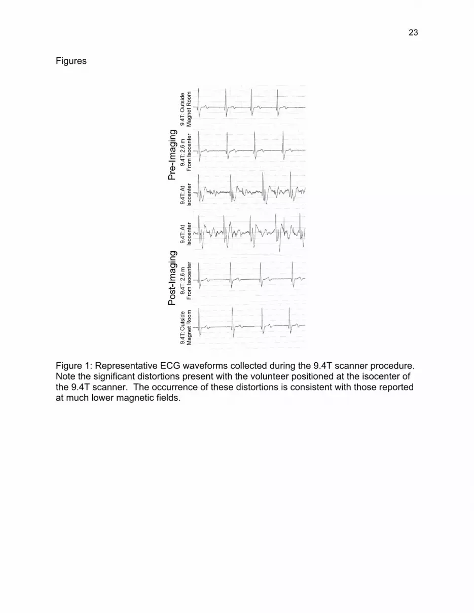
All ECGs recorded during the mock scanner procedure and those recorded
during the 9.4T scanner procedure with the subject outside the magnet room and
positioned 2.6 m from isocenter were unremarkable and consistent. Significant
distortions were observed in the ECG waveforms recorded while the volunteer was
positioned at the isocenter of the 9.4T scanner. Representative ECG waveforms
collected during the 9.4T scanner procedure that illustrate these distortions are shown
in Figure 1.
Table 3 shows the results of the ANOVA performed on the neuropsychological
data. No statistically significant changes in any of the cognitive abilities tested were
observed with respect to the order of exposure to the 9.4T and mock scanners. A
significant effect was observed with respect to the testing session for the LNS, written
SDMT, and delayed memory scale of the HVLT-R. The mean performance on the LNS
(scaled average score in each testing session for 9.4T then mock: 10.36, 10.92, 11.17;
mock then 9.4T: 10.84, 12.23, 12.69) and written SDMT (average number correct in
each testing session for 9.4T then mock: 57.67, 60.50, 65.60; mock then 9.4T: 63.38
64.15 64.08) improved or remained constant with testing session for both exposure
order groups, indicating a practice effect for these tests. The opposite was true for the
delayed memory scale of the HVLT-R (z-score in each testing session for 9.4T the

13
mock: -0.53, -2.07 -1.04; mock then 9.4T: -0.67, -1.05, -2.07) where the best average
performance was in testing session 1. No change in performance attributable to testing
session was present for the GDS, oral SDMT, immediate memory scale of HVLT-R, or
BFI.
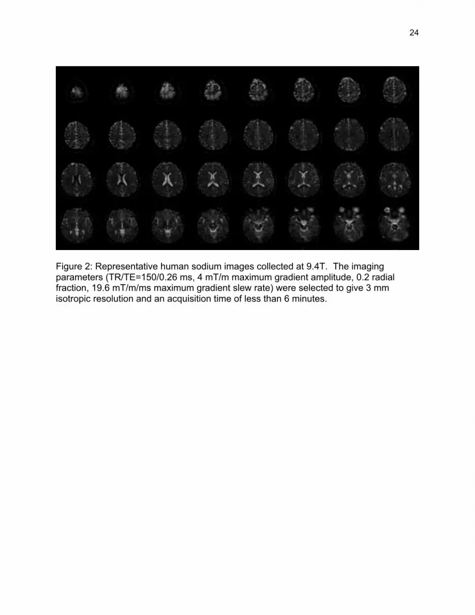
Figure 2 shows a representative human sodium image collected at 9.4T. The
imaging parameters (TR/TE=105/0.26 ms, 4 mT/m maximum gradient amplitude, 0.2
radial fraction, maximum gradient slew rate of 19.6 mT/m/ms) were selected to give a 3
mm isotropic resolution and a total acquisition time of 5 minutes and 56 seconds.
DISCUSSION
No statistically significant changes in heart rate, systolic blood pressure, diastolic
blood pressure, end-tidal CO2, respiratory rate, peripheral arterial O2 saturation, or skin
temperature were observed in human volunteers exposed to a 9.4T static magnetic field
and imaged at 105.92 MHz. During exposure to a mock MR scanner with no magnetic
field or imaging capabilities, no measured change in heart rate, systolic blood pressure,
diastolic blood pressure, end-tidal CO2, or respiratory rate was detected. A statistically
significant change in skin temperature and peripheral arterial O2 saturation was
observed over time during exposure to the mock scanner. Since the mock scanner has
no magnetic field or RF capabilities, the change in skin temperature can be attributed to
the limited airflow allowed within the mock scanner bore. During one mock scanner
procedure, the temperature inside the mock scanner bore increased by 5.7°C while the
volunteer was positioned at the mock isocenter. Since this temperature change occurs
slowly over the duration of the simulated imaging, it manifests itself as a temporal
temperature change rather than positional temperature change. Though statistically
14
significant, the mean changes in skin temperature (<1.2 °C) and peripheral arterial O2
saturation (<0.6 %) were small.
Significant ECG waveform distortions were observed during exposure to the 9.4T
static magnetic field. The measured ECG waveforms returned to baseline after the
volunteer was removed from the 9.4T static magnet field. Such degradations are well
known and are consistent with results reported at 1.5T, 4T, and 8T (2, 3, 5, 6).
The order of exposure to the 9.4T and mock scanners did not have a measurable
effect on working memory, immediate memory, delayed memory, information
processing speed, learning, sustained attention, or fatigue of the human volunteers.
Regardless of the exposure order, an improvement in working memory and information
processing speed was observed with repeated testing. This indicates that the
volunteers improved their performance due to task familiarity. Volunteers demonstrated
the best delayed memory at the beginning of the protocol, before exposure to either the
9.4T or mock scanners. A possible explanation for this finding is that repeated contact
with a lengthy word list (12 words) created an interference effect that led to a decrease
in performance over time. This is consistent with the proactive interference
phenomenon, a well-known source of memory inefficiency (20). The absence of a
corresponding performance difference with respect to exposure order implies that
exposure to the 9.4T static magnetic field had no deleterious effects on memory.
Overall, these data indicate that sodium imaging in a 9.4T static magnetic field does not
impact cognitive function in a statistically meaningful manner. No cognitive testing was
performed during exposure to the 9.4T static magnetic field, limiting the ability of the
data to reflect short-term cognitive effects of the 9.4T static magnetic field. Previous

15
studies completed at 1.5T and 8T have included cognitive testing during magnetic field
exposure and found no suggestion of permanent adverse effects (4-6).
Sodium imaging at 9.4T calls for an excitation frequency of 105.92 MHz, at which
the power distribution uniformity and dielectric resonance will be similar to clinically
approved 3T proton imaging performed at 128MHz. Real-time SAR monitoring
indicated that the FDA limit of 3.0 W/kg was not closely approached during sodium
imaging at 9.4T. The SAR was less than 50% of this guideline during most acquisitions.
The short transverse relaxation of sodium requires a non-Cartesian acquisition that
samples k-space starting at the origin. As a result, very low gradient switching rates
can be used to minimize the likelihood of peripheral nerve stimulation. Only one
volunteer reported any muscle twitching that was potentially due to gradient switching.
However, the volunteer indicated that twitching experienced was non-painful and
occurred as short, isolated incidents rather than continuously during imaging.
A high quality, 3 x 3 x 3 mm3 resolution human sodium image can be acquired at
9.4T in less than 6 minutes. This potentially enables applications such as quantitative
MR imaging of non-proton species to be completed using protocols acceptable for
human subjects.
The most frequently reported discomfort was lightheadedness or vertigo when
being moved through the magnetic field (18 of 25 volunteers). Lesser-reported
discomforts included a metallic taste (6 of 25 volunteers), nausea (2 of 25 volunteers),
and visual stimulation (1 of 25 volunteers). Volunteers indicated that these sensations
were primarily experienced when being moved through the static magnetic field of the
9.4T scanner and that the sensations did not persist once they were stationary for
16
several minutes. To limit these sensations, subjects were moved through that static field
at a slow, constant rate and instructed to minimize head movement. The occurrence of
these sensations and the accounts of them provided by the volunteers are consistent
with those previously reported (2-6, 12).
In conclusion, the combination of the neuropsychological testing results and the
absence of any vital sign changes during exposure to the 9.4T scanner suggests that
human exposure to a 9.4T static magnetic field does not represent a safety concern.
This is in agreement with the numerous human safety studies completed at 8T (2-6) and
is consistent with the expectation from an animal study completed at 9.4T (8).
17
Acknowledgements
The authors thank Dr. Edward Boskamp for design and construction of the sodium RF
coil, Theodore Claiborne for his technical assistance, and Michael Flannery for his
assistance in collecting the vital sign data.
18
References
1. Zaremba LA. Guidance for magnetic resonance diagnostic devices - criteria for significant risk investigations. United States CDRH, FDA, and DHHS. Issued July 14, 2003.
2. Kangarlu A, Brugess RE, Zhu H, Nakayama T, Hamlin RL, Adbuljalil AM, Robitaille PML. Cognitive, cardiac, and physiological safety studies in ultra high field magnetic resonance imaging. Magnetic Resonance Imaging. 1999:17:1407-1416.
3. Kangarlu A, Robitaille PL. Biological effect and health implications in magnetic resonance imaging. Concepts in Magnetic Resonance. 2000:12:321-359.
4. Chakeres DW, Bornstein R, Kangarlu A. Randomized comparison of cognitive function in humans at 0 and 8 Tesla. Journal of Magnetic Resonance Imaging. 2003:18:342-345.
5. Chakeres DW, Kangarlu A, Boudoulas H, Young D. Effect of up to 8 Tesla static magnetic field exposure on sequential human vital signs measurements. Journal of Magnetic Resonance Imaging. 2003:18:346-352.
6. Chakeres DW, Vocht F. Static magnetic fields on human subjects related to magnetic resonance imaging systems. Progress in Biophysics and Molecular Biology. 2005:87:255-265.
7. Miyakoshi J. Effects of static magnetic fields at the cellular level. Progress in Biophysics and Molecular Biology. 2005:87:213-223.
8. High WB, Sikora J, Ugurbil K, Garwood M. Subchronic in vivo effects of a high static magnetic field (9.4 T) in rats. Journal of Magnetic Resonance Imaging. 2000:12:122-139.
9. Magin RL, Lee JK, Klintsova A, Carnes KI, Dunn F. Biological effects of long-duration, high-field (4T) MRI on growth and development in the mouse. Journal of Magnetic Resonance Imaging. 2000:12:140-149.
10. Saunders R. Static magnetic fields: animal studies. Progress in Biophysics and Molecular Biology. 2005:87:225-239.
11. Schenck JF. Safety of strong, static magnetic fields. Journal of Magnetic Resonance Imaging. 2000:12:2-19.
12. Schenck JF. Physical interactions of static magnetic fields with living tissues. Progress in Biophysics and Molecular Biology. 2005:87:185-204.
13. Wechsler D. Wechsler Adult Intelligence Scale. Third Edition. The Psychological Corporation, San Antonio, TX. 1997.
14. Smith A. Symbol Digit Modalities Test. Western Psychological Services, Los Angles, CA. 1991.
15. Benedict RHB, Schretlen D, Groninger L, Brandt J. Hopkins verbal learning test - revised: normative data and analysis of inter-form and test-retest reliability. The Clinical Neuropsychologist. 1998:12:43-55.
19
16. Gordon M. Microprocessor-based assessment of attention deficit disorders. Psychopharmacology Bulletin. 1986:22:288–290.
17. Mendoza TR, Wang XS, Cleeland CS, et al. The rapid assessment of fatigue severity in cancer patients: Use of the Brief Fatigue Inventory. Cancer. 1991:85:1186-1196.
18. Boada FE, Gillen JS, Shen GX, Chang SY, Thulborn KR. Fast three dimensional sodium imaging. Magnetic Resonance in Medicine. 1997:37:706-715.
19. Jackson JJ, Meyer CH, Nishimura DG, Macovski A. Selection of a convolution function for Fourier inversion using gridding. IEEE Transactions on Medical Imaging. 1991:10:473-478.
20. Parkin, AJ. Amnesic syndrome: a lesion-specific disorder? Cortex. 1984:20:479-508.
20
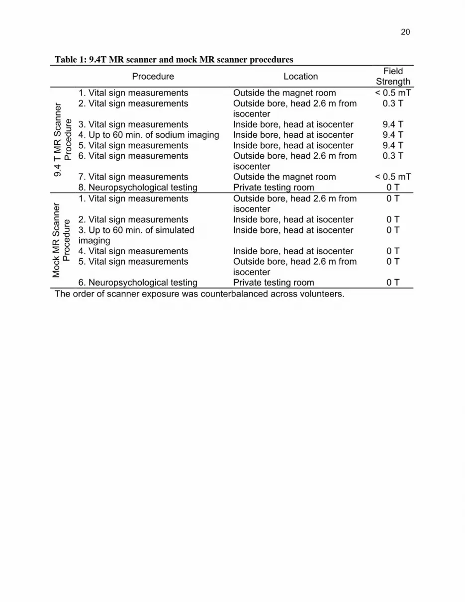
Table 1: 9.4T MR scanner and mock MR scanner procedures Procedure Location Field
Strength 1. Vital sign measurements Outside the magnet room < 0.5 mT 2. Vital sign measurements Outside bore, head 2.6 m from
isocenter 0.3 T
3. Vital sign measurements Inside bore, head at isocenter 9.4 T 4. Up to 60 min. of sodium imaging Inside bore, head at isocenter 9.4 T 5. Vital sign measurements Inside bore, head at isocenter 9.4 T 6. Vital sign measurements Outside bore, head 2.6 m from
isocenter 0.3 T
7. Vital sign measurements Outside the magnet room < 0.5 mT 9.4
T M
R S
cann
er
Pro
cedu
re
8. Neuropsychological testing Private testing room 0 T 1. Vital sign measurements Outside bore, head 2.6 m from
isocenter 0 T
2. Vital sign measurements Inside bore, head at isocenter 0 T 3. Up to 60 min. of simulated imaging
Inside bore, head at isocenter 0 T
4. Vital sign measurements Inside bore, head at isocenter 0 T 5. Vital sign measurements Outside bore, head 2.6 m from
isocenter 0 T
Moc
k M
R S
cann
er
Pro
cedu
re
6. Neuropsychological testing Private testing room 0 T The order of scanner exposure was counterbalanced across volunteers.
21
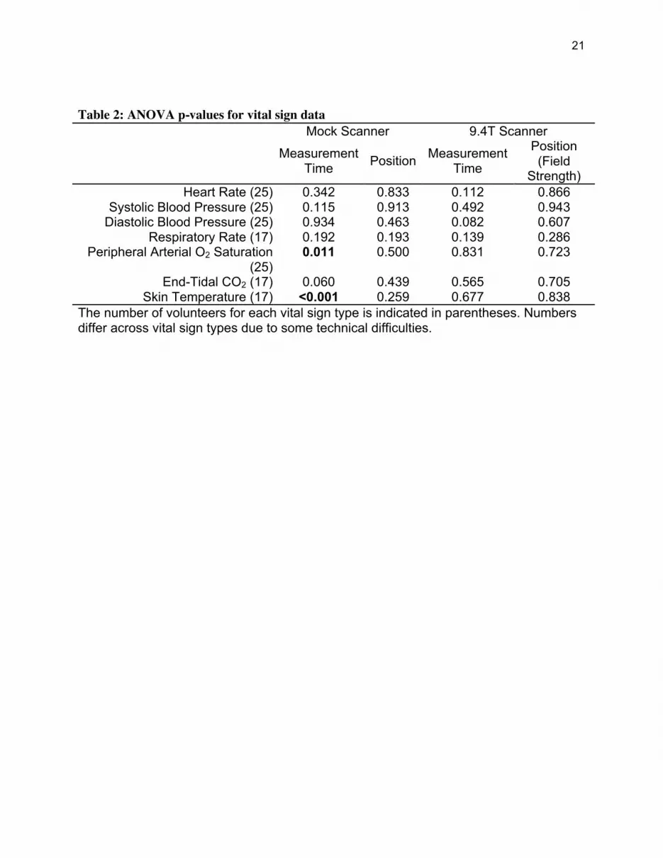
Table 2: ANOVA p-values for vital sign data Mock Scanner 9.4T Scanner
Measurement Time Position Measurement
Time
Position (Field
Strength) Heart Rate (25) 0.342 0.833 0.112 0.866
Systolic Blood Pressure (25) 0.115 0.913 0.492 0.943 Diastolic Blood Pressure (25) 0.934 0.463 0.082 0.607
Respiratory Rate (17) 0.192 0.193 0.139 0.286 Peripheral Arterial O2 Saturation
(25) 0.011 0.500 0.831 0.723
End-Tidal CO2 (17) 0.060 0.439 0.565 0.705 Skin Temperature (17) <0.001 0.259 0.677 0.838
The number of volunteers for each vital sign type is indicated in parentheses. Numbers differ across vital sign types due to some technical difficulties.

22
Table 3: ANOVA p-values for neuropsychological testing data
Exposure Ordera Testing Sessionb
Letter Number Sequencing (25) 0.235 0.016 Symbol Digit Modalities – Written (25) 0.448 0.001
Symbol Digit Modalities – Oral (25) 0.263 0.288 Hopkins Verbal Learning - Immediate Memory
(25) 0.610 0.053
Hopkins Verbal Learning - Delayed Memory (25) 0.924 0.022 Sustained Attention (GDS) (21) 0.611 0.599
Brief Fatigue Inventory (25) 0.124 0.724 The number of volunteers for each neuropsychological test is indicated in parentheses. Sustained attention data were not available from the first 4 volunteers. aExposure orders: 9.4T scanner then mock scanner, mock scanner than 9.4T scanner. bTesting sessions: before exposure, following exposure to 9.4T/mock scanner, following exposure to mock/9.4T scanner.
23
Figures
Figure 1: Representative ECG waveforms collected during the 9.4T scanner procedure. Note the significant distortions present with the volunteer positioned at the isocenter of the 9.4T scanner. The occurrence of these distortions is consistent with those reported at much lower magnetic fields.
24
Figure 2: Representative human sodium images collected at 9.4T. The imaging parameters (TR/TE=150/0.26 ms, 4 mT/m maximum gradient amplitude, 0.2 radial fraction, 19.6 mT/m/ms maximum gradient slew rate) were selected to give 3 mm isotropic resolution and an acquisition time of less than 6 minutes.