Idiopathic condylar resorption: Diagnosis, treatment protocol, and outcomes
Upload
independentCategory
view
0download
0
Original article
Safety and efficacy of exercise training in patientswith an idiopathic inflammatory myopathy—asystematic review
Gerarda E. A. Habers1,2 and Tim Takken1
Abstract
Objective. Idiopathic inflammatory myopathies (IIMs) are a group of rare heterogeneous autoimmune
skeletal muscle disorders characterized by muscle weakness, excessive muscle fatigue and diminished
aerobic fitness. Exercise training could be one way to prevent or delay the negative effects of the disease
and the impairments seen in patients with an IIM. The objective was to examine whether exercise training
is safe and effective in patients with an IIM.
Methods. All experimental studies that assessed the safety and/or efficacy of an exercise training pro-
gramme in patients with an IIM except for case studies were reviewed. Pre-MEDLINE, MEDLINE and
EMBASE database searching was done up to November 2010. Information was extracted on the number
of participants, characteristics of participants, type of intervention, type of outcome measure, type of
study design, report characteristics, geographical origin and risk of bias within studies. The change (per-
centage and significance) in group mean or median for each outcome measure in each study was
determined as well.
Results. Two randomized controlled trials, one non-randomized controlled trial and nine uncontrolled trials
were included. No studies in children were found. Safety measures did not worsen and efficacy measures
improved or did not change. Most of the included studies had a high selection and/or allocation bias.
Conclusions. In conclusion, it appears that exercise training is safe and effective in adult patients with
active as well as inactive stable IIMs. However, more studies with a well-controlled design are needed. In
addition, studies in children with an IIM are indicated.
Key words: Exercise training, Idiopathic inflammatory myopathies, Efficacy, Safety, Systematic review.
Introduction
Idiopathic inflammatory myopathies (IIMs) are a group of
rare heterogeneous autoimmune skeletal muscle dis-
orders. IIMs can be broadly subdivided into three main
subtypes: DM, PM and IBM that can all be characterized
by muscle weakness and inflammatory infiltrates in
muscle biopsies. The three subtypes of IIM differ from
each other in, among others, the age of onset, the location
of muscle involvement and the responsiveness to medical
treatment. DM and PM affect both children and adults;
IBM occurs only in adults. Patients with IBM show
mainly muscle weakness in the wrist and finger flexors
and in the quadriceps muscles. However, patients with
DM and PM show mainly muscle weakness at the prox-
imal muscles of both the upper and lower extremities.
In DM, the skin is involved as well. Patients with PM and
DM respond to drug therapy, while patients with IBM are
resistant to it [1].
Muscle inflammation, secondary metabolic disturb-
ances (e.g. reduced ATP and creatine phosphate levels
[2, 3] and capillary blood supply [4] in muscle tissue), ster-
oid treatment-induced myopathy [5], cardiac abnormal-
ities [6], pulmonary involvement [7] and physical
inactivity can all play a role in one or more of the following
1Child Development and Exercise Center, Wilhelmina Children’sHospital, University Medical Center Utrecht, Utrecht, The Netherlandsand 2Faculty of Human Movement Sciences, VU UniversityAmsterdam, Amsterdam, The Netherlands.
Correspondence to: Tim Takken, Child Development and ExerciseCenter, Wilhelmina Children’s Hospital, University Medical CenterUtrecht, Room KB2.056.0, PO Box 85090, 3508 AB Utrecht, TheNetherlands. E-mail: [email protected]
Submitted 18 February 2011; revised version accepted 14 June 2011.
! The Author 2011. Published by Oxford University Press on behalf of the British Society for Rheumatology. All rights reserved. For Permissions, please email: [email protected]
RHEUMATOLOGY
Rheumatology 2011;50:2113�2124
doi:10.1093/rheumatology/ker292
Advance Access publication 2 September 2011
CL
INIC
AL
SC
IEN
CE
at Vrije U
niversiteit- Library on M
ay 22, 2013http://rheum
atology.oxfordjournals.org/D
ownloaded from

impairments seen in patients with an IIM: muscle weak-
ness, excessive muscle fatigue and diminished aerobic
fitness [8]. These impairments can subsequently lead to
disabilities such as problems with walking and manual
control. Exercise training could be one way to prevent
or delay these negative effects of the disease and thus
possibly reduce the disabilities seen in patients with an
IIM. However, in the past, exercise training was discour-
aged in patients with an active IIM because it was thought
it might worsen the inflammatory process [9]. At present,
indications of beneficial effects without disease exacerba-
tions with exercise training have been found [10, 11].
Exercise training may not only delay the progression of
the disease or prevent a poor outcome, but it could also
decrease the risk of cardiovascular diseases as is
observed in healthy subjects [12, 13].
The objective of this systematic review was to examine
whether exercise training is safe and effective in patients
with an IIM. For this purpose, all experimental studies that
assessed the safety and/or efficacy of an exercise training
programme in these patients except for case studies were
reviewed. Furthermore, a summary of the training
programmes used in this population is given.
Materials and methods
Eligibility criteria
Types of report
No publication date or publication status restrictions. Only
English-language reports were included.
Types of study design
All experimental studies except for case studies.
Types of participants
Patients with PM, DM or IBM with any stage of the dis-
ease, including children and adults.
Types of intervention
Exercise training was defined as muscle strength training
and/or aerobic training (possibly combined with another
type of intervention) at home or in combination with
training on site for at least 3 weeks, 2 days a week and
20 min each time. Within these criteria, exercise training
programmes of all intensities, frequencies and durations
were considered.
Types of outcome measures
All outcome measures related to safety or efficacy were
included.
Information sources
Studies were identified by searching electronic data-
bases, scanning reference lists of included reports and
tracking forward citations of included reports. The elec-
tronic database searching was applied to Pre-MEDLINE,
MEDLINE and EMBASE. Relevant terms describing exer-
cise training and IIMs were used. No publication date or
publication status restrictions were imposed on the
search strategy. Details of the search strategy are
described the supplementary section 1, available as sup-
plementary data at Rheumatology Online. The last search
was run on 19 November 2010.
Study selection
Two reviewers performed eligibility assessment in an un-
blinded standardized manner. A detailed explanation of the
study selection methods is described in the supplementary
section 2 (Methods), available as supplementary data at
Rheumatology Online. A summary of the study selection
process is depicted in Fig. 1.
Data collection process and data items
A data extraction sheet was developed. To avoid double
counting, data from multiple reports of the same study were
merged. Information was extracted from each included
study on (i) number of participants; (ii) characteristics of
participants (including diagnosis, age, gender, stage of
disease, inclusion and exclusion criteria); (iii) type of inter-
vention (including type, location, intensity, frequency,
session duration and duration of the exercise training pro-
gramme, and whether the exercise training programme
was in combination with additional experimental treat-
ment); (iv) type of outcome measure (including disease
activity, pain, muscle strength, aerobic fitness, functional
performance, functional capacity, health status, muscle
characteristics, disease impact and fatigue measures
(lung function measures were added after the collection
process started); (v) type of study design; (vi) report char-
acteristics (including publication date and publication
status); and (vii) geographical origin.
For each study that measured muscle strength, the per-
centage change in group mean muscle strength score
was determined for each muscle function tested in that
study. The median and range of changes in the group
mean of all muscle functions tested in a specific study
were determined. It was also determined whether one or
more of these muscle functions changed significantly.
Furthermore, it was determined whether significant differ-
ences after the training period were reported between two
groups in one or more muscle functions tested. From the
other outcome measures, the baseline mean or median
value of a specific measurement tool and the percentage
change in group mean or median in this value was deter-
mined for each study. It was also determined whether this
change was significant. Furthermore, it was determined
whether significant differences after the training period
between two groups were reported.
Risk of bias within studies
To ascertain the risk of bias in the included studies, two
reviewers independently completed the effective public
health practice project quality assessment tool [14] in an
unblinded manner. This tool is able to deal with any study
design and was shown to be suitable for use in a system-
atic review [15]. It criticizes (i) selection bias, (ii) allocation
bias, (iii) confounders, (iv) blinding, (v) data collection
methods, (vi) withdrawals and drop-outs, (vii) analysis
2114 www.rheumatology.oxfordjournals.org
Gerarda E. A. Habers and Tim Takken
at Vrije U
niversiteit- Library on M
ay 22, 2013http://rheum
atology.oxfordjournals.org/D
ownloaded from

and (viii) intervention integrity. The first six components
were rated as strong (+), moderate (o), weak (�) or not
applicable (NA) in order to provide a global quality rating.
Discrepancy between the two reviewers with respect to
the component ratings was resolved by consensus. These
assessments were not meant for excluding studies from
the review or analysis, but for criticizing the risk of bias in
the included studies.
Risk of bias across studies
Selective reporting bias was considered by comparison of
the outcomes listed in the methods section of the pub-
lished report with those for which results were presented.
Results
Study selection
A total of 12 studies were identified for inclusion in
the review. These studies were described in 14 reports.
A detailed explanation of the study selection results is re-
ported in supplementary section 2 (Results), available as
supplementary data at Rheumatology Online. A summary
of the study selection results is depicted in Fig. 1.
Study characteristics
Report characteristics, study design information and par-
ticipant characteristics of each study are reported in Table 1.
Seven studies examined both patients with PM and
patients with DM, four studies examined only patients
with IBM and one study examined all three patient
groups. Disease stages studied were recent onset, active
stable and inactive stable. No studies in children were
found. Only four studies reported both inclusion and ex-
clusion criteria. One study reported only inclusion criteria
and another study reported only exclusion criteria.
Intervention characteristics of each study are reported in
Table 2. Tables 3�5 describe the different measure-
ment tools that were used by the different studies to exam-
ine disease activity and pain (Table 3), muscle strength
(Table 4), aerobic fitness, functional performance, func-
tional capacity, health status, lung function, muscle char-
acteristics, disease impact and fatigue (Table 5). As can
be seen in these tables, a large variety of measurement
tools was used. In the ‘Changes in outcome measures’
section, the measurement tools and results are described
in more detail.
Risk of bias within studies
The quality assessment scores are presented in Table 6.
In the randomized controlled trials, no confounders at
baseline were present. In the non-randomized controlled
clinical trial, baseline values for activities of daily living
differed significantly between the training group and the
control group; the training group had a higher baseline
score compared with the control group. For the other
studies, this component was not applicable.
Six studies reported that the outcome assessors were
blinded to the intervention or exposure status of the par-
ticipant. The other studies did not report about blinding.
There was only one study [25] that did sample size calcu-
lations. All studies used appropriate statistical methods
for analysis and performed the analysis by intervention
allocation status rather than the actual intervention
received.
The percentage of the participants who received
the allocated intervention of interest was >80% in seven
out of eight studies that reported about this item. One
study reported a percentage >60%. Six studies
FIG. 1 A summary of the study selection process.
www.rheumatology.oxfordjournals.org 2115
Exercise training in patients with an IIM
at Vrije U
niversiteit- Library on M
ay 22, 2013http://rheum
atology.oxfordjournals.org/D
ownloaded from
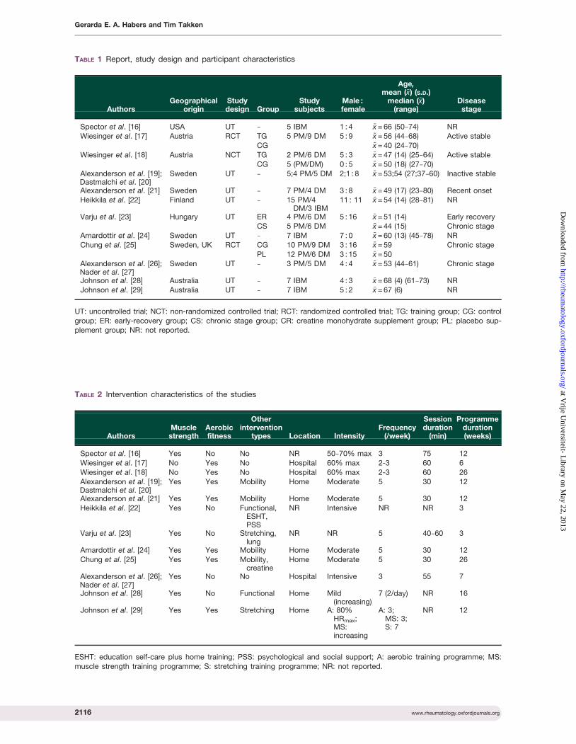
TABLE 1 Report, study design and participant characteristics
AuthorsGeographical
originStudydesign Group
Studysubjects
Male :female
Age,mean (�x) (S.D.)
median (~x)(range)
Diseasestage
Spector et al. [16] USA UT � 5 IBM 1 : 4 �x = 66 (50�74) NRWiesinger et al. [17] Austria RCT TG 5 PM/9 DM 5 : 9 ~x = 56 (44�68) Active stable
CG ~x = 40 (24�70)
Wiesinger et al. [18] Austria NCT TG 2 PM/6 DM 5 : 3 �x = 47 (14) (25�64) Active stable
CG 5 (PM/DM) 0 : 5 �x = 50 (18) (27�70)Alexanderson et al. [19];Dastmalchi et al. [20]
Sweden UT � 5;4 PM/5 DM 2;1 : 8 ~x = 53;54 (27;37�60) Inactive stable
Alexanderson et al. [21] Sweden UT � 7 PM/4 DM 3 : 8 �x = 49 (17) (23�80) Recent onsetHeikkila et al. [22] Finland UT � 15 PM/4
DM/3 IBM11 : 11 �x = 54 (14) (28�81) NR
Varju et al. [23] Hungary UT ER 4 PM/6 DM 5 : 16 �x = 51 (14) Early recoveryCS 5 PM/6 DM �x = 44 (15) Chronic stage
Arnardottir et al. [24] Sweden UT � 7 IBM 7 : 0 �x = 60 (13) (45�78) NR
Chung et al. [25] Sweden, UK RCT CG 10 PM/9 DM 3 : 16 �x = 59 Chronic stage
PL 12 PM/6 DM 3 : 15 �x = 50Alexanderson et al. [26];Nader et al. [27]
Sweden UT � 3 PM/5 DM 4 : 4 ~x = 53 (44�61) Chronic stage
Johnson et al. [28] Australia UT � 7 IBM 4 : 3 �x = 68 (4) (61�73) NRJohnson et al. [29] Australia UT � 7 IBM 5 : 2 �x = 67 (6) NR
UT: uncontrolled trial; NCT: non-randomized controlled trial; RCT: randomized controlled trial; TG: training group; CG: control
group; ER: early-recovery group; CS: chronic stage group; CR: creatine monohydrate supplement group; PL: placebo sup-plement group; NR: not reported.
TABLE 2 Intervention characteristics of the studies
AuthorsMusclestrength
Aerobicfitness
Otherintervention
types Location IntensityFrequency
(/week)
Sessionduration
(min)
Programmeduration(weeks)
Spector et al. [16] Yes No No NR 50�70% max 3 75 12
Wiesinger et al. [17] No Yes No Hospital 60% max 2-3 60 6
Wiesinger et al. [18] No Yes No Hospital 60% max 2-3 60 26
Alexanderson et al. [19];Dastmalchi et al. [20]
Yes Yes Mobility Home Moderate 5 30 12
Alexanderson et al. [21] Yes Yes Mobility Home Moderate 5 30 12
Heikkila et al. [22] Yes No Functional,ESHT,PSS
NR Intensive NR NR 3
Varju et al. [23] Yes No Stretching,lung
NR NR 5 40�60 3
Arnardottir et al. [24] Yes Yes Mobility Home Moderate 5 30 12
Chung et al. [25] Yes Yes Mobility,creatine
Home Moderate 5 30 26
Alexanderson et al. [26];Nader et al. [27]
Yes No No Hospital Intensive 3 55 7
Johnson et al. [28] Yes No Functional Home Mild(increasing)
7 (2/day) NR 16
Johnson et al. [29] Yes Yes Stretching Home A: 80%HRmax;MS:increasing
A: 3;MS: 3;S: 7
NR 12
ESHT: education self-care plus home training; PSS: psychological and social support; A: aerobic training programme; MS:
muscle strength training programme; S: stretching training programme; NR: not reported.
2116 www.rheumatology.oxfordjournals.org
Gerarda E. A. Habers and Tim Takken
at Vrije U
niversiteit- Library on M
ay 22, 2013http://rheum
atology.oxfordjournals.org/D
ownloaded from

reported whether the consistency of the intervention was
measured. This was done in all the studies. The reasons
for some discrepancies with respect to the component
ratings of the two reviewers were oversight and
differences in interpretation of criteria.
Risk of bias across studies
In all reports, the outcomes listed in the ‘Materials and
methods’ section were presented in the ‘Results’ section
as well.
Changes in outcome measures
Disease activity
None of the disease activity measures in any of the
studies worsened significantly from baseline to the end
of the exercise training programme. In two studies there
was even an improvement. Arnardottir et al. [24] found a
significant decrease in EN-4- and IL-1Ra-positive cells,
which indicates a decreased endothelial cell area and
decreased inflammatory activity, respectively. Nader
et al. [27] found a decreased amount of pro-inflammatory
and profibrotic mRNA and an increased amount of anti-
inflammatory and anti-fibrotic mRNA. Furthermore, tissue
fibrosis and extraskeletal muscle activity were both
decreased after training [26]. However, there was no sig-
nificant decrease of inflammatory molecules at the protein
level [27].
Pain
In all five studies that examined pain, none of the meas-
ures changed significantly from baseline to the end of the
training programme.
Muscle strength
Table 4 mentions the median and range of changes in
group means of all muscle functions tested with a specific
measurement tool in a specific study. It is also reported
whether a study reported significant improvement (+) or
worsening (�) in one or more muscle functions tested.
Furthermore, it is mentioned whether a study reported a
significant difference (*) between two groups in one or
more muscle functions tested.
Dynamometer isometric muscle strength. Spector et al.
[16] did not find significant improvements in isometric
muscle strength in the three muscle functions tested. In
the study of Varju et al. [23], all four of the muscle
functions tested in the chronic stage group and two in
the early recovery group were significantly improved. In
both studies of Wiesinger et al. [17, 18], the peak isometric
torque of the hip flexors/knee extensors improved signifi-
cantly in the training group. In the control group there was
no increase in the study of Wiesinger et al. [17], and even
a significant decrease was observed in the long-term
follow-up study of Wiesinger et al. [18].
Handheld myometer isometric muscle strength. In the
study of Johnson et al. [28], all nine muscle functions
that were tested on isometric muscle strength improved
significantly. In a recent study, Johnson et al. [29] found a
significant improvement in four of eight muscle functions
that were trained and tested.
Manual muscle test. None of the included studies that
used manual muscle strength tests reported significant
changes. However, at the end of the training period in
the study of Chung et al. [25], the creatine monohydrate
supplement group scored significantly higher than the
placebo supplement group on shoulder abduction and
hip flexion as measured by manual muscle testing. The
other muscle functions tested did not differ significantly
between the two groups at the end of the exercise training
period.
Peak isokinetic torque at 120�/s. Maximal voluntary con-
centric knee extension and flexion at angular velocity of
120�/s was not significantly changed after training in the
study of Arnardottir et al. [24].
Three repetition maximum. Improvements in three
voluntary repetition maximum dynamic muscle strength
in the study of Spector et al. [16] were significant in
three of five training exercises. The mean leg curl
dynamic muscle strength improved by 105%.
10�15 repetition maximum. Improvements in 10�15 vol-
untary repetition maximum dynamic muscle strength in
the study of Alexanderson et al. [26] were significant in
four of five tested muscle groups. The abdominal dynamic
muscle strength was improved 442%.
Grip strength. In the study of Alexanderson et al. [26],
neither maximal grip strength nor mean grip strength
during 10 s significantly changed. Dynamometer grip
strength improved significantly in the study of Johnson
et al. [28], but not in the study of Johnson et al. [29].
TABLE 3 The different measurement tools that were used
by the different studies to examine disease activity and
pain
Disease activitySerum levels of creatine kinase (all studies)
Serum levels of aldolase [17, 18]
Serum levels of cytokines [16]
Serum levels of epinephrine [16]Serum levels of CRP [19, 21, 23]
ESR [19, 21, 23, 25]
MRI inflammation [19, 21]
Multicolour flow cytometry inflammation [16]Muscle biopsy inflammation [16, 19, 21, 24]
Visual analogue scale of disease activity, patient [26]
Visual analogue scale of disease activity, physician [26]Extraskeletal muscle activity [26]
Genome-wide mRNA profiles [27]
Tissue fibrosis [27]
Immunohistochemistry [27]Pain
Visual analogue scale of pain [19, 22, 23]
Short-form McGill pain questionnaire [25]
Borg CR-10 scale [26]
www.rheumatology.oxfordjournals.org 2117
Exercise training in patients with an IIM
at Vrije U
niversiteit- Library on M
ay 22, 2013http://rheum
atology.oxfordjournals.org/D
ownloaded from

Table 5 mentions the baseline mean or median value
and the percentage change for each study that used a
specific measurement tool to examine aerobic fitness,
functional performance, functional capacity, health
status, long function, muscle characteristics, disease
impact or fatigue. In this table it is also reported whether
a study reported significant improvement (+) or worsening
(�). Furthermore, it is mentioned whether a study reported
a significant difference (*) or no significant difference (#)
between the two groups after the training period.
In supplementary section 3, available as supplementary
data at Rheumatology Online, the measures of functional
performance, functional capacity, health status, disease
impact and fatigue are defined.
Aerobic fitness
All four studies that examined aerobic fitness found a sig-
nificantly improved peak oxygen uptake in the training
groups. Despite the fact that both training groups in the
studies of Wiesinger et al. [17, 18] had the same baseline
values of peak oxygen uptake, the group that trained 26
weeks increased much more in peak oxygen uptake (27%
improvement) compared with the group that followed the
same exercise training programme but only for 6 weeks
(12% improvement). The control groups in the studies of
Wiesinger et al. showed no significant change [17] or even
a significant worsening [18] in this measure.
Functional performance
Five different measurement tools were used to measure
functional performance. Three of six studies that exam-
ined functional performance found no significant change.
In both studies of Wiesinger et al. [17, 18], the training
group improved significantly and the control group did
not change significantly. In the study of Varju et al. [23],
significant improvements were seen in both study groups
in the score on the HAQ, but not in the score on the
functional independence measure.
Functional capacity
Arnardottir et al. [24] observed no significant change in the
functional index. However, four other studies [19, 21, 22, 25]
reported a significant improvement in this measure after
training. The score did not differ significantly between the
creatine monohydrate supplement group and the placebo
group in the study of Chung et al. [25] at the end of the
training programme. The functional index-2 used by
Alexanderson et al. [26] showed a significant improvement
only in the amount of shoulder flexion repetitions. Johnson
et al. [28] found significant improvements in the time to
TABLE 4 The different measurement tools that were used by the different studies to examine muscle strength and the
median and range of change in group means of the muscle functions tested
Measurement tool Authors Group Changea Significance
Dynamometer isometricmuscle strength
Spector et al. [16] 19 (10�65) NS
Varju et al. [23] ER 21 (7�27) +CS 43 (19�53) +
Wiesinger et al. [17]* TG 29 +
CG 11 NS
Wiesinger et al. [18] TG 34 +CG �42 �
Handheld myometerisometric muscle strength
Johnson et al. [28] 44 (19�142) +
Johnson et al. [29] 3 (�10 to 40) +
Manual muscle test Spector et al. [16] NR NS
Arnardottir et al. [24] 4 NS
Chung et al. [25]* CR 2 (�1 to 13) NRPL 0 (�5 to 13) NR
Alexanderson et al. [26] 4 NS
Johnson et al. [28] NR NS
Peak isokinetic torque at 120�/s Arnardottir et al. [24] NR NSThree repetition maximum Spector et al. [16] 40 (22�105) +
10�15 repetition maximum Alexanderson et al. [26] 44 (8�442) +
Grip strength Alexanderson et al. [26] 2 (�3 to 8) NSJohnson et al. [28] (4�24) +
Johnson et al. [29] �8 NS
aMedian (range) change (%) in group means of the muscle functions tested. *Reported significant difference between the twogroups in one or more muscle functions tested after the training period. TG: training group; CG: control group; ER: early
recovery group; CS: chronic stage group; CR: creatine monohydrate supplement group; PL: placebo supplement group; NR:
not reported; +: reported significant improvement in one or more muscle functions tested; �: reported significant worsening in
one or more muscle functions tested; NS: not significant.
2118 www.rheumatology.oxfordjournals.org
Gerarda E. A. Habers and Tim Takken
at Vrije U
niversiteit- Library on M
ay 22, 2013http://rheum
atology.oxfordjournals.org/D
ownloaded from

TABLE 5 The different measurement tools that were used by the different studies to examine aerobic fitness, functional
performance, functional capacity, health status, lung function, muscle characteristics, disease impact and fatigue and
their corresponding baseline values and percentage changes in group means or medians after training
Outcome measureAuthors Group Baselinea Changeb Significanceand measurement tool
Aerobic fitness
Peak oxygen uptake Wiesinger et al. [17]* TG 17.4 ml/min/kg 12 +CG 16.9 ml/min/kg �2.6 NS
Wiesinger et al. [18] TG 17.5 ml/min/kg 27 +
CG 17.0 ml/min/kg �12 �
Nader et al. [27] � 26 ml/min/kg 19 +Johnson et al. [29] � 18.7 ml/min/kg 27 +
Oxygen uptake at VAT Wiesinger et al. [18] TG 9.8 ml/min/kg 14 +
CG NR NR NSResting heart rate Wiesinger et al. [18] TG 85 beats/min �7 NS
CG NR NR NS
Heart rate response Johnson et al. [29] � 134 beats/min 3 NS
Lactate levels Johnson et al. [29] � 4 mmol 15 NSSelf-reported RPE Johnson et al. [29] � 4.6 0 NS
Functional performance
Barthel index Spector et al. [16] � 2.2 �4.5 NS
Modified FASQ Wiesinger et al. [17]* TG 156.6 20.5 +CG 142.6 2.9 NS
Wiesinger et al. [18] TG 175 11 +
CG 116 �13 NSFIM Varju et al. [23] ER 113 6 NS
CS 125 0 NS
HAQ Heikkilla et al. [22] � 1.3 �8 NS
Varju et al. [23] ER 1.17 �22 +CS 1.04 �16 +
Alexanderson et al. [26] � 0.68 �1 NS
Myositis activity profile Alexanderson et al. [26] � MC MC NS
Functional capacityFunctional index Alexanderson et al. [19] � 48.0 19 +
Alexanderson et al. [21] � 50.5 14 +
Heikkilla et al. [22] � 43.9 9 +
Arnardottir et al. [24] � 35.7 2 NSChung et al. [25]# CR 50.3 13 +
PL 46.3 12 +
Functional index-2 Alexanderson et al. [26] � MC MC +AFPT Chung et al. [25]* CR 31 s �13 +
PL 30 s �7 NS
Time to walk 30 m Johnson et al. [28] � 41 s �17 +
Johnson et al. [29] � 43 s �31 NSStep count on 30-m walk Johnson et al. [28] � 59 paces �7 NS
Johnson et al. [29] � 67 paces �21 NS
Time to climb one stair Johnson et al. [28] � 14 s �21 +
Johnson et al. [29] � 19 s �20 NSMax. sit-to-stands Johnson et al. [28] � NR NR NS
Seven-minute walking distance Alexanderson et al. [19] � 312 m 30 +
Health status �Short form-36 Alexanderson et al. [19] � MC MC +
Alexanderson et al. [21] � MC MC +
NHP Chung et al. [25]# Both NR NR NS
Lung functionFVC Varju et al. [23] ER 2.9 l 17 +
CS 3.5 l 3 NS
FEV1/FVC Varju et al. [23] ER 78% �3 NS
CS 81% 3 NSFEF(25�75%) Varju et al. [23] ER 2.7 l/s 0 NS
CS 3.4 l/s 6 NS
(continued)
www.rheumatology.oxfordjournals.org 2119
Exercise training in patients with an IIM
at Vrije U
niversiteit- Library on M
ay 22, 2013http://rheum
atology.oxfordjournals.org/D
ownloaded from

walk 30 m and the time to climb one stair. However, these
improvements were not found in a later study of Johnson
et al. [29]. Alexanderson et al. [19] observed a significant
increase in the 7-min walking distance.
Health status
Physical functioning as assessed by one component of
the short form-36 was significantly improved in the studies
of Alexanderson et al. [19, 21]. The component
role-physical was only significantly improved in the
study of Alexanderson et al. [19]. Bodily pain and vitality
were only significantly improved in the study of
Alexanderson et al. [21]. The other components of the
short form-36 did not significantly change in either
study. Chung et al. [25] found no significant change in
health status as assessed by the Nottingham health profile
in both study groups.
Lung function
Only one study examined lung function [23]. The only
measure that improved significantly was the forced vital
capacity in the early recovery group.
Muscle characteristics
Different methods were used to examine muscle charac-
teristics. Some muscle biopsy cross-section areas and
relative proportions of fibre types changed significantly
in the studies of Arnardottir et al. [24] and Dastmalchi
TABLE 5 Continued
Outcome measureAuthors Group Baselinea Changeb Significanceand measurement tool
Muscle characteristics
MRI whole muscle CSA Spector et al. [16] � NR NR NS
Muscle biopsy CSA type I Arnardottir et al. [24] � 4730 mm2 17 +Dastmalchi et al. [20] � 4570 mm2 8 NS
Muscle biopsy CSA type II Arnardottir et al. [24] � 3793 mm2 4 NS
Dastmalchi et al. [20] � 3658 mm2 25 +
Muscle biopsy r.p. type I Arnardottir et al. [24] � 41% �3 NSDastmalchi et al. [20] � 32% 31 +
Muscle biopsy r.p. type IIA Arnardottir et al. [24] � 20% �3 NS
Dastmalchi et al. [20] � 39% �13 NS
Muscle biopsy r.p. type IIB Arnardottir et al. [24] � 31% �18 NSDastmalchi et al. [20] � 26% 0 NS
Muscle biopsy r.p. type IIC Arnardottir et al. [24] � 8% 88 NS
Dastmalchi et al. [20] � 3% �67 +Regeneration markers Dastmalchi et al. [20] � MC MC +
Cap. diam. muscle biopsy Arnardottir et al. [24] � 90 mm2 NR NS31P MRS PCr/�-NTP Chung et al. [25] CR 4.83 3 +
PL 4.03 0.5 NS31P MRS Pi/�-NTP Chung et al. [25] Both NR NR NS
Energy metabolism mRNAc Nader et al. [27] � MC MC +
Disease impact
SGDI Alexanderson et al. [21] � 3 �33 NSVAS disease impact Alexanderson et al. [26] � 2.9 14 NS
HADS Chung et al. [25]# Both NR NR NS
Fatigue
Fatigue severity scale Spector et al. [16] � 5.5 �5 NSVAS fatigue Varju et al. [23] ER 66 �17 +
CS 51 �61 +
Chalder fatigue score Chung et al. [25]# CR NR NR NSPL NR NR NS
aGroup mean or median. bChange (%) in group mean or median after training compared with baseline. cmRNA related to lipid
biosynthesis and oxidative metabolism. *Reported significant difference between the two groups after the training period.#Reported no significant difference between the two groups after the training period. +: reported significant improvement in
one or more components; �: reported significant worsening in one or more components; NS: not significant; TG: training
group; CG: control group; ER: early recovery group; CS: chronic stage group; CR: creatine monohydrate supplement group;
PL: placebo supplement group; NR: not reported; MC: multiple components; VAT: ventilatory anaerobic threshold; RPE: rateof perceived exertion; FASQ: functional assessment screening questionnaire; FIM: functional independence measure; HAQ:
health assessment questionnaire; AFPT: aggregate functional performance time; Max.: maximum; Min.: minutes; NHP:
Nottingham health profile; FVC: forced vital capacity; FEF(25-75%): forced expiratory flow; FEV1/FVC: forced expiratory
volume 1 s to FVC ratio; MRI: magnetic resonance spectroscopy; CSA: cross-sectional area; r.p.: relative proportion; Cap.diam.: capillary diameter; 31P MRS: 31P magnetic resonance spectroscopy; PCr: phosphocreatine; NTP: nucleoside triphos-
phate; SGDI: subjective global disease impact; VAS: visual analogue scale; HADS: hospital anxiety and depression scale.
2120 www.rheumatology.oxfordjournals.org
Gerarda E. A. Habers and Tim Takken
at Vrije U
niversiteit- Library on M
ay 22, 2013http://rheum
atology.oxfordjournals.org/D
ownloaded from

et al. [20]. Of the three markers of regeneration used in the
study of Dastmalchi et al. [20] (CD56, vimentin and neo-
natal myosin heavy chain), only the percentage of
vimentin-positive fibres was higher after the training pro-
gramme compared with baseline. No change in mean ca-
pillary diameter was observed in the study of Arnardottir
et al. [24]. Chung et al. [25] examined muscle bioenerget-
ics and found that the ratio of phosphocreatine/nucleotide
triphosphates was significantly increased after training in
the group that received creatine monohydrate supple-
ments, but not in the control group. Nader et al. [27]
observed a decreased level of lipid biosynthesis mRNA
and an increased amount of oxidative metabolism
mRNA after training.
Disease impact
None of the three studies that measured disease impact
found a significant change in this outcome measure.
Fatigue
Two out of three studies that examined fatigue found no
significant change in this outcome measure. However, the
score on the visual analogue scale in the study of Varju
et al. [23] improved significantly in both study groups.
Discussion
The objective of this systematic review was to examine
whether exercise training is safe and effective in patients
with an IIM. For this purpose, all experimental studies that
assessed the safety and/or efficacy of an exercise training
programme in this type of patients were reviewed.
Safety
Exercise training in patients with an IIM appears to be safe
since disease activity and pain measures as used in the
included studies worsened in none of the studies on a
group level. Some studies even reported an improvement
in disease activity measures [24, 26, 27].
Some studies reported at an individual level increased
creatine phosphokinase levels [22, 25, 26], ESRs [25],
muscle fibre pathology [24] and pain [22]. In most of
these cases, the values remained within the normal
range or the increment was small (422%). The increased
muscle fibre pathology observed in two patients in the
study of Arnardottir et al. [24] could be due to the fact
that the muscle fibre abnormalities have a patchy
distribution in IBM. Also, in a few studies, patients
stopped the exercise training programme early or the
programme had to be adjusted. Reasons for stopping
were considered either as related to the exercise training
programme or related to other diseases such as
osteoporosis and arthritis. None of the studies reported
increments in the immunosuppressive treatment during
the exercise training programme. In some cases, drug
treatment was even reduced due to clinical improvement
[19, 21].
It is important to realize that many the studies included
examined very specific patient groups, which were
expected to be at low risk of disease exacerbation.
Patients with other serious medical illnesses and/or pa-
tients that are unable to exercise sufficiently to participate
were excluded in most of the articles. All articles that
described inclusion criteria required that drug therapy
was stable for at least 3 months before the start of the
exercise programme. Unfortunately, not all studies re-
ported inclusion and exclusion criteria.
Efficacy
The efficacy of exercise training in patients with an IIM
was assessed with a large variety of outcome measures.
For pooling of future studies, the establishment of a core
set of outcome measures is strongly advised. It appears
TABLE 6 The scores of each study on the first six components of the effective public health practice project quality
assessment tool [14]
AuthorsSelection
biasAllocation
bias Confounders BlindingData collection
methods
Withdrawalsand
drop-outs
Spector et al. [16] � � NA + + +Wiesinger et al. [17] � + + + + +
Wiesinger et al. [18] � O � + + +
Alexanderson et al. [19];Dastmalchi et al. [20]
� � NA + + O
Alexanderson et al. [21] O � NA � + +
Heikkilla et al. [22] � � NA � + +
Varju et al. [23] � � NA � + +Arnardottir et al. [24] � � NA � + +
Chung et al. [25] O + + + + O
Alexanderson et al. [26];Nader et al. [27]
� � NA + + +
Johnson et al. [28] � � NA � + +
Johnson et al. [29] � � NA � + +
The specific scoring criteria can be found in reference [14]. (�): weak; (O): moderate; (+): strong; NA: not applicable.
www.rheumatology.oxfordjournals.org 2121
Exercise training in patients with an IIM
at Vrije U
niversiteit- Library on M
ay 22, 2013http://rheum
atology.oxfordjournals.org/D
ownloaded from

that exercise training is effective on several different
outcomes.
Muscle strength
Studies have reported loss of muscle strength over time in
patients with IBM [30, 31]. Moreover, Wiesinger et al. [18]
found a significant decrement in muscle strength after
26 weeks in patients with DM and PM who did not par-
ticipate in the exercise training programme. It appears
that exercise training might prevent this loss in muscle
strength or might even improve muscle strength in pa-
tients with an IIM, since all but one of the included studies
that examined muscle strength showed in one or more
muscle functions tested a significant and substantial
improvement in muscle strength after exercise training.
Spector et al. [16] observed the most marked increases
in muscle strength in the least weakened muscles. This
could be explained by disuse due to weakness and atro-
phy of the antagonist muscles, and consequently a
greater reserve for muscle contraction and force develop-
ment of the least weakened muscles.
Aerobic fitness
The results strongly indicate that aerobic training provides
benefits in aerobic fitness in active as well as inactive
stable patients with DM and PM and patients with IBM.
A training period of 26 weeks seems to be superior to a
training period of 6 weeks.
Functional performance and functional capacity
Functional measures did improve in most of the included
studies. Both studies of Wiesinger et al. [17, 18] showed
the additional value of exercise training over no exercise
training on functional performance.
Several studies reported decreased functional meas-
urements at individual level. However, these decrements
were small and were not accompanied by significant
worsening of the patients’ assessment of disease impact
on well-being and/or related to other diseases. One study
described a patient that showed deterioration in their
functional index (FI) score due to pulmonary fibrosis [22].
Another study described a patient with previous arthritis
that caused lessening of shoulder mobility and functional
measurements and consequently a decreased FI score
after training [21].
Lung function
Only one study included respiratory training and measure-
ments. Significant improvements were only observed in
the forced vital capacity (FVC) in the early recovery
group. This improvement is thought to be due to
increased strength of the respiratory muscles, since the
other two parameters, which are fairly independent of
muscle strength, did not change significantly [23].
Muscle characteristics
Dastmalchi et al. [20] showed that patients with DM and
PM had a significantly lower relative proportion of Type I
fibres and a higher relative proportion of Type IIB and
Type IIC fibres compared with healthy control subjects.
After exercise training, the relative proportion of Type I
fibres was increased, the relative proportion of Type IIC
fibres was decreased, and the cross-sectional area of
Type II fibres was increased [20]. This means a closer to
normal fibre-type composition, which corresponded to
clinical improvements in muscle function. In the study of
Arnardottir et al. [24], only the cross-sectional area of the
Type I fibres changed significantly. Possible explanations
for the absence of significant changes in other muscle
characteristic variables could be the small number of
patients (n = 4) in which muscle biopsy was done and
the low training intensity. Spector et al. [16] did not find
any change in whole muscle cross-sectional area as mea-
sured with MRI. mRNA results of the study of Nader et al.
[27] indicate that exercise training induces improvements
in oxidative metabolism.
Creatine monohydrate supplements
The improvement in functional capacity seen in patients
with DM and PM who underwent exercise training com-
bined with creatine monohydrate supplements was asso-
ciated with increased muscle creatine phosphate levels
[25]. The functional improvement in patients that received
placebo supplements was smaller and was not accomp-
anied by increases in muscle creatine phosphate levels.
Whether patients with IBM experience any benefit
from creatine monohydrate supplements has yet to be
investigated.
PM/DM vs IBM
Patients with PM/DM and patients with IBM differ from
each other in the location of muscle involvement and the
responsiveness to drug therapy. To show if there is also a
difference in response to exercise training, pooled effects
for patients with PM/DM and for patients with IBM should
be calculated. However, in this review, there were too few
studies using the same measurement tools to do this.
Reasons for no observed improvement
An absence of a significant improvement does not neces-
sarily mean that the exercise training programme was not
beneficial. It could be that the measurement tool was
unable to detect minor changes in the outcome measure
or that the measurement tool and the exercise training
programme did not correspond to each other. It could
also be that the number of patients was too low to draw
significant relevant conclusions. Other possible reasons
for the absence of improvements are an insufficient
intensity, frequency and/or duration of the exercise
programme.
Paediatric patients with an IIM
The subjects in the included studies were all 523 years.
Since children show different physiological responses to
exercise compared with adults and the pathophysiology
of myositis shows differences between children and
adults, the conclusions drawn in this review cannot be
extrapolated to children [32�35]. Only one report
described the effects of exercise training in a patient
2122 www.rheumatology.oxfordjournals.org
Gerarda E. A. Habers and Tim Takken
at Vrije U
niversiteit- Library on M
ay 22, 2013http://rheum
atology.oxfordjournals.org/D
ownloaded from
with inactive juvenile DM [36]. This child showed improve-
ments in muscle strength, aerobic fitness and muscle
function without increments in disease activity. Another
study showed that muscle inflammation as measured
with MRI, myometry and blood parameters did not in-
crease immediately after or within 60 min of exercise train-
ing in patients with active and inactive juvenile DM [37].
These are promising results, arguing for the relevance of
exercise training in children with an IIM. More research is
needed to confirm the positive effects of exercise training
in children with an IIM.
Bias
Most of the included studies have a high risk of selection
bias, which could bias the outcomes of the studies. It is
reasonable that only the most active subjects, the most
motivated subjects and/or the subjects that believe they
can benefit from the exercise programme agreed to par-
ticipate in the study. This was seen in the non-randomized
controlled trial of Wiesinger et al. [18] in which the patients
were asked whether they wanted to participate in an
exercise training programme or not. Patients who decided
to participate in the exercise training programme had a
much higher baseline score for activities of daily living
compared with the non-training patients.
Furthermore, most of the included studies have a high
risk of allocation bias as well. Only two randomized con-
trolled trials and one non-randomized controlled trial were
included. The other studies did not have a control group.
Without a control group it is difficult to prove that a certain
improvement was attributed to exercise training and not
to something else (e.g. drug therapy). Moreover, selective
reporting bias could not be ruled out.
Future research
Based on the findings of this review, it is not possible to
prefer one exercise training programme over another. It is
important that future studies compare different exercise
training programmes with no exercise programme in ran-
domized controlled multicentre trials. The studies have to
include enough subjects to allow significant conclusions
to be drawn. Moreover, appropriate measurement tools
corresponding to the intervention and with enough sensi-
tivity have to be used. The establishment of a core set of
outcome measures is indicated. To confirm the additional
effects of creatine monohydrate supplements on the train-
ing effects, more research is necessary. Furthermore, ef-
fects of exercise training in children have to be examined.
Conclusions
In conclusion, it appears that exercise training is safe in
adult patients with active as well as inactive stable IIMs.
Special attention has to be paid to patients with additional
diseases such as arthritis or osteoporosis, because those
patients are at increased risk of negative effects from the
exercise programme. Furthermore, the results of the
included studies strongly indicate that exercise training
provides benefits in muscle strength, aerobic fitness and
functional measurements. A few indications were found
for improved fatigue, health status and lung function
after exercise training. No indications for improved dis-
ease impact were found.
Rheumatology key messages
. Exercise training appears to be safe and effective inadult patients with a stable IIM.
. Randomized controlled multicentre trials comparingdifferent exercise training programmes with no ex-ercise programme should be done.
. Studies examining the effects of exercise training inchildren should be done.
Disclosure statement: The authors have declared no
conflicts of interest.
Supplementary data
Supplementary data are available at Rheumatology
Online.
References
1 Greenberg SA. Inflammatory myopathies: evaluation and
management. Semin Neurol 2008;28:241�9.
2 Park JH, Vital TL, Ryder NM et al. Magnetic resonance
imaging and P-31 magnetic resonance spectroscopy
provide unique quantitative data useful in the longitudinal
management of patients with dermatomyositis. Arthritis
Rheum 1994;37:736�46.
3 Park JH, Olsen NJ, King L Jr et al. Use of magnetic res-
onance imaging and P-31 magnetic resonance spectros-
copy to detect and quantify muscle dysfunction in the
amyopathic and myopathic variants of dermatomyositis.
Arthritis Rheum 1995;38:68�77.
4 Cea G, Bendahan D, Manners D et al. Reduced oxidative
phosphorylation and proton efflux suggest reduced ca-
pillary blood supply in skeletal muscle of patients with
dermatomyositis and polymyositis: a quantitative
31P-magnetic resonance spectroscopy and MRI study.
Brain 2002;125:1635�45.
5 Hollister JR. The untoward effects of steroid treatment on
the musculoskeletal system and what to do about them.
J Asthma 1992;29:363�8.
6 Lundberg IE. The heart in dermatomyositis and polymyo-
sitis. Rheumatology 2006;45(Suppl. 4):iv18�21.
7 Fathi M, Lundberg IE, Tornling G. Pulmonary complica-
tions of polymyositis and dermatomyositis. Semin
Respir Crit Care Med 2007;28:451�8.
8 Wiesinger GF, Quittan M, Nuhr M et al. Aerobic capacity in
adult dermatomyositis/polymyositis patients and healthy
controls. Arch Phys Med Rehabil 2000;81:1�5.
9 Plotz PH, Dalakas M, Leff RL, Love LA, Miller FW,
Cronin ME. Current concepts in the idiopathic inflamma-
tory myopathies: polymyositis, dermatomyositis, and
related disorders. Ann Intern Med 1989;111:143�57.
www.rheumatology.oxfordjournals.org 2123
Exercise training in patients with an IIM
at Vrije U
niversiteit- Library on M
ay 22, 2013http://rheum
atology.oxfordjournals.org/D
ownloaded from

10 Alexanderson H. Exercise effects in patients with adultidiopathic inflammatory myopathies. Curr Opin Rheumatol
2009;21:158�63.
11 de Salles Painelli V, Gualano B, Artioli GG et al. The pos-
sible role of physical exercise on the treatment of idio-
pathic inflammatory myopathies. Autoimmun Rev 2009;8:
355�9.
12 Bassuk SS, Manson JE. Physical activity and cardiovas-
cular disease prevention in women: a review of the epi-demiologic evidence. Nutr Metab Cardiovasc Dis 2010;20:
467�73.
13 Shaw I, Shaw BS, Brown GA, Cilliers JF. Concurrent re-
sistance and aerobic training as protection against heart
disease. Cardiovasc J Afr 2010;21:196�9.
14 Thomas H. Quality assessement tool for quantitative stu-
dies. Effective public health practice project. Toronto:McMaster University, 2003.
15 Deeks JJ, Dinnes J, D’Amico R et al. Evaluatingnon-randomised intervention studies. Health Technol
Assess 2003;7:iii�x, 1�173.
16 Spector SA, Lemmer JT, Koffman BM et al. Safety
and efficacy of strength training in patients with spor-
adic inclusion body myositis. Muscle Nerve 1997;20:1242�8.
17 Wiesinger GF, Quittan M, Aringer M et al. Improvement ofphysical fitness and muscle strength in polymyositis/
dermatomyositis patients by a training programme. Br J
Rheumatol 1998;37:196�200.
18 Wiesinger GF, Quittan M, Graninger M et al. Benefit of 6
months long-term physical training in polymyositis/derm-
atomyositis patients. Br J Rheumatol 1998;37:1338�42.
19 Alexanderson H, Stenstrom CH, Lundberg I. Safety of a
home exercise programme in patients with polymyositisand dermatomyositis: a pilot study. Rheumatology 1999;
38:608�11.
20 Dastmalchi M, Alexanderson H, Loell I et al. Effect of
physical training on the proportion of slow-twitch type I
muscle fibers, a novel nonimmune-mediated mechanismfor muscle impairment in polymyositis or dermatomyositis.
Arthritis Rheum 2007;57:1303�10.
21 Alexanderson H, Stenstrom CH, Jenner G, Lundberg I.
The safety of a resistive home exercise program in pa-
tients with recent onset active polymyositis or dermato-
myositis. Scand J Rheumatol 2000;29:295�301.
22 Heikkilla S, Viitanen JV, Kautiainen H, Rajamaki T,
Mantyvuo P, Harju T. Rehabilitation in myositis.Physiotherapy 2001;87:301�9.
23 Varju C, Petho E, Kutas R, Czirjak L. The effect of physicalexercise following acute disease exacerbation in patients
with dermato/polymyositis. Clin Rehabil 2003;17:83�7.
24 Arnardottir S, Alexanderson H, Lundberg IE, Borg K.
Sporadic inclusion body myositis: pilot study on the
effects of a home exercise program on muscle function,
histopathology and inflammatory reaction. J Rehabil Med
2003;35:31�5.
25 Chung YL, Alexanderson H, Pipitone N et al. Creatine
supplements in patients with idiopathic inflammatory
myopathies who are clinically weak after conventional
pharmacologic treatment: six-month, double-blind, ran-
domized, placebo-controlled trial. Arthritis Rheum 2007;
57:694�702.
26 Alexanderson H, Dastmalchi M, Esbjornsson-Liljedahl M,
Opava CH, Lundberg IE. Benefits of intensive resistance
training in patients with chronic polymyositis or dermato-
myositis. Arthritis Rheum 2007;57:768�77.
27 Nader GA, Dastmalchi M, Alexanderson H et al. A longi-
tudinal, integrated, clinical, histological and mRNA profil-
ing study of resistance exercise in myositis. Mol Med
2010;16:455�64.
28 Johnson LG, Edwards DJ, Walters S, Thickbroom GW,
Mastaglia FL. The effectiveness of an individualized,
home-based functional exercise program for patients with
sporadic inclusion body myositis. J Clin Neuromuscular
Dis 2007;8:187�94.
29 Johnson LG, Collier KE, Edwards DJ et al. Improvement in
aerobic capacity after an exercise program in sporadic
inclusion body myositis. J Clin Neuromuscul Dis 2009;10:
178�84.
30 Lindberg C, Persson LI, Bjorkander J, Oldfors A. Inclusion
body myositis: clinical, morphological, physiological and
laboratory findings in 18 cases. Acta Neurol Scand 1994;
89:123�31.
31 Rose MR, McDermott MP, Thornton CA, Palenski C,
Martens WB, Griggs RC. A prospective natural history
study of inclusion body myositis: implications for clinical
trials. Neurology 2001;57:548�50.
32 Hiketa T, Ohashi M. Juvenile dermatomyositis—statistical
observation of 105 patients with dermatomyositis. Nippon
Hifuka Gakkai Zasshi 1991;101:825�30.
33 Kovacs SO, Kovacs SC. Dermatomyositis. J Am Acad
Dermatol 1998;39:899�920.
34 Nagaraju K, Rider LG, Fan C et al. Endothelial cell acti-
vation and neovascularization are prominent in dermato-
myositis. J Autoimmune Dis 2006;3:2.
35 Wedderburn LR, Li CK. Paediatric idiopathic inflammatory
muscle disease. Best Pract Res Clin Rheumatol 2004;18:
345�58.
36 Omori C, Prado DM, Gualano B et al. Responsiveness to
exercise training in juvenile dermatomyositis: a twin study.
BMC Musculoskelet Disord 2010;11:270.
37 Maillard SM, Jones R, Owens CM et al. Quantitative
assessments of the effects of a single exercise session on
muscles in juvenile dermatomyositis. Arthritis Rheum
2005;53:558�64.
2124 www.rheumatology.oxfordjournals.org
Gerarda E. A. Habers and Tim Takken
at Vrije U
niversiteit- Library on M
ay 22, 2013http://rheum
atology.oxfordjournals.org/D
ownloaded from
Copyright © 2022 FDOKUMEN