The covering and boundedness problems for branching vector ...
Upload
independentCategory
view
1download
0
Role for Mitogen Activated Protein Kinase p38α in Lung EpithelialBranching Morphogenesis
Yuru Liu1,2,3, Lesly Martinez2, Kazumi Ebine2, and Mark K Abe2,4
1 Ben May Department for Cancer Research, University of Chicago, Chicago, IL 60637
2 Department of Pediatrics, University of Chicago, Chicago, IL 60637
AbstractIn the early stages of lung development, the endoderm undergoes extensive and stereotypic branchingmorphogenesis. During this process, a simple epithelial bud develops into a complex tree-like systemof tubes specialized for the transport and exchange of gas with blood. The endodermal cells in thedistal tips of the developing lung express a special set of genes, have a higher proliferation rate thanproximal part, undergo shape change and initiate branching morphogenesis. In this study, we foundthat of the four p38 genes, only p38α mRNA is localized specifically to the distal endodermsuggesting a role in the regulation of budding morphogenesis. Chemical inhibitors specific for thep38α and p38β isoforms suppress budding of embryonic mouse lung explants and isolated endodermin vitro. Specific knockdown of p38α in cultured lung endoderm using shRNA also inhibited buddingmorphogenesis, consistent with the chemical inhibition of the p38 signaling pathway. Disruption ofp38α did not affect proliferation or expression of the distal cell markers, Sox9 and Erm. However,the amount of E-cadherin protein increased significantly and ectopic expression of E-cadherin alsoimpaired budding of endoderm in vitro. These results suggest that p38α modulates epithelial cell-cell interactions and possibly cell rearrangement during branching morphogenesis. This studyprovides the first evidence that p38α is involved in the morphogenesis of an epithelial organ.
Keywordslung; p38; branching morphogenesis; E-cadherin
INTRODUCTIONMouse lung branching morphogenesis starts from embryonic day 9.5 (E9.5) when twoprimordial buds appear at the ventral lateral side of the foregut. The inner layer of the primordialbud is composed of endodermal epithelial cells and is surrounded by mesenchymal cellsderived from mesoderm. Between E9.5 and E16.5 (the pseudoglandular stage), the primordialbud undergoes repetitive rounds of budding and branching to give rise to a tree-like structure.This process is regulated by reciprocal interactions between the epithelial cells and thesurrounding mesenchyme and is mediated by extracellular signaling molecules, cell membranereceptors and intracellular signaling pathways. In response to signals from the mesenchyme,the epithelial cells at the distal tips of the developing lung undergo several changes, including
4Correspondence to M. K. A at: Department of Pediatrics, University of Chicago, Chicago, IL 60637, E-mail:[email protected] address: Department of Pharmacology, University of Illinois at Chicago, Chicago, IL 60612Publisher's Disclaimer: This is a PDF file of an unedited manuscript that has been accepted for publication. As a service to our customerswe are providing this early version of the manuscript. The manuscript will undergo copyediting, typesetting, and review of the resultingproof before it is published in its final citable form. Please note that during the production process errors may be discovered which couldaffect the content, and all legal disclaimers that apply to the journal pertain.
NIH Public AccessAuthor ManuscriptDev Biol. Author manuscript; available in PMC 2009 February 1.
Published in final edited form as:Dev Biol. 2008 February 1; 314(1): 224–235.
NIH
-PA Author Manuscript
NIH
-PA Author Manuscript
NIH
-PA Author Manuscript
increased proliferation rate, coordinated cell shape changes and the generation of the precursorsof different cell types along the proximal-distal axis. Thus, the epithelial cells in the distal tipand proximal stalk have distinct phenotypes, including different proliferation rates and cellshape (Cardoso, 2001; Hogan and Yingling, 1998; Warburton et al., 2000). In addition, thesetwo populations of cells express different sets of genes. For example, Bmp4, Erm, Zdhhc6 andSox9 are expressed at the distal tip while Netrin-1 and -4 are expressed in more proximal regions(Liu and Hogan, 2002; Liu et al., 2003; Liu et al., 2004).
Members of several families of conserved signaling molecules, including Fibroblast GrowthFactor (FGF), Sonic hedgehog, Bone Morphogenetic Protein/Transforming Growth Factor-β,Wnt, Epidermal Growth Factor and Platelet-Derived Growth Factor families, are expressed inlung during the pseudoglandular stage (Hogan and Yingling, 1998). Previous studies usinggenetically modified mice have highlighted the role of FGF signaling during lung development.For example, FGF receptor isoform 2 IIIb (FGFR2IIIb) in the endoderm is essential for normalbranching morphogenesis during lung development. Pulmonary agenesis is seen in FGFR2IIIb-null mice in addition to other developmental defects (De Moerlooze et al., 2000) and transgenicexpression of a dominant negative form of FGFR2IIIb in lung epithelium inhibits branchingmorphogenesis (Peters et al., 1994). Activating ligands of FGFR2IIIb include FGF7 andFGF10, which are released from the mesenchyme (Bellusci et al., 1997; Ornitz et al., 1996),bind receptors and initiate downstream signals. Although activation of FGFR2IIIb is requiredfor normal branching morphogenesis and lung development, the specific downstream signalingevents involved are largely uncharacterized. It is well established that mitogen-activatedprotein kinases (MAPKs) are key downstream components of FGF signaling pathways innumerous cell types. The MAPK family is divided into three subfamilies based on theiractivation motifs: extracellular signal-regulated protein kinase (ERK) has a Thr-Glu-Tyr motif;Jun NH2-terminal kinase (JNK) has a Thr-Pro-Tyr motif; and p38 kinase has a Thr-Ala-Tyrmotif. MAPKs have been shown to regulate several critical cellular functions includingproliferation, differentiation, and migration (Pearson et al., 2001). ERK1 and ERK2 areMAPKs typically activated by FGFs in several cell types and previous studies suggest that theyare important for normal lung development (Liu et al., 2004). In this study we provide evidencesupporting a functional role for p38 MAPK in lung branching morphogenesis.
Initially, p38 kinase was characterized as an inflammatory or stress activated kinase in a widevariety of cell types. Subsequently, p38 activation has been seen in response to growth factorsincluding FGFs, as well as cytokines, UV light, heat and osmotic stress. Like all MAPKs, p38is activated through a cascade of kinases broadly defined as MAPK kinase kinases (MKKKs)and MAPK kinases (MKKs). Included in these groups of upstream activators are Mixed-Lineage Kinase 3 (MLK3), Dual Leucine Zipper-Bearing Kinase (DLK) and Mitogen-Activated Protein Kinase Kinase 3/6 (MKK3/6). The downstream targets of p38 includeMAPK-Activated Protein Kinases 2/3 (MAPKAP2), Heat Shock Protein 25/27 (HSP25/27)and several transcription factors such as Activating Transcription Factor-2 (ATF-2) andMyocyte Enhancer Factor 2 (MEF2) (Herlaar and Brown, 1999). Recently, p38 signaling hasbeen implicated in normal developmental processes, such as wing development inDrosophila, and muscle formation in Xenopus (Adachi-Yamada et al., 1999; Keren et al.,2005; Nebreda and Porras, 2000). Four p38 genes have been identified in mice: p38α/MAPK14,p38β/MAPK11, p38δ/MAPK13 and p38γ/MAPK12 (Kuida et al., 2004). Although all fourisoforms are activated by stress and mitogenic stimuli, distinct functional roles for each issuggested, especially during mammalian development. Disruption of p38β or p38γ does notcause a detectable developmental defect (Kuida and Boucher, 2004) and a p38δ mutant mousehas not been reported. On the other hand, p38α null mutant mice die at E12.5 apparently dueto a defect in placental angiogenesis (Tamura et al., 2000). Recently, an embryo-specificdeletion of p38α was generated by crossing mice with floxed p38α allele to mice with a MORE-cre allele. These p38α-null mice die shortly after birth with distorted alveolar structures and
Liu et al. Page 2
Dev Biol. Author manuscript; available in PMC 2009 February 1.
NIH
-PA Author Manuscript
NIH
-PA Author Manuscript
NIH
-PA Author Manuscript

massive infiltration of hematopoietic cells in the lung. Whether a defect in branchingmorphogenesis coexists is not clear since morphometric analysis of the lungs prior to themassive hematopoietic cell infiltration at E17.5 was not performed (Hui et al., 2007). Postnataldeletion of p38α revealed an important regulatory role for p38α in lung stem and progenitorcell proliferation and differentiation (Ventura et al., 2007).
Additional evidence suggests that the p38α isoform has several important roles duringdevelopment. A chemical inhibitor SB203580 that specifically inhibits p38α and β impairscavity formation in mouse blastocysts before implantation (Maekawa et al., 2005). Recently,a mouse mutation disrupting p38-interacting protein (p38IP or droopy eye) was shown to resultin neural tube, eye and gastrulation defects. p38IP appears to specifically interact with p38α,but not other p38 isoforms, resulting in its activation. In the absence of normal p38IP activationof p38 signaling, E-cadherin down regulation and mesoderm migration away from the primitivestreak are impaired during gastrulation (Zohn et al., 2006).
In this study, we found that the expression pattern of p38α suggests a role in mouse lungbranching morphogenesis. Chemical inhibition of p38α/β reduced distal bud formation inembryonic whole lung explants. Using a three-dimensional culture system, we disruptedp38α in the embryonic lung endoderm with either chemical inhibitors or short hairpin RNA(shRNA). Both methods of targeting the p38α signaling pathway blocked buddingmorphogenesis. In addition, disruption of p38α and inhibition of budding was associated withincreased levels of E-cadherin protein. Ectopic expression of E-cadherin was sufficient toinhibit secondary budding of isolated endoderm. We propose that p38α plays a role inregulating E-cadherin levels related to epithelial branching morphogenesis.
MATERIALS AND METHODSIn situ hybridization
Whole-mount in situ hybridization was performed by using digoxygenin-labeled antisenseRNA probes as described (Liu et al., 2003). The probes for p38α, δ, γ were generated fromIMAGE clones 5100700 (GenBank Number BI220793), 6397838 (GenBank NumberBQ926696), 6333323 (GenBank Number BQ899725), respectively. The probes for p38β werederived from RT-PCR products using primer 5′-CCATGAAATTGAGCAGTG-3′ and 5-AGTTACTTGGTCAGCTCC-3′. For all probes, sense RNA was used as control and none ofthem show detectable signal.
Embryonic whole lung explant cultureLungs were dissected from ICR mice at E11.5 (noon on the day of plug is E0.5) and culturedin Costar Transwell plates (3 μm pores) containing BGJb medium supplemented with 0.2 mg/ml ascorbic acid and penicillin/streptomycin (P/S) in the lower chamber as described (Klinget al., 2002). Media was changed daily. Lung explants were photographed using a QImagingMicroPublisher 5.0 camera attached to a Leica MZ9.5 stereoscope. Images were captured usingQCapture Pro software and imported into Adobe Photoshop to count the terminal buds.Heparin-acrylic beads (Sigma) were soaked in FGF10 or BSA solutions (100 ng/μl) for 1 hourat RT prior to use.
Lung endodermal cultureLungs were dissected from ICR mice at E11.5. Distal endoderm was separated frommesenchyme and cultured in growth factor reduced Matrigel as described (Nogawa and Ito,1995). The endoderm was cultured in a serum-free medium consisting of Ham’s F12:DMEM1:1, 1% BSA and 2 mM glutamine. The media was supplemented with recombinant FGF7 (30ng/ml) or FGF10 (250 ng/ml) as indicated (R&D Systems). In some experiments, chemical
Liu et al. Page 3
Dev Biol. Author manuscript; available in PMC 2009 February 1.
NIH
-PA Author Manuscript
NIH
-PA Author Manuscript
NIH
-PA Author Manuscript
inhibitors SB203580, SB202190 or SP600125 (Calbiochem) were added at 10 μM, U0126 wasadded at 5 μM, at 24 hours after culture initiation. In other experiments, the endoderm wasinfected with lentivirus at the time of isolation and initial culturing. The endoderm was culturedfor a designated time, released from Matrigel and processed for immunohistochemistry,Western blot analysis or real-time RT-PCR analysis. Each culture condition was repeated atleast three times with about ten endoderm samples per experiment.
Cell CultureLA-4 cells (a generous gift from Marc B. Hershenson, Univ. of Michigan) were cultured inDMEM supplemented with 10% fetal bovine serum (FBS) and P/S at 37°C in 5% CO2 (Stoneret al., 1975). The cells were serum-starved for 24 hours in DMEM containing 0.2% BSA andP/S before treatment with FGF7 or FGF10.
Lentiviral Constructs, Production and TransductionThe shRNA constructs were generated using the lentiviral transfer vector pLVTHM (agenerous gift from Dr. Didier Trono at Swiss Institute of Technology Lausanne, Switzerland).This vector contains a self-inactivating long terminal repeat (SIN LTR), a human H1 promoterfor expression of the shRNA and a GFP marker expressed from an EF1α promoter(Wiznerowicz and Trono, 2003). The targeting sequences for shRNA constructs were asfollow: 5′-GGTCACTGGAGGAATTCAA-3′ and 5′-GGCATCGTGTGGCAGTTAA-3′ formouse p38α; and 5′-GAATGTTATAGGCATCCGA-3′ for mouse ERK1.
PCR was used to add a carboxy-terminal FLAG epitope tag to full-length mouse E-cadherincDNA (a generous gift from Masatoshi Takeichi, RIKEN Center for Developmental Biology,Japan). The FLAG-tagged E-cadherin was cloned into the lentiviral transfer vector pWPI (agenerous gift from Dr. Didier Trono at Swiss Institute of Technology Lausanne, Switzerland)which contains a SIN LTR, a human EF1α promoter for expression of the gene of interest andGFP expressed from an internal ribosomal entry site (Pham et al., 2004).
Lentivirus was produced by co-transfecting HEK293T cells with a transfer vector together witha second generation packaging vector, psPAX2 (a generous gift from Didier Trono) and theVSV-G envelope protein vector, pHCMV-G (a generous gift from Dr. Garry Nolan, Stanford)(Wolkowicz et al., 2004). The medium containing virus was harvested 48-hours post-transfection and the virus was concentrated by centrifugation at 6000×g for 24 hours at 4°C(Nanmoku et al., 2003). The titer of the viral supernatant was determined using serial dilutionsto infect NIH 3T3 cells. Successfully transduced cells were identified by the expression of GFPdetected by fluorescence microscopy. The percentage of GFP positive cells was determinedby manual counting and normalized to the total number of cell present in one field. A titer of107 to 108 transducing units/ml was typically obtained. Experiments were repeated using atleast three independently prepared batches of control and shRNA expressing virus withmatched viral titers.
Immunohistochemistry and immunoblottingWhole-mount antibody staining of cultured lung endoderm was performed as previouslydescribed (Liu et al., 2004). Anti-phospho-histone H3 (Ser10) (Upstate Cell Signaling) wasused at 1 μg/ml and anti-E-cadherin (BD Transduction Laboratories) was used at 0.25μg/ml.Cy3 conjugated secondary antibodies (Jackson Immunoresearch) were used at concentrationsrecommended by manufacturer. In certain experiment, the tissues were also labeled with 0.13μM Alexa Fluor 488 phalloidin or propidium iodide as previously described (Liu et al.,2004). The fluorescence images were captured using a Zeiss LSM 510 confocal microscopy(University of Chicago Cancer Research Center Integrated Microscopy Facility).Immunoblotting of the cultured endoderm was performed as previously described (Liu et al.,
Liu et al. Page 4
Dev Biol. Author manuscript; available in PMC 2009 February 1.
NIH
-PA Author Manuscript
NIH
-PA Author Manuscript
NIH
-PA Author Manuscript
2004). Specifically, protein extracts of 30 endoderm samples were loaded on each lane and thefollowing primary antibodies were used: anti-p38α (Chemicon) at 0.5μg/ml; anti-β-actin(Abcam) at 0.1μg/ml; anti-phospho-histone H3 (Ser10) (Upstate Cell Signaling) at 1 μg/ml;anti-E-cadherin (BD Transduction Laboratories) at 0.25μg/ml; and anti-Sox9 (Abcam) at0.5μg/ml. Total and phospho-specific p42/44 kinase antibodies (Cell Signaling) were used ata 1:1000 dilution.
Protein quantification by Western analysis was determined using densitometric scanning(SigmaScan Pro, Jandel Corp., San Rafael, CA) as previously described (Abe et al., 2001) withnormalization to β-actin protein levels.
RNA isolation, reverse transcription and real-time RT-PCRTotal RNA isolation and reverse transcription (RT) were performed using theRNAqueous-4PCR kit (Ambion) according to the manufacturer’s protocol. DNAse I treatedtotal RNA from about 30 cultured lung endoderm samples was used per RT reaction with anoligo-dT primer and either MMLV-RT or water. Real-time RT-PCR (qRT-PCR) reactionswere performed on an Applied Biosystems 7300 Real Time PCR System running SequenceDetection Software v1.2.3. Real time primer sets were designed using the Primer Expresssoftware (Applied Biosystems): ERM, 5′-GCCGAGGCATGGAATTTAAG-3′ and 5′-AGAGCGGCTCAGCTTGTCATA-3′; E-cadherin, 5′-AAAATCCATCTCAAGCTCGCG-3′and 5′-ATTCCCGCCTTCATGCAGTT-3′; Rab7, 5′-AAGGAGGCCATCAATGTGGAG-3′and 5′-CCCGGTCATTCTTGTCCAGTT-3′; Snail, 5′-TTCCTGCTTGGCTCTCTTGGT-3′and 5′-TATGGCTCGAAGCAGCTGTGT-3′; Slug, 5′-TGATGCCCAGTCTAGGAAATCG-3′ and 5′-GCCACAGATCTTGCAGACACAA-3′; andβ-actin, 5′-CACTATTGGCAACGAGCGGT-3′ and 5′-TTCTGCATCCTGTCAGCAATG-3′.Primers were used at a final concentration of 200 nM with SYBR Green Master Mix (AppliedBiosystems). Following a single denaturation step for 10 min at 95°C, 40 cycles of two-stepPCR was performed consisting of 15 seconds at 95°C and 1 min at 60°C. Melting curve analysisand detection by gel electrophoresis with ethidium bromide staining were performed todetermine specificity of the PCR products. Data were analyzed using the relative quantificationRQ Study Software (Applied Biosystems) using the comparative CT method. The amount oftarget mRNA was nomalized to β-actin. Relative quantification to the control sample calibratorwas calculated using the formula 2−ΔΔCT. For each sample set, the baseline and threshold(CT) were selected automatically with visual verification.
RESULTSExpression patterns of p38 isoforms in embryonic lung
To understand the role of p38 during branching morphogenesis, we compared the expressionpatterns of all four p38 isoforms in embryonic mouse lung at the pseudoglandular stage by insitu hybridization. p38α is expressed as early as E10.5 (Fig. 1A). From E11.5 to E13.5, whenthe lung undergoes active branching morphogenesis, p38α transcript is localized to theendodermal epithelial cells and is enriched at the distal tips. Epithelial cells in this region aremost actively engaged in budding and branching morphogenesis and have a higher proliferationrate compared to the proximal region. They also contain precursors of alveolar type I and IIcells (Liu et al., 2003;Perl et al., 2002;Weaver et al., 1999). Thus this pattern of expressionsuggests that p38α is involved one or more of the distal specific developmental processes suchas proliferation, differentiation, budding/branching. In contrast, at E11.5, p38β and p38δtranscripts are more uniformly distributed throughout the proximal and distal endoderm (Fig.1D and E). Expression of p38γ appears to be relatively low in both the endoderm and mesodermwithout a difference between proximal and distal regions (Fig. 1F). Unlike p38α, the expressionpatterns of p38β, δ and γ suggest that they are not involved in distal specific processes.
Liu et al. Page 5
Dev Biol. Author manuscript; available in PMC 2009 February 1.
NIH
-PA Author Manuscript
NIH
-PA Author Manuscript
NIH
-PA Author Manuscript

Inhibition of p38α/β signaling impairs branching morphogenesis of embryonic lung explantsTo test whether p38α is involved in early embryonic lung branching morphogenesis, wholelungs from E11.5 mice were cultured in the presence of a pyridinyl imidazole inhibitor,SB203580, which specifically inhibits the α and β isoforms of p38 by binding to their activationpockets and competing with ATP (Fitzgerald et al., 2003). Embryonic lungs cultured in thepresence of 10 μM of SB203580 displayed evidence of impaired branching morphogenesiswith a reduced distal bud count after 48 hours compared to untreated and vehicle (DMSO)treated explants (Fig. 2). These results suggest that p38α/β signaling plays a role in embryoniclung branching morphogenesis.
p38α is required for budding morphogenesis in vitroTo study the function of p38 in early lung branching morphogenesis and specifically in thedeveloping endoderm, a three-dimensional in vitro culture system was utilized in which distallung endoderm is isolated from surrounding mesenchyme and cultured in growth factor reducedMatrigel in serum-free medium supplemented with 30 ng/ml FGF7 (Fig. 3A) (Liu et al.,2004).
Within hours of isolation in culture, the endoderm seals and forms a vesicle with the apicalcell surface facing the lumen. Later, the endoderm grows and expands into a cyst. At 24–36hours after culture initiation, numerous secondary buds start to form at the surface of the cyst(Fig. 3B). Previously, the transcripts of multiple distal specific genes, such as Bmp4, Erm andSox9, have been shown to be preferentially expressed in the secondary buds (Liu et al.,2003;Liu et al., 2004) indicating that these secondary buds mimic the distal budding endodermin developing lung. Since expression of p38α transcript is also localized to the distal endoderm(Fig. 1), in situ hybridization was performed to determine its expression pattern in culturedlung endoderm. Consistently, p38α transcript is enriched in the secondary buds (Fig. 3C). Thisfinding further supports the idea that this three-dimensional embryonic lung endoderm culturesystem faithfully imitates budding and branching morphogenesis and proximal-distalpatterning seen in vivo making it an excellent model system to study of the function of p38αin the embryonic lung.
To determine the functional role of p38α in developing lung endoderm, the p38α/β inhibitor,SB203580 was utilized. In the presence of this p38α/β inhibitor, the isolated lung endodermcultured with FGF7 (30 ng/ml) formed hollow cysts, but consistently failed to developsecondary buds (Fig. 3D). Inhibition of FGF10 (250 ng/ml) induced secondary budding wasalso seen with SB203580 (Figs. 3E–F). While both FGF7 and FGF10 initiate signalsdownstream of FGFR2IIIb and induce expression of distal endoderm markers, their buddingphenotypes differ (Bellusci et al., 1997;Izvolsky et al., 2003;Liu et al., 2003;Weaver et al.,2000). An identical phenotype was seen with SB202190, another pyridinyl imidazole inhibitorspecific for p38α/β (data not shown). The phenotype seen with inhibition of p38α/β was distinctfrom that seen following treatment with U0126 and SP600125, chemical inhibitors of the ERKand JNK signaling pathways, respectively (Figs. 3G–H). In the presence of U0126, an inhibitorto ERK1, ERK2 and ERK5, FGF7 stimulated endodermal epithelium formed a cyst with foldsinside and no secondary buds (Fig. 3G). In the presence of SP600125, the FGF7 stimulatedendodermal epithelium formed a cyst, but the lumen filled with necrotic appearing cells (Fig.3H). These inhibitor studies suggest that p38α and/or p38β play a role during lung epithelialbudding morphogenesis.
While SB203580 and SB202190 are generally thought to be relatively specific for p38α/βcompared to other chemical inhibitors of protein kinases (Bain et al., 2003; Davies et al.,2000), RNA interference was used to confirm the findings seen with these inhibitors as wellas determine whether p38α or p38β is the major isoform involved in the regulation of bud
Liu et al. Page 6
Dev Biol. Author manuscript; available in PMC 2009 February 1.
NIH
-PA Author Manuscript
NIH
-PA Author Manuscript
NIH
-PA Author Manuscript

morphogenesis. In order to achieve stable knockdown at high efficiency in the cultured lungendoderm, a lentiviral delivery system for shRNA was used. As shown in Fig. 4, infection ratesof 100% within the entire epithelial cyst could be achieved with this viral delivery system.Furthermore, ectopic genes introduced using lentivirus express as early as 36 hours postinfection and continue to increase for as long as 60 hours post infection (Figs. 4A–D). Theectopic gene starts to express at approximately the same time as secondary buds initiation.Localized expression of p38α to regions of active branching in the developing lung (Fig. 1)and the lack of a developmental phenotype in p38β null mice, suggest that p38α is the isoformmost likely involved in the regulation of secondary budding. Therefore, p38α was knockeddown using lentiviral delivered shRNA in lung endoderm cultured in the presence of FGF7(Figs. 4E–F). A total of 102 p38α shRNA treated lung endoderm cysts were generated usingseveral independent lentiviral preparations and 80.4% showed a phenotype similar to thosetreated with the chemical inhibitor of p38α/β, with the formation of cysts that lack secondarybud formation. In contrast, only 6 of 90 control lung buds (6.7%) failed to develop secondarybuds (Table 1). The failure to see an identical phenotype in 100% of the p38α shRNA treatedlung buds could be the result of residual p38α expression due to the infection efficiency and/or shRNA effectiveness. A second shRNA construct that targets a different region of p38α alsoresulted in endodermal cysts that failed to develop secondary budding (data not shown). Incontrast, specific shRNA targeting of ERK1 that left ERK2 expression intact did not cause amorphological change in the cultured endoderm (Figs. 4H–J). While combined inhibition ofERK1 and ERK2 with U0126 affected budding morphogenesis (Fig. 3G), this result isconsistent with the lack of a developmental phenotype in the ERK1-null mouse (Kuida andBoucher, 2004). Importantly, it indicates that the observed budding morphogenesis defect seenwith p38α shRNA is the result of p38α knockdown rather than a nonspecific off-target effectof saturating the endogenous RNAi machinery. Taken together, the impaired formation ofsecondary buds resulting from treatment with either SB203580 or p38α shRNA stronglysuggest that p38α is involved in FGF induced budding/branching morphogenesis.
Extracellular FGF can regulate p38While activation of p38 signaling can occur in response to a wide variety of extracellular stimuliincluding growth factors, it is classically thought of as a pathway mediating stress-relatedsignals. In addition, only limited data exist suggesting that FGFs can activate p38 signaling.To determine whether activation of p38 can occur downstream of FGFs known to signalthrough the FGFR2IIIb receptor, a mouse lung epithelial cell line (LA-4) was serum-starvedand treated with either FGF7 (Fig. 5A) or FGF10 (Fig. 5B). The level of p38 activation wasdetermined by Western analysis using an antibody that specifically recognizes the activatedform of the kinase that is dually phosphorylated on the threonine and tyrosine residues withinits activation loop (Fig. 5A–B) (Kang et al., 2006). Both FGF7 and FGF10 stimulation activatedp38 in cultured mouse lung epithelial cells within 5 minutes. These results suggest that p38signaling can be activated downstream of FGF-FGFR2IIIb in lung epithelial cells.
Enhanced p38α expression in the distal endoderm of developing lung (Fig. 1) and in secondarybuds of isolated distal endoderm (Fig. 3), also suggests that p38α expression might be regulatedby FGF-FGF2IIIb signaling. To evaluate this possibility, beads saturated with either BSA (Fig.5C) or FGF10 (Fig. 5D) were engrafted in the mesoderm adjacent to the proximal bronchialregion of isolated whole embryonic lung where only low levels of p38α transcript are normallyexpressed. After 48 hours, an increase in p38α expression was seen by whole-mount in situhybridization in the regions adjacent to the FGF10, but not BSA soaked beads (Fig. 5D and5C, respectively). Detection of p38α expression in the proximal endoderm required longerdevelopment of the in situ hybridization. Nonetheless, these results suggest p38α expressioncan be induced by FGF-FGFR2IIIb initiated signaling.
Liu et al. Page 7
Dev Biol. Author manuscript; available in PMC 2009 February 1.
NIH
-PA Author Manuscript
NIH
-PA Author Manuscript
NIH
-PA Author Manuscript

p38α is not required for epithelial proliferation and the expression of distal specific markersTo understand how p38α directs budding morphogenesis, the effect of impaired p38α signalingon cellular proliferation of the cultured embryonic lung endoderm was determined sinceincreased proliferation is seen in the distal regions of active branching morphogenesis invivo (Weaver et al., 2000). Immunohistochemical detection of phospho-histone H3, as a markerof mitosis and cell proliferation, did not detect an apparent difference when p38α signalingwas disrupted by either SB203580 or p38α shRNA in the presence of FGF7 (Figs. 6A–F). Themitotic index of vector control versus p38α shRNA treated endoderms was calculated as thefraction of phospho-histone H3 positive nuclei among total number of nuclei, and no significantdifference was seen between the control (0.156 = 191/1223) and p38α shRNA (0.174 =224/1284) treatment. In addition, the proliferating cells were distributed randomly in theendoderm, instead of concentrated within the secondary buds. This distribution pattern isconsistent with previous data indicating that proliferation is not an initiating factor in secondarybudding (Nogawa et al., 1998). Thus, it is likely that p38α directs budding morphogenesis viaa mechanism independent of proliferation.
To determine whether p38α plays a role in regulating the expression of distal endoderm specificgenes, two distal markers Sox9 and Erm were analyzed in control versus p38α disruptedendoderm. Western blot analysis suggests that p38α does not regulate the expression of Sox9as neither SB203580 nor p38α shRNA treatment affects its expression level in response toFGF7 (Fig. 6G and data not shown). Similarly, Erm expression was evaluated by qRT-PCR.Although the development of FGF7 induced secondary buds was impaired in SB203580 treatedendoderm, Erm transcript levels remained relatively unaffected (Fig. 6H). Taken together,these results suggest that p38α regulation of budding morphogenesis occurs independently ofchanges in cell proliferation or expression of certain distal lung endoderm markers.
p38α is required for regulating the E-cadherin protein level in endodermal cellsOther possible mechanisms involved in the initiation and regulation of budding morphogenesisinclude the modulation of cytoskeletal components and/or cell-cell adhesion molecules. Duringbranching morphogenesis, epithelial cells undergo coordinated and dynamic shape changes,which may involve actin cytoskeleton rearrangement. Alexa 488 labeled phalloidin was usedto visualize the distribution of filamentous actin (F-actin) in the FGF7-stimulated isolatedembryonic lung endoderm following disruption of p38α signaling. Using this method, F-actinwas detected within the cell cortex region in control as well as in p38α shRNA and SB203580treated lung endoderm. A dramatic difference in the overall intensity of the staining was notseen with the disruption of p38α (Figs. 6A and C). F-actin appeared to be enriched in thebudding regions of the control lung endoderm and in certain regions of the p38α shRNA orSB203580 treated endoderm (Figs. 6A and C and data not shown). Whether these regionsrepresent sites of failed secondary buds in the p38α disrupted endoderm is not clear.Nonetheless, these results suggest that p38α can regulate secondary budding through amechanism other than F-actin rearrangement.
Regulation of cell-cell interaction is an important mechanism in embryonic development. Theamount and distribution of cell adhesion molecules, such as E-cadherin, in distal tip cells needto be tightly regulated during branching morphogenesis (Hirai et al, 1989). Recently, a p38αbinding protein, p38IP, has been shown to be necessary to control E-cadherin protein levelsduring mouse gastrulation (Zohn et al., 2006). To determine whether p38α regulates E-cadherinlevels during budding morphogenesis of the lung, isolated embryonic lung endoderms culturedin the presence of FGF7 or FGF10 were treated with vehicle (DMSO) or SB203580 to inhibitp38α/β signaling. The levels of E-cadherin protein were compared by Western blot analysisusing β-actin as a loading control. SB203580 treatment resulted in a two-fold increase in theamount of E-cadherin protein (Fig. 7A–B). In contrast, treatment with PD98059 or U0126,
Liu et al. Page 8
Dev Biol. Author manuscript; available in PMC 2009 February 1.
NIH
-PA Author Manuscript
NIH
-PA Author Manuscript
NIH
-PA Author Manuscript
chemical inhibitors of ERKs, did not affect the level of E-cadherin protein expression inisolated lung endoderm (data not shown).
To verify the role of p38α in the regulation of E-cadherin expression, embryonic lung endodermcultured in the presence of FGF7 was transduced with control virus or virus expressing p38αshRNA. Consistent with the chemical inhibitor data, the expression of E-cadherin protein wasmore than two-fold higher in endodermal cells in which p38α was knockdown by shRNA (Fig.7C). The overall distribution of E-cadherin, however, did not appear to be affected asimmunohistochemical staining suggests that it is localized at epithelial adherens junctions(Figs. 7E–F). In addition, qRT-PCR using RNA from control and SB203580 treated endodermdid not detect a significant difference in E-cadherin transcripts levels (Fig. 7G). Taken together,these results suggest that p38α regulates E-cadherin at a post-transcriptional level, which isconsistent with the regulation of E-cadherin by p38α and p38IP during gastrulation (Zohn etal., 2006).
E-cadherin based adherens junctions undergo dynamic assembly and turnover that is tightlyregulated in order to allow coordinated cell movement without complete cell dispersion duringdevelopment (Gumbiner, 2005). Inhibiting the ability of E-cadherin to form intercellularjunctions has been shown to disrupt morphogenetic movement and impair embryonic lungbranching morphogenesis (Halbleib and Nelson, 2006; Hirai et al., 1989). Our results suggestthat altered E-cadherin homeostasis resulting in elevated levels of E-cadherin can also impairbudding morphogenesis. To determine whether increased E-cadherin expression is sufficientto inhibit secondary budding, isolated embryonic lung endoderm was transduced withlentivirus expressing E-cadherin containing a C-terminal FLAG-tag. The presence of anepitope tag on the C-terminal end of E-cadherin does not impair its ability to form intercellularjunctions (Adams et al., 1998). Endoderm transduced with the vector control lentivirusunderwent budding morphogenesis in response to FGF10 (Fig. 8A). On the other hand, buddingmorphogenesis was severely impaired in isolated endoderm transduced with FLAG-tagged E-cadherin (Fig 8A). Ectopic expression of E-cadherin was verified by Western analysis (Fig.8B). This finding suggests that alterations in normal E-cadherin homeostasis may be sufficientto impair embryonic lung endoderm budding morphogenesis.
DISCUSSIONBranching morphogenesis is a major process during embryonic lung development that lays thefoundation for the three-dimensional organization of epithelial-lined airways and alveoli.While several critical extracellular factors, their cell surface receptors and the downstreamnuclear transcription factors involved in the regulation of lung branching morphogenesis havebeen identified, little is actually known about the specific intracellular signaling pathways. Thisdeficit is largely due to the difficulty in applying techniques used to define signaling pathwaysin cultured cells to model systems of complex organ development. Here, we used a three-dimensional primary endoderm culture coupled with shRNA and chemical inhibition toidentify a role for p38α MAPK in budding and branching morphogenesis. This study providesthe first evidence that p38 kinase has a specific functional role in epithelial branchingmorphogenesis during organogenesis.
In our three-dimensional primary mesenchyme-free endodermal lung epithelium culturesystem, both chemical inhibition of p38α/β signaling as well as targeted knockdown of p38αthrough RNA interference inhibit the development of secondary budding. Analysis ofdeveloping whole embryonic lung using in situ hybridization revealed that p38α is the onlyp38 isoform specifically expressed in distal endoderm. The relatively restricted expression ofp38α to the regions of active budding and branching suggests a role for branchingmorphogenesis. In contrast, the other p38 isoforms, β, δ, and γ, all have a more ubiquitous
Liu et al. Page 9
Dev Biol. Author manuscript; available in PMC 2009 February 1.
NIH
-PA Author Manuscript
NIH
-PA Author Manuscript
NIH
-PA Author Manuscript
expression pattern in developing lung. In addition, while a p38δ mutant mouse has not beenreported, the lack of a detectable developmental defect when p38β or p38γ is disrupted in micesupports the idea that these p38 isoforms may not have a specific role in branchingmorphogenesis (Kuida and Boucher, 2004).
The mechanistic regulation of bud morphogenesis in developing lung endodermal epitheliumis not fully understood, but appears to involve the coordination of a wide variety of cellularprocesses including proliferation, cytoskeletal rearrangement and cell-cell adhesion. In isolatedlung endoderm, regulation of bud morphogenesis by p38α dependent signaling does not appearto involve proliferation. Both chemical inhibition and RNAi knockdown of p38α did not altercellular proliferation as measured by histone H3 phosphorylation. Even though increased ratesof proliferation have been detected in the branching regions of whole lung endoderm (Weaveret al., 2000), our finding is consistent with previous data indicating that proliferation, asmeasured by BrdU staining, is not involved in the initiation of bud formation (Nogawa et al.,1998). The lack of an effect on proliferation may seem surprising since p38 signaling has beenimplicated in the negative regulation of the cell cycle in several model systems. In the heart,p38 activation appears to inversely correlate with normal embryonic cardiac growth andcardiomyocyte proliferation is increased in cardiac specific p38α null mice (Engel et al.,2005). Postnatal deletion of p38α leads to increased proliferation of lung stem and progenitorcells (Ventura et al., 2007). On the other hand, the effect of p38α on cellular proliferation isnot universal and may depend on environmental or developmental context or may be cell typespecific. While embryonic p38α inactivation leads to an increased number of SP-C expressingimmature type II cells in newborn mouse lung, a difference in lung cell proliferation was notseen with Ki67 and phospho-histone H3 staining (Hui et al., 2007). Similarly, proliferationwas increased in only a subset of hematopoietic lineages in the embryonic p38α null mice(Hui et al., 2007).
While impaired p38α signaling inhibited secondary bud formation, it did not appear to preventthe expression of previously identified distal endoderm markers, Sox9 and Erm, in responseto FGF7. This finding suggests that cells that normally form secondary buds undergo certainaspects of differentiation independent of physical bud formation. Inhibition of p38α signalingalso did not appear to alter actin rearrangement dramatically in FGF7 induced budding. Thisfinding is somewhat surprising since p38 signaling is known to play a significant role in theregulation of actin polymerization through its downstream targets MAPKAP2 and Hsp25/27(Herlaar and Brown, 1999). It is possible, however, that some subtle changes in actinrearrangement were not detected.
While p38α does not appear to regulate cellular proliferation, differentiation or actincytoskeletal rearrangement in early bud morphogenesis, it does appear to regulate E-cadherinprotein level in developing lung endoderm. Western analysis detected a 2–3 fold increase inE-cadherin protein in isolated endoderm following both chemical inhibition of p38α/β andspecific p38α knockdown. Although indirect immunofluoresence staining suggested thatinhibition of p38α did not alter the overall distribution of E-cadherin, a clear increase in signalintensity was not seen by this method. The reason for this discrepancy is not clear. It is possiblethat indirect immunofluoresence did not detect total cellular E-cadherin seen with whole celllysate Western blotting or other technical issues prevented detection of an overall 2–3 folddifference in expression. On the other hand, inhibition of secondary budding with over-expression of E-cadherin is consistent with the notion that p38α can control buddingmorphogenesis through regulation of E-cadherin.
The ability of p38α to regulate E-cadherin levels in developing lung endoderm appears to occurat a post-translational level since E-cadherin transcript levels were not affected significantlywith chemical inhibition of p38α. This is further supported by our finding that expression of
Liu et al. Page 10
Dev Biol. Author manuscript; available in PMC 2009 February 1.
NIH
-PA Author Manuscript
NIH
-PA Author Manuscript
NIH
-PA Author Manuscript
two transcriptional repressors of E-cadherin, Snail and Slug, were not affected by chemicalinhibition of p38α as measured by qRT-PCR (Fig. S1A). These findings are consistent withthe recent report that p38 signaling is critical for down-regulation of E-cadherin during mousegastrulation (Zohn et al., 2006). In that study, a p38 interacting protein, p38IP, that appears tointeract specifically with the p38α isoform is required for the activation of p38 leading to thepost-translational down regulation of E-cadherin expression during the epithelial tomesenchymal transition (EMT) as cells migrate away from the primitive streak duringgastrulation (Zohn et al., 2006).
While there is no evidence of complete EMT during branching morphogenesis where cells loseall intercellular adhesion and polarization, a partial EMT process may occur where cell-celladhesion could be regulated to allow coordinated movement of epithelial cells withoutcomplete cell scattering (Gumbiner, 2005). Steady-state regulation of E-cadherin at adherensjunctions typically involves transient removal of E-cadherin from the cell membrane throughendocytosis and recycling. Signaling involving p38 has been implicated in the regulation ofendosome mediated turnover leading to lysosomal degradation. Both activation of the smallGTPase Rab5, which facilitates the fusion of early endosomes, and expression of the smallGTPase Rab7, which is responsible for the transportation from early endosome to lateendosomes and lysosomes, is facilitated by p38 signaling (Bhattacharya et al., 2006).Pharmacological inhibition of p38α/β signaling reduced Rab7 mRNA by 50% compared tovehicle (DMSO) treatment in cultured embryonic lung endoderm (Fig. S1B). This findingsuggests that p38 signaling is required for maximal Rab7 expression and lysosomal degradationof E-cadherin. The overall E-cadherin level may regulate the strength of cell-cell adhesion.The ability of ectopic E-cadherin to impair secondary budding supports this concept.Accumulation of E-cadherin from impaired turnover could alter the ability of the developinglung epithelial cells to undergo collective movement to form buds. Conversely, reducedexpression of E-cadherin mediated by p38α signaling resulting in fewer adherens junctionscould facilitate coordinated epithelial cell movement and bud formation.
Regulation of E-cadherin mediated cell-cell interactions is important for normal developmentand has been shown to be important for bud morphogenesis. In developing salivary glands,down-regulation of E-cadherin occurs in epithelial cells immediately adjacent to the fibronectinfibrils forming clefts suggesting that E-cadherin mediated cell-cell interactions are replacedwith cell-matrix interactions (Sakai et al., 2003). In developing whole embryonic lung explants,antibody interference of E-cadherin mediated cell-cell interactions disrupted normal budding(Hirai et al., 1989). Our finding that p38α signaling affects E-cadherin levels and budmorphogenesis together with these previous findings suggest that tightly controlled E-cadherinhomeostasis is important for bud morphogenesis.
While it is an in vitro system, our three-dimensional culture closely mimics certain aspects ofin vivo lung development in that the secondary buds in our endodermal culture show thecharacteristic cell shape change and express all distal differentiation markers examined (Liuet al., 2004). Even though the endoderm produces certain signaling molecules such as Bmp4,our mesoderm-free culture is a simplified system in which the signaling pathways downstreamof isolated mesenchyme-derived signaling molecules, such as FGF, can be studied in culture.Reduced terminal bud count in whole lung explants treated with SB203580 suggests thatp38α signaling has a role in branching morphogenesis even in the presence of additionalmesenchyme derived signals. Recently, an embryo-specific deletion of p38α was reported toresult in perinatal lethality with distorted alveolar structures and massive infiltration ofhematopoietic cells in the lung (Hui et al., 2007). Whether a defect in branching morphogenesisalso exists is not clear since detailed morphometric analysis of the lungs prior to the massivehematopoietic cell infiltration was not performed (personal communication). Targeted deletion
Liu et al. Page 11
Dev Biol. Author manuscript; available in PMC 2009 February 1.
NIH
-PA Author Manuscript
NIH
-PA Author Manuscript
NIH
-PA Author Manuscript

in vivo in the developing distal lung endoderm should allow additional characterization of therole of p38α in lung branching morphogenesis.
Branching morphogenesis is a process critical for normal embryonic lung development. In thisstudy, we specifically identify p38α as an important signaling intermediate in the regulationof bud morphogenesis in developing lung. The apparent mechanism involves the regulation ofE-cadherin expression and presumably epithelial cell-cell interactions. Given the recent reportshowing that p38IP regulates gastrulation through p38α and through a post-transcriptionaldown regulation of E-cadherin (Zohn et al., 2006), our findings suggest a broader role forp38α in the development of epithelial–derived tissues.
Supplementary MaterialRefer to Web version on PubMed Central for supplementary material.
Acknowledgements
This work was supported by grants from NIH HL07605 (Y. L.) and the Childrens’ Research Foundation (Y. L.). Wethank Drs. Brigid Hogan Marsha Rosner for discussing the manuscript.
ReferencesAbe MK, Kahle KT, Saelzler MP, Orth K, Dixon JE, Rosner MR. ERK7 is an autoactivated member of
the MAPK family. J Biol Chem 2001;276:21272–9. [PubMed: 11287416]Adachi-Yamada T, Nakamura M, Irie K, Tomoyasu Y, Sano Y, Mori E, Goto S, Ueno N, Nishida Y,
Matsumoto K. p38 mitogen-activated protein kinase can be involved in transforming growth factorbeta superfamily signal transduction in Drosophila wing morphogenesis. Mol Cell Biol 1999;19:2322–9. [PubMed: 10022918]
Adams CL, Chen YT, Smith SJ, Nelson WJ. Mechanisms of epithelial cell-cell adhesion and cellcompaction revealed by high-resolution tracking of E-cadherin-green fluorescent protein. J Cell Biol1998;142:1105–1119. [PubMed: 9722621]
Bain J, McLauchlan H, Elliott M, Cohen P. The specificities of protein kinase inhibitors: an update.Biochem J 2003;371:199–204. [PubMed: 12534346]
Bellusci S, Grindley J, Emoto H, Itoh N, Hogan BL. Fibroblast growth factor 10 (FGF10) and branchingmorphogenesis in the embryonic mouse lung. Development 1997;124:4867–78. [PubMed: 9428423]
Bhattacharya M, Ojha N, Solanki S, Mukhopadhyay CK, Madan R, Patel N, Krishnamurthy G, KumarS, Basu SK, Mukhopadhyay A. IL-6 and IL-12 specifically regulate the expression of Rab5 and Rab7via distinct signaling pathways. EMBO J 2006;25:2878–88. [PubMed: 16763563]
Cardoso W. Molecular regulation of lung development. Annu Rev Physiol 2001;63:471–94. [PubMed:11181964]
Davies SP, Reddy H, Caivano M, Cohen P. Specificity and mechanism of action of some commonly usedprotein kinase inhibitors. Biochem J 2000;351:95–105. [PubMed: 10998351]
De Moerlooze L, Spencer-Dene B, Revest J, Hajihosseini M, Rosewell I, Dickson C. An important rolefor the IIIb isoform of fibroblast growth factor receptro 2 (FGFR2) in mesenchymal-epithelialsignalling during mouse organogenesis. Development 2000;127:483–92. [PubMed: 10631169]
Engel FB, Schebesta M, Duong MT, Lu G, Ren S, Madwed JB, Jiang H, Wang Y, Keating MT. p38 MAPkinase inhibition enables proliferation of adult mammalian cardiomyocytes. Genes Dev2005;19:1175–1187. [PubMed: 15870258]
Fitzgerald CE, Patel SB, Becker JW, Cameron PM, Zaller D, Pikounis VB, O’Keefe SJ, Scapin G.Structural basis for p38alpha MAP kinase quinazolinone and pyridol-pyrimidine inhibitor specificity.Nat Struct Biol 2003;10:764–9. [PubMed: 12897767]
Gumbiner BM. Regulation of cadherin-mediated adhesion in morphogenesis. Nat Rev Mol Cell Biol2005;6:622–634. [PubMed: 16025097]
Halbleib JM, Nelson WJ. Cadherins in development: cell adhesion, sorting, and tissue morphogenesis.Genes Dev 2006;20:3199–3214. [PubMed: 17158740]
Liu et al. Page 12
Dev Biol. Author manuscript; available in PMC 2009 February 1.
NIH
-PA Author Manuscript
NIH
-PA Author Manuscript
NIH
-PA Author Manuscript
Herlaar E, Brown Z. p38 MAPK signalling cascades in inflammatory disease. Mol Med Today1999;5:439–47. [PubMed: 10498912]
Hirai Y, Nose A, Kobayashi S, Takeichi M. Expression and role of E- and P-cadherin adhesion moleculesin embryonic histogenesis. I. Lung epithelial morphogenesis. Development 1989;105:263–270.[PubMed: 2806125]
Hogan BL, Yingling JM. Epithelial/mesenchymal interactions and branching morphogenesis of the lung.Curr Opin Genet Dev 1998;8:481–6. [PubMed: 9729726]
Hui L, Bakiri B, Mairhorfer A, Schweifer N, Haslinger C, Kenner L, Komnenovic V, Scheuch H, BeugH, Wagner EF. p38 alpha suppresses normal and cancer cell proliferation by antagonizing the JNK-c-Jun pathway. Nature Genetics 2007;39:741–749. [PubMed: 17468757]
Izvolsky K, Shoykhet D, Yang Y, Yu Q, Nugent M, Cardoso W. Heparan sulfate-FGF10 interactionsduring lung morphogenesis. Dev Biol 2003;258:185–200. [PubMed: 12781692]
Kang YJ, Seit-Nebi A, Davis RJ, Han J. Multiple Activation Mechanisms of p38{alpha} Mitogen-activated Protein Kinase. J Biol Chem 2006;281:26225–26234. [PubMed: 16849316]
Keren A, Bengal E, Frank D. p38 MAP kinase regulates the expression of XMyf5 and affects distinctmyogenic programs during Xenopus development. Dev Biol 2005;288:73–86. [PubMed: 16248994]
Kling DE, Lorenzo HK, Trbovich AM, Kinane TB, Donahoe PK, Schnitzer JJ. MEK-1/2 inhibitionreduces branching morphogenesis and causes mesenchymal cell apoptosis in fetal rat lungs. Am JPhysiol Lung Cell Mol Physiol 2002;282:L370–378. [PubMed: 11839529]
Kuida K, Boucher DM. Functions of MAP kinases: insights from gene-targeting studies. J Biochem(Tokyo) 2004;135:653–6. [PubMed: 15213239]
Liu Y, Hogan BL. Differential gene expression in the distal tip endoderm of the embryonic mouse lung.Mech Dev, Gene Expression Paterns 2002;2:229–233.
Liu Y, Jiang H, Crawford HC, Hogan B. Role for ETS domain transcription factors Pea3/Erm in mouselung development. Dev Biol 2003;261:10–24. [PubMed: 12941618]
Liu Y, Stein E, Oliver T, Li Y, Brunken WJ, Koch M, Tessier-Lavigne M, Hogan BL. Novel role forNetrins in regulating epithelial behavior during lung branching morphogenesis. Curr Biol2004;14:897–905. [PubMed: 15186747]
Maekawa M, Yamamoto T, Tanoue T, Yuasa Y, Chisaka O, Nishida E. Requirement of the MAP kinasesignaling pathways for mouse preimplantation development. Development 2005;132:1773–83.[PubMed: 15772134]
Nanmoku K, Kawano M, Iwasaki Y, Ikenaka K. Highly efficient gene transduction into the brain usinghigh-titer retroviral vectors. Dev Neurosci 2003;25:152–61. [PubMed: 12966213]
Nebreda AR, Porras A. p38 MAP kinases: beyond the stress response. Trends Biochem Sci 2000;25:257–60. [PubMed: 10838561]
Nogawa H, Ito T. Branching morphogenesis of embryonic mouse lung epithelium in mesenchyme-freeculture. Development 1995;121:1015–1022. [PubMed: 7538066]
Nogawa H, Morita KWVC. Bud formation precedes the appearance of differential cell proliferationduring branching morphogenesis of mouse lung epithelium in vitro. Dev Dyn 1998;213:228–35.[PubMed: 9786423]
Ornitz D, Xu J, Colvin J, McEwen D, MacArthur C, Coulier F, Gao G, Goldfarb M. Receptor specificityof the fibroblast growth factor family. J Biol Chem 1996;271:15292–7. [PubMed: 8663044]
Pearson G, Robinson F, Beers Gibson T, Xu BE, Karandikar M, Berman K, Cobb MH. Mitogen-activatedprotein (MAP) kinase pathways: regulation and physiological functions. Endocr Rev 2001;22:153–83. [PubMed: 11294822]
Perl AT, Wert S, Nagy A, Lobe CG, Whitsett J. Early restriction of peripheral and proximal cell lineagesduring formation of the lung. Proc Natl Acad Sci U S A 2002;99:10482–87. [PubMed: 12145322]
Peters K, Werner S, Liao X, Wert S, Whitsett J, Williams L. Targeted expression of a dominant negativeFGF receptor blocks branching morphogenesis and epithelial differentiation of the mouse lung. EmboJ 1994;13:3296–301. [PubMed: 8045260]
Pham HM, Arganaraz ER, Groschel B, Trono D, Lama J. Lentiviral vectors interfering with virus-inducedCD4 down-modulation potently block human immunodeficiency virus type 1 replication in primarylymphocytes. J Virol 2004;78:13072–13081. [PubMed: 15542659]
Liu et al. Page 13
Dev Biol. Author manuscript; available in PMC 2009 February 1.
NIH
-PA Author Manuscript
NIH
-PA Author Manuscript
NIH
-PA Author Manuscript
Sakai T, Larsen M, Yamada KM. Fibronectin requirement in branching morphogenesis. Nature2003;423:876–81. [PubMed: 12815434]
Stoner GD, Kikkawa Y, Kniazeff AJ, Miyai K, Wagner RM. Clonal isolation of epithelial cells frommouse lung adenoma. Cancer Res 1975;35:2177–2185. [PubMed: 167947]
Tamura K, Sudo T, Senftleben U, Dadak AM, Johnson R, Karin M. Requirement for p38alpha inerythropoietin expression: a role for stress kinases in erythropoiesis. Cell 2000;102:221–31.[PubMed: 10943842]
Ventura JJ, Tenbaum S, Perdiguero E, Huth M, Guerra C, Barbacid M, Pasparakis M, Nebreda AR. p38alpha MAP kinase is essential in lung stem and progenitor cell proliferation and differentiation.Nature Genetics 2007;39:750–758. [PubMed: 17468755]
Warburton D, Schwarz M, Tefft D, Flores-Delgado G, Anderson KD, Cardoso WV. The molecular basisof lung morphogenesis. Mech Dev 2000;92:55–81. [PubMed: 10704888]
Weaver M, Dunn NR, Hogan BL. Bmp4 and Fgf10 play opposing roles during lung bud morphogenesis.Development 2000;127:2695–704. [PubMed: 10821767]
Weaver M, Yingling JM, Dunn NR, Bellusci S, Hogan BL. Bmp signaling regulates proximal-distaldifferentiation of endoderm in mouse lung development. Development 1999;126:4005–15.[PubMed: 10457010]
Wiznerowicz M, Trono D. Conditional suppression of cellular genes: lentivirus vector-mediated drug-inducible RNA interference. J Virol 2003;77:8957–61. [PubMed: 12885912]
Wolkowicz R, Nolan GP, Curran MA. Lentiviral vectors for the delivery of DNA into mammalian cells.Methods Mol Biol 2004;246:391–411. [PubMed: 14970606]
Zohn IE, Li Y, Skolnik EY, Anderson KV, Han J, Niswander L. p38 and a p38-interacting protein arecritical for downregulation of E-cadherin during mouse gastrulation. Cell 2006;125:957–69.[PubMed: 16751104]
Liu et al. Page 14
Dev Biol. Author manuscript; available in PMC 2009 February 1.
NIH
-PA Author Manuscript
NIH
-PA Author Manuscript
NIH
-PA Author Manuscript
Fig. 1. Expression pattern of genes encoding four p38 isoforms during lung branchingmorphogenesisWhole mount in situ hybridization was performed in embryonic lungs during branchingmorphogenesis using anti-sense RNA probes for p38α (A–C), p38β (D), p38δ (E) and p38γ(F). Three time points during the pseudoglandular stage were analyzed: E10.5 (A); E11.5 (B,D, E, F); and E13.5 (C). (A) p38α is expressed at E10.5 shortly after the initiation of lungdevelopment. At E11.5 (B) and E13.5 (C), when lung endoderm is actively engaged inbranching morphogenesis, p38α transcripts remain localized at the distal tip of epithelium, thesites of active budding and branching. (D) p38β is expressed in both the mesenchyme andepithelium with similar levels throughout proximal and distal regions. (E) p38δ transcript islocated in the endoderm with similar levels of expression throughout proximal and distalregions. (F) The expression level of p38γ is very low and does not appear to have a specificproximal-distal pattern. Scale bar = 50 μm
Liu et al. Page 15
Dev Biol. Author manuscript; available in PMC 2009 February 1.
NIH
-PA Author Manuscript
NIH
-PA Author Manuscript
NIH
-PA Author Manuscript
Fig. 2. Inhibition of p38α/β reduces branching morphogenesis in embryonic lung explants(A) Whole lung explants isolated from E11.5 mice were treated with either vehicle (DMSO)or the p38α/β inhibitor SB203580 (10 μM). Untreated explants were used as a Control. Lungsare shown at the time of isolation (top row) and after 48 hours in culture (bottom row). (B)Graphical representation of the distal bud count after 48 hours in culture. Data are the mean ±SEM from six explants per group. *P<0.01 compared with either Control or DMSO groups(One way ANOVA followed by Bonferroni t-test).
Liu et al. Page 16
Dev Biol. Author manuscript; available in PMC 2009 February 1.
NIH
-PA Author Manuscript
NIH
-PA Author Manuscript
NIH
-PA Author Manuscript

Fig. 3. Morphological responses of cultured endoderm to the treatments of chemical inhibitors ofMAPKs(A) A diagram illustrating the method used to culture mesenchyme-free lung endoderm. E11.5embryonic mouse lung endoderm was dissected from mesenchyme, embedded in growth factorreduced Matrigel and cultured in serum-free media containing 30 ng/ml FGF7. (B) DIC imageof endoderm treated with vehicle (DMSO). (C) Whole mount in situ hybridization of an isolatedendoderm sample grown in the presence of FGF7 (30 ng/ml) using a probe for p38α. Note thepresence of numerous secondary buds in response to FGF7 (B) and the enrichment of p38αtranscripts in the secondary buds (arrows) (C). (D) DIC image of endoderm treated withSB203580 (10 μM). In the presence of SB203580, a chemical inhibitor of p38α/β, theendodermal epithelium forms a cyst that expands but fails to generate secondary buds. (E)Endoderm cultured in the presence of FGF10 (250 ng/ml) instead of FGF7 and treated withvehicle (DMSO) or (F) treated with SB203580 (10 μM). (G–H) DIC images of the endodermcultured in the presence of FGF7 (30 ng/ml) and treated with U0126 (5 μM) (G) or SP600125(10 μM) (H). In the presence of U0126, chemical inhibitor of ERKs, the endodermal epithelium
Liu et al. Page 17
Dev Biol. Author manuscript; available in PMC 2009 February 1.
NIH
-PA Author Manuscript
NIH
-PA Author Manuscript
NIH
-PA Author Manuscript

forms a cyst with folds inside and no secondary buds are formed (G). In the presence ofSP600125, an inhibitor of JNK, the endodermal epithelium forms a cyst, but the lumen fillswith necrotic appearing cells (H). Scale bar = 40μm.
Liu et al. Page 18
Dev Biol. Author manuscript; available in PMC 2009 February 1.
NIH
-PA Author Manuscript
NIH
-PA Author Manuscript
NIH
-PA Author Manuscript
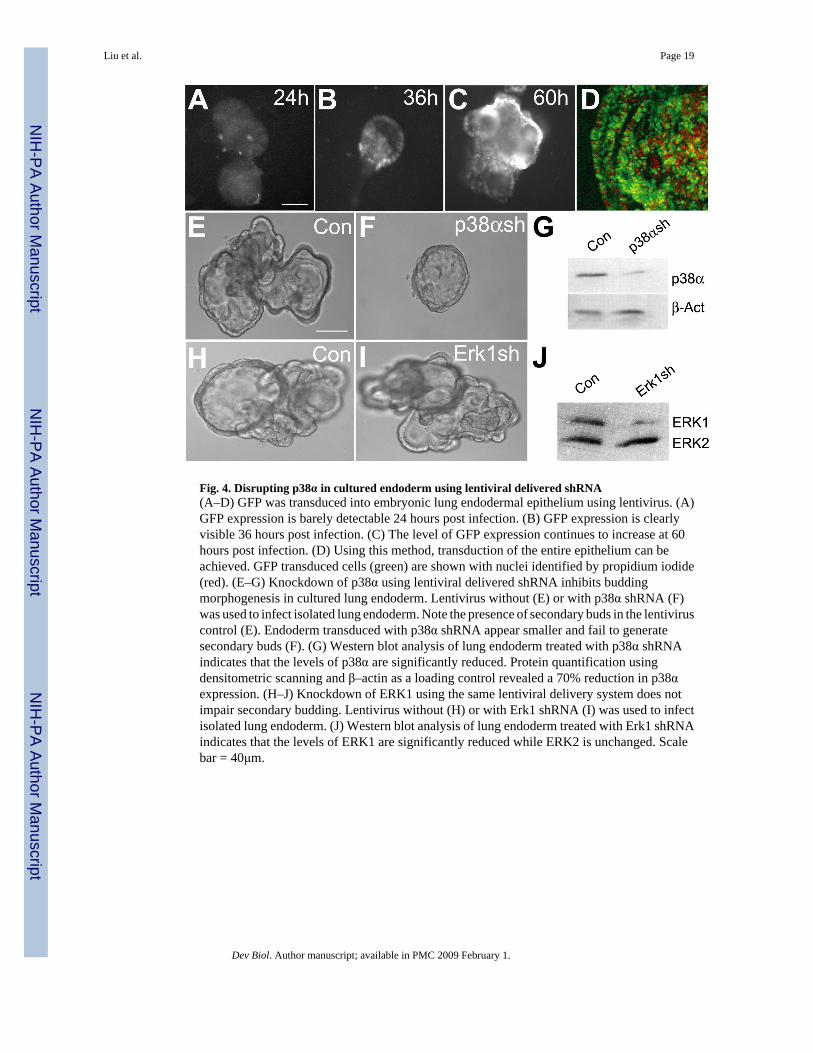
Fig. 4. Disrupting p38α in cultured endoderm using lentiviral delivered shRNA(A–D) GFP was transduced into embryonic lung endodermal epithelium using lentivirus. (A)GFP expression is barely detectable 24 hours post infection. (B) GFP expression is clearlyvisible 36 hours post infection. (C) The level of GFP expression continues to increase at 60hours post infection. (D) Using this method, transduction of the entire epithelium can beachieved. GFP transduced cells (green) are shown with nuclei identified by propidium iodide(red). (E–G) Knockdown of p38α using lentiviral delivered shRNA inhibits buddingmorphogenesis in cultured lung endoderm. Lentivirus without (E) or with p38α shRNA (F)was used to infect isolated lung endoderm. Note the presence of secondary buds in the lentiviruscontrol (E). Endoderm transduced with p38α shRNA appear smaller and fail to generatesecondary buds (F). (G) Western blot analysis of lung endoderm treated with p38α shRNAindicates that the levels of p38α are significantly reduced. Protein quantification usingdensitometric scanning and β–actin as a loading control revealed a 70% reduction in p38αexpression. (H–J) Knockdown of ERK1 using the same lentiviral delivery system does notimpair secondary budding. Lentivirus without (H) or with Erk1 shRNA (I) was used to infectisolated lung endoderm. (J) Western blot analysis of lung endoderm treated with Erk1 shRNAindicates that the levels of ERK1 are significantly reduced while ERK2 is unchanged. Scalebar = 40μm.
Liu et al. Page 19
Dev Biol. Author manuscript; available in PMC 2009 February 1.
NIH
-PA Author Manuscript
NIH
-PA Author Manuscript
NIH
-PA Author Manuscript

Fig. 5. Extracellular FGF can regulate p38 phosphorylation and p38α expression(A–B) FGF7 and FGF10 activate p38 in the LA-4 lung epithelial cell line. Serum-starved LA-4cells were treated with either FGF7 (20 ng/ml) or FGF10 (250ng/ml) for 0, 5, 10, 15, 30 and60 minutes. Lysate were analyzed by Western blotting for TGY phosphorylated p38 (P-p38)and total p38 (p38). (C–D) FGF10 induce ectopic p38α expression in the intact lung. BSA (C)or FGF 10 (D) saturated beads were implanted in the mesoderm of the bronchial region, wherenormally only low level of p38α is expressed. After 48 hours, p38α expression was assayedby whole-mount in situ hybridization. Red circles indicate the position of the implanted beads.Compare the p38α levels in the regions close to the beads (black arrows) with regions awayfrom the beads (white arrows).
Liu et al. Page 20
Dev Biol. Author manuscript; available in PMC 2009 February 1.
NIH
-PA Author Manuscript
NIH
-PA Author Manuscript
NIH
-PA Author Manuscript

Fig. 6. Filamentous actin (F-actin) distribution, cell proliferation and distal marker expression incontrol and p38α-disrupted endoderm(A–D) F-actin distribution and cell proliferation in control (A and B) and p38α shRNA treated(C and D) endoderm was evaluated by triple labeling with propidium iodide (B and D), AlexaFluor 488 phalloidin (A and C, green) and antibody to phospho-histone H3 (A and C, blue).Insets show the localized F-actin enrichment. (E and F) Cell proliferation of endoderm grownin the presence of FGF7 and treated with either vehicle (DMSO) or SB203580 (10 μM).Proliferating cells were detected using an anti-phospho-histone H3 antibody (red) andcounterstained with Alexa Fluor 488 phalloidin (green). (G) Western analysis of lysates fromcultured lung endoderm for Sox9 expression (top panel) following treatment with DMSO (Con)
Liu et al. Page 21
Dev Biol. Author manuscript; available in PMC 2009 February 1.
NIH
-PA Author Manuscript
NIH
-PA Author Manuscript
NIH
-PA Author Manuscript

or 10μM SB203580. The protein level of Sox9 in SB203580 treated sample was 90% of controlas determined by protein quantification using densitometric scanning. Expression of β-actin(bottom panel) was measured as a loading control. (H) Transcripts levels of Erm in control(red) and SB203580 (green) or U0126 (blue) treated sample. Cultured embryonic lungendoderm was treated with DMSO (Con), p38α/β inhibitor SB203580 (10 μM) or the ERK1/2inhibitor U0126 (5 μM). Relative Quantification of Erm was determined by two-step qRT-PCR. Data are the mean ± SEM. Scale bar = 40μm.
Liu et al. Page 22
Dev Biol. Author manuscript; available in PMC 2009 February 1.
NIH
-PA Author Manuscript
NIH
-PA Author Manuscript
NIH
-PA Author Manuscript
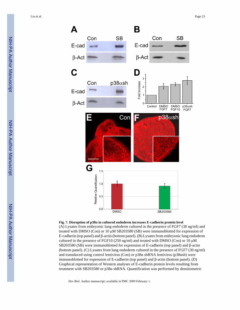
Fig. 7. Disruption of p38α in cultured endoderm increases E-cadherin protein level(A) Lysates from embryonic lung endoderm cultured in the presence of FGF7 (30 ng/ml) andtreated with DMSO (Con) or 10 μM SB203580 (SB) were immunoblotted for expression ofE-cadherin (top panel) and β-actin (bottom panel). (B) Lysates from embryonic lung endodermcultured in the presence of FGF10 (250 ng/ml) and treated with DMSO (Con) or 10 μMSB203580 (SB) were immunoblotted for expression of E-cadherin (top panel) and β-actin(bottom panel). (C) Lysates from lung endoderm cultured in the presence of FGF7 (30 ng/ml)and transduced using control lentivirus (Con) or p38α shRNA lentivirus (p38αsh) wereimmunoblotted for expression of E-cadherin (top panel) and β-actin (bottom panel). (D)Graphical representation of Western analyses of E-cadherin protein levels resulting fromtreatment with SB203580 or p38α shRNA. Quantification was performed by densitometric
Liu et al. Page 23
Dev Biol. Author manuscript; available in PMC 2009 February 1.
NIH
-PA Author Manuscript
NIH
-PA Author Manuscript
NIH
-PA Author Manuscript

scanning and normalized to β-actin expression as a loading control. Relative expression isdepicted with the expression level of the control group in each case arbitrarily defined as 1.0.Data are the mean ± SEM. Each Western blot image is representative of three independentexperiments. (E and F) E-cadherin localization in control (E) and p38α shRNA (F) transducedendoderm was detected by fluorescence confocal microscopy. (G) Transcripts levels of E-cadherin in control and SB203580 treated endoderm cultured in the presence of FGF7 (30 ng/ml). Cultured embryonic lung endoderm from E11.5 mice was treated with DMSO (red) orp38α/β inhibitor SB203580 (green). Relative quantification of E-cadherin was determined bytwo-step qRT-PCR. Data are the mean ± SEM. Scale bar = 40μm.
Liu et al. Page 24
Dev Biol. Author manuscript; available in PMC 2009 February 1.
NIH
-PA Author Manuscript
NIH
-PA Author Manuscript
NIH
-PA Author Manuscript

Fig. 8. E-cadherin over-expression impairs budding morphogenesis(A) Isolated embryonic lung endoderm was transduced with lentivirus containing either theempty vector (Control) or E-cadherin-FLAG (E-cadherin) in the presence of FGF10 (250 ng/ml) for 48 hours. (B) Lysates were immunoblotted for E-cadherin, the FLAG epitope tag, orβ-actin (top, middle and bottom panels, respectively).
Liu et al. Page 25
Dev Biol. Author manuscript; available in PMC 2009 February 1.
NIH
-PA Author Manuscript
NIH
-PA Author Manuscript
NIH
-PA Author Manuscript

NIH
-PA Author Manuscript
NIH
-PA Author Manuscript
NIH
-PA Author Manuscript
Liu et al. Page 26Ta
ble
1Ph
enot
ypic
scor
e of
p38α
shR
NA
trea
ted
endo
derm
sam
ples
Hig
h (>
10)
Mod
erat
e (5
–10)
Low
(<5)
No
2nd
buds
Tot
al%
(no
2nd/
tota
l)
con
4327
146
906.
7p3
8sh
02
1882
102
80.4
Dev Biol. Author manuscript; available in PMC 2009 February 1.
Copyright © 2022 FDOKUMEN