Further investigations into the nature of phrasal ... - Refubium
Upload
khangminh22Category
view
4download
0


Aus dem Institut für Parasitologie und Tropenveterinärmedizin
des Fachbereichs Veterinärmedizin
der Freien Universität Berlin
und dem
International Livestock Research Institute
Assessment of the parasitic burden in the smallholder pig value chain
and implications for public health in Uganda
Inaugural-Dissertation
zur Erlangung des Grades eines
PhD of Biomedical Sciences
an der
Freien Universität Berlin
vorgelegt von
Kristina Rösel
Tierärztin
aus Karl-Marx-Stadt (jetzt Chemnitz)
Berlin 2017
Journal-Nr.: 3973

Gedruckt mit Genehmigung
des Fachbereichs Veterinärmedizin
der Freien Universität Berlin
Dekan: Univ.-Prof. Dr. Jürgen Zentek
Erster Gutachter: Prof. Dr. Peter-Henning Clausen
Zweiter Gutachter: Univ.-Prof. Dr. Reinhard Fries
Dritter Gutachter: Prof. Dr. Eric Fèvre
Deskriptoren (nach CAB-Thesaurus): pigs, value chain, animal parasitic nematodes, Trichinella
spp., muscle larvae, newborn larvae, Toxoplasma gondii, public health, epidemiology, meat
hygiene, animal housing, Uganda
Tag der Promotion: 30.11.2017
Coverbild: Angella Musewa/ILRI

“Overcoming poverty is not a task of charity, it is an act of justice.” (Nelson Mandela)
Dedicated to the smallholder pig value chain actors in Uganda.


Table of contents
i
TABLE OF CONTENTS
List of figures ................................................................................................................................. iii
List of tables .................................................................................................................................... v
List of abbreviations ...................................................................................................................... vi
1 Preface ..................................................................................................................................... 1
1.1 Introduction and study objectives ..................................................................................... 1
1.2 Structure of the thesis ....................................................................................................... 3
2 Literature review ...................................................................................................................... 4
2.1 The history of pigs in Africa ............................................................................................. 4
2.2 Smallholder pig production systems in sub-Saharan Africa ............................................. 6
2.3 Parasites of domestic pigs and their occurrence in East Africa ...................................... 12
2.3.1 Trematoda (flukes) .......................................................................................................... 12
2.3.2 Nematoda (roundworms) ................................................................................................ 12
2.3.3 Protozoa .......................................................................................................................... 17
3 The context for researching smallholder pig value chains in Uganda ................................... 20
3.1 CGIAR Research Framework ......................................................................................... 20
3.2 Study area ....................................................................................................................... 22
3.3 Study site selection and characteristics........................................................................... 25
3.3.1 Geographical targeting .................................................................................................... 25
3.3.2 Stakeholder consultation ................................................................................................. 25
4 Parasites as a production constraint in smallholder pig production systems
(Publications I & II) ............................................................................................................... 28
5 Parasites with potential implications for public health (Publications III & IV) .................... 54
6 Knowledge, attitudes and practices of pork consumers (Publication V) ............................... 72
7 Summarizing discussion ........................................................................................................ 79
7.1 Value chain domains as a suitable proxy for the classification of heterogeneous
smallholder pig production systems ............................................................................... 79
7.2 Pig parasites as a constraint to farm productivity ........................................................... 80
7.3 Parasite infections with public health implications ........................................................ 81
7.3.1 Gastrointestinal parasites as soil-transmitted zoonotic helminths .................................. 81
7.3.2 Pork-borne zoonoses ....................................................................................................... 81

Table of contents
ii
7.4 Researching smallholder pig value chains through an integrated approach and
participatory methods ..................................................................................................... 85
7.5 Concluding remarks, limitations of the survey and potential future research ................ 87
7.5.1 Major research findings .................................................................................................. 87
7.5.2 Major conclusions ........................................................................................................... 88
8 References .............................................................................................................................. 91
Summary ..................................................................................................................................... 108
Zusammenfassung....................................................................................................................... 110
List of own publications.............................................................................................................. 112
Disclosure of own share in the body of the work ....................................................................... 124
Acknowledgements ..................................................................................................................... 126
Statement of authorship .............................................................................................................. 128
List of figures
iii
List of figures
Figure 2.1 Tethered local breed in Wakiso district, Central Uganda (Kristina Roesel). ................ 5
Figure 2.2 Exotic breed (Large White) in a commercial farm in Masaka district,
Central Uganda (Kristina Roesel). ............................................................................... 5
Figure 2.3 Fully confined cross-breed pig in an intensive production unit in Wakiso district,
Central Uganda (Kristina Roesel). ............................................................................... 5
Figure 2.4 Ugandan pig farmer tends to her Camborough pigs
(Edgar R. Batte in Uganda Daily Monitor, 7 November 2012 (Muhanguzi, 2012)). .. 5
Figure 2.5 Pig tethered under a tree and provided with sweet potato vines for feed,
Kiboga district in Central Uganda (Kristina Roesel). .................................................. 8
Figure 2.6 Adult pig confined but piglets able to roam freely in Kamuli district,
Eastern Uganda (Kristina Roesel). ............................................................................... 8
Figure 2.7 One type of confinement in a rural setting in peri-urban Wakiso district,
Central Uganda (Kristina Roesel). ............................................................................... 8
Figure 2.8 Pigs confined in Kamuli district, Eastern Uganda (Kristina Roesel). ........................... 8
Figure 2.9 Confined pigs in peri-urban Masaka, Masaka district, Central Uganda
(Kristina Roesel). ......................................................................................................... 8
Figure 2.10 Commercial pig farm with biosecurity protocol in peri-urban Kampala
(Kristina Roesel) .......................................................................................................... 8
Figure 2.11 The distribution of encapsulated Trichinella spp. in Africa reviewed by
Mukaratirwa et al. (2013), Murrell and Pozio (2011), Pozio (2007), and
Pozio et al. (2005). Map adapted from Pozio et al. (2005). ....................................... 16
Figure 3.1 A livestock value chain and its actors and beneficiaries.
Source: Nick Taylor, University of Reading, and Jonathan Rushton,
Royal Veterinary College, In: Roesel and Grace (2014). .......................................... 20
Figure 3.2 Delivering CGIAR Research Program on Livestock & Fish (CRP L&F).
Structure: Three integrated components. Source: CRP L&F, 2012. .......................... 21
Figure 3.3 Problem tree showing food safety and related market access in the context of
whole value chain development (ILRI, 2011). .......................................................... 22
Figure 3.4 Pig density by district shows a higher concentration in the Central, Western and
Eastern regions of Uganda. Source: Poole et al. (2015).
Category thresholds are as accurate as 14 decimals. ................................................. 23
Figure 3.5 Average pig densities in Uganda show production specific production foci in Mbale,
peri-urban Kampala, Kasese and Masaka. Source: Robinson et al. (2014).
Category thresholds are as accurate as 14 decimals. ................................................. 24
List of figures
iv
Figure 3.6 Spatial distribution of per capita pig meat consumption, based on population density.
Source: Center for International Earth Science Information
Network (CIESIN), 2011. Category thresholds are as accurate as 14 decimals. ....... 24
Figure 3.7 The CRP L&F study sites selected in 2012: Masaka and Mukono districts in the
Central region; Kamuli district in the Eastern region of Uganda
(ILRI/Pamela Ochungo). ............................................................................................ 26
Figure 7.1 An article published by the newspaper Uganda Daily Monitor on 6 June 2012
referring to a report that suggests high contamination of pork in Kampala.
On inquiry, the Kampala City Council Authority's (KCCA) senior veterinary
officer explained that there is no report. Statements on pork safety were
based on misperceptions not evidence.
Source: Safe Food, Fair Food blog post (ILRI, 2012c). ............................................ 84
Figure 7.2 One week later, on 13 June 2016, the Ugandan populist newspaper
Red Pepper declared a war on informal pork butcheries operating in Kampala,
referring to the same misleading “expert report”.
Source: Safe Food, Fair Food blog post (ILRI, 2012c). ............................................ 84
Figure 7.3 Synthesized results of the Ugandan smallholder pig value chain assessment
including porcine parasites. The work was presented by the author at
Tropentag conference in Prague, September 2014. Source: Ouma et al. (2014). ...... 86

List of tables
v
List of tables
Table 2.1 Simplified and selective classification of pig endoparasites.
Adapted from Deplazes et al. (2016), Gajadhar (2015), Greve (2012),
Lindsay et al. (2012), Kaufmann (1996). ....................................................................... 9
Table 2.2 Common ectoparasites of domestic pigs and their relevance
to the pig farm enterprise and public health.
Adapted from Deplazes et al. (2016) and Greve and Davies (2012). .......................... 11
Table 3.1 The final sites selected for the pig value chain assessment under the CRP L&F.
Source: Poole et al. (2015). .......................................................................................... 26
Table 3.2 Characteristics of the sites selected by the CRP L&F.
Sources: MAAIF/UBOS (2009); Twinomujini et al. (2011). ...................................... 27
Table 7.1 The median global numbers of foodborne illnesses, deaths and Disability Adjusted
Life Years (DALYs) compared to the median rates of DALYs
in the African sub-region E (AFR E) which includes Uganda.
Source: Havelaar et al. (2015). ..................................................................................... 83

List of abbreviations
vi
List of abbreviations
AIDS Acquired immunodeficiency syndrome
AAT Animal African trypanosomosis
ADG Average daily (weight) gain
ASF African swine fever
BMZ Bundesministerium für wirtschaftliche Zusammenarbeit und Entwicklung
(German Federal Ministry for Economic Cooperation and Development)
CGIAR Global research partnership for a food-secure future (formerly Consultative
Group for International Agricultural Research)
CI Confidence interval
CIESIN Center for International Earth Science Information Network
CRP CGIAR Research Program
CRP A4NH CGIAR Research Program on Agriculture for Nutrition and Health
CRP L&F CGIAR Research Program on Livestock and Fish
CSF Classical swine fever
DAD-IS Domestic Animal Diversity Information System
DALYs Disability Adjusted Life Years
ELISA Enzyme-linked immunosorbent assay
FAO Food and Agricultural Organization of the United Nations
FAOSTAT Statistics Division of the Food and Agricultural Organization of the United
Nations
FGD Focus group discussion
FMD Foot and mouth disease
GIS Geographic information system
GIZ Gesellschaft für Internationale Zusammenarbeit
HAT Human African trypanosomosis
HIV Human immunodeficiency virus
IFAD International Fund for Agricultural Development
IgG Immunoglobulin G
ILRI International Livestock Research Institute
IFPRI International Food Policy Research Institute

List of abbreviations
vii
KCCA Kampala City Council Authority
LC Local government council
MAAIF Ministry of Agriculture, Animal Industry and Fisheries (Government of Uganda)
NAADS National Agricultural Advisory Service
NEMA National Environment Management Agency
NGO Non-governmental organization
OIE World Organization for Animal Health
PCV Porcine circovirus
PE Participatory epidemiology
PRA Participatory rural appraisal
PRRS Porcine Reproductive and Respiratory Syndrome
RR Rural production for rural consumption (value chain type)
RU Rural production for urban consumption (value chain type)
SFFF Safe Food, Fair Food project
SPVCD Smallholder Pig Value Chain Development project
STH Soil-transmitted helminths
UBOS Uganda Bureau of Statistics
UU Urban production for urban consumption (value chain type)
VEDCO Volunteer Efforts for Development Concerns
WHO World Health Organization


Chapter 1: Preface
1 Preface
1.1 Introduction and study objectives
Population growth, urbanization and increasing consumers’ demand for products of animal origin
are fuelling the so-called Livestock Revolution which could provide poor smallholder farmers with
income (Delgado et al., 2001; Thornton, 2010) and thus contribute to poverty alleviation. In low-
income countries, meat and dairy consumption have grown at average annual rates of 5.1 and
3.6%, respectively, between 1971 and 2007 (Alexandratos and Bruinsma, 2012). Between 2000
and 2050, demand for livestock products is estimated to nearly double in sub-Saharan Africa
(Thornton, 2010), while globally, potential improvements in food security and nutrition could
benefit directly more than 750 million poor livestock keepers (Food and Agriculture Organization
of the United Nations (FAO), 2011). Uganda’s population is currently growing at 3.3% annually
(Worldbank, 2015a), and has increased seven times since the 1950s. The country is experiencing
rapid urbanization – like most developing countries – at currently 4.5% which could result in an
increase in the population of Uganda's cities from 6.4 million today to more than 30 million within
the next two decades (Worldbank, 2015b), demanding ever more food.
Pork is the most popular meat in the world today accounting for over 36% of the world meat
production, followed by poultry and beef (FAO, 2015). In Uganda, pork has only become
important over the past 25 years; the Statistics Division of the Food and Agricultural Organization
of the United Nations (FAOSTAT) estimated pig numbers have grown rapidly from 716,400 in
1989 to 2.18 million in 2008 (FAOSTAT, 2016). The 2008 national livestock census recorded 3.2
million pigs (Ministry of Agriculture, Animal Industry and Fisheries (MAAIF)/ Uganda Bureau
of Statistics (UBOS), 2009), significantly surpassing estimates of the FAO. Whereas pork
accounted for barely 1% of the annual per capita meat consumption in Uganda in 1961, it made up
27% of all meat consumed in 2011 (FAOSTAT, 2011). Little information is available regarding
the structure and composition of the pig sector in Uganda but it has been documented that almost
one fifth of the Ugandan households own pigs, on average three (MAAIF/ UBOS, 2009), while up
to 70% of all pork consumed in Uganda is consumed in urban and peri-urban areas (International
Livestock Research Institute (ILRI), 2011).
The majority of pigs in Kenya, Tanzania and Uganda are estimated to be reared in smallholder
households under extensive systems (Lekule and Kyvsgaard, 2003); e.g. mostly free-ranging with
tethering. Conventional knowledge states that traditional production systems are wasteful and
unprofitable due to poor feed conversion, low reproductive rates, final products of inferior quality,
and losses due to high animal mortality. A high gastrointestinal parasitic burden and direct losses
due to infectious animal diseases such as African swine fever, foot and mouth disease or rabies
(all endemic in Uganda) could contribute to low productivity (World Organisation for Animal
Health (OIE), 2016). Concurrent with growth in smallholder pig keeping and pork consumption,
zoonoses such as mycobacterial infections and porcine cysticercosis have increasingly been
reported (Muwonge et al., 2010; Waiswa et al., 2009; Phiri et al., 2003). However, traditional
production also has advantages compared to intensive systems, for example the small inputs
required in traditional production (little to no housing), and the pig’s natural scavenging behaviour
which allows them to feed on kitchen waste and agricultural waste that would otherwise be wasted.
The surging demand for livestock products provides an opportunity to set poor farmers on
pathways out of poverty by increasing production outputs and market access. At the same time,
1

Chapter 1: Preface
markets have become more demanding and threaten the continued presence of smallholder
farmers: livestock diseases and other factors affect a constant supply while foodborne pathogens
may impair the quality and safety of the farmers’ products. While the presence of food safety
hazards such as bacteria, parasites or drug residues in informally marketed food is high, the risk to
human health is mostly unknown and current food safety management is both ineffective and
inequitable.
In Uganda, intensification level may affect the parasitic burden of pigs and hence the profitability
of pig farming as well as the risk to human health associated with pork borne parasites. The present
study aimed to contribute to improving selected smallholder pig value chains in Uganda by
increasing the knowledge on prevalent parasitic diseases.
Specific objectives are to:
1. understand whether parasites are perceived as a production constraint by farmers;
2. estimate the burden of selected endoparasites in pigs and pork at farm, slaughter and retail
outlet level in three selected value chain types in Uganda. These parasites included
common gastrointestinal helminths and protozoa, as well as two major porkborne zoonotic
parasites Trichinella spp. and Toxoplasma gondii;
3. identify risk factors contributing to parasitic infections in pigs and pork, in order to better
understand the epidemiology of parasitic diseases in these value chains with emphasis on
pork borne zoonoses;
4. identify current practices that increase or reduce risks to public health associated with pork
consumption;
5. assess the risk to public health through the consumption of pork infested with parasites.
2

Chapter 1: Preface
1.2 Structure of the thesis
This cumulative thesis is structured as follows:
Chapter 1 Preface: introduction, study objectives and structure of the thesis.
Chapter 2 Literature review: outlines the history of pigs in Africa, describes the current
smallholder pig production systems in sub-Saharan Africa, and reviews the current state of
knowledge on parasitic pig diseases compromising farm productivity as well as selected parasitic
diseases that are potentially transmitted to humans through pork consumption. The review refers
to previous research from sub-Saharan Africa, and particularly Uganda.
Chapter 3 Research context: describes the context of the study and how it fits into overarching
research for development on smallholder pig value chains in Uganda. It further describes how
study sites were selected, value chain systems defined and characteristics of the area under study.
Chapter 4 Parasites as a production constraint: shows that gastrointestinal parasites are
perceived as an important production constraint by smallholder pig farmers in Central and Eastern
Uganda (Publication I) and summarizes baseline information on the prevalence and risk factors of
the most common intestinal parasites (Publication II).
Chapter 5 Parasites with implications for public health: provides baseline information on two
pork borne zoonoses of global importance, namely infection with Trichinella spp. (Publication III)
and Toxoplasma gondii (Publication IV), in smallholder pig value chains in Central and Eastern
Uganda.
Chapter 6 Knowledge, attitudes and practices of pork consumers: outlines the findings on
common preparation and consumption practices as well as sources of pork for consumption in the
study area to potentially draw conclusions on the risk to pork consumers (Publication V).
Chapter 7 Summarizing discussion: discusses major findings, conclusions and limitations of
the study, and generates assumptions for potential interventions and further research.
3

Chapter 2: Literature review
2 Literature review
2.1 The history of pigs in Africa
Literature on the domestication of pigs in Africa is scarce and highly debated (Amills et al., 2013;
Blench, 2000). The domestic pig (Sus scrofa domesticus) was domesticated from its ancestor the
wild boar, or Eurasian wild pig (Sus scrofa) which is a native to Europe, Asia and North Africa.
There are three distinct wild members of the family Suidae in tropical Africa: the bushpig
(Potamochoerus porcus), the warthog (Phacochoerus aethiopicus), and the giant forest hog
(Hylochoerus meinertzhageni). It is still debated to what extent African wild Suidae were
domesticated (Gifford-Gonzalez and Hanotte, 2011; Blench, 2000; Haltenorth and Diller, 1980);
but evidence is appearing that bush pigs and domestic pigs have indeed mated (Rothschild et al.,
2014).
Genetic and archaeological findings suggest that pig domestication began about 9,000 to 10,000
years ago at multiple sites across Eurasia, followed by their subsequent worldwide spread through
increased exploration and commercialization (Amills et al., 2010; Larson, 2005). According to
recent findings (Amills et al., 2013; Ramirez et al., 2009; Blench, 2000), it is likely that domestic
pigs have been introduced to sub-Saharan Africa from the Near East via Egypt approximately
6,000 years ago, from the Far East during the Indian Ocean trade in the 7th century, and during
European colonisation from the 15th century. By analysing the genetic diversity of African pigs,
Ramirez et al. (2009) showed the existence of a clear genetic dichotomy between East and West
Africa: While a clear Far Eastern genetic signature was found in pigs from the Indian Ocean coast
of Africa, it could not be detected in pigs from the African Atlantic shores. The findings have been
discussed in-depth (Gifford-Gonzalez and Hanotte, 2011; Ramirez et al., 2009). It has also been
hypothesized that African pig breeds were domesticated locally and independently in the Nile
Delta (Gautier, 2002; Houlihan, 1997); however, at present evidence is still weak (Amills et al.,
2013). African pig breeds are still poorly characterised but according to the Domestic Animal
Diversity Information System (DAD-IS) database hosted by FAO, there are currently 148 pig
breeds in Africa (FAO, 2016).
Phenotypically, domestic pigs in Uganda are divided into two major groups, the so-called “local”
pigs and the introduced “exotic” pigs. Local pigs are usually black with medium-sized, semi-erect
ears, a straight tail and a long snout (Figure 2.1). Exotic breeds such as Large White (Figure 2.2),
Landrace and Saddleback have been introduced during the colonial periods. Under local
management practices, local and exotic breeds interbred (Figure 2.3). Other pig breeds such as
Camborough® have recently become of interest to Ugandan pig keepers (Muhanguzi, 2012). The
Camborough® (Figure 2.4) is a product (material pig line) of a South Africa-based franchise of a
commercial pig breeding company called the Pig Improvement Company (PIC
International)1. The breed is derived from original crosses followed by heavy selection for
maternal traits. Approximately 40 Camborough® lines have been released by PIC over the past
years, and some have been introduced to Uganda by the Ministry of Agriculture (personal
communication).
The distribution of religion in Africa is another critical aspect of the history of pigs in Africa
(Blench, 2000). Muslims and Ethiopian Christian Orthodox are forbidden to eat pork which is
generally interpreted as a prohibition on any sort of contact with pigs. Islam spread to Africa in
1 http://www.picgenus.com/
4

Chapter 2: Literature review
the early 7th century when Muslims sought refuge in present-day Ethiopia during the Migration to
Abyssinia (hijarat); in Uganda, they arrived only in the late 19th century (David, 2004). Until the
spread of Islam, pigs were quite important, especially in the Egyptian and Berber cultures in
Northern Africa, already 6,000 years ago (Blench, 2000). All aspects of pig-keeping have been
relatively little researched in Africa. Even international research organizations – such as ILRI since
its inception in 1974 – have not included pigs in Africa in its research agenda until recently. Blench
(2000) argues this may have been related to prejudice against pigs from potential donor agencies
but also the belief that – as monogastric animals – pigs may compete with humans for food.
Figure 2.1 Tethered local breed in Wakiso district, Central
Uganda (Kristina Roesel).
Figure 2.2 Exotic breed (Large White) in a commercial farm
in Masaka district, Central Uganda (Kristina Roesel).
Figure 2.3 Fully confined cross-breed pig in an intensive
production unit in Wakiso district, Central Uganda (Kristina
Roesel).
Figure 2.4 Ugandan pig farmer tends to her Camborough
pigs (Edgar R. Batte in Uganda Daily Monitor, 7 November
2012 (Muhanguzi, 2012)).
5

2.2 Smallholder pig production systems in sub-Saharan Africa
Pig production has emerged as an important agribusiness over the past few decades. Not only in
Uganda but in many regions of sub-Saharan Africa, e.g. Nigeria (Machebe et al., 2009;
Adesehinwa et al., 2003), Ethiopia (Tekle et al., 2013; Seid and Abebaw, 2010), Tanzania
(Karimuribo et al., 2011; Esrony et al., 1997), Namibia (Petrus et al., 2011) and Kenya (Obonyo
et al., 2013; Kagira et al., 2010b; Mutua et al., 2010; Wabacha et al., 2004a) to mention but a few.
Given population growth and the pressure on the size of farm land, monogastrics seem to provide
better production efficiency per unit area of land (FAO, 2009; Wabacha et al., 2004a). In Western
Kenya, for instance, the average size of a smallholder pig farmers’ land is one acre2 or less (Mutua
et al., 2011a; Kagira et al., 2010b). Pigs are often reared by women (Obonyo et al., 2013; Petrus
et al., 2011; Kagira et al., 2010b; Mutua et al., 2010; Nsoso et al., 2006) as one of several income-
generating activities, and kept in close proximity to homesteads (Obonyo et al., 2013; Karimuribo
et al., 2011; Mutua et al., 2010). Income generation as well as savings and insurance against family
emergency is often a more important motivation for pig keeping than providing meat for house
hold consumption (Mbuthia et al., 2015; Tekle et al., 2013; Mutua et al., 2010). The regular
smallholder in Kenya and Uganda keeps 3-5 pigs (Kagira et al., 2010b; MAAIF/UBOS, 2009);
and herd size increases with proximity to urban centres (MAAIF/UBOS, 2009), with the level of
intensification (Mbuthia et al., 2015; Wabacha et al., 2004a), or concentrates in certain areas; this
is the case in Ethiopia where pig keeping is increasing but not as commonly practiced due stringent
religious laws (Tekle et al., 2013). Most of the pig farmers in traditional production systems in
sub-Saharan Africa keep cross-breeds (Kagira et al., 2010b) as they believe local breeds require
less feeds and are more tolerant to diseases (Petrus et al., 2011; Lekule and Kyvsgaard, 2003) while
exotic breeds grow faster.
Many small-scale farmers sell live animals produced in farrow-to-weaner (piglet) and porker-to-
finisher (fattening) units; few keep boars and those who do often rent them out to other farmers
for breeding (Muhanguzi et al., 2012; Kagira et al., 2010b), often against in-kind payment with a
piglet. Many of the pigs are kept in mixed confinement systems, often tethered, especially during
crop season, and otherwise free-roaming, especially during dry seasons when crop fields are
fallowing and feeds are scarce (Obonyo et al., 2013; Mutua et al., 2011a; Permin et al., 1999).
Housing is difficult to provide for many farmers because it is considered too expensive, and shelter
is often just temporary or rarely cleaned (Muhanguzi et al., 2012; Kagira et al., 2010b; Seid and
Abebaw, 2010). A study in rural Western Kenya reported that if farmers did not consider housing
unnecessary altogether, the main reasons for non-confinement are fear of the pig damaging the
houses, lack of food to provide for confined pigs, houses becoming muddy during the rainy season
and farmers lacking the time to manage confined pigs (Mutua et al., 2011a). Smallholders’ pigs
are mostly fed with locally available ingredients such as crop residues (Muhanguzi et al., 2012;
Kagira et al., 2010b; Petrus et al., 2011) but in some areas pigs are left grazing/scavenging or fed
with household leftovers (Obonyo et al., 2013; Seid and Abebaw, 2010), supplemented with
commercial feeds such as sunflower seed cake (Karimuribo et al., 2011), wheat bran (Tekle et al.,
2013), or ruminal contents from abattoirs (Muhanguzi et al., 2012). Marketing is often
disorganised and economically inefficient because record keeping is not common (Nsoso et al.,
2006; Wabacha et al., 2004a); farmers mostly sell when they are in need (e.g. for school fees), and
they do not weigh their animals and have therefore no objective basis for negotiation (Mutua et
2 one acre equals approx. 4,000 square metres
6
Chapter 2: Literature review

al., 2011a, 2011b; Petrus et al., 2011). Many other knowledge gaps on pig husbandry such as how
to feed monogastrics, regular provision of water and the difference between treatment and
vaccination have been documented in Kenya, and some of them seem to depend on the presence
and a positive attitude towards pig-keeping of the veterinary extension service in a region (Obonyo
et al., 2013; Mutua et al., 2011a; Kagira et al., 2010b).
Pig diseases (especially African swine fever and parasitic infestation), cost of feeds, lack of market
access and veterinary extension as well as lack of knowledge on pig husbandry and disease
prevention are perceived to be the greatest production constraints in many pig-keeping
communities in sub-Saharan Africa (Atuhaire et al., 2013; Tekle et al., 2013; Muhanguzi et al.,
2012; Karimuribo et al., 2011; Petrus et al., 2011; Kagira et al., 2010b). Important parasitic
diseases (based on signs observed by farmers) are sarcoptic mange, lice and gastrointestinal worms
(Karimuribo et al., 2011; Petrus et al., 2011; Kagira et al., 2010b). Different pig production systems
in Uganda are illustrated in Figures 2.5-2.10.
7
Chapter 2: Literature review

Figure 2.5 Pig tethered under a tree and provided with sweet
potato vines for feed, Kiboga district in Central Uganda
(Kristina Roesel).
Figure 2.6 Adult pig confined but piglets able to roam freely
in Kamuli district, Eastern Uganda (Kristina Roesel).
Figure 2.7 One type of confinement in a rural setting in peri-
urban Wakiso district, Central Uganda (Kristina Roesel).
Figure 2.8 Pigs confined in Kamuli district, Eastern Uganda
(Kristina Roesel).
Figure 2.9 Confined pigs in peri-urban Masaka, Masaka
district, Central Uganda (Kristina Roesel).
Figure 2.10 Commercial pig farm with biosecurity protocol
in peri-urban Kampala (Kristina Roesel).
8
Chapter 2: Literature review

Table 2.1 Simplified and selective classification of pig endoparasites. Adapted from Deplazes et al. (2016), Gajadhar (2015), Greve (2012), Lindsay et al. (2012), Kaufmann
(1996).
Order: species Common name Location in the pig host Economic relevance Zoonotic relevance
Cest
od
a
Cyclophyllidea: Taenia solium,
larval
Pork tapeworm,
pork measles
Muscle tissue At slaughter if infected meat is
condemned.
Ingestion of Cysticercus cellulosae in pork causes
taeniosis in humans; infection in pigs helps maintaining the life cycle. High risk for humans developing
neurocysticercosis when ingesting T. solium eggs
(autoinfection).
Cyclophyllidea: Taenia
hydatigena, larval
Omentum, mesentery Losses through trimming at slaughter. N/A
Cyclophyllidea: Echinococcus granulosus s.l., larval
Hydatid disease Liver, lungs At slaughter if infected organs are condemned.
Pigs can be a reservoir for echinococcosis in carnivores (maintain life cycle). Humans are accidental hosts when
ingesting eggs of E. intermedius (pig strain G7) passed in
dog faeces.
Trem
ato
da
3
Echinostomida: Fasciola
hepatica
Liver fluke Liver (bile system) At slaughter if infected livers are
condemned.
Accidental infection of humans through ingestion of
encysted metacercaria.
Strigeatida: Alaria alata Muscle tissue At slaughter if infected meat is condemned.
Emerging parasitic disease. Human infection through ingestion of mesocercaria.
Plagiorchiida: Paragonimus spp. Lungs N/A N/A
Nem
ato
da
4
Ascarida: Ascaris suum Large intestinal
roundworm
Lumen of small intestine Major growth reduction if piglets are
infected.
Especially for children in poor pig-keeping communities.
Enoplidae: Trichuris suis Whipworm Large intestine Haemorrhagic enteritis in heavy infection; weight losses in chronic infections.
Potential still under debate.
Rhabditida: Strongyloides suis
(syn. ransomi)
Threadworm Epithelium small intestine Major impact in suckling piglets. Larva migrans cutanea (creeping eruption).
Strongylida: Hyostrongylus
rubidus
Red stomach
worm
Stomach fundus Suck small amounts of blood and cause
catarrhal gastritis.
N/A
Strongylida: Metastrongylus apri Lung worm Bronchial tubes and
bronchioles
Respiratory disease, especially in young
pigs; can facilitate infection with
pathogens causing pneumonia.
N/A
Strongylida: Oesophagostomum
spp.
Nodular worm Large intestine Reoccuring enteritis with every new
infection (no protective immunity).
N/A
Strongylida: Globocephalus spp. Pig hookworm Attached to mucosa of
small intestine.
Aenemia in growers. N/A
Strongylida: Ollulanus tricuspis Stomach Weight loss and chronic-catarrhal gastritis. N/A
Strongylida: Stephanurus dentatus
Swine kidney worm
Peri-renal fat tissue Reduced growth; trimming at slaughter due to abscesses.
N/A
3 less significant trematodes reported in pigs: Dicrocoelium hospes, Gastrodiscus aegyptiacus, Postharmostomum suis, Schistosoma bovis, Schistosoma matthei, Eurytrema pancreaticum, Gongylonema pulchrum, Prosthogonimus cuneatus 4 less significant nematodes reported in pigs: Gongylonema pulchrum, Setaria congolensis, Gnathosoma hispidum, Trichostrongylus axei
9
Chapter 2: Literature review

Table 2.1 (continued). Simplified and selective classification of pig endoparasites. Adapted from Deplazes et al. (2016), Gajadhar (2015), Greve (2012), Lindsay et al. (2012),
Kaufmann (1996).
Order: species Common name Location in the pig host Economic relevance Zoonotic relevance
Nem
ato
da
4
Oligacanthorhynchida:
Macracanthorhynchus hirudinaceus
Thorny-headed
worms
Attached to mucosa of
small intestine
Only if burden is high. Accidental infection through ingestion of infected
arthropod.
Sprirurida:
Physocephalus sexalatus; Ascarops strongylina;
Gnathosoma spinigerum;
Simondsia paradoxa
Thick stomach
worms
Stomach Less important than H. rubidus. N/A
Spirurida: Gongylonema pulchrum
Esophageal worm Epithelium of tongue and esophagus
Minor irritation. Humans are susceptible but must ingest intermediate host insect to become infected.
Trichocephalida: Trichinella spp. Pork worm,
muscle worm
Adults in epithelium of
small intestines; first-stage
larvae encysted in striated muscle.
Not in pig farms but potentially at
slaughter if infected carcasses are
condemned.
Human trichinellosis mostly caused by ingestion of viable
first-stage larvae in infected and undercooked pork.
Pro
tozo
a5
Cryptosporida: Cryptosporidium
spp.
Epithelium of small
intestine
Diarrhoea in growers. C. parvum and C. suis are human-infective.
Eimeriida: Eimeria spp.
Epithelium of small intestine
Diarrhoea in weaners and finishers. N/A
Eimeriida:
Isospora suis
Epithelium of small
intestine
Diarrhoea in piglets. N/A
Eimeriida:
Toxoplasma gondii
Muscle and brain tissue Not in pig farms but potentially at
slaughter if infected carcasses are
condemned.
Undercooked pork considered source of foodborne
infection; occupational exposure to infective bradyzoites.
Eimeriida: Sarcocystis spp.
Muscle tissue On farm piglet diarrhoea. Transient diarrhoea in humans.
Eimeriida:
Neospora caninum
Nervous tissue On farm piglet losses due to abortions and
stillbirth.
N/A
Piroplasmida: Babesia trautmanni
Porcine piroplasmosis
Erythrocytes Up to 50% mortality in infected pigs of all ages.
N/A
Trypanosomatida: Trypanosoma spp.
Nagana Extracellular blood Peracute death in pigs caused by T. simiae or T. suis; reservoir for cattle pathogenic
trypanosomes.
Pigs as reservoir for human pathogenic Trypanosoma spp.
Diplomonadida:
Giardia duodenalis6
Small intestine Subclinical Assemblages A and B causative agent of human
giardiasis.
Vestibuliferida: Balantidium coli Large intestine Haemorrhagic enteritis if burden is high. Faecal-oral transmission route; low pathogenicity but
discussed as contributing factor to colitis.
4 less significant nematodes reported in pigs: Gongylonema pulchrum, Setaria congolensis, Gnathosoma hispidum, Trichostrongylus axei 5 less significant protozoa reported in pigs: Tritrichomonas suis, Trichomonas rotunda, Tetratrichomonas buttreyi, Entamoeba suis (syn. polecki) 6 syn. lamblia, syn. intestinales
10
Chapter 2: Literature review

Table 2.2 Common ectoparasites of domestic pigs and their relevance to the pig farm enterprise and public health. Adapted from Deplazes et al. (2016) and Greve and Davies
(2012).
Order: species Common name Economic relevance Zoonotic relevance
Art
hro
pod
a
Astigmata: Sarcoptes scabiei var. suis Sarcoptic mange mite, burrowing mite
Reduction of growth rates, feed efficiency, and fertility.
Transient and self-limiting dermatitis in humans.
Prostigmata: Demodex phylloides (syn. suis) Follicular mange mite, follicle mite N/A N/A
Metastigmata: Ornithodorus moubata complex Soft tick (e.g. eyeless tampan) Vector for African swine fever virus. Vector for rickettsiosis.
Metastigmata: Amblyomma/ Rhipicephalus spp. Hard tick (e.g. bont/blue/brown tick) Nuisance and hence stress-induced reduced growth rate or anaemia; vector for porcine
babesiosis
N/A
Phthiraptera: Haematopinus suis Hog louse, sucking louse Reduced growth and increased susceptibility to
disease in piglets due to anaemia; discussed as vector for African swine fever virus.
N/A
Siphonaptera: Tunga penetrans Sand flea, jigger flea Nuisance Pigs as reservoir for human tungiasis.
Siphonaptera: Pulex irritans/ Ctenocephalides
felis/ Echidnophaga gallinaecea
Human flea/ cat flea/ sticktight flea Nuisance N/A
Diptera: Musca domestica House fly Nuisance Mechanical vector of pathogens (faecal-oral
route).
Diptera: Stomoxys calcitrans Stable fly Nuisance and potential vector of pig pathogens N/A
Diptera: Chrysomyia bezziana, Calliphora spp. etc.
Screwworm/ blow fly/ carrion fly/ flesh fly
Excavation of primary wounds (myiasis, or fly strike) and exposure to secondary infections
N/A
Diptera: Glossina spp. Tsetse fly Vector for nagana in pigs caused by T. simiae or
T. suis; vector for cattle pathogenic Trypanosoma spp.
Vector for human pathogenic Trypanosoma
species for which pigs can be reservoirs.
Diptera: Aedes spp./ Culex spp. Mosquito Mechanical vector for PRRS virus and Mycoplasma (Eperythropozoon) suis.
Potential vector for human pathogenic arboviruses (e.g. Japanese encephalitis, West Nile fever,
Tahyna).
Diptera: Phlebotomus spp. Sand fly N/A N/A
Diptera: Simulium spp. Black fly/ gnat Nuisance N/A
Diptera: Culicoides spp. Biting midge Nuisance N/A
11
Chapter 2: Literature review

2.3 Parasites of domestic pigs and their occurrence in East Africa
In general, pig parasites can be divided into two major groups: endo- and ectoparasites.
Endoparasites live within the host, either inside the gastrointestinal lumen or organ tissue such as
liver, lungs or muscle. Many of them are heteroxenous, an important trait that needs to be
considered in diagnostics, management and control. This group of endoparasites in pigs (Table
2.1) includes representatives of major economic importance and the potential to cause disease in
humans. Ectoparasites (Table 2.2) are usually found outside of the pig’s body, on or in the skin,
either temporary or permanently. Many of them are economically important as a nuisance and as
a consequence, decreased weight gain due to the animals’ pressure to deter them. Others are known
vectors for more debilitating diseases, including parasites, such as ticks transmitting the African
swine fever virus or tsetse flies carrying Trypanosoma species. The following section outlines the
biology and occurrence of the endoparasites economic and veterinary public health importance
that are subject to this study, with particular reference to East Africa and Uganda. For the
denomination of parasitic diseases or infections, the author followed the Standardized
Nomenclature of Animal Parasitic Diseases (Kassai et al., 1988).
2.3.1 Trematoda (flukes)
Fasciola (F.) hepatica and Fasciola (F.) gigantica, the common or gigantic liver flukes, are
trematodes with a broad range of mammal hosts including ruminants, horses, camels, pigs and
humans. Pigs are not very susceptible to infection with Fasciola but can be carriers of adult flukes
shedding eggs (very rare). The World Health Organization (WHO) estimates that at least 2.4
million people are infected in more than 70 countries worldwide with several million at risk; and
that no continent is free from fasciolosis, and it is likely that where animal cases are reported,
human cases also exist (WHO, 2016a). In livestock production, the burden is mainly on herbivores,
especially ruminants (sheep > goat > cattle), and the result of reduced growth performance,
anaemia and condemnation of infected livers if meat inspection takes place and condemnation is
enforced. In Kenya, liver condemnation at beef cattle abattoirs due to fasciolosis led to losses
worth $25,000 in 2007 and 2008 (Kanyari et al., 2012). The life cycle is indirect whereby the pigs
ingest encysted metacercaria with contaminated food (pasture) or water. Within one day, juvenile
flukes penetrate the intestinal wall and migrate to the liver where they migrate the tissue for about
six weeks, and then move through the bile system. This can take months to years; from there up to
20,000 eggs per female per day are produced 8 to 10 weeks post infection at the earliest. The eggs
are passed in the pig faeces and develop in water where a miracidium hatches and penetrates
freshwater molluscs (Galba trunculata or Lymnaea natalensis) where it produces hundreds of
cercariae. These encyst on plants where they develop into infective metacercaria. They can survive
up to 10 weeks in a moist environment such as wetlands. In Uganda F. gigantica has been reported
in bovines from Mt. Elgon (Howell et al., 2012); and both F. gigantica and F. hepatica were
reported from the tropical highlands in Tanzania (Walker et al., 2008). Kagira et al. (2012) reported
3% of the pigs in Western Kenya infected with Fasciola spp. using McMaster fecal flotation
technique.
2.3.2 Nematoda (roundworms)
Ascaris (A.) suum, the large roundworm of the small intestine, is cosmopolitan in domestic and
wild Suidae. Infection is particularly important in the pig industry for two reasons: first, the adult
worms compete with their pig host for nutrients through disruption of enteral absorption which
12
Chapter 2: Literature review

reduces feed efficiency and average daily gains (Hale et al., 1985); secondly, once on the pig farm,
it is difficult to control because the eggs are extremely resilient and difficult to eliminate.
Reproduction follows a direct life cycle: ingestion of the egg with infectious third-stage larvae,
which hatches in the lumen of the jejunum and penetrates the intestinal wall, then migrates into
the liver via the portal vena where it migrates the tissue for one to two days causing temporary
inflammation and the well-known milk spots which disappear again after about 25 days once larval
burden decreases. Within the next week larvae are carried to the lungs where migration causes
inflammation and, potentially, respiratory signs. In the lungs larvae moult and further move to the
bronchioles from where they are coughed up and swallowed. About two weeks post infection the
larvae reach the small intestine again where they mature into adults. After another 4-6 weeks
females start laying eggs, sometimes more than 200,000 per day for about six months. The un-
embryonated eggs have a thick shell, which enables them to survive in moist soil for up to five
years. Most chemicals do not have an effect on the egg and they have a sticky coat which enables
them to be easily carried from farm to farm by live vectors (i.e. flies, beetles, cats or dogs but also
children) or other vectors (i.e. boots, vehicles, tools). Also, they can stick to the sow’s coat from
where nursing piglets ingest the eggs when suckling. Infection and clinical signs occur mostly in
pigs younger than five months, with age there is increased immunity though adult pigs may still
harbour worms that produce eggs intermittently. A. suum can infect humans, and recent research
including the application of molecular diagnostics increasingly suggests the possibility that A.
lumbricoides may be closely related or even be the same species as A. suum (Iñiguez et al., 2012;
Leles et al., 2012; Liu et al., 2012; Peng and Criscione, 2012; Zhou et al., 2012).
Trichuris (Tr.) suis, due to its whip-like oesophagus, is commonly known as the whipworm and
primarily located in the large intestine where it permanently penetrates the epithelium. This causes
minimal lesions which are consider to offer an entry point for swine dysentery complex (Beer,
1973; Greve, 2012); heavy infections are associated with haemorrhagic enteritis while chronic
infection may cause reduced growth. In experiments, pigs infected with Tr. suis required up to
33% more feeds (Hale and Stewart, 1979). Reproduction follows a direct life cycle, whereby the
egg with an infectious first-stage larvae is ingested and penetrates the walls of the smaller intestine
and caecum. Four moults inside the wall follow over the next two weeks and six to seven weeks
post infection, eggs are shed intermittently over the five month life span of the adult worm (Beer,
1973). It takes the first-stage larvae two months to reach infectivity which makes herd infection
better manageable. However, the eggs can remain viable on the ground for many years. The results
of genetic analysis of Trichuris species (Tr. suis and Tr. trichuria) obtained from naturally infected
pigs and humans in Uganda suggest cross-infections of humans with Tr. suis (Nissen et al., 2012).
Strongyloides (S.) ransomi is a very small threadworm (3-5 mm) that is found worldwide but
especially in the tropical and subtropical areas where temperature and humidity are favourable.
This roundworm is particularly pathogenic in nursing piglets (within first 10-14 days) and causes
poor weight gains (stunting) due to the destruction of the small intestines’ epithelium where the
adult worms are located. Malabsorption, diarrhoea, dullness, hypo-albuminaemia, and alterations
of organs (i.e. skin, liver, kidney, spleen, urinary bladder etc.) and even death may occur if the
burden is high. Reproduction follows a direct life cycle, and infection of the pig host with third-
stage larvae occur most often either orally through ingestion or by skin penetration. Through
penetration of the buccal mucosa or thin skin (i.e. hoof skin, belly) they reach the lymph vessels
and migrate to the heart and lungs from where they are coughed up and swallowed (tracheal
migration). By the time they reach the small intestines (6 to 10 days), they have developed into
13
Chapter 2: Literature review

parthogenic adult females laying embryonated eggs within a shell (up to 2,000 per day) that appear
in the faeces. The larvae hatch within a few hours, and develop into an infective third-stage larvae
within 3 to 4 days (and 2 moults). Besides that homogonic cycle, some of the larvae develop into
free-living males and females who sexually produce infective larvae after a few generations
(heterogonic cycle). A less common but potential infection of nursing piglets occurs via colostrum
as infective larvae can accumulate in the sow’s mammary fat for over 2.5. years; in the piglet,
larvae develop directly into adult worms causing diarrhoea only a few days after infection.
Acquired immunity is strong and therefore, older pigs are not usually presenting with clinical signs
in case of infection.
Hyostrongylus (H.) rubidus (red stomach worm) is a nematode specific to swine (Deplazes et al.,
2016) whose adults are found lodged in the stomach where they suck small amounts of blood. At
higher burdens infection causes catarrhal gastritis and eventually, the potential erosion of the
gastric mucosa may cause significant weight losses (Stewart et al., 1985) as well as anaemia, lack
of milk and fertility problems. Reproduction of the roundworm follows a direct life cycle, whereby
the pig ingests the infective third-stage larvae which moults twice in the stomach’s fundus glands
and matures into adults. The first eggs with the characteristic strongyle morula appear in the faeces
after 16 to 22 days. Within one week, a first-stage larvae has developed inside the egg, hatches
and migrates from the faeces onto the grass from where it moults twice into infectious third-stage
larvae and is eventually ingested by pigs feeding on pasture. In older animals, hypobiosis is
possible after the first moult in the stomach fundus glands and they are potentially reactivated
under peripartum stress.
Lung worms of porcines, the collective term for members of the genus Metastrongylus include
Metastrongylus (M.) apri, M. pudendotectus, and M. salmi in pigs, and mixed infections are
common. The adult worms (approx. 50 mm) are found in the pigs´ bronchi and bronchioli, live for
six to nine months and reproduce via indirect life cycle: the pigs ingest the infective third-stage
larvae lodged in earthworms on pasture. Within the pig, larvae penetrate the intestinal wall and
migrate through the lymph vessels to the right heart and lungs. Four to five weeks post infection,
adults have developed and start producing eggs which are coughed up, swallowed and then passed
in the faeces. They are thick-shelled with a rough coat and carry the embryonated first-stage larvae.
Earthworms ingest them and the larvae hatch and moult within the intermediate host. After about
ten days, the larvae in the earth worm has become infective for the pig. Clinical signs are only
found in heavy infection but lung tissue may be compromised due to the larval migration and
leaves the pig susceptible to secondary infection, e.g. Mycoplasma hyopneumoniae which then
presents with coughing. In young pigs up to six months, clinical signs such as coughing, bronchitis,
running nose, emphysema, anorexia and weight loss are more common; piglet mortality and
stunting may be high (Deplazes et al., 2016; Greve, 2012). Prevalence rates vary across East Africa
and are reported later in this section.
Two Oesophagostomum (Oe.) spp., Oe. dentatum and Oe. quadrispinulatum, are found in pigs
worldwide. All ages can be affected, but higher prevalence is usually found in older pigs where
the worms accumulate with age due to lacking acquired immunity. Infection causes enteritis that
may worsen with every new infection. The life cycle is direct: third-stage larvae are ingested by
the pig and enter the mucosal glands of the large intestine where they undergo moulting in the
lamina propria over the next two weeks causing the worm that gives rise to the common name of
the worm (nodular worm). The adults persist in the lumen of the large intestine for up to six
14
Chapter 2: Literature review

months, eggs are shed three to six weeks post infection. Once excreted, the third-stage larvae
develops in the faeces within one week and eventually, it moves away from the faeces onto the
grass from where it is ingested by a pasture-fed animal. Infection with Oesophagostomum spp. is
reported to extend the excretion of Salmonella Typhimurium in case of co-infection (Steenhard et
al., 2002).
In East African pigs, infection with gastrointestinal helminths has been frequently reported over
the past few years. Studies in Kenyan extensive and semi-intensive pig farms show that infection
with strongyles dominate, and where larvae were cultured Oesophagostomum spp. are the most
common followed by H. rubidus and a small fraction of Trichostrongylus spp. Infections with S.
ransomi are common, too, while eggs of Ascaris spp., Trichuris spp., Metastrongylus spp. and
spiruid eggs are found at varying levels (Obonyo et al., 2013; Kagira et al., 2012; Nganga et al.,
2008). The same studies reported that mixed infections are very common but findings on risk
factors diverged, especially with regards to the association between gastrointestinal worm
prevalence and deworming history (incl. frequency), housing and type of feeds. Interestingly, in
commercial farms in Central Kenya which supply formal pork processors and have a median of
60 animals, 94% of farms also showed infections with Oesophagostomum spp., A. suum, T. suis
and S. ransomi despite 48% reporting to regularly treat their animals with dewormers as well as
routine dung removal and disinfection (Kagira et al., 2008). In Tanzania, overall prevalences over
50% have been reported but infection with Oesophagostomum spp., A. suum, S. ransomi, and T.
suis were documented (Esrony et al., 1997), and significantly higher egg counts were found in
tropical highlands compared to the semi-arid zones. In Ethiopia, at least 25% of extensively
managed pigs were infected with at least one species of gastrointestinal helminth: A. suum (25.9%),
F. hepatica (1.8%), and T. suis (0.3%); moreover, 1.7% of the pigs carried oocysts of Eimeria spp.
and 7% of Cryptosporidium spp., and 72% of the soil samples in the study sites were contaminated
with A. suum eggs (Tomass et al., 2013). In growing pigs in Western Uganda, 89% of the animals
showed strongyle infections, 40% infection with A. suum, 17% with T. suis, and 48% spiruroid
eggs (Nissen et al., 2011). In the Eastern parts of the country, in Kamuli and Iganga districts, 94.8%
were infected, mainly with strongyles (55.2%), Metastrongylus spp. (49%), Coccidia oocysts
(68.6%), Ascaris spp. (42.2%), hookworms (18.6%), and Trichuris spp. (6.2%); 80.3 had mixed
infections (Waiswa et al., 2007). Other helminths of the gastrointestinal system are less common
and less pathogenic, except when burdens are very heavy (Table 2.1).
The nematode genus Trichinella is found everywhere except the Antarctica and comprises eight
species and four genotypes: five have been reported from Africa. Figure 2.11 shows the
distribution based on recent reviews (Mukaratirwa et al., 2013; Murrell and Pozio, 2011; Pozio,
2007; Pozio et al., 2005). While a wide range of host species can be infected (mammals, birds and
reptiles), only humans show clinical signs (Gottstein et al., 2009); infection is strictly related to
the consumption of raw or undercooked infected meat (Pozio, 2007). The life cycle is direct and
starts with the ingestion of infectious first-stage larvae in infected muscle tissue. Within the pig,
the larvae develop into adults (1-4 mm) within two days and lodge in the villi of the small
intestine’s epithelium. Females give birth to larvae in the lamina propria five days after mating
which may cause subclinical enteritis. The larvae are then carried away in the blood stream into
skeletal muscle cells which they transform into a nurse cell and where they mature into infective
and encysted first-stage larvae within two weeks. If the larvae do not enter muscle cells but other
organ tissue, they die in granulomas. The encysted muscle larvae is very resilient and survives
many years, even if the cyst starts calcifying or the infected carcass starts decaying. Transmission
15
Chapter 2: Literature review

to pigs can occur in case of tail biting within an infected herd, scavenging carcasses of wild,
infected animals (i.e. rodents) as well as feeding on garbage or kitchen swill that contains infected
meat scraps. Humans get infected in the same way, by ingesting infectious first-stage larvae in raw
or undercooked meat. The infection dose depends on the Trichinella species. Trichinella (T.)
spiralis is the most important and most researched Trichinella species in veterinary public health
and the ingestion of 500 first-stage larvae may result in diarrhoea, vomiting, high fever as well as
headache and periorbal oedema. At two to three weeks after infection muscle pain occurs and death
may result from cardiomyopathy and paralysis of respiratory muscles.
In 1962, Kozar presented a detailed and up-to-date account of the known distribution of
Trichinella. At that time, apart from the Mediterranean region, the map of Africa was a complete
blank (Nelson, 1970). Two years later, several outbreaks of the disease in humans in Kenya lead
to the discovery of an enzoonotic sylvatic cycle (Nelson, 1970), and since then wild animals have
increasingly been reported to carry Trichinella in Africa (Figure 2.11) (La Grange et al., 2013,
2010, 2009; Marucci et al., 2009; Pozio et al., 2007, 1997). Human outbreaks date back several
decades and were documented in Egypt (Sayed et al., 2010), Northwest Ethiopia (Kefenie and
Bero, 1992; Kefenie et al., 1988), Kenya (Kaminsky and Zimmerman, 1977) and Tanzania (Bura
and Willett, 1977). Studies on domestic pigs are rare but increasing: Trichinella-specific antibodies
have been found in 11-40% of surveyed pigs in Nigeria (Adediran et al., 2015; Momoh et al.,
2013) and in 0.8% of a study cohort of pigs in Madagascar (Rakotoharinome et al., 2014). Attempts
to isolate muscle larvae in pig abattoirs were unsuccessful in Tanzania and Ghana (Ngowi et al.,
2004; Permin et al., 1999) but T. spiralis has been isolated by means of artificial digestion from
domestic pigs in Egypt (Sayed et al., 2010; Morsy et al., 2000).
Figure 2.11 The distribution of Trichinella spp. in Africa reviewed by Mukaratirwa et al. (2013), Murrell and Pozio (2011), Pozio
(2007), and Pozio et al. (2005). Map adapted from Pozio et al. (2005).
16
Chapter 2: Literature review

2.3.3 Protozoa
Coccidia are very host specific protozoa, and pigs are susceptible to (mixed) infection with
Eimeria and Isospora (Levine and Ivens, 1986). Infection with Eimeria is usually considered
subclinical; however, while experimental infection with E. debliecki did not cause clinical disease
in suckling pigs or growers (Lindsay et al., 1987), infection with E. spinosa has indeed been
reported to cause clinical disease in weaners and finishers (Lindsay et al., 2002; Yaeger et al.,
2003). Infection with Isospora (I.) suis on the other hand can cause serious diarrhoea in nursing
piglets (piglet coccidiosis) and is of much higher significance to the pig industry. I. suis is found
globally, but mostly where pigs are kept in confinement. Infected suckling piglets appear healthy
until they suddenly develop a yellowish-grayish diarrhoea (caused by a catarrhal-necrotizing
enteritis), most often between the first and second week postpartum; morbidity in the litter is high,
and mortality moderate but it can increase rapidly with concurrent bacterial, viral or parasitic
infections (Lindsay et al., 2012). Piglets one to two days old at infection develop much more severe
disease than if infected at two to four weeks of age (Koudela and Kučerová, 1999). After pigs
ingest sporulated oocysts from their environment, they are excysted when passing the stomach and
small intestines. Here, the sporozoites are activated, leave the sporocyst and penetrate the
enterocytes. Endogenous development through asexual and sexual reproduction follows in the
cytoplasm of the enterocytes of the small intestine. Five days post infection, pigs pass un-
sporulated oocysts with their faeces; these are not yet infectious but sporogony into
environmentally very resistant oocysts follows rapidly after, if ambient temperature ranges
between 20-37°C (Lindsay et al., 1982). Pigs develop partial immunity but small amounts of
oocysts can be shed intermittently, and colostral antibodies do not prevent clinical coccidiosis in
piglets. Studies have shown that sows are apparently not the source of infection for suckling pigs
but that improved sanitation (including regular cleaning, disinfection, restricted farm access, pets
not entering farm, rodent control, sanitizing after every farrowing) has been the most successful
method for reducing losses due to piglet coccidiosis (Lindsay et al., 2012). In Eastern Uganda,
68.6% of the pigs have been found to pass coccidia oocysts (Waiswa et al., 2007); however,
oocysts were not sporulated and the prevalence was determined at animal level, not herd level.
Much lower levels of coccidia oocysts have been reported from Ethiopia (Tomass et al., 2013) and
infection with Eimeria spp. has been recorded in 33% of pigs studied in Western Kenya (Kagira
et al., 2010a).
Balantidium spp. is found in pigs and humans worldwide, but more commonly in tropical and
subtropical regions (Thompson and Smith, 2011). It has direct life cycle similar to Giardia, except
that infection is usually located in the large intestine. Infection in pigs and humans is usually
subclinical, but it is discussed as a contributing factor to colitis. Infection in pigs increases with
age but has not been found in pigs younger than four weeks (Schuster and Ramirez-Avila, 2008).
If the burden is high, then haemorrhagic enteritis may occur as the protozoa invade mucosal tissue.
Higher infection rates are sometimes associated with extensive pig keeping (reviewed by Schuster
and Ramirez-Avila, 2008) but the extent of this is not yet known. In West Africa, B. coli has been
found in pigs (Yatswako et al., 2007; Permin et al., 1999), raw vegetables (Ogbolu et al., 2009)
and non-human primates. In Western Kenya, 64% of pigs in extensive systems have been found
infected with B. coli (Kagira et al., 2010a).
Toxoplasmosis is a major zoonosis with a global distribution; it is caused by Toxoplasma (T.)
gondii. Infection is common where susceptible hosts have access to food, water, or soil
17
Chapter 2: Literature review

contaminated with sporulated oocysts passed in cat faeces. The life cycle is indirect with pigs as
intermediate host: pigs pick up sporulated oocysts from cat faeces or bradyzoites from infected
carcasses in the environment when scavenging. The oocysts survive passage through the stomach
and invade the lamina propria of the small intestine where they transform into quickly multiplying
tachyzoites. From there spread into the body through the blood stream and form cysts in brain and
muscle tissue for which they have a particular tropism. However, they can be found in virtually all
organ tissue. When forming tissue cysts, tachyzoites turn into bradyzoites that keep multiplying,
perhaps for the whole life of a food animal, but very slowly. If infected tissue of the intermediate
host is ingested by the definitive feline host, asexual and sexual multiplication takes place in the
cat’s intestines from where oocysts are shed into the environment again. These oocysts sporulate
within five days and can persist in moist and cool soil for up to one year. Tissue cysts have been
shown to be viable for up to 2.5 years and can be rendered nonviable by freezing at -12°C for three
days (Dubey, 1988) or salting/curing meat (Hill et al., 2004). Clinical toxoplasmosis is rare in pigs
(Dubey, 2009, 1986), but reproductive signs can occur such as abortion, stillbirth and foetal
mummification in females infected for the first time during gestation (Feitosa et al., 2014);
surviving piglets may be uncoordinated and show signs of tremor and coughing (Lindsay et al.,
2012). However, this is highly unlikely in settings where extensive production systems dominate.
Postnatal infection is more likely and may occur due to the ingestion of large numbers of oocysts,
e.g. in pig feeds, and clinical signs may include anorexia, fever, dyspnoea, limb weakness and even
death (Dubey, 2009; Weissenböck and Dubey, 1993). The presence of infected cats, rodents, or
birds is suggested to be the main source of infection for pigs; and herd size (e.g. risk for
cannibalism), source of water, disposal of dead pigs on the farm, garbage or swill feeding, cats
used for rodent control in pig feed stock have been reported to be risk factors (Feitosa et al., 2014;
Dubey, 2009; Weigel et al., 1995). Experimental studies also showed that dogs can be mechanical
vector of oocysts but sporulation does not occur in dog fur (Frenkel et al., 2003). Attempts to
develop pig vaccines are underway (Garcia, 2009).
Most human infections are asymptomatic in otherwise healthy people; approximately 25-30% of
the world’s human population is infected (Montoya and Liesenfeld, 2004) varying in geographical
regions, also depending on dietary habits, and increasing in resource-poor communities with
limited access to safe water (Bahia-Oliveira et al., 2003). The severity of toxoplasmosis depends
on the infection dose (Dubey, 1986) and the virulence of the genotype. A recent review by Robert-
Gangeux and Dardé (2012), found that in Africa the predominant genotypes are I and III
(cosmopolitan), coexisting with the Africa haplogroups 1 to 3. The synthesis of data in the review
also showed a higher rate of retinochoroiditis in Africa than in Europe. In Kampala, Uganda, all
three lineages have been reported in HIV-patients but also in chickens (Lindström et al., 2008,
2006) as well as one recombinant isolate of type II/III (Bontell et al., 2009).
Recent advances in molecular epidemiology suggest that there are no species-specific strains of T.
gondii, and that pigs can be infected with at least nine different genotypes (reviewed by Dubey,
2009). Nevertheless, studies on the prevalence of T. gondii infection in pigs in Africa is still
limited. In Nigeria, ELISA-based prevalences ranged from 25% on farms to 45.2% in slaughtered
pigs (Ayinmode and Abiola, 2016; Ayinmode and Olaosebikan, 2013) and 19.8% in dogs, which
are often used for food (Ayinmode et al., 2015). In Malagasy pigs, a seroprevalence of 22.8% was
reported (Rakotoharinome et al., 2014) while in Ghana 40.6% of 641 pigs were ELISA-positive
(Arko-Mensah et al., 2000), and in Ethiopian pigs the proportion was 32.1% (Gebremedhin et al.,
2015). Serological surveys in Zimbabwe showed that 9.3-26.8% of domestic pigs were ELISA-
18
Chapter 2: Literature review

positive, and IgGs have also been reported from wildlife and small ruminants (Hove, 2005; Hove
et al., 2005; Hove and Dubey, 1999); mouse-pathogenic T. gondii was isolated from a slaughtered
pig in Zimbabwe (Hove and Mukaratirwa, 2002). In Uganda, there is only one study on T. gondii
infection in livestock. It has investigated the IgG prevalence in goats and found 31% of the 784
animals originating from all parts of the country sero-positive; herd prevalence was 100% and
infection rates were significantly higher in urban locations (Bisson et al., 2000). Another sero-
survey on dogs kept near conservation areas showed very high levels of infection with T. gondii
and other, potentially zoonotic, agents (Millán et al., 2013).
In high-risk groups in the human population, namely sero-negative pregnant women or HIV-
patients, infection with any strain may result in severe clinical disease including foetal death
(abortion), congenital toxoplasmosis, encephalitis, or retinochoroiditis (Montoya and Liesenfeld,
2004). An estimated 50% of human toxoplasmosis can be attributed to foodborne transmission
where consumers ingest viable bradyzoites from raw or undercooked meat (Torgerson et al., 2014).
Estimates of the global burden have been limited to congenital toxoplasmosis for which the burden
is high at 1.2 million DALYs (Torgerson and Mastroiacovo, 2013). T. gondii is considered an
opportunistic pathogen and suggested to cause meningitis in HIV/Aids patients (Wright and Ford,
1995). A post mortem survey in Côte d’Ivoire showed cerebral toxoplasmosis in 21% of Aids
patients (Lucas et al., 1993). A previously subclinical infection can be reactivated and become
clinical if people’s immune system is supressed at a later time in life (Robert-Gangneux and Dardé,
2012), for instance through a disease or medication. Reactivation results from the rupture of tissue
cysts, a continuing process that is considered responsible for the continuous immune response in
a competent healthy human, which ensures dynamic control of the infection (Robert-Gangneux
and Dardé, 2012). Apart from the above-mentioned infection routes, people working with infected
meat have also been shown to be at higher risk to infection (Jones et al., 2009); potentially due to
accidentally ingesting bradyzoites from tissue cysts ruptured during slaughter or processing.
19
Chapter 2: Literature review

Chapter 3: Research context
3 The context for researching smallholder pig value chains in Uganda
Value chains resemble intricate webs and include all the links that start with the idea for a product
and continue through to the consumption of the product, often beyond to the disposal of the product
itself and its remainders. In the case of food products of animal origin, value chains include farm-
level inputs and services that enable its production (e.g. feeds, breeding services, animal health
services), transporting, processing, marketing and consumption of animal sourced foods and
related products (Figure 3.1). Moreover, value chains include institutional and governance
arrangements that facilitate the operation of these systems (e.g. Animal Disease or Meat Acts).
Value chain analysis considers how and by whom the value is utilized. Past livestock research in
low-income countries has focused on specific aspects of value chains, for instance on increased
farm output (e.g. by implementing interventions that reduce livestock disease burden but which
usually come at additional cost to the farmer) without considering if markets and purchasing power
are readily available.
Figure 3.1 A livestock value chain and its actors and beneficiaries. Source: Nick Taylor, University of Reading, and Jonathan
Rushton, Royal Veterinary College, In: Roesel and Grace (2014).
3.1 CGIAR Research Framework
In January 2012, the CGIAR launched research programs (CRP), in which different CGIAR
centres work in multi-centre and multi-disciplinary teams to increase research impact. The CRP
20

Chapter 3: Research context
on Livestock and Fish (L&F), led by ILRI, has the overall goal to sustainably increase the
productivity of small-scale livestock and fish systems in order to make animal sourced foods more
available and more affordable for poor consumers, and in doing so, to reduce poverty and poverty-
related impact through greater participation (ILRI, 2012a).
Figure 3.2 Delivering CGIAR Research Program on Livestock & Fish (CRP L&F). Structure: Three integrated components. Source:
CRP L&F, 2012.
The program follows an integrated approach (Figure 3.2) to transform selected value chains in
targeted livestock and fish commodities in nine countries, one of them being the smallholder pig
value chain in Uganda. In 2012/13, as an initial step, the value chain was described, its actors and
products mapped and constraints and opportunities for growth of the pig sector in Uganda assessed.
In 2013/14, in-depth surveys quantified disease prevalence and other elements previously
established as important constraints. The results and implications have been discussed with
stakeholders and potential solutions to constraints tested in pilot studies from 2014 until today.
ILRI is also leading one of four components in the first phase of the CRP on Agriculture for
Nutrition and Health (A4NH), led by the International Food Policy Research Institute (IFPRI). The
rationale for this program is based on the assumption that agriculture provides poor people with
nutritious food and income from agricultural products (IFPRI, 2012). However, agricultural
activities, especially in livestock production, bear potential health risks. The Food Safety and
Zoonoses program of ILRI leads an A4NH research component on the prevention and control of
agriculture-associated diseases which has four major research areas: emerging infectious diseases,
food safety, neglected zoonoses, and One Health/Ecohealth (ILRI, 2012b).
The thesis presented here was aligned with both of the programs outlined above and part of the
research project Safe Food, Fair Food: Risk-based approaches to improving food safety and
market access in smallholder meat, milk and fish value chains in four African countries under
A4NH (ILRI, 2012c). The project’s overall aim is to generate evidence on risks from animal
sourced food products of which the majority in sub-Saharan Africa is sold in informal markets.
Figure 3.3 outlines why food safety should be one of the components considered in a livestock
value chain analysis and why this research was aligned with L&F.
21

Figure 3.3 Problem tree showing food safety and related market access in the context of whole value chain development (ILRI,
2011).
3.2 Study area
Uganda is located in Equatorial Africa, west of Kenya, south of South Sudan, east of the
Democratic Republic of the Congo and north of Tanzania and Rwanda. It is situated in the heart
of the Great Lakes region and much of its borders being freshwater lakeshore; however, Uganda
is landlocked with no access to the sea. The country covers a total surface area of 241,551 square
kilometres of which about 17.3% are open water and wetlands (National Environment
Management Agency (NEMA), 2009). Uganda’s climate is tropical but mostly temperate due to
its location on the African Plateau with average altitudes between 900 and 1,500 metres above sea
level (NEMA, 2009). While the semiarid north-eastern part of the country is characterized by an
intense hot and dry season (November to March) followed by a single rainy season from April to
August, the Central and southern parts of the country have seasonal rains with peaks in April/May
and October/November but rainfall of lower intensity throughout the year (NEMA, 2009). Given
the favourable climatic conditions, mixed crop-livestock systems are most common and covering
just over 75.0% of the land (Robinson et al., 2011), 14% is under grasslands supporting agro-
pastoral livestock production in the North-eastern part of the country, and mixed irrigated systems
do not cover even one percent of the surface land area.
Uganda has one of the youngest and most rapidly growing populations in the world; from 2011 to
2015, it increased by 13.1% from 34.5 million to 39.05 million (Worldbank, 2016). For
comparison, in 1991 the population counted 16.7 million (Twinomujini et al., 2011). More than
50% are younger than 15 years, well above sub-Sahara Africa’s average of 43.2%, and about
22
Chapter 3: Research context

500,000 people are expected to enter the labour market every year (Worldbank, 2015c). Despite
good economic progress over the past few decades, about 62.0% and 78.5% of the population still
live on less than US$ 1.25 and US$ 2 per day, respectively (Center for International Earth Science
Information Network (CIESIN), 2011; Wood et al., 2010). Since 1993, when the Ugandan
Parliament enacted the Local Governments Statute, functions, powers and services were gradually
transferred from the central government to local governments (local government councils, LC).
By 2011, there were 112 districts spread across four administrative regions: Northern, Eastern,
Central and Western. Each district (LC5) is further divided into countries (LC4), sub-counties
(LC3), parishes (LC2) and zones/villages (LC1).
Figure 3.4 Pig density by district shows a higher concentration in the Central, Western and Eastern regions of Uganda. Source:
Poole et al. (2015). Category thresholds are as accurate as 14 decimals.
23
Chapter 3: Research context
Pig keeping is practiced across the whole country with concentrations around Kampala, in the
Central and Western regions and to the east in the Soroti-Mbale area (Figures 3.4 and 3.5). Per
capita consumption of pig meat is 3.4 kg (FAOSTAT, 2011) with the highest consumption levels
in urban areas and lowest in pastoral rangeland areas (CIESIN, 2011) (Figure 3.6).

Figure 3.5 Average pig densities in Uganda show production specific production foci in Mbale, peri-urban Kampala, Kasese and
Masaka. Source: Robinson et al. (2014). Category thresholds are as accurate as 14 decimals.
Figure 3.6 Spatial distribution of per capita pig meat consumption, based on population density. Source: Center for International
Earth Science Information Network (CIESIN), 2011. Category thresholds are as accurate as 14 decimals.
24
Chapter 3: Research context

3.3 Study site selection and characteristics
While in the long term, research findings are to be extrapolated and interventions scaled up and
out to the whole of Uganda, the CRP L&F selected three districts for initial smallholder pig value
chain scoping, assessment and piloting interventions. The process was completed in 2012 and
involved
3.3.1 Geographical targeting using GIS characterization by utilizing existing spatial data
(Van De Steeg et al., 2011). Candidate districts were established and classified into
value chain domains. The pre-selection of districts was based on data overlays of pig
population density and poverty levels. The time needed to reach the nearest urban
centre served as a proxy for market access and played an important role in classifying
the districts into different value chain domains. These are based on the time needed to
reach the nearest urban centre with a human population of at least 50,000 and describe
the spatial dimensions of production and consumption of pork: rural production for
rural consumption (rural-rural, RR), rural production for urban consumption (rural-
urban, RU) and urban production for urban consumption (urban-urban, UU).
3.3.2 Stakeholder consultation of step 3.3.1 and definition of soft selection criteria (based
on local knowledge) during a site selection workshop (Ochola, 2012). Stakeholders
critically discussed how poverty levels in some districts may be biased because there
are great gaps between rich and poor. For Masaka district, for instance, the overall
poverty levels seem to be low but it was emphasized that many farmers in the rural
areas are poor but the average poverty levels are raised by the number of wealthy people
in the urban areas. Other criteria such as the number of female-headed households,
levels of HIV/Aids, cross-border trade, market demand, seasonality of market access
and partnerships were thought necessary to be considered in site selection. From the
discussion, some criteria were added: potential partnerships, disease burden in pigs,
presence/access of input and service providers, and geographical access all year round.
At the end of the workshop the participants were asked to vote by scoring the relation
suitability of each candidate district established in 3.3.1 against the four soft criteria;
voting excluded ILRI and other CGIAR staff or affiliates.
Masaka, Mukono and Wakiso districts were voted the top three. Wakiso and Mukono districts are
both in close proximity to Kampala and are both considered peri-urban (UU value chain domain);
the participants therefore agreed to select Mukono, while Wakiso was omitted to include Kamuli
district which represents the typical RR value chain domain. The final districts selected are shown
in Figure 3.7. A Minimum validation checklist was administered in villages across the selected
districts between November and December 2012 (ILRI, 2012d). The objective was to validate if
preselected sites fit into the defined value chain domains as assumed in the steps above, and to
gather more information on sub-county and village data that could inform the finalization of the
value chain assessment tools. The final sites selected are listed in Table 3.1. Table 3.2 outlines
major characteristics of the selected districts.
25
Chapter 3: Research context

Figure 3.7 The CRP L&F study sites selected in 2012: Masaka and Mukono districts in the Central region; Kamuli district in the
Eastern region of Uganda (ILRI/Pamela Ochungo).
Table 3.1 The final sites selected for the pig value chain assessment under the CRP L&F. Source: Poole et al. (2015).
District Sub-county Dominant value chain domain No. of villages selected
Masaka Kkingo RR 3
Kyanamukaka RR 3
Kabonera RU 3
Kimanya-Kyabakuza UU 2
Katwe-Butego UU 2
Nyendo-Ssenyange UU 2
Kamuli Kitayunwa RR 2
Namwendwa RR 2
Bugulumbya RR 4
Mukono Mukono town council UU 2
Goma UU 2
Kyampisi RU 4
Ntenjeru RR 4
26
Chapter 3: Research context

Table 3.2 Characteristics of the sites selected by the CRP L&F. Sources: MAAIF/UBOS (2009); Twinomujini et al. (2011).
District Kamuli Masaka Mukono
Region Eastern Central Central
Area in sq km 1,154 2,1967 4,125
Human population
estimate (density
per sq km)
281,800
(181)
239,700
(109)
498,500
(120)
Estimated number
of pigs
55,989 236,150
(highest in Central region)
172,428
Livelihood
activities
Mainly agriculture-based:
Food crops: Soya bean, maize,
sorghum, cassava, sweet
potatoes, finger millet, rice,
groundnuts, simsim (sesame)
and sunflower.
Cash crops: Cotton, coffee and
sugar cane.
Vegetables: Tomatoes, onions
and cabbage.
Industry:
Manufacture of jiggery, brick
making, maize milling and
cotton ginning.
Mainly agriculture-based:
Food crops: Sweet potatoes,
bananas, beans, maize,
cassava, groundnuts, soya
bean, sorghum and finger
millet.
Cash crops: Coffee, cotton
and maize.
Fruits and vegetables:
Pineapples, tomatoes, onions
and cabbage.
Livestock farming.
Fishing on Lake Victoria.
Industry:
Manufacture of cassava
starch, curry powder,
garments, animal feeds, soft
drinks, footwear, laundry
soap, metal work, furniture,
jiggery, processing of tea,
coffee, hand weaving, glass
making and cotton ginning.
Mainly agriculture-based:
Food crops: Cassava, sweet
potatoes, beans, maize, finger
millet, groundnuts, soya bean,
bananas, sorghum, simsim
(sesame), cow peas, pigeon
peas and yams.
Cash crops: Cotton, coffee,
sugar cane and tea.
Fruits and vegetables:
Tomatoes, onions,
pineapples, vanilla, chillies,
passion fruit and cabbage.
Dairy farming.
Fishing on Lake Victoria.
Industry:
Processing of coffee, tea,
cocoa, sugar; manufacture of
textiles, animal feeds, beer,
boats, furniture, metal
products and grain milling.
7 While the map in Figure 3.7 represents the old district boundaries, numbers in Table 3.2 are based on the 2010 district reform whose implementation overlapped with the launch of the research.
27
Chapter 3: Research context

Chapter 4: Parasites as a production constraint
4 Parasites as a production constraint in smallholder pig production systems
(Publications I & II)
This chapter characterizes the smallholder pig production systems in the selected study sites,
documents husbandry practices and describes major constraints to pig health as perceived by the
pig farmers (publication I). It also summarizes base line information on the prevalence of major
gastrointestinal helminths and protozoa and factors correlating with increased presence
(publication II).
The first publication shows that expensive feeds and pig disease were reported to be the major
constraints to more productivity at farm as perceived by the farmers; disease causing 45-90% of
losses across the sites. Major diseases described by the farmers were based on clinical signs visible
to the farmers and included ASF and infestation with parasites, both gastrointestinal helminths and
sarcoptic mange. Other diseases causing losses mainly due to skin alterations and weight loss were
ectoparasites such as flies, lice, ticks and tungiasis (in Kamuli only). Piglet diarrhoea was also
widely distributed. With regards to parasite control, the majority of the farmers (93%) stated to
deworm their animals at least once, usually when the piglet enters the farm and a few days before
it is sold. For treatment they either call a local veterinary professional or buy the drugs and
administer them themselves. The most common anthelminthics used were albendazole and
ivermectin. Spraying with acaricides is practiced by 37% of the farmers. Cross-cutting constraints
to pig health at the production sites were lack of knowledge on pig management, especially on
managing disease prevention and treatment; how to formulate suitable feed rations; limited access
to animal health services; poor quality drugs and lack of capital to invest in improved housing.
Participatory methods were used for the assessments and included qualitative and semi-
quantitative tools such as proportional piling, listing, ranking, scoring, seasonal calendars, as well
as pair-wise comparison and problem-opportunity matrices. Two facilitators fluent in the local
languages Luganda (spoken in Masaka and Mukono districts) and Lusoga (spoken in Kamuli
district) were trained on the tools, and after pre-testing them with a group of farmers, they
conducted the group sessions in all 35 selected villages. Pig health was only one component
researched during the value chain assessments; other aspects covered were pig feeding and
breeding, marketing as well as food safety and nutrition. These have been published separately.
The second publication reports findings from a cross-sectional survey in 21 of the 35 villages
where the participatory value chain assessments were held approximately five months prior to the
sampling. A random sample was taken between April and July 2013, and investigated the
prevalence of common nematode and trematode eggs, and coccidian oocysts in faecal samples
from 932 pigs by means of combined sedimentation-flotation method. The majority (61.4%) of all
pigs were found infected with at least one species of gastrointestinal helminths: 57.1% strongyles,
7.6% Metastrongylus spp., 5.9% Ascaris suum, 4.2% Strongyloides ransomi, and 3.4% Trichuris
suis. Coccidia oocysts were found in 40.7% of the sampled pigs, with significantly higher levels
in the more urban Mukono district. None of the animals showed infection with Balantidium coli
or Fasciola hepatica. A logistic regression model showed that routine management factors had a
greater impact on the prevalence of infection than regular treatment or the level of confinement.
Factors negatively correlating with gastrointestinal helminth infection were routine manure
removal, and the routine use of disinfectants.
28

Chapter 4: Parasites as a production constraint
Full details of the scientific findings of this chapter have been subjected to peer review and
published as original research papers in Preventive Veterinary Medicine and Parasitology
Research. In compliance with the copyright transfer statement to Elsevier and Springer-Verlag
Berlin Heidelberg, copies of the published articles appear overleaf. The final publications, together
with the supplementary material published electronically, are available online:
Dione, M. M., Ouma, E.A., Roesel, K., Kungu, J., Lule, P., Pezo, D. 2014. Participatory
assessment of animal health and husbandry practices in smallholder pig production systems in
three high poverty districts in Uganda. Prev Vet Med. 2014 Dec 1;117(3-4):565-576. doi:
10.1016/j.prevetmed.2014.10.012. Erratum in: Prev Vet Med. 2015 May 1;119(3-4):239.
Roesel, K., Dohoo, I., Baumann, M., Dione, M., Grace, D., Clausen, P.-H. 2017. Prevalence and
risk factors for gastrointestinal parasites in small-scale pig enterprises in Central and Eastern
Uganda. Parasitol Res. [2016 Oct 26; Epub ahead of print] 116: 335-345. doi: 10.1007/s00436-
016-5296-7
29

Chapter 4: Publication I
30-41
Michel M. Dione, Emily A. Ouma, Kristina Roesel, Joseph Kungu,Peter Lule, Danilo Pezo
Preventive Veterinary Medicine, Vol. 117, Dec. 2014, Issues 3-4, Pages 565-576 DOI: 10.1016/j.prevetmed.2014.10.012
http://dx.doi.org/10.1016/j.prevetmed.2014.10.012
You have to purchase this part online.
Participatory assessment of animal health and husbandrypractices in smallholder pig production systems in three highpoverty districts in Uganda
© 2014 Elsevier B.V. All rights reserved.

Chapter 4: Publication I, Corrigendum
42
Original article: http://dx.doi.org/10.1016/j.prevetmed.2014.10.012.
Michel M. Dione, Emily A. Ouma, Kristina Roesel, Joseph Kungu,Peter Lule, Danilo Pezo© 2015 Elsevier B.V. All rights reserved.
Preventive Veterinary Medicine, Vol. 119, May 2015, Issues 3-4, Page 239 DOI: 10.1016/j.prevetmed.2015.03.010
http://dx.doi.org/10.1016/j.prevetmed.2015.03.010
You have to read this part online.
Corrigendum
Corrigendum to “Participatory assessment of animal healthand husbandry practices in smallholder pig productionsystems in three high poverty districts in Uganda”

Chapter 4: Publication II
43-53
Parasitology Research, January 2017, Vol. 116, Issue 1, pp 335-345 DOI: 10.1007/s00436-016-5296-7
http://dx.doi.org/10.1007/s00436-016-5296-7
You have to read this part online.
healthPrevalence and risk factors for gastrointestinal parasitesin small-scale pig enterprises in Central and Eastern Uganda
© The Author(s) 2016. This article is published with open access at Springerlink.com
Kristina Roesel, Ian Dohoo, Maximilian Baumann,Michel Dione, Delia Grace, Peter-Henning Clausen

Chapter 5: Parasites with implications for public health
5 Parasites with potential implications for public health (Publications III & IV)
This chapter presents the findings on the presence of two pig pathogens that are of global veterinary
public health importance: Trichinella spp. and Toxoplasma (T.) gondii. These two parasitic
infections have never been studied in pigs in Uganda, and we hope that our findings will inform
the pig industry and the planning of future in-depth investigations. The prevalence data refers to
the same cohort of pigs for which gastrointestinal parasite infection rates were presented in chapter
4.
The overall prevalence of Trichinella-specific antibodies was 6.9% out of 1,125 sampled animals
but showed significantly higher levels in value chain types with a rural origin of production (p <
0.05). Kamuli district, where the RR value chain type dominates showed a significantly higher
ELISA-prevalence than the more urban Masaka and Mukono districts (p < 0.001). For the first
time, a study in pigs in sub-Saharan Africa was able to confirm 31 % ELISA-positive samples by
Western Blot. Isolation of infective first-stage larvae from 499 samples in ELISA clusters to
identify the dominating Trichinella spp. was not successful. Due to the large sample size, we are
confident that even if domestic pigs are infected, the larval burden would be too low to pose a
major risk to consumers for developing trichinellosis.
T. gondii-specific antibodies were found in 28.7% of the animals using a commercial ELISA kit;
village herd prevalence was 100%. Significantly higher levels were shown for Masaka and
Mukono districts compared to rural Kamuli districts; and univariate analysis showed that pigs
raised in urban production settings (UU value chain types) showed significantly higher levels of
seropositivity than animals of a rural production origin (RR and RU value chain types) (p < 0.007).
Full details of the scientific findings of this chapter have been subjected to peer review, the work
on Trichinella was published as original paper in PLOS ONE (publication III). In compliance with
the copyright transfer statement to the Public Library of Science (PLOS), a copy of the article
appears overleaf. The final publication, together with the supplementary material published
electronically, is available online:
Roesel, K., Nöckler, K., Baumann, M.P.O., Fries, R., Dione, M.M., Clausen, P.-H., Grace, D. First
Report of the Occurrence of Trichinella-Specific Antibodies in Domestic Pigs in Cetral and
Eastern Uganda. 2016 Nov 21;11(11):e0166258. doi: 10.1371/journal.pone.0166258
The work on the occurrence of porcine T. gondii infections and related risk factors (publication
IV) has been accepted for presentation to a peer audience by the scientific committee of the First
Joint Conference of the Association of Institutions for Tropical Veterinary Medicine (AITVM)
and the Society of Tropical Veterinary Medicine (STVM). An author-created version of the
abstract appears overleaf and is available online:
Roesel, K., Schares, G., Grace, D., Baumann, M., Fries, R., Dione, M., Clausen, P.-H. 2016. The
occurrence of porcine Toxoplasma gondii infections in smallholder production systems in Central
and Eastern Uganda. In: Clausen, P.-H., Baumann, M., Nijhof, A. (eds), Tropical Animal Diseases
and Veterinary Public Health: Joining Forces to Meet Future Global Challenges. First Joint
ATIVM-STVM Conference, Berlin, Germany, 4-8 September 2016. Giessen, Germany: Verlag der
DVG Service GmbH. Page 22.
54
Chapter 5: Publication III
55
RESEARCH ARTICLE
First Report of the Occurrence of Trichinella-
Specific Antibodies in Domestic Pigs in Central
and Eastern Uganda
Kristina Roesel1,2*, Karsten Nockler3, Maximilian P. O. Baumann4, Reinhard Fries5¤,
Michel M. Dione6, Peter-Henning Clausen1, Delia Grace2
1 Institute for Parasitology and Tropical Veterinary Medicine, Department of Veterinary Medicine, Freie
Universitat Berlin, Berlin, Germany, 2 Food safety and zoonoses program, International Livestock Research
Institute, Nairobi, Kenya, 3 Department Biological Safety, Federal Institute for Risk Assessment, Berlin,
Germany, 4 FAO Reference Centre for Veterinary Public Health, Department of Veterinary Medicine, Freie
Universitat Berlin, Berlin, Germany, 5 Institute for Meat Hygiene, Department of Veterinary Medicine, Freie
Universitat Berlin, Berlin, Germany, 6 Animal science for sustainable productivity program, International
Livestock Research Institute, Kampala, Uganda
¤ Current address: Private residence, Obernkirchen OT Krainhagen, Germany
Abstract
Previous research on trichinellosis in Africa focused on isolating Trichinella from wildlife
while the role of domestic pigs has remained highly under-researched. Pig keeping in
Uganda is historically recent, and evidence on zoonotic pig diseases, including infection
with Trichinella species, is scarce. A cross-sectional survey on Trichinella seroprevalence in
pigs was conducted in three districts in Central and Eastern Uganda from April 2013 to Janu-
ary 2015. Serum from a random sample of 1125 pigs from 22 villages in Eastern and Central
Uganda was examined to detect immunoglobulin G (IgG) against any Trichinella spp. using
a commercially available ELISA based on excretory-secretory antigen. ELISA positive sam-
ples were confirmed using Western Blot based on somatic antigen of Trichinella spiralis as
recommended in previous validation studies. Diaphragm pillar muscle samples (at least 5 g
each) of 499 pigs from areas with high ELISA positivity were examined using the artificial
digestion method. Overall, 78 of all 1125 animals (6.9%, 95% CI: 5.6–8.6%) tested positive
for antibodies against Trichinella spp. in the ELISA at significantly higher levels in Kamuli
district compared to Masaka and Mukono districts. Thirty-one percent of the ELISA positive
samples were confirmed IgG positive by the Western Blot leading to an overall seropreva-
lence of 2.1% (95% CI: 1.4–3.2%). The large proportion of ELISA positive samples that
could not be confirmed using Western blot may be the result of cross-reactivity with other
gastrointestinal helminth infections or unknown host-specific immune response mecha-
nisms in local pig breeds in Uganda. Attempts to isolate muscle larvae for species determi-
nation using the artificial digestion method were unsuccessful. Due to the large number of
muscle samples examined we are confident that even if pigs are infected, the larval burden
in pork is too low to pose a major risk to consumers of developing trichinellosis. This was the
first large systematic field investigation of Trichinella infection in domestic pigs in Uganda
PLOS ONE | DOI:10.1371/journal.pone.0166258 November 21, 2016 1 / 16
a11111
OPENACCESS
Citation: Roesel K, Nockler K, Baumann MPO,
Fries R, Dione MM, Clausen P-H, et al. (2016) First
Report of the Occurrence of Trichinella-Specific
Antibodies in Domestic Pigs in Central and Eastern
Uganda. PLoS ONE 11(11): e0166258.
doi:10.1371/journal.pone.0166258
Editor: Alejandro Escobar-Gutierrez, Instituto de
Diagnostico y Referencia Epidemiologicos,
MEXICO
Received: July 18, 2016
Accepted: October 25, 2016
Published: November 21, 2016
Copyright: © 2016 Roesel et al. This is an open
access article distributed under the terms of the
Creative Commons Attribution License, which
permits unrestricted use, distribution, and
reproduction in any medium, provided the original
author and source are credited.
Data Availability Statement: Data are available
from Figshare: DOI:10.6084/m9.figshare.4219635;
URL https://figshare.com/articles/Trichinella_
Uganda_data_for_PLOS_ONE_final_xls/4219635.
Funding: The field research was funded by the
CGIAR Research Program on Agriculture for
Nutrition and Health, led by the International Food
Policy Research Institute (http://www.ifpri.org/
division/agriculture-nutrition-and-health-a4nh), the
Safe Food, Fair Food project funded by the Federal
Ministry for Economic Cooperation and

Chapter 5: Publication III
56
and its results imply that further studies are needed to identify the Trichinella species
involved, and to identify potential sources of infection for humans.
Introduction
Human trichinellosis is acquired through the ingestion of the first-stage larvae of the nematode
Trichinella in raw or undercooked meat from domestic animals, mainly pigs, and game. Clini-
cally relevant infections can initially cause diarrhoea and abdominal pain; subsequently,
migrating larvae and their metabolites may provoke fever, facial oedema and myalgia, or even
fatal myocarditis. The severity of the symptoms may depend on the infection dose and the Tri-chinella spp. causing the infection [1].
The nematode Trichinella is cosmopolitan and found everywhere except the Antarctica.
Eight species and four genotypes have so far been documented, five of them in Africa: T. brit-ovi in North and West Africa, Trichinella T8 in Namibia and South Africa, T. nelsoni in Eastern
and South Africa, T. zimbabwensis in Southern and Eastern Africa and T. spiralis in Egypt [2–
5]. While a wide range of host species can be infected (mammals, birds and reptiles), only
humans show clinical signs in case of infection [1]. While human infection is strictly related to
the consumption of raw or undercooked infected meat [3], the major risk factors associated
with infection in pigs are the ingestion of infectious first-stage larvae by scavenging, or feeding
on scraps from pig slaughter or game hunting [6].
Most previous research on trichinellosis in Africa focused on isolating Trichinella from
wildlife [7–13], and it was long believed that in Africa, Trichinella infection essentially affects
wild carnivores [14] and that pigs are not infected with Trichinella in East Africa. Outbreaks of
human trichinellosis in Africa date back several decades and were documented in Egypt [15],
Northwest Ethiopia [16,17], Kenya [18] and Tanzania [19]. Other cases have been diagnosed
in France in travellers returning from Senegal [20], a patient in Japan after travelling to Kenya
[21], and travellers returning to the United States of America from Africa [22]. These cases of
human trichinellosis have been mostly associated with the consumption of game meat, espe-
cially bush pig (Potamochoerus sp.) and warthog (Phacochoerus sp.). The prevalence in wildlife
by regions and countries in Africa has been synthesized in recent reviews [5,8] but the real
prevalence of human trichinellosis in Africa in general, and Uganda in particular, remains
unknown.
Only two studies on Trichinella-specific antibodies in domestic pigs in sub-Saharan Africa
have been published, both from Nigeria, and reporting seroprevalences of 40% (48/120) [23]
and 11% (49/450) [24]. Attempts to directly detect muscle larvae from potentially naturally
infected domestic pigs have been made in Northern Tanzania [25] and Ghana [26], but unsuc-
cessfully. However, in Egypt, where T. spiralis had previously been documented, muscle larvae
could be isolated by means of artificial digestion [15,27].
In sub-Saharan Africa, human population is growing rapidly and urbanization is increasing
while most of the food is produced in the rural areas by smallholder farmers. Pig keeping has
become a popular income-generating activity across Eastern Africa. However, pigs are not a
traditional livestock species in the region and, in Uganda, evidence on zoonotic pig diseases
has been scarce and limited to pigs as a reservoir for Trypanosoma species as well as the natural
intermediate host for Taenia solium [28]. The presence of porcine Trichinella infection in
Uganda was reported to the World Organisation for Animal Health (OIE) in 2008 but without
any further provision of details [29].
Trichinella and Domestic Pigs in Uganda
PLOS ONE | DOI:10.1371/journal.pone.0166258 November 21, 2016 2 / 16
Development, Germany (grant no.: 81141843) in
cooperation with the CGIAR Research Program on
Livestock and Fish (https://livestockfish.cgiar.org/)
through the Smallholder Pig Value Chains
Development project in Uganda (funded by the
International Fund for Agricultural Development –
European Commission, grant no. COFIN-ECG-63-
ILRI). We also acknowledge the CGIAR Fund
Donors (http://www.cgiar.org/who-we-are/cgiar-
fund/fund-donors-2). The funders had no role in
study design, data collection and analysis, decision
to publish, or preparation of the manuscript.
Competing Interests: The authors have declared
that no competing interests exist.

57
The objectives of the present survey were to investigate if domestic pigs in Uganda are
exposed to Trichinella by means of indirect methods in order to estimate if consumers are at
risk of contracting trichinellosis through pork consumption.
Materials and Methods
Ethics statement
The research involved obtaining information from pig farmers and sampling of live animals as
well as meat sampling from butcheries. Approval was obtained from the Research and Ethics
Committee at the College of Veterinary Medicine, Animal Resources and Biosecurity, Maker-
ere University, Kampala (Ref.: VAB/REC/13/103) and from the Uganda National Council for
Science and Technology (Ref.: A 525). Informed consent was obtained from all individual par-
ticipants included in the study.
Study area
A cross-sectional survey was conducted in three districts in Central and Eastern Uganda from
April 2013 to January 2015. Uganda is located between 4˚N and 1˚S of the equator in East
Africa at the northern shores of Lake Victoria. Due to its proximity to the equator, the climate
is tropical savannah in most parts of the country with abundant vegetation and rainfall.
Despite its small size, the country’s fauna is extremely diverse [30] and located in more than 60
protected areas, including ten national parks, and more than 500 forest reserves.
Site selection
As part of a research for development project led by the International Livestock Research Insti-
tute (ILRI) and partners, Kamuli, Mukono and Masaka districts were selected with a range of
stakeholders. Details of the process and criteria are described elsewhere [31,32]. Based on the
2008 livestock census [33], four to six sub-counties with a high pig population were purpo-
sively selected, and villages were further categorized into value chain domains by means of a
checklist and consultation with local partners. Value chain domains describe the spatial
dimensions of production and consumption of pigs: rural production for rural consumption
(rural-rural, RR), rural production for urban consumption (rural-urban, RU) and urban pro-
duction for urban consumption (urban-urban, UU) [31]. In each district, two sub-counties
were purposively selected, and within each sub-county, two to three villages were randomly
selected. From a total of 35 villages [31], 22 villages were purposively selected for the present
prevalence study (Fig 1), based on available financial and logistic resources.
Sample size calculation
The original sample size was calculated to estimate district-level prevalence and considering
an infinite population using n = [Z2P(1–P)]/d2; where n is the required sample size; Z is the
multiplier from a standard normal distribution (1.96) at a probability level of 0.05; P is the esti-
mated prevalence which is most conservatively estimated to be 50% considering there is no ref-
erence data from pigs in the area under study; (1–P) is the probability of having no disease and
d is the desired precision level (5%). Therefore, a sample size of 384 pigs per district was
required for the study. To increase precision, a sample size of 400 pigs in each district was
planned.
Trichinella and Domestic Pigs in Uganda
PLOS ONE | DOI:10.1371/journal.pone.0166258 November 21, 2016 3 / 16
Chapter 5: Publication III

58
Selection of the individual animals
In each of the 22 selected villages, a list of all households keeping pigs was generated in collabo-
ration with the local government. From that list, households were randomly selected and
invited to participate. Per household, one animal was selected for blood collection if it was
aged three months or older, not weak or emaciated, not pregnant, and did not have a litter less
than two months old.
Pigs were restrained using a snare and bled from the cranial vena cava using BD Vacutai-
ner1 needles and BD Vacutainer1 plain tubes. The blood samples were kept standing in an
ice box at 4˚C to avoid haemolysis. At the field laboratory, blood was centrifuged and serum
harvested into barcoded vials that were stored at -20˚C until processed at Makerere University
in Kampala, Uganda. Prior to shipment to Friedrich-Loeffler-Institute, Greifswald, Germany,
the samples were heat-inactivated at 56˚C for 20 minutes.
Fig 1. Twenty-two villages were selected for a multi-pathogen survey in pigs in three districts of Central and Eastern Uganda, April to July 2013.
(Pamela Ochungo/ ILRI)
doi:10.1371/journal.pone.0166258.g001
Trichinella and Domestic Pigs in Uganda
PLOS ONE | DOI:10.1371/journal.pone.0166258 November 21, 2016 4 / 16
Chapter 5: Publication III

59
Enzyme-linked immunosorbent assay (ELISA)
A commercially available ELISA based on excretory-secretory (E/S) antigen was used to detect
immunoglobulin G (IgG) against any Trichinella spp. in pig sera (PrioCHECK1 Trichinella
Ab, Prionics AG, Wagisstrasse 27A, CH-8952 Schlieren). According to the manufacturer, the
PrioCHECK1 Trichinella Ab ELISA showed a sensitivity and specificity of 100% in an in-
house validation study [34].
The assay was performed according to the manufacturer’s instructions in a four-step proto-
col: serum was diluted 1:50 and incubated on plates coated with E/S antigen of Trichinella spir-alis; a peroxidase-labelled anti-pig IgG was used as a secondary antibody bound to the antigen;
a chromogen (TMB) substrate was added and the optical density of the sample was measured
by reading the test plate at 450 nm wavelength (Sunrise™ Tecan, powered by Magellan™ soft-
ware). The results were calculated in percent positivity (PP) in reference to the positive control
sample. Results at or above the manufacturer’s cut-off of 15% PP were considered ELISA
positive.
Confirmatory testing
It is recommended that ELISA positive samples are tested by Western Blot for confirmation of
the presence of IgG antibodies against Trichinella spp. [35,36]. While the ELISA was based on
E/S antigen, the Western Blot used for confirmatory testing in this study was based on somatic
antigen from crude worm extract (CWE) of Trichinella spiralis first-stage larvae [37]. In addi-
tion to all relevant protein fractions of the E/S antigen (43–45 and 66–67 kDa), it includes frac-
tions 47, 61, and 102 kDa which are Trichinella-specific [36]. In validation studies evaluating
CWE- against E/S-Western Blot and using artificial digestion as ‘gold standard’, the CWE--
Western Blot showed a sensitivity ranging from 95.8–91.1% and a specificity ranging from
99.5–100% [35,36]. In this study, it was performed on all ELISA positive samples, and a ran-
domly selected subset of ELISA negative samples (n = 16) for internal quality control. Sera that
showed an antibody reaction to any of the five immunogenic protein bands mentioned above
were considered Western Blot positive.
Artificial digestion
In an attempt to isolate Trichinella muscle larvae for species determination, 499 pig diaphragm
samples from four clusters with a high antibody prevalence based on the ELISA test were col-
lected from October 2014 to January 2015. Diaphragm muscle is the major predilection site
for Trichinella spp. in domestic swine [38,39]. Artificial digestion according to OIE instruc-
tions [40] was carried out at the Central Diagnostic Laboratory at the College of Veterinary
Medicine, Animal Resources and Biosecurity, Makerere University in Kampala, Uganda. A
positive control of Trichinella spiralis was provided by the National Reference Laboratory for
Trichinella hosted by the Federal Institute for Risk Assessment in Berlin, Germany.
Muscle samples of at least 5 g per animal were tested to increase sensitivity of the artificial
digestion method [37,39,41]; 102 samples weighed 5 g; 396 samples 10 g and one sample 20 g.
Up to 20 samples with a total weight of 100 g were examined in a pool by artificial digestion
method [40].
Household survey data
Using a structured questionnaire, information on self-reported husbandry and consumption
practices was collected at each household from where a pig was sampled.
Trichinella and Domestic Pigs in Uganda
PLOS ONE | DOI:10.1371/journal.pone.0166258 November 21, 2016 5 / 16
Chapter 5: Publication III

60
Data management
ELISA results were exported to Microsoft Excel, Version 2010, from the ELISA reader using
Magellan™ software. Laboratory data from Western Blot and artificial digestion were entered
into Microsoft Excel, Version 2010. Household data were entered using Census and Survey
Processing System, Version 4.1. (U.S. Census Bureau), and subsequently exported to MySQL
for data validation. Datasets were merged for cleaning and descriptive analysis in STATA 13.1
(StataCorp).
Results
Antibody prevalence
Overall, 78 of 1125 animals (6.9%, 95% CI: 5.6–8.6%) tested positive for antibodies against Tri-chinella spp. in the ELISA (Table 1), with significant differences across districts (p< 0.001),
sub-counties (p< 0.05) and value chain types (p = 0.05) (Fig 2). Most ELISA positive samples
were found in Kamuli district, which is dominated by an RR value chain type, compared to
Mukono and Masaka districts.
Table 1. Prevalence estimates for anti-Trichinella IgG (PrioCHECK® Trichinella Ab) in three districts in Central and Eastern Uganda, sampled
between April and July 2013.
District Village Value chain typea Sample size (n) ELISA+ Prevalence estimateb (confidence limits)
Kamuli (1) Baluboinewa RR 46 3 6.5% (2.1–18.6%)
(2) Bukyonza B RR 28 4 14.3% (5.4–32.9%)
(3) Kantu zone RR 110 20 18.2% (12.0–26.6%)
(4) Ntansi RR 105 6 5.7% (2.6–12.2%)
(5) Butabala RR 39 1 2.6% (0.4–16.5%)
(6) Isingo A RR 65 16 24.6% (15.6–36.6%)
Total 393 50 12.7% (9.8–16.4%)
Masaka (7) Kyamuyimbwa-Kikalala RU 31 0 0%
(8) Butego RU 28 0 0%
(9) Kijjabwemi UU 41 0 0%
(10) Kyabakuza B UU 36 0 0%
(11) Kisoso RU 56 2 3.6% (0.9–13.4%)
(12) Ssenya RR 37 1 2.7% (0.4–17.3%)
(13) Kanoni-Bukunda RU 51 1 2.0% (0.3–12.9%)
(14) Lukindu RR 40 5 12.5% (5.2–27.0%)
(15) Ssenyange A UU 47 2 4.3% (1.1–15.7%)
Total 367 11 3.0% (1.7–5.3%)
Mukono (16) Dundu RU 56 2 3.6% (0.9–13.4%)
(17) Kyoga RU 52 1 1.9% (2.7–12.7%)
(18) Joggo RR 62 5 8.1% (3.4–18.1%)
(19) Kitete RR 54 2 3.7% (0.9–13.8%)
(20) Bugoye-Kabira RR 47 3 6.4% (2.1–18.2%)
(21) Kazo-Kalagala RR 51 0 0%
(22) Nsanja-Gonve RR 43 4 9.3% (3.5–22.5%)
Total 365 17 4.7% (2.9–7.4%)
Grand Total 1125 78 6.9% (5.6–8.6%)
aSpatial dimensions of production and consumption of pigs [31]: rural production for rural consumption (RR); rural production for urban consumption (RU);
urban production for urban consumption (UU)bCalculated at p = 0.05 and CI = 0.95
doi:10.1371/journal.pone.0166258.t001
Trichinella and Domestic Pigs in Uganda
PLOS ONE | DOI:10.1371/journal.pone.0166258 November 21, 2016 6 / 16
Chapter 5: Publication III

61
Twenty-four (30.8%) of the 78 ELISA positive samples were confirmed IgG positive by the
Western Blot leading to an overall seroprevalence of 2.1% (95% CI: 1.4–3.2%) (Fig 3 and
Table 2). There were no significant differences in the Western Blot prevalence between dis-
tricts (Fig 4). However, the proportion of confirmed samples showed a reverse order with
most ELISA positive samples confirmed in Mukono district (47%), followed by Masaka (36%)
and Kamuli (24%) districts. All ELISA negative samples used for internal quality control tested
negative in the Western Blot.
Artificial digestion
A total of 499 diaphragm samples were collected from 32 butcheries, representing all butcher-
ies operating in the four sub-counties with the highest ELISA prevalence at the time of sam-
pling. Trichinella muscle larvae were not detected in any of the samples.
Descriptive analysis
Two-thirds of the households participating in the survey were male-headed (67.0%) with a
more equal ratio (60:40) in Masaka district. The majority of the households in the study were
Fig 2. Prevalence estimates and confidence levels for anti-Trichinella IgG (PrioCHECK Trichinella Ab) in three districts in Central and
Eastern Uganda, sampled between April and July 2013.
doi:10.1371/journal.pone.0166258.g002
Trichinella and Domestic Pigs in Uganda
PLOS ONE | DOI:10.1371/journal.pone.0166258 November 21, 2016 7 / 16
Chapter 5: Publication III

62
Trichinella and Domestic Pigs in Uganda
PLOS ONE | DOI:10.1371/journal.pone.0166258 November 21, 2016 8 / 16
Chapter 5: Publication III

63
members of the Christian faith (94.0%) and four pig keeping households (0.4%) in Masaka
and Mukono districts were headed by Muslims. In all districts, the most commonly found
herd size was two to five pigs (39.4% of households); households keeping only one pig were
more common in rural Kamuli (28.5%), while in Masaka and Mukono districts, keeping six to
ten pigs was more frequent. The most commonly reared breeds (58.3%) were crosses between
the so-called local (black) and exotic (i.e. Large White, Camborough) breeds.
Most of the animals (76.7%) were raised in rural settings. According to the ILRI value chain
classification, 30.5% of the pigs kept in rural areas were destined for sale at urban markets
(RU) while the rest were intended to be marketed locally (RR). More than half of the house-
holds kept their pigs tethered or confined (53.5% and 52.7%, respectively), and only a small
fraction left their pigs roam free (2.1%). Tethering was the preferred method of restricting the
movement of pigs in Kamuli and Mukono districts, while in Masaka almost three-quarters of
pig farmers kept their pigs confined. Overall, all pig farming households fed mixed pig rations,
utilizing crop residues (98.3%) and commercial feeds (70.9%), while 35.0% practised swill feed-
ing (e.g. kitchen or restaurant leftovers and wasted bread). More than half of the pig farming
households (52.9%) performed routine pest/rodent control.
Direct and indirect contact with wild animals was proxied by reports of wildlife sightings in
the village (34.9% of pig farmers). However, only 4.1% of pig farmers reported that they con-
sumed game meat. Those who did ate meat from antelopes such as Sitatunga (Tragelaphus spe-kii) or Ugandan Kob (Kobus kob thomasi), but also rodents such as cane rats (Thryonomysspp., locally referred to as “edible rats”), or Canidae including stray dogs (Canis familiaris) and
civet cats (Viverridae) as well as wild pigs (Potamochoerus spp.), buffaloes (Syncerus caffer),hares (Leporidae) and guinea fowls (Numididae).
Slaughtering pigs at home was more frequent in Masaka and Mukono districts. Overall,
only 12.6% of pig farmers slaughtered at home at least once a year; the majority (71.2%) never
slaughtered their own pigs (Table 3). Of those farmers who slaughtered at home, more than
three-quarters reported that the meat had never been inspected. At slaughter, viscera and
intestinal contents were usually buried, thrown into the bush or given to dogs. Most of the pig
farmers in the study area were also pork consumers and ate pig meat at least once a month
(57.8%), while 21.7% never consumed pork and 15.4% ate it only occasionally. The most fre-
quent mode of processing the meat was frying (62.9%) or boiling (37.7%) (Table 3).
Discussion
The present survey is one of the few in sub-Saharan Africa and the first in Uganda to investi-
gate the occurrence of infection with Trichinella spp. in domestic pigs. To the authors’
Fig 3. Four of the 78 ELISA positive samples tested by Western Blot based on somatic antigen (strip
18–21). Strip 22 represents the positive control and strip 23 the negative control.
doi:10.1371/journal.pone.0166258.g003
Table 2. Western Blot (WB) confirmation of ELISA positive samples (anti-Trichinella IgG), collected between April and July 2013 in 22 villages
from three districts in Central and Eastern Uganda.
District Sample size (n) ELISA+ WB- WB+ Total confirmed (%) WB prevalence estimatea (confidence limits)
Kamuli 393 50 38 12 24.0 3.1% (1.7–5.3%)
Masaka 367 11 7 4 36.4 1.1% (0.4–2.9%)
Mukono 365 17 9 8 47.1 2.2% (1.1–4.3%)
Total 1125 78 54 24 30.8 2.1% (1.4–3.2%)
WB: Western BlotaCalculated at p = 0.05 and CI = 0.95
doi:10.1371/journal.pone.0166258.t002
Trichinella and Domestic Pigs in Uganda
PLOS ONE | DOI:10.1371/journal.pone.0166258 November 21, 2016 9 / 16
Chapter 5: Publication III

64
knowledge, it is also the first investigation in Africa confirming ELISA positive results by
Western Blot that resulted in an overall seroprevalence of 2.1%.
Two farm surveys in Nigeria found a prevalence of Trichinella IgG of 10.9% [24] and 40%
[23]. Both surveys used the same commercial ELISA based on E/S antigens as in the present
survey but reported higher prevalence rates. In previous studies, this ELISA detected antibod-
ies in samples with larval densities of 0.025 larvae per gram (LPG) [42]. The authors of those
validation studies were also able to detect antibodies in pigs infected with different Trichinellaspecies, like T. spiralis, T. britovi and T. pseudospiralis, but showed no cross-reactivity with
other pig pathogens such as Trichuris suis, Oesophagostomum, Strongyloides, Hyostrongylus,Oesophagostomum/Hyostrongy lus and Salmonella typhimurium [34]. T. nelsoni, a Trichinellaspecies previously reported in game animals in Eastern Africa, has not been included in those
validation studies.
ELISAs of the preceding generation were based on somatic antigen of Trichinella spiralis.Since then, the specificity of the ELISA has been improved by utilizing E/S antigens obtained
from metabolites of Trichinella muscle larvae incubated in vitro [37]. While the manufacturer
of the commercial kit used in this study reports a sensitivity and specificity of 100% [34], a
comparable in-house ELISA for the detection of Trichinella-specific antibodies had a test
Fig 4. Proportion of ELISA positive samples confirmed by Western Blot based on somatic antigen of Trichinella spiralis in three districts
in Central and Eastern Uganda, sampled between April and July 2013.
doi:10.1371/journal.pone.0166258.g004
Trichinella and Domestic Pigs in Uganda
PLOS ONE | DOI:10.1371/journal.pone.0166258 November 21, 2016 10 / 16
Chapter 5: Publication III
65
sensitivity and specificity of 96.8% and 97.9%, respectively [36]. As shown in other studies,
unspecific cross-reactions may not be fully excluded; therefore, a Western Blot was used for
confirmatory testing. Reportedly, the combined use of both serological methods shows a sensi-
tivity that is 31.4 times higher than that of the digestion assay [43], especially in naturally
infected wild pigs with a lower larval burden. Western Blot based on somatic antigen allows
Table 3. Pig slaughter, processing and consumption practices in Central and Eastern Uganda (2013).
Variable Totals n (%)
Frequency of home slaughter
�Once a month 26 (2.74%)
�Once a year 71 (7.49%)
�Once a year 22 (2.32%)
Never 675 (71.20%)
Cannot remember/don’t know 9 (0.95%)
Missing 145 (15.30%)
Total 948 (100.00%)
Frequency of meat inspection at home slaughter (if practiced)
Always 7 (5.88%)
Sometimes 9 (7.56%)
Cannot remember/don’t know 2 (1.68%)
Never 93 (76.15%)
Missing 8 (6.72%)
Total 119 (100.00%)
Disposal of viscera & intestinal contents
Throw away outside compound 17 (1.79%)
Throw away inside compound 2 (0.21%)
Manure 0
Feed the live pigs 0
Other: buried 28 (2.95%)
Other: given to dogs 12 (1.27%)
Other: given to people 2 (0.21%)
Other: not specified 81 (8.54%)
Missing 806 (85.02%)
Total 948 (100.00%)
Frequency of pork consumption
�Once a month 548 (57.81%)
�Once a year 146 (15.40%)
�Once a year 32 (3.38%)
Never 206 (21.73%)
Missing 16 (1.69%)
Total 948 (100.00%)
Pork preparation
Boiling 357/948 (37.66%)
Frying 596/948 (62.87%)
Barbeque 95/948 (10.02%)
Other 7/948 (0.74%)
Missing 213/948 (22.47%)
Consumption of game meat
39/948 (4.11%)
doi:10.1371/journal.pone.0166258.t003
Trichinella and Domestic Pigs in Uganda
PLOS ONE | DOI:10.1371/journal.pone.0166258 November 21, 2016 11 / 16
Chapter 5: Publication III

66
discriminating ELISA positive samples further because it includes all relevant protein fractions
of the E/S antigen (43–45 and 66–67 kDa) and fractions 47, 61 and 102 kDa which are Trichi-nella-specific [35,36]. The obvious discrepancy between ELISA positives and Western Blot
positives may be the result of cross-reactivity with other gastrointestinal helminth infections as
found in this study for co-infection with strongyle species (data not shown). It could also be
caused by unknown host-specific immune response mechanisms in local pig breeds from
Uganda because the validation studies involved European pig breeds.
We were not able to determine the Trichinella species infecting domestic pigs in the investi-
gated study areas of Uganda as artificial digestion of a large number of muscle samples did not
result in the isolation of muscle larvae. Infectivity and larval burden in domestic pigs largely
depends on the Trichinella species. While T. spiralis has a very high reproductive capacity
resulting in 427.4 LPG five weeks p.i., T. nelsoni and T. britovi result in a larval burden of 52.2
and 38.0 LPG, respectively [44]. This may explain why potentially low larval burdens of a non-
T. spiralis infestation in the muscle could not be detected by artificial digestion. For future
studies, testing larger amounts (up to 50–100g) of muscle tissue may compensate for a possible
decrease in sensitivity [40] due to unknown larval burden in predilection sites. In addition,
future sampling studies to detect larvae should concentrate in areas where seroprevalence was
highest by Western Blot. This was not applicable in this study due to logistics and timing of
sampling and testing.
While the Nigerian survey by Momoh et al. [23] reported potential risk factors, we deliber-
ately waived statistical risk factor analysis in our study. We performed Western Blot as a con-
firmatory test which reduced the number of positive samples by 69% and left us with too few
positives to conduct sound univariate analysis. Confirmation by Western Blot has not been
done in previous studies in domestic pigs in Africa where ELISA positivity was used as the out-
come variable and the number of observations was much higher. In the present study, the out-
come variable would ideally be the result of the Western Blot. In this study, 78 samples tested
ELISA positive and 24 were confirmed as Western Blot positive; 1047 samples tested ELISA
negative but an unknown number could be Western Blot positive. Since we had only sufficient
funding to carry out Western Blot on a subset of 16 ELISA negative samples, and although all
were negative in Western Blot, it is not possible to definitively identify which (if any) of the
untested 1031 ELISA negative samples would have tested positive or negative in Western Blot.
Thus, conducting a risk factor analysis on the small subset of ELISA positive samples was not
considered meaningful as the relation between ELISA positive, Western Blot positive, ELISA
negative and Western Blot negative samples was not known, and risk factor analysis would
then only apply to a small subset of pigs of unknown epidemiological status.
From the information gathered in this survey, potential reservoirs for Trichinella sp. such as
rodents and stray dogs should be investigated in the future. They may act as potential links
between the sylvatic and domestic life cycle as shown by researchers in Egypt who isolated
muscle larvae from dogs and rodents in urban settings and in the proximity of abattoirs
[45,46].
Information gathered on slaughter and consumption practices are indicative but not con-
clusive. However, previous reports in the same geographical area suggest that pig farmers do
not consume raw pork but thoroughly heat meat products [47]. Results on slaughter practices,
for instance, disposal of remains, or the consumption of game meat, are likely to be underre-
ported by the participants of the present survey. The frequency of wildlife sightings, too, may
be underreported as wildlife is very common in some areas and often only subconsciously
noticed as it is such an integral part of daily life.
Previous reviews suggest that Trichinella infection in pigs and people plays a negligible role
in Africa because the people of sub-Saharan Africa consist mainly of Bantu origin who do not
Trichinella and Domestic Pigs in Uganda
PLOS ONE | DOI:10.1371/journal.pone.0166258 November 21, 2016 12 / 16
Chapter 5: Publication III

67
consume meat [6,48]. Personal observation of an increasing number of roasted meat outlets
(both pork and beef) and Food and Agriculture Organization of the United Nations statistics
on per capita consumption of meat [49] strongly disagree with this argument. In Uganda, pig
numbers increased more than tenfold from 200,000 to more than 3 million since the 1980s
[33], while per capita consumption is highest in Eastern Africa at 3.4 kg [49] at increasing
trends. In general, the history of the domestic pig in Africa is highly controversial and has
been heavily neglected in research [50]. Uganda in particular is a very diverse country with
Muslims and Christians intermarrying and a substantial inflow of migrants, especially from
China and India [51], who traditionally consume a lot of pork and may contribute to an
increased demand in the near future.
In conclusion, we were able to demonstrate that domestic pigs in Kamuli, Masaka and
Mukono districts in Uganda have been infected with Trichinella spp. This was the first large
systematic investigation of Trichinella infection in domestic pigs in Uganda, potentially identi-
fying high-risk areas through the confirmation of ELISA positive samples using Western Blot.
We were not able to identify the species; however, this was not the objective of the study. Due
to the large number of diaphragm muscle samples examined at higher sample weight (at least
5 g), we are confident that even if pigs are infected, the larval burden in pork is too low to pose
a major risk to consumers of developing trichinellosis. Further studies are needed to identify
the Trichinella species infecting domestic pigs and to identify potential sources of infection
and modes of transmission.
Acknowledgments
We thank Dr. Joseph Erume, Dr. Joseph Kungu, Ms. Joyce Akol, Ms. Angella Musewa and Mr.
Dickson Ndoboli for the support provided during the farm survey and sample processing as
well as Mr. Edward Mawanda, Mrs. Milly Nanyolo and Mr. Joseph Sserwadda for facilitating
the collection of pork samples from butcheries. In addition, the authors are grateful for the
technical support provided by Dr. Gereon Schares and his staff at Friedrich-Loeffler-Institute
for the provision of the laboratory facilities and capacity building as well as Mr. Peter Bahn at
the Federal Institute for Risk Assessment for facilitating the confirmatory testing, Dr. Ian
Dohoo for his advice on data analysis, and Ms. Tezira Lore, ILRI communication specialist, for
proof-reading the manuscript. We extent our gratitude to the field veterinary staff and all pig
farmers in Masaka, Mukono and Kamuli districts for their active participation throughout the
field survey.
Author Contributions
Conceptualization: KR KN MB RF PHC.
Data curation: KR MD.
Formal analysis: KR.
Funding acquisition: DG.
Investigation: KR MD.
Methodology: KR KN MB RF MD PHC DG.
Project administration: KR DG.
Resources: KN MB RF PHC DG.
Software: DG.
Trichinella and Domestic Pigs in Uganda
PLOS ONE | DOI:10.1371/journal.pone.0166258 November 21, 2016 13 / 16
Chapter 5: Publication III

68
Supervision: KN MB RF PHC.
Validation: KR KN MB RF PHC MD DG.
Visualization: KR KN.
Writing – original draft: KR.
Writing – review & editing: KN MB RF MD PHC DG.
References
1. Gottstein B, Pozio E, Nockler K. Epidemiology, diagnosis, treatment, and control of trichinellosis. Clin
Microbiol Rev. 2009; 22: 127–145. doi: 10.1128/CMR.00026-08 PMID: 19136437
2. Pozio E, Pagani P, Marucci G, Zarlenga DS, Hoberg EP, De Meneghi D, et al. Trichinella britovi etiologi-
cal agent of sylvatic trichinellosis in the Republic of Guinea (West Africa) and a re-evaluation of geo-
graphical distribution for encapsulated species in Africa. Int J Parasitol. 2005; 35: 955–960. doi: 10.
1016/j.ijpara.2005.03.013 PMID: 15964575
3. Pozio E. World distribution of Trichinella spp. infections in animals and humans. Vet Parasitol. 2007;
149: 3–21. doi: 10.1016/j.vetpar.2007.07.002 PMID: 17689195
4. Murrell KD, Pozio E. Worldwide occurrence and impact of human trichinellosis, 1986–2009. Emerg
Infect Dis. 2011; 17: 2194–2202. doi: 10.3201/eid1712.110896 PMID: 22172230
5. Mukaratirwa S, La Grange L, Pfukenyi DM. Trichinella infections in animals and humans in sub-Saharan
Africa: A review. Acta Trop. Elsevier B.V.; 2013; 125: 82–89. doi: 10.1016/j.actatropica.2012.09.005
PMID: 23041114
6. Dupouy-Camet J, Murrell K. FAO/WHO/OIE Guidelines for the surveillance, management, prevention
and control of trichinellosis. Paris, France; 2007.
7. Pozio E, De Meneghi D, Roelke-Parker ME, La Rosa G. Trichinella nelsoni in carnivores from the Ser-
engeti ecosystem, Tanzania. J Parasitol. 1997; 83: 1195–1198. doi: 10.2307/3284388 PMID: 9406805
8. Pozio E, Foggin CM, Gelanew T, Marucci G, Hailu A, Rossi P, et al. Trichinella zimbabwensis in wild
reptiles of Zimbabwe and Mozambique and farmed reptiles of Ethiopia. Vet Parasitol. 2007; 143: 305–
310. doi: 10.1016/j.vetpar.2006.08.029 PMID: 16982152
9. Marucci G, La Grange LJ, La Rosa G, Pozio E. Trichinella nelsoni and Trichinella T8 mixed infection in
a lion (Panthera leo) of the Kruger National Park (South Africa). Vet Parasitol. 2009; 159: 225–8. doi:
10.1016/j.vetpar.2008.10.041 PMID: 19041185
10. La Grange LJ, Marucci G, Pozio E. Trichinella zimbabwensis in wild Nile crocodiles (Crocodylus niloti-
cus) of South Africa. Vet Parasitol. 2009; 161: 88–91. doi: 10.1016/j.vetpar.2008.12.015 PMID:
19167165
11. La Grange LJ, Marucci G, Pozio E. Trichinella zimbabwensis in a naturally infected mammal. J Hel-
minthol. 2010; 84: 35–38. doi: 10.1017/S0022149X09990241 PMID: 19580688
12. La Grange LJ, Govender D, Mukaratirwa S. The occurrence of Trichinella zimbabwensis in naturally
infected wild crocodiles (Crocodylus niloticus) from the Kruger National Park, South Africa. J Hel-
minthol. 2013; 87: 91–96. doi: 10.1017/S0022149X12000089 PMID: 22335961
13. La Grange LJ, Reininghaus B, Mukaratirwa S. First report of a mixed infection of Trichinella nelsoni and
Trichinella T8 in a leopard (Panthera pardus) from the Greater Kruger National Park, South Africa.
Onderstepoort J Vet Res. 2014; 81. Available: http://www.ncbi.nlm.nih.gov/pubmed/25686214
14. Young E, Whyte IJ. Trichinosis (Trichinella spiralis infestations) in wild animals of the Kruger National
Park. J S Afr Vet Assoc. 1975; 46: 233–234. Available: http://www.ncbi.nlm.nih.gov/pubmed/1240968
PMID: 1240968
15. Sayed A, Hussein A, Arafa M, Abdo B. Epidemiological study on trichinellosis in pigs and man in Upper
Egypt. Assiut Vet Med J. 2010; 56: 280–287.
16. Kefenie H, Bero G. Trichinosis from wild boar meat in Gojjam, north-west Ethiopia. Trop Geogr Med.
1992; 44: 278–280. Available: http://www.ncbi.nlm.nih.gov/pubmed/1455537 PMID: 1455537
17. Kefenie H, Wolde H, Abuherpo M. Trichinosis from wild boar meat in Arsi, central Ethiopia. Ethiop Med
J. 1988; 26: 97–100. Available: http://www.ncbi.nlm.nih.gov/pubmed/3360005 PMID: 3360005
18. Kaminsky RG, Zimmerman RR. Trichinella spiralis: incidental finding. East Afr Med J. 1977; 54: 643–
646. Available: http://www.ncbi.nlm.nih.gov/pubmed/614130 PMID: 614130
19. Bura MW, Willett WC. An outbreak of trichinosis in Tanzania. East Afr Med J. 1977; 54: 185–193. Avail-
able: http://www.ncbi.nlm.nih.gov/pubmed/561687 PMID: 561687
Trichinella and Domestic Pigs in Uganda
PLOS ONE | DOI:10.1371/journal.pone.0166258 November 21, 2016 14 / 16
Chapter 5: Publication III

69
20. Dupouy-Camet J, Lecam S, Talabani H, Ancelle T. Trichinellosis acquired in Senegal from warthog
ham, March 2009. Bull Eur sur les Mal Transm. 2009; 14. Available: http://www.ncbi.nlm.nih.gov/
pubmed/19480811
21. Nakamura T, Miura T, Nakaoka T, Nagano I, Takahashi Y, Iwamoto A. [A case of trichinellosis with
spontaneous remission]. Kansenshōgaku zasshi J Japanese Assoc Infect Dis. 2003; 77: 839–843.
Available: http://www.ncbi.nlm.nih.gov/pubmed/14608917
22. McAuley JB, Michelson MK, Schantz PM. Trichinella infection in travelers. J Infect Dis. 1991; 164:
1013–6. Available: http://www.ncbi.nlm.nih.gov/pubmed/1940453 PMID: 1940453
23. Momoh HA, Bello M, Inabo H, Wada Y, Adole EB, Madaiki BD, et al. Prevalence and some risk factors
associated with trichinellosis in backyard pig farms in Zaria, Nigeria. Trop Anim Health Prod. 2013; 45:
1149–1152. doi: 10.1007/s11250-012-0338-3 PMID: 23264057
24. Adediran O, Uwalaka E, Abiola J. Seroprevalence of Trichinella antibodies in pigs reared under different
husbandry systems in Southwestern Nigeria. Alexandria J Vet Sci. 2015; 47: 183. doi: 10.5455/ajvs.
191632
25. Ngowi HA, Kassuku AA, Maeda GEM, Boa ME, Willingham AL. A slaughter slab survey for extra-intesti-
nal porcine helminth infections in northern Tanzania. Trop Anim Health Prod. 2004; 36: 335–340. doi:
10.1023/B:TROP.0000026663.07862.2a PMID: 15241967
26. Permin A, Yelifari L, Bloch P, Steenhard N, Hansen N., Nansen P. Parasites in cross-bred pigs in the
Upper East Region of Ghana. Vet Parasitol. 1999; 87: 63–71. doi: 10.1016/S0304-4017(99)00159-4
PMID: 10628701
27. Morsy TA, Ibrahim BB, Haridy FM, Rifaat MM. Trichinella encysted larvae in slaughtered pigs in Cairo
(1995–1999). J Egypt Soc Parasitol. 2000; 30: 753–760. Available: http://www.ncbi.nlm.nih.gov/
pubmed/11198373 PMID: 11198373
28. Alonso S, Lindahl J, Roesel K, Traore SG, Yobouet BA, Ndour APN, et al. Where literature is scarce:
observations and lessons learnt from four systematic reviews of zoonoses in African countries. Anim
Heal Res Rev. 2016; 17: 28–38. doi: 10.1017/S1466252316000104 PMID: 27427191
29. OIE. Detailed country(ies) disease incidence. In: World Animal Health Information Database (WAHIS
Interface) [Internet]. 2016 [cited 1 Jan 2016]. Available: http://www.oie.int/wahis_2/public/wahid.php/
Diseaseinformation/statusdetail#
30. WCS. Biodiversity Conservation–the importance of Uganda. In: Wildlife Conservation Society [Inter-
net]. 2016 [cited 1 Jan 2016]. Available: http://uganda.wcs.org/Wildlife/Biodiversity.aspx
31. Ouma E, Dione M, Lule P, Roesel K, Pezo D. Characterization of smallholder pig production systems in
Uganda: constraints and opportunities for engaging with market systems. Livest Res Rural Dev. 2014;
26: Article #56, retrieved from http://www.lrrd.org/lr. Available: http://www.lrrd.org/lrrd26/3/ouma26056.
htm
32. Dione MM, Ouma EA, Roesel K, Kungu J, Lule P, Pezo D. Participatory assessment of animal health
and husbandry practices in smallholder pig production systems in three high poverty districts in Uganda.
Prev Vet Med. Elsevier B.V.; 2014; 117: 565–76. doi: 10.1016/j.prevetmed.2014.10.012 PMID:
25458705
33. MAAIF/UBOS. The National Livestock Census Report 2008. Entebbe/Kampala, Uganda; 2009.
34. Buholzer P, Price PC, Zwald D, Haupt-Gerber T, Bonilla W, Raeber AJ. PrioCHECK® Trichinella Ab, a
newly highly sensitive and specific ELISA for the detection of antibodies against Trichinella spp. in
serum and meat juice of pigs. In: Richey M, editor. 111th meeting of the United States Animal Health
Association. Reno, Nevada: Rapid Solutions Group, Kansas City, Missouri; 2008. pp. 116–130. Avail-
able: https://www.google.com/?gws_rd=ssl#q=USAHA+trichinella+buholzer
35. Frey CF, Schuppers ME, Nockler K, Marinculić A, Pozio E, Kihm U, et al. Validation of a Western Blot
for the detection of anti-Trichinella spp. antibodies in domestic pigs. Parasitol Res. 2009; 104: 1269–
1277. doi: 10.1007/s00436-008-1321-9 PMID: 19130084
36. Nockler K, Reckinger S, Broglia A, Mayer-Scholl A, Bahn P. Evaluation of a Western Blot and ELISA for
the detection of anti-Trichinella-IgG in pig sera. Vet Parasitol. 2009; 163: 341–347. doi: 10.1016/j.
vetpar.2009.04.034 PMID: 19473770
37. Nockler K, Pozio E, Voigt W., Heidrich J. Detection of Trichinella infection in food animals. Vet Parasitol.
2000; 93: 335–350. doi: 10.1016/S0304-4017(00)00350-2 PMID: 11099846
38. Kapel CMO, Webster P, Gamble HR. Muscle distribution of sylvatic and domestic Trichinella larvae in
production animals and wildlife. Vet Parasitol. 2005; 132: 101–105. doi: 10.1016/j.vetpar.2005.05.036
PMID: 15979801
39. Gamble HR, Bessonov AS, Cuperlovic K, Gajadhar AA, van Knapen F, Noeckler K, et al. International
Commission on Trichinellosis: recommendations on methods for the control of Trichinella in domestic
Trichinella and Domestic Pigs in Uganda
PLOS ONE | DOI:10.1371/journal.pone.0166258 November 21, 2016 15 / 16
Chapter 5: Publication III

70
and wild animals intended for human consumption. Vet Parasitol. 2000; 93: 393–408. Available: http://
www.ncbi.nlm.nih.gov/pubmed/11099850 PMID: 11099850
40. OIE. Trichinellosis (infection with Trichinella spp.). OIE Terrestrial Manual. Organisation Mondiale de la
Sante Animale/ World Organisation for Animal Health (OIE); 2012. pp. 305–313. Available: http://www.
oie.int/international-standard-setting/terrestrial-manual/access-online/
41. Forbes LB, Gajadhar AA. A validated Trichinella digestion assay and an associated sampling and qual-
ity assurance system for use in testing pork and horse meat. J Food Prot. 1999; 62: 1308–1313. Avail-
able: http://www.ncbi.nlm.nih.gov/pubmed/10571321 PMID: 10571321
42. Frey CF, Buholzer P, Beck R, Marinculic A, Raeber AJ, Gottstein B, et al. Evaluation of a new commer-
cial enzyme-linked immunosorbent assay for the detection of porcine antibodies against Trichinella spp.
J Vet Diagn Invest. 2009; 21: 692–697. doi: 10.1177/104063870902100516 PMID: 19737767
43. Gomez-Morales M, Ludovisi A, Amati M, Bandino E, Capelli G, Corrias F, et al. Indirect versus direct
detection methods of Trichinella spp. infection in wild boar (Sus scrofa). Parasit Vectors. 2014; 7: 171.
doi: 10.1186/1756-3305-7-171 PMID: 24708795
44. Kapel CM, Gamble HR. Infectivity, persistence, and antibody response to domestic and sylvatic Trichi-
nella spp. in experimentally infected pigs. Int J Parasitol. 2000; 30: 215–21. Available: http://www.ncbi.
nlm.nih.gov/pubmed/10704604 PMID: 10704604
45. Mikhail EM, Mansour NS, Awadalla HN. Identification of Trichinella isolates from naturally infected stray
dogs in Egypt. J Parasitol. 1994; 80: 151–4. Available: http://www.ncbi.nlm.nih.gov/pubmed/8308650
PMID: 8308650
46. Loutfy NF, Awad OM, El-Masry AG, Kandil GM. Study on rodents infestation in Alexandria and preva-
lence of Trichinella spiralis infection among them. J Egypt Soc Parasitol. 1999; 29: 897–909. Available:
http://www.ncbi.nlm.nih.gov/pubmed/12561929 PMID: 12561929
47. Roesel K, Ouma EA, Dione MM, Pezo D, Clausen P-H, Grace D. Smallholder pig producers in Uganda
and their pork consumption practices. The 6th All Africa Conference on Animal Agriculture Theme: Afri-
ca’s Animal Agriculture: Macro-trends and future opportunities. Nairobi, Kenya; 2014. pp. 166–168.
Available: https://cgspace.cgiar.org/handle/10568/51344
48. Bruschi F. Trichinellosis in developing countries: is it neglected? J Infect Dev Ctries. 2012; 6: 216–22.
Available: http://www.ncbi.nlm.nih.gov/pubmed/22421602 PMID: 22421602
49. FAOSTAT. Food Balance/ Food Supply [Internet]. 2011 [cited 1 Jan 2016]. Available: http://faostat3.
fao.org/browse/FB/CL/E
50. Blench R. A history of pigs in Africa. In: Blench R, MacDonald K, editors. Origins and development of
African livestock: Archaeology, genetics, linguistics and ethnography. Oxfordshire: Routledge;
2000. pp. 355–367.
51. IOM. Migration in Uganda: A Rapid Country Profile 2013. [Internet]. Kampala, Uganda; 2015. Available:
http://uganda.iom.int/publication/
Trichinella and Domestic Pigs in Uganda
PLOS ONE | DOI:10.1371/journal.pone.0166258 November 21, 2016 16 / 16
Chapter 5: Publication III

Chapter 5: Publication IV
The occurrence of porcine Toxoplasma gondii infections in smallholder production systems
in Central and Eastern Uganda
Kristina Roesel1,2, Gereon Schares3, Delia Grace2, Maximilian Baumann4, Reinhard Fries4, Michel
Dione2, Peter-Henning Clausen1
1Institute for Parasitology and Tropical Veterinary Medicine, Freie Universität Berlin, Germany
2International Livestock Research Institute (ILRI), Kampala, Uganda and Nairobi, Kenya
3Friedrich-Loeffler-Institute, Greifswald, Germany
4FAO Reference Centre for Veterinary Public Health, Freie Universität Berlin, Germany
Abstract
Pig production is an emerging agribusiness in Eastern Africa but baseline information on pig
diseases including zoonoses is still scarce. Infection with Toxoplasma gondii does not usually
present with clinical signs in pigs, yet it is considered an important source of human infection
when pork containing tissue cysts is poorly handled or consumed raw or undercooked.
In a cross-sectional survey between April and July 2013, we sampled 932 pigs between three
months to three years of age in 22 villages at smallholder farms. The sera were tested for the
presence of antibodies to T. gondii using a commercial ELISA (PrioCHECK Toxoplasma Ab
porcine) and an in-house assay (TgSAG1 p30). The overall seroprevalence based on the
commercial ELISA was 28.7% (95% CI: 25.8-31.7%). Seropositive animals were found in all
villages with significant differences across the three districts (P<0.05) and 12 sub-counties
(P<0.01) in the survey area. Cohen’s kappa statistic showed a very good level of agreement
(κ=0.7637) between the two serological assays.
Preliminary univariate analysis suggests a significant association between seropositivity and pig
age, value chain type, feeding of crop residues, source of drinking water, keeping cats on farm
compound, and frequent sightings of wildlife (especially antelopes, hares, wild and stray dogs)
near the village. The present report is the first survey documenting the seroprevalence of T. gondii
in domestic pigs in the East African Community (Burundi, Kenya, Rwanda, Tanzania and Uganda)
and investigating potential risk factors that may need attention when promoting smallholder pig
keeping as a livelihood activity in Central and Eastern Uganda. The research was carried out with
the financial support of the Federal Ministry for Economic Cooperation and Development
(BMZ/GIZ), Germany, the CRP A4NH, led by IFPRI through the SFFF project at ILRI as well as
the CRP L&F at ILRI as part of the SPVCD project.
71

Chapter 6: Knowledge, attitudes and practices of pork consumers
6 Knowledge, attitudes and practices of pork consumers (Publication V)
It is not possible to assess livestock value chains comprehensively without considering the
consumers of the principal output, in this case the meat. Not only is the consumers’ demand for
animal sourced food a main driver of livestock production, and secondly, if the edible end product
is infected with pathogens it is the consumer whose health may suffer. On the other hand, if the
consumer demands certain quality standards he or she can be critical in helping to improve the
entire value chain. We therefore wanted to find out what the attitudes of pork consumers are
towards the quality of meat and pork in particular, what they believe and know about pig and pork
zoonoses, how pork is usually prepared and how frequently it is consumed. Due to time and
financial restrictions we were not able to develop a separate sampling frame representative of all
pork consumers in the selected sites; therefore, we started by studying the knowledge, attitudes
and practices of the smallholder pig producers who are potentially pork consumers.
Tools from participatory epidemiology (PE) were used to discuss questions such as: Who eats
pork, when and why? What are reasons not to eat pork? What is the role of pork in farmers’ diets?
Are pig keepers pork eaters? How accessible is pork? Do pig feeds compete with human food?
How does knowledge, attitudes and practices increase or reduce the risk of pork-borne diseases?
Some of the key findings include that pork is widely popular among pig farmers, in rural areas
mostly during seasonal festivities and whenever cash is available, in urban areas much more
frequently. Pig keepers are pork eaters; however, they are not usually consuming their own animals
but buy pork at local (and informal) butcheries and pork joints. Reasons for not eating pork are
mainly religious and traditional beliefs, or sometimes taste preferences. According to pig farmers,
pig feed production does not compete with human food production; some festivals when pork is
consumed even coincide with times when food is not abundant. The consumption of raw pork is
considered unsafe, and at home meat is cooked for at least one hour.
Full details of the scientific findings of this chapter have been subjected to peer review and
presented as a conference paper (publication V) at the 6th All African Conference on Animal
Agriculture held in Nairobi, Kenya, from 27-30 October 2014. An author-created version of the
published article appears overleaf. The abstract is available online:
Roesel, K., Ouma, E.A., Dione, M.M., Pezo, D., Grace, D. 2016. Smallholder pig producers and
their pork consumption practices in Kamuli, Masaka and Mukono districts in Uganda. In: Africa’s
Animal Agriculture: Macro-trends and future opportunities. 6th All African Conference on Animal
Agriculture, Nairobi, Kenya, 27-30 October 2014, pages 166-168. Available online at
https://cgspace.cgiar.org/handle/10568/51344.
72

Chapter 6: Publication V
73
Smallholder pig producers and their pork consumption practices in Kamuli, Masaka
and Mukono districts in Uganda
Roesel K1,2
*, Ouma EA1, Dione MM
1, Pezo D
1, and Grace D
1
1 International Livestock Research Institute,
2 Freie Universitaet Berlin
Email addresses of authors:
[email protected]*; [email protected]; [email protected]; [email protected];
Abstract:
Pig production is thriving in Uganda and the demand for pork is increasing, therefore offering
potential for increased income through small-scale pig production and marketing. A multi-
disciplinary value chain assessment conducted by the International Livestock Research
Institute and partners aimed to identify constraints and opportunities for value chain actors as
well as shortcomings in the safety of pork products in three districts in Uganda. Prior to
quantitative surveys and biological sampling at various nodes of the chain, participatory rural
appraisals and focus group discussions were held with about 1,400 smallholder pig farmers to
map out qualitative aspects including the various actors involved in pig rearing (e.g. input and
service providers) and marketing (e.g. marketing channels and pricing). One particular aspect
covered pork consumption habits as well as knowledge, attitudes and practices on pork safety
among 294 participants, considering that they are also food consumers. Pork is widely
popular, mostly consumed well-cooked or thoroughly pan-fried. The frequency of
consumption follows seasonal patterns and increases from rural to more urban settings.
Practices such as roasting may lead to the ingestion of undercooked pork, and accompanying
dishes such as raw vegetables may lead to cross-contamination of the meat causing foodborne
diseases. The scarcity of data on zoonotic pig pathogens, such as erysipelas, salmonellosis,
brucellosis and pork-borne parasites requires further research.
Key words: participatory research, pork safety, quality attributes, risk, women
1

Chapter 6: Publication V
74
2
Introduction:
Over the past three decades, pig numbers in Uganda have at least quadrupled (FAO, 2011;
MAAIF/UBOS, 2009) and 70% percent of the estimated 3.2 million pigs are in the hands of
smallholder farmers, many of them women (MAAIF/UBOS, 2009). Pork per capita
consumption in Uganda currently ranks highest in East Africa at 3.4 kg per year (FAO,
2011). In a consultative process, the CGIAR Research Program on Livestock & Fish (CRP
L&F), led by the International Livestock Research Institute (ILRI), has identified pigs in
Uganda as one of nine selected livestock value chains where research for development has
potential and is targeted to make an impact for poor producers and consumers (ILRI, 2011).
Until 2012, little was known about how smallholder pig value chains in Uganda operate;
where pigs come from and who eats them, constraints and opportunities in smallholder
production systems and possible public health risks associated with pig farming and pork
consumption. The present study describes knowledge, attitude, practices and perceptions
related to pork consumption and quality at the consumers’ node of the value chain.
Materials and Methods:
The present study was carried out under the Safe Food, Fair Food project which aims to
improve food safety in selected CRP L&F value chains, and was part of multidisciplinary,
multi-project activities under the umbrella of the Smallholder Pig Value Chain Development
project (SPVCD). From November 2012 to January 2013, we carried out qualitative
assessments at the producers node to map out the various actors involved in pig rearing (e.g.
input and service providers) and marketing (e.g. marketing channels and pricing) as well as to
inform in-depth and quantitative assessments and prevalence surveys that followed in 2013
(ILRI and partners, forthcoming). We carried out participatory rural appraisals, key informant
interviews and focus group discussions in 35 villages with about 1,400 smallholder pig
farmers in Masaka and Mukono districts of the Central region (436,400 pig-owning
households; MAAIF/UBOS, 2009) and Kamuli district in the Eastern region (262,300 pig-
owning households; MAAIF/UBOS, 2009) in Uganda. The farmers were engaged in
discussions on feeds, breeds, marketing, animal health, and zoonoses including food safety,
during four parallel sessions (Ouma et al, 2014).
Figure 1. The study sites Kamuli, Masaka and Mukono districts, Uganda. (ILRI/Ochungo)

Chapter 6: Publication V
75
3
In the session on food safety and nutrition, qualitative and semi-quantitative data on pork
consumption patterns, preparation methods as well knowledge, attitudes and practices on
pork safety was gathered from 294 randomly selected pig farmers (103 men and 191 women)
in 34 villages in the above mentioned districts. Generic discussion guides were used with all
groups and included tools from Participatory Epidemiology such as focus group discussions,
ranking and scoring methods, Venn diagrams and seasonal calendars. These activities were
used to answer a set of research questions, specifically: Who eats pork, when and why? What
are reasons not to eat pork? What is the role of pork in farmers’ diets? Are pig keepers pork
eaters? How accessible is pork? Do pig feeds compete with human food? How does
knowledge, attitudes and practices increase or reduce the risk of pork-borne diseases?
Table 1. Pork consumption assessment tools used by district.
Tools used PRA guide producers PRA guide
consumers
FGD with mothers
Kamuli district 4 4 5
Masaka district 14 0 14
Mukono district 6 6 8
Total 24 10 27
The data was entered into MS Excel for basic descriptive analysis and visualization.
Results: Pig producers are generally pork eaters; 80% of the pig farmers in the survey
(n=234/294) eat pork, whereas the proportion of male consumers is marginally higher (89%)
than the female (74%). Consumption is mainly driven by festivals as shown in Figure 2.
More pork is consumed when cash is available, for instance after the coffee harvest in
Masaka during June/July. Less pork is consumed at the beginning of new school terms when
pigs are sold to pay for school fees.
Figure 2. Seasonality of pork consumption among 292 pig farmers in Kamuli, Masaka and Mukono districts.
(The bars indicate proportions established by group consensus and have not been further quantified.)
Pork ranks second after chicken in terms of taste and is occasionally given to children under
five years as food for good growth. In rural sites, pork is believed to clear the skin, cure
1 2 3 4 5 6 7 8 9 10 11 12
month
Kamuli district
Masaka district
Mukono district
rainfall average all districts
Christmas
Easter Independence
Day (Kamuli only)
Martyr's Day
school fees due for payment

76
Chapter 6: Publication V
4
measles and make “strong bones” whereas in the urban areas pork is sometimes believed to
cure HIV/AIDS (Ejobi et al, forthcoming). In the rural study sites, pig farmers rarely
slaughter their own animals as they use the money generated from sales of live pigs for
meeting family needs such as school fees. The closer to urban centres, the more frequently
pork and other animal sourced food is eaten, e.g. weekly to daily, and pigs are kept for both
sale and home consumption. The biggest constraint for eating more pork in rural areas is low
income; other factors include religion or traditional beliefs. Some of the women who do not
eat pork claimed that they were raised at times when women were denied pork because men
believed that eating it makes women too strong and outspoken. Moreover, according to local
tradition in rural Masaka district, elderly women are not supposed to eat pork, chicken and
red meat. They are given eggs, fish and even bone marrow as this is believed to keep them
strong. Almost all (93%) of the mothers emphasized that nobody eats offal, referring to white
offal, partly because pigs eat anything including faeces and snakes. Farmers across all sites,
especially in rural settings, indicated they may eat meat from diseased pigs if they cannot find
a market for their animals. Food for people does not compete with pig feeds as the animals
are fed with leftovers or fattened during “times of plenty”, shortly after the seasonal rains.
The main quality criteria that pig farmers consider as important when purchasing pork are
presented in Figure 3. Cleanliness of the meat ranked first and was defined as meat free from
visible dirt and flies; a moderate fat layer (ranked second) is important because in the view of
the discussants too thick may imply human disease (e.g. high blood pressure) and too thin
could indicate pig disease (e.g. it did not gain weight because it was sick). Unbalanced
feeding of pigs was not associated with the thickness of the subcutaneous fat layer.
Figure 3. Quality attributes as perceived by pig farmers in Kamuli, Mukono and Masaka district. (The bars
indicate proportions established by group consensus and have not been further quantified.)
Raw pork is considered unsafe for human consumption and a potential source of diseases
across all sites. Pork ranked fifth after chicken, fish, beef and eggs in terms of perceived
safety; milk ranked last. All pig farmers agreed that they can contract disease from pigs;
symptoms described were worms (26%), stomach pain (20%), diarrhoea (16%) and fever
(13%). It is believed that undercooked pork can cause madness or epilepsy in humans. Fifty
percent of the participants have heard about food borne diseases in their community and 31%
rural consumer (n=23) urban consumer (n=10)

Chapter 6: Publication V
77
5
of them agreed that children are most affected. Food borne diseases are not considered fatal
but weaken the person affected and reduce his or her ability to concentrate or work. At home,
pork is thoroughly cooked for at least one hour and attempts are made to preserve the shelf-
life of raw pork, for instance by smoking and roasting. When eaten outside of the homes,
fried or roasted meat is usually consumed with raw vegetables such as tomatoes, cabbage and
onions.
People have access to pork in all study sites, and about 70% is consumed outside the homes
in pork joints. In the rural areas, consumers have less choice of butchers and the retailers are
reported to slaughter diseased animals or sell products in a dirty and unsafe environment
(Figure 4).
Figure 4. Venn diagrams developed by the group discussants to describe availability, accessibility and
preferences for sources of pork. (left: Baluboinewa village in rural Kamuli district; right: Kitete village in urban
Mukono district; the size of the circle represents the relative importance of the source)
Discussion and conclusion:
Pork is consumed widely and consumers have access to pork in all study sites but quality
seems to be neglected in the rural sites where consumers reported less sources of pork and
more meat sold from unsanitary outlets. Pork consumption on the occasion of Easter and
Christmas often coincides with times of food scarcity. A safe product can therefore contribute
to the protein supply of poor farmers and their families during seasons of food shortage.
The consumption of offal is not commonly accepted in the study sites and may need
promotion as it provides a source of protein to those who cannot afford to buy pig meat. Offal
refers to the internal organs and entrails of an animal slaughtered for meat. While the farmers
in the study sites cook red offal (heart, tongue, lungs, spleen and kidneys), white offal are
only partly used. Meat is scraped off of feet and heads, and in Masaka district bone marrow is
sometimes given to old people to help them maintain their body strength. Brains and genital
parts including the uterus and teats are not eaten at all and people do not know about the
existence of the pancreas (sweet breads) which is usually discarded in the open by the
78
Chapter 6: Publication V
6
butchers together with the slaughtered animal’s stomach, intestines, blood and faeces. There
they are left for scavengers, potentially contributing to spread of diseases and environmental
pollution.
While the meat is consumed hot, the preparation of pork dishes with raw vegetables on the
side may lead to cross-contamination with foodborne pathogens. The pathognomonic signs of
diamond skin disease (Erysipelothrix rhusiopathiae) were reported in Kamuli district and
pose a risk to people handling raw pork like butchers and house wives preparing the meat.
The common misperception of the life cycle of Taenia solium causes inefficient management
of the disease risks. More research is required and currently conducted on pork parasites such
as trichinosis and both porcine and human cysticercosis due to the common practice of
roasting pork which may lead to the ingestion of parasitic larvae.
Acknowledgements:
The study was conducted under the Safe Food, Fair Food project led by the International
Livestock Research Institute and carried out with the financial support of the Federal
Ministry for Economic Cooperation and Development, Germany, and the CGIAR Research
Program on Agriculture for Nutrition and Health, led by the International Food Policy
Research Institute. We are grateful for the collaboration with and support of the Smallholder
Pig Value Chains Development Project in Uganda, financed by the European Commission
through the International Fund for Agricultural Development (IFAD) and the CGIAR
Research Program on Livestock and Fish, led by ILRI. We thank the smallholder pig farmers
who shared their views and perception on pork consumption as well as the local government
and non-governmental partners for their facilitation.
References:
Ejobi et al, 2012. Participatory Rural Appraisal with Urban Pork Consumers in Kampala,
Uganda, forthcoming.
Food and Agriculture Organization of the United Nations, Statistics Division 2011, accessed
from http://faostat3.fao.org/home/index.html#DOWNLOAD
ILRI, CIAT, ICARDA, WorldFish Center, 2011. Smallholder pig production and marketing
value chain in Uganda: Background proposals for the CGIAR Research Program on
Livestock and Fish. Nairobi, Kenya: ILRI
Ministry of Agriculture, Animal Industry and Fisheries and Uganda Bureau of Statistics,
2009. The National Livestock Census Report 2008
Ouma et al, 2014. Smallholder pig value chain assessment in Uganda: results from producer
focus group discussions and key informant interviews. ILRI (Research Report). Nairobi,
Kenya: ILRI.

Chapter 7: Summarizing discussion
7 Summarizing discussion
The aim of the present study was to contribute to the evidence base on parasitic infections in pigs
raised in smallholder production settings in Uganda. It was hypothesized that the intensification
level may affect the parasitic burden of pigs and hence potentially compromise farm productivity
and public health, whereby the value chain domain classification established by ILRI and partners
(chapter 3) served as a proxy for the level of intensification in smallholder pig value chains in
Uganda. While prevalence and potential risk factors for selected parasites have been discussed in-
depth in chapters 4 and 5, in this chapter, we will put the key findings in the overall context of
research in smallholder pig value chains in Uganda and how they contribute to the overarching
goals of the research programs led by ILRI.
7.1 Value chain domains as a suitable proxy for the classification of heterogeneous
smallholder pig production systems
Pig production in sub-Saharan Africa in general, and Uganda in particular, is increasing (see
chapter 2). The fact that numerous governments, including that of Uganda do not proactively
promote pig-keeping (Tatwangire, 2013), yet pig numbers are rising, shows that growth of this
agribusiness may be intrinsic, perhaps motivated by both the demand for pork driven by
urbanization (Robinson et al., 2014) and the preference for a livestock species that can generate
returns quickly with limited resources in the absence of access to financial infrastructure. The
various assessments of the sector and our research in Uganda show that there are many constraints
to the sector as a whole, such as poor organization and lack of governmental support, but also
particular constraints, especially to rural producers who face high disease burdens and high costs
of investments but also lack many basic skills both entrepreneurial (e.g. record keeping) and in pig
husbandry (e.g. biosecurity or pig nutrition) which makes them less competitive. But because pigs
are resilient they manage to survive under basic conditions; smallholder pig farmers could
potentially make (much more) profit if the range of knowledge gaps could be closed. While the
smallholder pig production systems in the selected study sites have been well described in chapter
4 (Dione et al., 2014) and by Ouma et al. (2014); the findings also show the heterogeneity of these
systems. All had in common that pigs are mostly kept for income generation, but important
determinants for pig health could not be categorized easily. An important example is the level of
confinement as pigs that roam and scavenge are more exposed to pathogens, yet confinement types
showed a wide variety defying rigid classification. Some farmers let pigs roam during the day and
tether or house them during the night. Others leave them roaming when fields are fallow and have
them tethered or housed during planting and harvesting season. For instance, the publication in
chapter 4 by Dione et al. (2014) showed that during the fallow season when the participatory rural
appraisals were conducted, 17.5% of the pigs were free ranging in rural production settings and
only 1% in urban production settings. A few months later, during the prevalence survey which
happened during the planting season, only a very small fraction of the pigs were free-ranging
(Roesel et al., 2017, 2016b). Yet others confine their animals if their farm is located in a
neighbourhood where pigs are not tolerated or if they have the resources to feed and house them,
which can fluctuate within a short time, for instance if the farmer experienced a poor coffee harvest
(this also illustrates the vulnerability of smallholder livestock farming systems). However, the
structured targeting process described in chapter 3 and the classification of production systems
into value chain domains offered a very suitable way to determine the level of intensification at
production and at the same time allowed to us draw conclusions on the location of the end
79

Chapter 7: Summarizing discussion
consumer and therefore, the population at risk for pork-borne parasitic infection. While there was
no significant difference in gastrointestinal infection between value chain types, the serological
findings on pork-borne diseases showed that the rural population is more exposed to infection with
Trichinella spp., though our findings suggest that the risk for developing trichinellosis is very low
(Roesel et al., 2016b). Antibodies against T. gondii tachyzoite antigen on the other hand were
found at significantly higher levels in the UU settings showing increased exposure to infection of
urban butchers and consumers.
7.2 Pig parasites as a constraint to farm productivity
Gastrointestinal parasites are common in the Ugandan production systems as shown by our survey
(chapter 4) but their impact is often underestimated because infection does not cause high mortality
and may be subclinical. Losses may go unnoticed as changes in pig performance are subtle and in
small daily amounts. Studies from Western Kenya showed that average daily gains in pigs kept in
rural areas were 110 grams versus 150 grams in peri-urban pig units (Carter et al., 2013). Reasons
hypothesized were malnourishment (i.e. poor feed availability or poor rations), high energy
expenditure for maintenance (i.e. as roaming pigs expend significant calories through movement),
high parasite prevalence, other diseases, and, low genetic potential (Carter et al., 2013). The same
study reported that the average body weight at marketing age (5-10 months) was 30 kg; this is very
low compared to market weight of exotic breeds in industrialized countries (more than 90 kg at 5
months age) or 40-60 kg in rural Nigeria (Machebe et al., 2009) and 50-70 kg in periurban Nigeria
(Adesehinwa et al., 2003). Feed costs account for 70-85% of smallholders pig production costs
and given that cost and availability of pig feeds are a major production constraint, parasite control
could potentially lead to a much higher feed efficiency. Experiments have shown that growers
infected with one or more species of gastrointestinal parasites, in particular A. suum, T. suis or
Oesophagostomum spp., experienced reduced growth rates of 31-33% (Kipper et al., 2011; Stewart
et al., 1985; Hale and Stewart, 1979) as well as changed body composition, e.g. heavier plucks and
less meat (Knecht et al., 2011; Roepstorff et al., 2011). However, these studies were carried out in
industrialized countries with potentially improved breeds and in a much more controlled
environment (e.g. an otherwise balanced diet and vaccination), and there is need to repeat them in
local settings and with local breeds.
Our study showed that husbandry practices such as regular removal of faeces from pig pens or
regular cleaning and disinfection correlate with a lower parasite burden in the pig population,
which was, perhaps surprisingly, not the case for the regular use of dewormers. However, we have
to be aware that findings presented here are the results of a cross-sectional study as in previous
assessments from Uganda (Waiswa et al., 2007). From the results we are thus not able to draw
conclusions on causal relationships between exposure to a pathogen and actual infection; rather
they show statistical correlation between those two, which may be suggestive of causation. More
experimental and longitudinal studies are needed to probe findings from cross-sectional studies
and to quantify gains and losses. These could include the effect of regular deworming (including
the frequency), and of sanitary and hygiene measures as well disease incidence and mortality. A
particular focus should be given to pre-weaning and grower pigs as these are the periods of fastest
growth (Carter et al., 2013) and when morbidity and mortality seems high in the East African
smallholder production systems (Ikwap et al., 2014; Wabacha et al., 2004b). Our study (chapter 4,
publication II), too, showed an increase of gastrointestinal helminth burden from 3 to 18 months
80

Chapter 7: Summarizing discussion
(pigs younger than that were not included into the study), which then fell between 18 and 36
months of age.
7.3 Parasite infections with public health implications
7.3.1 Gastrointestinal parasites as soil-transmitted zoonotic helminths
In addition to direct performance losses, internal parasite infection in pigs, especially growers, can
generally compromise vigour and may act synergistically with other endemic potential pathogens
(Greve, 2012). In humans, gastrointestinal, or soil-transmitted helminths (STHs) are among the
most common infections worldwide and considered a neglected tropical disease by the WHO
(2016b). Infection with STHs impact the wellbeing of the infected person with disorders ranging
from general malaise and stomach pain to nutritional impairment through weight or blood loss,
diarrhoea or dysentery, loss of appetite or reduced absorption of macro- and micronutrients.
Children may experience delay in physical, intellectual, and cognitive development (Bethony et
al., 2006). In some cases, STHs cause severe complications such as intestinal obstruction or rectal
prolapse, and can be fatal if unattended. As outlined in chapter 2, A. suum and T. suis are infective
for humans (Iñiguez et al., 2012; Leles et al., 2012; Liu et al., 2012; Nissen et al., 2012; Peng and
Criscione, 2012; Zhou et al., 2012), and infection with both species has been shown to be prevalent,
especially among school children in all regions of Uganda (Brooker et al., 2009; Standley et al.,
2009; Kolaczinski et al., 2006; Kabatereine et al., 2005).
7.3.2 Pork-borne zoonoses
A long anticipated report which, for the first time, estimated the global burden attributed to
foodborne diseases, found that the overall global burden of foodborne disease (including
waterborne disease) is comparable to those of the major infectious diseases (HIV/Aids, malaria
and tuberculosis). In the African subregion that includes Uganda (Table 7.4), foodborne parasitic
diseases play a smaller role than foodborne disease caused by bacteria but a bigger role than
foodborne disease caused by viruses (Havelaar et al., 2015). The data in Table 7.4 also show that
parasitic infections cause more chronic burden than acute illness, and that T. gondii and T. solium
infections have the greatest impact. While trends are perhaps accurate, the actual burden of
foodborne diseases attributed to biological hazards may be underestimated due to a lack of
differential diagnostics and thus lack of attribution data in low-income countries. Also, the
presence of parasitic diseases in food producing animals such as pigs is still underresearched in
sub-Saharan Africa (Alonso et al., 2016).
Of all the pork-borne parasites, “the three T” parasites have been responsible for most pork-borne
illness throughout history (Djurković-Djaković et al., 2013). While the research presented in
chapter 5 investigated the occurrence of infection with Trichinella and Toxoplasma gondii, the
“third T”, Taenia solium, was researched in the same cohort by a different scientist in the team. A
comparative literature analysis on the disease situation and predisposing factors in selected
countries including Uganda showed that there is limited knowledge on life cycle of the tapeworm
by key players including farmers, butchers, veterinary staff and consumers; poor husbandry and
sanitation practices contributing to the maintenance of the life cycle are very common; and that
prevalence data on porcine infection is scanty and reports on human infection levels are very scarce
(Kungu et al., 2015). A prevalence survey in the same pig cohort subject in chapters 4 and 5
showed lower levels of infection with T. solium in rural production settings (10.8%) than in urban
81

Chapter 7: Summarizing discussion
settings (17.1%) (Kungu et al., 2016). This was also true for pig infections with T. gondii but the
opposite case for infections with Trichinella spp.
FAO recommends the daily per capita consumption of 20 g animal protein per person (7.3 kg per
person per year) (FAO, 2015); the average daily protein intake in Uganda was 12.38 g from animal
sourced foods including milk, meat, eggs, fish, and offal (FAOSTAT, 2011), hence moderate pig
meat consumption could contribute to improved nutrition security. Some examples presented in
detail in chapters 4-6 show how smallholder farmers do indeed consume pork but only
occasionally, especially during festivals such as Christmas, but the frequency of consumption
increases towards more urban settings to an average of twice a week in Kampala (chapter 6 and
Heilmann et al., 2016). Only 10% of the pig producers practice home slaughter (chapters 5 and 6),
which is lower than the 33% in rural Western Kenya (Githigia et al., 2005), and most pigs are sold
for consumption in urban centres (RU, UU value chains) or local rural eateries (RR value chain),
often via middlemen. It is worrying that there is little or no meat inspection, a scenario similar to
rural Western Kenya (Githigia et al., 2005), and a characteristic trait of informal markets in low-
income countries. In the whole of Uganda, there is only one formally recognized pig abattoir where
meat inspectors are employed but a descriptive survey showed that inspection is not structured,
solely based on macroscopic lesions (e.g. Cysticercus cellulosae), and not risk-based. Pigs are not
traceable, data on diseases prevalent in the production areas is lacking and neither laboratory
diagnostics nor condemnation policies are enforced (Roesel et al., 2016a). On the other hand, raw
pork consumption is not commonly practiced and pork, if prepared and consumed at home, is
cooked for a long time (chapter 6) likely to kill parasitic stages in the meat. However, infection
with infectious parasitic stages in pork (e.g. ruptured cysts with T. gondii bradyzoites) may occur
during pork handling (occupational disease). In addition, methods such as roasting that are more
common at pork joints may lead to the ingestion of undercooked pork, and accompanying dishes
such as raw vegetables may lead to cross-contamination of the meat causing foodborne diseases
(Heilmann et al., 2016, 2015).
Across all sites, raw pork is generally considered unsafe for human consumption and is seen as a
potential source of disease (chapter 6); health aspects associated with visible meat quality (e.g.
cleanliness, colour or fat layer) are important to the pork-consumers in this study and have been
confirmed to be important criteria influencing the buying decision of urban pork consumers in
Kampala (Heilmann et al., 2016). As most pork is produced by rural farmers and consumed by
urban dwellers, organizing and educating urban consumers instead of trivializing (Figures 7.1 and
7.2), paired with increased income in the cities could potentially stimulate greater demand not only
for quantity but also for quality of meat products (Adesehinwa et al., 2003).
82

Table 7.1 The median global numbers of foodborne illnesses, deaths and Disability Adjusted Life Years (DALYs) compared to the median rates of DALYs in the African sub-
region E (AFR E) which includes Uganda. Source: Havelaar et al. (2015).
Hazard Foodborne illness,
global (%)
Foodborne deaths,
global (%)
Foodborne DALYs8, global (%) Foodborne DALYs,
AFR E9 (%)
Protozoa 75,124,588 (13.08) 5,913 (1.49) 1,290,360 (4.11) 37,700 (3.45)
Cryptosporidium spp. 8,584,805 (1.50) 3,759 (0.95) 296,156 (0.94) 1,200 (0.11)
Entamoeba histiolytica 28,023,571 (4.88) 1,470 (0.37) 138,863 (0.44) 5,000 (0.46)
Giardia spp. 28,236,123 (4.92) - 26,270 (0.08) 700 (0.06)
Toxoplasma gondii10 10,280,089 (1.79) 684 (0.17) 829,071 (2.64) 20,000 (1.83)
Helminths 12,924,132 (2.25) 44,897 (11.31) 5,752,017 (18.31) 181,819 (16.62)
Echinococcus granulosus 43,076 (0.01) 482 (0.12) 39,950 (0.13) 800 (0.07)
Echinococcus multilocularis 8,375 (<0.01) 7,771 (1.96) 312,461 (0.99) -
Taenia solium 370,710 (0.06) 28,114 (7.08) 2,788,426 (8.87) 176,000 (16.08)
Ascaris spp. 12,280,767 (2.14) 1,008 (0.25) 605,278 (1.93) 5,000 (0.46)
Trichinella spp. 4,472 (<0.01) 4 (<0.01) 550 (<0.01) 1 (<0.01)
Clonorchis sinensis 31,620 (0.01) 5,770 (1.45) 522,863 (1.66) -
Fasciola spp. 10,635 (<0.01) - 90,041 (0.29) 10 (<0.01)
Intestinal flukes11 18,924 (<0.01) - 155,165 (0.49) -
Opisthorchis spp. 16,315 (<0.01) 1,498 (0.38) 188,346 (0.60) -
Paragonimus spp. 139,238 (<0.02) 250 (0.06) 1,048,937 (3.34) 8 (<0.01)
Bacteria 347,415,818 (60.50) 263,925 (66.47) 19,632,153 (62.48) 785,380 (71.77)
Viruses 138,513,782 (24.12) 62,660 (15.78) 3,849,845 (12.25) 94,000 (8.59)
Chemicals and toxins 216,270 (0.04) 19,682 (4.96) 895,128 (2.85) 6,200 (0.57)
Total: 574,194,590 (100.00) 397,077 (100.0) 31,419,503 (100.00) 1,094,299 (100.00)
8 Disability Adjusted Life Years (DALYs) are used as a measure of population health and combine the years of life lost due to premature death and the years lived with disability from a disease or
condition, for varying degrees of severity (Murray and Lopez, 1997) 9 Subregion AFR E: Botswana; Burundi; Central African Republic; Congo; Côte d'Ivoire; Democratic Republic of the Congo; Eritrea; Ethiopia; Kenya; Lesotho; Malawi; Mozambique; Namibia;
Rwanda; South Africa; Swaziland; Uganda; United Republic of Tanzania; Zambia; Zimbabwe. The term subregion does not refer to the WHO member states, it combines the geographical region child
and adult mortality levels as described by Ezzati et al. (2002) 10 Numbers presented here refer to the burden of congenital toxoplasmosis 11 Includes families Echinostomatidae, Fasciolidae, Gymnophallidae, Heterophyidae, Nanophyetidae, Neodiplostomidae and Plagiorchiidae
Chapter 7: Summarizing discussion
83
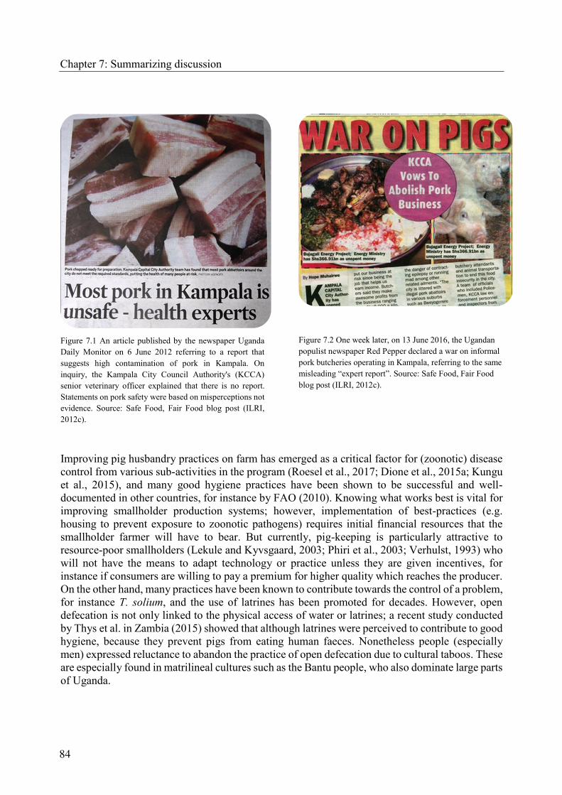
Chapter 7: Summarizing discussion
Figure 7.1 An article published by the newspaper Uganda
Daily Monitor on 6 June 2012 referring to a report that
suggests high contamination of pork in Kampala. On
inquiry, the Kampala City Council Authority's (KCCA)
senior veterinary officer explained that there is no report.
Statements on pork safety were based on misperceptions not
evidence. Source: Safe Food, Fair Food blog post (ILRI,
2012c).
Figure 7.2 One week later, on 13 June 2016, the Ugandan
populist newspaper Red Pepper declared a war on informal
pork butcheries operating in Kampala, referring to the same
misleading “expert report”. Source: Safe Food, Fair Food
blog post (ILRI, 2012c).
Improving pig husbandry practices on farm has emerged as a critical factor for (zoonotic) disease
control from various sub-activities in the program (Roesel et al., 2017; Dione et al., 2015a; Kungu
et al., 2015), and many good hygiene practices have been shown to be successful and well-
documented in other countries, for instance by FAO (2010). Knowing what works best is vital for
improving smallholder production systems; however, implementation of best-practices (e.g.
housing to prevent exposure to zoonotic pathogens) requires initial financial resources that the
smallholder farmer will have to bear. But currently, pig-keeping is particularly attractive to
resource-poor smallholders (Lekule and Kyvsgaard, 2003; Phiri et al., 2003; Verhulst, 1993) who
will not have the means to adapt technology or practice unless they are given incentives, for
instance if consumers are willing to pay a premium for higher quality which reaches the producer.
On the other hand, many practices have been known to contribute towards the control of a problem,
for instance T. solium, and the use of latrines has been promoted for decades. However, open
defecation is not only linked to the physical access of water or latrines; a recent study conducted
by Thys et al. in Zambia (2015) showed that although latrines were perceived to contribute to good
hygiene, because they prevent pigs from eating human faeces. Nonetheless people (especially
men) expressed reluctance to abandon the practice of open defecation due to cultural taboos. These
are especially found in matrilineal cultures such as the Bantu people, who also dominate large parts
of Uganda.
84

Chapter 7: Summarizing discussion
7.4 Researching smallholder pig value chains through an integrated approach and
participatory methods
Mbuthia et al. (2015) acknowledged that for smallholder pig farming improvement programmes
to be successful, there is the need for appropriate understanding of these systems so as to
holistically address their constraints. In the Ugandan smallholder pig value chains, the CRPs have
been the first to also include nodes to their assessments that were not only linked to inputs
increasing farm productivity (feeds, drugs) but also the value chain system at post-harvest (market
access, butchers, consumers) (Figure 7.3 and Ouma et al., 2014). The multi-pathogen prevalence
survey, of which the parasitic assessment was one component, has provided information on pig
pathogens that have never been documented from Uganda before (e.g. Brucella suis,
Erysipelothrix rhusiopathiae, Trichinella spp., T. gondii, Yersinia enterocolitica), and pathogens
known to be present in Uganda (e.g. African swine fever virus, Taenia solium). Moreover, testing
for more parasites (e.g. Sarcoptes scabiei var. suis, Trypanosoma spp.) as well as bacterial and
viral pathogens in the same cohort of pigs is still in progress. Joint resources from two CRPs
allowed collection of data from a big cohort, which increases statistical power of the results and in
the future will facilitate more analysis from the sample, for instance on multimorbidity or intra-
cluster correlation. These data will support scientists in designing follow up studies and target
limited resources for research.
Participatory methods have proven useful in identifying priority constraints to pig health in
smallholder production systems, and in describing production systems and husbandry practices as
well as in scoping diseases that present with clinical signs and can be seen macroscopically. These
include specific gastrointestinal worms, ectoparasites or diseases that present with pathognomonic
signs. For example, in Kamuli district, farmers reported the diamond-shaped skin lesions in their
pigs that are characteristic for acute swine erysipelas caused by Erysipelas rhusiopathiae (chapter
6). The pathogen was not on the initial list to investigate but its presence has then been confirmed
in the laboratory, showing a high level of infection among animals, meat and raw pork handlers
(Musewa, 2016). For other conditions such as disease syndromes, symptoms such as fever,
diarrhoea, and vomiting can be described, however, a conclusive etiologic diagnosis cannot be
made and findings have to be interpreted with care and supported by laboratory findings. This is
the case for ASF which has been identified a priority disease among farmers, yet in their pigs they
are only able to observe signs such as haemorrhage, fever, shivering, huddling, and sudden death
which are commonly seen in other diseases such as classical swine fever, salmonellosis, erysipelas,
infections with T. simiae among others. In the pig cohort for which laboratory results are presented
here, no antibodies to ASF were detected using INGENASIA protocols; however, one animal
tested positive for virus antigen using real time PCR (Akol et al., 2015).
Infections that are usually asymptomatic in pigs, for instance with T. solium, Trichinella spp. or T.
gondii, cannot be observed by the farmer and will therefore not be mentioned in participatory
epidemiology exercises. Nevertheless, through participatory methods we are able to get indications
on good and poor practices at production, processing and consumption level that allow better
targeting of resources towards in-depth assessments or interventions; these include husbandry
practices on farm, preparation of meat, frequency of consumption, modes of pork preparation and
knowledge on zoonotic pig diseases in smallholder pig farming communities. The use of Venn
Diagrams illustrated sources of pork, their distance from the homesteads and quality attributes that
are important to smallholder pig producers outside of the big urban centres (chapter 6, Figure 4).
85

Chapter 7: Summarizing discussion
86
Figure 7.3 Synthesized results of the Ugandan smallholder pig value chain assessment including porcine parasites. The work was presented by the author at Tropentag conference
in Prague, September 2014. Source: Ouma et al. (2014).

Chapter 7: Summarizing discussion
Participatory methods can indeed be a helpful tool in assessing exposure to food safety risks in
informal markets of low-income countries as described earlier (Roesel et al., 2014).
Feedback workshops, where the scientific findings of each sub-activity were discussed with the
value chain stakeholders, have been held since the program’s inception (chapter 3), and are not
only inclusive but help communicating evidence-based findings and solutions in a more bottom-
up approach. Based on the assessment findings, seven training modules including parasite control
were developed and are tested and validated in the field with smallholder pig producers (Dione et
al., 2015b), existing materials have been updated for use in the local setting in Uganda (ILRI and
Medical Research Council, 2005), and researchers have been invited to present findings at pig
farmer trainings organized by the private sector (ILRI, 2014a). Moreover, training workshops have
been held with different stakeholders at the various nodes of the pig value chain, i.e. veterinary
public health (ILRI, 2014b). The downside of the intensive interaction with all stakeholders
through the application of participatory methods is the increased pressure on scientists to
compensate for the big gap in public veterinary extension systems by not only generating data and
developing intervention strategies but to implement them at large scale with research resources.
7.5 Concluding remarks, limitations of the survey and potential future research
The present study was able to contribute to the increasing evidence base on prevalent pig parasites
that may compromise farm productivity but also pork safety in smallholder pig production settings
in Uganda.
7.5.1 Some of the major research findings are presented below:
Results from participatory rural appraisals show that pig keeping systems are dominated
by tethering and scavenging in rural areas, while in peri-urban and urban areas,
confinement in pens is more common. High disease burden, poor housing, poor feeding,
poor veterinary services, ineffective drugs and a general lack of knowledge on piggery
management were identified as the main production constraints by smallholder pig farmers.
Second only to ASF, parasites, especially gastrointestinal worms and sarcoptic mange,
were perceived to contribute largely to market losses due to stunting across all value chain
domains and study sites.
The majority (61.4%) of all pigs were found to be infected with at least one species of
gastrointestinal helminths: 57.1% strongyle species, 7.6% Metastrongylus spp., 5.9% A.
suum, 4.2% Strongyloides ransomi, and 3.4% Trichuris suis. There were no statistically
significant differences across the different value chain types but across the different study
districts. Infection with A. suum and Metastrongylus spp. were significantly more common
in rural Kamuli districts (p < 0.01) than in the other two districts while S. ransomi was
more commonly found in urban Mukono district (p < 0.01). Coccidia oocysts were found
in 40.7% of the sampled pigs, with significantly higher levels in the more urban Mukono
district. None of the animals showed infection with Balantidium coli or Fasciola hepatica.
A logistic regression model showed that routine management factors had a greater impact
on the prevalence of infection than regular treatment or the level of confinement. Factors
negatively correlating with gastrointestinal helminth infection were routine manure
removal, and the routine use of disinfectants.
87

Chapter 7: Summarizing discussion
The overall prevalence of Trichinella-specific antibodies was 6.9% out of 1,125 sampled
animals but showed significantly higher levels in value chain types with a rural origin of
production (RR and RU value chains) (p < 0.05). Kamuli district, where the RR value chain
type dominates showed a significantly higher ELISA-prevalence than the more urban
Masaka and Mukono districts (p < 0.001). For the first time, a study in pigs in sub-Saharan
Africa was able to confirm 31 % ELISA-positive samples by Western Blot. Isolation of
infective first-stage larvae from 499 samples in ELISA clusters to identify the dominating
Trichinella spp. was not successful.
Toxoplasma-specific antibodies were found in 28.7% of the animals using a commercial
ELISA kit; village herd prevalence was 100%. Pigs raised in urban production settings (UU
value chain types) showed significantly higher levels of seropositivity than animals of a
rural production origin (RR and RU value chain types) (p < 0.007); and significantly higher
levels were shown for Masaka and Mukono compared to rural Kamuli district (p > 0.05).
Moreover, univariate analysis suggested a significant association between seropositivity
and pig age, feeding of crop residues, source of drinking water, keeping cats on farm
compound, and frequent sightings of wildlife (especially antelopes, hares, wild and stray
dogs) near the village. The present report is the first survey documenting the seroprevalence
of T. gondii in domestic pigs in the East African Community.
Findings on pork consumption practices from participatory rural appraisals showed that
pork is widely popular among pig farmers. In rural areas it is consumed mostly during
seasonal festivities and whenever cash is available, in urban areas much more frequently.
According to the pig farmers, pig feed production does not compete with human food
production; some festivals when pork is consumed even coincide with times when other
food is not abundant. The consumption of raw pork is considered unsafe, and at home meat
is cooked for at least one hour.
7.5.2 Major conclusions that can be drawn from this assessment are:
Smallholder pig production systems in Uganda are highly heterogeneous and complex, and
the level of intensification cannot be characterized by a single variable such as the level of
confinement. Due to the informal setting of the pig sector, even a fully confined pig may
be exposed to parasites introduced to the farm, e.g. feeds contaminated with rodents or cat
faeces. Using a proxy of value chain domains that comprised different aspects, in this case
pig population density, poverty levels and the time to reach the nearest market, was useful
for comparing pig populations in different villages.
Infections with gastrointestinal parasites are common but weight loss, a consequence of
worm infection as perceived by pig farmers, could not be attributed to a particular parasite
species. Other factors causing malnutrition due to poor pig diets, or potentially a
combination of these two, have not been investigated in this cohort of pigs. Regular
treatment with anthelminthics alone is not useful in containing the burden of infection at
acceptable levels if efficient hygiene farming practices such as regular removal of faeces,
cleaning and disinfection of pig pens are not followed.
88

Chapter 7: Summarizing discussion
The Trichinella species infecting domestic pigs needs to be identified by isolation of
muscle larvae, which was not successful in this study. Subsequently, serological assays
developed for the diagnostics of Trichinella infection in domestic pigs in high-income
countries need to be adapted for use in the local settings of smallholder pig production in
the tropics.
The findings on the seroprevalence of antibodies to T. gondii in domestic pigs imply that
more research should be carried out to determine the genotype infecting pigs as well as the
tissue cyst burden to be able to re-assess the risk for human raw pork handlers and the
vulnerable population, e.g. pregnant women and HIV/Aids patients. Consequently, in-
depth research on transmission routes for domestic pigs and humans is warranted.
The high prevalence of gastrointestinal parasites, some of them zoonotic, may expose pig
farmers and their children, who often tend to the pigs, to disease. While, according to the
findings of this study, the risk for developing trichinellosis is low, the risk for infection
with T. gondii from raw pork handling or other practices on farm (e.g. handling feeds, or
drinking contaminated with oocysts) is higher.
Constraints to the parasitic assessment were mostly related to the design of the overall pig value
chain assessment which was cross-sectional and could therefore suggest but not prove causal
relationships between exposing variables and parasite infection. Despite the outweighing benefits
described above, a shortcoming of the prevalence survey for parasitic assessment, was the
exclusion of pigs younger than three months from the sample population as this is the age when
they are most susceptible to infection with gastrointestinal parasites. Moreover, the tight field
sampling schedule and limited time, space and human resources did not allow for the collection
and analysis of urine to determine the prevalence of Stephanurus dentatus (kidney worm), to
perform larval culture and coccidia oocyst sporulation, or to collect deep skin scrapings and
ectoparasites from all pigs, which could have added useful information. Specimens were collected
from a number of pigs but this was not done systematically.
Additional laboratory experiments conducted after the prevalence survey included artificial
digestion to potentially isolate first-stage larvae of Trichinella spp. clusters and a mouse bioassay
to isolate T. gondii bradyzoites for genotyping in ELISA clusters. While the sample for the
artificial digestion was large (n=499), no larvae could be isolated and reasons were exhaustively
discussed in chapter 5. However, the serological status of the digested samples could not be
confirmed due to limited resources. Attempts to isolate T. gondii in a mouse bioassay were also
not successful, even though only serologically positive samples had been used as recommended
(Dubey, 2009). T. gondii bioassays in cats have been shown to be more successful in the isolation
of viable cysts as larger volumes (500g) or more can be assayed than in mice; however, facilities
for experiments involving cats were not available in Uganda at the time of the study.
Future research on pig parasites in Uganda should aim at:
Quantifying actual losses due to gastrointestinal parasites on farm by means of
experimental studies and longitudinal surveys;
89

Chapter 7: Summarizing discussion
Characterizing pork pathogens, for instance the Trichinella species infecting pigs in
Uganda or the genotype involved in porcine infection with T. gondii;
Assessing the effect of co-infection on the sensitivity and specificity of serological assays
developed and validated in high-income countries;
Quantifying the risk for pork-borne parasitic diseases to consumers through experimental
studies and quantitative risk assessments;
Assessing host preferences of biting insects such as tsetse flies including pigs;
Assessing the role of Ugandan pigs as potential reservoir or amplifying host for zoonotic
arboviruses such as Ndumu transmitted by Culicoides spp. (Masembe et al., 2012); Zika
virus transmitted by Aedes spp. (Chan et al., 2016), or Japanese encephalitis transmitted by
Culex spp. (Mellor and Leake, 2000);
Characterizing African pig breeds and their genetic tolerance or susceptibility to parasitic
diseases as has been shown for African cattle (Gifford-Gonzalez and Hanotte, 2011);
Including wild Suidae in parasitic assessments as they could potentially share vector-borne
infections such as with B. trautmanni (Penzhorn, 2006), or trypanosomes (Mehlitz, 1987;
Mwambu and Woodford, 1972; Van Den Berghe and Zaghi, 1963);
Investigating the role of potential hosts such as dogs and rodents as mechanical vectors
(e.g. T. gondii, A. suum), or definite hosts (e.g. Trichinella spp., Echinococcus spp.,
Neospora caninum, Sarcocystis spp.) for potentially pig-mediated zoonoses in low-income
countries, where they are often stray;
Testing the feasibility of dogs or chickens as bio-indicators and sentinels for infections
with T. gondii (Ayinmode and Jones-Akinbobola, 2015; Davoust et al., 2015; Salb et al.,
2008; Meireles et al., 2004)
90

Chapter 8: References
8 References
Adediran, O., Uwalaka, E., Abiola, J., 2015. Seroprevalence of Trichinella antibodies in pigs
reared under different husbandry systems in Southwestern Nigeria. Alexandria J. Vet. Sci.
47, 183. doi:10.5455/ajvs.191632
Adesehinwa, A.O.K., Aribido, S.O., Oyediji, G.O., Obiniyi, A.A., 2003. Production strategies
for coping with the demand and supply of pork in some peri-urban areas of Southwestern
Nigeria. Livest. Res. Rural Dev. 15, Article #72. Retrieved
from:http://www.lrrd.org/lrrd15/10/ades1510.htm#Livestock Research for Rural
Development 15 (10) 2003
Akol, J., Dione, M., Bishop, R., Onzere, C., Muhangi, D., Masembe, C., 2015. A cross sectional
study of the assessment of African swine fever virus status in apparently healthy pigs in
three districts of Uganda. [WWW Document]. Poster Present. 4th Med. Vet. Virus Res.
(MVVR 4) Symp. 15- 16 Oct. 2015, Nairobi, Kenya. Retrieved
from:http://www.slideshare.net/ILRI/mvvr4-akol-oct2015
Alexandratos, N., Bruinsma, J., 2012. World Agriculture Towards 2030/50. The 2012 Revision.
Global Perspective Studies Team [WWW Document]. ESA Working Paper No. 12-03. URL
http://www.fao.org/docrep/016/ap106e/ap106e.pdf
Alonso, S., Lindahl, J., Roesel, K., Traore, S.G., Yobouet, B.A., Ndour, A.P.N., Carron, M.,
Grace, D., 2016. Where literature is scarce: observations and lessons learnt from four
systematic reviews of zoonoses in African countries. Anim. Heal. Res. Rev. 17, 28–38.
doi:10.1017/S1466252316000104
Amills, M., Clop, A., Ramírez, O., Pérez-Enciso, M., 2010. Origin and Genetic Diversity of Pig
Breeds, In: Encyclopedia of Life Sciences. John Wiley & Sons, Ltd, Chichester, UK.
doi:10.1002/9780470015902.a0022884
Amills, M., Ramírez, O., Galman-Omitogun, O., Clop, A., 2013. Domestic Pigs in Africa.
African Archaeol. Rev. 30, 73–82. doi:10.1007/s10437-012-9111-2
Arko-Mensah, J., Bosompem, K.M., Canacoo, E.A., Wastling, J.M., Akanmori, B.D., 2000. The
seroprevalence of toxoplasmosis in pigs in Ghana. Acta Trop. 76, 27–31.
doi:10.1016/S0001-706X(00)00085-1
Atuhaire, D., Ochwo, S., Afayoa, M., Mwiine, F.N., Ikwap, K., Arinaitwe, E., Ademun-Okurut,
R.A., Boniface Okuni, J., Nanteza, A., Ayebazibwe, C., Okedi, L., Olaho-Mukani, W.,
Ojok, L., 2013. Epidemiological Overview of African Swine Fever in Uganda (2001–2012).
J. Vet. Med. [epub], 1–9. doi:10.1155/2013/949638
Ayinmode, A., Abiola, J., 2016. Investigating Potential Sources of Toxoplasmosis for Humans
From Slaughtered Food Animals in Ibadan, Nigeria. Folia Vet. 60, 34–40.
doi:https://www.degruyter.com/view/j/fv.2016.60.issue-2/fv-2016-0016/fv-2016-0016.xml
Ayinmode, A., Ishola, O., Oderinu, T., 2015. Seroprevalence of Toxoplasma gondii in dogs
slaughtered for food in Southwestern Nigeria and assessment of consumer’s knowledge and
91

Chapter 8: References
behavior. Alexandria J. Vet. Sci. 45, 161. doi:10.5455/ajvs.181742
Ayinmode, A., Jones-Akinbobola, R., 2015. Detection of Toxoplasma gondii IgG antibodies in
Nigerian Free-range Chickens using Indirect Fluorescent Antibody Test (IFAT). Alexandria
J. Vet. Sci. 47, 187. doi:10.5455/ajvs.201626
Ayinmode, A., Olaosebikan, R., 2013. Antibodies to Toxoplasma gondii in backyard and
wandering pigs in Ibadan, Nigeria: implications for pork consumption. Bull. Anim. Heal.
Prod. Africa 61, 493–497. Retrieved
from:http://www.ajol.info/index.php/bahpa/article/view/105285
Bahia-Oliveira, L.M.G., Jones, J.L., Azevedo-Silva, J., Alves, C.C.F., Oréfice, F., Addiss, D.G.,
2003. Highly Endemic, Waterborne Toxoplasmosis in North Rio de Janeiro State, Brazil.
Emerg. Infect. Dis. 9, 55–62. doi:10.3201/eid0901.020160
Beer, R.J.S., 1973. Studies on the biology of the life-cycle of Trichuris suis Schrank, 1788.
Parasitology 67, 253. doi:10.1017/S0031182000046497
Bethony, J., Brooker, S., Albonico, M., Geiger, S.M., Loukas, A., Diemert, D., Hotez, P.J., 2006.
Soil-transmitted helminth infections: ascariasis, trichuriasis, and hookworm. Lancet 367,
1521–1532. doi:10.1016/S0140-6736(06)68653-4
Bisson, A., Maley, S., Rubaire-Akiiki, C.M., Wastling, J.M., 2000. The seroprevalence of
antibodies to Toxoplasma gondii in domestic goats in Uganda. Acta Trop. 76, 33–38.
doi:10.1016/S0001-706X(00)00086-3
Blench, R., 2000. A history of pigs in Africa., In: Blench, R., MacDonald, K. (Eds.), Origins and
Development of African Livestock: Archaeology, Genetics, Linguistics and Ethnography.
Routledge, Oxfordshire, pp. 355–367.
Bontell, I., Hall, N., Ashelford, K.E., Dubey, J., Boyle, J.P., Lindh, J., Smith, J.E., 2009. Whole
genome sequencing of a natural recombinant Toxoplasma gondii strain reveals chromosome
sorting and local allelic variants. Genome Biol. 10, R53. doi:10.1186/gb-2009-10-5-r53
Brooker, S., Kabatereine, N.B., Smith, J.L., Mupfasoni, D., Mwanje, M.T., Ndayishimiye, O.,
Lwambo, N.J., Mbotha, D., Karanja, P., Mwandawiro, C., Muchiri, E., Clements, A.C.,
Bundy, D.A., Snow, R.W., 2009. An updated atlas of human helminth infections: the
example of East Africa. Int. J. Health Geogr. 8, 42. doi:10.1186/1476-072X-8-42
Bura, M.W., Willett, W.C., 1977. An outbreak of trichinosis in Tanzania. East Afr. Med. J. 54,
185–193. PMID:561687
Carter, N., Dewey, C., Mutua, F., de Lange, C., Grace, D., 2013. Average daily gain of local pigs
on rural and peri-urban smallholder farms in two districts of Western Kenya. Trop. Anim.
Health Prod. 45, 1533–1538. doi:10.1007/s11250-013-0395-2
Center for International Earth Science Information Network (CIESIN), 2011. Global Rural-
Urban Mapping Project, Version 1 (GRUMPv1): Population Density Grid. [WWW
Document]. Socioecon. Data Appl. Cent. http://sedac.ciesin.columbia.edu/
92

Chapter 8: References
Chan, J.F.-W., Yip, C.C.-Y., Tsang, J.O.-L., Tee, K.-M., Cai, J.-P., Chik, K.K.-H., Zhu, Z.,
Chan, C.C.-S., Choi, G.K.-Y., Sridhar, S., Zhang, A.J., Lu, G., Chiu, K., Lo, A.C.-Y., Tsao,
S.-W., Kok, K.-H., Jin, D.-Y., Chan, K.-H., Yuen, K.-Y., 2016. Differential cell line
susceptibility to the emerging Zika virus: implications for disease pathogenesis, non-vector-
borne human transmission and animal reservoirs. Emerg. Microbes Infect. 5, e93.
doi:10.1038/emi.2016.99
David, R., 2004. Muslim Societies in African History. Cambridge University Press, New York,
NY, 242 pp.
Davoust, B., Mediannikov, O., Roqueplo, C., Perret, C., Demoncheaux, J.-P., Sambou, M.,
Guillot, J., Blaga, R., 2015. Enquête de séroprévalence de la toxoplasmose animale au
Sénégal. Bull. la Société Pathol. Exot. 108, 73–77. doi:10.1007/s13149-014-0403-4
Delgado, C.L., Rosegrant, M.W., Meijer, S., 2001. Livestock to 2020: the revolution continues.,
in: Summaries of Papers Presented at the Meeting of the International Agricultural Trade
Research Consortium (IATRC) in Auckland, New Zealand, January 18-19, 2001 Published
by the World Trade Organization (WTO). International Food Policy Research Institute, p.
72.
Deplazes, P., Eckert, J., Mathis, A., von Samson-Himmelstjerna, G., Zahner, H., 2016.
Parasitology in Veterinary Medicine. Wageningen Academic Publishers, The Netherlands.
doi:10.3920/978-90-8686-274-0
Dione, M., Akol, J., Roesel, K., Kungu, J., Ouma, E., Wieland, B., Pezo, D., 2015a. Risk Factors
for African Swine Fever in Smallholder Pig Production Systems in Uganda. Transbound.
Emerg. Dis. doi:10.1111/tbed.12452
Dione, M., Pezo, D., Ouma, E., Roesel, K., Brandes-van Dorresteijn, D., Kawuma, B., 2015b.
Training on management of endemic diseases for pig value chains in Uganda. Presented at
the 4th International Conference on Sustainable Livelihoods and Health in Africa, Kampala,
Uganda, 18-19 June 2015. Nairobi, Kenya. Retrieved
from:https://cgspace.cgiar.org/handle/10568/67273
Dione, M.M., Ouma, E.A., Roesel, K., Kungu, J., Lule, P., Pezo, D., 2014. Participatory
assessment of animal health and husbandry practices in smallholder pig production systems
in three high poverty districts in Uganda. Prev. Vet. Med. 117, 565–576.
doi:10.1016/j.prevetmed.2014.10.012
Djurković-Djaković, O., Bobić, B., Nikolić, A., Klun, I., Dupouy-Camet, J., 2013. Pork as a
source of human parasitic infection. Clin. Microbiol. Infect. 19, 586–594.
doi:10.1111/1469-0691.12162
Dubey, J.P., 2009. Toxoplasmosis in pigs—The last 20 years. Vet. Parasitol. 164, 89–103.
doi:10.1016/j.vetpar.2009.05.018
Dubey, J.P., 1988. Long-term persistence of Toxoplasma gondii in tissues of pigs inoculated with
T. gondii oocysts and effect of freezing on viability of tissue cysts in pork. Am. J. Vet. Res.
49, 910–3. doi:http://www.ncbi.nlm.nih.gov/pubmed/3400928
93

Dubey, J.P., 1986. A review of toxoplasmosis in pigs. Vet. Parasitol. 19, 181–223.
doi:10.1016/0304-4017(86)90070-1
Esrony, K., Kambarage, D.M., Mtambo, M.M.A., Muhairwa, A.P., Kusiluka, L.J.M., 1997.
Helminthosis in local and cross-bred pigs in the Morogoro region of Tanzania. Prev. Vet.
Med. 32, 41–46. doi:10.1016/S0167-5877(97)00011-1
Ezzati, M., Lopez, A.D., Rodgers, A., Vander Hoorn, S., Murray, C.J.L., Comparative Risk
Assessment Collaborating Group, 2002. Selected major risk factors and global and regional
burden of disease. Lancet (London, England) 360, 1347–60. doi:10.1016/S0140-
6736(02)11403-6
Feitosa, T.F., Vilela, V.L.R., Bezerra de Melo, L.R., de Almeida Neto, J.L., de Oliveira Souto,
D.V., de Morais, D.F., Athayde, A.C.R., Azevedo, S.S., de Jesus Pena, H.F., 2014.
Toxoplasma gondii and Neospora caninum in slaughtered pigs from Northeast, Brazil. Vet.
Parasitol. 202, 305–309. doi:10.1016/j.vetpar.2014.03.015
Food and Agriculture Organization of the United Nations (FAO), 2016. Domestic Animal
Diversity Information System (DAD-IS) database [WWW Document]. http://dad.fao.org
Food and Agriculture Organization of the United Nations (FAO), 2015. Meat Consumption
[WWW Document]. Website Dep. Agric. Consum. Prot.
http://www.fao.org/ag/againfo/themes/en/meat/background.html
Food and Agriculture Organization of the United Nations (FAO), 2011. Global Livestock
Production Systems. [WWW Document]. http://www.fao.org/docrep/014/i2414e/i2414e.pdf
Food and Agriculture Organization of the United Nations (FAO), 2010. Good practices for
biosecurity in the pig sector. Issues and options in developing and transition countries.
[WWW Document]. FAO Animal Production and Health Paper.
http://www.fao.org/docrep/012/i1435e/i1435e00.htm
Food and Agriculture Organization of the United Nations (FAO), 2009. The State of Food and
Agriculture. Livestock in the Balance. [WWW Document].
http://www.fao.org/docrep/012/i0680e/i0680e.pdf
Frenkel, J.K., Lindsay, D.S., Parker, B.B., Dobesh, M., 2003. Dogs as possible mechanical
carriers of Toxoplasma, and their fur as a source of infection of young children. Int. J.
Infect. Dis. 7, 292–293. doi:10.1016/S1201-9712(03)90112-3
Gajadhar, A.A. (Ed.), 2015. Foodborne Parasites in the Food Supply Web: Occurrence and
Control. Woodhead Publishing, Cambridge and Kidlington, UK and Waltham, USA.
Garcia, J.L., 2009. Vaccination concepts against Toxoplasma gondii. Expert Rev. Vaccines 8,
215–225. doi:10.1586/14760584.8.2.215
Gautier, A., 2002. The Evidence for the Earliest Livestock in North Africa: or Adventures with
Large Bovids, Ovicaprids, Dogs and Pigs, In: Droughts, Food and Culture. Kluwer
Academic Publishers, Boston, pp. 195–207. doi:10.1007/0-306-47547-2_12
94
Chapter 8: References

Chapter 8: References
Gebremedhin, E., Kebeta, M., Asaye, M., Ashenafi, H., Di Marco, V., Vitale, M., 2015. First
report on seroepidemiology of Toxoplasma gondii infection in pigs in Central Ethiopia.
BMC Vet. Res. 11, 59. doi:10.1186/s12917-015-0384-y
Gifford-Gonzalez, D., Hanotte, O., 2011. Domesticating Animals in Africa: Implications of
Genetic and Archaeological Findings. J. World Prehistory 24, 1–23. doi:10.1007/s10963-
010-9042-2
Githigia, S.M., Murekefu, K., Otieno, R.O., 2005. Prevalence of porcine cysticercosis and risk
factors for Taenia solium taeniosis in Funyula Division of Busia District, Kenya. Kenya
Vet. 29, 37–39. Retrieved from: http://www.ajol.info/index.php/kenvet/article/view/39594
Gottstein, B., Pozio, E., Nöckler, K., 2009. Epidemiology, diagnosis, treatment, and control of
trichinellosis. Clin. Microbiol. Rev. 22, 127–145. doi:10.1128/CMR.00026-08
Greve, J.H., 2012. Internal Parasites: Helminths, in: J. Zimmerman, L. Karriker, A. Ramirez, K.
Schwartz, G.S. (Ed.), Diseases of Swine. John Wiley & Sons, Inc., pp. 908–920.
Greve, J.H., Davies, P., 2012. External Parasites, in: Zimmerman, J.J., Karriker, L.A., Ramirez,
A., Schwartz, K.J., Stevenson, G.W. (Eds.), Diseases of Swine. John Wiley & Sons, Inc.,
pp. 885–894.
Hale, O.M., Stewart, T.B., 1979. Influence of an Experimental Infection of Trichuris suis on
Performance of Pigs. J. Anim. Sci. 49, 1000. doi:10.2527/jas1979.4941000x
Hale, O.M., Stewart, T.B., Marti, O.G., 1985. Influence of an experimental infection of Ascaris
suum on performance of pigs. J. Anim. Sci. 60, 220–225. doi:10.2134/jas1979.4941000x
Haltenorth, T., Diller, H., 1980. A field guide to the mammals of Africa, including Madagascar.
London, UK, 400 pp.
Havelaar, A.H., Kirk, M.D., Torgerson, P.R., Gibb, H.J., Hald, T., Lake, R.J., Praet, N.,
Bellinger, D.C., de Silva, N.R., Gargouri, N., Speybroeck, N., Cawthorne, A., Mathers, C.,
Stein, C., Angulo, F.J., Devleesschauwer, B., 2015. World Health Organization Global
Estimates and Regional Comparisons of the Burden of Foodborne Disease in 2010. PLOS
Med. 12, e1001923. doi:10.1371/journal.pmed.1001923
Heilmann, M., Ndoboli, D., Roesel, K., Grace, D., Huehn, S., Bauer, B., Clausen, P.-H., 2015.
Occurrence of Salmonella spp. in Flies and Foodstuff from Pork Butcheries in Kampala,
Uganda., In: Proceedings of the Annual Expert Meeting on Parasitology and Parasitic
Diseases at the German Veterinary Association, Stralsund, Germany, 29 June – 1 July 2015.
Stralsund, Germany.
Heilmann, M., Roesel, K., Clausen, P.-H., Grace, D., 2016. Knowledge, attitudes and practises
among customers at pork butcheries in Kampala, Uganda, In: Clausen, P.-H., Baumann, M.,
Nijhof, A. (Eds.), Tropical Animal Diseases and Veterinary Public Health: Joining Forces to
Meet Future Global Challenges. First Joint AITVM-STVM Conference. Berlin, p. 27.
Hill, D.E., Sreekumar, C., Gamble, H.R., Dubey, J.P., 2004. Effect of Commonly Used
Enhancement Solutions on the Viability of Toxoplasma gondii Tissue Cysts in Pork Loin. J.
95

Food Prot. 67, 2230–2233. doi:10.4315/0362-028X-67.10.2230
Houlihan, P.F., 1997. The animal world of the pharaohs, 1st ed. Thames & Hudson Ltd, 245 pp.
Hove, T., 2005. Seroprevalence of Toxoplasma gondii infection in domestic pigs reared under
different management systems in Zimbabwe. Onderstepoort J. Vet. Res. 72, 231–237.
Retrieved from:
http://search.proquest.com/openview/2a2fa28c7ea7d6e366ddf9986a10ad8a/1?pq-
origsite=gscholar
Hove, T., Dubey, J.P., 1999. Prevalence of Toxoplasma gondii antibodies in sera of domestic
pigs and some wild game species from Zimbabwe. J. Parasitol. 85, 372–3. PMID:10219323
Hove, T., Lind, P., Mukaratirwa, S., 2005. Seroprevalence of Toxoplasma gondii infection in
goats and sheep in Zimbabwe. Onderstepoort J. Vet. Res. 72, 267–272. Retrieved
from:http://search.proquest.com/openview/92e1017fe3c931b30208ff9e79a0255c/1?pq-
origsite=gscholar
Hove, T., Mukaratirwa, S., 2002. Isolation of Toxoplasma gondii in a pig from a local abattoir in
Zimbabwe. Zimbabwe Vet. J. 33, 79–83. Retrieved
from:http://www.ajol.info/index.php/zvj/article/view/5370
Howell, A., Mugisha, L., Davies, J., LaCourse, E., Claridge, J., Williams, D.J., Kelly-Hope, L.,
Betson, M., Kabatereine, N.B., Stothard, J., 2012. Bovine fasciolosis at increasing altitudes:
Parasitological and malacological sampling on the slopes of Mount Elgon, Uganda. Parasit.
Vectors 5, 196. doi:10.1186/1756-3305-5-196
Ikwap, K., Erume, J., Owiny, D.O., Nasinyama, G.W., Melin, L., Bengtsson, B., Lundeheim, N.,
Fellström, C., Jacobson, M., 2014. Salmonella species in piglets and weaners from Uganda:
Prevalence, antimicrobial resistance and herd-level risk factors. Prev. Vet. Med. 115, 39–47.
doi:10.1016/j.prevetmed.2014.03.009
Iñiguez, A.M., Leles, D., Jaeger, L.H., Carvalho-Costa, F.A., Araújo, A., 2012. Genetic
characterisation and molecular epidemiology of Ascaris spp. from humans and pigs in
Brazil. Trans. R. Soc. Trop. Med. Hyg. 106, 604–612. doi:10.1016/j.trstmh.2012.06.009
International Food Policy Research Institute, 2012. CGIAR Research Program on Agriculture for
Nutrition and Health [WWW Document]. http://a4nh.cgiar.org/
International Livestock Research Institute, 2014a. How to improve pig farming: A training
workshop by Pig Production and Marketing Uganda Limited. Report of a training course
held at Matugga, Uganda, 14-15 February 2014. Nairobi, Kenya. Retrieved from:
https://cgspace.cgiar.org/handle/10568/35046
International Livestock Research Institute, 2014b. Hands-on training on harvesting in the
smallholder pig value chains in Uganda. Report of a training course held at Kampala,
Uganda, 7-11 April 2014. Nairobi, Kenya. Retrieved from:
https://cgspace.cgiar.org/handle/10568/41681
International Livestock Research Institute, 2012a. CGIAR Research Program on Livestock and
96
Chapter 8: References

Chapter 8: References
Fish [WWW Document]. https://livestockfish.cgiar.org/
International Livestock Research Institute, 2012b. Prevention and control of agriculture-
associated diseases [WWW Document]. https://aghealth.wordpress.com/
International Livestock Research Institute, 2012c. Safe Food, Fair Food [WWW Document].
https://safefoodfairfood.ilri.org/
International Livestock Research Institute, 2012d. Minimum validation checklist. Nairobi,
Kenya. Retrieved from: http://livestock-fish.wikispaces.com/Uganda+site+selection
International Livestock Research Institute, 2011. The smallholder pig value chain: An
opportunity for growth and poverty reduction. Report of a Stakeholder Meeting, Kampala,
Uganda, 14 June 2011. Nairobi, Kenya. Retrieved from:
https://cgspace.cgiar.org/handle/10568/4049
International Livestock Research Institute and Medical Research Council, 2005. Let’s break the
pork tapeworm cycle. [WWW Document]. Educ. poster Dev. by Int. Cysticercosis Coord.
Center. https://cgspace.cgiar.org/handle/10568/35005
Jones, J.L., Dargelas, V., Roberts, J., Press, C., Remington, J.S., Montoya, J.G., 2009. Risk
Factors for Toxoplasma gondii Infection in the United States. Clin. Infect. Dis. 49, 878–884.
doi:10.1086/605433
Kabatereine, N.B., Tukahebwa, E.M., Kazibwe, F., Twa-Twa, J.M., Barenzi, J.F.Z., Zaramba, S.,
Stothard, J.R., Fenwick, A., Brooker, S., 2005. Short communication: Soil-transmitted
helminthiasis in Uganda: epidemiology and cost of control. Trop. Med. Int. Heal. 10, 1187–
1189. doi:10.1111/j.1365-3156.2005.01509.x
Kagira, J., Githigia, S., Ng’ang’a, J., Kanyari, P., Maingi, N., Gachohi, J., 2010a. Prevalence of
gastrointestinal protozoa and association with risk factors in free-range pigs in Kenya. J.
Protozool. Res. 20. Retrieved from: https://cgspace.cgiar.org/handle/10568/2433
Kagira, J., Kanyari, P., Githigia, S., Maingi, N., Ng’ang’a, J., Gachohi, J., 2012. Risk factors
associated with occurrence of nematodes in free range pigs in Busia District, Kenya. Trop.
Anim. Health Prod. 44, 657–664. doi:10.1007/s11250-011-9951-9
Kagira, J., Kanyari, P., Maingi, N., Githigia, S., Ng’ang’a, J., Karuga, J., 2010b. Characteristics
of the smallholder free-range pig production system in western Kenya. Trop. Anim. Health
Prod. 42, 865–873. doi:10.1007/s11250-009-9500-y
Kagira, J., Kanyari, P., Munyua, W., Waruiru, R., 2008. Relationship between the prevalence of
gastrointestinal nematode infections and management practises in pig herds in Thika
District, Kenya. Livest. Res. Rural Dev. 20. Retrieved from:
http://www.lrrd.org/lrrd20/10/kagi20161.htm
Kaminsky, R.G., Zimmerman, R.R., 1977. Trichinella spiralis: incidental finding. East Afr.
Med. J. 54, 643–646. PMID:614130
Kanyari, P.W.N., Kagira, J.M., Mhoma, J.R.L., Omemo, P., 2012. Parasitic causes of liver and
97

heart condemnation and their economic effects in the Lake Victoria Basin : a retrospective
abattoir survey in Kisumu Municipality , Kenya 13, 139–143. Retrieved from:
http://erepository.uonbi.ac.ke:8080/xmlui/handle/123456789/33468
Karimuribo, E.D., Chenyambuga, S.W., Makene, V.W., Mathias, S., 2011. Characteristics and
production constraints of rural-based small-scale pig farming in Iringa region, Tanzania.
Livest. Res. Rural Dev. 23. Retrieved from:
http://www.lrrd.cipav.org.co/lrrd23/8/Kari23172.htm
Kassai, T., Cordero Del Campillo, M., Euzeby, J., Gaafar, S., Hiepe, T., Himonas, C.A., 1988.
Standardized nomenclature of animal parasitic diseases (SNOAPAD). Vet. Parasitol. 29,
299–326. doi:10.1016/0304-4017(88)90148-3
Kaufmann, J., 1996. Parasites of Swine, in: Parasitic Infections of Domestic Animals: A
Diagnostic Manual. Birkhäuser Verlag, Basel, Switzerland, p. 423.
Kefenie, H., Bero, G., 1992. Trichinosis from wild boar meat in Gojjam, north-west Ethiopia.
Trop. Geogr. Med. 44, 278–280. PMID:1455537
Kefenie, H., Wolde, H., Abuherpo, M., 1988. Trichinosis from wild boar meat in Arsi, central
Ethiopia. Ethiop. Med. J. 26, 97–100. PMID:3360005
Kipper, M., Andretta, I., Monteiro, S.G., Lovatto, P.A., Lehnen, C.R., 2011. Meta-analysis of the
effects of endoparasites on pig performance. Vet. Parasitol. 181, 316–320.
doi:10.1016/j.vetpar.2011.04.029
Knecht, D., Popiołek, M., Zaleśny, G., 2011. Does meatiness of pigs depend on the level of
gastro-intestinal parasites infection? Prev. Vet. Med. 99, 234–239.
doi:10.1016/j.prevetmed.2011.01.009
Kolaczinski, J.H., Onapa, A.W., Ndyomugyenyi, R., Brooker, S., 2006. Neglected Tropical
Diseases and their control in Uganda, situation analysis and needs assessment. Retrieved
from: http://www.malariaconsortium.org/userfiles/file/NTD Resources/Neglected Diseases
in Uganda Final %25282%2529.pdf
Koudela, B., Kučerová, Š., 1999. Role of acquired immunity and natural age resistance on course
of Isospora suis coccidiosis in nursing piglets. Vet. Parasitol. 82, 93–99.
doi:10.1016/S0304-4017(99)00009-6
Kungu, J.M., Dione, M.M., Ejobi, F., Harrison, L.J.S., Poole, E.J., Pezo, D., Grace, D., 2016.
Sero-prevalence of Taenia spp. cysticercosis in rural and urban smallholder pig production
settings in Uganda. Acta Trop. [epub ahea. doi:10.1016/j.actatropica.2016.01.016
Kungu, J.M., Dione, M.M., Ocaido, M., Ejobi, F., 2015. Status of Taenia solium cysticercosis
and predisposing factors in developing countries involved in pig farming. Int. J. One Heal.
1, 6–13. doi:10.14202/IJOH.2015.6-13
La Grange, L.J., Govender, D., Mukaratirwa, S., 2013. The occurrence of Trichinella
zimbabwensis in naturally infected wild crocodiles (Crocodylus niloticus) from the Kruger
National Park, South Africa. J. Helminthol. 87, 91–96. doi:10.1017/S0022149X12000089
98
Chapter 8: References

Chapter 8: References
La Grange, L.J., Marucci, G., Pozio, E., 2010. Trichinella zimbabwensis in a naturally infected
mammal. J. Helminthol. 84, 35–38. doi:10.1017/S0022149X09990241
La Grange, L.J., Marucci, G., Pozio, E., 2009. Trichinella zimbabwensis in wild Nile crocodiles
(Crocodylus niloticus) of South Africa. Vet. Parasitol. 161, 88–91.
doi:10.1016/j.vetpar.2008.12.015
Larson, G., 2005. Worldwide Phylogeography of Wild Boar Reveals Multiple Centers of Pig
Domestication. Science (80-. ). 307, 1618–1621. doi:10.1126/science.1106927
Lekule, F.P., Kyvsgaard, N.C., 2003. Improving pig husbandry in tropical resource-poor
communities and its potential to reduce risk of porcine cysticercosis. Acta Trop. 87, 111–
117. doi:10.1016/S0001-706X(03)00026-3
Leles, D., Gardner, S.L., Reinhard, K., Iñiguez, A., Araujo, A., 2012. Are Ascaris lumbricoides
and Ascaris suum a single species? Parasit. Vectors 5, 42. doi:10.1186/1756-3305-5-42
Levine, N.D., Ivens, V., 1986. The coccidian parasites (Protozoa, Apicomplexa) of Artiodactyla.
University of Illinois, Illinois, USA, 265 pp.
Lindsay, D.S., Blagburn, B.L., Boosinger, T.R., 1987. Experimental Eimeria debliecki infections
in nursing and weaned pigs. Vet. Parasitol. 25, 39–45. doi:10.1016/0304-4017(87)90063-X
Lindsay, D.S., Current, W.L., Ernst, J. V, 1982. Sporogony of Isospora suis Biester, 1934 of
swine. J. Parasitol. 68, 861–5. PMID:7131190
Lindsay, D.S., Dubey, J.P., Santín-Durán, M., Fayer, R., 2012. Coccidia and Other Protozoa, in:
Zimmermann, G.J., Karriker, L., Ramirez, A., Schwartz, K. (Eds.), Diseases of Swine. John
Wiley & Sons, Inc., pp. 895–907.
Lindsay, D.S., Neiger, R., Hildreth, M., 2002. Porcine Enteritis Associated with Eimeria spinosa
Henry, 1931 Infection. J. Parasitol. 88, 1262–1263. doi:10.1645/0022-
3395(2002)088[1262:PEAWES]2.0.CO;2
Lindström, I., Kaddu-Mulindwa, D.H., Kironde, F., Lindh, J., 2006. Prevalence of latent and
reactivated Toxoplasma gondii parasites in HIV-patients from Uganda. Acta Trop. 100,
218–222. doi:10.1016/j.actatropica.2006.11.002
Lindström, I., Sundar, N., Lindh, J., Kironde, F., Kabasa, J.D., Kwok, O.C.H., Dubey, J.P.,
Smith, J.E., 2008. Isolation and genotyping of Toxoplasma gondii from Ugandan chickens
reveals frequent multiple infections. Parasitology 135. doi:10.1017/S0031182007003654
Liu, G.-H., Wu, C.-Y., Song, H.-Q., Wei, S.-J., Xu, M.-J., Lin, R.-Q., Zhao, G.-H., Huang, S.-Y.,
Zhu, X.-Q., 2012. Comparative analyses of the complete mitochondrial genomes of Ascaris
lumbricoides and Ascaris suum from humans and pigs. Gene 492, 110–116.
doi:10.1016/j.gene.2011.10.043
Lucas, S.B., Hounnou, A., Peacock, C., Beaumel, A., Djomand, G., N’Gbichi, J.M., Yeboue, K.,
Hondé, M., Diomande, M., Giordano, C., 1993. The mortality and pathology of HIV
infection in a west African city. AIDS 7, 1569–79. PMID:7904450
99

Machebe, N.S., Onyekuru, N.A., Ekweogu, N., 2009. Socio-economic factors affecting pig
production in Enugu state Nigeria. J. Agric. For. Soc. Sci. 7, 41–49. Retrieved from:
http://www.ajol.info/index.php/joafss/article/view/60306
Marucci, G., La Grange, L.J., La Rosa, G., Pozio, E., 2009. Trichinella nelsoni and Trichinella
T8 mixed infection in a lion (Panthera leo) of the Kruger National Park (South Africa). Vet.
Parasitol. 159, 225–8. doi:10.1016/j.vetpar.2008.10.041
Masembe, C., Michuki, G., Onyango, M., Rumberia, C., Norling, M., Bishop, R.P., Djikeng, A.,
Kemp, S.J., Orth, A., Skilton, R.A., Ståhl, K., Fischer, A., 2012. Viral metagenomics
demonstrates that domestic pigs are a potential reservoir for Ndumu virus. Virol. J. 9, 218.
doi:10.1186/1743-422X-9-218
Mbuthia, J.M., Rewe, T.O., Kahi, A.K., 2015. Evaluation of pig production practices, constraints
and opportunities for improvement in smallholder production systems in Kenya. Trop.
Anim. Health Prod. 47, 369–376. doi:10.1007/s11250-014-0730-2
Mehlitz, D., 1987. Animal Reservoir Hosts of Sleeping Sickness, An Overview: In: 19th Meeting
of the International Scientific Council for Trypanosomiasis Research & Control. Lomé,
Togo.
Meireles, L.R., Galisteo, A.J., Pompeu, E., Andrade, H.F., 2004. Toxoplasma gondii spreading in
an urban area evaluated by seroprevalence in free-living cats and dogs. Trop. Med. Int.
Heal. 9, 876–881. doi:10.1111/j.1365-3156.2004.01280.x
Mellor, P.S., Leake, C.J., 2000. Climatic and geographic influences on arboviral infections and
vectors. Rev. Sci. Tech. 19, 41–54. PMID:11189725
Millán, J., Chirife, A.D., Kalema-Zikusoka, G., Cabezón, O., Muro, J., Marco, I., Cliquet, F.,
León-Vizcaíno, L., Wasniewski, M., Almería, S., Mugisha, L., 2013. Serosurvey of Dogs
for Human, Livestock, and Wildlife Pathogens, Uganda. Emerg. Infect. Dis. 19, 680–682.
doi:10.3201/eid1904.121143
Ministry of Agriculture; Animal Industry and Fisheries (MAAIF)/ Uganda Bureau of Statistics
(UBOS), 2009. The National Livestock Census Report 2008. Entebbe/Kampala, Uganda.
Momoh, H.A., Bello, M., Inabo, H., Wada, Y., Adole, E.B., Madaiki, B.D., Aregbe, E.A., 2013.
Prevalence and some risk factors associated with trichinellosis in backyard pig farms in
Zaria, Nigeria. Trop. Anim. Health Prod. 45, 1149–1152. doi:10.1007/s11250-012-0338-3
Montoya, J., Liesenfeld, O., 2004. Toxoplasmosis. Lancet 363, 1965–1976. doi:10.1016/S0140-
6736(04)16412-X
Morsy, T.A., Ibrahim, B.B., Haridy, F.M., Rifaat, M.M., 2000. Trichinella encysted larvae in
slaughtered pigs in Cairo (1995-1999). J. Egypt. Soc. Parasitol. 30, 753–760.
PMID:11198373
Muhanguzi, D., Lutwama, V., Mwiine, F.N., 2012. Factors that influence pig production in
Central Uganda - Case study of Nangabo Sub-County, Wakiso district. Vet. World 5, 346–
351. doi:10.5455/vetworld.2012.346-351
100
Chapter 8: References

Chapter 8: References
Muhanguzi, N., 2012. Camborough Pigs: A breed that is worth the cost [WWW Document].
Daily Monitor Newspaper http://www.monitor.co.ug/Magazines/Farming/Camborough-
Pigs--A-breed-that-is-worth-the-cost/689860-1613278-15lwmqaz/index.html
Mukaratirwa, S., La Grange, L., Pfukenyi, D.M., 2013. Trichinella infections in animals and
humans in sub-Saharan Africa: A review. Acta Trop. 125, 82–89.
doi:10.1016/j.actatropica.2012.09.005
Murray, C.J., Lopez, A.D., 1997. Global mortality, disability, and the contribution of risk factors:
Global Burden of Disease Study. Lancet (London, England) 349, 1436–42.
doi:10.1016/S0140-6736(96)07495-8
Murrell, K.D., Pozio, E., 2011. Worldwide occurrence and impact of human trichinellosis, 1986-
2009. Emerg. Infect. Dis. 17, 2194–2202. doi:10.3201/eid1712.110896
Musewa, A., 2016. Prevalence and factors associated with Erysipelothrix rhusiopathiae infection
among raw pork handlers in Kamuli district, Eastern Uganda. Makerere University.
Retrieved from: https://cgspace.cgiar.org/handle/10568/77108
Mutua, F., Arimi, S., Ogara, W., Dewey, C., Schelling, E., 2010. Farmer Perceptions on
Indigenous Pig Farming in Kakamega District, Western Kenya. Nord. J. African Stud. 19,
43–57. Retrieved from: http://www.njas.helsinki.fi/pdf-files/vol19num1/mutua.pdf
Mutua, F., Dewey, C., Arimi, S., Ogara, W., Githigia, S., Levy, M., Schelling, E., 2011a.
Indigenous pig management practices in rural villages of Western Kenya. Livest. Res. Rural
Dev. 23. Retrieved from: http://www.lrrd.org/lrrd23/7/mutu23144.htm
Mutua, F., Dewey, C., Arimi, S., Schelling, E., Ogara, W., 2011b. Prediction of live body weight
using length and girth measurements for pigs in rural Western Kenya. J. Swine Heal. Prod.
Retrieved from: https://www.aasv.org/shap/issues/v19n1/v19n1p26.pdf
Muwonge, A., Kankya, C., Godfroid, J., Djonne, B., Opuda-Asibo, J., Biffa, D., Ayanaw, T.,
Munyeme, M., Skjerve, E., 2010. Prevalence and associated risk factors of mycobacterial
infections in slaughter pigs from Mubende district in Uganda. Trop. Anim. Health Prod. 42,
905–913. doi:10.1007/s11250-009-9506-5
Mwambu, P.M., Woodford, M.H., 1972. Trypanosomes from game animals of the Queen
Elizabeth National Park, Western Uganda. Trop. Anim. Health Prod. 4, 152–155.
doi:10.1007/BF02359763
National Environment Management Agency (NEMA) (Ed.), 2009. Uganda: Atlas of Our
changing environment. United Nations Environment Programme.
Nelson, G.S., 1970. Trichinosis in Africa, in: Gould, S.E. (Ed.), Trichinosis in Man and Animals.
Charles C. Thomas, Springfield, IL, pp. 473–492.
Nganga, C.J., Karanja, D.N., Mutune, M.N., 2008. The prevalence of gastrointestinal helminth
infections in pigs in Kenya. Trop. Anim. Health Prod. 40, 331–334. doi:10.1007/s11250-
007-9112-3
101

Ngowi, H.A., Kassuku, A.A., Maeda, G.E.M., Boa, M.E., Willingham, A.L., 2004. A slaughter
slab survey for extra-intestinal porcine helminth infections in northern Tanzania. Trop.
Anim. Health Prod. 36, 335–340. doi:10.1023/B:TROP.0000026663.07862.2a
Nissen, S., Al-Jubury, A., Hansen, T.V.A., Olsen, A., Christensen, H., Thamsborg, S.M.,
Nejsum, P., 2012. Genetic analysis of Trichuris suis and Trichuris trichiura recovered from
humans and pigs in a sympatric setting in Uganda. Vet. Parasitol. 188, 68–77.
doi:10.1016/j.vetpar.2012.03.004
Nissen, S., Poulsen, I.H., Nejsum, P., Olsen, A., Roepstorff, A., Rubaire-Akiiki, C., Thamsborg,
S.M., 2011. Prevalence of gastrointestinal nematodes in growing pigs in Kabale District in
Uganda. Trop. Anim. Health Prod. 43, 567–572. doi:10.1007/s11250-010-9732-x
Nsoso, S.J., Mannathoko, G.G., Modise, K., 2006. Monitoring production, health and marketing
of indigenous Tswana pigs in Ramotswa village of Botswana. Livest. Res. Rural Dev. 18.
Retrieved from: http://www.lrrd.org/lrrd18/9/nsos18125.htm
Obonyo, F., Maingi, N., Githigia, S., Ng’ang’a, C., 2013. Farming practices and risk factors for
transmission of helminths of free range pigs in Homabay District, Kenya. Livest. Res. Rural
Dev. 25. Retrieved from: http://www.lrrd.org/lrrd25/3/bon25036.htm
Ochola, W.O., 2012. Report of outcome mapping/site selection workshop 11-13 October 2012:
Smallholder Pig Value Chains Development (SPVCD) in Uganda Project. Nairobi, Kenya.
Retrieved from: https://cgspace.cgiar.org/handle/10568/29079
Ogbolu, D.O., Alli, O.A.T., Ogunleye, V.F., Olusoga-Ogbolu, F.F., Olaosun, I., 2009. The
presence of intestinal parasites in selected vegetables from open markets in south western
Nigeria. Afr. J. Med. Med. Sci. 38, 319–24. PMID:20499624
Ouma, E., Dione, M., Lule, P., Roesel, K., Pezo, D., 2014. Characterization of smallholder pig
production systems in Uganda: constraints and opportunities for engaging with market
systems. Livest. Res. Rural Dev. 26, Article #56. Retrieved from:
http://www.lrrd.org/lrrd26/3/ouma26056.htm
Ouma, E., Roesel, K., Dione, M., Carter, N., Pezo, D., Ejobi, F., Grace, D., 2014. Participatory
research for development to upgrade smallholder pig value chains in Uganda. Poster
presented at the Tropentag 2014 Conference on Bridging the Gap between Increasing
Knowledge and Decreasing Resources, Prague, Czech Republic, 17-19 September. Nairobi,
Kenya. Retrieved from:https://cgspace.cgiar.org/handle/10568/43794
Peng, W., Criscione, C.D., 2012. Ascariasis in people and pigs: New inferences from DNA
analysis of worm populations. Infect. Genet. Evol. 12, 227–235.
doi:10.1016/j.meegid.2012.01.012
Penzhorn, B.L., 2006. Babesiosis of wild carnivores and ungulates. Vet. Parasitol. 138, 11–21.
doi:10.1016/j.vetpar.2006.01.036
Permin, A., Yelifari, L., Bloch, P., Steenhard, N., Hansen, N.., Nansen, P., 1999. Parasites in
cross-bred pigs in the Upper East Region of Ghana. Vet. Parasitol. 87, 63–71.
102
Chapter 8: References

Chapter 8: References
doi:10.1016/S0304-4017(99)00159-4
Petrus, N.P., Mpofu, I., Schneider, M.B., Nepembe, M., 2011. The constraints and potentials of
pig production among communal farmers in Etayi Constituency of Namibia. Livest. Res.
Rural Dev. 23. Retrieved from: http://www.lrrd.org/lrrd23/7/petr23159.htm
Phiri, I.K., Ngowi, H., Afonso, S., Matenga, E., Boa, M., Mukaratirwa, S., Githigia, S., Saimo,
M., Sikasunge, C., Maingi, N., Lubega, G.W., Kassuku, A., Michael, L., Siziya, S., Krecek,
R.C., Noormahomed, E., Vilhena, M., Dorny, P., Willingham, A.L., 2003. The emergence
of Taenia solium cysticercosis in Eastern and Southern Africa as a serious agricultural
problem and public health risk 87, 13–23. doi:10.1016/S0001-706X(03)00051-2
Poole, J., Pezo, D., Asiimwe, G., Opio, F., Ouma, E., 2015. Site selection Report for the
Smallholder Pig Value Chain Development projects in Uganda. Nairobi, Kenya. Retrieved
from: http://livestock-fish.wikispaces.com/file/view/Site Selection Report_Uganda
SPVCD.pdf/601762532/Site Selection Report_Uganda SPVCD.pdf
Pozio, E., 2007. World distribution of Trichinella spp. infections in animals and humans. Vet.
Parasitol. 149, 3–21. doi:10.1016/j.vetpar.2007.07.002
Pozio, E., De Meneghi, D., Roelke-Parker, M.E., La Rosa, G., 1997. Trichinella nelsoni in
carnivores from the Serengeti ecosystem, Tanzania. J. Parasitol. 83, 1195–1198.
doi:10.2307/3284388
Pozio, E., Foggin, C.M., Gelanew, T., Marucci, G., Hailu, A., Rossi, P., Morales, M.A.G., 2007.
Trichinella zimbabwensis in wild reptiles of Zimbabwe and Mozambique and farmed
reptiles of Ethiopia. Vet. Parasitol. 143, 305–310. doi:10.1016/j.vetpar.2006.08.029
Pozio, E., Pagani, P., Marucci, G., Zarlenga, D.S., Hoberg, E.P., De Meneghi, D., La Rosa, G.,
Rossi, L., 2005. Trichinella britovi etiological agent of sylvatic trichinellosis in the
Republic of Guinea (West Africa) and a re-evaluation of geographical distribution for
encapsulated species in Africa. Int. J. Parasitol. 35, 955–960.
doi:10.1016/j.ijpara.2005.03.013
Rakotoharinome, M., Andriamanivo, H., Blaga, R., Perret, C., Lacour, S.A., Grasset-Chevillot,
A., Mace, P., Thomas, M., Villena, I., Aubert, D., Boireau, P., Porphyre, V., 2014.
Toxoplasmosis and Trichinellosis: An Epidemiological survey of pig population in
Madagascar. Rev. Elev. Med. Vet. Pays Trop. 67, 87–140. Retrieved from:
http://agritrop.cirad.fr/567208/
Ramirez, O., Ojeda, A., Tomas, A., Gallardo, D., Huang, L.S., Folch, J.M., Clop, A., Sanchez,
A., Badaoui, B., Hanotte, O., Galman-Omitogun, O., Makuza, S.M., Soto, H., Cadillo, J.,
Kelly, L., Cho, I.C., Yeghoyan, S., Perez-Enciso, M., Amills, M., 2009. Integrating Y-
Chromosome, Mitochondrial, and Autosomal Data to Analyze the Origin of Pig Breeds.
Mol. Biol. Evol. 26, 2061–2072. doi:10.1093/molbev/msp118
Robert-Gangneux, F., Dardé, M.-L., 2012. Epidemiology of and Diagnostic Strategies for
Toxoplasmosis. Clin. Microbiol. Rev. 25, 264–296. doi:10.1128/CMR.05013-11
103

Robinson, T.P., Thornton, P.K., Franceschini, G., Kruska, R.L., Chiozza, F., Notenbaert, A.,
Cecchi, G., Herrero, M., Epprecht, M., Fritz, S., You, L., Conchedda, G., See, L., 2011.
Global livestock production systems. Food and Agriculture Organization of the United
Nations (FAO) and International Livestock Research Institute (ILRI), Rome, Italy.
Retrieved from: http://www.fao.org/docrep/014/i2414e/i2414e.pdf
Robinson, T.P., Wint, G.R.W., Conchedda, G., Van Boeckel, T.P., Ercoli, V., Palamara, E.,
Cinardi, G., D’Aietti, L., Hay, S.I., Gilbert, M., 2014. Mapping the Global Distribution of
Livestock. PLoS One 9, e96084. doi:10.1371/journal.pone.0096084
Roepstorff, A., Mejer, H., Nejsum, P., Thamsborg, S.M., 2011. Helminth parasites in pigs: New
challenges in pig production and current research highlights. Vet. Parasitol. 180, 72–81.
doi:10.1016/j.vetpar.2011.05.029
Roesel, K., Dohoo, I., Baumann, M., Dione, M., Grace, D., Clausen, P.-H., 2017. Prevalence and
risk factors for gastrointestinal parasites in small-scale pig enterprises in Central and
Eastern Uganda. Parasitol. Res. 116, 335–345. doi:10.1007/s00436-016-5296-7
Roesel, K., Grace, D., Makita, K., Bonfoh, B., Kang’ethe, E., Kurwijila, L., Hendrickx, S.,
McCrindle, C.M.E., Tano-Debrah, K., Zewde, G., Matusse, H., Bomfeh, K., Desissa, F.,
Aké Assi Datté, Y., Traoré, S.G., Appiah, J., Ndongo, F., Spengler, M., Kouamé-Sina,
S.M., Kilango, K., Yobouet, A.B., Sow, I., Amenu, K., 2014. Can participation improve
food safety?, In: Roesel, K., Grace, D. (Eds.), Food Safety and Informal Markets: Animal
Products in Sub-Saharan Africa. Routledge, 284 pp.
Roesel, K., Holmes, K., Grace, D., 2016a. Fit for human consumption? A descriptive study of
Wambizzi pig abattoir, Kampala, Uganda. (No. 1), ILRI/A4NH Discussion Paper 1.
Nairobi, Kenya. Retrieved from: http://hdl.handle.net/10568/79993
Roesel, K., Nöckler, K., Baumann, M.P.O., Fries, R., Dione, M.M., Clausen, P.-H., Grace, D.,
2016b. First Report of the Occurrence of Trichinella-Specific Antibodies in Domestic Pigs
in Central and Eastern Uganda. PLoS One 11, e0166258. doi:10.1371/journal.pone.0166258
Rothschild, M.F., Kim, E.-S., Plastow, P.S., 2014. Novel Tools Provide New Opportunities for
Genetic Improvement of Swine, In: Proceedings of the 10th World Congress of Genetics
Applied to Livestock Production. American Society of Animal Science, Vancouver, p. 375.
Salb, A.L., Barkema, H.W., Elkin, B.T., Thompson, R.C.A., Whiteside, D.P., Black, S.R.,
Dubey, J.P., Kutz, S.J., 2008. Dogs as Sources and Sentinels of Parasites in Humans and
Wildlife, Northern Canada. Emerg. Infect. Dis. 14, 60–63. doi:10.3201/eid1401.071113
Sayed, A., Hussein, A., Arafa, M., Abdo, B., 2010. Epidemiological study on trichinellosis in
pigs and man in Upper Egypt. Assiut Vet. Med. J. 56, 280–287. Retrieved from:
http://www.cabdirect.org/abstracts/20103291070.html?resultNumber=7&start=0&q=trichin
ella+AND+Africa+AND+porcine+OR+pork+OR+pig+OR+swine&rows=10
Schuster, F.L., Ramirez-Avila, L., 2008. Current World Status of Balantidium coli. Clin.
Microbiol. Rev. 21, 626–638. doi:10.1128/CMR.00021-08
104
Chapter 8: References

Chapter 8: References
Seid, A., Abebaw, G., 2010. Production system dynamism and parasitic interaction of swine in
and around Holetta, Ethiopia. Ethiop. Vet. J. Retrieved from:
http://www.ajol.info/index.php/evj/article/view/63870
Standley, C.J., Adriko, M., Alinaitwe, M., Kazibwe, F., Kabatereine, N.B., Stothard, J.R., 2009.
Intestinal schistosomiasis and soil-transmitted helminthiasis in Ugandan schoolchildren: a
rapid mapping assessment. Geospat. Health 4, 39. doi:10.4081/gh.2009.209
Statistics Division of the Food and Agriculture Organization of the United Nations (FAOSTAT),
2016. Live Animals [WWW Document]. http://www.fao.org/faostat/en/#data/QA
Statistics Division of the Food and Agriculture Organization of the United Nations (FAOSTAT),
2011. Food Balance/ Food Supply [WWW Document]. Livest. Fish Prim. Equiv.
http://faostat3.fao.org/browse/FB/CL/E
Steenhard, N.R., Jensen, T.K., Baggesen, D.L., Roepstorff, A., Moller, K., 2002. Excretion in
feces and mucosal persistence of Salmonella ser. Typhimurium in pigs subclinically
infected with Oesophagostomum spp. Am. J. Vet. Res. 63, 130–136.
doi:10.2460/AJVR.2002.63.130
Stewart, T.B., Hale, O.M., Marti, O.G., 1985. Experimental infections with Hyostrongylus
rubidus and the effects on performance of growing pigs. Vet. Parasitol. 17, 219–227.
doi:10.1016/0304-4017(85)90034-2
Tatwangire, A., 2013. Successes and failures of institutional innovations to improve access to
services, input and output markets for smallholder pig production systems and value chains
in Uganda. Nairobi, Kenya. Retrieved from: https://cgspace.cgiar.org/handle/10568/33578
Tekle, T., Tesfay, A., Kifleyohannes, T., 2013. Smallholder pig production and its constraints in
Mekelle and southern zone of Tigray region, north Ethiopia. Livest. Res. Rural Dev. 25.
Retrieved from: http://www.lrrd.org/lrrd25/10/tekl25184.htm
Thompson, R.C.A., Smith, A., 2011. Zoonotic enteric protozoa. Vet. Parasitol. 182, 70–78.
doi:10.1016/j.vetpar.2011.07.016
Thornton, P.K., 2010. Livestock production: recent trends, future prospects. Philos. Trans. R.
Soc. B Biol. Sci. 365, 2853–2867. doi:10.1098/rstb.2010.0134
Thys, S., Mwape, K.E., Lefèvre, P., Dorny, P., Marcotty, T., Phiri, A.M., Phiri, I.K., Gabriël, S.,
2015. Why Latrines Are Not Used: Communities’ Perceptions and Practices Regarding
Latrines in a Taenia solium Endemic Rural Area in Eastern Zambia. PLoS Negl. Trop. Dis.
9, e0003570. doi:10.1371/journal.pntd.0003570
Tomass, Z., Imam, E., Kifleyohannes, T., Tekle, Y., Weldu, K., 2013. Prevalence of
gastrointestinal parasites and Cryptosporidium species in extensively managed pigs in
Mekelle and urban areas of southern zone of Tigray region, Northern Ethiopia. Vet. World
6, 433. doi:10.5455/vetworld.2013.433-439
Torgerson, P.R., de Silva, N.R., Fèvre, E.M., Kasuga, F., Rokni, M.B., Zhou, X.-N., Sripa, B.,
Gargouri, N., Willingham, A.L., Stein, C., 2014. The global burden of foodborne parasitic
105

diseases: an update. Trends Parasitol. 30, 20–26. doi:10.1016/j.pt.2013.11.002
Torgerson, P.R., Mastroiacovo, P., 2013. The global burden of congenital toxoplasmosis: a
systematic review. Bull. World Health Organ. 91, 501–8. doi:10.2471/BLT.12.111732
Twinomujini, N.K., Sempagala-Mpagi, R.A., Kasule, R.S., 2011. Uganda Districts Information
Handbook, Ninth. ed. Fountain Publishers, Kampala, Uganda, 372 pp.
Van De Steeg, J., Notenbaert, A., Herrero, M., 2011. Spatial Targeting Animal Production Value
Chains. Nairobi, Kenya. Retrieved from: http://livestock-fish.wikispaces.com/Site+selection
Van Den Berghe, L., Zaghi, A.J., 1963. Wild Pigs as Hosts of Glossina vanhoofi Henrard and
Trypanosoma suis Ochmann in the Central African Forest. Nature 197, 1126–1127.
doi:10.1038/1971126a0
Verhulst, A., 1993. Lessons from field experiences in the development of monogastric animal
production, in: Mack, S. (Ed.), Strategies for Sustainable Animal Agriculture in Developing
Countries: Proceedings of the FAO Expert Consultation Held in Rome, Italy 10-14
December 1990. Rome.
Wabacha, J.., Maribei, J.., Mulei, C.., Kyule, M.., Zessin, K.., Oluoch-Kosura, W., 2004a.
Characterisation of smallholder pig production in Kikuyu Division, central Kenya. Prev.
Vet. Med. 63, 183–195. doi:10.1016/j.prevetmed.2004.02.007
Wabacha, J.., Maribei, J.., Mulei, C.., Kyule, M.., Zessin, K.., Oluoch-Kosura, W., 2004b. Health
and production measures for smallholder pig production in Kikuyu Division, central Kenya.
Prev. Vet. Med. 63, 197–210. doi:10.1016/j.prevetmed.2004.02.006
Waiswa, C., Fèvre, E.M., Nsadha, Z., Sikasunge, C.S., Willingham, A.L., 2009. Porcine
cysticercosis in southeast Uganda: seroprevalence in kamuli and kaliro districts. J. Parasitol.
Res. 2009. doi:10.1155/2009/375493
Waiswa, C., Mubwoli, J., Wampande, E., Oweikanga, J.K., 2007. Prevalence of Endoparasitic
Infections in Pigs of South Eastern Uganda. Africa J. Anim. Biomed. Sci. 2, 36–41.
Retrieved from: http://www.ajabsjournal.com/publications/prevalence-endoparasitic-
infections-pigs-kept-south-eastern-uganda
Walker, S.M., Makundi, A.E., Namuba, F.V., Kassuku, A.A., Keyyu, J., Hoey, E.M., Prödohl,
P., Stothard, J.R., Trudgett, A., 2008. The distribution of Fasciola hepatica and Fasciola
gigantica within southern Tanzania--constraints associated with the intermediate host.
Parasitology 135, 495–503. doi:10.1017/S0031182007004076
Weigel, R.M., Dubey, J.P., Siegel, A.M., Kitron, U.D., Mannelli, A., Mitchell, M.A., Mateus-
Pinilla, N.E., Thulliez, P., Shen, S.K., Kwok, O.C.H., Todd, K.S., 1995. Risk Factors for
Transmission of Toxoplasma gondii on Swine Farms in Illinois. J. Parasitol. 81, 736.
doi:10.2307/3283964
Weissenböck, H., Dubey, J.P., 1993. [Toxoplasmosis epizootic in a fattening swine herd]. Dtsch.
Tierarztl. Wochenschr. 100, 370–4. PMID:8223241
106
Chapter 8: References

Chapter 8: References
Wood, S., Hyman, G., Deichmann, U., Barona, E., Tenorio, R., Guo, Z., Castano, S., Rivera, O.,
Diaz, E., Marin, J., 2010. Sub-national poverty maps for the developing world using
international poverty lines: Preliminary data release.
World Health Organization, 2016a. Foodborne tremadode infections: Fasciolosis [WWW
Document]. http://www.who.int/foodborne_trematode_infections/fascioliasis/en/
World Health Organization, 2016b. Neglected Tropical Diseases [WWW Document].
http://who.int/neglected_diseases/diseases/en/
World Organisation for Animal Health (OIE), 2016. Detailed country(ies) disease incidence
[WWW Document]. World Anim. Heal. Inf. Database (WAHIS Interface).
http://www.oie.int/wahis_2/public/wahid.php/Diseaseinformation/statusdetail#
Worldbank, 2016. Uganda Home [WWW Document].
http://www.worldbank.org/en/country/uganda
Worldbank, 2015a. Population growth (annual %) by country [WWW Document].
http://data.worldbank.org/indicator/SP.POP.GROW
Worldbank, 2015b. Uganda Economic Update, Fifth Edition: The Growth Challenge: Can
Ugandan Cities Get to Work? [WWW Document].
http://www.worldbank.org/en/country/uganda/brief/uganda-economic-update-fifth-edition-
the-growth-challenge-can-ugandan-cities-get-to-work-fact-sheet
Worldbank, 2015c. Empowering Uganda’s Youth to Be Job Creators [WWW Document].
Connect to Implement Dev. Proj.
http://www.worldbank.org/en/news/feature/2015/08/04/empowering-ugandas-youth-to-be-
job-creators
Wright, J.P., Ford, H.L., 1995. Bacterial Meningitis in Developing Countries. Trop. Doct. 25, 5–
8. doi:10.1177/004947559502500102
Yaeger, M.J., Holtcamp, A., Jarvinen, J.A., 2003. Clinical Coccidiosis in a Boar Stud. J. Vet.
Diagnostic Investig. 15, 387–389. doi:10.1177/104063870301500416
Yatswako, S., Faleke, O.O., Gulumbe, M.L., Daneji, A.I., 2007. Cryptosporidium oocysts and
Balantidium coli cysts in pigs reared semi-intensively in Zuru, Nigeria. Pakistan J. Biol. Sci.
PJBS 10, 3435–9. PMID:19090165
Zhou, C., Li, M., Yuan, K., Deng, S., Peng, W., 2012. Pig Ascaris: An important source of
human ascariasis in China. Infect. Genet. Evol. 12, 1172–1177.
doi:10.1016/j.meegid.2012.04.016
107

Summary
Summary
The surging demand for pork in Uganda provides an opportunity to set poor farmers on pathways
out of poverty by increasing production and market access. At the same time, markets have become
more complex and threaten the continuous presence of smallholder farmers: pig diseases and other
factors hinder a constant supply while foodborne diseases may impair the quality and safety of the
farmers’ products. The intensification level may affect the parasitic burden of pigs and hence the
profitability of pig farming as well as the risk to human health associated with pork borne parasites.
Research on parasitic hazards in smallholder pig value chains in Uganda has so far been limited,
and therefore, the present study aims, as described in chapter 1, to contribute to improving selected
smallholder pig value chains in Uganda by increasing the evidence base on prevalent parasitic
diseases.
Chapter 2 outlines the history of pigs in Africa, describes the current smallholder pig production
systems in sub-Saharan Africa, and reviews the current state of knowledge on parasitic pig diseases
compromising farm productivity as well as parasitic diseases that are potentially transmitted to
humans through pork consumption or for which pigs are known to be a natural disease reservoir.
The review refers to previous research from sub-Saharan Africa, and particularly Uganda.
Chapter 3 describes the context of the study and how it fits into overarching research for
development on smallholder pig value chains in Uganda led by the International Livestock
Research Institute (ILRI). It further describes how 35 villages were selected to be included into
the survey, how value chain systems were defined as a proxy for the level of intensification, and
characteristics of the area under study.
Chapter 4 shows that both endo- and ectoparasites are perceived as an important production
constraint by smallholder pig farmers in Central and Eastern Uganda, second only to African swine
fever. Herd dynamics and husbandry practices in the predominating production settings were
established by means of participatory rural appraisals with 340 smallholder pig farmers in the
selected sites. Consecutively, a cross-sectional parasitological study in 21 of the 35 selected
villages established baseline information on the rate and determinants of infection with
gastrointestinal helminths and coccidia in 932 randomly sampled pigs between 3 to 36 months of
age. The majority of pigs (61.4%; 95% confidence interval [CI]: 58.2-64.5%) tested positive for
one or more gastrointestinal helminths, namely strongyles (57.1%; 95% CI: 53.8-60.3%),
Metastrongylus spp. (7.6%; 95% CI: 6.1-9.6%), Ascaris suum (5.9%; 95% CI: 4.5-7.6%),
Strongyloides ransomi (4.2%; 95% CI: 3.1-5.7%) and Trichuris suis (3.4%; 95% CI: 2.4-4.8%).
Coccidia oocysts were found in 40.7% of all pigs sampled (95% CI: 37.5-44.0%). All animals
tested negative for Fasciola spp. and Balantidium coli. There were no statistically significant
differences in prevalence across value chain domains, and regression analysis showed that routine
management factors had a greater impact on the prevalence of infection than regular preventive
medical treatment or the level of confinement.
Chapter 5 provides baseline information on two pork borne zoonoses of global importance, namely
infection with Trichinella spp. and Toxoplasma gondii, in the same cohort of pigs. Trichinella-
specific immunoglobulin G was found in 6.9% of the animals (95% CI: 5.6-8.6%) using a
commercially available enzyme-linked immunosorbent assay (ELISA). Confirmatory testing by
Western blot implied an overall seroprevalence of 2.1% (95% CI: 1.4-3.2%), and attempts to
isolate muscle larvae using artificial digestion were unsuccessful. Implications for diagnostics
108

Summary
were discussed and the risk to pork consumers for developing trichinellosis was classified as low.
Antibodies to T. gondii were found in 28.7% of the animals (95% CI: 25.8-31.7%) at significantly
higher levels in urban areas of production and consumption, and risk factors that potentially
contribute to infection (e.g. biosecurity practices and access to cats) were identified.
Chapter 6 outlines the findings on common preparation and consumption practices as well as
sources of pork for consumption in the study area. These show that pork is very popular among
pig farmers. In rural areas it is consumed mostly during seasonal festivities and whenever cash is
available, in urban areas much more frequently. Pig feed production does not compete with human
food production, and some festivals when pork is consumed even coincide with times when other
food is scarce. The consumption of raw pork is considered unsafe, and at home meat is cooked for
at least one hour.
Chapter 7 discusses the findings, conclusions and limitations of the overall study and generates
recommendations for potential interventions and further research. Parasitic infections in
smallholder pig value chains in Uganda are common; and while no significant difference was
observed in overall infection rates with gastrointestinal helminths, specific parasites were detected
at significantly higher levels in urban (e.g. T. gondii) or rural value chain types (e.g. Trichinella
spp.). Further in-depth research is needed to identify the circulating Trichinella spp. and T. gondii
genotype, as well as more experimental and longitudinal studies to quantify economic losses of
parasite infection, and to establish predominant transmission routes and thus control points for
parasite control.
109

Zusammenfassung
Zusammenfassung
Untersuchungen zu parasitären Infektionen, inklusive ausgewählter Zoonosen, in
Hausschweinepopulationen in kleinbäuerlichen Wertschöpfungsketten in Uganda
Die steigende Nachfrage nach Schweinefleisch in Uganda bietet ressourcenschwachen
Kleinbauern einen Ausweg aus ihrer Armut, sollte es ihnen gelingen, Schweine rentabel zu halten
und zu vermarkten. Gleichzeitig werden diese Märkte immer komplexer und erhöhen den Druck
auf kleinbäuerliche Schweinehalter: Infektionskrankheiten und andere Faktoren bedrohen eine
konstante Versorgung des Marktes, und zoonotische Erreger beinträchtigen gegebenenfalls die
Qualität und Sicherheit des Endproduktes. Der Intensivierungsgrad bedingt möglicherweise das
Ausmaß der Infektion mit Parasitosen und somit die Wirtschaftlichkeit der Schweinehaltung und
das Risiko für den Verbraucher. Es gibt nur wenige Untersuchungen zu Parasitosen in
kleinbäuerlichen Schweinebetrieben in Uganda, und die vorliegende Arbeit hat zum Ziel, wie in
Kapitel 1 beschrieben, einige dieser Kenntnislücken schließen zu helfen.
Kapitel 2 skizziert die Geschichte des Hausschweins in Afrika, beschreibt die derzeitigen
kleinbäuerlichen Schweinehaltungssysteme im Afrika südlich der Sahara und bespricht den
aktuellen Wissensstand zu wirtschaftlich relevanten Schweineparasitosen als auch zoonotischen
Erkrankungen, die über den Fleischverzehr übertragen werden oder für die Schweine natürliches
Reservoir sind. Die Literaturrecherche bezieht sich besonders auf den Sachstand in Subsahara-
Afrika und hier ganz besonders Uganda.
Kapitel 3 beschreibt den Kontext der Studie und ordnet sie in ein umfassendes Programm des
International Livestock Research Instituts (ILRI) ein, das durch „Forschung für Entwicklung“ mit
der Optimierung kleinbäuerlicher Nutztierwertschöpfungsketten zur Armutsbekämpfung
beizutragen versucht. Das Kapitel erläutert ferner, wie die 35 Studienorte ausgewählt,
Wertschöpfungsketten stellvertretend für den Intensivierungsgrad definiert wurden und faßt
Charakteristika der Studienstandorte zusammen.
Kapitel 4 zeigt, dass Endo- und Ektoparasiten als bedeutendes Produktionshemmnis von
kleinbäuerlichen Schweinehaltern in Zentral- und Ost-Uganda wahrgenommen werden;
Parasitosen stehen an zweiter Stelle nach der Afrikanischen Schweinepest. Herdendynamik und
Haltungsbedingungen wurden mittels partizipativer Methoden mit 340 Schweinehaltern
beschrieben und teils quantifiziert. Danach wurde in 21 der 35 Studienstandorte eine
Querschnittsstudie durchgeführt, die in einer Stichprobe von 932 Tieren im Alter zwischen 3 und
36 Monaten Daten zu Infektionsrate und korrelierenden Faktoren mit gastrointestinalen
Helminthen und Kokzidien erhob. Die Mehrheit der Schweine (61.4%; 95% Konfidenzintervall
[KI]: 58.2-64.5%) war mit mindestens einer Helminthenart infiziert, hauptsächlich Magen-Darm-
Strongyliden (57.1%; 95% KI: 53.8-60.3%), Metastrongylus spp. (7.6%; 95% KI: 6.1-9.6%),
Ascaris suum (5.9%; 95% KI: 4.5-7.6%), Strongyloides ransomi (4.2%; 95% KI: 3.1-5.7%) und
Trichuris suis (3.4%; 95% KI: 2.4-4.8%). Kokzidienoozysten wurden in 40.7% aller Tiere
diagnostiziert (95% KI: 37.5-44.0%) und der Befund für Infektionen mit Fasciola spp. und
Balantidium coli war in allen Tieren negativ. Es gab keine statistisch signifikanten Unterschiede
in der Prävalenz zwischen den verschieden Typen der Wertschöpfungsketten und eine
Regressionsanalyse zeigte, dass routinemäßig ausgeführte Hygienepraktiken einen größeren
Einfluss auf die Prävalenz hatten als regelmäßige medikamentöse Prophylaxe oder Aufstallung.
110

Zusammenfassung
Kapitel 5 präsentiert Basisdaten in der gleichen Kohorte von Tieren zu zwei weltweit
vorkommenden Zoonosen, die durch den Verzehr von Schweinefleisch übertragen werden
können: Trichinella spp. und Toxoplasma gondii. Trichinella-spezifisches Immunglobulin G
wurde mittels kommerziellem ELISA in 6.9% der Tiere detektiert (95% KI: 5.6-8.6%,). Nach
Durchführung eines Western Blot zur Bestätigung sank die Seroprävalenz auf 2.1% (95% KI: 1.4-
3.2%,). Der Versuch, infektiöse Larven mithilfe der Verdaumethode zu isolieren, blieb erfolglos.
Schlussfolgerungen für die Diagnostik wurden diskutiert und das Risiko des Verbrauchers, durch
den Verzehr von Schweinefleisch klinische Trichinellose zu entwickeln, wurde als gering
eingestuft. Spezifische Antikörper gegen T. gondii wurden in 28.7% der Tiere festgestellt (95%
KI: 25.8-31.7%,), das Infektionsniveau war signifikant höher in urbanen Produktions- und
Verzehrsgebieten. Faktoren, die mit einer Infektion korrelierten, waren Hygienepraktiken und der
Zugang von Schweinen zu Katzen.
Kapitel 6 stellt die Ergebnisse einer Studie zu den Verzehrsgewohnheiten von Schweinefleisch
unter den kleinbäuerlichen Produzenten vor. Schweinefleisch ist sehr beliebt, und während es in
ländlichen Gebieten eher unregelmäßig konsumiert wird, z.B. an besonderen Anlässen oder wenn
die Familie genug Geld zur Verfügung hat, nimmt der Verbraucher im Städtischen viel häufiger
Schweinefleisch zu sich. Die Bereitstellung von Schweinefutter konkurriert nicht mit
Lebensmitteln für den menschlichen Verzehr, manche Festlichkeiten, an denen Schweinefleisch
gegessen wird, fallen sogar in Zeiten, in denen das Nahrungsmittelangebot knapp ist. Der Verzehr
von rohem Fleisch wird als nicht sicher angesehen und Schweinefleisch für den Verzehr zu Hause
mindestens eine Stunde erhitzt.
Kapitel 7 diskutiert die Ergebnisse und Schlussfolgerungen der vorangegangenen Kapitel,
Grenzen der vorgelegten Arbeit und Möglichkeiten für künftige Interventionen und
Untersuchungen. Parasitäre Infektionen in kleinbäuerlich gehaltenen Schweinen in Uganda sind
weit verbreitet. Während es zwischen Infektionsraten mit gastrointenstinalen Helminthen keine
statistisch signifikanten Unterschiede zwischen den verschiedenen Wertschöpfungskettentypen
gab, war die Infektionsrate mit T. gondii signifikant höher in urbanen Produktionssystemen und
das Vorkommen von Trichinella spp. Antikörper signifikant höher in ländlichen Gebieten.
Tiefergehende Studien zur Bestimmung der zirkulierenden Trichinella-Spezies und T.-gondii-
Genotypen sind nötig, ebenso wie experimentelle und Langzeitstudien, um wirtschaftliche
Verluste zu quantifizieren und Übertragungswege und Kritische Kontrollpunkte für Parasitosen zu
identifizieren.
111

Publications
List of own publications
The following section lists the author’s publications during the period of the study.
1. Peer-reviewed journal publications
Kungu, J.M., Dione, M., Roesel, K., Ejobi, F., Ocaido, M. and Grace, D. 2017. Assessment of
hygiene practices of pork retail outlets in Kampala district, Uganda. International Food
Research Journal 24(4): 1368–1373. http://www.ifrj.upm.edu.my/ifrj-2017-24-issue-4.html
Heilmann, M., Roesel, K., Grace, D., Bauer, B. and Clausen, P.-H. 2017. The impact of
insecticide treated material to reduce flies among pork outlets in Kampala, Uganda.
Parasitology Research 116(6):1617-1626. http://dx.doi.org/10.1007/s00436-017-5450-x
Roesel, K., Nöckler, K., Baumann, M.P.O., Fries, R., Dione, M.M., Clausen, P.-H. and Grace,
D. 2016. First report of the occurrence of Trichinella-specific antibodies in domestic pigs in
Central and Eastern Uganda. PLOS ONE 11(11): e0166258.
http://dx.doi.org/10.1371/journal.pone.0166258
Roesel, K., Dohoo, I., Baumann, M., Dione, M., Grace, D. and Clausen, P.-H. 2016.
Prevalence and risk factors for gastrointestinal parasites in small-scale pig enterprises in
Central and Eastern Uganda. Parasitology Research http://dx.doi.org/10.1007/s00436-016-
5296-7
Alonso, S., Lindahl, J., Roesel, K., Traoré, S.G., Yobouet, B.A., Ndour, A.P.N., Carron, M.
and Grace, D. 2016. Where literature is scarce: observations and lessons learnt from four
systematic reviews of zoonoses in African countries. Animal Health Research Reviews
17(01): 28–38. http://dx.doi.org/10.1017/S1466252316000104
Erume, J., Roesel, K., Dione, M.M., Ejobi, F., Mboowa, G., Kungu, J.M., Akol, J., Pezo, D.,
El-Adawy, H., Melzer, F., Elschner, M., Neubauer, H. and Grace, D. 2016. Serological and
molecular investigation for brucellosis in swine in selected districts of Uganda. Tropical
Animal Health and Production 48(6): 1147–1155. http://dx.doi.org/10.1007/s11250-016-
1067-9
Stentiford, G.D., Becnel, J.J., Weiss, L.M., Keeling, P.J., Didier, E.S., Williams, B.A.P.,
Bjornson, S., Kent, M.L., Freeman, M.A., Brown, M.J.F., Troemel, E.R., Roesel, K.,
Sokolova, Y., Snowden, K.F. and Solter, L. 2016. Microsporidia – Emergent pathogens in
the global food chain. Trends in Parasitology 32(4): 336-348.
http://dx.doi.org/10.1016/j.pt.2015.12.004
Atherstone, C., Smith, E., Ochungo, P., Roesel, K. and Grace, D. 2017. Assessing the potential
role of pigs in the epidemiology of Ebola virus in Uganda. Transboundary and Emerging
Diseases. https://dx.doi.org/10.1111/tbed.12394 [Epub Aug 2015]
Dione, M.M., Akol, J., Roesel, K., Kungu, J., Ouma, E.A., Wieland, B. and Pezo, D. 2017.
Risk factors for African swine fever in smallholder pig production systems in Uganda.
Transboundary and Emerging Diseases 64(3):872-882.
https://dx.doi.org/10.1111/tbed.12452 [Epub Dec 2015]
112

Publications
Dione, M.M., Ouma, E.A., Roesel, K., Kungu, J., Lule, P. and Pezo, D. 2014. Participatory
assessment of animal health and husbandry practices in smallholder pig production systems
in three high poverty districts in Uganda. Preventive Veterinary Medicine 117(3-4):565-576.
https://dx.doi.org/10.1016/j.prevetmed.2014.10.012;
https://dx.doi.org/10.1016/j.prevetmed.2015.03.010
Under review at the time of graduation (shared first authorship):
Musewa, A., Roesel, K., Grace, D., Dione, M., Erume, J. Detection of Erysipelothrix
rhusiopathiae in naturally infected pigs in Kamuli district, Uganda.
Ndoboli, D., Roesel, K., Heilmann, M., Alter, T., Clausen, P.-H., Wampande, E., Grace, D.,
Huehn, S. Serotypes and antimicrobial resistance patterns of Salmonella enterica subsp.
enterica in pork and related fresh vegetable servings among pork outlets in Kampala,
Uganda.
2. Conference presentations
Roesel, K., Makita, K., Grace, D., 2017. Food safety research for development in sub-Saharan
Africa: Tapping the expertise of German partners. Poster presented at the Joint International
Symposium “Global Past, Present and Future Challenges in Risk Assessment –
Strengthening Consumer Health Protection” at the German Federal Institute for Risk
Assessment (BfR), Berlin, Germany, 29 November to 1 December 2017. Nairobi, Kenya:
ILRI.
Alonso, S., Roesel, K., Opiyo, S., Stomeo, F. and Grace, D. 2017. Metagenomics in food
safety: What's the added value? Case studies from the livestock sector in Tanzania and
Uganda. Presented at the FAO Regional Meeting on Agricultural Biotechnologies in
Sustainable Food Systems and Nutrition in sub-Saharan Africa, Addis Ababa, Ethiopia, 22–
24 November 2017. Nairobi, Kenya: ILRI. https://cgspace.cgiar.org/handle/10568/89560
Grace, D., Alonso, S., Dominguez-Salas, P., Fahrion, A., Häsler, B., Heilmann, M., Hoffmann,
V., Kang'ethe, E. and Roesel, K. 2017. Food safety metrics relevant to low- and middle-
income countries. Poster prepared for the 2nd annual Agriculture, Nutrition and Health
(ANH) Academy Week, Kathmandu, Nepal, 9–13 July 2017. Nairobi, Kenya: ILRI.
https://cgspace.cgiar.org/handle/10568/89032
Roesel, K. 2017. Food safety interventions: economic and health outcomes and impacts.
Presentation at a Brussels Development Briefing on "Better targeting food safety
investments in low and middle income countries", Brussels, Belgium, 24 May 2017. Nairobi,
Kenya: ILRI. https://cgspace.cgiar.org/handle/10568/82661
Roesel, K. 2016. Verbreitung des Hausschweins in Uganda: Chancen & Herausforderungen.
Presentation at 57. Conference of the food safety chapter of the German Veterinary
Association (DVG), 29 September 2016, Garmisch-Partenkirchen, Germany.
https://cgspace.cgiar.org/handle/10568/79996
113

Roesel, K., Schares, G., Grace, D., Baumann, M.P.O., Fries, R., Dione, M. and Clausen, P.-
H. 2016. The occurrence of porcine Toxoplasma gondii infections in smallholder production
systems in Uganda. Presentation at the first joint conference of the Association of Institutions
for Tropical Veterinary Medicine and the Society of Tropical Veterinary Medicine, Berlin,
Germany, 4–8 September 2016. Nairobi, Kenya: ILRI.
https://cgspace.cgiar.org/handle/10568/77110
Ndoboli, D., Heilmann, M., Roesel, K., Clausen, P.-H., Wampande, E., Grace, D., Alter, T.
and Huehn, S. 2016. Antimicrobial resistance of Salmonella enterica in pork and vegetable
servings at pork joints in Kampala, Uganda. Poster presented at the first joint conference of
the Association of Institutions for Tropical Veterinary Medicine and the Society of Tropical
Veterinary Medicine, Berlin, Germany, 4–8 September 2016. Nairobi, Kenya: ILRI.
https://cgspace.cgiar.org/handle/10568/77109
Heilmann, M., Roesel, K., Clausen, P.-H. and Grace, D. 2016. Knowledge, attitudes and
practices among customers at pork butcheries in Kampala, Uganda. Presentation at the first
joint conference of the Association of Institutions for Tropical Veterinary Medicine and the
Society of Tropical Veterinary Medicine, Berlin, Germany, 4–8 September 2016. Berlin,
Germany: Freie Universität Berlin. https://cgspace.cgiar.org/handle/10568/77090
Musewa, A., Roesel, K., Nakanjako, D., Grace, D., Ssenyonga, R., Nangendo, J., Kawooya, I.
and Erume, J. 2016. Erysipelothrix rhusiopathiae infection in pigs, pork and raw pork
handlers in Kamuli District, Eastern Uganda. Presentation at the first joint conference of the
Association of Institutions for Tropical Veterinary Medicine and the Society of Tropical
Veterinary Medicine, Berlin, Germany, 4–8 September 2016. Nairobi, Kenya: ILRI.
https://cgspace.cgiar.org/handle/10568/77111
Hyera, E., Msalya, G., Karimuribo, E.D., Kurwijila, L.R., Alonso, S., Roesel, K. and Grace,
D. 2016. Isolation and identification of Listeria species along the milk value chain in one
region of Tanzania. Poster presented at the first joint conference of the Association of
Institutions for Tropical Veterinary Medicine and the Society of Tropical Veterinary
Medicine, Berlin, Germany, 4–8 September 2016. Kilimanjaro, Tanzania: Tanzania
Livestock Research Institute. https://cgspace.cgiar.org/handle/10568/77092
Alonso, S., Kang'ethe, E., Roesel, K., Dror, I. and Grace, D. 2015. 'You have an SMS’:
Innovative knowledge transfer for agriculture and health. Poster prepared for the European
College of Veterinary Public Health (ECVPH) annual scientific conference, Belgrade,
Serbia, 7-10 October 2015. Nairobi, Kenya: ILRI.
https://cgspace.cgiar.org/handle/10568/68716
Roesel, K., Dione, M., Nöckler, K., Fries, R., Baumann, M.P.O., Clausen, P.-H. and Grace, D.
2015. Exposure of pigs to Trichinella spp. in three districts in Central and Eastern Uganda.
Abstract of paper presented at the 14th International Conference on Trichinellosis, Berlin,
Germany, 14-18 September 2015. https://cgspace.cgiar.org/handle/10568/68510
Heilmann, M., Mtimet, N., Roesel, K. and Grace, D. 2015. Assessing Ugandan pork butchers’
practices and their perception of customers’ preferences: A best-worst approach. Poster
prepared for the 9th European Congress on Tropical Medicine and International Health,
114
Publications

Publications
Basel, Switzerland, 6–10 September 2015. Nairobi, Kenya: ILRI.
https://cgspace.cgiar.org/handle/10568/68509
Grace, D., Unger, F., Roesel, K., Tinega, G., Ndoboli, D., Sinh Dang-Xuan, Hung Nguyen-
Viet and Robinson, T. 2015. Present and future use of antimicrobials in pigs with case studies
from Uganda and Vietnam. Presented at the Safe Pork 2015 Conference, Porto, Portugal, 8-
10 September 2015. Nairobi, Kenya: ILRI. https://cgspace.cgiar.org/handle/10568/68304
Heilmann, M., Ndoboli, D., Roesel, K., Grace, D., Huehn, S., Bauer, B. and Clausen, P.-H.
2015. Occurrence of Salmonella spp. in flies and foodstuff from pork butcheries in Kampala,
Uganda. Presented at the Annual Expert Meeting on Parasitology and Parasitic Diseases at
the German Veterinary Association, Stralsund, Germany, 29 June–1 July 2015. Nairobi,
Kenya: ILRI. https://cgspace.cgiar.org/handle/10568/68283
Roesel, K., Dione, M., Fries, R., Baumann, M.P.O., Nöckler, K., Schares, G., Grace, D. and
Clausen, P.-H. 2015. Assessment of the parasitic burden in smallholder pig value chains and
implications for public health in Uganda. Abstract of paper presented at the annual expert
meeting on parasitology and parasitic diseases at the German Veterinary Association,
Stralsund, Germany, 29 June – 1 July 2015. https://cgspace.cgiar.org/handle/10568/68301
Dione, M.M., Pezo, D.A., Ouma, E.A., Roesel, K., Brandes-van Dorresteijn, D. and Kawuma,
B. 2015. Training on management of endemic diseases for pig value chains in Uganda.
Presented at the 4th International Conference on Sustainable Livelihoods and Health in
Africa, Kampala, Uganda, 18-19 June 2015. Nairobi, Kenya: ILRI.
https://cgspace.cgiar.org/handle/10568/67273
Alonso, S., Ocaido, M., Carron, M., Roesel, K. and Grace, D. 2015. Foodborne hazards in the
scientific literature: Results of a systematic literature review in East African countries.
Presented at the Regional Conference on Zoonotic Diseases in Eastern Africa, Naivasha,
Kenya, 9–12 March 2015. Nairobi, Kenya: ILRI.
https://cgspace.cgiar.org/handle/10568/59790
Ouma, E., Roesel, K., Dione, M., Carter, N., Pezo, D., Ejobi, F. and Grace, D. 2014.
Participatory research for development to upgrade smallholder pig value chains in Uganda.
Poster presented at the Tropentag 2014 Conference on Bridging the Gap between Increasing
Knowledge and Decreasing Resources, Prague, Czech Republic, 17-19 September 2014.
Nairobi, Kenya. ILRI. https://cgspace.cgiar.org/handle/10568/43794
Pezo, D., Ouma, E. A., Lule, P., Dione, M., Lukuyu, B., Carter, N. and Roesel, K. 2014. The
feeding component in rural and peri-urban smallholder pig systems in Uganda. Poster
presented at the Tropentag 2014 Conference on Bridging the Gap between Increasing
Knowledge and Decreasing Resources, Prague, Czech Republic, 17-19 September 2014.
Nairobi, Kenya. ILRI. https://cgspace.cgiar.org/handle/10568/43793
Grace, D., Roesel, K., Bett, B. and Unger, F. 2014. Healthy lives: Tackling food-borne diseases
and zoonoses. Presented at the Tropentag 2014 Conference on Bridging the Gap between
Increasing Knowledge and Decreasing Resources, Prague, Czech Republic, 17-19
September 2014. Nairobi, Kenya: ILRI. https://cgspace.cgiar.org/handle/10568/44918
115

Makita, K., Roesel, K., Hung Nguyen-Viet, Bonfoh, B., Kang'ethe, E., Lapar, L. and Grace,
D. 2014. Food safety issues and scientific advances related to animal-source foods. Presented
at the Asia-Pacific Association of Agricultural Research Institutions (APAARI)-Japan
International Research Center for Agricultural Sciences (JIRCAS) expert consultation on
assuring food safety in Asia-Pacific, Tsukuba, Japan, 4-5 August 2014. Nairobi, Kenya:
ILRI. https://cgspace.cgiar.org/handle/10568/56885
Grace, D., Kang'ethe, E., Bonfoh, B., Roesel, K. and Makita, K. 2014. Food safety policy in 9
African countries. Presented at the 4th annual Leverhulme Centre for Integrative Research
on Agriculture and Health (LCIRAH) conference, London, UK, 3-4 June 2014. Nairobi,
Kenya: ILRI. https://cgspace.cgiar.org/handle/10568/68865
Shija F, Nonga H, Kurwijila LR, Roesel K, Grace D and Misinzo G. 2013. Determination of
risk factors contributing to microbial contamination in milk and identification of presence of
selected pathogenic bacteria along dairy value chain in Tanga. Presentation at the 31st
Tanzania Veterinary Association scientific conference, Arusha, Tanzania, 3-5 December
2013. https://cgspace.cgiar.org/handle/10568/34880
Atherstone, C., Roesel, K. and Grace, D. 2013. Ebola risk in the pig value chain in Uganda.
Presented at the First African Regional Conference of the International Association on
Ecology and Health (Africa 2013 Ecohealth), Grand Bassam, Côte d'Ivoire, 1-5 October
2013. Nairobi, Kenya: ILRI. https://cgspace.cgiar.org/handle/10568/34388
Grace, D. and Roesel, K. 2013. Gender aspects of informal markets. Presented at the First
African Regional Conference of the International Association on Ecology and Health (Africa
2013 Ecohealth), Grand-Bassam, Côte d'Ivoire, 1-5 October 2013. Nairobi, Kenya: ILRI.
https://cgspace.cgiar.org/handle/10568/33806
Roesel, K., Grace, D., Dione, M.M., Ouma, E.A., Pezo, D., Kungu, J., Ejobi, F. and Clausen,
P.H. 2013. Assessment of knowledge, attitudes and practices on pork safety among
smallholder pig farmers in Uganda. Poster prepared for the First African Regional
Conference of the International Association on Ecology and Health (Africa 2013 Ecohealth),
Grand-Bassam, Côte d'Ivoire, 1-5 October 2013. Nairobi, Kenya: ILRI.
https://cgspace.cgiar.org/handle/10568/33796 (Award for best poster)
Msalya, G., Joseph, E., Shija, F., Kurwijila, L.R., Grace, D., Roesel, K., Haesler, B., Ogutu,
F.,Fetsch, A., Misinzo, G. and Nonga, H. 2013. An integrated approach to assessing and
improving milk safety and nutrition in the Tanzanian dairy chain. Presentation at the First
African Regional Conference of the International Association on Ecology and Health (Africa
2013 Ecohealth), Grand-Bassam, Côte d'Ivoire, 1-5 October 2013. Morogoro, Tanzania:
Sokoine University of Agriculture. https://cgspace.cgiar.org/handle/10568/34867
Ocaido, M., Roesel, K. and Grace, D. 2013. Food safety and zoonotic hazards in pig value
chains in East Africa. Oral presentation at the First African Regional Conference of the
International Association on Ecology and Health (Africa 2013 Ecohealth), Grand-Bassam,
Côte d'Ivoire, 1-5 October 2013. Makerere, Uganda: Makerere University.
https://cgspace.cgiar.org/handle/10568/34869
116
Publications

Publications
Dewe, T., Roesel, K., Fekele, A., Legese, G. and Grace, D. 2013. An integrated approach to
assessing and improving meat and milk safety and nutrition in the Ethiopian sheep and goat
value chain. Presented at the First African Regional Conference of the International
Association on Ecology and Health (Africa 2013 Ecohealth), Grand-Bassam, Côte d'Ivoire,
1-5 October 2013. Nairobi, Kenya: ILRI. https://cgspace.cgiar.org/handle/10568/33805
Dione, M.M., Ouma, E.A., Roesel, K., Mayega, L., Nadiope, G., Kiryabwire, D. and Pezo, D.
2013. Participatory assessment of animal health constraints and husbandry practices in the
pig production system in three districts of Uganda. Poster prepared for the 14th International
Conference of the Association of Institutions for Tropical Veterinary Medicine (AITVM),
Johannesburg, South Africa, 25-29 August 2013. Nairobi, Kenya: ILRI.
https://cgspace.cgiar.org/handle/10568/33980
Shija, F., Misinzo, G., Nonga, H., Kurwijila, L.R., Roesel, K. and Grace, D. 2013. The use of
polymerase chain reaction (PCR) to confirm presence of selected pathogenic bacteria along
milk value chain in Tanga region. Paper presented at the 14th international conference of the
Association of Institutions for Tropical Veterinary Medicine (AITVM), Johannesburg, South
Africa, 25-29 August 2013. https://cgspace.cgiar.org/handle/10568/33742
Roesel, K., Holmes, K., Kungu, J., Grace, D., Pezo, D.Q., Ouma, E.A., Baumann, M., Fries,
R., Ejobi, F. and Clausen, P.H. 2013. Fit for human consumption? A qualitative survey at a
Ugandan pig abattoir. Oral presentation at the 14th international conference of the
Association of Institutions for Tropical Veterinary Medicine (AITVM), Johannesburg, South
Africa, 25-29 August 2013. https://cgspace.cgiar.org/handle/10568/33725
Dewé, T.C.M., Roesel, K., Legese, G. and Grace, D. 2013. Lambs to the slaughter: Are meat
and milk from small ruminants a health risk to Ethiopians? Poster prepared for the 2013
annual conference of the Society for Veterinary Epidemiology and Preventive Medicine held
at Madrid, Spain, 20-22 March 2013. Nairobi, Kenya: ILRI.
https://cgspace.cgiar.org/handle/10568/32848
3. Conference papers
Roesel, K. and Grace, D. 2015. Parasites in food chains. Presented at “Microsporidia in the
Animal to Human Food Chain: An International Symposium to Address Chronic Epizootic
Disease”, Vancouver, Canada, 9-13 August 2015. Nairobi, Kenya: ILRI.
https://cgspace.cgiar.org/handle/10568/68006
Roesel, K., Grace, D., Baumann, M., Fries, R. and Clausen, P.-H. 2015. Food safety in low
income countries. Presented at the 15th expert conference on meat and poultry hygiene,
Berlin, Germany, 3-4 March 2015. Nairobi, Kenya: ILRI.
https://cgspace.cgiar.org/handle/10568/61834
Roesel, K., Ouma, E.A., Dione, M.M., Pezo, D., Grace, D. and Alonso, S. 2014. Smallholder
pig producers and their pork consumption practices in three districts in Uganda. Presented
at the 6th All Africa Conference on Animal Agriculture, Nairobi, Kenya, 27-30 October
2014. Nairobi, Kenya: ILRI. https://cgspace.cgiar.org/handle/10568/51344
117

4. Presentations at stakeholder workshops
Ouma, E., Dione, M., Roesel, K., Lule, P., Kawuma, B., Birungi, R., Asiimwe, G., Opio, F.
and Lukuyu, B. 2017. Smallholder pig value chains transformation in Uganda: Results,
lessons and insights. Presented at the Uganda Livestock Sector Consultative Meeting,
Kampala, 14 March 2017. Nairobi, Kenya: ILRI.
https://cgspace.cgiar.org/handle/10568/81344
Roesel, K. 2017. Deutsche Veterinärmedizin in Sub-Sahara Afrika: Wo kann unser Beitrag
liegen? Beispiele aus der Praxis. Invited presentation at the Berlin Veterinary Association
(Berliner Tierärztliche Gesellschaft), Berlin, Germany, 12 April 2017. Nairobi, Kenya: ILRI.
https://cgspace.cgiar.org/handle/10568/80791
Roesel, K. 2016. Nahrungsmittelsicherheit und informelle Märkte in Afrika südlich der
Sahara. Invited presentation at the GIZ expert colloquium, 20 July 2016, Bonn, Germany.
https://cgspace.cgiar.org/handle/10568/79987
Roesel, K. 2015. The role of informal food markets: Towards professionalizing, not
criminalizing. Presented at the 16th Annual Meeting of the Inter-Agency Donor Group on
Pro-Poor Livestock Research and Development, Berlin, Germany, 18-20 November 2015.
Nairobi, Kenya: ILRI. https://cgspace.cgiar.org/handle/10568/69118
Roesel, K. and Grace, D. 2015. Food safety and informal markets: Animal products in sub-
Saharan Africa. Presented at the monthly CGIAR Uganda Research Seminar, Kampala,
Uganda, 6 May 2015. Nairobi, Kenya: ILRI. https://cgspace.cgiar.org/handle/10568/66360
Roesel, K. and Tumwesige, V. 2014. From (bio)mass to (bio)gas - or, how to best utilize urban
slaughter waste. Presentation at an inception meeting of Wambizzi abattoir biogas study,
Kampala, Uganda, 24 September 2014. Nairobi, Kenya: ILRI.
https://cgspace.cgiar.org/handle/10568/44915
Kasibule, D., Nsadha, Z. and Roesel, K. 2014. Value chain business models: The case of two
centralized slaughter slabs established in Kamuli. Presented at the Pre-evaluation workshop
of the ILRI Uganda Smallholder Pig Value Chain Development Project, Kampala. August
2014. Nairobi, Kenya: ILRI. https://cgspace.cgiar.org/handle/10568/64461
Roesel, K. 2014. Milk, meat and fish – More than just food: Current research on food safety
in sub-Saharan Africa. Presented at the University of Veterinary Medicine (Vetmeduni),
Vienna, Austria, 23 May 2014. Nairobi, Kenya: ILRI.
https://cgspace.cgiar.org/handle/10568/41577
Roesel, K. and Ejobi, F. 2014. Safe Food Fair Food, Uganda: Rapid assessment report 2014.
Presented at the Safe Food, Fair Food Annual Project Planning Meeting, Addis Ababa,
Ethiopia, 15-17 April 2014. Nairobi, Kenya: ILRI.
https://cgspace.cgiar.org/handle/10568/35447
Roesel, K., Clausen, P.H., Fries, R., Baumann, M., Noeckler, K. and Grace, D. 2013. More
pork and less parasites: A farm to fork approach for assessment and management of pork
meat associated diseases in Uganda. Presented at a parasitological colloquium, Berlin,
118
Publications

Publications
Germany, 18 October 2013. Nairobi, Kenya: ILRI.
https://cgspace.cgiar.org/handle/10568/34052
Ouma, E., Dione, M., Lule, P., Roesel, K., Mayega, L., Kiryabwire, D., Nadiope, G., Carter,
N. and Pezo, D. 2013. The Uganda pig value chain: Constraints and characteristics of actors.
Poster prepared for the CGIAR Research Program on Livestock and Fish Gender Working
Group Planning Workshop, Addis Ababa, Ethiopia, 14-18 October 2013. Nairobi, Kenya:
ILRI. https://cgspace.cgiar.org/handle/10568/33981
Dione, M., Ouma, E., Roesel, K. and Pezo, D. 2013. Strengthening pig value chains and
managing ASF risk in Uganda: Identifying best bet solutions. Presented at the closing
Workshop of the BecA‐ILRI‐CSIRO‐AusAID Project on Understanding ASF Epidemiology
as a basis for Control, Nairobi, Kenya, 2‐3 October 2013. Nairobi, Kenya: ILRI.
https://cgspace.cgiar.org/handle/10568/33941
Ouma, E.A., Pezo, D., Dione, M., Roesel, K., Mayega, L., Kiryabwire, D., Nadiope, G. and
Lule, P. 2013. Assessing smallholder pig value chains in Uganda: Tools used at the farmers'
node. Poster presented at the Agrifood Chain Toolkit Conference on Livestock and Fish
Value Chains in East Africa, Kampala, Uganda, 9-11 September 2013. Nairobi, Kenya:
ILRI. https://cgspace.cgiar.org/handle/10568/34085
Roesel, K. and Carter, N. 2013. Safe Food, Fair Food in Uganda. Presented at the PigRisk
Project Outcome Mapping and in-depth Survey Workshop, Hanoi, Vietnam, 18 June 2013.
Nairobi, Kenya: ILRI. https://cgspace.cgiar.org/handle/10568/34352
Roesel, K. 2013. Rapid integrated assessment of food safety and nutrition: Context. Presented
at the PigRisk Project Outcome Mapping and in-depth Survey Workshop, Hanoi, Vietnam,
18 June 2013. Nairobi, Kenya: ILRI. https://cgspace.cgiar.org/handle/10568/34350
Dione, M.M., Ouma, E.A., Roesel, K., Mayega, L., Nadiope, G., Kyriabwire, D. and Pezo, D.
2013. Participatory assessment of animal health constraints and husbandry practices in the
pig production system in three districts of Uganda. Poster prepared for the ILRI APM 2013,
Addis Ababa, 15-17 May 2013. Nairobi, Kenya: ILRI.
https://cgspace.cgiar.org/handle/10568/32685
Roesel, K. and Ejobi, F. 2013. Safe Food, Fair Food: Reporting on the consumer end of the
pig value chain in Uganda. Presented at the Workshop on In-depth smallholder pig value
chain assessment and preliminary identification of best-bet interventions, Kampala, 9-11
April 2013. Nairobi, Kenya: ILRI. https://cgspace.cgiar.org/handle/10568/29035
Pezo, D., Ouma, E., Dione, M. and Roesel, K. 2013. The smallholder pig in-depth value chain
assessment in Uganda: How was it conducted? Presented at the Workshop on In-depth
smallholder pig value chain assessment and preliminary identification of best-bet
interventions, Kampala, 9-11 April 2013. Nairobi, Kenya: ILRI.
https://cgspace.cgiar.org/handle/10568/28979
Rischkowsky, B., Dewe, T. and Roesel, K. 2013. Safe Food, Fair Food: Summary of findings
within lowland goat and sheep value chains in Ethiopia. Presented at the Multi-stakeholder
Workshop for Targeting Action Research on Lowland Sheep and Goat Value Chains in
119

Ethiopia, Debre Zeit 1-2 April 2013. Addis Ababa: CGIAR Research Program on Livestock
and Fish. https://cgspace.cgiar.org/handle/10568/27841
Rischkowsky, B., Dewe, T. and Roesel, K. 2013. Safe Food, Fair Food: Summary of findings
within sheep value chains in the Atsbi and Abergelle districts of the Ethiopian Highlands.
Presented at the Multi-stakeholder Workshop for Targeting Action Research on Atsbi sheep
and Abergelle goat Value Chains in Tigray, Ethiopia, Mekelle, 19-20 March 2013. Addis
Ababa: CGIAR Research Program on Livestock and Fish.
https://cgspace.cgiar.org/handle/10568/27837
Rischkowsky, B., Dewe, T. and Roesel, K. 2013. Safe Food, Fair Food: Summary of findings
within sheep value chains in the Ethiopian Highlands. Presented at the Multi-stakeholder
Workshop for Targeting Action Research on Small Ruminant Value Chains in Ethiopia,
Addis Ababa, 14th-15th March 2013. Addis Ababa: CGIAR Research Program on Livestock
and Fish. https://cgspace.cgiar.org/handle/10568/27854
Roesel, K. and Holmes, K. 2012. Preliminary Survey Findings on Slaughter Hygiene at
Wambizzi Abattoir Presented at Bioversity Kampala, Uganda, 16 August 2012. Nairobi,
Kenya: ILRI. https://cgspace.cgiar.org/handle/10568/21693
Pezo, D. and Roesel, K. 2012. Smallholder pig value chain R4D projects in Uganda. Presented
at the workshop on Preliminary Survey Findings on Slaughter Hygiene at Wambizzi
Abattoir, Bioversity Kampala, Uganda, 16 August 2012. Nairobi, Kenya: ILRI.
https://cgspace.cgiar.org/handle/10568/21692
Pezo D and Roesel K. 2012. Smallholder pig value chain development in Uganda. Presentation
at a stakeholders' meeting, Wakiso, Uganda, 13 June 2012. Nairobi, Kenya: ILRI.
https://cgspace.cgiar.org/handle/10568/21122
5. Working papers/ research reports
Roesel, K., Holmes, K. and Grace, D. 2016. Fit for human consumption? A descriptive study
of Wambizzi pig abattoir, Kampala, Uganda. ILRI/A4NH Discussion Paper 1. Nairobi,
Kenya: ILRI. https://cgspace.cgiar.org/handle/10568/79993
Grace, D., Roesel, K., Kang’ethe, E., Bonfoh, B. and Theis, S. 2015. Gender roles and food
safety in 20 informal livestock and fish value chains. IFPRI Discussion Paper 1489.
Washington, DC: International Food Policy Research Institute.
https://cgspace.cgiar.org/handle/10568/72831
Ouma, E.A., Dione, M.M., Lule, P., Pezo, D., Marshall, K., Roesel, K., Mayega, L.,
Kiryabwire, D., Nadiope, G. and Jagwe, J. 2015. Smallholder pig value chain assessment in
Uganda: Results from producer focus group discussions and key informant interviews. ILRI
Project Report. Nairobi, Kenya: ILRI. https://cgspace.cgiar.org/handle/10568/68011
120
Publications

Publications
Atherstone, C., Roesel, K. and Grace, D. 2014. Ebola risk assessment in the pig value chain in
Uganda. ILRI Research Report 34. Nairobi, Kenya: ILRI.
https://cgspace.cgiar.org/handle/10568/41667
6. Books
Roesel, K. and Grace, D. (eds.) 2014. Food safety and informal markets: Animal products in
sub-Saharan Africa. London, UK: Routledge. https://cgspace.cgiar.org/handle/10568/42438
Roesel, K. and Grace, D. (eds.). 2016. Sécurité sanitaire des aliments et marchés informels:
les produits d’origine animale en Afrique Subsaharienne. Nairobi, Kenya: ILRI.
https://cgspace.cgiar.org/handle/10568/79976
7. Book Chapters
Boqvist, S., Roesel, K. and Magnusson, U. 2014. Microbial food safety and zoonoses in urban
and peri-urban agriculture. IN: Magnusson, U. and Bergman, K.F. (eds), Urban and peri-
urban agriculture for food security in low-income countries – Challenges and knowledge
gaps. Uppsala, Sweden: Swedish University of Agricultural Sciences: 27-32.
https://cgspace.cgiar.org/handle/10568/68876
8. Training presentations/ reports
ILRI. 2017. MoreMilk project: Laboratory training on milk hygiene and safety. Report of a
training course held from 29 May to 10 June 2017. Nairobi, Kenya: ILRI.
https://cgspace.cgiar.org/handle/10568/83314
Nakatudde, P., Dione, M.M., Roesel, K., Kawuma, B., Brandes-van Dorresteijn, D. and Smith,
J. 2015. Parasite control in pigs: Uganda smallholder pig value chain capacity development
training manual. ILRI manual 13. Nairobi, Kenya: ILRI.
https://cgspace.cgiar.org/handle/10568/56639
ILRI. 2014. Hands-on training on harvesting in the smallholder pig value chains in Uganda.
Report of a training course held at Kampala, Uganda, 7-11 April 2014. Nairobi, Kenya: ILRI.
https://cgspace.cgiar.org/handle/10568/41681
Roesel K. 2014. Pig and pork zoonoses in Uganda. Presented at a training course for pig
farmers organized by Pig Production and Marketing Uganda Ltd., Matugga, Uganda, 15
February 2014. Nairobi, Kenya: ILRI. https://cgspace.cgiar.org/handle/10568/35008
ILRI. 2014. How to improve pig farming: A training workshop by Pig Production and
Marketing Uganda Limited. Report of a training course held at Matugga, Uganda, 14-15
February 2014. Nairobi, Kenya: ILRI. https://cgspace.cgiar.org/handle/10568/35046
121
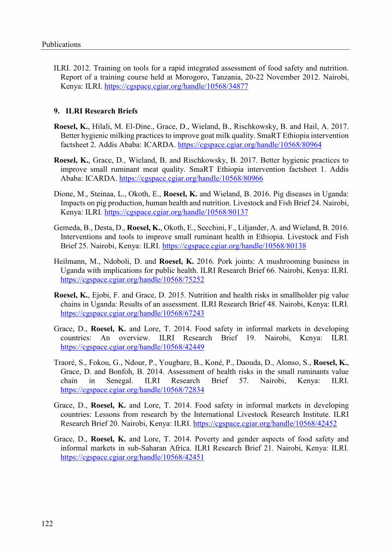
ILRI. 2012. Training on tools for a rapid integrated assessment of food safety and nutrition.
Report of a training course held at Morogoro, Tanzania, 20-22 November 2012. Nairobi,
Kenya: ILRI. https://cgspace.cgiar.org/handle/10568/34877
9. ILRI Research Briefs
Roesel, K., Hilali, M. El-Dine., Grace, D., Wieland, B., Rischkowsky, B. and Hail, A. 2017.
Better hygienic milking practices to improve goat milk quality. SmaRT Ethiopia intervention
factsheet 2. Addis Ababa: ICARDA. https://cgspace.cgiar.org/handle/10568/80964
Roesel, K., Grace, D., Wieland, B. and Rischkowsky, B. 2017. Better hygienic practices to
improve small ruminant meat quality. SmaRT Ethiopia intervention factsheet 1. Addis
Ababa: ICARDA. https://cgspace.cgiar.org/handle/10568/80966
Dione, M., Steinaa, L., Okoth, E., Roesel, K. and Wieland, B. 2016. Pig diseases in Uganda:
Impacts on pig production, human health and nutrition. Livestock and Fish Brief 24. Nairobi,
Kenya: ILRI. https://cgspace.cgiar.org/handle/10568/80137
Gemeda, B., Desta, D., Roesel, K., Okoth, E., Secchini, F., Liljander, A. and Wieland, B. 2016.
Interventions and tools to improve small ruminant health in Ethiopia. Livestock and Fish
Brief 25. Nairobi, Kenya: ILRI. https://cgspace.cgiar.org/handle/10568/80138
Heilmann, M., Ndoboli, D. and Roesel, K. 2016. Pork joints: A mushrooming business in
Uganda with implications for public health. ILRI Research Brief 66. Nairobi, Kenya: ILRI.
https://cgspace.cgiar.org/handle/10568/75252
Roesel, K., Ejobi, F. and Grace, D. 2015. Nutrition and health risks in smallholder pig value
chains in Uganda: Results of an assessment. ILRI Research Brief 48. Nairobi, Kenya: ILRI.
https://cgspace.cgiar.org/handle/10568/67243
Grace, D., Roesel, K. and Lore, T. 2014. Food safety in informal markets in developing
countries: An overview. ILRI Research Brief 19. Nairobi, Kenya: ILRI.
https://cgspace.cgiar.org/handle/10568/42449
Traoré, S., Fokou, G., Ndour, P., Yougbare, B., Koné, P., Daouda, D., Alonso, S., Roesel, K.,
Grace, D. and Bonfoh, B. 2014. Assessment of health risks in the small ruminants value
chain in Senegal. ILRI Research Brief 57. Nairobi, Kenya: ILRI.
https://cgspace.cgiar.org/handle/10568/72834
Grace, D., Roesel, K. and Lore, T. 2014. Food safety in informal markets in developing
countries: Lessons from research by the International Livestock Research Institute. ILRI
Research Brief 20. Nairobi, Kenya: ILRI. https://cgspace.cgiar.org/handle/10568/42452
Grace, D., Roesel, K. and Lore, T. 2014. Poverty and gender aspects of food safety and
informal markets in sub-Saharan Africa. ILRI Research Brief 21. Nairobi, Kenya: ILRI.
https://cgspace.cgiar.org/handle/10568/42451
122
Publications

Publications
Kang’ethe, E., Grace, D., Roesel, K., Hendrickx, S. and Makita, K. 2014. Safety of animal-
source foods in informal markets in the East African Community: Policy engagements. ILRI
Policy Brief 13. Nairobi, Kenya: ILRI. https://cgspace.cgiar.org/handle/10568/52350
123

Disclosure
Disclosure of own share in the body of the work
The share of the authors who were involved in the publications of this work is listed below
according to the following criteria:
1. Idea
2. Design of study and/or experiment
3. Test execution
4. Data analysis
5. Manuscript writing
6. Mobilisation of funding and other resources
Publication I
Dione, M. M., Ouma, E.A., Roesel, K., Kungu, J., Lule, P., Pezo, D. 2014. Participatory
assessment of animal health and husbandry practices in smallholder pig production systems
in three high poverty districts in Uganda. Prev Vet Med. 2014 Dec 1;117(3-4):565-76. doi:
10.1016/j.prevetmed.2014.10.012. Erratum in: Prev Vet Med. 2015 May 1;119(3-4):239.
1. CRP L&F
2. Dione, Roesel, Grace, Ouma, Pezo
3. Dione, Roesel, Ouma, Pezo, Lule, Kungu
4. Dione
5. Dione, Roesel, Ouma, Pezo, Lule
6. Randolph, Grace
Publication II
Roesel, K., Dohoo, I., Baumann, M., Dione, M., Grace, D., Clausen, P.-H. 2017. Prevalence and
risk factors for gastrointestinal parasites in small-scale pig enterprises in Central and
Eastern Uganda. Parasitol Res. [2016 Oct 26; Epub ahead of print] 116: 335-345. doi:
10.1007/s00436-016-5296-7
1. Roesel, Baumann, Clausen
2. Roesel, Dione
3. Roesel
4. Roesel, Dohoo
5. Roesel, Dohoo, Baumann, Grace, Dione, Clausen
6. Randolph, Grace
124

Disclosure
Publication III
Roesel, K., Nöckler, K., Baumann, M.P.O., Fries, R., Dione, M.M., Clausen, P.-H., Grace, D.
2016. First Report of the Occurrence of Trichinella-Specific Antibodies in Domestic Pigs in
Cetral and Eastern Uganda. 2016 Nov 21; 11(11):e0166258. doi: 10.1371/journal.pone.0166258
1. Roesel, Clausen, Baumann, Fries, Nöckler
2. Roesel, Clausen, Baumann, Fries, Dione, Nöckler
3. Roesel, Nöckler
4. Roesel
5. Roesel, Nöckler, Clausen, Grace, Dione, Baumann, Fries
6. Grace, Randolph, Nöckler
Publication IV
Roesel, K., Schares, G., Grace, D., Baumann, M.P.O., Fries, R., Dione, M., Clausen, P.-H. 2016.
The occurrence of porcine Toxoplasma gondii infections in smallholder production systems
in Central and Eastern Uganda. In: Clausen, P.-H., Baumann, M., Nijhof, A. (eds), Tropical
Animal Diseases and Veterinary Public Health: Joining Forces to Meet Future Global Challenges.
First Joint ATIVM-STVM Conference, Berlin, Germany, 4-8 September 2016. Giessen, Germany:
Verlag der DVG Service GmbH. Page 22.
1. Roesel, Baumann, Nöckler, Fries, Clausen
2. Roesel, Schares, Clausen, Baumann, Fries, Dione
3. Roesel, Schares
4. Roesel, Dohoo
5. Roesel, Schares, Clausen, Baumann, Fries, Dione, Grace
6. Grace, Randolph, Schares
Publication V
Roesel, K., Ouma, E.A., Dione, M.M., Pezo, D., Grace, D. 2014. Smallholder pig producers and
their pork consumption practices in Kamuli, Masaka and Mukono districts in Uganda. In:
Africa’s Animal Agriculture: Macro-trends and future opportunities. 6th All African Conference
on Animal Agriculture, Nairobi, Kenya, 27-30 October 2014, pages 166-168.
1. Roesel, Grace
2. Roesel, Grace, Ouma, Dione, Pezo
3. Roesel
4. Roesel
5. Roesel, Grace, Dione, Pezo, Ouma
6. Randolph, Grace
125

Acknowledgements
Acknowledgements
First and foremost, I want to thank my supervisor at the International Livestock Research Institute
(ILRI), Dr Delia Grace, a role model for female junior scientists. She had the confidence in me
and my research topic but moreover, she had faith that I could handle the double burden of
coordinating and implementing a multi-country project in parallel to my research project. She
found just the right balance of giving me freedom and support to grow my skills and to grow as a
person, especially as a women in science.
I am deeply grateful for my academic supervisor, Professor Peter-Henning Clausen who has
known and supported me for the past ten years. We share the same passion for research that
benefits smallholder livestock producers in sub-Saharan Africa, and I very much hope to continue
working with him over the years to come.
I am also endowed to the other members of my mentoring committee: Dr Maximilian Baumann
who introduced me to ILRI in 2011 as well as to basic statistics, Professor Reinhard Fries who has
supported my study beyond his retirement and even during Christmas breaks, and PD Dr Karsten
Nöckler at the Federal Institute for Risk Assessment who is incredibly organized, efficient and
made me get to the point more than once. Though not on paper, I was blessed with two more
outstanding mentors who must not go unmentioned. I want to thank Dr Gereon Schares at the
Friedrich-Loeffler-Institute in Greifswald, Germany, who hosted me at his laboratory twice in
2013 and 2014, and despite his busy schedule trained me in a number of assays to diagnose T.
gondii, together with his lab technicians Andrea Bärwald and Mareen Sens. Secondly, I want to
thank Dr Ian Dohoo, a world leading veterinary epidemiologist at the University of Prince Edwards
Islands in Canada. It was during an ILRI-hosted epidemiology training in Kenya in 2015 that he
got hooked with pig parasites in the tropics and I believe he may not comprehend the extent of
support, both in statistics but also mentally, he gave to me during that last year of my work on the
thesis.
I am obliged to my colleagues at ILRI-Uganda who went with me through the challenges of
aligning two ILRI research programs in Uganda: Dr Emily Ouma, Dr Michel Dione, Dr Danilo
Pezo, Rachel Miwanda, Paul Basajja, and Peter Lule as well as Dr Thomas Randolph who has led
the CRP Livestock and Fish from its inception in 2012. There would be no results to present
without all the support I received in the field during data collection. I am therefore deeply grateful
for all my colleagues in the field: The district veterinary officers Dres Lawrence Mayega, Daniel
Kasibule and David Kiryabwire; the veterinary officers Edward Mawanda, Milly Nanyolo, and
Joseph Sserwadda as well as our data and sample collectors and local facilitators Joyce Akol, Janet
Bagonza, David Buguma, Moses Dhukusooka, Joseph Erume, Robert Isabirye, Fred Kabanda,
Ivan Kakeeto, Charles Kategere, Henry Kirya, Noah Kiwanuka, Godfrey Kizito, Joseph Kungu,
Eve Luvumu, Abel Lyundhu, Lawrence Mayanja, Noah Mayanja, John Matovu, Josephine
Mukasa, Irene Mutambo, Angella Musewa, Gideon Nadiope, Winnie Nairuba, Peter Ssenabulya,
Peter Wagabaza, and Joan Wotali. At the College of Veterinary Medicine, Animal Resources and
Biosecurity I want to thank Dickson Ndoboli, Maria Tumwebaze, Steven Kakooza, Dr Edward
Wampande, William Muyumbya, Wilfred Eneku, and Dr Francis Ejobi. At Wambizzi abattoir I
thank Simon Lubega, David Waalabyeki, and Jane Lwanira for the kind cooperation and
facilitation. In addition, I am grateful for the support given by Dr Anne Mayer-Scholl and Peter
Bahn at the Federal Institute for Risk Assessment in Berlin in planning and carrying out the sub-
activity on the occurrence of Trichinella spp. I also want to thank Emmanuel Muunda and Nicholas
126

Acknowledgements
Ngwili, research assistants at ILRI-Nairobi, for validating all the data entered from our surveys.
Finally, I am grateful to the Uganda Ministry of Agriculture, Animal Industry and Fisheries for
supporting research on smallholder pig value chains in Uganda for the benefit of resource-poor
livestock keepers, and especially Dr Noelima Nantima for her active provision of help throughout
the study. Behind the administrative scenes at ILRI, I want to thank Wacera Ndonga, Rosekellen
Njiru and Sarah Ndung’u for helping with finances and logistics, Grace Miceka at IT for her remote
support to keep my laptop well maintained and healthy (a vital aspect when working in the field
in a low-income country!), and Tezira Lore, ILRI communications officer for proofreading many
of my reports and papers. At Freie Universität Berlin, I want to thank Angela Daberkow,
coordinator of the PhD Biomedical Sciences program at Dahlem Research School, and her helper
Marie-Kamala Hecker for keeping track of all my outputs and achievements. At the Institute for
Parasitology and Tropical Veterinary Medicine, I want to thank Louise Le Bel, Antje Hoppenheit,
Katharina Seidl, and Peggy Hoffmann-Köhler for support during my stays in Berlin.
When working abroad it becomes challenging to maintain old friendships. But some are meant to
stay forever. In my case it is Anne, my dear gal pal, whom I know since sixth grade and who knows
the pains and gains of preparing a PhD thesis and supported me during mine. I also thank Albert,
my Dutch-African friend, who, during my first stay in Malawi in 2005, taught me so much about
African society, culture and politics and briefed me perfectly for my later career. And I want to
thank my friend Jana who always gave me a place to stay when I visited Berlin for course work or
mentoring meetings, and helped me not to forget the great cultural life in Europe. In Uganda I
made new friends who lived with me through good and bad times of the life after work, and I am
incredibly grateful for Ulrike and Jonas (with Elly) as well as Amrei and Oliver (with Schappi) for
you-know-what. I also thank Christine, Ibrahim, Benson and Jasper for all the daily amenities,
including taking me to the abattoir at four in the morning despite the rains and collecting my meat
samples from the butchers on Christmas Day!
I want to thank my parents, Ingrid and Holger, for giving me plenty of opportunities which I
believe I utilized to the fullest. And lastly I want to thank Hubertus, my love, for supporting me
throughout most parts of this project, and still growing strong with me.
The research was carried out with the financial support of the Federal Ministry for Economic
Cooperation and Development, Germany, and the CGIAR Research Program on Agriculture for
Nutrition and Health, led by the International Food Policy Research Institute through the „Safe
Food, Fair Food“ project and the CGIAR Research Program on Livestock & Fish, led by the
International Livestock Research Institute.
127

Statement of authorship
Statement of authorship
Except where reference is made in the text, this dissertation contains no material published
elsewhere or extracted in whole or in parts from a dissertation presented by myself towards another
degree or diploma. Nobody else’s work has been used without due acknowledgement in the main
text of the dissertation. This dissertation has not been submitted for an award of any other degree
or diploma in any tertiary institution.
Berlin, 30th November 2017 Kristina Rösel
128


Copyright © 2022 FDOKUMEN