
Robust prescan calibration for multiple spin-echo sequences: application to FSE and b-SSFP
11
Robust prescan calibration for multiple spin-echo sequences: application to FSE and b-SSFP Chris V. Bowen a, 4 , Joseph S. Gati b , Ravi S. Menon b,c,d a Institute for Biodiagnostics (Atlantic), National Research Council, Halifax, Nova Scotia, Canada B3H 3A7 b The Laboratory for Functional Magnetic Resonance Research, Robarts Research Institute, London, Ontario, Canada N6A 5K8 c Department of Medical Biophysics, University of Western Ontario, London, Ontario, Canada N6A 5K8 d Department of Diagnostic Radiology and Nuclear Medicine, University of Western Ontario, London, Ontario, Canada N6A 5K8 Received 16 January 2006; revised 29 March 2006 Abstract The collection of fast imaging techniques that use multiple spin-echo (MSE) sequences relies on a precise phase relationship between spin echoes and stimulated echoes that form along the radiofrequency refocusing pulse train. Failure to achieve this condition produces dark banding artifacts that result from destructive interference between signal coherence pathways. Satisfying this condition on the microsecond timescale required is technically challenging for conditions involving strong diffusion-weighted gradients, for arbitrary orientation acquisitions and at large field strengths with high-resolution acquisitions. Two clinically significant MSE sequences, fast spin echo (FSE) and balanced steady-state free precession (b-SSFP), are investigated in this work using a 4-T whole-body scanner. We developed a readout- projection-based prescan technique that ensures coherent signal formation by utilizing banding artifacts to automatically adjust gradient balance. Subsequent image acquisition using the results of this prescan permits the formation of coherent-echo images, which are robust under challenging imaging conditions. The robustness of this approach is demonstrated for FSE and b-SSFP images obtained from the knees of human volunteers. We believe that the use of this prescan calibration technique for the alignment of signal pools in MSE sequences is critical at high fields and will facilitate the implementation of high-quality clinically significant sequences such as FSE and b-SSFP. D 2006 Elsevier Inc. All rights reserved. Keywords: Fast spin echo; TrueFISP; FIESTA; SSFP; Artifact; Prescan; Knee; MRI 1. Introduction In recent years, the use of high-field magnetic resonance scanners for human body imaging has grown rapidly owing to improved signal-to-noise ratio (SNR) and achievable susceptibility contrast [1,2]. These advantages may be employed to acquire images with greater resolution (hence, greater anatomic detail) or images with greater sensitivity to susceptibility-related signal intensity changes, as with functional magnetic resonance imaging (fMRI) [3,4]. However, coupled with these benefits are additional technical challenges resulting from an increased difficulty in producing adequate shim quality (B 0 ) and radiofrequency (RF) homogeneity (B 1 ) [5]. These problems are most apparent within the family of multiple spin-echo (MSE) sequences, where signals from multiple refocusing path- ways must coherently combine to prevent destructive signal interference [6,7]. Any microsecond-scale misalignment of primary spin echoes and stimulated echoes within the readout window produces a large spatial variation of image signal intensity for MSE sequences, such as fast spin echo (FSE) [8,9] and balanced steady-state free precession (b- SSFP) [10,11]. Although high-quality FSE and b-SSFP images have become commonplace in the clinical setting over the past decade, MSE acquisitions can present signal incoherence arti- facts at any field strength. In addition to specific difficulties presented at high fields (greater than 3 T) [12], the use of transverse magnetization preparation schemes, such as diffu- sion, T 2 or T 2 * weighting, can produce phase errors that cause destructive interference between coherence pathways [13,14]. A great number of system calibration imperfections can be problematic for MSE sequences. Difficulties associ- ated with gradient fidelity, such as unbalanced amplifier gain along each gradient polarity, amplifier nonlinearity, 0730-725X/$ – see front matter D 2006 Elsevier Inc. All rights reserved. doi:10.1016/j.mri.2006.04.008 4 Corresponding author. Tel.: +1 902 473 1858; fax: +1 902 473 1851. E-mail address: [email protected] (C.V. Bowen). Magnetic Resonance Imaging 24 (2006) 857 – 867
Transcript of Robust prescan calibration for multiple spin-echo sequences: application to FSE and b-SSFP
Magnetic Resonance Im
Robust prescan calibration for multiple spin-echo sequences:
application to FSE and b-SSFP
Chris V. Bowena,4, Joseph S. Gatib, Ravi S. Menonb,c,d
aInstitute for Biodiagnostics (Atlantic), National Research Council, Halifax, Nova Scotia, Canada B3H 3A7bThe Laboratory for Functional Magnetic Resonance Research, Robarts Research Institute, London, Ontario, Canada N6A 5K8
cDepartment of Medical Biophysics, University of Western Ontario, London, Ontario, Canada N6A 5K8dDepartment of Diagnostic Radiology and Nuclear Medicine, University of Western Ontario, London, Ontario, Canada N6A 5K8
Received 16 January 2006; revised 29 March 2006
Abstract
The collection of fast imaging techniques that use multiple spin-echo (MSE) sequences relies on a precise phase relationship between spin
echoes and stimulated echoes that form along the radiofrequency refocusing pulse train. Failure to achieve this condition produces dark
banding artifacts that result from destructive interference between signal coherence pathways. Satisfying this condition on the microsecond
timescale required is technically challenging for conditions involving strong diffusion-weighted gradients, for arbitrary orientation
acquisitions and at large field strengths with high-resolution acquisitions. Two clinically significant MSE sequences, fast spin echo (FSE) and
balanced steady-state free precession (b-SSFP), are investigated in this work using a 4-T whole-body scanner. We developed a readout-
projection-based prescan technique that ensures coherent signal formation by utilizing banding artifacts to automatically adjust gradient
balance. Subsequent image acquisition using the results of this prescan permits the formation of coherent-echo images, which are robust
under challenging imaging conditions. The robustness of this approach is demonstrated for FSE and b-SSFP images obtained from the knees
of human volunteers. We believe that the use of this prescan calibration technique for the alignment of signal pools in MSE sequences is
critical at high fields and will facilitate the implementation of high-quality clinically significant sequences such as FSE and b-SSFP.
D 2006 Elsevier Inc. All rights reserved.
Keywords: Fast spin echo; TrueFISP; FIESTA; SSFP; Artifact; Prescan; Knee; MRI
1. Introduction
In recent years, the use of high-field magnetic resonance
scanners for human body imaging has grown rapidly owing
to improved signal-to-noise ratio (SNR) and achievable
susceptibility contrast [1,2]. These advantages may be
employed to acquire images with greater resolution (hence,
greater anatomic detail) or images with greater sensitivity to
susceptibility-related signal intensity changes, as with
functional magnetic resonance imaging (fMRI) [3,4].
However, coupled with these benefits are additional
technical challenges resulting from an increased difficulty
in producing adequate shim quality (B0) and radiofrequency
(RF) homogeneity (B1) [5]. These problems are most
apparent within the family of multiple spin-echo (MSE)
sequences, where signals from multiple refocusing path-
0730-725X/$ – see front matter D 2006 Elsevier Inc. All rights reserved.
doi:10.1016/j.mri.2006.04.008
4 Corresponding author. Tel.: +1 902 473 1858; fax: +1 902 473 1851.
E-mail address: [email protected] (C.V. Bowen).
ways must coherently combine to prevent destructive signal
interference [6,7]. Any microsecond-scale misalignment of
primary spin echoes and stimulated echoes within the
readout window produces a large spatial variation of image
signal intensity for MSE sequences, such as fast spin echo
(FSE) [8,9] and balanced steady-state free precession (b-
SSFP) [10,11].
Although high-quality FSE and b-SSFP images have
become commonplace in the clinical setting over the past
decade,MSE acquisitions can present signal incoherence arti-
facts at any field strength. In addition to specific difficulties
presented at high fields (greater than 3 T) [12], the use of
transverse magnetization preparation schemes, such as diffu-
sion, T2 or T2* weighting, can produce phase errors that
cause destructive interference between coherence pathways
[13,14]. A great number of system calibration imperfections
can be problematic for MSE sequences. Difficulties associ-
ated with gradient fidelity, such as unbalanced amplifier
gain along each gradient polarity, amplifier nonlinearity,
aging 24 (2006) 857–867

C.V. Bowen et al. / Magnetic Resonance Imaging 24 (2006) 857–867858
unaccounted gradient delays or imperfect eddy current
compensation, are not uncommon [14–17]. Although gradi-
ent fidelity problems are an ongoing consideration even with
low-field-strength MSE acquisitions, they are of particular
concern with increased field strength where high-resolution
acquisitions are often used to exploit SNR advantages. For a
given gradient performance, high-resolution acquisitions
require both increased gradient strength and RF pulse
refocusing times. Each condition further exposes technical
imperfections in gradient fidelity and field homogeneity and
increases difficulty in aligning multiple signal coherence
pathways relative to that normally observed clinically.
Compensation for these problems through the use of
postprocessing algorithms is not possible with MSE acquis-
itions, as with echo-planar imaging [18]; thus, a prospective
correction is required.
A number of pulse sequence modifications have been
proposed to suppress artifact formation in the presence of
technical imperfections. In the case of FSE, suggested
approaches include ensuring that refocusing pulses are
precisely 1808 [19], suppressing all coherence pathways
other than the primary echo [13,20] or using a non-Carr–
Purcell–Meiboom–Gill (CPMG) phase-cycling scheme for
the refocusing pulses to limit sensitivity to phase errors [21].
These approaches are limited by practical considerations or
by reduction in SNR efficiency due to the suppression of
useful signal coherence pathways [22]. For b-SSFP acquis-
itions, the reduction of artifact through maximum intensity
projections of multiple b-SSFP images, where the artifact
has been deliberately shifted to alternate spatial locations, is
feasible but suffers from reduction in SNR efficiency [23].
Two clinically significant MSE sequences, FSE and
b-SSFP (also known as TrueFISP and FIESTA), are
investigated in this work using a 4-T whole-body scanner.
We developed a readout-projection-based prescan technique
that automatically adjusts the read/slice gradient balance to
ensure coherent signal formation to first order in these
two dimensions. This technique utilizes banding artifacts
that form through a deliberate misadjustment of readout
gradient balance from a nominal value in order to calculate
calibration parameters. The theoretical basis for artifact
formation with FSE and b-SSFP acquisitions is first reviewed
prior to the description of a prescan strategy suggested from
this. The robustness of the prescan approach is demonstra-
ted through multiple prescan acquisitions from the heads
of volunteers. Subsequent image acquisition using the results
of this prescan calibration is demonstrated for FSE and
b-SSFP images obtained from the knees of human volunteers.
The significant signal heterogeneity produced by the anato-
my of the knee presents a challenging condition for the
prescan approach. We believe that the use of this prescan
calibration technique for the alignment of signal pools
in MSE sequences is critical at high fields for high-
resolution acquisitions and will facilitate the implementation
of high-quality clinically significant sequences such as FSE
and b-SSFP.
2. Theory
The generation of artifact-free images for the family of
MSE sequences relies upon a precise phase relationship
between transverse magnetization and refocusing pulse B1
vectors [6,13,22]. Failure to achieve this condition results in
destructive interference between echoes that are formed
through a broad range of signal coherence pathways; a
significant problem with as many as 750 distinct signal
pathways present after only eight spin echoes [24].
Deviation from proper phase coherence can result from
nonidealities associated with either the phase of transverse
magnetization or the phase of the refocusing pulse B1 field.
The phase of different spin packets in the transverse plane
(which sum to transverse magnetization) is altered by
factors such as imperfectly compensated eddy currents,
gradient amplifier nonlinearity, unaccounted gradient delays
and off-resonance, while errors in the phase of the
refocusing pulse B1 field may be introduced through
imperfect referencing of synthesizer phase following off-
center slice excitation. Regardless of the source of deviation,
appropriate coherence conditions are described below for
the two MSE sequences investigated in this work (FSE and
b-SSFP), and the consequences for an appropriate calibra-
tion prescan are described.
2.1. FSE calibration
For MSE sequences where nonzero gradient encoding is
present during the application of refocusing pulses, spatially
invariant phase-cycling conditions cannot exist [20]. For
instance, alignment of transverse magnetization and the
refocusing pulse B1 field, in accordance with CPMG
condition, cannot be achieved for all spatial locations with
FSE. Considering the effects of imaging gradients alone, the
phase of transverse magnetization relative to the refocusing
pulse B1 field immediately prior to the first and second
refocusing pulses is given by /1 and /2, respectively,
as follows:
/1 x; y; zð Þ ¼ cZD=2
0
Gd r dt ¼ c Ax1xþ Ay1yþ Az1z� �
/2 x; y; zð Þ ¼ cZ3D=2
D=2
Gd r dt � /1
¼ c Ax2xþ Ay2yþ Az2z� �
� /1 ð1Þ
where c is the gyromagnetic ratio, D is the refocusing pulse
interval, and G and r are the imaging gradient and spatial
position vectors, respectively. The logical-read, phase-
encode and slice directions are defined by x, y and z,
respectively, and have the indicated gradient–time integrals
Ax, Ay and Az, respectively. In practice, phase encoding is
completely rewound prior to each refocusing pulse; hence,

C.V. Bowen et al. / Magnetic Resonance Imaging 24 (2006) 857–867 859
Ay1 and Ay2 are ideally zero, while the nonzero values of Ax
and Az are determined by the applied read and slice gradient
crusher area, respectively. Therefore, neglecting the phase-
encode axis and substituting from Eq. (1), we define D/ as
the difference in phase between /1 and /2, as follows:
D/u /2 � /1 ¼ c Ax2 � 2Ax1ð Þxþ Az2 � 2Az1ð Þz�½ ð2Þ
Fig. 1 illustrates the phase condition required for the
coherent addition of stimulated echoes and spin echoes for
FSE imaging. The first refocusing pulse separates free
induction decay (FID) magnetization (with phase /1) into
two coherence pathways that contribute to image formation:
one comprising a spin echo and another stored on the
longitudinal axis to be excited by a subsequent refocusing
pulse as a stimulated echo [24]. The number of coherence
pathways that potentially contribute to image formation
expands with each subsequent refocusing pulse in this
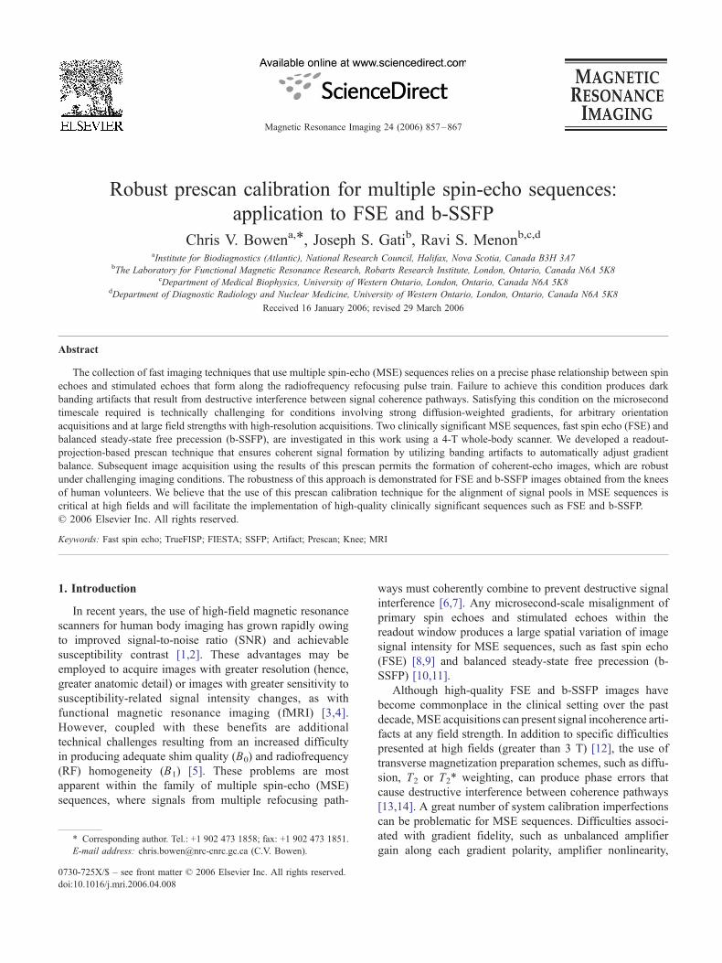
Fig. 1. Illustration of the FSE signal phase condition in the transverse plane.
Magnetization vectors are shown immediately before (left side) and after
(right side) the application of the first (A), second (B) and third (C)
refocusing pulses along the yV axis, respectively. Subscripts indicate
whether the magnetization is FID (fid), spin echo (se), stimulated echo (ste)
or stored on the longitudinal axis (z), with the subscript number indicating
the number of refocusing pulse intervals traversed by the respective signal
pools. The angles associated with Mz components indicate the encoded
phase produced by subsequent stimulated echoes, with /1 and /2 defined
for some spatial position by Eq. (1). After three refocusing pulses (C),
assuming that the condition defined by Eq. (3a) is met, all transverse
magnetization components pool into those stimulated echoes that have had
an even number (Me) or an odd number (Mo) of refocusing interval
passages, with phase relative to the yV axis given by /1 and /2,
respectively. The spin-echo signal combines with the Me component.
fashion, with the phase of the spin-echo magnetization given
by /2 immediately prior to the second refocusing pulse.
Two conditions are required for the constructive summa-
tion of signal from all coherence pathways: the gradient–time
integral applied during each refocusing pulse interval must
be identical, and the difference in phase D/ between /1 and
/2 must be zero [6]. The first condition ensures that signals
combine coherently, while the second condition ensures that
signals combine constructively. These conditions are defined
by Eqs. (3a) and (3b), respectively, as follows:
/n ¼ /2 for all n N 2 ð3aÞ
D/ ¼ 0 ð3bÞ
where /n is the phase accrued during the refocusing pulse
interval prior to the nth refocusing pulse and D/ is defined
in Eq. (2). As is evident from Fig. 1C, the satisfaction of Eq.
(3a) causes magnetization from each of the rapidly expand-
ing number of coherence pathways to converge along one
of two orientations, with phase /1 or /2, depending on
whether the stimulated-echo signal has traversed an even
number or an odd number of refocusing pulse intervals,
respectively [25]. These two magnetization pools rapidly
approach with equivalent strength and with comparable
signal intensity after as few as four echoes [20,24,25].
Failure to satisfy Eq. (3a) causes incoherent mixing of
signals from various coherence pathways and apparent rapid
signal decay along the FSE train.
Failure to satisfy Eq. (3b) causes resultant magnetization,
which is derived from a combination of even and odd echo
magnetization pools (Me and Mo in Fig. 1C) to be reduced,
as follows [6]:
M 2 ¼ M 2e þM 2
o þ 2MeMocosD/ ð4Þ
where D/ is defined in Eq. (2), and a spatial cosine mod-
ulation of signal strength is observed if Eq. (3b) is not met.
In the case of unbalanced readout gradients (i.e., Ax2p 2Ax1
within Eq. (2)), a strong cosine modulation in the readout
direction is observed for late effective-echo FSE images
[6] — a property that is central to the prescan calibration
approach involving the read profiles described below.
2.2. b-SSFP calibration
Unlike the case with FSE, b-SSFP is an MSE sequence
that requires a complete rewinding of all gradient encodings
prior to subsequent RF excitation [7,11,26]. One parameter
that determines b-SSFP signal response is the phase
evolution of magnetization on a TR interval /TR. Again,
considering the effects of imaging gradients alone, /TR is
given, as follows:
/TR x; y; zð Þ ¼ cZTR
0
Gd r dt ¼ c Axxþ Ayyþ Azz� �
ð5Þ
where Ax, Ay and Az are gradient–time integrals over a TR
interval in the read, phase-encode and slice directions,

C.V. Bowen et al. / Magnetic Resonance Imaging 24 (2006) 857–867860
respectively. As with FSE, the two conditions required for a
coherent and a constructive addition of multiple coherence
pathways are given by Eqs. (6a) and (6b), respectively, as
follows [7,11]:
/ nð ÞTR ¼ / nþ1ð Þ
TR for all n ð6aÞ
/TR ¼ 0 ð6bÞ
where n is the number of TR interval in the RF pulse train.
Failure to satisfy Eq. (6a) causes incoherent summation
of signals from multiple coherence pathways, with the
extreme limiting case being RF spoiling, where random
phase jumps are deliberately added between RF pulses to
suppress echo formation.
Failure to satisfy Eq. (6b) results in destructive interfer-
ence between multiple signal coherence pathways and
causes periodic variation in signal intensity for changes in
/TR. However, unlike the spatial cosine variation for FSE
imaging, as described in Eq. (4), b-SSFP signal intensity
varies with /TR as a complex function of T1, T2, TR and flip
angle [7,11]. Nonetheless, unbalanced readout gradients
(Axp 0 from Eq. (5)) result in periodic banding profile in the
readout direction; thus, both FSE and b-SSFP are amenable
to the same readout-profile-based prescan calibration tech-
nique described in this work.
3. Methods
All experiments described in this work were performed on
a 4-T Varian/Siemens whole-body scanner equipped with
Siemens Sonata imaging gradients (an amplitude of 40 mT/
m operating at a slew rate of 120 T/m/s). Phantom studies
involved an 11-cm-diameter Ni-doped water sphere and a
27-cm-diameter quadrature birdcage coil. Phantom acquis-
itions had certain imaging parameters for FSE [14 cm field
of view (FOV), 256�256 matrix, 5 mm slice, 15 ms echo
spacing, 8 echoes, 3000/60 ms TR/TE] and b-SSFP (14 cm
FOV, 256�256 matrix, 5 mm slice, 10/5 ms TR/TE). Normal
human volunteers were used for both head and knee imaging
studies. All measurements in the human head used either
a 27-cm-diameter quadrature birdcage coil or an 11-cm-
diameter quadrature surface coil pair. For head imaging,
certain imaging parameters were used for FSE (22 cm FOV,
256�256 matrix, 4 mm slice, 15 ms echo spacing, 8 echoes,
4500/60 ms TR/TE) and b-SSFP (22 cm FOV, 256�256 matrix, 4 mm slice, 8/4 ms TR/TE). An 18-cm quadrature
birdcage coil was used for all high-resolution knee imaging
with certain acquisition parameters for FSE (16 cm FOV,
512�512 matrix, 2 mm slice, 15 ms echo spacing, 8 echoes,
3000/60 ms TR/TE) and b-SSFP (16 cm FOV, 512�512 matrix, 2 mm slice, 13/6.5 ms TR/TE).
3.1. Pulse sequences
Both 2D FSE and 2D/3D b-SSFP sequences were
developed in-house following conventional approaches.
The 2D multislice FSE sequence employed CPMG phase
cycling along the refocusing train and combined slice select/
gradient crusher waveforms for refocusing pulses to achieve
minimum echo-spacing conditions [8,9]. Refocusing pulses
with a 1608 flip angle were employed for SAR consid-
erations at 4 T [12] and to facilitate a more rapid equi-
libration of stimulated and spin echoes along the refocusing
pulse train [25]. RF pulses were optimally designed using
the SLR algorithm [27]. In order to satisfy the phase-balance
condition described in Eq. (3b) for the logical frequency-
encode axis x, the area of the readout prewinder gradient
(Ax1) was adjusted by scaling gradient amplitude according
to a procedure described in the Results section. Eq. (3b) was
satisfied for the slice select axis z by adjusting the phase of
the transmitter for the first refocusing pulse (/1) according
to the calibration procedure developed in the Results
section. The phase is adjusted for Varian console through
specific off-resonance frequency hops for a finite amount of
time (coincident with the readout prewinder gradient lobe)
to achieve target phase offset.
The 2D and 3D b-SSFP sequences were developed in-
house according to an approach described in the literature
for cardiac imaging [26]. This version of the b-SSFP
sequence can be gated to the cardiac cycle or to the
respiratory cycle through the use of a catalyzing train of RF
pulses to rapidly achieve steady-state condition. A catalyz-
ing train of 20 RF pulses was applied prior to data
acquisition, where the flip angle was linearly increased to
the target flip angle a. This catalyzing pulse strategy is
known to be robust with respect to off-resonance for a rapid
approach to steady state [28] — an important consideration
at higher field strengths. The phase-balance condition
described in Eq. (6b) was satisfied for both the frequency-
encode axis (x) and the slice axis (z) in the same manner as
that described above for the FSE sequence.
Several approaches were taken to reduce the phase-
encode ghosting induced by signal oscillation along the
acquisition train. A �a/2 bflipbackQ pulse was applied with
a delay of TR/2 after the last RF pulse in the acquisition
train in order to return magnetization to equilibrium con-
dition along the longitudinal axis. This promoted a more
rapid approach to steady-state condition by subsequent
catalyzing trains [29]. A linear phase-encode pattern was
used in order to prevent signal oscillation from being
introduced by subtle differences in /TR for adjacent TR
intervals. The presence of residual eddy currents causes
differences in /TR that make adjacent TR intervals more
pronounced when the phase-encode increment is large [30].
Additionally, in order to minimize the phase-encode
increment for adjacent TR intervals, each uncollected echo
in the catalyzing train was phase-encoded using the first
phase-encode value in the acquisition train [31].
3.2. Readout profile prescan acquisition
Prior to every FSE or b-SSFP acquisition, an array of
10 readout profiles having a different readout prewinder

C.V. Bowen et al. / Magnetic Resonance Imaging 24 (2006) 857–867 861
gradient area was acquired for each slice (phase-encode
gradients turned off). The readout prewinder gradient area
(Ax1 from Eq. (1) for FSE, or Ax from Eq. (5) for b-SSFP)
was deliberately adjusted from a nominal value by a fraction
fx of the full readout gradient area (acquisition window).
The fx array was uniformly spaced by about 0% deviation
from nominal, where the maximum fx deviation was
selected to produce a number of signal intensity modulation
bands in the readout direction Nb of approximately 30. As
can be deduced from Eq. (2) and (5) for FSE and b-SSFP,
respectively, this corresponds to an fx value having a max-
imum offset fraction of Nb /Nro, where Nro is the number of
frequency-encode points in the acquisition.
The total acquisition time for this prescan was 10 TR
cycles for FSE, while the acquisition of b-SSFP profiles
required approximately 21 TR cycles per slice per fx value
(20 catalyzing pulses to approach steady state plus one
profile acquisition). This corresponds to a prescan acqui-
sition time of approximately 30 s in each case (10 fxprofiles with a TR of 3 s for FSE, or 10 fx profiles with a
TR of 10 ms and 15 slices for b-SSFP). For the most part,
the profiles were acquired with identical acquisition
parameters and slices, as prescribed for imaging. However,
for b-SSFP prescan acquisitions, the flip angle was
Fig. 2. Demonstration of banding in frequency-encode profiles obtained from a pha
(A) and from b-SSFP acquisitions (B) with deliberate imbalance in the read prew
readout gradient area. The 1D Fourier transform magnitude of the same profiles
profile is obtained from the peak location for the first harmonic (indicated by arrow
harmonic for FSE profiles but a peak location similar to that of b-SSFP.
adjusted to 708, independent of the prescribed flip angle,
since magnitude banding profiles in the readout direction
are more sinusoidal for flip angles that exceed the Ernst
angle [23].
Processing of prescan data to extract calibration param-
eters (according to a strategy described in the Results
section) was performed online and automatically, using
compiled routines written in MATLAB (The Mathworks,
Natick MA). These routines are stand-alone (do not require
a MATLAB license or do not require a running MATLAB
interface) and require approximately 4 s of processing to
compute and to set calibration parameters (computation on a
SUN Ultra80 workstation).
4. Results
A calibration procedure for satisfying the required phase
conditions for FSE (Eq. (3b)) and b-SSFP (Eq. (6b)) is first
developed below. This prescan procedure requires the
acquisition of a number of readout profiles for each slice
prior to imaging. The efficacy of this approach for in vivo
head and knee imaging is then demonstrated for oblique
slices in the case of volume and surface coil acquisitions for
both b-SSFP and FSE at 4 T.
ntom. Read profiles are shown from FSE acquisitions using the fourth echo
inder gradient equal to 2.7% (solid line) and 5.3% (dashed line) of the full
is shown for FSE (C) and b-SSFP (D). The banding frequency for a given
s for profiles with fx =2.7%). Note the additional relative height of the first

Fig. 3. Determination of calibration parameters. (A) The banding frequency
determined from read profiles of FSE acquisitions (see Fig. 2) is shown as a
function of fractional read prewinder gradient error ( fx) for two slices
(� and o symbols). The optimum fx value for imaging ( fx(opt)) is
determined from a linear fit extrapolated to zero banding frequency.
(B) The phase of 1D Fourier transform profiles corresponding to the
location of the first harmonic magnitude peak (see Fig. 2C and D) is shown
as a function of fx for the same two slices. The arrow indicates the band
phase (/0) of one slice corresponding to fx(opt), as obtained from linear
extrapolation. (C) Slice calibration parameters are obtained from the slope
( Psl) and the intercept ( P0) of a variance-normalized weighted linear
regression of the band phase (/0) versus slice position (z). Error bars
indicate the variance of the residuals from curves shown in (B).
C.V. Bowen et al. / Magnetic Resonance Imaging 24 (2006) 857–867862
4.1. Readout-profile-based calibration procedure
Fig. 2 shows read profiles obtained from a uniform
spherical phantom for both b-SSFP (Fig. 2A) and FSE
(Fig. 2B) acquisitions, where the readout prewinder lobe has
been deliberately scaled from a nominal value. The prom-
inent feature for magnitude profiles obtained from each
sequence is a banding of the signal intensity in the readout
direction. For FSE acquisitions, profiles obtained from the
fourth echo demonstrate a sinusoidal banding profile in
accordance with predictions from Eq. (4). Additionally, null
signal regions in the profiles (stopband) have nearly 100%
signal loss, indicating nearly equivalent strength between
the even and the odd echo magnetization pools by the fourth
echo (Me and Mo in Fig. 1C). For b-SSFP acquisitions, the
profiles exhibit a more complex banding modulation, with a
flatter passband that occupies more than 50% of each cycle
and with less pronounced stopbands.
However, profiles from both FSE and b-SSFP acquis-
itions show increased banding frequency as the readout
prewinder gradient is deliberately adjusted from a nominal
value by a fraction fx of the full readout gradient area. These
profiles correspond to the satisfaction of Eqs. (3a) and (6a),
but not of Eqs. (3b) and (6b), for FSE and b-SSFP,
respectively. The spatial frequency of modulation is
extracted from the 1D Fourier transform of magnitude
profiles (Fig. 2C and D for FSE and b-SSFP, respectively).
Spatial frequency is determined from the peak location of
the first harmonic in Fourier transform magnitude profiles
(arrows in Fig. 2C and D). Fourier transform profiles have
been premultiplied by a Hanning window to suppress
truncation artifacts and by a Gaussian smoothing function,
since only low spatial frequency banding information is
sought. Despite differences in banding shape for FSE and b-
SSFP profiles, both sequences exhibit similar banding
frequencies (~7 cycles per FOV for an fx of 2.7%). The
identification of the first harmonic peak location from
Fourier transform profiles is slightly more robust for FSE
acquisitions, as this peak exhibits a greater peak height
relative to those from b-SSFP Fourier transform profiles.
Fig. 3A shows the band frequency for FSE acquisitions
(extracted according to the approach described in Fig. 2) as a
function of fx for two representative slices from a phantom.
Data points are only present for fx values (nonzero)
producing a clearly defined band frequency, with the
extracted banding frequency negated for negative fx values.
Excellent linearity and very consistent curves are present for
all slices within a phantom. The optimal fx value needed to
produce no spatial banding in the frequency-encode direc-
tion is obtained from an extrapolation of linear fits for each
slice to zero band frequency. The optimal calibration
parameter for imaging fx(opt) is obtained from the mean of
the optimal fx values derived from each slice, weighted
inversely by the variance of residuals from fits for each slice.
Fig. 3B shows the phase of Fourier transform profiles
at the first harmonic peak location (Fig. 2C) as a function of
fx. The plots are again quite linear, with minimal slopes and
different intercept positions for each slice. Physically, for
FSE, this band phase corresponds to the relative angle
between two signal pools (Me and Mo in Fig. 1C) at the
gradient isocenter (x=0 cm line; Fig. 2A). The band phase is
also the relative angle between Me and Mo that remains after
the readout gradient has been properly balanced through

C.V. Bowen et al. / Magnetic Resonance Imaging 24 (2006) 857–867 863
fractional weighting by fx(opt). The band phase for each slice
at the optimal fx setting (/0) is derived from the linear
extrapolation of each curve to fx(opt). Fig. 3C plots the band
phase as a function of slice position (z). A variance-
normalized linear regression of these data produces the final
two calibration parameters required to correct for slice-
dependent variation in phase angle, as follows:
/0 ¼ P0 þ Pslz ð7Þ
where Psl and P0 are the slope and intercept from Fig. 3C,
respectively. The required correction for slice-dependent
phase variation /cor is applied through phase shifts, as
Fig. 4. Demonstration of robust band frequency extraction using frequency-encode
acquisitions for oblique slices (A), a quadrature surface coil pair (C) and a 3-cm-t
magnitude profiles are shown (B, D and F) with deliberate imbalance in the read pr
gradient area. Note the identical banding frequency for two slices separated by 22
relative to each other (A and C).
described in the Methods section, where /cor=� /0 from
Eq. (7). These three correction factors ( fx(opt), Psl and P0), as
calculated from profile prescan, are employed for subse-
quent acquisition of images, with the resultant image quality
demonstrated in Section 4.2.
4.2. In vivo image quality
The robustness of the readout profile calibration proce-
dure is demonstrated in Fig. 4 under potentially challenging
conditions, such as those involving oblique imaging
(Fig. 4A and B), use of surface coils (Fig. 4C and D) and
3D slab selection (Fig. 4E and F). Both the read profiles
profiles obtained from a human head. Read profiles are shown from b-SSFP
hick 3D slab-selective profile (E). The corresponding 1D Fourier transform
ewinder gradient ( fx) equal to 8% (B) and 13% (D and F) of the full readout
mm (A–D), while the bands have different phases and are spatially shifted

Fig. 5. Comparison of images from the head of a normal volunteer
acquired using optimized calibration parameters (A and C) or nominal
settings (B and D). Images from both T2-weighted FSE (A and B) and
b-SSFP (C and D) illustrate the successful removal of banding for
acquisitions obtained using calibration parameters.
C.V. Bowen et al. / Magnetic Resonance Imaging 24 (2006) 857–867864
(Fig. 4A, C and E) and the corresponding 1D Fourier
transforms (Fig. 4B, D and F) are shown for a representative
slice obtained from the head of a normal human volunteer
using the b-SSFP sequence. As described in the Theory
section and as illustrated in Fig. 2B, b-SSFP signal
modulation in readout profiles does not follow a sinusoidal
behavior, as with FSE profiles, and thus provides the more
challenging application of the readout profile calibration
procedure. In each case, the dominant modulation observed
in the 1D Fourier transform readout profiles (Fig. 4B, D and
F) results from imbalanced read gradients rather than from
any structure within the image or from the sensitivity profile
of the receive coil.
Table 1 shows the mean and the standard deviation of the
three correction factors ( fx(opt), Psl and P0) obtained from five
repeat measures of the profiles shown in Fig. 4. In the case of
oblique imaging, surface coil imaging and 3D imaging, the
standard deviation of parameters is sufficiently small to
ensure that avoidable signal loss resulting from imprecise
calibration is less than 2.7%, 3.5% and 3.7%, respectively,
for any spatial position within the imaging volume. This
estimate assumes one standard deviation in each parameter
and a more pessimistic sinusoidal modulation of signal
intensity, given by Eq. (4), for FSE acquisitions.
Fig. 5 shows oblique 2D FSE and b-SSFP images from
a normal human volunteer obtained with optimized (Fig. 5A
and C) or nominal (Fig. 5B and D) calibration factors
( fx(opt), Psl and P0). Oblique images were acquired parallel
to the anterior commissure–posterior commissure line. The
effective removal of banding for oblique images acquired
using optimized parameters is clearly apparent for both late
effective-echo FSE imaging and b-SSFP imaging.
Fig. 6 shows fat-suppressed fourth effective-echo
FSE images (Fig. 6A and B) and 2D b-SSFP images
(Fig. 6C and D) for two sagittal slices through the knee of a
normal human volunteer. All images were acquired after
the optimization of calibration factors using readout profile
technique. An excellent contrast between synovial fluid and
cartilage is observed for both 2D b-SSFP and T2-weighted
FSE images. The minimization of phase-encode direction
blurring is accomplished by preservation of the signal
intensity along the refocusing pulse trains through satisfac-
tion of the conditions given by (Eqs. (3a), (3b), (6a), and
(6b) for FSE and b-SSFP imaging, respectively. The large
degree of signal heterogeneity within knee images presents
challenges that illustrate the robustness of the readout
profile calibration procedure where identification of spatial
frequencies produced by readout gradient imbalance might
Table 1
Calibration parameters for b-SSFP acquisitions in the human head
fx(opt) (%) P0 (8) Psl (8/cm)
Oblique slice 2.188F0.008a 36.5F2.9 �119.3F0.5
Surface coil 1.842F0.008 28.9F2.4 �128.7F1.0
3D slab 0.022F0.016 �1.5F0.5 N/A
a Uncertainties indicate standard deviation from five repeat measures.
be problematic. Banding patterns far dominate any intrinsic
modulation present even within complex in vivo images, as
demonstrated by the quality of sagittal knee images shown
in Fig. 6.
5. Discussion
Both the importance and the reliability of the proposed
readout profile calibration technique can be assessed from
calibration parameter estimates obtained with acquisitions
from various anatomic locations. The peak fractional signal
loss expected relative to acquisitions using only nominal
settings is an indicator of the importance of implementing
calibration and can be derived from the mean values of
calibration parameter estimates. For the example shown in
Fig. 3A using axial slice FSE acquisitions obtained from a
phantom, fx(opt) is estimated to be �0.042%. Although
apparently small in this ideal case, failure to account for this
deviation results in 0.11 band cycles across the FOV
(Nb=Nrofx) or a maximum signal loss of 22%, assuming

Fig. 6. Comparison of images from the knee of a normal volunteer acquired using T2-weighted FSE (A and B) or b-SSFP (C and D). Matched sagittal slices that
are medial (A and C) and midline (B and D) are shown. Excellent contrast between cartilage and synovial fluid is apparent for all images (white arrow in medial
images), while trabecular bone contrast is particularly striking in b-SSFP. All images were obtained using optimized calibration parameters and show the
absence of banding artifacts.
C.V. Bowen et al. / Magnetic Resonance Imaging 24 (2006) 857–867 865
that sinusoidal modulation was predicted from Eq. (4). A
similar calculation based on the mean parameter estimates
listed in Table 1 for b-SSFP profiles obtained from the head
indicates that one or more bands of signal loss would be
expected across the imaging volume for acquisitions using
nominal settings rather than calibrated settings (see Fig. 5).
In practice, much greater fx(opt) values are encountered in
vivo and for high-resolution acquisitions that employ larger
gradient strengths. In vivo acquisitions, particularly at high-
field strength, present difficult conditions for shimming
that may result in uncorrected first-order B0 variation that
can be compensated for by a proposed calibration technique.
Although improved shim algorithms are helpful [32], MSE
artifacts from problems associated with gradient fidelity,
such as gradient amplifier nonlinearity, unaccounted gradi-
ent delays and imperfect eddy current compensation, are not
uncommon in many systems regardless of field strength
[14–17] and cannot be removed through shimming. These
artifacts are most prevalent for high-resolution acquisitions
since the larger gradient strengths employed magnify
gradient fidelity problems, while the increased RF pulse
spacing required increases the sensitivity of MSE acquis-
itions to banding artifacts. The relatively long RF pulse
refocusing intervals used to produce the images in Figs. 5
and 6 demon strate the robust remova l of bandin g artifacts
under these conditions.
The reliability of the proposed technique can be assessed
from the variance in parameter estimates obtained for a
given anatomic location under varied acquisition conditions.
Table 1 lists the variance of parameter estimates obtained
using b-SSFP profiles measured in the head of volunteers
for oblique, surface coil and 3D acquisitions. In all cases
studied, adequate reproducibility was achieved to ensure that
calibrations limited avoidable signal loss to less than 4% for
any spatial location within the imaging volume. Nonlinear
B0 field variation from imperfect higher order shimming
results in destructive MSE signal coherence that is not
avoidable, since this cannot be compensated as a result
of linearity assumptions intrinsic to the proposed prescan
technique. Equivalent measurements for FSE acquisitions
(not shown) were more reliable than those of b-SSFP and
produced parameter estimates limiting avoidable signal loss
to 1.5% throughout the imaging volume. The superior
reliability of parameter estimation for FSE profiles results
from both the sinusoidal modulation described in Eqs. (4),
(6a) and (6b) and the near 100% signal loss in the readout

C.V. Bowen et al. / Magnetic Resonance Imaging 24 (2006) 857–867866
profile stopbands by the fourth echo [20,24,25]. Together,
these properties produce easily extracted spatial frequency
peaks using Fourier transform (see Fig. 2C and D). The
robustness of parameter estimation for b-SSFP acquisitions
was improved by collecting profiles with flip angles (708)higher than the Ernst angle (258 in gray matter at 4 T). The
use of higher flip angles produces signal modulation that is
more rounded in the passband [7,11] and results in more
easily extracted spatial frequencies.
Surface coil acquisitions present the greatest challenge,
particularly for smaller surface coils, because the signal
intensity modulation resulting from the falloff of the RF
coil sensitivity profile may occur over spatial scales similar
to those of the signal banding induced by the readout profile
prescan technique. This problem could be addressed by
a modification of the range of fx values used in profile
acquisition to produce a higher range of spatial frequencies
that are more separable from spatial frequencies present in
the coil sensitivity modulation along the readout direction.
All 3D profile arrays involved a single, thick, slab-
selective excitation (32 mm for Fig. 4E and F), with
excellent precision of both estimated calibration parameters
( fx(opt) and P0). An intrinsic limitation of 3D profile acqui-
sitions is an inability to correct for more than the DC off-
resonance, rather than any linear field variation present in
the slice direction (as compensated for by Psl for 2D
multislice acquisitions). This could potentially be problem-
atic for very thick slab selections, with 3D acquisitions
having a large number of slices. Indeed, a limitation of
any readout profile technique is an inherent inability to
compensate for effects that are intrinsically 3D and vary
spatially in the phase-encode direction. However, we
employ this prescan calibration technique daily within our
laboratory and have observed robust artifact suppression in
the phase-encode direction, as well as the slice direction for
3D acquisitions. The application of symmetric gradient
waveforms with opposite polarity for both phase encoding
and rewinding tends to cancel (over a refocusing pulse
interval) any undesired residual gradient encoding that
results from imperfect gradient fidelity. Mathematically,
this indicates that phase-encode gradients do not disturb the
conditions for coherent signal formation along the echo
train, as given by Eqs. (3a) and (6a) for FSE and b-SSFP,
respectively. Despite their asymmetry, the gradient wave-
forms along the slice and the readout directions are identical
for each refocusing period and, thus, only two coherent
signal pools may form through imperfect gradient fidelity.
For this reason, the proposed projection prescan technique
has been successfully employed to constructively align the
coherent signal pools for each spatial direction.
6. Conclusions
In this work, we have proposed a readout-projection-
based prescan technique that automatically adjusts read
gradient–slice gradient balance to ensure coherent signal
formation in MSE sequences to first order. This technique
utilizes banding artifacts that form through a deliberate
adjustment of readout gradient balance from a nominal
value in order to calculate calibration parameters for use
in subsequent image acquisition. The robustness of this
approach was demonstrated for FSE and b-SSFP images
obtained from the knees of human volunteers. The cali-
bration technique is very reliable even within complex
in vivo images such as the knee, since banding patterns
far dominate any intrinsic modulation in typical images.
Although, as a projection technique, calibration compen-
sates for gradient imperfections in the slice direction and in
the read direction to first order only, we employ this prescan
technique daily within our laboratory in multiple anatomic
locations and have observed robust behavior for all oblique
imaging, surface coil and 3D scans. An extension of this
prescan calibration approach to diffusion-weighted MSE
sequences [13,14,22], or those requiring precise phase-
cycling patterns for producing T2 quantification maps [33],
might compensate for inherent technical difficulties and
might facilitate their implementation. The correction is
expected to be of particular importance for high-field
strength acquisitions with extended RF pulse spacing in
the development of fMRI techniques with high BOLD
sensitivity and improved suppression of large draining veins
[34]. We believe that application of the proposed prescan
technique will greatly compensate for many of the technical
challenges present at higher fields and will permit the
implementation of many MSE sequences, including the FSE
and b-SSFP sequences demonstrated within this work.
Acknowledgments
This work was supported, in part, by Canadian Institutes
of Health Research grant MOP-64399 and, in part, by a
BRAIN grant from the Ontario R&D Challenge Fund.
References
[1] Chen CN, Sank VJ, Cohen SM, Hoult DI. The field dependence of
NMR imaging: I. Laboratory assessment of signal-to-noise ratio and
power deposition. Magn Reson Med 1986;3:722–9.
[2] Kennan RP, Zhong J, Gore JC. Intravascular susceptibility contrast
mechanisms in tissues. Magn Reson Med 1994;31:9–21.
[3] Ogawa S, Lee TM, Kay AR, Tank DW. Brain magnetic resonance
imaging with contrast dependent on blood oxygenation. Proc Natl
Acad Sci U S A 1990;87:9868–72.
[4] Menon RS, Ogawa S, Kim SG, Ellermann JM, Merkle H, Tank DW,
et al. Functional brain mapping using magnetic resonance imaging.
Signal changes accompanying visual stimulation. Invest Radiol
1992;27(Suppl 2):S47–S53.
[5] Hoult DI, Phil D. Sensitivity and power deposition in a high-field
imaging experiment. J Magn Reson Imaging 2000;12:46–67.
[6] Norris DG, Bornert P, Reese T, Leibfritz D. On the application of
ultra-fast RARE experiments. Magn Reson Med 1992;27:142–64.
[7] Freeman R, Hill HDW. Phase and intensity anomalies in Fourier
transform NMR. J Magn Reson 1971;4:366–83.
[8] Hennig J, Nauerth A, Friedburg H. RARE imaging: a fast imaging
method for clinical MR. Magn Reson Med 1986;3:823–33.

C.V. Bowen et al. / Magnetic Resonance Imaging 24 (2006) 857–867 867
[9] Mulkern RV, Wong ST, Winalski C, Jolesz FA. Contrast manipulation
and artifact assessment of 2D and 3D RARE sequences. Magn Reson
Imaging 1990;8:557–66.
[10] Zur Y, Wood ML, Neuringer LJ. Motion-insensitive, steady-state free
precession imaging. Magn Reson Med 1990;16:444–59.
[11] Scheffler K, Hennig J. Is TrueFISP a gradient-echo or a spin-echo
sequence? Magn Reson Med 2003;49:395–7.
[12] Thomas DL, De Vita E, Roberts S, Turner R, Yousry TA, Ordidge RJ.
High-resolution fast spin echo imaging of the human brain at 4.7 T:
implementation and sequence characteristics. Magn Reson Med
2004;51:1254–64.
[13] Alsop DC. Phase insensitive preparation of single-shot RARE:
application to diffusion imaging in humans. Magn Reson Med 1997;
38:527–33.
[14] Seifert MH, Jakob PM, Jellus V, Haase A, Hillenbrand C. High-
resolution diffusion imaging using a radial turbo-spin-echo sequence:
implementation, eddy current compensation, and self-navigation.
J Magn Reson 2000;144:243–54.
[15] Peters DC, Derbyshire JA, McVeigh ER. Centering the projection
reconstruction trajectory: reducing gradient delay errors. Magn Reson
Med 2003;50:1–6.
[16] Vasilic B, Song HK, Wehrli FW. Coherence-induced artifacts in large-
flip-angle steady-state spin-echo imaging. Magn Reson Med 2004;
52:346–53.
[17] Bieri O, Markl M, Scheffler K. Analysis and compensation of eddy
currents in balanced SSFP. Magn Reson Med 2005;54:129–37.
[18] Reeder SB, Atalar E, Faranesh AZ, McVeigh ER. Referenceless
interleaved echo-planar imaging. Magn Reson Med 1999;41:87–94.
[19] Beaulieu CF, Zhou X, Cofer GP, Johnson GA. Diffusion-weighted
MR microscopy with fast spin-echo. Magn Reson Med 1993;30:
201–6.
[20] Poon CS, Henkelman RM. Practical T2 quantitation for clinical
applications. J Magn Reson Imaging 1992;2:541–53.
[21] Le Roux P. Non-CPMG fast spin echo with full signal. J Magn Reson
2002;155:278–92.
[22] Bastin ME, Le Roux P. On the application of a non-CPMG single-shot
fast spin-echo sequence to diffusion tensor MRI of the human brain.
Magn Reson Med 2002;48:6–14.
[23] Bangerter NK, Hargreaves BA, Vasanawala SS, Pauly JM, Gold GE,
Nishimura DG. Analysis of multiple-acquisition SSFP. Magn Reson
Med 2004;51:1038–47.
[24] Crawley AP, Henkelman RM. Errors in T2 estimation using multislice
multiple-echo imaging. Magn Reson Med 1987;4:34–47.
[25] Wan X, Parker DL, Lee JN, Buswell HR, Gullberg GT. Reduction of
phase error ghosting artifacts in thin slice fast spin-echo imaging.
Magn Reson Med 1995;34:632–8.
[26] Deshpande VS, Shea SM, Laub G, Simonetti OP, Finn JP, Li D. 3D
magnetization-prepared true-FISP: a new technique for imaging
coronary arteries. Magn Reson Med 2001;46:494–502.
[27] Pauly J, Le Roux P, Nishimura DG, Macovski A. Parameter relations
for the Shinnar–LeRoux selective excitation pulse design algorithm.
IEEE Trans Med Imaging 1991;10:53–65.
[28] Deshpande VS, Chung YC, Zhang Q, Shea SM, Li D. Reduction of
transient signal oscillations in true-FISP using a linear flip angle series
magnetization preparation. Magn Reson Med 2003;49:151–7.
[29] Zwanenburg J, Kuijer J, Marcus J, Heethaar R. Startup method for
magnetization-prepared SSFP cine imaging. ISMRM Proc 2003:978.
[30] Foxall DL. Frequency-modulated steady-state free precession imag-
ing. Magn Reson Med 2002;48:502–8.
[31] Scheffler K, Hennig J. Eddy current optimized phase encoding
schemes to reduce artifacts in balanced SSFP imaging. ISMRM Proc
2003:294.
[32] Klassen LM, Menon RS. Robust automated shimming technique
using arbitrary mapping acquisition parameters (RASTAMAP). Magn
Reson Med 2004;51:881–7.
[33] Foltz WD, Stainsby JA, Wright GA. T2 accuracy on a whole-body
imager. Magn Reson Med 1997;38:759–68.
[34] Constable RT, Kennan RP, Puce A, McCarthy G, Gore JC. Functional
NMR imaging using fast spin echo at 1.5 T. Magn Reson Med 1994;
31:686–90.