Risk Factors for Infections, Antibiotic Therapy, and Its Impact ...
25
life Review Risk Factors for Infections, Antibiotic Therapy, and Its Impact on Cancer Therapy Outcomes for Patients with Solid Tumors Ondˇ rej Kubeˇ cek 1 , Pavla Paterová 2, * and Martina Novosadová 3 Citation: Kubeˇ cek, O.; Paterová, P.; Novosadová, M. Risk Factors for Infections, Antibiotic Therapy, and its Impact on Cancer Therapy Outcomes for Patients with Solid Tumors. Life 2021, 11, 1387. https://doi.org/ 10.3390/life11121387 Academic Editor: Milan Kolᡠr Received: 29 November 2021 Accepted: 8 December 2021 Published: 11 December 2021 Publisher’s Note: MDPI stays neutral with regard to jurisdictional claims in published maps and institutional affil- iations. Copyright: © 2021 by the authors. Licensee MDPI, Basel, Switzerland. This article is an open access article distributed under the terms and conditions of the Creative Commons Attribution (CC BY) license (https:// creativecommons.org/licenses/by/ 4.0/). 1 Department of Oncology and Radiotherapy, Faculty of Medicine and University Hospital in Hradec Králové, Charles University, Sokolská 581, 50005 Hradec Králové, Czech Republic; [email protected] 2 Department of Clinical Microbiology, Faculty of Medicine and University Hospital in Hradec Králové, Charles University, Sokolská 581, 50005 Hradec Králové, Czech Republic 3 Department of Clinical Pharmacy, Hospital Pharmacy, University Hospital in Hradec Králové, Sokolská 581, 50005 Hradec Králové, Czech Republic; [email protected] * Correspondence: [email protected]; Tel.: +420-495-833-774 Abstract: Infections represent a significant cause of morbidity and mortality in cancer patients. Multiple factors related to the patient, tumor, and cancer therapy can affect the risk of infection in patients with solid tumors. A thorough understanding of such factors can aid in the identification of patients with substantial risk of infection, allowing medical practitioners to tailor therapy and apply prophylactic measures to avoid serious complications. The use of novel treatment modalities, including targeted therapy and immunotherapy, brings diagnostic and therapeutic challenges into the management of infections in cancer patients. A growing body of evidence suggests that antibiotic therapy can modulate both toxicity and antitumor response induced by chemotherapy, radiotherapy, and especially immunotherapy. This article provides a comprehensive review of potential risk factors for infections and therapeutic approaches for the most prevalent infections in patients with solid tumors, and discusses the potential effect of antibiotic therapy on toxicity and efficacy of cancer therapy. Keywords: infection; risk factors; antibiotic therapy; cancer; solid tumors; targeted therapy; immunotherapy 1. Introduction Patients with solid tumors have an increased risk of infectious complications [1]. There are several factors contributing to the susceptibility of cancer patients to infections. The tumor itself affects the host immune system by inducing a catabolic state and malnutrition, which leads to immunosuppression [2]. Tumor growth can cause obstruction of tubular organs and disruption of anatomical barriers, leading to penetration of bacteria into the bloodstream [2,3]. Radiation therapy, targeted agents, and cytotoxic drugs used in cancer therapy may further increase the risk of infections by inducing neutropenia, mucositis, and skin toxicity [3]. The incidence of sepsis is 16.4 cases per 1000 cancer patients per year, with in-hospital mortality of 37.8% in severe cases [4]. Bloodstream infections are significantly more com- mon in patients with hematologic malignancies than in those with solid tumors. Interest- ingly, while there is a significant downward trend in bloodstream infection in hematologic patients over time, the incidence remains stable in patients with solid tumors [5]. Infections are one of the leading causes of mortality in cancer patients, and the rate of fatal infections is approximately three times higher than that of the general population [6]. This underscores the importance of appropriate management of infections in cancer patients. Life 2021, 11, 1387. https://doi.org/10.3390/life11121387 https://www.mdpi.com/journal/life
-
Upload
khangminh22 -
Category
Documents
-
view
3 -
download
0
Transcript of Risk Factors for Infections, Antibiotic Therapy, and Its Impact ...

life
Review
Risk Factors for Infections, Antibiotic Therapy, and Its Impacton Cancer Therapy Outcomes for Patients with Solid Tumors
Ondrej Kubecek 1 , Pavla Paterová 2,* and Martina Novosadová 3
�����������������
Citation: Kubecek, O.; Paterová, P.;
Novosadová, M. Risk Factors for
Infections, Antibiotic Therapy, and its
Impact on Cancer Therapy Outcomes
for Patients with Solid Tumors. Life
2021, 11, 1387. https://doi.org/
10.3390/life11121387
Academic Editor: Milan Kolár
Received: 29 November 2021
Accepted: 8 December 2021
Published: 11 December 2021
Publisher’s Note: MDPI stays neutral
with regard to jurisdictional claims in
published maps and institutional affil-
iations.
Copyright: © 2021 by the authors.
Licensee MDPI, Basel, Switzerland.
This article is an open access article
distributed under the terms and
conditions of the Creative Commons
Attribution (CC BY) license (https://
creativecommons.org/licenses/by/
4.0/).
1 Department of Oncology and Radiotherapy, Faculty of Medicine and University Hospital in Hradec Králové,Charles University, Sokolská 581, 50005 Hradec Králové, Czech Republic; [email protected]
2 Department of Clinical Microbiology, Faculty of Medicine and University Hospital in Hradec Králové,Charles University, Sokolská 581, 50005 Hradec Králové, Czech Republic
3 Department of Clinical Pharmacy, Hospital Pharmacy, University Hospital in Hradec Králové, Sokolská 581,50005 Hradec Králové, Czech Republic; [email protected]
* Correspondence: [email protected]; Tel.: +420-495-833-774
Abstract: Infections represent a significant cause of morbidity and mortality in cancer patients.Multiple factors related to the patient, tumor, and cancer therapy can affect the risk of infection inpatients with solid tumors. A thorough understanding of such factors can aid in the identificationof patients with substantial risk of infection, allowing medical practitioners to tailor therapy andapply prophylactic measures to avoid serious complications. The use of novel treatment modalities,including targeted therapy and immunotherapy, brings diagnostic and therapeutic challenges intothe management of infections in cancer patients. A growing body of evidence suggests that antibiotictherapy can modulate both toxicity and antitumor response induced by chemotherapy, radiotherapy,and especially immunotherapy. This article provides a comprehensive review of potential riskfactors for infections and therapeutic approaches for the most prevalent infections in patients withsolid tumors, and discusses the potential effect of antibiotic therapy on toxicity and efficacy ofcancer therapy.
Keywords: infection; risk factors; antibiotic therapy; cancer; solid tumors; targeted therapy;immunotherapy
1. Introduction
Patients with solid tumors have an increased risk of infectious complications [1]. Thereare several factors contributing to the susceptibility of cancer patients to infections. Thetumor itself affects the host immune system by inducing a catabolic state and malnutrition,which leads to immunosuppression [2]. Tumor growth can cause obstruction of tubularorgans and disruption of anatomical barriers, leading to penetration of bacteria into thebloodstream [2,3]. Radiation therapy, targeted agents, and cytotoxic drugs used in cancertherapy may further increase the risk of infections by inducing neutropenia, mucositis, andskin toxicity [3].
The incidence of sepsis is 16.4 cases per 1000 cancer patients per year, with in-hospitalmortality of 37.8% in severe cases [4]. Bloodstream infections are significantly more com-mon in patients with hematologic malignancies than in those with solid tumors. Interest-ingly, while there is a significant downward trend in bloodstream infection in hematologicpatients over time, the incidence remains stable in patients with solid tumors [5]. Infectionsare one of the leading causes of mortality in cancer patients, and the rate of fatal infections isapproximately three times higher than that of the general population [6]. This underscoresthe importance of appropriate management of infections in cancer patients.
Life 2021, 11, 1387. https://doi.org/10.3390/life11121387 https://www.mdpi.com/journal/life

Life 2021, 11, 1387 2 of 25
2. Immunosuppression in Cancer Patients
Patients with solid tumors are generally considered to be immunocompromised, withthe level of immunosuppression depending on the tumor, patient characteristics, andtherapy regimen used [7]. There are a number of differences between patients with solidtumors and those with hematologic malignancies. First, the neoplastic process of thosewith solid tumors does not involve the effectors of the host immune system. Second, thetherapy of solid tumors usually does not lead to prolonged neutropenia [3]. Consequently,the incidence of infections in patients with solid tumors is significantly lower compared tohematologic patients [5,8,9]. Prophylactic use of antibiotics is, therefore, rarely used for pa-tients with solid tumors. A possible exception is the use of trimethoprim/sulfamethoxazolefor the prevention of Pneumocystis jirovecii pneumonia in patients receiving temozolo-mide together with radiation therapy, and moderate-to-high-dose corticosteroid therapy(prednisone equivalents ≥ 20 mg for ≥4 weeks) [7].
3. Immunosuppression Induced by Chemotherapy
The immunosuppressive activity of cytotoxic drugs is usually expressed as theirpotential to induce febrile neutropenia (FN; defined as oral temperature of >38.3 ◦C or twoconsecutive readings of >38.0 ◦C for two hours together with an absolute neutrophil count[ANC] of <0.5 × 10⁹/L, or expected to decline below 0.5 × 10⁹/L) [10]. Chemotherapyregimens are usually divided into three risk groups according to the likelihood of FN: highrisk (>20%), intermediate risk (10–20%), and low risk (<10%). Common chemotherapyregimens for the therapy of solid tumors with a significant risk of FN are listed in Table 1.Patients treated with high-risk regimens benefit from the primary prophylactic use ofgranulocyte colony-stimulating factors (G-CSF) [7,10,11]. The indication of G-CSF use inthe intermediate-risk group is based on the presence of additional risk factors, including age> 65 years, advanced disease, history of FN, no antibiotic prophylaxis, poor performancestatus, patient’s comorbidities, open wounds, previous chemotherapy or radiation therapy,bone marrow involvement with tumor, and poor nutritional status [11,12].
Table 1. Selected chemotherapy regimens with a significant risk of febrile neutropenia.
Tumor Type Chemotherapy Regimen Risk of FN (%) Reference
Breast cancer AC (Doxorubicin/Cyclophosphamide) 7–13 * Truong et al. [13]AC−→D (Doxorubicin/Cyclophosphamide−→Docetaxel) 25 Perez et al. [14]TAC (Docetaxel/Doxorubicin/Cyclophosphamide) 22 Von Minckwitz et al. [15]TC (Docetaxel/Cyclophosphamide) 70 Kosaka et al. [16]TCH (Docetaxel/Carboplatin/Trastuzumab) 41 Gilbar et al. [17]Docetaxel 17 Marty et al. [18]
Bladder cancer MVAC (Methotrexate/Vinblastine/Doxorubicin/Cisplatin) 26 Sternberg et al. [19]
Cervical cancer Cisplatin/Paclitaxel 28 Rose et al. [20]Cisplatin/Topotecan 18 Long et al. [21]
Gastric cancer DCF (Docetaxel/Cisplatin/5-FU) † 29 Van Cutsem et al. [22]TCF (Docetaxel/Cisplatin/5-FU) ‡ 41 Roth et al. [23]
ECF (Epirubicin/Cisplatin/5-FU) 13–18 Roth et al. [23],Cunningham et al. [24]
ECX (Epirubicin/Cisplatin/Capecitabine) 11 Cunningham et al. [24]
Germ cell tumors BEP (Bleomycin/Etoposide/Cisplatin) 13 Fossa et al. [25]EP (Etoposide/Cisplatin) 10 Motzer et al. [26]VIP (Etoposide/Ifosfamide/Cisplatin) 15 Fujiwara et al. [27]VeIP (Vinblastine/Etoposide/Cisplatin) 67 Miller et al. [28]TIP (Paclitaxel/Ifosfamide/Cisplatin) 48 Kondagunta et al. [29]
HNSCC TPF (Docetaxel/Cisplatin/5-FU) 11 Pointreau et al. [30]

Life 2021, 11, 1387 3 of 25
Table 1. Cont.
Tumor Type Chemotherapy Regimen Risk of FN (%) Reference
NSCLC Cisplatin/Paclitaxel 16 Schiller et al. [31]Cisplatin/Vinorelbine 22 Pujol et al. [32]
Cisplatin/Docetaxel 5–11 Fossella et al. [33],Schiller et al. [31]
Cisplatin/Etoposide 54 §
12 ¶Font et al. [34]Cardenal et al. [35]
Docetaxel/Carboplatin 26 Millward et al. [36]
Ovarian cancer Topotecan 42 Swisher et al. [37]Docetaxel 33 Verschraegen et al. [38]Paclitaxel 22 Omura et al. [39]
Pancreatic cancer FOLFIRINOX (5-FU/Leucovorin/Oxaliplatin/Irinotecan) 17 Hosein et al. [40]
SCLC Etoposide/Carboplatin 14 Yilmaz et al. [41]Topotecan 28 Von Pawel et al. [42]ICE (Ifosfamide/Carboplatin/Etoposide) 24 Lorigan et al. [43]CAV (Cyclophosphamide/Doxorubicin/Vincristine) 14 White et al. [44]
Soft tissue sarcoma MAID (Mesna/Doxorubicin/Ifosfamide/Dacarbazin) 58 Binh Nguyen et al. [45]Ifosfamide 18 #, 20 ## Lorigan et al. [46]
5-FU–5-fluorouracil, HNSCC–Head and neck squamous cell carcinoma, NSCLC–Non-small cell lung cancer, SCLC–Small cell lung cancer,* Results from a systematic meta-analysis (randomized controlled trials and observational studies, respectively), † Docetaxel 75 mg/m2 D1+ Cisplatin 75 mg/m2 D1 + 5-FU 750 mg/m2/day D1–5 q3w, ‡ Docetaxel 85 mg/m2 D1 + Cisplatin 75 mg/m2 D1 + 5-FU 300 mg/m2/dayD1–14, § Cisplatin 35 mg/m2 D1–3 + Etoposide 200 mg/m2 D1–3, ¶ Cisplatin 100 mg/m2 D1 + Etoposide 100 mg/m2 D1–3, # Ifosfamide3 g/m2 3-h infusion D1–3, ## Ifosfamide 9 g/m2 continuous D1–3. For more information, see Aapro et al. (2011) [11].
4. Immunosuppression Induced by Targeted Therapy
Besides cytostatic therapy, some targeted agents used in the therapy of solid tumorsshow immunosuppressive activity. Inhibitors of the mammalian target of rapamycin(mTOR inhibitors—everolimus, temsirolimus) are known to induce immunosuppressionby altering the balance between effector T cells and regulatory T cells (Tregs), and inhibitingNK cells, B cells, antigen presenting cells, neutrophils, and mast cells [47]. Consequently,patients treated with mTOR inhibitors have an increased risk of all-grade (RR = 2.00; 95%CI, 1.76–2.28, P < 0.001) and high-grade (RR = 2.60; 95% CI, 1.54–4.41, P < 0.001) infec-tions [48]. Most infections affect the respiratory tract (61.7%), followed by genitourinarytract (29.4%) and skin/soft tissue (4.2%) infections [48]. In addition, patients treated withmTOR inhibitors can develop non-infectious pneumonitis, which may cause diagnosticproblems [49]. Currently, there are no specific recommendations for antimicrobial pro-phylaxis in patients treated with mTOR inhibitors, although a high level of alertness isrequired [50].
Other targeted agents, such as BRAF kinase inhibitors (dabrafenib, encorafenib, ve-murafenib) and multi-target protein kinase inhibitors (sorafenib), do not show a directimmunosuppressive effect, but the therapy may be associated with neutropenia and/orlymphocytopenia [7]. Similarly, therapy with cyclin-dependent kinase 4/6 (CDK 4/6)inhibitors used in the therapy of breast cancer (palbociclib and ribociclib) is associated witha high incidence of neutropenia [51]. However, despite this relatively high incidence, fewpatients develop FN, and infectious complications are rare [51]. This can be attributed to thefact that CDK 4/6 inhibitor-induced bone marrow suppression occurs through cell-cyclearrest with no apoptosis and is therefore reversible upon therapy withdrawal [52]. Thisis in contrast to chemotherapy, which can induce apoptosis of hematopoietic precursorand progenitor cells. Furthermore, mucositis and skin toxicity, which are considered riskfactors for infections in chemotherapy-related neutropenia, are rare in CDK 4/6 inhibitortherapy [51]. Leucopenia is rare in another CDK 4/6 inhibitor, abemaciclib, which isexplained by it being a more potent inhibitor of CDK4 than CDK6 [53].
Immune checkpoint inhibitors (ICIs), including anti-CTLA-4, anti-PD-1, and anti-PD-L1 monoclonal antibodies, do not confer a higher risk for infectious complications.

Life 2021, 11, 1387 4 of 25
However, they can induce immune-related adverse events (irAEs), necessitating the use ofcorticosteroids and other immunosuppressive drugs [50]. Adequate prophylaxis shouldbe used, depending on the level of induced immunosuppression and the drugs used [7].One of the most common irAEs—immune-mediated colitis—can lead to disruption of themucosal barrier, resulting in bacteremia and intestinal perforation with peritonitis [54].
5. Risk Scores for Febrile Neutropenia
Considering the complexity of risk factors for FN, there has been an effort to developrisk scores capable of predicting the risk for individual patients. Several studies developedrisk scores for FN [55–59], some of which have been prospectively validated [56,58]. Theoverview of these studies, including independent risk factors for FN, is shown in Table 2.Aagard et al. introduced an internally validated risk score for FN during the first cycle ofchemotherapy (the FENCE Score) [57]. Recently, an update providing cycle-specific riskfor FN (during cycle 2–6) in solid tumors has been published [59]. Such risk scores couldbe used to predict the risk of FN in a particular patient and initiate preventive measures,including the use of G-CSF in high-risk patients, although external validation in largeprospective trials is required [59].
Table 2. Independent risk factors for FN in solid tumors (risk score model studies).
Reference Study Population Risk Factors (Multivariate Analysis)
Aagaard et al.(2018) [57]
Patients with solid tumors and DLBCLtreated with first-line chemotherapy(n = 9458)
Female sex, age > 65 years, cancer type, disease stage, lowalbumin, elevated bilirubin, low estimated glomerular filtrationrate, infection before baseline, treatment with more than onechemotherapy drug (two to four), receiving taxane-basedchemotherapy
Aagaard et al.(2020) [59]
Patients with solid tumors who initiatedcycle 2 of standard first-line chemotherapy(n = 6885)
Higher predicted risk for FN in the first cycle, platinum- andtaxane-containing regimens, concurrent radiotherapy, treatmentin cycle 2 compared to later cycles, previous FN or neutropenia,not receiving G-CSF
Hosmer et al.(2011) [55]
Elderly patients with breast, lung, colorectal,and prostate cancer(n = 58,053)
Advanced age at diagnosis, number of associated comorbidconditions, receipt of immunosuppressive chemotherapy,receipt of chemotherapy within one month of diagnosis
Lyman et al.(2011) [56] *
Patients with breast, lung, colorectal, ovariancancer, and lymphoma patients (n = 4458)
Prior chemotherapy, use of other immunosuppressivemedications, abnormal hepatic and renal function, low whiteblood count, chemotherapy and planned delivery ≥ 85%, smallcell lung cancer, specific classes of chemotherapy(anthracyclines, taxanes, certain alkylating agents[cyclophosphamide, ifosfamide], class I/II topoisomeraseinhibitors, platinum derivates [cisplatin, carboplatin],gemcitabine, vinorelbine)
Razzaghdoustet al. (2018) [58]*
Patients with various solid tumors andlymphomas (n = 305)
High-risk chemotherapy regimen without G-CSF,intermediate-risk regimen without G-CSF, age > 65 years,elevated ferritin, BMI < 1.73 kg/m2, BSA < 2 m2, estimatedglomerular filtration rate < 60 mL/min/1.73 m2, elevatedC-reactive protein
* prospectively validated studies, DLBCL–Diffuse large B-cell lymphoma, FN–febrile neutropenia, G-CSF–Granulocyte colony-stimulatingfactors, BMI–body mass index, BSA–body surface area.
6. Risk Factors for Infections in Patients with Solid Tumors
Patients with solid tumors are a heterogeneous group regarding the risk of infectioncomplications [1]. The National Comprehensive Cancer Network (NCCN) guidelinesdetermine three risk categories based on the overall risk of developing an infection (low-,intermediate-, and high-risk categories) [7]. Most patients with solid tumors belong to thelow-risk (standard chemotherapy regimens with anticipated neutropenia <7 days) andintermediate-risk categories (anticipated neutropenia 7–10 days) [7]. Besides the depth and
Life 2021, 11, 1387 5 of 25
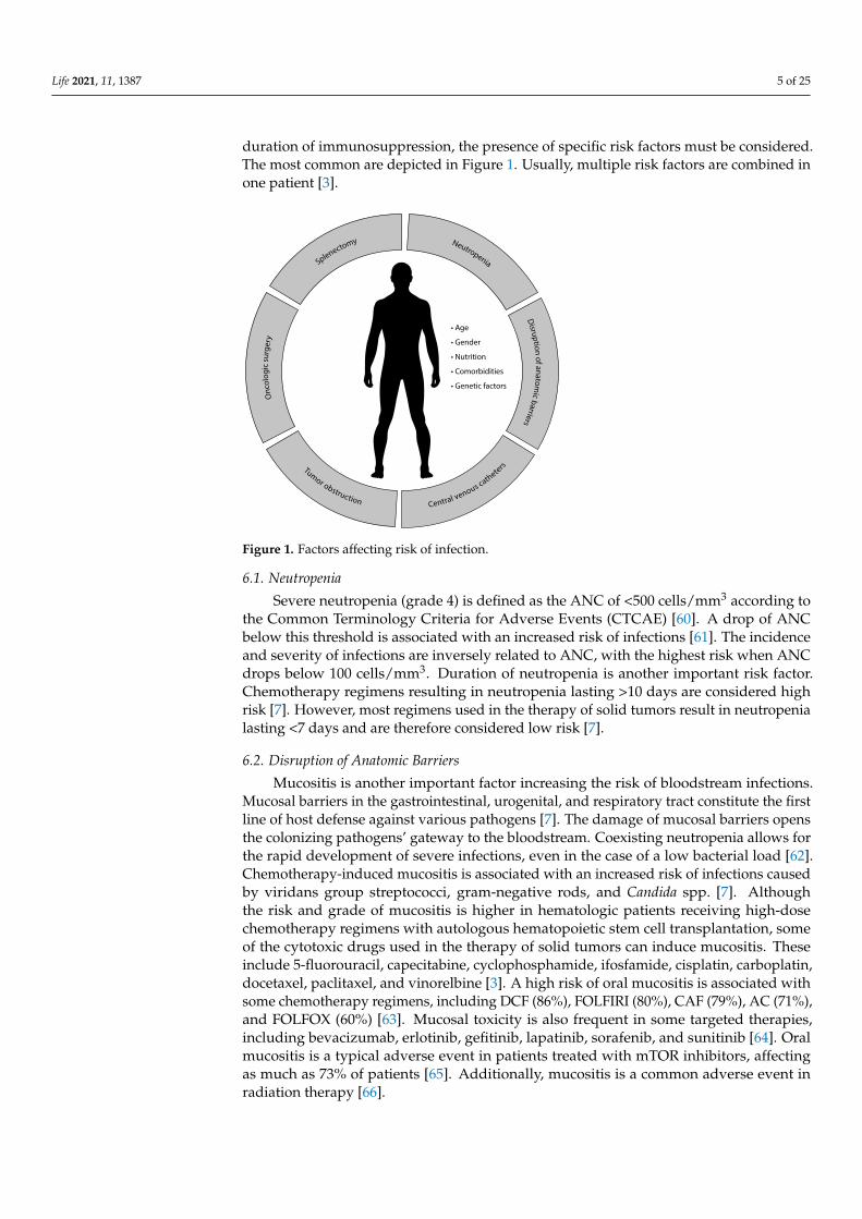
duration of immunosuppression, the presence of specific risk factors must be considered.The most common are depicted in Figure 1. Usually, multiple risk factors are combined inone patient [3].
Life 2021, 11, x FOR PEER REVIEW 5 of 26
6. Risk Factors for Infections in Patients with Solid Tumors
Patients with solid tumors are a heterogeneous group regarding the risk of infection
complications [1]. The National Comprehensive Cancer Network (NCCN) guidelines
determine three risk categories based on the overall risk of developing an infection (low-,
intermediate-, and high-risk categories) [7]. Most patients with solid tumors belong to the
low-risk (standard chemotherapy regimens with anticipated neutropenia < 7 days) and
intermediate-risk categories (anticipated neutropenia 7–10 days) [7]. Besides the depth
and duration of immunosuppression, the presence of specific risk factors must be con-
sidered. The most common are depicted in Figure 1. Usually, multiple risk factors are
combined in one patient [3].
Figure 1. Factors affecting risk of infection.
6.1. Neutropenia
Severe neutropenia (grade 4) is defined as the ANC of < 500 cells/mm3 according to
the Common Terminology Criteria for Adverse Events (CTCAE) [60]. A drop of ANC
below this threshold is associated with an increased risk of infections [61]. The incidence
and severity of infections are inversely related to ANC, with the highest risk when ANC
drops below 100 cells/mm3. Duration of neutropenia is another important risk factor.
Chemotherapy regimens resulting in neutropenia lasting > 10 days are considered high
risk [7]. However, most regimens used in the therapy of solid tumors result in neutro-
penia lasting < 7 days and are therefore considered low risk [7].
6.2. Disruption of Anatomic Barriers
Mucositis is another important factor increasing the risk of bloodstream infections.
Mucosal barriers in the gastrointestinal, urogenital, and respiratory tract constitute the
first line of host defense against various pathogens [7]. The damage of mucosal barriers
opens the colonizing pathogens’ gateway to the bloodstream. Coexisting neutropenia
allows for the rapid development of severe infections, even in the case of a low bacterial
load [62]. Chemotherapy-induced mucositis is associated with an increased risk of infec-
tions caused by viridans group streptococci, gram-negative rods, and Candida spp. [7].
Although the risk and grade of mucositis is higher in hematologic patients receiving
high-dose chemotherapy regimens with autologous hematopoietic stem cell transplanta-
tion, some of the cytotoxic drugs used in the therapy of solid tumors can induce mucosi-
tis. These include 5-fluorouracil, capecitabine, cyclophosphamide, ifosfamide, cisplatin,
carboplatin, docetaxel, paclitaxel, and vinorelbine [3]. A high risk of oral mucositis is
Figure 1. Factors affecting risk of infection.
6.1. Neutropenia
Severe neutropenia (grade 4) is defined as the ANC of <500 cells/mm3 according tothe Common Terminology Criteria for Adverse Events (CTCAE) [60]. A drop of ANCbelow this threshold is associated with an increased risk of infections [61]. The incidenceand severity of infections are inversely related to ANC, with the highest risk when ANCdrops below 100 cells/mm3. Duration of neutropenia is another important risk factor.Chemotherapy regimens resulting in neutropenia lasting >10 days are considered highrisk [7]. However, most regimens used in the therapy of solid tumors result in neutropenialasting <7 days and are therefore considered low risk [7].
6.2. Disruption of Anatomic Barriers
Mucositis is another important factor increasing the risk of bloodstream infections.Mucosal barriers in the gastrointestinal, urogenital, and respiratory tract constitute the firstline of host defense against various pathogens [7]. The damage of mucosal barriers opensthe colonizing pathogens’ gateway to the bloodstream. Coexisting neutropenia allows forthe rapid development of severe infections, even in the case of a low bacterial load [62].Chemotherapy-induced mucositis is associated with an increased risk of infections causedby viridans group streptococci, gram-negative rods, and Candida spp. [7]. Althoughthe risk and grade of mucositis is higher in hematologic patients receiving high-dosechemotherapy regimens with autologous hematopoietic stem cell transplantation, someof the cytotoxic drugs used in the therapy of solid tumors can induce mucositis. Theseinclude 5-fluorouracil, capecitabine, cyclophosphamide, ifosfamide, cisplatin, carboplatin,docetaxel, paclitaxel, and vinorelbine [3]. A high risk of oral mucositis is associated withsome chemotherapy regimens, including DCF (86%), FOLFIRI (80%), CAF (79%), AC (71%),and FOLFOX (60%) [63]. Mucosal toxicity is also frequent in some targeted therapies,including bevacizumab, erlotinib, gefitinib, lapatinib, sorafenib, and sunitinib [64]. Oralmucositis is a typical adverse event in patients treated with mTOR inhibitors, affectingas much as 73% of patients [65]. Additionally, mucositis is a common adverse event inradiation therapy [66].

Life 2021, 11, 1387 6 of 25
6.3. Central Venous Catheters
Central venous catheters (CVCs) are widely used in cancer patients and offer benefitsto those who receive chemotherapy. However, the presence of CVCs is considered a riskfactor for infections in cancer patients and may affect the etiology of bacteremia [62]. Theincidence of CVC-associated bloodstream infections in cancer patients is estimated to be0.5–10 per 1000 CVC days, with mortality ranging from 12% to 40% [67].
There are two main types of indwelling CVCs used for chemotherapy administration—centrally inserted totally implanted vascular access ports (PORTs) and peripherally insertedcentral catheters (PICCs) [68]. PICCs are an alternative to traditional PORTs, and their usehas increased owing to their lower cost and ease of insertion and removal [68]. Severalstudies have compared these CVCs in terms of infection complications, with conflictingresults [68–70]. A recent meta-analysis found a significantly higher risk of infectiouscomplications in the PICC groups (RR 3.43; 95% CI 2.58–4.56; P < 0.05) [69]. Both the localinfections of punctures and catheter-related infections were more frequent in patients withPICCs [69]. In addition, infection complications are more frequent in multi-lumen CVCsthan in single-lumen CVCs [71].
Most CVC infections originate from the skin flora (65%), catheter or catheter joints(30%), or other pathways (5%) [72]. The most commonly detected pathogens causing CVC-related infections in cancer patients are coagulase-negative staphylococci, followed by othergram-positive bacteria, including Staphylococcus aureus, enterococci, and streptococci [67,73].As in other bloodstream infections in cancer patients, a shift toward gram-negative flora(including Escherichia coli, Pseudomonas aeruginosa, and Klebsiella spp.) has been noted inCVC-related infections [74,75]. Therefore, a broad-spectrum empirical therapy coveringgram-negative pathogens should be considered in CVC-related bloodstream infections [74].
6.4. Tumor Obstruction
Direct expansion of the tumor may cause obstruction of tubular organs [3]. Bronchialobstruction caused by local growth of bronchogenic carcinomas and metastatic tumorsmay cause post-obstructive pneumonia and occasionally be the first manifestation of thedisease [76]. The post-obstructive component can be found in ~45–55% of patients withpulmonary neoplasms who develop pneumonia [77]. Bronchial obstruction develops morecommonly in tumors arising centrally, such as small cell lung cancer (SCLC) and squamouscell carcinoma (SCC) [78]. Pathogens causing post-obstructive pneumonia include gram-positive organisms (St. aureus [incl. MRSA], viridans group streptococci, beta-hemolyticstreptococci [groups A, B, C, F, and G]), gram-negative organisms (E. coli, Klebsiella spp.,other Enterobacteriaceae, Ps. aeruginosa, Stenotrophomonas maltophilia, Acinetobacter spp.),anaerobes (Peptococcus spp., Peptostreptococcus spp., Fusobacterium nucleatum, Bacteroidesspp.), and fungi (Candida spp.) [79]. Despite therapy with broad-spectrum antibiotics,responses tend to be low, and persistent or recurrent infections are common. Moreover,~10–15% of patients develop severe complications, including lung abscess, empyema,hemorrhage, and fistula formation [77].
Obstructive uropathy is a frequent complication of advanced solid tumors, especiallyprostate, retroperitoneal, and pelvic tumors [80]. It is usually managed by placing a ureteralstent or permanent nephrostomy tube (PNT) [81]. However, the presence of PNT in cancerpatients is associated with a high risk of PNT-associated pyelonephritis (with a rate of19%) [82]. Patients with a history of previous urinary tract infection (UTI) and neutropeniaare at higher risk [82]. Many pathogens are capable of forming biofilms on indwellingimplants, including E. coli, Enterococcus faecalis, Ps. aeruginosa, Proteus mirabilis, St. aureus,and Candida spp. [82]. Eradication of these microorganisms is challenging, and long-termsuppressive antibiotic therapy may be required in patients with recurrent urosepsis [3].Nevertheless, prophylactic antibiotic therapy does not seem to prevent the developmentof pyelonephritis and asymptomatic bacteriuria in cancer patients with PNT [82]. Inaddition to complicated UTIs, patients with prostatic carcinoma may develop prostatitisand prostatic abscesses [3].

Life 2021, 11, 1387 7 of 25
Malignant biliary obstruction (MBO) is caused by pancreatic adenocarcinoma, cholan-giocarcinoma, ampullary/duodenal adenocarcinoma, gallbladder carcinoma, lymphoma,and compressive metastatic proximal lymph nodes [83]. Pancreatic adenocarcinoma isthe most common cause of MBO, and as many as 70–90% of pancreatic cancer patientsdevelop jaundice during the course of their disease [84]. MBO frequently results in as-cending cholangitis, which may be the initial manifestation of underlying malignancy [3].In cases of persistent obstruction, hepatic abscesses may develop [85]. The etiology isusually polymicrobial, with enteric gram-negative rods, Enterococcus spp., and anaerobesbeing most common [86]. Management of MBO includes percutaneous transhepatic biliarydrainage [87] and endoscopic retrograde cholangiopancreatography (ERCP) with the place-ment of plastic or self-expandable metallic stents (SEMS), which is considered the currentmainstay of treatment [84,88]. In patients with resectable disease, up-front surgery withouta stent placement is an option and does not increase the risk of complications compared topreoperative biliary drainage [84].
Additionally, large tumors can overgrow the capacity of their blood supply andbecome necrotic, forming the seeds of infection. Direct invasion of colorectal cancer throughthe mucosa may lead to abscess formation and sepsis by enteric bacteria [7].
6.5. Oncologic Surgery
Patients who undergo extensive tumor resection are at increased risk for postoperativenosocomial infection, especially in tumors involving the respiratory and gastrointestinaltract [1]. In a large retrospective study, the rate of serious postoperative infections was9.4% and led to a nearly 12-fold increase in the odds of in-hospital mortality [89]. Surgicalprocedures associated with the highest risk of serious infections were esophagectomy (25%),gastrectomy (19%), pancreas resection (17%), and lung resection (10%). The incidence ofinfections is significantly lower in high-volume centers than in low-volume hospitals [89].Besides the type of surgery, the risk of infection may be related to the tumor burden,preoperative performance status, and previous therapy [7]. However, it seems that surgicaland intensive care unit-related factors play a more significant role than previous oncologictherapy [62].
6.6. Splenectomy and Function Asplenia
The number of indications for splenectomy in cancer patients has declined over theyears. In the case of solid tumors, splenectomy is traditionally performed in gastric cancer todissect the splenic hilar lymph nodes, although it seems to have no benefit in tumors locatedat lesser curvature [90]. Splenectomy may also be performed in cases of oligometastaticdisease from other sites, especially ovarian cancer [91]. Additionally, intraoperative splenicinjury during abdominal surgery may result in splenectomy [92]. Besides surgical splenec-tomy resulting in asplenia, radiotherapy and some pathologic conditions (including graftversus host disease following allogeneic hematopoietic stem cell transplantation) lead to adecreased function of the spleen—hyposplenism (function asplenia) [7].
The spleen is a lymphoid organ that plays an important role in regulating immunehomeostasis through both innate and adaptive immunity. The function of the spleen iscrucial in the elimination of encapsulated bacteria [93]. Asplenic patients are therefore atrisk of sepsis caused by encapsulated bacteria—most commonly Streptococcus pneumoniae(50–70%), but also Haemophilus influenzae and Neisseria meningitidis (15–25% each) [94].Other pathogens causing serious infections in asplenic patients include Capnocytophaga can-imorsus after animal bites, Bordetella holmesii, Ehrlichia spp., and intraerythrocytic parasitessuch as Babesia spp. after tick bites [94].
6.7. Patient-Related Factors6.7.1. Age
Age is an important factor regarding the risk for infections. Age-related changes inthe immune system, referred to as immunosenescence, play a major role in increased sus-
Life 2021, 11, 1387 8 of 25
ceptibility to infections in elderly patients [95]. Other risk factors for infections frequentlypresent in elderly patients include malnutrition, comorbidities, age-related organ changes,functional dysfunction (i.e., impairments in the performance of activities of daily living),polypharmacy, and social factors [96,97]. Furthermore, age is associated with an increasedrisk of FN [98], owing to reduced bone marrow reserves and reduced renal and hepaticfunctions [99].
6.7.2. Gender
Men are at increased risk for most infections. The explanation for this observationis rather complex and involves both biological and social factors [100]. Sexual steroidhormones play an important role in susceptibility to infections through differential modu-lation of pro-inflammatory and anti-inflammatory cytokine expression, toll-like receptorexpression, antibody production, metabolism, growth, and virulence of pathogenic bacte-ria [101]. Estrogens can enhance both cell-mediated and humoral immune responses, whileprogesterone and testosterone have anti-inflammatory effects and suppress innate immuneresponses [100–102]. Additionally, it seems that genetic factors related to sex chromosomes(X and Y) may play a part [103], and differences in occupational activities and lifestyleresult in different exposures to pathogens [100].
In contrast to the overall lower risk of infections in the female population, UTIs andgenital tract infections are more common in women owing to anatomic and physiologicaldifferences [104]. Available data suggest that female gender is a risk factor for the develop-ment of FN [98,105]. One possible explanation is that female patients are more frequentlytreated with breast cancer chemotherapy regimens, which confer a higher risk for FN (upto 23% in standard chemotherapy and 98% in high-dose chemotherapy regimens) [106].Gender-related differences in pharmacokinetics and pharmacodynamics of anticancerdrugs provide another rationale [107]. A recent study has found that polymorphisms ofgenes involved in drug metabolism are distributed unevenly in women and men, and thatthese polymorphisms have different impacts on adverse event occurrence (including FN)between genders [108].
6.7.3. Nutrition
Malnutrition affects as many as 75% of cancer patients, with the highest prevalence inthose with tumors of the gastrointestinal tract [109]. The association between malnutritionand infections is well-established and can be explained by impaired cell-mediated immu-nity, phagocyte function, cytokine production, and complement system function [110].Therefore, improvement of the patient’s nutritional status is of great importance to reducethe risk of infections and improve survival [111].
Obesity is associated with an increased risk of infections when compared to normal-weight subjects [112,113]. The highest increase was observed for skin infections in bothgenders and for gastrointestinal tract infections, UTIs, and sepsis in obese women [113].The underlying mechanisms involve altered adipokine signaling (e.g., leptin, adiponectin),immune system dysregulation, impaired chemotaxis, and metabolic changes [112,114].
6.7.4. Comorbidities
Many internal diseases are associated with an increased risk of infection. Patientswith type 1 and 2 diabetes are at increased risk of all infections, particularly bone andjoint infections, sepsis, and cellulitis [115]. Chronic kidney disease is associated withan increased risk of infections, especially in patients undergoing hemodialysis [116,117].Patients with chronic obstructive pulmonary disease (COPD) are more likely to developrespiratory infections, but the incidence of infections outside the respiratory tract doesnot seem to be affected [118]. An increased risk of infections is observed in patients withrheumatological disorders owing to both altered function of the immune system andimmunosuppressive therapy [119]. Opportunistic infections, including P. jirovecii, are

Life 2021, 11, 1387 9 of 25
particularly common [120]. Other comorbidities with increased risk of infection includechronic heart failure [121] and cirrhosis [122].
6.7.5. Genetic Factors
Defects in innate and adaptive immunity are naturally associated with an increasedrisk of infections [123]. Single nucleotide polymorphisms (SNPs) in genes involved incytotoxic drug metabolism are associated with an increased risk of FN [98]. In breastcancer patients treated with the FEC (5-FU + epirubicin + cyclophosphamide) regimen,MDM2 SNP309 and TP53 R72P genotypes [124], as well as SNPs of the ABCC1/MRP1,UGT2B7, and FGFR4 genes [125], were significantly associated with an increased riskof FN. UGT1A1 gene polymorphism is associated with increased risk of FN in patientstreated with irinotecan [126–128]. Similarly, DPYD gene polymorphism resulting in di-hydropyrimidine dehydrogenase (DPD) deficiency is associated with significant toxicity,including FN, in patients treated with fluoropyrimidines [127]. Prospective validation ofthese polymorphisms as predictive factors for FN could identify patients at high risk ofinfectious complications and suggest treatment deescalation or application of prophylacticmeasures in affected individuals [127].
7. Antibiotic Therapy in Patients with Solid Tumors
Early detection of infections is essential in cancer patients. Clinical signs of infectionmight be vague, especially in neutropenic patients, but fever remains an early, althoughnon-specific, sign of infection [129]. Approximately 50–60% of patients who became febrilehave an underlying infection [130]. However, non-infectious causes of fever, includingparaneoplastic etiology (neoplastic fever) and drug reactions, are not rare in cancer pa-tients [131] Moreover, laboratory markers of infection, including elevated C-reactive proteinand leukocytosis, are frequently present in cancer patients [132,133], making the diagnosisof infection challenging. In such circumstances, additional markers such as procalcitonincan be used [134].
The symptoms-oriented diagnostic process should be initiated as soon as possiblewith identification of the most likely infection source. The empiric antibiotic should beinitiated immediately after obtaining cultivation samples. The antibiotic choice should betailored according to the suspected infection site, microbial colonization of the patient (i.e.,known presence of multiresistant strains in previous cultivation samples), and the localepidemiological situation [10]. The most likely tumor-specific infections should be ruledout first, including cholangitis in pancreatic cancer, obstructive pneumonia in lung tumors,urinary infection in prostate cancer patients, etc. [135].
The use of antibiotics with broad-spectrum coverage, including anti-pseudomonalactivity, is recommended in patients presenting with severe neutropenia [7]. Deescalationof the antibiotic therapy should be performed as soon as the results of cultivation samplesare available, and the duration of antibiotic therapy should be tailored to the type of infec-tion, level of immunosuppression, and other risk factors (type of malignancy, anticancertherapy, patient’s comorbidities, etc.). It should be kept in mind that prolonged antibiotictherapy can lead to severe complications, including vulvovaginal candidosis in women,Clostridioides difficile infection (CDI), and possible detrimental effects on anticancer therapyeffectiveness due to dysmicrobia [136].
The rise in multiresistant strains of bacteria (including extended-spectrum β-lactamase[ESBL]-producing Enterobacteriaceae, methicillin-resistant St. aureus [MRSA], and vancomycin-resistant Enterococcus faecium) has become a significant problem, especially in oncologicpatients [1]. Antimicrobial stewardship and strict adherence to infection control recommen-dations are essential to reduce the risk of emergence and spread of multiresistant bacteriastrains [3]. However, approximately one third of antibiotics prescribed in the United Statesacute care hospitals are either unnecessary or suboptimal [137,138]. In up to 50% of cases, thetreatment indication, choice of agent, or duration of antibiotic therapy may even be incor-rect [139]. This is of upmost importance considering that 30-day mortality can reach 70% in

Life 2021, 11, 1387 10 of 25
patients with bloodstream infections not receiving appropriate antimicrobial treatment [140].It is, therefore, the policy of many hospitals to restrict the use of broad-spectrum antibioticsunless approved by the hospital’s antibiotic center. This approach can reduce treatmentexpanses and limit the emergence of multiresistant strains and CDI [141,142].
The advent of modern treatment modalities, including targeted therapy and im-munotherapy, has brought new challenges for the management of infections in cancerpatients. Targeting checkpoints of immune response with ICIs has become a new treatmentstrategy in many solid tumors, including melanoma, renal cell carcinoma, lung cancer, andothers. Despite great improvements in patients’ survival, a new class of adverse events,known as immune-related adverse events (irAE), has emerged [143]. This form of autoim-mune reaction can affect various organs and be potentially life-threatening. Importantly, thediscrimination between irAE and infectious complications may be challenging. This is ofhigh clinical significance, considering that corticosteroids and other immunosuppressantsused in irAE management may aggravate the course of infectious diseases and increase therisk of opportunistic infections [144]. Improper use of antibiotics in patients treated withICIs may weaken the treatment outcome via antibiotic-induced dysbiosis [145]. Therefore,antibiotic therapy should be prescribed cautiously in patients treated with ICIs, and thediagnosis of infection should be confirmed with appropriate tests. Narrow-spectrum antibi-otics are the preferred option in this setting, and consultation with the hospital’s antibioticcenter before treatment initiation is highly encouraged [145].
8. Antibiotic Therapy of Specific Infections8.1. Febrile Neutropenia
FN represents a severe and potentially life-threatening complication of cancer therapy,with an overall in-hospital mortality of ~10% [10]. The definition and risk factors for FN inpatients with solid tumors have already been addressed. The management of FN followsinternational guidelines with slight variations reflecting national differences (i.e., localepidemiological situation and prevalence of multiresistant strains) [10,146].
The first step is to stratify patients into risk categories based on clinical characteristics(nature of the underlying malignancy, comorbidities, performance status, presence ofhypotension, dehydration, and stress-induced hyperglycemia at presentation, outpatientstatus, and age). Evaluation of these characteristics allows us to stratify patients into low-and high-risk groups using the Multinational Association for Supportive Care in Cancer(MASCC) Risk Index Score [10] or Clinical Index of Stable Febrile Neutropenia (CISNE)score [147], the latter having been specifically validated for patients with solid tumors. FNpatients within the high-risk group or having high-risk features as assessed by the admittingphysician should be admitted to hospital and administered broad-spectrum antibioticsintravenously within one hour of admission (Table 3) [7,10]. Local epidemiological bacterialisolate and resistance patterns are crucial to determining the optimal empirical antibiotictherapy [10]. Inadequate antibiotic regimen use is associated with a significantly higherICU admission and death rate during hospital stay [148]. Culture specimens, includingtwo sets of blood cultures (from a peripheral vein and any indwelling venous catheter),urine specimens, and specimens from any suspected site of infection, should be obtainedbefore antibiotic therapy initiation [7,10].
Patients at low risk of developing serious complications (<10%) may receive oralantibiotics in an outpatient setting after being provided careful patient education [146].Outpatients should be instructed on how to monitor their symptoms and when and howto contact appropriate medical services. Compliance of the patient and ability to reach thehealthcare facility are prerequisites for this approach [149].

Life 2021, 11, 1387 11 of 25
Table 3. Appropriate initial antibiotics in febrile neutropenia groups with different risk of serious infection development.
Risk of Serious Complications Low High
Initial antibioticOral or parenteral Parenteral
Inpatient or outpatient InpatientAmoxicillin-clavulanate +
fluoroquinolone (ciprofloxacin orlevofloxacin)
Antipseudomonal beta-lactam *(cefepime or meropenem or imipenem or
piperacillin-tazobactam)
Suspicion of catheter-related infection,severe skin and soft tissue infection,
pneumonia, or risk of MRSA infectionShift to high-risk group
Add gram-positive bacteria targetedantibiotic (vancomycin or linezolid or
daptomycin †), in case of VRE addlinezolid or daptomycin †
Suspicion of abdominal infection Shift to high-risk group Add metronidazole
Risk of multiresistant strain infection Shift to high-risk groupChoose carbapenem (in case of ESBL),add polymyxin-colistin or tigecycline
(in cases of KPC)
ESBL–extended-spectrum beta-lactamase producing strains, KPC–carbapenemase producing strains, MRSA–methicillin-resistant St. au-reus, VRE–vancomycin-resistant enterococci, * choice depends on the local epidemiological situation, † not in cases of pneumonia.References: [10,146,150,151].
8.2. Central Venous Catheter-Related Infections
CVC-related infections represent ~10% of bloodstream infections in cancer patients [152,153]. In case of suspected CVC-related infection, blood culture from the CVC and peripheralvein must be performed in order to determine differential time to positivity. A differencein time to positivity of >2 h (blood culture from CVC must be the first positive) is a highlysensitive and specific CVC-related infection indicator [154].
In particular situations, it might not be necessary to remove the infected CVC, andantimicrobial therapy alone is sufficient. The main premise for such an approach is astable patient and the assumption of successful antimicrobial therapy without developingcomplications (Table 4) [155,156]. There is no consensus regarding the length of antimi-crobial therapy. However, a recent meta-analysis suggests that a short-course therapy(seven days) for gram-negative bacteria, seven days for enterococci, and three days forcoagulase-negative staphylococci could be sufficient in uncomplicated CVC-related infec-tion [157]. However, the authors conclude that shorter courses may not be appropriate forimmunocompromised patients, and prospective studies are warranted [157]. In case ofcomplications, including tunnel infection, port abscess, septic thrombosis, endocarditis, andosteomyelitis, the catheter must always be removed, and appropriate pathogen-directedantimicrobial therapy should be used [155].
Another therapeutic option for stable patients with catheter-related infections causedby low-virulence pathogens—coagulase-negative staphylococci (except from Staphylococcuslugdunensis), Corynebacterium spp., and some gram-negative rods—is the use of antimicro-bial lock therapy (ALT) either with or without delayed CVC removal [158]. This approachshould only be used in combination with systemic administration of antibiotics [155,158].The choice of antimicrobial agent depends on the type of isolated pathogen (Table 5). Thedwell time of the lock solution differs according to the stability of the substance solutionat the body temperature, but it should not exceed 48–72 h [158]. The type of CVC or portdetermines the volume of instilled antibiotic lock solution, which is ~2–5 mL. The mostfrequent antibiotics used for ALT are listed in Table 5. Appropriate duration of ALT isunknown, but generally 10–14 day therapy is recommended [155]. Importantly, the locksolution must be removed before catheter reuse.
Besides therapy for CVC-related infections, prophylactic ALT might decrease theincidence of central-line associated infections in cancer patients [159]. This approach seemspromising and has demonstrated its cost-effectiveness specifically in cancer therapy [160],although further studies are warranted to optimize prophylactic ALT use.

Life 2021, 11, 1387 12 of 25
Table 4. Appropriate antibiotic treatment of bacterial CVC-related infection according to isolated pathogens.
Isolated Pathogen Catheter RemovalAntibiotic Therapy
Choice Duration
Coagulase-negative staphylococci Not necessaryRisk factor for recurrence
VancomycinOxacillin *Flucloxacillin *Cefazoline *
Catheter removed: 5–7 daysRetained catheter: 10–14 days +ALT 10–14 days
St. aureus, St. lugdunensis Yes VancomycinOxacillin *Flucloxacillin *Cefazoline *
≥14 days. Necessary to rule outcomplications. Complications:4–6 weeks
Enterococci YesLong-term CVC may retain
VancomycinAmpicillin *
5–14 days. Retained long-termCVC: 7–14 days + ALT 7–14days
gram-negative bacilli Yes, especially in case ofmultiresistant bacteriaCVC retaining unsuitable forimmunosuppressed patients
Based on severity of disease:Piperacillin/tazobactam *,4th gen. Cephalosporin *,Carbapenem +/- Aminoglycoside *
7–14 days
* According to sensitivity pattern and local susceptibility data. CVC–central venous catheter, ALT–antimicrobial lock therapy.References: [155,156].
Table 5. List of the most frequently used antibiotic catheter lock solutions.
Antibiotic Spectrum of Bacteria Concentration *(mg/mL)
Heparin Content(IU/mL) Stability (Hours) References
Vancomycin gram-positive 2.0–5.0 2500 or 5000 72 [161,162]Teicoplanin gram-positive 5.0–10.0 0 or 100 96 [163,164]Daptomycin gram-positive 5.0 0 or 5000 72 [165]
Gentamicin gram-positive,gram-negative 1.0–5.0 0, 2500 or 5000 72 [166,167]
Amikacin gram-positive,gram-negative 1.0–40.0 † 0 or 5000 72 [168]
Ceftazidime gram-negative 0.5–10.0 0 or 5000 48 [167,169,170]
Cefazolin Methicillin-sensitivestaphylococci 5.0–10.0 2500 or 5000 72 [168]
Ciprofloxacin gram-negative 0.2–5.0 0 or 5000 48 [171,172]
Ampicillin Ampicillin-sensitiveenterococci 10.0 10 or 5000 8 ‡ [161]
Ethanol gram-positive,gram-negative 70% 0 24 [173]
* Concentration should exceed 100–1000×minimal inhibitory concentration (MIC). † Most commonly used 2.0 mg/mL, ‡ according toSPC [174]. For more detailed information on ACL see Justo et al. [168].
8.3. Pneumonia
Cancer patients are at increased risk of pneumonia due to impaired immune functioncaused by the tumor itself and cancer therapy [175], together with frequent tumor obstruc-tion causing post-obstructive pneumonia [79]. The therapy of community-acquired pneu-monia in cancer patients who are not neutropenic does not differ from that of the generalpopulation and should follow respective guidelines [7]. Beta-lactam antibiotics are the main-stay of therapy, but the choice depends on the local epidemiological situation (i.e., the locallevel of pneumococcal resistance). Antibiotics covering atypical pathogens (Mycoplasmapneumoniae, Chlamydophila pneumoniae, and Legionella pneumophila) are necessary for patientswith community-acquired pneumonia [10]. In patients requiring hospital admission, respi-ratory fluoroquinolone or a combination of macrolide with a third generation cephalosporin(ceftriaxone or cefotaxime) or ertapenem are the options [7]. Besides gram-positive andgram-negative activity (excluding Ps. aeruginosa and Acinetobacter spp.), ertapenem hasanaerobic activity useful for suspected aspiration and post-obstructive pneumonia [7,176].Severe community-acquired pneumonia and pneumonia in neutropenic patients should betreated with a combination of an antipseudomonal beta-lactam (piperacillin/tazobactam)and a respiratory fluoroquinolone or azithromycin [7,10].

Life 2021, 11, 1387 13 of 25
In patients who come from a medical facility or are long-term oncologically treatedand repeatedly hospitalized, etiological microorganisms of pneumonia have changed, andtherefore the empirical choice of antibiotic should be different, covering St. aureus, Ps. aerug-inosa, or other gram-negative rods: piperacillin/tazobactam, cefepime, or carbapenemsin combination with antibiotics covering gram-positive cocci (linezolid, vancomycin, orteicoplanin) [177]. In this regard, the choice of initial antibiotic regimen should be basedon knowledge of the local patterns of antibiotic susceptibility [7,177]. Clinical samples, in-cluding good-quality sputum, lower respiratory tract samples for culture, viral (influenza,COVID-19), mycoplasma, chlamydial PCR detection, and urine samples for detectionof pneumococcal and legionella antigens, should be obtained before antibiotic therapyinitiation in case of severe pneumonia in immunocompromised patients [178,179]. Micro-biological results can help to deescalate and target antibiotic treatment [177]. In patientsreceiving a prednisone equivalent of ≥20 mg for ≥4 months, or treated with RT and con-comitant temozolomide without reliable antipneumocystis prophylaxis, the addition ofhigh-dose trimethoprim/sulfamethoxazole should be considered [7,10].
8.4. Intra-Abdominal Infections
In addition to common intra-abdominal infections, cancer patients are at risk forinfections complicating the underlying disease (infiltration of intra-abdominal organs bytumor, compression of adjacent organs by the tumor and associated stagnation of secre-tions, tumor disintegration with subsequent rupture, or creation of intra-abdominal orpelvic abscess) [180]. Antibiotic choice depends on the patient’s condition, previous antibi-otic treatment, and colonization with multidrug-resistant bacteria. A high probability ofpolymicrobial pathogens and the presence of endogenous anaerobic flora has to be kept inmind [7]. Initial empirical treatment is based on administration of beta-lactam antibiotics(ampicillin/sulbactam, piperacillin/tazobactam, cefotaxime, ceftriaxone, carbapenem). Ifsecond-, third-, or fourth-generation cephalosporins are used, it is necessary to add metron-idazole to cover anaerobic bacteria [10,180]. Antibiotic therapy with antipseudomonalactivity is required in neutropenic patients [7]. Repeated cultivation of suitable materials(i.e., secretion from the drain, deep wound sample, and puncture fluid) may be required toavoid long-term administration of broad-spectrum antibiotics [180,181].
Cancer patients are susceptible to developing CDI as a result of the malignancy itself,immunosuppression, chemotherapy administration, antibiotic exposure, and frequenthospital stays [182]. This translates into a six- to nine-fold higher risk of developing CDIcompared to non-cancer patients [183]. Orally administered fidaxomicin or vancomycin arethe treatment of choice in initial and subsequent CDI episodes [184]. Orally administeredmetronidazole should be restricted to non-severe CDI cases when the above-mentionedagents are not available [184]. Fecal microbiota transplantation seems promising in themanagement of recurrent CDI, albeit the data in cancer patients are limited [7].
Neutropenic enterocolitis (referred to as typhlitis when located in the coecum) is notcommon in patients with solid tumors, but its association with taxanes (docetaxel, paclitaxel)and vinorelbine therapy has been reported [3]. Computed tomography is the preferreddiagnostic tool for revealing thickening of the bowel wall [185]. The therapy consists of generalsupportive measures (including bowel rest and parenteral nutrition) and broad-spectrumantibiotics with coverage for C. difficile, aerobic pathogens, and anaerobic pathogens [3,7].Complications develop in ~5% of patients requiring surgical intervention [186].
8.5. Urinary Tract Infections
UTIs are common in cancer patients due to cancer therapy, immunosuppression, andindwelling urinary catheters [187–189]. Importantly, the urinary tract is the source of~20% of bloodstream infections in cancer patients [152,153]. The most common pathogensinclude E. coli (40–58%), Kl. pneumoniae (10–25%), Ps. aeruginosa (4–11%), Enterococcus spp.(8–11%), Staphylococcus spp. (11%), and P. mirabilis (1–5%) [187,188]. Multidrug resistanceis frequent in cancer patients with UTIs (96%) [188]. A high proportion of resistance to

Life 2021, 11, 1387 14 of 25
fluroquinolones (90–96%), cephalosporins (68–80%), and aminoglycosides (46–50%) hasbeen observed [189,190].
Uncomplicated UTIs (women without risk factors) can be treated according to generalguidelines with orally administered fosfomycin, pivmecillinam, or nitrofurantoin [191]. Treat-ment with antimicrobials penetrating the prostate tissue (trimethoprim/sulfamethoxazoleor fluoroquinolone) is required in male patients, provided cystitis can be associated withprostatitis [191]. However, most UTIs in oncological patients are associated with other risk fac-tors, including anatomical or functional abnormalities of the urinary tract, indwelling urinarycatheters, stents, renal disease, cancer-related immunosuppression, oncologic therapy, andsurgical intervention [3,80]. Antimicrobial therapy should be guided by cultivation results,as repeated antibiotic therapy in cancer patients may lead to the selection of non-predictablebacterial variants and multiresistant strains [192]. Antimicrobial agents with extended spec-trum of activity (piperacillin/tazobactam, imipenem, meropenem, ceftazidime/avibactam,ceftolozane/tazobactam) should be used for empirical treatment of severe infections inaccordance with previous individual culture or local resistance data [191]. A switch to narrow-spectrum antibiotics should be performed as soon as the results of antibiotic sensitivity testsare available to avoid unnecessary adverse effects and ecological consequences [191,192].Treatment for 7–14 days is generally recommended, but the duration should be guided by thetherapy of the underlying abnormality [191,192].
9. The Impact of Antibiotic Therapy on Cancer Therapy Outcomes
Despite the undeniable contribution of antibiotic therapy to the management ofinfections in cancer patients, a potential detrimental effect on treatment outcome andtoxicity has to be considered [193,194]. Several contributing factors have been proposedto decrease the efficacy of cancer therapy, the most prominent being alteration of themicrobiome and its interaction with the patient’s immune system, potentially resulting inreduced immune surveillance [194,195].
9.1. Impact on Cancer Therapy Efficacy
There is a growing body of evidence that antibiotic therapy can negatively affectanticancer treatment efficacy and cancer-specific survival, especially in patients treatedwith immunotherapy [196]. It is now understood that the microbiome alters antitumorimmunity and influences the efficacy of cancer therapies mediated via systemic immuneresponse [194]. The effect of gut microbiome composition on immunotherapy outcome issupported by the finding that fecal transplants of gut microbiota from patients respondingto ICI therapy to germ-free mice results in an antitumor response [197–199]. The pres-ence of several species of microbiota have been associated with ICI efficacy, includingBacteroides spp. [200], Bifidobacterium spp. [198,201], Faecalibacterium spp. [202], Akkerman-sia muciniphila [199], Collinsella aerofaciens [198], E. faecium [198], and bacteria from theRuminococcaceae family [197]. Changing the balance and diversity of the patient’s micro-biome by antibiotic therapy may alter the antitumor immune response induced by ICIs,resulting in poor therapeutic outcomes [145]. Multiple clinical studies have reported anegative association between antibiotic use and response to ICIs in different solid tumors,including melanoma [203,204], NSCLC [203,205–208], and RCC [203,205,209]. Additionally,several meta-analyses have confirmed these results [210–212]. Interestingly, the negativeeffect of antibiotics on survival was also observed in adjuvant therapy with ICIs [204]. Themost detrimental effect on overall survival in the multivariate analysis was observed inpatients treated with penicillins, cephalosporins, and fluoroquinolones [204]. In light ofthese findings, the use of broad-spectrum antibiotics should be avoided in patients treatedwith immunotherapy whenever possible [195]. In addition to the antibiotic class, thelength of antibiotic therapy seems to play an important role, as the detrimental effect wasmost commonly observed in patients receiving multiple or prolonged cycles of antibiotictherapy [203,207].

Life 2021, 11, 1387 15 of 25
Besides affecting the efficacy of immunotherapy, the use of antibiotics can potentiatethe effect of radiotherapy. Vancomycin, a glycopeptide antibiotic active against gram-positive bacteria with minimal absorption from the gut when administered orally, showed apotentiating antitumor effect when combined with radiotherapy in a preclinical model [213].The effect was mediated through changes in gut microbiota composition, which led toincreased antigen presentation by CD11c+ dendritic cells in the tumor-draining lymphnodes of the radiotherapy-treated mice. Interestingly, the vancomycin effect was abrogatedby butyrate, a metabolite produced by vancomycin-depleted gut bacteria [213]. Recently,these results were confirmed by another study, suggesting that butyrate-producing bacteria,such as Lachnospiraceae and Ruminococcaceae, could be novel therapeutic targets [214].
Conversely, antibiotic therapy during curative chemoradiotherapy in patients withlocally advanced head and neck cancers was associated with a significant reduction ofprogression-free survival, overall survival, and disease-specific survival [215]. The potentialharm of broad-spectrum and prophylactic antibiotic therapy in these patients should,therefore, be considered [215].
9.2. Impact on Cancer Therapy Toxicity
In addition to possible pharmacokinetic interactions, the use of antibiotics can lead toincreased toxicity of anticancer drugs by modulation of microbiome [195]. In patients withmetastatic pancreatic adenocarcinoma treated with gemcitabine, antibiotic therapy wasassociated with increased chemotherapy-related toxicity [216]. The authors suggest thatintratumor bacteria may be responsible for a clinically meaningful portion of gemcitabinemetabolism [216]. In melanoma patients treated with immunotherapy, the use of antibioticswas associated with moderate to severe immune-mediated colitis [204]. The underlyingmechanism seems to involve changes in the gut microbiome, resulting in inhibition ofregulatory T cells [217] together with regrowth of pro-inflammation bacterial species [204].
10. Conclusions
Infections are one of the most common causes of death in patients with solid tumorsand can complicate cancer therapy. Thorough evaluation of possible risk factors couldidentify patients at high risk for infections and aid in the decision-making process regardingthe use of G-CSF and antibiotics. In this regard, prognostic scores considering multiplerisk factors together are being evaluated to calculate each individual patient’s risk. Earlyinitiation of antibiotic therapy is essential, especially in immunocompromised patients.However, other pathologic conditions mimicking infection, including irAE in patientstreated with ICIs, have to be excluded first. Consultation with the hospital’s antibioticcenter before antibiotic therapy initiation and strict adherence to the antibiotic stewardshipprogram is highly encouraged to reduce the occurrence of multidrug-resistant pathogensand decrease therapy costs. Potential risks of antibiotic therapy in cancer patients have tobe considered, including potential detrimental effects on treatment efficacy and toxicity.This holds especially true for patients treated with ICIs, for whom antibiotic therapy shouldbe restricted to unequivocally diagnosed infections, and broad-spectrum antibiotics shouldbe avoided.
Author Contributions: Writing—original draft preparation, O.K., P.P.; writing—review and editing,O.K., P.P., M.N.; visualization, O.K.; supervision, P.P. All authors have read and agreed to thepublished version of the manuscript.
Funding: This work was supported by the Charles University Faculty of Medicine in Hradec Královégrant Progress Q40/06.
Data Availability Statement: Not applicable.
Conflicts of Interest: The authors declare no conflict of interest.

Life 2021, 11, 1387 16 of 25
References1. Gudiol, C.; Aguado, J.M.; Carratalà, J. Bloodstream infections in patients with solid tumors. Virulence 2016, 7, 298–308. [CrossRef]2. Safdar, A.; Armstrong, D. Infectious morbidity in critically ill patients with cancer. Crit. Care Clin. 2001, 17, 531–570. [CrossRef]3. Rolston, K.V.I. Infections in Cancer Patients with Solid Tumors: A Review. Infect. Dis. Ther. 2017, 6, 69–83. [CrossRef]4. Williams, M.D.; Braun, L.A.; Cooper, L.M.; Johnston, J.; Weiss, R.V.; Qualy, R.L.; Linde-Zwirble, W. Hospitalized cancer patients
with severe sepsis: Analysis of incidence, mortality, and associated costs of care. Crit. Care 2004, 8, R291–R298. [CrossRef][PubMed]
5. Schelenz, S.; Nwaka, D.; Hunter, P.R. Longitudinal surveillance of bacteraemia in haematology and oncology patients at a UKcancer centre and the impact of ciprofloxacin use on antimicrobial resistance. J. Antimicrob. Chemother. 2013, 68, 1431–1438.[CrossRef]
6. Zheng, Y.; Chen, Y.; Yu, K.; Yang, Y.; Wang, X.; Yang, X.; Qian, J.; Liu, Z.-X.; Wu, B. Fatal Infections among Cancer Patients: APopulation-Based Study in the United States. Infect. Dis. Ther. 2021, 10, 871–895. [CrossRef]
7. National Comprehensive Cancer Network. NCCN Clinical Practice Guidelines in Oncology. Prevention and Treatment ofCancer-Related Infections 1. 2021. Available online: https://www.nccn.org/professionals/physician_gls/pdf/infections.pdf(accessed on 21 September 2021).
8. Marin, M.; Gudiol, C.; Ardanuy, C.; Garcia-Vidal, C.; Calvo, M.; Arnan, M.; Carratalà, J. Bloodstream infections in neutropenicpatients with cancer: Differences between patients with haematological malignancies and solid tumours. J. Infect. 2014, 69,417–423. [CrossRef]
9. Fillatre, P.; Decaux, O.; Jouneau, S.; Revest, M.; Gacouin, A.; Robert-Gangneux, F.; Fresnel, A.; Guiguen, C.; Le Tulzo, Y.; Jégo,P.; et al. Incidence of Pneumocystis jiroveci Pneumonia among Groups at Risk in HIV-negative Patients. Am. J. Med. 2014, 127,1242.e11–1242.e17. [CrossRef] [PubMed]
10. Klastersky, J.; de Naurois, J.; Rolston, K.; Rapoport, B.; Maschmeyer, G.; Aapro, M.; Herrstedt, J. Management of febrileneutropaenia: ESMO Clinical Practice Guidelines. Ann. Oncol. 2016, 27, v111–v118. [CrossRef] [PubMed]
11. Aapro, M.; Bohlius, J.; Cameron, D.; Lago, L.D.; Donnelly, J.P.; Kearney, N.; Lyman, G.; Pettengell, R.; Tjan-Heijnen, V.; Walewski,J.; et al. 2010 update of EORTC guidelines for the use of granulocyte-colony stimulating factor to reduce the incidence ofchemotherapy-induced febrile neutropenia in adult patients with lymphoproliferative disorders and solid tumours. Eur. J. Cancer2011, 47, 8–32. [CrossRef]
12. De Miguel, S.C.; Calleja-Hernández, M.; Menjón-Beltrán, S.; Vallejo-Rodríguez, I. Granulocyte colony-stimulating factors asprophylaxis against febrile neutropenia. Support. Care Cancer 2015, 23, 547–559. [CrossRef] [PubMed]
13. Truong, L.D.; Shen, S.S. Immunohistochemical diagnosis of renal neoplasms. Arch. Pathol. Lab. Med. 2011, 135, 92–109. [CrossRef][PubMed]
14. Perez, E.A.; Geeraerts, L.; Suman, V.J.; Adjei, A.A.; Baron, A.T.; Hatfield, A.K.; Maihle, N.; Michalak, J.C.; Kuross, S.A.; Kugler,J.W.; et al. A randomized phase II study of sequential docetaxel and doxorubicin/cyclophosphamide in patients with metastaticbreast cancer. Ann. Oncol. 2002, 13, 1225–1235. [CrossRef] [PubMed]
15. von Minckwitz, G.; Schneeweiss, A.; Loibl, S.; Salat, C.; Denkert, C.; Rezai, M.; Blohmer, J.U.; Jackisch, C.; Paepke, S.; Gerber, B.;et al. Neoadjuvant carboplatin in patients with triple-negative and HER2-positive early breast cancer (GeparSixto; GBG 66): Arandomised phase 2 trial. Lancet Oncol. 2014, 15, 747–756. [CrossRef]
16. Kosaka, Y.; Rai, Y.; Masuda, N.; Takano, T.; Saeki, T.; Nakamura, S.; Shimazaki, R.; Ito, Y.; Tokuda, Y.; Tamura, K. Phase IIIplacebo-controlled, double-blind, randomized trial of pegfilgrastim to reduce the risk of febrile neutropenia in breast cancerpatients receiving docetaxel/cyclophosphamide chemotherapy. Support. Care Cancer 2015, 23, 1137–1143. [CrossRef] [PubMed]
17. Gilbar, P.; McPherson, I.; Sorour, N.; Sanmugarajah, J. High incidence of febrile neutropenia following adjuvant breast chemother-apy with docetaxel, carboplatin and trastuzumab. Breast Cancer Manag. 2014, 3, 327–333. [CrossRef]
18. Marty, M.; Cognetti, F.; Maraninchi, D.; Snyder, R.; Mauriac, L.; Tubiana-Hulin, M.; Chan, S.; Grimes, D.; Antón, A.; Lluch, A.;et al. Randomized phase II trial of the efficacy and safety of trastuzumab combined with docetaxel in patients with humanepidermal growth factor receptor 2–Positive metastatic breast cancer administered as first-line treatment: The M77001 studygroup. J. Clin. Oncol. 2005, 23, 4265–4274. [CrossRef]
19. Sternberg, C.N.; De Mulder, P.H.; Schornagel, J.H.; Théodore, C.; Fossa, S.D.; Van Oosterom, A.T.; Witjes, F.; Spina, M.; VanGroeningen, C.J.; De Balincourt, C.; et al. Randomized phase III trial of high-dose-intensity methotrexate, vinblastine, doxorubicin,and cisplatin (MVAC) chemo-therapy and recombinant human granulocyte colony-stimulating factor versus classic MVAC inadvanced urothelial tract tumors: European Organization for Research and Treatment of Cancer Protocol no. 30924. J. Clin. Oncol.2001, 19, 2638–2646. [CrossRef]
20. Rose, P.G.; Blessing, J.A.; Gershenson, D.M.; McGehee, R. Paclitaxel and cisplatin as first-line therapy in recurrent or advancedsquamous cell carcinoma of the cervix: A gynecologic oncology group study. J. Clin. Oncol. 1999, 17, 2676–2680. [CrossRef]
21. Long, H.J., III; Bundy, B.N.; Grendys, E.C., Jr.; Benda, J.A.; McMeekin, D.S.; Sorosky, J.; Miller, D.; Eaton, L.A.; Fiorica, J.V.Randomized Phase III Trial of Cisplatin with or without Topotecan in Carcinoma of the Uterine Cervix: A Gynecologic OncologyGroup Study. J. Clin. Oncol. 2005, 23, 4626–4633. [CrossRef]
22. Van Cutsem, E.; Moiseyenko, V.; Tjulandin, S.; Majlis, A.; Constenla, M.; Boni, C.; Rodrigues, A.; Fodor, M.; Chao, Y.; Voznyi, E.;et al. Phase III study of docetaxel and cisplatin plus fluorouracil compared with cisplatin and fluorouracil as first-line therapy foradvanced gastric cancer: A report of the v325 study group. J. Clin. Oncol. 2006, 24, 4991–4997. [CrossRef]

Life 2021, 11, 1387 17 of 25
23. Roth, A.D.; Fazio, N.; Stupp, R.; Falk, S.; Bernhard, J.; Saletti, P.; Köberle, D.; Borner, M.M.; Rufibach, K.; Maibach, R.; et al.Docetaxel, cisplatin, and fluorouracil; Docetaxel and cisplatin; and epirubicin, cisplatin, and fluorouracil as systemic treatmentfor advanced gastric carcinoma: A randomized phase II trial of the swiss group for clinical cancer research. J. Clin. Oncol. 2007,25, 3217–3223. [CrossRef]
24. Cunningham, D.; Starling, N.; Rao, S.; Iveson, T.; Nicolson, M.; Coxon, F.; Middleton, G.; Daniel, F.; Oates, J.; Norman, A.R.Capecitabine and oxaliplatin for advanced esophagogastric cancer. N. Engl. J. Med. 2008, 358, 36–46. [CrossRef] [PubMed]
25. Fossa, S.D.; Kaye, S.B.; Mead, G.M.; Cullen, M.; De Wit, R.; Bodrogi, I.; Van Groeningen, C.J.; De Mulder, P.H.; Stenning,S.; Lallemand, E.; et al. Filgrastim during combination chemotherapy of patients with poor-prognosis metastatic germ cellmalignancy. J. Clin. Oncol. 1998, 16, 716–724. [CrossRef] [PubMed]
26. Motzer, R.J.; Sheinfeld, J.; Mazumdar, M.; Bajorin, D.F.; Bosl, G.J.; Herr, H.; Lyn, P.; Vlamis, V. Etoposide and cisplatin adjuvanttherapy for patients with pathologic stage II germ cell tumors. J. Clin. Oncol. 1995, 13, 2700–2704. [CrossRef] [PubMed]
27. Fujiwara, M.; Tanaka, H.; Yuasa, T.; Komai, Y.; Oguchi, T.; Fujiwara, R.; Numao, N.; Yamamoto, S.; Fujii, Y.; Fukui, I.; et al.First-Line combination chemotherapy with etoposide, ifosfamide and cisplatin for the treatment of disseminated germ cell cancer:Efficacy and feasibility in current clinical practice. Int. J. Urol. 2021, 28, 920–926. [CrossRef]
28. Miller, K.D.; Loehrer, P.J.; Gonin, R.; Einhorn, L.H. Salvage chemotherapy with vinblastine, ifosfamide, and cisplatin in recurrentseminoma. J. Clin. Oncol. 1997, 15, 1427–1431. [CrossRef]
29. Kondagunta, G.V.; Bacik, J.; Donadio, A.; Bajorin, D.; Marion, S.; Sheinfeld, J.; Bosl, G.J.; Motzer, R.J. Combination of paclitaxel,ifosfamide, and cisplatin is an effective second-line therapy for patients with relapsed testicular germ cell tumors. J. Clin. Oncol.2005, 23, 6549–6555. [CrossRef]
30. Pointreau, Y.; Garaud, P.; Chapet, S.; Sire, C.; Tuchais, C.; Tortochaux, J.; Faivre, S.; Guerrif, S.; Alfonsi, M.; Calais, G. Randomizedtrial of induction chemotherapy with cisplatin and 5-fluorouracil with or without docetaxel for larynx preservation. J. Natl. CancerInst. 2009, 101, 498–506. [CrossRef] [PubMed]
31. Schiller, J.H.; Harrington, D.; Belani, C.; Langer, C.; Sandler, A.; Krook, J.; Zhu, J.; Johnson, D.H. Comparison of four chemotherapyregimens for advanced non–small-cell lung cancer. N. Engl. J. Med. 2002, 346, 92–98. [CrossRef]
32. Pujol, J.-L.; Breton, J.-L.; Gervais, R.; Rebattu, P.; Depierre, A.; Morère, J.-F.; Milleron, B.; Debieuvre, D.; Castéra, D.; Souquet,P.-J.; et al. Gemcitabine–Docetaxel versus cisplatin–vinorelbine in advanced or metastatic non-small-cell lung cancer: A phase IIIstudy addressing the case for cisplatin. Ann. Oncol. 2005, 16, 602–610. [CrossRef] [PubMed]
33. Fossella, F.; Pereira, J.R.; Von Pawel, J.; Pluzanska, A.; Gorbounova, V.; Kaukel, E.; Mattson, K.V.; Ramlau, R.; Szczesna, A.; Fidias,P.; et al. Randomized, Multinational, Phase III Study of Docetaxel Plus Platinum Combinations Versus Vinorelbine Plus Cisplatinfor Advanced Non–Small-Cell Lung Cancer: The TAX 326 Study Group. J. Clin. Oncol. 2003, 21, 3016–3024. [CrossRef]
34. Font, A.; Moyano, A.J.; Puerto, J.M.; Tres, A.; Garcia-Giron, C.; Barneto, I.; Anton, A.; Sanchez, J.J.; Salvador, A.; Rosell, R.Increasing dose intensity of cisplatin-etoposide in advanced nonsmall cell lung carcinoma. A phase III randomized trial of thespanish lung cancer group. Cancer 1999, 85, 855–863. [CrossRef]
35. Cardenal, F.; López-Cabrerizo, M.P.; Antón, A.; Alberola, V.; Massuti, B.; Carrato, A.; Barneto, I.; Lomas, M.; García, M.; Lianes, P.;et al. Randomized phase III study of gemcitabine-cisplatin versus etoposide-cisplatin in the treatment of locally advanced ormetastatic non-small-cell lung cancer. J. Clin. Oncol. 1999, 17, 12. [CrossRef]
36. Millward, M.J.; Boyer, M.J.; Lehnert, M.; Clarke, S.; Rischin, D.; Goh, B.-C.; Wong, J.; McNeil, E.; Bishop, J.F. Docetaxel andcarboplatin is an active regimen in advancednon-small-cell lung cancer: A phase II study in Caucasian and Asian patients. Ann.Oncol. 2003, 14, 449–454. [CrossRef]
37. Swisher, E.M.; Mutch, D.G.; Rader, J.S.; Elbendary, A.; Herzog, T.J. Topotecan in platinum- and paclitaxel-resistant ovarian cancer.Gynecol. Oncol. 1997, 66, 480–486. [CrossRef] [PubMed]
38. Verschraegen, C.F.; Sittisomwong, T.; Kudelka, A.P.; Guedes, E.D.P.; Steger, M.; Nelson-Taylor, T.; Vincent, M.; Rogers, R.;Atkinson, E.N.; Kavanagh, J.J. Docetaxel for Patients With Paclitaxel-Resistant Müllerian Carcinoma. J. Clin. Oncol. 2000, 18,2733–2739. [CrossRef]
39. Omura, G.A.; Brady, M.F.; Look, K.Y.; Averette, H.E.; Delmore, J.E.; Long, H.J.; Wadler, S.; Spiegel, G.; Arbuck, S.G. Phase III trialof paclitaxel at two dose levels, the higher dose accompanied by filgrastim at two dose levels in platinum-pretreated epithelialovarian cancer: An intergroup study. J. Clin. Oncol. 2003, 21, 2843–2848. [CrossRef]
40. Hosein, P.J.; MacIntyre, J.; Kawamura, C.; Maldonado, J.C.; Ernani, V.; Loaiza-Bonilla, A.; Narayanan, G.; Ribeiro, A.; Portelance,L.; Merchan, J.R.; et al. A retrospective study of neoadjuvant FOLFIRINOX in unresectable or borderline-resectable locallyadvanced pancreatic adenocarcinoma. BMC Cancer 2012, 12, 199. [CrossRef]
41. Yilmaz, U.; Anar, C.; Polat, G.; Halilcolar, H. Carboplatin plus etoposide for extensive stage small-cell lung cancer: An experiencewith AUC 6 doses of carboplatin. Indian J. Cancer 2011, 48, 454–459. [CrossRef]
42. Von Pawel, J.; Schiller, J.H.; Shepherd, F.A.; Fields, S.Z.; Kleisbauer, J.; Chrysson, N.G.; Stewart, D.J.; Clark, P.I.; Palmer, M.C.; DePierre, A.; et al. Topotecan versus cyclophosphamide, doxorubicin, and vincristine for the treatment of recurrent small-cell lungcancer. J. Clin. Oncol. 1999, 17, 658–667. [CrossRef] [PubMed]
43. Lorigan, P.; Woll, P.; O’Brien, M.E.R.; Ashcroft, L.F.; Sampson, M.R.; Thatcher, N. Randomized phase III trial of dose-densechemotherapy supported by whole-blood hematopoietic progenitors in better-prognosis small-cell lung cancer. J. Natl. CancerInst. 2005, 97, 666–674. [CrossRef] [PubMed]

Life 2021, 11, 1387 18 of 25
44. White, S.C.; Lorigan, P.; Middleton, M.R.; Anderson, H.; Valle, J.; Summers, Y.; Burt, P.A.; Arance, A.; Stout, R.; Thatcher, N.Randomized phase II study of cyclophosphamide, doxorubicin, and vincristine compared with single-agent carboplatin inpatients with poor prognosis small cell lung carcinoma. Cancer 2001, 92, 601–608. [CrossRef]
45. Bui, B.N.; Chevallier, B.; Chevreau, C.; Krakowski, I.; Peny, A.M.; Thyss, A.; Maugard-Louboutin, C.; Cupissol, D.; Fargeot, P.;Bonichon, F. Efficacy of lenograstim on hematologic tolerance to MAID chemotherapy in patients with advanced soft tissuesarcoma and consequences on treatment dose-intensity. J. Clin. Oncol. 1995, 13, 2629–2636. [CrossRef]
46. Lorigan, P.; Verweij, J.; Papai, Z.; Rodenhuis, S.; Le Cesne, A.; Leahy, M.; Radford, J.; Van Glabbeke, M.M.; Kirkpatrick, A.;Hogendoorn, P.; et al. Phase III trial of two investigational schedules of ifosfamide compared with standard-dose doxorubicin inadvanced or metastatic soft tissue sarcoma: A european organisation for research and treatment of cancer soft tissue and bonesarcoma group study. J. Clin. Oncol. 2007, 25, 3144–3150. [CrossRef] [PubMed]
47. Thomson, A.W.; Turnquist, H.R.; Raimondi, G. Immunoregulatory functions of mTOR inhibition. Nat. Rev. Immunol. 2009, 9,324–337. [CrossRef]
48. Kaymakcalan, M.; Je, Y.; Sonpavde, G.; Galsky, M.; Nguyen, P.L.; Heng, D.Y.C.; Richards, C.J.; Choueiri, T.K. Risk of infections inrenal cell carcinoma (RCC) and non-RCC patients treated with mammalian target of rapamycin inhibitors. Br. J. Cancer 2013, 108,2478–2484. [CrossRef] [PubMed]
49. Alvarez, R.H.; Bechara, R.I.; Naughton, M.J.; Adachi, J.A.; Reuben, J.M. Emerging perspectives on mtor inhibitor-associatedpneumonitis in breast cancer. Oncologist 2018, 23, 660–669. [CrossRef]
50. Maschmeyer, G.; De Greef, J.; Mellinghoff, S.C.; Nosari, A.; Thiebaut-Bertrand, A.; Bergeron, A.; Franquet, T.; Blijlevens, N.M.A.;Maertens, J.A.; on behalf of the European Conference on Infections in Leukemia. Infections associated with immunotherapeuticand molecular targeted agents in hematology and oncology. A position paper by the European Conference on Infections inLeukemia (ECIL). Leukemia 2019, 33, 844–862. [CrossRef]
51. Finn, R.S.; Aleshin, A.; Slamon, D.J. Targeting the cyclin-dependent kinases (CDK) 4/6 in estrogen receptor-positive breastcancers. Breast Cancer Res. 2016, 18, 17. [CrossRef]
52. Hu, W.; Sung, T.; Jessen, B.A.; Thibault, S.; Finkelstein, M.B.; Khan, N.K.; Sacaan, A.I. Mechanistic Investigation of Bone MarrowSuppression Associated with Palbociclib and its Differentiation from Cytotoxic Chemotherapies. Clin. Cancer Res. 2016, 22,2000–2008. [CrossRef] [PubMed]
53. Gelbert, L.M.; Cai, S.; Lin, X.; Sanchez-Martinez, C.; Del Prado, M.; Lallena, M.J.; Torres, R.; Ajamie, R.T.; Wishart, G.N.; Flack,R.S.; et al. Preclinical characterization of the CDK4/6 inhibitor LY2835219: In-Vivo cell cycle-dependent/independent anti-tumoractivities alone/in combination with gemcitabine. Investig. N. Drugs 2014, 32, 825–837. [CrossRef]
54. Som, A.; Mandaliya, R.; Alsaadi, D.; Farshidpour, M.; Charabaty, A.; Malhotra, N.; Mattar, M.C. Immune checkpoint inhibitor-induced colitis: A comprehensive review. World J. Clin. Cases 2019, 7, 405–418. [CrossRef]
55. Hosmer, W.; Malin, J.; Wong, M. Development and validation of a prediction model for the risk of developing febrile neutropeniain the first cycle of chemotherapy among elderly patients with breast, lung, colorectal, and prostate cancer. Support. Care Cancer2011, 19, 333–341. [CrossRef] [PubMed]
56. Lyman, G.H.; Kuderer, N.M.; Crawford, J.; Wolff, D.A.; Culakova, E.; Poniewierski, M.S.; Dale, D.C. Predicting individual risk ofneutropenic complications in patients receiving cancer chemotherapy. Cancer 2011, 117, 1917–1927. [CrossRef]
57. Aagaard, T.; Roen, A.; Reekie, J.; Daugaard, G.; Brown, P.D.N.; Specht, L.; Sengeløv, H.; Mocroft, A.; Lundgren, J.; Helleberg, M.Development and validation of a risk score for febrile neutropenia after chemotherapy in patients with cancer: The FENCE score.JNCI Cancer Spectr. 2018, 2, pky053. [CrossRef]
58. Razzaghdoust, A.; Mofid, B.; Moghadam, M. Development of a simplified multivariable model to predict neutropenic complica-tions in cancer patients undergoing chemotherapy. Support. Care Cancer 2018, 26, 3691–3699. [CrossRef]
59. Aagaard, T.; Reekie, J.; Roen, A.; Daugaard, G.; Specht, L.; Sengeløv, H.; Mocroft, A.; Lundgren, J.; Helleberg, M. Developmentand validation of a cycle-specific risk score for febrile neutropenia during chemotherapy cycles 2–6 in patients with solid cancers:The CSR FENCE score. Int. J. Cancer 2020, 146, 321–328. [CrossRef] [PubMed]
60. NCI. Common Terminology Criteria for Adverse Events (CTCAE) Version 4.0. Available online: https://evs.nci.nih.gov/ftp1/CTCAE/CTCAE_4.03/CTCAE_4.03_2010-06-14_QuickReference_8.5x11.pdf (accessed on 22 May 2021).
61. Bodey, G.P.; Buckley, M.; Sathe, Y.S.; Freireich, E.J. Quantitative relationships between circulating leukocytes and infection inpatients with acute leukemia. Ann. Intern. Med. 1966, 64, 328–340. [CrossRef] [PubMed]
62. Castagnola, E.; Mikulska, M.; Viscoli, C. Prophylaxis and Empirical Therapy of Infection in Cancer Patients. In Mandell, Douglas,and Bennett’s Principles and Practice of Infectious Diseases, 8th ed.; Bennett, J.E., Dolin, R., Blaser, M.J., Eds.; Elsevier: Philadelphia,PA, USA, 2015; pp. 3395–3413.
63. Nishimura, N.; Yamada, S.; Ueda, K.; Mishima, Y.; Yokoyama, M.; Saotome, T.; Terui, Y.; Takahashi, S.; Hatake, K.; Nishimura, M.Incidence and severity of oral mucositis induced by conventional chemotherapy: A comprehensive prospective analysis of 227cancer patients. J. Clin. Oncol. 2010, 28, e19634. [CrossRef]
64. Elting, L.S.; Chang, Y.-C.; Parelkar, P.; Boers-Doets, C.B.; Michelet, M.; Hita, G.; Rouleau, T.; Cooksley, C.; Halm, J.; Vithala, M.;et al. Risk of oral and gastrointestinal mucosal injury among patients receiving selected targeted agents: A meta-analysis. Support.Care Cancer 2013, 21, 3243–3254. [CrossRef]
65. Kwitkowski, V.E.; Prowell, T.M.; Ibrahim, A.; Farrell, A.T.; Justice, R.; Mitchell, S.S.; Sridhara, R.; Pazdur, R. FDA ApprovalSummary: Temsirolimus as Treatment for Advanced Renal Cell Carcinoma. Oncologist 2010, 15, 428–435. [CrossRef] [PubMed]

Life 2021, 11, 1387 19 of 25
66. Peterson, D.E.; Boers-Doets, C.; Bensadoun, R.J.; Herrstedt, J. Management of oral and gastrointestinal mucosal injury: ESMOClinical Practice Guidelines for diagnosis, treatment, and follow-up. Ann. Oncol. 2015, 26, v139–v151. [CrossRef]
67. Böll, B.; Schalk, E.; Buchheidt, D.; Hasenkamp, J.; Kiehl, M.; Kiderlen, T.R.; Kochanek, M.; Koldehoff, M.; Kostrewa, P.; Claßen,A.Y.; et al. Central venous catheter–related infections in hematology and oncology: 2020 updated guidelines on diagnosis,management, and prevention by the Infectious Diseases Working Party (AGIHO) of the German Society of Hematology andMedical Oncology (DGHO). Ann. Hematol. 2021, 100, 239–259. [CrossRef]
68. Taxbro, K.; Hammarskjöld, F.; Thelin, B.; Lewin, F.; Hagman, H.; Hanberger, H.; Berg, S. Clinical impact of peripherally insertedcentral catheters vs implanted port catheters in patients with cancer: An open-label, randomised, two-centre trial. Br. J. Anaesth.2019, 122, 734–741. [CrossRef]
69. Pu, Y.-L.; Li, Z.-S.; Zhi, X.-X.; Shi, Y.-A.; Meng, A.-F.; Cheng, F.; Ali, A.; Li, C.; Fang, H.; Wang, C. Complications and costs ofperipherally inserted central venous catheters compared with implantable port catheters for cancer patients. Cancer Nurs. 2020,43, 455–467. [CrossRef]
70. Corti, F.; Brambilla, M.; Manglaviti, S.; Di Vico, L.; Pisanu, M.N.; Facchinetti, C.; Dotti, K.F.; Lanocita, R.; Marchianò, A.; DeBraud, F.; et al. Comparison of outcomes of central venous catheters in patients with solid and hematologic neoplasms: An Italianreal-world analysis. Tumori J. 2021, 107, 17–25. [CrossRef] [PubMed]
71. Dezfulian, C.; Lavelle, J.; Nallamothu, B.K.; Kaufman, S.R.; Saint, S. Rates of infection for single-lumen versus multilumen centralvenous catheters: A meta-analysis. Crit. Care Med. 2003, 31, 2385–2390. [CrossRef] [PubMed]
72. Bouza, E.; Burillo, A.; Muñoz, P. Catheter-related infections: Diagnosis and intravascular treatment. Clin. Microbiol. Infect. 2002, 8,265–274. [CrossRef]
73. Wisplinghoff, H.; Seifert, H.; Wenzel, R.P.; Edmond, M. Current trends in the epidemiology of nosocomial bloodstream infectionsin patients with hematological malignancies and solid neoplasms in hospitals in the united states. Clin. Infect. Dis. 2003, 36,1103–1110. [CrossRef] [PubMed]
74. Marcos, M.; Soriano, A.; Iñurrieta, A.; Martínez, J.A.; Romero, A.; Cobos, N.; Hernández, C.; Almela, M.; Marco, F.; Mensa,J. Changing epidemiology of central venous catheter-related bloodstream infections: Increasing prevalence of Gram-negativepathogens. J. Antimicrob. Chemother. 2011, 66, 2119–2125. [CrossRef]
75. Chaftari, A.M.; Hachem, R.; Jiang, Y.; Shah, P.; Hussain, A.; Al Hamal, Z.; Yousif, A.; Jordan, M.; Michael, M.; Raad, I. ChangingEpidemiology of Catheter-Related Bloodstream Infections in Cancer Patients. Infect. Control. Hosp. Epidemiol. 2018, 39, 727–729.[CrossRef]
76. Abers, M.S.; Sandvall, B.P.; Sampath, R.; Zuno, C.; Uy, N.; Yu, V.L.; Stager, C.E.; Musher, D.M. Postobstructive pneumonia: Anunderdescribed syndrome. Clin. Infect. Dis. 2016, 62, 957–961. [CrossRef]
77. Rolston, K.V. Postobstructive pneumonia in cancer patients. Clin. Infect. Dis. 2016, 63, 707–708. [CrossRef] [PubMed]78. Kalkat, M.S.; Bonser, R.S. Obstructive pneumonia: An indication for surgery in mega aorta syndrome. Ann. Thorac. Surg. 2003, 75,
1313–1315. [CrossRef]79. Rolston, K.V.I.; Nesher, L. Post-Obstructive pneumonia in patients with cancer: A review. Infect. Dis. Ther. 2018, 7, 29–38.
[CrossRef]80. Seo, S.K.; Liu, C.; Dadwal, S.S. Infectious disease complications in patients with cancer. Crit. Care Clin. 2021, 37, 69–84. [CrossRef]81. Battaglia, C.C.; Hale, K. Hospital-Acquired infections in critically III patients with cancer. J. Intensive Care Med. 2018, 34, 523–536.
[CrossRef] [PubMed]82. Bahu, R.; Chaftari, A.-M.; Hachem, R.Y.; Ahrar, K.; Shomali, W.; El Zakhem, A.; Jiang, Y.; AlShuaibi, M.; Raad, I.I. Nephrostomy
tube related pyelonephritis in patients with cancer: Epidemiology, infection rate and risk factors. J. Urol. 2013, 189, 130–135.[CrossRef] [PubMed]
83. Pu, L.Z.C.T.; Singh, R.; Loong, C.K.; de Moura, E.G.H. Malignant Biliary Obstruction: Evidence for Best Practice. Gastroenterol.Res. Pract. 2016, 2016, 3296801–3296807. [CrossRef] [PubMed]
84. Cassani, L.; Lee, J.H. Management of malignant distal biliary obstruction. Gastrointest. Interv. 2015, 4, 15–20. [CrossRef]85. Shi, S.; Xia, W.; Guo, H.; Kong, H.; Zheng, S. Unique characteristics of pyogenic liver abscesses of biliary origin. Surgery 2016, 159,
1316–1324. [CrossRef] [PubMed]86. Rolston, K.V.I.; Dholakia, N.; Rodriguez, S.; Rubenstein, E.B. Nature and outcome of febrile episodes in patients with pancreatic
and hepatobiliary cancer. Support. Care Cancer 1995, 3, 414–417. [CrossRef]87. Xu, C.; Lv, P.-H.; Huang, X.-E.; Wang, S.-X.; Sun, L.; Wang, F.-A. Analysis of different ways of drainage for obstructive jaundice
caused by hilar cholangiocarcinoma. Asian Pac. J. Cancer Prev. 2014, 15, 5617–5620. [CrossRef] [PubMed]88. Aljahdli, E.S. Management of distal malignant biliary obstruction. Saudi J. Gastroenterol. 2018, 24, 71–72. [CrossRef]89. Avritscher, E.B.C.; Cooksley, C.D.; Rolston, K.V.; Swint, J.M.; Delclos, G.L.; Franzini, L.; Swisher, S.G.; Walsh, G.L.; Mansfield, P.F.;
Elting, L.S. Serious postoperative infections following resection of common solid tumors: Outcomes, costs, and impact of hospitalsurgical volume. Support. Care Cancer 2014, 22, 527–535. [CrossRef]
90. Yang, K.; Zang, Z.-Y.; Niu, K.-F.; Sun, L.-F.; Zhang, W.-H.; Zhang, Y.-X.; Chen, X.-L.; Zhou, Z.-G.; Hu, J.-K. The survival benefitand safety of splenectomy for gastric cancer with total gastrectomy: Updated results. Front. Oncol. 2021, 10, 2786. [CrossRef]
91. Lee, S.S.; Morgenstern, L.; Phillips, E.H.; Hiatt, J.R.; Margulies, D.R. Splenectomy for splenic metastases: A changing clinicalspectrum. Am. Surg. 2000, 66, 837–840. [PubMed]

Life 2021, 11, 1387 20 of 25
92. Feola, A.; Niola, M.; Conti, A.; Delbon, P.; Graziano, V.; Paternoster, M.; Della Pietra, B. Iatrogenic splenic injury: Review of theliterature and medico-legal issues. Open Med. 2016, 11, 307–315. [CrossRef]
93. Di Sabatino, A.; Carsetti, R.; Corazza, G.R. Post-Splenectomy and hyposplenic states. Lancet 2011, 378, 86–97. [CrossRef]94. Buzelé, R.; Barbier, L.; Sauvanet, A.; Fantin, B. Medical complications following splenectomy. J. Visc. Surg. 2016, 153, 277–286.
[CrossRef]95. Pawelec, G. Immunosenescence: Impact in the young as well as the old? Mech. Ageing Dev. 1999, 108, 1–7. [CrossRef]96. Esme, M.; Topeli, A.; Yavuz, B.B.D.; Akova, M. Infections in the elderly Critically-III patients. Front. Med. 2019, 6, 118. [CrossRef]97. Tannou, T.; Koeberle, S.; Manckoundia, P.; Aubry, R. Multifactorial immunodeficiency in frail elderly patients: Contributing
factors and management. Med. Mal. Infect. 2019, 49, 167–172. [CrossRef]98. Lyman, G.H.; Abella, E.; Pettengell, R. Risk factors for febrile neutropenia among patients with cancer receiving chemotherapy: A
systematic review. Crit. Rev. Oncol. Hematol. 2014, 90, 190–199. [CrossRef] [PubMed]99. Balducci, L.; Hardy, C.L.; Lyman, G.H. Hemopoiesis and aging. Cancer Treat. Res. 2005, 124, 109–134. [CrossRef]100. Gay, L.; Melenotte, C.; Lakbar, I.; Mezouar, S.; Devaux, C.; Raoult, D.; Bendiane, M.-K.; Leone, M.; Mège, J.-L. Sexual dimorphism
and gender in infectious diseases. Front. Immunol. 2021, 12, 698121. [CrossRef] [PubMed]101. García-Gómez, E.; González-Pedrajo, B.; Camacho-Arroyo, I. Role of Sex Steroid Hormones in Bacterial-Host Interactions. BioMed
Res. Int. 2013, 2013, 928290. [CrossRef] [PubMed]102. Ahmed, S.A.; Karpuzoglu, E.; Khan, D. Effects of sex steroids on innate and adaptive immunity. In Sex Hormones and Immunity to
Infection; Klein, S.L., Roberts, C.W., Eds.; Springer: Berlin/Heidelberg, Germany, 2010; pp. 19–51.103. Fish, E.N. The X-files in immunity: Sex-Based differences predispose immune responses. Nat. Rev. Immunol. 2008, 8, 737–744.
[CrossRef] [PubMed]104. Harrington, R.D.; Hooton, T.M. Urinary tract infection risk factors and gender. J. Gend. Specif. Med. 2000, 3, 27–34.105. Abdel-Rahman, O. Impact of sex on chemotherapy toxicity and efficacy among patients with metastatic colorectal cancer: Pooled
analysis of 5 randomized trials. Clin. Color. Cancer 2019, 18, 110–115.e2. [CrossRef] [PubMed]106. Fontanella, C.; Bolzonello, S.; Lederer, B.; Aprile, G. Management of breast cancer patients with chemotherapy-induced neutrope-
nia or febrile neutropenia. Breast Care 2014, 9, 239–245. [CrossRef]107. Özdemir, B.C.; Csajka, C.; Dotto, G.-P.; Wagner, A.D. Sex differences in efficacy and toxicity of systemic treatments: An
undervalued issue in the era of precision oncology. J. Clin. Oncol. 2018, 36, 2680–2683. [CrossRef] [PubMed]108. Ruzzo, A.; Graziano, F.; Galli, F.; Galli, F.; Rulli, E.; Lonardi, S.; Ronzoni, M.; Massidda, B.; Zagonel, V.; Pella, N.; et al. Sex-Related
Differences in impact on safety of pharmacogenetic profile for colon cancer patients treated with FOLFOX-4 or XELOX adjuvantchemotherapy. Sci. Rep. 2019, 9, 11527. [CrossRef]
109. Bossi, P.; Delrio, P.; Mascheroni, A.; Zanetti, M. The spectrum of malnutrition/cachexia/sarcopenia in oncology according todifferent cancer types and settings: A narrative review. Nutrients 2021, 13, 1980. [CrossRef] [PubMed]
110. Chandra, R.K. Nutrition, immunity and infection: From basic knowledge of dietary manipulation of immune responses topractical application of ameliorating suffering and improving survival. Proc. Natl. Acad. Sci. USA 1996, 93, 14304–14307.[CrossRef]
111. Triarico, S.; Rinninella, E.; Cintoni, M.; Capozza, M.A.; Mastrangelo, S.; Mele, M.C.; Ruggiero, A. Impact of malnutrition onsurvival and infections among pediatric patients with cancer: A retrospective study. Eur. Rev. Med. Pharmacol. Sci. 2019, 23,1165–1175.
112. Falagas, M.E.; Kompoti, M. Obesity and infection. Lancet Infect. Dis. 2006, 6, 438–446. [CrossRef]113. Ghilotti, F.; Bellocco, R.; Ye, W.; Adami, H.-O.; Lagerros, Y.T. Obesity and risk of infections: Results from men and women in the
Swedish National March Cohort. Int. J. Epidemiol. 2019, 48, 1783–1794. [CrossRef]114. Huttunen, R.; Syrjänen, J. Obesity and the risk and outcome of infection. Int. J. Obes. 2013, 37, 333–340. [CrossRef]115. Carey, I.M.; Critchley, J.A.; DeWilde, S.; Harris, T.; Hosking, F.J.; Cook, D.G. Risk of infection in type 1 and type 2 diabetes
compared with the general population: A matched cohort study. Diabetes Care 2018, 41, 513–521. [CrossRef] [PubMed]116. Berman, S.J.; Johnson, E.W.; Nakatsu, C.; Alkan, M.; Chen, R.; LeDuc, J. Burden of infection in patients with end-stage renal
disease requiring long-term dialysis. Clin. Infect. Dis. 2004, 39, 1747–1753. [CrossRef] [PubMed]117. Cohen, G.; Hörl, W.H. Immune dysfunction in Uremia—An update. Toxins 2012, 4, 962–990. [CrossRef] [PubMed]118. Lange, P. Chronic obstructive pulmonary disease and risk of infection. Pneumonol. Alergol. Polska 2009, 77, 284–288.119. Fragoulis, G.E.; Sipsas, N.V. When rheumatology and infectious disease come together. Ther. Adv. Musculoskelet. Dis. 2019, 11, 1–3.
[CrossRef]120. Hsu, C.-Y.; Ko, C.-H.; Wang, J.-L.; Hsu, T.-C.; Lin, C.-Y. Comparing the burdens of opportunistic infections among patients with
systemic rheumatic diseases: A nationally representative cohort study. Arthritis Res. 2019, 21, 211. [CrossRef] [PubMed]121. McDonagh, T.A.; Metra, M.; Adamo, M.; Gardner, R.S.; Baumbach, A.; Böhm, M.; Burri, H.; Butler, J.; Celutkiene, J.; Chioncel,
O.; et al. 2021 ESC Guidelines for the diagnosis and treatment of acute and chronic heart failure: Developed by the task forcefor the diagnosis and treatment of acute and chronic heart failure of the European Society of Cardiology (ESC) with the specialcontribution of the Heart Failure Association (HFA) of the ESC. Eur. Heart J. 2021, 42, 3599–3726. [CrossRef] [PubMed]
122. Fernández, J.; Gustot, T. Management of bacterial infections in cirrhosis. J. Hepatol. 2012, 56 (Suppl. 1), S1–S12. [CrossRef]123. McCusker, C.; Warrington, R. Primary immunodeficiency. Allergy Asthma Clin. Immunol. 2011, 7 (Suppl. 1), S11. [CrossRef]
Life 2021, 11, 1387 21 of 25
124. Okishiro, M.; Kim, S.J.; Tsunashima, R.; Nakayama, T.; Shimazu, K.; Shimomura, A.; Maruyama, N.; Tamaki, Y.; Noguchi,S. MDM2 SNP309 and TP53 R72P associated with severe and febrile neutropenia in breast cancer patients treated with 5-FU/epirubicin/cyclophosphamide. Breast Cancer Res. Treat. 2012, 132, 947–953. [CrossRef]
125. Vulsteke, C.; Lambrechts, D.; Dieudonné, A.; Hatse, S.; Brouwers, B.; van Brussel, T.; Neven, P.; Belmans, A.; Schöffski, P.;Paridaens, R.; et al. Genetic variability in the multidrug resistance associated protein-1 (ABCC1/MRP1) predicts hematologicaltoxicity in breast cancer patients receiving (neo-)adjuvant chemotherapy with 5-fluorouracil, epirubicin and cyclophosphamide(FEC). Ann. Oncol. 2013, 24, 1513–1525. [CrossRef]
126. McLeod, H.L.; Sargent, D.; Marsh, S.; Green, E.M.; King, C.R.; Fuchs, C.S.; Ramanathan, R.K.; Williamson, S.K.; Findlay, B.P.;Thibodeau, S.N.; et al. Pharmacogenetic predictors of adverse events and response to chemotherapy in metastatic colorectalcancer: Results from north american gastrointestinal intergroup trial N9741. J. Clin. Oncol. 2010, 28, 3227–3233. [CrossRef][PubMed]
127. Cremolini, C.; Del Re, M.; Antoniotti, C.; Lonardi, S.; Bergamo, F.; Loupakis, F.; Borelli, B.; Marmorino, F.; Citi, V.; Cortesi, E.;et al. DPYD and UGT1A1 genotyping to predict adverse events during first-line FOLFIRI or FOLFOXIRI plus bevacizumab inmetastatic colorectal cancer. Oncotarget 2017, 9, 7859–7866. [CrossRef]
128. Yamaguchi, T.; Iwasa, S.; Shoji, H.; Honma, Y.; Takashima, A.; Kato, K.; Hamaguchi, T.; Higuchi, K.; Boku, N. Association betweenUGT1A1 gene polymorphism and safety and efficacy of irinotecan monotherapy as the third-line treatment for advanced gastriccancer. Gastric Cancer 2019, 22, 778–784. [CrossRef] [PubMed]
129. Wood, A.J.; Pizzo, P.A. Management of fever in patients with cancer and treatment-induced neutropenia. N. Engl. J. Med. 1993,328, 1323–1332. [CrossRef]
130. DiNubile, M.J. Fever and neutropenia: Still a challenge. Contemp. Intern. Med. 1995, 7, 35–41. [PubMed]131. Zell, J.A.; Chang, J.C. Neoplastic fever: A neglected paraneoplastic syndrome. Support. Care Cancer 2005, 13, 870–877. [CrossRef]
[PubMed]132. Kasuga, I.; Makino, S.; Kiyokawa, H.; Katoh, H.; Ebihara, Y.; Ohyashiki, K. Tumor-Related leukocytosis is linked with poor
prognosis in patients with lung carcinoma. Cancer 2001, 92, 2399–2405. [CrossRef]133. Hart, P.C.; Rajab, I.M.; Alebraheem, M.; Potempa, L.A. C-Reactive protein and cancer—Diagnostic and therapeutic insights. Front.
Immunol. 2020, 11, 595835. [CrossRef]134. Vincenzi, B.; Fioroni, I.; Pantano, F.; Angeletti, S.; Dicuonzo, G.; Zoccoli, A.; Santini, D.; Tonini, G. Procalcitonin as diagnostic
marker of infection in solid tumors patients with fever. Sci. Rep. 2016, 6, 28090. [CrossRef]135. Palmore, T.N.; Parta, M.; Cuellar-Rodriguez, J.; Gea-Banacloche, J.C. Infections in the Cancer Patient. In DeVita, Hellman, and
Rosenberg’s Cancer: Principles & Practice of Oncology, 10th ed.; Vincent, T.D., Jr., Theodore, S.L., Steven, A.R., Eds.; LippincottWilliams and Wilkins: Philadelphia, PA, USA, 2011; pp. 1931–1959.
136. Gao, Y.; Shang, Q.; Li, W.; Guo, W.; Stojadinovic, A.; Mannion, C.; Man, Y.-G.; Chen, T. Antibiotics for cancer treatment: Adouble-edged sword. J. Cancer 2020, 11, 5135–5149. [CrossRef]
137. Hecker, M.T.; Aron, D.C.; Patel, N.P.; Lehmann, M.K.; Donskey, C.J. Unnecessary use of antimicrobials in hospitalized patients:Current patterns of misuse with an emphasis on the antianaerobic spectrum of activity. Arch. Intern. Med. 2003, 163, 972–978.[CrossRef]
138. Fridkin, S.; Baggs, J.; Fagan, R.; Magill, S.; Pollack, L.A.; Malpiedi, P.; Slayton, R.; Khader, K.; Rubin, M.A.; Jones, M.; et al. Vitalsigns: Improving antibiotic use among hospitalized patients. MMWR. Morb. Mortal. Wkly. Rep. 2014, 63, 194–200. [PubMed]
139. Dellit, T.H.; Owens, R.C.; McGowan, J.E., Jr.; Gerding, D.N.; Weinstein, R.A.; Burke, J.P.; Huskins, W.C.; Paterson, D.L.; Fishman,N.O.; Carpenter, C.F.; et al. Infectious diseases society of america and the society for healthcare epidemiology of americaguidelines for developing an institutional program to enhance antimicrobial stewardship. Clin. Infect. Dis. 2007, 44, 159–177.[CrossRef] [PubMed]
140. Islas-Muñoz, B.; Volkow-Fernández, P.; Ibanes-Gutiérrez, C.; Villamar-Ramírez, A.; Vilar-Compte, D.; Cornejo-Juárez, P. Blood-stream infections in cancer patients. Risk factors associated with mortality. Int. J. Infect. Dis. 2018, 71, 59–64. [CrossRef][PubMed]
141. Baur, D.; Gladstone, B.P.; Burkert, F.; Carrara, E.; Foschi, F.; Döbele, S.; Tacconelli, E. Effect of antibiotic stewardship on theincidence of infection and colonisation with antibiotic-resistant bacteria and Clostridium difficile infection: A systematic reviewand meta-analysis. Lancet Infect. Dis. 2017, 17, 990–1001. [CrossRef]
142. Nathwani, D.; Varghese, D.; Stephens, J.; Ansari, W.; Martin, S.; Charbonneau, C. Value of hospital antimicrobial stewardshipprograms [ASPs]: A systematic review. Antimicrob. Resist. Infect. Control. 2019, 8, 35. [CrossRef]
143. Ramos-Casals, M.; Brahmer, J.R.; Callahan, M.K.; Flores-Chávez, A.; Keegan, N.; Khamashta, M.A.; Lambotte, O.; Mariette,X.; Prat, A.; Suárez-Almazor, M.E. Immune-Related adverse events of checkpoint inhibitors. Nat. Rev. Dis. Prim. 2020, 6, 38.[CrossRef]
144. Del Castillo, M.; Romero, F.A.; Argüello, E.; Kyi, C.; Postow, M.A.; Redelman-Sidi, G. The spectrum of serious infections amongpatients receiving immune checkpoint blockade for the treatment of melanoma. Clin. Infect. Dis. 2016, 63, 1490–1493. [CrossRef]
145. Elkrief, A.; DeRosa, L.; Kroemer, G.; Zitvogel, L.; Routy, B. The negative impact of antibiotics on outcomes in cancer patientstreated with immunotherapy: A new independent prognostic factor? Ann. Oncol. 2019, 30, 1572–1579. [CrossRef]

Life 2021, 11, 1387 22 of 25
146. Freifeld, A.G.; Bow, E.J.; Sepkowitz, K.A.; Boeckh, M.J.; Ito, J.I.; Mullen, C.A.; Raad, I.I.; Rolston, K.V.; Young, J.-A.H.; Wingard, J.R.Clinical practice guideline for the use of antimicrobial agents in neutropenic patients with cancer: 2010 update by the infectiousdiseases society of america. Clin. Infect. Dis. 2011, 52, e56–e93. [CrossRef]
147. Carmona-Bayonas, A.; Jiménez-Fonseca, P.; Echaburu, J.V.; Cánovas, M.S.; De La Peña, F.A. The time has come for new models infebrile neutropenia: A practical demonstration of the inadequacy of the MASCC score. Clin. Transl. Oncol. 2017, 19, 1084–1090.[CrossRef]
148. Peyrony, O.; Gerlier, C.; Barla, I.; Ellouze, S.; Legay, L.; Azoulay, E.; Chevret, S.; Fontaine, J.-P. Antibiotic prescribing and outcomesin cancer patients with febrile neutropenia in the emergency department. PLoS ONE 2020, 15, e0229828. [CrossRef]
149. Elting, L.S.; Lu, C.; Escalante, C.P.; Giordano, S.H.; Trent, J.C.; Cooksley, C.; Avritscher, E.B.; Shih, Y.-C.T.; Ensor, J.; Bekele, B.N.;et al. Outcomes and cost of outpatient or inpatient management of 712 patients with febrile neutropenia. J. Clin. Oncol. 2008, 26,606–611. [CrossRef] [PubMed]
150. AJMC. Guidelines in the Management of Febrile Neutropenia for Clinical Practice. Available online: https://www.ajmc.com/view/guidelines-in-the-management-of-febrile-neutropenia-for-clinical-practice (accessed on 20 November 2021).
151. Taplitz, R.A.; Kennedy, E.B.; Bow, E.J.; Crews, J.; Gleason, C.; Hawley, D.K.; Langston, A.A.; Nastoupil, L.J.; Rajotte, M.; Rolston,K.; et al. Outpatient management of fever and neutropenia in adults treated for malignancy: American society of clinical oncologyand infectious diseases society of america clinical practice guideline update. J. Clin. Oncol. 2018, 36, 1443–1453. [CrossRef][PubMed]
152. Anatoliotaki, M.; Valatas, V.; Mantadakis, E.; Apostolakou, H.; Mavroudis, D.; Georgoulias, V.; Rolston, K.V.; Kontoyiannis, D.P.;Galanakis, E.; Samonis, G. Bloodstream infections in patients with solid tumors: Associated factors, microbial spectrum andoutcome. Infection 2004, 32, 65–71. [CrossRef]
153. Marín, M.; Gudiol, C.; Garcia-Vidal, C.; Ardanuy, C.; Carratala, J. Bloodstream Infections in patients with solid tumors:Epidemiology, antibiotic therapy, and outcomes in 528 episodes in a single cancer center. Medicine 2014, 93, 143–149. [CrossRef][PubMed]
154. Seifert, H.; Cornely, O.; Seggewiss, K.; Decker, M.; Stefanik, D.; Wisplinghoff, H.; Fätkenheuer, G. Bloodstream infection inneutropenic cancer patients related to short-term nontunnelled catheters determined by quantitative blood cultures, differentialtime to positivity, and molecular epidemiological typing with pulsed-field gel electrophoresis. J. Clin. Microbiol. 2003, 41, 118–123.[CrossRef]
155. Mermel, L.A.; Allon, M.; Bouza, E.; Craven, D.E.; Flynn, P.; O’Grady, N.P.; Raad, I.I.; Rijnders, B.J.A.; Sherertz, R.J.; Warren, D.K.Clinical practice guidelines for the diagnosis and management of intravascular catheter-related infection: 2009 update by theinfectious diseases society of america. Clin. Infect. Dis. 2009, 49, 1–45. [CrossRef]
156. Cantón-Bulnes, M.L.; Garnacho-Montero, J. Practical approach to the management of catheter-related bloodstream infection. Rev.Esp. Quimioter 2019, 32, 38–41.
157. Muff, S.; Tabah, A.; Que, Y.-A.; Timsit, J.-F.; Mermel, L.; Harbarth, S.; Buetti, N. Short-Course versus long-course systemicantibiotic treatment for uncomplicated intravascular catheter-related bloodstream infections due to gram-negative bacteria,enterococci or coagulase-negative staphylococci: A systematic review. Infect. Dis. Ther. 2021, 10, 1591–1605. [CrossRef] [PubMed]
158. Chaves, F.; Garnacho-Montero, J.; Del Pozo, J.L.; Bouza, E.; Capdevila, J.; de Cueto, M.; Domínguez, M.; Esteban, J.; Fernández-Hidalgo, N.; Sampedro, M.F.; et al. Diagnosis and treatment of catheter-related bloodstream infection: Clinical guidelines of theSpanish Society of Infectious Diseases and Clinical Microbiology and (SEIMC) and the Spanish Society of Spanish Society ofIntensive and Critical Care Medicine and Coronary Units (SEMICYUC). Med. Intensiv. 2018, 42, 5–36. [CrossRef]
159. Norris, L.B.; Kablaoui, F.; Brilhart, M.K.; Bookstaver, P.B. Systematic review of antimicrobial lock therapy for prevention ofcentral-line-associated bloodstream infections in adult and pediatric cancer patients. Int. J. Antimicrob. Agents 2017, 50, 308–317.[CrossRef]
160. Pliakos, E.E.; Andreatos, N.; Ziakas, P.; Mylonakis, E. The cost-effectiveness of antimicrobial lock solutions for the prevention ofcentral line–associated bloodstream infections. Clin. Infect. Dis. 2019, 68, 419–425. [CrossRef] [PubMed]
161. Robinson, J.L.; Tawfik, G.; Saxinger, L.; Stang, L.; Etches, W.; Lee, B. Stability of heparin and physical compatibility of hep-arin/antibiotic solutions in concentrations appropriate for antibiotic lock therapy. J. Antimicrob. Chemother. 2005, 56, 951–953.[CrossRef]
162. Luther, M.K.; Mermel, L.A.; Laplante, K.L. Comparison of linezolid and vancomycin lock solutions with and without heparinagainst biofilm-producing bacteria. Am. J. Health Pharm. 2017, 74, e193–e201. [CrossRef] [PubMed]
163. Del Pozo, J.L. Role of antibiotic lock therapy for the treatment of catheter-related bloodstream infections. Int. J. Artif. Organs 2009,32, 678–688. [CrossRef] [PubMed]
164. Bookstaver, P.B.; Rokas, K.E.E.; Norris, L.B.; Edwards, J.M.; Sherertz, R.J. Stability and compatibility of antimicrobial lock solutions.Am. J. Health Syst. Pharm. 2013, 70, 2185–2198. [CrossRef]
165. LaPlante, K.L.; Mermel, L.A. In Vitro activity of daptomycin and vancomycin lock solutions on staphylococcal biofilms in acentral venous catheter model. Nephrol. Dial. Transpl. 2007, 22, 2239–2246. [CrossRef] [PubMed]
166. Krishnasami, Z.; Carlton, D.; Bimbo, L.; Taylor, M.E.; Balkovetz, D.F.; Barker, J.; Allon, M. Management of hemodialysiscatheter-related bacteremia with an adjunctive antibiotic lock solution. Kidney Int. 2002, 61, 1136–1142. [CrossRef]
167. Vercaigne, L.M.; Sitar, D.S.; Penner, S.B.; Bernstein, K.; Wang, G.Q.; Burczynski, F. Antibiotic-Heparin lock: In Vitro antibioticstability combined with heparin in a central venous catheter. Pharmacotherapy 2000, 20, 394–399. [CrossRef]

Life 2021, 11, 1387 23 of 25
168. Justo, J.A.; Bookstaver, P.B. Antibiotic lock therapy: Review of technique and logistical challenges. Infect. Drug Resist. 2014, 7,343–363. [CrossRef]
169. Rijnders, B.J.; Van Wijngaerden, E.; Vandecasteele, S.J.; Stas, M.; Peetermans, W.E. Treatment of long-term intravascular catheter-related bacteraemia with antibiotic lock: Randomized, placebo-controlled trial. J. Antimicrob. Chemother. 2005, 55, 90–94.[CrossRef] [PubMed]
170. Lee, J.-Y.; Ko, K.S.; Peck, K.R.; Oh, W.S.; Song, J.-H. In Vitro evaluation of the antibiotic lock technique (ALT) for the treatment ofcatheter-related infections caused by staphylococci. J. Antimicrob. Chemother. 2006, 57, 1110–1115. [CrossRef] [PubMed]
171. Droste, J.C.; Jeraj, H.A.; Macdonald, A.; Farrington, K. Stability and in vitro efficacy of antibiotic-heparin lock solutions potentiallyuseful for treatment of central venous catheter-related sepsis. J. Antimicrob. Chemother. 2003, 51, 849–855. [CrossRef]
172. Lee, M.Y.; Ko, K.S.; Song, J.-H.; Peck, K.R. In Vitro effectiveness of the antibiotic lock technique (ALT) for the treatment ofcatheter-related infections by Pseudomonas aeruginosa and Klebsiella pneumoniae. J. Antimicrob. Chemother. 2007, 60, 782–787.[CrossRef] [PubMed]
173. Onland, W.; Shin, C.E.; Fustar, S.; Rushing, T.; Wong, W.-Y. Ethanol-Lock technique for persistent bacteremia of long-termintravascular devices in pediatric patients. Arch. Pediatr. Adolesc. Med. 2006, 160, 1049–1053. [CrossRef]
174. EMC. Ampicillin 500 mg powder for solution for injection—Summary of Product Characteristics (SPC). Available online:https://www.medicines.org.uk/emc/product/12892/smpc#gref (accessed on 12 November 2021).
175. Vila-Corcoles, A.; Ochoa-Gondar, O.; Rodriguez-Blanco, T.; Raga-Luria, X.; Gomez-Bertomeu, F. Epidemiology of community-acquired pneumonia in older adults: A population-based study. Respir. Med. 2009, 103, 309–316. [CrossRef]
176. Parakh, A.; Krishnamurthy, S.; Bhattacharya, M. Ertapenem. Kathmandu Univ. Med. J. (KUMJ) 2009, 7, 454–460. [CrossRef]177. Kalil, A.C.; Metersky, M.L.; Klompas, M.; Muscedere, J.; Sweeney, D.A.; Palmer, L.B.; Napolitano, L.M.; O’Grady, N.P.; Bartlett,
J.G.; Carratala, J.; et al. Management of adults with hospital-acquired and ventilator-associated pneumonia: 2016 clinical practiceguidelines by the infectious diseases society of america and the american thoracic society. Clin. Infect. Dis. 2016, 63, e61–e111.[CrossRef]
178. Metlay, J.P.; Waterer, G.W.; Long, A.C.; Anzueto, A.; Brozek, J.; Crothers, K.; Cooley, L.A.; Dean, N.C.; Fine, M.J.; Flanders, S.A.;et al. Diagnosis and treatment of adults with community-acquired pneumonia. An official clinical practice guideline of theamerican thoracic society and infectious diseases society of America. Am. J. Respir. Crit. Care Med. 2019, 200, e45–e67. [CrossRef][PubMed]
179. Hanson, K.E.; Azar, M.M.; Banerjee, R.; Chou, A.; Colgrove, R.C.; Ginocchio, C.C.; Hayden, M.K.; Holodiny, M.; Jain, S.; Koo,S.; et al. Molecular testing for acute respiratory tract infections: Clinical and diagnostic recommendations from the IDSA’sdiagnostics committee. Clin. Infect. Dis. 2020, 71, 2744–2751. [CrossRef]
180. Sartelli, M.; Chichom-Mefire, A.; Labricciosa, F.M.; Hardcastle, T.; Abu-Zidan, F.M.; Adesunkanmi, A.K.; Ansaloni, L.; Bala, M.;Balogh, Z.J.; Beltrán, M.A.; et al. The management of intra-abdominal infections from a global perspective: 2017 WSES guidelinesfor management of intra-abdominal infections. World J. Emerg. Surg. 2017, 12, 29. [CrossRef]
181. Solomkin, J.S.; Mazuski, J.E.; Bradley, J.S.; Rodvold, K.A.; Goldstein, E.J.; Baron, E.J.; O’Neill, P.J.; Chow, A.W.; Dellinger, E.P.;Eachempati, S.R.; et al. Diagnosis and management of complicated intra-abdominal infection in adults and children: Guidelinesby the surgical infection society and the infectious diseases society of america. Clin. Infect. Dis. 2010, 50, 133–164. [CrossRef]
182. Guevara, E.A.Y.; Aitken, S.L.; Olvera, A.V.; Carlin, L.; Fernandes, K.E.; Bhatti, M.M.; Garey, K.W.; Adachi, J.; Okhuysen, P.C.Clostridioides difficile infection in cancer and immunocompromised patients: Relevance of a two-step diagnostic algorithm andinfecting ribotypes on clinical outcomes. Clin. Infect. Dis. 2020, 72, e460–e465. [CrossRef]
183. Abughanimeh, O.; Qasrawi, A.; Kaddourah, O.; Al Momani, L.; Abu Ghanimeh, M. Clostridium difficileinfection in oncologypatients: Epidemiology, pathophysiology, risk factors, diagnosis, and treatment. Hosp. Pract. 2018, 46, 266–277. [CrossRef]
184. Johnson, S.; Lavergne, V.; Skinner, A.M.; Gonzales-Luna, A.J.; Garey, K.W.; Kelly, C.P.; Wilcox, M.H. Clinical practice guidelineby the infectious diseases society of america (idsa) and society for Healthcare Epidemiology of America (SHEA): 2021 focusedupdate guidelines on management of clostridioides difficile infection in adults. Clin. Infect. Dis. 2021, 73, e1029–e1044. [CrossRef][PubMed]
185. Kirkpatrick, I.D.C.; Greenberg, H.M. Gastrointestinal complications in the neutropenic patient: Characterization and differentia-tion with abdominal CT. Radiology 2003, 226, 668–674. [CrossRef]
186. Song, H.K.; Kreisel, D.; Canter, R.; Krupnick, A.S.; Stadtmauer, E.A.; Buzby, G. Changing presentation and management ofneutropenic enterocolitis. Arch. Surg. 1998, 133, 979–982. [CrossRef] [PubMed]
187. Tigabu, A.; Ferede, W.; Belay, G.; Gelaw, B. Prevalence of asymptomatic bacteriuria and antibiotic susceptibility patterns ofbacterial isolates among cancer patients and healthy blood donors at the University of Gondar Specialized Hospital. Int. J.Microbiol. 2020, 2020, 3091564. [CrossRef] [PubMed]
188. Shrestha, G.; Wei, X.; Hann, K.; Soe, K.; Satyanarayana, S.; Siwakoti, B.; Bastakoti, S.; Mulmi, R.; Rana, K.; Lamichhane, N.Bacterial profile and antibiotic resistance among cancer patients with urinary tract infection in a national tertiary cancer hospitalof Nepal. Trop. Med. Infect. Dis. 2021, 6, 49. [CrossRef] [PubMed]
189. Parikh, P.; Bhat, V. Urinary tract infection in cancer patients in a tertiary cancer setting in India: Microbial spectrum and antibioticsusceptibility pattern. Antimicrob. Resist. Infect. Control 2015, 4, 221. [CrossRef]
190. Khaparkuntikar, M.; Siddiqui, N.; Bhirud, P. Urinary tract infection in cancer patients at Government Cancer Hospital Aurangabad,India. Int. J. Curr. Microbiol. Appl. Sci. 2017, 6, 2259–2263. [CrossRef]

Life 2021, 11, 1387 24 of 25
191. Bonkat, G.; Bartoletti, R.; Bruyère, F.; Cai, T.; Geerlings, S.E.; Köves, B.; Schubert, S.; Wagenlehner, F. EAU Guidelines on UrologicalInfections; EAU Guidelines Office: Arnhem, The Netherlands, 2020.
192. Nicolle, L. Complicated urinary tract infection in adults. Can. J. Infect. Dis. Med. Microbiol. 2005, 16, 349–360. [CrossRef]193. Lopes, M.S.M.; Machado, L.M.; Silva, P.A.I.A.; Uchiyama, A.A.T.; Yen, C.T.; Ricardo, E.D.; Mutao, T.S.; Pimenta, J.R.; Shimba,
D.S.; Hanriot, R.M.; et al. Antibiotics, cancer risk and oncologic treatment efficacy: A practical review of the literature.Ecancermedicalscience 2020, 14, 1106.
194. Shui, L.; Yang, X.; Li, J.; Yi, C.; Sun, Q.; Zhu, H. Gut microbiome as a potential factor for modulating resistance to cancerimmunotherapy. Front. Immunol. 2020, 10, 2989. [CrossRef]
195. Ma, W.; Mao, Q.; Xia, W.; Dong, G.; Yu, C.; Jiang, F. Gut microbiota shapes the efficiency of cancer therapy. Front. Microbiol. 2019,10, 1050. [CrossRef]
196. Reed, J.P.; Devkota, S.; Figlin, R.A. Gut microbiome, antibiotic use, and immunotherapy responsiveness in cancer. Ann. Transl.Med. 2019, 7, S309. [CrossRef] [PubMed]
197. Gopalakrishnan, V.; Spencer, C.N.; Nezi, L.; Reuben, A.; Andrews, M.C.; Karpinets, T.V.; Prieto, P.A.; Vicente, D.; Hoffman, K.;Wei, S.C.; et al. Gut microbiome modulates response to anti–PD-1 immunotherapy in melanoma patients. Science 2018, 359,97–103. [CrossRef]
198. Matson, V.; Fessler, J.; Bao, R.; Chongsuwat, T.; Zha, Y.; Alegre, M.-L.; Luke, J.J.; Gajewski, T.F. The commensal microbiome isassociated with anti–PD-1 efficacy in metastatic melanoma patients. Science 2018, 359, 104–108. [CrossRef] [PubMed]
199. Routy, B.; Le Chatelier, E.; DeRosa, L.; Duong, C.P.M.; Alou, M.T.; Daillère, R.; Fluckiger, A.; Messaoudene, M.; Rauber, C.; Roberti,M.P.; et al. Gut microbiome influences efficacy of PD-1–based immunotherapy against epithelial tumors. Science 2018, 359, 91–97.[CrossRef]
200. Vétizou, M.; Pitt, J.M.; Daillère, R.; Lepage, P.; Waldschmitt, N.; Flament, C.; Rusakiewicz, S.; Routy, B.; Roberti, M.P.; Duong,C.P.M.; et al. Anticancer immunotherapy by CTLA-4 blockade relies on the gut microbiota. Science 2015, 350, 1079–1084.[CrossRef] [PubMed]
201. Sivan, A.; Corrales, L.; Hubert, N.; Williams, J.B.; Aquino-Michaels, K.; Earley, Z.M.; Benyamin, F.W.; Lei, Y.M.; Jabri, B.; Alegre,M.-L.; et al. Commensal Bifidobacterium promotes antitumor immunity and facilitates anti-PD-L1 efficacy. Science 2015, 350,1084–1089. [CrossRef] [PubMed]
202. Chaput, N.; Lepage, P.; Coutzac, C.; Soularue, E.; Le Roux, K.; Monot, C.; Boselli, L.; Routier, E.; Cassard, L.; Collins, M.; et al.Baseline gut microbiota predicts clinical response and colitis in metastatic melanoma patients treated with ipilimumab. Ann.Oncol. 2017, 28, 1368–1379. [CrossRef] [PubMed]
203. Tinsley, N.; Zhou, C.; Tan, G.; Rack, S.; Lorigan, P.; Blackhall, F.; Krebs, M.; Carter, L.; Thistlethwaite, F.; Graham, D.; et al.Cumulative antibiotic use significantly decreases efficacy of checkpoint inhibitors in patients with advanced cancer. Oncologist2020, 25, 55–63. [CrossRef]
204. Mohiuddin, J.J.; Chu, B.; Facciabene, A.; Poirier, K.; Wang, X.; Doucette, A.; Zheng, C.; Xu, W.; Anstadt, E.J.; Amaravadi, R.K.;et al. Association of antibiotic exposure with survival and toxicity in patients with melanoma receiving immunotherapy. J. Natl.Cancer Inst. 2020, 113, 162–170. [CrossRef]
205. Derosa, L.; Hellmann, M.; Spaziano, M.; Halpenny, D.; Fidelle, M.; Rizvi, H.; Long, N.; Plodkowski, A.; Arbour, K.; Chaft, J.; et al.Negative association of antibiotics on clinical activity of immune checkpoint inhibitors in patients with advanced renal cell andnon-small-cell lung cancer. Ann. Oncol. 2018, 29, 1437–1444. [CrossRef]
206. Rubio, X.M.; Chara, L.; Sotelo-Lezama, M.; Castro, R.L.; Rubio-Martínez, J.; Velastegui, A.; Olier-Garate, C.; Falagan, S.; Gómez-Barreda, I.; Bautista-Sanz, P.; et al. MA10.01 antibiotic use and PD-1 inhibitors: Shorter survival in lung cancer, especially whengiven intravenously. Type of infection also matters. J. Thorac. Oncol. 2018, 13, S389. [CrossRef]
207. Galli, G.; Triulzi, T.; Proto, C.; Signorelli, D.; Imbimbo, M.; Poggi, M.; Fucà, G.; Ganzinelli, M.; Vitali, M.; Palmieri, D.; et al.Association between antibiotic-immunotherapy exposure ratio and outcome in metastatic non small cell lung cancer. Lung Cancer2019, 132, 72–78. [CrossRef]
208. Geum, M.; Kim, C.; Kang, J.; Choi, J.; Kim, J.; Son, E.; Lim, S.; Rhie, S. Broad-Spectrum antibiotic regimen affects survival inpatients receiving nivolumab for non-small cell lung cancer. Pharmaceuticals 2021, 14, 445. [CrossRef]
209. Lalani, A.-K.A.; Xie, W.; Braun, D.A.; Kaymakcalan, M.; Bossé, D.; Steinharter, J.A.; Martini, D.; Simantov, R.; Lin, X.; Wei, X.X.;et al. Effect of antibiotic use on outcomes with systemic therapies in metastatic renal cell carcinoma. Eur. Urol. Oncol. 2020, 3,372–381. [CrossRef] [PubMed]
210. Huang, X.-Z.; Gao, P.; Song, Y.-X.; Xu, Y.; Sun, J.-X.; Chen, X.-W.; Zhao, J.-H.; Wang, Z.-N. Antibiotic use and the efficacy ofimmune checkpoint inhibitors in cancer patients: A pooled analysis of 2740 cancer patients. OncoImmunology 2019, 8, e1665973.[CrossRef] [PubMed]
211. Lurienne, L.; Cervesi, J.; Duhalde, L.; de Gunzburg, J.; Andremont, A.; Zalcman, G.; Buffet, R.; Bandinelli, P.-A. NSCLCimmunotherapy efficacy and antibiotic use: A systematic review and meta-analysis. J. Thorac. Oncol. 2020, 15, 1147–1159.[CrossRef] [PubMed]
212. Wilson, B.E.; Routy, B.; Nagrial, A.; Chin, V.T. The effect of antibiotics on clinical outcomes in immune-checkpoint blockade:A systematic review and meta-analysis of observational studies. Cancer Immunol. Immunother. 2020, 69, 343–354. [CrossRef][PubMed]

Life 2021, 11, 1387 25 of 25
213. Uribe-Herranz, M.; Rafail, S.; Beghi, S.; Gil-De-Gómez, L.; Verginadis, I.; Bittinger, K.; Pustylnikov, S.; Pierini, S.; Perales-Linares,R.; Blair, I.A.; et al. Gut microbiota modulate dendritic cell antigen presentation and radiotherapy-induced antitumor immuneresponse. J. Clin. Investig. 2020, 130, 466–479. [CrossRef] [PubMed]
214. Yang, K.; Hou, Y.; Zhang, Y.; Liang, H.; Sharma, A.; Zheng, W.; Wang, L.; Torres, R.; Tatebe, K.; Chmura, S.J.; et al. Suppression oflocal type I interferon by gut microbiota–derived butyrate impairs antitumor effects of ionizing radiation. J. Exp. Med. 2021, 218.[CrossRef] [PubMed]
215. Nenclares, P.; Bhide, S.A.; Sandoval-Insausti, H.; Pialat, P.; Gunn, L.; Melcher, A.; Newbold, K.; Nutting, C.M.; Harrington,K.J. Impact of antibiotic use during curative treatment of locally advanced head and neck cancers with chemotherapy andradiotherapy. Eur. J. Cancer 2020, 131, 9–15. [CrossRef]
216. Corty, R.W.; Langworthy, B.W.; Fine, J.P.; Buse, J.B.; Sanoff, H.K.; Lund, J.L. Antibacterial Use Is Associated with an IncreasedRisk of Hematologic and Gastrointestinal Adverse Events in Patients Treated with Gemcitabine for Stage IV Pancreatic Cancer.Oncologist 2020, 25, 579–584. [CrossRef]
217. Lee, N.; Kim, W.-U. Microbiota in T-cell homeostasis and inflammatory diseases. Exp. Mol. Med. 2017, 49, e340. [CrossRef]