Ribosomal protein S1 functions as a termination factor in RNA synthesis by Qβ phage replicase
7
ARTICLE Received 24 Sep 2012 | Accepted 26 Mar 2013 | Published 30 Apr 2013 Ribosomal protein S1 functions as a termination factor in RNA synthesis by Qb phage replicase Nikita N. Vasilyev 1, * ,w , Zarina S. Kutlubaeva 1, *, Victor I. Ugarov 1 , Helena V. Chetverina 1 & Alexander B. Chetverin 1 S1 is the largest ribosomal protein, and is vitally important for the cell. S1 is also a subunit of Qb replicase, the RNA-directed RNA polymerase of bacteriophage Qb. In both protein and RNA syntheses, S1 is commonly believed to bind to a template RNA at the initiation step, and not to be involved in later events. Here, we show that in Qb replicase-mediated RNA synthesis, S1 functions at the termination step by promoting release of the product strand in a single-stranded form. This function is fulfilled by the N-terminal fragment comprising the first two S1 domains. The results suggest that S1 might also have a role other than mRNA binding in the ribosome. DOI: 10.1038/ncomms2807 1 Institute of Protein Research of the Russian Academy of Sciences, Pushchino, Moscow 142290, Russia. * These authors contributed equally to the work. w Present address: New York University School of Medicine, New York, New York 10016, USA. Correspondence and requests for materials should be addressed to A.B.C. (email: [email protected]). NATURE COMMUNICATIONS | 4:1781 | DOI: 10.1038/ncomms2807 | www.nature.com/naturecommunications 1 & 2013 Macmillan Publishers Limited. All rights reserved.
Transcript of Ribosomal protein S1 functions as a termination factor in RNA synthesis by Qβ phage replicase

ARTICLE
Received 24 Sep 2012 | Accepted 26 Mar 2013 | Published 30 Apr 2013
Ribosomal protein S1 functions as a terminationfactor in RNA synthesis by Qb phage replicaseNikita N. Vasilyev1,*,w, Zarina S. Kutlubaeva1,*, Victor I. Ugarov1, Helena V. Chetverina1 & Alexander B. Chetverin1
S1 is the largest ribosomal protein, and is vitally important for the cell. S1 is also a subunit of
Qb replicase, the RNA-directed RNA polymerase of bacteriophage Qb. In both protein and
RNA syntheses, S1 is commonly believed to bind to a template RNA at the initiation step, and
not to be involved in later events. Here, we show that in Qb replicase-mediated RNA
synthesis, S1 functions at the termination step by promoting release of the product strand in a
single-stranded form. This function is fulfilled by the N-terminal fragment comprising the first
two S1 domains. The results suggest that S1 might also have a role other than mRNA binding
in the ribosome.
DOI: 10.1038/ncomms2807
1 Institute of Protein Research of the Russian Academy of Sciences, Pushchino, Moscow 142290, Russia. * These authors contributed equally to the work.w Present address: New York University School of Medicine, New York, New York 10016, USA. Correspondence and requests for materials should be addressedto A.B.C. (email: [email protected]).
NATURE COMMUNICATIONS | 4:1781 | DOI: 10.1038/ncomms2807 | www.nature.com/naturecommunications 1
& 2013 Macmillan Publishers Limited. All rights reserved.

Qb replicase is an enzyme most efficiently amplifying RNAin vitro1. By reading a single-stranded RNA template, Qbreplicase synthesizes the complementary single-stranded
RNA product2. In the next replication round, both of the strandsserve as templates for the synthesis of their complementarycopies, and so on. Thus, the number of templates doubles in eachround, and increases exponentially as long as the replicaseremains in molar excess over RNA3. Overall, the processresembles PCR4, except unlike PCR, it occurs under isothermalconditions, at physiological temperature, as there are no double-stranded products that need to be melted.
Qb replicase consists of four different subunits, one of which(b) is encoded by the phage genomic RNA5,6 and the other threeare host (Escherichia coli) proteins involved in protein synthesis:ribosomal protein S17–9 and elongation factors EF-Tu and EF-Ts10. While the roles of the elongation factors remain unclear, thefunction of S1 in Qb replicase seemed to be well established aslong as 40 years ago, when it was for the first time separated fromthe three-subunit core enzyme7. Since then, it was commonlyaccepted that S1 (i) is required for copying the phage genomic(plus) strand RNA; (ii) triggers initiation of RNA synthesis byspecifically binding to this template; (iii) is not involved in laterevents in the replication cycle; and (iv) is not needed for thecopying of other Qb replicase templates, including the Qb RNAminus strand and non-genomic small replicable RNAs (‘6S’ orRQ RNAs, for ‘Replicable by Qb replicase’)7,11–14.
Ascribing to S1 a role at the initiation step is congenial with thetraditional view of S1 as an RNA-binding protein whose functionis attracting single-stranded RNA15. In the E. coli ribosome, S1 isthought to be involved in the initiation of protein synthesis bycapturing mRNA15 and/or unwinding the mRNA secondarystructure16,17. The S1 sequence contains six OB (Oligonucleotide/oligosaccharide Binding) motifs presumably corresponding todistinct structural domains18,19; of these, the first two wereimplicated in binding with the ribosome20 and Qb replicase21,whereas the middle two possess affinity to RNA15.
Here, we show that none of the above statements regarding therole of S1 in Qb replicase is correct. In particular, we demonstratethat S1 is dispensable for one-round copying of any template, butensures exponential amplification of either Qb RNA or RQ RNAby promoting release of the product strand in a single-strandedform. In this function, S1 can be replaced by its fragmentcomprising the first two OB motifs. The results suggest that in theribosome too, S1 might have a role other than binding of mRNAduring initiation of its translation.
ResultsS1 is needed for efficient replication of RQ RNA. In the courseof studies on functional properties of the core enzyme, we dis-covered that, in contrast to the common belief, the rate and yieldof the exponential amplification of an RQ RNA, a 140-nucleotide(nt)-long variant of non-genomic RQ135 RNA22, dramaticallyincreased upon the addition of equimolar amount of protein S1(Fig. 1a,b). In agreement with the earlier report7, the stimulatoryaction of S1 was weak, if any, when the same RNA was amplifiedlinearly, that is, when the RNA concentration equalled orexceeded that of replicase, and at copying poly(C)(Supplementary Fig. S1).
The observed stimulation by S1 of the exponential replicationof RQ RNA could be attributed to either stabilization of vacantQb replicase molecules, which were in excess initially, andtherefore to a higher specific activity of the enzyme late in thereaction, and/or to enhanced initiation due to increased affinity ofQb replicase for the RNA template because of the RNA-bindingcapacity of S1, the result being accumulated in a number of
successive rounds. The first possibility was eliminated by theexperiment in Supplementary Fig. S2a showing that S1 did notinfluence inactivation of Qb replicase in the absence of ligands.
To explore the second possibility, we used the earlier reportedprotocol23 to measure time course of initiation in the presenceand absence of S1, taking advantage of the ability ofaurintricarboxylic acid to stop initiation without affectingelongation of the initiated strands24. Initiation, including thesynthesis of a short oligo(G) primer, occurred during incubationof the enzyme with RNA template and GTP. At specified timepoints, aurintricarboxylic acid was added along with the missingNTPs. After the initiated strands were allowed to be elongated tothe length of the template, the products were separated by gelelectrophoresis and the amount of the full-sized product strandswas determined. Although S1 slightly increased the rate and yieldof initiation on RQ RNA (reflected by the slope and plateau of thecurves, respectively; Fig. 2a, left panel), the effect was too weak toaccount for the observed enhancement of the exponentialreplication (Fig. 1a). Moreover, S1 did not affect stability of theinitiation complex (Supplementary Fig. S2b).
Initiation requires S1 only in high-salt buffer. The concept thatS1 is exclusively needed for initiation on the Qb(þ ) RNA and isnot needed for initiation on other templates was based on dataobtained in a high-salt buffer7. The authors argued that salthelped to avoid the interfering ‘spontaneous’ RNA synthesis thatwas later shown to be caused by traces of contaminating RQRNAs25. Further, the authors of the concept measured the totalRNA synthesis rather than the initiation resulting in the full-sizedtemplate-specific products; they assumed that S1 promoted thesynthesis through enhanced initiation, but neither they nor othersever tested this hypothesis directly. It was therefore important tocheck whether S1 affects initiation on the Qb(þ ) RNA, whetherthis effect is different from the one seen with RQ RNA, andwhether high salt modulates the S1 action.
Contrary to the expectation, initiation on the Qb(þ ) RNA inthe standard low-salt buffer depended on S1 almost as weakly asdid initiation on RQ RNA (cf. Fig. 2a,b, left panels). Addition of50 mM ammonium sulfate in the presence of S1 inhibitedinitiation on each of the templates to roughly the same extent,approximately two-fold (Fig. 2a,b, brown symbols). However, theabsence of S1 resulted in a nearly total suppression by the salt ofinitiation on the Qb(þ ) RNA, whereas marginally affectedinitiation on RQ RNA (cf. brown and blue symbols on Fig. 2a,b,right panels). Thus, S1 indeed stimulates initiation on the Qb(þ )RNA and not on RQ RNA, but this stimulation is observed in thespecial case of a high-salt buffer only.
S1 reduces the proportion of double-stranded RNA. Althoughnot seen at monitoring the total RQ RNA synthesis in the linearmode (Supplementary Fig. S1a), the effect of S1 became apparentwhen the reaction products were analyzed by gel electrophoresis,which revealed a greater proportion of single-stranded RNA inthe presence of S1 (Fig. 3a). This could help explaining the muchhigher replication rate in the exponential mode (Fig. 1a), as onlysingle-stranded RNA would be utilized by Qb replicase as atemplate in the subsequent rounds of synthesis2.
Qualitatively similar electrophoresis pattern was obtained withthe Qb(þ ) RNA too. In the absence of S1, a labeled product wasformed that co-migrated with the double-stranded Qb RNA,suggesting that a full-sized minus strand was synthesized thatannealed with the unlabeled template. In contradistinction, therewas also a large amount of single-stranded product in thepresence of S1 (Fig. 3b). Thus, contrary to the common belief, S1turned out to be dispensable for a one-round copying of the
ARTICLE NATURE COMMUNICATIONS | DOI: 10.1038/ncomms2807
2 NATURE COMMUNICATIONS | 4:1781 | DOI: 10.1038/ncomms2807 | www.nature.com/naturecommunications
& 2013 Macmillan Publishers Limited. All rights reserved.

Qb(þ ) RNA. However, mostly, a double-stranded product wasformed in the absence of S1. It looked as if S1 either helped theproduct strand to stay single-stranded or unwound its duplexwith the complementary template. Each of these possibilitiesseemed plausible in view of S1’s ability of binding to a single-stranded RNA and unwinding a double-stranded RNA15–17.
The patterns of Fig. 3 might have been distorted by the phenolextraction of samples before electrophoresis, which could causeconversion of single-stranded replicative complexes into the
double-stranded form26. The true proportion of single-strandedand double-stranded forms can be determined by digesting thereaction products with a single-strand-specific ribonuclease priorto phenol extraction27. The results of such an assay showed thatindeed, the core replicase produced mainly double-strandedRNA, whereas the holoenzyme produced mostly single-strandedproduct (Fig. 4a,b).
S1 can be replaced by its first two domains. To check whetherthe RNA-binding or helix-unwinding ability of S1 contributed tothe observed effects, we prepared a series of nested N-terminal S1fragments containing 1–4 of the total six OB motifs(Supplementary Fig. S3) and tested them for the ability to func-tionally replace S1, bearing in mind that the third and the forthmotifs harbor the RNA-binding site28 and are therefore requiredfor the unwinding activity15.
To our surprise, fragment OB1–2 only containing two first OBmotifs stimulated replication of both RQ RNA (Fig. 1c) and Qb
RN
A s
ynth
esiz
edCore
+S1
Core
+S1+OB1–2
+OB10
20
40
60
80
100
0 20 40 60Reaction time, min
0
20
40
60
80
100
0 10 20 30Reaction time, min
0
20
40
60
80
100
0 1 2 3S1/core ratio
Figure 1 | Effect of protein S1 and its fragments on the replication of RQ RNA. Initially, reactions contained 1.66 nM RQ135 RNA and 360 nM Qbreplicase core. Plotted are amounts of full-sized product strands expressed as per cent of the maximum value attained in a particular experiment in the
presence of S1. Shown are mean values; error bars represent ranges of data from n independent experiments. (a) Time course of RNA synthesis in the
absence or presence of 360 nM S1; n¼4. (b) RNA synthesized by the 10th min at varied S1-to-Qb replicase core ratio; n¼ 3. (c) Time course of RNA
synthesis by the core enzyme alone or combined with 360 nM S1 or its fragment OB1 or OB1–2; n¼ 2.
Time of incubation with GTP, s
No salt added +50 mM (NH4)2SO4
No salt added
Time of incubation with GTP, s
+50 mM (NH4)2SO4
RN
A s
ynth
esiz
ed
+S1+OB1–3
Core+OB1–2
+S1+OB1–3
Core+OB1–2
0
20
40
60
80
100
0 5 10 15 20 0 5 10 15 20
+S1
Core+S1
Core
0 50 100
0
20
40
60
80
100
0
20
40
60
80
100
RN
A s
ynth
esiz
ed
0
20
40
60
80
100
0 50 100
Figure 2 | Effect of protein S1 and its fragments on the initiation of RNA
synthesis. After incubation for the indicated time intervals with 1 mM GTP,
in the presence or absence of 50 mM ammonium sulfate, of (a) 0.1mM
RQ135 RNA or (b) 0.1mM Qb RNA with 1mM replicase core alone or
combined with 1 mM of S1 or its fragment OB1–2 or OB1–3, the other three
NTPs and aurintricarboxylic acid were added to the final concentrations of
0.1 and 1 mM, respectively, and reactions were incubated for additional
10 min to elongate the initiated strands. Plotted are amounts of full-sized
product strands expressed as per cent of the maximum value attained in a
particular experiment in the presence of S1 without the addition of
ammonium sulfate. Shown are mean values; error bars represent ranges of
data from three independent experiments.
Reaction time, min
1.51 2 50.25 0.5 3
Core
ds
ss
+S1
ds
ss
+50 mM (NH4)2SO4
+S
1
+O
B1–
2
+O
B1–
3
+O
B1–
4
Cor
e
+S
1
+O
B1–
2
+O
B1–
3
+O
B1–
4
Cor
e
+O
B1
+O
B2
+O
B1,
OB
2
No salt added
1.51 2 50.25 0.5 3
Figure 3 | Effect of S1 and its fragments on the accumulation of single-
stranded products. The single-stranded (ss) and double-stranded (ds)
phenol-extracted 32P-labeled reaction products were separated by gel
electrophoresis. (a) Time course of replication of RQ135 RNA (140 nt) in
the absence or presence of S1. Initially, reactions contained 0.1 mM each of
RQ135 RNA, core replicase, and S1 (where indicated). (b) Products
synthesized during 30 min on Qb(þ ) RNA (4217 nt) by the replicase core
alone or in combination with S1 or its fragments in the absence or presence
of 50 mM ammonium sulphate.
NATURE COMMUNICATIONS | DOI: 10.1038/ncomms2807 ARTICLE
NATURE COMMUNICATIONS | 4:1781 | DOI: 10.1038/ncomms2807 | www.nature.com/naturecommunications 3
& 2013 Macmillan Publishers Limited. All rights reserved.

RNA (Fig. 3b, left panel) in the standard low-salt reaction bufferas efficiently as did the full-sized S1, although it did not formdetectable complexes with RNA (Supplementary Fig. S4). Earlier,this fragment was shown to be responsible for the binding of S1with Qb replicase21. In contrast, fragment OB1–2 failed tosubstitute for S1 in a high-salt buffer, whether in initiation(Fig. 2b) or in replication (Fig. 3b, right panel). In its anti-saltaction, S1 could entirely be replaced by fragment OB1–3
containing the first three OB motifs (Figs 2b and 3b, rightpanel). Unlike fragment OB1–2, fragment OB1–3 showed affinityfor RNA in a gel-shift test, albeit an order of magnitude weakerthan did the full-sized S1 (Supplementary Fig. S4). It should benoted that, in addition to OB1–2, fragment OB1–3 contains thethird OB motif, one of the two RNA-binding domains15, whichseems to confer on the fragment the affinity for RNA.
S1 is not needed during elongation of the nascent strand. Thelength of the Qb(þ ) RNA (4217 nt (ref. 14), E30 times largerthan that of RQ135 RNA) allowed us to monitor the growth ofthe complementary minus strand. Figure 4b,c shows the result ofsuch an experiment. The 32P-labeled products synthesized byspecified time points were either treated or not with ribonuclease,extracted with phenol, and then separated by electrophoresisthrough agarose. In the untreated samples, the nascent productstrand became annealed with the unlabeled template upon phenolextraction, and hence, migrated between the single-stranded anddouble-stranded Qb RNA: the longer the nascent strand, thelower its mobility. Therefore, untreated samples (lanes ‘� ’)enabled monitoring the strand elongation; this can mostconveniently be done on an ethidium bromide-stained image(Fig. 4c). To facilitate location of the nascent strand/templatehybrids at different time points, the respective bands in Fig. 4cwere marked with blue dots. It is seen that whether or not was S1present during elongation, the dots fit into the same curveindicating that S1 does not appreciably influence the rate ofelongation.
Samples treated with ribonuclease (lanes ‘þ ’) enabledassessment of the single-strandedness of the nascent strand(and hence of the replicative complex): only double-strandedRNA would survive the treatment. It is seen that product RNAremained sensitive to ribonuclease both in the presence andabsence of S1 during the entire elongation phase (Fig. 4b). Itfollows that S1 is needed neither for the elongation of nascentstrands, nor for maintenance of the elongating replicativecomplex single-stranded.
S1 promotes release of the product strand. In the presence of S1,the labeled single-stranded product RNA began to release fromthe replicative complex (and therefore became nonannealeablewith the template at phenol extraction) soon after completion ofthe elongation, by the 4th min (Fig. 4b ‘þ S1’, lanes ‘–’), whereasin the absence of S1, a similar amount of the single-strandedproduct was only released by the 10th min (Fig. 4b ‘core’, lanes ‘–’),with most of the replicative complex having been collapsed bythis time into a ribonuclease-insensitive duplex (Fig. 4b ‘core’,lanes ‘þ ’). It follows that S1 increases the rate of the product-strand release after elongation29, that is, functions as RNA releasefactor at the termination step, thereby accelerating the replicaseturnover and preventing spontaneous annealing of thecomplementary template and product strands that are keptclose to each other in the unterminated replicative complex. Thefragment OB1–2 acts similarly (Fig. 4d).
ds
ss
32 410Time of S1 addition, min
– – – –+ + + +T1 – +
ds
ss
32 4 101 5Reaction time, min
+S1
ds
ss
– – – – – –+ + + +++T1
Core
ds
ss
ds
ss
– – – –+ + + +T1
2 4 101Reaction time, min
+OB1–2
Core
Core
– – – – – –+ + + +++T1
ds
ss
+S1
1.51.0 2.00.25 0.5 3.0
ds
ss
Reaction time, min
ds
ss
32 4 101 5Reaction time, min
+S1
ds
ss
– – – – – –+ + + +++ T1
Core
0
–
Figure 4 | Effect of S1 and OB1-2 on product strand release and
strandedness of the replicative complex. 32P-labeled replication products
were either digested (lanes ‘þ ’) or not (lanes ‘� ’) with ribonuclease T1
(capable of cleaving single-stranded, and not double-stranded RNAs47)
prior to phenol extraction and gel electrophoresis. (a) Time course of
replication of RQ135 RNA (140 nt) in the absence or presence of S1 carried
out as in Fig. 3a. At the indicated time points, the reactions were stopped by
the addition of 10 ml of 20 mM EDTA containing or not 100 U of
ribonuclease T1, and further incubated at 22 �C for 2 min. (b) Time course
of replication of Qb(þ ) RNA (4217 nt) by the replicase core alone or in
combination with S1. Here and below, samples in lanes ‘þ ’ were mixed at
the indicated time points with 1 ml of 100 Uml� 1 ribonuclease T1 and further
incubated for 15 s at 30 �C. (c) The same gel as in (b) stained with ethidium
bromide. Blue dots indicate median position of bands comprising the
nascent product strands annealed with the template. (d) Time course of
Qb(þ ) RNA replication by the replicase core alone or in combination with
fragment OB1–2. (e) Products of a 5-min Qb(þ ) RNA replication by the
replicase core enzyme to which S1 was added at the indicated time points.
ARTICLE NATURE COMMUNICATIONS | DOI: 10.1038/ncomms2807
4 NATURE COMMUNICATIONS | 4:1781 | DOI: 10.1038/ncomms2807 | www.nature.com/naturecommunications
& 2013 Macmillan Publishers Limited. All rights reserved.

S1 added before completion of elongation rescues replication.The conclusion that S1 functions at the termination step wasfurther substantiated by the observation that it performed itsproduct strand release function even if added to the core enzymeafter initiation and before the completion of elongation at about3 min from the reaction onset (Fig. 4e). If S1 was added later, itcould not catalyze the product strand release, and the replicativecomplex collapsed into duplex upon further incubation even if itremained single-stranded at the time of S1 addition. For example,in the reaction performed by the core enzyme, the productremained entirely sensitive to ribonuclease by the 3rd min(Fig. 4b ‘core’, lanes ‘þ ’). However, if the reaction received S1 atthis time point and further incubated for the total of 5 min, thesensitivity of the product to ribonuclease decreased to roughly thesame degree as it did by the 5th min when S1 was not added at all(cf. the 3-min point of Fig. 4e and 5-min point of Fig. 4b ‘core’).
S1 promotes termination during Qb RNA minus strandcopying. It is commonly adopted that, at replication of the Qbphage RNA, S1 is only needed for copying the plus strand, andnot the minus strand7,11–14. To check this issue, we exploredeffects of S1 on copying the transcript synthesized from a plasmidcarrying the Qb(� ) cDNA under the T7 promoter30. Figure 5ademonstrates that the transcript is indeed the minus strand as, incontrast to the plus strand, it is copied by Qb replicaseindependently of the host factor (protein Hfq)31; yet the effectsof S1 on copying the two templates is the same: in each case, S1promotes accumulation of the single-stranded RNA and reducesthe proportion of double-stranded RNA in the reaction products.Figure 5b further shows that, as with the plus strand, S1 does notaffect the elongation phase and single-strandedness of thereplicative intermediate during copying the minus strand, butpromotes release of the synthesized plus strand after elongation.
DiscussionIn their pioneering work, Weissmann et al.7 had reported that,when assayed in a buffer to which 100 mM CsCl or KCl was
added in order to suppress the interfering synthesis of RQ RNAs,the RNA synthesis on the Qb(þ ) RNA was absolutely dependenton the presence of the Qb replicase a subunit, later identified asthe ribosomal protein S18,9. When the salt was not added, somesynthesis occurred in the absence of a subunit, yet it wasstimulated 10-fold upon the addition of S1. Almost nodependence on S1 was observed with other templates tested,including poly(C), RQ RNAs and Qb(� ) RNA. The authorsconcluded that unlike other templates, Qb(þ ) RNA has a lowintrinsic affinity for a-less Qb replicase (the core enzyme), whichis further reduced at high ionic strength, and that S1 is needed fortight binding of this template by the enzyme. Later, this statementhas acquired a stronger form; namely, that S1 is only required forbinding Qb(þ ) RNA to the replicase at the initiation step, andthat even with this RNA, elongation and termination steps are notaltered by S112. Other authors also concluded that S1 solelyfunctions at initiation of the Qb(þ ) RNA copying32,33.
Our data confirm some observations of Weissmann et al.7; inparticular, the dependence on S1 of copying the Qb(þ ) RNAstrand in a high-salt buffer and independence on S1 of copyingpoly(C) and replication of RQ RNA (in the linear mode).However, in our experiments, both initiation and single-roundcopying of the Qb(þ ) RNA were largely independent of S1 in alow-salt buffer, whereas replication of RQ RNA proved to bestrongly dependent on S1 if assayed in the exponential mode.Separate monitoring of single-stranded and double-strandedRNAs in the reaction products revealed the reason for suchparadoxical results. S1 essentially prevents the formation ofdouble-stranded RNAs, thereby conferring on Qb replicase afeature critically important for its ability2,26 of exponentiallyamplifying RNA in the absence of double-stranded intermediates.Surprisingly, this function of S1 does not utilize its knownabilities of binding single-stranded RNAs15,34 and unwindingRNA duplexes16,17. Moreover, Qb replicase core itself is able tomaintain the single-strandedness of the replicative intermediateduring the entire elongation phase. After completion ofelongation, in the absence of S1, the full-sized product strandbegins to slowly anneal to the template, with only a small fractionbeing spontaneously released single-stranded; whereas in thepresence of S1, the product strand rapidly dissociates from thereplicative complex.
It follows that S1 promotes termination of RNA synthesis, andthis seems to be the main function of S1, equally important forany template capable of exponential amplification by Qbreplicase. Moreover, S1 can perform its terminating functioneven if added during elongation, but not after that. Our data showthat there is a ‘no-return point’ somewhere at the end ofelongation after which the replicative complex cannot be rescuedby extraneous S1. What could that point be?
During initiation on a legitimate template, Qb replicaseundergoes GTP-triggered transition into a closed conformation,from which neither the template nor the product strand candissociate until the full-sized product is synthesized35. Beingadvantageous for elongation of the initiated strands, the closedconformation must become an obstacle for the continuation ofreplication after elongation. To enable product strand release, areverse transition to the initial (open) conformation must occurat the termination step. We hypothesize that the reversetransition is triggered by the non-templated adenylylation ofthe 30-terminal oligo(C) known to occur at termination of everylegitimate product strand12,36, and that S1 needs to build into thereplicative complex before this transition to be able of catalyzingthe product strand release. Further experiments are needed tocheck this hypothesis.
The finding that protein S1 functions as termination factordoes not undermine its role during initiation. Besides stimulating
HfqS1
ds
ss
ds
ss
– + – + – + – +– + – +– + – +
HfqS1
Qβ RNA (–) strand Qβ RNA (+) strand
ds
ss
32 4 101 5Reaction time, min
+S1
ds
ss
– – – – – –+ + + +++T1
Core
Figure 5 | Effect of protein S1 on copying the Qb RNA minus strand.
(a) Products synthesized during 30 min on the Qb(� ) and (þ ) RNAs
(4,217 nt each) in the presence or absence of S1 and Hfq (host factor).
(b) Time course of the Qb(� ) RNA replication in the absence of Hfq by
the replicase core alone or in combination with S1. Experimental conditions
were as in Fig. 4b.
NATURE COMMUNICATIONS | DOI: 10.1038/ncomms2807 ARTICLE
NATURE COMMUNICATIONS | 4:1781 | DOI: 10.1038/ncomms2807 | www.nature.com/naturecommunications 5
& 2013 Macmillan Publishers Limited. All rights reserved.

initiation on the Qb(þ ) RNA in a high-salt buffer, S1 mightpromote re-initiation, at least by providing both the originaltemplate and the newly synthesized product strand in the single-stranded form. No doubt, S1 has an important role in vivo duringde novo initiation on the phage RNA strand that has infected thecell, when it helps the newly synthesized replicase to free thestrand of the translating ribosomes37 by binding upstream of thecoat protein cistron38.
S1 is the largest ribosomal protein that has also been implicatedin a number of cellular functions other than protein synthesis39.Amber mutation40 or deletion41 of S1 is lethal, suggesting that S1is vitally important to the cell. In this report, we demonstrate thatthe main function of S1 in the RNA synthesis by Qb replicase isdisplacing RNA away from the replicase complex. This findingchanges the S1 paradigm and provokes re-examination of the roleof this protein in other instances, including its functioning in theribosome. For many years, the role of S1 in translation wasconsidered to be mRNA binding to the ribosome at the initiatingstep. However, this concept did not explain why, unlike initiationfactors, S1 is present in the ribosome in stoichiometric amountsand stays bound to the ribosome after completion of the initiationstep15. Assuming that Qb replicase employs the function that hasalready evolved for the use in protein synthesis, one mightspeculate that, apart from enhancing the mRNA binding, S1might also promote release of single-stranded RNAs (mRNA and/or tRNAs) from the ribosome.
Finally, this paper shows that in Qb replicase, the RNA releaseduring termination and anti-salt function during initiation areentirely fulfilled by S1 fragments comprising the first two andthree OB motifs, respectively. The assignment of clear-cutfunctions to particular S1 domains will advance future structuraland functional studies of both Qb replicase and the ribosome.
MethodsTemplates. The used variant of RQ135 RNA was prepared by transcription ofSmaI-digested plasmid pT7RQ135–1(–)42 modified by inserting CGAUCC betweenpositions 52 and 53 of the original RQ135–1(–) sequence; the template properties ofthis variant were indistinguishable35 from those of the authentic RQ135 RNA22.Qb(þ ) RNA was isolated from purified wild-type phage particles lysed withSDS, followed by centrifugation through a 15% sucrose cushion and phenolextraction25. Qb(� ) RNA was prepared by transcription of SmaI-digested plasmidpQb830.
Reaction conditions and product analysis. Unless otherwise indicated, all reac-tions were run in 10ml of the reaction buffer (100 mM HEPES-NaOH pH 7.5,10 mM MgCl2, 1 mM EDTA, 0.1% Triton X-100) at 30 �C, in the presence of 1 mMeach of NTPs. The reactions were stopped by the addition of 10 ml of 20 mMEDTA, treated or not with ribonuclease T1 as indicated, extracted with 20 ml ofphenol/chloroform (1:1, v/v), and 10 ml of the aqueous phase was subjected to non-denaturing electrophoresis through a 8% polyacrylamide gel35 in case of RQ RNAor 1% agarose gel in case of Qb RNA. The 32P-labeled RNA bands on dried gelswere revealed using a Cyclone phosphor storage system (Packard Instrument) andeither presented here as original images, or quantified by measuring the bandintensities43. In experiments with the Qb(þ ) RNA, the reaction mixture contained0.1 mM RNA template, 1 mM core replicase, 0.1 mM E. coli Hfq (host factor forphage Qb) and, where indicated, 1 mM protein S1 or its fragment. Synthesis onQb(� ) RNA was studied similarly, but except otherwise indicated, Hfq was notadded.
S1-free Qb replicase core. E. coli BL21 (DE3) cells co-transformed with plasmidspET-TsE-TuE encoding E. coli elongation factors EF-Tu and EF-Ts under thecontrol of T7 promoter and pBAD-REP encoding the b subunit under the controlof the E. coli araBAD arabinose operon promoter (PBAD) and protein AraC(Supplementary Methods) were grown in the LB medium containing 100 mg ml� 1
ampicillin, 50mg ml� 1 kanamycin and 1 mM D(þ )-lactose at 30 �C in 5-l flaskswith shaking at 150 r.p.m. until the optical density of E2 at 600 nm. Synthesis ofthe replicase b-subunit was then induced by adding L(þ )-arabinose to 0.2% (w/v),and the growth was continued for another 3 h. Cells were harvested by cen-trifugation for 20 min at 4,000 r.p.m. in a centrifuge J-6B Swinging Bucket Rotor(Beckman) at 4 �C, then frozen and stored at � 20 �C until use. The yield was
about 5 g of cell paste per 1 l of culture; co-expression of the two plasmids providedfor accumulation in the cells of large amounts of soluble Qb replicase withoutprecipitation of the b subunit.
For the enzyme isolation, 10 g of the cell paste was thawed, suspended in 20 ml ofthe lysis buffer (50 mM Tris-HCl pH 7.5, 500 mM NaCl, 50 mM MgCl2, 1 mMEDTA, 7 mM 2-mercaptoethanol, 1 mM phenylmethylsulfonyl fluoride), and dis-rupted with an Ultrasonic Homogenizer 4710 (Cole-Parmer) in a beaker con-tinuously cooled in an ice bath. Four rounds of sonication were performed with thepower switch set to 7, each round being 30–40 s with 1.5–2 min intervals inbetween. To the lysate, 0.9 ml of a 10% polyethyleneimine-HCl (pH 7.5) was addeddropwise with vigorous stirring, which then continued for another 20 min. Theprecipitate formed was removed along with the cell debris at 10,000 g in Centrifuge2804r (Eppendorf) for 60 min at 4 �C. After a five-fold dilution with buffer TEM(50 mM Tris-HCl pH 7.5, 5 mM MgCl2, 1 mM EDTA, 7 mM 2-mercaptoethanol),the supernatant was applied to a 20-ml column of Q Sepharose High Performance(GE Healthcare Life Science) equilibrated with buffer TEM containing 0.1 M NaCl,and the protein was eluted with 250 ml of a linear gradient of 0.1–0.4 M NaCl inbuffer TEM. To the combined fractions containing the replicase b-subunit (asdetermined by gel electrophoresis in SDS), a 4 M ammonium sulfate was added to afinal concentration of 0.6 M. The sample was then applied to a 20-ml column ofButyl Sepharose High Performance (GE Healthcare Life Science) equilibrated inbuffer TEM containing 0.8 M ammonium sulfate. The protein was eluted with200 ml of a linear gradient of 0.8–0 M ammonium sulfate in buffer TEM. Thiscompletely separated the core enzyme from protein S1. To the combined fractionscontaining the replicase b-subunit and no protein S1, 5 M NaCl was added to afinal concentration of 0.5 M. After reducing the volume to 1 ml in concentratorVivaspin 20 MWCO 30,000 (GE Healthcare Life Science), the sample was subjectedto gel filtration through a column of Superdex 200 HR 10/30 (GE Healthcare LifeScience) equilibrated in buffer 20 mM Tris-HCl pH 7.5, 500 mM NaCl, 5 mMMgCl2, 1 mM EDTA and 7 mM 2-mercaptoethanol. Fractions whose absorption at280 nm exceeded 50% of the peak value were pooled and concentrated to30–35 mg/ml in Vivaspin 20 MWCO 30,000. The protein concentration wasdetermined by measuring the sample absorbance at 280 nm using the value ofA0.1%¼ 0.767 calculated from the aminoacid composition of the core enzymeusing the ProtParam tool (au.expasy.org/tools/protparam.html). The enzyme pre-paration was frozen in aliquots in liquid nitrogen and stored at � 70 �C until use.
Ribosomal protein S1 and its fragments. Each of these proteins was isolated fromE. coli cells transformed with a plasmid encoding S1 or its fragment(Supplementary Methods) that bore at the N terminus a His6 tag followed by therecognition site for tobacco etch virus (TEV) protease44. E. coli BL21 (DE3) cellstransformed with appropriate plasmid were grown with shaking at 150 r.p.m.during 16 h at 37 �C in the LB medium containing 100 mg l� 1 ampicillin, 25 mMNa2HPO4, 25 mM KH2PO4, 10 mM (NH4)2SO4, 2 mM MgSO4, 0.5% glycerol,0.05% D(þ )-glucose and 0.2% L(þ )-lactose, and harvested as above. The yield wasabout 10 g of a cell paste per 1 l of culture. Overproduced S1 and its fragmentsaccumulated in the cells in a non-soluble form; therefore, they were isolated indenatured state in urea and renatured as follows.
Ten grams of a cell paste were suspended in 20 ml of the lysis buffer (20 mMTris-HCl pH 8.0, 9.6 M urea, 7 mM 2-mercaptoethanol) and homogenized asabove. Cleared lysate was loaded on a 5-ml Ni2þ Sepharose HisTrap FF column(GE Healthcare Life Science), the column was washed with the lysis buffercontaining 8 M urea, the adsorbed protein being renatured in situ by washing thecolumn with a high-salt buffer (20 mM Tris-HCl pH 8.0, 0.5 M NaCl, 7 mM2-mercaptoethanol), and then eluted with the same buffer supplemented with250 mM imidazole. Fractions were analyzed by SDS electrophoresis through a 10%polyacrylamide gel, and those containing the desired protein were digestedovernight at 4 �C with TEV protease, 1 mg of the protease being added per 50 mg ofprotein. This removed the His6 tag and produced a protein whose N-terminalaminoacid was Gly (Supplementary Fig. S3b). After a 10-fold dilution of the digestwith buffer TE (20 mM Tris-HCl pH 8.0, 1 mM EDTA, 7 mM 2-mercaptoethanol),a substantially pure protein were isolated by ion-exchange chromatographythrough a 20-ml column of Q Sepharose HP (GE Healthcare Life Science) using alinear gradient of 50–500 mM NaCl in buffer TE. If SDS electrophoresis showednotable impurities, the preparation was additionally purified by gel filtrationthrough the Superdex 200 HR 10/30 column (see above) equilibrated in buffer20 mM Tris-HCl pH 7.5, 150 mM NaCl, 5 mM MgCl2, 1 mM EDTA, 7 mM2-mercaptoethanol. Protein concentration in the preparations obtained wasdetermined by measuring their absorbance at 280 nm, using the values of A0.1%
calculated by ProtParam (see above): 0.777 (S1), 0.505 (OB1), 0.372 (OB1–2), 0.487(OB1–3), 0.727 (OB1–4). As fragment OB2 did not absorb at 280 nm, its amount wasestimated by measuring the absorbance of a sample before digesting it with TEVprotease, using the A0.1% value of 0.162. The purified preparations were frozen inaliquots in liquid nitrogen and stored at � 70 �C until use.
Host factor. Hfq was isolated from a 1 M NH4Cl ribosomal wash45 bychromatography on poly(A) Sepharose46 and freed of RNA by passing through aDEAE cellulose column in a buffer containing 8 M urea and 50 mM NaCl.
ARTICLE NATURE COMMUNICATIONS | DOI: 10.1038/ncomms2807
6 NATURE COMMUNICATIONS | 4:1781 | DOI: 10.1038/ncomms2807 | www.nature.com/naturecommunications
& 2013 Macmillan Publishers Limited. All rights reserved.
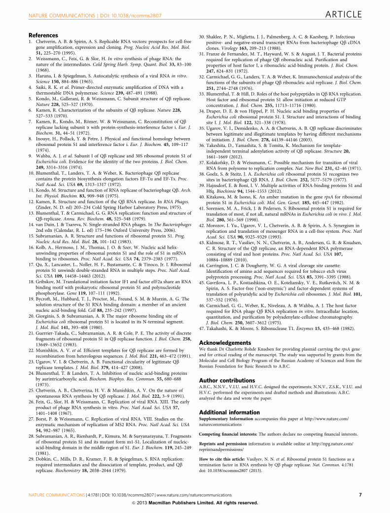
References1. Chetverin, A. B. & Spirin, A. S. Replicable RNA vectors: prospects for cell-free
gene amplification, expression and cloning. Prog. Nucleic Acid Res. Mol. Biol.51, 225–270 (1995).
2. Weissmann, C., Feix, G. & Slor, H. In vitro synthesis of phage RNA: thenature of the intermediates. Cold Spring Harb. Symp. Quant. Biol. 33, 83–100(1968).
3. Haruna, I. & Spiegelman, S. Autocatalytic synthesis of a viral RNA in vitro.Science 150, 884–886 (1965).
4. Saiki, R. K. et al. Primer-directed enzymatic amplification of DNA with athermostable DNA polymerase. Science 239, 487–491 (1988).
5. Kondo, M., Gallerani, R. & Weissmann, C. Subunit structure of Qb replicase.Nature 228, 525–527 (1970).
6. Kamen, R. Characterization of the subunits of Qb replicase. Nature 228,527–533 (1970).
7. Kamen, R., Kondo, M., Romer, W. & Weissmann, C. Reconstitution of Qbreplicase lacking subunit a with protein-synthesis-interference factor i. Eur. J.Biochem. 31, 44–51 (1972).
8. Inouye, H., Pollack, Y. & Petre, J. Physical and functional homology betweenribosomal protein S1 and interference factor i. Eur. J. Biochem. 45, 109–117(1974).
9. Wahba, A. J. et al. Subunit I of Qb replicase and 30S ribosomal protein S1 ofEscherichia coli. Evidence for the identity of the two proteins. J. Biol. Chem.249, 3314–3316 (1974).
10. Blumenthal, T., Landers, T. A. & Weber, K. Bacteriophage Qb replicasecontains the protein biosynthesis elongation factors EF-Tu and EF-Ts. Proc.Natl Acad. Sci. USA 69, 1313–1317 (1972).
11. Kondo, M. Structure and function of RNA replicase of bacteriophage Qb. Arch.Int. Physiol. Biochim. 83, 909–948 (1975).
12. Kamen, R. Structure and function of the Qb RNA replicase. In RNA Phages.(Zinder, N. D. ed) 203–234 Cold Spring Harbor Laboratory Press, 1975).
13. Blumenthal, T. & Carmichael, G. G. RNA replication: function and structure ofQb-replicase. Annu. Rev. Biochem. 48, 525–548 (1979).
14. van Duin, J. & Tsareva, N. Single-stranded RNA phages. In The Bacteriophages2nd edn (Calendar, R. L. ed) 175–196 Oxford University Press, 2006).
15. Subramanian, A. R. Structure and functions of ribosomal protein S1. Prog.Nucleic Acid Res. Mol. Biol. 28, 101–142 (1983).
16. Kolb, A., Hermoso, J. M., Thomas, J. O. & Szer, W. Nucleic acid helix-unwinding properties of ribosomal protein S1 and the role of S1 in mRNAbinding to ribosomes. Proc. Natl Acad. Sci. USA 74, 2379–2383 (1977).
17. Qu, X., Lancaster, L., Noller, H. F., Bustamante, C. & Tinoco, Jr. I. Ribosomalprotein S1 unwinds double-stranded RNA in multiple steps. Proc. Natl Acad.Sci. USA 109, 14458–14463 (2012).
18. Gribskov, M. Translational initiation factor IF1 and factor eIF2a share an RNAbinding motif with prokaryotic ribosomal protein S1 and polynucleotidephosphorylase. Gene 119, 107–111 (1992).
19. Bycroft, M., Hubbard, T. J., Proctor, M., Freund, S. M. & Murzin, A. G. Thesolution structure of the S1 RNA binding domain: a member of an ancientnucleic acid-binding fold. Cell 88, 235–242 (1997).
20. Giorginis, S. & Subramanian, A. R The major ribosome binding site ofEscherichia coli ribosomal protein S1 is located in its N-terminal segment.J. Mol. Biol. 141, 393–408 (1980).
21. Guerrier-Takada, C., Subramanian, A. R. & Cole, P. E. The activity of discretefragments of ribosomal protein S1 in Qb replicase function. J. Biol. Chem. 258,13649–13652 (1983).
22. Munishkin, A. V. et al. Efficient templates for Qb replicase are formed byrecombination from heterologous sequences. J. Mol. Biol. 221, 463–472 (1991).
23. Ugarov, V. I. & Chetverin, A. B. Functional circularity of legitimate Qbreplicase templates. J. Mol. Biol. 379, 414–427 (2008).
24. Blumenthal, T. & Landers, T. A. Inhibition of nucleic acid-binding proteinsby aurintricarboxylic acid. Biochem. Biophys. Res. Commun. 55, 680–688(1973).
25. Chetverin, A. B., Chetverina, H. V. & Munishkin, A. V. On the nature ofspontaneous RNA synthesis by Qb replicase. J. Mol. Biol. 222, 3–9 (1991).
26. Feix, G., Slor, H. & Weissmann, C. Replication of viral RNA. XIII. The earlyproduct of phage RNA synthesis in vitro. Proc. Natl Acad. Sci. USA 57,1401–1408 (1967).
27. Borst, P. & Weissmann, C. Replication of viral RNA. VIII. Studies on theenzymatic mechanism of replication of MS2 RNA. Proc. Natl Acad. Sci. USA54, 982–987 (1965).
28. Subramanian, A. R., Rienhardt, P., Kimura, M. & Suryanarayana, T. Fragmentsof ribosomal protein S1 and its mutant form m1-S1. Localization of nucleic-acid-binding domain in the middle region of S1. Eur. J. Biochem. 119, 245–249(1981).
29. Dobkin, C., Mills, D. R., Kramer, F. R. & Spiegelman, S. RNA replication:required intermediates and the dissociation of template, product, and Qbreplicase. Biochemistry 18, 2038–2044 (1979).
30. Shaklee, P. N., Miglietta, J. J., Palmenberg, A. C. & Kaesberg, P. Infectiouspositive- and negative-strand transcript RNAs from bacteriophage Qb cDNAclones. Virology 163, 209–213 (1988).
31. Franze de Fernandez, M. T., Hayward, W. S. & August, J. T. Bacterial proteinsrequired for replication of phage Qb ribonucleic acid. Purification andproperties of host factor I, a ribonucleic acid-binding protein. J. Biol. Chem.247, 824–831 (1972).
32. Carmichael, G. G., Landers, T. A. & Weber, K. Immunochemical analysis of thefunctions of the subunits of phage Qb ribonucleic acid replicase. J. Biol. Chem.251, 2744–2748 (1976).
33. Blumenthal, T. & Hill, D. Roles of the host polypeptides in Qb RNA replication.Host factor and ribosomal protein S1 allow initiation at reduced GTPconcentration. J. Biol. Chem. 255, 11713–11716 (1980).
34. Draper, D. E. & von Hippel, P. H. Nucleic acid binding properties ofEscherichia coli ribosomal protein S1. I. Structure and interactions of bindingsite I. J. Mol. Biol. 122, 321–338 (1978).
35. Ugarov, V. I., Demidenko, A. A. & Chetverin, A. B. Qb replicase discriminatesbetween legitimate and illegitimate templates by having different mechanismsof initiation. J. Biol. Chem. 278, 44139–44146 (2003).
36. Takeshita, D., Yamashita, S. & Tomita, K. Mechanism for template-independent terminal adenylation activity of Qb replicase. Structure 20,1661–1669 (2012).
37. Kolakofsky, D. & Weissmann, C. Possible mechanism for transition of viralRNA from polysome to replication complex. Nat. New Biol. 231, 42–46 (1971).
38. Goelz, S. & Steitz, J. A. Escherichia coli ribosomal protein S1 recognizes twosites in bacteriophage Qb RNA. J. Biol. Chem. 252, 5177–5179 (1977).
39. Hajnsdorf, E. & Boni, I. V. Multiple activities of RNA-binding proteins S1 andHfq. Biochimie 94, 1544–1553 (2012).
40. Kitakawa, M. & Isono, K. An amber mutation in the gene rpsA for ribosomalprotein S1 in Escherichia coli. Mol. Gen. Genet. 185, 445–447 (1982).
41. Sorensen, M. A., Frick, J. & Pedersen, S. Ribosomal protein S1 is required fortranslation of most, if not all, natural mRNAs in Escherichia coli in vivo. J. Mol.Biol. 280, 561–569 (1998).
42. Morozov, I. Yu., Ugarov, V. I., Chetverin, A. B. & Spirin, A. S. Synergism inreplication and translation of messenger RNA in a cell-free system. Proc. NatlAcad. Sci. USA 90, 9325–9329 (1993).
43. Kidmose, R. T., Vasiliev, N. N., Chetverin, A. B., Andersen, G. R. & Knudsen,C. R. Structure of the Qb replicase, an RNA-dependent RNA polymeraseconsisting of viral and host proteins. Proc. Natl Acad. Sci. USA 107,10884–10889 (2010).
44. Carrington, J. C. & Dougherty, W. G. A viral cleavage site cassette:Identification of amino acid sequences required for tobacco etch viruspolyprotein processing. Proc. Natl Acad. Sci. USA 85, 3391–3395 (1988).
45. Gavrilova, L. P., Kostiashkina, O. E., Koteliansky, V. E., Rutkevitch, N. M. &Spirin, A. S. Factor-free (‘non-enzymic’) and factor-dependent systems oftranslation of polyuridylic acid by Escherichia coli ribosomes. J. Mol. Biol. 101,537–552 (1976).
46. Carmichael, G. G., Weber, K., Niveleau, A. & Wahba, A. J. The host factorrequired for RNA phage Qb RNA replication in vitro. Intracellular location,quantitation, and purification by polyadenylate-cellulose chromatography.J. Biol. Chem. 250, 3607–3612 (1975).
47. Takahashi, K. & Moore, S. Ribonuclease T1. Enzymes 15, 435–468 (1982).
AcknowledgementsWe thank Dr Charlotte Rohde Knudsen for providing plasmid carrying the rpsA geneand for critical reading of the manuscript. The study was supported by grants from theMolecular and Cell Biology Program of the Russian Academy of Sciences and from theRussian Foundation for Basic Research to A.B.C.
Author contributionsA.B.C., N.N.V., V.I.U. and H.V.C. designed the experiments; N.N.V., Z.S.K., V.I.U. andH.V.C. performed the experiments and drafted methods and illustrations; A.B.C.analyzed the data and wrote the paper.
Additional informationSupplementary Information accompanies this paper at http://www.nature.com/naturecommunications
Competing financial interests: The authors declare no competing financial interests.
Reprints and permission information is available online at http://npg.nature.com/reprintsandpermissions/
How to cite this article: Vasilyev, N. N. et al. Ribosomal protein S1 functions as atermination factor in RNA synthesis by Qb phage replicase. Nat. Commun. 4:1781doi: 10.1038/ncomms2807 (2013).
NATURE COMMUNICATIONS | DOI: 10.1038/ncomms2807 ARTICLE
NATURE COMMUNICATIONS | 4:1781 | DOI: 10.1038/ncomms2807 | www.nature.com/naturecommunications 7
& 2013 Macmillan Publishers Limited. All rights reserved.