Revisiting Excitotoxicity in Traumatic Brain Injury - MDPI
26
Citation: Baracaldo-Santamaría, D.; Ariza-Salamanca, D.F.; Corrales-Hernández, M.G.; Pachón-Londoño, M.J.; Hernandez-Duarte, I.; Calderon-Ospina, C.-A. Revisiting Excitotoxicity in Traumatic Brain Injury: From Bench to Bedside. Pharmaceutics 2022, 14, 152. https://doi.org/10.3390/ pharmaceutics14010152 Academic Editor: Maria A. Tikhonova Received: 8 November 2021 Accepted: 5 January 2022 Published: 8 January 2022 Publisher’s Note: MDPI stays neutral with regard to jurisdictional claims in published maps and institutional affil- iations. Copyright: © 2022 by the authors. Licensee MDPI, Basel, Switzerland. This article is an open access article distributed under the terms and conditions of the Creative Commons Attribution (CC BY) license (https:// creativecommons.org/licenses/by/ 4.0/). pharmaceutics Review Revisiting Excitotoxicity in Traumatic Brain Injury: From Bench to Bedside Daniela Baracaldo-Santamaría 1,† , Daniel Felipe Ariza-Salamanca 2,† , María Gabriela Corrales-Hernández 1 , Maria José Pachón-Londoño 1 , Isabella Hernandez-Duarte 1 and Carlos-Alberto Calderon-Ospina 1,3, * 1 Pharmacology Unit, Department of Biomedical Sciences, School of Medicine and Health Sciences, Universidad del Rosario, Bogotá 111221, Colombia; [email protected] (D.B.-S.); [email protected] (M.G.C.-H.); [email protected] (M.J.P.-L.); [email protected] (I.H.-D.) 2 Medical and Health Sciences Education Research Group, School of Medicine and Health Sciences, Universidad del Rosario, Bogotá 111221, Colombia; [email protected] 3 GENIUROS Research Group, Center for Research in Genetics and Genomics (CIGGUR), School of Medicine and Health Sciences, Universidad del Rosario, Bogotá 111221, Colombia * Correspondence: [email protected]; Tel.: +57-1-2970200 (ext. 3318) † These authors contributed equally to this work. Abstract: Traumatic brain injury (TBI) is one of the leading causes of morbidity and mortality. Conse- quences vary from mild cognitive impairment to death and, no matter the severity of subsequent sequelae, it represents a high burden for affected patients and for the health care system. Brain trauma can cause neuronal death through mechanical forces that disrupt cell architecture, and other secondary consequences through mechanisms such as inflammation, oxidative stress, programmed cell death, and, most importantly, excitotoxicity. This review aims to provide a comprehensive understanding of the many classical and novel pathways implicated in tissue damage following TBI. We summarize the preclinical evidence of potential therapeutic interventions and describe the available clinical evaluation of novel drug targets such as vitamin B12 and ifenprodil, among others. Keywords: excitotoxicity; traumatic brain injury; novel therapeutics; NMDA receptor; GABA; vita- min B12; astrocyte; calcium; oxidative stress; neuroinflammation 1. Introduction Traumatic brain injury (TBI) is defined as the acquired cerebral disease that results from an external force in the form of mechanical, chemical, thermal, or electrical energy, or a combination of these [1]. These forces have the potential to cause structural harm to the cranial cavity, brain tissue, and blood vessels. Globally, TBI is one of the highest causes of morbidity and mortality. According to statistics from the United States, up to 43% of severe TBI survivors will probably suffer long-term disability, disrupting individual development [2]. The burden of TBI extends to the patient’s family and the health care system [3,4]. The incidence of TBI is notably high, affecting up to 69 million people worldwide per year, with an estimated annual incidence of 811 to 1507 cases per 100,000 people, depending on the region [5–7]. In 2018, the highest incidence was in North American Countries, the highest burden was in the Southeast Asia and Western Pacific regions, and low- and middle-income countries needed to improve their data availability [5]. Nonetheless, a recent, encouraging analysis showed that TBI incidence gradually decreased 4.4% between 1990 and 2017 in the US [8]. For therapeutic and pathophysiological purposes, the pathogenesis of TBI has been classically divided into two injury mechanisms. The initial external mechanical force (ro- tational or shear forces) is considered as the primary injury mechanism [9], which can be further classified as penetrating or non-penetrating, and can lead to epidural, subdural, Pharmaceutics 2022, 14, 152. https://doi.org/10.3390/pharmaceutics14010152 https://www.mdpi.com/journal/pharmaceutics
-
Upload
khangminh22 -
Category
Documents
-
view
3 -
download
0
Transcript of Revisiting Excitotoxicity in Traumatic Brain Injury - MDPI
�����������������
Citation: Baracaldo-Santamaría, D.;
Ariza-Salamanca, D.F.;
Corrales-Hernández, M.G.;
Pachón-Londoño, M.J.;
Hernandez-Duarte, I.;
Calderon-Ospina, C.-A. Revisiting
Excitotoxicity in Traumatic Brain
Injury: From Bench to Bedside.
Pharmaceutics 2022, 14, 152.
https://doi.org/10.3390/
pharmaceutics14010152
Academic Editor: Maria
A. Tikhonova
Received: 8 November 2021
Accepted: 5 January 2022
Published: 8 January 2022
Publisher’s Note: MDPI stays neutral
with regard to jurisdictional claims in
published maps and institutional affil-
iations.
Copyright: © 2022 by the authors.
Licensee MDPI, Basel, Switzerland.
This article is an open access article
distributed under the terms and
conditions of the Creative Commons
Attribution (CC BY) license (https://
creativecommons.org/licenses/by/
4.0/).
pharmaceutics
Review
Revisiting Excitotoxicity in Traumatic Brain Injury: From Benchto BedsideDaniela Baracaldo-Santamaría 1,†, Daniel Felipe Ariza-Salamanca 2,† , María Gabriela Corrales-Hernández 1 ,Maria José Pachón-Londoño 1 , Isabella Hernandez-Duarte 1 and Carlos-Alberto Calderon-Ospina 1,3,*
1 Pharmacology Unit, Department of Biomedical Sciences, School of Medicine and Health Sciences,Universidad del Rosario, Bogotá 111221, Colombia; [email protected] (D.B.-S.);[email protected] (M.G.C.-H.); [email protected] (M.J.P.-L.);[email protected] (I.H.-D.)
2 Medical and Health Sciences Education Research Group, School of Medicine and Health Sciences,Universidad del Rosario, Bogotá 111221, Colombia; [email protected]
3 GENIUROS Research Group, Center for Research in Genetics and Genomics (CIGGUR), School of Medicineand Health Sciences, Universidad del Rosario, Bogotá 111221, Colombia
* Correspondence: [email protected]; Tel.: +57-1-2970200 (ext. 3318)† These authors contributed equally to this work.
Abstract: Traumatic brain injury (TBI) is one of the leading causes of morbidity and mortality. Conse-quences vary from mild cognitive impairment to death and, no matter the severity of subsequentsequelae, it represents a high burden for affected patients and for the health care system. Braintrauma can cause neuronal death through mechanical forces that disrupt cell architecture, and othersecondary consequences through mechanisms such as inflammation, oxidative stress, programmedcell death, and, most importantly, excitotoxicity. This review aims to provide a comprehensiveunderstanding of the many classical and novel pathways implicated in tissue damage followingTBI. We summarize the preclinical evidence of potential therapeutic interventions and describe theavailable clinical evaluation of novel drug targets such as vitamin B12 and ifenprodil, among others.
Keywords: excitotoxicity; traumatic brain injury; novel therapeutics; NMDA receptor; GABA; vita-min B12; astrocyte; calcium; oxidative stress; neuroinflammation
1. Introduction
Traumatic brain injury (TBI) is defined as the acquired cerebral disease that resultsfrom an external force in the form of mechanical, chemical, thermal, or electrical energy,or a combination of these [1]. These forces have the potential to cause structural harmto the cranial cavity, brain tissue, and blood vessels. Globally, TBI is one of the highestcauses of morbidity and mortality. According to statistics from the United States, up to43% of severe TBI survivors will probably suffer long-term disability, disrupting individualdevelopment [2]. The burden of TBI extends to the patient’s family and the health caresystem [3,4].
The incidence of TBI is notably high, affecting up to 69 million people worldwide peryear, with an estimated annual incidence of 811 to 1507 cases per 100,000 people, dependingon the region [5–7]. In 2018, the highest incidence was in North American Countries,the highest burden was in the Southeast Asia and Western Pacific regions, and low- andmiddle-income countries needed to improve their data availability [5]. Nonetheless, arecent, encouraging analysis showed that TBI incidence gradually decreased 4.4% between1990 and 2017 in the US [8].
For therapeutic and pathophysiological purposes, the pathogenesis of TBI has beenclassically divided into two injury mechanisms. The initial external mechanical force (ro-tational or shear forces) is considered as the primary injury mechanism [9], which can befurther classified as penetrating or non-penetrating, and can lead to epidural, subdural,
Pharmaceutics 2022, 14, 152. https://doi.org/10.3390/pharmaceutics14010152 https://www.mdpi.com/journal/pharmaceutics
Pharmaceutics 2022, 14, 152 2 of 26
and intracerebral hematoma, intraventricular hemorrhage, or diffuse axonal injury. Skullfractures and vasogenic edema are also consequences of primary injury and they cannotalways be fully repaired, though they can be surgically mitigated [1,10]. The main sec-ondary injury hallmarks of TBI are changes in neurotransmitter release and metabolism,intracellular calcium dysregulation [11], biochemical reagent overproduction [12], bloodbrain barrier (BBB) disruption [13], cellular lysis, cellular and cytotoxic edema [14,15], andinflammation [16–18].
Head elevation, hyperventilation, seizure prophylaxis, hyperosmolar therapy, inducedcoma, cooling, and decompressive craniectomy are the current pillars for the acute treat-ment of patients following TBI [19]. Regarding disability prevention in acute and chronictreatment after TBI, Sveen and colleagues showed that an immediate discharge to special-ized rehabilitation therapy is related to better outcomes and independence level of thepatient when compared to delayed discharge or no rehabilitation at all 12 months aftera severe TBI [20]. Identified barriers regarding access to rehabilitation include limitedresources, patient’s safety concerns, lack of support from family members, or health careprovider restrictions [21].
Disability rates vary depending on the severity of the trauma, as well as the accuracyand quality of the treatment given and subsequent follow-up. Events related to the highestdisability rates are firearm injury and fall [22]. The prevalence of disability has beenestimated at 3.32 million to 5.3 million in the US [6,23]. Direct costs related to TBI havebeen estimated at 9.2 billion per year, and indirect costs related to missed work and lossof productivity are up to 51.2 billion dollars [6]. It is noteworthy that the greatest burdenof disease comes from disability and loss of productivity. In that sense, it is mandatory torecognize therapeutic approaches to mitigate disability, or maybe even avoid it.
Excitotoxicity, understood as neural death secondary to the excessive release of excita-tory neurotransmitters, is broadly described as a common pathway leading to cell deathin many neuropathological conditions [24–28]. When it comes to TBI, excitotoxicity ispromoted by an increase of glutamate concentrations released into the extracellular spacedue to cellular lysis, followed by a rise of positive charges within the cell [29]. Calcium(Ca2+) activates second messenger pathways, exacerbates oxidative stress (OS), and initi-ates programmed cell death [30]. These factors can act as enhancers and perpetuators ofexcitotoxicity.
In this paper, we will focus on revisiting the classical and novel pathophysiologicalpathways involved in TBI to adequately understand new potential drug therapies. Wewill describe, in detail, mechanisms such as NMDAr (N-methyl-D-aspartate receptor)-dependent excitotoxicity, mechanoporation, GABA (Gamma-Aminobutyric acid)-mediatedexcitotoxicity, and the role of protagonist processes in TBI such as neuroinflammation,BBB disruption, and OS. New therapeutic agents regarding these pathways will also bepresented and critically assessed to propose which of them could have a positive impactto avoid or mitigate secondary lesions after TBI and therefore alleviate the subsequentlong-term disability and disease burden.
2. Mechanisms for Excitotoxicity
NMDAr activation is a major contributor to the enhancement of excitotoxicity follow-ing TBI. To better understand its activation, a review of the receptor structure is needed.NMDA receptors are formed by the assembly of seven subunits divided into three mainsubfamilies (GluN1, GluN2A-D and GluN3A-B) [31], each encoded by seven differentgenes, to form a final heterotetrametric structure that forms an ion channel that is per-meable to Na+, K+ and Ca2+. It has two obligatory subunits: the GluN1 subunit whichbinds glycine and D-serine and a GluN2 subunit which binds glutamate; and/or twoGluN3 (A or B) subunits [32]. The GluN2 subunit has a great influence on the EC50 (halfmaximal effective concentration) of glutamate depending on which of the four subunits isexpressed (A-D). NMDA receptors that contain one GluN1 and two GluN2D subunits havean EC50 five-fold lower for glutamate than those receptors with two GluN2A, 2B, or 2C
Pharmaceutics 2022, 14, 152 3 of 26
subunits [31]. Similarly, some subunits have a higher permeability to Ca2+ and express ahigher sensitivity to the magnesium block (GluN1/2A, and GluN1/2B) [31]. Interestingly,NMDA receptors containing the GluN2B subunit have been implicated in aiding the spreadof excitotoxicity among neighbor cells in cultured hippocampal neurons. In contrast, thesame study suggested that NMDA receptors containing GluN2A favor neuroprotectivesignaling mechanisms [33].
In addition, NMDA receptors are capable of binding small ligands, especially to theamino terminus of the receptor subunits. These ligands can act as allosteric modulators,which differ from NMDA antagonists in that they can modulate the receptor activitywithout altering its biological functions [34], and are thus a potential drug target. TheGluN2B subunit has three different binding sites for both negative and positive allostericmodulators. They interact with zinc and ifenprodil, producing an allosteric inhibition ofthe receptor [34].
2.1. Glutamate-Mediated Activation
Glutamate has been addressed as the most important neurotransmitter and it isinvolved in many processes including synaptic connectivity, cellular ionic homeostasis,long term potentiation, and even hormonal release [35,36]. All these cellular processes takeplace mainly through the regulation of intracellular concentrations of Ca2+ ([Ca2+]i). Whenstudied in traumatic brain injury, the extracellular concentrations of glutamate rise [37] as aconsequence of cellular lysis. The later aberrant efflux of glutamate from neurons and gliaends up worsening the scenario. High concentrations of glutamate are harmful, as it canover-activate synaptic NMDAr as well as extrasynaptic glutamate receptors [38].
Different studies have pinpointed NMDAr activation as the main cause of excitotoxi-city after TBI [37,39]. NMDAr activation precedes Ca2+ and Na+ entry into intracellularspaces. High concentrations of [Ca2+]i activate second messenger pathways, caspasesand calpain proteins, and the release of neurotransmitter vesicles, catabolic enzymes asphospholipases, proteases and endoproteases [40]. An indirect way to measure glutamateconcentration is by recording cortical spreading depolarizations, which reach their max-imum 48 h after trauma [41]. Essentially, after a TBI there is an increase in extracellularglutamate concentrations and an upregulation in ionotropic glutamate receptors such asAMPA (α-amino-3-hydroxy-5-methyl-4-isoxazolepropionic acid) and NMDA [40]. Together,these factors promote Ca2+ and Na+ entry into the neuron. Once Ca2+ and Na+ are free inthe neuron’s cytosol, different processes will occur. Two main organelles, the mitochondriaand the endoplasmic reticulum, play an important role in the regulation of [Ca2+]i.
Furthermore, a study by Lee and colleagues [42] demonstrated the expression ofNMDA receptors on the astrocytic membrane, accompanied by an increase in glutamaterelease following high concentrations of intracellular calcium. With this being said, it canbe stated that, as with neurons, higher concentrations of extracellular glutamate activateNMDAr, thereby augmenting calcium influx into the astrocyte. Higher calcium concentra-tions will override the astrocyte’s mechanism of glutamate reuptake and transformation,thus potentiating the chain reaction leading to excitotoxicity.
2.2. Mechanical Activation of NMDAr and Mechanoporation of Neuronal Membranes
Mechanical NMDA activation can occur secondary to lipid bilayer tension, allowingCa2+ to enter the neuron in the absence of NMDA agonists [43]. The physiological responseof rat astrocytes to shear stress confirmed the mechanosensitive properties of NMDA in theabsence of agonists that effectively remove the Mg+ blockade [44], and therefore linkingthe shearing and stretching forces produced in TBI to glutamate-independent excitotoxicity.Some studies have attributed this mechanosensitive nature of the receptor to the GluN2Bsubunit, specifically the serine 1323 residue in the C-terminus [45].
Mechanoporation refers to the formation of pores in the cell membrane induced bymechanical forces [46–48]. These pores form in milliseconds because of membrane stretch-ing, which leads to the inhibition of action potential generation [49]. Mechanoporation
Pharmaceutics 2022, 14, 152 4 of 26
alters the permeability of the membrane and allows the flow of ions and molecules thatlead to cell death. This mechanism of injury has been demonstrated through indirectmeasurements using different markers that show alteration of membrane permeability [50].Understanding the creation of pores in the membrane of neurons following brain traumaled to the conclusion that Ca2+ could enter the cell by the same pathway as the fluorescentmarkers evaluated in some studies [50], and could therefore exert direct excitotoxicitywithout the need for a transporter. In addition, the extent of membrane damage dependson the mechanism of injury: in three-dimensional neural cultures analyzing cell survivaland membrane permeability, it was shown that cells were more sensitive to bulk sheardeformation than compression [51].
It has been proposed that the creation of these membrane pores can occur in twostages [51]: the initial injury that causes stretch and shearing forces, therefore creatingmechanical damage to the membrane; and a secondary injury that consists of membranedegradation through the activation of degradative enzymes (e.g., calpain) [51]. Interestingly,Ca2+ influx secondary to mechanoporation has been postulated in some studies as necessaryfor calpain activation in axons of cultured neurons [52]. Furthermore, following calpainactivation, axonal microtubule damage ensues, causing impaired axonal transport [53].
Ultimately, excitotoxicity will lead to high [Ca2+]i, which is responsible for the tissuedamage and cellular death that will be discussed in the next section.
2.3. Consequences of High Intracellular Calcium
In the mitochondria, elevated calcium concentrations will have different catastrophicrepercussions, among which are the generation of free radicals and reactive oxygen species(ROS), protease and phospholipase activation, nitric oxide synthase (NOS) activation, andthe generation of mitochondrial pores through which contents such as Cytochrome c willleak and promote the activation of caspases and nucleases, eventually leading to signalingand cellular death. ROS-induced OS is a major cause of neuronal death in the context ofsecondary injury after brain trauma, as it damages nucleic acids, proteins and lipids [54].OS contributes to mitochondrial dysfunction and therefore energy depletion. In a studyabout OS in rats following mild and severe TBI, OS did not show a pattern in terms ofproportionality between the energy of the trauma and the extent of the secondary injury.Additionally, OS was consistent across the areas of the brain without preference [55].
Another mechanism by which high [Ca2+]i is detrimental to the cell is the acidificationresulting from Ca2+ influx, as this acidification will activate the Na+-H+ exchanger, a pH-regulating mechanism that will decrease the activity of the Na+-Ca2+ exchanger, affectingthe cell’s ability to pump calcium out [56].
Mitochondrial Ca2+ overload added to a densely ROS-concentrated environment willlead to the formation and opening of mitochondrial transition pores. Given the closeproximity of the outer mitochondrial membrane with the outer endoplasmic reticulum,these pores will function as a Ca2+ efflux mechanism. Mitochondrial transition poresare not selective, meaning that although calcium is flowing out of the cell, other intra-mitochondrial components such as Cytochrome c will also leak out, activating a chainreaction and ultimately resulting in cell death [57]. Moreover, the continuous opening ofmitochondrial transition pores will alter the mitochondrial membrane potential, leading toa decrease in ATP (adenosine triphosphate) production, thus resulting in mitochondrialdestruction [58]. Additionally, as Ca2+ and Cytochrome c are being liberated, water andcytosolic solutes are entering the mitochondria, leading to swelling and, consequently,matrix rupture [59].
Caspases are a family of intracellular cysteine proteases involved in inflammatory pro-cesses and programmed cell death, and are activated by high [Ca2+]i [60]. Caspase 1 cleavesacid residues, aspartic acid, and matures interleukin 1, which exacerbates damage [61,62].As for Caspase 3, its activation will lead to the proteolysis of DNA-repairing proteins andthe degradation of cytoskeletal proteins, eventually resulting in morphological changesthat lead to apoptosis [63,64].
Pharmaceutics 2022, 14, 152 5 of 26
Calpains are Ca2+-activated neutral proteases involved in multiple pathways such asremodeling, cell signaling, synaptic plasticity, neuroprotection, learning, and programmedcell death [65,66]. Evidence suggests calpains are early regulators of cell death after TBI.Calpain activation is evident five hours after trauma in the dendrites, with later expressionin the soma after 10 h [66]. Recent evidence shows calpain 1 and calpain 2 are directlyimplicated in cell death. Wang and colleagues [67] showed calpain-2 is responsible forNMDA-induced excitotoxicity through the activation of striatal-enriched protein tyrosinephosphatase (STEP) when extra synaptic NMDA receptors are activated in rat-culturedneurons. STEP activation mediated by calpain-2 results in p38 expression and, therefore,downstream cell death [65,68]. After TBI, high [Ca2+]i, OS, and ischemia, induce endoplas-mic reticulum (ER) stress, which oversees membrane protein synthesis, folding, and qualitycontrol. When the ER is stressed, the unfolded protein response (UPR) is started by PERK(PKR-like ER kinase), ATF6 (activating transcription factor 6), and IRE-1 (inositol requiringenzyme 1) when they are dissociated from glucose-regulated protein/binding immunoglob-ulin protein (GRP78/BIP) [69]. PERK phosphorylates eukaryotic initiation factor 2α (eIF2α),which reduces protein synthesis, while IRE-1 promotes the UPR response, and cleavedATF6 activates chaperones and intrinsic systems in charge of ER-associated degradationsystems [70,71]. These pathways ultimately lead to C/EBP homologous protein (CHOP)upregulation, resulting in cellular apoptosis.
It is remarkable that high intracellular calcium is the common consequence of mul-tiple damages sustained during TBI, such as the formation of cellular and mitochondrialmembrane pores and ER stress, resulting in cellular death. Therefore, most therapies havefocused on preventing or controlling the rise of [Ca2+]i.
2.4. Hippocampal Vulnerability to TBI
Following TBI, hippocampus-dependent cognitive impairment, such as memory loss,represents a significant burden for the patient and their family. The hippocampus is aninteresting area because it is one of the specific regions of the brain where adult neurogene-sis occurs [72] and where most animal studies in different TBI models (controlled corticalimpact [73], fluid percussion [74] and stretch injury [75]) have shown the most vulnerabil-ity [76]. In particular, the subventricular zone of the lateral ventricle and the subgranularzone of the dentate gyrus (SGZ-DG) in the hippocampus have been linked to neurogenesis.Normally, these newborn neurons are continuously being generated. Approximately 700new neurons are added every day to the hippocampus and then, as maturation occurs,they migrate and incorporate into the hippocampal circuit. The adult-born SGZ-DG cellsdiffer from their mature counterparts in that they have a higher membrane resistance, lackglutamatergic input, and are depolarized by GABAergic input [77]. They have been linkedto the consolidation of new memories, long-term spatial memory, cognitive flexibility,pattern separation and increased performance in behavioral tasks [78]. Similarly, theymight be involved in the cognitive impairment seen in the chronic phase following TBI asthere are studies that have shown the selective death of hippocampal newborn neuronsafter moderate brain trauma.
The distribution of degenerating neurons in the hippocampus of the mouse brainfollowing moderate TBI was assessed by Gao and colleagues, who found that most of thedegenerating hippocampal neurons were located in SGZ-DG [76]. Furthermore, anotherstudy [79] demonstrated that most of the neuron death that occurs in the hippocampusoccurs within 24 h post-injury and continues for another 14 days. They also found that mostof the dying cells in the SGZ-DG were immature. In addition, another study utilized an ionicfluorescein (FJP-fluoro-jade B) derivative reported to be a specific marker for degeneratingneurons in young male rats subjected to controlled cortical impact [80]. By using thefluorescein marker, they found that there were FJP-positive neurons in the hippocampus,with higher expression in the moderate TBI group compared to the mild TBI group. Theregional distribution of FJB staining was different according to the days post-injury and theseverity of the TBI, and there was a direct relationship between the number of FJB-positive
Pharmaceutics 2022, 14, 152 6 of 26
neurons and the severity of the trauma. The SGZ-DG had more FJB-positive neuronsthan other hippocampal subregions, regardless of the level of severity, as early as 24 hpost-injury. Another study used the same marker (FJB) to assess neuronal injury afterlateral fluid percussion brain injury in rats [81]. Cell loss was evident seven days post-injury in the cortex, hippocampus and thalamus. Three hours post-injury, injured neuronsappeared in the hippocampus (CA1 and DG), thalamus, and vermis of the cerebellum, andthe number of degenerating neurons was greatest after one and three days in the cortexand hippocampus, respectively.
The question remains as to why newborn immature neurons in the SGZ-DG are morevulnerable to TBI, and whether we can prevent it. We know that immature neurons areelectrophysiologically different from their mature counterparts, that GABA A receptors arethe first receptors expressed, and that during the early stages, GABAergic transmission isexcitatory because of high intracellular Cl− [77]. There is a possibility that the mechanismfor the observed vulnerability of the hippocampus may be due to GABA-mediated excito-toxicity [82]. However, mechanisms for hippocampal neuron vulnerability are not known.ER stress has recently been assigned a role in the apoptotic death of newborn neuronsafter TBI through the activation of the pro-apoptotic transcription factor CHOP [83,84].ER stress results in the accumulation of unfolded proteins, which leads to the activationof the PERK-eIF2 pathway. PERK phosphorylates eIF2, which inhibits general proteinsynthesis, thereby reducing ER stress. If the stress is intense and cannot be resolved bythis pathway, activating transcription factor 4 (ATF4) increases the expression of CHOP,initiating apoptotic cell death. Therefore, Hood and colleagues used CHOP knockout miceto evaluate the role of this protein in TBI-induced apoptosis of doublecortin (a markerof hippocampal newborn neurons) positive neurons. They also demonstrated that thepharmacological inhibition of PERK by GSK2606414 exacerbated newborn neuron loss, andthe administration of guanabenz (a drug that causes dose-dependent increases in eIF2 alphaphosphorylation) can modify the protein production rates, therefore reducing newbornneuronal loss.
3. Blood–Brain Barrier Disruption and Neuroinflammation
Disruption of the BBB after TBI results in hyperpermeability, increased intracranialpressure, edema, and decreased cerebral perfusion pressure, perpetuating cellular damageand hypoxia. Primary lesion leads to secondary injuries through neuroinflammationand cellular and cytotoxic edema, resulting in more neuronal death. These secondarylesions are mainly mediated by the activation of resident microglia and the recruitmentof immune cells such as lymphocytes, monocytes, and neutrophils. Immunity cells willreach brain parenchyma two to 48 h after the trauma. Activation leads to increases inmetalloproteinases, proteases, ROS, and reactive nitrogen species (RNS), boosting BBB andcellular damage [85–88].
The inflammatory response is initiated in resident cells, such as microglia and as-trocytes, which detect pathogen-associated molecular patterns (PAMPs) and damage-associated molecular patterns (DAMPs), or by an increase of ATP that activates the mTORpathway [89]. When exposed to an inflammatory environment, these can perpetuate theinflammatory response through differentiation to type 1 or classical activation, resultingin an enhancement of proinflammatory cytokines such as IL-6, IL-1, TNF-α and nitricoxide synthase production [90]. IL6 and TNF-α are responsible for increasing BBB perme-ability, IL-1 induces astrocytes to promote the expression of VCAM-1/ICAM-1 (vascularcell adhesion molecule -1 and intercellular cell adhesion molecule) in the endothelium,therefore perpetuating immune cell entry to the CNS (central nervous system), while theoverstimulation of nitric oxide synthase results in more OS by increasing RNS (reactivenitrogen species) [91,92].
Microglia are CNS resident immune cells with pleiotropic functions. When exposed toa proinflammatory-enriched environment, microglia can transform into their M1 type, alsoknown as the classical type, in order to control damage, eliminate residues, and repair tis-
Pharmaceutics 2022, 14, 152 7 of 26
sue [93,94]. It has been shown that microglia not only mediate acute inflammatory response,but are also involved in the subsequent tissue remodeling and modulation of inflamma-tion through M2-type differentiation. In response to inflammation, M1 microglia produceproinflammatory cytokines and microparticles, modulate the neuronal microenvironment,and, through astrocytes, can indirectly influence blood brain perfusion [95–97]. ChronicM1 microglial activation is related to neural degeneration due to sustained inflammatorystates [98]. Therefore, the proper modulation of microglia function and switching M1microglia to M2 can prevent the perpetuation of secondary injury-related brain damage.
Ndode-Ekane and colleagues shed some light on the complex role of T-lymphocytes(T cells) after TBI. According to their findings, and those of other authors, lymphocytesmigrate soon after TBI, reaching their peak one to two days post-injury, and some remainingcell clusters are found up to 90 days post-injury [99]. The higher the presence of T cells,the poorer the neurological recovery [99,100]. T cell migration is a consequence of BBBdisruption, increased endothelial expression of adhesion molecules such as VCAM/ICAM-1, excessive ROS production, and cell-mediated activation and proliferation [100,101]. CD8+T cells sustain inflammatory states and prompt cellular damage through the activation ofother cells, such as microglia, neutrophils, macrophages and astrocytes, and the productionof granzyme B, which causes neuron myelin degeneration [102]. B cells have also beenrelated to TBI-secondary injury due to autoreactive antibody production directed to brain-specific proteins freed into the medium after the trauma [102,103]. Neutrophils are alsoprotagonists of inflammation after TBI because, like lymphocytes, they quickly migrateto the brain parenchyma through the BBB, attracted by the chemokine (C-C motif) ligand.Once in the brain parenchyma, they interact with lymphocytes and resident cells, producecytokines and chemokines to potentialize immune reactions such as CXCL-1 and CXCL-2,release metalloproteinases, and present antigens and phagocytes in order to clean theextracellular space and prepare the tissue for scarring [85,88].
Focusing on astrocytes, this type of glia not only contributes to neuroinflammation,but also to maintaining a homeostatic balance for neurons by regulating synaptic andextrasynaptic glutamate and other neurotransmitters, stabilizing the BBB, and modulatingsynaptic activity, among others [104,105]. Regarding inflammation [106], once differentiatedto A1 by microglia, astrocytes will quickly lose their synaptic functions and phagocyticcapacity, and will start killing neurons and oligodendrocytes with no clear mechanism, butlikely by inducing cellular apoptosis as the blockage of caspases 1 and 2 prevents cellulardeath [107]. Taking these findings together and connecting high [Ca2+]i consequences, weaddress again the importance of modulating Ca2+ entry to cells, including astrocytes.
Regarding glutamate uptake, Dorssett and colleagues showed that the post-TBI acti-vation of different kinases, such as kinase B (Akt) and protein kinase C (PKC), negativelymodulates glutamate transporter 1 (GLT-1) function in the frontal cortex 24 h after in-jury [108]. GLT-1 is the most important channel of glutamate uptake in rats. In that sense,its homologous human excitatory amino acid transporter 2 (EAAT2) can also be affectedafter TBI [109]. In an old mouse model, Gupta and colleagues showed GLT-1 is brieflydownregulated after brain trauma [110]. This means that, no matter the model or the timeafter trauma, glutamate reuptake is altered in astrocytes and neurons, leading to highextracellular concentrations, thus activating different glutamate receptors which promotethe influx of Ca2+ to the cell and activating programmed cell death.
In conclusion, enhancers and perpetuators of excitotoxicity include the BBB, glial cellsand immunity cells. They are activated after trauma through different mechanisms such asATP release to the cellular medium, cytokine production relying on the activation of NF-kB(nuclear factor kappa-light-chain-enhancer of activated B cells) triggered by DAMPS andPAMPS, and changes in cellular structure and function promoted by a proinflammatoryenvironment. We reflect about how to modulate these enhancers later in this paper. Wesummarized these pathways in Figure 1.

Pharmaceutics 2022, 14, 152 8 of 26
Pharmaceutics 2022, 14, x FOR PEER REVIEW 8 of 27
of NF-kB (nuclear factor kappa-light-chain-enhancer of activated B cells) triggered by
DAMPS and PAMPS, and changes in cellular structure and function promoted by a pro-
inflammatory environment. We reflect about how to modulate these enhancers later in
this paper. We summarized these pathways in Figure 1.
Figure 1. Illustration of pathophysiological events leading to excitotoxicity after TBI. BBB disruption
leads to edema, changes in ionic homeostasis, and the migration of systemic immune cells. Release
of intracellular contents in the medium activates microglia and astrocytes, enhancing OS, inflam-
mation, and neural damage. Increase in extracellular [Ca2+] and glutamate activates NMDAr, while
mechanical activation and mechanoporation of NMDAr lead to an increase in [Ca2+]i, creating an
environment stressful to mitochondria, ER, and DNA, and a decrease of pH. Many pathways lead
to cellular apoptosis: organelle stress and the activation of caspases, calpains, and death receptors.
In black: events related to the activation and flux of ions. In red: pathways related to cellular apop-
tosis. Numbers address potential therapies listed in Table 1. [Ca2+]i: concentration of intracellular
calcium; [Ca2+]e: concentration of extracellular calcium; BBB: blood–brain barrier; ROS: reactive ox-
ygen species; RNS: reactive nitrogen species; NMDAr: N-methyl-D-Aspartate receptors; TLR: toll-
like receptor; ATP: adenosine triphosphate; EAAT2: excitatory amino-acid transporter 2; NOS: nitric
oxide synthase; DAMPS: damage-associated molecular patterns; PAMPS: pathogen-associated mo-
lecular patterns; AQ4: aquaporin-4; ICAM1: intercellular cell adhesion molecule-1; VCAM1: vascu-
lar cell adhesion molecule-1; mGluR5: glutamate metabotropic receptor; RAGE: receptor for ad-
vanced glycation end products; ER: endoplasmic reticulum; PSD-95: postsynaptic density protein-
95; DR: death receptor.
Figure 1. Illustration of pathophysiological events leading to excitotoxicity after TBI. BBB disruptionleads to edema, changes in ionic homeostasis, and the migration of systemic immune cells. Release ofintracellular contents in the medium activates microglia and astrocytes, enhancing OS, inflammation,and neural damage. Increase in extracellular [Ca2+] and glutamate activates NMDAr, while mechani-cal activation and mechanoporation of NMDAr lead to an increase in [Ca2+]i, creating an environmentstressful to mitochondria, ER, and DNA, and a decrease of pH. Many pathways lead to cellular apop-tosis: organelle stress and the activation of caspases, calpains, and death receptors. In black: eventsrelated to the activation and flux of ions. In red: pathways related to cellular apoptosis. Numbersaddress potential therapies listed in Table 1. [Ca2+]i: concentration of intracellular calcium; [Ca2+]e:concentration of extracellular calcium; BBB: blood–brain barrier; ROS: reactive oxygen species; RNS:reactive nitrogen species; NMDAr: N-methyl-D-Aspartate receptors; TLR: toll-like receptor; ATP:adenosine triphosphate; EAAT2: excitatory amino-acid transporter 2; NOS: nitric oxide synthase;DAMPS: damage-associated molecular patterns; PAMPS: pathogen-associated molecular patterns;AQ4: aquaporin-4; ICAM1: intercellular cell adhesion molecule-1; VCAM1: vascular cell adhesionmolecule-1; mGluR5: glutamate metabotropic receptor; RAGE: receptor for advanced glycation endproducts; ER: endoplasmic reticulum; PSD-95: postsynaptic density protein-95; DR: death receptor.
Pharmaceutics 2022, 14, 152 9 of 26
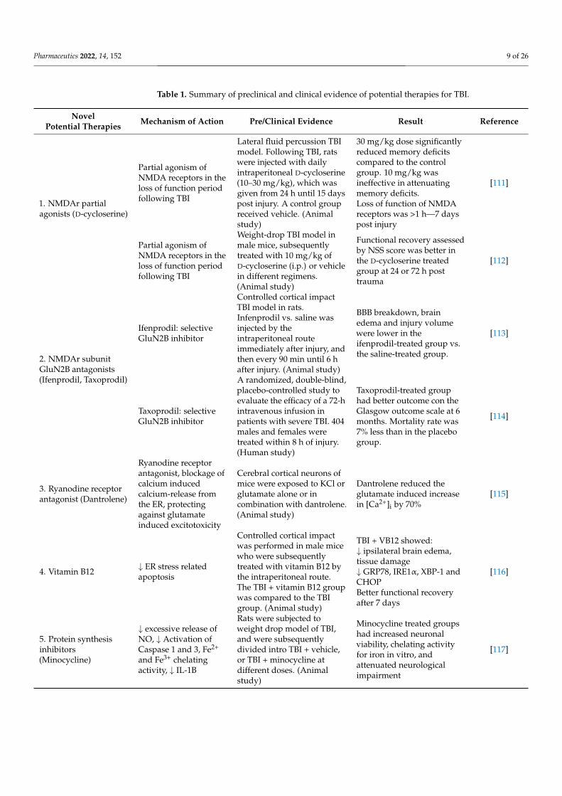
Table 1. Summary of preclinical and clinical evidence of potential therapies for TBI.
NovelPotential Therapies Mechanism of Action Pre/Clinical Evidence Result Reference
1. NMDAr partialagonists (D-cycloserine)
Partial agonism ofNMDA receptors in theloss of function periodfollowing TBI
Lateral fluid percussion TBImodel. Following TBI, ratswere injected with dailyintraperitoneal D-cycloserine(10–30 mg/kg), which wasgiven from 24 h until 15 dayspost injury. A control groupreceived vehicle. (Animalstudy)
30 mg/kg dose significantlyreduced memory deficitscompared to the controlgroup. 10 mg/kg wasineffective in attenuatingmemory deficits.Loss of function of NMDAreceptors was >1 h—7 dayspost injury
[111]
Partial agonism ofNMDA receptors in theloss of function periodfollowing TBI
Weight-drop TBI model inmale mice, subsequentlytreated with 10 mg/kg ofD-cycloserine (i.p.) or vehiclein different regimens.(Animal study)
Functional recovery assessedby NSS score was better inthe D-cycloserine treatedgroup at 24 or 72 h posttrauma
[112]
2. NMDAr subunitGluN2B antagonists(Ifenprodil, Taxoprodil)
Ifenprodil: selectiveGluN2B inhibitor
Controlled cortical impactTBI model in rats.Infenprodil vs. saline wasinjected by theintraperitoneal routeimmediately after injury, andthen every 90 min until 6 hafter injury. (Animal study)
BBB breakdown, brainedema and injury volumewere lower in theifenprodil-treated group vs.the saline-treated group.
[113]
Taxoprodil: selectiveGluN2B inhibitor
A randomized, double-blind,placebo-controlled study toevaluate the efficacy of a 72-hintravenous infusion inpatients with severe TBI. 404males and females weretreated within 8 h of injury.(Human study)
Taxoprodil-treated grouphad better outcome con theGlasgow outcome scale at 6months. Mortality rate was7% less than in the placebogroup.
[114]
3. Ryanodine receptorantagonist (Dantrolene)
Ryanodine receptorantagonist, blockage ofcalcium inducedcalcium-release fromthe ER, protectingagainst glutamateinduced excitotoxicity
Cerebral cortical neurons ofmice were exposed to KCl orglutamate alone or incombination with dantrolene.(Animal study)
Dantrolene reduced theglutamate induced increasein [Ca2+]i by 70%
[115]
4. Vitamin B12 ↓ ER stress relatedapoptosis
Controlled cortical impactwas performed in male micewho were subsequentlytreated with vitamin B12 bythe intraperitoneal route.The TBI + vitamin B12 groupwas compared to the TBIgroup. (Animal study)
TBI + VB12 showed:↓ ipsilateral brain edema,tissue damage↓ GRP78, IRE1α, XBP-1 andCHOPBetter functional recoveryafter 7 days
[116]
5. Protein synthesisinhibitors(Minocycline)
↓ excessive release ofNO, ↓ Activation ofCaspase 1 and 3, Fe2+
and Fe3+ chelatingactivity, ↓ IL-1B
Rats were subjected toweight drop model of TBI,and were subsequentlydivided intro TBI + vehicle,or TBI + minocycline atdifferent doses. (Animalstudy)
Minocycline treated groupshad increased neuronalviability, chelating activityfor iron in vitro, andattenuated neurologicalimpairment
[117]
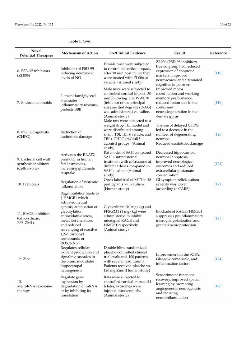
Pharmaceutics 2022, 14, 152 10 of 26
Table 1. Cont.
NovelPotential Therapies Mechanism of Action Pre/Clinical Evidence Result Reference
6. PSD-95 inhibitors(ZL006)
Inhibition of PSD-95reducing neurotoxiclevels of NO
Female mice were subjectedto controlled cortical impact,after 30 min post injury theywere treated with ZL006 orvehicle. (Animal study)
ZL006 (PSD-95 inhibitor)treated group had reducedexpression of apoptoticmarkers, improvedneuroscores, and attenuatedcognitive impairment
[118]
7. Endocannabinoids
2-arachidonylglycerolattenuatesinflammatory response,protects BBB
Male mice were subjected tocontrolled cortical impact. 30min following TBI, WWL70(inhibitor of the principalenzyme that degrades 2-AG)was administered vs. saline.(Animal study)
Improved motorcoordination and workingmemory performance,reduced lesion size in thecortex andneurodegeneration in thedentate gyrus
[119]
8. mGLU5 agonists(CHPG)
Reduction ofexcitotoxic damage
Male rats were subjected to aweight drop TBI model andwere distributed amongsham, TBI, TBI + vehicle, andTBI + CHPG (mGluR5agonist) groups. (Animalstudy)
The use of delayed CHPGled to a decrease in thenumber of degeneratingneurons.Reduced excitotoxic damage
[120]
9. Bacterial cell wallsynthesis inhibitors(Ceftriaxone)
Activates the EAAT2promoter in humanfetal astrocytes,increasing glutamatereuptake
Rat model of SAH comparedSAH + intracisternaltreatment with ceftriaxone atdifferent doses compared toSAH + saline. (Animalstudy)
Decreased hippocampalneuronal apoptosis,improved neurologicaloutcomes and reducedextracellular glutamateconcentration
[121]
10. Prebiotics Regulation of systemicinflammation
Open-label trial of MTT in 18participants with autism.(Human study)
GI symptom relief, autismseverity was lower(according to CARS)
[122]
11. RAGE inhibitors(Glycyrrhizin,FPS-ZM1)
Rage inhibition leads to↑ HMGB1 whichactivated neuralgenesis, attenuation ofglycosylation,antioxidative stress,metal ion chelation,and reducedscavenging of reactive1,2-dicarbonylcompounds orROS/RNS
Glycyrrhizin (10 mg/kg) andFPS-ZM1 (1 mg/kg) wereadministered to inhibitmicroglial RAGE andHMGB1 respectively(Animal study)
Blockade of RAGE/HMGB1suppresses proinflammatorymicroglia polarization andgranted neuroprotection
[123]
12. Zinc
Regulates cellularoxidant production andsignaling cascades inthe brain, modulateshippocampalneurogenesis
Double-blind randomizedplacebo-controlled clinicaltrial evaluated 100 patientswith severe head trauma.Patients received placebo vs.120 mg Zinc (Human study)
Improvement in the SOFA,Glasgow coma scale, andinflammation factors
[124]
13.MicroRNA/exosometherapy
Regulate geneexpression bydegradation of mRNAor by inhibiting itstranslation
Rats were subjected tocontrolled cortical impact; 24h later, exosomes wereinjected intravenously.(Animal study)
Sensorimotor functionalrecovery, improved spatiallearning by promotingangiogenesis, neurogenesisand reducingneuroinflammation
[125]
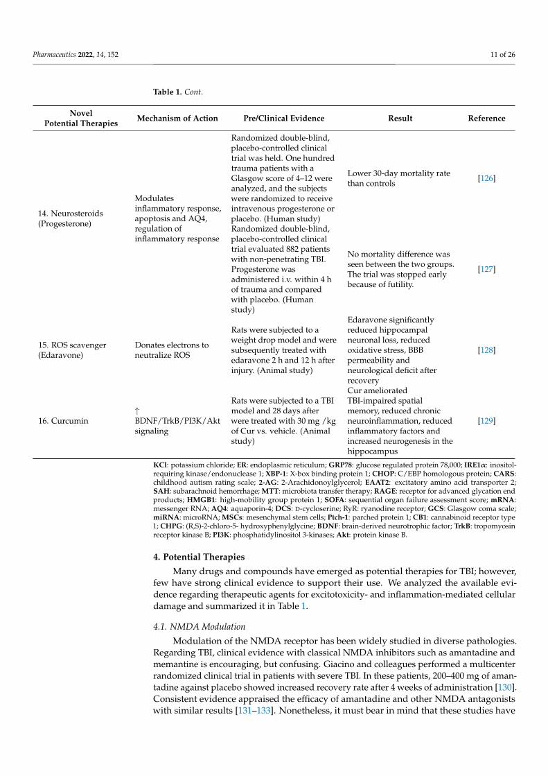
Pharmaceutics 2022, 14, 152 11 of 26
Table 1. Cont.
NovelPotential Therapies Mechanism of Action Pre/Clinical Evidence Result Reference
14. Neurosteroids(Progesterone)
Modulatesinflammatory response,apoptosis and AQ4,regulation ofinflammatory response
Randomized double-blind,placebo-controlled clinicaltrial was held. One hundredtrauma patients with aGlasgow score of 4–12 wereanalyzed, and the subjectswere randomized to receiveintravenous progesterone orplacebo. (Human study)
Lower 30-day mortality ratethan controls [126]
Randomized double-blind,placebo-controlled clinicaltrial evaluated 882 patientswith non-penetrating TBI.Progesterone wasadministered i.v. within 4 hof trauma and comparedwith placebo. (Humanstudy)
No mortality difference wasseen between the two groups.The trial was stopped earlybecause of futility.
[127]
15. ROS scavenger(Edaravone)
Donates electrons toneutralize ROS
Rats were subjected to aweight drop model and weresubsequently treated withedaravone 2 h and 12 h afterinjury. (Animal study)
Edaravone significantlyreduced hippocampalneuronal loss, reducedoxidative stress, BBBpermeability andneurological deficit afterrecovery
[128]
16. Curcumin↑BDNF/TrkB/PI3K/Aktsignaling
Rats were subjected to a TBImodel and 28 days afterwere treated with 30 mg /kgof Cur vs. vehicle. (Animalstudy)
Cur amelioratedTBI-impaired spatialmemory, reduced chronicneuroinflammation, reducedinflammatory factors andincreased neurogenesis in thehippocampus
[129]
KCl: potassium chloride; ER: endoplasmic reticulum; GRP78: glucose regulated protein 78,000; IRE1α: inositol-requiring kinase/endonuclease 1; XBP-1: X-box binding protein 1; CHOP: C/EBP homologous protein; CARS:childhood autism rating scale; 2-AG: 2-Arachidonoylglycerol; EAAT2: excitatory amino acid transporter 2;SAH: subarachnoid hemorrhage; MTT: microbiota transfer therapy; RAGE: receptor for advanced glycation endproducts; HMGB1: high-mobility group protein 1; SOFA: sequential organ failure assessment score; mRNA:messenger RNA; AQ4: aquaporin-4; DCS: D-cycloserine; RyR: ryanodine receptor; GCS: Glasgow coma scale;miRNA: microRNA; MSCs: mesenchymal stem cells; Ptch-1: parched protein 1; CB1: cannabinoid receptor type1; CHPG: (R,S)-2-chloro-5- hydroxyphenylglycine; BDNF: brain-derived neurotrophic factor; TrkB: tropomyosinreceptor kinase B; PI3K: phosphatidylinositol 3-kinases; Akt: protein kinase B.
4. Potential Therapies
Many drugs and compounds have emerged as potential therapies for TBI; however,few have strong clinical evidence to support their use. We analyzed the available evi-dence regarding therapeutic agents for excitotoxicity- and inflammation-mediated cellulardamage and summarized it in Table 1.
4.1. NMDA Modulation
Modulation of the NMDA receptor has been widely studied in diverse pathologies.Regarding TBI, clinical evidence with classical NMDA inhibitors such as amantadine andmemantine is encouraging, but confusing. Giacino and colleagues performed a multicenterrandomized clinical trial in patients with severe TBI. In these patients, 200–400 mg of aman-tadine against placebo showed increased recovery rate after 4 weeks of administration [130].Consistent evidence appraised the efficacy of amantadine and other NMDA antagonistswith similar results [131–133]. Nonetheless, it must bear in mind that these studies have

Pharmaceutics 2022, 14, 152 12 of 26
confounding factors such as the concomitant use of other medications, the natural progressof the disease, and the severity of head trauma. There is also an intrinsic problem in block-ing NMDAr, as glutamate is an essential neurotransmitter related to memory consolidation,neuroprotection, and cell survival, and, in that sense, an effective blockade of NMDArwithout altering other beneficial functions is very hard to achieve [29,134,135]. However,not all NMDA antagonists have shown clinical efficacy in TBI [136]. Other well-studiedNMDAr antagonists such as MK801 [137] and dextromethorphan/quinidine [138] havebeen shown to reduce excitotoxicity, OS, microglial activation, and, therefore, cellulardamage in animal and human studies.
Allosteric modulators have shown better pharmacological properties as they selec-tively block GluN2B, avoiding the undesirable effects of an NMDA blockade [34]. Thisprompted the study of selective GluN2B inhibitors in TBI. Taxoprodil, a GluN2B inhibitor,was studied in a randomized, double-blind, placebo-controlled study to evaluate the effi-cacy of a 72-h intravenous infusion in patients with severe TBI, with improved mortalityand scores in the dichotomized Glasgow scale, although neither showed a significant differ-ence [114]. Ifenprodil, another GluN2B inhibitor, has been shown to reduce excitotoxicity bymodulating [Ca2+]i concentration through blockade of NMDAr in the presence of agonistsor mechanical forces [45], and has also demonstrated reduced BBB breakdown, cerebraledema, and injury volume after controlled cortical impact in rats [113].
On the other hand, there have been studies that suggest NMDA receptors havedifferent pathophysiological implications in TBI depending on the time post-injury. Initially,there is an overactivation of these receptors, leading to excitotoxicity; however, this isfollowed by the desensitization and loss of functional NMDA receptors from more than onehour to seven days post-injury. This was demonstrated by quantitative autoradiographyof MK801 binding to NMDAr [139]. Therefore, under this premise, a partial agonist,D-cycloserine (DCS), was evaluated in TBI. Temple and colleagues [111] evaluated DCSin a fluid percussion TBI model in rats, with a daily intraperitoneal injection of DCS(10–30 mg/kg) given from 24 h until 15 days post-injury. The spatial memory of the ratswas then tested in the Morris water maze, and they found a dose-dependent improvementin the treated group. Similar studies in mice have shown improvement in neurobehavioralfunction at the same doses beginning at 24 h post-injury [112].
During TBI, excessive glutamate release activates glutamate receptors includingmGLUR5. A study evaluated mGLUR5 brain distribution in rat cortices following TBI andsubsequent administration of (R,S)-2-chloro-5-hydroxyphenylglycine (CHPG) as a selectivemGluR5 agonist. Results showed that mGLUR5 was upregulated after TBI in neurons,astrocytes, and microglia. The use of delayed CHPG showed a decrease in the numberof degenerating neurons following TBI due to the reduction of excitotoxic damage [120].Recent encouraging evidence has shown mGLUR5 can modulate inflammation and neu-rotoxicity in a model of Parkinson’s disease, and scientists are also getting encouragingresults in other fields [140,141]. We consider this body of evidence as encouraging for theprospects of mGLUR5 modulators being a feasible therapy in TBI [142,143]. Nonetheless,we do not have a complete understanding of mGLUR5 kinetics and how it can decreaseneurotoxicity and neurodegeneration; therefore, more research still needs to be carried out.
4.2. Dantrolene
Dantrolene is a muscle relaxant that has been used as an anti-spastic agent and for thetreatment of malignant hyperthermia and neuroleptic malignant syndrome, among otherpathologies. Dantrolene acts as a ryanodine receptor (RyR) antagonist; said antagonismends up being beneficial, as the blockade of the RyR reduces the calcium-induced calciumrelease from the endoplasmic reticulum. This mechanism has mainly been studied inskeletal muscle; however, studies in cultured neurons have shown that dantrolene canblock up to 70% of the Ca2+ rise coming from the endoplasmic reticulum because ofNMDAr activation, as well as reducing neuronal apoptosis [115]. The dose required forthe treatment of malignant hyperthermia and neuroleptic malignant syndrome is similar,
Pharmaceutics 2022, 14, 152 13 of 26
oscillating between 1 and 2.5 mg/kg for neuroleptic malignant syndrome and 2.5 mg/kg formalignant hyperthermia [144]. As for the dosage used to achieve the desired effect in TBI,studies have shown how an interval from 10–100 µM of dantrolene administered duringthe appropriate therapeutic window will target and reduce excitotoxicity. Said windowappears to be 40 min either before or after the injury in in vitro models and approximately30 min for in vivo models, making this narrow therapeutic window one of the limitationsfor its usage [145]. We hypothesize that reducing the Ca2+ efflux from the ER will reduce[Ca2+]i, thus reducing excitotoxicity [145–149] and positioning dantrolene as a promisingtherapy for patients with acute TBI; nevertheless, further clinical studies are still needed tomake a definitive clinical recommendation.
4.3. Vitamin B12
Vitamin B12 has been studied widely in animal models for its antinociceptive proper-ties, linking multiple mechanisms of action such as neuronal regeneration, myelin synthesis,Schwann cell differentiation, and the induction of axonal growth to this antinociceptiveeffect [150–152]. These properties motivated the study of vitamin B12 in TBI. A studywas conducted to assess the effect of vitamin B12 on nerve regeneration and on ER stressafter TBI in vivo and in vitro. Controlled cortical impact was performed in male mice,some of which were subsequently treated with vitamin B12 by the intraperitoneal route,and the TBI + vitamin B12 group was compared to the TBI group. The results showedthat vitamin-B12-treated TBI groups had reduced ipsilateral brain edema (assessed bybrain water content), better functional recovery after seven days (evaluated by Garcia test),less tissue damage in the histological morphology, reduced caspase-12-dependent apop-tosis (shown by immunofluorescence staining), downregulation of the ER stress-relatedapoptosis signaling pathway proteins (GRP78, IRE1α, XBP-1 and CHOP) evaluated byWestern blot and immunofluorescence staining, and maintained microtubule stability (adeterminant of axonal growth) [116]. Therefore, vitamin B12 could have a role in preventingspecific hippocampal cell loss (see Section 2.4). Even though the role of vitamin B12 in TBIis very new and not many studies are available, the studies made in animals for nociceptive,nociplastic, neuropathic pain and neuropathy [153,154], combined with the mechanismsdescribed in this review, suggest it is possible that vitamin B12 could work as a logical,effective, and safe therapy which requires further research as a potential treatment in TBIto mitigate secondary injuries and, therefore, long-term disability.
4.4. Ceftriaxone
This beta-lactam antibiotic is proposed as a possible treatment for TBI. Ceftriaxonepenetrates the BBB and increases the expression of EAAT2. As previously discussed,glutamate is the principal contributor for excitotoxicity and its consequences. Glutamatetransporters, such as GLT-1/EAAT2, have been shown to be reduced 24 h followingTBI, thus reducing glutamate reuptake and contributing to excitotoxicity. The potentialtherapeutic capacity of ceftriaxone in TBI is thought to be because it can activate theEAAT2 promoter in human fetal astrocytes [155,156]. In a rat model of subarachnoidhemorrhage (SAH), the use of ceftriaxone was evaluated to prevent early brain injury.Intracisternal treatment with ceftriaxone led to improved neurological outcomes and itwas able to lessen glutamate accumulation after SAH [121]. Furthermore, a recent studyevaluating ceftriaxone in a TBI rat model found that the ceftriaxone-treated group hadsignificantly lower intracranial pressure, decreased infarct volume, decreased neuronalapoptosis, increased GLT-1 expression in both neurons and microglia, and improved motordysfunction [157].
4.5. Minocycline
Tetracyclines have been studied beyond their antibiotic properties, and, as a result,they have been linked to neuroprotection, as well as anti-inflammatory and antiapoptoticactivity. Minocycline is a lipid-soluble, second-generation tetracycline that has potential
Pharmaceutics 2022, 14, 152 14 of 26
benefits in TBI. A study conducted to evaluate if minocycline reduced excitotoxicity inprimary neuronal cultures showed that it increased neuronal survival [158]. The studyshowed minocycline prevented excitotoxic-induced microglial proliferation, and excessiverelease of nitric oxide (NO) and IL-1β. In addition, it has been shown to reduce caspase-1 activation following TBI in mice, as has been seen before in many animal models ofischemia and Huntington’s disease. However, the exact mechanism by which minocyclineinhibits caspase 1 is still to be elucidated and it is beyond the scope of this review [159].Furthermore, a study evaluating the effects of minocycline in TBI-induced neurologicalimpairment found that minocycline increased neuronal viability, caused iron chelationin vitro, and attenuated neurological impairment in rats [117]. Minocycline safety hasbeen studied in a phase IIa clinical trial in TBI (NCT01058395). The study consisted ofadministering intravenous minocycline via a central line within six hours of the initialinjury to patients with a GCS of less than 12, and this treatment continued for seven days.It was found to be safe for moderate to severe TBI [160].
4.6. PSD-95 Inhibitors
NMDA and neuronal NOS are linked via the post synaptic density protein (PSD-95), a guanylate kinase. This protein binds the GluN2B subunit of NMDA to the aminoterminus of nNOS, finally creating the NMDA/PSD-95/nNOS complex. Thus, NMDAoverstimulation in excitotoxicity leads to neurotoxic levels of NO, which participates incytotoxic actions, ultimately leading to neuronal death [161]. Consequently, the inhibition ofPSD-95 has emerged as a possible therapeutic target. A recent study evaluated an inhibitorof this PSD-95/NMDA interaction called ZL006, a small molecule that can readily cross theBBB and that had good outcomes in mouse stroke models [118,162]. This study evaluatedZL006 in cortical neuronal cultures, showing reduced glutamate-induced neuronal death,and showed improvement in somatosensory, motor, and memory deficits, as well ascognitive impairment in rodents [118]. Furthermore, UCCB01-147, a dimeric PSD-95inhibitor, was evaluated in rats to assess its neuroprotective effects. Rats were subjected tocontrolled cortical impact followed by 10 mg/kg of UCCB01. However, it failed to reducecell death [163].
4.7. MicroRNA (miRNA), Mesenchymal Stem Cells (MSC’s), and Exosome Therapy
MiRNA are a family of short, non-coding RNA molecules that can regulate geneexpression at the post-transcriptional level [164]. They have been implicated in the patho-physiology of cancer, as well as cardiovascular and metabolic diseases. They can regulategene expression through the degradation of mRNA or by inhibiting its translation [164]. Ina TBI rat model, the upregulation of miRNA-21 was found to inhibit apoptosis and pro-mote angiogenesis [165]. Following controlled cortical impact in rats, the upregulation ofmiRNA-9-5 improved cellular viability and improved apoptosis by the post-transcriptionalmodulation of Ptch-1 (patched protein 1). It also promoted the expression of factors thatpromote angiogenesis (VEGF, MMP-9 and cyclin D1) [166].
The important role of miRNA has also been elucidated when studying the use ofmesenchymal stem cells (MSCs) in TBI. Exosomes (membrane vesicles) are thought to bean important part in the therapeutic capacity of MSCs because they can carry proteins,lipids, mRNA, and miRNA that can be transferred among cells. Cell-free exosomes derivedfrom MSCs are therefore considered a potential therapeutic strategy, as they can deliverspecific miRNA with neuroprotective effects. Xin and colleagues found that rats exposedto treatment with MSCs after middle cerebral artery occlusion significantly increased theexpression of miRNA-133b, which contributed to neurite outgrowth [167,168]. Zhang andcolleagues studied the systemic administration of cell-free exosomes extracted from humanbone marrow MSCs in rats after TBI [125]. Rats were subjected to controlled cortical impact,and a venous injection of exosomes was administered 24 h later. The exosome treatmentgroup did not have a reduction in lesion size, but had sensorimotor functional recovery

Pharmaceutics 2022, 14, 152 15 of 26
and improved spatial learning by promoting angiogenesis and neurogenesis and reducingneuroinflammation [125].
4.8. Progesterone
Progesterone is a neurosteroid that is involved in neuroprotection and in mechanismsregarding repair after brain injury [169]. It has been tested in various injury models,including TBI, spinal cord injury, and stroke. Some clinical trials have also been carriedout, with reductions in mortality and improved neurologic outcomes [170], whereafter arandomized, double-blind, placebo-controlled clinical trial was held. This study analyzedone hundred trauma patients with a GCS of 4–12, showing no serious adverse effects,and reduced mortality in the progesterone group [126]. Various mechanisms have beenproposed for these neuroprotective effects. Progesterone influences the expression of genesinvolved in the regulation of the inflammatory response, as well as apoptosis. It also reducesedema after TBI by modulating AQ4 (aquaporin 4) expression on astrocytes [171]. Thereare also studies showing that progesterone can reduce lipid peroxidation by inhibitingfree radical formation [169]. Regarding excitotoxicity, progesterone can attenuate neuronalexcitotoxicity by inhibiting voltage-gated calcium channels [172]. However, in 2014, aphase III randomized, placebo-controlled clinical trial was held to evaluate the efficacyof intravenous progesterone in the treatment of non-penetrating TBI. The study analyzed882 patients with a GCS of 4–12. Nevertheless, after the second interim analysis, thetrial was stopped because of futility. There were no significant differences in mortalitybetween groups [127]. Thus, progesterone’s role as neurosteroid is still secondary, despiteits importance.
4.9. Endocannabinoids
Recent published data shows cannabis can have a use in TBI [173]. When 2- arachi-donylglycerol (2-AG) was administered to mice after moderate or severe TBI, activationof cannabinoid receptor type 1 (CB1) attenuated the inflammatory response, protectedthe BBB, and improved clinical recovery. Similar results were obtained by Tchantchouet al. [119] using WWL70 which acts as an inhibitor of monoacylglycerol lipase, an enzymeresponsible for degradation of 2-AG, the most abundant endocannabinoid. WWL70, aswell as other selective inhibitors of endocannabinoid metabolism such as URB597 [174]palmitylsulfonyl fluoride (AM374) [175] have given similar results. This shows the endo-cannabinoid system has potential therapeutical effects by modulating inflammation, andeven excitotoxicity, in TBI. It is important to recognize that the setting in which CB1 isactivated can have different consequences, as some studies suggest that CB1 agonism canact as an enhancer of oxidative stress and inflammation [176]. However, no human studiesin patients with acute TBI in the setting of neuro-ICU care have been done yet.
4.10. Intestinal Microbiota
Microbiota’s role on human physiology is an emerging field of study. Many authorshave pinpointed bacteria as one of the main actors in the metabolism of nutrients, regulationof the immune system, and activation of gene expression, among others, while cooperatingand cohabitating with human beings [177]. It is worth saying that, by this point, there is nosuch thing as a “bad” microbiota and authors have addressed a lack of microbiota diversityor aggressive species colonization as an altered microbiota. Taking this into account,one must have in mind that intestinal microbiota varies depending on the individual,their diet habits, and specific situations such as antibiotic use. Gut–brain signaling is afact, and the vagus nerve, along with the immune and vascular systems, is the bridgein between the two. Bidirectional communication and compliance can be, and has been,used as pharmacological targets in different conditions. For example, in autism, antibiotictherapy, probiotics, and prebiotics have shown improvement in patients’ symptomatologyby altering host microbiota [178]. The hypothesis argues that these treatments enhance fattyacid metabolism, thereby improving the supply of the main products for brain function
Pharmaceutics 2022, 14, 152 16 of 26
and regulating systemic inflammation by controlling proinflammatory cytokines. Commonmolecular pathways, such as pyrin domain-containing 3 (NLRP3) activation, are known toalter intestinal cell homeostasis, thereby promoting bacterial inflammatory states [179,180].NLRP3 inflammasome is also activated after TBI, ultimately leading to calpain activationthrough the rise of [Ca2+]i.
TBI patients can have comorbidities such as obesity and diabetes, which are wellknown to induce systemic inflammatory states [181]. Specific prebiotics enhance anti-inflammatory microorganisms, such as F. prausnitzii, that, through the augmentation ofshort chain fatty acid production, can regulate systemic inflammation and can thereforemodulate brain parenchyma inflammation [182,183]. Nonetheless, there is still muchmore research to be done in this field [184], as TBI patients frequently have concomitantinfections which are treated with antibiotics, which can then influence microbiota diversityand function. We consider that encouraging evidence in autism can be applied in TBI [122].A directed antibiotic regimen depending on individual microbiota characterization and aconsequent reconstitution through microbiota transfer therapy enhancing, for example, thegrowth of Acinetobacter spp., Bacteroides fragilis., and Proteobacteria [185], etc., as well as diet,probiotics and prebiotics, can modulate inflammation and oxidative stress, attenuatingdisability and prompting recovery after TBI.
4.11. RAGE Inhibitors
Inflammation following TBI can activate the receptor for advanced glycation endproducts (RAGE). RAGE activates NF-kB [186], and researchers have broadly addressedan increase of NF-kB activation after TBI [187,188]. Recently published data showed anincreased expression of high mobility group box protein 1 (HMGB1), which activatesinflammation in the acute setting, but has a subsequent correlation to neural genesis bytargeting specific gene expression [123,189,190]. HMGB1–RAGE interaction is well known,but not clearly understood. Apparently, RAGE inhibits HMGB1 expression by directbinding and through NF-kB-mediated endocytosis. Given this complex interaction, weconsider the inhibition of RAGE, but not HMGB1, as a therapeutic target to promote long-term neurogenesis and as an attenuator of inflammation. Nonetheless, no studies have beencompleted with RAGE inhibitors following TBI; despite that, we consider that the effects ofRAGE inhibitors in other fields can shed some light on how we could start exploring thisreceptor in TBI [191,192].
4.12. Zinc Supplementation
Excess free zinc was initially thought to be part of the secondary injury mechanismsfollowing TBI. Therefore, zinc chelation was evaluated for a while as a potential ther-apy [193–195]. However, the chelation of zinc was subsequently associated with neuronaldamage due to overexcitation [196] and increased apoptosis and necrosis [197]. Moreover, ithas been shown that patients with severe head trauma have urinary zinc excretion up to 14-fold higher than normal [198]. Zinc deficiency impairs neurogenesis [199], cell proliferation,neuronal survival, and contributes to the inflammatory response [200]. Furthermore, zinccan function as a neurotransmitter or secondary messenger, and it also regulates cellularoxidant production and signaling cascades in the brain [200]. Therefore, zinc supplementa-tion, rather than zinc chelation, was evaluated in a randomized, controlled clinical trial in100 patients with severe head trauma, aged 18–65 years old, with a GCS of 6–8. Patientswere treated with 528 mg of zinc sulfate for 15 days; results showed improvement of theinflammatory markers, SOFA and GCS, although mortality did not significantly differbetween groups [124].
4.13. Antioxidants
Free radical scavengers such as glutathione and edaravone are reasonable therapies, asthey are antioxidants that can donate electrons to neutralize the toxic effects of ROS [54,201].In a weight drop model conducted by Wang and colleagues, male rats with induced brain
Pharmaceutics 2022, 14, 152 17 of 26
trauma in the right cerebral cortex were subsequently treated with edaravone two hours and12 h after injury. Edaravone significantly reduced hippocampal neuronal loss, oxidativestress, BBB permeability, and neurological deficit after recovery. Therefore, edaravonemight be considered neuroprotective in the case of stroke and TBI [128,202].
4.14. Curcumin
Curcumin (Cur) is a natural polyphenol found in the plant Curcuma longa (turmeric) [203].It has been explored for its properties in promoting neurogenesis and improving memory,as well as for its anti-inflammatory functions. Sun and colleagues evaluated Cur in a murinemodel of TBI, in which they administered 30 mg/kg of Cur vs. vehicle treatment to rats, andsubsequently evaluated neuroinflammation (by assessing inflammatory factors, astrocytehypertrophy, and activated microglia in the hippocampus), and performed behavioral watermaze studies. The authors found Cur ameliorated TBI-impaired spatial memory, reducedchronic neuroinflammation, reduced inflammatory factors, and increased neurogenesis inthe hippocampus [129]. The mechanism by which Cur can cause these effects is thoughtto be through increased signaling of the BDNF/TrkB/PI3K/Akt pathway. The BDNFand TrkB pathways play a role in reducing inflammation and promoting hippocampalneurogenesis, while PI3K/Akt signaling has been linked with the promotion of neuronalsurvival and reducing neuroinflammation [204]. Furthermore, the neuroprotective effectsof Cur have been attributed to the activation of the Nrf2-ARE (nuclear erythroid 2-relatedfactor 2- antioxidant response element) pathway, which modulates oxidative stress. Thiswas documented in a weight drop TBI model in rats in which Cur was shown to increasethe translocation of Nrf2 from the cytoplasm to the nucleus [205].
5. Conclusions
In conclusion, traumatic brain injury represents a high burden for the patient andtheir family, as well as for the healthcare system. Damage to the brain following TBI canbe divided into primary and secondary injuries, the latter being the focus of this review.Managing the secondary mechanisms that lead to tissue damage following TBI has been amajor challenge as these processes are related to tissue recovery, scar formation, and residuemetabolism, leading to further tissue rehabilitation. Despite having many potential drugs,it is difficult to assess which of them can have a positive impact in the recovery pathways.Given the many mechanisms involved, a vast number of potential compounds, mostcoming from drug repurposing, have been proposed as potential therapies. Treatmentsranging from drugs targeting different subunits or associated proteins of the NMDAreceptor to antibiotics, progesterone, vitamin B12, CB1 agonism, prebiotics, and evenmesenchymal stem cell therapy have shown a potential therapeutic role in TBI. However,few have achieved the bench to bedside translation. We strongly believe that widely studiedcompounds, such as selective GluN2B inhibitors and vitamin B12, could have a relevantrole in the treatment of TBI and can have a positive impact on long term disability, therebyreducing disease burden and costs to the health care system.
Author Contributions: Conceptualization: D.B.-S. and D.F.A.-S.; writing—original draft preparation:D.B.-S., D.F.A.-S., M.G.C.-H., M.J.P.-L., I.H.-D. and C.-A.C.-O.; writing—review and editing: D.B.-S.,D.F.A.-S., M.G.C.-H. and C.-A.C.-O.; project administration: C.-A.C.-O.; funding acquisition: D.B.-S.,D.F.A.-S. and C.-A.C.-O.; Figure 1 construction: D.F.A.-S.; Table 1 construction: D.B.-S. All authorshave read and agreed to the published version of the manuscript.
Funding: This project was supported by the Universidad del Rosario.
Acknowledgments: We thank Laura Arango for the illustration of Figure 1.
Conflicts of Interest: The authors declare no conflict of interest.
Pharmaceutics 2022, 14, 152 18 of 26
Abbreviation
TBI: Traumatic Brain Injury; NAC: North American Countries; BBB: Blood Brain Barrier; NMDAr:N-methyl D- aspartate receptor; GABA: Gamma-Aminobutyric acid; EC50: Half maximal effectiveconcentration; AMPA: α-amino-3-hydroxy-5-methyl-4-isoxazolepropionic acid; NMDA: N-methyl D-aspartate; NOS: Nitric Oxide Synthase; ROS: Reactive Oxygen Species; ATP: Adenosine Triphosphate;DNA: Deoxyribonucleic acid; STEP: Striatal-Enriched Protein Tyrosine Phosphatase; ER: EndoplasmicReticulum; UPR: Unfolded Protein Response; PERK: PKR-like ER kinase; ATF6: Activating Transcrip-tion Factor 6; IRE-1: Inositol Requiring Enzyme 1; GRP78/BIP: Glucose-Regulated Protein/BindingImmunoglobulin Protein; eIF2α: PERK phosphorylates eukaryotic initiation factor 2α; CHOP: C/EBPHomologous Protein; SGZ-DG: Subgranular Zone of the Dentate Gyrus; FJP: Ionic Fluorescein; ATF4:Activating Transcription Factor 4; RNS: Reactive Nitrogen Species; PAMPs: Pathogen-AssociatedMolecular Patterns; DAMPs: Damage-Associated Molecular Patterns; mTOR: Mechanistic Target OfRapamycin Kinase; VCAM-1: Vascular Cell Adhesion Molecule-1; ICAM-1: Intercellular Cell Adhe-sion Molecule-1; CNS: Central Nervous System; PKC: Protein kinase C; AKT: Protein kinase B (PKB);GLT-1: Glutamate transporter 1; EAAT2: Excitatory amino acid transporter 2; NF-kB: Nuclear factorkappa-light-chain-enhancer of activated B cells; KCL: Potassium Chloride; GRP78: Glucose-regulatedprotein 78; XBP-1: X-box binding protein 1; NO: Nitric Oxide; PSD-95: Postsynaptic density protein 95;2-AG: 2-Arachidonoylglycerol; CHPG: (R,S)-2-chloro-5- hydroxyphenylglycine; SAH: SubarachnoidHemorrhage; MTT: Microbiota Transfer Therapy; RAGE: Receptor For Advanced Glycation EndProducts; HMGB1: High-Mobility group protein 1; SOFA: Sequential Organ Failure AssessmentScore; mRNA: Messenger RNA; AQ4: Aquaporin-4; DCS: D-cycloserine; RyR: Ryanodine Receptor;GCS: Glasgow Coma Scale; miRNA: MicroRNA; MSCs: Mesenchymal Stem Cells; Ptch-1: ParchedProtein 1; CB1: Cannabinoid Receptor type 1; AM 374: Palmitoyl Sulfonyl Fluoride; NLRP3: PyrinDomain-Containing 3; BDNF/TrkB/PI3K/Akt: Brain-derived Neurotrophic Factor/Tropomyosin re-ceptor kinase B/Phosphatidylinositol 3-kinase/Protein kinase B; Nrf2-ARE: Nuclear factor erythroid2-related factor 2-Antioxidant Response Element.
References1. Capizzi, A.; Woo, J.; Verduzco-Gutierrez, M. Traumatic Brain Injury: An Overview of Epidemiology, Pathophysiology, and
Medical Management. Med. Clin. N. Am. 2020, 104, 213–238. [CrossRef]2. Hackenberg, K.; Unterberg, A. [Traumatic Brain Injury]. Der Nervenarzt 2016, 87, 203–216. [CrossRef]3. Saban, K.; Griffin, J.; Urban, A.; Janusek, M.; Pape, T.; Collins, E. Perceived Health, Caregiver Burden, and Quality of Life in
Women Partners Providing Care to Veterans with Traumatic Brain Injury. J. Rehabil. Res. Dev. 2016, 53, 681–692. [CrossRef][PubMed]
4. Kanmani, T.; Thimmappur, R.; Birudu, R.; Reddy N, K.; Raj, P. Burden and Psychological Distress of Intensive Care UnitCaregivers of Traumatic Brain Injury Patients. Indian J. Crit. Care Med. Peer-Rev. Off. Publ. Indian Soc. Crit. Care Med. 2019, 23,220–223. [CrossRef]
5. Dewan, M.; Rattani, A.; Gupta, S.; Baticulon, R.; Hung, Y.; Punchak, M.; Agrawal, A.; Adeleye, A.; Shrime, M.; Rubiano, A.; et al.Estimating the Global Incidence of Traumatic Brain Injury. J. Neurosurg. 2018, 130, 1080–1097. [CrossRef]
6. Ma, V.; Chan, L.; Carruthers, K. Incidence, Prevalence, Costs, and Impact on Disability of Common Conditions RequiringRehabilitation in the United States: Stroke, Spinal Cord Injury, Traumatic Brain Injury, Multiple Sclerosis, Osteoarthritis,Rheumatoid Arthritis, Limb Loss, and Back Pain. Arch. Phys. Med. Rehabil. 2014, 95, 986–995. [CrossRef] [PubMed]
7. Langlois, J.; Rutland-Brown, W.; Wald, M. The Epidemiology and Impact of Traumatic Brain Injury: A Brief Overview. J. HeadTrauma Rehabil. 2006, 21, 375–378. [CrossRef]
8. Feigin, V.; Vos, T.; Alahdab, F.; Amit, A.; Bärnighausen, T.; Beghi, E.; Beheshti, M.; Chavan, P.; Criqui, M.; Desai, R.; et al. Burdenof Neurological Disorders Across the US From 1990-2017: A Global Burden of Disease Study. JAMA Neurol. 2021, 78, 165–176.[CrossRef]
9. Mckee, A.; Daneshvar, D. The Neuropathology of Traumatic Brain Injury. Handb. Clin. Neurol. 2015, 127, 45–66. [CrossRef][PubMed]
10. Davanzo, J.; Sieg, E.; Timmons, S. Management of Traumatic Brain Injury. Surg. Clin. N. Am. 2017, 97, 1237–1253. [CrossRef]11. Greve, M.W.; Zink, B.J. Pathophysiology of Traumatic Brain Injury. Mt. Sinai J. Med. A J. Transl. Pers. Med. 2009, 76, 97–104.
[CrossRef] [PubMed]12. Pun, P.B.L.; Lu, J.; Moochhala, S. Involvement of ROS in BBB Dysfunction. Free Radic. Res. 2009, 43, 348–364. [CrossRef] [PubMed]13. Alves, J. Blood-Brain Barrier and Traumatic Brain Injury. J. Neurosci. Res. 2014, 92, 141–147. [CrossRef] [PubMed]
Pharmaceutics 2022, 14, 152 19 of 26
14. Alam, A.; Thelin, E.P.; Tajsic, T.; Khan, D.Z.; Khellaf, A.; Patani, R.; Helmy, A. Cellular Infiltration in Traumatic Brain Injury. J.Neuroinflamm. 2020, 17, 1–17. [CrossRef]
15. Winkler, E.; Minter, D.; Yue, J.; Manley, G. Cerebral Edema in Traumatic Brain Injury: Pathophysiology and ProspectiveTherapeutic Targets. Neurosurg. Clin. N. Am. 2016, 27, 473–488. [CrossRef]
16. Jarrahi, A.; Braun, M.; Ahluwalia, M.; Gupta, R.; Wilson, M.; Munie, S.; Ahluwalia, P.; Vender, J.; Vale, F.; Dhandapani, K.;et al. Revisiting Traumatic Brain Injury: From Molecular Mechanisms to Therapeutic Interventions. Biomedicines 2020, 8, 389.[CrossRef]
17. Clark, D.P.Q.; Perreau, V.M.; Shultz, S.R.; Brady, R.D.; Lei, E.; Dixit, S.; Taylor, J.M.; Beart, P.M.; Boon, W.C. Inflammation inTraumatic Brain Injury: Roles for Toxic A1 Astrocytes and Microglial–Astrocytic Crosstalk. Neurochem. Res. 2019, 44, 1410–1424.[CrossRef]
18. Loane, D.; Kumar, A. Microglia in the TBI Brain: The Good, the Bad, and the Dysregulated. Exp. Neurol. 2016, 275 Pt 3, 316–327.[CrossRef]
19. Galgano, M.; Toshkezi, G.; Qiu, X.; Russell, T.; Chin, L.; Zhao, L. Traumatic Brain Injury: Current Treatment Strategies and FutureEndeavors. Cell Transplant. 2017, 26, 197–206. [CrossRef]
20. Sveen, U.; Roe, C.; Sigurdardorrit, S.; Skandsen, T.; Andelic, N.; Manskow, U.; Berntsen, S.; Soberg, H.; Anke, A. Rehabilitationpathways and functional independence one year after severe traumatic brain injury. Eur. J. Phys. Rehabil. Med. 2016, 52, 650–661.[PubMed]
21. Kreitzer, N.; Rath, K.; Kurowski, B.; Bakas, T.; Hart, K.; Lindsell, C.; Adeoye, O. Rehabilitation Practices in Patients With Moderateand Severe Traumatic Brain Injury. J. Head Trauma Rehabil. 2019, 34, E66–E72. [CrossRef] [PubMed]
22. Selassie, A.; Zaloshnja, E.; Langlois, J.; Miller, T.; Jones, P.; Steiner, C. Incidence of Long-Term Disability Following TraumaticBrain Injury Hospitalization, United States, 2003. J. Head Trauma Rehabil. 2008, 23, 123–131. [CrossRef]
23. Zaloshnja, E.; Miller, T.; Langlois, J.; Selassie, A. Prevalence of Long-Term Disability from Traumatic Brain Injury in the CivilianPopulation of the United States, 2005. J. Head Trauma Rehabil. 2008, 23, 394–400. [CrossRef] [PubMed]
24. Rigotto, G.; Zentilin, L.; Pozzan, T.; Basso, E. Effects of Mild Excitotoxic Stimulus on Mitochondria Ca2+ Handling in HippocampalCultures of a Mouse Model of Alzheimer’s Disease. Cells 2021, 10, 2046. [CrossRef]
25. Jankovic, M.; Novakovic, I.; Dawod, P.G.A.; Dawod, A.G.A.; Drinic, A.; Motaleb, F.I.A.; Ducic, S.; Nikolic, D. Current Conceptson Genetic Aspects of Mitochondrial Dysfunction in Amyotrophic Lateral Sclerosis. Int. J. Mol. Sci. 2021, 22, 9832. [CrossRef][PubMed]
26. Villanueva, J.R.; Esteban, J.M.; Villanueva, L.J.R. Retinal Cell Protection in Ocular Excitotoxicity Diseases. Possible AlternativesOffered by Microparticulate Drug Delivery Systems and Future Prospects. Pharmaceutics 2020, 12, 94. [CrossRef] [PubMed]
27. Kostic, M.; Zivkovic, N.; Stojanovic, I. Multiple Sclerosis and Glutamate Excitotoxicity. Rev. Neurosci. 2013, 24, 71–88. [CrossRef][PubMed]
28. Hynd, M.; Scott, H.; Dodd, P. Glutamate-Mediated Excitotoxicity and Neurodegeneration in Alzheimer’s Disease. Neurochem. Int.2004, 45, 583–595. [CrossRef]
29. Ng, S.Y.; Lee, A.Y.W. Traumatic Brain Injuries: Pathophysiology and Potential Therapeutic Targets. Front. Cell. Neurosci. 2019, 13,528. [CrossRef] [PubMed]
30. Kyyriäinen, J.; Kajevu, N.; Bañuelos, I.; Lara, L.; Lipponen, A.; Balosso, S.; Hämäläinen, E.; das Gupta, S.; Puhakka, N.; Natunen,T.; et al. Targeting Oxidative Stress with Antioxidant Duotherapy after Experimental Traumatic Brain Injury. Int. J. Mol. Sci. 2021,22, 10555. [CrossRef]
31. Hansen, K.B.; Yi, F.; Perszyk, R.E.; Furukawa, H.; Wollmuth, L.P.; Gibb, A.J.; Traynelis, S.F. Structure, Function, and AllostericModulation of NMDA Receptors. J. Gen. Physiol. 2018, 150, 1081–1105. [CrossRef]
32. Guo, H.; Camargo, L.M.; Yeboah, F.; Digan, M.E.; Niu, H.; Pan, Y.; Reiling, S.; Soler-Llavina, G.; Weihofen, W.A.; Wang, H.-R.; et al.A NMDA-Receptor Calcium Influx Assay Sensitive to Stimulation by Glutamate and Glycine/D-Serine. Sci. Rep. 2017, 7, 1–13.[CrossRef]
33. Samson, A.; Robertson, G.; Zagnoni, M.; Connolly, C. Neuronal Networks Provide Rapid Neuroprotection against SpreadingToxicity. Sci. Rep. 2016, 6, 33746. [CrossRef]
34. Zhu, S.; Paoletti, P. Allosteric Modulators of NMDA Receptors: Multiple Sites and Mechanisms. Curr. Opin. Pharmacol. 2015, 20,14–23. [CrossRef]
35. Chiang, V.S.-C.; Park, J.H. Glutamate in Male and Female Sexual Behavior: Receptors, Transporters, and Steroid Independence.Front. Behav. Neurosci. 2020, 14, 211. [CrossRef] [PubMed]
36. Palacios-Filardo, J.; Mellor, J. Neuromodulation of Hippocampal Long-Term Synaptic Plasticity. Curr. Opin. Neurobiol. 2019, 54,37–43. [CrossRef] [PubMed]
37. Ladak, A.; Enam, S.; Ibrahim, M. A Review of the Molecular Mechanisms of Traumatic Brain Injury. World Neurosurg. 2019, 131,126–132. [CrossRef] [PubMed]
38. Hardingham, G.E.; Fukunaga, Y.; Bading, H. Extrasynaptic NMDARs Oppose Synaptic NMDARs by Triggering CREB Shut-offand Cell Death Pathways. Nat. Neurosci. 2002, 5, 405–414. [CrossRef]
39. Kunz, A.; Dirnagl, U.; Mergenthaler, P. Acute Pathophysiological Processes after Ischaemic and Traumatic Brain Injury. Best Pract.Res. Clin. Anaesthesiol. 2010, 24, 495–509. [CrossRef] [PubMed]
Pharmaceutics 2022, 14, 152 20 of 26
40. Guerriero, R.; Giza, C.; Rotenberg, A. Glutamate and GABA Imbalance Following Traumatic Brain Injury. Curr. Neurol. Neurosci.Rep. 2015, 15, 27. [CrossRef]
41. Tasker, R. Spreading Depolarisations and Traumatic Brain Injury: Time Course and Mechanisms. Lancet. Neurol. 2012, 11, 389.[CrossRef]
42. Lee, M.C.; Ting, K.K.; Adams, S.; Brew, B.J.; Chung, R.; Guillemin, G.J. Characterisation of the Expression of NMDA Receptors inHuman Astrocytes. PLoS ONE 2010, 5, e14123. [CrossRef] [PubMed]
43. Kloda, A.; Lua, L.; Hall, R.; Adams, D.J.; Martinac, B. Liposome Reconstitution and Modulation of Recombinant N-Methyl-d-Aspartate Receptor Channels by Membrane Stretch. Proc. Natl. Acad. Sci. USA 2007, 104, 1540–1545. [CrossRef]
44. Maneshi, M.M.; Maki, B.; Gnanasambandam, R.; Belin, S.; Popescu, G.K.; Sachs, F.; Hua, S.Z. Mechanical Stress Activates NMDAReceptors in the Absence of Agonists. Sci. Rep. 2017, 7, 1–10. [CrossRef]
45. Singh, P.; Doshi, S.; Spaethling, J.; Hockenberry, A.; Patel, T.; Geddes-Klein, D.; Lynch, D.; Meaney, D. N-Methyl-D-AspartateReceptor Mechanosensitivity Is Governed by C Terminus of NR2B Subunit. J. Biol. Chem. 2012, 287, 4348–4359. [CrossRef][PubMed]
46. Hånell, A.; Rostami, E. How Can a Punch Knock You Out? Front. Neurol. 2020, 11, 1280. [CrossRef]47. Pettus, E.; Povlishock, J. Characterization of a Distinct Set of Intra-Axonal Ultrastructural Changes Associated with Traumatically
Induced Alteration in Axolemmal Permeability. Brain Res. 1996, 722, 1–11. [CrossRef]48. Farkas, O.; Lifshitz, J.; Povlishock, J.T. Mechanoporation Induced by Diffuse Traumatic Brain Injury: An Irreversible or Reversible
Response to Injury? J. Neurosci. 2006, 26, 3130–3140. [CrossRef]49. Boothe, D.L.; Yu, A.B.; Kudela, P.; Anderson, W.S.; Vettel, J.M.; Franaszczuk, P.J. Impact of Neuronal Membrane Damage on the
Local Field Potential in a Large-Scale Simulation of Cerebral Cortex. Front. Neurol. 2017, 8, 236. [CrossRef] [PubMed]50. LaPlaca, M.; Lessing, M.; Prado, G.; Zhou, R.; Tate, C.; Geddes-Klein, D.; Meaney, D.; Zhang, L. Mechanoporation Is a Potential
Indicator of Tissue Strain and Subsequent Degeneration Following Experimental Traumatic Brain Injury. Clin. Biomech. (BristolAvon) 2019, 64, 2–13. [CrossRef] [PubMed]
51. Cullen, D.K.; Vernekar, V.N.; LaPlaca, M.C. Trauma-Induced Plasmalemma Disruptions in Three-Dimensional Neural CulturesAre Dependent on Strain Modality and Rate. J. Neurotrauma 2011, 28, 2219–2233. [CrossRef]
52. Kilinc, D.; Gallo, G.; Barbee, K. Mechanical Membrane Injury Induces Axonal Beading through Localized Activation of Calpain.Exp. Neurol. 2009, 219, 553–561. [CrossRef]
53. Park, J.; Jang, S.; Shin, Y.; Suh, D.; Park, H. Calcium-Dependent Proteasome Activation Is Required for Axonal NeurofilamentDegradation. Neural Regen. Res. 2013, 8, 3401–3409. [CrossRef] [PubMed]
54. Lucke-Wold, B.; Logsdon, A.; Nguyen, L.; Eltanahay, A.; Turner, R.; Bonasso, P.; Knotts, C.; Moeck, A.; Maroon, J.; Bailes, J.; et al.Supplements, Nutrition, and Alternative Therapies for the Treatment of Traumatic Brain Injury. Nutr. Neurosci. 2018, 21, 79–91.[CrossRef]
55. Petronilho, F.; Feier, G.; de Souza, B.; Guglielmi, C.; Constantino, L.S.; Walz, R.; Quevedo, J.; Dal-Pizzol, F. Oxidative Stress inBrain According to Traumatic Brain Injury Intensity. J. Surg. Res. 2010, 164, 316–320. [CrossRef]
56. Dong, X.; Wang, Y.; Qin, Z. Molecular Mechanisms of Excitotoxicity and Their Relevance to Pathogenesis of NeurodegenerativeDiseases. Acta Pharmacol. Sin. 2009, 30, 379–387. [CrossRef]
57. Lewén, A.; Fujimura, M.; Sugawara, T.; Matz, P.; Copin, J.-C.; Chan, P.H. Oxidative Stress–Dependent Release of MitochondrialCytochrome c after Traumatic Brain Injury. J. Cereb. Blood Flow Metab. 2016, 21, 914–920. [CrossRef]
58. Schimmel, S.; Acosta, S.; Lozano, D. Neuroinflammation in Traumatic Brain Injury: A Chronic Response to an Acute Injury. BrainCirc. 2017, 3, 135. [CrossRef] [PubMed]
59. Ryan, K.C.; Ashkavand, Z.; Norman, K.R. The Role of Mitochondrial Calcium Homeostasis in Alzheimer’s and Related Diseases.Int. J. Mol. Sci. 2020, 21, 9153. [CrossRef] [PubMed]
60. Denault, J.-B.; Salvesen, G.S. Caspases. Curr. Protoc. Protein Sci. 2001, 26, 21–28. [CrossRef] [PubMed]61. Liu, W.; Chen, Y.; Meng, J.; Wu, M.; Bi, F.; Chang, C.; Li, H.; Zhang, L. Ablation of Caspase-1 Protects against TBI-Induced
Pyroptosis in Vitro and in Vivo. J. Neuroinflamm. 2018, 15, 1–16. [CrossRef]62. Abdul-Muneer, P.M.; Long, M.; Conte, A.A.; Santhakumar, V.; Pfister, B.J. High Ca2+ Influx During Traumatic Brain Injury Leads
to Caspase-1-Dependent Neuroinflammation and Cell Death. Mol. Neurobiol. 2016, 54, 3964–3975. [CrossRef]63. McIlwain, D.R.; Berger, T.; Mak, T.W. Caspase Functions in Cell Death and Disease. Cold Spring Harb. Perspect. Biol. 2013, 5,
a008656. [CrossRef]64. Clark, R.; Kochanek, P.; Watkins, S.; Chen, M.; Dixon, C.; Seidberg, N.; Melick, J.; Loeffert, J.; Nathaniel, P.; Jin, K.; et al. Caspase-3
Mediated Neuronal Death after Traumatic Brain Injury in Rats. J. Neurochem. 2000, 74, 740–753. [CrossRef]65. Wang, Y.; Liu, Y.; Bi, X.; Baudry, M. Calpain-1 and Calpain-2 in the Brain: New Evidence for a Critical Role of Calpain-2 in
Neuronal Death. Cells 2020, 9, 2698. [CrossRef]66. Saatman, K.E.; Creed, J.; Raghupathi, R. Calpain as a Therapeutic Target in Traumatic Brain Injury. Neurotherapeutics 2010, 7,
31–42. [CrossRef]67. Wang, Y.; Briz, V.; Chishti, A.; Bi, X.; Baudry, M. Distinct Roles for µ-Calpain and m-Calpain in Synaptic NMDAR-Mediated
Neuroprotection and Extrasynaptic NMDAR-Mediated Neurodegeneration. J. Neurosci. 2013, 33, 18880–18892. [CrossRef][PubMed]
Pharmaceutics 2022, 14, 152 21 of 26
68. Xu, J.; Kurup, P.; Zhang, Y.; Goebel-Goody, S.M.; Wu, P.H.; Hawasli, A.H.; Baum, M.L.; Bibb, J.A.; Lombroso, P.J. ExtrasynapticNMDA Receptors Couple Preferentially to Excitotoxicity via Calpain-Mediated Cleavage of STEP. J. Neurosci. 2009, 29, 9330–9343.[CrossRef] [PubMed]
69. Wang, Z.F.; Gao, C.; Chen, W.; Gao, Y.; Wang, H.C.; Meng, Y.; Luo, C.L.; Zhang, M.Y.; Chen, G.; Chen, X.P.; et al. Salubrinal OffersNeuroprotection through Suppressing Endoplasmic Reticulum Stress, Autophagy and Apoptosis in a Mouse Traumatic BrainInjury Model. Neurobiol. Learn. Mem. 2019, 161, 12–25. [CrossRef] [PubMed]
70. Liu, S.; Jin, R.; Xiao, A.; Chen, R.; Li, J.; Zhong, W.; Feng, X.; Li, J. Induction of Neuronal PI3Kγ Contributes to EndoplasmicReticulum Stress and Long-Term Functional Impairment in a Murine Model of Traumatic Brain Injury. Neurother. J. Am. Soc. Exp.NeuroTher. 2019, 16, 1320–1334. [CrossRef]
71. Sun, D.; Wang, J.; Liu, X.; Fan, Y.; Yang, M.; Zhang, J. Dexmedetomidine Attenuates Endoplasmic Reticulum Stress-InducedApoptosis and Improves Neuronal Function after Traumatic Brain Injury in Mice. Brain Res. 2020, 1732, 146682. [CrossRef]
72. Gage, F. Mammalian Neural Stem Cells. Science 2000, 287, 1433–1438. [CrossRef] [PubMed]73. Hall, E.D.; Sullivan, P.G.; Gibson, T.R.; Pavel, K.M.; Thompson, B.M.; Scheff, S.W. Spatial and Temporal Characteristics of
Neurodegeneration after Controlled Cortical Impact in Mice: More than a Focal Brain Injury. J. Neurotrauma 2005, 22, 252–265.[CrossRef]
74. Thompson, H.J.; Lifshitz, J.; Marklund, N.; Grady, M.S.; Graham, D.I.; Hovda, D.A.; McIntosh, T.K. Lateral Fluid Percussion BrainInjury: A 15-Year Review and Evaluation. J. Neurotrauma 2005, 22, 42–75. [CrossRef] [PubMed]
75. Mehrholz, J.; Major, Y.; Meissner, D.; Sandi-Gahun, S.; Koch, R.; Pohl, M. The Influence of Contractures and Variation inMeasurement Stretching Velocity on the Reliability of the Modified Ashworth Scale in Patients with Severe Brain Injury. Clin.Rehabil. 2005, 19, 63–72. [CrossRef] [PubMed]
76. Gao, X.; Deng-Bryant, Y.; Cho, W.; Carrico, K.; Hall, E.; Chen, J. Selective Death of Newborn Neurons in Hippocampal DentateGyrus Following Moderate Experimental Traumatic Brain Injury. J. Neurosci. Res. 2008, 86, 2258–2270. [CrossRef] [PubMed]
77. Deng, W.; Aimone, J.B.; Gage, F.H. New Neurons and New Memories: How Does Adult Hippocampal Neurogenesis AffectLearning and Memory? Nat. Rev. Neurosci. 2010, 11, 339–350. [CrossRef] [PubMed]
78. Dupret, D.; Revest, J.-M.; Koehl, M.; Ichas, F.; de Giorgi, F.; Costet, P.; Abrous, D.N.; Piazza, P.V. Spatial Relational MemoryRequires Hippocampal Adult Neurogenesis. PLoS ONE 2008, 3, e1959. [CrossRef]
79. Zhou, H.; Chen, L.; Gao, X.; Luo, B.; Chen, J. Moderate Traumatic Brain Injury Triggers Rapid Necrotic Death of ImmatureNeurons in the Hippocampus. J. Neuropathol. Exp. Neurol. 2012, 71, 348–359. [CrossRef]
80. Anderson, K.; Miller, K.; Fugaccia, I.; Scheff, S. Regional Distribution of Fluoro-Jade B Staining in the Hippocampus FollowingTraumatic Brain Injury. Exp. Neurol. 2005, 193, 125–130. [CrossRef]
81. Sato, M.; Chang, E.; Igarashi, T.; Noble, L. Neuronal Injury and Loss after Traumatic Brain Injury: Time Course and RegionalVariability. Brain Res. 2001, 917, 45–54. [CrossRef]
82. Zhao, Y.; Xiang, Q.; Shi, S.; Li, S.; Tan, L.; Wang, J.; Jin, X.; Luo, A. GABAergic Excitotoxicity Injury of the Immature HippocampalPyramidal Neurons’ Exposure to Isoflurane. Anesth. Analg. 2011, 113, 1152–1160. [CrossRef]
83. Hood, K.N.; Zhao, J.; Redell, J.B.; Hylin, M.J.; Harris, B.; Perez, A.; Moore, A.N.; Dash, P.K. Endoplasmic Reticulum StressContributes to the Loss of Newborn Hippocampal Neurons after Traumatic Brain Injury. J. Neurosci. 2018, 38, 2372–2384.[CrossRef]
84. Redell, J.B.; Maynard, M.E.; Underwood, E.L.; Vita, S.M.; Dash, P.K.; Kobori, N. Traumatic Brain Injury and HippocampalNeurogenesis: Functional Implications. Exp. Neurol. 2020, 331, 113372. [CrossRef] [PubMed]
85. Liu, Y.-W.; Li, S.; Dai, S.-S. Neutrophils in Traumatic Brain Injury (TBI): Friend or Foe? J. Neuroinflamm. 2018, 15, 1–18. [CrossRef][PubMed]
86. Shi, K.; Zhang, J.; Dong, J.; Shi, F.-D. Dissemination of Brain Inflammation in Traumatic Brain Injury. Cell. Mol. Immunol. 2019, 16,523–530. [CrossRef]
87. Szmydynger-Chodobska, J.; Strazielle, N.; Gandy, J.R.; Keefe, T.H.; Zink, B.J.; Ghersi-Egea, J.-F.; Chodobski, A. PosttraumaticInvasion of Monocytes across the Blood—Cerebrospinal Fluid Barrier. J. Cereb. Blood Flow Metab. 2011, 32, 93–104. [CrossRef][PubMed]
88. Corps, K.N.; Roth, T.L.; McGavern, D.B. Inflammation and Neuroprotection in Traumatic Brain Injury. JAMA Neurol. 2015, 72,355–362. [CrossRef] [PubMed]
89. Hu, Y.; Mai, W.; Chen, L.; Cao, K.; Zhang, B.; Zhang, Z.; Liu, Y.; Lou, H.; Duan, S.; Gao, Z. MTOR-Mediated MetabolicReprogramming Shapes Distinct Microglia Functions in Response to Lipopolysaccharide and ATP. Glia 2020, 68, 1031–1045.[CrossRef]
90. Carpentier, P.A.; Begolka, W.S.; Olson, J.K.; Elhofy, A.; Karpus, W.J.; Miller, S.D. Differential Activation of Astrocytes by Innateand Adaptive Immune Stimuli. Glia 2005, 49, 360–374. [CrossRef] [PubMed]
91. An, Y.; Chen, Q.; Quan, N. Interleukin-1 Exerts Distinct Actions on Different Cell Types of the Brain in Vitro. J. Inflamm. Res. 2011,4, 11–20. [CrossRef]
92. Ghafourifar, P.; Bringold, U.; Klein, S.D.; Richter, C. Mitochondrial Nitric Oxide Synthase, Oxidative Stress and Apoptosis.Neurosignals 2001, 10, 57–65. [CrossRef]
Pharmaceutics 2022, 14, 152 22 of 26
93. Izzy, S.; Liu, Q.; Fang, Z.; Lule, S.; Wu, L.; Chung, J.Y.; Sarro-Schwartz, A.; Brown-Whalen, A.; Perner, C.; Hickman, S.E.; et al.Time-Dependent Changes in Microglia Transcriptional Networks Following Traumatic Brain Injury. Front. Cell. Neurosci. 2019, 13,307. [CrossRef]
94. Witcher, K.G.; Bray, C.E.; Dziabis, J.E.; McKim, D.B.; Benner, B.N.; Rowe, R.K.; Kokiko-Cochran, O.N.; Popovich, P.G.; Lifshitz,J.; Eiferman, D.S.; et al. Traumatic Brain Injury-Induced Neuronal Damage in the Somatosensory Cortex Causes Formationof Rod-Shaped Microglia That Promote Astrogliosis and Persistent Neuroinflammation. Glia 2018, 66, 2719–2736. [CrossRef][PubMed]
95. Dinet, V.; Petry, K.G.; Badaut, J. Brain–Immune Interactions and Neuroinflammation After Traumatic Brain Injury. Front. Neurosci.2019, 13, 1178. [CrossRef] [PubMed]
96. Kumar, A.; Stoica, B.A.; Loane, D.J.; Yang, M.; Abulwerdi, G.; Khan, N.; Kumar, A.; Thom, S.R.; Faden, A.I. Microglial-DerivedMicroparticles Mediate Neuroinflammation after Traumatic Brain Injury. J. Neuroinflamm. 2017, 14, 1–17. [CrossRef]
97. Fei, M.; Wang, H.; Zhou, M.; Deng, C.; Zhang, L.; Han, Y. Podoplanin Influences the Inflammatory Phenotypes and Mobility ofMicroglia in Traumatic Brain Injury. Biochem. Biophys. Res. Commun. 2020, 523, 361–367. [CrossRef]
98. Gentleman, S.M.; Leclercq, P.D.; Moyes, L.; Graham, D.I.; Smith, C.; Griffin, W.S.T.; Nicoll, J.A.R. Long-Term IntracerebralInflammatory Response after Traumatic Brain Injury. Forensic Sci. Int. 2004, 146, 97–104. [CrossRef] [PubMed]
99. Ndode-Ekane, X.E.; Matthiesen, L.; Bañuelos-Cabrera, I.; Palminha, C.A.P.; Pitkänen, A. T-Cell Infiltration into the PerilesionalCortex Is Long-Lasting and Associates with Poor Somatomotor Recovery after Experimental Traumatic Brain Injury. Restor.Neurol. Neurosci. 2018, 36, 485–501. [CrossRef]
100. Clausen, F.; Lorant, T.; Lewén, A.; Hillered, L. T Lymphocyte Trafficking: A Novel Target for Neuroprotection in Traumatic BrainInjury. J. Neurotrauma 2007, 24, 1295–1307. [CrossRef]
101. Newell-Rogers, M.K.; Rogers, S.K.; Tobin, R.P.; Mukherjee, S.; Shapiro, L.A. Antagonism of Macrophage Migration InhibitoryFactory (MIF) after Traumatic Brain Injury Ameliorates Astrocytosis and Peripheral Lymphocyte Activation and Expansion. Int. J.Mol. Sci. 2020, 21, 7448. [CrossRef] [PubMed]
102. Daglas, M.; Draxler, D.F.; Ho, H.; McCutcheon, F.; Galle, A.; Au, A.E.; Larsson, P.; Gregory, J.; Alderuccio, F.; Sashindranath, M.;et al. Activated CD8 + T Cells Cause Long-Term Neurological Impairment after Traumatic Brain Injury in Mice. Cell Rep. 2019, 29,1178–1191.e6. [CrossRef]
103. Raad, M.; Nohra, E.; Chams, N.; Itani, M.; Talih, F.; Mondello, S.; Kobeissy, F. Autoantibodies in Traumatic Brain Injury andCentral Nervous System Trauma. Neuroscience 2014, 281, 16–23. [CrossRef] [PubMed]
104. Sajja, V.S.S.S.; Hlavac, N.; VandeVord, P.J. Role of Glia in Memory Deficits Following Traumatic Brain Injury: Biomarkers of GliaDysfunction. Front. Integr. Neurosci. 2016, 10, 7. [CrossRef]
105. González-Reyes, R.E.; Nava-Mesa, M.O.; Vargas-Sánchez, K.; Ariza-Salamanca, D.; Mora-Muñoz, L. Involvement of Astrocytes inAlzheimer’s Disease from a Neuroinflammatory and Oxidative Stress Perspective. Front. Mol. Neurosci. 2017, 10, 427. [CrossRef]
106. Jassam, Y.N.; Izzy, S.; Whalen, M.; McGavern, D.B.; el Khoury, J. Neuroimmunology of Traumatic Brain Injury: Time for aParadigm Shift. Neuron 2017, 95, 1246–1265. [CrossRef]
107. Liddelow, S.A.; Guttenplan, K.A.; Clarke, L.E.; Bennett, F.C.; Bohlen, C.J.; Schirmer, L.; Bennett, M.L.; Münch, A.E.; Chung, W.-S.;Peterson, T.C.; et al. Neurotoxic Reactive Astrocytes Are Induced by Activated Microglia. Nature 2017, 541, 481–487. [CrossRef][PubMed]
108. Dorsett, C.; McGuire, J.; Niedzielko, T.; DePasquale, E.; Meller, J.; Floyd, C.; McCullumsmith, R. Traumatic Brain Injury InducesAlterations in Cortical Glutamate Uptake without a Reduction in Glutamate Transporter-1 Protein Expression. J. Neurotrauma2017, 34, 220–234. [CrossRef] [PubMed]
109. Pajarillo, E.; Rizor, A.; Lee, J.; Aschner, M.; Lee, E. The Role of Astrocytic Glutamate Transporters GLT-1 and GLAST inNeurological Disorders: Potential Targets for Neurotherapeutics. Neuropharmacology 2019, 161, 107559. [CrossRef]
110. Gupta, R.; Prasad, S. Early down Regulation of the Glial Kir4.1 and GLT-1 Expression in Pericontusional Cortex of the Old MaleMice Subjected to Traumatic Brain Injury. Biogerontology 2013, 14, 531–541. [CrossRef] [PubMed]
111. Temple, M.; Hamm, R. Chronic, Post-Injury Administration of D-Cycloserine, an NMDA Partial Agonist, Enhances CognitivePerformance Following Experimental Brain Injury. Brain Res. 1996, 741, 246–251. [CrossRef]
112. Adeleye, A.; Shohami, E.; Nachman, D.; Alexandrovich, A.; Trembovler, V.; Yaka, R.; Shoshan, Y.; Dhawan, J.; Biegon, A.D-Cycloserine Improves Functional Outcome after Traumatic Brain Injury with Wide Therapeutic Window. Eur. J. Pharmacol.2010, 629, 25–30. [CrossRef] [PubMed]
113. Dempsey, R.; Baskaya, M.; Dogan, A. Attenuation of Brain Edema, Blood-Brain Barrier Breakdown, and Injury Volume byIfenprodil, a Polyamine-Site N-Methyl-D-Aspartate Receptor Antagonist, after Experimental Traumatic Brain Injury in Rats.Neurosurgery 2000, 47, 399–406. [CrossRef] [PubMed]
114. Yurkewicz, L.; Weaver, J.; Bullock, M.R.; Marshall, L.F. The Effect of the Selective NMDA Receptor Antagonist Traxoprodil in theTreatment of Traumatic Brain Injury. J. Neurotrauma 2005, 22, 1428–1443. [CrossRef]
115. Frandsen, A.; Schousboe, A. Dantrolene Prevents Glutamate Cytotoxicity and Ca2+ Release from Intracellular Stores in CulturedCerebral Cortical Neurons. J. Neurochem. 1991, 56, 1075–1078. [CrossRef]
116. Wu, F.; Xu, K.; Lei, L.; Zhang, K.; Xia, L.; Zhang, M.; Teng, C.; Tong, H.; He, Y.; Xue, Y.; et al. Vitamin B12 Enhances Nerve Repairand Improves Functional Recovery After Traumatic Brain Injury by Inhibiting ER Stress-Induced Neuron Injury. Front. Pharmacol.2019, 10, 406. [CrossRef] [PubMed]
Pharmaceutics 2022, 14, 152 23 of 26
117. Zhang, L.; Xiao, H.; Yu, X.; Deng, Y. Minocycline Attenuates Neurological Impairment and Regulates Iron Metabolism in a RatModel of Traumatic Brain Injury. Arch. Biochem. Biophys. 2020, 682, 108302. [CrossRef]
118. Qu, W.; Liu, N.-K.; Wu, X.; Wang, Y.; Xia, Y.; Sun, Y.; Lai, Y.; Li, R.; Shekhar, A.; Xu, X.-M. Disrupting NNOS–PSD95 InteractionImproves Neurological and Cognitive Recoveries after Traumatic Brain Injury. Cereb. Cortex 2020, 30, 3859–3871. [CrossRef][PubMed]
119. Tchantchou, F.; Zhang, Y. Selective Inhibition of Alpha/Beta-Hydrolase Domain 6 Attenuates Neurodegeneration, AlleviatesBlood Brain Barrier Breakdown, and Improves Functional Recovery in a Mouse Model of Traumatic Brain Injury. J. Neurotrauma2013, 30, 565–579. [CrossRef]
120. Wang, J.; Wang, H.; Zhong, W.; Li, N.; Cong, Z. Expression and Cell Distribution of Metabotropic Glutamate Receptor 5 in the RatCortex Following Traumatic Brain Injury. Brain Res. 2012, 1464, 73–81. [CrossRef] [PubMed]
121. Feng, D.; Wang, W.; Dong, Y.; Wu, L.; Huang, J.; Ma, Y.; Zhang, Z.; Wu, S.; Gao, G.; Qin, H. Ceftriaxone Alleviates Early BrainInjury after Subarachnoid Hemorrhage by Increasing Excitatory Amino Acid Transporter 2 Expression via the PI3K/Akt/NF-KBSignaling Pathway. Neuroscience 2014, 268, 21–32. [CrossRef]
122. Kang, D.-W.; Adams, J.B.; Coleman, D.M.; Pollard, E.L.; Maldonado, J.; McDonough-Means, S.; Caporaso, J.G.; Krajmalnik-Brown,R. Long-Term Benefit of Microbiota Transfer Therapy on Autism Symptoms and Gut Microbiota. Sci. Rep. 2019, 9, 1–9. [CrossRef][PubMed]
123. Fan, H.; Tang, H.-B.; Chen, Z.; Wang, H.-Q.; Zhang, L.; Jiang, Y.; Li, T.; Yang, C.-F.; Wang, X.-Y.; Li, X.; et al. Inhibiting HMGB1-RAGE Axis Prevents pro-Inflammatory Macrophages/Microglia Polarization and Affords Neuroprotection after Spinal CordInjury. J. Neuroinflamm. 2020, 17, 1–17. [CrossRef] [PubMed]
124. Khazdouz, M.; Mazidi, M.; Ehsaei, M.; Ferns, G.; Kengne, A.P.; Norouzy, A.-R. Impact of Zinc Supplementation on the ClinicalOutcomes of Patients with Severe Head Trauma: A Double-Blind Randomized Clinical Trial. J. Diet Suppl. 2017, 15, 1–10.[CrossRef] [PubMed]
125. Zhang, Y.; Chopp, M.; Zhang, Z.; Katakowski, M.; Xin, H.; Qu, C.; Ali, M.; Mahmood, A.; Xiong, Y. Systemic Administration ofCell-Free Exosomes Generated by Human Bone Marrow Derived Mesenchymal Stem Cells Cultured under 2D and 3D ConditionsImproves Functional Recovery in Rats after Traumatic Brain Injury. Neurochem. Int. 2017, 111, 69–81. [CrossRef] [PubMed]
126. Wright, D.; Kellermann, A.; Hertzberg, V.; Clark, P.; Frankel, M.; Goldstein, F.; Salomone, J.; Dent, L.; Harris, O.; Ander, D.; et al.ProTECT: A Randomized Clinical Trial of Progesterone for Acute Traumatic Brain Injury. Ann. Emerg. Med. 2007, 49, 391–402.[CrossRef]
127. Wright, D.W.; Yeatts, S.D.; Silbergleit, R.; Palesch, Y.Y.; Hertzberg, V.S.; Frankel, M.; Goldstein, F.C.; Caveney, A.F.; Howlett-Smith,H.; Bengelink, E.M.; et al. Very Early Administration of Progesterone for Acute Traumatic Brain Injury. N. Engl. J. Med. 2014, 371,2457–2466. [CrossRef]
128. Wang, G.-H.; Jiang, Z.-L.; Li, Y.-C.; Li, X.; Shi, H.; Gao, Y.-Q.; Vosler, P.S.; Chen, J. Free-Radical Scavenger Edaravone TreatmentConfers Neuroprotection Against Traumatic Brain Injury in Rats. J. Neurotrauma 2011, 28, 2123–2134. [CrossRef]
129. Sun, G.; Miao, Z.; Ye, Y.; Zhao, P.; Fan, L.; Bao, Z.; Tu, Y.; Li, C.; Chao, H.; Xu, X.; et al. Curcumin Alleviates Neuroinflammation,Enhances Hippocampal Neurogenesis, and Improves Spatial Memory after Traumatic Brain Injury. Brain Res. Bull. 2020, 162,84–93. [CrossRef] [PubMed]
130. Giacino, J.T.; Whyte, J.; Bagiella, E.; Kalmar, K.; Childs, N.; Khademi, A.; Eifert, B.; Long, D.; Katz, D.I.; Cho, S.; et al. Placebo-Controlled Trial of Amantadine for Severe Traumatic Brain Injury. N. Engl. J. Med. 2012, 366, 819–826. [CrossRef]
131. Carlson, A.P.; Abbas, M.; Alunday, R.L.; Qeadan, F.; Shuttleworth, C.W. Spreading Depolarization in Acute Brain Injury Inhibitedby Ketamine: A Prospective, Randomized, Multiple Crossover Trial. J. Neurosurg. 2018, 130, 1513–1519. [CrossRef] [PubMed]
132. Williams, S.E. Amantadine Treatment Following Traumatic Brain Injury in Children. Brain Inj. 2009, 21, 885–889. [CrossRef][PubMed]
133. Spritzer, S.; Kinney, C.; Condie, J.; Wellik, K.; Hoffman-Snyder, C.; Wingerchuk, D.; Demaerschalk, B. Amantadine for Patientswith Severe Traumatic Brain Injury: A Critically Appraised Topic. Neurology 2015, 19, 61–64. [CrossRef] [PubMed]
134. Papadia, S.; Hardingham, G. The Dichotomy of NMDA Receptor Signaling. Neurosci. A Rev. J. Bringing Neurobiol. Neurol.Psychiatry 2007, 13, 572–579. [CrossRef]
135. Hardingham, G.E. Coupling of the NMDA Receptor to Neuroprotective and Neurodestructive Events. Biochem. Soc. Trans. 2009,37, 1147–1160. [CrossRef] [PubMed]
136. Morris, G.; Bullock, R.; Marshall, S.; Marmarou, A.; Maas, A.; Marshall, L. Failure of the Competitive N-Methyl-D-AspartateAntagonist Selfotel (CGS 19755) in the Treatment of Severe Head Injury: Results of Two Phase III Clinical Trials. The SelfotelInvestigators. J. Neurosurg. 1999, 91, 737–743. [CrossRef] [PubMed]
137. Yi, N.; Zhou, L.; Wang, X.; Song, J.; Han, H.; Zhang, T.; Wang, Y.; Shi, Q.; Xu, H.; Liang, Q.; et al. MK-801 Attenuates LesionExpansion Following Acute Brain Injury in Rats: A Meta-Analysis. Neural Regen. Res. 2019, 14, 1919–1931. [CrossRef]
138. Hammond, F.; Sauve, W.; Ledon, F.; Davis, C.; Formella, A. Safety, Tolerability, and Effectiveness of Dextromethorphan/Quinidinefor Pseudobulbar Affect Among Study Participants With Traumatic Brain Injury: Results From the PRISM-II Open Label Study.PM R J. Inj. Funct. Rehabil. 2018, 10, 993–1003. [CrossRef]
139. Biegon, A.; Fry, P.; Paden, C.; Alexandrovich, A.; Tsenter, J.; Shohami, E. Dynamic Changes in N-Methyl-D-Aspartate Receptorsafter Closed Head Injury in Mice: Implications for Treatment of Neurological and Cognitive Deficits. Proc. Natl. Acad. Sci. USA2004, 101, 5117–5122. [CrossRef]
Pharmaceutics 2022, 14, 152 24 of 26
140. Zhang, Y.; Fan, J.; Gu, L.; Yang, H.; Zhan, S.; Zhang, H. Metabotropic Glutamate Receptor 5 Inhibits α-Synuclein-InducedMicroglia Inflammation to Protect from Neurotoxicity in Parkinson’s Disease. J. Neuroinflamm. 2021, 18, 23. [CrossRef] [PubMed]
141. Barnes, S.; Sheffler, D.; Semenova, S.; Cosford, N.; Bespalov, A. Metabotropic Glutamate Receptor 5 as a Target for the Treatment ofDepression and Smoking: Robust Preclinical Data but Inconclusive Clinical Efficacy. Biol. Psychiatry 2018, 83, 955–962. [CrossRef]
142. Byrnes, K.R.; Loane, D.J.; Stoica, B.A.; Zhang, J.; Faden, A.I. Delayed MGluR5 Activation Limits Neuroinflammation andNeurodegeneration after Traumatic Brain Injury. J. Neuroinflamm. 2012, 9, 1–15. [CrossRef]
143. Wang, J.; Wang, H.; Cong, Z.; Zhang, X.; Zhou, X.; Zhang, D. Activation of Metabotropic Glutamate Receptor 5 Reduces theSecondary Brain Injury after Traumatic Brain Injury in Rats. Biochem. Biophys. Res. Commun. 2013, 430, 1016–1021. [CrossRef][PubMed]
144. Ngo, V.; Guerrero, A.; Lanum, D.; Burgett-Moreno, M.; Fenati, G.; Barr, S.; Neeki, M. Emergent Treatment of NeurolepticMalignant Syndrome Induced by Antipsychotic Monotherapy Using Dantrolene. Clin. Pract. Cases Emerg. Med. 2019, 3, 16–23.[CrossRef] [PubMed]
145. Muehlschlegel, S.; Sims, J.R. Dantrolene: Mechanisms of Neuroprotection and Possible Clinical Applications in the NeurointensiveCare Unit. Neurocrit. Care 2008, 10, 103–115. [CrossRef]
146. Yano, Y.; Nakayama, R.; Imaizumi, T.; Terasaki, H.; Ushijima, K. Dantrolene Ameliorates Delayed Cell Death and ConcomitantDNA Fragmentation in the Rat Hippocampal CA1 Neurons Subjected to Mild Ischemia. Resuscitation 2001, 50, 117–125. [CrossRef]
147. Wei, H.; Leeds, P.; Chen, R.; Wei, W.; Leng, Y. Neuronal Apoptosis Induced by Pharmacological Concentrations of 3-Hydroxykynurenine: Characterization and Protection by Dantrolene and Bcl-2 Overexpression. J. Neurochem. 2000, 75, 81–90.[CrossRef]
148. Nakayama, R.; Yano, T.; Ushijima, K.; Abe, E.; Terasaki, H. Effects of Dantrolene on Extracellular Glutamate Concentration andNeuronal Death in the Rat Hippocampal CA1 Region Subjected to Transient Ischemia. Anesthesiology 2002, 96, 705–710. [CrossRef]
149. Mody, I.; MacDonald, J. NMDA Receptor-Dependent Excitotoxicity: The Role of Intracellular Ca2+ Release. Trends Pharmacol. Sci.1995, 16, 356–359. [CrossRef]
150. Tamaddonfard, E.; Farshid, A.; Samadi, F.; Eghdami, K. Effect of Vitamin B12 on Functional Recovery and HistopathologicChanges of Tibial Nerve-Crushed Rats. Drug Res. 2014, 64, 470–475. [CrossRef] [PubMed]
151. Okada, K.; Tanaka, H.; Temporin, K.; Okamoto, M.; Kuroda, Y. Methylcobalamin Increases Erk1/2 and Akt Activities throughthe Methylation Cycle and Promotes Nerve Regeneration in a Rat Sciatic Nerve Injury Model. Exp. Neurol. 2010, 222, 191–203.[CrossRef] [PubMed]
152. Suzuki, K.; Tanaka, H.; Ebara, M.; Uto, K.; Matsuoka, H.; Nishimoto, S.; Okada, K.; Murase, T.; Yoshikawa, H. ElectrospunNanofiber Sheets Incorporating Methylcobalamin Promote Nerve Regeneration and Functional Recovery in a Rat Sciatic NerveCrush Injury Model. Acta Biomater. 2017, 53, 250–259. [CrossRef] [PubMed]
153. Chen, C.H.; Huang, Y.K.; Jaw, F.S. Ultrasound-Guided Perineural Vitamin B12 Injection for Peripheral Neuropathy. J. Med.Ultrasound 2015, 23, 104–106. [CrossRef]
154. Sun, H.; Yang, T.; Li, Q.; Zhu, Z.; Wang, L.; Bai, G.; Li, D.; Li, Q.; Wang, W. Experimetal Research Dexamethasone and Vitamin B12Synergistically Promote Peripheral Nerve Regeneration in Rats by Upregulating the Expression of Brain-Derived NeurotrophicFactor. Arch. Med. Sci. 2012, 8, 924–930. [CrossRef] [PubMed]
155. Shakkour, Z.; Habashy, K.J.; Berro, M.; Takkoush, S.; Abdelhady, S.; Koleilat, N.; Eid, A.H.; Zibara, K.; Obeid, M.; Shear, D.; et al.Drug Repurposing in Neurological Disorders: Implications for Neurotherapy in Traumatic Brain Injury. Neuroscientists 2021, 27,620–649. [CrossRef] [PubMed]
156. Lee, S.; Su, Z.; Emdad, L.; Gupta, P.; Sarkar, D. Mechanism of Ceftriaxone Induction of Excitatory Amino Acid Transporter-2Expression and Glutamate Uptake in Primary Human Astrocytes. J. Biol. Chem. 2008, 283, 13116–13123. [CrossRef]
157. Lim, S.; Su, H.; Nyam, T.; Chio, C.; Kuo, J.; Wang, C. Ceftriaxone Therapy Attenuates Brain Trauma in Rats by Affecting GlutamateTransporters and Neuroinflammation and Not by Its Antibacterial Effects. BMC Neurosci. 2021, 22, 54. [CrossRef]
158. Tikka, T.; Fiebich, B.L.; Goldsteins, G.; Keinänen, R.; Koistinaho, J. Minocycline, a Tetracycline Derivative, Is Neuroprotectiveagainst Excitotoxicity by Inhibiting Activation and Proliferation of Microglia. J. Neurosci. 2001, 21, 2580–2588. [CrossRef]
159. Sanchez Mejia, R.; Ona, V.; Li, M.; Friedlander, R. Minocycline Reduces Traumatic Brain Injury-Mediated Caspase-1 Activation,Tissue Damage, and Neurological Dysfunction. Neurosurgery 2001, 48, 1393–1401. [CrossRef]
160. Meythaler, J.; Fath, J.; Fuerst, D.; Zokary, H. Safety and Feasibility of Minocycline in Treatment of Acute Traumatic Brain Injury.Brain Inj. 2019, 33, 679–689. [CrossRef] [PubMed]
161. Castillo, J.; Rama, R.; Dávalos, A. Nitric Oxide–Related Brain Damage in Acute Ischemic Stroke. Stroke 2000, 31, 852–857.[CrossRef]
162. Zhou, L.; Li, F.; Xu, H.-B.; Luo, C.-X.; Wu, H.-Y.; Zhu, M.-M.; Lu, W.; Ji, X.; Zhou, Q.-G.; Zhu, D.-Y. Treatment of Cerebral Ischemiaby Disrupting Ischemia-Induced Interaction of NNOS with PSD-95. Nat. Med. 2010, 16, 1439–1443. [CrossRef] [PubMed]
163. Sommer, J.B.; Bach, A.; Malá, H.; Strømgaard, K.; Mogensen, J.; Pickering, D.S. In Vitro and in Vivo Effects of a Novel DimericInhibitor of PSD-95 on Excitotoxicity and Functional Recovery after Experimental Traumatic Brain Injury. Eur. J. Neurosci. 2017,45, 238–248. [CrossRef] [PubMed]
164. de Sousa, M.C.; Gjorgjieva, M.; Dolicka, D.; Sobolewski, C.; Foti, M. Deciphering MiRNAs’ Action through MiRNA Editing. Int. J.Mol. Sci. 2019, 20, 6249. [CrossRef]
Pharmaceutics 2022, 14, 152 25 of 26
165. Ge, X.-T.; Lei, P.; Wang, H.-C.; Zhang, A.-L.; Han, Z.-L.; Chen, X.; Li, S.-H.; Jiang, R.-C.; Kang, C.-S.; Zhang, J.-N. MiR-21 Improvesthe Neurological Outcome after Traumatic Brain Injury in Rats. Sci. Rep. 2014, 4, 1–11. [CrossRef] [PubMed]
166. Wu, J.; He, J.; Tian, X.; Li, H. Upregulation of MiRNA-9-5p Promotes Angiogenesis after Traumatic Brain Injury by InhibitingPtch-1. Neuroscience 2020, 440, 160–174. [CrossRef]
167. Ghosh, S.; Garg, S.; Ghosh, S. Cell-Derived Exosome Therapy: A Novel Approach to Treat Post-Traumatic Brain Injury MediatedNeural Injury. ACS Chem. Neurosci. 2020, 11, 2045–2047. [CrossRef]
168. Xin, H.; Li, Y.; Buller, B.; Katakowski, M.; Zhang, Y.; Wang, X.; Shang, X.; Zhang, Z.G.; Chopp, M. Exosome-Mediated Transfer ofMiR-133b from Multipotent Mesenchymal Stromal Cells to Neural Cells Contributes to Neurite Outgrowth. STEM CELLS 2012,30, 1556–1564. [CrossRef] [PubMed]
169. Wei, J.; Xiao, G. The Neuroprotective Effects of Progesterone on Traumatic Brain Injury: Current Status and Future Prospects.Acta Pharmacol. Sin. 2013, 34, 1485–1490. [CrossRef]
170. Xiao, G.; Wei, J.; Yan, W.; Wang, W.; Lu, Z. Improved Outcomes from the Administration of Progesterone for Patients with AcuteSevere Traumatic Brain Injury: A Randomized Controlled Trial. Crit. Care (Lond. Engl.) 2008, 12, R61. [CrossRef]
171. Guo, Q.; Sayeed, I.; Baronne, L.; Hoffman, S.; Guennoun, R.; Stein, D. Progesterone Administration Modulates AQP4 Expressionand Edema after Traumatic Brain Injury in Male Rats. Exp. Neurol. 2006, 198, 469–478. [CrossRef]
172. Smith, S. Progesterone Administration Attenuates Excitatory Amino Acid Responses of Cerebellar Purkinje Cells. Neuroscience1991, 42, 309–320. [CrossRef]
173. Cristino, L.; Bisogno, T.; di Marzo, V. Cannabinoids and the Expanded Endocannabinoid System in Neurological Disorders. Nat.Rev. Neurol. 2019, 16, 9–29. [CrossRef]
174. Coomber, B.; O’Donoghue, M.F.; Mason, R. Inhibition of Endocannabinoid Metabolism Attenuates Enhanced HippocampalNeuronal Activity Induced by Kainic Acid. Synapse 2008, 62, 746–755. [CrossRef] [PubMed]
175. Arizzi, M.; Cervone, K.; Aberman, J.; Betz, A. Behavioral Effects of Inhibition of Cannabinoid Metabolism: The Amidase InhibitorAM374 Enhances the Suppression of Lever Pressing Produced by Exogenously Administered Anandamide. Life Sci. 2004, 74,1001–1011. [CrossRef]
176. Mukhopadhyay, P.; Pan, H.; Rajesh, M.; Bátkai, S.; Patel, V.; Harvey-White, J.; Mukhopadhyay, B. CB1 Cannabinoid ReceptorsPromote Oxidative/Nitrosative Stress, Inflammation and Cell Death in a Murine Nephropathy Model. Br. J. Pharmacol. 2010, 160,657–668. [CrossRef]
177. Heintz-Buschart, A.; Wilmes, P. Human Gut Microbiome: Function Matters. Trends Microbiol. 2018, 26, 563–574. [CrossRef]178. Lamichhane, S.; Sen, P.; Dickens, A.M.; Orešic, M.; Bertram, H.C. Gut Metabolome Meets Microbiome: A Methodological
Perspective to Understand the Relationship between Host and Microbe. Methods 2018, 149, 3–12. [CrossRef] [PubMed]179. Hayes, C.L.; Dong, J.; Galipeau, H.J.; Jury, J.; McCarville, J.; Huang, X.; Wang, X.-Y.; Naidoo, A.; Anbazhagan, A.N.; Libertucci, J.;
et al. Commensal Microbiota Induces Colonic Barrier Structure and Functions That Contribute to Homeostasis. Sci. Rep. 2018, 8,1–14. [CrossRef] [PubMed]
180. Laurans, L.; Venteclef, N.; Haddad, Y.; Chajadine, M.; Alzaid, F.; Metghalchi, S.; Sovran, B.; Denis, R.G.P.; Dairou, J.; Cardellini,M.; et al. Genetic Deficiency of Indoleamine 2,3-Dioxygenase Promotes Gut Microbiota-Mediated Metabolic Health. Nat. Med.2018, 24, 1113–1120. [CrossRef] [PubMed]
181. Scheithauer, T.P.M.; Rampanelli, E.; Nieuwdorp, M.; Vallance, B.A.; Verchere, C.B.; van Raalte, D.H.; Herrema, H. Gut Microbiotaas a Trigger for Metabolic Inflammation in Obesity and Type 2 Diabetes. Front. Immunol. 2020, 11, 2546. [CrossRef]
182. al Bander, Z.; Nitert, M.D.; Mousa, A.; Naderpoor, N. The Gut Microbiota and Inflammation: An Overview. Int. J. Environ. Res.Public Health 2020, 17, 7618. [CrossRef] [PubMed]
183. Boulangé, C.L.; Neves, A.L.; Chilloux, J.; Nicholson, J.K.; Dumas, M.E. Impact of the Gut Microbiota on Inflammation, Obesity,and Metabolic Disease. Genome Med. 2016, 8, 1–12. [CrossRef]
184. Brenner, L.A.; Stearns-Yoder, K.A.; Hoffberg, A.S.; Penzenik, M.E.; Starosta, A.J.; Hernández, T.D.; Hadidi, D.A.; Lowry, C.A.Growing Literature but Limited Evidence: A Systematic Review Regarding Prebiotic and Probiotic Interventions for Those withTraumatic Brain Injury and/or Posttraumatic Stress Disorder. Brain Behav. Immun. 2017, 65, 57–67. [CrossRef]
185. Blander, J.M.; Longman, R.S.; Iliev, I.D.; Sonnenberg, G.F.; Artis, D. Regulation of Inflammation by Microbiota Interactions withthe Host. Nat. Immunol. 2017, 18, 851–860. [CrossRef]
186. Saglam, E.; Zırh, S.; Aktas, C.C.; Muftuoglu, S.F.; Bilginer, B. Papaverine Provides Neuroprotection by Suppressing Neuroinflam-mation and Apoptosis in the Traumatic Brain Injury via RAGE- NF- B Pathway. J. Neuroimmunol. 2021, 352, 77476. [CrossRef][PubMed]
187. Hu, Y.; Sun, Q.; Li, W.; Zhang, D.; Ma, B.; Li, S.; Li, W.; Zhou, M.; Hang, C. Biphasic Activation of Nuclear Factor Kappa B andExpression of P65 and C-Rel after Traumatic Brain Injury in Rats. Inflamm. Res. Off. J. Eur. Histamine Res. Soc. 2014, 63, 109–115.[CrossRef] [PubMed]
188. Yang, K.; Mu, X.S.; Hayes, R.L. Increased Cortical Nuclear Factor-KB (NF-KB) DNA Binding Activity after Traumatic Brain Injuryin Rats. Neurosci. Lett. 1995, 197, 101–104. [CrossRef]
189. Manivannan, S.; Marei, O.; Elalfy, O.; Zaben, M. Neurogenesis after Traumatic Brain Injury—The Complex Role of HMGB1 andNeuroinflammation. Neuropharmacology 2021, 183, 108400. [CrossRef] [PubMed]
190. Andersson, U.; Yang, H.; Harris, H. Extracellular HMGB1 as a Therapeutic Target in Inflammatory Diseases. Expert Opin. Ther.Targets 2018, 22, 263–277. [CrossRef]
Pharmaceutics 2022, 14, 152 26 of 26
191. El-Far, A.H.; Sroga, G.; al Jaouni, S.K.; Mousa, S.A. Role and Mechanisms of RAGE-Ligand Complexes and RAGE-Inhibitors inCancer Progression. Int. J. Mol. Sci. 2020, 21, 3613. [CrossRef] [PubMed]
192. Shen, C.-Y.; Lu, C.-H.; Wu, C.-H.; Li, K.-J.; Kuo, Y.-M.; Hsieh, S.-C.; Yu, C.-L. The Development of Maillard Reaction, andAdvanced Glycation End Product (AGE)-Receptor for AGE (RAGE) Signaling Inhibitors as Novel Therapeutic Strategies forPatients with AGE-Related Diseases. Molecules 2020, 25, 5591. [CrossRef] [PubMed]
193. Levenson, C.W. Zinc and Traumatic Brain Injury: From Chelation to Supplementation. Med. Sci. 2020, 8, 36. [CrossRef] [PubMed]194. Suh, S.; Chen, J.; Motamedi, M.; Bell, B.; Listiak, K.; Pons, N.; Danscher, G.; Frederickson, C. Evidence That Synaptically-Released
Zinc Contributes to Neuronal Injury after Traumatic Brain Injury. Brain Res. 2000, 852, 268–273. [CrossRef]195. Hellmich, H.; Frederickson, C.; DeWitt, D.; Saban, R.; Parsley, M.; Stephenson, R.; Velasco, M.; Uchida, T.; Shimamura, M.; Prough,
D. Protective Effects of Zinc Chelation in Traumatic Brain Injury Correlate with Upregulation of Neuroprotective Genes in RatBrain. Neurosci. Lett. 2004, 355, 221–225. [CrossRef]
196. Domínguez, M.; Blasco-Ibáñez, J.; Crespo, C.; Marquez Mari, A.; Martínez-Gui, F. Zinc Chelation during Non-LesioningOverexcitation Results in Neuronal Death in the Mouse Hippocampus. Neuroscience 2003, 116, 791–806. [CrossRef]
197. Doering, P.; Stoltenberg, M.; Penkowa, M.; Rungby, J.; Larsen, A.; Danscher, G. Chemical Blocking of Zinc Ions in CNS IncreasesNeuronal Damage Following Traumatic Brain Injury (TBI) in Mice. PLoS ONE 2010, 5, e10131. [CrossRef]
198. Levenson, C.W. Zinc Supplementation: Neuroprotective or Neurotoxic? Nutr. Rev. 2005, 63, 122–125. [CrossRef] [PubMed]199. Suh, S.; Won, S.; Hamby, A.; Yoo, B.; Fan, Y.; Sheline, C.; Tamano, H.; Takeda, A.; Liu, J. Decreased Brain Zinc Availability Reduces
Hippocampal Neurogenesis in Mice and Rats. J. Cereb. Blood Flow Metab. Off. J. Int. Soc. Cereb. Blood Flow Metab. 2009, 29,1579–1588. [CrossRef]
200. Adamo, A.M.; Oteiza, P.I. Zinc Deficiency and Neurodevelopment: The Case of Neurons. BioFactors (Oxf. Engl.) 2010, 36, 117.[CrossRef]
201. Ismail, H.; Shakkour, Z.; Tabet, M.; Abdelhady, S.; Kobaisi, A.; Abedi, R.; Nasrallah, L.; Pintus, G.; Al-Dhaheri, Y.; Mondello, S.;et al. Traumatic Brain Injury: Oxidative Stress and Novel Anti-Oxidants Such as Mitoquinone and Edaravone. Antioxidants 2020,9, 943. [CrossRef]
202. Zhang, M.; Teng, C.; Wu, F.; Ge, L.; Xiao, J.; Zhang, H.; Chen, D. Edaravone Attenuates Traumatic Brain Injury throughAnti-inflammatory and Anti-oxidative Modulation. Exp. Ther. Med. 2019, 18, 467–474. [CrossRef] [PubMed]
203. Hewlings, S.J.; Kalman, D.S. Curcumin: A Review of Its Effects on Human Health. Foods 2017, 6, 92. [CrossRef] [PubMed]204. Xu, D.; Lian, D.; Wu, J.; Liu, Y.; Zhu, M.; Sun, J.; He, D.; Li, L. Brain-Derived Neurotrophic Factor Reduces Inflammation and
Hippocampal Apoptosis in Experimental Streptococcus Pneumoniae Meningitis. J. Neuroinflamm. 2017, 14, 1–13. [CrossRef]205. Dai, W.; Wang, H.; Fang, J.; Zhu, Y.; Zhou, J.; Wang, X.; Zhou, Y.; Zhou, M. Curcumin Provides Neuroprotection in Model of
Traumatic Brain Injury via the Nrf2-ARE Signaling Pathway. Brain Res. Bull. 2018, 140, 65–71. [CrossRef] [PubMed]