BODIL: a molecular modeling environment for structure-function analysis and drug design
Upload
khangminh22Category
view
0download
0
Chemistry & Biology, Vol. 10, 787–797, September, 2003, 2003 Elsevier Science Ltd. All rights reserved. DOI 10.1016/j .chembiol .2003.09.002
ReviewThe Process of Structure-BasedDrug Design
speed at which drug leads can be identified and evalu-ated in silico.
Structure-based drug design is most powerful when
Amy C. Anderson*Dartmouth CollegeDepartment of ChemistryBurke Laboratories it is a part of an entire drug lead discovery process. A
review by J. Antel [9] states that the combination ofHanover, New Hampshire 03755combinatorial chemistry and structure-based designcan lead to the parallel synthesis of focused compoundlibraries. It is also important to consider that structure-based drug design directs the discovery of a drug lead,Summarywhich is not a drug product but, specifically, a com-pound with at least micromolar affinity for a target [10].The field of structure-based drug design is a rapidly
growing area in which many successes have occurred The time devoted to the structure-based drug designprocess, as outlined in this review, may represent only ain recent years. The explosion of genomic, proteomic,
and structural information has provided hundreds of fraction of the total time toward developing a marketabledrug product. Many years of research may be necessarynew targets and opportunities for future drug lead
discovery. This review summarizes the process of to convert a drug lead into a drug that will be botheffective and tolerated by the human body. Additionalstructure-based drug design and includes, primarily,
the choice of a target, the evaluation of a structure of years of research and development will bring the drugthrough clinical trials to finally reach the market.that target, the pivotal questions to consider in choos-
ing a method for drug lead discovery, and evaluation This review is intended to provide an overview of theprocess of structure-based drug design from the selec-of the drug leads. Key principles in the field of struc-
ture-based drug design will be illustrated through a tion of a target to the generation and evaluation of leadcompounds. An in-depth discussion or evaluation of thecase study that explores drug design for AmpC
�-lactamase. computational methods involved in drug discovery willnot be provided here, since that subject has been cov-ered in reviews elsewhere [11–17].Introduction
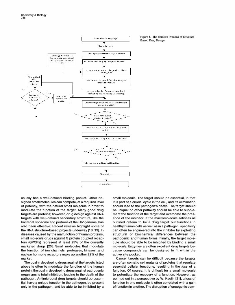
During the early 1980s, the ability to rationally designdrugs using protein structures was an unrealized goal Overview of the Processfor many structural biologists. The first projects were The process of structure-based drug design is an itera-underway in the mid-80s, and by the early 1990s the tive one (see Figure 1) and often proceeds through multi-first success stories were published [1–3]. Today, even ple cycles before an optimized lead goes into phase Ithough there is still quite a bit of fine-tuning necessary clinical trials. The first cycle includes the cloning, purifi-to perfect the process, structure-based drug design is cation and structure determination of the target proteinan integral part of most industrial drug discovery pro- or nucleic acid by one of three principal methods: X-raygrams [4] and is the major subject of research for many crystallography, NMR, or homology modeling. Usingacademic laboratories. computer algorithms, compounds or fragments of com-
The completion of the human genome project, the pounds from a database are positioned into a selectedstart of both the proteomics and structural genomics region of the structure. These compounds are scoredrevolutions, and developments in information technol- and ranked based on their steric and electrostatic inter-ogy are fueling an even greater opportunity for structure- actions with the target site, and the best compoundsbased drug design to be part of the success story in are tested with biochemical assays. In the second cycle,the discovery of new drug leads. Excellent drug targets structure determination of the target in complex with aare identified at an increased pace using developments promising lead from the first cycle, one with at leastin bioinformatics. The genes for these targets can be micromolar inhibition in vitro, reveals sites on the com-cloned quickly, and the protein expressed and purified pound that can be optimized to increase potency. Addi-to homogeneity. Advances in high-throughput crystal- tional cycles include synthesis of the optimized lead,lography, such as automation at all stages, more intense structure determination of the new target:lead complex,synchrotron radiation, and new developments in phase and further optimization of the lead compound. Afterdetermination, have shortened the timeline for determin- several cycles of the drug design process, the optimizeding structures. Structure determination using nuclear compounds usually show marked improvement in bind-magnetic resonance (NMR) has also seen a number of ing and, often, specificity for the target.advances in the past years, including magnet and probeimprovements, automated assignment [5–7], and new
Choice of a Drug Targetexperimental methods to determine larger structuresThe choice of a drug target is primarily made on a biolog-[8]. Faster computers and the availability of relativelyical and biochemical basis. The ideal target macromole-inexpensive clusters of computers have increased thecule for structure-based drug design is one that isclosely linked to human disease and binds a small mole-cule in order to carry out a function. The target molecule*Correspondence: [email protected]
brought to you by COREView metadata, citation and similar papers at core.ac.uk
provided by Elsevier - Publisher Connector
Chemistry & Biology788
Figure 1. The Iterative Process of Structure-Based Drug Design
usually has a well-defined binding pocket. Other de- small molecule. The target should be essential, in thatit is part of a crucial cycle in the cell, and its eliminationsigned small molecules can compete, at a required level
of potency, with the natural small molecule in order to should lead to the pathogen’s death. The target shouldbe unique: no other pathway should be able to supple-modulate the function of the target. Many good drug
targets are proteins; however, drug design against RNA ment the function of the target and overcome the pres-ence of the inhibitor. If the macromolecule satisfies alltargets with well-defined secondary structure, like the
bacterial ribosome and portions of the HIV genome, has outlined criteria to be a drug target but functions inhealthy human cells as well as in a pathogen, specificityalso been effective. Recent reviews highlight some of
the RNA structure-based projects underway [18, 19]. In can often be engineered into the inhibitor by exploitingstructural or biochemical differences between thediseases caused by the malfunction of human proteins,
small molecule drugs against G protein coupled recep- pathogenic and human forms. Finally, the target mole-cule should be able to be inhibited by binding a smalltors (GPCRs) represent at least 25% of the currently
marketed drugs [20]. Small molecules that modulate molecule. Enzymes are often excellent drug targets be-cause compounds can be designed to fit within thethe function of ion channels, proteases, kinases, and
nuclear hormone receptors make up another 22% of the active site pocket.Cancer targets can be difficult because the targetsmarket.
The goal in developing drugs against the targets listed are often somatic cell mutants of proteins that regulateessential cellular functions, resulting in the loss of aabove is often to modulate the function of the human
protein; the goal in developing drugs against pathogenic function. Of course, it is difficult for a small moleculeto potentiate the recovery of a function. However, asorganisms is total inhibition, leading to the death of the
pathogen. Antimicrobial drug targets should be essen- pointed out in a perspective by W. Kaelin [21], a loss offunction in one molecule is often correlated with a gaintial, have a unique function in the pathogen, be present
only in the pathogen, and be able to be inhibited by a of function in another. The disruption of oncogenic com-
Review789
plexes is another difficult problem for anticancer drug Expected error �design. For example, a chromosomal translocation in
0.642 � 0.00852e� B7.88� � 0.687 � 0.00223e� B
6.16�e(�2)(atom/refl).core binding factor � causes the formation of a novelchimeric protein that sequesters necessary transcrip-
Temperature factors of atoms in the region of interesttion factor subunits [22]. Despite the difficulty of design-should be no greater than the average temperature fac-ing a small molecule to disrupt an unwanted proteintor for the molecule. High temperature factors can reflectassociation, the specific interface between the fusiondisorder due to motion of the residue or ligand or aprotein and the transcription factor does provide a targetgeneral indication of error, adding to the inaccuracy ofthat can be exploited. Finally, malignancy often altersatomic positions. In a study reported by Carson et al.the target from its normal behavior, leading to interest[26], the temperature factor was the most highly corre-in the design of specificity for the malignant state.lated determinant of R factor. Finally, the moleculeshould be refined to be consistent with all rules of ste-reochemical “correctness” known from small moleculeEvaluating a Structure for Structure-Based
Drug Design structures; deviations from ideal bond lengths shouldbe no greater than 0.015 A or 3� for bond angles. PlanarOnce a target has been identified, it is necessary to
obtain accurate structural information. There are three atoms should be no more than 0.015 A out of the plane,and there should be no incorrect chiral centers. Finally,primary methods for structure determination that are
useful for drug design: X-ray crystallography, NMR, and at least 90% of the backbone � and � angles should fallinto the most favored regions of the Ramachandran plot.homology modeling. The evaluation of structures from
each method will be discussed. The PDB header record lists these statistics, and theyshould be evaluated before drug design attempts con-Crystal structures are the most common source of
structural information for drug design, since structures tinue. The results of a structure evaluation program,PROCHECK [27], are also available from the PDB anddetermined to high resolution may be available, and the
method is useful for proteins that range in size from a provide additional detail.Structures determined by nuclear magnetic reso-few amino acids to 998 kDa [23]. Another advantage
of crystallography is that ordered water molecules are nance, using a concentrated protein or nucleic acid insolution, are also valuable sources for drug design.visible in the experimental data and are often useful in
drug lead design. A crystal structure should be evalu- Since the target is in solution, it is sometimes possibleto interpret the dynamics of the target from the dataated for the resolution of the diffracted amplitudes (often
simply called resolution); reliability, or R factors; coordi- [28]. Ensembles of structures are deposited in the PDB,all of which satisfy the distance restraints from the ex-nate error; temperature factors; and chemical “correct-
ness.” Typically, crystal structures determined with data perimental data and show reasonable stereochemicalparameters. There is no analogous reliability factor asextending to beyond 2.5 A are acceptable for drug de-
sign purposes since they have a high data to parameter in crystallography, but the quality of the structure isoften measured by the rms deviations of the coordinatesratio, and the placement of residues in the electron den-
sity map is unambiguous. The R factor and Rfree reported of the members of the ensemble from the average struc-ture (often divided into main chain and side positions)for a model are measures of the correlation between
the model and experimental data. The Rfree value should and overall stereochemical soundness, including vander Waals violations, phi/psi conformational angle anal-be below 28% and ideally below 25%, and the R factor
should be well below 25% in order to use the structure in ysis, side chain torsion angle analysis, bond lengths,bond angles, and planarity. NMR data are often col-drug design. If the only structure available for a particular
target does not meet the resolution or R factor criteria, lected by measuring nuclear Overhauser effect (NOE)peaks between resonant nuclei that are a distance ofdrug design projects can still be considered, but the
results should be judged carefully. less than 5 A apart in the tertiary structure. Anotherimportant statistic for evaluating NMR-derived struc-Low coordinate error in a crystal structure is crucial
since van der Waals interactions modulate with the sixth tures is the number of unfulfilled NOE restraints, other-wise called violations. NOE violations are crosspeakspower of the distance between atoms, and directional
bonds, such as hydrogen bonds and electrostatic inter- between resonant nuclei that appear in the experimentaldata but are unexplained in the model. A final evaluationactions, have a narrow tolerance for both the angle and
distance (approximately 0.2 A). Coordinate error can be statistic is the total number of NOE restraints per resi-due, or data:parameter ratio.measured in many different ways, but two significant
methods are the Luzzati method [24], based on averag- In a survey of 97 deposited NMR structures in thePDB [29], Doreleijers et al. found that the average struc-ing coordinate error as a function of R factors that vary
with resolution, and methods in which expected errors ture had 11.3 restraints per residue and 61 NOE viola-tions. The precision of the structures, as defined by theare calculated based on the temperature factor, or B
factor, of an atom and the atom:reflection ratio [25]. The circular variance of the backbone dihedral angles, isclearly correlated with the number of restraints per resi-Luzzati coordinate error is often reported in coordinates
deposited with the Protein Data Bank (PDB) and should due. The number of residues in the most favored regionsof the Ramachandran plot is also correlated with thebe in the range of 0.2–0.3 A. For further accuracy in error
determination, the B factor (B) and atom:reflection ratio number of restraints per residue and a low number ofNOE violations. The programs PROCHECK-NMR [30](atom/refl) can be included, as in the Stroud and Fauman
method: and WHAT IF [31], the results of which are available from
Chemistry & Biology790
the PDB, provide additional structure-based details for torial chemistry, in which thousands of compounds aretested for biochemical effects.evaluating NMR structures. One other note to consider
is that the average structure from the ensemble may or The computer-aided methods can be further classi-fied into at least three categories: inspection, virtualmay not actually exist; therefore, one of the members
of the ensemble or the entire ensemble itself may be a screening, and de novo generation. In the first category,inspection, known molecules that bind the site, suchbetter target choice.
If no experimentally determined structure is available, as substrates or cofactors in the case of enzymes, orpeptides in the case of protein:protein or protein:nucleica homology model can be used for drug design [32–34].
To evaluate a homology model, SWISS-MODEL [35] out- acid interactions, are modified to become inhibitorsbased on maximizing complementary interactions in theputs a confidence factor per residue that reflects the
amount of structural information used to create that target site [1, 3, 41, 42]. In virtual screening, databasesof available small molecules are docked into the regionportion of the model. A higher confidence number re-
flects a lower number of templates and therefore a de- of interest in silico and scored based on predicted inter-actions with the site. Finally, for de novo generationcreased accuracy. All other methods for judging protein
structures, such as stereochemical soundness (bond small fragments of molecules, such as benzene rings,carbonyl groups, amino groups, etc., are positioned inlengths, bond angles, planarity, and packing) and resi-
dues in the most favored regions of the Ramachandran the site, scored, and linked in silico. The final com-pounds, created in silico from the linked fragments, thenplot, apply to analyzing a homology model as well as
to experimentally derived models. must be synthesized in the laboratory. There is someoverlap between the virtual screening and de novo gen-Using the structural information obtained through the
above techniques, the structure is then prepared for eration classifications. Some programs, for example,LUDI, which is usually used to dock fragments of com-drug design programs by first adding hydrogen atoms,
usually absent in crystal structures determined with data pounds, are also capable of docking and scoring entirecompounds. The programs are classified in Table 1 ac-at a resolution lower than 1.0 A. The protonation and
tautomeric states of residues as well as the state of cording to their primary use.There are many excellent drug design software meth-histidine residues (�, , or both nitrogens protonated)
should be assigned. Small molecules, such as ions and ods available capable of either virtual screening or denovo generation. This review will focus on a few of thewater molecules, can be included during the lead gener-
ation phase in cases where they play structural roles major points necessary to decide on a particular routefor lead generation. Extensive reviews of the softwarethat are crucial for the conformation of the target, other-
wise they are usually removed to allow any potential are available [11, 12, 14, 15, 43] and are highly recom-mended for further reading.lead to occupy their positions.
Questions that are pivotal in deciding on a methodfor lead generation are as follows: (1) are moleculesIdentification of the Target Siteavailable which can be modified to be inhibitors, (2) isStructure-based design begins with the identification ofthere a means for synthesizing novel molecules, and (3)a potential ligand binding site on the target molecule.what is the degree of accuracy required at a particularIdeally, the target site is a pocket or protuberance withstage of the design process versus the time needed fora variety of potential hydrogen bond donors and ac-the calculation? Factors such as the inclusion of proteinceptors, hydrophobic characteristics, and sizes of mo-or ligand flexibility and the effects of solvent increaselecular surfaces. The ligand binding site can be the ac-the time needed for the calculation but also increasetive site, as in an enzyme, an assembly site with anotherthe predictive value. Each of these questions will bemacromolecule, or a communication site necessary indiscussed with reference to available drug design algo-the mechanism of the molecule. In addition to the well-rithms.accepted protein target sites, RNA secondary structuralModifying an Initial Compoundelements can provide excellent target sites since theySubstrates and cofactors for many proteins have beenare species specific, bind ligands, and can be specificmodified to become excellent inhibitors [1, 3, 41, 42, 44,for a disease state [36, 37]. Target sites for protein-45] (see Figure 2 for an example). Initially, the crystalprotein interactions can be difficult to locate since thesestructure is solved in the presence of a substrate, cofac-surfaces are often flat, large, and hydrophobic, but eventor, or drug lead. Then, modifications to direct the smallthese difficulties can be surmounted [38–40]. Cocrystal-molecule toward being a potent inhibitor are designedlization studies in which the target macromolecule isin silico based on the interactions of the molecule withcrystallized with an initial small molecule inhibitor canthe target site. The newly designed compounds are then
be invaluable for the determination of a good target site.scored for binding using evaluative scoring algorithmsavailable in virtual screening methods.
Drug Design Methods Docking Available Small Molecules versus DeOnce the structure and target site are identified, there Novo Generationare several paths to developing a good lead based on The main advantage to docking compounds from data-the structure of the target. These paths can be broadly bases such as the Available Chemicals Database (ACD)classified as computer aided versus experimental. Com- into the target site is that hit compounds can be pur-puter-aided methods will be the main focus of this re- chased and tested using biochemical assays. Alterna-view. An example of an experimental method, by way tively, instead of testing the entire database, a database
can be refined to select molecules with a specific motif.of contrast, is high-throughput screening with combina-
Review791
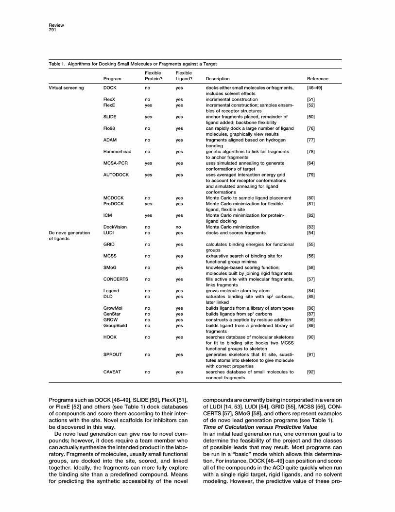
Table 1. Algorithms for Docking Small Molecules or Fragments against a Target
Flexible FlexibleProgram Protein? Ligand? Description Reference
Virtual screening DOCK no yes docks either small molecules or fragments, [46–49]includes solvent effects
FlexX no yes incremental construction [51]FlexE yes yes incremental construction; samples ensem- [52]
bles of receptor structuresSLIDE yes yes anchor fragments placed, remainder of [50]
ligand added; backbone flexibilityFlo98 no yes can rapidly dock a large number of ligand [76]
molecules, graphically view resultsADAM no yes fragments aligned based on hydrogen [77]
bondingHammerhead no yes genetic algorithms to link tail fragments [78]
to anchor fragmentsMCSA-PCR yes yes uses simulated annealing to generate [64]
conformations of targetAUTODOCK yes yes uses averaged interaction energy grid [79]
to account for receptor conformationsand simulated annealing for ligandconformations
MCDOCK no yes Monte Carlo to sample ligand placement [80]ProDOCK yes yes Monte Carlo minimization for flexible [81]
ligand, flexible siteICM yes yes Monte Carlo minimization for protein- [82]
ligand dockingDockVision no no Monte Carlo minimization [83]
De novo generation LUDI no yes docks and scores fragments [54]of ligands
GRID no yes calculates binding energies for functional [55]groups
MCSS no yes exhaustive search of binding site for [56]functional group minima
SMoG no yes knowledge-based scoring function; [58]molecules built by joining rigid fragments
CONCERTS no yes fills active site with molecular fragments, [57]links fragments
Legend no yes grows molecule atom by atom [84]DLD no yes saturates binding site with sp3 carbons, [85]
later linkedGrowMol no yes builds ligands from a library of atom types [86]GenStar no yes builds ligands from sp3 carbons [87]GROW no yes constructs a peptide by residue addition [88]GroupBuild no yes builds ligand from a predefined library of [89]
fragmentsHOOK no yes searches database of molecular skeletons [90]
for fit to binding site; hooks two MCSSfunctional groups to skeleton
SPROUT no yes generates skeletons that fit site, substi- [91]tutes atoms into skeleton to give moleculewith correct properties
CAVEAT no yes searches database of small molecules to [92]connect fragments
Programs such as DOCK [46–49], SLIDE [50], FlexX [51], compounds are currently being incorporated in a versionof LUDI [14, 53]. LUDI [54], GRID [55], MCSS [56], CON-or FlexE [52] and others (see Table 1) dock databases
of compounds and score them according to their inter- CERTS [57], SMoG [58], and others represent examplesof de novo lead generation programs (see Table 1).actions with the site. Novel scaffolds for inhibitors can
be discovered in this way. Time of Calculation versus Predictive ValueIn an initial lead generation run, one common goal is toDe novo lead generation can give rise to novel com-
pounds; however, it does require a team member who determine the feasibility of the project and the classesof possible leads that may result. Most programs cancan actually synthesize the intended product in the labo-
ratory. Fragments of molecules, usually small functional be run in a “basic” mode which allows this determina-tion. For instance, DOCK [46–49] can position and scoregroups, are docked into the site, scored, and linked
together. Ideally, the fragments can more fully explore all of the compounds in the ACD quite quickly when runwith a single rigid target, rigid ligands, and no solventthe binding site than a predefined compound. Means
for predicting the synthetic accessibility of the novel modeling. However, the predictive value of these pro-

Chemistry & Biology792
Figure 2. Inhibitors for Thymidylate SynthaseWere Designed Based on Modifications of theCofactor 5,10-Methylene Tetrahydrofolate
Several potent inhibitors are shown: (B)CB3717, (C) OSI 1843U89, and (D) ZD1694(Tomudex).
grams can be greatly increased when routines that dicted by molecular dynamics [62], or generated usingrotamers of protein side chains [50, 63]. Using a molecu-model protein and ligand flexibility as well as solventlar dynamics simulation to generate multiple proteincontribution are added.conformations, Carlson et al. have experimentally veri-Protein and Ligand Flexibility. There have been manyfied a dynamic pharmacophore model for HIV-1 integ-reports which emphasize the crucial effects of includingrase [62]. Programs which mimic protein flexibilityprotein and ligand flexibility in the docking and scoringthrough the use of ensembles include SLIDE [50], FlexEprocess [15, 43, 59]. Most proteins and most ligands[52], and MCSA-PCR [64].are quite flexible in solution and may experience a full
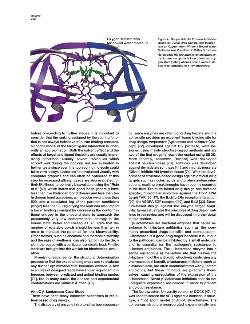
Solvent Effects. Solvent plays an important role inensemble of possible conformations. As a result, leadsligand binding in several ways. In one capacity, orderedgenerated from a single, rigid structure may have dif-water molecules seen in the structure can be incorpo-fering results in solution than in silico [60]. In order torated into the designed ligand, effectively increasingaccount for the landscape of protein and ligand confor-ligand binding by increasing the entropy of the systemmations, several drug design algorithms incorporate(releasing the bound water molecule). As an example,protein and/or ligand flexibility. However, modeling mo-inhibitors for HIV protease [65] incorporate an oxygenlecular flexibility, especially for the target macromole-atom to substitute for a key water molecule coordinatedcule, drastically increases the compute time requiredby residues of the flap region of the active site (seefor the structure-based drug design (SBDD) search.Figure 4). In a second capacity, ordered water moleculesMany programs that allow protein flexibility incorpo-can be treated as bound ligands, and contacts withrate information from multiple protein structures. En-them can be maximized [66]. In a third capacity, thesembles of structures can be experimentally deter-effect of the solvent can be incorporated into the scoringmined, such as NMR ensembles (see Figure 3) orscheme for the target:ligand interaction. The steps ofmultiple crystal structures [61], computationally pre-increased accuracy in modeling the solvent effect duringscoring are as follows: (1) making the assumption thatthe molecules are in a vacuum, i.e., no solvent modeling;(2) using a fixed dielectric constant in estimating electro-static contributions; (3) explicit solvation models; and(4) modeling the Born equation. The Born equation cal-culates the polarization contribution to solvation whena charge is placed within a spherical solvent cavity.In general, increased accuracy comes with increasedcomputational cost.
The correct value for the dielectric constant of themedium is critical in properly evaluating electrostaticeffects and estimating binding affinity. In the Northwest-ern University version of DOCK [49], a solvation correc-tion can be added to the score. Possible approachesto achieve an exact solution to the solvent probleminclude solving the Poisson-Boltzmann equation, oftenby using finite differences, or using a free-energy pertur-bation technique. Three approaches have been usedin practice: a modified Born equation [49] to calculatesolvation energies, an approximation to the electrostaticdesolvation by modeling the first solvation shell at thebinding interface [67], and an implicit model which ac-counts for desolvation by computationally generatingpossible positions of water molecules in the bindingpocket [68].Figure 3. An Ensemble of Six Structures of Dihydrofolate Reductase
Six (out of a total of 24 reported) structures of dihydrofolate reduc-Drug Lead Evaluationtase bound to trimethoprim (red) and NADPH (orange) (1LUD; [93])Once a small molecule has been identified as potentiallyare shown. Each member of the ensemble is separately colored,
and hydrogens are omitted for clarity. binding to the target molecule, it must be evaluated
Review793
Figure 4. Nonpeptide HIV Protease InhibitorsBased on Cyclic Urea Compounds Incorpo-rate an Oxygen Atom Where a Bound WaterMolecule Was Visualized in X-Ray Structures
Nonpeptide HIV protease inhibitors based oncyclic urea compounds incorporate an oxy-gen atom (noted) where a bound water mole-cule was visualized in X-ray structures.
before proceeding to further stages. It is important to ful, since enzymes are often good drug targets and theactive site provides an excellent ligand binding site forconsider that the ranking assigned by the scoring func-
tion is not always indicative of a true binding constant, drug design. Amprenavir (Agenerase) and nelfinavir (Vira-cept) [72], developed against HIV protease, were de-since the model of the target:ligand interaction is inher-
ently an approximation. Both the solvent effect and the signed using mainly structure-based methods and aretwo of the first drugs to reach the market using SBDD.effects of target and ligand flexibility are usually impre-
cisely described. Usually, several molecules which More recently, zanamivir (Relenza) was developedagainst neuraminidase [73], Tomudex was developedscored well during the docking run are evaluated in
further tests since even the top scoring molecule could against thymidylate synthase [44], and imitinab mesylate(Glivec) inhibits Abl tyrosine kinase [74]. With the devel-fail in vitro assays. Leads are first evaluated visually with
computer graphics and can often be optimized at this opment of structure-based design against difficult drugtargets such as nucleic acids and protein:protein inter-step for increased affinity. Leads are also evaluated for
their likelihood to be orally bioavailable using the “Rule actions, exciting breakthroughs have recently occurredin the field. Structure-based drug design has revealedof 5” [69], which states that good leads generally have
less than five hydrogen bond donors and less than ten specific, micromolar inhibitors against the HIV-1 RNAtarget TAR [36, 37], the IL-2/IL-2R receptor interactionhydrogen bond acceptors, a molecular weight less than
500, and a calculated log of the partition coefficient [39], the VEGF/VEGF receptor [40], and Bcl2 [33]. Struc-ture-based design against the enzyme target AmpC(clogP) less than 5. Rigidifying the lead can also impart
a lower binding constant by decreasing the conforma- �-lactamase illustrates the principles of drug design out-lined in this review and will be discussed in further detailtional entropy in the unbound state to approach the
presumably very low conformational entropy in the in this section.�-lactamases are bacterial enzymes that cause re-bound state. Veber and colleagues [70] state that the
number of rotatable bonds should be less than ten in sistance to �-lactam antibiotics such as the com-monly prescribed drugs penicillin and cephalosporin.order to increase the potential for oral bioavailability.
Other factors, such as chemical and metabolic stability �-lactamase is a good drug target because it is uniqueto the pathogen, can be inhibited by a small molecule,and the ease of synthesis, can also factor into the deci-
sion to proceed with a particular candidate lead. Finally, and is essential for the pathogen’s resistance to�-lactam antibiotics. The �-lactamase enzyme has aleads are brought into the wet lab for biochemical evalu-
ation. serine nucleophile at the active site that cleaves the�-lactam ring of the antibiotic, effectively destroying anyPromising leads reenter the structural determination
process to find the exact binding mode and to evaluate pharmaceutical benefit. �-lactamase inhibitors, such asclavulanic acid, are often coadministered with �-lactamany further optimization that becomes evident. A few
examples of designed leads have shown significant dif- antibiotics, but these inhibitors are �-lactams them-selves, causing upregulation of the expression of theferences between predicted and actual binding modes
[71], but in many cases the docked and experimental �-lactamase. Novel �-lactamase inhibitors that do notupregulate expression are needed in order to preventconformations are within 2 A rmsd [16].antibiotic resistance.
The Northwestern University version of DOCK [47, 49]AmpC �-Lactamase Case StudyThere have been many important successes in struc- was used to screen the ACD against a consensus struc-
ture, a “hot spot” model of AmpC �-lactamase. Theture-based drug design.The discovery of enzyme inhibitors has been success- consensus structure incorporated experimentally and

Chemistry & Biology794
Figure 5. Drug Design against AmpC �-Lactamase
(A) Ball-and-stick representation of compound 1 (red), discovered with a DOCK screen, bound to AmpC �-lactamase.(B) Compound 1 (space filling) bound to AmpC �-lactamase (residues within 7 A are shown with van der Waals surfaces).
computationally derived ligand binding data from 13 found to be essential. The addition of a piperidine ringto the distal aryl ring increased binding by 2-fold. Finally,AmpC �-lactamase structures [75]. The consensus bind-
ing sites for AmpC �-lactamase include an amide recog- compound 1 is relatively “drug-like,” according to Lip-inski’s rules [69], and has sites for future synthetic elabo-nition site, an oxyanion hole, hydroxyl and carboxyl
binding sites, and, finally, four ordered water molecules ration.In summary, AmpC �-lactamase is an excellent drugshown to consistently bind either the enzyme or the
inhibitors. The top 500 scoring molecules from the target with accurate structural information. The North-western University version of DOCK was used to screenDOCK run were examined graphically for complemen-
tarity, polar interactions, and agreement with the identi- the ACD to find novel inhibitor scaffolds. The top-scoringcompounds were novel and predicted to have comple-fied binding sites. Fifty-six compounds were purchased
and tested with in vitro assays. Three compounds inhibit mentary interactions with the target site, but were shownto have relatively low binding constants in solution. Fur-with Ki � 650 �M or better. Compound 1 was shown to
be selective for AmpC �-lactamase over other serine ther improvement will be needed before the drug leadcan proceed into future trials. Structural studies of thenucleophile enzymes and was selected for further study.
Powers et al. [66] determined the cocrystal structure selected inhibitor and the enzyme are invaluable in fu-ture chemical elaboration.of AmpC �-lactamase and compound 1 (Figure 5). The
structure was determined to a resolution of 1.94 A, with The results of the AmpC �-lactamase case study alsoexemplify the sort of reasonable expectations oneR factor 17.3% and Rfree 20.7%, coordinate error 0.19 A,
average B factor 23 A2, and average B factor for com- should have for initial structure-based drug design stud-ies. One, micromolar inhibitors were discovered throughpound 1, 37 A2. The structure is stereochemically cor-
rect, citing an rmsd from ideality for bond lengths � the docking procedure and will serve as lead com-pounds requiring further modification for increased po-0.009 A and bond angles � 1.5�. The DOCK-predicted
conformation of compound 1 closely resembles the tency. It is very rare that extremely potent inhibitors(nM inhibition or better) are discovered during dockingcrystallographically determined conformation of com-
pound 1. In fact, the rmsd for all inhibitor atoms is 1.87 A screens. Two, 56 top-scoring compounds were pur-chased and tested in vitro after the initial dockingfor one molecule in the asymmetric unit of the crystal
and 1.75 A for the second molecule in the asymmetric screen. Due to approximations in the models of proteinand ligand interactions in the scoring algorithms, theunit. The predicted interactions were also highly corre-
lated with the crystallographically determined interac- docked compounds may be ranked in slightly differentorder than their in vitro assays reveal. In fact, some of thetions: of nine hydrogen bonds observed in the crystal
structure, seven were predicted, and of eight hydrogen hits from the docking study may not exhibit successful invitro results at all. Structure-based drug design methodsbonds predicted, only one was not observed crystallo-
graphically. increase the chance that a “hit” will be found in the top-ranked ligands.Compound 1 was tested in microbiology experiments
and found to reduce the minimum inhibitory concentra-tion (MIC) of ampicillin by 4-fold in �-lactamase-positive Promise for the Future
Structure-based drug design is a powerful method, es-bacteria. Analogs of compound 1 were tested to deter-mine which functional moieties were essential. The car- pecially when used as a tool within an armamentarium,
for discovering new drug leads against important tar-boxylate group, the proton donating ability of the sulfon-amide, and the atom order of the sulfonamide were gets. After a target and a structure of that target are
Review795
flexibility in computational drug design. Mol. Pharmacol. 57,chosen, new leads can be designed from chemical prin-213–218.ciples or chosen from a subset of small molecules that
16. Shoichet, B., McGovern, S., Wei, B., and Irwin, J. (2002). Leadscored well when docked in silico against the target.discovery using molecular docking. Curr. Opin. Chem. Biol. 6,
After a preliminary assessment of bioavailability, the 439–446.candidate leads continue in an iterative process of reen- 17. Klebe, G., and Bohm, H. (1997). Energetic and entropic factors
determining binding affinity in protein-ligand complexes. J. Re-tering structural determination and reevaluation for opti-cept. Signal Transduct. Res. 17, 459–473.mization. Focused libraries of synthesized compounds
18. Gallego, J., and Varani, G. (2001). Targeting RNA with small-based on the structure-based lead can create a verymolecule drugs: therapeutic promise and chemical challenges.promising lead which can continue to phase I clinicalAcc. Chem. Res. 34, 836–843.
trials. 19. Afshar, M., Prescott, C., and Varani, G. (1999). Structure-basedAs structural genomics, bioinformatics, and computa- and combinatorial search for new RNA-binding drugs. Curr.
Opin. Biotechnol. 10, 59–63.tional power continue to explode with new advances,20. Hopkins, A., and Groom, C. (2002). The druggable genome. Nat.further successes in structure-based drug design are
Rev. Drug Discov. 1, 727–730.likely to follow. Each year, new targets are being identi-21. Kaelin, W. (1999). Choosing anticancer drug targets in the post-fied, structures of those targets are being determined genomic era. J. Clin. Invest. 104, 1503–1506.
at an amazing rate, and our capability to capture a quan- 22. Lukasik, S., Zhang, L., Corpora, T., Tomanicek, S., Li, Y., Kundu,titative picture of the interactions between macromole- M., Hartman, K., Liu, P., Laue, T., Biltonen, R., et al. (2002).
Altered affinity of CBFb-SMMHC for Runx1 explains its role incules and ligands is accelerating.leukemogenesis. Nat. Struct. Biol. 9, 674–679.
23. Nissen, P., Hansen, J., Ban, N., Moore, P., and Steitz, T. (2000).ReferencesThe structural basis of ribosome activity in peptide bond synthe-sis. Science 289, 920–930.1. Roberts, N., Martin, J., Kinchington, D., Broadhurst, A., Craig,
24. Luzzati, V. (1952). The statistical treatment of errors in crystalJ., Duncan, I., Galpin, S., Handa, B., Kay, J., Krohn, A., et al.
structures. Acta Crystallogr. 5, 802–810.(1990). Rational design of peptide-based HIV proteinase inhibi-
25. Stroud, R., and Fauman, E. (1995). Significance of structuraltors. Science 248, 358–361.
changes in proteins: Expected errors in refined protein struc-2. Erickson, J., Neidhart, D., VanDrie, J., Kempf, D., Wang, X.,
tures. Protein Sci. 4, 2392–2404.Norbeck, D., Plattner, J., Rittenhouse, J., Turon, M., Wideburg,
26. Carson, M., Bugg, C., DeLucas, L., and Narayana, S. (1994).N., et al. (1990). Design, activity and 2.8 A crystal structure of
Comparison of homology model to the experimental structurea C2 symmetric inhibitor complexed to HIV-1 protease. Science of a novel serine protease. Acta Crystallogr. D Biol. Crystallogr.249, 527–533. 50, 889–899.
3. Dorsey, B.D., Levin, R.B., McDaniel, S.L., Vacca, J.P., Guare, 27. Laskowski, R., MacArthur, M., Moss, D., and Thornton, J. (1993).J.P., Darke, P.L., Zugay, J.A., Emini, E.A., Schleif, W.A., Quintero, PROCHECK: a program to check the stereochemical quality ofJ.C., et al. (1994). L-735,524: the design of a potent and orally protein structures. J. Appl. Crystallogr. 26, 283–291.available HIV protease inhibitor. J. Med. Chem. 37, 3443–3451. 28. Pellecchia, M., Sern, D., and Wutrich, K. (2002). NMR in drug
4. Mountain, V. (2003). Astex, Structural Genomix, and Syrrx. discovery. Nat. Rev. Drug Discov. 1, 211–219.Chem. Biol. 10, 95–98. 29. Doreleijers, J., Rullman, J., and Kaptein, R. (1998). Quality as-
5. Zheng, D., Huang, Y., Moseley, H., Xiao, R., Aramini, J., Swapna, sessment of NMR structures: a statistical survey. J. Mol. Biol.G., and Montelione, G. (2003). Automated protein fold determi- 281, 149–164.nation using a minimal NMR constraint strategy. Protein Sci. 30. Laskowski, R., Rullman, J., MacArthur, M., Kaptein, R., and12, 1232–1246. Thornton, J. (1996). AQUA and PROCHECK-NMR: programs for
6. Oezguen, N., Adamian, L., Xu, Y., Rajarathnam, K., and Braun, checking the quality of protein structures solved by NMR. J.W. (2002). Automated assignment and 3D structure calculations Biomol. NMR 8, 477–486.using combinations of 2D homonuclear and 3D heteronuclear 31. Vriend, G. (1990). WHAT IF: a molecular modeling and drugNOESY spectra. J. Biomol. NMR 22, 249–263. design program. J. Mol. Graph. 8, 52–56.
7. Bailey-Kellogg, C., Widge, A., Kelley, J., Berardi, M., Bushweller, 32. Enyedy, I., Lee, S., Kuo, A., Dickson, R., Lin, C., and Wang,J., and Donald, B. (2000). The NOESY jigsaw: automated protein S. (2001). Structure-based approach for the discovery of bis-secondary structure and main-chain assignment from sparse, benzamidines as novel inhibitors of matriptase. J. Med. Chem.unassigned NMR data. J. Comput. Biol. 7, 537–558. 44, 1349–1355.
8. Pervushin, K., Riek, R., Wider, G., and Wutrich, K. (1997). Attenu- 33. Enyedy, I., Ling, Y., Nacro, K., Tomita, Y., Wu, X., Cao, Y., Guo,ated T2 relaxation by mutual cancellation of dipole-dipole cou- R., Li, B., Zhu, X., Huang, Y., et al. (2001). Discovery of small-pling and chemical shift anistropy indicates an avenue to NMR molecule inhibitors of Bcl-2 through structure-based computerstructures of very large biological macromolecules in solution. screening. J. Med. Chem. 44, 4313–4324.Proc. Natl. Acad. Sci. USA 94, 12366–12371. 34. Schapira, M., Raaka, B., Samuels, H., and Abagyan, R. (2001).
9. Antel, J. (1999). Integration of combinatorial chemistry and In silico discovery of novel retinoic acid receptor agonist struc-structure-based drug design. Curr. Opin. Drug Discov. Dev. 2, tures. BMC Struct. Biol. 1, 1.224–233. 35. Peitsch, M., and Guex, N. (1997). Large-scale comparative pro-
10. Verlinde, C., and Hol, W. (1994). Structure-based drug design: tein modeling. In Proteome Research: New Frontiers in Func-progress, results and challenges. Structure 2, 577–587. tional Genomics, M. Wilkins, K. Williams, R. Appel, and D. Hoch-
11. Taylor, R., Jewsbury, P., and Essex, J. (2002). A review of pro- strasser, eds. (New York: Springer), pp. 177–186.tein-small molecule docking methods. J. Comput. Aided Mol. 36. Lind, K., Du, Z., Fujinaga, K., Peterlin, B., and James, T. (2002).Des. 16, 151–166. Structure-based computational database screening, in vitro
12. Joseph-McCarthy, D. (1999). Computational approaches to assay, and NMR assessment of compounds that target TARstructure-based ligand design. Pharmacol. Ther. 84, 179–191. RNA. Chem. Biol. 9, 185–193.
13. Carlson, H., and McCammon, J. (1999). Method for including 37. Filikov, A., Mohan, V., Vickers, T., Griffey, R., Cook, P., Abagyan,the dynamic fluctuations of a protein in computer-aided drug R., and James, T. (2000). Identification of ligands for RNA targetsdesign. J. Phys. Chem. A 103, 10213–10219. via structure-based virtual screening: HIV-1 TAR. J. Comput.
14. Bohacek, R., and McMartin, C. (1997). Modern computational Aided Mol. Des. 14, 593–610.chemistry and drug discovery: structure generating programs. 38. Gadek, T., and Nicholas, J. (2003). Small molecule antagonistsCurr. Opin. Chem. Biol. 1, 157–161. of proteins. Biochem. Pharmacol. 65, 1–8.
39. Tilley, J., Chen, L., Fry, D., Emerson, S., Powers, G., Biondi, D.,15. Carlson, H., and McCammon, J. (2000). Accommodating protein
Chemistry & Biology796
Varnell, T., Trilles, R., Guthrie, R., Mennona, F., et al. (1997). 61. Knegtel, R., Kuntz, I., and Oshiro, C. (1997). Molecular dockingto ensembles of protein structures. J. Mol. Biol. 266, 424–440.Identification of a small molecule inhibitor of the IL-2/IL-2R re-
ceptor interaction which binds IL-2. J. Am. Chem. Soc. 119, 62. Carlson, H., Masukawa, K., Jorgensen, W., Lins, R., Briggs, J.,and McCammon, J. (2000). Developing a dynamic pharmaco-7589–7590.
40. Wiesmann, C., Christinger, H., Cochran, A., Cunningham, B., phore model for HIV-1 integrase. J. Med. Chem. 43, 2100–2114.63. Lovell, S., Word, J., Richardson, J., and Richardson, D. (2000).Fairbrother, W., Keenan, C., Meng, G., and DeVos, A. (1998).
Crystal structure of the complex between VEGF and a receptor- The penultimate rotamer library. Proteins 40, 389–408.64. Ota, N., and Agard, D. (2001). Binding mode prediction for ablocking peptide. Biochemistry 37, 117765–117772.
41. Varney, M., Marzoni, G., Palmer, C., Deal, J., Webber, S., Welsh, flexible ligand in a flexible pocket using multi-conformation sim-ulated annealing pseudo crystallographic refinement. J. Mol.K., Bacquet, R., Bartlett, C., Morse, C., Booth, C., et al. (1992).
Crystal structure-based drug design and synthesis of benz[c- Biol. 314, 607–617.65. Lam, P., Jadhav, P., Eyerman, C., Hodge, C., Ru, Y., Bacheler,d]indole-containing inhibitors of thymidylate synthase. J. Med.
Chem. 35, 663–676. L., Meek, J., Otto, M., Rayner, M., and Wong, Y. (1994). Rationaldesign of potent, bioavailable, nonpeptide cyclic ureas as HIV42. Chan, D., Laughton, C., Queener, S., and Stevens, M. (2001).protease inhibitors. Science 263, 380–384.Structural studies on bioactive compounds. 34. Design, synthe-
66. Powers, R., Morandi, F., and Shoichet, B. (2002). Structure-sis, and biological evaluation of triazenyl-substituted pyrimeth-based discovery of a novel, noncovalent inhibitor of AmpC beta-amine inhibitors of Pneumocystis carinii dihydrofolate reduc-lactamase. Structure 10, 1013–1023.tase. J. Med. Chem. 44, 2555–2564.
67. Majeux, N., Scarsi, M., and Caflisch, A. (2001). Efficient electro-43. Carlson, H. (2002). Protein flexibility and drug design: how tostatic solvation model for protein-fragment docking. Proteinshit a moving target. Curr. Opin. Chem. Biol. 6, 447–452.42, 256–268.44. Rutenber, E., and Stroud, R. (1996). Binding of the anticancer
68. Bohm, H. (1998). Prediction of binding constants of protein li-drug ZD1694 to E. coli thymidylate synthase: assessing specific-gands: A fast method for the prioritization of hits obtained fromity and affinity. Structure 4, 1317–1324.de novo design or 3D database search programs. J. Comput.45. Stout, T., and Stroud, R. (1996). The complex of the anti-cancerAided Mol. Des. 12, 309–323.therapeutic, BW1843U89, with thymidylate synthase at 2.0 A
69. Lipinski, C., Lombardo, F., Dominy, B., and Feeney, P. (1997).resolution: implications for a new mode of inhibition. StructureExperimental and computational approaches to estimate solu-4, 67–77.bility and permeability in drug discovery and development set-46. Kuntz, I., Blaney, J., Oatley, S., Langridge, R., and Ferrin, T.tings. Adv. Drug Deliv. Rev. 23, 3–25.(1982). A geometric approach to macromolecular-ligand inter-
70. Veber, D., Johnson, S., Cheng, H., Smith, B., Ward, K., andactions. J. Mol. Biol. 161, 269–288.Kopple, K. (2002). Molecular properties that influence the oral47. Lorber, D., and Shoichet, B. (1998). Flexible ligand docking usingbioavailability of drug candidates. J. Med. Chem. 45, 2615–2623.conformational ensembles. Protein Sci. 7, 938–950.
71. Fritz, T., Tondi, D., Finer-Moore, J., Costi, M., and Stroud, R.48. Ewing, T., Makino, S., Skillman, G., and Kuntz, I. (2001). DOCK(2001). Predicting and harnessing protein flexibility in the design4.0: search strategies for automated molecular docking of flexi-of species-specific inhibitors of thymidylate synthase. Chem.ble molecule databases. J. Comput. Aided Mol. Des. 15,Biol. 8, 981–995.411–428.
72. Kaldor, S., Kalish, V., Davies, J., Shetty, B., Fritz, J., Appelt, K.,49. Shoichet, B., Leach, A., and Kuntz, I. (1999). Ligand solvationBurgess, J., Campanale, K., Chirgadze, N., Clawson, D., et al.in molecular docking. Proteins 34, 4–16.(1997). Viracept (Nelfinavir Mesylate AG1343): A potent, orally50. Schnecke, V., Swanson, C., Getzoff, E., Tainer, J., and Kuhn, L.bioavailable inhibitor of HIV-1 protease. J. Med. Chem. 40, 3979–(1998). Screening a peptidyl database for potential ligands to3985.proteins with side-chain flexibility. Proteins 33, 74–87.
73. Varghese, J. (1999). Development of neuraminidase inhibitors51. Kramer, B., Metz, G., Rarey, M., and Lengauer, T. (1999). Ligandas anti-influenza virus drugs. Drug Dev. Res. 46, 176–196.docking and screening with FlexX. Med. Chem. Res. 9, 463–478.
74. Schindler, T., Bornmann, W., Pellicena, P., Miller, W., Clarkson,52. Claussen, H., Buning, C., Rarey, M., and Lengauer, T. (2001).B., and Kuriyan, J. (2000). Structural mechanism for STI-571FlexE. Efficient molecular docking considering protein structureinhibition of abelson tyrosine kinase. Science 289, 1938–1942.variations. J. Mol. Biol. 308, 377–395.
75. Powers, R., and Shoichet, B. (2002). Structure-based approach53. Boehm, H. (1996). Towards the automatic design of syntheticallyfor binding site identification on AmpC �-lactamase. J. Med.accessible protein ligands: Peptides, amides and peptidomi-Chem. 45, 3222–3234.metics. J. Comput. Aided Mol. Des. 10, 265–272.
76. McMartin, C., and Bohacek, R. (1997). QXP: Powerful, rapid54. Boehm, H. (1992). The computer program, LUDI: a new method
computer algorithms for structure-based drug design. J. Com-for the de novo design of enzyme inhibitors. J. Comput. Aided
put. Aided Mol. Des. 11, 333–344.Mol. Des. 6, 61–78.
77. Mitzutani, M., Tomioka, N., and Itai, A. (1994). Rational automatic55. Goodford, P. (1985). A computational procedure for determining search method for stable docking models of protein and ligand.
energetically favorable binding sites on biologically important J. Mol. Biol. 243, 310–326.macromolecules. J. Med. Chem. 28, 849–857. 78. Welch, W., Ruppert, J., and Jain, A. (1996). Hammerhead: fast,
56. Caflisch, A., Miranker, A., and Karplus, M. (1993). Multiple copy fully automated docking of flexible ligands to protein bindingsimultaneous search and construction of ligands in binding sites. Chem. Biol. 3, 449–462.sites: applications to inhibitors of HIV-1 aspartic proteinase. J. 79. Goodsell, D., Morris, G., and Olson, A. (1996). Automated dock-Med. Chem. 36, 2142–2167. ing of flexible ligands: applications of AutoDock. J. Mol. Recog-
57. Pearlman, D., and Murcko, M. (1996). CONCERTS: dynamic con- nit. 9, 1–5.nection of fragments as an approach to de novo ligand design. 80. Liu, M., and Wang, S. (1999). MCDOCK: A Monte Carlo simula-J. Med. Chem. 39, 1651–1663. tion approach to the molecular docking problem. J. Comput.
58. DeWitte, R., and Shakhnovich, E. (1996). SMoG: a de novo de- Aided Mol. Des. 13, 435–451.sign method based on simple, fast, and accurate free energy 81. Trosset, J., and Scheraga, H. (1999). Prodock: software packageestimates. 1. Methodology and supporting evidence. J. Am. for protein modeling and docking. J. Comput. Chem. 20,Chem. Soc. 118, 11733–11744. 412–427.
59. Davis, A., and Teague, S. (1999). Hydrogen bonding, hydropho- 82. Abagyan, R., Totrov, M., and Kuznetsov, D. (1994). ICM—a newbic interactions, and failure of the rigid receptor hypothesis. method for protein modeling and design—applications to dock-Angew. Chem. Int. Ed. Engl. 38, 736–749. ing and structure prediction from the distorted native conforma-
60. Anderson, A., O’Neil, R., Surti, T., and Stroud, R. (2001). Ap- tion. J. Comput. Chem. 15, 488–506.proaches to solving the rigid receptor problem by identifying a 83. Hart, T., and Read, R. (1992). Proteins 13, 206–222.minimal set of flexible residues during ligand docking. Chem. 84. Nishibata, Y., and Itai, A. (1993). Confirmation of usefulness of
a structure construction program based on three-dimensionalBiol. 8, 445–457.
Review797
receptor structure for rational lead generation. J. Med. Chem.36, 2921–2928.
85. Miranker, A., and Karplus, M. (1995). An automated method fordynamic ligand design. Proteins 23, 472–490.
86. Bohacek, R., and McMartin, C. (1994). Multiple highly diversestructues complementary to enzyme binding sites: Results ofextensive application of a de novo design method incorporatingcombinatorial growth. J. Am. Chem. Soc. 116, 5560–5571.
87. Rotstein, S., and Murcko, M. (1993). GenStar: a method for denovo drug design. J. Comput. Aided Mol. Des. 7, 23–43.
88. Moon, J., and Howe, W. (1991). Computer design of bioactivemolecules: a method for receptor-based de novo ligand design.Proteins 11, 29–34.
89. Rotstein, S., and Murcko, M. (1993). GroupBuild: a fragment-based method for de novo drug design. J. Med. Chem. 36,1700–1710.
90. Eisen, M., Wiley, D., Karplus, M., and Hubbard, R. (1994). HOOK:a program for finding novel molecular architectures that satisfythe chemical and steric requirements of macromolecule bindingsites. Proteins 19, 199–221.
91. Gillet, V., Johnson, P., Mata, P., Sike, S., and Williams, P. (1993).SPROUT: a program for structure generation. J. Comput. AidedMol. Des. 7, 127–153.
92. Bartlett, P., Shea, G., Telfer, S., and Waterman, S. (1989). CA-VEAT: a program to facilitate the structure-derived design ofbiologically active molecules. In Molecular Recognition: Chemi-cal and Biological Problems, S. Roberts, ed. (Cambridge: RoyalSociety of Cambridge), pp. 182–196.
93. Polshakov, V., Smirnov, E., Birdsall, B., Kelly, G., and Feeney,J. (2001). NMR-based solution structure of the complex of Lac-tobacillus casei dihydrofolate reductase with trimethoprim andNADPH. J. Biomol. NMR 24, 67–70.
Copyright © 2022 FDOKUMEN