
Carbohydrate PEGylation, an approach to improve pharmacological potency
Upload
independentCategory
view
0download
0
Structure-Based Drug Design of Novel Aurora Kinase A Inhibitors: Structural Basis forPotency and Specificity
Mohane Selvaraj Coumar,†,¶ Jiun-Shyang Leou,†,¶ Paritosh Shukla,† Jian-Sung Wu,† Ajay Kumar Dixit,† Wen-Hsing Lin,†
Chun-Yu Chang,† Tzu-Wen Lien,† Uan-Kang Tan,‡ Chun-Hwa Chen,† John T.-A. Hsu,† Yu-Sheng Chao,† Su-Ying Wu,*,† andHsing-Pang Hsieh*,†
DiVision of Biotechnology and Pharmaceutical Research, National Health Research Institutes, 35 Keyan Road, Zhunan, Miaoli County 350,Taiwan, ROC, and Department of Chemical and Materials Engineering, Technology and Science Institute of Northern Taiwan, Taipei 112,Taiwan, ROC
ReceiVed October 7, 2008
Aurora kinases have emerged as attractive targets for the design of anticancer drugs. Through structure-based virtual screening, novel pyrazole hit 8a was identified as Aurora kinase A inhibitor (IC50 ) 15.1 µM).X-ray cocrystal structure of 8a in complex with Aurora A protein revealed the C-4 position ethyl carboxylateside chain as a possible modification site for improving the potency. On the basis of this insight, bioisostericreplacement of the ester with amide linkage and changing the ethyl substituent to hydrophobic 3-acetami-dophenyl ring led to the identification of 12w with a ∼450-fold improved Aurora kinase A inhibition potency(IC50 ) 33 nM), compared to 8a. Compound 12w showed selective inhibition of Aurora A kinase overAurora B/C, which might be due to the presence of a unique H-bond interaction between the 3-acetamidogroup and the Aurora A nonconserved Thr217 residue, which in Aurora B/C is Glu and found to stericallyclash with the 3-acetamido group in modeling studies.
Introduction
Aurora kinases belong to serine/threonine subclass of kinasesand are involved in the regulation of mitosis.1 Three isoforms(A, B, and C) are known, which differ in their amino acid lengthand sequence at the N-terminal domain but have a conservedATP binding site.2 Aurora A is involved in centrosomematuration and separation, bipolar spindle assembly, and mitoticentry. While Aurora B is essential for accurate chromosomesegregation and cytokinesis, Aurora C complements the functionof Aurora B.1 Aurora A and B levels both are up-regulated invarious cancers, including breast and colorectal cancers;3
deregulated Aurora kinase activity has been linked to geneticinstability, defects in centrosome function, spindle assembly,chromosome alignment, and cytokinesis, all of which can leadto tumorigenesis.1 Hence, inhibition of Aurora kinase activityby targeting the ATP binding site with small molecules isemerging as a new anticancer target-based therapy. Moreover,in vivo studies with Aurora inhibitors 1 (MK-0457/VX-680),4,5
2 (PHA-739358),6,7 3 (MLN8054),8 and 4 (AZD1152)9,10
(Figure 1) in various animal models have shown tumorregression and are now in different stages of clinical develop-ment for various cancers.
Molecular targeted therapies, such as Aurora kinase inhibition,target cancer cells more specifically than traditional cancertreatment options, such as antimitotic agents. Since Aurorakinase is emerging as a promising molecular drug target forcancer, we continued our efforts to find anticancer drugs11 bydeveloping a program to search for novel Aurora kinaseinhibitors. Various research groups have reported different leadgeneration strategies for Aurora inhibitors, such as high
throughput screening (HTSa), fragment-based screening, andsynthetic modifications of known kinase core structures.6,12,13
However, only a few studies have reported virtual screening-based lead generation of modestly potent compounds and havenot further demonstrated the utility of these lead compounds ingenerating potent Aurora kinase inhibitors.14,15 We, herein,report our virtual screening strategy, which led to the identifica-tion of a novel pyrazole lead compound 8a (Aurora A IC50 ≈15 µM), and the X-ray cocrystal structure-guided design forlead optimization, which culminated in the identification of 12w(Aurora A IC50 ≈ 0.033 µM) with a 450-fold improved Aurorakinase A inhibition compared to the lead 8a. In addition 12wshowed selective Aurora A inhibition compared to Aurora B/Cand also inhibited the cellular targets of Aurora kinase in theHCT-116 colon cancer cell line.
Lead Identification
The Aurora kinase structure6,12,16,17 represents a typicalbilobal kinase fold containing the N-terminal �-strand domainand the C-terminal R-helix domain. The two domains are linkedby a hinge region (residues 210-216), a loop forming theconserved hydrogen bonding interactions with the ATP orinhibitors, which is sandwiched between the two domains. Theconserved H-bonding interactions of inhibitors with hinge regionresidues Glu211 and Ala213 are observed in most of theinhibitor-Aurora complex structures and are essential formaintaining activity. For the virtual screening, 59 488 com-pounds from the Maybridge database (Tintagel, Cornwall,
* To whom correspondence should be addressed. For S.-Y.W.: phone,+886-37-246-166, extension 35713; fax, +886-37-586-456; e-mail,[email protected]. For H.-P.H.: phone, +886-37-246-166, extension35708; fax, +886-37-586-456, e-mail, [email protected].
† National Health Research Institutes.¶ These authors contributed equally to this work.‡ Technology and Science Institute of Northern Taiwan.
a Abbreviations: CLogP, calculated logarithm of partition coefficient;EDC, 1-(3-dimethylaminopropyl)-3-ethylcarbodiimide; EPHA1, EPH recep-tor A1; HCT-116, human colon cancer-116 cell line; HOBt, 1-hydrozy-benzotriazole; HTS, high throughput screening; JAK2, Janus-activatedkinase 2; KIT, Hardy-Zuckerman 4 feline sarcoma viral oncogenehomologue; LCMS, liquid chromatography coupled mass spectrometry; PDBID, Protein Data Bank identification number; RET, Ret proto-oncogene;SBDD, structure-based drug design; TBST, Tris-buffered saline Tween-20; TPSA, total polar surface area; TRKA, neurotrophic tyrosine kinasereceptor type 1; VEGFR, vascular endothelial growth factor receptor.
J. Med. Chem. 2009, 52, 1050–10621050
10.1021/jm801270e CCC: $40.75 2009 American Chemical SocietyPublished on Web 01/13/2009
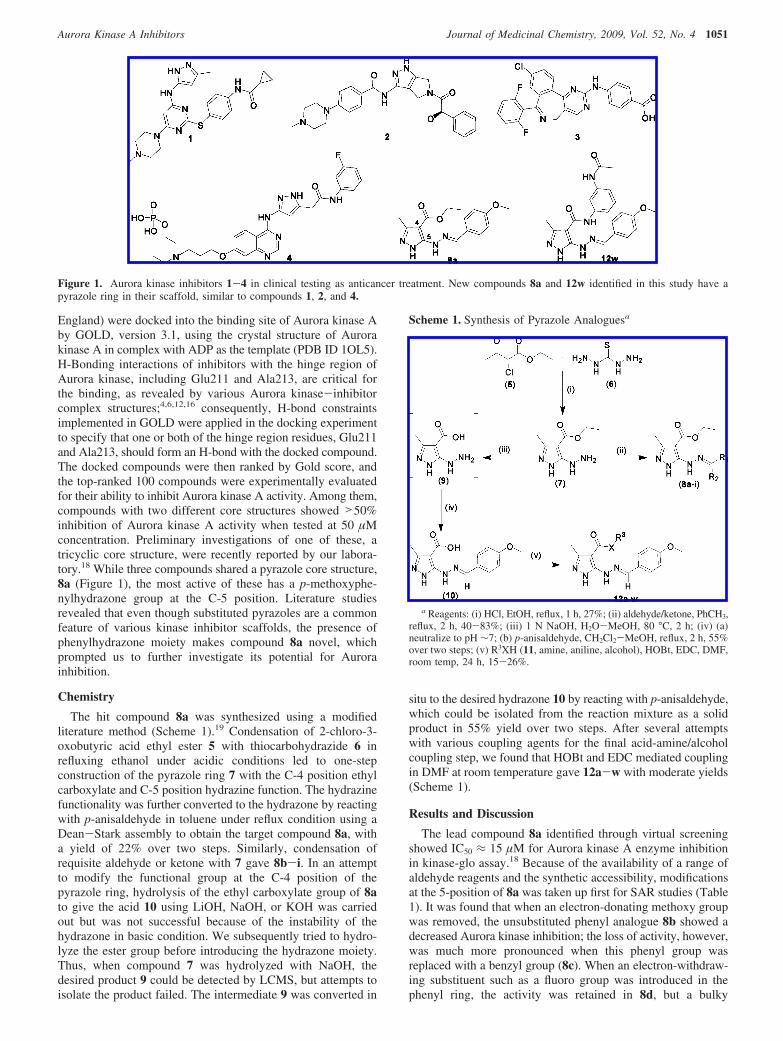
England) were docked into the binding site of Aurora kinase Aby GOLD, version 3.1, using the crystal structure of Aurorakinase A in complex with ADP as the template (PDB ID 1OL5).H-Bonding interactions of inhibitors with the hinge region ofAurora kinase, including Glu211 and Ala213, are critical forthe binding, as revealed by various Aurora kinase-inhibitorcomplex structures;4,6,12,16 consequently, H-bond constraintsimplemented in GOLD were applied in the docking experimentto specify that one or both of the hinge region residues, Glu211and Ala213, should form an H-bond with the docked compound.The docked compounds were then ranked by Gold score, andthe top-ranked 100 compounds were experimentally evaluatedfor their ability to inhibit Aurora kinase A activity. Among them,compounds with two different core structures showed >50%inhibition of Aurora kinase A activity when tested at 50 µMconcentration. Preliminary investigations of one of these, atricyclic core structure, were recently reported by our labora-tory.18 While three compounds shared a pyrazole core structure,8a (Figure 1), the most active of these has a p-methoxyphe-nylhydrazone group at the C-5 position. Literature studiesrevealed that even though substituted pyrazoles are a commonfeature of various kinase inhibitor scaffolds, the presence ofphenylhydrazone moiety makes compound 8a novel, whichprompted us to further investigate its potential for Aurorainhibition.
Chemistry
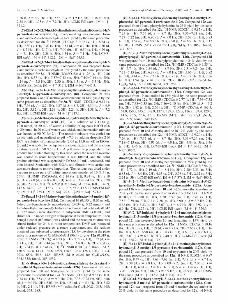
The hit compound 8a was synthesized using a modifiedliterature method (Scheme 1).19 Condensation of 2-chloro-3-oxobutyric acid ethyl ester 5 with thiocarbohydrazide 6 inrefluxing ethanol under acidic conditions led to one-stepconstruction of the pyrazole ring 7 with the C-4 position ethylcarboxylate and C-5 position hydrazine function. The hydrazinefunctionality was further converted to the hydrazone by reactingwith p-anisaldehyde in toluene under reflux condition using aDean-Stark assembly to obtain the target compound 8a, witha yield of 22% over two steps. Similarly, condensation ofrequisite aldehyde or ketone with 7 gave 8b-i. In an attemptto modify the functional group at the C-4 position of thepyrazole ring, hydrolysis of the ethyl carboxylate group of 8ato give the acid 10 using LiOH, NaOH, or KOH was carriedout but was not successful because of the instability of thehydrazone in basic condition. We subsequently tried to hydro-lyze the ester group before introducing the hydrazone moiety.Thus, when compound 7 was hydrolyzed with NaOH, thedesired product 9 could be detected by LCMS, but attempts toisolate the product failed. The intermediate 9 was converted in
situ to the desired hydrazone 10 by reacting with p-anisaldehyde,which could be isolated from the reaction mixture as a solidproduct in 55% yield over two steps. After several attemptswith various coupling agents for the final acid-amine/alcoholcoupling step, we found that HOBt and EDC mediated couplingin DMF at room temperature gave 12a-w with moderate yields(Scheme 1).
Results and Discussion
The lead compound 8a identified through virtual screeningshowed IC50 ≈ 15 µM for Aurora kinase A enzyme inhibitionin kinase-glo assay.18 Because of the availability of a range ofaldehyde reagents and the synthetic accessibility, modificationsat the 5-position of 8a was taken up first for SAR studies (Table1). It was found that when an electron-donating methoxy groupwas removed, the unsubstituted phenyl analogue 8b showed adecreased Aurora kinase inhibition; the loss of activity, however,was much more pronounced when this phenyl group wasreplaced with a benzyl group (8c). When an electron-withdraw-ing substituent such as a fluoro group was introduced in thephenyl ring, the activity was retained in 8d, but a bulky
Figure 1. Aurora kinase inhibitors 1-4 in clinical testing as anticancer treatment. New compounds 8a and 12w identified in this study have apyrazole ring in their scaffold, similar to compounds 1, 2, and 4.
Scheme 1. Synthesis of Pyrazole Analoguesa
a Reagents: (i) HCl, EtOH, reflux, 1 h, 27%; (ii) aldehyde/ketone, PhCH3,reflux, 2 h, 40-83%; (iii) 1 N NaOH, H2O-MeOH, 80 °C, 2 h; (iv) (a)neutralize to pH ∼7; (b) p-anisaldehyde, CH2Cl2-MeOH, reflux, 2 h, 55%over two steps; (v) R3XH (11, amine, aniline, alcohol), HOBt, EDC, DMF,room temp, 24 h, 15-26%.
Aurora Kinase A Inhibitors Journal of Medicinal Chemistry, 2009, Vol. 52, No. 4 1051

hydrophobic phenyl substituent (8e) was a deterrent for theactivity. On the other hand, introduction of a hydrophilic4-methylpiperazine moiety (8f) on the phenyl ring was tolerated.When the phenyl ring was replaced with a 5- or 4-indolyl ringsystem (8g,h), the activity level slightly improved compared to8a. However, 3-, 6-, and 7-indolyl analogues were not betterthan 8a (data not shown). Also, when the benzylidine H in 8a(R2 ) H) was replaced with a methyl group in 8i (R2 ) CH3),the activity declined (later X-ray cocrystal of 8a-Aurora Arevealed that this methyl group might cause steric clash withLeu139 and Arg137).
Concurrent with our forementioned SAR studies to understandthe binding mode and to get structural insights that could helpin the design of more potent Aurora inhibitors, we cocrystallized8a with Aurora kinase A and solved the inhibitor-Aurora Aprotein complex structure. The electron density maps of Aurorakinase A in complex with 8a are clear (Figure 2) except forsome disordered regions that include residues 280-291 and393-401. The activation loop residues 280-291 containing theT288D mutation are highly flexible in many published
structures.6,12,16 The complex structure revealed that the com-pound 8a binds to Aurora kinase A in the ATP-binding sitelocated in the cleft between the two domains. The pyrazole ringof 8a fitted into the hinge region, and the pyrazole ring N2 andN1H atoms formed two H-bonds (2.92 Å and 3.03 Å) with themain chain of Ala213. Similar H-bond interactions between theligand and hinge region residues of Aurora kinase wereconserved in most Aurora inhibitor complex structures and areimportant for the inhibitor binding.4,6 In addition, the pyrazolering also formed hydrophobic interactions with the surroundingresidues, including Leu139, Val147, Ala160, and Leu263.Particularly, Leu263 made extensive interactions with thepyrazole ring. The C-5 position p-methoxyphenyl group of 8aextended into the solvent-exposed region of the binding siteand had hydrophobic interactions with Gly216 and Pro214,while the hydrazone group (-NH-NdCH-) that served as alinker between the pyrazole and p-methoxyphenyl group madefew interactions (only with Gly216). The C-4 position ethylcarboxylate group was situated under the p-loop and had closecontact with protein residues, such as Gly140, Val147, and
Table 1. Physicochemical Properties and Inhibition of Aurora Kinase A by Pyrazoles 8a-i
a MW and CLogP calculated using ChemDraw Ultra, version 7.0. b TPSA computed from www.molinspiration.com. c Values are expressed as the meanof at least two independent determinations and are within (15%.
1052 Journal of Medicinal Chemistry, 2009, Vol. 52, No. 4 Coumar et al.

Thr217. Furthermore, the ethyl carboxylate group is adjacentto a hydrophobic region consisting of Leu139, Gly140, Val147,Thr217, Glu260, and Leu263. Finally, it is worth noting that8a adopts a conformation to form an intramolecular H-bond(2.94 Å) between the two side chains at position C-4 and C-5,through N6H in the hydrazone group (-NH-NdCH-) andO10 in the ethyl carboxylate (-OC2H5) group, thereby forminga pseudobicyclic ring structure when bound to Aurora A protein.
On the basis of our findings from the X-ray structure (thatthe C-5 position hydrazone substituent of the molecule isprojected into the solvent accessesible part of the Aurora protein,while the 4-position ethyl carboxylate functionality is projectedinto a possible hydrophobic portion of the Aurora kinaseprotein), we envisaged that functional modification of the ethylcarboxylate at the C-4 position of the pyrazole could lead toenhanced activity. In addition, the limited success in improvingthe activity of the lead 8a by modifying the C-5 positionhydrazone part prompted us to focus our efforts on replacingthe C-4 position ethyl group with a hydrophobic phenyl ring,which would provide additional hydrophobic interactions be-tween the ligand and the protein and could enhance the potency.Thus, in an attempt to increase the hydrophobic interactionbetween the ligand and Aurora kinase, benzyl carboxylate 12awas synthesized; as envisaged, this showed a 3-fold increasein Aurora inhibitory activity compared to ethyl carboxylate 8a(Table 2). Next, by utilization of the concepts of bioisosterism,the ester linkage of 12a was replaced with the amide linkage togive 12b. Conversion of the C-4 position ester to the amidelinkage led to another 3-fold improvement in activity for 12b(X ) NH) compared to 12a (X ) O). To examine the role ofbioisosteric replacement of O with NH in enhancing the potencyand to understand the difference in the molecular level interac-tion between the ligand and Aurora kinase protein, the X-raycocrystal structures of 12a and 12b were solved.
Comparison of the cocrystal structure of 12a with 8a revealsthat the pyrazole ring, hydrazone linker, and p-methoxyphenylgroups of these two compounds superimpose well. The ad-ditional phenyl ring of 12a in the C-4 position sandwiched itselfbetween the p-loop and the hydrophobic region, and it made
extra hydrophobic interactions with Gly140, Thr217, andGlu260. The additional hydrophobic interactions contributed tothe improved potency of 12a compared to 8a. Overall, both 8aand 12a have similar conformation when bound to Aurora kinaseand formed pseudobicyclic structures through intramolecularH-bonding. In contrast, comparison of 12b and 12a (Figure 3)revealed that although these are bioisosteric with only one atomdifference (NH vs O), 12b adopts a different conformation andhas a different three-dimensional spatial arrangement whenbound to Aurora A (Figure 3). The pyrazole ring of 12b movedcloser to Glu211, which led to the formation of an H-bond withthe carbonyl O of Glu211 (2.84 Å) and another H-bond withthe backbone NH of Ala213 (2.70 Å). The shift of the pyrazolering in 12b consequently moved the hydrazone linker close tothe Ala213 and allowed a third H-bond formation between thehydrazone N6H and carbonyl O of Ala213 (2.84 Å). Therefore,12b has three H-bonds with the hinge region while, similar to8a, 12a formed only two H-bonds with the hinge region.
Moreover, the torsion angle of -N1-C5-N6-N7d moietyrotated from -11.8° (in 12a structure) to -164.8° (in 12bstructure), which led to different intramolecular H-bondingpatterns between the two side chains of both compounds. Inthe structure of 12b, the N7 atom of hydrazone linker formedan intramolecular H-bond with the amide N10H (2.87 Å), whilein the structure of 12a the N6H of hydrazone formed anintramolecular H-bond with the ester O10 (3.0 Å). Also, in 12bthe intramolecular H-bond generated a pseudobicyclic structureand likely stabilized the orientation of the pyrazole ring, whichwould contribute to its interactions with the protein. Notably,the N6H of the hydrazone group played a different role in thesetwo structures. It provided an external H-bond with the hingeresidue of the protein in the complex structure of 12b withAurora A, while it provided an intramolecular H-bond withinthe compound in the structure of 12a and had no interactionswith the protein. Finally, the phenyl ring of 12b orientateddifferently from that of 12a and made additional interactionswith Glu260 and Leu263; this, along with the additionalH-bonding with the hinge region, contributed to the improvedactivity of 12b compared to 12a. Thus, bioisosteric replacement
Figure 2. X-ray cocrystal structure of 8a in complex with Aurora A. Compound 8a binds to the ATP-binding pocket of Aurora A through twoH-bond interactions with hinge Ala213 residue. An intramolecular H-bond between the two side chains of 8a is seen, resulting in pseudobicyclicconformation of the molecule. The 4-methoxy group projects to the solvent exposed part, and the ester function of 8a is projected into a hydrophobicpocket of Aurora A.
Aurora Kinase A Inhibitors Journal of Medicinal Chemistry, 2009, Vol. 52, No. 4 1053

of O with NH had resulted in altering both the intramolecularand intermolecular interactions because of the difference in theirability to act as a hydrogen bond donor and/or acceptor groups.
Following the important observation that the intramolecularH-bonding network is different for ester linked compound 12aand amide linked compound 12b in protein bound conformation,we investigated the effect of the length of the alkyl chainconnecting the phenyl ring to the amide bond in 12b. Increasingthe chain length as in 12c,d did not alter the activity level muchin comparison to 12b. When the phenyl ring was directly linkedto the amide bond in 12e, however, the Aurora inhibitionreached the highest level. To rationalize these structure-activity
relationship trends, we also solved the X-ray cocrystal structuresof 12c-e. Superimposition of the X-ray cocrystal structures of12b-e revealed that the intramolecular H-bonding network,hinge interacting residues, and the interaction of the C-5 positionhydrazone side chain are the same. All four compounds adoptvery similar conformations except for the phenyl rings in theC-4 position carboxamide side chain. The phenyl ring of 12cand 12e moved closer to the p-loop and interacted with thesurrounding residues (for example, Leu139, Gly140, andLeu141), whereas the phenyl ring of 12b and 12d rotated 90°and shifted away from the p-loop (Figure 4). The closeinteraction of the phenyl ring of 12e with the p-loop might have
Table 2. Physicochemical Properties and Inhibition of Aurora Kinase A by Pyrazoles 8a and 12a-w
a MW and CLogP calculated using ChemDraw Ultra, version 7.0. b TPSA computed from www.molinspiration.com. c Values are expressed as the meanof at least two independent determinations and are within (15%. d Values are expressed as the mean of four independent determinations and are within(15%.
1054 Journal of Medicinal Chemistry, 2009, Vol. 52, No. 4 Coumar et al.

stabilized the p-loop and consequently led to the improvedpotency of this compound. The X-ray cocrystal structure of 8aand 12a-e reveals that changing the ester (8a, 12a) to the amidegroup (12b-e) resulted in a change in the intramolecularhydrogen bonding pattern and provided additional H-bondinteractions with the hinge region of the Aurora kinase.Moreover, compounds 12b-e had additional hydrophobicinteraction with the Aurora kinase through the C-4 positionphenyl rings, all of which contributed to the enhanced activity.
The presence of pseudobicyclic ring through intramolecularhydrogen bonding in all six cocrystals that were solved promptedus to investigate the importance of this molecular feature forAurora kinase activity. Hence, the disubstituted amide 12f wassynthesized, which through interference of the N-CH3 groupwould impede the pseudobicyclic ring formation throughhydrogen bonding between the two side chains at the C-4 andC-5 positions. Compound 12f (IC50 > 50 µM) was found to beinactive when compared to 12e (IC50 ) 0.8 µM), revealing that
for this class of compounds the ability to adopt a pseudobicyclicring conformation through intramolecular H-bonding whenbound to Aurora protein is essential for maintaining Aurorakinase activity. Similarly, the N-CH3 analogue 12g was alsoinactive, which further validates this observation. Even thoughintramolecular H-bond leading to pseudobicyclic ring structureshas been documented in other ligands, such as VEGFRinhibitors,20 the presence of distinct intramolecular H-bondingnetwork in bioisosterically related compounds (12a and 12b)leading to the formation of pseudobicyclic ring conformationis unique.
X-ray cocrystals of 12b,c,e reveal that the C-4 position sidechain phenyl group is surrounded closely by amino acid residues,e.g., Thr271, Asn261, and Glu160. This prompted us toinvestigate the effect of ring variations and aromatic ringsubstitutions, which could possibly form additional hydrogenbonding interactions with the Aurora protein and thus mightlead to an enhancement in potency. To this end, we studiedheteroaromatic systems, such as pyridine (12h), imidazole (12i),and indole (12j), which have the potential to form hydrogenbonding interaction by acting as a hydrogen donor or acceptorsystem. None of these compounds (12h-j), however, weresuperior to the corresponding phenyl compound, 12c. Havingfound that compound 12e has the optimal activity for furtherSAR studies and also noting that a large array of substitutedanilines, benzylamines, and phenylethylamines are commerciallyavailable for investigation, we carried out focused SAR studieson three compounds: 12b,c,e. In the benzylamine seriesintroduction of either 4-methoxy (12k) or 3-acetamido (12m)function did not result in improvement in potency compared tothe unsubstituted compound, 12b. When a 3-methoxy (12l)group was introduced, however, a 2-fold improvement of activitywas realized. Introduction of other functionalities, such as 4-Fand the 4-NH2 group, did not alter the activity level (data notshown) compared to the unsubstituted compound. SAR studiesin the phenylethylamine series revealed that, similar to thesituation with the benzylamine series, introduction of 4-methoxy(12n) or 3-methoxy (12o) has no advantage over the unsubsti-tuted compound, 12c. Other functional groups (such as 4-F,
Figure 3. Comparison of X-ray cocrystal structures of 12a (yellow) and 12b (pink) in complex with Aurora A. Both the intra- and intermolecularH-bonding patters are different when 12a (X ) O) is converted to 12b (X ) NH). Compared to 12a, 12b forms an additional H-bond interactionwith the hinge Glu211 residue of Aurora A, resulting in enhanced potency for 12b.
Figure 4. Comparison of X-ray cocrystal structures of 12d (orange)and 12e (cyan) in complex with Aurora A. The C-4 position phenylring in 12e is closer and makes extra hydrophobic contacts with thep-loop, compared to 12d.
Aurora Kinase A Inhibitors Journal of Medicinal Chemistry, 2009, Vol. 52, No. 4 1055

4-OH, and 4-SO2NH2) were also found to be ineffective inimproving the potency when compared to the unsubstitutedcompound (data not shown).
Since attempts to improve the potency in benzylamine andphenylethylamine series (12b and 12c, respectively) by aromaticring substitution showed less success, we turned our attentionto the effect of aromatic ring substitution in aniline series 12e.Introduction of electron-donating substitution, such as methoxyin either para- (12p) or meta-position (12q), led to a 2-foldimprovement in potency (IC50 ≈ 450 nM) compared to theunsubstituted compound 12e (IC50 ) 804 nM). However,methoxy substitution in ortho-position (12r) or 3,4-dimethoxy(12s) or 4-N(CH3)2 (12t) substitution showed no advantage overthe unsubstituted 12e. Also, introduction of electron-withdrawinggroups such as 4-fluoro (12u) and 4-acetamido (12v) was notfound to improve the Aurora inhibition of the unsubstituted 12e.Subsequently, the acetamido function was moved from para tometa position; the resulting 3-acetamido compound, 12w,showed a dramatic improvement in potency for Aurora Ainhibition (IC50 ) 33 nM), which is approximately 450-foldbetter than the initial lead, 8a (IC50 ) 15.1 µM).
Having identified a potent Aurora kinase A inhibitor, 12w,through X-ray cocrystal guided lead optimization, we wereinterested to see the biochemical outcome of Aurora kinaseinhibition at the cellular level using this novel inhibitor. At thecellular level, it is known that Aurora A undergoes autophos-phorylation at Thr288 residue and decreased levels of phospho-Aurora A (Thr288) are observed after treatment with Aurora Aselective inhibitor 3.8 Histone H3 (Ser10) is known to be asubstrate of Aurora B, and inhibition of Aurora B kinase activityhas been shown to decrease the levels of phosphorylated H3Histone (ser10) in HCT-116 cells. Thus, the level of histoneH3 phosphorylation is considered a useful biomarker to evaluatethe effect of Aurora kinase inhibitors in both preclinical andclinical settings.21 Since modulation of Aurora A (Thr288) andhistone H3 (Ser10) phosphorylation levels could act as a usefulbiochemical marker to evaluate the Aurora kinase A and Binhibition at the cellular level, we treated the HCT-116 coloncancer cell line, which is sensitive to Aurora inhibition, withdifferent concentrations of 12w and analyzed the phosphorylatedAurora A (Thr288) and phosphorylated histone H3 (Ser10)protein contents using Western blotting analysis (Figure 5).Treatment of the HCT-116 cell line with 12w resulted in lowerlevels of both phosphorylated histone H3 (Ser10) and phos-phorylated Aurora A (Thr288) in a dose-dependent manner.Even though the concentration required to achieve this inhibitionis in the micromolar range, it is important to note that the levelsof inhibition of phosphorylated Aurora A (Thr288) and phos-phorylated histone H3 (Ser10) are different; for example, Aurora
A showed more inhibition than Aurora B at 1.25, 2.5, 5, and10 µM of 12w, indicating that 12w might be a selective inhibitorof Aurora A.
To confirm Aurora A selective inhibition by 12w and also toknow the selectivity over other kinases, we profiled 12w overa panel of 30 therapeutically important kinases, including allthree Aurora kinases (Table 3). Compound 12w was tested forits ability to inhibit these kinases at 1 µM concentration; theresults showed that 12w indeed inhibits Aurora A better thanall other kinases tested. Compared to Aurora A (91% inhibitionat 1 µM), Aurora B (36%), and Aurora C (45%), kinaseinhibition levels were lower, supporting the observation madein the Western blotting analysis that 12w is a potent Aurora Asubtype selective inhibitor. Among the other kinases profiled,all kinases showed <50% inhibition at 1 µM except for thefollowing five kinases: RET (76%), TRKA (66%), KIT (61%),JAK2 (56%), and EPHA1 (51%). Because aberrant signalingof these kinases has been reported in various cancers, theirinhibition might be an advantage and targeting them could leadto novel cancer treatment.
Figure 5. Western blotting analysis for cellular target modulation by12w inhibitor. Compound 12w inhibits both phospho Aurora A (T288)and phospho histone H3 (Ser10) formation in HCT-116 cells.
Table 3. Kinase Profiling of 12wa
kinase testedb % inhibition at 1 µM
ABL1 17AKT1 (PKB R) 13ALK 1AMPK A1/B1/G1 31AURKA (Aurora A) 91AURKB (Aurora B) 36AURKC (Aurora C) 45CDK2/cyclin A 48CHEK1 (CHK1) 21EGFR (ErbB1) 8EPHA1 51ERBB2 (HER2) 26FLT1 (VEGFR1) 27FLT3 48FRAP1 (mTOR) -6IGF1R 11IKBKE (IKK epsilon) -8JAK2 JH1 JH2 56KIT 61MAP2K1 (MEK1) -14MAPK14 (p38 R) 19MET (cMet) 19NTRK1 (TRKA) 66PDK1 17PLK1 11RAF1 (cRAF) Y340D Y341D -1RET 76SRC 33TEK (Tie2) 4PIK3CG (p110 γ) -3GSK3B (GSK3 �) 32
a Carried out by Invitrogen. b Table abbreviations: ABL1, c-abl oncogene1 receptor tyrosine kinase; AKT1, v-akt murine thymoma viral oncogenehomologue 1; ALK, anaplastic lympoma kinase; AMPK, protein kinase,AMP-activated � 1 noncatalytic subunit; CDK2, cyclin-dependent kinase2; CHEK1 (CHK1), CHK1 checkpoint kinase; EGFR, epidermal growthfactor receptor; EPHA1, EPH receptor A1; ERBB2, V-erb-b2 erythroblasticleukemia viral oncogene homologue 2; FLT1, Fms-related tyrosine kinase1; FLT3, Fms-like tyrosine kinase 3; IGF1R, insulin-like growth factor Ireceptor; IKBKE (IKK ε), inhibitor of κ light polypeptide gene enhancerin B-cells, kinase ε; JAK2, Janus-activated kinase 2; KIT, Hardy-Zuckerman4 feline sarcoma viral oncogene homologue; MAP2K1 (MEK1), mitogen-activated protein kinase kinase 1; MAPK14 (p38 R), mitogen-activatedprotein kinase 14; MET (cMet), met proto-oncogene; NTRK1 (TRKA),neurotrophic tyrosine kinase receptor type 1; PDK1, 3-phosphoinositide-dependent protein kinase; PLK1, polo-like kinase; RAF1 (cRAF), v-raf-leukemia viral oncogene 1; RET, ret proto-oncogene; SRC, Rous sarcomaoncogene cellular homologue; TEK (Tie2), TEK tyrosine kinase, endothelial;PIK3CG (p110 γ), phosphoinositide-3-kinase catalytic γ polypeptide;GSK3B (GSK3 �), glycogen synthase kinase 3 �.
1056 Journal of Medicinal Chemistry, 2009, Vol. 52, No. 4 Coumar et al.

Since introduction of the 3-acetamido group in 12e led to a25-fold improvement in activity for 12w vs 12e, we sought tounderstand the significance of the 3-acetamido group in enhanc-ing the Aurora A inhibitory activity via solving the X-raycocrystal structure of 12w with Aurora A (Figure 6, PDB ID3FDN). Superimposition of 12e and 12w revealed that the mostsignificant difference between the two structures is that the3-acetamido group is positioned near where the hinge loop isconnected to the C-terminal lobe and forms H-bond interactionswith Thr217. The O atom of the 3-acetamido group in 12w ishydrogen-bonded with the main chain NH of Thr217 with adistance of 3.17 Å. The hydrogen bond with Thr217 is a uniquefeature of the 12w cocrystal structures, as it is absent in all otheravailable Aurora kinase-inhibitor complex structures. It isinteresting to note that the Thr217 is a nonconserved residue inthe active site of Aurora kinase, which is Thr217 in Aurora Aand Glu in both Aurora B and Aurora C. Structural alignmentof the 12w-Aurora A complex structure with Aurora B (PDBID 2VGP) showed that the 3-acetamido group of 12w had asteric clash with the corresponding amino acid residue Glu177in Aurora B (Figure 7), which could be the reason for theobserved selectivity of 12w for Aurora A kinase over AuroraB/C.
In addition to this extra hydrogen bond interaction with theAurora A protein, the methyl group of 3-acetamido group hasclose contacts with Thr217 and Arg220. Moreover, in 12w itwas observed that the C-4 position side chain of pyrazole ringmoved closer to the C-5 position hydrazone linker and formeda stronger intramolecular H-bond between the N7 and N10Hatoms with a shorter distance of 2.77 Å, while in the case of12e the intramolecular H-bond distance was 2.91 Å. Thestronger intramolecular H-bond interactions might better sta-bilize the pseudobicyclic conformation and contribute to theimproved potency of 12w.
With the X-ray cocrystal of 12w in hand, we were interestedto know how the molecular level interaction of this inhibitorcompared with a known Aurora kinase inhibitor, which couldhelp in designing better inhibitors in the future. Out of the fourcompounds that are in phase I/II clinical trials (1-4), the X-raycocrystal structures of compounds 14 and 26 are disclosed. Both1 and 2 have a common pyrazole ring in their respective
structures, which forms an H-bond interaction with the hingeresidue. We compared the X-ray cocrystal structure of 12w with2 (Figure 8), which is in phase II clinical trials and reported tobe a potent pan-Aurora inhibitor with multikinase bindingproperties. Overlapping the two X-ray structures revealed thatthe pyrazole ring of 12w superimposed well with the pyrazolering of the tetrahydropyrrolopyrazole bicyclic core of 2 to formthe conserved H-bonds with the Glu211 and Ala213. The4-methoxyphenyl group of 12w and the N-methylpiperazinegroup of 2 both extended to the solvent-exposed region ofAurora A. The major difference occurred in the orientation ofthe C4-position side chain bearing the 3-acetamido moiety in12w, which adopted a spatial position different from that ofthe phenylacetamide group of 2. The 3-acetamido moiety of12w was directed toward the hinge loop adjacent to theC-terminal lobe and made the H-bonding interactions withThr217, while the phenylacetamide group of 2 was close to thep-loop, and the OCH3 group formed the H-bonding interactionswith Lys162. The H-bond interactions with Lys162 contributed
Figure 6. X-ray cocrystal structure of 12w in complex with AuroraA. Compound 12w binds to the ATP-binding pocket of Aurora Athrough four H-bond interactions, three with hinge Ala213 and Glu211residues, and one with nonconserved Thr217 residue (PDB ID 3FDN).
Figure 7. Molecular determinants for compound 12w’s selectivity toAurora A. Shown is the overlay of Aurora A-compound 12w complexstructure with Aurora B (PDB ID 2VGP). Glu177 of Aurora B wouldcause a steric clash with the 3-acetamido group in compound 12w,indicated by the orange line.
Figure 8. Comparison of X-ray cocrystal structures of 12w (cyan)and 2 (yellow) in complex with Aurora A. Compound 12w forms anH-bond with nonconserved Thr217 residue of Aurora A, which is absentin compound 2.
Aurora Kinase A Inhibitors Journal of Medicinal Chemistry, 2009, Vol. 52, No. 4 1057

significantly to the activity of 2.6 In the case of 12w boundwith Aurora A, the distance of the carbonyl oxygen to the aminonitrogen of Lys162 is 6.61 Å, which is too far for H-bondinginteractions. However, 12w formed additional H-bond interac-tions with Thr217, which greatly contributed to the improvedactivity, as discussed in the previous section, and may com-pensate for the absence of the H-bonding interactions withLys162.
Conclusion
Targeting Aurora kinases with novel small molecular inhibi-tors is gaining momentum as several inhibitors are undergoingclinical trials for cancer therapy. We have identified novelpyrazole analogue 8a through structure-based virtual screeningas an Aurora kinase A inhibitor. Structural insights obtainedthrough X-ray cocrystal studies of 8a suggested that modificationof the C-4 position ethyl carboxylate side chain would improvepotency. Bioisosteric replacement of the ester with amide linkageand changing the ethyl substituent to 3-acetamidophenyl led tothe identification of 12w, with a 450-fold improvement inAurora A inhibition compared to 8a. Different intramolecularH-bonding patterns in the X-ray cocrystal structure of the lead8a and 12w were observed between the C-4 and C-5 positionside chains, which locked the molecules in a pseudobicyclicconformation when bound to Aurora kinase A. The ability ofthis series of compounds to adapt to a pseudobicyclic ringconformation through intramolecular H-bonding is essential fordemonstrating potent Aurora kinase activity. This informationmay provide a new insight in designing Aurora kinase inhibitorswith a fused bicyclic system to mimic the pseudobicyclic ringconformation adapted by 12w. Moreover, because of thedifferent intramolecular H-bonding pattern observed in 8a and12w, there was a change in the conformation/torsion angle of12w, leading to an additional H-bond formation with the hingeresidue for 12w. This additional H-bond interaction with thehinge residue, along with the H-bond interaction of 3-acetamidogroup with Thr217 and hydrophobic interactions provided bythe phenyl group, contributed to the 450-fold enhancement inactivity for 12w compared to 8a. The hydrogen bond withThr217isnotpresent inanyother reportedAurorakinase-inhibitorcomplex structure other than the 12w cocrystal structure. Mostinterestingly, H-bond interaction of 12w with the nonconservedresidue Thr217 of Aurora A might have resulted in selectiveinhibition of Aurora A over Aurora B/C. Development of AuroraA or B/C specific inhibitors is useful in probing the importanceof selective inhibition with respect to tumor development andtreatment.
Experimental Section
General methods. All commercial chemicals and solvents arereagent grade and were used without further treatment unlessotherwise noted. All reactions were carried out under an atmosphereof dry nitrogen. Reactions were monitored by TLC using Merck60 F254 silica gel glass backed plates (5 × 10 cm); zones weredetected visually under ultraviolet irradiation (254 nm) or byspraying with phosphomolybdic acid reagent (Aldrich) followedby heating at 80 °C. Flash column chromatography was done usingsilica gel (Merck Kieselgel 60, no. 9385, 230-400 mesh ASTM).For small quantity purification, preparative TLC was carried outon precoated aluminum sheets (Merck no. 1.05554.0001, silica gel60 F254, 0.2 mm × 20 cm × 20 cm). 1H NMR spectra were obtainedwith a Varian Mercury-300 spectrometer operating at 300 MHz.Chemical shifts were recorded in parts per million (ppm, δ) andwere reported relative to the solvent peak or TMS. High-resolutionmass spectra (HRMS) were measured with a Finnigan (MAT-95XL)
electron impact (EI) mass spectrometer. LCMS data were measuredon an Agilent MSD-1100 ESI-MS/MS system.
Ethyl 5-Hydrazinyl-3-methyl-1H-pyrazole-4-carboxylate(7). To a stirred suspension of thiocarbohydrazide (6) (15.9 g, 150.0mmol) in refluxing ethanol (150 mL) was added graduallyconcentrated HCl (50.0 mL) during 30 min, followed by dropwiseaddition of ethyl 2-chloroacetoacetate (5) (24.7 g, 20.7 mL, 150.0mmol) during 30 min. The mixture was refluxed for a further 2 hand then brought to 10 °C by stirring over an ice bath for 15 min.The reaction mixture turned into a thick red mass, which was filteredon a G-3 sintered funnel. The residue was washed with cold ethanol(25 mL × 2). The yellow residue was dissolved in 60 mL of boilingwater and then filtered to remove the sulfur impurities. The filtratewas neutralized on an ice bath to pH ∼7.0 using 5 N aqueous NaOHsolution. The neutral aqueous solution was extracted with n-BuOH(50 mL × 3). The n-BuOH layers were combined, washed withwater (20 mL × 1), dried (MgSO4), and evaporated to obtain theresidue. This residue was dissolved in a mixture of methanol/CH2Cl2(1:1, 100 mL) and filtered; the filtrate was evaporated and dried toobtain a yellowish-white amorphous powder of 7 (7.4 g, 27%). 1HNMR (CDCl3): δ 4.26 (q, J ) 6.9 Hz, 2H), 2.39 (s, 3H), 1.34 (t,J ) 7.1 Hz, 3H). LCMS-ESI (m/z): [M + 1]+ 185.1, [M + Na]+
207.1.(E)-Ethyl 5-(2-(4-Methoxybenzylidene)hydrazinyl)-3-methyl-1H-
pyrazole-4-carboxylate (8a). Compound 7 (0.028 g, 0.15 mmol)and p-anisaldehyde (0.023 g, 0.17 mmol) in freshly dried toluenewere refluxed for 2 h using a Dean-Stark assembly. Completionof reaction was monitored by TLC. The reaction mixture wasevaporated, and the residue was subjected to preparative TLC usingCH2Cl2/MeOH (98:2) to give 8a (0.038 g, 83%). 1H NMR (CDCl3):δ 9.27 (s, 1H), 7.76 (s, 1H), 7.57 (d, J ) 9.0 Hz, 2H), 6.90 (d, J) 8.7 Hz, 2H), 5.67 (bs, 1H), 4.30 (q, J ) 6.9 Hz, 2H), 3.83 (s,3H), 2.40 (s, 3H), 1.37 (t, J ) 7.2 Hz, 3H). 13C NMR (CDCl3): δ165.1, 160.9, 150.3, 149.4, 142.5, 128.3, 126.6, 114.2, 92.5, 59.8,55.3, 14.5, 14.1. HRMS (M+): calcd for C15H18N4O3, 302.1379;found, 302.1361.
(E)-Ethyl 5-(2-Benzylidenehydrazinyl)-3-methyl-1H-pyrazole-4-carboxylate (8b). Compound 8b was prepared from7 and benzaldehyde in 71% yield by the same procedure asdescribed for 8a. 1H NMR (CDCl3): δ 9.40 (s, 1H), 7.78 (s, 1H),7.57-7.60 (m, 2H), 7.31-7.35 (m, 3H), 4.30 (q, J ) 7.2 Hz, 2H),2.39 (s, 3H), 1.36 (t, J ) 7.2 Hz, 3H). 13C NMR (CDCl3): δ 165.2,150.5, 148.9, 142.2, 134.0, 129.4, 128.6, 126.7, 92.8, 59.7, 14.4,13.9. LCMS-ESI (m/z): [M + 1]+ 273.1, [M + Na]+ 295.1.
(E)-3-Methyl-5-(N ′ -phenethylidene-hydrazino)-1H-pyrazole-4-carboxylic Acid Ethyl Ester (8c). Compound 8cwas prepared from 7 and phenylethanal in 41% yield by the sameprocedure as described for 8a. 1H NMR (CDCl3): δ 9.07 (s, 1H),7.20-7.37 (m, 6H), 4.26 (q, J ) 7.2 Hz, 2H), 3.62 (d, J ) 6.0 Hz,2H), 2.36 (s, 3H), 1.38 (t, J ) 7.2 Hz, 3H). LCMS-ESI (m/z): [M+ 1]+ 287.1, [M + Na]+ 309.1.
(E)-5-[N′-(4-Fluorobenzylidene)hydrazino]-3-methyl-1H-pyrazole-4-carboxylic Acid Ethyl Ester (8d). Compound 8dwas prepared from 7 and 4-fluorobenzaldehyde in 40% yield bythe same procedure as described for 8a. 1H NMR (CDCl3): δ 9.39(s, 1H), 7.79 (s, 1H), 7.59-7.63 (m, 2H), 7.06 (t, 2H), 4.31 (q, J) 7.2 Hz, 2H), 2.41 (s, 3H), 1.38 (t, J ) 7.2 Hz, 3H). LCMS-ESI(m/z): [M + 1]+ 291.1, [M + Na]+ 313.1.
(E)-Ethyl 5-(2-(Biphenyl-4-ylmethylene)hydrazinyl)-3-methyl-1H-pyrazole-4-carboxylate (8e). Compound 8e was prepared from 7and 4-biphenylcarboxaldehyde in 42% yield by the same procedureas described for 8a. 1H NMR (CDCl3): δ 9.43 (s, 1H), 7.79 (s,1H), 7.68 (d, J ) 8.4 Hz, 2H), 7.56-7.58 (m, 4H), 7.35-7.45 (m,4H), 5.58 (bs, 2H), 4.30 (q, J ) 7.1 Hz, 2H), 2.47 (s, 3H), 1.35 (t,J ) 7.2 Hz, 3H). LCMS-ESI (m/z): [M + 1]+ 349.2.
(E)-Ethyl 3-Methyl-5-(2-(4-(4-methylpiperazin-1-yl)-benzylidene)hydrazinyl)-1H-pyrazole-4-carboxylate (8f). Com-pound 8f was prepared from 7 and 4-(4-methylpiperazin-1-yl)benzaldehyde in 51% yield by the same procedure as describedfor 8a. 1H NMR (CDCl3): δ 9.22 (s, 1H), 7.71 (s, 1H), 7.49 (d, J) 8.7 Hz, 2H), 6.85 (d, J ) 8.7 Hz, 2H), 4.28 (q, J ) 7.2 Hz, 2H),
1058 Journal of Medicinal Chemistry, 2009, Vol. 52, No. 4 Coumar et al.
3.26 (t, J ) 4.9 Hz, 4H), 2.56 (t, J ) 4.9 Hz, 4H), 2.38 (s, 3H),2.34 (s, 3H), 1.35 (t, J ) 7.2 Hz, 3H). LCMS-ESI (m/z): [M + 1]+
371.2.(E)-Ethyl 5-(2-((1H-Indol-5-yl)methylene)hydrazinyl)-3-methyl-1H-
pyrazole-4-carboxylate (8g). Compound 8g was prepared from7 and indole-5-carboxaldehyde in 47% yield by the same procedureas described for 8a. 1H NMR (CDCl3): δ 9.22 (s, 1H), 8.84 (s,1H), 7.80 (s, 1H), 7.70 (s, 1H), 7.53 (d, J ) 8.7 Hz, 1H). 7.30 (d,J ) 8.7 Hz, 1H). 7.17 (s, 1H), 7.00 (br, 1H), 6.50 (s, 1H), 4.29 (q,J ) 7.1 Hz, 2H), 2.38 (s, 3H), 1.34 (t, J ) 7.1 Hz, 3H). LCMS-ESI (m/z): [M + 1]+ 312.2, [2M + Na]+ 645.3.
(E)-Ethyl 5-(2-((1H-Indol-4-yl)methylene)hydrazinyl)-3-methyl-1H-pyrazole-4-carboxylate (8h). Compound 8h was prepared from7 and indole-4-carboxaldehyde in 73% yield by the same procedureas described for 8a. 1H NMR (DMSO-d6): δ 11.26 (s, 1H), 9.86(bs, 1H), 8.57 (s, 1H), 7.37-7.43 (m, 3H), 7.10-7.14 (m, 2H),4.24 (q, J ) 5.3 Hz, 2H), 2.30 (s, 3H), 1.31 (t, J ) 5.3 Hz, 3H).LCMS-ESI (m/z): [M + 1]+ 312.1, [2M + Na]+ 645.3.
(E)-Ethyl 5-(2-(1-(4-Methoxyphenyl)ethylidene)hydrazinyl)-3-methyl-1H-pyrazole-4-carboxylate. (8i). Compound 8i wasprepared from 7 and 4′-methoxyacetophenone in 61% yield by thesame procedure as described for 8a. 1H NMR (CDCl3): δ 9.14 (s,1H), 7.66 (d, J ) 8.7, 2H), 6.87 (d, J ) 8.7, 2H), 4.30 (q, J ) 6.9Hz, 2H), 3.82 (s, 3H), 2.39 (s, 3H), 2.24 (s, 3H), 1.38 (t, J ) 7.2Hz, 3H). LCMS-ESI (m/z): [M + 1]+ 317.1.
(E)-5-(2-(4-Methoxybenzylidene)hydrazinyl)-3-methyl-1H-pyrazole-4-carboxylic Acid (10). To a solution of 7 (1.84 g,10.0 mmol) in 20 mL of water, a solution of aqueous NaOH (0.8g, 20 mmol, in 20 mL of water) was added, and the reaction mixturewas heated at 90 °C for 2 h. The reaction mixture was cooled onan ice bath and neutralized to pH ∼7.0 by adding dropwise 2 NHCl. A solution of p-anisaldehyde (2.04 g, 15.0 mmol) in EtOAc(10 mL) was added to the aqueous reaction mixture and the reactionmixture heated to 90 °C for 1 h. A yellow-white precipitate of theproduct had started floating by this time. After the reaction mixturewas cooled to room temperature, it was filtered, and the solidproduct obtained was suspended in EtOAc (10 mL), sonicated, andthen filtered. Sonication with EtOAc and filtration was carried outtwo more times to remove impurities. The residue was dried undervacuum to give pure off-white amorphous powder of 10 (1.51 g,55%). 1H NMR (DMSO-d6): δ12.14 (bs, 2H), 9.84 (s, 1H), 8.19(s, 1H), 7.68 (d, J ) 8.4 Hz, 2H), 6.96 (d, J ) 8.4 Hz, 2H), 3.79(s, 3H), 2.26 (s, 3H). 13C NMR (DMSO-d6): δ 165.4, 160.0, 149.9,147.6, 142.0, 128.1, 127.7, 114.1, 92.2, 55.2, 13.8. LCMS-ESI (m/z): [M + 1]+ 275.1, [M + Na]+ 297.1, [2M + Na]+ 571.2.
(E)-Benzyl 5-(2-(4-Methoxybenzylidene)hydrazinyl)-3-methyl-1H-pyrazole-4-carboxylate (12a). Compound 10 (0.055 g, 0.20 mmol),N-hydroxybenzotriazole monohydrate (0.034 g, 0.22 mmol), and1-(3-dimethylaminopropyl)-3-ethylcarbodiimide hydrochloride (0.042g, 0.22 mmol) were dissolved in anhydrous DMF (4.0 mL) andstirred for 1 h under nitrogen atmosphere at room temperature. Thenbenzyl alcohol (0.3 mmol) was added and the reaction mixture wasstirred further for 24 h at room temperature. DMF was removedunder reduced pressure on a rotary evaporator, and the residueobtained was subjected to preparative TLC by developing the platetwice in a mixture of CH2Cl2/MeOH (96:4) to give 12a (0.019 g,26%). 1H NMR (CDCl3): δ 9.23 (s, 1H), 7.72 (s, 1H), 7.57 (d, J )8.7 Hz, 2H), 7.34-7.44 (m, 5H), 6.91 (d, J ) 8.7 Hz, 2H), 5.31 (s,2H), 3.84 (s, 3H), 2.41 (s, 3H). 13C NMR (CDCl3): δ 164.9, 161.2,150.6, 149.8, 143.1, 136.6, 128.9, 128.6, 128.4, 128.1, 126.7, 114.4,92.4, 65.9, 55.6, 14.5. HRMS (M+): calcd for C20H20N4O3,364.1535; found, 363.1539.
(E)-N-Benzyl-5-(2-(4-methoxybenzylidene)hydrazinyl)-3-methyl-1H-pyrazole-4-carboxamide (12b). Compound 12b wasprepared from 10 and benzylamine in 26% yield by the sameprocedure as described for 12a. 1H NMR (CDCl3): δ 9.85 (s, 1H),7.74 (s, 1H), 7.54 (d, J ) 9.0 Hz, 2H), 7.38-7.28 (m, 5H), 6.88(d, J ) 9.0 Hz, 2H), 6.03 (br, 1H), 4.61 (d, J ) 5.4 Hz, 2H), 3.82(s, 3H), 2.41 (s, 3H). HRMS (M+): calcd for C20H21N5O2, 363.1695;found, 363.1683.
(E)-5-(2-(4-Methoxybenzylidene)hydrazinyl)-3-methyl-N-phenethyl-1H-pyrazole-4-carboxamide (12c). Compound 12c wasprepared from 10 and phenylethylamine in 23% yield by the sameprocedure as described for 12a. 1H NMR (CDCl3): δ 9.87 (s, 1H),7.75 (s, 1H), 7.55 (d, J ) 8.7 Hz, 2H), 7.36-7.31 (m, 2H),7.27-7.22 (m, 3H), 6.90 (d, J ) 9.0 Hz, 2H), 5.58 (br, 1H), 3.83(s, 3H), 3.68 (q, J ) 6.9 Hz, 2H), 2.90 (t, J ) 6.9 Hz, 2H), 2.17(s, 3H). HRMS (M+): calcd for C21H23N5O2, 377.1852; found,377.1832.
(E)-5-(2-(4-Methoxybenzylidene)hydrazinyl)-3-methyl-N-(3-phenylpropyl)-1H-pyrazole-4-carboxamide (12d). Compound 12dwas prepared from 10 and phenylpropylamine in 24% yield by thesame procedure as described for 12a. 1H NMR (CDCl3): δ 9.85 (s,1H), 7.74 (s, 1H), 7.54 (d, J ) 8.7 Hz, 2H), 7.31-7.24 (m, 2H),7.21-7.15 (m, 3H), 6.89 (d, J ) 9.0 Hz, 2H), 5.66 (br, 1H), 3.82(s, 3H), 3.44 (q, J ) 7.2 Hz, 2H), 2.71 (t, J ) 7.7 Hz, 2H), 2.36(s, 3H), 1.94 (p, J ) 7.2 Hz, 2H). HRMS (M+): calcd forC22H25N5O2, 391.2008; found, 391.1986.
(E)-5-(2-(4-Methoxybenzylidene)hydrazinyl)-3-methyl-N-phenyl-1H-pyrazole-4-carboxamide (12e). Compound 12e wasprepared from 10 and aniline in 15% yield by the same procedureas described for 12a. 1H NMR (CDCl3): δ 7.77 (s, 1H), 7.56-7.52(m, 5H), 7.38-7.33 (m, 2H), 7.16-7.10 (m, 1H), 6.89 (d, J ) 7.2Hz, 2H), 3.82 (s, 3H), 2.56 (s, 3H). 13C NMR (CDCl3): δ 163.3,161.0, 150.5, 145.5, 142.9, 137.9, 129.3, 128.5, 126.8, 124.4, 120.6,114.5, 95.5, 55.6, 15.1. HRMS (M+): calcd for C19H19N5O2,349.1539; found, 349.1522.
(E)-5-(2-(4-Methoxybenzylidene)hydrazinyl)-N,3-dimethyl-N-phenyl-1H-pyrazole-4-carboxamide (12f). Compound 12f wasprepared from 10 and N-methylaniline in 17% yield by the sameprocedure as described for 12a. 1H NMR (CDCl3): δ 9.20 (s, 1H),7.76 (s, 1H), 7.57 (d, J ) 8.7 Hz, 2H), 7.31-7.26 (m, 2H),7.18-7.13 (m, 3H), 6.91 (d, J ) 9.0 Hz, 2H), 3.84 (s, 3H), 3.46(s, 3H), 1.46 (s, 3H). LCMS-ESI (m/z): [M + 1]+ 364.2, [M +Na]+ 386.2.
(E)-N-Benzyl-5-(2-(4-methoxybenzylidene)hydrazinyl)-N,3-dimethyl-1H-pyrazole-4-carboxamide (12g). Compound 12g wasprepared from 10 and N-methylbenzylamine in 25% yield by thesame procedure as described for 12a. 1H NMR (CDCl3): δ 8.94 (s,1H), 7.72 (s, 1H), 7.49 (d, J ) 8.4 Hz, 2H), 7.20-7.33 (m, 5H),6.83 (d, J ) 8.4 Hz, 2H), 4.63 (s, 2H), 3.78 (s, 3H), 2.92 (s, 3H),2.24 (s, 3H). LCMS-ESI (m/z): [M + 1]+ 378.2, [M + Na]+ 400.2.
(E)-5-(2-(4-Methoxybenzylidene)hydrazinyl)-3-methyl-N-(2-(pyridin-3-yl)ethyl)-1H-pyrazole-4-carboxamide (12h). Com-pound 12h was prepared from 10 and 3-(2-aminoethyl)pyridine in23% yield by the same procedure as described for 12a. 1H NMR(CDCl3): δ 9.80 (s, 1H), 8.50-8.52 (m, 2H), 7.76 (s, 1H),7.52-7.59 (m, 3H), 7.23-7.28 (m, 1H), 6.90 (d, J ) 8.7 Hz, 2H),5.68 (bs, 1H), 3.83 (s, 3H), 3.67 (q, J ) 6.9 Hz, 2H), 2.92 (t, J )6.9 Hz, 2H), 2.27 (s, 3H). LCMS-ESI (m/z): [M + 1]+ 379.3.
(E)-N-(2-(1H-Imidazol-4-yl)ethyl)-5-(2-(4-methoxybenzylidene)-hydrazinyl)-3-methyl-1H-pyrazole-4-carboxamide (12i). Com-pound 12i was prepared from 10 and histamine in 15% yield bythe same procedure as described for 12a. 1H NMR (CDCl3): δ 10.16(bs, 1H), 8.10 (s, 1H), 7.69 (d, J ) 8.7 Hz, 2H), 7.65 (s, 1H), 7.16(bs, 1H), 6.93-6.98 (m, 3H), 3.83 (s, 3H), 3.60 (q, J ) 6.6 Hz,2H), 2.83 (t, J ) 6.6 Hz, 2H), 2.40 (s, 3H). LCMS-ESI (m/z): [M+ 1]+ 368.2, [M + Na]+ 390.1.
(E)-N-(2-(1H-Indol-3-yl)ethyl)-5-(2-(4-methoxybenzylidene)-hydrazinyl)-3-methyl-1H-pyrazole-4-carboxamide (12j). Com-pound 12j was prepared from 10 and tryptamine in 20% yield bythe same procedure as described for 12a. 1H NMR (CDCl3): δ 9.87(bs, 1H), 8.47 (s, 1H), 7.61-7.62 (m, 2H), 7.48 (d, J ) 8.7 Hz,2H), 7.38 (d, J ) 7.8 Hz, 1H), 7.09-7.23 (m, 2H), 7.05 (d, J )1.8 Hz, 1H), 6.84 (d, J ) 8.7 Hz, 2H), 5.76 (t, J ) 4.7 Hz, 1H),3.70-3.79 (m, 5H), 3.06 (t, J ) 6.5 Hz, 2H), 2.05 (s, 3H). LCMS-ESI (m/z): [M + 1]+ 417.2, [M + Na]+ 439.2.
(E)-N-(4-Methoxybenzyl)-5-(2-(4-methoxybenzylidene)-hydrazinyl)-3-methyl-1H-pyrazole-4-carboxamide (12k). Com-pound 12k was prepared from 10 and 4-methoxybenzylamine in25% yield by the same procedure as described for 12a. 1H NMR
Aurora Kinase A Inhibitors Journal of Medicinal Chemistry, 2009, Vol. 52, No. 4 1059

(CDCl3): δ 9.85 (s, 1H), 7.75 (s, 1H), 7.55 (d, J ) 8.7 Hz, 2H),7.26 (d, J ) 8.4 Hz, 2H), 6.87-6.91 (m, 4H), 5.93 (bs, 1H), 4.54(d, J ) 5.7 Hz, 2H), 3.83 (s, 3H), 3.80 (s, 3H), 2.39 (s, 3H). LCMS-ESI (m/z): [M + 1]+ 394.2, [M + Na]+ 416.1.
(E)-N-(3-Methoxybenzyl)-5-(2-(4-methoxybenzylidene)-hydrazinyl)-3-methyl-1H-pyrazole-4-carboxamide (12l). Com-pound 12l was prepared from 10 and 3-methoxybenzylamine in25% yield by the same procedure as described for 12a. 1H NMR(CDCl3): δ 9.85 (s, 1H), 7.76 (s, 1H), 7.56 (d, J ) 9.0 Hz, 2H),7.25-7.30 (m, 2H), 6.81-6.94 (m, 4H), 5.95 (bs, 1H), 4.59 (d, J) 5.7 Hz, 2H), 3.84 (s, 3H), 3.80 (s, 3H), 2.42 (s, 3H). LCMS-ESI(m/z): [M + 1]+ 394.0.
(E)-N-(3-Acetamidobenzyl)-5-(2-(4-methoxybenzylidene)-hydrazinyl)-3-methyl-1H-pyrazole-4-carboxamide (12m). Com-pound 12m was prepared from 10 and 3-acetamidobenzylamine in15% yield by the same procedure as described for 12a. 1H NMR(CDCl3 + CD3OD): δ 7.72 (s, 1H), 7.44-7.55 (m, 4H), 7.23 (d, J) 8.1 Hz, 1H), 7.01 (d, J ) 7.5 Hz, 1H), 6.86 (d, J ) 9.0 Hz, 2H),4.51 (s, 2H), 3.80 (s, 3H), 2.42 (s, 3H), 2.10 (s, 3H). LCMS-ESI(m/z): [M + 1]+ 421.2, [M + Na]+ 443.1.
(E)-5-(2-(4-Methoxybenzylidene)hydrazinyl)-N-(4-methoxyphenethyl)-3-methyl-1H-pyrazole-4-carboxamide (12n).Compound 12n was prepared from 10 and 4-methoxyphenethy-lamine in 22% yield by the same procedure as described for 12a.1H NMR (CDCl3): δ 9.87 (s, 1H), 7.74 (s, 1H), 7.53 (d, J ) 8.4Hz, 2H), 7.14 (d, J ) 8.4 Hz, 2H), 6.84-6.89 (m, 4H), 5.61 (bs,1H), 3.82 (s, 3H), 3.79 (s, 3H), 3.64 (q, J ) 6.8 Hz, 2H), 2.83 (t,J ) 6.8 Hz, 2H), 2.20 (s, 3H). LCMS-ESI (m/z): [M + 1]+ 408.2.
(E)-5-(2-(4-Methoxybenzylidene)hydrazinyl)-N-(3-methoxyphenethyl)-3-methyl-1H-pyrazole-4-carboxamide (12o).Compound 12o was prepared from 10 and 3-methoxyphenethy-lamine in 24% yield by the same procedure as described for 12a.1H NMR (CDCl3): δ 9.87 (s, 1H), 7.75 (s, 1H), 7.54 (d, J ) 8.7Hz, 2H), 7.24 (td, J ) 7.2, 1.2 Hz, 1H), 6.89 (d, J ) 8.7 Hz, 2H),6.77-6.83 (m, 3H), 5.62 (bs, 1H), 3.83 (s, 3H), 3.79 (s, 3H), 3.67(q, J ) 6.9 Hz, 2H), 2.87 (t, J ) 6.5 Hz, 2H), 2.19 (s, 3H). LCMS-ESI (m/z): [M + 1]+ 408.2.
(E)-5-(2-(4-Methoxybenzylidene)hydrazinyl)-N-(4-methoxyphenyl)-3-methyl-1H-pyrazole-4-carboxamide (12p).Compound 12p was prepared from 10 and 4-methoxyaniline in 17%yield by the same procedure as described for 12a. 1H NMR (CDCl3):δ 9.81 (s, 1H), 7.76 (s, 1H), 7.57 (d, J ) 8.7 Hz, 2H), 7.42 (d, J) 8.7 Hz, 2H), 7.38 (bs, 1H), 6.89-6.93 (m, 4H), 3.84 (s, 3H),3.81 (s, 3H), 2.56 (s, 3H). HRMS (M+): calcd for C20H21N5O3,379.1644; found, 379.1646.
(E)-5-(2-(4-Methoxybenzylidene)hydrazinyl)-N-(3-methoxyphenyl)-3-methyl-1H-pyrazole-4-carboxamide (12q). Compound 12q wasprepared from 10 and 3-methoxyaniline in 17% yield by the sameprocedure as described for 12a. 1H NMR (CDCl3): δ 9.78 (bs, 1H),7.78 (s, 1H), 7.56 (d, J ) 8.7 Hz, 2H), 7.28 (t, J ) 2.1 Hz, 1H),7.23 (d, J ) 8.1 Hz, 1H), 7.01 (dd, J ) 7.8, 2.1 Hz, 1H), 6.90 (d,J ) 8.7 Hz, 2H), 6.68 (d, J ) 8.1 Hz, 1H), 3.83 (s, 3H), 3.82 (s,3H), 2.56 (s, 3H). HRMS (M+): calcd for C20H21N5O3, 379.1644;found, 379.1650.
(E)-5-(2-(4-Methoxybenzylidene)hydrazinyl)-N-(2-methoxyphenyl)-3-methyl-1H-pyrazole-4-carboxamide (12r). Compound 12r wasprepared from 10 and 2-methoxyaniline in 15% yield by the sameprocedure as described for 12a. 1H NMR (CDCl3): δ 9.96 (s, 1H),8.42 (dd, J ) 7.5, 2.0 Hz, 1H), 8.18 (s, 1H), 7.79 (s, 1H), 7.56 (d,J ) 8.1 Hz, 2H), 6.97-7.26 (m, 2H), 6.88-6.93 (m, 3H), 3.92 (s,3H), 3.82 (s, 3H), 2.58 (s, 3H). HRMS (M+): calcd for C20H21N5O3,379.1644; found, 379.1641.
(E)-N-(3,4-Dimethoxyphenyl)-5-(2-(4-methoxybenzylidene)-hydrazinyl)-3-methyl-1H-pyrazole-4-carboxamide (12s). Com-pound 12s was prepared from 10 and 3,4-dimethoxyaniline in 17%yield by the same procedure as described for 12a. 1H NMR (CDCl3):δ 9.74 (s, 1H), 7.78 (s, 1H), 7.56 (d, J ) 8.4 Hz, 2H), 7.50 (bs,1H), 7.30 (d, J ) 2.4 Hz, 1H), 6.88-6.93 (m, 3H), 6.84 (d, J )9.0 Hz, 1H), 3.90 (s, 3H), 3.88 (s, 3H), 3.83 (s, 3H), 2.57 (s, 3H).LCMS-ESI (m/z): [M + 1]+ 410.1.
(E)-N-(4-(Dimethylamino)phenyl)-5-(2-(4-methoxybenzyl-idene)hydrazinyl)-3-methyl-1H-pyrazole-4-carboxamide (12t).Compound 12t was prepared from 10 and N,N-dimethylaniline in17% yield by the same procedure as described for 12a. 1H NMR(CDCl3): δ 9.88 (s, 1H), 7.76 (s, 1H), 7.58 (d, J ) 8.7 Hz, 2H),7.35 (d, J ) 8.7 Hz, 2H), 6.92 (d, J ) 9.0 Hz, 2H), 6.75 (d, J )9.0 Hz, 2H), 3.84 (s, 3H), 2.94 (s, 6H), 2.55 (s, 3H). LCMS-ESI(m/z): [M + 1]+ 393.1, [M + Na]+ 415.2.
(E)-N-(4-Fluorophenyl)-5-(2-(4-methoxybenzylidene)-hydrazinyl)-3-methyl-1H-pyrazole-4-carboxamide (12u). Com-pound 12u was prepared from 10 and 4-fluoroaniline in 15% yieldby the same procedure as described for 12a. 1H NMR (CDCl3): δ9.70-9.98 (br, 1H), 7.79 (s, 1H), 7.58 (d, J ) 8.7 Hz, 2H),7.46-7.51 (m, 2H), 7.06 (t, J ) 8.7 Hz, 2H), 6.92 (d, J ) 8.7 Hz,2H), 3.85 (s, 3H), 2.57 (s, 3H). LCMS-ESI (m/z): [M + 1]+ 368.1,[M + Na]+ 390.1.
(E)-N-(4-Acetamidophenyl)-5-(2-(4-methoxybenzylidene)-hydrazinyl)-3-methyl-1H-pyrazole-4-carboxamide (12v). Com-pound 12v was prepared from 10 and 4-acetamidoaniline in 15%yield by the same procedure as described for 12a. 1H NMR (CDCl3
+ CD3OD): δ 7.82 (s, 1H), 7.58 (d, J ) 8.7 Hz, 2H), 7.37-7.47(m, 4H), 6.86 (d, J ) 9.0 Hz, 2H), 3.78 (s, 3H), 2.57 (s, 3H), 2.08(s, 3H). HRMS (M+): calcd for C21H22N6O3, 406.1753; found,406.1756.
(E)-N-(3-Acetamidophenyl)-5-(2-(4-methoxybenzylidene)-hydrazinyl)-3-methyl-1H-pyrazole-4-carboxamide (12w). Com-pound 12w was prepared from 10 and 3-acetamidoaniline in 15%yield by the same procedure as described for 12a. 1H NMR (CDCl3
+ CD3OD): δ 7.72-7.74 (m, 2H), 7.54 (d, J ) 8.7 Hz, 2H), 7.33(d, J ) 7.5 Hz, 1H), 7.12-7.23 (m, 2H), 6.86 (d, J ) 9.0 Hz, 2H),3.79 (s, 3H), 2.48 (s, 3H), 2.10 (s, 3H). 13C NMR (CDCl3): δ 168.3,163.2, 160.9, 150.3, 145.3, 142.7, 138.5, 138.4, 129.6, 128.3, 126.6,115.9, 115.3, 114.2, 111.5, 55.4, 24.7, 15.0. HRMS (M+): calcdfor C21H22N6O3, 406.1753; found, 406.1755.
Virtual Screening. The GOLD program (version 3.1) wasapplied to screen 59 488 molecules from Maybridge database(Tintagel, Cornwall, England). The crystal structure of Aurorakinase A in complex with ADP (PDB ID 1OL5) was used as thetemplate for docking. Residues within a radius of 15 Å centeredon CA atom of Glu211 were defined as the active site. Twentygenetic algorithm (GA) runs were performed for each molecule.For each GA run, 100 000 operations were applied on a set of fiveislands with a population size of 100. The weights for three typesof operations (crossover, mutation, and migration) were chosen as95%, 95%, and 10%, respectively. The selection pressure, whichis the ratio between the probability of the most fit member selectedas a parent to the probability of an average member selected as aparent, was set at 1:1. The annealing parameters of van der Waalsand hydrogen bonding were set at 4.0 and 2.5 Å to allow a fewbad bumps and poor hydrogen bonds at the beginning of a GArun. The “early-termination” option was turned off to allow thecontinued docking even though the first three docking solutionswere very similar, with the root-mean-square deviation (rmsd) lessthan 1.5 Å. Moreover, analysis of various Aurora kinase-inhibitorcomplex structures revealed important hydrogen bonding interac-tions with Glu211 and Ala213. Therefore, hydrogen bond con-straints with a constraint weight of 10 were applied to specify thatGlu211 and/or Ala213 should form the H-bond with the dockedcompounds. Absence of the hydrogen bonding interactions witheither Glu211 or Ala213 was penalized by the value of 10 in theGoldScore function. GoldScore implemented in GOLD was appliedto rank the docked compounds.
Expression and Purification of Aurora A. Aurora A catalyticdomain (residues 123-401), with one mutation at residue 288(T288D) and six His as the tag at the N-terminus, was cloned intothe pET-28a vector and expressed in BL21 DE3 E. coli. The proteinwas then purified by nickel column following the proceduressuggested by the suppliers (Amersham Biosciences, Piscataway,NJ). The bound protein was washed with 10% of buffer solution(40 mmol HEPES (pH 7.5), 50 mmol NaCl, and 500 mmolimidazole) and eluted with 100% of buffer solution. The fractions
1060 Journal of Medicinal Chemistry, 2009, Vol. 52, No. 4 Coumar et al.
containing Aurora A catalytic domain were then treated with TEVprotease (Invitrogen) overnight at 4 °C to remove the His tag andconcentrated to 8 mg/mL in a buffer containing 40 mmol of HEPES,pH 7.5, 50 mmol NaCl, 1 mmol DTT.
Crystallization and Structure Determination. The hangingdrop method was used to obtain the crystals of Aurora A in complexwith the compounds. A drop of 1.5 µL of protein preincubated withthe compound for a half-hour on ice was mixed with an equalvolume of reservoir solution (22% PEG400 and 0.1 mmol ofammonia sulfate). The crystals were grown at 18 °C for 3-7 days.Before being flash-frozen in liquid nitrogen, the crystal wasimmersed briefly in a cryoprotectant containing 37% PEG400.Diffraction data were collected on beamline SP12B2 at the SPring-8(Japan) and beamlines BL13B1 and BL13C1 at the NSRRC(Taiwan). The data were processed by DENZO22 and reduced withSCALEPACK. The structure was solved by molecular replacementin MOLREP23 using the published Aurora A structure (PDB ID1MQ4) as the search model. The refinement calculations wereperformed by REFMAC5,24 and model building was carried outwith the program O9.0.25 The coordinates of the aurora A/com-pound 12w have been deposited in the Protein Data Bank withaccession code “3FDN”.
Determination of Phospho Histone H3 (Ser10) andPhospho Aurora A (Thr288) Suppression in HCT-116Tumor Cells by Western Blotting Analysis. HCT-116 cells (4 ×10⁵) were seeded in each well of a six-well plate for 24 h in culturemedium, followed by synchronization with 40 ng/mL nocodazolefor 16 h. Then the cells were exposed to various concentrations oftest compound (40, 20, 10, 5, 2.5, 1.25, 0.625 µM for 12w and0.25 µM for 1) at 37 °C for 2 h. Cells were harvested by washingtwice with cold PBS and lysed in 128 µL of 1× SDS sample buffer(125 mmol/L Tris-HCl, pH 6.8, and 2% SDS). After addition ofSDS sample buffer, the lysate was heated at 95 °C for 10 min andan amount of 50 µg of samples was redissolved by 10% or 15%SDS-polyacrylamide gel electrophoresis and then transferred to anitrocellular membrane (BioTrace). After transfer, the membranewas incubated in TBST buffer (20 mmol Tris, pH 7.5, 136 mmolNaCl, and 0.1% (v/v) Tween-20) with 5% (w/v) milk powder andthen incubated with the indicated primary antibody (dilution ofantibodies was performed according to the manufacturer’s instruc-tions), washed, and blotted with horseradish peroxidase conjugatedsecondary antibody. Immunodetection was done using the followingprimary antibodies: anti-histone H3 (Cell Signaling), anti-Ser10P-histone H3 (Cell Signaling), anti-Aurora A (Abcam), anti-Thr288P-Aurora A (Cell Signaling), and anti-γ-tubulin (Sigma). Themembrane was then developed using Super-Signal reagent (Pierce)and exposed to X-ray film.
Acknowledgment. We thank the staff at beamline BL13B1and BL13C1 at National Synchrotron Radiation Research Centre(NSRRC), Taiwan, and the staff at beamline SP12B2 at SPring-8, Japan, for technical assistance. Also, we thank Mark Swoffordfor helping with the English editing. The authors acknowledgethe financial support by National Science Council, Taiwan(Grant Nos. NSC-96-2113-M-400-001-MY3 for S.-Y.W; NSC-95-2113-M-400-001-MY3 and NSC-95-2752-B-007-002-PAEfor H.-P.H).
Supporting Information Available: X-ray refinement statisticsfor 8a and 12a-e,w; HPLC purity data for 8a,b,d,f,g-i and12a-c,e,j,n,p,q,w. This material is available free of charge via theInternet at http://pubs.acs.org.
References
(1) Fu, J.; Bian, M.; Jiang, Q.; Zhang, C. Roles of Aurora kinases inmitosis and tumorigenesis. Mol. Cancer Res. 2007, 5, 1–10.
(2) Carmena, M.; Earnshaw, W. C. The cellular geography of aurorakinases. Nat. ReV. Mol. Cell Biol. 2003, 4, 842–854.
(3) Agnese, V.; Bazan, V.; Fiorentino, F. P.; Fanale, D.; Badalamenti,G.; Colucci, G.; Adamo, V.; Santini, D.; Russo, A. The role of
Aurora-A inhibitors in cancer therapy. Ann. Oncol. 2007, 18 (Suppl.6), vi47-vi52.
(4) Cheetham, G. M.; Charlton, P. A.; Golec, J. M.; Pollard, J. R. Structuralbasis for potent inhibition of the Aurora kinases and a T315I multi-drug resistant mutant form of Abl kinase by VX-680. Cancer Lett.2007, 251, 323–329.
(5) Harrington, E. A.; Bebbington, D.; Moore, J.; Rasmussen, R. K.; Ajose-Adeogun, A. O.; Nakayama, T.; Graham, J. A.; Demur, C.; Hercend,T.; Diu-Hercend, A.; Su, M.; Golec, J. M.; Miller, K. M. VX-680, apotent and selective small-molecule inhibitor of the Aurora kinases,suppresses tumor growth in vivo. Nat. Med. 2004, 10, 262–267.
(6) Fancelli, D.; Moll, J.; Varasi, M.; Bravo, R.; Artico, R.; Berta, D.;Bindi, S.; Cameron, A.; Candiani, I.; Cappella, P.; Carpinelli, P.; Croci,W.; Forte, B.; Giorgini, M. L.; Klapwijk, J.; Marsiglio, A.; Pesenti,E.; Rocchetti, M.; Roletto, F.; Severino, D.; Soncini, C.; Storici, P.;Tonani, R.; Zugnoni, P.; Vianello, P. 1,4,5,6-Tetrahydropyrrolo[3,4-c]pyrazoles: identification of a potent Aurora kinase inhibitor with afavorable antitumor kinase inhibition profile. J. Med. Chem. 2006,49, 7247–7251.
(7) Carpinelli, P.; Ceruti, R.; Giorgini, M. L.; Cappella, P.; Gianellini,L.; Croci, V.; Degrassi, A.; Texido, G.; Rocchetti, M.; Vianello, P.;Rusconi, L.; Storici, P.; Zugnoni, P.; Arrigoni, C.; Soncini, C.; Alli,C.; Patton, V.; Marsiglio, A.; Ballinari, D.; Pesenti, E.; Fancelli, D.;Moll, J. PHA-739358, a potent inhibitor of Aurora kinases with aselective target inhibition profile relevant to cancer. Mol. Cancer Ther.2007, 6, 3158–3168.
(8) Manfredi, M. G.; Ecsedy, J. A.; Meetze, K. A.; Balani, S. K.;Burenkova, O.; Chen, W.; Galvin, K. M.; Hoar, K. M.; Huck, J. J.;LeRoy, P. J.; Ray, E. T.; Sells, T. B.; Stringer, B.; Stroud, S. G.; Vos,T. J.; Weatherhead, G. S.; Wysong, D. R.; Zhang, M.; Bolen, J. B.;Claiborne, C. F. Antitumor activity of MLN8054, an orally activesmall-molecule inhibitor of Aurora A kinase. Proc. Natl. Acad. Sci.U.S.A. 2007, 104, 4106–4111.
(9) Mortlock, A. A.; Foote, K. M.; Heron, N. M.; Jung, F. H.; Pasquet,G.; Lohmann, J. J.; Warin, N.; Renaud, F.; De Savi, C.; Roberts, N. J.;Johnson, T.; Dousson, C. B.; Hill, G. B.; Perkins, D.; Hatter, G.;Wilkinson, R. W.; Wedge, S. R.; Heaton, S. P.; Odedra, R.; Keen,N. J.; Crafter, C.; Brown, E.; Thompson, K.; Brightwell, S.; Khatri,L.; Brady, M. C.; Kearney, S.; McKillop, D.; Rhead, S.; Parry, T.;Green, S. Discovery, synthesis, and in vivo activity of a new class ofpyrazoloquinazolines as selective inhibitors of aurora B kinase. J. Med.Chem. 2007, 50, 2213–2224.
(10) Wilkinson, R. W.; Odedra, R.; Heaton, S. P.; Wedge, S. R.; Keen,N. J.; Crafter, C.; Foster, J. R.; Brady, M. C.; Bigley, A.; Brown, E.;Byth, K. F.; Barrass, N. C.; Mundt, K. E.; Foote, K. M.; Heron, N. M.;Jung, F. H.; Mortlock, A. A.; Boyle, F. T.; Green, S. AZD1152, aselective inhibitor of Aurora B kinase, inhibits human tumor xenograftgrowth by inducing apoptosis. Clin. Cancer Res. 2007, 13, 3682–3688.
(11) Liou, J. P.; Chang, Y. L.; Kuo, F. M.; Chang, C. W.; Tseng, H. Y.;Wang, C. C.; Yang, Y. N.; Chang, J. Y.; Lee, S. J.; Hsieh, H. P.Concise synthesis and structure-activity relationships of combret-astatin A-4 analogues, 1-aroylindoles and 3-aroylindoles, as novelclasses of potent antitubulin agents. J. Med. Chem. 2004, 47, 4247–4257.
(12) Heron, N. M.; Anderson, M.; Blowers, D. P.; Breed, J.; Eden, J. M.;Green, S.; Hill, G. B.; Johnson, T.; Jung, F. H.; McMiken, H. H.;Mortlock, A. A.; Pannifer, A. D.; Pauptit, R. A.; Pink, J.; Roberts,N. J.; Rowsell, S. SAR and inhibitor complex structure determinationof a novel class of potent and specific Aurora kinase inhibitors. Bioorg.Med. Chem. Lett. 2006, 16, 1320–1323.
(13) Warner, S. L.; Bashyam, S.; Vankayalapati, H.; Bearss, D. J.; Han,H.; Mahadevan, D.; Von Hoff, D. D.; Hurley, L. H. Identification ofa lead small-molecule inhibitor of the Aurora kinases using a structure-assisted, fragment-based approach. Mol. Cancer Ther. 2006, 5, 1764–1773.
(14) Fu, D. H.; Jiang, W.; Zheng, J. T.; Zhao, G. Y.; Li, Y.; Yi, H.; Li,Z. R.; Jiang, J. D.; Yang, K. Q.; Wang, Y.; Si, S. Y. Jadomycin B, anAurora-B kinase inhibitor discovered through virtual screening. Mol.Cancer Ther. 2008, 7, 2386–2393.
(15) Deng, X. Q.; Wang, H. Y.; Zhao, Y. L.; Xiang, M. L.; Jiang, P. D.;Cao, Z. X.; Zheng, Y. Z.; Luo, S. D.; Yu, L. T.; Wei, Y. Q.; Yang,S. Y. Pharmacophore modelling and virtual screening for identificationof new Aurora-A kinase inhibitors. Chem. Biol. Drug Des. 2008, 71,533–539.
(16) Tari, L. W.; Hoffman, I. D.; Bensen, D. C.; Hunter, M. J.; Nix, J.;Nelson, K. J.; McRee, D. E.; Swanson, R. V. Structural basis for theinhibition of Aurora A kinase by a novel class of high affinitydisubstituted pyrimidine inhibitors. Bioorg. Med. Chem. Lett. 2007,17, 688–691.
(17) Nowakowski, J.; Cronin, C. N.; McRee, D. E.; Knuth, M. W.; Nelson,C. G.; Pavletich, N. P.; Rogers, J.; Sang, B. C.; Scheibe, D. N.;
Aurora Kinase A Inhibitors Journal of Medicinal Chemistry, 2009, Vol. 52, No. 4 1061

Swanson, R. V.; Thompson, D. A. Structures of the cancer-relatedAurora-A, FAK, and EphA2 protein kinases from nanovolumecrystallography. Structure 2002, 10, 1659–1667.
(18) Coumar, M. S.; Wu, J. S.; Leou, J. S.; Tan, U. K.; Chang, C. Y.;Chang, T. Y.; Lin, W. H.; Hsu, J. T.; Chao, Y. S.; Wu, S. Y.; Hsieh,H. P. Aurora kinase A inhibitors: identification, SAR exploration andmolecular modeling of 6,7-dihydro-4H-pyrazolo-[1,5-a]pyrrolo[3,4-d]pyrimidine-5,8-dione scaffold. Bioorg. Med. Chem. Lett. 2008, 18,1623–1627.
(19) Bailey, J. Synthesis of 1H-pyrazolo[3,2-c]-s-triazoles and derivedazamethine dyes. J. Chem. Soc., Perkin Trans. 1 1977, 18, 2047–2052.
(20) Jansma, A.; Zhang, Q.; Li, B.; Ding, Q.; Uno, T.; Bursulaya, B.; Liu,Y.; Furet, P.; Gray, N. S.; Geierstanger, B. H. Verification of adesigned intramolecular hydrogen bond in a drug scaffold by nuclearmagnetic resonance spectroscopy. J. Med. Chem. 2007, 50, 5875–5877.
(21) Carpinelli, P.; Moll, J. Aurora kinase inhibitors: identification andpreclinical validation of their biomarkers. Expert Opin. Ther. Targets2008, 12, 69–80.
(22) Otwinowski, Z.; Minor, W. Processing of X-ray diffraction datacollected in oscillation mode. Methods Enzymol. 1997, 276, 307–326.
(23) Vagin, A.; Teplyakov, A. MOLREP: an automated program formolecular replacement. J. Appl. Crystallogr. 1997, 30, 1022–1025.
(24) Murshudov, G. N.; Vagin, A. A.; Dodson, E. J. Refinement ofmacromolecular structures by the maximum-likelihood method. ActaCrystallogr. D 1997, 240–255.
(25) Jones, T. A.; Zou, J. Y.; Cowan, S. W.; Kjeldgaard, M. Improvedmethods for building protein models in electron density maps and thelocation of errors in these models. Acta Crystallogr. A 1991, 110–119.
JM801270E
1062 Journal of Medicinal Chemistry, 2009, Vol. 52, No. 4 Coumar et al.
Copyright © 2022 FDOKUMEN