Review of the symbiotic genus Haplosyllides (Polychaeta: Syllidae), with a description of a new...
10
2009 Zoological Society of Japan ZOOLOGICAL SCIENCE 26: 646–655 (2009) Review of the Symbiotic Genus Haplosyllides (Polychaeta: Syllidae), with a Description of a New Species Daniel Martin 1 *, Maria Teresa Aguado 2 and Temir A. Britayev 3 1 Centre d’Estudis Avançats de Blanes (CSIC), Carrer d’accés a la cala Sant Francesc 14, 17300 Blanes (Girona), Catalunya, Spain 2 Laboratorio de Biología Marina, Facultad de Ciencias (Biológicas), Universidad Autónoma de Madrid, Cantoblanco, 28049, Madrid, Spain 3 A.N. Severtzov Institute of Ecology and Evolution (RAS), Leninsky pr. 33, 117071 Moscow, Russia The genus Haplosyllides was considered as monotypic, with H. floridana as the only valid species. The present revision includes two more species in this genus: H. aberrans comb. nov. and H. ophiocomae sp. nov. Syllis (Haplosyllis) aberrans (from Vietnam) was considered a junior synonym of H. floridana (from the Caribbean). The finding of additional specimens from Vietnam and Indonesia, and the study of the type series, allowed us to redescribe H. aberrans comb. nov. on the basis of morpho- logical, ecological and biogeographical characteristics. Haplosyllides aberrans comb. nov. differs from H. floridana in having posterior simple chaetae with tips twice as long, a pharyngeal tooth in all non-reproductive individuals, and the granules inside the dorsal cirri oval, elongated, and roughly distributed in longitudinal parallel rows. Haplosyllides ophiocomae sp. nov. was previously reported (as H. aberrans) from Puerto Rico. Although geographically close, it differs from H. floridana in having serration on the upper edge of the major teeth of simple chaetae, relatively shorter dorsal cirri, and a distinct mode of life. Haplosyllides floridana lives as an endosimbiont of Xetospongia muta, H. aberrans comb. nov. as a facultative parasite of Platycaris latirostris, and H. ophiocomae sp. nov. as a commensal of Ophiocoma pumila and other brittle stars. The meaning of these associations is discussed in light of the available information. The remaining records of “Haplosyllides aberrans” from the Marshall Islands (associated with corals of the genus Heliopora) and from Brazil (among corals and calcareous algae) are considered as doubtful. Key words: Vietnam, Indonesia, Cuba, Puerto Rico, taxonomic review, symbiotic species, Haplosyllides, Polychaeta INTRODUCTION The genus Haplosyllides was considered monotypic after Uebelacker (1982) synonymized Syllis (Haplosyllis) aberrans Fauvel, 1939 from Vietnam with Haplosyllides floridana Augener, 1924 from Florida. This synonymy also included all previous records of Haplosyllis aberrans by Hartman (1954), Hartmann-Schröder (1978), and Rullier and Amoureux (Rullier and Amoureux, 1979). It was then validated by San Martín et al. (1997), who re-described H. floridana based on material from Cuba, living as endosym- bionts of the sponge Xetospongia muta (De Laubenfels, 1930), without checking Fauvel’s Vietnamese material. During a survey of the fauna associated with the scler- actinian coral Galaxea astreata (Lamarck, 1816) in Nhatrang Bay, Vietnam (Marin, in prep.), the same area from where Syllis (Haplosyllis) aberrans was described (Fauvel, 1939), a specimen was found attached to the base of a pleopod of the symbiotic pontoniin shrimp Platycaris latirostris Holthuis, 1952. Careful observations revealed that this specimen belonged to the genus Haplosyllides, like several additional specimens found within the collections of the Siboga (1899) and Snellius II (1984) expeditions to Indonesian waters. The study of Fauvel’s syntypes of Syllis (Haplosyllis) aberrans (Musée National d’Histoire Naturelle, Paris; MNHNP), the holotype of Haplosyllides floridana (Zoologisches Museum, Museum für Naturkunde, Berlin; ZMB), and a large collec- tion of H. floridana from Cuba (Museo Nacional de Ciencias Naturales, Madrid; MNCNM) led us to demonstrate that, in addition to differing biogeographically and ecologically, Vietnamese/Indonesian specimens also differed morpholog- ically from Cuban/Floridian specimens. Consequently, the former are here re-described as Haplosyllides aberrans comb. nov. Specimens of Haplosyllis aberrans collected by M. Youngbluth in Tortuguero (Puerto Rico) and reported as commensal with the brittle star Ophiocoma pumila Lütken, 1859 by Hartmann-Schröder (1978) were analyzed on the basis of the original description and the collections of the Zoologisches Institut und Zoologisches Museum of Hamburg University (ZMH) and the Bernice P. Bishop Museum (BPBM). Our observations on the morphological * Corresponding author. Phone: +34-972-336-101; Fax : +34-972-337-806; E-mail: [email protected] doi:10.2108/zsj.26.646
Transcript of Review of the symbiotic genus Haplosyllides (Polychaeta: Syllidae), with a description of a new...

2009 Zoological Society of JapanZOOLOGICAL SCIENCE 26: 646–655 (2009)
Review of the Symbiotic Genus Haplosyllides (Polychaeta:
Syllidae), with a Description of a New Species
Daniel Martin1*, Maria Teresa Aguado2 and Temir A. Britayev3
1Centre d’Estudis Avançats de Blanes (CSIC), Carrer d’accés a la cala Sant Francesc 14,17300 Blanes (Girona), Catalunya, Spain
2Laboratorio de Biología Marina, Facultad de Ciencias (Biológicas),Universidad Autónoma de Madrid, Cantoblanco, 28049, Madrid, Spain
3A.N. Severtzov Institute of Ecology and Evolution (RAS), Leninsky pr. 33,117071 Moscow, Russia
The genus Haplosyllides was considered as monotypic, with H. floridana as the only valid species. The
present revision includes two more species in this genus: H. aberrans comb. nov. and H. ophiocomaesp. nov. Syllis (Haplosyllis) aberrans (from Vietnam) was considered a junior synonym of H. floridana(from the Caribbean). The finding of additional specimens from Vietnam and Indonesia, and the
study of the type series, allowed us to redescribe H. aberrans comb. nov. on the basis of morpho-
logical, ecological and biogeographical characteristics. Haplosyllides aberrans comb. nov. differs
from H. floridana in having posterior simple chaetae with tips twice as long, a pharyngeal tooth in
all non-reproductive individuals, and the granules inside the dorsal cirri oval, elongated, and
roughly distributed in longitudinal parallel rows. Haplosyllides ophiocomae sp. nov. was previously
reported (as H. aberrans) from Puerto Rico. Although geographically close, it differs from H. floridana in having serration on the upper edge of the major teeth of simple chaetae, relatively
shorter dorsal cirri, and a distinct mode of life. Haplosyllides floridana lives as an endosimbiont of
Xetospongia muta, H. aberrans comb. nov. as a facultative parasite of Platycaris latirostris, and H.ophiocomae sp. nov. as a commensal of Ophiocoma pumila and other brittle stars. The meaning
of these associations is discussed in light of the available information. The remaining records of
“Haplosyllides aberrans” from the Marshall Islands (associated with corals of the genus Heliopora)
and from Brazil (among corals and calcareous algae) are considered as doubtful.
Key words: Vietnam, Indonesia, Cuba, Puerto Rico, taxonomic review, symbiotic species, Haplosyllides,
Polychaeta
INTRODUCTION
The genus Haplosyllides was considered monotypic
after Uebelacker (1982) synonymized Syllis (Haplosyllis)
aberrans Fauvel, 1939 from Vietnam with Haplosyllides floridana Augener, 1924 from Florida. This synonymy also
included all previous records of Haplosyllis aberrans by
Hartman (1954), Hartmann-Schröder (1978), and Rullier and
Amoureux (Rullier and Amoureux, 1979). It was then
validated by San Martín et al. (1997), who re-described H. floridana based on material from Cuba, living as endosym-
bionts of the sponge Xetospongia muta (De Laubenfels,
1930), without checking Fauvel’s Vietnamese material.
During a survey of the fauna associated with the scler-
actinian coral Galaxea astreata (Lamarck, 1816) in Nhatrang
Bay, Vietnam (Marin, in prep.), the same area from where
Syllis (Haplosyllis) aberrans was described (Fauvel, 1939),
a specimen was found attached to the base of a pleopod of
the symbiotic pontoniin shrimp Platycaris latirostris Holthuis,
1952. Careful observations revealed that this specimen
belonged to the genus Haplosyllides, like several additional
specimens found within the collections of the Siboga (1899)
and Snellius II (1984) expeditions to Indonesian waters. The
study of Fauvel’s syntypes of Syllis (Haplosyllis) aberrans(Musée National d’Histoire Naturelle, Paris; MNHNP), the
holotype of Haplosyllides floridana (Zoologisches Museum,
Museum für Naturkunde, Berlin; ZMB), and a large collec-
tion of H. floridana from Cuba (Museo Nacional de Ciencias
Naturales, Madrid; MNCNM) led us to demonstrate that, in
addition to differing biogeographically and ecologically,
Vietnamese/Indonesian specimens also differed morpholog-
ically from Cuban/Floridian specimens. Consequently, the
former are here re-described as Haplosyllides aberranscomb. nov.
Specimens of Haplosyllis aberrans collected by M.
Youngbluth in Tortuguero (Puerto Rico) and reported as
commensal with the brittle star Ophiocoma pumila Lütken,
1859 by Hartmann-Schröder (1978) were analyzed on the
basis of the original description and the collections of the
Zoologisches Institut und Zoologisches Museum of
Hamburg University (ZMH) and the Bernice P. Bishop
Museum (BPBM). Our observations on the morphological
* Corresponding author. Phone: +34-972-336-101;
Fax : +34-972-337-806;
E-mail : [email protected]
doi:10.2108/zsj.26.646

Review of Symbiotic Haplosyllides 647
peculiarities of these specimens, which fully agree with the
original description by Hartmann-Schröder (1978), together
with the apparent specificity of its life-habit, led us to
describe a new species, Haplosyllides ophiocomae.
MATERIALS AND METHODS
The parasitic Vietnamese specimen was collected by I. Marin
at Mung Island, Nhatrang Bay (2003). It is deposited with the
collections of the MNHNP. The specimens from Indonesia were
collected during the Siboga Expedition in 1899 and the Snelius II
Expedition in 1984. They are deposited in the collections of the
Zoologisch Museum, Universiteit van Amsterdam (ZMA). All speci-
mens were fixed in 10% formaldehyde-seawater and preserved in
70% alcohol. The specimens from the BPBM were all dried. To
allow observations, they were first submerged in a mixture of dis-
tilled water and neutral soap until rehydrated and then preserved in
70% alcohol.
For light microscope (LM) observations, the specimens were
directly placed on slides in a solution of glycerine and distilled
water. LM drawings were made with a Nikon Optiphotoptic com-
pound microscope equipped with a camera lucida and interference
contrast (Nomarsky) optics. LM micrographs were made with a
ProgRes C10 Plus digital camera (Jenoptics, Jena) attached to a
Zeiss Stemi 2000-C compound microscope. The width of the spec-
imens was measured at the proventricle level, without parapodia.
To avoid any possible size-related bias when comparing the length
pattern of the dorsal cirri, all lengths were standardized by dividing
them by the length of the longest first dorsal cirrus. All measure-
ments were an average of three specimens. Appendage length
patterns for H. floridana and H. aberrans comb. nov. were com-
pared by means of the paired t-test using SYSTAT 5.2.1 software
(SYSTAT, 1990–1992).
For observations with a scanning electron microscope (SEM),
selected specimens of Haplosyllides aberrans comb. nov., H. floridana, and H. ophiocomae sp. nov. were run through a series of
increasing ethanol concentrations and stored in 90% ethanol until
observation. Immediately prior to viewing, they were transferred to
100% alcohol, air-dried, mounted on a grid with double-sided sticky
tape, attached to a stub, and coated with gold palladium.
The type series of Haplosyllis floridana and Syllis (Haplosyllis)
aberrans were loaned by ZMB and MNHNP, respectively.
Haplosyllides floridana from Cuba was loaned by MNCNM, and the
materials currently assigned to H. ophiocomae sp. nov. were
loaned by ZMH and BPBM.
TAXONOMIC ACCOUNTS
Haplosyllides aberrans (Fauvel, 1939) comb. nov.
(Figs. 1–7)
Syllis (Haplosyllis) aberrans – Fauvel, 1939: p. 290, 291,
Fig. 3.
Haplosyllides floridana – Uebelacker, 1982: p. 585 (partim);
San Martín et al. 1997: p. 366–378 (partim).
Fig. 1. Haplosyllides aberrans comb. nov. ZMA V. Pol. 2046. (A)
Anterior part, dorsal view. (B) Midbody aciculum. (C) Midbody para-
podium, anterior view; Scale bars, 0.5 mm (A), 0.1 mm (B), 20 μm
(C).
Fig. 2. (A–D) Haplosyllides floridana. (A) Anterior chaetae, form
A, MNCNM 16.01/8810. (B) Posterior chaetae, form A, MNCNM
16.01/8810. (C) Anterior chaetae, form B, MNCNM 16.01/8808. (D)
Posterior chaetae, form B, MNCNM 16.01/8808. (E–H) Haplosyllidesaberrans comb. nov. (E) Anterior chaetae, specimen ZMA V. Pol.
2046. (F) Posterior chaetae, specimen ZMA V. Pol. 2046. (G) Anterior
chaetae, specimen ZMA V.Pol. 5271. (H) Posterior chaetae, speci-
men ZMA V.Pol. 5271. a, distance between chaetal tip and subdistal
constriction; b, width of chaetal apex. Scale bar, 20 μm.
D. Martin et al.648
Type locality. South China Sea, Vietnam,
Cauda and Spratly Reefs.
Material examined. MNHNP POLY TYPE
0733, as syntypes, Vietnam. Two specimens
from South China Sea, Vietnam, Cauda and
Spratly Reefs; collected by M.C. Dawydoff.
MNHNP TYPE 1497, as paratype: one speci-
men from South China Sea, Vietnam, Nhatrang
Bay, Mung Island, 2–3 m deep, attached to
Platycaris latirostris; collected by I. Marin,
October 6, 2003. ZMA V.Pol. 2046: 2 speci-
mens from Indonesia, Anchorage off Pulu
Tongkil Sulu archipelago, 6°04’N, 121°45’E, St.
109, 13 m deep; collected during the SibogaExpedition, June 5–6, 1899. ZMA V.Pol. 5271,
four specimens from Indonesia ZMA V.Pol.
5271, NE coast of Sumba, 09°57’S 120°48’E,
sandy bottom with sponges and gorgonians,
St. 4.068, 50 m deep, 1.2 m Agassiz trawl; col-
lected by H. A. Ten Hove during the Snellius II
Expedition, September 16, 1984.
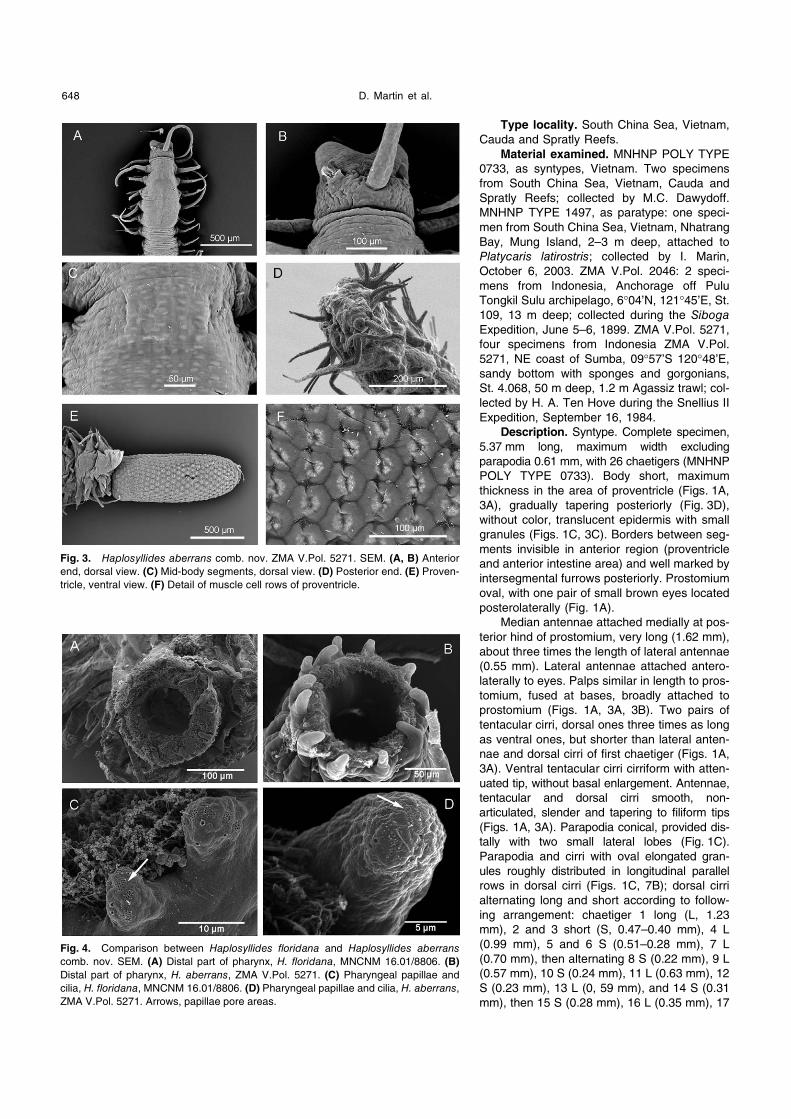
Description. Syntype. Complete specimen,
5.37 mm long, maximum width excluding
parapodia 0.61 mm, with 26 chaetigers (MNHNP
POLY TYPE 0733). Body short, maximum
thickness in the area of proventricle (Figs. 1A,
3A), gradually tapering posteriorly (Fig. 3D),
without color, translucent epidermis with small
granules (Figs. 1C, 3C). Borders between seg-
ments invisible in anterior region (proventricle
and anterior intestine area) and well marked by
intersegmental furrows posteriorly. Prostomium
oval, with one pair of small brown eyes located
posterolaterally (Fig. 1A).
Median antennae attached medially at pos-
terior hind of prostomium, very long (1.62 mm),
about three times the length of lateral antennae
(0.55 mm). Lateral antennae attached antero-
laterally to eyes. Palps similar in length to pros-
tomium, fused at bases, broadly attached to
prostomium (Figs. 1A, 3A, 3B). Two pairs of
tentacular cirri, dorsal ones three times as long
as ventral ones, but shorter than lateral anten-
nae and dorsal cirri of first chaetiger (Figs. 1A,
3A). Ventral tentacular cirri cirriform with atten-
uated tip, without basal enlargement. Antennae,
tentacular and dorsal cirri smooth, non-
articulated, slender and tapering to filiform tips
(Figs. 1A, 3A). Parapodia conical, provided dis-
tally with two small lateral lobes (Fig. 1C).
Parapodia and cirri with oval elongated gran-
ules roughly distributed in longitudinal parallel
rows in dorsal cirri (Figs. 1C, 7B); dorsal cirri
alternating long and short according to follow-
ing arrangement: chaetiger 1 long (L, 1.23
mm), 2 and 3 short (S, 0.47–0.40 mm), 4 L
(0.99 mm), 5 and 6 S (0.51–0.28 mm), 7 L
(0.70 mm), then alternating 8 S (0.22 mm), 9 L
(0.57 mm), 10 S (0.24 mm), 11 L (0.63 mm), 12
S (0.23 mm), 13 L (0, 59 mm), and 14 S (0.31
mm), then 15 S (0.28 mm), 16 L (0.35 mm), 17
Fig. 3. Haplosyllides aberrans comb. nov. ZMA V.Pol. 5271. SEM. (A, B) Anterior
end, dorsal view. (C) Mid-body segments, dorsal view. (D) Posterior end. (E) Proven-
tricle, ventral view. (F) Detail of muscle cell rows of proventricle.
Fig. 4. Comparison between Haplosyllides floridana and Haplosyllides aberranscomb. nov. SEM. (A) Distal part of pharynx, H. floridana, MNCNM 16.01/8806. (B)
Distal part of pharynx, H. aberrans, ZMA V.Pol. 5271. (C) Pharyngeal papillae and
cilia, H. floridana, MNCNM 16.01/8806. (D) Pharyngeal papillae and cilia, H. aberrans,
ZMA V.Pol. 5271. Arrows, papillae pore areas.

Review of Symbiotic Haplosyllides 649
S (0.28 mm), and 18 L (0.68 mm). Cirri from posterior-most
chaetigers ranging from 0.1–0.26 mm (S) and 0.31–0.39
mm (L) in length. Ventral cirri cirriform, with attenuated tips,
smooth, slightly shorter than or equal to parapodial lobes in
length (Fig. 1C). Two (occasionally three) thick simple
chaetae on each parapodium, usually one thicker than the
others; all with tridentate curved tips and sub-distal constric-
tions at different levels (Figs. 2E–H, 5D–F). Posterior-most
chaetae (Figs. 2F, 2H, 5F) more elongated than anterior-
most (Figs. 2E, 2G, 5D). Distance between chaetal tip and
subdistal constriction (distance a, Fig. 2) 3.2±0.2 as long as
width of chaetal apex (distance b, Fig. 2). One aciculum per
parapodium (occasionally two on the posterior-most),
straight, with truncated tip, similar in width to the thicker
chaetae (Fig. 1B).
Pharynx similar in length to proventricle (0.74 mm), with
an anterior pharyngeal tooth, bearing a crown of 10–12 dis-
tal papillae and a distal ring of cilia (Fig. 4B). Pharyngeal
papillae with pores of two sizes (Fig. 4D). Proventricle short,
0.71 mm long by 0.36 mm wide, extending from chaetiger 2
to the end of chaetiger 4, with 31 muscle cell rows (Figs. 1A,
3E, 3F). Pygidium small, rounded. Two anal cirri, absent in
one of the syntypes, but similar in length to the long poste-
rior-most dorsal cirri in the other specimens (Fig. 3D).
Remarks. Morphometric variation in the specimens
studied is summarized in Table 1. Haplosyllides aberranscomb. nov. from Vietnam and Indonesia differs slightly but
significantly from the Caribbean specimens of H. floridana.
Both species had two similar simple chaetae per parapo-
dium, occasionally three in H. aberrans comb. nov., but
these were less robust and the tips were half as long in the
latter as in the former (Figs. 2, 5).
San Martín et al. (1997) found two different forms in the
material studied from Cuba: non-reproductive individuals
(form A, MNCNM 16.01/8810) and specimens with attached
sexual stolons (form B, MNCNM 16.01/8808). Form A
lacked a pharyngeal tooth (present in form B) and had a
longer proventricle than form B; however, both forms had
similar chaetal shapes (Fig. 2A–D). Conversely, all nine
known specimens of H. aberrans comb. nov. were morpho-
logically similar, having a pharyngeal tooth and insignificant
variation in proventricle shape. Only one specimen (ZMA
V.Pol. 5271) had slightly shorter anterior chaetae (Fig. 2G)
than was typical (Figs. 2E, F, 5B, D, F), and thus more sim-
ilar to the chaetae of H. floridana (Fig. 2A–D). However, the
posterior chaetae were slender and longer (Fig. 2H), similar
to the typical form (Figs. 2E, F, 5B, D, F). This variability in
the anterior-most chaetae allows us to assume that
differences in chaetal arrangement between the two species
are more reliably detected in the posterior-most chaetae.
The particular shape of the chaetae, with a subdistal
enlargement separating the distal tip from the chaetal shaft,
may be viewed as additional support for the hypothesis that
the simple chaetae in Haplosyllidesoriginated by fusion of the blade and
shaft of a typical articulate syllid cha-
eta. This hypothesis was postulated
for two species of Haplosyllis, H. anthogorgicola Utinomi, 1939 (Mar-
tin et al., 2002) and H. loboi (Paola
et al., 2006), where possible origins
by loss of the blade of compound fal-
cigers or by evolution of the simple
chaetae “de novo” were rejected in
favor of the fusion hypothesis.
In addition to the chaetal differ-
ences, the muscular cells of the
proventricle in H. floridana were
more quadrangular in shape, and the
granules inside the body were less
prominent than in H. aberrans comb.
nov. Moreover, there were 10–12
Fig. 5. Comparison between Haplosyllides floridana and
Haplosyllides aberrans comb. nov. SEM. (A–C) Haplosyllides floridana, MNCNM 16.01/8806. (A) Anterior chaetae. (B) Midbody
chaetae. (C) Posterior chaetae. (D–F) Haplosyllides aberrans, ZMA
V. Pol. 5271. (D) Anterior chaeta. (E) Midbody chaetae. (F) Posterior
chaeta.
Fig. 6. Comparison of standardised appendage-length patterns among Haplosyllides floridana,
Haplosyllides aberrans comb. nov., and Haplosyllides ophiocomae sp. nov. CA, central anten-
nae; LA, lateral antennae; DTC, dorsal tentacular cirri; VTC, ventral tentacular cirri; AC, anal cirri;
1 to 15, dorsal cirri.
D. Martin et al.650
pharyngeal papillae in H. aberrans comb. nov. and only 10
in H. floridana (Fig. 4A, B). However, there were no differ-
ences in the sensory organs of the respective pharyngeal
papillae (Fig. 4C, D). In general, all body measurements
were, on average, longer in H. aberrans than in H. floridana,
except for the proventricle width and the length of tentacular
and anal cirri (Table 1). Conversely, when comparable, the
lengths of dorsal cirri tended to be significantly shorter (t
test, t=4.05; p<0.001) in H. aberrans comb. nov. than in H. floridana (Fig. 6). Finally, the granules inside the dorsal cirri
were oval, elongated, and roughly distributed in longitudinal
parallel rows (Fig. 7B, D) in H. aberrans comb. nov., and
round and randomly distributed in H. floridana (Fig. 7G).
In summary, we consider the morphological differences
(together with the ecological ones, see below) between the
material from distant geographical areas (Caribbean Sea vs.
Vietnam and Indonesia) to be sufficient to allocate them into
different species, respectively: i) Haplosyllides floridanasensu Augener (1924) and, partly, San Martín et al. (1997);
ii) Haplosyllides aberrans, a new combination for the origi-
nally described Syllis (Haplosyllis) aberrans by Fauvel
(1939).
Distribution. Nhatrang Bay and Spratly Archipelago,
Vietnam, South China Sea; NE coast of Sumba and Pulu
Tongkil, Sulu archipelago, Indonesia.
Ecology. Specimen MNHNP TYPE 1497 was found at
Mung Island (coast of Vietnam, South China Sea), at 2–3 m
deep, attached to a pleopod of a female specimen of the
pontoniin shrimp Platycaris latirostris, an obligatory associ-
ate of the scleractinian coral Galaxea astreata (Martin et al.,
2008). Specimens from Indonesia were found with sponges
and gorgonians on sandy bottoms 13–50 m deep; however,
these organisms were only reported as accompanying fauna
and there was no mention of a possible specific association
with H. aberrans.
Haplosyllides floridana Augener, 1924
(Figs. 2, 4–7)
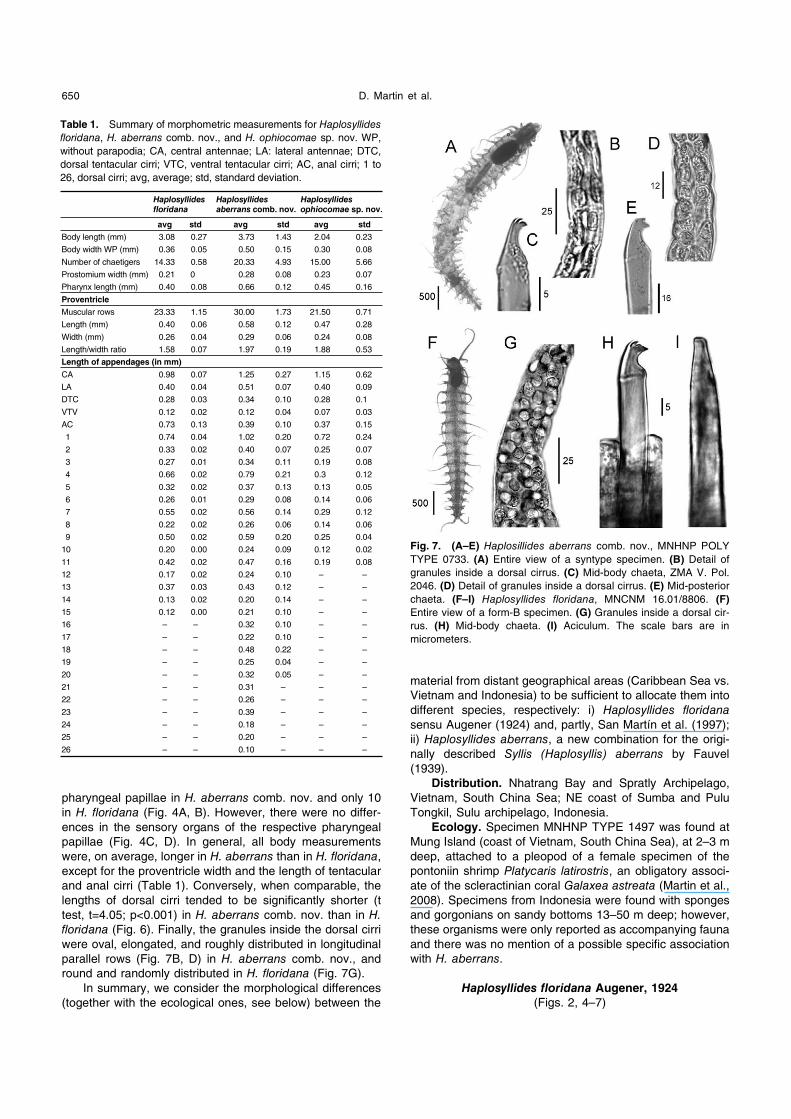
Table 1. Summary of morphometric measurements for Haplosyllides floridana, H. aberrans comb. nov., and H. ophiocomae sp. nov. WP,
without parapodia; CA, central antennae; LA: lateral antennae; DTC,
dorsal tentacular cirri; VTC, ventral tentacular cirri; AC, anal cirri; 1 to
26, dorsal cirri; avg, average; std, standard deviation.
Haplosyllides floridana
Haplosyllides aberrans comb. nov.
Haplosyllides ophiocomae sp. nov.
avg std avg std avg std
Body length (mm) 3.08 0.27 3.73 1.43 2.04 0.23
Body width WP (mm) 0.36 0.05 0.50 0.15 0.30 0.08
Number of chaetigers 14.33 0.58 20.33 4.93 15.00 5.66
Prostomium width (mm) 0.21 0 0.28 0.08 0.23 0.07
Pharynx length (mm) 0.40 0.08 0.66 0.12 0.45 0.16
Proventricle
Muscular rows 23.33 1.15 30.00 1.73 21.50 0.71
Length (mm) 0.40 0.06 0.58 0.12 0.47 0.28
Width (mm) 0.26 0.04 0.29 0.06 0.24 0.08
Length/width ratio 1.58 0.07 1.97 0.19 1.88 0.53
Length of appendages (in mm)
CA 0.98 0.07 1.25 0.27 1.15 0.62
LA 0.40 0.04 0.51 0.07 0.40 0.09
DTC 0.28 0.03 0.34 0.10 0.28 0.1
VTV 0.12 0.02 0.12 0.04 0.07 0.03
AC 0.73 0.13 0.39 0.10 0.37 0.15
1 0.74 0.04 1.02 0.20 0.72 0.24
2 0.33 0.02 0.40 0.07 0.25 0.07
3 0.27 0.01 0.34 0.11 0.19 0.08
4 0.66 0.02 0.79 0.21 0.3 0.12
5 0.32 0.02 0.37 0.13 0.13 0.05
6 0.26 0.01 0.29 0.08 0.14 0.06
7 0.55 0.02 0.56 0.14 0.29 0.12
8 0.22 0.02 0.26 0.06 0.14 0.06
9 0.50 0.02 0.59 0.20 0.25 0.04
10 0.20 0.00 0.24 0.09 0.12 0.02
11 0.42 0.02 0.47 0.16 0.19 0.08
12 0.17 0.02 0.24 0.10 – –
13 0.37 0.03 0.43 0.12 – –
14 0.13 0.02 0.20 0.14 – –
15 0.12 0.00 0.21 0.10 – –
16 – – 0.32 0.10 – –
17 – – 0.22 0.10 – –
18 – – 0.48 0.22 – –
19 – – 0.25 0.04 – –
20 – – 0.32 0.05 – –
21 – – 0.31 – – –
22 – – 0.26 – – –
23 – – 0.39 – – –
24 – – 0.18 – – –
25 – – 0.20 – – –
26 – – 0.10 – – –
Fig. 7. (A–E) Haplosillides aberrans comb. nov., MNHNP POLY
TYPE 0733. (A) Entire view of a syntype specimen. (B) Detail of
granules inside a dorsal cirrus. (C) Mid-body chaeta, ZMA V. Pol.
2046. (D) Detail of granules inside a dorsal cirrus. (E) Mid-posterior
chaeta. (F–I) Haplosyllides floridana, MNCNM 16.01/8806. (F)
Entire view of a form-B specimen. (G) Granules inside a dorsal cir-
rus. (H) Mid-body chaeta. (I) Aciculum. The scale bars are in
micrometers.

Review of Symbiotic Haplosyllides 651
Haplosyllides floridana – Augener, 1924: p. 44; San Martín
et al. 1997: p. 366–378, Figs. 1–3 (partim).
Haplosyllis floridana – Uabelacker, 1982: p. 585, Fig. 1,
(partim). San Martín et al. 1997: p. 366–378, Figs. 1–3.
Material examined. Haplosyllides floridana. Holotype
(ZMB 6608), Tortugas. Hundreds of specimens from the
Caribbean Sea (MNCNM 16.01/8806, 8808, 8809, 8810)
Cuba, off La Habana, 5 m deep, living inside the sponge
Xetospongia muta, collected and identified by G. San Martín
in June1992, July and October 1993 and January and April
1994.
Diagnosis. Modified from San Martín et al. (1997). Body
small with few chaetigers, epidermis translucent with small
granules. Segments without intersegmental furrows. Prosto-
mium with two eyes and three antennae, median one very
long. Palps fused to a single, anteriorly bilobed piece. Two
pairs of tentacular cirri, ventral ones considerably shorter
than dorsal pair. Antennae, tentacular, and dorsal cirri
smooth, slender and filiform. Parapodia and cirri with round,
randomly distributed granules. First dorsal cirri long, 2nd
and 3rd shorter than both 1st cirri and body width; 4th long,
5th and 6th short; remaining cirri alternating in length. Dorsal
cirri of posterior end short, all similar in length. Parapodia
conical, with two small lateral lobes and one more dorsally
located. Two thick simple chaetae on each parapodium, with
two small distal teeth, and one distal tooth more laterally
located. Distance between chaetal tip and subdistal constric-
tion (distance a, Fig. 2) 1.6±0.2 as long as width of chaetal
apex (distance b, Fig. 2). Acicula solitary, straight, distally
truncate. Pharynx with a distal crown of 10 papillae and a
ring of cilia. Pharyngeal tooth present only on small speci-
mens, at anterior end of pharynx. Proventricle long (2.5–5
chaetigers) on larger specimens, short (less than 2.5 chae-
tigers) on smaller. Pygidium small, rounded, with two anal
cirri, similar in shape and length to longest dorsal cirri. Mor-
phometric variation in the specimens studied is summarized
in Table 1.
Distribution. Florida, Tortugas, Cuba.
Ecology. Haplosyllides floridana was described on the
basis of a swimming male stolon (Holotype ZMB 6608) and
then fully re-described as an exclusive endosymbiont of the
sponge Xetospongia muta from Cuba (San Martín et al.,
1997). Several thousands or millions of worms inhabited the
aquiferous system of the host. Haplosyllides floridanaseemed to spend most of its life cycle inside the host
sponge. All different reproductive phases were found as
endosymbionts (from young juveniles to female epitokous
forms, including large asexual specimens), with the excep-
tion of male stolons (which seemed to be free swimmers).
No additional information on the exact nature of the relation-
ships with the host was provided, and it is unclear whether
the species should be considered a parasite or a commensal.
Haplosyllides ophiocomae sp. nov.
(Figs. 6, 8–10)
Haplosyllis aberrans – Hartman-Schröder, 1978: p. 49–53,
Figs. 1–7.
Type locality. Tortuguero, Puerto Rico.
Type material. Holotype ZMH P-22072, one specimen
collected by M. Youngbluth on January 29, 1973, 15 m
deep, associated with Ophiocoma pumila. Paratype BPBM-R
917, one specimen collected by M. Youngbluth on January
29, 1973, 15 m deep, associated with Ophiocoma pumila.
Paratypes BPBM-R 917, 10 specimens collected by M.
Youngbluth on January 29, 1973, 15 m deep, associated
with Ophiocoma pumila and other brittle stars.
Description. Holotype a complete specimen, 1.87 mm
long, maximum width excluding parapodia 0.24 mm, with 12
chaetigers. Body short, maximum thickness around proven-
tricle, gradually tapering posteriorly, colorless; translucent
epidermis with small, round granules (Fig. 9A). Borders
between anterior-most segments not well marked, except
between prostomium and first chaetiger. Prostomium oval,
with one pair of small, brownish eyespots (difficult to see on
holotype due to long-term preservation) located anterolaterally
near the bases of lateral antennae (Fig. 8C). Morphometric
variation in the specimens studied is summarized in Table 1.
Median antennae attached medially on posterior hind of
prostomium (Figs. 8C, 10A), very long (0.71 mm), about
three times as long as lateral antennae (0.23 mm). Lateral
antennae attached anterolaterally to eyes, near the bases of
palps. Palps similar in length to prostomium, fused at bases,
without a clear distinction from the anterior prostomium edge
(Fig. 8C). Two pairs of tentacular cirri, dorsal ones three
times as long as ventral ones (0.21 and 0.07 mm respec-
tively), but shorter than lateral antennae and first dorsal cirri
(Table 1). Antennae, and tentacular and dorsal cirri smooth,
non-articulated, slender, tapering to filiform tips (Figs. 8C,
9A, 10A). Parapodia conical, with two small lateral lobes
Fig. 8. Haplosyllides ophiocomae sp. nov. (A) Mid-anterior chaeta
(holotype, ZMH P-22072). (B) Aciculum from a posterior chaetiger
(holotype, ZMH P-22072). Paratype BPBM-R 917, redrawn from
Hartmann-Schröder (1978). (C) Anterior end. (D) Parapodium from
segment 14. (E) Chaetae of segment 8. (F) Chaeta of segment 9.
(G) Chaeta of segment 10. (H) Chaetae of segment 14. The scale
bars are in micrometers.
D. Martin et al.652
distally (Fig. 8D). Parapodia and dorsal cirri with round gran-
ules distributed at random (Fig. 9D). Dorsal cirri alternating
long and short, according to following arrangement: chaeti-
ger 1 long (L, 0.55 mm), 2 and 3 short (S, 0.20–0.14 mm),
4 L (0.30 mm), 5 and 6 S (0.13–0.14 mm), 7 L (0.29 mm),
8 S (0.14 mm), 9 L (0.25 mm), 10 S (0.10–0.16 mm), and
11 L (0.12–0.21 mm) (Table 1). Ventral cirri cirriform, with
attenuated tips, smooth, slightly shorter than or equal to
parapodial lobes in length.
Two (occasionally three) thick simple chaetae on each
parapodium, one usually thicker; all with tridentate curved
tips, with serration on upper edge of largest teeth, more
clearly visible under light microscope than under SEM, and
in mid-body and posterior most chaetae, with subdistal
constrictions at different levels (Figs. 8A, E–H, 9C, 10C–G).
Distance between chaetal tip and subdistal constriction
(distance a, Fig. 2) 1.9±0.2 as long as width of chaetal apex
(distance b, Fig. 2). One aciculum per parapodium (occasion-
ally two on the posterior-most), straight, with pointed, oblique
tip, similar in width to the thicker chaetae (Figs. 8B, 9B).
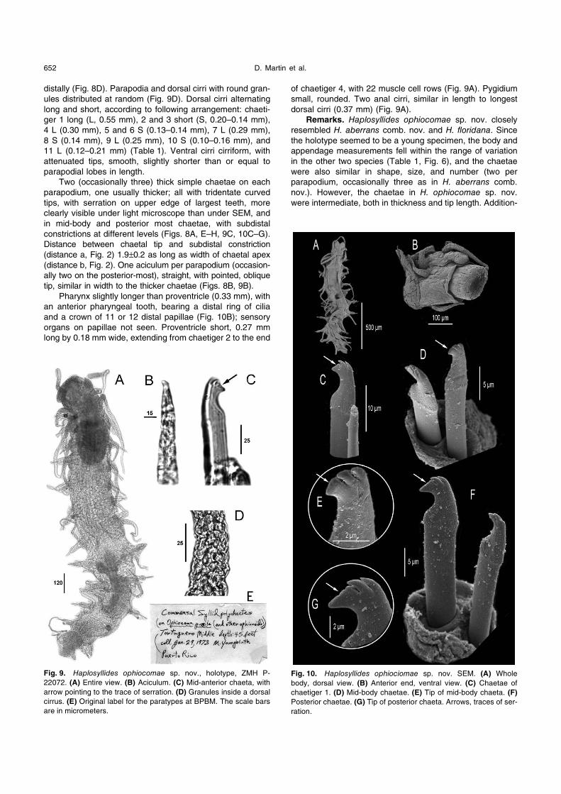
Pharynx slightly longer than proventricle (0.33 mm), with
an anterior pharyngeal tooth, bearing a distal ring of cilia
and a crown of 11 or 12 distal papillae (Fig. 10B); sensory
organs on papillae not seen. Proventricle short, 0.27 mm
long by 0.18 mm wide, extending from chaetiger 2 to the end
of chaetiger 4, with 22 muscle cell rows (Fig. 9A). Pygidium
small, rounded. Two anal cirri, similar in length to longest
dorsal cirri (0.37 mm) (Fig. 9A).
Remarks. Haplosyllides ophiocomae sp. nov. closely
resembled H. aberrans comb. nov. and H. floridana. Since
the holotype seemed to be a young specimen, the body and
appendage measurements fell within the range of variation
in the other two species (Table 1, Fig. 6), and the chaetae
were also similar in shape, size, and number (two per
parapodium, occasionally three as in H. aberrans comb.
nov.). However, the chaetae in H. ophiocomae sp. nov.
were intermediate, both in thickness and tip length. Addition-
Fig. 9. Haplosyllides ophiocomae sp. nov., holotype, ZMH P-
22072. (A) Entire view. (B) Aciculum. (C) Mid-anterior chaeta, with
arrow pointing to the trace of serration. (D) Granules inside a dorsal
cirrus. (E) Original label for the paratypes at BPBM. The scale bars
are in micrometers.
Fig. 10. Haplosyllides ophiociomae sp. nov. SEM. (A) Whole
body, dorsal view. (B) Anterior end, ventral view. (C) Chaetae of
chaetiger 1. (D) Mid-body chaetae. (E) Tip of mid-body chaeta. (F)
Posterior chaetae. (G) Tip of posterior chaeta. Arrows, traces of ser-
ration.

Review of Symbiotic Haplosyllides 653
ally, the serration evident on the upper edge of the major
teeth, previously observed and illustrated by Hartmann-
Schröder (1978) (Fig. 8E–H) and clearly visible under the
light microscope (Fig. 9C) and with difficulty by SEM (Fig.
10E, G), was absent in the other two species.
Granules were very abundant in the body and append-
ages of H. ophiocomae sp. nov., especially in the append-
ages, where they were round and distributed at random as
in H. floridana. The holotype had 11 pharyngeal papillae, but
the range was the same as in the BPBM specimens of H. aberrans comb. nov. with everted pharynx. The central
antennae of the holotype were about twice as long as the
lateral ones, as in H. floridana, but the larger specimen
described by Hartmann-Schröder (1978) had a central
antennae more than three times as long as the lateral ones,
longer even than in H. aberrans. Only three specimens in
the BPBM had antennae: one was likely the same illustrated
by Hartmann-Schröder (1978), the other had the central
antennae broken (Fig. 10A), and the third was a relatively
small worm, with central antennae about three times as long
as the lateral ones. All the remaining appendages were rel-
atively shorter than in H. floridana and H. aberrans comb.
nov., except for the ventral tentacular cirri (equal to those in
H. aberrans comb. nov.) and anal cirri (longer than in H. aberrans comb. nov.). The proventricle was also shorter
than in the other two species, although the width/length ratio
was closer to that in H. floridana. In turn, the pharynx was
intermediate in length (shorter than in H. aberrans comb.
nov. and longer than in H. floridana). Due to insufficient
specimens, it was not possible to assess whether the spec-
imens of H. ophiocomae sp. nov. had uniform morphology
(as in H. aberrans comb. nov.) or had two morphologies (as
in H. floridana).The geographical differences between H. ophiocomae
sp. nov. and H. floridana are not as marked as those
between these species and H. aberrans comb. nov. How-
ever, we consider that the observed morphological differ-
ences (particularly clear in the length pattern of dorsal cirri
and chaetal shape), together with the ecological differences
(but see Discussion), in material between Puerto Rico and
Cuba are sufficient to validate the description of the former
as a new species.
Distribution. Tortuguero, Puerto Rico.
Ecology. Living in symbiosis (commensalism?) with
Ophiocoma pumila and other brittle stars.
DISCUSSION
In their review of symbiotic polychaetes, Martin and
Britayev (1998) reported on about 20 different syllid genera
that include symbiotic species. Since then, there have been
major improvements in the knowledge of symbiotic syllids,
the most significant dealing with two genera closely related
to Haplosyllides, Haplosyllis Langerhans, 1879 and
Alcyonosyllis Glasby & Watson, 2001.
In Haplosyllis, recent studies have demonstrated that
the widespread sponge symbiont H. spongicola (Grube,
1875) corresponds, in fact, to a species complex whose
components are often restricted in biogeographical distribu-
tion and show marked host specificity (Martin et al., 2003;
Lattig et al., 2007). This certainly applies the Caribbean and
Indopacific regions, where most previous records of H.
spongicola must be viewed with caution, as they may cor-
respond to different, likely new species: for instance, records
from the Bahamas, Barbados, USA (Florida), and Jamaica
and Puerto Rico (Bacescu, 1971; Dauer, 1973; Reiswig,
1973; Uebelacker, 1978; Humann, 1992; Tsurumi and
Reiswig, 1997; Maldonado and Young, 1998b; a; López et
al., 2001), and those from Cambodia and Vietnam (López et
al., 2001), Korea (Lee and Rho, 1994), Japan (Imajima and
Hartman, 1964; Imajima, 1966; 2003), Tanzania (Magnino
and Gaino, 1998), and Papua New Guinea (Magnino et al., 1999).
Alcyonosyllis was initially defined as comprising strict
octocoral symbionts, based on A. phili from Australia and
Papua New Guinea (Glasby and Watson, 2001). An Indopa-
cific distribution, as well as host specificity, has been attrib-
uted to all new species currently included in the genus,
including A. glasby recently described from Japan (San
Martín and Nishi, 2003) and species formerly assigned to
Haplosyllis inhabiting Japanese, Chinese and Indonesian
waters, as well as the Red Sea: A. xeniaecola (Hartmann-
Schröder, 1993), A. onkylochaeta (Hartmann-Schröder,
1991), A. bisetosa (Hartmann-Schröder, 1960), A. exiliformis(Imajima, 2003) and A. gorgoniacola (Sun and Yang, 2004)
(Glasby and Watson, 2001; San Martín and Nishi, 2003;
Lattig and Martin, 2009).
The biogeography and specific peculiarities of the mode
of life of symbiotic syllids thus have an intrinsic interest, and
this certainly applies to Haplosyllides and its previously
known representatives, H. floridana (an exclusive endosym-
biont of the sponge Xetospongia muta from the Caribbean
Sea) and H. aberrans comb. nov. (a facultative parasitic
hypersymbiont — symbiont of a symbiont — of the shrimp
Platycaris latirostris, associated with the coral Galaxea astreata in Vietnamese waters, and found also in Indonesian
waters). These differences triggered the present revision
and proved to be excellent support for the taxonomic distinc-
tion between these species early synonymized by
Uebelacker (1982) and San Martín et al. (1997). Moreover,
our review also led us to study material corresponding to
another population from the Caribbean, reported by Hart-
man-Schröder (1978) to be commensal on the brittle star
Ophiocoma pumilla, that is here described as a third, new
species in the genus. Although the differences in ecology
and behavior among the three species of Haplosyllidesappeared to be clear, analysis of the BPBM material for H. ophiocomae sp. nov. cast some doubts on the previously
reported information.
The ecology and behavior of Haplosyllides floridanaseem to be well defined. There is no doubt that this species
lives in dense aggregates as an endosymbiont of its host
sponge, despite the initial discovery of the species as a
swimming specimen (in this case a male epitoke). In con-
trast, knowledge of the mode of life of H. aberrans comb.
nov. is fragmental, contradictory, and to some extent
enigmatic. The specimens described by Fauvel (1939) were
collected by Dawydoff with the help of a light trap, demon-
strating the ability of the worms to swim, even if there were
no epitokes. Whether this swimming capacity is related to
the ability to parasitize shrimps reported by Martin et al.(2008) is certainly a matter of speculation. Moreover, there
are no data on the habitat or on how this species was

D. Martin et al.654
collected during the Siboga Expedition. The specimens
collected during the Snellius II Expedition were found
“among sandy bottoms with sponges and gorgonians”,
which lacks enough precision to provide additional data on
the behavioral specificity of this population.
Although Hartman-Schröder (1978) did not provide
enough detailed ecological information on Haplosyllides ophiocomae sp. nov., she considered the species to be a
specific commensal of Ophiocoma pumilla. However,
according to the original label included in the BPBM loan
(Fig. 9E), the worms were collected not only from this brittle
star, but also from “other ophiuroids”, which was not men-
tioned in the 1978 paper. Although these additional hosts
were not identified, they could have belonged to various
species, and thus the postulated association might not be
species specific.
In fact, the finding of Haplosyllides aberrans as a para-
site of Platycaris latirostris (Martin et al., 2008) triggered an
exchange of information between the first author (D. Martin)
and Dr. Leslie Harris (from the Natural History Museum of
Los Angeles County), who kindly offered us her accurate
observations and knowledge on living sponge endosymbiotic
specimens of Haplosyllides and the closely related genus
Haplosyllis from the Caribbean and Hawaiian regions.
These worms always remain inside their host sponges, but
once disturbed (e.g., by reducing the oxygen availability in
the surrounding water), they rapidly emerge from the
sponges and immediately start to evert and retract the
pharynx. If the pharynx contacts the surface of any other
organism (e.g., other polychaetes, molluscs, crustaceans,
tunicates, bryozoans, anemones, brittle stars, or echinoids),
the proventricle automatically starts to pump, and the worms
are able to remain attached to these organisms through the
pharynx, even sucking their tissues.
As in many free-living polychaetes (such as phyllodocids
or glycerids, D. Martin, personal observations) when col-
lected and left “in vivo” out of the sediment, these pharyn-
geal movements may certainly be an accidental response
caused by the stress of being out of the host. Alternatively,
however, this may also be a normal component of the life
cycle, where behavioral elements of free living polychaetes
have been specifically integrated into a symbiotic mode of
life, as previously reported for other symbionts such as the
nereidid polychaete Nereis fucata (Gilpin-Brown, 1969;
Cram and Evans, 1980). If so, the pharyngeal movements
may be helpful in two situations.
The first could be finding and maintaining an attachment
to a temporary or intermediate host, as in the case of
Haplosyllides aberrans comb. nov. and Platycaris latirostris,
where tissues from the shrimp were seen deep inside the
gut of the syllid and the pleopod was consequently reduced
(Martin et al., 2008). Although it seems quite evident that H. aberrans comb. nov. was feeding on the shrimp, whether
this intermediate host provides simple support (with the
tissues inside the gut resulting from automatic pumping of
the proventricle) or an additional food source is difficult to
confirm without specific experiments.
The second situation was also suggested by Dr. Harris,
who addressed us in a web forum on sea slugs to demon-
strate that endosymbiotic syllids (likely Haplosyllis, but also
perhaps Haplosyllides) were able to effectively defend their
host sponges against attacks from spongivorous nudi-
branchs of the genus Hypselodoris by protruding from the
host sponge and repeatedly biting the intruder with the help
of the pharynx (see Imamoto, 2002; Rudman, 2002;
Velarde, 2002; Harris, 2006; Rudman, 2006). In this case,
the pharyngeal movements may be an intrinsic part of the
mode of life of the symbiont, but they are certainly interest-
ing for the host too, suggesting a mutualistic rather than a
commensalistic association, which may also be the case for
Haplosyllides floridana.
In summary, although Haplosyllides was considered
monotypic after Uebelacker (1982) and San Martín et al.
(1997), the present review differentiates two previously syn-
onymized species (H. floridana and H. aberrans comb. nov.)
and describes H. ophiocomae sp. nov. through a careful
classical morphological approach, supported by biogeo-
graphical and, to some extent, ecological/behavioral fea-
tures. The peculiarities of the respective associations of the
three Haplosyllis species, although poorly known at present,
are certainly an interesting topic for further study. Moreover,
on the whole, our review also casts some doubt on all
remaining “H. aberrans” populations previously reported. In
particular, the populations in the Marshall Islands, associ-
ated with corals of the genus Heliopora (Hartman, 1954),
and in Brazil, found within corals and calcareous algae
(Rullier and Amoureux, 1979), were only mentioned in these
previous papers, without formal description or additional
data on ecology and behavior. As it has not been possible
to obtain reference material to check the status of these
populations, their taxonomic assessment also remains open
to future research.
ACKNOWLEDGMENTS
We thank Dr. Guillermo San Martín and Dr. Leslie Harris for
fruitful discussions on the genus Haplosyllides and its morphologi-
cal variability and ecology; Dr. Harry A. Ten Hove for collecting
polychaetes during the Snellius II Expedition and providing the syl-
lids from the Siboga Expedition; Birger Neuhaus (ZMB), Tarik
Meziane (MNHNP), Miguel Villena (MNCNM), Angelika Brandt
(ZMH), and Holly Bolick (BPBM) for loaning the holotype of Haplo-syllides floridana, the type series of H. aberrans comb. nov., spec-
imens of H. floridana from Cuba, and specimens of H. ophiocomaesp. nov., respectively; and the staff of the SEM service at the Insti-
tute of Marine Sciences of Barcelona and at the Servicio Interde-
partamental de Investigación of the Universidad Autónoma of
Madrid for help with sample treatment and observation. This study
was financed by the European Commission’s Research Infrastruc-
ture Action via the SYNTHESYS Project, and partially supported by
the project “Taxonomía y Sistemática de la Familia Syllidae (Poly-
chaeta)” (CGL2005-02442), financed by the Ministerio de Edu-
cación y Ciencia of Spain and by the Russian Foundation for Basic
Research (08-04-92244-GFEN and 09-05-00736-a).
REFERENCES
Augener H (1924) Über litorale polychäten von Westindien. Sitz Ges
Naturf Freunde Berlin 1922: 38–53
Bacescu M (1971) Les Spongiaires: un des plus intéressants
biotopes benthiques marins. Rapp Comm Int Mer Medit 20:
239–241
Cram A, Evans SM (1980) Shell entry behaviour in the commensal
ragworm Nereis fucata. Mar Behav Physiol 7: 57–64
Dauer DM (1973) Polychaete fauna associated with Gulf of Mexico
sponges. Florida Sci 36(2-4): 192–196

Review of Symbiotic Haplosyllides 655
Fauvel P (1939) Annélides Polychètes de l’Indochine recueilles par
M. C. Dawidoff. Comm Pont Acad Sci 3: 243–368
Gilpin-Brown JB (1969) Host-adoption in the commensal polychaete
Nereis fucata. J Mar Biol Assoc UK 49: 121–127
Glasby CJ, Watson C (2001) A new genus and species of Syllidae
(Annelida: Polychaeta) commensal with octocorals. Beagle Rec
N Terr Mus Art Sci 17: 43–51
Harris LH (2006) Re: H. bullocki feeding - or at least tasting. http://
www.seaslugforum.net/find.cfm?id=17308 Sea Slug Forum, http://
www.seaslugforum.net/find.cfm?id=17308, Australian Museum,
Sydney
Hartman O (1954) Marine annelids from the northern Marshall
Islands. US Geol Surv Prof Pap 260 Q: 615–644
Hartmann-Schröder G (1978) Einige Sylliden-Arten (Polychaeta)
von Hawaii und aus dem Karibischen Meer. Mitt Hamburg Zool
Mus Inst 75: 49–61
Humann P (1992) Reef Creature Identification. Florida, Caribbean,
Bahamas. Vaughan Press, Orlando
Imajima M (1966) The Syllidae (polychaetous annelids) from Japan.
IV. Syllinae (1). Publ Seto Mar Biol Lab 14: 219–252
Imajima M (2003) Polychaetous Annelids from Sagami Bay and
Sagami Sea collected by the Emperor Showa of Japan and
deposited at the Showa Memorial Institute, National Science
Museum, Tokyo II. Orders included within the Phyllodocida,
Amphinomida, Spintherida and Eunicida. Natl Sci Mus Monogr
23: 1–221
Imajima M, Hartman O (1964) The Polychaetous Annelids from
Japan. Pt. I. Allan Hancock Found Spec Publ 26: 1–237
Imamoto J (2002) Feeding in Hypselodoris festiva. Sea Slug Forum,
http://www.seaslugforum.net/find.cfm?id=7858, Australian Mus-
eum, Sydney
Lattig P, Martin D (2009) A taxonomic revision of the genus HaplosyllisLangerhans, 1887 (Polychaeta: Syllidae: Syllinae). Zootaxa
2220: 1–40
Lattig P, San Martín G, Martin D (2007) Taxonomic and morphometric
analyses of the Haplosyllis spongicola complex (Polychaeta:
Syllidae: Syllinae) from Spanish seas, with re-description of the
type species and descriptions of two new species. Sci Mar 71:
551–570
Lee JW, Rho BJ (1994) Systematic studies on Syllidae (Annelida,
Polychaeta) from the South Sea and the East Sea in Korea.
Korean J Syst Zool 10: 131–144
López E, Britayev TA, Martin D, San Martín G (2001) New symbiotic
associations involving Syllidae (Annelida: Polychaeta), with
some taxonomic and biological remarks on Pionosyllis magnifica and Syllis cf. armillaris. J Mar Biol Assoc UK 81: 399–
409
Magnino G, Gaino E (1998) Haplosyllis spongicola (Grübe)
(Polychaeta, Syllidae) associated with two species of sponges
from east Africa (Tanzania, Indian Ocean). PSZN I: Mar Ecol
19: 77–87
Magnino G, Pronzato R, Sará A, Gaino E (1999) Fauna associated
with the horny sponge Anomoianthella lamella Pulitzer-Finali &
Pronzato, 1999 (Ianthellidae, Deponpongiae) from Papua-New
Guinea. Ital J Zool 66: 175–181
Maldonado M, Young CM (1998a) Limits on the bathymetric distri-
bution of keratose sponges: a fied test in deep water. Mar Ecol
Prog Ser 174: 123–139
Maldonado M, Young CM (1998b) Reevaluation of stalked aplysinid
sponges, with description of a new species from the upper
Bahamian slope. Bull Mar Sci 63: 417–426
Martin D, Britayev TA (1998) Symbiotic polychaetes: review of
known species. Oceanogr Mar Biol Annu Rev 36: 217–340
Martin D, Núñez J, Riera R, Gil J (2002) On the associations
between Haplosyllis (Polychaeta, Syllidae) and gorgonians
(Cnidaria, Octocorallaria), with a description of a new species.
Biol J Linn Soc 77: 455–477
Martin D, Britayev TA, San Martín G, Gil J, Martín D (2003) Inter-
population variability and character description in the sponge
associated Haplosyllis spongicola complex. (Polychaeta:
Syllidae). Hydrobiologia 496: 145–162
Martin D, Marin I, Britayev TA (2008) Features of the first known
parasitic association between Syllidae (Annelida, Polychaeta)
and crustaceans. Organism Divers Evol 8: 279–281
Paola A, San Martín G, Martin D (2006) A new species of HaplosyllisLangerhans, 1879 (Polychaeta: Syllidae: Syllinae) from Argen-
tina. Proc Biol Soc Wash 119: 346–354
Reiswig HM (1973) Population dynamics of Jamaican Demospongiae.
Bull Mar Sci 23: 191–226
Rudman WB (2002) Comment on Re: Feeding in Hypselodoris festivaby Andrej Jaklin. Sea Slug Forum, http://www.seaslugforum.net/
find.cfm?id=7896, Australian Museum, Sydney
Rudman WB (2006) Comment on Hypselodoris bullocki feeding - or
at least tasting by Brian Francisco. Sea Slug Forum, http://
www.seaslugforum.net/find.cfm?id=17283, Australian Museum,
Sydney
Rullier F, Amoureux L (1979) Campagne de la Calypso au large des
côtes Atlantiques de l’Amérique du Sud (1961–1962). I. 33.
Annélides Polychètes. Ann Inst Océanogr Monaco 55: 145–206
San Martín G, Nishi E (2003) A new species of Alcyonosyllis Glasby
and Watson, 2001 (Polychaeta: Syllidae: Syllinae) from
Shimoda, Japan commensal with the gorgonian Melithaea flabellifera. Zool Sci 20: 371–375
San Martín G, Ibarzábal DR, Jiménez M, López E (1997) Redescrip-
tion of Haplosyllides floridana Augener, 1924 (Polychaeta,
Syllidae, Syllinae), with notes on morphological variability and
comments on the generic status. Bull Mar Sci 60: 364–370
Tsurumi M, Reiswig HM (1997) Sexual versus asexual reproduction
in an oviparous rope-form sponge, Aplysina cauliformis(Porifera, Verongida). Invertebr Reprod Dev 32: 1–9
Uebelacker JM (1978) A new parasitic Polychaetous Annelid
(Arabellidae) from the Bahamas. J Parasitol 64: 151–154
Uebelacker JM (1982) Review of some little-known species of
Syllides (Annelida: Polychaeta) described from the Gulf of
Mexico and Caribbean by Hermann Augener in 1924. Proc Biol
Soc Wash 95: 583–593
Velarde RG (2002) Re: Feeding in Hypselodoris festiva. Sea Slug
Forum, http://www.seaslugforum.net/find.cfm?id=790, Australian
Museum, Sydney
(Received April 25, 2008 / Accepted June 21, 2009)