The genus Pluteus (Basidiomycota, Agaricales ... - Mycosphere
20
Submitted 2 March 2018, Accepted 7 April 2018, Published 25 June 2018 Corresponding Author: Dennis E. Desjardin – e-mail – [email protected] 598 The genus Pluteus (Basidiomycota, Agaricales, Pluteaceae) from Republic of São Tomé and Príncipe, West Africa Desjardin DE 1 and Perry BA 2 1 Department of Biology, San Francisco State University, 1600 Holloway Ave., San Francisco, California 94132, USA 2 Department of Biological Sciences, California State University East Bay, 25800 Carlos Bee Blvd., Hayward, California 94542, USA Desjardin DE, Perry BA 2018 – The genus Pluteus (Basidiomycota, Agaricales, Pluteaceae) from Republic of São Tomé and Príncipe, West Africa. Mycosphere 9(3), 598–617, Doi 10.5943/mycosphere/9/3/10 Abstract Six species of Pluteus are reported from the African island nation, Republic of São Tomé and Príncipe. Two represent new species (P. hirtellus, P. thomensis) and the other four represent new distribution records. Comprehensive descriptions, line drawings, colour photographs, comparisons with allied taxa, a dichotomous key to aid identification, and a phylogenetic analysis of pertinent Pluteus species based on ITS rDNA sequence data are provided. Key words – 2 new species – fungal diversity – Gulf of Guinea – mushrooms – pluteoid fungi – taxonomy Introduction In April 2006 (2 weeks) and April 2008 (3 weeks), expeditions led by scientists from the California Academy of Sciences and joined by mycologists from San Francisco State University visited the West African islands of São Tomé and Príncipe to document the diversity of plants, amphibians, marine invertebrates and macrofungi. This is the sixth in a series of papers focused on documenting the basidiomycetous macrofungi from the Republic (Desjardin & Perry 2009, 2015a, b, 2016, 2017). The genus Pluteus Fr. (Pluteaceae, Agaricales), comprising about 300 species, is quite common in tropical habitats. Most species grow on well-decayed wood and are characterized by basidiomes with free lamellae, pinkish brown, non-ornamented, inamyloid basidiospores, and convergent hymenophoral trama (Singer 1986). We follow the infrageneric classification of Justo et al. (2011a, b) in recognizing three sections, Pluteus, Celluloderma Fayod and Hispidoderma Fayod. Prior to this accounting, no species of Pluteus have been reported from the Republic of São Tomé and Príncipe. Geographically, the closest species have been reported from the Democratic Republic of Congo (Beeli 1928, Horak 1977, 1978, Menolli et al. 2014), several of which are recognized herein from São Tomé and Príncipe. We describe, illustrate, and compare six species of Pluteus from the Republic, based on morphological and molecular (ITS sequences) data. Two of these represent new species, while the other four are new distributional records. Materials & Methods Specimens were dried on a Nesco food dehydrator, packed in airtight plastic bags and hand Mycosphere 9(3): 598–617 (2018) www.mycosphere.org ISSN 2077 7019 Article Doi 10.5943/mycosphere/9/3/10 Copyright © Guizhou Academy of Agricultural Sciences
-
Upload
khangminh22 -
Category
Documents
-
view
2 -
download
0
Transcript of The genus Pluteus (Basidiomycota, Agaricales ... - Mycosphere
Submitted 2 March 2018, Accepted 7 April 2018, Published 25 June 2018
Corresponding Author: Dennis E. Desjardin – e-mail – [email protected] 598
The genus Pluteus (Basidiomycota, Agaricales, Pluteaceae) from
Republic of São Tomé and Príncipe, West Africa
Desjardin DE1 and Perry BA2
1Department of Biology, San Francisco State University, 1600 Holloway Ave., San Francisco, California 94132, USA 2Department of Biological Sciences, California State University East Bay, 25800 Carlos Bee Blvd., Hayward,
California 94542, USA
Desjardin DE, Perry BA 2018 – The genus Pluteus (Basidiomycota, Agaricales, Pluteaceae) from
Republic of São Tomé and Príncipe, West Africa. Mycosphere 9(3), 598–617, Doi
10.5943/mycosphere/9/3/10
Abstract
Six species of Pluteus are reported from the African island nation, Republic of São Tomé and
Príncipe. Two represent new species (P. hirtellus, P. thomensis) and the other four represent new
distribution records. Comprehensive descriptions, line drawings, colour photographs, comparisons
with allied taxa, a dichotomous key to aid identification, and a phylogenetic analysis of pertinent
Pluteus species based on ITS rDNA sequence data are provided.
Key words – 2 new species – fungal diversity – Gulf of Guinea – mushrooms – pluteoid fungi –
taxonomy
Introduction
In April 2006 (2 weeks) and April 2008 (3 weeks), expeditions led by scientists from the
California Academy of Sciences and joined by mycologists from San Francisco State University
visited the West African islands of São Tomé and Príncipe to document the diversity of plants,
amphibians, marine invertebrates and macrofungi. This is the sixth in a series of papers focused on
documenting the basidiomycetous macrofungi from the Republic (Desjardin & Perry 2009, 2015a,
b, 2016, 2017).
The genus Pluteus Fr. (Pluteaceae, Agaricales), comprising about 300 species, is quite
common in tropical habitats. Most species grow on well-decayed wood and are characterized by
basidiomes with free lamellae, pinkish brown, non-ornamented, inamyloid basidiospores, and
convergent hymenophoral trama (Singer 1986). We follow the infrageneric classification of Justo et
al. (2011a, b) in recognizing three sections, Pluteus, Celluloderma Fayod and Hispidoderma Fayod.
Prior to this accounting, no species of Pluteus have been reported from the Republic of São Tomé
and Príncipe. Geographically, the closest species have been reported from the Democratic Republic
of Congo (Beeli 1928, Horak 1977, 1978, Menolli et al. 2014), several of which are recognized
herein from São Tomé and Príncipe. We describe, illustrate, and compare six species of Pluteus
from the Republic, based on morphological and molecular (ITS sequences) data. Two of these
represent new species, while the other four are new distributional records.
Materials & Methods
Specimens were dried on a Nesco food dehydrator, packed in airtight plastic bags and hand
Mycosphere 9(3): 598–617 (2018) www.mycosphere.org ISSN 2077 7019
Article
Doi 10.5943/mycosphere/9/3/10
Copyright © Guizhou Academy of Agricultural Sciences

599
carried back to the USA. Macromorphological data were derived from fresh specimens, whereas
micromorphological data were derived from dried specimens rehydrated in ethanol followed by
distilled water, 3% KOH or Melzer's reagent. Colour terms and notations are those of Kornerup &
Wanscher (1978). Basidiospore statistics include: xm, the arithmetic mean of the spore length by
spore width (± standard deviation) for n spores measured in a single specimen; xmr, the range of
spore means, and xmm, the mean of spore means (± SD) when more than one specimen is available;
Q, the quotient of spore length by spore width in any one spore, indicated as a range of variation in
n spores measured; Qm, the mean of Q-values in a single specimen; Qmr, the range of Qm values and
Qmm, the mean of Qm values where more than one specimen is available; n, the number of spores
measured per specimen; s, the number of specimens involved. In the line drawings, cross-hatching
represents pigmented cell contents. All cited specimens are deposited in the H.D. Thiers
Herbarium, Dept. of Biology, San Francisco State University (SFSU).
Total genomic DNA was extracted from dried material using the Extract-N-Amp Plant PCR
Kit (Sigma-Aldrich, St. Louis, MO) following the manufacturer instructions. PCR protocols
followed those outlined in Perry et al. (2007). For all species included, the nuclear ribosomal
internal transcribed spacer region (ITS) was amplified using primers ITS1-F/ITS4 (Gardes & Bruns
1993, White et al. 1990). Amplification products were cleaned using the Exo-SAPit kit
(Affymetrix, Santa Clara, CA), and sent to Elim Biopharmaceuticals (Hayward, CA) for
sequencing with the same primer pairs used for amplification. Resulting sequencing products were
edited and assembled in Geneious 9.0 (Biomatters Ltd., Auckland, New Zealand). All ITS
sequences generated as part of this study have been deposited in GenBank (accessions MG968798–
MG968804).
To further investigate the taxonomic affinities of the African material referable to the genus
Pluteus, ITS sequences were analyzed phylogenetically within a broad sample of the genus.
Sequence data obtained from the African collections, as well as supplemental sequences
downloaded from GenBank, were aligned within the existing datasets of Justo et al. (2011a) for
Pluteus sections Celluloderma, Hispidoderma and Pluteus, which are available from TreeBase.org.
All alignments were manually edited in Mesquite (Maddison & Maddison 2015). Maximum
likelihood analyses were run in RAxML 8.2.9 (Stamatakis 2014) under a GTRGAMMAX model
and consisted of 100 alternative runs using the default parameters, with node support estimated by
100 RAxML bootstrap replicates. Bayesian analyses were performed within the CIPRES Science
Gateway (Miller et al. 2010), using Metropolis Coupled MCMC methods as implemented in
MrBayes 3.2.6 (Huelsenbeck & Ronquist 2001, Ronquist & Huelsenbeck 2003) under an
HKY+I+G (sect. Celluloderma) or GTR+I+G (sects. Hispidoderma and Pluteus) models of
sequence evolution as determined under the Bayesian Information Criterion in PAUP* 4.0a.159
(Swofford 2002). Bayesian analyses consisted of two parallel searches, run for 12 million
generations and initiated with random starting trees, and default chain temperature and swaps per
generation parameters. Eight chains were sampled every 1200 generations for a total of 1001 trees
each, sampled from the posterior distribution. Those trees sampled prior to the runs reaching an
average standard deviation of split frequencies of 0.01 were discarded as the burn-in, while the
remaining trees were used to calculate the posterior probabilities of the individual clades. Default
settings were used in MrBayes to set unconstrained branch lengths and uninformative topology
priors. The aligned ITS datasets and associated tree files have been deposited in TreeBase
(http://purl.org/phylo/treebase/phylows/study/TB2:S22346?x-access-
code=e12b54e6c68cf59b9fe606f2395b5d83&format=html).
Results
Based on a combination of morphological and molecular (ITS sequences) characters, the six
species of Pluteus from São Tomé and Príncipe belong to three infrageneric sections. Within sect.
Hispidoderma, P. albidus belongs to the longistriatus clade of Justo et al. (2011a), while P.
chrysaegis and P. thomensis belong to the leoninus clade (Fig. 1). In sect. Pluteus, P. albostipitatus
belongs to the albostipitatus clade, sister to P. salicinus, while P. losulus belongs to the losulus
600
clade, sister to P. atromarginatus (Fig. 6). Within sect. Celluloderma, P. hirtellus forms a relatively
long branch sister to P. riberaltensis var. conquistensis in the ephebeus clade (Fig. 11).
Artificial Key to Pluteus of São Tomé and Príncipe
1. Pileus pure white overall ............................................................................................. 1. P. albidus
1. Pileus yellow, grey, greyish brown, brown or dark brown ............................................................ 2
2. Pileus yellow, glabrous ........................................................................................... 2. P. chrysaegis
2. Pileus grey, greyish brown, brown or dark brown, appressed-fibrillose, fibrillose-punctate,
granulose or shaggy ........................................................................................................................... 3
3. Pleurocystidia thick-walled, at least some of them cornute, with apical horns; pileipellis a cutis
with undifferentiated terminal cells ................................................................................................... 4
3. Pleurocystidia thin-walled, lacking apical horns; pileipellis a cutis to trichodermium with
differentiated, fusoid to fusoid-ventricose terminal cells .................................................................. 5
4. Clamp connections absent; only some pleurocystidia cornute, apical horns often bifid; pileus
distinctly striate, disc fibrillose-punctate to granulose .......................................... 4. P. albostipitatus
4. Clamp connections present; all pleurocystidia cornute, apical horns conical; pileus non-striate,
radially streaked, appressed-fibrillose .............................................................................. 5. P. losulus
5. Pileus surface granulose, radially wrinkled; basidiospores subglobose to broadly ellipsoid (Q =
1.1–1.3), mean 6.8 5.8 µm; pileipellis a trichodermium of erect terminal cells ...... 3. P. thomensis
5. Pileus surface shaggy, with tiny fibrillose scales; basidiospores globose to subglobose (Q = 1.0–
1.1), mean 5.3 5 µm; pileipellis a cutis to trichodermium with clustered to scattered, erect to
repent terminal cells ....................................................................................................... 6. P. hirtellus
Taxonomy
Sect. Hispidoderma Fayod
Pluteus albidus Beeli, Bull. Soc. R. Bot. Belg. 61(1): 82. 1928. Fig. 2
non Pluteus albidus Pegler, Kew Bull., Addit. Ser. 6: 267. 1977 (nom. illeg., Art. 53.1)
Facesoffungi number: FoF04778
Pileus 10–25 mm diam, obtusely conical-umbonate to campanulate, expanding to broadly
convex with a small umbo, pellucid-striate; surface dull, dry, disc minutely granulose when young,
glabrous in age, pure white overall. Context <1 mm thick, soft, white. Lamellae free, close with 1–2
series of lamellulae, broad (2–3 mm), white when young, soon pink (7A2-3). Stipe 13–34 1–2
mm, central, terete, cylindrical above a subbulbous base, solid to stuffed; surface dull, dry,
pruinose, white, base with white tomentum. Odor indistinct.
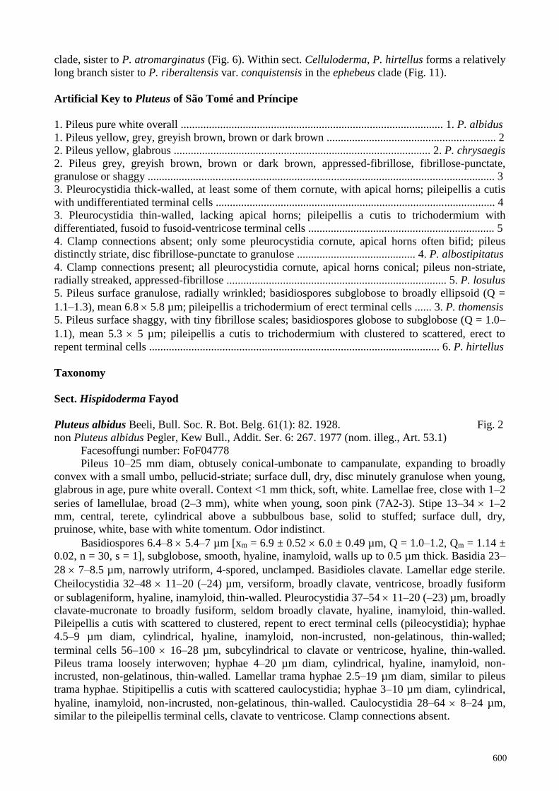
Basidiospores 6.4–8 5.4–7 µm [xm = 6.9 ± 0.52 6.0 ± 0.49 µm, Q = 1.0–1.2, Qm = 1.14 ±
0.02, n = 30, s = 1], subglobose, smooth, hyaline, inamyloid, walls up to 0.5 µm thick. Basidia 23–
28 7–8.5 µm, narrowly utriform, 4-spored, unclamped. Basidioles clavate. Lamellar edge sterile.
Cheilocystidia 32–48 11–20 (–24) µm, versiform, broadly clavate, ventricose, broadly fusiform
or sublageniform, hyaline, inamyloid, thin-walled. Pleurocystidia 37–54 11–20 (–23) µm, broadly
clavate-mucronate to broadly fusiform, seldom broadly clavate, hyaline, inamyloid, thin-walled.
Pileipellis a cutis with scattered to clustered, repent to erect terminal cells (pileocystidia); hyphae
4.5–9 µm diam, cylindrical, hyaline, inamyloid, non-incrusted, non-gelatinous, thin-walled;
terminal cells 56–100 16–28 µm, subcylindrical to clavate or ventricose, hyaline, thin-walled.
Pileus trama loosely interwoven; hyphae 4–20 µm diam, cylindrical, hyaline, inamyloid, non-
incrusted, non-gelatinous, thin-walled. Lamellar trama hyphae 2.5–19 µm diam, similar to pileus
trama hyphae. Stipitipellis a cutis with scattered caulocystidia; hyphae 3–10 µm diam, cylindrical,
hyaline, inamyloid, non-incrusted, non-gelatinous, thin-walled. Caulocystidia 28–64 8–24 µm,
similar to the pileipellis terminal cells, clavate to ventricose. Clamp connections absent.

601
Habitat and known distribution – Solitary on woody branches or on trunk of standing dead
tree in primary forest. Africa (DR Congo, São Tomé).
Material examined – AFRICA. São Tomé, Parque Nacional Obo, trail to Lagoa Amelia,
between N00˚17.112', E06˚35.767' and N00˚16.922', E06˚36.062, 14 April 2008, coll. by B.A.
Perry, BAP 612 (MG968798, SFSU); same location, 28 April 2006, coll. by D.E. Desjardin
(material lost).
Figure 1 – Maximum likelihood phylogeny of Pluteus sect. Hispidoderma based on ITS sequence
data (-lnL = 4341.716170). Sequences of species from São Tomé are indicated in bold type. Values
separated by/refer to nonparametric ML bootstrap proportions and Bayesian posterior probabilities.
Only values greater than 70/0.70 are shown (- designates a value below 70% or 0.70). Nodes
receiving support values greater than 90/0.95 are highlighted in bold.
Notes – Pluteus albidus is characterized by small white basidiomes with conical-umbonate
pileus, broad lamellae, pruinose, subbulbous stipe, subglobose basidiospores with mean 6.9 6 µm,
versiform cheilocystidia, thin-walled, broadly clavate-mucronate to broadly fusiform
pleurocystidia, a cutis-type pileipellis with clavate to ventricose terminal cells 16–28 µm diam,
scattered caulocystidia similar to the pileocystidia, an absence of clamp connections, and growth on
woody debris. The protologue (Beeli 1928), based on a collection from the DR Congo, matches

602
quite nicely the material from São Tomé. In combination, these features indicate placement in sect.
Hispidoderma. Interestingly, Pegler's (1977) illegitimate homonym P. albidus, based on a specimen
from Tanzania, may represent the same species. His material differs only subtly from our São
Tomé specimen in forming a convex-depressed (not umbonate) pileus, and he did not report the
presence of clavate to ventricose pileipellis terminal cells and caulocystidia. White forms of Pluteus
plautus (Weinm.) Gillet are similar in basidiospore size, and cystidium shape and size (Vellinga &
Schreurs 1985, Vellinga 1990), but the later species forms a hymeniderm-type pileipellis, quite
different from the cutis-type pileipellis of P. albidus.
Pairwise comparisons of aligned, overlapping ITS sequences of P. albidus (BAP 612) with
the top ten BLAST results indicate closest similarity of 95.1% to an undetermined Pluteus species
from New Zealand (KU131676) and 90.0–92.6% similarity to eight other undetermined Pluteus. In
our ITS phylogenetic analyses (Fig. 1), P. albidus fell into sect. Hispidoderma, on a long branch
inside a well-supported clade (100% BS, 1.0 PP) with P. granulatus, P. plautus, P. semibulbosus,
P. heteromarginatus and P. longistriatus.
Figure 2 – Micromorphological features of Pluteus albidus (BAP 612). a Basidiospores. b Basidia
and basidiole. c Cheilocystidia. d Pleurocystidia. e Pileipellis terminal cells. Scale bar = 10 µm
Pluteus chrysaegis (Berk. & Broome) Petch, Ann. R. Bot. Gdns Peradeniya 5(4): 271. 1912.
Figs 3–4
Basionym: Agaricus chrysaegis Berk. & Broome, J. Linn. Soc., Bot. 11(56): 536. 1871.
= Entoloma chrysaeges (Berk. & Broome) Sacc., Syll. Fung, (Abellini) 5: 61. 1887.
Facesoffungi number: FoF04780
Pileus 20–24 mm diam, plano-convex, expanding to plane-depressed, striate half way to
center, disc smooth; surface dull, moist to dry, glabrous; disc greyish yellow (3B4–5, 4C6–7),
margin yellow (3A3–5) to yellowish white (3A2). Context 1 mm thick, white. Lamellae horizontal,

603
free, close with 2–3 uneven series of lamellulae, broad (2–3 mm), pale pink to pale pinkish brown
(9A–B3). Stipe 22–23 1.5–2.0 mm, central, terete, cylindrical above a subbulbous base, solid,
curved; surface dull, dry, minutely pruinose; apex white to dingy buff, base pale greyish pink
(7B2–3) to dingy pink (8A2–3). Odor indistinct.
Figure 3 – Basidiomes of Pluteus chrysaegis (DED 8226). Scale bar = 10 mm
Basidiospores 5.2–6 4.5–5.5 µm [xm = 5.6 ± 0.22 5.0 ± 0.25 µm, Q = 1.0–1.2, Qm = 1.12
± 0.04, n = 20, s = 1], subglobose to ovoid, smooth, pale golden in KOH, inamyloid, thick-walled,
typically uniguttulate. Basidia 25–30 6.5–7 µm, utriform, 4-spored, unclamped. Basidioles
subcylindrical to subclavate. Lamellar edge sterile. Cheilocystidia 22–36 6.5–9.5 µm, subfusoid
to lageniform, hyaline, thin-walled. Pleurocystidia common, 35–77 12–24 µm, lageniform,
hyaline, thin-walled or with walls up to 0.6 µm thick, especially in central portion of cell. Pileipellis
a hymeniderm with pileocystidia; majority of cells 12–20 6.5–12 µm, broadly clavate to
vesiculose, hyaline, thin-walled; pileocystidia 22–36 6.5–10 µm, subfusoid to lageniform,
hyaline, thin-walled. Pileus and lamellar trama hyphae 2.5–16 µm diam, cylindrical to inflated,
604
hyaline, inamyloid, non-gelatinous, thin-walled. Stipitipellis a cutis; hyphae 7–10 µm diam,
cylindrical to slightly inflated, hyaline, inamyloid, non-incrusted, non-gelatinous, thin-walled.
Caulocystidia scattered or clustered, 17–32 9.5–20 µm, broadly clavate to vesiculose, seldom
lageniform, hyaline, thin-walled. Clamp connections absent in all tissues.
Figure 4 – Micromorphological features of Pluteus chrysaegis (DED 8226). a Basidiospores.
b Basidia and basidioles. c Cheilocystidia. d Pleurocystidia. e Pileipellis and pileocystidia.
f Caulocystidia. Scale bar = 10 µm
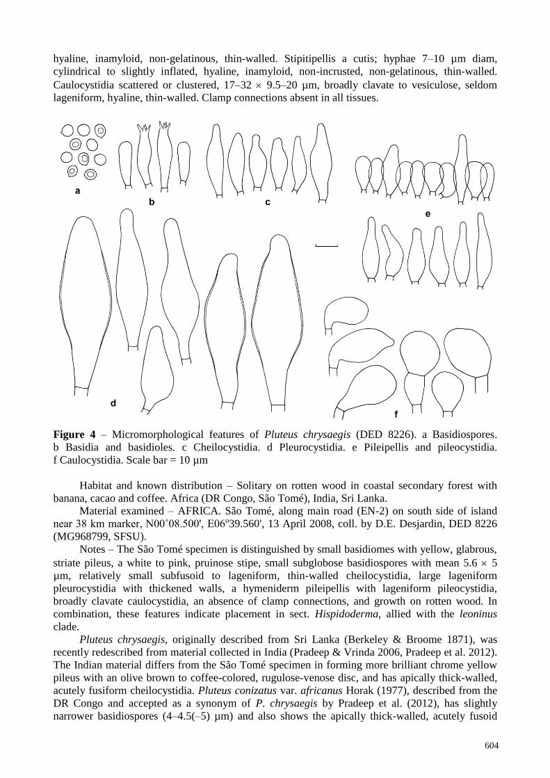
Habitat and known distribution – Solitary on rotten wood in coastal secondary forest with
banana, cacao and coffee. Africa (DR Congo, São Tomé), India, Sri Lanka.
Material examined – AFRICA. São Tomé, along main road (EN-2) on south side of island
near 38 km marker, N00˚08.500', E06o39.560', 13 April 2008, coll. by D.E. Desjardin, DED 8226
(MG968799, SFSU).
Notes – The São Tomé specimen is distinguished by small basidiomes with yellow, glabrous,
striate pileus, a white to pink, pruinose stipe, small subglobose basidiospores with mean 5.6 5
µm, relatively small subfusoid to lageniform, thin-walled cheilocystidia, large lageniform
pleurocystidia with thickened walls, a hymeniderm pileipellis with lageniform pileocystidia,
broadly clavate caulocystidia, an absence of clamp connections, and growth on rotten wood. In
combination, these features indicate placement in sect. Hispidoderma, allied with the leoninus
clade.
Pluteus chrysaegis, originally described from Sri Lanka (Berkeley & Broome 1871), was
recently redescribed from material collected in India (Pradeep & Vrinda 2006, Pradeep et al. 2012).
The Indian material differs from the São Tomé specimen in forming more brilliant chrome yellow
pileus with an olive brown to coffee-colored, rugulose-venose disc, and has apically thick-walled,
acutely fusiform cheilocystidia. Pluteus conizatus var. africanus Horak (1977), described from the
DR Congo and accepted as a synonym of P. chrysaegis by Pradeep et al. (2012), has slightly
narrower basidiospores (4–4.5(–5) µm) and also shows the apically thick-walled, acutely fusoid

605
cheilocystidia (Horak 1978) not seen in the São Tomé specimen.
Pairwise comparisons of aligned, overlapping ITS sequences of the São Tomé specimen
(DED 8226) with the top ten BLAST results indicate 97.5% similarity with two sequences of P.
chrysaegis (JN603206 – India; MF153092 – Florida) and 98.7% similarity with the sequence of the
holotype of P. conizatus var. africanus (HM562142). Our phylogenetic analysis of ITS sequences
of members of sect. Hispidoderma (Fig. 1) place the São Tomé specimen in a well-supported clade
(100% BS, 1.0 PP) with P. chrysaegis and P. conizatus var. africanus, and we recognize this
morphologically variable taxon as P. chrysaegis.
Pluteus thomensis Desjardin & B.A. Perry, sp. nov. Fig. 5
Mycobank: MB824540; Facesoffungi number: FoF04777
Holotype – AFRICA. São Tomé, Macambrara radio antenna area, N00˚16.557', E06˚36.326',
elev. 1300 m, 25 April 2008, coll. by B.A. Perry, DED 8333 (SFSU).
Etymology – Named in honor of the island São Tomé where the holotype specimen was
collected.
Diagnosis – Pileus 16–35 mm diam, broadly convex, expanding to plano-convex with a low,
broad umbo, radially wrinkled; surface dull, dry, granulose overall, disc and wrinkles dark brown
(7F6–8), margin brown (6E6–8). Context 1–2 mm thick, soft, white. Lamellae free, crowded, broad
(3–4 mm), greyish red (7B4). Stipe 24–50 2–2.5 mm, central, terete, cylindrical or gradually
enlarged downward to a bulbous or clavate base, solid; surface dull, dry, appressed-silky to
pruinose, pale pinkish white (7A2). Odor indistinct.
Figure 5 – Micromorphological features of Pluteus thomensis (DED 8333, Holotype).
a Basidiospores. b Basidia and basidiole. c Cheilocystidia. d Pleurocystidia. e Pileipellis terminal
cells. Scale bar = 10 µm

606
Basidiospores 6–7.7 5–6.5 µm [xm = 6.84 ± 0.42 5.77 ± 0.30 µm, Q = 1.1–1.3, Qm = 1.19
± 0.06, n = 25, s = 1], subglobose to broadly ellipsoid, smooth, subhyaline, inamyloid, thick-walled
(–0.5 µm). Basidia 22–26 7–8 µm, clavate to subutriform, 4-spored, unclamped. Basidioles
clavate. Lamellar edge sterile. Cheilocystidia 32–52 9.5–16 µm, broadly clavate, seldom
mucronate, hyaline, thin-walled to firm-walled (–0.5 µm). Pleurocystidia scattered, common, 56–70
16–27 µm, broadly lageniform to broadly clavate, hyaline, firm-walled (–0.5 µm). Pileipellis an
interrupted trichodermium of erect, fusiform to clavate cells 60–165 8–24 (–32) µm, acute to
subobtuse, hyaline or more commonly with brown cytoplasmic pigments, thin-walled. Pileus trama
interwoven; hyphae 3.5–20 µm diam, cylindrical or inflated, hyaline, non-incrusted, non-
gelatinous, thin-walled. Lamellar trama hyphae 4–14 µm diam, cylindrical, hyaline, non-gelatinous.
Stipitipellis a cutis with scattered caulocystidia; hyphae repent, 4–12 µm diam, cylindrical, hyaline,
inamyloid, non-incrusted, non-gelatinous, thin-walled. Caulocystidia uncommon, 32–64 6.5–11
µm, cylindrical, obtuse, hyaline, thin-walled. Clamp connections absent in all tissues.
Habitat and known distribution – Solitary on decaying wood in montane primary forest.
Africa (São Tomé).
Material examined – AFRICA. São Tomé, Macambrara radio antenna area, N00˚16.557',
E06˚36.326', elev. 1300 m, 25 April 2008, coll. by B.A. Perry, DED 8333 (MG968800, SFSU); São
Tomé, between Bom Sucesso at 1170 m elev. and Lagoa Amelio at 1480 m elev., near
N00˚17.317', E06˚36.746', 2 May 2006 (material lost in transit).
Notes – Pluteus thomensis is distinguished by relatively small, radially wrinkled, dark brown
pileus, a pale pinkish white, solid stipe, subglobose to broadly ellipsoid basidiospores with mean
6.8 5.8 µm, broadly clavate cheilocystidia, firm-walled, broadly lageniform pleurocystidia, a
trichoderm-type pileipellis of large, fusiform to clavate, brown terminal cells, scattered, hyaline,
cylindrical caulocystidia, an absence of clamp connections, and growth on rotten wood in a
montane primary forest. In combination, these features indicate placement in sect. Hispidoderma.
Pluteus thomensis shows morphological similarities to P. leoninus (Schaeff.) P. Kumm., P.
castri Justo & E.F. Malysheva, P. roseipes Höhn., and P. umbrosus (Pers.) P. Kumm. Pluteus
leoninus differs in forming a larger (30–60 mm diam), yellow to yellowish brown pileus, a white to
pale yellowish brown stipe, narrowly fusiform cheilocystidia, and pileipellis terminal cells with
yellowish contents (Vellinga 1990). Pluteus castri forms a bright yellow to yellowish orange
pileus, a cream to yellow stipe, slightly smaller basidiospores (mean 6.1 5 µm), fusiform to
lageniform cheilocystidia, and pileipellis terminal cells with yellowish contents (Justo et al. 2011a).
Pluteus roseipes has a dark brown, radially wrinkled pileus and solid pink stipe as in P. thomensis,
but shares similar micromorphology with P. leoninus and grows on conifer wood (Vellinga 1990).
Pluteus umbrosus from Europe, is much larger (pileus 55–115 mm diam, stipe 55–120 3.5–13
mm), forms brown-marginate lamellae, a stipe with dark brown fibrils, smaller basidiospores (5.5–
6.5 4–5.5 µm), and hymenial cystidia with brown contents (Vellinga 1990).
Pairwise comparisons of aligned, overlapping ITS sequences of P. thomensis (DED 8333)
with the top ten BLAST results indicate closest similarity (89.2%) to northern California (USA)
sequences of P. roseipes (KF306003, KC147681) and P. leoninus (KC147682, KC147680). In our
phylogenetic analysis of ITS sequences of members of sect. Hispidoderma (Fig. 1), P. thomensis is
on a relatively long branch sister to P. castri plus P. chrysaegis in a well-supported clade (99% BS,
1.0 PP) with P. leoninus.
Sect. Pluteus Fr.
Pluteus albostipitatus (Dennis) Singer, Lloydia 21: 240. 1959 (1958). Figs 7–8
Basionym: Pluteus spilopus var. albostipitatus Dennis, Bull. Trimest. Soc. Mycol. Fr. 69(2):
195. 1953.
Probable heterotypic synonyms:
= Pluteus phaeoleucus E. Horak, Bull. Jard. Bot. Natn. Belg. 47(1–2): 89. 1977.
= Pluteus densifibrillosus Menolli & Capelari, Mycologia 102(3): 698. 2010.

607
Figure 6 – Maximum likelihood phylogeny of Pluteus sect. Pluteus based on ITS sequence data (-
lnL = 4078.335883). Sequences of species from São Tomé and Príncipe are indicated in bold type.
Values separated by / refer to nonparametric ML bootstrap proportions and Bayesian posterior
probabilities. Only values greater than 70/0.70 are shown (- designates a value below 70% or 0.70).
Nodes receiving support values greater than 90/0.95 are highlighted in bold.
Facesoffungi number: FoF04779
Pileus 15–50 mm diam, plano-convex, becoming depressed in age, striate nearly to center,
margin decurved, weakly folded; surface dull to shiny, moist to dry, disc appressed fibrillose-
punctate with black wart-like granules, subglabrous to glabrous elsewhere; disc dark brown
(7–8F4–8) to black, margin dark brown (6F4–6) to brown (6E6–8) or greyish brown (6E–F3).
Context 1–2 mm thick, soft, white. Lamellae free, close with 2–3 series of lamellulae, broad (3–7
mm), pale pinkish white, becoming greyish red (8B3, 8C4–5) to reddish brown (8D5–6) in age.

608
Stipe 15–45 2–4 mm, central, terete, cylindrical, solid; surface dull, dry, glabrous to silky, white.
Odor indistinct.
Figure 7 – Basidiomes of Pluteus albostipitatus (DED 8205). Scale bar = 10 mm
Basidiospores 6–8.6 5.5–7 (–8) µm [xmr = 7.2–7.8 6.5–6.6 µm, xmm = 7.5 ± 0.44 6.57
± 0.04 µm, Q = 1.0–1.3, Qm = 1.14 ± 0.07, n = 25, s = 2], subglobose to broadly ellipsoid, rarely
globose, smooth, subhyaline, inamyloid, thick-walled (–0.5 µm). Basidia 19–36 7.5–9.5 µm,
clavate to utriform, 4-spored, unclamped. Basidioles subcylindrical to subclavate or ventricose.
Lamellar edge sterile. Cheilocystidia 35–64 9.5–16 µm, broadly clavate, seldom lageniform,
hyaline, thin-walled. Pleurocystidia abundant, (48–) 64–100 13–25 µm, clavate to lageniform,
ventricose or fusoid-subcapitate, obtuse and without outgrowths, or corniculate with 4–6 or more
small, short horn-like apical outgrowths, sometimes horns bifid, firm- to thick-walled (0.5–1.5 µm),
often thinning towards the base of cell, hyaline. Pileipellis a cutis of repent hyphae (3–) 4.5–12
(–16) µm diam, cylindrical, with brown cytoplasmic pigments, non-incrusted, non-gelatinous, thin-
walled; terminal cells undifferentiated, cylindrical to subclavate, obtuse, clustered and suberect
over pileus disc, repent elsewhere. Pileus trama interwoven; hyphae 4.5–16 (–24) µm diam,
cylindrical to inflated, hyaline, inamyloid, thin-walled. Lamellar trama hyphae 3.5–12 (–16) µm
diam, cylindrical to inflated, hyaline, non-gelatinous, thin-walled. Stipitipellis a cutis; hyphae 3–12
µm diam, cylindrical, hyaline, inamyloid, non-incrusted, non-gelatinous, thin-walled. Caulocystidia
absent. Clamp connections absent in all tissues.
Habitat and known distribution – Solitary on very rotten wood of undetermined
dicotyledonous tree in montane primary forest, and on rotting coconut palm wood in coastal
coconut grove. Africa (DR Congo, São Tomé), Caribbean (Martinique, Trinidad), South America
(Argentina, Bolivia, Brazil).

609
Figure 8 – Micromorphological features of Pluteus albostipitatus (DED 8205). a Basidiospores.
b Basidia and basidiole. c Cheilocystidia. d Pleurocystidia (DED 8205). e Pleurocystidia (DED
8220). Scale bar = 10 µm
Material examined – AFRICA. São Tomé, Macambrara radio antenna area, N00˚16.557',
E06˚36.326', elev. 1300 m, 11 April 2008, coll. by D.E. Desjardin, DED 8205 (MG968801, SFSU);
same location, 25 April 2006 (material lost in transit); São Tomé, along main road (EN-2) on south
side of island at N00˚12.126', E06˚42.362', 13 April 2008, coll. by D.E. Desjardin, DED 8220
(MG968802, SFSU).
Notes – Pluteus albostipitatus as represented in São Tomé is characterized by basidiomes
with a dark brown, fibrillose-punctate, distinctly striate pileus, a solid, cylindrical white stipe,
relatively large, subglobose to broadly ellipsoid basidiospores with mean 7.5 6.6 µm, broadly
clavate, thin-walled cheilocystidia, thick-walled, ventricose to fusoid-subcapitate pleurocystidia
without or with 4–6 small, sometimes bifid apical horns, and an absence of caulocystidia and clamp
connections. In combination, these features indicate placement in sect. Pluteus.
Pairwise comparisons of aligned, overlapping ITS sequences of the São Tomé specimens
(DED 8205, DED 8220) with the top ten BLAST results indicate 99.4–99.9% similarity to four
specimens of Pluteus albostipitatus from DR Congo (HM562130) and Brazil (JQ065032,
JQ801373, JQ065033). In addition, ITS sequences from the holotype specimens of P. phaeoleucus
E. Horak (Goossens-Fontana 5102; HM562141) from the DR Congo, and P. densifibrillosus
Menolli & Capelari (SP393696 HM562159) from Brazil, showed 99.5% and 99.3% similarity,
respectively, to the São Tomé specimens herein recognized as P. albostipitatus. Justo et al. (2011a,
Fig. 1) provided a phylogeny of Pluteus sect. Pluteus based on ITS sequences in which the six
sequences reported above from the DR Congo and Brazil formed a well-supported clade (≥90% BS,
≥0.95 PP) that they accepted as representing P. albostipitatus, indicating that P. phaeoleucus and P.
densifibrillosus represent heterotypic synonyms of P. albostipitatus. Our ITS phylogeny (Fig. 6)
places the São Tomé specimens firmly inside of the P. albostipitatus clade.

610
A comparison of published descriptions of the latter three species suggests that there is
extensive morphological variability within P. albostipitatus. Dennis (1953), Singer (1958), Horak
(1978), Pegler (1983) report the pileus margin of P. albostipitatus as striate to sulcate, while that of
P. phaeoleucus (Horak 1978) and P. densifibrillosus (Menolli & Capelari 2010) are reported as
non-striate. The pleurocystidia of P. albostipitatus are reported as lageniform, thin-walled (Horak
1978), fusoid with a subapical constriction and truncate apex, thin-walled (Pegler 1983), or
ventricose with a constriction above the broadest portion and subcapitate-truncate, thin-walled
(Singer 1958). In comparison, pleurocystidia of P. densifibrillosus were reported as slightly
ventricose to lageniform with a rounded or sometimes constricted apex, thin-walled to slightly
thick-walled (Menolli & Capelari 2010), Horak (1978) reported the pleurocystidia as absent in P.
phaeoleucus. Only Singer (1958) noted of the pleurocystidia "frequently tip truncate or with short
obtuse or subacute small outgrowths." The São Tomé specimens encompass this range of
pleurocystidia morphological variability. DED 8205 has numerous thick-walled pleurocystidia with
small often bifid apical projections and others lacking projections with subapical constriction and
truncate apex, while the pleurocystidia of DED 8220 are thin-walled, broadly clavate to lageniform,
and lack apical projections.
Pluteus losulus Justo, in Justo, Minnis, Ghignone, Menolli, Capelari, Rodríguez, Malysheva,
Contu & Vizzini, Mycol. Progr. 10(4): 473. 2011. Figs 9–10
Basionym: Pluteus cervinus var. ealaensis Beeli, Bull. Soc. R. Bot. Belg. 61(1): 81. 1928.
[nom. nov., non Pluteus ealaensis Beeli, Bull. Soc. R. Bot. Belg. 61(1): 80. 1928.]
Facesoffungi number: FoF04776
Figure 9 – Basidiomes of Pluteus losulus (DED 8313). Scale bar = 10 mm
Pileus 40–80 mm diam, campanulate to broadly convex-umbonate, expanding to plano-
convex with a shallow broad umbo; margin decurved, split in age, non-striate; surface dull, dry,
radially streaked, appressed-fibrillose, greyish brown (7E3) with a dark greyish brown (7F4) disc
and streaks. Context 2–4 mm thick, soft, white. Lamellae horizontal, free, close to crowded with 3
series of lamellulae, broad (5–8 mm), initially white, becoming greyish red (7B3). Stipe 50–110

611
4–9 mm, central, terete, cylindrical above an enlarged base, solid, pliant; surface dull, dry, glabrous
above, base appressed-fibrillose, white overall when young, in age base becoming greyish brown
(7E3). Odor mild.
Basidiospores 6.5–8 5.5–6.5 µm [xm = 7 ± 0.51 6 ± 0.32 µm, Q = 1.1–1.3, Qm = 1.17 ±
0.03, n = 20, s = 1], subglobose to ovoid, smooth, subhyaline, inamyloid, thick-walled (–0.5 µm);
pinkish brown in deposit. Basidia 28–32 8–9.5 µm, clavate to utriform, hyaline, 4-spored.
Basidioles clavate. Lamellar edge sterile. Cheilocystidia 42–52 7–13 µm, clavate to subclavate,
hyaline, inamyloid, thin-walled. Pleurocystidia common, 46–65 16–25 µm, ventricose to
utriform, cornute, with 3–4 apical, straight to recurved hooks, hyaline, thick-walled (–2 µm),
seldom bifid with 1–2 apical hooks per arm. Pileipellis a cutis of repent hyphae 3.5–10 µm diam,
cylindrical, hyaline or with brown cytoplasmic pigments, inamyloid, non-incrusted, non-gelatinous,
thin-walled; terminal cells undifferentiated or slightly narrowed. Pileus trama interwoven; hyphae
similar to pileipellis hyphae but all hyaline. Lamellar trama hyphae 2.5–10 µm diam, cylindrical,
hyaline, thin-walled. Stipitipellis a cutis of repent hyphae 5–14 µm diam, cylindrical, hyaline or
with brown cytoplasmic pigments, non-incrusted, non-gelatinous, thin-walled; terminal cells
undifferentiated. Caulocystidia absent. Clamp connections present in all tissues.
Figure 10 – Micromorphological features of Pluteus losulus (DED 8313). a Basidiospores.
b Basidia and basidiole. c Cheilocystidia. d Pleurocystidia. Scale bar = 10 µm
Habitat and known distribution – Solitary, scattered on very rotten wood in coastal secondary
forest. Africa (DR Congo, Príncipe).
Material examined – AFRICA. Príncipe, east side of island at N01˚35.728', E07˚25.263', 23
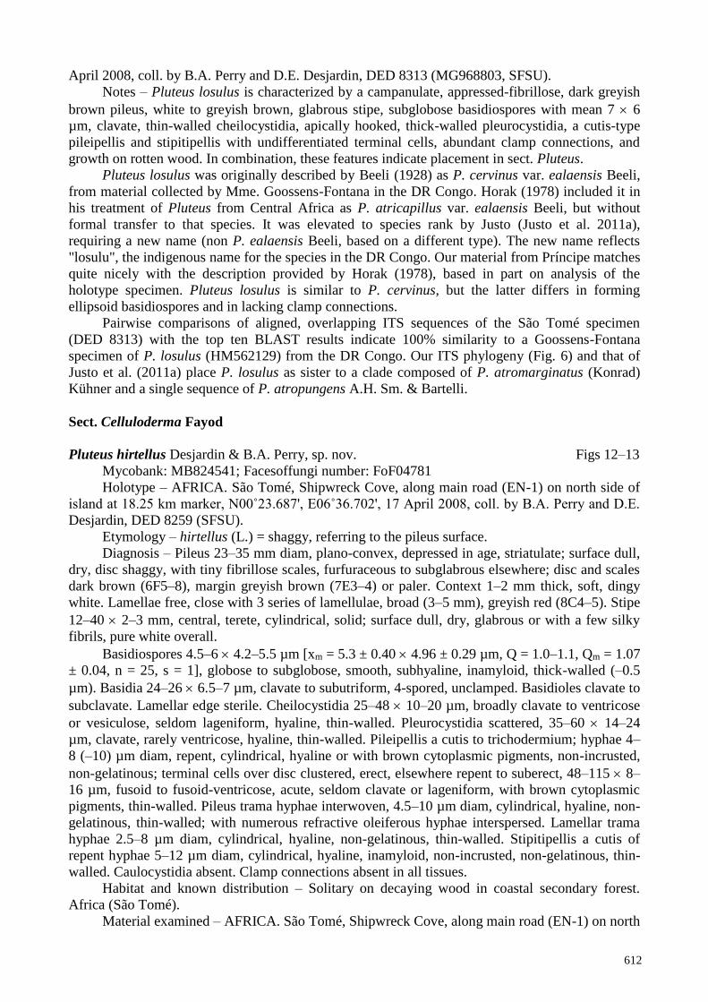
612
April 2008, coll. by B.A. Perry and D.E. Desjardin, DED 8313 (MG968803, SFSU).
Notes – Pluteus losulus is characterized by a campanulate, appressed-fibrillose, dark greyish
brown pileus, white to greyish brown, glabrous stipe, subglobose basidiospores with mean 7 6
µm, clavate, thin-walled cheilocystidia, apically hooked, thick-walled pleurocystidia, a cutis-type
pileipellis and stipitipellis with undifferentiated terminal cells, abundant clamp connections, and
growth on rotten wood. In combination, these features indicate placement in sect. Pluteus.
Pluteus losulus was originally described by Beeli (1928) as P. cervinus var. ealaensis Beeli,
from material collected by Mme. Goossens-Fontana in the DR Congo. Horak (1978) included it in
his treatment of Pluteus from Central Africa as P. atricapillus var. ealaensis Beeli, but without
formal transfer to that species. It was elevated to species rank by Justo (Justo et al. 2011a),
requiring a new name (non P. ealaensis Beeli, based on a different type). The new name reflects
"losulu", the indigenous name for the species in the DR Congo. Our material from Príncipe matches
quite nicely with the description provided by Horak (1978), based in part on analysis of the
holotype specimen. Pluteus losulus is similar to P. cervinus, but the latter differs in forming
ellipsoid basidiospores and in lacking clamp connections.
Pairwise comparisons of aligned, overlapping ITS sequences of the São Tomé specimen
(DED 8313) with the top ten BLAST results indicate 100% similarity to a Goossens-Fontana
specimen of P. losulus (HM562129) from the DR Congo. Our ITS phylogeny (Fig. 6) and that of
Justo et al. (2011a) place P. losulus as sister to a clade composed of P. atromarginatus (Konrad)
Kühner and a single sequence of P. atropungens A.H. Sm. & Bartelli.
Sect. Celluloderma Fayod
Pluteus hirtellus Desjardin & B.A. Perry, sp. nov. Figs 12–13
Mycobank: MB824541; Facesoffungi number: FoF04781
Holotype – AFRICA. São Tomé, Shipwreck Cove, along main road (EN-1) on north side of
island at 18.25 km marker, N00˚23.687', E06˚36.702', 17 April 2008, coll. by B.A. Perry and D.E.
Desjardin, DED 8259 (SFSU).
Etymology – hirtellus (L.) = shaggy, referring to the pileus surface.
Diagnosis – Pileus 23–35 mm diam, plano-convex, depressed in age, striatulate; surface dull,
dry, disc shaggy, with tiny fibrillose scales, furfuraceous to subglabrous elsewhere; disc and scales
dark brown (6F5–8), margin greyish brown (7E3–4) or paler. Context 1–2 mm thick, soft, dingy
white. Lamellae free, close with 3 series of lamellulae, broad (3–5 mm), greyish red (8C4–5). Stipe
12–40 2–3 mm, central, terete, cylindrical, solid; surface dull, dry, glabrous or with a few silky
fibrils, pure white overall.
Basidiospores 4.5–6 4.2–5.5 µm [xm = 5.3 ± 0.40 4.96 ± 0.29 µm, Q = 1.0–1.1, Qm = 1.07
± 0.04, n = 25, s = 1], globose to subglobose, smooth, subhyaline, inamyloid, thick-walled (–0.5
µm). Basidia 24–26 6.5–7 µm, clavate to subutriform, 4-spored, unclamped. Basidioles clavate to
subclavate. Lamellar edge sterile. Cheilocystidia 25–48 10–20 µm, broadly clavate to ventricose
or vesiculose, seldom lageniform, hyaline, thin-walled. Pleurocystidia scattered, 35–60 14–24
µm, clavate, rarely ventricose, hyaline, thin-walled. Pileipellis a cutis to trichodermium; hyphae 4–
8 (–10) µm diam, repent, cylindrical, hyaline or with brown cytoplasmic pigments, non-incrusted,
non-gelatinous; terminal cells over disc clustered, erect, elsewhere repent to suberect, 48–115 8–
16 µm, fusoid to fusoid-ventricose, acute, seldom clavate or lageniform, with brown cytoplasmic
pigments, thin-walled. Pileus trama hyphae interwoven, 4.5–10 µm diam, cylindrical, hyaline, non-
gelatinous, thin-walled; with numerous refractive oleiferous hyphae interspersed. Lamellar trama
hyphae 2.5–8 µm diam, cylindrical, hyaline, non-gelatinous, thin-walled. Stipitipellis a cutis of
repent hyphae 5–12 µm diam, cylindrical, hyaline, inamyloid, non-incrusted, non-gelatinous, thin-
walled. Caulocystidia absent. Clamp connections absent in all tissues.
Habitat and known distribution – Solitary on decaying wood in coastal secondary forest.
Africa (São Tomé).
Material examined – AFRICA. São Tomé, Shipwreck Cove, along main road (EN-1) on north

613
side of island at 18.25 km marker, N00˚23.687', E06˚36.702', 17 April 2008, coll. by B.A. Perry
and D.E. Desjardin, DED 8259 (MG968804, SFSU).
Figure 11 – Maximum likelihood phylogeny of Pluteus sect. Celluloderma based on ITS sequence
data (-lnL = 6516.135752). Sequences of species from São Tomé are indicated in bold type. Values
separated by/refer to nonparametric ML bootstrap proportions and Bayesian posterior probabilities.
Only values greater than 70/0.70 are shown (- designates a value below 70% or 0.70). Nodes
receiving support values greater than 90/0.95 are highlighted in bold.

614
Notes – Pluteus hirtellus is characterized by a relatively small pileus with dark brown
minutely scaly disc, a pure white, solid stipe, small, globose to subglobose basidiospores with mean
5.3 5.0 µm, broadly clavate to ventricose cheilocystidia, thin-walled, clavate pleurocystidia, a
cutis to trichodermium-type pileipellis with long, fusoid-acute, brown terminal cells, an absence of
caulocystidia and clamp connections, and growth on rotten wood in a coastal secondary forest. In
combination, these features indicate placement in sect. Celluloderma of Justo et al. (2011a) or sect.
Villosi of Vellinga & Schreurs (1985).
Figure 12 – Basidiomes of Pluteus hirtellus (DED 8259, Holotype). Scale bar = 10 mm
Pluteus hirtellus differs only subtly from the protologue of P. riberaltensis (Singer 1958),
described from Bolivia, in forming pileipellis terminal cells that are more acutely fusiform.
Basidiome features, basidiospore and cystidia size and shape match nicely, although ITS data are
quite distinct (see below and Fig. 11). The new species shows morphological similarities to P.
ephebeus (Fr.) Gillet, P. hispidulus (Fr.) Gillet, and P. escharites (Berk. & Broome) Sacc. Pluteus
ephebeus forms larger basidiomes with pileus 35–70 mm diam, a stipe 45–90 4–8 mm covered
with brown fibrils, larger broadly ellipsoid basidiospores (5.5–8.0 4.5–6.0 µm), and utriform to
fusiform or lageniform hymenial cystidia (Vellinga & Schreurs 1985, Vellinga 1990). Pluteus
hispidulus forms a conical to applanate-umbonate pileus, larger broadly ellipsoid basidiospores
(6–8 5–6 µm), and lacks pleurocystidia (Vellinga 1990). Pluteus escharites, described from Sri
Lanka and reported from Africa (Pegler 1977, 1986), shares basidiome macrofeatures, small
subglobose basidiospores and cheilocystidia shape, but differs in lacking pleurocystidia which are
abundant and conspicuous in P. hirtellus.

615
Pairwise comparisons of aligned, overlapping ITS sequences of Pluteus hirtellus (DED 8259)
with the top ten BLAST results indicate 98.6% similarity to three specimens from India determined
as Pluteus sp. (KR002882, KR002899, KT186192); 92.4% similarity to two specimens of Pluteus
aff. ephebeus from Illinois (USA; JQ065025, JQ065026); 92.3% similarity to a specimen of P.
ephebeus from Spain (HM562044); and 92.0–92.2% similarity to three specimens of P. aff.
ephebeus from Europe (HM562198, HM562080, KM983671). The ITS sequence of P. hirtellus is
only 88.2% similar to that of P. riberaltensis var. conquistensis (HM562162). In our ITS phylogeny
(Fig. 11), P. hirtellus is on a relatively long branch sister to P. riberaltensis var. conquistensis
(from Brazil), and together sister to P. ephebeus.
Figure 13 – Micromorphological features of Pluteus hirtellus (DED 8259, Holotype).
a Basidiospores. b Basidia and basidioles. c Cheilocystidia. d Pleurocystidia. e Pileipellis terminal
cells. Scale bar = 10 µm
Acknowledgments
We thank Dr. Robert C. Drewes (California Academy of Sciences) who continues to initiate,
coordinate and lead multi-organism biotic surveys on São Tomé and Príncipe; Eng. Arlindo de
Ceita Carvalho, Director General of the Ministry of Environment, Victor Bonfim, Salvador Sousa
Pontes and Danilo Barbero for permission to collect and export specimens for study. We are
indebted to Société de Conservation et Développement for logistics and housing support, especially
the wonderful staffs of Omali Lodge and Bom Bom Island. We are grateful for the support and
cooperation of Bastien Loloumb of Zuntabawe and Faustino Oliviera, former Director of the
botanical garden at Bom Sucesso. We were assisted in the field by Jose Ramos Maria Vital Pires on
Príncipe and by Quintino Quade Cabral, Martinho Nascimiento and Jose Clara on São Tomé, and
in the lab at Cal State East Bay by Jonathan del Rosario. For continuing support, we are most
grateful to Ned Seligman, Quintino Quade Cabral and Roberta dos Santos of STePUP. We are
grateful to the College of Science and Engineering at San Francisco State University for partial
616
funding to support travel to São Tomé and Príncipe, and to the G. Lindsay Field Research Fund of
the California Academy of Sciences (CAS) for financially supporting the expedition in 2006 and
the Hagey Research Venture Fund (CAS) in 2008. Lastly, we are especially grateful to Roderick
C.M. Hall, Coleman P. Burke and William K. Bowes Jr. whose generous philanthropy has
supported our research on São Tomé and Príncipe.
References
Beeli M. 1928 – Contribution a l'étude de la Flore mycologique du Congo: VI: Fungi Goossensiani,
Agaricacées, Rhodosporées. Bulletin de la Société Royale de Botanique de Belgique 61(1),
78–107.
Berkeley MJ, Broome CE. 1871 – The fungi of Ceylon. Journal of the Linnean Society 11, 494–
567.
Dennis RWG. 1953 – Les Agaricales de l'Ille de la Trinité: Rhodosporae-Ochrosporae. Bulletin de
la Société Mycologique de France. 69(2), 145–198.
Desjardin DE, Perry BA. 2009 – A new species of Phallus from São Tomé, Africa. Mycologia
101(4), 543-545. DOI: 10.3852/08-166
Desjardin DE, Perry BA. 2015a – A new species of Scytinopogon from the island of Príncipe,
Republic of São Tomé and Príncipe, West Africa. Mycosphere 6(4), 433–440. DOI:
10.5943/mycosphere/6/4/5
Desjardin DE, Perry BA. 2015b – Clavarioid fungi and Gasteromycetes from the Republic of São
Tomé and Príncipe, West Africa. Mycosphere 6(4), 515–531. DOI:
10.5943/mycosphere/6/5/2
Desjardin DE, Perry BA. 2016 – Dark-spored species of Agaricineae from Republic of São Tomé
and Príncipe, West Africa. Mycosphere 7(3), 359–391. DOI: 10.5943/mycosphere/7/3/8
Desjardin DE, Perry BA. 2017 – The gymnopoid fungi (Basidiomycota, Agaricales) from Republic
of São Tomé and Príncipe, West Africa. Mycosphere 8(9), 1317–1391. DOI:
10.5943/mycosphere/8/9/5
Gardes M, Bruns TD. 1993 – ITS primers with enhanced specificity for basidiomycetes –
application to the identification of mycorrhizae and rusts. Molecular Ecology 2, 113–118.
Horak E. 1977 – Neue zairische Arten aus der Gattung Pluteus Fr. Bulletin du Jardin Botanique
National de Belgique 47, 87–89.
Horak E. 1978 – Flore illustrée des champignons d’Afrique centrale. Fasc 6: Pluteus (Pluteaceae).
National Botanical Garden of Belgium, Meise. pp 107–118.
Huelsenbeck JP, Ronquist F. 2001 – MrBayes: Bayesian inference of phylogeny. Bioinformatics
17, 754–755.
Justo A, Minnis AM, Ghignone S, Menolli JrN et al. 2011a – Species recognition in Pluteus and
Volvopluteus (Pluteaceae, Agaricales): morphology, geography and phylogeny. Mycological
Progress 10(4), 453–479.
Justo A, Vizzini A, Minnis AM, Menolli JrN et al. 2011b – Phylogeny of the Pluteaceae
(Agaricales, Basidiomycota): taxonomy and character evolution. Fungal Biology 115, 1–20.
DOI: 10.1016/j.funbio.2010.09.012
Kornerup A, Wanscher JH. 1978 – Methuen Handbook of Colour. 3rd ed. Eyre Methuen, London.
Maddison WP, Maddison DR. 2015 – Mesquite: a modular system for evolutionary analysis.
Version 3.04 (http://mesquiteproject.org).
Menolli JrN, Capelari M. 2010 – Notes on Pluteus (Pluteaceae, Agaricvales) from Brazil including
two new species and a new record. Mycologia 102(3), 697–707.
Menolli JrN, Justo A, Arrillaga P, Pradeep CK et al. 2014 – Taxonomy and phylogeny of Pluteus
glaucotinctus sensu lato (Agaricales, Basidiomycota), a multicontinental species complex.
Phytotaxa 188(2), 78–90. DOI: 10.11646/phytotaxa.188.2.2
Miller MA, Pfeiffer W, Schwartz T. 2010 – “Creating the CIPRES Science Gateway for inference
of large phylogenetic trees” in Proceedings of the Gateway Computing Environments
617
Workshop (GCE), 14 Nov. 2010, New Orleans, LA pp 1–8.
Pegler DN. 1977 – A preliminary agaric flora of East Africa. Kew Bulletin Additional Series 6,
1–615.
Pegler DN. 1983 – Agaric flora of the Lesser Antilles. Kew Bulletin Additional Series 9, 1–668.
Pegler DN. 1986 – Agaric flora of Sri Lanka. Kew Bulletin Additional Series 12, 1–519.
Perry BA, Hansen K, Pfister DH. 2007 – A phylogenetic overview of the family Pyronemataceae
(Ascomycota, Pezizales). Mycological Research 111, 549–571.
Pradeep CK, Vrinda KB. 2006 – New and noteworthy species of Pluteus (Pluteaceae, Agaricales)
from Kerala State, India. Persoonia 19(1), 95–99.
Pradeep CK, Justo A, Vrinda KB, Shibu VP. 2012 – Two new species of Pluteus (Pluteaceae,
Agaricales) from India and additional observations on Pluteus chrysaegis. Mycological
Progress 11, 869–878. DOI 10.1007/s11557-011-0801-y
Ronquist F, Huelsenbeck JP. 2003 – MrBayes 3: Bayesian phylogenetic inference under mixed
models. Bioinformatics 19, 1572–1574.
Singer R. 1958 – Monographs of South American Basidiomycetes, especially those of the east
slope of the Andes and Brazil. 1. The genus Pluteus in South America. Lloydia 21(4), 195–
299.
Singer R. 1986 – The Agaricales in Modern Taxonomy. 4th Ed. Koeltz Scientific Books,
Koenigstein, Germany. 981 p.
Stamatakis A. 2014 – RAxML Version 8: A tool for Phylogenetic Analysis and Post-Analysis of
Large Phylogenies. Bioinformatics 10.1093/bioinformatics/btu033.
Swofford DL. 2002 – PAUP*. Phylogenetic Analysis Using Parsimony (*and Other Methods).
Version 4. Sinauer Associates, Sunderland, Massachusetts.
Vellinga EC. 1990 – Pluteaceae. In: Bas C, Kuyper TH, Noordeloos ME, Vellinga EC, eds. Flora
Agaricina Neerlandica 2, 31–55.
Vellinga EC, Schreurs J. 1985 – Notulae ad floram Agaricinam Neerlandicam – VIII. Persoonia
12(4), 337–373.
White TJ, Bruns T, Lee S, Taylor J. 1990 – Amplification and direct sequencing of fungal
ribosomal RNA genes for phylogenetics. In: Innis MA, Gelfand DH, Sninsky JJ, White TJ,
eds. PCR Protocols: a guide to methods and applications. San Diego: Academic Press. p.
315–322.