REVIEW ARTICLES - Anesthesia and Pain Medicine
146
KSNACC KSAP KSOA KSPA KNRS KSCVA KSTA KSPS KSRA Vol. 15. No. 2. April 2020 http://anesth-pain-med.org REVIEW ARTICLES 133 Perioperative management of patients receiving non-vitamin K antagonist oral anticoagulants: up-to-date recommendations 143 Viscoelastic coagulation test for liver transplantation pISSN: 1975-5171 eISSN: 2383-7977 Vol. 15/No. 2 Apr. 2020
-
Upload
khangminh22 -
Category
Documents
-
view
0 -
download
0
Transcript of REVIEW ARTICLES - Anesthesia and Pain Medicine

http://anesth-pain-med.orghttp://anesth-pain-med.orghttp://anesth-pain-med.orghttp://anesth-pain-med.orghttp://anesth-pain-med.org
KSNACC KSAP KSOA KSPA KNRS KSCVA KSTA KSPS KSRA
Vol. 15. No. 2. April 2020
http://anesth-pain-med.org
REVIEW ARTICLES
133 Perioperative management of patients receiving non-vitamin K antagonist oral anticoagulants: up-to-date recommendations
143 Viscoelastic coagulation test for liver transplantation
pISSN: 1975-5171eISSN: 2383-7977
Vol. 15/No. 2Apr. 2020


D R | PASSIST"Infusion Rate Monitor
Specifications
Battery One AA battery, lasts 360 hours Weight 110 g (3.9 oz)
Size 134 x 67 x 31 mm (5.3 x 2.6 x 1.2 in) Measurement Units Flow rate (ml/h), drops per minute (dp/m) and total
volume (ml)
Alarm 80 dB at 10cm approx. Sounds at 土13% rate change or when flow stops
Compatible Drip Sets Use only with sets labeled as "compatible with DripAssist Plus"
Accuracy 士 1 % drip rate War『anty 1 year limited warranty
Standards Compliance
Radiated Emissions CISPR 11 :2010 Ing『ess Protection IP22
Elect『omagnetic compatibility IEC 60601-1-6
Regulatory MDD 93/42/EEC, ISO 13485:2016, CE Mark
The specifications detailed in this brochure may vary from models and equipment available in your area. Please ask your local distributor for the details.
«ii,KCPM3D www.kcpmed.com KCPMED B/D 54, Donggyo-ro 27-gil, Mapo-gu, Seoul, 03990, Korea /T: 02-3141-2070
DRIPA1S IST® G Infusion Rate Monitor
Simple saves lives.
O뺄T
The simplest IV infusion management. SAFE, ACCURATE IV MEDICATION ADMINISTRATION


THE K
OREA
N SOCIETY OF OBSTETRIC ANES
THES
IOLO
GIS
TS
iAnesth Pain Med
Aims and ScopeAnesthesia and Pain Medicine (APM) is the official scientific journal of Korean Society of Neuroscience in Anesthesiology and Critical Care (KSNACC), The Korean Society for Anesthetic Pharmacology (KSAP), The Korean Society of Obstetric Anesthesiologists
(KSOA), The Korean Society of Pediatric Anesthesiologists (KSPA), Korean Neuromuscular Research Society (KNRS), Korean Society
of Cardiothoracic and Vascular Anesthesiologists (KSCVA), Korean Society of Transplantation Anesthesiologists (KSTA), and The
Korean Spinal Pain Society (KSPS) and Korean Society of Regional Anesthesia (KSRA). The abbreviated title is "Anesth Pain Med". It is
published four times a year on the last day of January, April, July, and October in English.
The mission of APM is to improve safety and quality of care of related patients and clinical practice of anesthesiologists by publishing
definitive articles in the field of anesthesiology including practice of perioperative management, critical care, and pain medicine. The
scopes of APM are as foallows : anesthesia-related issues from affiliated neuroanesthesiology (KSNACC), experimental, laboratory
works or clinical relevance of anesthetic pharmacology (KSAP), anesthesia for operative delivery, pain relief in labor, care of the
critically ill parturient, perinatal physiology and pharmacology (KSOA), anesthetic care, perioperative management, and alleviation
of pain in children (KSPA), physiology of neuromuscular transmission and block, pharmacology of neuromuscular blocking agents
and their reversal agents, principles and applications of neuromuscular monitoring, and drug interaction between neuromuscular
blocking agents and other substances (KNRS), anesthesia for cardiothoracic and vascular surgery and management of patients
undergoing various surgeries for patients with cardiac, pulmonary, and vascular diseases (KSCVA), perioperative anesthesia care
of transplantation surgery, physiology or pharmacology related with transplantation anesthesiology (KSTA), pathophysiology,
pharmacology, and all respects of spine related pain (KSPS), clinical techniques of regional blocks, anatomy, patient safety issues,
basic sciences such as pharmacology of local anesthetics or sedative drugs (KSRA).
All or part of the Journal is indexed/tracked/covered by KoreaMed, KoMCI, Google Scholar, Science Central.
Full text is freely available from http://anesth-pain-med.org
The circulation number per issue is 400.
Anesthesia and Pain Medicine April 2020; Volume 15, Number 2, Serial No. 56ⓒ 2020 the Korean Society of Anesthesiologists.
Korean Society of Neuroscience in Anesthesiologyand Critical Care
The Korean Society for Anesthetic Pharmacology
Korean Neuromuscular Research Society Korean Society of Cardiothoracic and VascularAnesthesiologists
The Korean Society of Obstetric Anesthesiologists The Korean Society of Pediatric Anesthesiologists
Korean Society of Transplantation Anesthesiologists The Korean Spinal Pain Society
Korean Society of Regional Anesthesia
pISSN: 1975-5171eISSN: 2383-7977

Anesth Pain Medii
Vol.15 No.2 April 2020
PublisherIn-cheol Choi (University of Ulsan)
Editor-in-ChiefYoung-Cheol Woo (Chung-Ang University)
Associate EditorChong Wha Baek (Chung-Ang University)
Jung-Won Hwang (Seoul National University)Hyun Kyo Lim (Yonsei University)
Editorial BoardYang Hoon Chung (Soonchunhyang University)
Hyun Joo Ahn (Sungkyunkwan University)Hyungseok Seo (Kyung Hee University)
Seongtae Jeong (Chonnam National University)Jae-hun Kim (Konkuk University)
Jeong Rim Lee (Yonsei University)
Young Duck Shin (Chungbuk National University)Woo-jong Choi (University of Ulsan)Byung Gun Lim (Korea University)Wonjin Lee (Inje University)Yong Seon Choi (Yonsei University)Young Ju Won (Korea University)
Ethic EditorYoung Yoo (Korea University)
Statistics EditorJunyong In (Dongguk University), Dong-Kyu Lee (Korea University)
Illustrated EditorYong Beom Kim (Gachon University of Medicine and Science)
Manuscript EditorJi Youn Ha (The Korean Society of Anesthesiologists), Se Jueng Kim (MEDrang Inc.)
Contacting the Anesthesia and Pain Medicine
All manuscripts must be submitted online through the APM e-Submission system athttp://submit-apm.orgElectronic files of the manuscript contents must be uploaded at the web site.Items pertaining to manuscripts submitted for publication, as well as letters or other forms of communication regardingthe editorial management of APM should be sent to:
Editor-in-Chief
Young-Cheol Woo
Publishing/Editorial Office
The Korean Society of Anesthesiologists101-3503, Lotte Castle President, 109 Mapo-daero, Mapo-gu, Seoul 04146, KoreaTel +82-2-795-5129, Fax +82-2-792-4089, E-mail [email protected]
Printed by M2community Co.
8th FL, DreamTower, 66 Seongsui-ro, Seongdong-gu, Seoul 04784, Korea

iiiAnesth Pain Med
Vol.15 No.2 April 2020
Table of Contents
Reviews133 Perioperative management of patients receiving non-vitamin K antagonist oral anticoagulants: up-to-date recommendations Kwang-Sub Kim, Jong Wook Song, Sarah Soh, Young-Lan Kwak, Jae-Kwang Shim
143 Viscoelastic coagulation test for liver transplantation Sun Young Park
Anesthetic Pharmacology Clinical Research
152 Effect-site concentration of remifentanil for preventing propofol injection pain during induction of balanced anesthesia Joungmin Kim, Daehoon Kim, Hyung Gong Lee
157 Effect of preoperative administration of systemic alpha-2 agonists on postoperative pain: a systematic review and meta-analysis Ji Youn Ju, Kye-Min Kim, Sangseok Lee
Spinal Pain Case Report
193 Carpal tunnel syndrome caused by thrombosed persistent median artery Sang Yoon Jeon, Kwangmin Lee, Weon-Joon Yang
199 Ultrasound-guided treatment of common peroneal neuropathy caused by Baker’s cyst: a clinical note Hana Cho, Dong-Rim Kim, Je Jin Lee, Seung Young Lee, Yong Bum Park, Hee Sung Kim, Hwa-Yong Shin
Obstetric Anesthesia Clinical Research
167 Hemodynamic effects of carbetocin administered as an intravenous bolus or infusion during cesarean delivery Kihyug Kwon, Dohyung Kim, Hyunmin Jo, Ji Eun Park, Kyung Ok Kim
Pediatric Anesthesia Clinical Research
173 Clinical performance of Ambu AuraGainTM versus i-gelTM in anesthetized children: a prospective, randomized controlled trial Ji-Hyun Lee, Seungpyo Nam, Young-Eun Jang, Eun-Hee Kim, Hee-Soo Kim, Jin-Tae Kim
Cardiothoracic and Vascular Anesthesia Case Report
181 Acute normovolemic hemodilution for a patient with secondary polycythemia undergoing aortic valve replacement due to severe aortic stenosis Ilsang Han, Young Woo Cho, Soon Eun Park, Min Gi An, Ho June Kang, A-ran Lee
Transplantation Anesthesia Clinical Research
187 Systolic anterior motion of mitral chordae tendineae: prevalence and clinical implications in liver transplantation Hye-Mee Kwon, Kyoung-Sun Kim, Gyu-Sam Hwang
pISSN: 1975-5171eISSN: 2383-7977

Anesth Pain Mediv
205 Herpes zoster in the ophthalmic branch of the trigeminal ganglia obscuring cavernous sinus thrombosis due to Streptococcus constellatus subsp. constellatus Ji Hye Lee, Hyun Joo Heo, Ki Man Kim, Han Gyeol Lee, Seung Min Baek, Da Wa Jung
Regional Anesthesia Clinical Research
209 Analgesic effect of ropivacaine with fentanyl in comparison with ropivacaine alone for continuous femoral nerve block after knee replacement arthroplasty: a prospective, randomized, double-blinded study Gunn Hee Kim, Joon Woo Lee, Go Eun Kim, Seong Su Lee, Shill Lee Son, Byung Uk Kim, Ha Na Cho, Mi Young Kwon, Min Seok Koo, Ji Eun Kim, Mi Jung Yun
General Articles Clinical Research
217 The question of preoperative anxiety and depression in older patients and family protectors Sehun Lim, Younmi Oh, Kwangrae Cho, Myoung-hun Kim, Sungho Moon, Seunghee Ki
226 Vocal cord paralysis following general anesthesia with endotracheal intubation: a clinical review on 43 cases Sehun Lim, Dong-chun Kim, Kwangrae Cho, Myoung-hun Kim, Sungho Moon, Hakmoo Cho, Seunghee Ki
233 Risk factors of emergency reoperations Tae Kwan Kim, Jun Rho Yoon, Yu Na Choi, Ui Jin Park, Kyoung Rim Kim, Taehee Kim
241 Correlation between patient health questionnaire-2 and postoperative pain in laparoscopic cholecystectomy Yusom Shin, Tae Woo Park, Huiyoung Kim, Dong-jin Shim, Hochul Lee, Joo-Duck Kim, Donghee Kang
Case Report
247 Novel alternative for submental intubation Inyoung Jung, Byung Hoon Yoo, Ji Youn Ju, Sijin Choi, Jun Heum Yon, Kye-Min Kim, Yun-Hee Lim, Woo Yong Lee
251 Abdominal compartment syndrome caused by gastric distension in bulimia nervosa and fatal injury following surgical decompression Byeong Hun Eom, Hyun Kyoung Lim, Nayoung Tae, Helen Ki Shinn

KS
CVA
INTRODUCTION
Atrial fibrillation, the most frequently encountered ar-
rhythmia, is associated with thromboembolism and stroke
which need to be prevented amongst other therapies in-
volving rhythm control [1]. For that purpose, vitamin K an-
tagonist, warfarin, has long been used despite its incon-
stant and unpredictable anticoagulation effect which re-
Perioperative management of patients receiving non-vitamin K antagonist oral anticoagulants: up-to-date recommendations
Kwang-Sub Kim, Jong Wook Song, Sarah Soh, Young-Lan Kwak, and Jae-Kwang Shim
Department of Anesthesiology and Pain Medicine, Anesthesia and Pain Research Institute,
Severance Cardiovascular Hospital, Yonsei University College of Medicine, Seoul, Korea
Review ArticleAnesth Pain Med 2020;15:133-142 https://doi.org/10.17085/apm.2020.15.2.133pISSN 1975-5171 • eISSN 2383-7977
Received March 2, 2020 Revised March 5, 2020 Accepted: April 9, 2020
Corresponding author Jae-Kwang Shim, M.D., Ph.D. Department of Anesthesiology and Pain Medicine, Anesthesia and Pain Research Institute, Severance Cardiovascular Hospital, Yonsei University College of Medicine, 50-1 Yonsei-ro, Seodaemun-gu, Seoul 03722, Korea Tel: 82-2-2228-8500 Fax: 82-2-364-2951 E-mail: [email protected]
Indications of non-vitamin K antagonist oral anticoagulants (NOACs), consisting of two types: direct thrombin inhibitor (dabigatran) and direct factor Xa inhibitor (rivaroxaban, apixaban, and edoxaban), have expanded over the last few years. Accordingly, increasing number of patients presenting for surgery are being exposed to NOACs, despite the fact that NOACs are inevitably related to increased perioperative bleeding risk. This review arti-cle contains recent clinical evidence-based up-to-date recommendations to help set up a multidisciplinary management strategy to provide a safe perioperative milieu for patients receiving NOACs. In brief, despite the paucity of related clinical evidence, several key rec-ommendations can be drawn based on the emerging clinical evidence, expert consensus, and predictable pharmacological properties of NOACs. In elective surgeries, it seems safe to perform high-bleeding risk surgeries 2 days after cessation of NOAC, regardless of the type of NOAC. Neuraxial anesthesia should be performed 3 days after cessation of NOACs. In both instances, dabigatran needs to be discontinued for an additional 1 or 2 days, de-pending on the decrease in renal function. NOACs do not require a preoperative heparin bridge therapy. Emergent or urgent surgeries should preferably be delayed for at least 12 h from the last NOAC intake (better if > 24 h). If surgery cannot be delayed, consider using specific reversal agents, which are idarucizumab for dabigatran and andexanet alfa for ri-varoxaban, apixaban, and edoxaban. If these specific reversal agents are not available, consider using prothrombin complex concentrates.
Keywords: Anticoagulants; Blood loss, surgical; Emergency; Non-vitamin K antagonist; Re-versal.
quires constant dose adjustments and laboratory monitor-
ing [2,3]. Non-vitamin K antagonist oral anticoagulants
(NOACs), also called direct oral anticoagulants (DOACs),
were developed as an alternative to warfarin in order to
overcome the aforementioned pharmacological limitations
of warfarin [4,5].
Based on cumulating clinical evidence stemming from
large multicenter randomized trials, NOACs were shown to
This is an Open Access article distributed under the terms of the Creative Commons Attribution Non-Commercial License (http://creativecommons.org/licenses/by-nc/4.0) which permits unrestricted non-commercial use, distribution, and reproduction in any medium, provided the original work is properly cited.Copyright © the Korean Society of Anesthesiologists, 2020
133

be non-inferior to warfarin in preventing stroke and throm-
boembolism with lower risk of serious bleeding events in
patients with non-valvular atrial fibrillation [6–9]. Addi-
tionally, owing to the reliable pharmacokinetic properties
of NOACs, they were prescribed in fixed doses without lab-
oratory monitoring. This led to the incorporation of NOACs
as valuable therapeutic options for anticoagulation in atrial
fibrillation patients, by the American Heart Association
(AHA)/American College of Cardiology (ACC)/Heart
Rhythm Society (HRS) in 2014 [1]. With the emergence of
newer evidences showing favorable clinical efficacy and
safety of NOACs in various subsets of patients [10–12], fo-
cused update of the 2014 guideline by the AHA/ACC/HRS
in 2019 recommended the use of NOACs as first-line agents
over warfarin in eligible patients with non-valvular atrial
fibrillation (i.e., except those with moderate-to-severe mi-
tral stenosis or a mechanical heart valve) [13]. A similar
preference of NOACs over warfarin was also advocated by
the European Heart Rhythm Association in 2018 [14]. Fur-
thermore, current indications of NOACs include treatment
or prevention of deep vein thrombosis and pulmonary em-
bolism, promoting its widespread use [15–17].
Accordingly, increasing number of patients presenting
for surgery are exposed to NOACs, despite the fact that NO-
ACs can inevitably increase risk of bleeding as other anti-
coagulants. This review aimed to provide essential knowl-
edge on NOACs, and evidence-based up-to-date recom-
mendations regarding the perioperative management of
NOACs.
PHARMACOLOGICAL ASPECTS OF NOACS
Unlike warfarin which affects multiple vitamin K-depen-
dent coagulation factors II, VII, IX, and X, NOACs were de-
signed to directly act on a single target factor to yield a
more predictable anticoagulant response [18]. Currently,
there are 4 approved NOACs which can be divided in 2
types depending on their action mechanisms (Fig. 1): the
direct thrombin inhibitor (dabigatran) [19], and the direct
factor Xa inhibitors (rivaroxaban, apixaban, and edoxaban)
which imped the conversion of prothrombin to thrombin
[20].
Compared to warfarin, the pharmacokinetic advantages
of NOACs include a more rapid onset (time to peak: 1 to 3
h), shorter elimination half-life (5 to 15 h), lower predispo-
sition to food and drug interaction (do not require restric-
tion on vitamin K-containing food), and a more predictable
anticoagulation effect (Table 1) [18,20]. These features al-
low fixed-dose administration in the absence of routine
therapeutic laboratory monitoring. Thus, the major studies
that compared the efficacy of NOACs with warfarin did not
carry out dose adjustments or perform routine laboratory
testing to detect the therapeutic level of NOACs [6–9].
NOACs undergo hepatic metabolism and plasma hydro-
lysis, and are substrates for the multidrug transporter
P-glycoprotein and CYP 3A4 metabolism, while edoxaban
exists mostly in an unchanged form in plasma, being mini-
Table 1. Pharmacological Properties of Non-vitamin K Antagonists
Non-vitamin K antagonists Dabigatran Rivaroxaban Apixaban Edoxaban
Inhibitory target Thrombin Factor Xa Factor Xa Factor XaTime to peak 1–2 h 2–4 h 1–4 h 1–2 hHalf-life 12–17 h 5–9 h 8–15 h 10–14 hRenal elimination 80% 33% 20% 50%Dialyzable Yes No No NoReversal agent Idarucizumab Andexanet Andexanet Andexanet
Intrinsic pathway
XII
XI XIa
Warfarin
Warfarin
Warfarin
Warfarin
Common Pathway
RivaroxabanApixabanEdoxaban
Fibrinogen Fibrin
Dabigatran
IXa
XIIa VIIa VII
Xa
IIa (Thrombin)
IX
X
II (Prothrombin)
Extrinsic Pathway
Fig. 1. Comparison of action mechanisms between warfarin and non-vitamin K antagonists.
134 www.anesth-pain-med.org
Anesth Pain Med Vol. 15 No. 2

KS
CVA
mally metabolized through CYP 3A4 [18,20]. Therefore,
concomitant administration of drugs that strongly inhibit
these pathways, such as dronedarone, amiodarone, and
verapamil, may increase the active drug levels of the NO-
ACs, except edoxaban [21]. NOACs are mostly excreted via
the kidney, and approximately 80%, 33%, 27%, and 50% of
dabigatran, rivaroxaban, apixaban, and edoxaban, respec-
tively, undergo unchanged renal elimination, mandating
the need for regular monitoring of renal function [4].
BLEEDING RISK ASSOCIATED WITH NOACS AND REVERSAL AGENTS
Although NOACs were shown to be associated with lower
rates of intracranial and life-threatening bleeding when com-
pared with warfarin [22], all anticoagulants have the innate
potential to increase bleeding risk. In patients with non-valvu-
lar atrial fibrillation treated with NOACs, the estimated pooled
incidence of hemorrhagic stroke was 0.4% [22]. In contrast,
NOACs conferred a 1.5-fold increased risk of gastrointestinal
bleeding, which accounted for approximately 90% of the ma-
jor extracranial bleeding, compared to warfarin [6,7,9,23],
with an overall 3.3% incidence of major bleeding [24].
Unlike warfarin which can be readily reversed by vitamin
K, prothrombin complex concentrates (PCC), or fresh fro-
zen plasma (FFP), there were no available reversal agents
for NOACs during the major phase III clinical trials. Still, the
fatality rate of patients on NOACs who exhibited major
bleeding was similar or even less than that of patients on
warfarin [22]. Nonetheless, bleeding complications happen,
whether spontaneous in nature or associated with an inva-
sive procedure/ surgery. Accordingly, the reversal agents
developed for NOACs were shown to be effective in stop-
ping major bleeding events [25–27]. Although there is limit-
ed clinical evidence on these agents due to the unexpected
nature of spontaneous bleeding events, two reversal agents
were approved by the U.S. Food and Drug Administration
(FDA): idarucizumab for dabigatran reversal and andexanet
alfa for rivaroxaban and apixaban reversal [13]. Additional-
ly, another reversal agent, ciraparantag, which can theoreti-
cally reverse the anticoagulation effects of all NOACs is be-
ing studied, and the results are being awaited [26].
Idarucizumab
Idarucizumab is a humanized monoclonal antibody
fragment (antigen-binding fragment; Fab) which has a 350-
fold higher binding affinity to dabigatran than thrombin
[28]. Thus, it frees thrombin from dabigatran inhibition
and immediately reverses the anticoagulation effect in a
dose-dependent manner after intravenous administration
[29]. The recommended administration protocol suggests
two 2.5 g intravenous boluses (total of 5 g), each given in 50
ml infusion over 5–10 min in order to reverse 99% of the es-
timated dabigatran’s anticoagulation effect [27]. Although
its elimination half-life is approximately 45 min, doses of 2
g or more have been shown to exert a complete and sus-
tained effect over 72 h [29]. Yet, administration of a second
dose of 5 g may be considered, if necessary.
While relevant clinical evidence is limited, overall, idaru-
cizumab has been shown to be effective in reversing dabig-
atran-induced major bleeding. Its efficacy has also been
shown in patients requiring emergency surgery, and nor-
mal hemostasis with its use could be confirmed by the sur-
geons in approximately 93% of the patients, while the inci-
dence of thromboembolic events at 30 days after idaruci-
zumab administration was 4.8% [27]. Thus, despite the
paucity of related clinical evidence, the U.S. FDA has ap-
proved the use of idarucizumab for patients receiving dab-
igatran who exhibit life-threatening bleeding or require
emergent surgery as incorporated in the 2019 update of
AHA/ACC/HRS guidelines (class I recommendation, level
of evidence B-NR) [13].
Andexanet alfa
Andexanet is an inactive variant of human recombinant
factor Xa in which the active serine-residue is replaced by
alanine to eliminate its catalytic activity and to prevent the
formation of prothrombin complex [30]. Thus, theoretical-
ly, andexanet can reverse the anticoagulant effect of all
NOACs that are factor Xa inhibitors, except dabigatran. An-
dexanet’s binding affinity to factor Xa inhibitors is similar
to that of the native factor Xa [26].
Considering the importance of a specific reversal agent,
the U.S. FDA has recently approved (accelerated-approval
pathway) the use of andexanet alfa for reversal of rivarox-
aban- or apixaban-induced life-threatening or uncon-
trolled bleeding, based on the limited evidence from
healthy volunteers, and this has newly been incorporated
in the 2019 update of AHA/ACC/HRS guidelines (class IIa
recommendation, level of evidence B-NR) [13]. Shortly af-
ter the approval of andexanet and the publication of rele-
vant focused update by the AHA/ACC/HRS in 2019, full
www.anesth-pain-med.org 135
NOAC and surgery
study results of a prospective multicenter trial addressing
the efficacy of andexanet alfa for bleeding associated with
factor Xa inhibitors (ANNEXA-4 trial) were published [25].
In that study, treatment with andexanet resulted in imme-
diate reduction of anti-factor Xa activity (92% reduction in
both apixaban and rivaroxaban), yielding good hemostatic
efficacy in 82% of the patients at 12 h, with a thromboem-
bolic event rate of 10% at 30 days.
Current dosing recommendations are intravenous bolus
over 15–30 min, followed by 2 h of continuous infusion: 1)
400 mg bolus, 480 mg infusion in patients who received ri-
varoxaban (last intake > 7 h) or apixaban, and 2) 800 mg
bolus, 960 mg infusion in patients who received rivarox-
aban within 7 h (or unknown timing) or edoxaban [14,25].
Notably, andexanet also binds to heparin-antithrombin
III complex, reversing the actions of low molecular-weight
heparin and unfractionated heparin [31].
Ciraparantag
Ciraparantag is a synthetic cationic molecule that was de-
veloped to reverse the anticoagulation effect of unfractionated
or low molecular-weight heparin via non-covalent hydrogen
linkage and charge-charge interaction [32]. Also, it directly
binds to Xa inhibitors and thrombin inhibitors in a similar
manner [20]. Thus, it would be able to reverse the anticoagu-
lation effect of all NOACs, irrespective of their action mecha-
nism. Available data which show its promising results in re-
versing the anticoagulation effect of all NOACs are limited to
animal studies or healthy volunteers [33]. Currently, ci-
raparantag is not approved for clinical use.
ELECTIVE SURGERY AND NOACS
Approximately 10% of patients who require oral antico-
agulants undergo surgery or invasive procedures yearly
[34,35]. For patients’ safety, it is unarguable that NOACs
should be appropriately discontinued in patients undergo-
ing intermediate/ high bleeding risk procedures. So far,
clinical evidence is not enough to support a uniform guide-
line, and current recommendations by responsible societ-
ies including the AHA, European Heart Rhythm Associa-
tion, and the European Society of Anaesthesiologists pub-
lished in 2017, 2018, and 2017, respectively, are largely
based on limited clinical studies and expert consensus
[14,20,36–38]. Nonetheless, NOACs’ reliable pharmacolog-
ic profiles would permit safe surgery and recovery by main-
taining the balance between bleeding and thromboembol-
ic risk.
To provide the patients with a safe perioperative milieu,
two major questions arise: 1) when to discontinue NOACs
before surgery, and 2) the need for bridge-anticoagulation
therapy. First, NOACs have a relatively short half-life, rang-
ing from 5 to 15 h in patients with normal renal function
[20]. Thus, discontinuing NOACs for 2 days before surgery
with high bleeding risk would allow negligible residual
drug concentration (usually < 10% corresponding to dis-
continuation for 3 to 4 half-lives), whereas discontinuation
for 1 day would suffice for surgeries or procedures with low
bleeding risk (15 to 25% residual activity) [38]. Notably, the
elimination of NOACs depends on the renal function to
various degrees which must be assessed and properly tak-
en into consideration before surgery. Based on creatinine
clearance (CrCl), dabigatran needs to be discontinued for 3
days and 4 days with CrCl of 50 to 79 ml/min and 30 to 49
ml/min, respectively [14]. In case of rivaroxaban, apixaban,
and edoxaban, 2 days would suffice in most of the patients,
regardless of the renal function. In all patients, further con-
sideration should be given when receiving concomitant
dronedarone, amiodarone, or verapamil, such as discon-
tinuation for an additional 1 day when the thromboembol-
ic risk is not high [14,21].
Second, preoperative bridge therapy with heparin is usu-
ally recommended for patients at high-risk of thromboem-
bolic complication, such as those with mechanical heart
valve [13]. However, as NOACs are currently not indicated
in patients with mechanical heart valve, this recommenda-
tion does not apply to patients receiving NOACs. Also, the
short elimination half-lives of NOACs require a short duration
of cessation before surgery as opposed to the 5 days required
in warfarin [20,39]. Moreover, discontinuation of NOACs has
not been shown to result in rebound hypercoagulability [7–9].
Indeed, sub-analysis of major NOAC trials showed a low inci-
dence of thromboembolic events ranging from 0.2 to 0.6%
without bridging, whereas bridging with heparin resulted in
increased bleeding complications without any benefit in
terms of thromboembolic risk [24,40,41]. Thus, bridging ther-
apy for NOACs in the preoperative period is currently not rec-
ommended, but it should be restarted after surgery as soon as
possible [14].
So far, clinical evidence adhering to the above-men-
tioned recommendations for interruption of NOACs before
surgery resulted in a similar rate of postoperative bleeding
events when compared to patients receiving warfarin [38].
136 www.anesth-pain-med.org
Anesth Pain Med Vol. 15 No. 2

KS
CVA
Data from pivotal NOACs studies including the German
and Canadian registry, reported major bleeding incidences
ranging from 0.6 to 3% after surgery [24,42]. Recently, full
data from the perioperative anticoagulation use for surgery
evaluation (PAUSE) cohort trial was published, and so far,
it is the largest prospective multicenter trial that provided
more insights regarding the perioperative NOACs manage-
ment [43]. In that study, NOACs were discontinued for 1
day and 2 days for low- and high-bleeding risk procedures,
respectively. In patients receiving dabigatran, longer inter-
ruption was applied accounting for CrCl. NOACs were re-
sumed 1 day and 2 to 3 days after low- and high-bleeding
risk surgeries, respectively. Overall, major bleeding rates
were less than 2%, and the rates of thromboembolism were
less than 1%, showing similar efficacies as with warfarin
and confirming the clinical usefulness of the simple man-
agement strategy.
Neuraxial anesthesia, such as spinal or epidural, is con-
sidered a high-bleeding risk procedure. The most recent
recommendations by the American Society of Regional An-
esthesia and Pain Medicine published in 2018 approaches
NOACs on a more conservative basis considering the even
more limited clinical evidence in that regard [44]. Dabiga-
tran was recommended to be discontinued for 3, 4, and 5
days in patients with CrCl of > 80, 50 to 79, and < 50 ml/
min, respectively. Rivaroxaban, apixaban, and edoxaban
were recommended to be discontinued for 3 days before
Neuraxial anesthesia.
A summary of the current recommendations incorporat-
ing the most recent clinical evidences are displayed in Fig. 2.
EMERGENT/URGENT SURGERY AND NOACS
In an emergent situation, NOACs should be immediately
stopped, and the following detailed knowledge should be ac-
quired: 1) type of NOAC used, 2) last time of intake, 3) renal
function, and 4) full panel of coagulation tests (prothrombin
time [PT], activated partial thromboplastin time [aPTT], and
possibly chromogenic anti-factor Xa assay, or diluted throm-
bin time [dTT]/ecarin-based assays [ECA]) [14].
In life-threatening or salvage emergencies such as cardi-
Neuroaxial Anesthesia
≥ 72 h
≥ 72 h
≥ 72 h
Not advised
Duration of non-vitamin K antagonists interruption
Additional considerationsConsider adding an extra 24 h in patients taking dronedarone, amiodarone, or verapamil
No preoperative bridge theray with heparin is indicated during the interruption of non-vitamin K antagonists
Dabigatran is usually not indicated in patients with creatinine clearance < 30 ml/minHowever, patients on dabigatran may present at the time of surgery with creatinine clearance < 30 ml/min and may require
longer than 4 days of interruption, while related evidence ensuring the safety of this protocal is lacking
Neuraxial anesthesia is not advised at creatinine clearance < 30 ml/min regardless of the type of non-vitamin K antagonist
Dabigatran Rivaroxban, apixban, edoxaban
Surgical bleeding risk Surgical bleeding riskNeuroaxial Anesthesia
Creatinine clearance
Low
≥ 80 ml/min
50-79 ml/min
30-49 ml/min
< 30 ml/min
LowHigh High
≥ 72 h ≥ 24 h ≥ 24 h ≥ 48 h ≥ 48 h
≥ 96 h ≥ 36 h ≥ 24 h ≥ 72 h ≥ 48 h
≥ 120 h ≥ 48 h ≥ 24 h ≥ 96 h ≥ 48 h
Not enough evidence Not advised ≥ 36 h ≥ 48 h
Fig. 2. Perioperative management of non-vitamin K antagonists for elective surgery.
www.anesth-pain-med.org 137
NOAC and surgery

ac, vascular, or neurosurgical surgeries that cannot be de-
layed even for a few hours, consideration should be given
to administer specific reversal agents: idarucizumab for
dabigatran and andexanet for rivaroxaban, apixaban, and
edoxaban [14]. Yet, in case of surgeries requiring systemic
heparinization, such as cardiac or vascular, the use of an-
dexanet may be deferred until heparin reversal with prota-
mine, as it may inhibit the anticoagulant effect of heparin
[31] which is an absolute necessity for surgery. It should be
noted that the incidence of thromboembolic events
showed a dramatic increase to 18% after administration of
the reversal agents [45,46], whereas it was less than 1% in
case of planned interruption of NOACs [43]. Thus, apart
from their high cost, the use of specific reversal agents
should be carefully decided.
If these specific reversal agents are not accessible, PCC
may be given, although the supporting clinical evidence is
limited and controversial [47–49]. Suggested regimens of
PCC include 2 doses of 4-factor PCC or an initial bolus of
50 IU/kg followed by an additional 25 IU/kg if necessary
[14]. FFP is not likely to effectively reverse NOACs, unless
used in large volumes (at least 8–16 units of FFP would
equal the dose of 25–50 IU/kg of 4-factor PCC), and thus, it
is not recommended for that purpose [50]. Also, without
related clinical evidence, other therapies aimed at reducing
perioperative blood loss, such as tranexamic acid, which is
an antifibrinolytic agent that may be considered due to its
proven efficacy and relative safety in major surgeries [36].
In urgent cases that need to be done within hours, con-
sideration should be given to delaying the surgery for at
least 12 h (preferably 24 h) after the last NOAC administra-
tion, as a considerable amount of the given NOAC would
be eliminated within this timespan. After delay, the coagu-
lation tests should be performed again. Routine coagula-
tion tests, such as PT and aPTT, cannot quantify or deter-
mine the activity of any given NOAC. Yet, a normal dTT or
aPTT would most likely exclude high therapeutic levels of
dabigatran, whereas normal PT would rule out high levels
of rivaroxaban as well as edoxaban (to a lesser extent) [51].
Despite these associations, it should be noted that none of
the routine coagulation tests ensure the absence of clini-
cally significant levels of NOACs even when the test results
are normal [51]. Preferably, specific tests to measure the
activity of NOACs should be performed to guide the need
for reversal agents. These include ECA for dabigatran and
anti-factor Xa assays for rivaroxaban, apixaban, or edox-
aban [52–54]. However, these tests may not be readily avail-
able in all institutions, and clinical evidence on targeting
therapies according to the specific test results is lacking,
leaving the clinical judgment at the discretion of the at-
tending physician.
In case of dabigatran, hemodialysis may be considered,
as it has been shown that approximately 50 to 60% of the
drug was removed after 4 h of hemodialysis administration
[55]. But, the practicability of hemodialysis remains ques-
tionable considering that it requires anticoagulation. Other
NOACs are unlikely to be removed by hemodialysis due to
their high-protein binding properties [56].
Other non-specific measures to decrease its absorption is
the use of activated charcoal (30 to 50 g), which has been
shown to effectively reduce the absorption of recently over-
dosed NOACs [36]. Thus, it may be considered in patients
who ingested NOAC within 2 to 4 h before urgent surgery.
However, its efficacy in patients who received a prescribed
dose of NOAC, and not accidental overdosed, remains ques-
tionable considering the side effects of charcoal including
nausea/vomiting and aspiration [57].
A summary of the current recommendations incorpo-
rating the most recent clinical evidences are displayed in
Table 2 and Fig. 3.
CONCLUSIONS
Emerging evidence advocates the use of NOACs over war-
farin in patients with non-valvular atrial fibrillation, with in-
dications expanding to patients at increased risk of deep
vein thrombosis or pulmonary embolism [13,14,16,58]. As
the field of anesthesiology has expanded to perioperative
medicine, critical care, and pain medicine, patients receiv-
ing NOACs will be encountered more frequently in our daily
practice. Practice guidelines regarding the management of
NOACs should be available in every institution incorporat-
ing the recent evidence regarding the interruption strategy
and specific reversal agents to provide optimal care in pa-
tients requiring surgeries.
CONFLICTS OF INTEREST
No potential conflict of interest relevant to this article
was reported.
AUTHOR CONTRIBUTIONS
Conceptualization: Kwang-Sub Kim, Jae-Kwang Shim.
138 www.anesth-pain-med.org
Anesth Pain Med Vol. 15 No. 2

KS
CVA
Table 2. Reversal Agents and Alternative Options for Patients on Non-vitamin K Antagonist Requiring Emergent/Urgent Surgery
Non-vitamin K antagonists Dabigatran Rivaroxaban, apixaban, edoxaban
Reversal agents Idarucizumab Andexanet alfa Mode of action Humanized monoclonal antibody fragment Inactive variant of human recombinant factor Xa Binds to dabigatran with 350-fold higher affinity than
thrombinBinds to factor Xa inhibitors with similar affinity to na-
tive factor Xa Also binds to heparin-antithrombin III complex Dosage IV bolus of 5 g (2.5 g over 5–10 min × 2) IV bolus over 15–30 min + 2 h of continuous infusion: 1) 400 mg bolus, 480 mg infusion (rivaroxaban intake
> 7 h or apixaban) 2) 800 mg bolus, 960 mg infusion (rivaroxaban intake
within 7 h [or unknown] or edoxaban)Alternative options Hemodialysis for 4 h Hemodialysis not applicable
PCC, 2 doses of 4-factor PCC or bolus of 50 IU/kg (+ 25 IU/kg as necessary)Tranexamic acid, bolus 10–30 mg/kg (10–20 min) + continuous infusion 3–5 mg/kg/h
IV: intravenous, PCC: prothrombin complex concentrates.
Emergency (requires immediate surgery)
Consider using specific reversal agents:1) Idarucizumab for dabigatran
2) Andexanet for rivaroxaban, apixaban, or edoxban
Consider using prothrombin complex concentrates, if specfic reversal agents are not available
Repeat full coagulation tests
Repeat administration of specific reversal agents orprothrombin complex concentrates, if necessary
Urgency (requires surgery within hours)
Delay surgery for > 12 h from the last non-vitamin Kantagonist (preferably > 24 h)
Repeat full coagulation tests
Consider using specific reversal agents:1) Idarucizumab for dabigatran
2) Andexanet for rivaroxaban, apixaban, or edoxban
Consider using prothrombin complex concentrates,if specific reversal agents are not available
Repeat administration of specific reversal agents orprothrombin complex concentrates, if necessary
Full coagulation tests(PT, aPTT, and possibly dTT and/or ECT for dabigatran and anti-factor Xa assay for rivaroxaban, apixban, or edoxaban)
Additional considerationsA normal aPTT or dTT may rule out high levels of dabigatran
A normal PT may rule out high levels of revaroxaban, apixban, edoxabanNone of routine coagulation tests can ensure the absence of clinically signficant levels of non-vitamin K antagonists
Risk of thromboembolic events may rise considerably after using specific reversal agents
In case of cardiac or vascular surgeries requiring systemic heparinization, use of andexanet should be dalayed until after the reversal of heparin as it may interfere with necessary action heparin-antithrombin III complex
Fig. 3. Perioperative management of non-vitamin K antagonists for emergent/urgent surgery. PT: prothrombin time, aPTT: activated partial thromboplastin time, dTT: diluted thrombin time, ECT: ecarin-based assay.
www.anesth-pain-med.org 139
NOAC and surgery

Data acquisition: Sarah Soh, Jong Wook Song. Supervision:
Young-Lan Kwak. Writing—original draf: Kwang-Sub Kim,
Jae-Kwang Shim.
ORCID
Kwang-Sub Kim, https://orcid.org/0000-0003-2945-0753
Jong Wook Song, https://orcid.org/0000-0001-7518-2070
Sarah Soh, https://orcid.org/0000-0001-5022-4617
Young-Lan Kwak, https://orcid.org/0000-0002-2984-9927
Jae-Kwang Shim, https://orcid.org/0000-0001-9093-9692
Engl J Med 2011; 365: 981-92.
9. Giugliano RP, Ruff CT, Braunwald E, Murphy SA, Wiviott SD,
Halperin JL, et al.; ENGAGE AF-TIMI 48 Investigators. Edox-
aban versus warfarin in patients with atrial fibrillation. N Engl J
Med 2013; 369: 2093-104.
10. Fanola CL, Giugliano RP, Ruff CT, Trevisan M, Nordio F, Mercuri
MF, et al. A novel risk prediction score in atrial fibrillation for a net
clinical outcome from the ENGAGE AF-TIMI 48 randomized clin-
ical trial. Eur Heart J 2017; 38: 888-96.
11. Ezekowitz MD, Nagarakanti R, Noack H, Brueckmann M, Lith-
erland C, Jacobs M, et al. Comparison of dabigatran and warfa-
rin in patients with atrial fibrillation and valvular heart disease:
the RE-LY trial (randomized evaluation of long-term anticoag-
ulant therapy). Circulation 2016; 134: 589-98.
12. Piccini JP, Hellkamp AS, Washam JB, Becker RC, Breithardt G,
Berkowitz SD, et al. Polypharmacy and the efficacy and safety
of rivaroxaban versus warfarin in the prevention of stroke in
patients with nonvalvular atrial fibrillation. Circulation 2016;
133: 352-60.
13. January CT, Wann LS, Calkins H, Chen LY, Cigarroa JE, Cleve-
land JC Jr, et al. 2019 AHA/ACC/HRS focused update of the
2014 AHA/ACC/HRS guideline for the management of patients
with atrial fibrillation: a report of the American College of Car-
diology/American Heart Association Task Force on clinical
practice guidelines and the Heart Rhythm Society in collabora-
tion with the Society of Thoracic Surgeons. Circulation 2019;
140: e125-51.
14. Steffel J, Verhamme P, Potpara TS, Albaladejo P, Antz M,
Desteghe L, et al. The 2018 European Heart Rhythm Associa-
tion practical guide on the use of non-vitamin K antagonist
oral anticoagulants in patients with atrial fibrillation. Eur Heart
J 2018; 39: 1330-93.
15. Chan NC, Eikelboom JW, Weitz JI. Evolving treatments for arte-
rial and venous thrombosis: role of the direct oral anticoagu-
lants. Circ Res 2016; 118: 1409-24.
16. Kearon C, Akl EA, Ornelas J, Blaivas A, Jimenez D, Bounameaux
H, et al. Antithrombotic therapy for VTE disease: CHEST
guideline and expert panel report. Chest 2016; 149: 315-52.
17. Yeh CH, Gross PL, Weitz JI. Evolving use of new oral anticoagu-
lants for treatment of venous thromboembolism. Blood 2014;
124: 1020-8.
18. Eriksson BI, Quinlan DJ, Weitz JI. Comparative pharmacody-
namics and pharmacokinetics of oral direct thrombin and fac-
tor xa inhibitors in development. Clin Pharmacokinet 2009; 48:
1-22.
19. Levy JH, Spyropoulos AC, Samama CM, Douketis J. Direct oral
anticoagulants: new drugs and new concepts. JACC Cardiovasc
Interv 2014; 7: 1333-51.
20. Raval AN, Cigarroa JE, Chung MK, Diaz-Sandoval LJ, Diercks D,
REFERENCES
1. January CT, Wann LS, Alpert JS, Calkins H, Cigarroa JE, Cleve-
land JC Jr, et al.; American College of Cardiology/American
Heart Association Task Force on Practice Guidelines. 2014
AHA/ACC/HRS guideline for the management of patients with
atrial fibrillation: a report of the American College of Cardiolo-
gy/American Heart Association Task Force on practice guide-
lines and the Heart Rhythm Society. J Am Coll Cardiol 2014; 64:
e1-76.
2. Ansell J, Hirsh J, Hylek E, Jacobson A, Crowther M, Palareti G.
Pharmacology and management of the vitamin K antagonists:
American College of Chest Physicians Evidence-Based Clinical
Practice Guidelines (8th edition). Chest 2008; 133(6 Suppl):
160S-98S.
3. Ageno W, Gallus AS, Wittkowsky A, Crowther M, Hylek EM,
Palareti G. Oral anticoagulant therapy: antithrombotic therapy
and prevention of thrombosis, 9th ed: American College of
Chest Physicians Evidence-Based Clinical Practice Guidelines.
Chest 2012; 141(2 Suppl): e44S-88S.
4. Yeh CH, Hogg K, Weitz JI. Overview of the new oral anticoagu-
lants: opportunities and challenges. Arterioscler Thromb Vasc
Biol 2015; 35: 1056-65.
5. Levy JH, Faraoni D, Spring JL, Douketis JD, Samama CM. Man-
aging new oral anticoagulants in the perioperative and inten-
sive care unit setting. Anesthesiology 2013; 118: 1466-74.
6. Connolly SJ, Ezekowitz MD, Yusuf S, Eikelboom J, Oldgren J,
Parekh A, et al.; RE-LY Steering Committee and Investigators.
Dabigatran versus warfarin in patients with atrial fibrillation. N
Engl J Med 2009; 361: 1139-51.
7. Patel MR, Mahaffey KW, Garg J, Pan G, Singer DE, Hacke W, et
al.; ROCKET AF Investigators. Rivaroxaban versus warfarin in
nonvalvular atrial fibrillation. N Engl J Med 2011; 365: 883-91.
8. Granger CB, Alexander JH, McMurray JJ, Lopes RD, Hylek EM,
Hanna M, et al.; ARISTOTLE Committees and Investigators.
Apixaban versus warfarin in patients with atrial fibrillation. N
140 www.anesth-pain-med.org
Anesth Pain Med Vol. 15 No. 2

KS
CVA
Piccini JP, et al.; American Heart Association Clinical Pharma-
cology Subcommittee of the Acute Cardiac Care and General
Cardiology Committee of the Council on Clinical Cardiology;
Council on Cardiovascular Disease in the Young; and Council
on Quality of Care and Outcomes Research. Management of
patients on non-vitamin K antagonist oral anticoagulants in
the acute care and periprocedural setting: a scientific state-
ment from the American Heart Association. Circulation 2017;
135: e604-33.
21. Godier A, Dincq AS, Martin AC, Radu A, Leblanc I, Antona M,
et al. Predictors of pre-procedural concentrations of direct oral
anticoagulants: a prospective multicentre study. Eur Heart J
2017; 38: 2431-9.
22. Ruff CT, Giugliano RP, Braunwald E, Hoffman EB, Deenadayalu
N, Ezekowitz MD, et al. Comparison of the efficacy and safety
of new oral anticoagulants with warfarin in patients with atrial
fibrillation: a meta-analysis of randomised trials. Lancet 2014;
383: 955-62.
23. Fang MC, Go AS, Chang Y, Hylek EM, Henault LE, Jensvold NG,
et al. Death and disability from warfarin-associated intracrani-
al and extracranial hemorrhages. Am J Med 2007; 120: 700-5.
24. Beyer-Westendorf J, Gelbricht V, Förster K, Ebertz F, Köhler C,
Werth S, et al. Peri-interventional management of novel oral
anticoagulants in daily care: results from the prospective Dres-
den NOAC registry. Eur Heart J 2014; 35: 1888-96.
25. Connolly SJ, Crowther M, Eikelboom JW, Gibson CM, Curnutte
JT, Lawrence JH, et al.; ANNEXA-4 Investigators. Full study re-
port of andexanet alfa for bleeding associated with factor Xa
inhibitors. N Engl J Med 2019; 380: 1326-35.
26. Levy JH, Douketis J, Weitz JI. Reversal agents for non-vitamin K
antagonist oral anticoagulants. Nat Rev Cardiol 2018; 15: 273-
81.
27. Pollack CV Jr, Reilly PA, van Ryn J, Eikelboom JW, Glund S, Ber-
nstein RA, et al. Idarucizumab for dabigatran reversal - full co-
hort analysis. N Engl J Med 2017; 377: 431-41.
28. Glund S, Moschetti V, Norris S, Stangier J, Schmohl M, van Ryn J,
et al. A randomised study in healthy volunteers to investigate
the safety, tolerability and pharmacokinetics of idarucizumab, a spe-
cific antidote to dabigatran. Thromb Haemost 2015; 113: 943-51.
29. Glund S, Stangier J, Schmohl M, Gansser D, Norris S, van Ryn J,
et al. Safety, tolerability, and efficacy of idarucizumab for the
reversal of the anticoagulant effect of dabigatran in healthy
male volunteers: a randomised, placebo-controlled, dou-
ble-blind phase 1 trial. Lancet 2015; 386: 680-90.
30. Crowther M, Crowther MA. Antidotes for novel oral anticoagu-
lants: current status and future potential. Arterioscler Thromb
Vasc Biol 2015; 35: 1736-45.
31. Lu G, DeGuzman FR, Hollenbach SJ, Karbarz MJ, Abe K, Lee G,
et al. A specific antidote for reversal of anticoagulation by di-
rect and indirect inhibitors of coagulation factor Xa. Nat Med
2013; 19: 446-51.
32. Sullivan DW Jr, Gad SC, Laulicht B, Bakhru S, Steiner S. Non-
clinical safety assessment of PER977: a small molecule reversal
agent for new oral anticoagulants and heparins. Int J Toxicol
2015; 34: 308-17.
33. Ansell JE, Bakhru SH, Laulicht BE, Steiner SS, Grosso M, Brown
K, et al. Use of PER977 to reverse the anticoagulant effect of
edoxaban. N Engl J Med 2014; 371: 2141-2.
34. Douketis JD, Berger PB, Dunn AS, Jaffer AK, Spyropoulos AC,
Becker RC, et al. The perioperative management of antithrom-
botic therapy: American College of Chest Physicians evi-
dence-based clinical practice guidelines (8th edition). Chest
2008; 133(6 Suppl): 299S-339S.
35. Healey JS, Eikelboom J, Douketis J, Wallentin L, Oldgren J, Yang
S, et al. Periprocedural bleeding and thromboembolic events
with dabigatran compared with warfarin: results from the Ran-
domized Evaluation of Long-Term Anticoagulation Therapy
(RE-LY) randomized trial. Circulation 2012; 126: 343-8.
36. Eikelboom JW, Kozek-Langenecker S, Exadaktylos A, Batorova
A, Boda Z, Christory F, et al. Emergency care of patients receiv-
ing non-vitamin K antagonist oral anticoagulants. Br J Anaesth
2018; 120: 645-56.
37. Kozek-Langenecker SA, Ahmed AB, Afshari A, Albaladejo P,
Aldecoa C, Barauskas G, et al. Management of severe perioper-
ative bleeding: guidelines from the European Society of Anaes-
thesiology: first update 2016. Eur J Anaesthesiol 2017; 34: 332-
95.
38. Verma A, Ha ACT, Rutka JT, Verma S. What surgeons should
know about non-vitamin K oral anticoagulants: a review. JAMA
Surg 2018; 153: 577-85.
39. Gallego P, Apostolakis S, Lip GY. Bridging evidence-based prac-
tice and practice-based evidence in periprocedural anticoagu-
lation. Circulation 2012; 126: 1573-6.
40. Douketis JD, Healey JS, Brueckmann M, Eikelboom JW, Eze-
kowitz MD, Fraessdorf M, et al. Perioperative bridging antico-
agulation during dabigatran or warfarin interruption among
patients who had an elective surgery or procedure. Substudy of
the RE-LY trial. Thromb Haemost 2015; 113: 625-32.
41. Sherwood MW, Douketis JD, Patel MR, Piccini JP, Hellkamp AS,
Lokhnygina Y, et al.; ROCKET AF Investigators. Outcomes of
temporary interruption of rivaroxaban compared with warfa-
rin in patients with nonvalvular atrial fibrillation: results from
the rivaroxaban once daily, oral, direct factor Xa inhibition
compared with vitamin K antagonism for prevention of stroke
and embolism trial in atrial fibrillation (ROCKET AF). Circula-
tion 2014; 129: 1850-9.
42. Schulman S, Carrier M, Lee AY, Shivakumar S, Blostein M,
Spencer FA, et al.; Periop Dabigatran Study Group. Periopera-
www.anesth-pain-med.org 141
NOAC and surgery

tive management of dabigatran: a prospective cohort study.
Circulation 2015; 132: 167-73.
43. Douketis JD, Spyropoulos AC, Duncan J, Carrier M, Le Gal G,
Tafur AJ, et al. Perioperative management of patients with atri-
al fibrillation receiving a direct oral anticoagulant. JAMA Intern
Med 2019; 179: 1469-78.
44. Horlocker TT, Vandermeuelen E, Kopp SL, Gogarten W, Leffert
LR, Benzon HT. Regional anesthesia in the patient receiving
antithrombotic or thrombolytic therapy: American Society of
Regional Anesthesia and Pain Medicine evidence- based
guidelines (fourth edition). Reg Anesth Pain Med 2018; 43:
263-309.
45. Connolly SJ, Milling TJ Jr, Eikelboom JW, Gibson CM, Curnutte
JT, Gold A, et al.; ANNEXA-4 Investigators. Andexanet alfa for
acute major bleeding associated with factor Xa inhibitors. N
Engl J Med 2016; 375: 1131-41.
46. Pollack CV Jr, Reilly PA, Eikelboom J, Glund S, Verhamme P,
Bernstein RA, et al. Idarucizumab for dabigatran reversal. N
Engl J Med 2015; 373: 511-20.
47. Dickneite G, Hoffman M. Reversing the new oral anticoagulants
with prothrombin complex concentrates (PCCs): what is the
evidence? Thromb Haemost 2014; 111: 189-98.
48. Grottke O, Aisenberg J, Bernstein R, Goldstein P, Huisman MV,
Jamieson DG, et al. Efficacy of prothrombin complex concen-
trates for the emergency reversal of dabigatran-induced anti-
coagulation. Crit Care 2016; 20: 115.
49. Raphael J, Mazer CD, Subramani S, Schroeder A, Abdalla M,
Ferreira R, et al. Society of Cardiovascular Anesthesiologists
clinical practice improvement advisory for management of
perioperative bleeding and hemostasis in cardiac surgery pa-
tients. Anesth Analg 2019; 129: 1209-21.
50. Kaatz S, Crowther M. Reversal of target-specific oral anticoagu-
lants. J Thromb Thrombolysis 2013; 36: 195-202.
51. Eikelboom JW, Quinlan DJ, Hirsh J, Connolly SJ, Weitz JI. Labo-
ratory monitoring of non-vitamin K antagonist oral anticoagu-
lant use in patients with atrial fibrillation: a review. JAMA Car-
diol 2017; 2: 566-74.
52. Cuker A. Laboratory measurement of the non-vitamin K antag-
onist oral anticoagulants: selecting the optimal assay based on
drug, assay availability, and clinical indication. J Thromb
Thrombolysis 2016; 41: 241-7.
53. Dale BJ, Chan NC, Eikelboom JW. Laboratory measurement of
the direct oral anticoagulants. Br J Haematol 2016; 172: 315-36.
54. van Ryn J, Grottke O, Spronk H. Measurement of dabigatran in
standardly used clinical assays, whole blood viscoelastic coag-
ulation, and thrombin generation assays. Clin Lab Med 2014;
34: 479-501.
55. Khadzhynov D, Wagner F, Formella S, Wiegert E, Moschetti V,
Slowinski T, et al. Effective elimination of dabigatran by hae-
modialysis. A phase I single-centre study in patients with end-
stage renal disease. Thromb Haemost 2013; 109: 596-605.
56. Siegal DM, Garcia DA, Crowther MA. How I treat target-specific
oral anticoagulant-associated bleeding. Blood 2014; 123: 1152-
8.
57. Heidbuchel H, Verhamme P, Alings M, Antz M, Diener HC,
Hacke W, et al. Updated European Heart Rhythm Association
practical guide on the use of non-vitamin K antagonist antico-
agulants in patients with non-valvular atrial fibrillation. Eu-
ropace 2015; 17: 1467-507.
58. Bromley A, Plitt A. A review of the role of non-vitamin K oral
anticoagulants in the acute and long-term treatment of venous
thromboembolism. Cardiol Ther 2018; 7: 1-13.
142 www.anesth-pain-med.org
Anesth Pain Med Vol. 15 No. 2

KS
TA
Viscoelastic coagulation test for liver transplantation
Sun Young Park
Department of Anesthesiology and Pain Medicine, Soonchunhyang University Seoul
Hospital, Seoul, Korea
INTRODUCTION
As the liver plays an important role in the blood coagula-
tion system, hepatic dysfunction could lead to coagulopa-
thy. Even during hepatic failure, the coagulation system
can maintain a balance via various mechanisms, but it is
not stable. Most patients undergoing liver transplantation
experience severe changes in blood coagulation status
during surgery that results in bleeding tendency. Because
excessive bleeding and blood transfusion during liver
transplantations are risk factors for patients’ poor out-
comes [1], it is important to rapidly perceive and manage
the changes of the blood coagulation status to prevent ex-
cessive bleeding and reduce blood transfusion during sur-
gery. Conventional coagulation test (CCT) to diagnose co-
agulation disorders includes tests for bleeding time (BT),
prothrombin time (PT), activated partial prothrombin time
Received March 2, 2020 Accepted March 5, 2020
Corresponding author Sun Young Park, M.D., Ph.D. Department of Anesthesiology and Pain Medicine, Soonchunhyang University Seoul Hospital, 59 Daesagwan-ro, Yongsan-gu, Seoul 04401, Korea Tel: 82-2-709-9291Fax: 82-2-790-0394E-mail: [email protected]
(aPTT), thrombin time (TT), platelet count, and fibrinogen.
However, studies have reported that it is difficult to predict
the possibility of bleeding or blood transfusion with CCT
[2–4] and that CCT is insufficient to distinguish whether a
coagulopathy has occurred [5,6]. Furthermore, CCT has
the disadvantage of being time-consuming, and hence, it
cannot be applied in situations that require rapid judgment
and treatment. Recently, viscoelastic coagulation test has
been garnering attention as a method that can overcome
these limitations [7]. This test was developed in 1948 and
approved for use in enabling targeted treatment by distin-
guishing deficient coagulation factors in patients with co-
agulation factor deficiencies. Since the 1960s, cases of
perioperative applications have been reported. Viscoelastic
coagulation test is a method to determine coagulation in
real-time by obtaining the thromboelastrography while the
blood coagulation is in process. It is reported that this test
Coagulation and transfusion management in patients undergoing liver transplantation is challenging. Proper perioperative monitoring of hemostasis is essential to predict the risk of bleeding during surgery, to detect potential causes of hemorrhage in time, and to guide hemostatic therapy. The value of conventional coagulation test is questionable in the acute perioperative setting due to their long turnaround time and the inability to adequate-ly reflect the complex changes in hemostasis in patients with liver disease. Viscoelastic coagulation tests provide simultaneous measurement of multiple aspects of whole-blood coagulation including plasmatic coagulation and fibrinolytic factors and inhibitors that re-flect most aspects of hemostasis. Coagulation initiation, mechanical clot stability, and fi-brinolysis can be estimated immediately using point-of-care techniques. Therefore, visco-elastic coagulation tests including ROTEM & TEG would be useful to guide patient blood management strategy during liver transplantation.
Keywords: Blood coagulation disorders; Blood coagulation tests; Liver transplantation; Thromboelastography.
Review ArticleAnesth Pain Med 2020;15:143-151https://doi.org/10.17085/apm.2020.15.2.143pISSN 1975-5171 • eISSN 2383-7977
This is an Open Access article distributed under the terms of the Creative Commons Attribution Non-Commercial License (http://creativecommons.org/licenses/by-nc/4.0) which permits unrestricted non-commercial use, distribution, and reproduction in any medium, provided the original work is properly cited.Copyright © the Korean Society of Anesthesiologists, 2020
143

can provide a more comprehensive and accurate informa-
tion about coagulation function than conventional tests [8].
Other than the platelet count, most CCTs are plasma-based
tests and can only reflect the initial stages of the coagula-
tion. In contrast, viscoelastic coagulation tests are plasma
and cell-based tests using the whole blood and can provide
information from coagulation initiation to fibrinolysis, the
strength and stability of the clot. Therefore, it provides a
real time accurate biological representation of coagulation
[6] and can be used as a point-of-care testing. The visco-
elastic coagulation test is often applied to patients under-
going surgery for trauma in which bleeding can be prob-
lematic, and various studies on its utility has been con-
ducted. And there are many studies have been conducted
on its use during liver transplantation recently. Therefore,
this review article aims to examine the usefulness of the
viscoelastic coagulation test and its application in liver
transplantation.
VISCOELASTIC COAGULATION TEST
Viscoelastic coagulation test is a method that measures,
digitalizes, and graphically plots the viscoelasticity gener-
ated between the fibrin strands and the platelets in the
process of blood coagulation. There are currently two
types of equipment that can be used for viscoelastic coag-
ulation test: classical thrombelastography (TEG, Haemon-
etics, USA) and rotational thromboelastometry (ROTEM,
Tem International GmbH, Germany). The parameter that
can be obtained first through these tests is the clotting
time (clotting time or CT in ROTEM; reaction time or R in
TEG) which is the time it takes until the graph amplitude
reaches 2 mm. This can be considered an index of the co-
agulation factor function. Delay in this parameter can sig-
nal coagulation factor deficiency or dysfunction. The next
parameter is the amount of time it takes for the amplitude
to increase from 2 mm to 20 mm which is referred to as the
clot formation time (clot formation time or CFT in RO-
TEM; k in TEG), and this reflects fibrin formation. Platelet
response is considered to start at this time, and the slope
of the graph until this point is represented as the a angle.
Then, the amplitude gradually increases and reaches max-
imum. This maximum amplitude (maximum clot firmness
or MCF in ROTEM; maximum amplitude or MA in TEG) is
an index that reflects the strength of the clot. To quickly
assess the strength of the clot, estimates can be made from
the amplitudes measured at the 5, 10, and 20 min after the
clotting time. The final parameter represents the fibrinoly-
sis. The amplitude 30 min or 60 min after clotting time is
calculated as a percentage of the maximum amplitude
(LI30, LI60 in ROTEM; LY30, LY60 in TEG) to represent the
stability of the clot (Fig. 1). Because the activators used in
the two equipment are slightly different, the reference
ranges are different.
The types of tests that can be performed with TEG in-
clude Native and Kaolin TEG as the basic tests, Rapid TEG
which enables quick response by inducing responses with
the tissue factors, heparinase method that shows results by
removing the effects of heparin, and Heparinase method
that shows results by removing the effects of heparin, and
Fig. 1. Viscoelastic coagulation test using whole blood. CT: clotting time, R: reaction time, CFT: clot formation time, MCF: maximum clot firmness, MA: maximum amplitude.
Clot initiation
Coagulation
CT
R
K
CFTMCF
MA
30 min
30 min
LI30
LY30
Fibrinolysis
LysisEnzymatic Fibrin
Platelets
ROTEM
TEG
Clot kinetics Clot strength Clot stability
144 www.anesth-pain-med.org
Anesth Pain Med Vol. 15 No. 2

KS
TA
Functional Fibrinogen (FF) which shows the fibrin func-
tions. In addition, the platelet function test is added. The
tests that can be performed with ROTEM include NATEM
which is a basic test that adds calcium to the citrated blood.
The remaining tests can be classified into two types: EX-
TEM group and INTEM group. EXTEM is a method of mak-
ing the response speed faster by activating the coagulation
process using the tissue factors. Through this method, the
extrinsic pathway of the coagulation can be identified. By
adding aprotinin to this test and inhibiting fibrinolysis, AP-
TEM can be obtained. By adding cytochalasin D and inhib-
iting platelet functions, FIBTEM can be performed. The
other family of tests is INTEM by which the intrinsic path-
way of the blood coagulation can be identified by adding
ellagic acid. HEPTEM can be performed by adding hepari-
nase and neutralizing the effect of heparin (Table 1). A test
that can separately assess the platelet coagulation is soon
to be commercialized. Both TEG and ROTEM were previ-
ously used by putting the blood in the testing cup and at-
taching it to the machine, the convenience of testing has in-
creased presently with the introduction of the cartridge-type
models.
Although the reference ranges of clotting time varies be-
tween testing equipment and method, plasma coagulation
factor deficiency can generally be assessed within 5 min.
Abnormal clot strength can be assessed within 25 min if
the values at 5, 10, or 20 min after clotting time are used
before the maximum amplitude is observed. Additionally,
by interpreting the amplitude results of FIBTEM or FF to-
gether, we could discriminate between the fibrin and plate-
let function or quantity problems. Although it can take
more than one hour to completely assess the state of fibri-
nolysis, prediction can be made within 35 min if the abnor-
mal fibrinolysis is severe by referencing the values at 30
min after the clotting time. In other words, using the visco-
elastic coagulation test enables the assessment of the ur-
gent need for fresh frozen plasma within 5 min, the need
for fibrin or platelet supplement other than the coagulation
factors and the risk of bleeding within 25 min.
NECESSITY AND ADVANTAGES OF VISCOELASTIC COAGULATION TEST FOR
LIVER TRANSPLANTATION
Because patients undergoing liver transplantation have
severely reduced liver functions, they are unable to gener-
ate sufficient amounts of vitamin K-dependent procoagu-
lants, protein C, and protein S, which play important roles
Table 1. Description of ROTEM & TEG Assays
Test Description
ROTEMNATEM Native whole blood sample analyzed following only recalcification
Impractical for clinical use given long CFT timeEXTEM Tissue factor activation: reagent contains tissue factor as an activator and provides information similar
to that of the PTAPTEM Contains aprotinin for inhibiting fibrinolysis; used in conjunction with EXTEM reagent and compared to
EXTEM analysis to assess fibrinolysisFIBTEM Utilizes cytochalasin D, an actin polymerization inhibitor to exclude the platelet contribution
When compared to EXTEM analysis, allows qualitative analysis of the fibrinogen contribution to clot strength
INTEM Contact activation: Reagent contains phospholipid and ellagic acid as activators and provides informa-tion similar to that of the aPTT
HEPTEM Contains lyophilized heparinase for neutralizing unfractionated heparin; used in conjunction with IN-TEM reagent and compared to ITEM analysis to assess heparin effect
TEGKaolin An intrinsic pathway activated assay identifies underlying hemostatic characteristics and risk of bleed-
ing or thrombosisKaolin with hepari-nase
Eliminates the effect of heparin in the test sample, and used in conjunction with Kaolin assess the presence of systemic heparin
Rapid TEGTM An intrinsic and extrinsic pathway activated assay speeds the coagulation process to more rapidly as-sess coagulation properties
Functional fibrinogen Used in conjunction with Kaolin or Rapid TEG assess relative contribution of platelets and fibrin to overall clot strength.
CFT: clot formation time, PT: prothrombin time, aPTT: activated partial prothrombin time.
www.anesth-pain-med.org 145
Viscoelastic coagulation test for liver transplantation

in coagulation. Furthermore, they simultaneously show
decreased alpha-2-antiplasmin, thrombin activatable fibri-
nolysis inhibitor (TAFI), and plasminogen and increased
tissue plasminogen activator (tPA) and plasminogen acti-
vator inhibitor-1 (PAI-1) which must be removed by the liv-
er. Besides deficiency in liver functions, decrease in platelet
count and its functions can be observed due to spleen en-
largement and bone marrow suppression in patients with
liver diseases. In addition, decrease in fibrinogen and its
functions are common. These changes in the coagula-
tion-related substances maintain an equilibrium but are
unstable. Therefore, coagulopathy disorders and the result-
ing worsening of bleeding during liver transplantation is
common. Because pre-operative CCT only reflects the
pro-coagulant aspects of the plasma, it cannot provide ac-
curate information, and the results are not related to the
volume of bleeding or the transfusion of blood products
during liver transplantation. Thus, it is difficult to predict
bleeding or the need for blood transfusion [9,10]. Further-
more, because there are severe changes in the state of co-
agulation among the stages of surgery during liver trans-
plantation, perioperative real-time monitoring is more im-
portant than the results from pre-operative tests. CCT re-
quires the process of obtaining blood and sending it to a
diagnostic lab, and generally 45–90 min are required to ob-
tain the results. Therefore, CCT is not suitable for point-of-
care testing during liver transplantation. In contrast, the
viscoelastic coagulation test enables point-of-care testing.
It can lessen the burden of sending the blood to the lab, as
long as the testing equipment is available, and generally
15–25 min are sufficient to obtain the main results for treat-
ment. Therefore, it enables real-time monitoring and
goal-directed therapy [11]. Although it can take 60–90 min
to obtain all results, only few minutes are necessary to pre-
dict an immediate coagulopathy and clinically assess the
need for treatment and the type of treatment. Many studies
suggest that the viscoelastic coagulation test is more sensi-
tive to coagulation disorders than CCT [12,13]. Moreover,
in vivo studies on liver transplantation showed that pre-op-
erative ROTEM test results are good predictors of the need
for blood transfusion, particularly the need for fresh frozen
plasma blood transfusion [14]. A retrospective analysis of
patients undergoing liver transplantation reported that the
MCF was strongly correlated with platelet count and fibrin
concentration, and thus, can replace conventional tests
[15]. In particular, because the amplitude obtained 5 or 10
min after the clotting time reflect the decrease in platelet or
fibrin concentration, a quick assessment of the clot
strength is possible [16]. Additionally, the viscoelastic test
showed better results than CCT in predicting bleeding after
the liver transplantation due to pathologic coagulopathy
[17]. Thus, viscoelastic test is often used to test the coagu-
lation function of the patients undergoing liver transplan-
tation. It was reported in the 1980s by the University of
Pittsburgh that blood transfusion can be decreased by ap-
plying TEG during liver transplantation [18]. A decrease in
the transfusion of fresh frozen plasma by applying the
transfusion strategy using this viscoelastic coagulation test
for liver transplantation was also reported recently [19].
Because this test enables the immediate assessment of the
state of fibrinolysis which cannot be obtained through
CCT, it is suggested that transfusion may be decreased if
the viscoelastic coagulation test is applied for liver trans-
plantation where hyperfibrinolysis commonly occurs [20].
Depending on the type of the liver diseases that caused
the hepatic failure, the state of coagulopathy may differ
among patients undergoing liver transplantation. For ex-
ample, patients with hepatocellular carcinoma, cholestatic
hepatitis, and non-alcoholic steatohepatitis (NASH) show
a relative hypercoagulation. In these patients, thrombosis
is as important as bleeding. Although the prevalence of
thrombosis is not high in Korea, liver disease and liver
transplantation are clear risk factors for thromboembolism.
Even if there is a bleeding tendency due to a coagulopathy,
the risk of thrombosis does not decrease [21]. Furthermore,
patients undergoing organ transplantation have a risk of fi-
brinolytic shutdown [22], and this is reported to increase
the risk of thrombosis and bleeding during surgery. Krzan-
icki et al. [23] showed that many patients suffer from hyper-
coagulation during liver transplantation. Data on the re-
sulting pulmonary embolism in liver transplantation
showed that although the incidence was not high, the mor-
tality rate was high, and it was particularly common just
before or after graft reperfusion [24]. Therefore, diagnosing
hypercoagulation state during liver transplantation is an
important issue. According to the prospective study by Mc-
Crath et al. [25], the increase in MA is an independent pre-
dictive factor for post-operative acute myocardial infarc-
tion. Another research using ROTEM by Hincker et al. [26]
showed that the increase in MCF can predict thrombosis.
This emphasizes the need for viscoelastic coagulation test
in liver transplantation.
However, some studies reported that there were no dif-
ferences in bleeding or blood transfusion before and after
146 www.anesth-pain-med.org
Anesth Pain Med Vol. 15 No. 2

KS
TA
studies vary, it is difficult to conclude which method was
the best. Additionally, the reference values of viscoelastic
coagulation test are just average values of healthy subjects.
Thus, values that are outside of this range does not neces-
sarily suggest a coagulation disorder. Because values out-
side the reference range is not directly linked to bleeding, it
is difficult to make a protocol based on the test values. In
particular, in situations such as liver transplantations in
which the state of coagulation changes rapidly, it is not ap-
propriate to perform treatment based only on test results,
and it is essential to consider the clinical situation. Re-
search on patients undergoing liver transplantation
showed that the predicted cut-off values of risk for bleeding
or blood transfusion were within the reference range which
further emphasized that treatment decisions based only on
test results are inappropriate [14].
Severe fluctuations in the coagulation status appear at
each phase of liver transplantation surgery (Fig. 2). Defi-
ciency or functional decline in blood coagulation factor
and platelet is typical for the preanhepatic phase. There-
fore, thin graphs with extended R and CT and low values of
MA and MCF are common. Because it better reflects the
balanced coagulation state, stable results can be seen on
the viscoelastic test even in patients with severe coagulop-
athy according to the CCT in the beginning of the surgery.
On the other hand, with the progression of the surgery, the
worsening of the coagulopathy can be observed in re-
al-time by viscoelastic test. Therefore, while unnecessary
plasma transfusion is possible when using only the pallia-
tive coagulation test results in the beginning of the surgery,
this can be prevented by applying the viscoelastic coagula-
tion test and a treatment can be performed when it is really
needed. In the anhepatic phase, the previously mentioned
coagulopathy worsens and tPA is not eliminated, and thus,
hyperfibrinolysis can occur. Thus, LI30, LI60, LY30, and
LY60 increase or worsen. Here, an appropriate administra-
tion of an anti-fibrinolysis medication (tranexamic acid)
can prevent the worsening of bleeding. In particular, all
previously discussed coagulation disorders worsen at the
end of the anhepatic phase and immediately after reperfu-
sion, due to the increase in tPA and PAI-1, decrease in al-
pha-2-antiplasmin, and the influx of heparin-like sub-
stances in the donated liver [33–35]. Although coagulopa-
thy is observed in combination, referencing the changes in
each value and the results of fibrinogen and platelet func-
tions from the viscoelastic test can be of much help in as-
sessing the most urgently needed treatment. In the neohe-
using this coagulation testing [27]. It can be difficult to defi-
nitely conclude whether applying the viscoelastic coagula-
tion test to the blood transfusion strategy during liver
transplantation can really improve patient prognosis. How-
ever, this test has the clear advantage of reflecting the over-
all process of coagulation, from clot formation to fibrinoly-
sis, and it can more sensitively and accurately diagnose co-
agulopathy than CCT during the surgery. For this reason,
the application of viscoelastic coagulation test for liver
transplantation can reduce bleeding and blood transfu-
sion. Therefore, the Korean guideline for perioperative
blood transfusion [28] and the guideline by the American
Society of Anesthesiologists [29] consider the blood trans-
fusion strategies using TEG and ROTEM to be important
for not only liver transplantation but also perioperative
blood management. Moreover, the guidelines for manage-
ment of severe perioperative bleeding published by the
European Society of Anaesthesiology [30] state that CCT is
not suitable for surgeries in patients with liver disease and
recommend TEG or ROTEM to diagnose perioperative co-
agulation disorders. Although further study is necessary,
this recommendation based on that, viscoelastic coagula-
tion test can improve prognosis by reducing blood transfu-
sion, facilitate targeted management and can identify hy-
perfibrinolysis.
BLOOD TRANSFUSION STRATEGY USING VISCOELASTIC COAGULATION TEST FOR
LIVER TRANSPLANTATION
To date, there is no protocol that applies viscoelastic co-
agulation test, which was found to be clearly superior as
blood transfusion strategies for liver transplantation. A
protocol published in 1993 [18] recommended the transfu-
sion of 2–4 units of fresh frozen plasma if R time, which re-
flects the start of coagulation, is delayed by 15 minutes or
more; transfusion of 1 unit/10 kg of platelet if MA, which
reflects the clot strength, is thinner than 40 mm; and trans-
fusion of 6–12 units of cryoprecipitate if these blood trans-
fusions do not improve blood coagulation or if the a angle
is less than 40 degrees. Findings from research in 2012
showed that using higher TEG threshold for blood transfu-
sion than suggested in this protocol did not increase bleed-
ing [31]. Because there are large variances in the indicators
of the coagulation test during liver transplantation, the
protocols of the tests are significantly related but not highly
consistent [32]. Moreover, since protocols applied in the
www.anesth-pain-med.org 147
Viscoelastic coagulation test for liver transplantation

Fig. 2. Examples of thromboelastography during liver transplantation. (A) Preanhepatic phase. (B) Anhepatic phase. (C) Five minutes after reperfusion. (D) Neohepatic phase. R: reaction time, MA: maximum amplitude (Continued to the next page).
A
B
R 15.2 min 4–8K 5.8 min 0–4
Angle 32.3 deg 47–74MA 39.4 mm 54–72PMA 1.0
G 3.2 K d/sc 6.0 K–13.2 KEPL 0.0 % 0–15A 41.6 mmCI –12.6 –3–3
LY30 0.0 % 0–8A30 39.4 mmCL30 100.0 % 92–100A60 38.7 mmCL60 100.0 % 85–100LY60 0.0 % 0–15CLT 33.7 minTPI 5.6 /sec 32–527
TMA 42.4 minE 65.0 d/sc 120–264SP 13.1 minLTE > 3 h min
R 4.5 min 4–8K 3.8 min 0–4
Angle 52.6 deg 47–74MA 37.4 mm 54–72PMA 1.0
G 3.0 K d/sc 6.0 K–13.2 KEPL 34.1 % 0–15A 6.3 mmCI –3.6 –3–3
LY30 34.1 % 0–8A30 6.6 mmCL30 17.6 % 92–100A60 6.3 mmCL60 16.8 % 85–100LY60 34.2 % 0–15CLT 31.1 minTPI 7.8 /sec 32–527
TMA 20.2 minE 59.7 d/sc 120–264SP 4.0 minLTE 33.6 min
Sample data:
Sample data:
Rmin15.24–8
Rmin4.54–8
Kmin5.80–4
Kmin3.80–4
Angledeg32.3
47–74
Angledeg52.6
47–74
MAmm39.4
54–72
MAmm37.4
54–72
PMA
1.0
PMA
1.0
Normal range:
Normal range:
148 www.anesth-pain-med.org
Anesth Pain Med Vol. 15 No. 2

KS
TA
Fig. 2. (Continued from the previous page).
C
D
R 10.7 min 4–8K 13.5 min 0–4
Angle 23.0 deg 47–74MA 20.4 mm 54–72PMA 1.0
G 1.3 K d/sc 6.0 K–13.2 KEPL 65.7 % 0–15A 0.6 mmCI –15.6 –3–3
LY30 65.7 % 0–8A30 0.6 mmCL30 2.9 % 92–100A60 0.6 mmCL60 2.9 % 85–100LY60 62.3 % 0–15CLT 20.2 minTPI 0.9 /sec 32–527
TMA 24.7 minE 25.6 d/sc 120–264SP 8.9 minLTE 20.2 min
R 9.8 min 4–8K 1.7 min 0–4
Angle 66.1 deg 47–74MA 57.1 mm 54–72PMA 0.0
G 6.6 K d/sc 6.0 K–13.2 KEPL 0.0 % 0–15A 60.1 mmCI –2.8 –3–3
LY30 0.0 % 0–8A30 57.1 mmCL30 100.0 % 92–100A60 57.1 mmCL60 100.0 % 85–100LY60 0.0 % 0–15CLT 30.8 minTPI 39.9 /sec 32–527
TMA 34.4 minE 133.0 d/sc 120–264SP 9.3 minLTE > 3 h min
Sample data:
Sample data:
Rmin10.74–8
Rmin9.84–8
Kmin13.50–4
Kmin1.70–4
Angledeg23.0
47–74
Angledeg66.1
47–74
MAmm20.4
54–72
MAmm57.1
54–72
PMA
1.0
PMA
0.0
Normal range:
Normal range:
www.anesth-pain-med.org 149
Viscoelastic coagulation test for liver transplantation

patic phase, the coagulopathy can improve due to the
functions of the transplanted liver, and this is reflected in
the viscoelastic coagulation test results. If the coagulopathy
appears unchanged or worsened according to the visco-
elastic coagulation test in this phase, the possibility of ex-
cessive bleeding must be considered even if this is a gener-
ally stable period; in addition, the possibility that the trans-
planted liver is dysfunctional must be considered as well.
CONCLUSION
Viscoelastic coagulation test is a point-of-care testing
that can show not only the beginning of the coagulation
but also the clot strength and stability. It enables faster
judgment and treatment than CCT. As its utility has already
been shown in clinical situations with coagulation disor-
ders and severe bleeding such as trauma using both clini-
cal and academic studies, this method can be used effec-
tively for liver transplantation which has a high risk of
bleeding. Through further research, it is anticipated that
this test can be used as one of the standardized patient
blood management strategies for liver transplantation.
SUPPLEMENTARY MATERIALS
Supplementary data containing Korean version of this
article is available at https://doi.org/10.17085/apm.2020.
15.2.143.
CONFLICTS OF INTEREST
No potential conflict of interest relevant to this article
was reported.
ORCID
Sun Young Park, https://orcid.org/0000-0003-2588-3324
REFERENCES
1279-85.
3. Chowdary P, Saayman AG, Paulus U, Findlay GP, Collins PW.
Efficacy of standard dose and 30 ml/kg fresh frozen plasma in
correcting laboratory parameters of haemostasis in critically ill
patients. Br J Haematol 2004; 125: 69-73.
4. Plotkin AJ, Wade CE, Jenkins DH, Smith KA, Noe JC, Park MS,
et al. A reduction in clot formation rate and strength assessed
by thrombelastography is indicative of transfusion require-
ments in patients with penetrating injuries. J Trauma 2008;
64(2 Suppl): S64-8.
5. Fries D, Innerhofer P, Schobersberger W. Time for changing
coagulation management in trauma-related massive bleeding.
Curr Opin Anaesthesiol 2009; 22: 267-74.
6. Johansson PI, Stissing T, Bochsen L, Ostrowski SR. Thrombe-
lastography and tromboelastometry in assessing coagulopathy
in trauma. Scand J Trauma Resusc Emerg Med 2009; 17: 45.
7. Kim GS. Thromboelastography. Korean J Anesthesiol 2004; 47:
297-304.
8. Mallett SV. Clinical utility of viscoelastic tests of coagulation
(TEG/ROTEM) in patients with liver disease and during liver
transplantation. Semin Thromb Hemost 2015; 41: 527-37.
9. Findlay JY, Rettke SR. Poor prediction of blood transfusion re-
quirements in adult liver transplantations from preoperative
variables. J Clin Anesth 2000; 12: 319-23.
10. Massicotte L, Beaulieu D, Roy JD, Marleau D, Vandenbroucke F,
Dagenais M, et al. MELD score and blood product require-
ments during liver transplantation: no link. Transplantation
2009; 87: 1689-94.
11. Watson GA, Sperry JL, Rosengart MR, Minei JP, Harbrecht BG,
Moore EE, et al. Inflammation and Host Response to Injury In-
vestigators. Fresh frozen plasma is independently associated
with a higher risk of multiple organ failure and acute respirato-
ry distress syndrome. J Trauma 2009; 67: 221-7.
12. Davenport R, Manson J, De’Ath H, Platton S, Coates A, Allard S,
et al. Functional definition and characterization of acute trau-
matic coagulopathy. Crit Care Med 2011; 39: 2652-8.
13. Woolley T, Midwinter M, Spencer P, Watts S, Doran C, Kirkman
E. Utility of interim ROTEM® values of clot strength, A5 and
A10, in predicting final assessment of coagulation status in se-
verely injured battle patients. Injury 2013; 44: 593-9.
14. Fayed N, Mourad W, Yassen K, Görlinger K. Preoperative
thromboelastometry as a predictor of transfusion require-
ments during adult living donor liver transplantation. Transfus
Med Hemother 2015; 42: 99-108.
15. Jeong SM, Song JG, Seo H, Choi JH, Jang DM, Hwang GS. Quan-
tification of both platelet count and fibrinogen concentration
using maximal clot firmness of thromboelastometry during
liver transplantation. Transplant Proc 2015; 47: 1890-5.
16. Song JG, Jeong SM, Jun IG, Lee HM, Hwang GS. Five-minute
1. Miki C, Iriyama K, Gunson BK, Mayer AD, Buckels JA, McMas-
ter P. Influence of intraoperative blood loss on plasma levels of
cytokines and endotoxin and subsequent graft liver function.
Arch Surg 1997; 132: 136-41.
2. Abdel-Wahab OI, Healy B, Dzik WH. Effect of fresh-frozen plas-
ma transfusion on prothrombin time and bleeding in patients
with mild coagulation abnormalities. Transfusion 2006; 46:
150 www.anesth-pain-med.org
Anesth Pain Med Vol. 15 No. 2
KS
TA
parameter of thromboelastometry is sufficient to detect throm-
bocytopenia and hypofibrinogenaemia in patients undergoing
liver transplantation. Br J Anaesth 2014; 112: 290-7.
17. Dötsch TM, Dirkmann D, Bezinover D, Hartmann M, Treck-
mann JW, Paul A, et al. Assessment of standard laboratory tests
and rotational thromboelastometry for the prediction of post-
operative bleeding in liver transplantation. Br J Anaesth 2017;
119: 402-10.
18. Kang YG, Martin DJ, Marquez J, Lewis JH, Bontempo FA, Shaw
BW Jr, et al. Intraoperative changes in blood coagulation and
thrombelastographic monitoring in liver transplantation.
Anesth Analg 1985; 64: 888-96.
19. Wang SC, Shieh JF, Chang KY, Chu YC, Liu CS, Loong CC, et al.
Thromboelastography-guided transfusion decreases intraoper-
ative blood transfusion during orthotopic liver transplantation:
randomized clinical trial. Transplant Proc 2010; 42: 2590-3.
20. Trzebicki J, Flakiewicz E, Kosieradzki M, Blaszczyk B, Kołacz M,
Jureczko L, et al. The use of thromboelastometry in the assess-
ment of hemostasis during orthotopic liver transplantation re-
duces the demand for blood products. Ann Transplant 2010;
15: 19-24.
21. Dabbagh O, Oza A, Prakash S, Sunna R, Saettele TM. Coagulop-
athy does not protect against venous thromboembolism in
hospitalized patients with chronic liver disease. Chest 2010;
137: 1145-9.
22. Raveh Y, Souki F, Livingstone J, Beduschi T, Vianna R, Rodri-
guez Y, et al. Fibrinolytic shutdown is associated with intraop-
erative thrombosis and hemorrhage during visceral transplant.
Semin Cardiothorac Vasc Anesth 2019; 23: 300-8.
23. Krzanicki D, Sugavanam A, Mallett S. Intraoperative hyperco-
agulability during liver transplantation as demonstrated by
thromboelastography. Liver Transpl 2013; 19: 852-61.
24. Warnaar N, Molenaar IQ, Colquhoun SD, Slooff MJ, Sherwani S,
de Wolf AM, et al. Intraoperative pulmonary embolism and in-
tracardiac thrombosis complicating liver transplantation: a
systematic review. J Thromb Haemost 2008; 6: 297-302.
25. McCrath DJ, Cerboni E, Frumento RJ, Hirsh AL, Bennett-Guer-
rero E. Thromboelastography maximum amplitude predicts
postoperative thrombotic complications including myocardial
infarction. Anesth Analg 2005; 100: 1576-83.
26. Hincker A, Feit J, Sladen RN, Wagener G. Rotational throm-
boelastometry predicts thromboembolic complications after
major non-cardiac surgery. Crit Care 2014; 18: 549.
27. Roullet S, Freyburger G, Cruc M, Quinart A, Stecken L, Audy M,
et al. Management of bleeding and transfusion during liver
transplantation before and after the introduction of a rotation-
al thromboelastometry-based algorithm. Liver Transpl 2015;
21: 169-79.
28. Koo BN, Kwon MA, Kim SH, Kim JY, Moon YJ, Park SY, et al. Ko-
rean clinical practice guideline for perioperative red blood cell
transfusion from Korean Society of Anesthesiologists. Korean J
Anesthesiol 2019; 72: 91-118.
29. American Society of Anesthesiologists Task Force on Perioper-
ative Blood Management. Practice guidelines for perioperative
blood management: an updated report by the American Soci-
ety of Anesthesiologists Task Force on Perioperative Blood
Management. Anesthesiology 2015; 122: 241-75.
30. Kozek-Langenecker SA, Afshari A, Albaladejo P, Santullano CA,
De Robertis E, Filipescu DC, et al. Management of severe
perioperative bleeding: guidelines from the European Society
of Anaesthesiology. Eur J Anaesthesiol 2013; 30: 270-382.
31. Wang SC, Lin HT, Chang KY, Mandell MS, Ting CK, Chu YC, et
al. Use of higher thromboelastogram transfusion values is not
associated with greater blood loss in liver transplant surgery.
Liver Transpl 2012; 18: 1254-8.
32. Coakley M, Reddy K, Mackie I, Mallett S. Transfusion triggers in
orthotopic liver transplantation: a comparison of the throm-
boelastometry analyzer, the thromboelastogram, and conven-
tional coagulation tests. J Cardiothorac Vasc Anesth 2006; 20:
548-53.
33. Kettner SC, Gonano C, Seebach F, Sitzwohl C, Acimovic S, Stark
J, et al. Endogenous heparin-like substances significantly im-
pair coagulation in patients undergoing orthotopic liver trans-
plantation. Anesth Analg 1998; 86: 691-5.
34. Kang Y. Coagulation and liver transplantation. Transplant Proc
1993; 25: 2001-5.
35. Bakker CM, Metselaar HJ, Groenland TN, Gomes MJ, Knot EA,
Hesselink EJ, et al. Increased tissue-type plasminogen activator
activity in orthotopic but not heterotopic liver transplantation:
the role of the anhepatic period. Hepatology 1992; 16: 404-8.
www.anesth-pain-med.org 151
Viscoelastic coagulation test for liver transplantation

INTRODUCTION
Propofol is widely used as the induction agent of anesthe-
sia owing to its pharmacological advantages, including rapid
onset time, short duration of action, easy titration, and a fa-
vorable side-effect profile [1]. Recently, its use has gradually
expanded to sedation for outpatient procedures [2,3].
Despite these advantages, pain during vascular injection
is a major drawback of propofol use. Without other treat-
ments, pain is experienced after propofol injection by ap-
proximately 70% of the patients [4]. A few patients com-
plained that the propofol injection site was the most pain-
Effect-site concentration of remifentanil for preventing propofol injection pain during induction of balanced anesthesia
Joungmin Kim1,2, Daehoon Kim2, and Hyung Gong Lee1,2
Department of Anesthesiology and Pain Medicine, 1Chonnam National University Medical School, 2Chonnam National University Hospital, Gwangju, Korea
Clinical ResearchAnesth Pain Med 2020;15:152-156https://doi.org/10.17085/apm.2020.15.2.152pISSN 1975-5171 • eISSN 2383-7977
Received August 21, 2019 Revised September 26, 2019 Accepted September 27, 2019
Corresponding author Hyung Gong Lee, M.D., Ph.D. Department of Anesthesiology and Pain Medicine, Chonnam National University Hospital, Chonnam National University Medical School, 42 Jebong- ro, Dong-gu, Gwangju 61469, Korea Tel: 82-62-220-6893Fax: 82-62-232-6294E-mail: [email protected]
Background: Despite various strategies designed for preventing pain from propofol injec-tion, it is still common and distressing to the patients. The purpose of the present study was to investigate the adequate effect-site concentration (Ce) of remifentanil to prevent pain due to propofol injection.
Methods: A total of 160 adults scheduled for elective surgery were randomly assigned to one of four groups receiving normal saline (group S) or remifentanil at a Ce of 2 (group R2), 3 (group R3), or 4 ng/ml (group R4), administered via target-controlled infusion, fol-lowed by the injection of 2 mg/kg of propofol (delivered with 1% lipid propofol). The severi-ty and incidence of injection pain were assessed on a four-point scale.
Results: The incidence of propofol injection pain was significantly lower in group R2, R3, or R4 than in group S (30%, 5%, or 2.5% vs. 70%, respectively). Moreover, the intensity of the pain was lesser in group R2, R3, or R4 than in group S. However, the incidence or se-verity of injection was not different between groups R3 and R4.
Conclusions: During the induction of balanced anesthesia using propofol injection, pre-treatment with remifentanil at a target Ce of 3 ng/ml effectively reduced propofol injection pain in adults.
Keywords: Injection; Pain; Propofol; Remifentanil; Target-controlled infusion.
ful site throughout the perioperative period.
Therefore, numerous techniques have been use to re-
duce propofol injection pain. In a recent systematic review,
Jalota et al. [5] recommended the routine use of opioids in
all patients before propofol injection for general anesthe-
sia, unless contraindicated. There have been several stud-
ies on the preventive effect of remifentanil on pain from a
single injection or continuous infusion of propofol in
adults [6–8]. Compared to other opioids, remifentanil has
faster onset, shorter duration of action, and shorter con-
text-sensitive half-life [9].
Lee et al. [10] showed that effect-site concentrations (Ce)
This is an Open Access article distributed under the terms of the Creative Commons Attribution Non-Commercial License (http://creativecommons.org/licenses/by-nc/4.0) which permits unrestricted non-commercial use, distribution, and reproduction in any medium, provided the original work is properly cited.Copyright © the Korean Society of Anesthesiologists, 2020
152

KS
AP
of remifentanil of 4 and 6 ng/ml reduced the injection pain
from 2% propofol infusions during the induction of anes-
thesia with target-controlled infusion (TCI). The 2% propo-
fol solution is commonly used for the induction of total in-
travenous anesthesia (TIVA), but the 1% propofol solution
is usually used for the induction of balanced anesthesia. As
the 2% propofol solution elicits more intense pain than the
1% propofol solution, Ce of remifentanil required to pre-
vent propofol injection pain may be different.
The aim of this study was to determine the effective Ce of
remifentanil to reduce pain from the injection of 1% propo-
fol, which is used to induce anesthesia in adults.
MATERIALS AND METHODS
This study was approved by the Institutional Review Board
(2015–2019) (IRB No: CNUHH-2015-019), and written in-
formed consent was obtained from all study participants.
We prospectively performed this study on a total of 160 pa-
tients, aged 20–65 years (American Society of Anesthesiolo-
gists physical status I or II), undergoing elective surgery un-
der general anesthesia. The exclusion criteria were: (1)
known allergies to opioids and propofol, (2) neurological
deficits or psychiatric disorders, (3) current use of pain med-
ication or sedatives, and (4) a history of drug abuse.
Using a computer-generated randomization table, each
patient was randomly allocated to group S, R2, R3, or R4
(each n = 40) and administered with normal saline or
remifentanil at a Ce of 2, 3, or 4 ng/ml, respectively (Fig. 1).
Remifentanil 1 mg (Ultiva®, GlaxoSmithKline, Belgium)
was diluted with 20 ml of normal saline. Normal saline was
delivered with a 20-ml syringe to controls. All study drugs
were administered using a Minto pharmacokinetic model
[11] in a TCI device (Orchestra, Fresenius-Vial, France).
The infusion pump was covered to blind the patients, anes-
thesiologists, and investigators.
The patients were not medicated before the surgery, and
a 20-gauge venous cannula was placed in their cephalic
vein. Standard monitoring and assessment were performed
continuously, which included electrocardiography, pulse
oximetry, and non-invasive monitoring of arterial pressure.
When the predetermined target Ce of remifentanil was
achieved, propofol 2 mg/kg (1% Anepol®, Hana Pharm.
Co., Ltd., Korea) was administered intravenously over 30 s.
During the propofol injection, the pain severity was as-
sessed using the following four-point scale: 1 (no pain), no
verbal/facial/motor reaction to the injection; 2 (mild pain),
a minor verbal/facial/motor reaction to the injection; 3
(moderate pain), the patient complained of pain, but there
was no accompanied grimacing or withdrawal; 4 (severe
pain), the patient complained of pain, and there was facial
grimacing or withdrawal of the arm [12]. After the patients
were unconscious, tracheal intubation was performed fol-
lowing an injection of 0.8 mg/kg of rocuronium, and bal-
anced anesthesia was maintained with sevoflurane and
continuous remifentanil infusion via the TCI pump. Heart
rates (HRs) and mean arterial pressures (MAPs) were com-
pared before the infusion and after the remifentanil target
Ce was reached. Remifentanil-related hypotension ( > 20%
decrease in MAP) and bradycardia (HR < 45 beats/min)
were assessed. Chest wall rigidity, desaturation, and minor
complications, such as dizziness and cough, were also as-
sessed. The patients were assessed for 24 postoperative
hours for adverse effects, such as pain, edema, and flare re-
sponse at the propofol injection site.
Based on previous studies [4], the sample size of 160 sub-
jects in four groups (with an assumed dropout rate of 5%)
was calculated to have a power of 85% to detect a linearly
decreasing trend using a one-sided z-test with continuity
correction, with a significance level of at least 0.05 (PASS
13.0.05, NCSS, LCC, USA). All statistical analyses were per-
formed with the SPSS software, version 15.0 (SPSS Inc.,
USA). The one-way analysis of variance test was used for
normally distributed continuous variables (e.g., age, height,
and weight), and the chi-squared test was used for discrete
variables (e.g., sex). Fisher’s exact test and the Kruskal–
Wallis rank test were used to detect any differences in the
incidence of propofol injection pain and mean pain inten-
sity among groups, respectively. P values less than 0.05 Fig. 1. CONSORT flow diagram of participants through the study. Ce: effect-site concentration.
Enrollment (n = 160)
Group SNormal saline(n = 40)
Group R2Ce of 2 ng/ml(n = 40)
Group R3Ce of 3 ng/ml(n = 40)
Group R4Ce of 4 ng/ml(n = 40)
Excluded (n = 31) · Patient refusal (n = 22) · Sedative medication (n = 9)
Screening (n = 191)
www.anesth-pain-med.org 153
Ce of remifentanil for propofol injection pain

were considered statistically significant. Results are ex-
pressed as mean ± SD or absolute numbers (%).
RESULTS
The study enrolled 160 people, none of whom dropped
out. There were no differences in demographic characters,
such as age, gender, weight, or height, among the groups
(Table 1).
The incidence of propofol-induced pain was significantly
lower in groups R2, R3, and R4 (30%, 5%, and 2.5%, respec-
tively) than in group S (70%, P < 0.05, Table 2). In addition, the
severity of propofol-induced pain was significantly lower in
groups R2, R3, and R4 compared to group S (P < 0.05, Table 2).
However, there were no significant differences in pain inci-
dence or severity between groups R3 and R4.
Although the changes in HR and MAP after remifentanil
or saline infusion were significant in all groups compared
to baseline, reductions in HR and MAP were of no clinical
importance, and no patients required treatment for hypo-
tension or bradycardia (Table 3). Participants did not expe-
rience major adverse events, such as desaturation or chest
wall rigidity during the study period (Table 4). Minor com-
plications, including dizziness and cough, occurred more
frequently in groups R2, R3, and R4 than in group S (P <
0.05). However, the incidence of complications did not dif-
fer significantly among the three groups receiving remifen-
tanil (Table 4). No side effects at the injection site were
seen in any patient.
DISCUSSION
This study showed that the administration of remifent-
anil at target Ce of 2, 3, or 4 ng/ml significantly reduced the
Table 1. Demographic Characteristic
Group S (n = 40) Group R2 (n = 40) Group R3 (n = 40) Group R4 (n = 40)
Target Ce of remifentanil (ng/ml) 0 2 3 4Sex (M/F) 15/25 15/25 14/26 15/25Age (yr) 49.0 ± 9.5 48.0 ± 7.7 50.0 ± 9.3 48.0 ± 9.6Weight (kg) 63.6 ± 10.7 62.6 ± 9.2 63.5 ± 11.6 65.1 ± 11.0Height (cm) 159.9 ± 10.2 164.0 ± 6.5 162.2 ± 9.0 161.7 ± 10.0
Values are presented as number of patients or mean ± SD. There were no significant differences among the four groups. Ce: effect-site concentration.
Table 2. Incidence and Severity of Pain with Propofol Injection
Group S (n = 40) Group R2* (n = 40) Group R3*,† (n = 40) Group R4*,† (n = 40)
Incidence of pain 28 12 2 1Severity of pain 1 (no pain) 12 28 38 39 2 (mild pain) 16 12 2 1 3 (moderate pain) 11 0 0 0 4 (severe pain) 1 0 0 0
Values are presented as number of patients (%). *P < 0.05 for comparison of the incidence and severity of pain with the control. †P < 0.05 for comparison of the incidence and severity of pain with Group R2. However, there was no significant difference in the incidence or severity of pain between groups R3 and R4.
Table 3. Hemodynamic Changes during the Induction of General Anesthesia
Heart rate Mean arterial pressure
Baseline Before intubation Baseline Before intubation
Group S (n = 40) 78.4 ± 9.7 68.6 ± 8.9* 99.6 ± 10.3 80.0 ± 8.3*Group R2 (n = 40) 75.2 ± 11.4 64.4 ± 9.5* 100.9 ± 9.9 76.4 ± 7.3*Group R3 (n = 40) 79.9 ± 13.7 65.4 ± 10.6* 101.0 ± 10.4 72.6 ± 8.4*Group R4 (n = 40) 81.5 ± 13.6 64.4 ± 8.1* 99.2 ± 13.5 70.6 ± 9.2*
Values are presented as mean ± SD. Although changes in the heart rate (HR) and mean arterial pressure (MAP) after remifentanil or saline infusion were significant compare to baseline (*P < 0.05) in all groups, reductions in HR or MAP were of no clinical importance, and no patients required treatment for hypotension or bradycardia.
154 www.anesth-pain-med.org
Anesth Pain Med Vol. 15 No. 2
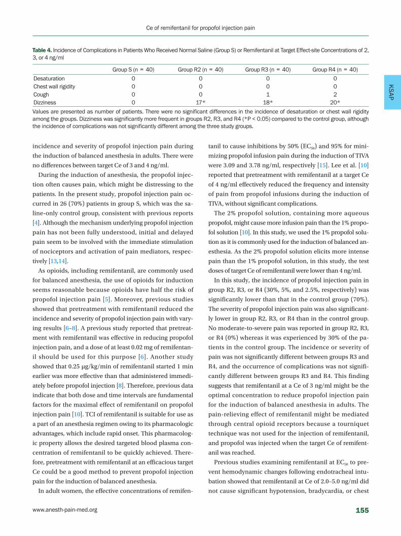
KS
AP
incidence and severity of propofol injection pain during
the induction of balanced anesthesia in adults. There were
no differences between target Ce of 3 and 4 ng/ml.
During the induction of anesthesia, the propofol injec-
tion often causes pain, which might be distressing to the
patients. In the present study, propofol injection pain oc-
curred in 26 (70%) patients in group S, which was the sa-
line-only control group, consistent with previous reports
[4]. Although the mechanism underlying propofol injection
pain has not been fully understood, initial and delayed
pain seem to be involved with the immediate stimulation
of nociceptors and activation of pain mediators, respec-
tively [13,14].
As opioids, including remifentanil, are commonly used
for balanced anesthesia, the use of opioids for induction
seems reasonable because opioids have half the risk of
propofol injection pain [5]. Moreover, previous studies
showed that pretreatment with remifentanil reduced the
incidence and severity of propofol injection pain with vary-
ing results [6–8]. A previous study reported that pretreat-
ment with remifentanil was effective in reducing propofol
injection pain, and a dose of at least 0.02 mg of remifentan-
il should be used for this purpose [6]. Another study
showed that 0.25 μg/kg/min of remifentanil started 1 min
earlier was more effective than that administered immedi-
ately before propofol injection [8]. Therefore, previous data
indicate that both dose and time intervals are fundamental
factors for the maximal effect of remifentanil on propofol
injection pain [10]. TCI of remifentanil is suitable for use as
a part of an anesthesia regimen owing to its pharmacologic
advantages, which include rapid onset. This pharmacolog-
ic property allows the desired targeted blood plasma con-
centration of remifentanil to be quickly achieved. There-
fore, pretreatment with remifentanil at an efficacious target
Ce could be a good method to prevent propofol injection
pain for the induction of balanced anesthesia.
In adult women, the effective concentrations of remifen-
tanil to cause inhibitions by 50% (EC50) and 95% for mini-
mizing propofol infusion pain during the induction of TIVA
were 3.09 and 3.78 ng/ml, respectively [15]. Lee et al. [10]
reported that pretreatment with remifentanil at a target Ce
of 4 ng/ml effectively reduced the frequency and intensity
of pain from propofol infusions during the induction of
TIVA, without significant complications.
The 2% propofol solution, containing more aqueous
propofol, might cause more infusion pain than the 1% propo-
fol solution [10]. In this study, we used the 1% propofol solu-
tion as it is commonly used for the induction of balanced an-
esthesia. As the 2% propofol solution elicits more intense
pain than the 1% propofol solution, in this study, the test
doses of target Ce of remifentanil were lower than 4 ng/ml.
In this study, the incidence of propofol injection pain in
group R2, R3, or R4 (30%, 5%, and 2.5%, respectively) was
significantly lower than that in the control group (70%).
The severity of propofol injection pain was also significant-
ly lower in group R2, R3, or R4 than in the control group.
No moderate-to-severe pain was reported in group R2, R3,
or R4 (0%) whereas it was experienced by 30% of the pa-
tients in the control group. The incidence or severity of
pain was not significantly different between groups R3 and
R4, and the occurrence of complications was not signifi-
cantly different between groups R3 and R4. This finding
suggests that remifentanil at a Ce of 3 ng/ml might be the
optimal concentration to reduce propofol injection pain
for the induction of balanced anesthesia in adults. The
pain-relieving effect of remifentanil might be mediated
through central opioid receptors because a tourniquet
technique was not used for the injection of remifentanil,
and propofol was injected when the target Ce of remifent-
anil was reached.
Previous studies examining remifentanil at EC50 to pre-
vent hemodynamic changes following endotracheal intu-
bation showed that remifentanil at Ce of 2.0–5.0 ng/ml did
not cause significant hypotension, bradycardia, or chest
Table 4. Incidence of Complications in Patients Who Received Normal Saline (Group S) or Remifentanil at Target Effect-site Concentrations of 2, 3, or 4 ng/ml
Group S (n = 40) Group R2 (n = 40) Group R3 (n = 40) Group R4 (n = 40)
Desaturation 0 0 0 0Chest wall rigidity 0 0 0 0Cough 0 0 1 2Dizziness 0 17* 18* 20*
Values are presented as number of patients. There were no significant differences in the incidence of desaturation or chest wall rigidity among the groups. Dizziness was significantly more frequent in groups R2, R3, and R4 (*P < 0.05) compared to the control group, although the incidence of complications was not significantly different among the three study groups.
www.anesth-pain-med.org 155
Ce of remifentanil for propofol injection pain

tightness [16]. Although in the present study, reductions in
HR and MAP following the administration of remifentanil
were significant, no adult patients with American Society
of Anesthesiologists physical status I or II required treat-
ment for hypotension or bradycardia, consistent with pre-
vious reports [10,16].
This study had two limitations, and the findings of this
study should be considered within the context of these lim-
itations.
First, the study population was limited to relatively young
patients. As older patients are more sensitive to opioids,
age-related variations in the effects should be considered.
Second, the prevalence of propofol injection pain could be
influenced by the patient’s sex. Remifentanil at EC50 used
to reduce propofol injection pain was lower in women than
in men [15].
In conclusion, we showed that remifentanil infusion at a
target Ce of 3 ng/ml for the induction of balanced anesthe-
sia using a single propofol injection was a useful strategy to
minimize propofol injection pain in adults, without caus-
ing significant complications.
CONFLICTS OF INTEREST
No potential conflict of interest relevant to this article
was reported.
AUTHOR CONTRIBUTIONS
Conceptualization: Joungmin Kim, Hyung Gon Lee. Data
acquisition: Hyung Gon Lee. Formal analysis: Hyung Gon
Lee, Daehoon Kim. Supervision: Hyung Gon Lee. Writing—
original draft: Joungmin Kim, Daehoon Kim. Writing—re-
view & editing: Joungmin Kim, Hyung Gon Lee.
ORCID
Joungmin Kim, https://orcid.org/0000-0003-1135-1968
Daehoon Kim, https://orcid.org/0000-0001-6275-2433
Hyung Gong Lee, https://orcid.org/0000-0003-4898-4355
REFERENCES
nonbenzodiazepine sedatives in the intensive care unit. Crit
Care Clin 2001; 17: 863-80.
3. Külling D, Rothenbühler R, Inauen W. Safety of nonanesthetist
sedation with propofol for outpatient colonoscopy and esoph-
agogastroduodenoscopy. Endoscopy 2003; 35: 679-82.
4. Picard P, Tramèr MR. Prevention of pain on injection with
propofol: a quantitative systematic review. Anesth Analg 2000;
90: 963-9.
5. Jalota L, Kalira V, George E, Shi YY, Hornuss C, Radke O, et al.
Prevention of pain on injection of propofol: systematic review
and meta-analysis. BMJ 2011; 342: d1110.
6. Iyilikci L, Balkan BK, Gökel E, Günerli A, Ellidokuz H. The ef-
fects of alfentanil or remifentanil pretreatment on propofol in-
jection pain. J Clin Anesth 2004; 16: 499-502.
7. Roehm KD, Piper SN, Maleck WH, Boldt J. Prevention of propo-
fol-induced injection pain by remifentanil: a placebo-con-
trolled comparison with lidocaine. Anaesthesia 2003; 58: 165-
70.
8. Basaranoglu G, Erden V, Delatioglu H, Saitoglu L. Reduction of
pain on injection of propofol using meperidine and remifent-
anil. Eur J Anaesthesiol 2005; 22: 890-2.
9. Egan TD, Kern SE, Muir KT, White J. Remifentanil by bolus in-
jection: a safety, pharmacokinetic, pharmacodynamic, and age
effect investigation in human volunteers. Br J Anaesth 2004; 92:
335-43.
10. Lee JR, Jung CW, Lee YH. Reduction of pain during induction
with target-controlled propofol and remifentanil. Br J Anaesth
2007; 99: 876-80.
11. Minto CF, Schnider TW, Egan TD, Youngs E, Lemmens HJ, Gam-
bus PL, et al. Influence of age and gender on the pharmacoki-
netics and pharmacodynamics of remifentanil. I. Model devel-
opment. Anesthesiology 1997; 86: 10-23.
12. King SY, Davis FM, Wells JE, Murchison DJ, Pryor PJ. Lidocaine
for the prevention of pain due to injection of propofol. Anesth
Analg 1992; 74: 246-9.
13. Briggs LP, Clarke RS, Dundee JW, Moore J, Bahar M, Wright PJ.
Use of di-isopropyl phenol as main agent for short procedures.
Br J Anaesth 1981; 53: 1197-202.
14. Scott RP, Saunders DA, Norman J. Propofol: clinical strategies for
preventing the pain of injection. Anaesthesia 1988; 43: 492-4.
15. Lee JY, Yang H, Choi SH, Shin DW, Hong SK, Chun DH. The op-
timal effect-site concentration of remifentanil to attenuate the
pain caused by propofol. Korean J Anesthesiol 2012; 63: 108-12.
16. Yon JH, Jo JK, Kwon YS, Park HG, Lee S. Effect-site concentration
of remifentanil for blunting hemodynamic responses to trache-
al intubation using light wand during target controlled infu-
sion-total intravenous anesthesia. Korean J Anesthesiol 2011;
60: 398-402.
1. Marik PE. Propofol: therapeutic indications and side-effects.
Curr Pharm Des 2004; 10: 3639-49.
2. Angelini G, Ketzler JT, Coursin DB. Use of propofol and other
156 www.anesth-pain-med.org
Anesth Pain Med Vol. 15 No. 2

KS
AP
Effect of preoperative administration of systemic alpha-2 agonists on postoperative pain: a systematic review and meta-analysis
Ji Youn Ju, Kye-Min Kim, and Sangseok Lee
Department of Anesthesiology and Pain Medicine, Sanggye Paik Hospital, Inje University
College of Medicine, Seoul, Korea
Clinical ResearchAnesth Pain Med 2020;15:157-166 https://doi.org/10.17085/apm.2020.15.2.157pISSN 1975-5171 • eISSN 2383-7977
Received September 2, 2019Revised October 20, 2019 Accepted October 25, 2019
Corresponding author Sangseok Lee, M.D. Department of Anesthesiology and Pain Medicine, Sanggye Paik Hospital, Inje University College of Medicine, 1342 Dongil-ro, Nowon-gu, Seoul 01757, Korea Tel: 82-2-950-1171Fax: 82-2-950-1323E-mail: [email protected]
Background: Alpha-2 agonists have sedative, analgesic, and opioid-sparing effects. More-over, intraoperative or postoperative systemic administration of alpha-2 adrenergic ago-nists is known to reduce postoperative pain and opioid consumption. This meta-analysis investigated whether preoperative administration of alpha-2 agonists can affect postoper-ative pain and opioid consumption.
Methods: We searched the MEDLINE, EMBASE, Cochrane Library (CENTRAL), KoreaMed, and KMbase databases through March 2019 to identify relevant randomized controlled trials (RCTs) on the effect of preoperative systemic administration of alpha-2 agonists on postoperative pain and opioid consumption. We conducted a meta-analysis according to the Cochrane Collaboration guidelines.
Results: Eleven RCTs involving 748 participants were included in this meta-analysis. Pre-operative administration of systemic alpha-2 agonists significantly reduced cumulative opioid consumption up to 6 h (SMD, –0.52; 95% confidence interval [– 0.90 to –0.14]) and 24 h (SMD, –0.68 [–1.27 to –0.09]) after surgery. Moreover, preoperative adminis-tration of alpha-2 agonists significantly reduced postoperative pain intensity at 6 h (SMD, –0.50 [–0.78 to –0.21]) and 24 h (SMD, – 0.44 [–0.86 to –0.03]).
Conclusions: In this meta-analysis, high degree of heterogeneity limits the preoperative administration of alpha-2 agonists in reducing postoperative opioid consumption and pain intensity. Future powered large RCTs are required to increase the certainty of evidence on the effect in reducing postoperative opioid consumption and pain intensity.
Keywords: Adrenergic alpha-2 receptor agonists; Anesthesia; Clonidine; Dexmedetomi-dine; Meta-analysis; Analgesics, opioid; Postoperative pain; Systematic review.
INTRODUCTION
Alpha-2 adrenergic agonists, which are widely used for
various purposes. As an anesthetic premedication, alpha-2
agonists mainly have anxiolytic effects and have the effect
of keeping hemodynamic index stable and reducing the
amount of anesthetics during surgery. Also, intraoperative
alpha-2 agonists is often administered to perform sedation
during regional anesthesia. Because of their analgesic
properties, intraoperative or postoperative use of alpha-2
agonists may be good adjuvants for use in postoperative
analgesia and may reduce consumption of analgesics [1,2].
Recently, multimodal analgesia with reduced doses of opi-
oids has been emphasized since increased doses of opioids
lead to adverse events, such as excessive sedation, hyper-
sensitivity, postoperative nausea, vomiting, and respiratory
This is an Open Access article distributed under the terms of the Creative Commons Attribution Non-Commercial License (http://creativecommons.org/licenses/by-nc/4.0) which permits unrestricted non-commercial use, distribution, and reproduction in any medium, provided the original work is properly cited.Copyright © the Korean Society of Anesthesiologists, 2020
157

depression [1,3].
A previous meta-analysis demonstrated that intraopera-
tive or postoperative systemic administration of alpha-2 ad-
renergic agonists reduced postoperative pain and opioid
consumption [4]. That meta-analysis, which included 30
studies, provided evidence of postoperative morphine-spar-
ing at 24 h and showed that the weighted mean difference
was 4.1 mg (95% confidence interval [CI], 6.0 to 2.2) with
clonidine and 14.5 mg (22.1 to 6.8) with dexmedetomidine.
This meta-analysis also provided evidence of a decrease in
pain intensity at 24 h and showed that the weighted mean
difference on a 10-cm visual analog scale (VAS) was 0.7 cm
(1.2 to 0.1) with clonidine and 0.6 cm (0.9 to 0.2) with dex-
medetomidine.
Nevertheless, controversy exists regarding whether pre-
operative use of alpha-2 agonists as sedatives has an effect
on reducing postoperative pain. Shariffuddin et al. [5] con-
cluded that a single dose of dexmedetomidine was a useful
adjuvant in reducing postoperative pain, and Sung et al. [6]
demonstrated that oral clonidine premedication reduced
the requirement of postoperative analgesia in patients un-
dergoing laparoscopic cholecystectomy. However, Lee et
al. [7] found that pre-anesthetic administration of a single
loading dose of dexmedetomidine given 10 min before in-
duction did not reduce patient-controlled anesthesia con-
sumption of postoperative fentanyl or the pain score.
The aim of this study was to evaluate the effect of alpha-2
agonists administered before general anesthesia and surgery
on postoperative pain and opioid consumption for postop-
erative analgesia.
MATERIALS AND METHODS
Study design
This systematic review and meta-analysis was performed
according to recent methodological guidelines [8]. The
protocol was registered on PROSPERO (https://www.crd.
york.ac.uk/PROSPERO, no. CRD42016051454). The study
was approved by the Institutional Review Board of the Inje Uni-
versity Sanggye Paik Hospital (no. 2017-08-013).
Information sources and search strategy
Three researchers (JJ, KMK, and SL) systematically
searched electronic databases such as the PubMed/MED-
LINE, EMBASE, and Cochrane Library (CENTRAL) without
restriction on the year of publication. Additionally, we
manually searched the reference lists of included studies to
identify other relevant trials in the Korean databases (Ko-
reaMed [https://koreamed.org] and KMbase [http://km-
base.medric.or.kr]). The search terms included variants of
alpha-2 adrenergic receptor stimulating agents and post-
operative pain, as well as Medical Subject Heading (MeSH)
or EMBASE Subject Heading (EMTREE) terms. A primary
investigation was performed to confirm the search key-
words and strategy (Appendix 1). The language of the arti-
cles was limited to Korean and English. The last search was
conducted on March 26, 2019.
Study selection and eligibility criteria
The draft search of the electronic databases for identify-
ing potentially relevant research was completed by two au-
thors (JJ and KMK). Thereafter, they independently select-
ed or excluded the studies. Primary selection was based on
an article’s title and abstract. Secondary selection was per-
formed after full-text review of an article. Studies for final
assessment were selected in consensus by the two investi-
gators. If necessary, a third investigator (SL) participated in
study selection, and the final decision was made on the ba-
sis of a majority.
Studies were included in the meta-analysis if they satis-
fied the following criteria: (1) patients underwent non-car-
diac surgery or neurosurgery under general anesthesia; (2)
intravenous or oral administration of an alpha-2 agonist
was used prior to surgery as an intervention; (3) results of
the control group were reported; and (4) postoperative
pain scores or opioid consumption was reported as a pri-
mary outcome.
Studies were excluded for the following reasons: (1) in-
tranasal, rectal, local, or topical administration of an al-
pha-2 agonist; (2) did not report appropriate outcomes or
outcome measurements as mentioned; (3) patients under-
went epidural anesthesia combined with general anesthe-
sia; (4) non-randomized controlled trials (RCTs) or studies
without a control group; (5) non-human studies; and (6)
articles not in Korean or English.
Risk of bias in individual studies
Two authors (JJ and KMK) reviewed the articles inde-
pendently to assess the risk of bias by using the “risk of bias
(ROB)” tool provided in the Review Manager software ver-
158 www.anesth-pain-med.org
Anesth Pain Med Vol. 15 No. 2

KS
AP
sion 5.3 (The Cochrane Collaboration, UK) based on Co-
chrane’s assessment of risk of bias. If necessary, a third re-
viewer (SL) was included to sort disagreements. The fol-
lowing seven domains to assess the risk of bias were used
in each trial: random sequence generation, allocation
concealment, blinding of participants and personnel,
blinding of outcome assessment, incomplete outcome
data, selective reporting, and other bias. We specified the
seventh domain “other bias” for assessing the adequate
estimation of sample size calculation. The methodology
for each trial was graded as “high,” “low,” or “unclear” to
reflect a high risk of bias, low risk of bias, or uncertainty of
bias, respectively.
The agreement between the two independent reviewers
for the level of risk of bias regarding the seven domains was
assessed using Cohen’s kappa [9].
Data collection process and extracted items
Three authors (JJ, KMK, and HK) extracted information
from the original articles, and another author (SL) inde-
pendently confirmed all the extracted data. The following
data were included in the data extraction form: patient
characteristics, age, sex, intervention dose and method,
pain-measurement tool and pain score, and postoperative
analgesic method and usage. If the data were insufficient
or unclear, the data were extracted from the text, tables, or
graphs.
Regarding preoperative administration of alpha-2 ago-
nists, only intravenous (IV) or oral administration method
was included, but the mode of administration (bolus or
continuous infusion) and dose were not restricted. A study
using continuous infusion of intervention drugs during
surgery was excluded.
Before we selectively extracted data from various studies,
the pain scores measured at postoperative 6 h or at the
nearest time point within postoperative 6 h were defined as
“early period pain scores.” The scores measured at postop-
erative 24 h were defined as “late period pain scores.” In
case of lack of data at postoperative 24 h, the latest pain
scores between postoperative 6 and 24 h were considered
as “late period pain scores.” Cumulative opioid consump-
tion up to 6 h after surgery were defined as “early period
opioid consumption.” If the data for cumulative opioid con-
sumption were not available at postoperative 6 h, supersed-
ed data were collected in the same way with those of pain
scores. “Late period opioid consumption” was defined as
the cumulative opioid consumption up to 24 h after surgery.
The VAS scores were extracted for analyzing postopera-
tive pain intensity. Pain scores included mean and stan-
dard deviation (SD) at the time of measurement. We in-
cluded postoperative opioid consumption data only from
those studies that performed IV or oral administration of
opioids, but no restriction was applied to the mode of ad-
ministration. We also excluded studies that reported the
time to the first request for opioid analgesics or the number
of times opioid analgesics was required. When studies used
opioid analgesics other than morphine, the doses were
converted into morphine equivalents, and the cumulative
consumption at the time of measurement was recorded.
Statistical analysis
Summary measures Opioid consumption data were extracted as mean and
SD of the cumulative morphine consumption in the early
and late periods. To measure pain intensity, we extracted
the mean and SD of the pain scores of the early and late
periods.
Synthesis of results Changes in continuous outcomes, such as postoperative
pain intensity or dose of opioid consumption, were pooled
as standardized mean differences (SMDs), because differ-
ent scaling of outcome measurements were expected
across trials. We also calculated the 95% CIs for all esti-
mates. A random-effect model was used to pool the study
results, taking into account possible variations in effect siz-
es across trials. The heterogeneity statistic Cochrane Q and
its corresponding (df) and P value as well as Higgins’ I2 as a
measure of heterogeneity were calculated. P < 0.05 was
considered representative of statistically significant hetero-
geneity, and I2 > 50% was considered to represent signifi-
cant heterogeneity.
Post-hoc subgroup analyses were performed where pos-
sible for each outcome to explore heterogeneity based on
the different route of administration between clonidine
(per oral) and dexmedetomidine (intravenous). Chi-
squared tests for heterogeneity were performed to identify
the differences between subgroups.
Publication bias was not evaluated because only few
studies less than 10 were included in this meta-analysis.
We performed a sensitivity analysis to evaluate the sta-
bility of the results. Each study included in the meta-analy-
www.anesth-pain-med.org 159
Effect of preoperative alpha-2 agonists

sis was deleted each time to reflect the influence of an indi-
vidual data set on the pooled effect estimate. We performed
all analyses using R 3.51 (R Foundation for Statistical Com-
puting, Austria) and Review Manager (RevMan, version 5.3,
The Cochrane Collaboration).
RESULTS
Study selection and characteristics
We retrieved 2,477 articles after the initial database
search: PubMed/MEDLINE (n = 960), EMBASE (n = 651),
CENTRAL (n = 704), and Korean databases (n = 162).
After removing 1,845 duplicate articles, primary selec-
tion was performed on 632 articles. First, we excluded 550
unrelated articles by analyzing their titles and abstracts.
Thereafter, full-text reviews were conducted on 82 articles.
Of these 82 full-text articles, 71 were excluded for the fol-
lowing reasons: general anesthesia not performed or com-
bined with regional anesthesia (n = 7), cardiac surgery (n
= 1), neurosurgery wherein neurological symptoms could
occur (n = 1), continuous infusion during or after surgery
or unclear injection time (n = 18), administration via
routes other than the IV or oral route (n = 7), used in com-
bination with other drugs (i.e., ketamine, gabapentin, or
remifentanil) (n = 3), did not report appropriate outcomes
such as pain score or opioid consumption (n = 13), no
placebo group or study protocol without results (n = 9),
articles not in Korean or English (n = 4), duplicated study
(n = 3), and full text unavailable (n = 5). Finally, ROB
evaluation and data extraction were performed on 11
studies [5–7,10–17]. Three studies [7,11,14] that did not re-
port SD values were excluded from the meta-analysis of
pain intensity (Fig. 1).
In all studies, alpha-2 agonists were administered via the
oral [6,13,16] or IV route [5,7,10–12,14,15,17], but the doses
and timing of injection differed among the studies (in most
studies, the timing was between 2 h and right before anes-
thesia induction; in study of Pawlik et al. [13], the timing
was the evening before and 2 h before anesthesia induc-
tion). Moreover, the type of opioids used differed across
studies (morphine [14,17], fentanyl [7,11,15], and pethidine
[6]). The frequency and interval of measurement of the
pain score also varied across studies (ranging from every
30 min to once daily) (Table 1).
Quality assessment of the included studies (risk of bias within studies)
The ROB evaluation revealed an overall low risk of bias
(Figs. 2, 3).The proportion of studies assessed as “unclear”
was 18.2% in random sequence generation, 27.3% in allo-
cation concealment, 27.3% in blinding of participants and
personnel, 9.1% in incomplete outcome data, and 18.2% in
other bias. Random sequence generation was unclear in
two studies [6,10] that did not specify a randomization
method. Allocation concealment was unclear in three
studies [6,10,11] that did not mention the specific alloca-
tion concealment method. Three studies that did not de-
scribe the blinding method for placebos were also rated as
“unclear” [6,13,16]. The risk of incomplete outcome data
was unclear in the study by Lee et al. [7], because the drop-
out rate was 5 to 20%. Among the risk assessments of other
biases mentioned in the Materials and Methods section, a
Records identified throughdatabase searchingPubMed (n = 960)EMBASE (n = 651)
Cochrane Library (n = 704)
Additional records identifiedthrough sources
Korea DB (n = 162)
Records after duplicates removed(n = 1,845)
Records screened(n = 632)
Records excluded(n = 550)
Full-text articles excluded, with reasons (n = 71) · Not general anesthesia (n = 7) · Cardiac surgery (n = 1) · Neurosurgery (n = 1) · Not administered only
preoperatively (n = 18) · Administered other route except IV
or PO (n = 7) · Associated with other compounds
(n = 3) · Not appropriate pain score or opioid
consumption date (n = 13) · Not controlled with placebo (n = 9) · Not in Korea or English (n = 4) · Unable to obtain full text (n = 5) · Duplicated study (n = 3)
Studies included in qualitative synthesis
(meta-analysis)(n = 11)
Full-text articles assessed for
eligibility (n = 82)
Fig. 1. Flow diagram of search strategy and study selection. DB: database, IV: intravenous, PO: postoperative.
160 www.anesth-pain-med.org
Anesth Pain Med Vol. 15 No. 2

KS
AP
power analysis to calculate the required sample size was
not performed by Mizrak et al. [14], leading to a high risk of
bias. The proportion of studies assessed as “unclear” was
54.5% in blinding of outcome assessment and 90.9% in se-
lective reporting. Six studies inaccurately described wheth-
er blinding of outcome assessment was implemented
[6,7,10,11,15,16]. Most studies did not provide information
about Web-based study registration sites, except for one
study [16].
Effects on postoperative pain and opioid consump-tion
The early period opioid consumption data extracted
from two studies [10,11] were data collected from the
post-anesthesia care unit (PACU) instead of those collected
up to 6 h. The early period pain score data from two other
studies [12,13] were data collected from the PACU instead
of those collected up to 6 h after surgery. Moreover, anoth-
er study [6] analyzed pain score data measured at the 2nd
hour. For the late period pain score analysis, data collected
up to 8 h after surgery were extracted from one study [13]
instead of those collected up to 24 h after surgery.
Opioid consumption for early postoperative pain (6 h
after surgery)
A meta-analysis synthesizing data from five effect sizes
from five studies [7,10,11,15,17] (n = 236; 118 in the experi-
mental group and 118 in the control group) suggested that
opioid consumption for early postoperative pain was sig-
nificantly reduced in the preoperative alpha-2 agonist
group (SMD, –0.52; 95% CI [–0.90 to –0.14]; P = 0.093; I2 =
50%; Fig. 4A).
The test for subgroup differences indicated that there is
no statistically significant subgroup effect (P = 0.63, analy-
sis not presented), suggesting that the route of administra-
tion does not modify the effect of alpha-2 agonist in com-
parison to control drug on the opioid consumption for ear-
ly postoperative pain.
Opioid consumption for late postoperative pain (24 h
after surgery)
We performed a meta-analysis synthesizing data from
three studies [6,7,17] (n = 202; 89 in the experimental
group and 113 in the control group), which suggested that
opioid consumption for late postoperative pain was signifi-
cantly reduced in the preoperative alpha-2 agonist group
Tabl
e 1
. Cha
ract
eris
tics
of th
e In
clud
ed S
tudi
es
Stud
ySu
rger
yIn
terv
entio
n (n
umbe
r of p
atie
nts)
Rep
orte
d ou
tcom
esO
pioi
d co
nsum
ptio
nPa
in s
core
Sam
anta
ray
et a
l. 20
12
[15]
Thor
acic
sur
gery
Plac
ebo
(30)
/IV
clon
idin
e 3
μg/k
g +
N/S
50
ml (
30)
Cum
ulat
ive
fent
anyl
con
sum
ptio
n at
6 h
VAS
(1–1
00) a
t 6th
, 24t
h ho
ur
Beh
dad
et a
l. 20
13 [1
6]Tr
ansa
bdom
inal
hys
tere
ctom
yPl
aceb
o PO
(30)
/PO
clo
nidi
ne 1
00 μ
g (3
0)-
VAS
(1–1
0) a
t 6th
, 24t
h ho
urU
nlug
enc
et a
l. 20
05 [1
7]M
ini-l
apar
otom
y, c
hole
cyst
ecto
my,
sp
lene
ctom
y an
d in
guin
al, u
mbi
lical
, in
cisi
onal
her
nia
repa
ir
Plac
ebo
(30)
/IV
dexm
edet
omid
ine
100
μg +
N/S
10
0 m
l (30
)Cu
mul
ativ
e m
orph
ine
cons
umpt
ion
at 6
, 24
hVR
S (1
–10)
at 6
th, 2
4th
hour
Paw
lik e
t al.
2005
[13]
ENT
surg
ery
Plac
ebo
(15)
/PO
clo
nidi
ne 2
μg/
kg (1
5)-
VAS
(1–1
0) a
t PAC
U, 8
th h
our
Miz
rak
et a
l. 20
10 [1
4]In
guin
al h
erni
a, la
paro
scop
ic c
hole
cys-
tect
omy,
bre
ast b
iops
yPl
aceb
o (3
0)/I
V de
xmed
etom
idin
e 0.
5 μg
/kg
+ N
/S
10 m
l (30
)-
VAS
(1–1
0) a
t 30
min
Cara
bine
et a
l. 19
92 [1
0]La
paro
scop
yPl
aceb
o (1
2), I
V cl
onid
ine
100
μg +
N/S
5 m
l (12
)Cu
mul
ativ
e m
orph
ine
cons
umpt
ion
at P
ACU
-Su
ng e
t al.
2000
[6]
Lapa
rosc
opic
cho
lecy
stec
tom
yPl
aceb
o (6
5), P
O c
loni
dine
150
μg
(43)
Cum
ulat
ive
peth
idin
e co
nsum
ptio
n 24
hVA
S (1
–10)
at 2
nd h
our
Shar
iffud
din
et a
l. 20
18 [5
]Am
bula
tory
ure
tero
scop
y, u
rete
ric
sten
ting
Plac
ebo
(30)
, IV
dexm
edet
omid
ine
0.5
μg/k
g +
N/S
20
ml (
30)
-VA
S (1
–10)
at P
ACU,
24t
h ho
ur
Park
et a
l. 20
19 [1
1]La
paro
scop
ic c
hole
cyst
ecto
my
Plac
ebo
(30)
, IV
dexm
edet
omid
ine
0.5
μg/k
g +
N/S
50
ml (
30)
Fent
anyl
con
sum
ptio
n at
PAC
UVA
S (1
–10)
at 3
0 m
in, 2
4 h
Lee
et a
l. 20
17 [7
]Tr
ansa
bdom
inal
hys
tere
ctom
yPl
aceb
o (1
8), I
V de
xmed
etom
idin
e 1
μg/k
g (1
6)Cu
mul
ativ
e fe
ntan
yl c
onsu
mpt
ion
at 6
, 24
hVA
S (1
–10)
at 6
th, 2
4th
hour
Shin
et a
l. 20
13 [1
2]G
ynec
olog
ical
sur
gery
Plac
ebo
(21)
, IV
dexm
edet
omid
ine
1 μg
/kg
+ N
/S
10 m
l (21
)-
VAS
(1–1
0) a
t PAC
U
ENT:
ear
, nos
e, a
nd th
roat
, IV:
intr
aven
ous,
N/S
: nor
mal
sal
ine,
PO
: pos
tope
rativ
e, V
AS: v
isua
l ana
logu
e sc
ale,
PAC
U: p
ost-a
nest
hesi
a ca
re u
nit.
www.anesth-pain-med.org 161
Effect of preoperative alpha-2 agonists

(SMD, –0.68; 95% CI [–1.27 to –0.09]; P = 0.025; I2 = 73%;
Fig. 4B).
A subgroup analysis comparing the effect in postopera-
tive opioid consumption in late period (24 h) was not pos-
sible due to there being too few trials in this category.
Pain score for early postoperative pain (6 h after
surgery)
A meta-analysis synthesizing data from seven studies
[5,6,12,13,15–17] (n = 418; 199 in the experimental group
and 219 in the control group) suggested that the pain score
for early postoperative pain was significantly reduced in
the preoperative alpha-2 agonist group (SMD, –0.50; 95%
CI [–0.78 to –0.21]; P = 0.058; I2 = 51%; Fig. 4C).
The test for subgroup differences indicated that there is
no statistically significant subgroup effect (P = 0.86, analy-
sis not presented), suggesting that the route of administra-
tion does not modify the effect of alpha-2 agonist in com-
parison to control drug on the early postoperative pain.
Pain score for late postoperative pain (24 h after
surgery)
Five studies [5,13,15–17] (n = 268; 135 in the experimental
group and 133 in the control group) reported data on the pain
score for late postoperative pain. Preoperative alpha-2 ago-
nists significantly reduced the postoperative pain score (SMD,
– 0.44; 95% CI [–0.86 to –0.03]; P = 0.026; I2 = 64%; Fig. 4D).
The test for subgroup differences indicated that there is
no statistically significant subgroup effect (P = 0.53, analy-
sis not presented), suggesting that the route of administra-
tion does not modify the effect of alpha-2 agonist in com-
parison to control drug on the late postoperative pain. Fig. 3. Risk of bias summary: review authors' judgements about each risk of bias item for each included study.
Behedad 2013
Carabine 1992
Lee 2017
Mizrak 2010
Park 2019
Pawlik 2005
Samantaray 2012
Shariffuddin 2018
Shin 2013
Sung 2000
Unlugenc 2005
Ran
dom
seq
uenc
e ge
nera
tion
(sel
ectio
n bi
as)
Allo
catio
n co
ncea
lmen
t (se
lect
ion
bias
)
Blin
ding
par
ticip
ants
and
per
sonn
el (p
erfo
rman
ce b
ias)
Blin
ding
of o
utco
me
asse
ssm
ent (
dete
ctio
n bi
as)
Inco
mpl
ete
outc
ome
data
(attr
ition
bia
s)
Sele
ctiv
e re
porti
ng (r
epor
ting
bias
)
Oth
er b
ias
Random sequence generation (selection bias)
Allocation concealment (selection bias)
Blinding participants and personnel (performance bias)
Blinding of outcome assessment (detection bias)
Incomplete outcome data (attrition bias)
Selective reporting (reporting bias)
Other bias
Low risk of bias Unclear risk of bias High risk of bias
0% 25% 50% 75% 100%
Fig. 2. Risk of bias graph: review authors' judgements about each risk of bias item presented as percentages across all included studies.
162 www.anesth-pain-med.org
Anesth Pain Med Vol. 15 No. 2

KS
AP
Sensitivity analysis
Opioid consumption in the early postoperative period
was 38% lower (SMD, –0.32; 95% CI [–0.62 to –0.02]; P =
0.036) than the pooled estimate effect size (SMD, –0.52;
95% CI [–0.90 to –0.14]; P = 0.007) after omitting one trial
[17]. Opioid consumption in the late postoperative period
was 35% lower (SMD, –0.44; 95% CI [–0.78 to –0.10]; P =
0.010) than the pooled estimate effect size (SMD, –0.68;
95% CI [–1.27 to –0.09]; P = 0.025) after omitting one trial
[17]. The pain scores in the early and late postoperative pe-
Fig. 4. Forest plot diagram showing the effect of alpha-2 agonist on postoperative pain intensity and opioid consumption. (A) Early period opioid consumption. (B) Late period opioid consumption. (C) Early period pain score. (D) Late period pain score. Std. Mean difference: standardized mean difference, IV: intravenous, CI: confidence interval, SD: standard deviation.
riods were not statistically significantly different between
the alpha-2 agonist group and control group after omitting
one trial each [16,17], which had a great influence on het-
erogeneity (results not shown).
DISCUSSION
In this meta-analysis, we evaluated the effect of preoper-
ative administration of alpha-2 agonists on total opioid
consumption for postoperative analgesia and postopera-
tive pain score. We found that opioid consumption and
A
B
C
D
www.anesth-pain-med.org 163
Effect of preoperative alpha-2 agonists

pain score were significantly lower in both the early and
late periods. This may be due to a preemptive analgesic ef-
fect, anxiolytic effect, analgesic-sparing effect, or a residual
additive effect of alpha-2 agonists [5,17].
Recently, intraoperative administration of alpha-2 ago-
nists has mainly been used for multimodal analgesia [1].
According to the results of a meta-analysis conducted by
Schnabel et al. [18], intraoperative use of dexmedetomi-
dine leads to lower postoperative pain intensity and re-
duced opioid consumption. Therefore, in our analysis, why
did the analgesic effect continue after surgery when al-
pha-2 agonists were administered before surgery? This was
the reason why we aimed to investigate whether preopera-
tive administration of alpha-2 agonists affects postopera-
tive pain as does intraoperative administration of alpha-2
agonists. Intraoperative or postoperative administration of
alpha-2 agonists is likely more beneficial when considering
only the duration of drug administration for postoperative
pain management. However, this was difficult to analyze
because each included study used different types of anes-
thetics, analgesics, and surgeries, as well as different dura-
tions of surgery. First, this could be considered a result of
reducing preoperative anxiety in patients. Preoperative al-
pha-2 agonists are frequently used to reduce a patient’s
anxiety, which is also associated with a patient’s preopera-
tive well-being and decreased levels of postoperative pain
[19]. Moreover, the preemptive effects could already have
occurred as a result of the administration of alpha-2 ago-
nists before surgical stimulation [19,20].
This meta-analysis has several limitations. The number
of studies included was quite small. To maintain good
quality, only complete RCTs and studies published in En-
glish and Korean were included in this meta-analysis,
thereby resulting in the small number of studies. Moreover,
most studies had small sample sizes and some studies did
not report postoperative pain and opioid consumption as
primary endpoints. Since the types of anesthetics and an-
algesics used in each study were different, they could pos-
sibly have affected the endpoints we were measuring. In
addition, the weight of the results of the meta-analysis was
not high because of the inclusion of few studies that were
conducted in accordance with our inclusion criteria.
Although not directly analyzed in this study, because
opioid usage and opioid-related side effects increase pro-
portionally [21], preoperative administration of alpha-2 ag-
onists can be expected to reduce opioid-related side effects,
as suggested by the results of this meta-analysis [22–24].
Furthermore, we did not distinguish between dexmede-
tomidine and clonidine when analyzing alpha-2 agonists.
Because these two drugs have different selectivity for al-
pha-2 adrenal receptors, their effects may be different.
In summary, we demonstrated that preoperative IV or
oral administration of alpha-2 agonists, such as clonidine
and dexmedetomidine, may reduce postoperative opioid
consumption and pain intensity until 24 h. However, high
degree of heterogeneity due to variations in the study
method and small sized studies, limits the recommenda-
tions in the preoperative administration of alpha-2 ago-
nists. Future powered large RCTs are required to increase
the certainty of evidence on the effect in reducing postop-
erative opioid consumption and pain intensity.
CONFLICTS OF INTEREST
No potential conflict of interest relevant to this article
was reported.
AUTHOR CONTRIBUTIONS
Conceptualization: Sangseok Lee. Study design: Ji Youn
Ju, Kye-Min Kim, Sangseok Lee. Data acquision: Ji Youn Ju,
Kye-Min Kim. Data analysis: Ji Youn Ju, Sangseok Lee.
Writing—original draft: Ji Youn Ju, Kye-Min Kim, Sangseok
Lee. Writing—review & editing: Ji Youn Ju, Kye-Min Kim,
Sangseok Lee.
ORCID
Ji Youn Ju, https://orcid.org/0000-0001-9069-4189
Kye-Min Kim, https://orcid.org/0000-0003-1298-7642
Sangseok Lee, https://orcid.org/0000-0001-7023-3668
REFERENCES
1. Rasmussen ML, Mathiesen O, Dierking G, Christensen BV,
Hilsted KL, Larsen TK, et al. Multimodal analgesia with gab-
apentin, ketamine and dexamethasone in combination with
paracetamol and ketorolac after hip arthroplasty: a prelimi-
nary study. Eur J Anaesthesiol 2010; 27: 324-30.
2. Khan ZP, Ferguson CN, Jones RM. Alpha-2 and imidazoline
receptor agonists. Their pharmacology and therapeutic role.
Anaesthesia 1999; 54: 146-65.
3.Kamibayashi T, Maze M. Clinical uses of alpha2-adrenergic ag-
onists. Anesthesiology 2000; 93: 1345-9.
164 www.anesth-pain-med.org
Anesth Pain Med Vol. 15 No. 2

KS
AP
4. Blaudszun G, Lysakowski C, Elia N, Tramèr MR. Effect of
perioperative systemic α2 agonists on postoperative morphine
consumption and pain intensity: systematic review and me-
ta-analysis of randomized controlled trials. Anesthesiology
2012; 116: 1312-22.
5. Shariffuddin II, Teoh WH, Wahab S, Wang CY. Effect of sin-
gle-dose dexmedetomidine on postoperative recovery after
ambulatory ureteroscopy and ureteric stenting: a double blind
randomized controlled study. BMC Anesthesiol 2018; 18: 3.
6. Sung CS, Lin SH, Chan KH, Chang WK, Chow LH, Lee TY. Ef-
fect of oral clonidine premedication on perioperative hemo-
dynamic response and postoperative analgesic requirement
for patients undergoing laparoscopic cholecystectomy. Acta
Anaesthesiol Sin 2000; 38: 23-9.
7. Lee KY, Lee WY, Kim KM, Yoo BH, Lee S, Lim YH, et al. The ef-
fect of pre-anesthetic administration of dexmedetomidine on
the consumption of opioids in postoperative gynecologic pa-
tients. Anesth Pain Med 2017; 12: 37-41.
8. Moher D, Liberati A, Tetzlaff J, Altman DG; PRISMA Group.
Preferred reporting items for systematic reviews and me-
ta-analyses: the PRISMA statement. BMJ 2009; 339: b2535.
9. Viera AJ, Garrett JM. Understanding interobserver agreement:
the kappa statistic. Fam Med 2005; 37: 360-3.
10. Carabine UA, Allen RW, Moore J. Partial attenuation of the
pressor response to endotracheal intubation. A comparison of
the effects of intravenous clonidine and fentanyl. Eur J Anaes-
thesiol 1992; 9: 325-9.
11. Park H, Kim HS, Kim JW, Lee GG, Park DH, Jeong CY, et al. Ef-
fects of preanesthetic single administration of dexmedetomi-
dine on the remifentanil and propofol requirement during
laparoscopic cholecystectomy. Anesth Pain Med 2019; 14: 29-
34.
12. Shin HW, Yoo HN, Kim DH, Lee H, Shin HJ, Lee HW. Preanes-
thetic dexmedetomidine 1 µg/kg single infusion is a simple,
easy, and economic adjuvant for general anesthesia. Korean J
Anesthesiol 2013; 65: 114-20.
13. Pawlik MT, Hansen E, Waldhauser D, Selig C, Kuehnel TS.
Clonidine premedication in patients with sleep apnea syn-
drome: a randomized, double-blind, placebo-controlled
study. Anesth Analg 2005; 101: 1374-80.
14. Mizrak A, Koruk S, Bilgi M, Kocamer B, Erkutlu I, Ganidagli S, et
al. Pretreatment with dexmedetomidine or thiopental decreas-
es myoclonus after etomidate: a randomized, double-blind
controlled trial. J Surg Res 2010; 159: e11-6.
15. Samantaray A, Rao MH, Chandra A. The effect on post-opera-
tive pain of intravenous clonidine given before induction of
anaesthesia. Indian J Anaesth 2012; 56: 359-64.
16. Behdad S, Ayatollahi V, Yazdi AG, Mortazavizadeh A, Niknam F.
Effect of oral low dose clonidine premedication on postopera-
tive pain in patients undergoing abdominal hysterectomy: a
randomized placebo controlled clinical trial. Rev Med Chir
Soc Med Nat Iasi 2013; 117: 934-41.
17. Unlugenc H, Gunduz M, Guler T, Yagmur O, Isik G. The effect
of pre-anaesthetic administration of intravenous dexmedeto-
midine on postoperative pain in patients receiving pa-
tient-controlled morphine. Eur J Anaesthesiol 2005; 22: 386-
91.
18. Schnabel A, Meyer-Frießem CH, Reichl SU, Zahn PK, Pogatzki-
Zahn EM. Is intraoperative dexmedetomidine a new option
for postoperative pain treatment? A meta-analysis of random-
ized controlled trials. Pain 2013; 154: 1140-9.
19. Kalkman CJ, Visser K, Moen J, Bonsel GJ, Grobbee DE, Moons
KG. Preoperative prediction of severe postoperative pain. Pain
2003; 105: 415-23.
20. Kelly DJ, Ahmad M, Brull SJ. Preemptive analgesia I: physio-
logical pathways and pharmacological modalities. Can J An-
aesth 2001; 48: 1000-10.
21. Roberts GW, Bekker TB, Carlsen HH, Moffatt CH, Slattery PJ,
McClure AF. Postoperative nausea and vomiting are strongly
influenced by postoperative opioid use in a dose-related man-
ner. Anesth Analg 2005; 101: 1343-8.
22. Liu SS, Strodtbeck WM, Richman JM, Wu CL. A comparison of
regional versus general anesthesia for ambulatory anesthesia:
a meta-analysis of randomized controlled trials. Anesth Analg
2005; 101: 1634-42.
23. Marret E, Kurdi O, Zufferey P, Bonnet F. Effects of nonsteroidal
antiinflammatory drugs on patient-controlled analgesia mor-
phine side effects: meta-analysis of randomized controlled
trials. Anesthesiology 2005; 102: 1249-60.
24. Gurbet A, Basagan-Mogol E, Turker G, Ugun F, Kaya FN, Ozcan
B. Intraoperative infusion of dexmedetomidine reduces
perioperative analgesic requirements. Can J Anaesth 2006; 53:
646-52.
www.anesth-pain-med.org 165
Effect of preoperative alpha-2 agonists
Appendix 1. Example of electronic database searching strategy for MEDLINE
#16. #14 AND #15
#15. random*
#14. #13 AND [english]/lim
#13. #5 AND #11 AND #12
#12. postop* OR postane* OR 'post op*' OR 'post ane*' OR postanae* OR 'post anae*'
#11. #6 OR #7 OR #8 OR #9 OR #10
#10. pca OR 'patient controlled analgesia'
#9. analgesic*
#8. fentanyl
#7. morphine
#6. opioid*
#5. #1 OR #2 OR #3 OR #4
#4. (alpha AND 2 OR 'alpha-2' OR a2 OR 'a 2') AND agonist*
#3. precedex
#2. dexmedetomidine
#1. clonidine
166 www.anesth-pain-med.org
Anesth Pain Med Vol. 15 No. 2

KS
OA
INTRODUCTION
Postpartum hemorrhage (PPH) is associated with nearly
one quarter of all maternal deaths globally. It is also the
leading cause of maternal mortality in most low-income
countries [1,2]. Uterine atony is the most common cause of
PPH [3]. The World Health Organization (WHO) recom-
mends active management of the third stage of labor and
use of uterotonics for the prevention of PPH during vaginal
Hemodynamic effects of carbetocin administered as an intravenous bolus or infusion during cesarean delivery
Kihyug Kwon, Dohyung Kim, Hyunmin Jo, Ji Eun Park, and Kyung Ok Kim
Department of Anesthesiology and Pain Medicine, Dongguk University Ilsan Hospital,
Goyang, Korea
Clinical ResearchAnesth Pain Med 2020;15:167-172 https://doi.org/10.17085/apm.2020.15.2.167pISSN 1975-5171 • eISSN 2383-7977
Received August 7, 2019 Revised January 28, 2020 Accepted January 31, 2020
Corresponding author Kihyug Kwon, M.D. Department of Anesthesiology and Pain Medicine, Dongguk University Ilsan Hospital, 27 Dongguk-ro, Ilsandong-gu, Goyang 10326, Korea Tel: 82-31-961-7868Fax: 82-31-961-7869E-mail: [email protected]
Background: Postpartum hemorrhage is the leading cause of maternal mortality. Oxytocin being the most popular uterotonic agent, has been routinely administered after both vagi-nal delivery and cesarean section. Carbetocin is a newer uterotonic agent and provides the benefit of a longer duration of action without additional administration post-delivery.
Methods: We recruited 34 women undergoing elective cesarean section under spinal an-esthesia. All patient was received spinal anesthesia using 0.5% hyperbaric Marcaine 8–10 mg in conjugation with fentanyl 20 μg in the left lateral decubitus position. Hartmann’s solution 10–15 ml/kg was administered before carbetocin. The operation started as soon as sensory block at level T4–T6 was confirmed. A non-invasive hemodynamic monitoring cuff (Finometer®) was attached to the patient’s finger soon after the induction of spinal an-esthesia. Using the Finometer, we recorded the heart rate and mean arterial pressure at every 15 s, starting from 15 s before the administration of carbetocin to 5 min after. After the removal of the placenta, the bolus group was administered intravenous bolus injection of carbetocin 100 μg and the infusion group was administered carbetocin 100 μg diluted in 50 ml normal saline, over 5 min using an infusion pump.
Results: The demographic data showed no significant difference between the two groups. Furthermore, there were no significant hemodynamic differences between the two groups.
Conclusions: The method of administration of carbetocin does not influence its hemody-namic effects.
Keywords: Carbetocin; Cesarean section; Hypotension; Tachycardia.
delivery and cesarean section [2].
Oxytocin being the most popular uterotonic agent, has
been routinely administered after both vaginal delivery and
cesarean section [4]. However, it is associated with dose-re-
lated side effects, including hypotension, tachycardia, nau-
sea and vomiting [5]. Other reported side effects include
pulmonary edema due to the antidiuretic effect of oxytocin
[6,7]. Thomas et al. [8] compared hemodynamic change be-
tween a group that was administered oxytocin by bolus in-
This is an Open Access article distributed under the terms of the Creative Commons Attribution Non-Commercial License (http://creativecommons.org/licenses/by-nc/4.0) which permits unrestricted non-commercial use, distribution, and reproduction in any medium, provided the original work is properly cited.Copyright © the Korean Society of Anesthesiologists, 2020
167

jection and another group that was administered oxytocin
by infusion. They concluded that the infusion group had
lesser hemodynamic changes than the bolus group.
Carbetocin is a newer uterotonic agent and a long-acting
synthetic octapeptide analogue of oxytocin with oxytocin
receptor agonist properties. Because carbetocin has a 4–10
fold longer elimination half-life than that of oxytocin, it
provides the benefit of longer duration of oxytocic action
without additional administration in post-delivery [4]. Al-
though the difference between carbetocin and oxytocin in
terms of hemodynamic changes, postpartum blood loss
and incidence of side effects is not statistically significant
[4,9], carbetocin is superior to oxytocin in terms of addi-
tional dosing and cost-effectiveness [10].
There is no definite consensus about how to administer
carbetocin. We compared the hemodynamic effects of car-
betocin when administered as an intravenous bolus and as
an infusion over 5 minutes using a non-invasive hemody-
namic monitoring device.
MATERIALS AND METHODS
This study was approved by the Hospital Ethics Commit-
tee (Dongguk University Ilsan Hospital Institutional Review
Board, registration no. 2012-77). The study was registered
with Clinical Research Information Service, Korea (https://
cris.nih.go.kr, registration no. KCT0000715). We recruited
34 women undergoing elective cesarean section under spi-
nal anesthesia. The patients were 37 weeks or more into
their pregnancy and were classified as Class 1 or 2 accord-
ing to the American Society of Anesthesiologists physical
status classification system. The patients who had pre-
eclampsia, placenta previa, other diseases affecting hemo-
dynamic change and past medical history of hypertension
or diabetes were excluded. The written consent was given
by all subjects before operation. The subjects were ran-
domly divided into two groups. Prior to enrolling patients,
group assignments were placed in 34 opaque envelopes.
These envelopes were mixed and placed in a container. Be-
fore each patient entered the operating room, the anesthe-
sia provider randomly selected one of these opaque con-
cealed envelopes to ensure group randomization.
None of patients were premedicated. Non-invasive blood
pressure monitoring device, pulse oximeter and electrocar-
diograph were attached to the patient as soon as they arrived
in the operating room. All patients received spinal anesthe-
sia at L3/4 or L4/5 level using 0.5% hyperbaric Marcaine
8–10 mg and fentanyl 20 μg in the left lateral decubitus posi-
tion. Hartmann’s solution 10–15 ml/kg was administered
before administration of carbetocin. The operation started
as soon as the sensory block at level T4–T6 was confirmed.
A non-invasive hemodynamic monitoring cuff (Finome-
ter®, Finapres medical system, Netherlands) was attached to
the patient’s finger soon after the induction of spinal anes-
thesia. Using the Finometer, heart rate (HR) and mean arte-
rial pressure (MAP) was recorded at every 15 s starting from
15 s before the administration of carbetocin to 5 min after.
After the removal of placenta, the bolus group received
an intravenous bolus injection of carbetocin 100 μg and the
infusion group received 100 μg carbetocin diluted in 50 ml
normal saline, over 5 min using an infusion pump (Orches-
tra® Module DPS, Fresenius Kabi, France). We defined hy-
potension as less than 20% of the preoperative value or be-
low 80 mmHg systolic blood pressure. Hypotension was
treated with ephedrine 5 mg boluses. If ephedrine was
used during the study period, that patient would be ex-
cluded in study.
The primary endpoint was the mean difference in HR
and MAP between study groups at each time point. The
secondary endpoints were estimated blood loss (EBL), the
presence of postoperative hemorrhage and the require-
ment for additional administration of uterotonic agent.
A MAP difference of 10 mmHg between the two groups
was considered clinically significant. According to the pre-
vious study reporting a standard deviation of 8.7 mmHg [8],
a sample of 17 patients per group was required with error
of 0.05 and power of 90%.
Demographic data variables including age, height, weight,
and body mass index, ephedrine dose, and operation time
were analyzed using unpaired t-tests. The primary endpoints
were analyzed at each time point using unpaired t-tests. EBL of
each group was compared using the Mann-Whitney test. Oth-
er secondary endpoints were compared using Fisher’s exact
test. All statistical analysis were performed using R software
version 3.3.2 (R Core Team [2016]. R: A language and environ-
ment for statistical computing. R Foundation for Statistical
Computing, Austria. URL: https://www.R-project.org/).
RESULTS
The study was conducted over a continuous period of 1
year. A total 34 patients were recruited. No patient was ex-
cluded except one whose data were missing (Fig. 1). The
patient characteristics are shown in Table 1. No statistical
168 www.anesth-pain-med.org
Anesth Pain Med Vol. 15 No. 2

KS
OA
Table 1. The Demographic Data of Two Groups
Variable Bolus group (n = 16) Infusion group (n = 17) P value
Age (yr) 33.8 ± 3.1 33.3 ± 4.9 0.721Height (cm) 159.6 ± 6.3 158.3 ± 4.3 0.469Weight (kg) 71.0 ± 12.5 66.3 ± 12.3 0.291Body mass index (kg/m2) 27.7 ± 3.9 26.4 ± 4.5 0.390Indication for cesarean delivery Previous cesarean section 12 6 Breech 1 8 Others 3 3Ephedrine dose (mg) 13.8 ± 12.0 11.8 ± 8.6 0.589Number of ephedrine use 13 14Operation time (min) 65.9 ± 13.5 61.4 ± 14.1 0.354
Values are presented as mean ± SD or number only.
Table 2. Initial Heart Rate and Mean Arterial Pressure of Two Groups
Initial hemodynamic variable Bolus group Infusion group
Initial HR (beats/min) 83.6 ± 15.3 84.4 ± 13.0Initial MAP (mmHg) 101.2 ± 12.9 97.6 ± 13.5
Values are presented as mean ± SD. HR: heart rate, MAP: mean arterial pressure.
difference was observed between the two groups in demo-
graphic data. None of patient received ephedrine during
the study period and no difference in the total dose of
ephedrine used between the two groups. Baseline HR and
MAP were not statistically different either (Table 2).
Moreover, there was no statistical difference in the mean
HR and MAP between the two groups (Figs. 2, 3). In both
groups, we found a rapid decrease of MAP until 30 s after
the administration of carbetocin regardless of the method
of administration. Then, the trends showed a gradual in-
crease. After 300 s, the mean MAP of the bolus group was
88.73 mmHg and that of the infusion group was 86.76
mmHg. The mean MAP of the bolus group was higher than
that of the infusion group from 105 to 300 s, but there was
no statistically significant difference at any time point.
In both groups, we found a rapid increase of HR until 45
s after the administration of carbetocin. Then, the trends
Screened total elective cesarean section (n = 92)
Randomized (n = 51)
Infusion group (n = 17)Bolus group (n = 16)
Excluded (n = 41) · Not meeting inclusion criteria (n = 18) · Declined to participate (n = 23)
Excluded (n = 7) · Mechanical error (n = 2) · Protocol violence (n = 5)
Excluded (n = 11) · Mechanical error (n = 4) · Protocol violence (n = 7)
Fig. 1. Patients CONSORT flow chart.
showed a gradual decrease till the end of the study. After
300 s, the mean HR of the bolus group was 92.35 beats/
min and that of the infusion group was 91.71 beats/min.
Again, the difference between the two groups at any time
point was not statistically significant. We also investigated
EBL, additional administration of uterotonic agent in
both groups and the incidence of postoperative interven-
tion such as uterine artery embolization or hysterectomy
(Table 3). There were no significant differences between
the two groups. Only one patient received postoperative
www.anesth-pain-med.org 169
Hemodynamic effects of carbetocin

uterine artery embolization and transfusion during PPH.
DISCUSSION
Our study showed that the method of administration of
carbetocin does not influence its hemodynamic effects in
cesarean section and does not affect additional uterotonic
agent use or incidence of postoperative intervention such
as uterine artery embolization or hysterectomy.
Carbetocin is a long-acting synthetic nonapeptide ana-
logue of oxytocin with agonist properties. It can be admin-
Table 3. Secondary Endpoint of Two Group
Secondary endpoints Bolus group Infusion group
Number of postoperative utero-tonic agent
0 0
Estimated blood loss (ml) 520.0 ± 207.7 535.3 ± 264.4Number of postoperative interventions
0 1*
Number of patients received transfusion
0 1
Values are presented as number only or mean ± SD. *1 patient was received uterine artery embolization and the patients were also only patients received transfusion.
110
100
90
80
70
HR
(bea
ts/m
in)
Time (s)
GroupBolus groupInfusion group
-15 0 15 30 45 60 75 90 105 120 135 150 165 180 195 210 225 240 255 270 285 300
110
100
90
80
70
60
MAP
(mm
Hg)
Time (s)
GroupBolus groupInfusion group
-15 0 15 30 45 60 75 90 105 120 135 150 165 180 195 210 225 240 255 270 285 300
Fig. 2. The changes of heart rate (HR) in two groups. Values are presented as mean ± SD.
Fig. 3. The changes of mean arterial pressure (MAP) in two groups. Values are presented as mean ± SD.
170 www.anesth-pain-med.org
Anesth Pain Med Vol. 15 No. 2

KS
OA
istered intravenously as a single dose immediately follow-
ing vaginal delivery or cesarean section, to prevent uterine
atony and postpartum hemorrhage. Similar to oxytocin, it
selectively binds to oxytocin receptors present on the
smooth musculature of the uterus, resulting in rhythmic
contractions of the uterus, increased frequency of existing
contractions, and increased uterine tone [11].
The Finometer® measures finger blood pressure nonin-
vasively on a beat-to-beat basis and gives waveform mea-
surements similar to those of intra-arterial recordings. One
disadvantage is the measured value changes with the
movement of the patient’s finger or posture. However, its
use is comfortable for conscious patients because it is
non-invasive and the correlation can be easily calculated
by measuring the brachial cuff pressure and hydrostatic
height of the finger [12].
According to WHO guidelines for prevention of PPH,
oxytocin (10 IU, intravenous or intramuscular) is the utero-
tonic agent of choice during the third stage of labor and ce-
sarean section. Although the use of carbetocin as an addi-
tional uterotonic agent has been recommended, according
to WHO guidelines, carbetocin has no effect on the inci-
dence of major obstetric hemorrhage and is more expen-
sive than oxytocin [2].
Recent studies have suggested that carbetocin is superior
to oxytocin in cost-effectiveness and prevention of PPH.
van der Nelson et al. [10] compared the cost-effectiveness
of carbetocin used for the prevention of PPH in cesarean
section to that of oxytocin. They analyzed 1,500 patients re-
ceiving carbetocin or oxytocin in cesarean section and
compared the costs for additional drugs and blood prod-
ucts used, which in turn depends on the incidence and se-
verity of PPH. According to this study, in UK, the use of
carbetocin instead of oxytocin reduced the costs by EUR
27,518. Jin et al. [13] examined 12 randomized controlled
studies and found that carbetocin lowered the requirement
for additional uterotonic agents and uterine massage in
women who underwent cesarean section. However, their
analysis could not detect a significant difference in PPH,
EBL or adverse effects.
In the case of oxytocin, several reports have shown an
association between an infusion of diluted oxytocin and
milder hemodynamic changes compared to bolus admin-
istration. Thomas et al. [8] compared the effects of oxytocin
administered as an intravenous bolus and as an infusion
for 5 min in 30 women undergoing cesarean section. They
reported marked cardiovascular changes in the bolus
group. Kim et al. [14] also reported a similar result with re-
gards to cardiovascular changes with a difference in the
uterine tone and EBL.
Our study showed that the method of administration
does not have a significant effect on the patient’s hemody-
namics in the case of carbetocin. Dell-Kuster et al. [15] in-
vestigated obstetric and hemodynamic differences be-
tween carbetocin bolus and infusion. They found that the
administration of carbetocin as a short infusion does not
compromise the uterine tone and that two methods have
similar cardiovascular effects and vasoconstrictor dose re-
quirements.
Intraoperative hypotension during spinal anesthesia for
elective cesarean delivery are frequent above 70%, when
pharmacological prophylaxis is not used [16]. However, hy-
potension is prevented or improved by several methods
such as prevention of aorto-caval compression, use of va-
sopressors and intravenous fluid loading. In our study, no
occurrence of hypotension during the investigation period
might be a result of a mere coincidence. But, we thought
that sufficient fluid loading, the exclusion of emergency
surgery and other effective treatment for preventing hypo-
tension contributed to that result.
There were several limitations to this study. First, the cal-
culated EBL of both groups were statistically similar, but
there may have been a significant difference in the actual
blood loss which is difficult to quantify [17,18]. A recent
study recommends a colorimetric system for more accu-
rate measurement of the amount of blood loss. Although
we could not apply a colorimetric system, the difference in
the actual blood loss between the two groups in our study
is not expected to be large since the calculated EBL in the
above mentioned study was similar to that of our study
[18]. In the future, we aim to develop and optimize meth-
ods for the accurate measurement of blood loss and intra-
vascular volume status. Furthermore, this study does not
investigate postoperative hemodynamic change. Although
there were no differences between the two groups in terms
of additional uterotonic drug use or postoperative compli-
cations, it is necessary to measure postoperative vital sign
for investigating the precise hemodynamic effect of car-
betocin.
In conclusion, the administration method of carbetocin
does not influence the hemodynamic changes. In addition,
the administration method has no effect on postoperative
complications, additional administration of uterotonic
agents or blood loss.
www.anesth-pain-med.org 171
Hemodynamic effects of carbetocin

CONFLICTS OF INTEREST
No potential conflict of interest relevant to this article
was reported.
AUTHOR CONTRIBUTIONS
Conceptualization: Kyung Ok Kim. Data acquisition: Ki-
hyug Kwon, Kyung Ok Kim. Formal analysis: Dohyung Kim,
Hyunmin Jo, Ji Eun Park. Supervision: Kyung Ok Kim. Writ-
ing—original draft: Dohyung Kim, Hyunmin Jo. Writing—
review & editing: Kihyug Kwon, Ji Eun Park.
ORCID
Kihyug Kwon, https://orcid.org/0000-0001-7766-7178
Dohyung Kim, https://orcid.org/0000-0001-8245-8995
Hyunmin Jo, https://orcid.org/0000-0002-0957-8488
Ji Eun Park, https://orcid.org/0000-0002-2466-7040
Kyung Ok Kim, https://orcid.org/0000-0001-7509-3668
REFERENCES
ing oxytocin administration: a life threatening complication. J
Indian Med Assoc 2006; 104: 261-2.
8. Thomas JS, Koh SH, Cooper GM. Haemodynamic effects of
oxytocin given as i.v. bolus or infusion on women undergoing
Caesarean section. Br J Anaesth 2007; 98: 116-9.
9. Moertl MG, Friedrich S, Kraschl J, Wadsack C, Lang U, Schlem-
bach D. Haemodynamic effects of carbetocin and oxytocin giv-
en as intravenous bolus on women undergoing caesarean de-
livery: a randomised trial. BJOG 2011; 118: 1349-56.
10. van der Nelson HA, Draycott T, Siassakos D, Yau CWH, Hats-
well AJ. Carbetocin versus oxytocin for prevention of post-par-
tum haemorrhage at caesarean section in the United Kingdom:
an economic impact analysis. Eur J Obstet Gynecol Reprod
Biol 2017; 210: 286-91.
11. Sweeney G, Holbrook AM, Levine M, Yip M, Alfredsson K, Cap-
pi S, et al. Pharmacokinetics of carbetocin, a long-acting oxyto-
cin analogue, in nonpregnant women. Curr Ther Res Clin Exp
1990; 47: 528-40.
12. Schutte AE, Huisman HW, van Rooyen JM, Malan NT, Schutte
R. Validation of the Finometer device for measurement of
blood pressure in black women. J Hum Hypertens 2004; 18: 79-
84.
13. Jin B, Du Y, Zhang F, Zhang K, Wang L, Cui L. Carbetocin for the
prevention of postpartum hemorrhage: a systematic review
and meta-analysis of randomized controlled trials. J Matern
Fetal Neonatal Med 2016; 29: 400-7.
14. Kim TS, Bae JS, Park JM, Kang SK. Hemodynamic effects of
continuous intravenous injection and bolus plus continuous
intravenous injection of oxytocin in Cesarean section. Korean J
Anesthesiol 2011; 61: 482-7.
15. Dell-Kuster S, Hoesli I, Lapaire O, Seeberger E, Steiner LA, Bu-
cher HC, et al. Efficacy and safety of carbetocin given as an in-
travenous bolus compared with short infusion for Caesarean
section - double-blind, double-dummy, randomized con-
trolled non-inferiority trial. Br J Anaesth 2017; 118: 772-80.
16. Mercier FJ, Augè M, Hoffmann C, Fischer C, Le Gouez A. Ma-
ternal hypotension during spinal anesthesia for caesarean de-
livery. Minerva Anestesiol 2013; 79: 62-73.
17. Lopez-Picado A, Albinarrate A, Barrachina B. Determination of
perioperative blood loss: accuracy or approximation? Anesth
Analg 2017; 125: 280-6.
18. Doctorvaladan SV, Jelks AT, Hsieh EW, Thurer RL, Zakowski MI,
Lagrew DC. Accuracy of blood loss measurement during Ce-
sarean delivery. AJP Rep 2017; 7: e93-100.
1. Say L, Chou D, Gemmill A, Tunçalp Ö, Moller AB, Daniels J, et
al. Global causes of maternal death: a WHO systematic analysis.
Lancet Glob Health 2014; 2: e323-33.
2. World Health Organization. WHO recommendations for the
prevention and treatment of postpartum haemorrhage. Gene-
va: World Health Organization. 2012, p. 41.
3. Knight M, Callaghan WM, Berg C, Alexander S, Bouvier-Colle
MH, Ford JB, et al. Trends in postpartum hemorrhage in high
resource countries: a review and recommendations from the
International Postpartum Hemorrhage Collaborative Group.
BMC Pregnancy Childbirth 2009; 9: 55.
4. Meshykhi LS, Nel MR, Lucas DN. The role of carbetocin in the
prevention and management of postpartum haemorrhage. Int J
Obstet Anesth 2016; 28: 61-9.
5. Petersson M. Cardiovascular effects of oxytocin. Prog Brain Res
2002; 139: 281-8.
6. Shahin J, Guharoy SR. Pulmonary edema possibly developing
secondary to the intravenous administration of oxytocin. Vet
Hum Toxicol 1991; 33: 587-8.
7. Ghai B, Vayjnath AM, Lal S. Acute pulmonary oedema follow-
172 www.anesth-pain-med.org
Anesth Pain Med Vol. 15 No. 2

KS
PA
Clinical performance of Ambu AuraGainTM versus i-gelTM in anesthetized children: a prospective, randomized controlled trial
Ji-Hyun Lee, Seungpyo Nam, Young-Eun Jang, Eun-Hee Kim, Hee-Soo Kim, and Jin-Tae Kim
Department of Anesthesiology and Pain Medicine, Seoul National University Hospital,
Seoul National University College of Medicine, Seoul, Korea
Clinical ResearchAnesth Pain Med 2020;15:173-180 https://doi.org/10.17085/apm.2020.15.2.173pISSN 1975-5171 • eISSN 2383-7977
Received September 5, 2019 Revised October 28, 2019 Accepted: October 29, 2019
Corresponding author Jin-Tae Kim, M.D., Ph.D. Department of Anesthesiology and Pain Medicine, Seoul National University Hospital, Seoul National University College of Medicine, 101 Daehak-ro, Jongno-gu, Seoul 03080, Korea Tel: 82-2-2072-3592Fax: 82-2-747-8364E-mail: [email protected]
Ji-Hyun Lee and Seungpyo Nam contributed equally to this study.
Background: Ambu AuraGain and i-gel have different characteristics in design each other. However, few reports evaluate which device has more benefits for ventilation in children undergoing paralyzed general anesthesia. This prospective, randomized controlled trial compared the clinical performance AuraGain and i-gel in anesthetized children.
Methods: Children aged between 1 month and 7 years undergoing elective surgery were randomly assigned to the AuraGain and i-gel groups. The primary outcome was initial oro-pharyngeal leak pressure (OLP). Secondary outcomes were OLP at 10 min post-insertion, first-attempt and total insertion success rates, number of attempts and ease of gastric suc-tion catheter placement, peak inspiratory pressure, fiberoptic bronchoscopic view score, ventilation quality, requirement of additional manipulation post-insertion, and complications.
Results: Data of 93 patients were analyzed. The initial OLPs of the AuraGain and i-gel were 27.5 ± 7.7 and 25.0 ± 8.0 cmH2O, respectively (P = 0.130). The OLP was significant-ly increased 10 min post-insertion in both groups. The initial success rates of the Au-raGain and i-gel insertion were comparable. Suction catheter placement via the gastric port was easier (P = 0.018) and fiberoptic bronchoscopic view was better with the Au-raGain (P < 0.001). The i-gel required additional manipulations post-insertion (P = 0.038). The incidence of complications during the emergence period was 10.8% for the i-gel and 2.2% for the AuraGain (P = 0.1).
Conclusions: OLP is comparable between AuraGain and i-gel. The AuraGain would be more favorable than the i-gel for use in pediatric patients under general anesthesia con-sidering other outcomes.
Keywords: Airway management; General anesthesia; Laryngeal masks; Pediatrics.
INTRODUCTION
Appropriate airway management is critical in the pediat-
ric population with a lower oxygen reserve. Supraglottic
airway devices (SADs) have been used to secure and main-
tain airways in patients during surgery or emergency situa-
tions, as well as in the management of expected and unex-
pected difficult airways. Among various SADs, the i-gelTM
(Inter-surgical Ltd., UK) which has a soft non-inflatable
cuff and a gastric access port, has been widely used in chil-
dren. According to a recent meta-analysis, the i-gel has a
high oropharyngeal leak pressure (OLP) and a low risk of
blood staining, compared with LMA®-Classic [1].
Another device, Ambu AuraGainTM (Ambu, Denmark), a
This is an Open Access article distributed under the terms of the Creative Commons Attribution Non-Commercial License (http://creativecommons.org/licenses/by-nc/4.0) which permits unrestricted non-commercial use, distribution, and reproduction in any medium, provided the original work is properly cited.Copyright © the Korean Society of Anesthesiologists, 2020
173
recently released second-generation SAD, has an inflatable
cuff and a gastric port. One of the key features of the Au-
raGain is the 90-degree angulated shaft, following the anat-
omy of the upper airway, which facilitates proper insertion
and is associated with a high success rate for insertion [2,3].
Although use of the AuraGain has increased, research re-
garding its clinical performance is limited, especially
among children [4,5].
There has been some reports comparing use of the Au-
raGain and i-gel in pediatric patients, which concluded that
the i-gel may be superior to the AuraGain in terms of OLP
[5,6]. However, the incidence of additional manipulation or
device failure which was defined as the abandonment of the
SAD and replacement with a tracheal tube or another device
was higher for the i-gel than for other SADs, which should be
evaluated further in pediatric populations [1,6]. In addition,
there is limited evidence in paralyzed children. Therefore, in
this study, we hypothesized that the clinical performance of
the AuraGain would be comparable to that of the i-gel in the
pediatric populations.
The objective of this prospective, randomized trial was to
compare the clinical performance of the AuraGain and
i-gel in terms of OLP, successful insertion, ventialtion qual-
ity, ease of suction catheter placement, additional manipu-
lation after insertion, and complications in children under
general anesthesia.
MATERIALS AND METHODS
Ethics and study population
This prospective randomized, controlled, parallel-de-
signed trial was approved by the Institutional Review Board
(no. H1704-083-846) and registered at http://clinicaltrials.
gov (no. NCT03118245). The study was performed in accor-
dance with the ethical standards set forth in the 1964 Decla-
ration of Helsinki and its later amendments. One day before
surgery, an anesthesiologist met with each child’s parents,
explained the study protocol, and obtained written informed
consent from them.
Children aged between 1 month and 7 years who were
scheduled for simple superficial or peripheral limb surgery
within 2 h, under general anesthesia, were enrolled in this
study. Children with American society of anesthesiologist
physical status over than II, recent upper respiratory tract
infection, any respiratory disease, a history of cervical dis-
order or surgery, risk factors for aspiration including insuf-
ficient fasting times, difficult airways, or body mass indices
> 30 kg/m2 were excluded from this study. In addition,
children who were scheduled for emergency surgery, ab-
dominal or thoracic surgeries and all laparoscopic proce-
dures were also excluded.
Group allocation
Patients were randomly assigned to one of two groups,
the i-gel group or the AuraGain group, using a stratified
randomization procedure (computerized random number;
http://www.randomizer.org). The allocation ratio was 1 : 1,
and an anesthetic nurse who was not associated with the
study performed the random allocation by preparing cod-
ed and sealed opaque envelopes for allocation conceal-
ment. The patients were blinded to group allocation. The
bedside anesthesiologists, however, were not blinded.
Anesthesia and study protocol
Before induction of anesthesia, all pediatric patients
were sedated with intravenous thiopental sodium (5 mg/
kg) or propofol (2 mg/kg) and were taken to the operating
room. After electrocardiographic monitoring, pulse oxygen
saturation and non-invasive blood pressure measurement
were initiated, and anesthesia was induced with sevoflu-
rane 4–6 vol% in 100% oxygen. Following muscle relaxation
with rocuronium (0.6 mg/kg) and mask ventilation for 90 s,
the i-gel or deflated AuraGain which were lubricated with
2% lidocaine jelly was gently inserted into the oropharynx
by a single experienced anesthesiologist (L.J.H), according
to group allocation. The sizes of the AuraGain and i-gel
were selected based on body weight, as suggested by the
manufacturer as follows: size 1.5 (for 5–12 kg), 2 (10–25 kg),
and 2.5 (25–35 kg) for the i-gel; size 1.5 (5–10 kg), 2 (10–20
kg), and 2.5 (20–30 kg) for the AuraGain.
After insertion, the cuff of the AuraGain was inflated to an
intracuff pressure of 40 cmH2O using a Portex® cuff inflator
(Smiths Medical, USA). Mechanical ventilation was started,
and adequate ventilation was confirmed by square-wave
capnography and bilateral chest excursion. When adequate
ventilation was not achieved, the SAD was removed and re-
inserted. Up to three insertion attempts were allowed. Tra-
cheal intubation was planned in cases of three failed inser-
tion attempts, and such patients were subsequently exclud-
ed from the study. During insertion of the SAD, the patient’s
head and neck were left in the neutral position with the
174 www.anesth-pain-med.org
Anesth Pain Med Vol. 15 No. 2

KS
PA
face straight up and the Frankfort plane angled at approxi-
mately 70–80 degrees to the horizontal plane of the bed, as
described by Kobayashi et al. [7]. Mechanical ventilation
was started in the volume-controlled mode, with a tidal
volume of 8 ml/kg, and the respiratory rate was adjusted to
maintain ETCO2 of 35–40 mmHg. Sevoflurane in an air-oxy-
gen mixture, with fractional inspired O2 at 40%, was used to
maintain anesthesia at a targeted Bispectral index of 40–60.
Immediately after insertion, OLP was determined by
closing the adjustable pressure limiting valve of the breath-
ing circuit, with a fresh gas flow of 3 L/min [8,9]. While in-
creasing the airway pressure up to 40 cmH2O, oropharyn-
geal leak was assessed by placing the stethoscope over the
patient’s neck, immediately lateral to the thyroid cartilage.
OLP was re-assessed 10 min after insertion.
After determination of the OLP, fiberoptic views were ob-
tained by a single anesthesiologist (L.J.H) by placement of
a fiberoptic bronchoscope (Olympus LF-DP, Olympus Cor-
poration, Japan) through the SAD. The view was scored us-
ing the Okuda score as follows [10]: 1) the view was com-
pletely covered by the anterior epiglottis, but SAD function
was adequate, 2) the anterior epiglottis covered more than
two-thirds of the diameter of the view, 3) the anterior epi-
glottis covered one-third to two-thirds of the diameter of
the view, and 4) the anterior epiglottis covered less than
one-third of the diameter of the view.
Next, a lubricated suction catheter was inserted through
the drainage port each SAD for gastric decompression. The
size of suction catheter was selected according to allowable
maximal catheter size, as suggested by the manufacturer as
follows: 10 Fr for size 1.5, 2 and 2.5 of the i-gel; 8 Fr for size
1.5, and 10 Fr for size 2 and 2.5 of the AuraGain. The ease of
placement of the suction catheter was assessed as follows:
1) easy – suction catheter enters without resistance at once,
2) difficult – suction catheter withdrawal and re-insertion
are tried more than once due to resistance, and 3) suction
catheter is unable to pass.
Ventilation quality was assessed by auscultating the lung
sound and evaluating the waveform of capnography and
bilateral chest excursion [11] as follows: 1) clear, 2) mini-
mal obstruction, 3) partial obstruction, and 4) complete
obstruction. If ventilation was inadequate, additional ma-
nipulations were performed, which included gentle push-
ing or pulling of the device, lifting of the chin, jaw thrust-
ing, or head and neck re-positioning. The additional ma-
nipulation required was recorded.
The occurrence of complications during surgery and the
emergence period were recorded. These included desatu-
ration (SpO2 < 90%), bronchospasm, laryngospasm,
coughing, aspiration and bleeding or blood stain on the
SAD.
Outcomes and statistical analysis
The primary outcome of this study was the OLP mea-
sured immediately after insertion. The secondary out-
comes were OLP 10 min after insertion, first-attempt suc-
cess rate and total success rate for insertion, number of at-
tempts at and ease of suction catheter placement, peak in-
spiratory pressure, fiberoptic bronchoscopic view score,
ventilation quality, requirement of additional manipula-
tion after insertion, and complications.
The required sample size of the present study was deter-
mined based on a previous pediatric study, which compared
the performance of the AuraOnce and the i-gel [12]. The
mean OLP was 22 ± 5 cmH2O with the i-gel and 19 ± 3 cm-
H2O with the AuraOnce. Thus, the sample size required for
the present study was determined to be approximately 41
patients per group, with an alpha error of 0.05 and a power
of 0.9, using PASS 2008 software (ver. 8.0.16; NCSS statistical
software, USA). A total of 98 patients were required, consid-
ering an attrition rate of about 20%.
All data were analyzed using SPSS for Windows (ver. 23.0;
IBM Co., USA). Normality of data distribution was assessed
using the Kolmogorov–Smirnov test. Categorical variables
were expressed as numbers and percentages, and continu-
ous variables were expressed as means ± standard devia-
tions (SDs) or medians and interquartile ranges. The chi-
square test was used to test categorical data significance,
and Fisher’s exact test was used when the expected count
of > 20% cells was less than five. Student’s t -test or the
Mann–Whitney rank-sum test was used to determine the
significance of continuous data. A P value of < 0.05 was
considered statistically significant.
RESULTS
From April 2017 to January 2018, 98 pediatric patients
were screened and 4 patients who did not meet the inclu-
sion criteria were excluded. Ninety-four pediatric patients
were enrolled in the study and randomly allocated to two
groups. Among them, one patient from the AuraGain group
was excluded because of failure to obtain respiratory data.
Thus, data from 93 children (46 in the AuraGain group and
www.anesth-pain-med.org 175
Ambu AuraGain vs. i-gel for pediatric anesthesia

47 in the i-gel group) were analyzed (Fig. 1, CONSORT dia-
gram). Table 1 shows the baseline characteristics in both
groups; there was no significant difference in demographics
and type of surgery between the two groups.
There was no significant difference in initial OLP be-
tween the AuraGain (27.5 ± 7.7 cmH2O) and i-gel group
(25.0 ± 8.0 cmH2O; mean difference [95% confidence inter-
val, 95% CI], 2.5 [–0.7 to 5.8] cmH2O; P = 0.130). In addi-
tion, post-10-min OLP did not differ statistically, between
the AuraGain (30.2 ± 7.1 cmH2O) and i-gel group (28.1 ±
7.9 cmH2O; mean difference [95% CI], 2.2 [–1.0 to 5.4] cm-
H2O; P = 0.182). The OLP was significantly increased by 10
min after insertion in both the AuraGain group (mean dif-
ferences [95% CI], 2.4 [0.5 to 4.3]; P = 0.016) and the i-gel
group (mean differences [95% CI], 3.4 [2.0 to 4.8]; P <
0.001) (Table 2).
We did not experience insertion failure in either group.
Both the AuraGain and i-gel were inserted successfully
within two attempts in all participants. The initial success
Fig. 1. Consolidated standards of reporting trials (CONSORT) diagram.
Table 1. Baseline Characteristics of Patients Undergoing Anaes- thesia with the AuraGain and the i-gel
Variable AuraGain (n = 46) i-gel (n = 47)
Age (mo) 36 ± 26 36 ±26 Sex (M/F) 32/14 31/16Weight (kg) 15.7 ± 7.0 16.1 ± 7.6Height (cm) 94.5 ± 21.0 94.9 ± 20.7Procedure time (min) Anaesthesia time 64.2 ± 44.1 72.4 ± 42.6 Operation time 33.3 ± 23.9 45.4 ± 36.4SAD size 1.5/2.0/2.5 15/16/15 16/19/12Type of surgery Orthopedic surgery 4 (8.7) 10 (21.3) Plastic surgery 9 (19.6) 11 (23.4) Urologic surgery 25 (54.3) 22 (46.8) Other superficial surgery 8 (17.4) 4 (8.5)
Values are presented as mean ± SD or number (percentage). SAD: supraglottic airway device.
Assessed for eligibility (n = 98)
Excluded (n = 4)· Not meeting inclusion criteria (n = 4)· Declined to participate (n = 0)· Other reasons (n = 0)
Allocated to i-gel group (n = 47)· Received allocated intervention (n = 47)· Did not receive allocated intervention (n = 0)
Lost to follow-up (n = 0)Discontinued intervention (n = 0)
Analysed (n = 47)· Exculded from analysis (n = 0)
Allocated to AuraGain group (n = 47)· Received allocated intervention (n = 47)· Did not receive allocated intervention (n = 0)
Discontinued intervention (failure to data acquistion) (n = 1)
Analysed (n = 46)· Exculded from analysis (n = 0)
Enrollment
Allocation
Follow-up
Analysis
Randomized (n = 94)
176 www.anesth-pain-med.org
Anesth Pain Med Vol. 15 No. 2

KS
PA
rates of AuraGain and i-gel insertion were 93.5% and 100%
(mean difference [95% CI], 6.5 [–2.2 to 17.5]%, P = 0.071),
respectively. After each SAD insertion, gastric suction cath-
eter insertion was attempted in all patients of the AuraGain
group and 44 of 47 patients of i-gel group. The suction
catheter was not passed in one patient from each group.
Number of patients with difficult suction catheter insertion
was one in the AuraGain group while eight in the i-gel
group (Table 2).
Although ventilation quality did not differ significantly be-
tween the AuraGain and i-gel groups, the glottis view
through the fiberoptic bronchoscope was better with the
AuraGain than with the i-gel (P < 0.001). In six patients in
the i-gel group, the laryngeal structures could not be seen
owing to the epiglottis. In addition, four of the i-gel partici-
pants (8.5%) required additional intraoperative manipula-
tion, including optimization of head position and change in
the insertion depth of the SAD, as the tidal volume de-
creased significantly due to airway leak. In contrast, the Au-
raGain group did not require additional external manipula-
tions to maintain adequate ventilation (8.5% vs. 0%; mean
difference [95% CI], 8.5 [–0.7 to 20.0]%, P = 0.038) (Table 3).
The incidence of complications during the emergence pe-
riod was higher with i-gel than with the AuraGain, but with-
out statistical significance (2.2% vs. 10.8%; mean difference
[95% CI], 8.4 [–2.6 to 20.5]%; P = 0.1). Complications in the
i-gel group included laryngospasm, coughing, breath-hold-
ing, and desaturation ( < 90%). However, none of these cases
required additional management procedures such as tra-
cheal intubation. One child had a blood stain on the Au-
raGain after removal.
DISCUSSION
In this study, we evaluated the clinical performance of
the AuraGain and i-gel in children aged between 1 month
and 7 years, who were undergoing simple surgical proce-
dures. To compare the efficacy of the AuraGain and i-gel,
OLP was measured at initial device placement and 10 min af-
ter placement. There were no significant differences in the
initial or the post-10-min OLP between the two groups. In ad-
dition, ventilation quality was comparable between two SADs.
However, the i-gel required more additional manipulation af-
ter insertion, provided a poorer fiberoptic bronchoscopic
Table 2. Comparative Data for the AuraGain and the i-gel Immediately after Insertion
Variable AuraGain (n = 46) i-gel (n = 47) P value
Oropharyngeal leak pressure (cmH2O) Initial 27.5 ± 7.7 25.0 ± 8.0 0.130 After 10 min 30.2 ± 7.1 28.1 ± 7.9 0.182No. of attempts for device placement (1st/2nd) 43/3 47/0 0.071No. of attempts of suction catheter placement (1st/2nd)
45/1 38/6 0.039
Ease of suction catheter placement (1/2/3)* 44/1/1 35/8/1 0.018
Values are presented as mean ± SD or number. *Ease of suction catheter placement, as graded by the following subjective scale: 1 = easy (suction catheter enters without resistance at once), 2 = difficult (suction catheter withdrawal and re-insertion are tried more than once due to resistance), 3 = unable to pass.
Table 3. Comparative Data for the AuraGain and the i-gel during Anesthesia
Variable AuraGain (n= 46) i-gel (n= 47) P value
Peak inspiratory pressure (cmH2O) 13.0 (12.0, 16.0) 14.0 (12.0, 15.0) 0.5Fiberoptic grading* (1/2/3/4) 0/9/26/11 6/32/7/2 < 0.001Ventilation quality† (1/2/3/4) 44/0/2/0 45/1/1/0 0.974Additional manipulation required during operation‡ 0 4 (8.5) 0.038Complications§ 1 (2.2) 5 (10.8) 0.1
Values are presented as median (1Q, 3Q) or number (percentage). *Fiberoptic grading: 1 = the view through the aperture bars is completely covered by the anterior epiglottis, but the supraglottic airway device (SAD) function is adequate, 2 = anterior epiglottis covering more than two-thirds of the diameter of the view, 3 = anterior epiglottis covering more than one-third, but less than two-thirds of the diameter of the view, and 4 = anterior epiglottis covering less than one-third of the diameter of the view. †Ventilation quality: 1 = clear, 2 = intermittent partial obstruction, 3 = intermittent complete obstruction, 4 = complete obstruction. ‡Additional manipulation included gentle pushing or pulling of the device, lifting of the chin, jaw thrusting, or head and neck re-positioning. §Complications: desaturation (< 90%), bronchospasm, laryngospasm, coughing, aspiration.
www.anesth-pain-med.org 177
Ambu AuraGain vs. i-gel for pediatric anesthesia

view, and was associated with more difficulty in placing the
suction catheter in children.
The results of the present study regarding OLP differ
from that of recent, previous studies [5,6]. According Miha-
ra et al.[5] and Kim et al.[6], the OLP of i-gel was about 23
cmH2O immediately after insertion, which was similar to
our results, and it was significantly higher than the OLP of
the AuraGain, which was 17–18 cmH2O [5]. However, in our
trial, the OLP of the AuraGain group immediately after in-
sertion was 27.5 cmH2O. In addition, our previous study
evaluating the performance of AuraGain at different head
and neck position, the OLP in the neutral position was 26
cmH2O [13].
There are some possible explanations for the differing re-
sults between our trial and the previous study. First, the in-
tracuff pressure of the AuraGain in our trial was 40 cmH2O,
whereas it was 30 cmH2O in the Mihara et al.’s study [5]. On
the other hand, there was no information about an intracuff
pressure for AuraGain in Kim et al.’s study [6]. The informa-
tion about intracuff pressure is important to compare the
OLP, because adequate cuff pressure was associated with
higher OLP [14]. Second, we used a neuromuscular blocker
that could relax the pharyngeal muscle and improve the air-
way seal by the AuraGain. In Mihara et al.’s study [5], more
than half of the children maintained spontaneous ventila-
tion or were ventilated by pressure support ventilation. In
addition, no neuromuscular blockade was used in Kim et
al.’s study [6]. The effect of neuromuscular blockade on OLP
is different among the SADs [15,16]. However, the evidence
is limited regarding OLP with AuraGain and i-gel in para-
lyzed children. We speculated that the AuraGain could pro-
vide adequate airway sealing pressure in paralyzed children
under positive pressure ventilation.
Interestingly, the OLP of both devices improved during
the early anesthesia period. This suggests that both devices
have the ability to sustain a stable laryngeal seal during the
initial phase of anesthetic maintenance. There are reports
that there might be a chronological improvement in the
OLP [17]. However, the reason for the increase in OLP is
unclear. There has been some speculation about this phe-
nomenon; it has been suggested that this might be due to
the thermoplastic properties of the gel cuff [18], or that
some degree of molding of the device in the posterior
pharynx improves the airway seal [19]. In addition, saliva
may improve the sealing due to the adhesive properties of
liquids.
The overall insertion success rate within the first two at-
tempts was 100% for both the AuraGain and i-gel. The first-at-
tempt success rates for AuraGain and i-gel insertion were
93.5% and 100%, respectively. This finding was in concor-
dance with previous findings [4,20,21]. In this study, four pa-
tients in the i-gel group (8.3%) required additional manipula-
tions during surgery, because the tidal volume could not be
adequately achieved, whereas no patients in the AuraGain
group required additional manipulations. For two patients,
the i-gel had to be inserted deeper and fixed with additional
tape, as it was dislodged from its initial position. Another
two patients required head and neck extension to achieve
an adequate tidal volume. Previously published studies in-
dicated that the i-gel tends to slide out and requires addi-
tional manipulations [12,22,23]. Accoriding to Kim et al.
[23], approximately 33% of pediatric patients required ad-
ditional manipulations which was mainly further insertion
of the i-gel. Hughes et al. [22] reported that the elastic char-
acteristics of the i-gel may contribute to its instability upon
insertion and cause it to slip out. We speculate that this prob-
lem occurs more commonly in children than in adults, be-
cause of the unique anatomical features of children. The pe-
diatric i-gel is a smaller version of the adult model, but the
child’s upper airway anatomy is not a mere miniature version
of an adult’s anatomy. The key differentiating features of a
child’s upper airway are a larger tongue in proportion to the
mouth, a smaller pharynx, a larger and more flaccid epiglottis,
an anterior and superior positioned larynx, and a conical
shaped larynx [24]. Even though the manufacturer claims that
the i-gel is anatomically designed to seal the larynx, pediatric
anatomical features may not have been taken into account in
its conception. Unlike the i-gel, the AuraGain has a pro-
nounced angulation, a feature that makes it less prone to slid-
ing out and stabilizes its position upon insertion [2,12].
The initial success rate for gastric suction catheter inser-
tion was significantly lower in the i-gel group than in the
AuraGain group. The suction catheter placement in the
i-gel was significantly more difficult than in the AuraGain,
subjectively. This may be due to the relative instability of
the i-gel in the hypopharyngeal space. The i-gel’s tendency
to rotate and slip out from the mouth may displace the gas-
tric channel inlet away from the opening of the esophagus
and make it difficult for the gastric suction catheter to pass
through.
The fiberoptic bronchoscopic view score for the Au-
raGain was markedly better than that for the i-gel. It
demonstrated a complete or partial view of the vocal cord
in 87.2% of the i-gel group and 100% of the AuraGain
178 www.anesth-pain-med.org
Anesth Pain Med Vol. 15 No. 2

KS
PA
group. A recent study reported a similar result that regard-
less of size, AuraGain provided a better fiberoptic view than
i-gel in pediatric patients [6]. This observation can be ex-
plained by the 90-degree tube angle of the AuraGain,
which may lift up the tongue base, improving the view of
the larynx and allowing the fiberoptic bronchoscope to ap-
proach closer, at a more acute angle, to the vocal cord. In
contrast, the i-gel was more frequently rotated with respect
to the pharyngeal structure, which makes it difficult for the
observer to view the hypopharynx structures [12]. Addi-
tionally, epiglottic downfolding may be more common
with the i-gel, particularly in children [22]. Ventilation
quality remained the same between the AuraGain and i-gel
groups. However, in terms of using SADs as an intubation
conduit during difficult airway management, the poor fi-
beroptic view provided by the i-gel may make it a less fa-
vorable choice compared to the AuraGain.
In this study, adverse events were rare in both groups. A
complication occurred in one patient (2.2%) from the Au-
raGain group and in four patients (10.8%) from the i-gel
group. This is a much lower complication rate than that re-
ported in previous studies (11–42%) [25–27]. Although it
was not statistically significant, the complication rate was
relatively higher in the i-gel group. We speculate that the
additional manipulation required in the i-gel group may
have irritated the upper airway and caused complications
during the emergence period. Further studies are required
to validate this relationship.
Several limitations of this study should be mentioned.
First, the sample size was calculated from a previous study
that evaluated a different SAD, the AuraOnce, which has a
significantly lower OLP compared with the AuraGain in the
present study and other recent studies [12,28]. Second,
those measuring the outcomes were not blinded to group
allocation because it was impossible to blind them to the
SAD used. Third, we used a muscle relaxant in all children,
which might be a limitation in generalizing our results to
all anaesthetized children. However, it can provide new
data for the evaluation of SADs in this specific population.
Finally, we did not perform intubation through the SADs.
We speculated that the AuraGain may be a better intuba-
tion conduit than the i-gel, because of the better fiberoptic
view score, but further studies are required to validate this
theory.
In summary, both the AuraGain and i-gel provided com-
parable oropharyngeal sealing and ventilation quality in
children receiving neuromuscular blockers during anes-
thesia. However, the i-gel required additional manipulation
after insertion, which might be associated with a higher in-
cidence of complication. In addition, the fiberoptic view
score and ease of suction catheter placement through the
gastric port were better with the AuraGain. Therefore, the
AuraGain is more favorable than the i-gel for use in pediat-
ric patients under general anesthesia.
CONFLICTS OF INTEREST
No potential conflict of interest relevant to this article
was reported.
AUTHOR CONTRIBUTIONS
Conceptualization: Jin-Tae Kim. Data acquisition: Ji-
Hyun Lee, Seungpyo Nam. Data analysis: Young-Eun Jang,
Eun-Hee Kim. Supervision: Hee-Soo Kim. Writing: Ji-Hyun
Lee, Seungpyo Nam. Writing—review & editing: Jin-Tae
Kim.
ORCID
Ji-Hyun Lee, https://orcid.org/0000-0002-8384-8191
Seungpyo Nam, https://orcid.org/0000-0002-8700-0593
Young-Eun Jang, https://orcid.org/0000-0002-7511-4104
Eun-Hee Kim, https://orcid.org/0000-0003-0697-1935
Hee-Soo Kim, https://orcid.org/0000-0002-2661-7944
Jin-Tae Kim, https://orcid.org/0000-0002-3738-0081
REFERENCES
1. Mihara T, Asakura A, Owada G, Yokoi A, Ka K, Goto T. A net-
work meta-analysis of the clinical properties of various types of
supraglottic airway device in children. Anaesthesia 2017; 72:
1251-64.
2. Vaida SJ, Yodfat UA. Angulation of the airway tube in the AMBU
laryngeal mask could be responsible for improved insertion
success. Anesth Analg 2006; 103: 264.
3. Hagberg CA, Jensen FS, Genzwuerker HV, Krivosic-Horber R,
Schmitz BU, Hinkelbein J, et al. A multicenter study of the
Ambu laryngeal mask in nonparalyzed, anesthetized patients.
Anesth Analg 2005; 101: 1862-6.
4. Jagannathan N, Hajduk J, Sohn L, Huang A, Sawardekar A, Geb-
hardt ER, et al. A randomised comparison of the Ambu® Au-
raGainTM and the LMA® supreme in infants and children. An-
aesthesia 2016; 71: 205-12.
www.anesth-pain-med.org 179
Ambu AuraGain vs. i-gel for pediatric anesthesia

5. Mihara T, Nakayama R, Ka K, Goto T. Comparison of the clinical
performance of i-gel and Ambu AuraGain in children: a ran-
domised noninferiority clinical trial. Eur J Anaesthesiol 2019;
36: 411-7.
6. Kim HJ, Park HS, Kim SY, Ro YJ, Yang HS, Koh WU. A random-
ized controlled trial comparing Ambu AuraGain and i-gel in
young pediatric patients. J Clin Med 2019; 8: E1235.
7. Kobayashi M, Ayuse T, Hoshino Y, Kurata S, Moromugi S,
Schneider H, et al. Effect of head elevation on passive upper
airway collapsibility in normal subjects during propofol anes-
thesia. Anesthesiology 2011; 115: 273-81.
8. Xue FS, Mao P, Liu HP, Yang QY, Li CW, He N, et al. The effects
of head flexion on airway seal, quality of ventilation and oro-
gastric tube placement using the ProSeal laryngeal mask air-
way. Anaesthesia 2008; 63: 979-85.
9. Keller C, Brimacombe JR, Keller K, Morris R. Comparison of
four methods for assessing airway sealing pressure with the la-
ryngeal mask airway in adult patients. Br J Anaesth 1999; 82:
286-7.
10. Okuda K, Inagawa G, Miwa T, Hiroki K. Influence of head and
neck position on cuff position and oropharyngeal sealing pres-
sure with the laryngeal mask airway in children. Br J Anaesth
2001; 86: 122-4.
11. Lee YC, Yoon KS, Park SY, Choi SR, Chung CJ. A comparison of
i-gelTM and Laryngeal Mask Airway SupremeTM during general
anesthesia in infants. Korean J Anesthesiol 2018; 71: 37-42.
12. Theiler LG, Kleine-Brueggeney M, Luepold B, Stucki F, Seiler S,
Urwyler N, et al. Performance of the pediatric-sized i-gel com-
pared with the Ambu AuraOnce laryngeal mask in anesthetized
and ventilated children. Anesthesiology 2011; 115: 102-10.
13. Lee JH, Jang YE, Kim EH, Kim HS, Kim JT. Flexion decreases the
ventilation quality of the Ambu® AuraGainTM laryngeal mask in
paralysed children: a prospective randomised crossover study.
Acta Anaesthesiol Scand 2018; 62: 1080-5.
14. Zhang L, Seet E, Mehta V, Subramanyam R, Ankichetty SP,
Wong DT, et al. Oropharyngeal leak pressure with the laryngeal
mask airway SupremeTM at different intracuff pressures: a ran-
domized controlled trial. Can J Anaesth 2011; 58: 624-9.
15. Shin HW, Yoo HN, Bae GE, Chang JC, Park MK, You HS, et al.
Comparison of oropharyngeal leak pressure and clinical per-
formance of LMA ProSealTM and i-gel® in adults: meta-analysis
and systematic review. J Int Med Res 2016; 44: 405-18.
16. Goldmann K, Hoch N, Wulf H. [Influence of neuromuscular
blockade on the airway leak pressure of the ProSeal laryngeal
mask airway]. Anasthesiol Intensivmed Notfallmed Schmerz-
ther 2006 41: 228-32. German.
17. Yoshikawa Y, Hirata N, Nawa Y, Yamakage M. Chronological
change in oropharyngeal leak pressure of pediatric i-gelTM.
Paediatr Anaesth 2019; 29: 107-8.
18. Gabbott DA, Beringer R. The iGEL supraglottic airway: a poten-
tial role for resuscitation? Resuscitation 2007; 73: 161-2.
19. Jagannathan N, Sohn LE, Sawardekar A, Shah R, Ryan K, Jagan-
nathan R, et al. A randomised comparison of the self-pres-
surised air-QTM intubating laryngeal airway with the LMA
UniqueTM in children. Anaesthesia 2012; 67: 973-9.
20. Jagannathan N, Ramsey MA, White MC, Sohn L. An update on
newer pediatric supraglottic airways with recommendations
for clinical use. Paediatr Anaesth 2015; 25: 334-45.
21. White MC, Cook TM, Stoddart PA. A critique of elective pediat-
ric supraglottic airway devices. Paediatr Anaesth 2009; 19 Sup-
pl 1: 55-65.
22. Hughes C, Place K, Berg S, Mason D. A clinical evaluation of the
i-gelTM supraglottic airway device in children. Paediatr Anaesth
2012; 22: 765-71.
23. Kim H, Lee S, Shin HJ, Lee JH, Choi SR, Chung CJ. A clinical
evaluation of i-gelTM during general anesthesia in children.
Anesth Pain Med 2015; 10: 46-51.
24. Prakash M, Johnny JC. Whats special in a child’s larynx? J
Pharm Bioallied Sci 2015; 7(Suppl 1): S55-8.
25. Mizushima A, Wardall GJ, Simpson DL. The laryngeal mask air-
way in infants. Anaesthesia 1992; 47: 849-51.
26. Bagshaw O. The size 1.5 laryngeal mask airway (LMA) in paedi-
atric anaesthetic practice. Paediatr Anaesth 2002; 12: 420-3.
27. Uppal V, Fletcher G, Kinsella J. Comparison of the i-gel with the
cuffed tracheal tube during pressure-controlled ventilation. Br
J Anaesth 2009; 102: 264-8.
28. Stögermüller B, Ofner S, Ziegler B, Keller C, Moser B, Gasteiger
L. Ambu® Aura GainTM versus Ambu® Aura OnceTM in children:
a randomized, crossover study assessing oropharyngeal leak
pressure and fibreoptic position. Can J Anaesth 2019; 66: 57-
62.
180 www.anesth-pain-med.org
Anesth Pain Med Vol. 15 No. 2

KS
CVA
Polycythemia, a disease state in which the hematocrit
level is higher than normal, is associated with increased
blood viscosity that increases the risk for thromboembo-
lism. Specifically, caution is required with cardiopulmo-
nary bypass via thoracotomy, as it reinforces the risk for
this disease state [1]. According to Cundy [2], there are 3
intraoperative management procedures that can be em-
ployed on patients with polycythemia. First, compression
stockings can be used in conjunction with low-dose hepa-
rin to prevent thromboembolism during surgery. Second,
phlebotomy can be performed as a management proce-
dure once a week prior to surgery to lower the hematocrit
level to below 45% as a means to significantly reduce the
Case ReportAnesth Pain Med 2020;15:181-186https://doi.org/10.17085/apm.2020.15.2.181pISSN 1975-5171 • eISSN 2383-7977
Received July 18, 2019 Revised August 27, 2019 Accepted September 18, 2019
Corresponding authorA-ran Lee, M.D. Department of Anesthesiology and Pain Medicine, Ulsan University Hospital, 877 Bangeojinsunhwando-ro, Dong-gu, Ulsan 44033, Korea Tel: 82-52-250-7000Fax: 82-52-250-7249E-mail: [email protected]
Acute normovolemic hemodilution for a patient with secondary polycythemia undergoing aortic valve replacement due to severe aortic stenosis - A case report -
Ilsang Han, Young Woo Cho, Soon Eun Park, Min Gi An, Ho June Kang, and A-ran Lee
Department of Anesthesiology and Pain Medicine, Ulsan University Hospital, Ulsan, Korea
Background: A high hematocrit level in patients with erythrocytosis is linked with increased blood viscosity and increased risk of thromboembolism. Therefore, it is necessary to ade-quately lower the hematocrit level before performing a high-risk surgery.
Case: A 67-year-old male was scheduled for aortic valve replacement due to severe aortic stenosis. The preoperative hematocrit level of this patient was very high due to secondary polycythemia by hypoxia. We decided to perform acute normovolemic hemodilution after anesthetic induction to reduce the risk of thromboembolism in the patient. The patient was discharged after a successful surgery and a post-operative period without any side effects.
Conclusions: We estimate that patients with secondary polycythemia may benefit from acute normovolemic hemodilution to reduce their hematocrit levels while undergoing cardi-ac surgery using cardiopulmonary bypass. However, it is necessary to control the hemato-crit level, since a significant decrease can cause side effects.
Keywords: Cardiopulmonary bypass; Erythrocytosis; Hemodilution; Polycythemia; Thrombo-embolism.
blood viscosity. The blood drawn during this procedure
can be stored for future use, such as in the case of a mas-
sive hemorrhage. Last, intraoperative hemodilution is per-
formed by drawing a specific amount of blood and replac-
ing the same amount with colloid solution. This last proce-
dure is noteworthy, as it provides an immediate response
to changes in a patient’s physiological status and rapid re-
covery of normal hematocrit levels during surgery.
Surgeries for patients with polycythemia require a col-
laboration between the operating surgeon, oncologist or
hematologist, and anesthesiologist. While patients with a
considerable preoperative period prior to surgery can be
considered for radical treatment options to target the root
This is an Open Access article distributed under the terms of the Creative Commons Attribution Non-Commercial License (http://creativecommons.org/licenses/by-nc/4.0) which permits unrestricted non-commercial use, distribution, and reproduction in any medium, provided the original work is properly cited.Copyright © the Korean Society of Anesthesiologists, 2020
181
cause of the disease, other patients require phlebotomy to
lower their hematocrit level prior to surgery or hemodilu-
tion on the day of their scheduled surgery. However, acute
normovolemic hemodilution under anesthesia can be per-
formed on patients that cannot undergo phlebotomy or
that require immediate treatment, such as emergency sur-
gery [3]. Herein, we report a case of acute normovolemic
hemodilution under anesthesia that was performed on a
patient scheduled for sutureless aortic valve replacement
to treat severe aortic stenosis with secondary polycythemia
in order to prevent thromboembolism. We also discuss the
effectiveness of the procedure based on previous case
studies. We obtained informed consent from the patient.
CASE REPORT
Our patient, a 67-year-old male weighing 66.8 kg and
measuring 169.4 cm in height, was scheduled for sutureless
aortic valve replacement to treat severe aortic stenosis. He
was diagnosed and treated for pulmonary tuberculosis 25
years ago, which resulted in severe sequelae in both lungs,
and also underwent surgical resection 14 years prior to
treat abdominal liposarcoma. Additionally, our patient was
hospitalized for treatment of atrial fibrillation and cardiac
insufficiency 7 years ago and diagnosed with hypertension,
diabetes, and chronic obstructive pulmonary disease 4
years ago. He was prescribed the following medications: ri-
varoxaban, digoxin, angiotensin receptor blocker, calcium
channel blocker, and an oral hypoglycemic agent. Howev-
er, rivaroxaban was interrupted 3 days before surgery.
The patient exhibited atrial fibrillation, premature ven-
tricular contraction, left anterior fascicular block, and an-
terior infarction from electrocardiogram (ECG), with an
aortic valve blood flow velocity of 4.3 m/s, mean pressure
of 46 mmHg, and aortic valve surface area of 0.7 cm2 from
echocardiography. The results indicated severe aortic ste-
nosis, overall decrease in cardiac activity, moderate right
ventricular dysfunction, moderate resting pulmonary hy-
pertension, in conjunction with bilateral atrial extension,
nonvalvular atrial fibrillation and left ventricular systolic
dysfunction. The patient also presented a left ventricular
ejection fraction of 17%. A normal result was obtained with
coronary arteriography, which is used to identify coronary
artery disease. A chest radiography showed pleural effu-
sion in the right lung, an atypical pulmonary nodule, lung
parenchymal distortion, bilateral upper lobe pleural thick-
ening, and emphysema, while a pulmonary function test
revealed FEV1/FVC was 40%, FEV1 was 27%, FVE was 47%,
and TLC was 68%, indicating mixed respiratory failure. A
blood test showed 18.5 g/dl of hemoglobin and 57.3% of
hematocrit (Table 1), with normal leukocyte and thrombo-
cyte levels. In order to identify the cause of polycythemia,
the patient tested negative for janus kinase 2 (JAK2) V617F
mutation, and the result of a plasma erythropoietin test
was 20.4 IU/L. A bone marrow biopsy was not performed
due to patient refusal. However, arterial blood gas analysis
during room air breathing was PaO2 of 65.4 mmHg, PCO2 of
50.3 mmHg, and SpO2 of 93% (Table 2), suggesting that the
patient may have experienced a secondary polycythemia
induced by hypoxia from underlying disease rather than
bone marrow disease, in which elevated hemoglobin level
could increase the risk of thromboembolism. Thus, after
consultation with a thoracic surgeon, the decision was
made for the patient to undergo acute normovolemic he-
modilution under anesthesia.
Five-lead ECG, pulse oxygen saturation (pulse oximetry),
non-invasive blood pressure, arterial pressure from left ul-
nar artery, bispectral index, and cerebral oxygen saturation
were all measured as part of physiological monitoring in
the operating room. The patient had a blood pressure of
144/104 mmHg, heart rate of 110 beats/min, and oxygen
saturation of 90%. Endotracheal intubation was performed
after the patient was anesthetized with administration of
2% lidocaine (60 mg), etomidate (12 mg), rocuronium (100
mg), and fentanyl (150 μg); subsequently, a maintenance
dose of midazolam (5 mg), fentanyl (100 μg), and vecuro-
nium bromide (5 mg) were also administered. Arterial
pressure was monitored using a left femoral arterial cathe-
ter, pulmonary artery catheter (Swan-Ganz catheter, Ed-
ward Lifescience, USA), and a central vein catheter, in
which the cannulation to the internal jugular vein was con-
ducted using ultrasound guidance. Transesophageal echo-
Table 1. Pre- and Postoperative Collected Blood Count Change
Variable Preoperative value At end of surgery 10 days after surgery 30 days after surgery 60 days after surgery
Hemoglobin (g/dl) 18.5 8.9 9.7 10.5 11.0Hematocrit (%) 57.3 25.9 29.7 33.1 35.5Platelet (K/μl) 127 122 309 281 229
182 www.anesth-pain-med.org
Anesth Pain Med Vol. 15 No. 2

KS
CVA
cardiography was set up for intraoperative assessment of
cardiac function and to assess the condition of the heart
valves.
Nine hundred and sixty millimeters of blood was drawn
from the central vein catheter at a rate of 50 ml/min for 20
min, while injecting 960 ml of 6% hydroxyethyl starch (HES,
Volulyte®, Fresenius Kabi Deutchland GmbH, Germany), as
the intra-operative arterial blood gas analysis revealed 19.7 g/
dl of hemoglobin and 58% of hematocrit. After acute nor-
movolemic hemodilution, the patient’s blood pressure
(118/82 mmHg) and heart rate (88 beats/min), as well as he-
moglobin (15 g/dl) and hematocrit (44%) were measured.
Test results of ROTEM® (Rotational Thromboelastometry,
Pentapharm GmbH, Germany) indicated that both EXTEM
and FIBTEM were within the normal range (Table 3). The test
to determine the infection, acid-fast bacilli smear microscopy,
and a culture test could not be completed before the surgery;
a blood collected from the patient was disposed of because
pleural effusion was observed only on the right side indicating
the possibility of infection.
Extracorporeal circulation was initiated 25 min after the
start of the surgery, and was sustained for 75 min, during
which the hematocrit was maintained between 25 and 30%
(Table 2). After weaning from extracorporeal circulation,
the patient’s vital signs were stable and heart valve loca-
tions and function, as well as the overall cardiac function,
were normal based on transesophageal echocardiography.
The arterial blood gas analysis results were 9.9 g/dl of he-
Table 2. Intraoperative Time Course of ABGA and Hemodynamic Variables
Variable Preoperative value (room air)
After anesthesia (FiO2 = 0.6)
After ANH (FiO2 = 0.6)
After CPB on(FiO2 = 0.6)
After weaning(FiO2 = 1.0)
At the end of surgery (FiO2 = 0.6)
ABGA pH 7.39 7.43 7.51 7.48 7.47 7.38 PaO2 (mmHg) 65.4 162 286 382 463 267 PCO2 (mmHg) 50.3 52 38 39 36 47 HCO3
- (mM/L) 30 34.5 30.3 29.2 29 29.9 HCT (%) 56 58 44 26 29 27 SaO2 (%) 93.0 99.0 100 100 100 100Hemodynamics HR (beats/min) 110 88 85 86 90 SBP (mmHg) 144 118 105 102 106 DBP (mmHg) 104 82 64 60 62 MAP (mmHg) 118 94 80 60 74 76 rSO2 (%) 52/51 50/52 41/40 56/54
ABGA: artery blood gas analysis, FiO2: fraction of inspired oxygen, ANH: acute normovolemic hemodilution, CPB: cardiopulmonary bypass, PaO2: partial pressure of arterial oxygen, PCO2: partial pressures of CO2, HCT: hematocrit, SaO2: arterial oxygen saturation, HR: heart rate, SBP: systolic blood pressure, DBP: diastolic blood pressure, MAP: mean arterial pressure, rSO2: regional cerebral oxygen saturation.
Table 3. ROTEM® Data Change in Perioperative State
Variable Reference value After acute normovolemic hemodilution After weaning At the end of surgery
EXTEM CT (s) 38–79 82 101 70 CFT (s) 34–159 129 151 143 α (°) 63–83 65 61 62 A10 (mm) 43–65 46 41 45 A20 (mm) 50–71 53 49 52 MCF (mm) 50–72 53 52 55FIBTEM CT (s) 38–62 74 104 93 A10 (mm) 7–23 9 6 7 A20 (mm) 8–24 10 6 7 MCF (mm) 9–25 9 7 7
CT: clotting time, CFT: clot formation time, α: alpha angle, A10: amplitude 10 minutes after start of assay, A20: amplitude 20 minutes after start of assay, MCF: maximum clot firmness.
www.anesth-pain-med.org 183
ANH for a patient with erythrocytosis

moglobin and 29% of hematocrit; the ROTEM® test result-
ed in 101 seconds of clotting time (CT) in EXTEM, which
was elongated to 104 s in FIBTEM and 41 mm of amplitude
10 min after the start of the assay (A10) in EXTEM, and was
reduced to 6 mm in FIBTEM, indicating a minor decrease
in the fibrinogen level [4]. Two units of packed red blood
cells, 3 units of fresh frozen plasma, and 8 units of platelets
were transfused after consulting with the operating sur-
geon, due to the fact that the patient showed a bleeding
tendency after weaning from extracorporeal circulation.
Cerebral oxygen saturation during surgery measured
52/51, which was maintained after acute normovolemic
hemodilution but reduced to 41/40 during extracorporeal
circulation. Cerebral oxygen saturation eventually recov-
ered to 56/54 after weaning from extracorporeal circula-
tion. The patient was transported to the intensive care unit
in a sedated state with endotracheal intubation, which was
removed 4 days after admission to the intensive care unit
and was then transferred to the general ward the following
day. The patient was discharged 15 days after the surgery
without further complications. Meanwhile, the patient was
diagnosed with secondary polycythemia without further
testing, as the blood test results acquired over 2 months
post-operatively showed hemoglobin and hematocrit to be
lower than normal (Table 1).
DISCUSSION
Polycythemia is a condition where the red blood cell
count is higher than normal and can be categorized as
polycythemia vera and secondary polycythemia. The 2017
World Health Organization diagnostic criteria describes
polycythemia vera as a disease in which the red blood cell
count increases due to disorders regarding red blood cell
production and provide 3 major and 1 minor diagnostic
criteria. The first major criterion is hemoglobin greater
than 16.5 g/dl in men and 16.0 g/dl in women, hematocrit
greater than 49% in men and 48% in women, or red blood
cell mass greater than 25% above the mean predicted val-
ue. The second major criterion consists of a bone marrow
biopsy indicating hypercellularity through prominent
erythroid, granulocytic, and megakaryocytic proliferation,
with polymorphic mature megakaryocytic cells of varying
sizes. The third major criterion is presence of JAK2 V617F
or JAK2 exon 12 mutation and the 1 minor criterion is se-
rum erythropoietin level lower than normal [5]. A diagnosis
of polycythemia vera requires that the patient exhibit all 3
major criteria or the first 2 major criteria and the 1 minor cri-
terion. Secondary polycythemia, defined as an increase in
red blood cell mass due to elevated serum erythropoietin
level, arises as a compensatory mechanism for chronic hy-
poxia from chronic lung diseases, obstructive sleep apnea,
congenital heart diseases, and smoking or, in rare cases, from
erythropoietin production from an erythropoietin-releasing
tumor, such as those that develop with hepatocellular and re-
nal carcinoma, among others. Our patient underwent sur-
gery without a preoperative assessment, as he refused a bone
marrow biopsy or testing for the JAK2 exon 12 mutations,
which are required for accurate diagnosis. No further tests,
including bone marrow biopsy, were performed, as hemo-
globin levels recorded for more than 2 months post-opera-
tively were within the normal range (Table 1).
The main purpose of polycythemia treatment is to pre-
vent thromboembolism, which induces cardiocerebrovas-
cular complications. For this reason, the hematocrit level is
maintained below 45% via phlebotomy [2] and cell reduc-
tion therapy, such as hydroxyurea, which are also used in
patients over 60 years of age or those who have a history of
thrombosis. However, the correlation between a high he-
matocrit level and increased incidence of thromboembo-
lism in secondary polycythemia is controversial. According
to the literature, Braekkan et al. [6] reported the risk of ve-
nous thromboembolism to be proportional to hematocrit
level, and Ristić et al. [7] described polycythemia to be the
single most significant risk factor for pulmonary embolism
in chronic hypoxemic patients. However, Nadeem et al. [8]
reported that secondary polycythemia alone might not sig-
nify increased risk for venous thromboembolism.
In a clinical setting, while secondary polycythemia
caused by hypoxia from chronic lung diseases and cyanotic
heart diseases is treated with phlebotomy, cases related to
smoking, erythropoietin-releasing tumor, and anabolic ste-
roid injections are not treated with phlebotomy [9]. Al-
though our patient was a good fit for phlebotomy, as he was
elderly and had hypertension, diabetes and atrial fibrilla-
tion, which are associated with an increased risk for throm-
boembolism, phlebotomy was not carried out, as the pa-
tient could not complete the treatment for the duration re-
quired to reach the target level (once a week for 6 weeks)
[10]. Due to severe aortic stenosis characterized by a low left
ventricular ejection fraction (17%), accelerated phleboto-
my, in which the procedure is carried out twice a week, was
deemed unsafe, due to the risk of sharp deterioration in the
patient’s condition. After consultation with the operating
184 www.anesth-pain-med.org
Anesth Pain Med Vol. 15 No. 2

KS
CVA
surgeon, it was decided that the patient would undergo
acute normovolemic hemodilution under anesthesia.
Acute normovolemic hemodilution involves collecting a
specific amount of blood, based on the target hematocrit,
and injecting the same amount of colloid solution, and it is
mainly used in patients with high risk for massive hemor-
rhage so that the blood drawn during the procedure can be
stored for self-transfusion to prevent side effects of allogen-
ic blood transfusion. High-risk emergency surgeries for pa-
tients with severe polycythemia, such as coronary artery
bypass graft using an extracorporeal circulator, uses acute
normovolemic hemodilution to prevent side effects of ab-
normal hematocrit [3]. Acute normovolemic hemodilution
in patients with polycythemia causes increased cardiac
output and tissue oxygen extraction fraction, as well as de-
creased systemic vascular resistance as a result of blood di-
lution, which reduces blood viscosity. A reduced increase
in cardiac output from acute normovolemic hemodilution
in patients under anesthesia can be attributed to the effect
of the anesthetic on the autonomic nervous system and
cardiovascular system, which can be manifested as a sup-
pression of change in heart rate and cardiac output in-
crease only contributing to stroke volume increase [11].
Therefore, the absence of heart rate increase under anes-
thesia makes this a good fit for patients with severe aortic
stenosis. Also, acute normovolemic hemodilution under
anesthesia allows for an effective response to change in the
patient’s physiological status compared to phlebotomy or
acute normovolemic hemodilution in an awake state.
The target hematocrit of acute normovolemic hemodilu-
tion is determined based on the patient’s condition and the
type of surgery, as no specific target exists. In a patient with
severe aortic stenosis, accompanied by left ventricular hy-
pertrophy who is undergoing aortic valve replacement,
lowering hematocrit to 28% via acute normovolemic he-
modilution optimized oxygen transport, and metabolic de-
mand and stimulated postoperative erythropoietin release
by decreasing blood viscosity which, in turn, reduced myo-
cardial injury that can cause cardiac complications [12].
However, patients undergoing cardiac surgery under car-
diopulmonary bypass need additional caution with hemo-
dilution, as hematocrit below 22% could lead to postopera-
tive complications, such as stroke, myocardial infarction,
cardiac arrest, renal failure, and pulmonary edema [13].
We set the target hematocrit level to 45%, considering the
patient’s underlying diseases and red blood cell loss during
the surgery, and the exchangeable blood volume (V) of 960
ml was calculated using the Bourke and Smith formula V =
EBV × ln(HCT0/HCTt) (HCT0: initial hematocrit, HCTt: tar-
get hematocrit, EBV: estimated blood volume [0.417 ×
height + 0.045 × weight – 0.03, 3.8 L for this patient]) [14].
There was no significant change in blood pressure, heart
rate, and regional cerebral oxygen saturation from acute
normovolemic hemodilution (Table 2). Previous work
showed a sharp decline in regional cerebral oxygen satura-
tion when hematocrit reached 30% with acute normovole-
mic hemodilution [15]. The patient in this case report also
exhibited 20% decrease in regional cerebral oxygen satura-
tion when hematocrit became lower than 30% during ex-
tracorporeal circulation.
Currently, the target hematocrit level that should be
maintained while the patient is getting extracorporeal cir-
culation is unknown. In the past, hematocrit level below
20% during extracorporeal circulation was thought to min-
imize microcirculation impairment and improve oxygen
transport and tissue perfusion. However, recent findings
show that a hematocrit level below 22% can increase the
perioperative prevalence rate of myocardial infarction,
hospitalization period, and cost, as well as the likelihood of
patients requiring a blood transfusion [13]. In addition, ex-
cessive hemodilution during extracorporeal circulation is
shown to cause a sharp decline in regional cerebral oxygen
saturation [15]. Therefore, the target hematocrit in this case
was set to 25–30%.
Acute normovolemic hemodilution can be a good alter-
native for patients presenting severe aortic stenosis with
secondary polycythemia and undergoing valve replace-
ment that are not eligible for phlebotomy due to time con-
straints or their physiological conditions. Nonetheless, as
aforementioned, an excessively low hematocrit level
during extracorporeal circulation can lead to several com-
plications and, thus, requires careful management. Further
work is needed to identify the optimal hematocrit range.
SUPPLEMENTARY MATERIALS
Supplementary data containing Korean version of this
article is available at https://doi.org/10.17085/apm.2020.
15.2.181.
CONFLICTS OF INTEREST
No potential conflict of interest relevant to this article
was reported.
www.anesth-pain-med.org 185
ANH for a patient with erythrocytosis

AUTHOR CONTRIBUTIONS
Conceptualization: Ilsang Han, A-ran Lee. Data acquisi-
tion: Ho June Kang. Supervision: Young Woo Cho. Writ-
ing—original draft: Ilsang Han, Min Gi An. Writing—review
& editing: Soon Eun Park, A-ran Lee.
ORCID
Ilsang Han, https://orcid.org/0000-0002-6475-9310
Young Woo Cho, https://orcid.org/0000-0001-9683-1367
Soon Eun Park, https://orcid.org/0000-0002-7184-2067
Min Gi An, https://orcid.org/0000-0001-5138-8955
Ho June Kang, https://orcid.org/0000-0002-0123-2570
A-ran Lee, https://orcid.org/0000-0003-1045-2644
REFERENCES
search on Cancer (IARC). 2017.
6. Braekkan SK, Mathiesen EB, Njølstad I, Wilsgaard T, Hansen JB.
Hematocrit and risk of venous thromboembolism in a general
population. The Tromso study. Haematologica 2010; 95: 270-5.
7. Ristić L, Rančić M, Radović M, Cirić Z, Kutlešić Kurtović D. Pul-
monary embolism in chronic hypoxemic patients with and
without secondary polycythemia--analysis of risk factors in
prospective clinical study. Med Glas (Zenica) 2013; 10: 258-65.
8. Nadeem O, Gui J, Ornstein DL. Prevalence of venous thrombo-
embolism in patients with secondary polycythemia. Clin Appl
Thromb Hemost 2013; 19: 363-6.
9. Assi TB, Baz E. Current applications of therapeutic phlebotomy.
Blood Transfus 2014; 12(Suppl 1): s75-83.
10. Kong JH, Lee SN, Eom HS, Lee HW, Han JY, Yoo Heon, et al. As-
sessment of effects of phlebotomy in patients with polycythe-
mia vera and secondary polycythemia. Korean J Blood Transfus
2013; 24: 265-74.
11. Ickx BE, Rigolet M, Van Der Linden PJ. Cardiovascular and met-
abolic response to acute normovolemic anemia. Effects of an-
esthesia. Anesthesiology 2000; 93: 1011-6.
12. Licker M, Sierra J, Kalangos A, Panos A, Diaper J, Ellenberger C.
Cardioprotective effects of acute normovolemic hemodilution
in patients with severe aortic stenosis undergoing valve re-
placement. Transfusion 2007; 47: 341-50.
13. Licker M, Sierra J, Kalangos A, Panos A, Diaper J, Ellenberger C.
Cardioprotective effects of acute normovolemic hemodilution
in patients with severe aortic stenosis undergoing valve re-
placement. Transfusion 2007; 47: 341-50.
14. Meier J, Kleen M, Habler O, Kemming G, Messmer K. New
mathematical model for the correct prediction of the exchange-
able blood volume during acute normovolemic hemodilution.
Acta Anaesthesiol Scand 2003; 47: 37-45.
15. Han SH, Ham BM, Oh YS, Bahk JH, Ro YJ, Do SH, et al. The effect
of acute normovolemic haemodilution on cerebral oxygen-
ation. Int J Clin Pract 2004; 58: 903-6.
1. Edmunds LH Jr, Colman RW. Thrombin during cardiopulmo-
nary bypass. Ann Thorac Surg 2006; 82: 2315-22.
2. Cundy J. The perioperative management of patients with poly-
cythaemia. Ann R Coll Surg Engl 1980; 62: 470-5.
3. Im H, Min JJ, Yang J, Lee SM, Lee JH. Anesthetic management of
a patient with polycythemia vera undergoing emergency repair
of a type-A aortic dissection and concomitant coronary artery
bypass grafting: a case report. Korean J Anesthesiol 2015; 68:
608-12.
4. Ji SM, Kim SH, Nam JS, Yun HJ, Choi JH, Lee EH, et al. Predictive
value of rotational thromboelastometry during cardiopulmo-
nary bypass for thrombocytopenia and hypofibrinogenemia af-
ter weaning of cardiopulmonary bypass. Korean J Anesthesiol
2015; 68: 241-8.
5. Swerdlow SH, Campo E, Harris NL, Jaffe ES, Pileri SA, Stein H,
et al. WHO classification of tumours of haematopoietic and
lymphoid tissues. 4th ed. Lyon: International Agency for Re-
186 www.anesth-pain-med.org
Anesth Pain Med Vol. 15 No. 2
KS
TA
Systolic anterior motion of mitral chordae tendineae: prevalence and clinical implications in liver transplantation
Hye-Mee Kwon, Kyoung-Sun Kim, and Gyu-Sam Hwang
Department of Anesthesiology and Pain Medicine, Laboratory for Cardiovascular
Dynamics, Asan Medical Center, University of Ulsan College of Medicine, Seoul, Korea
Clinical ResearchAnesth Pain Med 2020;15:187-192 https://doi.org/10.17085/apm.2020.15.2.187pISSN 1975-5171 • eISSN 2383-7977
Received June 17, 2019 Revised September 3, 2019 Accepted September 23, 2019
Background: Although systolic anterior motion (SAM) of the mitral valve anterior leaflet is well-known to cause hemodynamic perturbation in many anesthetic situations, the preva-lence and clinical implication of SAM of mitral chordae tendineae (chordal SAM) in liver transplantation (LT) has not been evaluated. We aimed to assess the impact of chordal SAM on intraoperative postreperfusion syndrome and short and long-term all-cause mortality.
Methods: We retrospectively evaluated 1751 LT recipients from January 2011 to June 2017 who had preoperative echocardiography. Echocardiography-derived parameters and the prevalence of post-reperfusion syndrome between those with chordal SAM and without chordal SAM were compared. The cumulative mortality rate according to the presence of chordal SAM was evaluated by the Kaplan-Meier survival curve.
Results: Of the enrolled recipients, 21 (1.2%) had chordal SAM in preoperative echocardiog-raphy. Compared to those without chordal SAM, patients with chordal SAM had a smaller end-systolic volume index (median 18 ml/m2 vs. 22 ml/m2, P = 0.015) and end-diastolic volume index (median 52 ml/m2 vs. 63 ml/m2, P = 0.011). However, there was no differ-ence in systolic and diastolic function in echocardiography. The prevalence of intraoperative post-reperfusion syndrome did not show any difference (42.9% vs. 45.3%, P = 1.000). Over the mean 4.8-year follow-up, cumulative 90-day and overall mortality also did not show a difference (Log rank P > 0.05, both).
Conclusions: Preoperative screening of echocardiography in LT recipients detects 1.2% of chordal SAM. It is found with small left ventricular volume, but is not related with intraopera-tive post-reperfusion syndrome and short- and long-term postoperative all-cause mortality in LT.
Keywords: Liver transplantation; Mitral chordae tendineae; Mortality; Post-reperfusion syn-drome; Systolic anterior motion.
INTRODUCTION
Systolic anterior motion (SAM) of the mitral chordae
tendineae (chordal SAM) is commonly observed with sep-
tal hypertrophy in hypertrophic cardiomyopathy (HCM)
and may cause severe hypotension by obstructing the left
ventricular outflow tract (LVOT) during the systole phase.
However, various patterns of SAM are known to occur even
in cases other than HCM [1]. This disease has drawn the
attention of anesthesiologists due to the fact that SAM
causes an obstruction of the LVOT during anesthesia and
induces severe hemodynamic instability that may be
This is an Open Access article distributed under the terms of the Creative Commons Attribution Non-Commercial License (http://creativecommons.org/licenses/by-nc/4.0) which permits unrestricted non-commercial use, distribution, and reproduction in any medium, provided the original work is properly cited.Copyright © the Korean Society of Anesthesiologists, 2020
This is an Open Access article distributed under the terms of the Creative Commons Attribution Non-Commercial License (http://creativecommons.org/licenses/by-nc/4.0) which permits unrestricted non-commercial use, distribution, and reproduction in any medium, provided the original work is properly cited.Copyright © the Korean Society of Anesthesiologists, 2020
187
Corresponding author Gyu-Sam Hwang, M.D. Department of Anesthesiology and Pain Medicine, Laboratory for Cardiovascular Dynamics, Asan Medical Center, University of Ulsan College of Medicine, 88 Olympic-ro 43-gil, Songpa-gu, Seoul 05505, Korea Tel: 82-2-3010-3868Fax: 82-2-470-1363E-mail: [email protected]
low-responsive to the conventional treatment [2].
In chordal SAM, the chordae tendineae moves anteriorly
during the systolic phase and is known to be associated
with mitral valve prolapse [3]. It is a rare endocardial
change with a prevalence of 3.9% among patients with mi-
tral valve prolapse and 1% among the normal population
[3]. However, its hemodynamic impact during anesthesia
or effects on postoperative clinical outcomes are unknown.
We hypothesized that the incidence of chordal SAM might
be higher among patients with end-stage liver disease who
have hyperdynamic circulation compared to that among the
general population. Moreover, we also hypothesized that pa-
tients with chordal SAM will be more vulnerable to postreper-
fusion syndrome because of increased hyperdynamic circula-
tion due to a sudden drop in systemic vascular resistance
during graft reperfusion and the use of cardiovascular drugs
like epinephrine could induce a structural change of the
heart.
Therefore, this study aims to examine the prevalence of
chordal SAM in echocardiography before liver transplanta-
tion (LT), characteristics of patients with chordal SAM, and
the effects of chordal SAM on reperfusion syndrome during
LT, postoperative mortality, and liver failure.
MATERIALS AND METHODS
This observational study was approved by the Institu-
tional Review Board at the Asan Medical Center (no. 2019-
0174), and patients with end-stage liver disease who un-
derwent LT at our hospital between January 2011 and June
2017 were retrospectively reviewed. Patients under the age
of 18, patients undergoing a re-transplantation, patients
who did not undergo echocardiography preoperatively,
patients with chronic kidney disease, and patients with
missing data were excluded. At our hospital, all LT patients
undergo transthoracic echocardiography. In this study, we
collected information about patients’ state, including
transthoracic echocardiography results, and laboratory test
results from electronic medical records and retrospectively
analyzed them. Transthoracic echocardiography (Hewl-
ett-Packard Sonos 2500 or 5500 imaging system, Hewl-
ett-Packard Inc., USA) was performed by a skilled echocar-
diography technician using a 2.5 MHz transducer and the
results were confirmed by an attending cardiologist. The
incidence of reperfusion syndrome and the epinephrine
dose during the reperfusion period was collected. The 90-
day mortality as well as overall mortality and graft failure
during the entire period of observation were assessed.
Postreperfusion syndrome was defined as a more than 30%
drop of the mean arterial pressure from the baseline that
persists for at least one minute during the first five minutes
after reperfusion of the transplanted liver. Time until graft
failure was defined as death after transplantation or
re-transplantation, whichever came earlier. Information
about the patient’s death was collected from our LT patient
list and electronic medical records. The mean observation
period was 4.8 ± 2.0 years, with a mean period of 4.3 ± 2.4
years for patients with SAM and 4.8 ± 2.0 years for those
without SAM.
Statistical analysis
Continuous variables were described as mean ± SD or
median (1Q, 3Q), and categorical variables were described
as the number of patients and percentage. Continuous
variables were analyzed with the Shapiro Wilk normality
test followed by the Student’s t-test or Mann-Whitney test,
while categorical variables were analyzed with either the
chi-square test or Fisher’s exact test. The differences in
mortality rates between the two groups were analyzed us-
ing the Kaplan-Meier survival curve and log-rank test. The
“moonBook” [4] and “survival” [5] R package were used for
statistical analyses. Statistical analyses were performed us-
ing R (version 3.3.1, R foundation for statistical Computing,
Austria). A P value < 0.05 was considered statistically sig-
nificant.
RESULTS
Of the LT patients between January 2011 and June 2017,
a total of 1,751 patients was enrolled after the exclusion
and 21 (1.2%) showed chordal SAM. The representative
manifestation of chordal SAM is presented in Fig. 1 and
Supplementary Video 1. Table 1 shows the patients’ demo-
graphics and cause of liver cirrhosis according to chordal
SAM. There were no differences in the age, sex, Model for
End-Stage Liver Disease score, and the prevalence of un-
derlying diseases such as hypertension and diabetes melli-
tus between the two groups. Table 2 shows the results of
echocardiography between the two groups.
There was no difference in the grade of mitral regurgita-
tion (P = 0.815) or in left ventricular systolic and diastolic
functions between the two groups (Table 2). However, re-
garding heart volume, patients with chordal SAM had a
188 www.anesth-pain-med.org
Anesth Pain Med Vol. 15 No. 2

KS
TA
smaller end-systolic volume index (median 18 [15, 25] ml/
m2 vs. 22 [19, 27] ml/m2, P = 0.015) and end-diastolic vol-
ume index (median 52 [42, 68] ml/m2 vs. 63 [54, 74] ml/m2,
P = 0.011) compared to that of patients without chordal
SAM.
Table 3 shows a comparison of intraoperative hemody-
namic changes (presence of postreperfusion syndrome)
and the epinephrine dose used as well as 90-day mortality,
overall mortality, 90-day graft failure, and overall graft fail-
ure after LT. There were no differences in the incidence of
intraoperative reperfusion syndrome (42.9% vs. 45.3%, P =
1.000) and the epinephrine dose used (P = 0.925) between
the two groups. Further, the two groups also did not differ
in the 90-day mortality (4.8% vs. 2.6%, P = 1.000), overall
mortality (9.5% vs. 10.6%, P = 1.000), 90-day graft failure
(4.8% vs. 3.1%, P = 1.000), and overall graft failure (9.5% vs.
12.3%, P = 0.964) (Fig. 2). There were also no differences in
cumulative 90-day and overall mortality (Log-rank P for
90-day mortality = 0.548; Log-rank P for overall mortality
= 0.900) according to the presence of chordal SAM (Fig. 3).
Table 1. Demographic of Liver Transplant Recipients according to Prevalence of Chordal Systolic Anterior Motion (SAM) of Mitral Value
Variable Without chordal SAM (n = 1,730) Chordal SAM (n = 21) P value
Age (yr) 54.0 (48.0, 58.0) 55.0 (50.0, 59.0) 0.388Sex (male) 424 (24.5) 1 (4.8) 0.065Body mass index (kg/m2) 23.5 (21.5, 25.8) 23.4 (21.7, 26.2) 0.884Model for End-Stage Liver Disease score
13 (9, 22) 12 (8, 16) 0.131
Diabetes 407 (23.5) 6 (28.6) 0.777Hypertension 266 (15.4) 6 (28.6) 0.175Beta blocker use 76 (4.4) 1 (4.8) 1.000Diuretics use 490 (28.3) 6 (28.6) 1.000Etiology of liver cirrhosis Hepatitis B virus 1,059 (61.2) 12 (57.1) 0.877 Hepatitis C virus 125 (7.2) 2 (9.5) 1.000 Alcoholic cirrhosis 337 (19.5) 5 (23.8) 0.825 Biliary disease 66 (3.8) 0 (0.0) 0.737 Combined hepatocellular carcinoma 842 (48.7) 10 (47.6) 1.000
Values are presented as median (1Q, 3Q) or number (%).
Fig. 1. Representative figure of echocardiographic finding of chordae systolic anterior motion of mitral valve in parasternal long axis view. Arrow represents the systolic motion of mitral valve.
www.anesth-pain-med.org 189
SAM of mitral chordae in LC patients
Fig. 2. Comparison of rate of 90-day and overall mortality between patients with or without chordae systolic anterior motion of the mitral valve. SAM: systolic anterior motion of mitral valve.
90-day mortality
P = 0.901
P = 0.733
Perc
enta
ge o
f no
n-su
rviv
ors
15
12.5
10
7.5
5
2.5
0Overall mortality
Without SAMWith SAM
Table 2. Echocardiography of Liver Transplant Recipients according to Prevalence of Chordal Systolic Anterior Motion (SAM) of Mitral Value
Echocardiographic parameters Without chordal SAM (n = 1,730) Chordal SAM (n = 21) P value
Grade of mitral regurgitation 0.815 0 1,532 (88.6) 19 (90.5) 1 165 (9.5) 2 (9.5) 2 33 (1.9) 0 (0.0)Parameters of Left ventricular Structures and Systolic function LV dimension in systole (mm) 30 (27, 33) 28 (25, 30) 0.048 LV dimension in diastole (mm) 50 (47, 54) 48 (45, 51) 0.092 LV posterior wall thickness in systole (mm) 14 (13, 15) 14 (13, 15) 0.749 LV posterior wall thickness in diastole (mm) 9 (8, 10) 9 (9, 10) 0.262 Interventricular septal thickness in systole (mm) 13 (12, 15) 14 (13, 15) 0.167 Interventricular septal thickness in diastole (mm) 9 (8, 10) 9 (8, 10) 0.106 Left atrium (mm) 39 (36, 43) 39 (35, 45) 0.936 End-systolic volume index (ml/m2) 22 (18, 27) 18 (15, 25) 0.015 End-diastolic volume index (ml/m2) 63 (54, 74) 52 (42, 67) 0.011 Stroke volume (ml) 70 (58, 84) 63 (48, 78) 0.087 LV Mass index (g/m2) 153.8 (128.6, 182.5) 159.1 (114.1, 176.7) 0.672 LV ejection fraction (%) 64.5 (61.4, 67.3) 64.4 (62.9, 66.3) 0.627 S’ medial (cm/s) 8.4 (7.5, 9.6) 9.0 (8.3, 9.9) 0.066Parameters of Diastolic function E/A ratio 1.11 (0.88, 1.37) 1.11 (0.84, 1.27) 0.550 ≤ 1.0 674 (39.0) 10 (48) > 1.0 1,056 (61) 11 (52) Peak E velocity (cm/s) 73 (61, 87) 70 (61, 88) 0.672 Peak A velocity (cm/s) 66 (55, 79) 63 (58, 78) 0.921 Deceleration time (ms) 209 (181, 238) 223 (195, 238) 0.244 e’ medial (cm/s) 7.5 (6.3, 8.7) 7.0 (6.3, 7.6) 0.090 a’ medial (cm/s) 9.4 (8.1, 10.7) 10.7 (9.8, 11.9) 0.007 E/e’ 10 (8, 12) 11 (9, 11) 0.313
Values are presented as number (%) or median (1Q, 3Q). LV: left ventricle, E/A: early and late diastolic velocity ratio, E/e’: ratio of early diastolic to tissue doppler imaging velocities.
Table 3. Intraoperative Hemodynamic Changes and Outcomes after Post-liver Transplantation
Variables Without chordal SAM (n = 1,730) Chordal SAM (n = 21) Total (n = 1,751) P value
Intraoperative pRBC transfusion 7 (2, 14) 5.0 (0, 14) 7 (2, 14) 0.425Post-reperfusion syndrome 783 (45.3) 9 (42.9) 792 (45.2) 1.000Epinephrine dose (μg)* 0 (0, 10) 0 (0, 10) 0 (0, 10) 0.92590-day mortality 45 (2.6) 1 (4.8) 46 (2.6) 1.000Overall mortality 184 (10.6) 2 (9.5) 186 (10.6) 1.00090-day graft failure 54 (3.1) 1 (4.8) 55 (3.1) 1.000Overall graft failure 212 (12.3) 2 (9.5) 214 (12.2) 0.964
Values are presented as median (1Q, 3Q) or number (%). SAM: systolic anterior motion of mitral valve, pRBC: packed red blood cell. *Epinephrine usage during post-reperfusion syndrome.
DISCUSSION
The prevalence of chordal SAM discovered in the preop-
erative echocardiography was 1.2%, and patients who had
chordal SAM did not significantly differ in the incidence of
postreperfusion syndrome during LT from patients who
did not have chordal SAM. Moreover, the two groups of pa-
tients did not significantly differ in the 90-day mortality
and overall mortality during the observation period.
End-stage liver disease is known to cause cardiac structur-
al impairments and dysfunctions, such as tachycardia, sys-
tolic and/or diastolic functions, and/or electrophysiology,
which is defined as cirrhotic cardiomyopathy (CCMP) [6].
Structural changes of the heart generally involve left ventric-
ular enlargement and hypertrophy and cardiac tissues show
myocardial fibrosis and subendocardial edema. Further, the
190 www.anesth-pain-med.org
Anesth Pain Med Vol. 15 No. 2

KS
TA
increase of heart rate in response to beta receptor agonists,
such as isoproterenol, is diminished in patients with liver
cirrhosis, as seen in in rat models [7]. Although the systolic
functions and cardiac output are generally normal or elevat-
ed during rest, patients with CCMP have been reported to
show reduced cardiac systolic function during stress situa-
tions, such as exercise and bleeding [8–11]. Moreover, dia-
stolic dysfunction frequently occurs, and particularly, pre-
operative diastolic disorder is closely related to increased
postoperative mortality following LT [12].
In our study, a comparison of clinical features between pa-
tients with and without chordal SAM revealed that patients
with chordal SAM had a smaller left ventricle (LV) dimension
in systole (28 vs. 30, P = 0.048). Considering that LV dimension
in systole is an indicator of cardiac systolic function, this result
suggests that chordal SAM occurred in patients with a small
heart (low end-diastolic volume index) with greater hyperdy-
namic circulation. However, the two groups did not signifi-
cantly differ in other systolic indices, such as ejection fraction,
therefore further research is needed regarding the relationship
between chordal SAM and hyperdynamic circulation. Further-
more, additional studies are needed to identify the clinical im-
plications of such differences. In addition, whereas previous
studies reported that chordal SAM is more common among
patients with mitral valve prolapse syndrome, no significant
difference was found in our study. The reason behind this
should also be studied on a larger study population [3].
SAM is a phenomenon in which the anterior motion of
the anterior mitral leaflet causes an obstruction of the
LVOT, and eventually, a severe cardiovascular collapse. Al-
though SAM is commonly known to be induced by myocar-
dial thickening and asymmetrical interventricular septal
thickening, it can be caused by severe hypovolemia, use of
dobutamine, and/or elevated catecholamine secretion
caused by stress even in patients without anatomical de-
fects, necessitating close attention. Particularly, LT can
cause severe hypovolemia due to ligation of the inferior
vena cava and massive bleeding during surgery. Therefore
SAM can occur in patients with CCMP, ischemic heart dis-
ease, or hepatorenal syndrome who develop a postreperfu-
sion syndrome because high doses of cardiovascular drugs
are used in such cases. Therefore, it is standard protocol to
suspect SAM as a cause of unexplained severe hypovolemia
during LT, and numerous relevant case studies have been
reported [13–15]. We hypothesized that chordal SAM,
though not the typical SAM of the anterior mitral leaflet,
may have some impact on reperfusion syndrome. However,
our results showed no significant differences. Moreover,
there were no significant differences in the postoperative
90-day and long-term mortality after LT.
One limitation of this study is that although we examined
a relatively large number of LT patients, our specific study
Fig. 3. Cumulative 90-day (A) and overall mortality (B) compared between the groups of patients with or without chordae systolic anterior motion of mitral valve. No difference was found between the patients. SAM: systolic anterior motion of mitral valve.
www.anesth-pain-med.org 191
SAM of mitral chordae in LC patients
A B
Log-rank P = 0.548
Number at risk Number at risk
1730
21
1706
21
1691
21
1685
20
1730
21
1628
20
1580
19
1432
14
1109
10
832
8
554
6
281
3
16
2
Without SAM
With SAM
Without SAM
With SAM
Log-rank P = 0.548
0 30 60 90
0 30 60 90
0 1 2 3 4 5 6 7 8
0 1 2 3 4 5 6 7 8
Without SAM Without SAMWith SAM With SAM
Log-rank P = 0.900
90-D
ay S
urvi
val P
roba
blity
Time in Days Time in YearsO
vera
ll Su
rviv
al P
roba
blity
1.0
0.9
0.8
0.7
0.6
1.0
0.9
0.8
0.7
0.6

population was small, as chordal SAM is a rare disease. Sub-
sequent studies should recruit a larger study population.
Further, as this study is a retrospective analysis, we could not
include the use of echocardiography upon the onset of post-
reperfusion syndrome. Even if the incidence of postreperfu-
sion syndrome may not differ, the onset of SAM that causes
hemodynamic changes can be observed, so subsequent
studies should utilize echocardiography during LT.
In conclusion, chordal SAM was found in 1.2% of pa-
tients before LT, and these patients had smaller cardiac
systolic and diastolic volumes than those without chordal
SAM. Unlike with the SAM of the anterior mitral leaflet, the
incidence of intraoperative postreperfusion syndrome or
postoperative 90-day and overall mortality did not signifi-
cantly differ according to chordal SAM. Therefore, it seems
safe for patients with chordal SAM to undergo LT, but more
prospective studies with echocardiography data during
postreperfusion period are needed.
SUPPLEMENTARY MATERIALS
Supplementary data including video 1 and Korean version
of this article are available at https://doi.org/10.17085/
apm.2020.15.2.187.
CONFLICTS OF INTEREST
No potential conflict of interest relevant to this article
was reported.
AUTHOR CONTRIBUTIONS
Conceptualization: Hye-Mee Kwon. Data acquisition:
Kyoung-Sun Kim. Formal analysis: Hye-Mee Kwon, Gyu-
Sam Hwang. Supervision: Gyu-Sam Hwang. Writing—orig-
inal draft: Hye-Mee Kwon. Writing—review & editing: Gyu-
Sam Hwang.
ORCID
Hye-Mee Kwon, https://orcid.org/0000-0001-7788-9555
Kyoung-Sun Kim, https://orcid.org/0000-0002-6643-9177
Gyu-Sam Hwang, https://orcid.org/0000-0002-3627-1107
REFERENCES
1. Manabe S, Kasegawa H, Arai H, Takanashi S. Management of
systolic anterior motion of the mitral valve: a mechanism-based
approach. Gen Thorac Cardiovasc Surg 2018; 66: 379-89.
2. Moon YJ, Park JH, Oh J, Lee S, Hwang GS. Harmful effect of epi-
nephrine on postreperfusion syndrome in an elderly liver
transplantation recipient with sigmoid ventricular septum: a
case report. Medicine (Baltimore) 2016; 95: e4394.
3. Pearson AC, Pasierski TJ, Orsinelli DA, Gray P, Huschart K. Sys-
tolic anterior motion of the mitral chordae tendineae: preva-
lence and clinical and Doppler-echocardiographic features. Am
Heart J 1996; 131: 748-53.
4. Moon KW. R statistics and graphs for medical papers. Seoul,
Hannaare. 2015.
5. Therneau TM. A Package for survival analysis in S. R package
version 2.37-7. 2014 [cited 2019 Apr 27]. Available from: http://
CRAN.R-project.org/package = survival.
6. Kwon HM, Hwang GS. Cardiovascular dysfunction and liver
transplantation. Korean J Anesthesiol 2018; 71: 85-91.
7. Ramond MJ, Comoy E, Lebrec D. Alterations in isoprenaline
sensitivity in patients with cirrhosis: evidence of abnormality of
the sympathetic nervous activity. Br J Clin Pharmacol 1986; 21:
191-6.
8. Wachsberg RH. Cardiac response to exercise in cirrhosis. Gut
2002 51: 755; author reply 755.
9. Wong F, Girgrah N, Graba J, Allidina Y, Liu P, Blendis L. The car-
diac response to exercise in cirrhosis. Gut 2001; 49: 268-75.
10. Møller S, Henriksen JH. Cirrhotic cardiomyopathy: a pathophys-
iological review of circulatory dysfunction in liver disease. Heart
2002; 87: 9-15.
11. Møller S, Henriksen JH. Cirrhotic cardiomyopathy. J Hepatol
2010; 53: 179-90.
12. Mittal C, Qureshi W, Singla S, Ahmad U, Huang MA. Pre-trans-
plant left ventricular diastolic dysfunction is associated with
post transplant acute graft rejection and graft failure. Dig Dis
Sci 2014; 59: 674-80.
13. Harley ID, Jones EF, Liu G, McCall PR, McNicol PL. Orthotopic
liver transplantation in two patients with hypertrophic obstruc-
tive cardiomyopathy. Br J Anaesth 1996; 77: 675-7.
14. Essandoh M, Otey AJ, Dalia A, Dewhirst E, Springer A, Henry M.
Refractory hypotension after liver allograft reperfusion: a case
of dynamic left ventricular outflow tract obstruction. Front Med
(Lausanne) 2016; 3: 3.
15. Lim YC, Doblar DD, Frenette L, Fan PH, Poplawski S, Nanda NC.
Intraoperative transesophageal echocardiography in orthotopic
liver transplantation in a patient with hypertrophic cardiomy-
opathy. J Clin Anesth 1995; 7: 245-9.
192 www.anesth-pain-med.org
Anesth Pain Med Vol. 15 No. 2

Carpal tunnel syndrome caused by thrombosed persistent median artery - A case report -
Sang Yoon Jeon1, Kwangmin Lee2, and Weon-Joon Yang2
Department of Anesthesiology and Pain Medicine, 1Dongkang Medical Center, Ulsan, 2Cheju Halla General Hospital, Jeju, Korea
Carpal tunnel syndrome (CTS) is one of the most com-
mon entrapment neuropathies. Its prevalence is 3.8% [1].
Any condition that constantly compresses the median
nerve can impede capillary circulation in the median
nerve, leading to ischemia, demyelination, and axonal de-
generation [1]. Patients generally present symptoms of
pain, nocturnal paresthesia, and numbness in the distribu-
tion of the median nerve [1]. In severe cases, motor symp-
toms with weakness and atrophy of thenar muscles as well
as hypesthesia are also shown [1].
The median artery is a transitory artery that forms the
arterial axis of the forearm [2]. It generally vanishes by the
eighth week of gestation [2]. It occasionally persists in
adulthood with a prevalence of 6.6% [3]. Persistent median
artery (PMA) is at risk of thrombosis, leading to CTS [4–6].
In this paper, we report a rare case of carpal tunnel syn-
drome caused by a thrombosed persistent median artery.
We used ultrasonography, magnetic resonance imaging
Case ReportAnesth Pain Med 2020;15:193-198 https://doi.org/10.17085/apm.2020.15.2.193pISSN 1975-5171 • eISSN 2383-7977
Received May 16, 2019 Revised October 1, 2019Accepted October 2, 2019
(MRI), and 3D computed tomography (CT) angiography to
diagnose the patient with CTS caused by thrombosis of the
PMA and treated him conservatively.
CASE REPORT
We obtained the patient’s verbal consent to publish this
case. A 42-year-old man was referred to our pain clinic via
the emergency center with a chief complaint of acute se-
vere pain in the right hand. The symptom started three
days before he was referred. The pain worsened at night.
Numeric rating scale score was 8 over 10. He was a chassis
worker who had been doing a lot of handworks. He had no
medical, trauma, or surgery history except a right cervical
radiculopathy at the level of C6. The cervical radiculopathy
was diagnosed clinically and radiographically with cervical
spine CT 2 weeks before at the neurosurgery clinic. It had
caused mild pain and paresthesia from the neck to the
This is an Open Access article distributed under the terms of the Creative Commons Attribution Non-Commercial License (http://creativecommons.org/licenses/by-nc/4.0) which permits unrestricted non-commercial use, distribution, and reproduction in any medium, provided the original work is properly cited.Copyright © the Korean Society of Anesthesiologists, 2020
193
KS
PS
Background: A rare case of carpal tunnel syndrome caused by a thrombosed persistent median artery is presented here.
Case: The diagnosis was delayed due to the overlapping cervical radiculopathy. Acute se-vere pain and nocturnal paresthesia were chief complaints. Ultrasonography, magnetic res-onance imaging, and computed tomography angiography revealed that the median nerve was compressed by the occluded median artery. Instead of surgery, conservative therapy was tried. It worked well for six months.
Conclusions: The importance of using modalities for decision making of diagnosis and treatment is emphasized in this report.
Keywords: Carpal tunnel syndrome; Computed tomography angiography; Persistent median artery; Thrombosis.
Corresponding author Sang Yoon Jeon, M.D. Department of Anesthesiology and Pain Medicine, Dongkang Medical Center, 239 Taehwa-ro, Jung-gu, Ulsan 44455, Korea Tel: 82-52-241-1004Fax: 82-52-241-1180E-mail: [email protected]

right hand (Fig. 1). These symptoms had been somewhat
relieved by oral medications (gabapentin, tramadol, acet-
aminophen). However, when he was referred to our pain
clinic, the localized pain in the right hand was aggravated
severely and the medication had no effect. On physical ex-
amination, he presented no weakness. However, the pain
was aggravated by doing palmar flexion of right wrist. We
also observed tenderness on the palmaris longus tendon
and positive Tinel’s sign.
Under the impression of CTS, we investigate it through ul-
trasonography. In the ultrasonography, we observed that an
oval mass was compressing the median nerve which was
edematous in the right carpal tunnel (Fig. 2). The mass was
scanned from the wrist to the forearm. By doppler image, we
found that it was the right median artery traveling along with
the median nerve from the forearm. It was occluded by hyper-
echoic thrombosis at the proximal carpal tunnel level (Fig. 3).
The patient was firstly diagnosed with CTS. Because he had
suspicious lesions in cervical CT and the prescribed oral
medication was effective for his symptoms, differential di-
agnosis was not executed. Thus, the diagnosis of CTS was
delayed.
For further imaging study, we performed MRI. During
the interval time before taking MRI, although we found
thrombosis of the PMA, since he had been diagnosed with
cervical radiculopathy, we prescribed him more dose of
gabapentin. However, he showed no improvement of the
pain. In the MRI, we found that the right median nerve was
compressed by a tubular enhancing structure considered
as the median artery located at the volar aspect of the me-
dian nerve at the level of hook of hamate (Fig. 4B). The me-
dian artery was occluded by thrombus. It was relatively
large at about 2.7 mm in diameter at that level. At the level
of the pisiform, we found the right median nerve which be-
came edematous (Fig. 4A). Interestingly, the PMA was trav-
eling along with two veins.
After consulting a cardiovascular surgeon considering
the need of thrombectomy of the right PMA, we mapped
arteries through 3D CT angiography (Fig. 5). In the arteriog-
raphy, we observed four arteries originated from the right
brachial artery. Of these four arteries traveling to the anterior
forearm, one remained artery except the ulnar artery, the an-
Fig. 3. Longitudinal doppler ultrasonogram of the patient’s right persistent median artery (PMA).
Fig. 1. Transverse and sagittal images at the level of C5-6 of the cervical computed tomography show that the disc is bulging mildly and the right uncovertebral joint is prominent without significant foraminal narrowing.
Fig. 2. Ultrasonogram of the thrombosed persistent median artery (dotted circle) and the compressed median nerve (arrow) in the patient’s right proximal (prox) carpal tunnel.
194 www.anesth-pain-med.org
Anesth Pain Med Vol. 15 No. 2

terior interosseous artery, and the posterior interosseous ar-
tery was the PMA. The radial artery originated directly from
the axillary artery. Moreover, we found that not only the right
median artery, but also the left median artery existed. The left
median artery was originated from the left brachial artery too.
Both were occluded by the thrombus from the distal radioul-
nar joint level to the metacarpal base level, at which palmar
collateral vessels reconstituted the arterial flow of both hands
(Fig. 6).
Since there was no ischemic sign in the hand with the
help of collateral vessels, we decided to treat the patient
conservatively. We prescribed oral antiplatelet drug (ci-
lostazol) and made ultrasound-guided peri-neural steroid
injection (triamcinolone 20 mg mixed with 0.1% ropiva-
caine 2 ml) at the right carpal tunnel. After that, his symp-
toms gradually subsided. They did not recur in one month.
The patient was satisfied with the result. He was lost to fol-
low-up.
DISCUSSION
CTS may be clinically classified into three stages with re-
spect to severity: nocturnal hand pain and paresthesia ra-
diating from wrist to shoulder and being relieved by hand
shaking, sensory symptoms during the day and motor defi-
cit, and thenar atrophy and disappearance of sensory
symptom [1]. The Katz hand diagrams can help patient lo-
calize symptoms in the hand [7]. They are classified into
Fig. 4. Persistent median artery in carpal tunnel. Axial gadolinium-enhanced spin-echo T1-weighted magnetic resonance images at levels of pisiform (A) and hook of hamate (B) show persistent median artery (arrow) and two small veins in the carpal tunnel located at volar aspect of median nerve (arrowhead), causing mild impingement of median nerve.
A B
Fig. 5. 3D computed tomography angiogram of the patient’s right upper extremity. Each persistent median artery (arrow) and radial artery (arrowhead) are originated from each brachial artery and axillary artery, respectively.
www.anesth-pain-med.org 195
CTS caused by thrombosed PMA
KS
PS

three patterns [7]. Among them, classical or probable pat-
tern is indicative of CTS [7]. According to Keith et al. [8],
clinical tests including manual muscle tests and several
provocative tests such as Phalen’s test, Tinel’s test, and me-
dian nerve compression test may increase the sensitivity
and specificity to diagnose CTS when they are combined
together, although each alone has low value for identifying
CTS. Therefore, it is required to obtain focused history on
symptom onset, pain localization and irradiation, provoca-
tive factors, relieving factors, predisposing factors, and
working activities [7]. Moreover, combination of symptoms,
clinical tests, and electrodiagnostic tests including nerve
conduction study and electromyography may be better to
diagnose CTS and provide positive correlation with post-
surgical outcomes [8].
In fact, the aims of electrodiagnostic tests are to confirm
a focal neural damage, to quantify neurophysiological se-
verity, to define the nerve pathophysiology, and to find
confounding factors [7]. However, since false negative and
false positives can occur, electrodiagnostic tests may con-
firm CTS, but cannot rule out CTS [1]. Thus, electrodiag-
nostic tests are recommended when differentiation is re-
quired, when thenar atrophy and/or persistent numbness
(that is, severe symptoms) are present, and when clinical
and/or provocative tests are positive and surgical manage-
ment is being considered [8]. Ultrasonography has almost
the same sensitivity in diagnosing CTS as electrodiagnostic
test [1]. It is useful for finding the occupying lesion and de-
termining its severity by measuring cross-sectional area of
the median nerve [1]. However, other imaging modalities
like MRI are not recommended routinely [1].
CTS must be differentiated from several neuropathies
which include cervical radiculopathy, especially C6 and
C7, brachial plexopathy in upper trunk, proximal median
neuropathy, thoracic outlet syndrome, and CNS disorders
[1]. When a patient suffers from neuropathic symptoms be-
lieved to be caused by cervical radiculopathy, we must not
exclude other possible causes. Moreover, if the symptoms
are localized to the dermatome of median nerve, differen-
tial diagnoses and investigations of peripheral entrapment
neuropathies including CTS are required. In comparison
with CTS of which the distribution of sensory symptoms
ranges between thumb and half of ring finger, C6 or C7 cer-
vical radiculopathy has the distribution of sensory symp-
toms limited to thumb and index finger or middle finger.
C6 or C7 cervical radiculopathy might be provoked by
Spurling’s test and Jackson’s compression test [8]. Thus,
physical examination should include other areas of the
neck and the upper extremity [8].
According to Upton and McComas [9], double crush syn-
drome (DCS) describes a clinical entity that when a single
peripheral nerve is compressed in two or more sites, the first
compression leads to disruption of axonal transport along
the nerve, thus increasing the vulnerability of distal axons to
the second compression. There was also a case of DCS in
which the thoracic outlet syndrome was masked by CTS [10].
Thus, the diagnosis was delayed [10]. Therefore, even if a pa-
tient is diagnosed to have a certain neuropathy, we should
check other confounding factors that may co-exist.
In the present case, both PMAs were found to be occlud-
ed by the thrombus. However, the patient experienced
acute symptoms in the right hand without symptoms in the
left hand. This showed that the occlusion of the PMA might
not always cause CTS. We assumed that there might be two
factors that caused CTS only in the right hand: the mass ef-
fect and the DCS. As we saw in the ultrasonography and the
MRI, the occluded right PMA was dilated. That is, high pres-
sure produced by occlusion of the PMA caused luminal dila-
tation which directly compressed the right median nerve, the
so-called mass effect. This effect made the blood supply from
collateral vessels to the right median nerve so short that isch-
emic nerve injury occurred which resulted in edematous sta-
tus. Moreover, since the patient was diagnosed with right C6
cervical radiculopathy, the proximally compressed root might
make the right median nerve weaker to distal compression
and poorer blood supply than the left. In fact, it was likely that
the mass effect was more dominant than the effect of DCS be-
cause the cervical CT images showed mild disc bulging with-
out significant foraminal narrowing and the patient’s symp-
tom disappeared after the conservative therapy was done. Ac-
Fig. 6. Computed tomography angiograms of the right upper extremity at levels of hook of hamate (A) in which the persistent median artery (PMA) (arrow) is occluded and base of metacarpal bones (B) in which the PMA’s flow is reconstituted by palmar collaterals.
A B
196 www.anesth-pain-med.org
Anesth Pain Med Vol. 15 No. 2

tually, it would be better if we examined more about cervical
radiculopathy with provocative tests for cervical radiculopa-
thy and electrodiagnostic test to measure the effect of the DCS
of the cervical radiculopathy and to predict the benefit of sur-
gical procedure which was proposed. On the other hand, al-
though the left PMA was occluded, there was no mass effect.
In addition, no DCS existed in the left median nerve. Thus,
there was no symptom in the left hand. Therefore, the mass
effect was the main mechanism that caused his symptoms.
Although this case showed that thrombosis of the PMA
caused CTS, one study reported that the incidence of PMA
in a group of CTS patients was 2.4% [11]. An ipsilateral PMA
does not seem to increase the incidence of CTS [11]. Howev-
er, PMA seems to be associated with anomalies of the medi-
an nerve which consists of three forms: a normal median
nerve with an eccentric PMA, a bifid median nerve, and high
division of the median nerve with an intermediate position
of the PMA whose occurrence percentages are 37.5%,
18.75%, and 43.7%, respectively [12]. It was reported that
about 8% of PMAs reached the palm [12]. In some instances,
the PMA participated in the formation of the superficial pal-
mar arch supplying blood flow in hands [12]. PMA accom-
panying veins have also been reported [13].
In our patient, PMAs were not associated with median
nerve anomalies. He had a normal right median nerve with
an eccentric PMA. We also found some vascular variants, in-
cluding palmar collaterals extending from each PMA, two ac-
companying veins along with the right PMA, and high origi-
nations of both radial arteries originated from both axillary
arteries. With these vascular variants, the fact that he was a
chassis worker who frequently used vibrating tools with hands
might have promoted the formation of thrombus in the PMAs.
As we incidentally found that his symptoms were caused
by occluded PMA by ultrasonography, it was hard to get
impression about what the cause was before scanning with
ultrasonography. Since CTS caused by occluded PMA is
rare, it seems that data about clinical features of CTS
caused by occluded PMA distinguished from other causes
are insufficient. In a case reported by Dahmam et al. [4],
there was a change of temperature in hand, although this
was not found in our case or other cases [5,6]. This differ-
ence may be dependent on the dominance of the median
artery of the hand. Its sensitivity may be low because the
median artery usually shrinks. However, if a patient com-
plaints CTS accompanying temperature or color changes
in hand that may imply vascular disorder, occlusion of
PMA might be considered as a possibility.
Patients with CTS can be treated by conservative cares
including local and systemic steroids, vitamins B6 and B12,
non-steroidal anti-inflammatory drugs, ultrasound, yoga,
carpal bone mobilization and wrist splints, and surgical
treatment in the form of carpal tunnel release [1]. When
patients suffer from severe acute symptoms caused by oc-
cluded PMAs, some reports recommend surgery [4,5]. In
another report, when collateral vessels exist, conservative
therapy using anti-coagulants was successful [6]. Since we
confirmed the collateral’s existence by CT angiography
without observing ischemic signs, we tried conservative
treatment which worked well.
Since acute CTS is unusual without trauma, investigating
the etiology by imaging modality is recommended. In the
present case, using ultrasonography, we could diagnose
CTS and find the occluded PMA as the cause simultane-
ously and promptly. However, we could not acquire the ex-
act lesion of the compressed median nerve or the occluded
PMA because of the low resolution of ultrasonography.
With the aid of MRI, it was possible to obtain the exact
anatomy of the patient’s wrist which might have had an
anomaly. MRI also revealed the accurate lesion and the se-
verity of the median nerve compressed which helped us
decide on the management and predict the benefit from
surgical intervention. 3D CT angiography provided us the
information about variants of arteries and existence of col-
lateral vessels which was also helpful for deciding manage-
ment. It would have been useful for surgery to reduce sur-
gical and iatrogenic injuries.
In conclusion, when a patient who is suspected to have
acute CTS comes to clinic, despite low incidence of PMA, it
is important to investigate the existence of PMA and make
differential diagnosis. When a thrombosed PMA is found to
be the cause of CTS, if there are collateral vessels without
signs of ischemia, conservative therapy instead of surgery
may be tried to treat the patient.
CONFLICTS OF INTEREST
No potential conflict of interest relevant to this article
was reported.
AUTHOR CONTRIBUTIONS
Conceptualization: Sang Yoon Jeon. Data acquisition:
Sang Yoon Jeon. Formal analysis: Sang Yoon Jeon. Supervi-
sion: Weon-Joon Yang. Writing—original draft: Kwangmin
www.anesth-pain-med.org 197
CTS caused by thrombosed PMA
KS
PS

Lee. Writing—review & editing: Kwangmin Lee, Sang Yoon
Jeon, Weon-Joon Yang.
ORCID
Sang Yoon Jeon, https://orcid.org/0000-0003-1251-1396
Kwangmin Lee, https://orcid.org/0000-0003-4061-9740
Weon-Joon Yang, https://orcid.org/0000-0001-6505-9490
REFERENCES
6. Srivastava A, Sharma P, Pillay S. Persistent median artery
thrombosis: a rare cause of carpal tunnel syndrome. Australas J
Ultrasound Med 2015; 18: 82-5.
7. Alfonso C, Jann S, Massa R, Torreggiani A. Diagnosis, treatment
and follow-up of the carpal tunnel syndrome: a review. Neurol
Sci 2010; 31: 243-52.
8. Keith MW, Masear V, Chung K, Maupin K, Andary M, Amadio
PC, et al. Diagnosis of carpal tunnel syndrome. J Am Acad Or-
thop Surg 2009; 17: 389-96.
9. Upton AR, McComas AJ. The double crush in nerve entrapment
syndromes. Lancet 1973; 2: 359-62.
10. Seok JH, Lee JH, Sim KS, Ban JS, Lee JH, Kim EJ. Overlapped
multiple distal entrapment neuropathies hindering diagnosis of
thoracic outlet syndrome -a case report-. Anesth Pain Med
2012; 7: 348-51.
11. Altinkaya N, Leblebici B. Prevalence of persistent median artery
in carpal tunnel syndrome: sonographic assessment. Surg Ra-
diol Anat 2016; 38: 511-5.
12. Gassner EM, Schocke M, Peer S, Schwabegger A, Jaschke W,
Bodner G. Persistent median artery in the carpal tunnel: color
Doppler ultrasonographic findings. J Ultrasound Med 2002; 21:
455-61.
13. Feintisch AM, Ayyala HS, Datiashvili R. An anatomic variant of
persistent median artery in association with carpal tunnel syn-
drome: case report and review of the literature. J Hand Surg
Asian Pac Vol 2017; 22: 523-5.
1. Ibrahim I, Khan WS, Goddard N, Smitham P. Carpal tunnel syn-
drome: a review of the recent literature. Open Orthop J 2012; 6:
69-76.
2. Kopuz C, Baris S, Gulman B. A further morphological study of
the persistent median artery in neonatal cadavers. Surg Radiol
Anat 1997; 19: 403-6.
3. Singla RK, Kaur N, Dhiraj GS. Prevalence of the persistant medi-
an artery. J Clin Diagn Res 2012; 6: 1454-7.
4. Dahmam A, Matter-Parrat V, Manguila F, Giannikas D, Marin
Braun F. Acute carpal tunnel syndrome due to a thrombosed
persistent median artery: unusual cause in athletes. J Trauma-
tol Sport 2015; 32: 126-8.
5. Kele H, Verheggen R, Reimers CD. Carpal tunnel syndrome
caused by thrombosis of the median artery: the importance of
high-resolution ultrasonography for diagnosis. Case report. J
Neurosurg 2002; 97: 471-3.
198 www.anesth-pain-med.org
Anesth Pain Med Vol. 15 No. 2

KS
PS
Ultrasound-guided treatment of common peroneal neuropathy caused by Baker’s cyst: a clinical note - A case report -
Hana Cho1, Dong-Rim Kim1, Je Jin Lee1, Seung Young Lee2, Yong Bum Park3, Hee Sung Kim4, and Hwa-Yong Shin1,5
1Department of Anesthesiology and Pain Medicine, Chung-Ang University Hospital, Seoul, 22nd Armored Brigade, Republic of Korea Army, Paju, Departments of 3Orthopedic
Surgery and 4Pathology, Chung-Ang University Hospital, 5Department of Anesthesiology
and Pain Medicine, Chung-Ang University College of Medicine, Seoul, Korea
A popliteal cyst (Baker’s cyst) is a fluid-filled mass gener-
ated in the popliteal fossa. Baker’s cysts are commonly de-
veloped in the posterior aspect of the knee, mostly within
the gastrocnemio-semimembranosus bursa [1,2]. Baker’s
cysts are mainly caused by intra-articular pathologies such
as osteoarthritis and meniscus tears [3]. Baker’s cysts may
present with various symptoms and complications. Com-
mon symptoms include swelling and pain, but many are
asymptomatic [1,3].
Complications include compartment syndrome, throm-
bophlebitis, and entrapment neuropathy. Without neurop-
athy, initial treatment is non-operative and is focused on
Case ReportAnesth Pain Med 2020;15:199-204 https://doi.org/10.17085/apm.2020.15.2.199pISSN 1975-5171 • eISSN 2383-7977
Received June 5, 2019 Revised July 2, 2019 Accepted: July 3, 2019
pain relief. If this conservative approach fails, surgical exci-
sion could be considered [2].
Peroneal neuropathy is the third most common focal
neuropathy [4]. The most significant symptom is weakness
of ankle dorsiflexion [4,5]. The causes of peripheral neu-
ropathy are direct nerve injury, infection, compression,
and generalized diseases, like diabetes mellitus. Treat-
ments of peroneal neuropathy are focused on symptomatic
relief [5].
Although there have been many case reports on peroneal
neuropathy, peroneal neuropathy caused by Baker’s cyst
has been rarely reported. Here, we present a case of com-
Background: Baker’s cysts are usually located in the posteromedial side of the knee and seldom cause neuropathy.
Case: We describe the rare case of a 57-year-old woman with a popliteal cyst who present-ed with limping gait and pain in her lower leg. She was electronically diagnosed with com-mon peroneal neuropathy and transferred to our pain clinic. On ultrasound examination, about 2.0 × 1.2 cm sized popliteal cyst was found to extend to the fibular head, compress-ing the common peroneal nerve. Therefore, ultrasound-guided aspiration of the cyst and a common peroneal nerve block were performed. Immediately after the procedure, the pain, dysesthesia, and limping gait were relieved. Although her pain and dysesthesia were re-lieved, she underwent the surgery because of limping gait.
Conclusions: In this case, we found the Baker’s cyst, the cause of the common peroneal neuropathy, and treated it immediately by just simple ultrasound examination and aspira-tion.
Keywords: Osteoarthritis, knee; Peroneal neuropathies; Popliteal cyst; Ultrasonography.
Corresponding author Hwa-Yong Shin, M.D., Ph.D. Department of Anesthesiology and Pain Medicine, Chung-Ang University Hospital, 102 Heukseok-ro, Dongjak-gu, Seoul 06973, Korea Tel: 82-2-6299-3164Fax: 82-2-6299-2585E-mail: [email protected]
This is an Open Access article distributed under the terms of the Creative Commons Attribution Non-Commercial License (http://creativecommons.org/licenses/by-nc/4.0) which permits unrestricted non-commercial use, distribution, and reproduction in any medium, provided the original work is properly cited.Copyright © the Korean Society of Anesthesiologists, 2020
199

mon peroneal neuropathy, which developed due to com-
pression of an extraneural popliteal cyst, as well as a review
of the literature.
CASE REPORT
A 57-year-old woman visited the emergency room with
left foot drop and mild pain, which had started suddenly 3
days prior. The pain severity was 3/10 on the visual analog
scale (VAS) score. The patient had a medical history of dia-
betes mellitus for a year and hypertension. She had re-
ceived an operation of osteochondral autograft transfer on
her left knee 10 years ago. On physical examination, she
presented with a tingling sensation and numbness in her
left lateral lower leg and foot dorsum, and was positive for
Tinel’s sign. The motor strength of ankle dorsiflexion and
big toe extension were grade I, and the motor strength of
hip flexion and hip abduction were grade IV. Upon a simple
radiological study of the left knee, severe osteoarthritis was
revealed. Findings were compatible with Kellgren-Law-
rence grade III. There were no other remarkable findings.
Common peroneal neuropathy was suspected; thus, ankle
foot orthosis was applied and she was referred to the or-
thopedic department.
Seven days after symptom onset, she visited orthopedic
department. Under the same diagnosis, 10 mg of predniso-
lone was prescribed for 3 days and electromyography on
her lower leg was planned.
Seventeen days after symptom onset, a motor nerve con-
duction study and needle electromyography (EMG) were
performed. The motor nerve conduction study showed no
response of the left peroneal nerve contrary to the normal
findings in both the tibial and right peroneal nerves. A sen-
sory nerve conduction study of the left superficial and deep
peroneal nerve also showed no response (Table 1).
In the needle EMG results, abnormal spontaneous activ-
ities (positive sharp waves, fibrillation potentials) were re-
corded in the left tibialis anterior, peroneus longus, exten-
sor halluces longus, and extensor digitorum brevis muscle,
which are innervated by the common peroneal nerve. Mo-
tor unit action potential analysis and recruitment showed
no activity in these muscles. A needle EMG of the other
muscles did not reveal any abnormalities (Table 2).
Considering the initial nerve conduction study and nee-
dle EMG findings, left common peroneal neuropathy
around the knee level with severe partial axonotmesis or
severe conduction block was highly suspected. For further
evaluation, magnetic resonance imaging (MRI) study of
her left knee was planned.
Meanwhile, the patient’s pain was getting worse; therefore,
150 mg of pregabalin, 1,300 mg of acetaminophen, and 150 mg
of tramadol were prescribed. One month after symptom
Table 1. Nerve Conduction Study Performed 17 Days after Symptom Onset
Nerve Segment Distal latency (ms) Amplitude (mV) Conduction velocity (m/s)
Motor nerve conduction study Rt. Tibial 4.38 15.7 46 Peroneal EDB 4.64 0.8 50.9
TA - 4 cm below FH 2.29 1.9 - TA - FH 2.97 1.7 59.1TA - 6 cm above FH 3.96 1.6 60.6
Lt. Tibial 4.32 14.4 43.2 Peroneal EDB NR NR NR
TA - 4 cm below FH NR NR NRTA - FH NR NR NRTA - 6 cm above FH NR NR NR
Distal latency (ms) Peak latency (ms) Amplitude (μV)
Sensory nerve conduction study Rt. Superficial peroneal 1.25 1.98 17.2 Deep peroneal 2.6 3.49 7.3 Sural 1.82 2.66 24.4 Lt. Superficial peroneal NR NR NR Deep peroneal NR NR NR Sural 1.61 2.24 25.6
EDB: extensor digitorum brevis, TA: tibialis anterior, FH: fibula head, NR: no response.
200 www.anesth-pain-med.org
Anesth Pain Med Vol. 15 No. 2
KS
PS
onset, she was referred to our pain clinic. Physical exam-
ination findings were the same as her first visit at the emer-
gency room; however, her pain got worse as the VAS score
was 8/10 compared to 3/10 on her first visit.
Upon ultrasound examination of her left knee, about 2.0
× 1.2 cm sized popliteal cyst was found to extend to the
fibular head, compressing the common peroneal nerve
(Fig. 1). Thus, ultrasound-guided aspiration and common
Table 2. Needle Electromyography Performed 17 Days after Symptom Onset
Muscles Insertional activity
Spontaneous activity Voluntary MUAP RecruitmentpatternPSW Fib CRD Fasc Amp Dur PPP
Lumbar paraspinal (both)L4-L5 0 0 0 0L5-S1 0 0 0 0
Lt. Gluteus maximus 0 0 0 0 N N N CGluteus medius 0 0 0 0 N N N CTensor fascia latas 0 0 0 0 N N N CVastus medialis 0 0 0 0 N N N CBiceps femoris (long) 0 0 0 0 N N N CBiceps femoris (short) 0 0 0 0 N N N CTibialis anterior Inc. + + 0 0 NA NA NA NAPeroneus longus Inc. + + 0 0 NA NA NA NAGastrocnemius (medial) 0 0 0 0 N N N CTibialis posterior 0 0 0 0 N N N CExtensor hallucis longus Inc. + + 0 0 NA NA NA NAExtensor digitorum brevis Inc. + + 0 0 NA NA NA NAAbductor hallucis 0 0 0 0 N N N C
MUAP: motor unit action potential, PSW: positive sharp waves, Fib: fibrillation potentials, CRD: complex repetitive discharge, Fasc: fasciculation, Amp: amplitude, Dur: duration, PPP: polyphasic potentials, o: none, +: weakly positive, Inc.: increased, N: normal, C: complete, NA: no activity.
Fig. 1. Ultrasound images of the Baker’s cyst. It follows the uncommon route, which arises from the popliteal space and extends to the fibular head (dagger) posterior to lateral femoral condyle compressing the common peronaeal nerve (asterisk).
Fig. 2. An ultrasound image of the aspiration of the Baker’s cyst, which arises from the popliteal space and extends to the fibular head (dagger).
peroneal nerve block were performed (Fig. 2). The aspirat-
ed fluid was yellow, thick, and mucinous with a volume of
5 ml. Nerve block was performed with 0.1875% ropivacaine
and 20 mg triamcinolone. Immediately after nerve block,
pain and paresthesia were relieved. We prescribed 50 mg
of pregabalin, 1,300 mg of acetaminophen, 150 mg of tra-
madol, and 1,000 mg of naproxen for 3 weeks.
Six weeks after symptom onset, MRI of her left knee was
www.anesth-pain-med.org 201
Peroneal neuropathy by Baker’s cyst
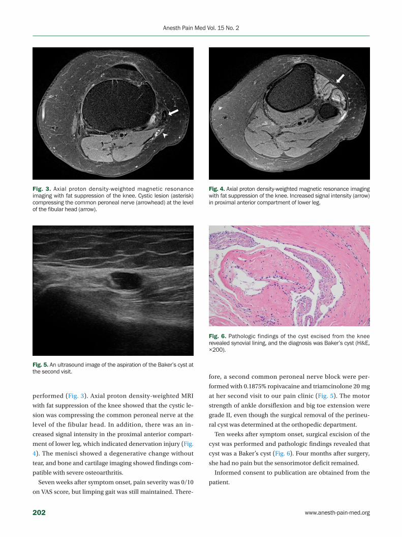
performed (Fig. 3). Axial proton density-weighted MRI
with fat suppression of the knee showed that the cystic le-
sion was compressing the common peroneal nerve at the
level of the fibular head. In addition, there was an in-
creased signal intensity in the proximal anterior compart-
ment of lower leg, which indicated denervation injury (Fig.
4). The menisci showed a degenerative change without
tear, and bone and cartilage imaging showed findings com-
patible with severe osteoarthritis.
Seven weeks after symptom onset, pain severity was 0/10
on VAS score, but limping gait was still maintained. There-
Fig. 3. Axial proton density-weighted magnetic resonance imaging with fat suppression of the knee. Cystic lesion (asterisk) compressing the common peroneal nerve (arrowhead) at the level of the fibular head (arrow).
Fig. 4. Axial proton density-weighted magnetic resonance imaging with fat suppression of the knee. Increased signal intensity (arrow) in proximal anterior compartment of lower leg.
Fig. 5. An ultrasound image of the aspiration of the Baker’s cyst at the second visit.
fore, a second common peroneal nerve block were per-
formed with 0.1875% ropivacaine and triamcinolone 20 mg
at her second visit to our pain clinic (Fig. 5). The motor
strength of ankle dorsiflexion and big toe extension were
grade II, even though the surgical removal of the perineu-
ral cyst was determined at the orthopedic department.
Ten weeks after symptom onset, surgical excision of the
cyst was performed and pathologic findings revealed that
cyst was a Baker’s cyst (Fig. 6). Four months after surgery,
she had no pain but the sensorimotor deficit remained.
Informed consent to publication are obtained from the
patient.
Fig. 6. Pathologic findings of the cyst excised from the knee revealed synovial lining, and the diagnosis was Baker’s cyst (H&E, ×200).
202 www.anesth-pain-med.org
Anesth Pain Med Vol. 15 No. 2

KS
PS
DISCUSSION
Distention of the popliteal bursa, such as the gastrocne-
mio-semimembranosus bursa, is known as Baker’s cyst [1–
3]. Baker’s cyst is usually connected to the knee joint by a
valvular opening, and is commonly associated with in-
tra-articular diseases and inflammation, such as meniscus
tears, rheumatoid arthritis, and osteoarthritis [1,2]. Even
though the most common location of a popliteal cyst is be-
tween the gastrocnemius and semimembranosus, there are
a few case reports of the lateral occurrence of popliteal
cysts [6]. Usually, Baker’s cyst presents with an asymptom-
atic course or mild pain; however, it can sometimes present
with moderate to severe complications, including com-
partment syndrome, thrombophlebitis, and entrapment
neuropathy [2]. Diagnosis is based on the patient’s past
medical history, pathological examination, and imaging
workup such as MRI [2,3]. If there is no neuropathological
complication, only conservative treatment is required [2,3].
In our case, it was not easy to reveal the cause of neurop-
athy due to the uncommon location of the Baker’s cyst,
which was located on the lateral side of popliteal area. Be-
cause Baker’s cyst is usually asymptomatic, her severe pain
and muscle weakness could have made diagnosis difficult.
She was initially diagnosed based on just her symptoms
and EMG results without knowing the exact cause. Thus,
the focus of the treatment was symptom relief. When she
first came to our pain clinic, she had already had motor
weakness. So we considered about surgical treatment.
However, she was referred from orthopedic surgery only
for pain control and was scheduled to revisit there soon.
Therefore we did not consider surgical treatment in our
department.
The common peroneal nerve arises from the sciatic
nerve, which is formed from the L4-S4 ventral rami. This
nerve originates from the upper angle of the popliteal fossa
and travels along the medial border of the biceps femoris.
It then turns around the head of the fibula, and it is divided
into the deep peroneal nerve and superficial peroneal
nerve [4,5]. The deep peroneal nerve innervates sensory
information between the first and second toe, and has mo-
tor control of toe extension and ankle dorsiflexion. The su-
perficial peroneal nerve provides sensory information of
the foot dorsum and lateral calf, and has motor control of
ankle eversion [4,5,7]. Peroneal neuropathy is associated
with various causes, such as direct nerve damage, nerve
compression, nerve traction, and metabolic syndrome
[4,5]. Peroneal neuropathy could present with diverse
symptoms depending on where the nerve is affected, but
symptoms are mostly sensory loss and foot drop [5,7,8]. For
this reason, we could identify the location of the injury
based on the clinical findings. The assessment includes
electrodiagnostic examinations, which included a motor
conduction study, sensory conduction study, and needle
EMG. Imaging studies such as MRI is also helpful [4,5,9].
Treatments differ from medication to surgery according to
causes and symptoms.
In the present case, our patient suffered from foot drop,
paresthesia, and mild pain. On the physical examination,
the motor grade was I on ankle dorsiflexion and big toe ex-
tension. Numbness and tingling sensation were observed
on the common peroneal nerve territory. Also, the electro-
diagnostic result was indicative of common peroneal neu-
ropathy. To find out the cause of common peroneal neu-
ropathy, we performed an ultrasound examination. We
placed the probe on the lateral side of her left knee to ex-
amine the nerve pathway and lesion and surprisingly
found a huge cyst near the left fibular head. Thus, we
thought that the cause of neuropathy was compression by
a cystic lesion. To relieve the compression, we aspirated
the cystic fluid and blocked the common peroneal nerve;
the patient experienced dramatic reliefs after the proce-
dure. Soon, pain severity reduced to 0/10 on the VAS score.
What we missed was the pathological examination of the
aspirated fluid. In fact, the lateral presentation of a poplite-
al cystic lesion should be diagnosed fast to rule out a me-
niscal cyst and soft tissue neoplasm [2]. If the examination
had been performed, the diagnosis would have made earli-
er. Despite the unusual location, we assumed that the cys-
tic lesion would be Baker’s cyst for a number of reasons.
First, ultrasound examination of her left knee, we find a
large popliteal cyst was extend to the fibular head. This cyst
was most likely to be a ganglion, but we could not exclude
the Baker’s cyst. Second, the characteristics of the aspirat-
ed fluid were the same as that of the synovial fluid, i.e., yel-
low, thick, and mucinous. Third, she had a medical history
of knee surgery, severe osteoarthritis, and diabetes melli-
tus. The treatment was rapid, so we thought the treatment
would be effective. However, the motor recovery was slow-
er than our expectation, so we planned to do surgery. We
were at least able to achieve the goal of pain relief.
Like most other diseases, eliminating the cause is the best
treatment for common peroneal neuropathy. In this aspect,
since ultrasound is easily and immediately applicable, diag-
www.anesth-pain-med.org 203
Peroneal neuropathy by Baker’s cyst

nosable, and a treatable device, it would be the best choice
to examine common peroneal neuropathy and other types
of peripheral neuropathy. Furthermore, it is cost effective.
In our case, using ultrasound, we easily found the Baker’s
cyst at the level of the fibular head, which was causing the
neuropathy. If the diagnosis was faster, perfect recovery of
the nerve may have been achieved using only the ultra-
sound-guided procedure. It is a good idea to perform a
minimal examination in consideration of the general loca-
tion and condition of the lesion. We think performing ul-
trasound examination on peripheral neuropathy patients
to examine various lesions could be a good option. Even if
the ultrasound-guided procedure does not allow for per-
fect recovery of the nerve, it could at least provide pain re-
lief and satisfaction to patients waiting for surgery or exam-
ination, like that in our case.
CONFLICTS OF INTEREST
No potential conflict of interest relevant to this article
was reported.
AUTHOR CONTRIBUTIONS
Conceptualization: Hwa-Yong Shin. Data acquisition:
Hana Cho, Yong Bum Park, Hee Sung Kim. Formal analysis:
Hee Sung Kim. Funding: Hwa-Yong Shin. Supervision:
Hwa-Yong Shin. Writing—original draft: Hana Cho, Seung
Young Lee. Writing—review & editing: Hana Cho, Dong-
Rim Kim, Je Jin Lee, Seung Young Lee.
ORCID
Hana Cho, https://orcid.org/0000-0002-9077-9972
Dong-Rim Kim, https://orcid.org/0000-0001-6448-783X
Je Jin Lee, https://orcid.org/0000-0002-9986-9026
Seung Young Lee, https://orcid.org/0000-0003-2734-1342
Yong Bum Park, https://orcid.org/0000-0002-3741-2311
Hee Sung Kim, https://orcid.org/0000-0002-8154-2391
Hwa-Yong Shin, https://orcid.org/0000-0002-8721-3070
REFERENCES
1. Herman AM, Marzo JM. Popliteal cysts: a current review. Ortho-
pedics 2014; 37: e678-84.
2. Frush TJ, Noyes FR. Baker’s cyst: diagnostic and surgical consid-
erations. Sports Health 2015; 7: 359-65.
3. Demange MK. Baker’s cyst. Rev Bras Ortop 2015; 46: 630-3.
4. Marciniak C. Fibular (peroneal) neuropathy: electrodiagnostic
features and clinical correlates. Phys Med Rehabil Clin N Am
2013; 24: 121-37.
5. Baima J, Krivickas L. Evaluation and treatment of peroneal neu-
ropathy. Curr Rev Musculoskelet Med 2008; 1: 147-53.
6. Manik P, Vasudeva N. Unusual lateral presentation of popliteal
cyst: a case report. Nepal Med Coll J 2006; 8: 284-5.
7. Masakado Y, Kawakami M, Suzuki K, Abe L, Ota T, Kimura A.
Clinical neurophysiology in the diagnosis of peroneal nerve pal-
sy. Keio J Med 2008; 57: 84-9.
8. Ferraresi S, Garozzo D, Buffatti P. Common peroneal nerve inju-
ries: results with one-stage nerve repair and tendon transfer.
Neurosurg Rev 2003; 26: 175-9.
9. Lee DI. New paradigm for the neuropathic pain. Korean J Pain
2004; 17: 99-103.
204 www.anesth-pain-med.org
Anesth Pain Med Vol. 15 No. 2

KS
PS
Herpes zoster is a common disease affecting approxi-
mately one in three people [1], in which the varicella zoster
virus (VZV) in the dorsal root ganglion of the nerve is reac-
tivated. Herpes zoster that appears in the ophthalmic (V1)
branch of the trigeminal ganglia is called herpes zoster
ophthalmicus (HZO). Signs and symptoms of HZO are sim-
ilar to herpes zoster along with ocular manifestations, in-
cluding eye pain, poor vision, photophobia, and others [2].
Cavernous sinus thrombosis (CST) is the formation of a
blood clot in the cavernous sinus and is associated with a
high mortality rate [3–6]. CST can be of two types. Septic
CST is caused by an infection whereas, aseptic CST is usu-
ally associated with trauma, surgery, or vascular. CST is a
life-threatening condition; therefore, prompt diagnosis and
treatment are necessary. Signs and symptoms of CST in-
clude fever, headache, and ocular findings, such as perior-
bital swelling, ophthalmoplegia, and others [3–6]. These
signs and symptoms are similar to those of HZO.
We report a case of septic CST discovered after blood
culture and magnetic resonance imaging (MRI) were per-
formed on a patient who was transferred to our hospital for
treatment of HZO and had continuous fever, headache,
and blurred vision. The study was approved by the Institu-
tional Review Board of the Presbyterian Medical Center
(no. E2018-023).
Herpes zoster in the ophthalmic branch of the trigeminal ganglia obscuring cavernous sinus thrombosis due to Streptococcus constellatus subsp. constellatus - A case report -
Ji Hye Lee, Hyun Joo Heo, Ki Man Kim, Han Gyeol Lee, Seung Min Baek, and Da Wa Jung
Department of Anesthesiology and Pain Medicine, Presbyterian Medical Center, Jeonju,
Korea
Case Report Anesth Pain Med 2020;15:205-208 https://doi.org/10.17085/apm.2020.15.2.205pISSN 1975-5171 • eISSN 2383-7977
Received October 14, 2019 Revised December 5, 2019 Accepted December 11, 2019
Corresponding author Ji Hye Lee, M.D. Department of Anesthesiology and Pain Medicine, Presbyterian Medical Center, 365 Seowon- ro, Wansan-gu, Jeonju 54987, Korea Tel: 82-63-230-1593 Fax: 82-63-230-8919 E-mail: [email protected]
Background: Herpes zoster ophthalmicus (HZO) is an infectious disease that results from the reactivation of latent varicella zoster virus in the ophthalmic branch of the trigeminal ganglia. HZO manifests with herpes zoster-like symptoms such as rash with or without signs of ocular involvement. Cavernous sinus thrombosis (CST) is a life-threatening condition ac-companied by signs and symptoms involving the eyes and the cranial nerves.
Case: We report a case of septic cavernous sinus thrombosis (caused by Streptococcus constellatus subsp. constellatus) which was masked by the simultaneous occurrence of HZO in this patient, resulting in delayed diagnosis.
Conclusions: CST may be obscured by HZO, prompt diagnosis and treatment is necessary when such case arrive.
Keywords: Cavernous sinus thrombosis; Herpes zoster; Streptococcus constellatus; Trigemi-nal ganglion.
This is an Open Access article distributed under the terms of the Creative Commons Attribution Non-Commercial License (http://creativecommons.org/licenses/by-nc/4.0) which permits unrestricted non-commercial use, distribution, and reproduction in any medium, provided the original work is properly cited.Copyright © the Korean Society of Anesthesiologists, 2020
205

CASE REPORT
A 44-year-old man was brought to a primary care hospi-
tal with a rash and vesicle on the left side of his forehead
since 3 days. Moreover, he had a headache since 6 days.
Serologic tests for VZV (immunoglobulin M [IgM] and im-
munoglobulin G [IgG]) were conducted using the en-
zyme-linked immunosorbent assay in the primary care
hospital. The patient tested positive for VZV IgG and IgM.
He was diagnosed with HZO, and treated with antiviral
drugs and analgesics. Before hospitalization, he had con-
tinuous fever (38°C and higher). After three days of hospi-
talization, the patient presented with worsening of left-sid-
ed frontal headache, nausea, blurred vision, and diplopia.
He was transferred to our hospital for further evaluation
and treatment.
The patient was alert but febrile (38.3°C) when he arrived
at our hospital. There was no marked facial or periorbital
swelling, except for the crust on his forehead. The cardio-
pulmonary and neurological findings were normal. His
heart rate, blood pressure, and respiratory rate were 78
beats/min, 110/60 mmHg, and 20 breaths/min. An oph-
thalmologist examined his eyes and the visual acuity was
6/12 bilaterally. There were no abnormalities in the ocular
tissues (cornea, conjunctiva, sclera, uvea, or retina) and no
visual field defects. We suspected that his horizontal diplo-
pia and lateral rectus muscle paresis in the left eye were
because of 6th cranial nerve (abducens nerve) palsy. There
was no evidence of infection in the facial region (except
HZO), respiratory tract, urinary tract, or on the skin. Thus,
the study focused on fever and cause of 6th cranial nerve
palsy.
The white blood cell count was normal (10,100 mm3),
but the erythrocyte sedimentation rate (120 mm/h) and
C-reactive protein level (10.17 mg/dl) were elevated. Blood
culture was performed; however, the patient did not pro-
vide consent for a cerebrospinal fluid (CSF) analysis. The
brain MRI revealed lateral bulging and irregular marginal
enhancement in the cavernous sinus, which is consistent
with bilateral CST and superior ophthalmic vein thrombo-
sis (Fig. 1).
We admitted the patient to the intensive care unit and
asked a neurologist and an infectious medicine doctor to
assist with the treatment. Patient’s history was taken again
and he was kept under observation during the course of
the treatment. Although he had no medical history of dia-
betes mellitus, hypertension, or etc., he did undergo dental
treatment about 3 months ago and had experienced pain
in the left maxillary area approximately 1 month ago. Given
the dental history, high body temperature, and aggravated
symptoms, we started his antibiotic treatment despite the
antiviral treatment that he had received at the primary care
hospital. Second-generation cephalosporin was adminis-
tered until we received his blood culture results. Strepto-
A B
Fig. 1. Both superior ophthalmic veins presenting with engorgement and thrombus in the posterior region (A: axial, B: coronal view).
206 www.anesth-pain-med.org
Anesth Pain Med Vol. 15 No. 2

KS
PS
coccus constellatus subsp. constellatus was detected in the
blood culture, therefore we switched from cephalosporin
to clindamycin. We performed echocardiography to detect
sings of infective endocarditis; however, it did not reveal
any special findings. Antithrombotic therapy was per-
formed with low molecular weight heparin. After 7 days of
treatment in the intensive care unit, the symptoms im-
proved, and the patient was discharged after being trans-
ferred to the general room. No complications were ob-
served during one year of follow-up since the patient was
discharged.
DISCUSSION
Herpes zoster is frequently encountered in primary care
hospitals. The lifetime risk of herpes zoster is estimated to
be about 30% and, HZO appears in 20% of these cases, with
risks in about 1–4% throughout life [1,2]. Herpes zoster pa-
tients complain of systemic symptoms of influenza-like ill-
ness, such as fatigue, malaise, and low-grade fever, prior to
the development of a unilateral rash. A rash and a vesicle
characteristically appear along the nerve segment where
the virus reactivates, but like zoster sine herpete, the occa-
sional skin lesions do not appear. HZO presents with the
general symptoms of herpes zoster infection and the erup-
tion of vesicle and cluster of papules in the trigeminal
nerve dermatome divisions located on the forehead, upper
eyelids, eyes, and nose. Ocular manifestations may occur,
including ptosis and swelling, injected appearance, pete-
chial hemorrhage in the conjunctiva, keratitis and scarring
ulcer in the cornea, uveitis, and acute retinal necrosis. In
addition, patients with HZO may manifest symptoms of
optic neuritis, swelling, edematous optic nerve, and oculo-
motor palsy [1,2]. The ocular symptoms in HZO can be ex-
plained by the inflammation of the ocular nerve through
cavernous sinus caused by the reactivation of VZV in the
trigeminal ganglion.
The signs and symptoms of VZV primary infection, also
known as chicken pox, are attributed to vasculopathy,
which may manifest in the form of transient ischemic at-
tacks, ischemic or hemorrhagic stroke, aneurysm, sinus
thrombosis, giant cell arteritis, and granulomatous aortitis.
CST is a type of cerebral venous sinus thrombosis (CVST).
The CVST is mostly reported in pediatric patients with VZV
primary infection accompanied with purpura fulminans.
By contrast, the VZV primary infection is rare in adults and
has more severe symptoms [7–9]. Herpes zoster, that is re-
activation of VZV, has no reported cases of CVST. Herpes
zoster is, however, a risk factor for stroke and myocardial
infarction. Moreover, the incidence of stroke increases in
patients who have a history of HZO [10].
Cavernous sinus is connected to the dural venous sinus
in the midline of the skull with direct or indirect nasal, eth-
moidal, and sphenoidal sinus veins. CST may be septic or
aseptic. Causes of aseptic CST are trauma, surgery, hema-
tological disorder, malignancy, or it could be idiopathic in
origin [5]. Septic CST occurs because of the infection
spreading through the ethmoidal or sphenoidal sinuses, or
from teeth, ear, or orbit through the venous system. Septic
CST is more common than aseptic CST [3,5]. In the current
study, the patient’s blood culture detected Streptococcus
constellatus subsp. constellatus, which belongs to the
milleri group. Streptococci in this group are part of the resi-
dent flora of the oral cavity and the respiratory tract. How-
ever, they may cause suppurative and deep-seated infec-
tions via the venous system. The incidence of CVST is less
than 1% of all strokes [4]. The exact incidence rate of CST is
not known, but it is rare like CVST. Infection, pregnancy,
receiving oral contraceptives, thrombophilic disorders, and
obesity could contribute to increased CST risk. CST is a
life-threatening condition [3–6]. Although the symptoms of
CST may vary, in most cases, the symptoms appear in rela-
tion to the anatomical structures near the cavernous sinus,
such as cranial nerves, orbit, and eyes, because their ve-
nous drainage is in the cavernous sinus. Typical symptoms
of CST are headache, unilateral periorbital edema, photo-
phobia, and bulging eyeballs. In addition, involvement of
the cranial nerve may result in palsy or sensory nerve inju-
ry, such as sensory deficit of the ophthalmic and maxillary
branches of the 5th cranial nerve [3–6].
The signs and symptoms of HZO and CST are similar. In
addition, HZO can cause vasculopathy, such as transient
ischemic attacks and ischemic or hemorrhagic stroke [7].
In our patient, skin lesions occurred at the trigeminal nerve
V1 site, three days before the visit, as seen in cases of HZO,
along with fever and myalgia. The patient was diagnosed
with HZO in the primary care hospital and was treated ac-
cordingly. However, even after the treatment, he had per-
sistent headache and high fever and was transferred to our
hospital to rule out meningitis and stroke. The patient did not
provide consent for a CSF study, but blood culture was per-
formed because of high fever, and CST was confirmed on
MRI. The preceding dental treatment may have been the
cause of CST in this patient.
www.anesth-pain-med.org 207
HZO with cavernous sinus thrombosis

Herpes zoster-associated vasculopathy is treated with
antiviral agents. However, infectious CST is treated with
high-dose antibiotics. CST has a high mortality rate; there-
fore, prompt treatment is necessary. We started the treat-
ment for CST immediately while continuing, the HZO
treatment. HZO misdiagnosis may delay the diagnosis of
CST because vasculopathy makes it difficult to differenti-
ate the symptoms of CST and HZO. If high fever, severe
headache, and eye symptoms are observed with HZO, the
patient history should be accurately evaluated to exclude
CST by performing not only CSF analysis but also MRI,
blood culture, etc.
In conclusion, CST is a life-threatening condition and its
diagnosis may be obscured by HZO. Therefore, prompt di-
agnosis and treatment are necessary in such cases.
CONFLICTS OF INTEREST
No potential conflict of interest relevant to this article
was reported.
AUTHOR CONTRIBUTIONS
Conceptualization: Ji Hye Lee, Hyun Joo Heo. Data ac-
quisition: Ki Man Kim, Han Gyeol Lee, Seung Min Baek, Da
Wa Jung. Supervision: Ji Hye Lee. Writing—original draft: Ji
Hye Lee. Writing—review & editing: Ji Hye Lee, Hyun Joo
Heo.
ORCID
Ji Hye Lee, https://orcid.org/0000-0003-3269-3844
Hyun Joo Heo, https://orcid.org/0000-0003-2507-6629
Ki Man Kim, https://orcid.org/0000-0002-4257-9167
Han Gyeol Lee, https://orcid.org/0000-0001-7835-0172
Seung Min Baek, https://orcid.org/0000-0003-0290-0208
Da Wa Jung, https://orcid.org/0000-0002-1566-7903
REFERENCES
1. Ragozzino MW, Melton LJ 3rd, Kurland LT, Chu CP, Perry HO.
Population-based study of herpes zoster and its sequelae. Med-
icine (Baltimore) 1982; 61: 310-6.
2. Johnson JL, Amzat R, Martin N. Herpes zoster ophthalmicus.
Prim Care 2015; 42: 285-303.
3. Coutinho JM. Cerebral venous thrombosis. J Thromb Haemost
2015; 13 Suppl 1: S238-44.
4. Einhäupl K, Stam J, Bousser MG, De Bruijn SF, Ferro JM, Marti-
nelli I, et al.; European Federation of Neurological Societies.
EFNS guideline on the treatment of cerebral venous and sinus
thrombosis in adult patients. Eur J Neurol 2010; 17: 1229-35.
5. Lai PF, Cusimano MD. The spectrum of cavernous sinus and or-
bital venous thrombosis: a case and a review. Skull Base Surg
1996; 6: 53-9.
6. Weerasinghe D, Lueck CJ. Septic cavernous sinus thrombosis:
case report and review of the literature. Neuroophthalmology
2016; 40: 263-76.
7. Siddiqi SA, Nishat S, Kanwar D, Ali F, Azeemuddin M, Wasay M.
Cerebral venous sinus thrombosis: association with primary
varicella zoster virus infection. J Stroke Cerebrovasc Dis 2012;
21: 917.e1-4.
8. Paul G, Paul BS, Singh G. Unseen face of varicella-zoster infec-
tion in adults. Indian J Crit Care Med 2016; 20: 731-4.
9. Khan R, Yasmeen A, Pandey AK, Al Saffar K, Narayanan SR. Ce-
rebral venous thrombosis and acute pulmonary embolism fol-
lowing varicella infection. Eur J Case Rep Intern Med 2019; 6:
001171.
10. Nagel MA, Jones D, Wyborny A. Varicella zoster virus vasculop-
athy: the expanding clinical spectrum and pathogenesis. J Neu-
roimmunol 2017; 308: 112-7.
208 www.anesth-pain-med.org
Anesth Pain Med Vol. 15 No. 2

KS
RA
Clinical ResearchAnesth Pain Med 2020;15:209-216https://doi.org/10.17085/apm.2020.15.2.209pISSN 1975-5171 • eISSN 2383-7977
Analgesic effect of ropivacaine with fentanyl in comparison with ropivacaine alone for continuous femoral nerve block after knee replacement arthroplasty: a prospective, randomized, double-blinded study
Gunn Hee Kim1, Joon Woo Lee2, Go Eun Kim1, Seong Su Lee1, Shill Lee Son1, Byung Uk Kim1, Ha Na Cho1, Mi Young Kwon1, Min Seok Koo1, Ji Eun Kim1, and Mi Jung Yun1
1Department of Anesthesiology and Pain Medicine, National Medical Center, Seoul,2Department of Radiology, Seoul National University Bundang Hospital, Seongnam, Korea
Received April 25, 2019Revised June 19, 2019Accepted July 15, 2019
Corresponding authorMi Jung Yun, M.D., Ph.D. Department of Anesthesiology and Pain Medicine, National Medical Center, 245 Eulji-ro, Jung-gu, Seoul 04564, Korea Tel: 82-2-2260-7370Fax: 82-2-2262-4766 E-mail: [email protected]
Background: The analgesic effect of perineural opioid in clinical practice are still controver-sial. This randomized controlled trial compared analgesic effect of ropivacaine with fentanyl or ropivacaine alone for continuous femoral nerve block following unilateral total knee ar-throplasty.
Methods: Fourty patients of ASA PS I or II receiving total knee arthroplasty with spinal anes-thesia were enlisted and randomly allocated into two groups. Group R; bolus injection of 0.375% ropivacaine, 30 ml and an infusion of 0.2% ropivacaine at 8 ml/h (n = 20). Group RF; 0.375% ropivacaine, 29 ml added with 50 μg of fentanyl as a bolus and an infusion of 0.2% ropivacaine mixed with 1 μg/ml of fentanyl at 8 ml/h (n = 20). Local anesthetic infu-sion via a femoral nerve catheter was started at the end of operation and continued for 48 h. Intravenous patient-controlled analgesia with hydromorphone (0.15 mg/ml, 0-1-10) were used for adjuvant analgesics. Position of catheter tip and contrast distribution, visual ana-logue scale of pain, hydromorphone consumption, side effects were recorded for 48 h after operation. Patient satisfaction for the pain control received were noted.
Results: The pain visual analogue scale, incidences of side effects and satisfaction were not different between the two groups (P > 0.05), but the hydromorphone usage at 48 h after op-eration were lower in the Group RF than in the Group R (P = 0.047).
Conclusions: The analgesic effect of ropivacaine with fentanyl for continuous femoral nerve block after knee replacement arthroplasty was not superior to that of the ropivacaine alone.
Keywords: Anesthesia and analgesia; Femoral nerve; Fentanyl; Nerve block; Ropivacaine.
INTRODUCTION
The direct analgesic activity of the opioid drugs in the
central nervous system [1], and the peripheral action of
them in primary afferent fibers such as suppression of ei-
ther spread of action potentials or the discharge of excit-
atory transmitters, have been reported [2,3].
From a clinical perspective, however, the effect of peri-
This is an Open Access article distributed under the terms of the Creative Commons Attribution Non-Commercial License (http://creativecommons.org/licenses/by-nc/4.0) which permits unrestricted non-commercial use, distribution, and reproduction in any medium, provided the original work is properly cited.Copyright © the Korean Society of Anesthesiologists, 2020
209

neural opioid are controversial. In a systematic qualitative
review [4], it was reported that the buprenorphine are reas-
suring agent for use in extension of duration in peripheral
nerve blocks of local anesthetic. Bazin et al. [5] reported
that the durations of analgesia provided by a composite of
lignocaine and bupivacaine, added with morphine, bu-
prenorphine, or sufentanil, were prolonged in 80 patients
after brachial plexus block for orthopedic surgery.
Kardash et al. [6], however, documented that the addi-
tion of 75 μg of fentanyl to mepivacaine had no significant
effects on characteristics of supraclavicular blocks. The 1
μg/kg of fentanyl added to 0.75% ropivacaine did not pro-
vide significant benefit in terms of onset time, condition
and duration of combined sciatic-femoral nerve block in
patients receiving elective hallux valgus repair [7]. Until
now, most of the clinical studies were performed using a
single neural blockade. The analgesic effect of continuous
perineural infusion of fentanyl added to local anesthetic
has not yet been fully clarified.
We investigated the analgesic effect of ropivacaine with
fentanyl in comparison with ropivacaine alone for a con-
tinuous femoral nerve blockade following total knee ar-
throplasty.
MATERIALS AND METHODS
This prospective, double blinded, randomized controlled
trial was conducted at one hospital between October 2015
and August 2016. Institutional Review Board of our hospi-
tal approved clinical protocol and informed consent docu-
mentation (no. H-1507-056-001). Written informed con-
sents were acquired from all the patients. This clinical in-
vestigation was registered at clinicalTrials.gov.
Patients who were planned to receive unilateral total
knee replacement arthroplasty (TKA) under spinal anes-
thesia were evaluated for their eligibility. Adult of Ameri-
can Society of Anesthesiologists physical status I–II were
enlisted in the study.
Patients who had contraindications to a regional anes-
thetic technique (e.g., local infection, sepsis, coagulation
abnormality), allergy to local anesthetic or fentanyl, estab-
lished neurologic deficits in the lower extremities, and in-
ability to know the pain scales or use intravenous pa-
tient-controlled analgesia (IV-PCA) device were excluded
from this study.
Patients were randomly assigned to one of two groups in
a 1 : 1 ratio using a computer-generated randomization se-
quence (www.randomizer.org): ropivacaine (R) or ropiva-
caine with fentanyl (RF) group. Anesthesiologists who did
not participate in the patient care and assessment of vari-
ables generated random allocation table, enlisted partici-
pants and assigned them to interventions. Suitable subjects
were allocated to treatment in order of enrollment. For the
double-blind assignment, a anesthesia nurse who did not
know of the study prepared treatments and ensured them
with their treatment number concealed in envelop. The
study drug was offered to the anesthesiologist responsible
for the patient care and administering the dose.
Description of both the visual analogue scale (VAS) of
pain and the use of the IV-PCA device was given to all pa-
tients before anesthesia. Patients were premedicated with
1–2 mg of IV midazolam. On entrance in the operating
room, general monitoring including electrocardiography,
automated oscillotonometry and pulse oximetry. Systolic
and diastolic blood pressure and heart rate were recorded
every 5 min during the operation. All the patients were in-
fused with 6 ml/kg of crystalloid solution before beginning
of the regional anesthesia.
The femoral nerve catheter was inserted before the in-
duction of spinal anesthesia by one anesthesiologist expe-
rienced in these techniques who was blinded to the study.
For the femoral nerve blockade, the patient was placed
in supine position and a linear 6–13 MHz probe (SonoSite
M-Turbo®, SonoSite Bothell, USA) was positioned both in a
transverse and longitudinal direction to the femoral crease
at 2 cm below the inguinal crease and 2.0 cm lateral to the
femoral artery. Once the femoral nerve was identified, the
probe was positioned in a longitudinal direction to the
femoral nerve. Subsequently, a 22-gauge 50 mm insulated
needle (Stimuplex®, B. Braun, Germany) was inserted us-
ing the in-plane approach near the femoral nerve. A nerve
stimulator (Pajunk, Fisher & Paykel, New Zealand) was
used to prove exact location of the femoral nerve. The
block needle was advanced cephalad toward the femoral
nerve with an initial output of 1 mA (1 Hz) until quadriceps
femoris muscle contractions and patella snap were ob-
tained. The position was then considered appropriate
when contractions of the quadriceps and patella snap are
still elicited when the voltage is reduced to an output of 0.5
mA, but those responses are lost when the voltage is re-
duced to output of 0.3 mA as it can avoid intraneural place-
ment. If the muscular twitch was stopped immediately fol-
lowing the administration of 1 ml of the study solution, the
needle location was regarded appropriate and the catheter
210 www.anesth-pain-med.org
Anesth Pain Med Vol. 15 No. 2

KS
RA
was inserted. For the confirmation of catheter function, 10
ml of 1% lidocaine as a test dose was injected through the
catheter after negative aspiration for blood in all patients
and were checked for loss of cold sense in the anterior and
medial thigh, as well as for motor weakness in all major
muscles including quadriceps femoris.
Three milliliters of contrast medium (OmnipaqueTM 300
mg Inj., 10 ml, Iohexol 647 mg/ml, GE Health Care AS Ko-
rea, Korea) were administered through the catheter and
the spot of the catheter tip and the diffusion of the contrast
media were confirmed using a C-arm fluoroscope.
One blind radiologist who specialized in interventional
radiology randomly interpreted and qualified the radio-
graphs of the pelvic region based on his decision. The cath-
eter tip position were as following; type 1 = medial, be-
tween sacroiliac (SI) joint and sacral promontory, cranial
to hip joint, distal to L4-5 disc level, type 2 = lateral, lateral
to SI joint, cranial to hip joint, distal to L4-5 disc level, type
3 = proximal, proximal to L4-5 disc level, type 4 = distal,
distal to hip joint, type 5 = indetermined.
The distribution of contrast dye in the femoral sheeth
were classified such as type 1 = internal spread under the
psoas muscle fascia, type 2 = external spread under the ili-
acus muscle fascia, type 3 = spread near the roots of the
lumbar plexus, type 4 = spread along femoral nerve, type 5
= loculated at unknown anatomic location, possible far
from nerve sheath.
Patients were allocated randomly to group R (bolus ad-
ministration with 30 ml of 0.375% ropivacaine and a con-
tinuous infusion with a 0.2% ropivacaine at 8 ml/h for 48 h
after operation, n = 20) or group RF (bolus dose of 0.375%
ropivacaine, 29 ml added with 50 μg [1 ml] of fentanyl, and
a continuous infusion of 0.2% ropivacaine added with 1 μg/
ml of fentanyl [7,8], at 8 ml/h for 48 h after operation) (n =
20). The present study was performed to assess the postop-
erative analgesia and side effects of two infusion regimens
during continuous femoral nerve block (FNB) after unilat-
eral TKA.
Patients received spinal anesthesia with 10 mg of bupiva-
caine (Marcaine®, Spinal 0.5% Heavy, 5 mg/ml, AstraZene-
ca, Sweden). All TKA were performed by one orthopedic
surgeon.
At the completion of the operation, 30 ml of a local anes-
thetic solution was injected through the FNB catheter over
a 10-min period depending on the group allocation. The
infusion of 0.2% ropivacaine via femoral catheter was start-
ed using an infusor (AutoFuser®, AceMedical Corp., Ltd.,
Korea) at a rate of 8 ml/h, which was continued into the
postoperative period for 48 h. Duration for operation, vol-
ume of infused crystalloid during operation were noted.
In the postanesthetic care unit, the patients were provid-
ed with a IV-PCA device (AutoMed3200, AceMedical Corp.,
Ltd., Korea), containing hydromorphone (0.15 mg/ml, 0-1-
10). The PCA system was adjusted to a 1 ml of bolus on de-
mand, with a 10 min lockout interval and no background
infusion of the hydromorphone solution. The patient was
told to press the button if his or her pain VAS is over 30 out
of 100. The cumulative bolus use of IV-PCA (ml) at 12, 24,
36, 48 h after operation was noted as it was stored from the
electronic memory of the PCA machine.
The pain intensities both at rest and with 30 degree of
passive flexion of the knee were evaluated using a VAS (0 =
no pain and 100 = worst pain) at 6, 24, 48 h after operation
as a primary outcome measures.
The patients received intramuscular injection of 50 mg/
ml of ketoprofen (TOPREN INJ 50 mg®, Samsung Pharm.
Ind. Co., Ltd., Korea) when discontented with the IV-PCA.
The salvage doses of ketoprofen at 12, 24, 48 h after opera-
tion, time for self-voiding from the end of operation, side
effects (nausea, vomiting, dizziness, pruritus, urinary re-
tention, urinary catheterization, chest discomfort, leakage)
and patients’ satisfaction score (1–5: 1, very unsatisfied; 2,
unsatisfied; 3, moderate; 4, satisfied; 5, very satisfied) were
recorded as secondary outcome measures.
The respiratory rate was monitored by the ward nurses
every 4 h in all patients. If the respiratory rate was less than
10 breath/min, incremental doses of IV naloxone was in-
jected as needed and oxygen 3 L/min was administered via
a nasal cannula.
All data was gathered by a blind anesthesiologist not par-
ticipated both in the anesthesia administration and patient
care. The sample size was determined based on the result
of a similar study [5]. The difference of mean VAS (0–100)
at 10, 15 h after operation between the control group used
local anesthetic alone and the group used local anesthetic
plus opioid were 15, 20, respectively. Twenty patients in
each group were needed to obtain a mean VAS difference
of 15 at 10, 15 h after the operation, accepting an alpha er-
ror of 0.05 and 90% power. Four more patients were recruit-
ed to compensate for the possible loss of follow up.
Statistical analyses were carried out using the statistical
software IBM SPSS Statistic 25 (IBM Co., USA). Continuous
variables were analyzed using the t-test following normal-
ization test. Categorical variables were compared using the
www.anesth-pain-med.org 211
Femoral nerve block and fentanyl

chi-square test (sex, American Society of Anesthesiologists
physical status score, satisfaction score) or the Fisher’s ex-
act test (side effects). A P value of < 0.05 was regarded sta-
tistically significant.
RESULTS
At the start, 44 patients were approached, 2 of whom did
not consent to be involved in the current study. The 42 pa-
tients of remainder were enrolled and randomly allocated
for the study. One patient per each group was dropped
from the study due to leakage of local anesthetics through
the catheter. The 40 patients finished the study and their
data were statistically analyzed.
The patient characteristics and duration of surgery were
similar in the two groups (Table 1). There were no statisti-
cally significant differences between the two groups.
The pain VAS were not significantly different between
the two groups (Table 2). The ropivacaine with fentanyl
group showed significantly lower usage of hydromorphone
at 48 h after the operation compared to the ropivacaine
alone group (Table 2). The frequencies of ketoprofen injec-
tions, time to self voiding (Table 2), side effects and satis-
faction of patients (Table 3) were similar between the two
groups.
The type of catheter tip position (1–5) (3/11/2/4/0 vs.
3/10/3/4/0 for group R vs. RF, P = 0.970) and contrast dis-
tribution (1–5) (4/4/0/5/7 vs. 3/3/1/3/10 for group R vs. RF,
P = 0.678) were not different between the two groups.
DISCUSSION
The purpose of the current study was to examine if the
additional fentanyl to ropivacaine for the continuous FNB
Table 1. Patient Characteristics
Variable Group R (n = 20) Group RF (n = 20)
Age (yr) 69.1 ± 7.5 67.4 ± 6.4
Sex (F/M) 20/0 18/2
Weight (kg) 59.3 ± 9.8 62.7 ± 7.44
Height (cm) 150.5 ± 5.6 152.2 ± 6.1
Operation time (min) 112.6 ± 22.0 105.7 ± 18.3
Crystalloid (ml) 562.5 ± 153.7 702.5 ± 178.2
ASA PS I/II 9/11 7/13
Values are presented as the mean ± SD or number of patients. There was no statistically significant difference between groups in any parameter. R: ropivacaine, RF: ropivacaine + fentanyl, ASA PS: American Society of Anesthesiologist physical status.
Table 2. Visual Analog Scale of Pain, Rescue Analgesics and Time to Self-voiding
Variable Group R (n = 20) Group RF (n = 20) P value
Pain VAS (resting) POP 6 h 43.1 ± 29.2 43.0 ± 20.3 0.990POP 24 h 47.8 ± 25.2 42.7 ± 16.4 0.459POP 48 h 42.1 ± 16.2 32.6 ± 16.4 0.079
Pain VAS (movement) POP 6 h 64.7 ± 27.7 64.9 ± 23.1 0.979POP 24 h 74.3 ± 14.9 63.3 ± 22.9 0.090POP 48 h 65.9 ± 18.1 54.5 ± 19.0 0.068
Hydromorphone (mg) POP 12 h 1.29 ± 1.08 0.70 ± 0.90 0.348POP 24 h 2.00 ± 1.65 1.13 ± 1.40 0.264POP 36 h 2.75 ± 2.24 1.40 ± 1.62 0.123POP 48 h 3.52 ± 2.53 1.40 ± 1.74 0.047*
Ketoprofen (yes/no) POP 12 h 3/17 1/19 0.605POP 24 h 5/15 3/17 0.695POP 48 h 6/14 4/16 0.537
Time to self-voiding (min) 778.2 ± 492.4 886.0 ± 472.8 0.840
Values are presented as the mean ± SD or number of patients. R: ropivacaine, RF: ropivacaine + fentanyl, VAS: visual analog scale (0–100), POP: postoperative. *P < 0.05.
Table 3. Side Effects and Satisfaction Score
Variable Group R(n = 20)
Group RF(n = 20) P value
Nausea 8 (40) 10 (50) 0.751Vomiting 3 (15) 4 (20) 1.000Dizziness 1 (5) 3 (15) 0.605Pruritus 1 (5) 1 (5) 0.765Urinary retention 10 (50) 13 (56) 0.523Foley catheter 9 (45) 11 (55) 1.000Chest discomfort 0 (0) 1 (5) 0.231Satisfaction score (1–5) 2.9 ± 0.8 3.3 ± 0.6 0.089
Values are presented as the number of patients (%) or mean ± SD. R: ropivacaine, RF: ropivacaine + fentanyl.
212 www.anesth-pain-med.org
Anesth Pain Med Vol. 15 No. 2
KS
RA
would improve the analgesic effect of ropivacaine alone.
The pain VAS were lower in the group RF than in the group
R but there were no statistical significantes. The hydromor-
phone usage at 48 h after operation were significantly low-
er in the RF group in comparison with the R group.
The disagreement in the analgesic effect of peripheral
opioid added to local anesthetic seems to be related with
the dose, degree of approximation of peripheral nerve
blocked to the spinal dorsal horn and the existence of pe-
ripheral inflammation.
The analgesic effect of epidural opioid added to local anes-
thetic has been proved through several clinical studies. The
coadministration of fentanyl (100 μg) and 1% ropivacaine ex-
pedited the beginning of sensory and motor blocks during epi-
dural ropivacaine [9] or lidocaine [10] anesthesia without sig-
nificant fentanyl-related side effects. The additional fentanyl (2
μg/ml) markedly improved the analgesic effect of a low-dose
thoracic epidural infusion of bupivacaine and adrenaline [11].
The analgesic effect of epidural opioid seem to be related
with the location of receptor sites in spinal cord. In rat
brains, opiate receptor sites were labeled in vivo by a po-
tent opiate antagonist and it was localized by autoradiogra-
phy to make the distribution of opiate receptors visible. Sil-
ver grains indicative of the binding of opiate antagonist are
separately localized in many areas of the brain with really
high densities including the substantia gelatinosa of the
spinal cord [12].
In separate areas of rhesus monkey spinal cord, the opi-
ate receptor binding was measured [13]. The authors local-
ized the receptor binding to the upper dorsal horn and re-
ported a drop of it in this area following dorsal root trans-
section.
At the dorsal horn of the spinal cord, the impulse from
peripheral nerve are modulated before it is transmitted
centrally to evoke perception and response. Along with
these mechanisms in the central nervous system, intrinsic
modification of nociception can take place at the peripher-
al terminals of afferent nerves [3]. With the method of more
peripheral administration of opioid such as peripheral ax-
onal nerve block, intraarticular or wound infiltration, the
analgesic effect of opioid added to local anesthetic are con-
troversial.
For the interscalene brachial plexus block, the addition of 75
μg of fentanyl to 30 ml of 1.5% lidocaine (n = 41) speeded up
the onset of sensory and motor blockade in comparison with
the control group that used 30 ml of 1.5% lidocaine plus 1.5 ml
of isotonic saline (n = 39) in a randomized clinical study [14].
Twenty patients undergoing upper extremity surgery
with supraclavicular blocks were prospectively randomized
to receive 75 μg fentanyl either added to the local anesthet-
ic (30 ml mepivacaine 1.5% with epinephrine 5 μg/ml) or
given intramuscularly in a clinical study. An equivalent
volume of normal saline was given in one of the two sites
as a control in a double-blind fashion. The pain VAS was
significantly lower only at 1 h after the operation in the pa-
tients who received fentanyl added to local anesthetic [6].
The effects of fentanyl added to lidocaine for axillary bra-
chial plexus block were evaluated in 66 adult patients
planned for hand and forearm surgery [15]. Patients who
received mixture of lidocaine and 100 μg of fentanyl for
nerve block and 2 ml of normal saline IV, had higher suc-
cess rate of sensory blockade in comparison with the pa-
tients who were given lidocaine plus 2 ml of normal saline
for neural block and 2 ml of normal saline IV or 100 μg of
fentanyl IV.
However, there are studies that failed to observe signifi-
cant improvement of quality of analgesia when morphine
was injected via interscalene approach for brachial plexus
block [16]. Furthermore, there have been reports that pre-
sented the clinical efficacy of the peripheral administration
of morphine such as intraarticular injection [17,18]. So, the
degree of approximation to the spinal dorsal horn would
not be the only factor for the determination of opiate anal-
gesic effect coadministered with local anesthetics. Multiple
factors including dose, route of administration and degree
of inflammation may contribute to the analgesic effect of
peripheral opioid.
A prospective, randomized, double-blinded study exam-
ined the analgesic effects of adding fentanyl to ropivacaine
for continuous femoral nerve block using patient-con-
trolled analgesia for 24 h after TKA [19]. The authors re-
ported that the additional fentanyl did not show significant
enhancement of analgesic effect after TKA. The doses of
fentanyl (100 μg as a bolus and 3 μg/ml for infusion) and
concentration of ropivacaine (0.75%) and infusion volume
(10 ml/h) were much higher than those used in our study.
The patients received general anesthesia, so the baseline
VAS measured was higher in comparison with that of our
study. In spite of these differences, the result of the primary
end point, that is VAS, was similar to that of the present
study that may reflect the importance of both route of ad-
ministration and degree of inflammation in analgesic effect
of perineural opioid.
The administration of same drug into the various routes
www.anesth-pain-med.org 213
Femoral nerve block and fentanyl

such as perineural area or nerve terminal could possibly
produce different efficacy: axonal receptors might be func-
tionally less efficient in pain modulation than receptors at
the nerve terminals, especially when considering the pos-
sible influence of inflammatory condition in peripheral
area, as it is generally assumed that peripheral antinoci-
ceptive effects are mainly provided by an action on primary
afferent neurones [2].
During inflammation, opioid agonists could approach
more esasily to neuronal opioid receptors as the perineuri-
um is disrupted by the inflammation [20]. In sciatic nerve
fibers, the axonal transport of opioid receptors was en-
hanced a few days after the commencement of peripheral
inflammation. In the inflamed tissue, the number of opioid
receptors on cutaneous nerve fibers increased but they
were abolished by ligating the sciatic nerve [21]. In the
present study, the results seem to be related with axonal
receptors of femoral nerve and noninflammatory condition
that is immediate postoperative and by the administration
of preoperative antibiotics. Although it is not possible to
strongly recommend the use of opioids for femoral nerve
block, as the results of the present study showed that there
was no difference in the pain VAS score between the two
groups, it is possible to infer the mechanism of action of
opioids. On the basis of the findings of the present study,
opioids are thought to exert an analgesic effect when ap-
plied to the peripheral nerves that are close to the dorsal
horn of the spinal cord. In addition, the analgesic effect of
opioids on the peripheral nerves with no inflammatory re-
sponse will be small. We believe that these suggestions im-
ply the clinical implications for the present study.
Fig. 1. The position of catheter tip (type 2 = lateral, lateral to sacroiliac joint, cranial to hip joint, open arrow).
Fig. 2. The distribution of contrast dye (type 5 = loculated at unknown anatomic location, possible far from nerve sheath, open arrow).
The position of catheter tip did not influence on the an-
algesic effect as there was no intergroup difference. The
position of the catheter tip was of type 2 (lateral; lateral to
the SI joint, cranial to the hip joint, and distal to the L4-5
disc level; Fig. 1) in 11 (55%) patients of group R and in 10
(50%) patients of group RF. With respect to the distribution
patterns of the contrast dye, the least were of type 3 (spread
near the roots of the lumbar plexus; 0,5% in both groups),
and various other types were noted (Fig. 2; type 5). The
Lower hydromorphone usage in the group RF than in the
group R seemed to influence the similar incidences of opi-
oid related side effects between the two groups.
The patients’ satisfaction was not different between the
two groups (P = 0.089) and the reason for this result seems
to be that both the VAS score and the incidence of side ef-
fects were similar between the two groups. The limitations
of the FNB, that it reduces the pain mainly in the patient’s
anterior knee area and the high incidence of nausea in
both groups (40% in Group R and 50% in Group RF) are
thought to have contributed to the mean satisfaction score
of below 4 in both groups.
Control group receiving the same dose of IV fentanyl was
not included in the current study. It has been reported that
the mean serum fentanyl concentration after epidural infu-
sion of 1 μg/ml [8] or 2 μg/ml [11] were lower than the min-
imum effective analgesic concentration for fentanyl in se-
rum [11,22]. In the present study, the degree of degenera-
tion or preoperative pain of the knee joint to be operated,
were not assessed. It was assumed that the postoperative
214 www.anesth-pain-med.org
Anesth Pain Med Vol. 15 No. 2

KS
RA
pain would not much being influenced with them.
In summary, ropivacaine with fentanyl (1 μg/ml) for the
continuous FNB did not provide significantly increased an-
algesic effect after TKA in comparison with the ropivacaine
alone. Further studies about the analgesic effect of various
doses and route of administration of opioids added to local
anesthetic seem warranted.
CONFLICTS OF INTEREST
No potential conflict of interest relevant to this article
was reported.
AUTHOR CONTRIBUTIONS
Conceptualization: Mi Jung Yun. Data acquisition: Go
Eun Kim, Seong Su Lee, Shill Lee Son, Byung Uk Kim, Ha
Na Cho. Formal analysis: Mi Jung Yun, Joon Woo Lee. Su-
pervision: Mi Young Kwo, Min Seok Koo, Ji Eun Kim. Writ-
ing—original draft: Gunn Hee Kim, Mi Jung Yun. Writing—
review & editing: Mi Jung Yun, Gunn Hee Kim.
ORCID
Gunn Hee Kim, https://orcid.org/0000-0002-0014-8297
Joon Woo Lee, https://orcid.org/0000-0002-7106-5229
Go Eun Kim, https://orcid.org/0000-0002-7223-6317
Seong Su Lee, https://orcid.org/0000-0003-1225-4617
Shill Lee Son, https://orcid.org/0000-0001-9738-3973
Byung Uk Kim, https://orcid.org/0000-0001-9152-2709
Ha Na Cho, https://orcid.org/0000-0002-9077-9972
Mi Young Kwon, https://orcid.org/0000-0002-0667-9494
Min Seok Koo, https://orcid.org/0000-0002-2012-9616
Ji Eun Kim, https://orcid.org/0000-0002-8265-1952
Mi Jung Yun, https://orcid.org/0000-0001-5838-4893
REFERENCES
5. Bazin JE, Massoni C, Bruelle P, Fenies V, Groslier D, Schoeffler
P. The addition of opioids to local anaesthetics in brachial
plexus block: the comparative effects of morphine, buprenor-
phine and sufentanil. Anaesthesia 1997; 52: 858-62.
6. Kardash K, Schools A, Concepcion M. Effects of brachial plex-
us fentanyl on supraclavicular block. A randomized, dou-
ble-blind study. Reg Anesth 1995; 20: 311-5.
7. Magistris L, Casati A, Albertin A, Deni F, Danelli G, Borghi B, et
al. Combined sciatic-femoral nerve block with 0.75% ropiva-
caine: effects of adding a systemically inactive dose of fentanyl.
Eur J Anaesthesiol 2000; 17: 348-53.
8. Lerman J, Nolan J, Eyres R, Schily M, Stoddart P, Bolton CM, et
al. Efficacy, safety, and pharmacokinetics of levobupivacaine
with and without fentanyl after continuous epidural infusion
in children: a multicenter trial. Anesthesiology 2003; 99: 1166-
74.
9. Cherng CH, Yang CP, Wong CS. Epidural fentanyl speeds the
onset of sensory and motor blocks during epidural ropivacaine
anesthesia. Anesth Analg 2005; 101: 1834-7.
10. Cherng CH, Wong CS, Ho ST. Epidural fentanyl speeds the on-
set of sensory block during epidural lidocaine anesthesia. Reg
Anesth Pain Med 2001; 26: 523-6.
11. Niemi G, Breivik H. Epidural fentanyl markedly improves tho-
racic epidural analgesia in a low-dose infusion of bupivacaine,
adrenaline and fentanyl. A randomized, double-blind cross-
over study with and without fentanyl. Acta Anaesthesiol Scand
2001; 45: 221-32.
12. Pert CB, Kuhar MJ, Snyder SH. Opiate receptor: autoradio-
graphic localization in rat brain. Proc Natl Acad Sci U S A 1976;
73: 3729-33.
13. Lamotte C, Pert CB, Snyder SH. Opiate receptor binding in pri-
mate spinal cord: distribution and changes after dorsal root
section. Brain Res 1976; 112: 407-12.
14. Moharari R, Sadeghi J, Khajavi M, Davari M, Mojtahedzadeh M.
Fentanyl supplement expedites the onset time of sensory and
motor blocking in interscalene lidocaine anesthesia. Daru
2010; 18: 298-302.
15. Nishikawa K, Kanaya N, Nakayama M, Igarashi M, Tsunoda K,
Namiki A. Fentanyl improves analgesia but prolongs the onset
of axillary brachial plexus block by peripheral mechanism.
Anesth Analg 2000; 91: 384-7.
16. Flory N, Van-Gessel E, Donald F, Hoffmeyer P, Gamulin Z. Does
the addition of morphine to brachial plexus block improve an-
algesia after shoulder surgery? Br J Anaesth 1995; 75: 23-6.
17. Khoury GF, Chen AC, Garland DE, Stein C. Intraarticular mor-
phine, bupivacaine, and morphine/bupivacaine for pain con-
trol after knee videoarthroscopy. Anesthesiology 1992; 77:
1. Millan MJ. Multiple opioid systems and pain. Pain 1986; 27:
303-47.
2. Stein C. Peripheral mechanisms of opioid analgesia. Anesth
Analg 1993; 76: 182-91.
3. Stein C. The control of pain in peripheral tissue by opioids. N
Engl J Med 1995; 332: 1685-90.
4. Kirksey MA, Haskins SC, Cheng J, Liu SS. Local anesthetic pe-
ripheral nerve block adjuvants for prolongation of analgesia: a
systematic qualitative review. PLoS One 2015; 10: e0137312.
www.anesth-pain-med.org 215
Femoral nerve block and fentanyl

263-6.
18. Stein C, Comisel K, Haimerl E, Yassouridis A, Lehrberger K,
Herz A, et al. Analgesic effect of intraarticular morphine after
arthroscopic knee surgery. N Engl J Med 1991; 325: 1123-6.
19. Heo BH, Lee HJ, Lee HG, Kim MY, Park KS, Choi JI, et al. Femo-
ral nerve block for patient undergoing total knee arthroplasty:
Prospective, randomized, double-blinded study evaluating an-
algesic effect of perineural fentanyl additive to local anesthet-
ics. Medicine (Baltimore) 2016; 95: e4771.
20. Antonijevic I, Mousa SA, Schäfer M, Stein C. Perineurial defect
and peripheral opioid analgesia in inflammation. J Neurosci
1995; 15(1 Pt 1):165-72.
21. Hassan AH, Ableitner A, Stein C, Herz A. Inflammation of the
rat paw enhances axonal transport of opioid receptors in the
sciatic nerve and increases their density in the inflamed tissue.
Neuroscience 1993; 55: 185-95.
22. Gourlay GK, Murphy TM, Plummer JL, Kowalski SR, Cherry
DA, Cousins MJ. Pharmacokinetics of fentanyl in lumbar and
cervical CSF following lumbar epidural and intravenous ad-
ministration. Pain 1989; 38: 253-9.
216 www.anesth-pain-med.org
Anesth Pain Med Vol. 15 No. 2

General
The question of preoperative anxiety and depression in older patients and family protectors
Sehun Lim, Younmi Oh, Kwangrae Cho, Myoung-hun Kim, Sungho Moon, and Seunghee Ki
Department of Anesthesiology and Pain Medicine, Busan Paik Hospital, Inje University
College of Medicine, Busan, Korea
INTRODUCTION
Anxiety for patients undergoing surgery is a common
emotional response. Many individuals have mispercep-
tions or vague anxiety about anesthesia, whether they have
received information from a surgeon or have obtained
fragmentary information about anesthesia via the Internet
or from other individuals. Excessive anxiety can affect im-
mune mechanisms through the release of corticosteroids
Clinical ResearchAnesth Pain Med 2020;15:217-225 https://doi.org/10.17085/apm.2020.15.2.217pISSN 1975-5171 • eISSN 2383-7977
Received August 5, 2019 Revised October 2, 2019 Accepted October 23, 2019
Corresponding author Seunghee Ki, M.D. Department of Anesthesiology and Pain Medicine, Busan Paik Hospital, Inje University College of Medicine, 75 Bokji-ro, Busanjin-gu, Busan 47392, Korea Tel: 82-51-890-6520Fax: 82-51-898-4216 E-mail: [email protected]
Background: Higher levels of anxiety increase the risks of surgery, including morbidity and mortality. The objectives of this study were to measure anxiety and depression during the preoperative period and to identify the degree of knowledge and concerns of older patients and their family protectors regarding anesthesia, and the causes of these concerns.
Methods: We administered a questionnaire to older patients scheduled to undergo surgery and their family protectors one day prior to the surgery. The questionnaire included tools for quantifying anxiety and depression (Anxiety-Visual Analogue Scale, the Amsterdam Preoper-ative Anxiety and Information Scale, State-Trait Anxiety Inventory Korean YZ Form, and Short Form Geriatric Depression Scale). We also asked about the concrete causes of anxiety using pre-created forms.
Results: There were 140 older patients and family protectors who participated in the study. The majority of older patients (n = 114, 81.4%) undergoing surgery and their family protec-tors (n = 114, 81.4%) indicated that they were anxious. Most of the older patients and their family protectors responded that they had insufficient knowledge about anesthesia, and they were mostly worried about failure to awaken following surgery, and postoperative pain. Older patients with higher anxiety scores showed higher depression scores. There were sig-nificant differences in depression scores depending on the presence of cohabitating family members.
Conclusions: It is important to remember that older patients with higher depression scores have higher anxiety during the preoperative period.
Keywords: Aged; Anxiety; Caregivers; Depression; Preoperative period.
and may be associated with abnormal hemodynamics as a
consequence of endocrine changes or autonomic varia-
tion. Higher levels of anxiety increase the risks of surgery,
including morbidity and mortality [1,2]. Specifically, stress
can lead to delayed wound healing, increased risk of post-
operative complications, greater demand for painkillers,
and extension of hospital stay, along with increased risk of
mortality and morbidity [2]. Therefore, it is important to
reduce preoperative anxiety for good prognosis and patient
This is an Open Access article distributed under the terms of the Creative Commons Attribution Non-Commercial License (http://creativecommons.org/licenses/by-nc/4.0) which permits unrestricted non-commercial use, distribution, and reproduction in any medium, provided the original work is properly cited.Copyright © the Korean Society of Anesthesiologists, 2020
217

comfort [3].
Degeneration of the autonomic nervous system occurs
gradually with aging, leading to gradual reduction in an in-
dividual’s ability to adapt [4]. In addition, the homeostatic
balance is more easily altered by exogenous stimuli [5].
Older patients facing surgery may be more vulnerable to
this physiological response [6]. Anxiety may be caused by
anesthesia, surgery, and recovery, among others [7]. De-
pression is also a common problem in older patients, caus-
ing emotional suffering and increased mortality [8]. There-
fore, for an older patient undergoing surgery, anxiety and
depression are issues that must be addressed together [9].
Early interventions to minimize preoperative anxiety and
the stress response may affect outcomes [1,8,10]. The pur-
poses of the present study were to measure anxiety and de-
pression during the preoperative period, and to identify the
degree of knowledge and concerns of older patients and
their family protectors with respect to anesthesia, as well as
the causes of these concerns. Ultimately, we provide relief
by providing information to patients and their family pro-
tectors.
MATERIALS AND METHODS
Study design
This cross-sectional study was reviewed and approved by
the institutional review board (no. 19-0033), and written
informed consent was obtained prior to the interviews.
Study subjects consisted of one set of older patients and
family protectors. The patients included were 65 years of
age or older; inpatients undergoing elective surgery under
general or regional anesthesia; and individuals who could
understand the study questionnaire. The exclusion criteria
were as follows: (1) individuals with difficulty completing
the survey and interviews; and (2) patients taking pre-
scribed sedatives and antidepressants or anxiolytics. Fami-
ly protectors included in the study were in the same room
as the patient when the questionnaire was administered.
Relationships between older patients and their family pro-
tectors included spouse, child, daughter-in-law, son-in-
law, or sibling. When there were two or more possible fam-
ily protectors, we selected the first applicants among these
individuals.
To calculate the number of samples we used G*Power
3.1.9.2 with the alpha error set at 5% and power set at 95%,
effect size at 15%, number of predictors at 4 (number of key
metrics in questionnaire questions: (Anxiety-Visual Ana-
logue Scale [A-VAS], Amsterdam Preoperative Anxiety and
Information Scale [APAIS], State-Trait Anxiety Inventory
Korean YZ Form [STAI-KYZ], Short Form Geriatric Depres-
sion Scale [SGDS]), F-test, and linear multiple regression.
Based on these calculations, a sample size of 129 individu-
als was required in the study, and a total of 140 people were
selected, allowing for dropouts. Effect size and power re-
ferred to the values suggested by Cohen.
Surveys and interviews were conducted in the ward on
the day before surgery. After a brief explanation by the re-
searcher, older patients and their family protectors were
asked to complete the questionnaire. During the survey,
when an individual asked the researcher a question about
an item on the questionnaire, the explanation was repeat-
ed. Factors known to influence anxiety such as calmness,
open attitude, and friendly atmosphere were kept the same
for all older patients and their family protectors. It took
about 15 min for the individuals to complete the questions.
Data regarding sociodemographic factors (age, sex, reli-
gion, educational background, economic power, marital
status, family number, cohabitation status) and medical
history were obtained directly inquired from patients and
their family protectors.
We used self-reported structured questionnaires (A-VAS,
APAIS, STAI-KYZ, SGDS) and questionnaire developed for
this study.
The contents of the questionnaires for older patients and
family protectors were different. The questionnaires for the
older patients were composed of a total of 8 questions, and
those for their family protectors consisted of a total of 6
questions. The questionnaire used in the study is shown in
Supplementary Material. Commonly used tools for older
patients and their family protectors are the A-VAS and the
APAIS. As additional tools for the older patients only, we
selected the anxiety state version of the STAI-KYZ and the
SGDS. We also asked the individuals about their specific
knowledge of anesthesia (Cronbach’s a [reliability coeffi-
cient] was 0.7 and 0.6 for older patients and their family
protectors, respectively) and the concrete causes of anxiety
regarding anesthesia and surgery (Cronbach’s a 0.7 and 0.6
for older patients and their family protectors, respectively)
using the survey questions we designed.
Anxiety measuring tools
The A-VAS is a scale graded with a score from 1 (slightly
218 www.anesth-pain-med.org
Anesth Pain Med Vol. 15 No. 2

General
anxious) to 10 (extremely anxious). Values of A-VAS around
5 are a reliable threshold for a clinically meaningful level of
preoperative anxiety [11].
The APAIS consists of 6 questions using a five-point
Likert scale (1: never, 2: low, 3: moderate, 4: strong, 5: ex-
treme) and assesses anxiety state (1st, 2nd, 4th, 5th ques-
tions of the APAIS) and desire for information related to
surgery and anesthesia (3rd, 6th questions of the APAIS).
An APAIS anxiety subscore of 11 points or more indicates a
significant anxiety state. A score with respect to desire for
information of 8 points or more means that an individual
would like to know as much as possible about anesthesia
or surgery [12,13]. Cronbach’s a of the APAIS used in this
study was 0.9 for older patients and 0.9 for their family pro-
tectors. In the APAIS, Cronbach’s a of the 4 questions (1st,
2nd, 4th, 5th questions of the APAIS) representing anxiety
with respect to anesthesia and surgery was 0.8 for older pa-
tients and 0.8 for their family protectors. Cronbach’s a of
the 2 questions (3rd, 6th questions of the APAIS) represent-
ing the need for information was 0.8 for older patients and
0.8 for their family protectors.
The STAI-KYZ contains 20 questions each for anxiety
state and trait, respectively (with 40 questions in total).
Anxiety state represents a situational emotional state by
subjective feelings of fear and tension. We only used the 20
questions rated on a 4-point scale (1: not at all, 2: some-
what, 3: moderately, 4: very much so) for anxiety state in
the STAI-KYZ in this study. Out of the 20, 10 questions (3rd,
4th, 6th, 7th, 9th, 12th, 13th, 14th, 17th, 18th questions of
the STAI-KYZ) are worded in a way such that higher scores
indicate greater anxiety (e.g., ‘tense’). On the other hand,
the remaining 10 questions (1st, 2nd, 5th, 8th, 10th, 11th,
15th, 16th, 19th, 20th questions of the STAI-KYZ) are nega-
tively loaded and must be reverse-scored to reduce the ef-
fects of acquiescence (e.g., ‘am happy’). A cutoff point of
54–55 on the STAI-KYZ has been suggested to detect clini-
cally significant symptoms of anxiety for older adults [14].
Cronbach’s α of the STAI-KYZ was 0.7 in the present study.
Depression measuring tool
The SGDS contains 15 questions that are answered “yes” or
“no”. In scoring the SGDS, each item is scored 0 or 1 depending
upon whether the item is worded positively (‘am depressive’)
or negatively (‘am happy’). That is, the 1st, 5th, 7th, 11th, 13th
questions of the SGDS are negatively loaded (‘am happy’) and
must be reverse-scored. Scores on the SGDS range from 0 to
15, and a score > 5 points is suggestive of depression, with a
score ≥ 10 points almost always indicative of depression [8,15].
Cronbach’s a of the SGDS was 0.6 in this study.
Statistical analysis
Statistical analysis was performed using MedCalc Software
Version 17.1 for Windows (Medcalc Software, Belgium) and
Graphpad Prism Software Version 8 (Graphpad Software Inc.,
USA). A P value < 0.05 was considered statistically significant.
Assessment of the relationship between environmental
factors and anxiety or depression was performed using
correlation analysis (Pearson correlation coefficient or
Spearman’s coefficient of rank correlation) and one-way
analysis of variance (ANOVA). We used medium 0.15 of ef-
fect size f2 commonly used in regression analysis and cor-
relation analysis.
RESULTS
None of the older patients or family protectors withdrew
from the present study. Finally, 140 older patients and 140
family protectors participated in our study.
Characteristics of the older patients and their family pro-
tectors are presented in Table 1.
Older patients (n = 114, 81.4%) and their family protec-
tors (n = 114, 81.4 %) answered yes to the question, ‘‘are
you anxious ahead of surgery?’’. Among them, 103 older pa-
tients and family protectors replied equally. Fig. 1 shows
the A-VAS distribution of the patients (Fig. 1A) and their
family protectors (Fig. 1B). The reliability threshold of the
A-VAS is 5 points, and the mean value of the A-VAS in older
patients was 4.5 ± 3.3 compared to 4.9 ± 3.3 in their family
protectors (paired t-test, P = 0.115). There was a statistical-
ly significant moderate positive linear correlation (r =
0.383, P < 0.001) between the A-VAS of the older patients
and their family protectors.
The mean values of the anxiety score (A-VAS, anxiety
subscores of APAIS and STAI-KYZ) of older patients and
their family protectors and the depression score (SGDS) of
older patients are shown in Table 2. There was a positive
correlation between the A-VAS, the anxiety subscore of the
APAIS, and the STAI-KYZ, an an instrument for assessing
anxiety. The specific results of the correlation analysis of
anxiety assessment tools were as follows: between A-VAS
and anxiety subscore of APAIS (r = 0.684, P < 0.001); be-
tween A-VAS and STAI-KYZ (r = 0.681, P < 0.001); between
www.anesth-pain-med.org 219
Preoperative anxiety in older patients

anxiety subscore of APAIS and STAI-KYZ (r = 0.659, P <
0.001) in older patients, and between A-VAS and anxiety
subscore of APAIS (r = 0.644, P < 0.001) in family protec-
tors. The mean APAIS anxiety subscores were 12.5 ± 5.1
and 12.9 ± 4.7, respectively, in older patients and their
family protectors. In this study, the mean values of the
STAI-KYZ were 54.2 ± 11.3 and 40.1 ± 9.4 for the patients
who answered that they were anxious and not anxious, re-
spectively, and there was a significant difference (P <
0.001). More than half of the patients (n = 80, 57.1%) and
their family protectors (n = 72, 51.4%) answered that they
had insufficient knowledge about the anesthesia adminis-
Table 1. Characteristics of Participants
Variable Older patients (n = 140) Family protectors (n = 140)
Age (yr) 73.3 ± 6.2 59.8 ± 13.8Sex (M/F) 56 (40)/84 (60) 59 (42.1)/81 (57.9)ASA class (II/III) 114 (81.4)/26 (18.6)Anesthetic experience (yes/no) 97 (69.3)/43 (30.7) 77 (55)/63 (45)Scheduled anesthetic type (general/regional) 116 (82.9)/24 (17.1)Number of families 1 36 (25.7)
2 81 (57.9)3 17 (12.1)4 or more 6 (4.3)
Religion With 52 (37.1) 60 (42.9)Without 88 (62.9) 80 (57.1)
Level of education Elementary school 48 (34.3) 14 (10)Middle school 45 (32.1) 33 (23.6)High school 31 (22.1) 49 (35)College 6 (4.3) 43 (30.7)Illiteracy 10 (7.1) 1 (0.7)
Economic condition Low 1 (0.7) 7 (5)Middle 138 (98.6) 130 (92.9)High 1 (0.7) 1 (2.1)
Living together (yes/no) 65 (4.4)/75 (53.6)Relationship with patient Spouse 60 (42.9)
Child 60 (42.9)Daughter-in law 8 (5.7)Son-in-law 2 (1.4)Sibling 10 (7.1)
Values are presented as mean ± SD or number (%). ASA class: American Society of Anesthesiologists physical status classification, Number of family: number of family members containing patients (If 1, the patient lives alone), Living together: currently living with the patient.
Fig. 1. Distribution of A-VAS in older patients (A) and family protectors (B). A-VAS: anxiety visual analogue scale.
A B30
20
10
0
30
20
10
00 1 2 3 4 5 6 7 8 9 10 0 1 2 3 4 5 6 7 8 9 10
A-VAS A-VAS
Num
ber
Num
ber
220 www.anesth-pain-med.org
Anesth Pain Med Vol. 15 No. 2

General
tered during surgery (Fig. 2). The degree of knowledge re-
garding anesthesia was not correlated with previous anes-
thesia history (rho = 0.074, P = 0.386) and had no influence
on anxiety scores (rho = –0.102, P = 0.232 for A-VAS, rho =
–0.065, P = 0.444 for anxiety subscore of APAIS) in older pa-
tients. After the interviews, it was found that a significant
number of older patients and their family protectors were
anxious about the possibility of failure to awaken following
surgery, and postoperative pain, in order (Table 3).
According to the SGDS scores, there were three groups: 0–5
points (normal), 6–9 points (suggestive of depression), 10–15
points (indicative of depression), and the number of patients
in each group was 98 (70%), 22 (15.7%), and 20 (14.2%), re-
spectively. Table 4 shows the anxiety scores (A-VAS, anxiety
subscore of APAIS, STAI-KYZ) by group divided by SGDS
score. The higher the anxiety score (A-VAS, anxiety subscore
of APAIS, STAI-KYZ), the higher the depression score (SGDS)
(Fig. 3).
The A-VAS, anxiety subscore of APAIS, and STAI-KYZ
scores were higher in older female patients than in older
male patients. No statistically significant differences were
detected in either anxiety and depression scores in older
patients and family protectors’ according to previous expe-
rience with anesthesia, scheduled anesthetic type (general
anesthesia or regional anesthesia), or American Society of
Anesthesiologists physical status class. We classified the
operations as “minor” or “major” to evaluate the differenc-
es in anxiety and depression scores according to the type of
surgery. ‘‘Minor’’ was defined as less invasive surgery of
limited duration ( < 2 h) (non-cancer surgery, minor ortho-
Fig. 2. Degree of knowledge about anesthesia of older patient and family protectors. The answer to the 4th question in the questionnaire, “How much do you know about anesthesia done during surgery?”, orange: the number (%) of persons answered “I have no idea.”, blue: the number (%) of persons answered “I know a little.”, gray: the number (%) of persons answered “I know it well.”
Table 2. Anxiety and Depression Score before the Surgery
Assessment tool Older patients Family protectors
Anxiety A-VAS 4.5 ± 3.3 4.9 ± 3.3APAIS 18.6 ± 7.5 19.3 ± 6.9 Anxiety 12.5 ± 5.1 12.9 ± 4.8 Information desire 6.1 ± 2.8 6.3 ± 2.5 Surgery 9.5 ± 3.9 9.9 ± 3.7 Anesthesia 9.1 ± 4.0 9.4 ± 3.8STAI-KYZ 51.6 ± 12.2
Depression SGDS 4.4 ± 3.9
Values are presented as mean ± SD. A-VAS: anxiety visual analogue scale, a ten graded score from 1 (slightly anxious) to 10 (extremely anxious), APAIS: Amsterdam preoperative anxiety information scale, total score of APAIS (range from 6 to 30 points), Anxiety: the degree to be uneasy about surgery and anesthesia in APAIS (range from 4 to 20 points), Information desire: want to know about anesthesia and surgery in APAIS (range from 2 to 10 points), Surgery: the degree to be afraid or want to know about surgery in APAIS (range from 3 to 15 points), Anesthesia: the degree to be afraid or want to know about anesthesia in APAIS (range from 3 to 15 points), STAI-KYZ: State Trait Anxiety Inventory Korean YZ Form (range from 20 to 80 points), SGDS: Short Form Geriatric Depression Scale (range from 0 to 15 points).
Older patients, n (%) Family protectors, n (%)
72 (51.4)80 (57.1) 49 (35)42 (30)
18 (12.9) 19 (13.6)
www.anesth-pain-med.org 221
Preoperative anxiety in older patients

Table 3. Concerns Related to Anesthesia and Surgery
Question Older patients Family protectors
I am anxious about whether a life-threatening situation will arise for anesthesia. 2.9 ± 1.5 3.2 ± 1.4I am worried that anesthesia will affect my intellectual ability. 2.3 ± 1.6 2.4 ± 1.5I am worried about loss of memory after anesthesia. 2.4 ± 1.6 2.5 ± 1.5I am worried about waking up during surgery or being insufficient anesthesia. 2.4 ± 1.6 0.6 ± 0.8I am worried about the pain after surgery. 3.5 ± 1.5 3.8 ± 3.8I'm worried that there is no way for illness after surgery. 3.2 ± 1.5 3.5 ± 1.5I am worried about postoperative complications. 3.3 ± 1.5 3.6 ± 1.4I am anxious about whether a life-threatening situation will arise for surgery. 3.2 ± 1.5 3.4 ± 1.5I'm worried I will not wake up from anesthesia. 3.6 ± 1.5 3.9 ± 1.5
Values are presented as mean ± SD. The question item consists of a total of 9 sentences. It was scored by a five-point Likert scale (1: never, 2: low, 3: moderate, 4: strong, 5: extreme) and the scores range from 9 to 45 points.
Table 4. Mean of A-VAS, APAIS, and STAI-KYZ for Each Group Divided by SGDS Score
Assessment toolSGDS score
0–5 ( n = 98) 6–9 (n = 22) > 10 (n = 20)
A-VAS 4.0 ± 3.3 5.4 ± 2.9* 5.8 ± 3.2*APAIS Sum 17.5 ± 7.6 21.1 ± 6.7* 21.3 ± 6.2*
Anxiety subscore 11.7 ± 5.2 14.3 ± 4.4* 14.5 ± 4.2*STAI-KYZ 49.6 ± 12.4 52.7 ± 9.4 60.5 ± 10.3*
Values are presented as mean ± SD. A-VAS: anxiety visual analogue scale, APAIS: Amsterdam preoperative anxiety information scale, Sum: total score of APAIS (range from 6 to 30 points), Anxiety subscore: the degree to be uneasy about surgery and anesthesia in APAIS (range from 4 to 20 points), STAI-KYZ: State Trait Anxiety Inventory Korean YZ Form, SGDS: Short Form Geriatric Depression Scale. *P < 0.05 vs. the group with SGDS score 0–5, one-way analysis of variance.
Fig. 3. Association between the SGDS score and the A-VAS (A), anxiety subscore of APAIS (B) or STAI-KYZ (C). (A) The red solid line represents linear regression of SGDS vs. A-VAS (A-VAS = 0.2469 × SGDS + 3.392, P < 0.001) and red dotted lines represent 95% confidence interval (CI). Green filled circles show distribution of A-VAS score. (B) The red solid line represents linear regression of SGDS vs. Anxiety subscore of APAIS (Anxiety subscore of APAIS = 0.3743 × SGDS + 10.88, P = 0.001) and red dotted lines represent 95% CI. Blue filled circles show distribution of Anxiety subscore of APAIS score. (C) The red solid line represents linear regression of SGDS vs. STAI-KYZ (STAI-KYZ = 1.178 × SGDS + 46.40, P < 0.001) and red dotted lines represent 95% CI. Grey filled circles show distribution of STAI-KYZ. SGDS: Short Form Geriatric Depression Scale, A-VAS: anxiety visual analogue scale, Anxiety subscore of APAIS: Anxiety subscore of Amsterdam preoperative anxiety information scale, STAI-KYZ: State Trait Anxiety Inventory Korean YZ Form.
A B C
SGDS SGDS SGDS
A-VA
S
Anxi
ety
subs
core
of
APAI
S
STAI
-KYZ
10
5
0
20
15
10
5
0
80
60
40
20
00 5 10 15 0 5 10 15 0 5 10 15
pedic surgery, diagnostic procedure, arthroscopies, lapa-
roscopies, inguinal hernia). Operations classified as ‘‘ma-
jor’’ were extensive operations with a high impact and long
duration ( ≥ 2 h) (cancer surgery, laryngectomy, cardiac
surgery, brain surgery, spine or hip surgery). There were
no significant differences in the A-VAS, APAIS, STAI-KYZ,
and SGDS scores of older patients according to major or
minor surgery. In contrast, the A-VAS of family protectors
222 www.anesth-pain-med.org
Anesth Pain Med Vol. 15 No. 2

General
showed a significant difference (P = 0.03).
Anxiety scores (A-VAS, anxiety subscore of APAIS, STAI-
KYZ) did not significantly differ depending on the presence
of cohabitating family members, while there were signifi-
cant differences in the depression score (SGDS) (P =
0.019). That is, the SGDS mean score of patients living
alone was 5.8 ± 4.6, while that of patients living with a fam-
ily was 3.9 ± 3.6.
A significant difference in the desire for information in
older patients was detected on the APAIS according to de-
gree of education (P = 0.023). That is, older patients who
graduated from college (9.2 ± 0.9) had a stronger desire to
know about anesthesia or surgery than those who graduat-
ed from elementary school (5.6 ± 2.7). However, there were
no differences in the anxiety subscores of the APAIS ac-
cording to the level of education, or in the A-VAS, STAI-
KYZ, or SGDS. The religiosity and economic situation of
the patients also did not affect the anxiety scores or depres-
sion scores.
DISCUSSION
The aim of the current study was to investigate the de-
gree of preoperative anxiety and depression and the specif-
ic reasons for these concerns in older patients undergoing
surgery and their family protectors. The point that differen-
tiates our study from others was that the older patients and
their family protectors were both subjected to our investi-
gation [1,16,17]. High levels of preoperative anxiety are
known to cause many changes in physical function. Older
patients, with decreased reserves as a result of the aging
process, are more vulnerable to these changes than are
healthy young patients.
Our findings indicated the presence of a statistically sig-
nificant correlation (r = 0.383, P < 0.001) in the A-VAS
scores between older patients and their family protectors.
Most of the family protectors of older patients present
during the interview were spouses or children. Based on
the information described above, our authors assumed
that the degree of anxiety of family protectors might be
higher than that of older patients. There is research sup-
porting the idea that family members feel anxiety during
the preoperative period and that their STAI anxiety-state
scores are higher than those of the surgical patients [18].
However, unlike our predictions, we did not find significant
differences in the anxiety scores of older patients and their
family protectors in the present study.
Most of the older patients and their family protectors
surveyed responded that they had not received enough
knowledge about anesthesia prior to surgery. In older pa-
tients, no difference was seen in the anxiety scores for sur-
gery and anesthesia in the APAIS, but family protectors
were significantly more worried about surgery than anes-
thesia. Interestingly, older patients (n = 26, 18.6%) and
their family protectors (n = 26, 18.6%) responded that they
were not anxious even before surgery. This finding might
indicate that the individuals’ fear of surgery was really low,
or that they placed a great deal of faith in the medical staff.
Depression scores (SGDS) showed a significant positive
linear correlation with anxiety scores in older patients.
Older patients with depression scores between 0 and 5 had
different anxiety scores compared to those with depression
scores 6 or higher. In other words, older patients with a de-
pression score of 6 or higher had a higher anxiety score
compared to those with a depression score less than 5. De-
pression and anxiety are two common mental health con-
cerns among older patients [19]. These results suggest that
it is important to pay greater attention to interventions in
both anxiety and depression in older patients during the
preoperative period.
The present study revealed that older female patients
have higher anxiety scores than older male patients. There
are many studies in the literature that corroborate the re-
sults of our study [20,21]. The authors of the current study
believe that the low anxiety scores in the male patients
were the results of a tendency to not easily reveal their feel-
ings. An earlier study reported that previous experience of
surgery decreases the degree of anxiety [22]. However, we
found that previous anesthetic experience and anxiety lev-
els were not correlated, although many of the participants
(n = 97, 69.3%) had experienced anesthesia before.
Another study suggested that patients with more education
are more anxious [23]. The more educated individuals were
more likely to be active in their expression of anxiety. Howev-
er, in the present study, no correlation of education with anxi-
ety score was observed in older patients and their family pro-
tectors. In older patients who graduated from college, the de-
sire to know about anesthesia was greater than that in patients
who graduated from elementary school. We anticipated that
the older patients without religion would be more nervous
during the preoperative period than those who had religion.
Indeed, there was a previous study to support this presump-
tion [23]. Contrary to expectations, however, religion was not
associated with any difference in anxiety scores in the present
www.anesth-pain-med.org 223
Preoperative anxiety in older patients

study.
McCleane and Cooper [24] reported that the most fre-
quent survey responses in research investigating the rea-
sons for preoperative anxiety were postoperative pain,
awakening during surgery, waiting time before surgery,
nausea and vomiting, and loss of inhibition due to anesthe-
sia. Our study showed the reasons for anxiety before surgery
were mainly postoperative pain and fear regarding failure to
awaken following anesthesia. Unlike other studies, the de-
gree of fear of waking up during surgery was low [7].
A limitation of this study arises from the fact that the de-
gree of anxiety may vary according to the place and time of
the administration of questionnaires. We interviewed pa-
tients admitted the day before surgery, but difference in
the degree of anxiety between the day before surgery and
the day of surgery will surely exist.
In conclusion, older patients undergoing surgery were
not only anxious but also depressive, factors which should
not be overlooked. When visiting older patients preopera-
tively, anesthesiologists should deal with concerns regard-
ing postoperative pain and the fear of failing to awaken fol-
lowing anesthesia. In the present study, the older patients
showed similar anxiety regarding anesthesia or surgery,
but the family protectors showed higher anxiety for surgery
than for anesthesia. Therefore, not only explanations or ef-
forts on the part of the anesthesiologist, but also on the
part of the surgeon are necessary. Providers should attempt
to reduce the vague anxiety caused by lack of knowledge
regarding anesthesia through sufficient communication
with older patients and their family protectors to build
trust with these individuals.
SUPPLEMENTARY MATERIALS
Supplementary data (survey questionnaire for older patients)
are available at https://doi.org/10.17085/apm.2020.15.2.217.
CONFLICTS OF INTEREST
No potential conflict of interest relevant to this article
was reported.
AUTHOR CONTRIBUTIONS
Conceptualization: Seunghee Ki. Data acquisition: Youn-
mi Oh, Sungho Moon, Kwangrae Cho, Myoung-hun Kim.
Formal analysis: Seunghee Ki. Supervision: Sehun Lim.
Writing—original draft: Younmi Oh. Writing—review & ed-
iting: Sehun Lim, Seunghee Ki.
ORCID
Sehun Lim, https://orcid.org/0000-0001-8450-0595
Younmi Oh, https://orcid.org/0000-0002-8313-181X
Kwangrae Cho, https://orcid.org/0000-0002-9805-9582
Myoung-hun Kim, https://orcid.org/0000-0002-4350-0078
Sungho Moon, https://orcid.org/0000-0002-3602-9441
Seunghee Ki, https://orcid.org/0000-0002-1792-3771
REFERENCES
1. Johnston M, Carpenter L. Relationship between pre-operative
anxiety and post-operative state. Psychol Med 1980; 10: 361-7.
2. Basak F, Hasbahceci M, Guner S, Sisik A, Acar A, Yucel M, et
al. Prediction of anxiety and depression in general surgery in-
patients: A prospective cohort study of 200 consecutive pa-
tients. Int J Surg 2015; 23(Pt A): 18-22.
3. Manyande A, Chayen S, Priyakumar P, Smith CC, Hayes M,
Higgins D, et al. Anxiety and endocrine responses to surgery:
paradoxical effects of preoperative relaxation training. Psy-
chosom Med 1992; 54: 275-87.
4. Wang Y, Dong Y, Li Y. Perioperative psychological and music
interventions in elderly patients undergoing spinal anesthe-
sia: effect on anxiety, heart rate variability, and postoperative
pain. Yonsei Med J 2014; 55: 1101-5.
5. Mari G, Costanzi A, Crippa J, Falbo R, Miranda A, Rossi M, et
al. Surgical stress reduction in elderly patients undergoing
elective colorectal laparoscopic surgery within an ERAS pro-
tocol. Chirurgia (Bucur) 2016; 111: 476-80.
6. Williams JB, Alexander KP, Morin JF, Langlois Y, Noiseux N,
Perrault LP, et al. Preoperative anxiety as a predictor of mor-
tality and major morbidity in patients aged > 70 years under-
going cardiac surgery. Am J Cardiol 2013; 111: 137-42.
7. Celik F, Edipoglu IS. Evaluation of preoperative anxiety and
fear of anesthesia using APAIS score. Eur J Med Res 2018; 23:
41.
8. Conradsson M, Rosendahl E, Littbrand H, Gustafson Y, Olofs-
son B, Lövheim H. Usefulness of the Geriatric Depression
Scale 15-item version among very old people with and with-
out cognitive impairment. Aging Ment Health 2013; 17: 638-
45.
9. Takagi H, Ando T, Umemoto T. Perioperative depression or
anxiety and postoperative mortality in cardiac surgery: a sys-
tematic review and meta-analysis. Heart Vessels 2017; 32:
1458-68.
224 www.anesth-pain-med.org
Anesth Pain Med Vol. 15 No. 2

General
10. Ng SK, Chau AW, Leung WK. The effect of pre-operative infor-
mation in relieving anxiety in oral surgery patients. Commu-
nity Dent Oral Epidemiol 2004; 32: 227-35.
11. Facco E, Stellini E, Bacci C, Manani G, Pavan C, Cavallin F, et
al. Validation of visual analogue scale for anxiety (VAS-A) in
preanesthesia evaluation. Minerva Anestesiol 2013; 79: 1389-
95.
12. Moerman N, van Dam FS, Muller MJ, Oosting H. The Amster-
dam Preoperative Anxiety and Information Scale (APAIS).
Anesth Analg 1996; 82: 445-51.
13. Laufenberg-Feldmann R, Kappis B. Assessing preoperative
anxiety using a questionnaire and clinical rating: a prospec-
tive observational study. Eur J Anaesthesiol 2013; 30: 758-63.
14. Kvaal K, Ulstein I, Nordhus IH, Engedal K. The Spielberger
State-Trait Anxiety Inventory (STAI): the state scale in detect-
ing mental disorders in geriatric patients. Int J Geriatr Psychi-
atry 2005; 20: 629-34.
15. Yesavage JA, Brink TL, Rose TL, Lum O, Huang V, Adey M, et
al. Development and validation of a geriatric depression
screening scale: a preliminary report. J Psychiatr Res 1982; 17:
37-49.
16. Latif A, Shamsher Khan RM, Nawaz K. Depression and anxiety
in patients undergoing elective and emergency surgery:
cross-sectional study from Allama Iqbal Memorial Teaching
Hospital, Sialkot. J Pak Med Assoc 2017; 67: 884-8.
17. Badner NH, Nielson WR, Munk S, Kwiatkowska C, Gelb AW.
Preoperative anxiety: detection and contributing factors. Can
J Anaesth 1990; 37(4 Pt 1): 444-7.
18. Hinojosa RJ. Anxiety of elective surgical patients’ family mem-
bers: relationship between anxiety levels, family characteris-
tics. Plast Surg Nurs 1996; 16: 43-5.
19. Bakkane Bendixen A, Engedal K, Selbæk G, Hartberg CB. Anx-
iety symptoms in older adults with depression are associated
with suicidality. Dement Geriatr Cogn Disord 2018; 45: 180-9.
20. Mavridou P, Dimitriou V, Manataki A, Arnaoutoglou E, Papa-
dopoulos G. Patient’s anxiety and fear of anesthesia: effect of
gender, age, education, and previous experience of anesthe-
sia. A survey of 400 patients. J Anesth 2013; 27: 104-8.
21. Weinstock LS. Gender differences in the presentation and
management of social anxiety disorder. J Clin Psychiatry 1999;
60 Suppl 9: 9-13.
22. Caumo W, Schmidt AP, Schneider CN, Bergmann J, Iwamoto
CW, Bandeira D, et al. Risk factors for preoperative anxiety in
adults. Acta Anaesthesiol Scand 2001; 45: 298-307.
23. Domar AD, Everett LL, Keller MG. Preoperative anxiety: is it a
predictable entity? Anesth Analg 1989; 69: 763-7.
24. McCleane GJ, Cooper R. The nature of pre-operative anxiety.
Anaesthesia 1990; 45: 153-5.
www.anesth-pain-med.org 225
Preoperative anxiety in older patients

INTRODUCTION
Although vocal cord paralysis (VCP) is a rare complica-
tion associated with endotracheal intubation during sur-
gery [1–3], it could be a stressful experience for patients
following anesthesia [4,5]. Moreover, it is a risk factor for
aspiration pneumonia and could increase morbidity and
Vocal cord paralysis following general anesthesia with endotracheal intubation: a clinical review on 43 cases
Sehun Lim, Dong-chun Kim, Kwangrae Cho, Myoung-hun Kim, Sungho Moon, Hakmoo Cho, and Seunghee Ki
Department of Anesthesiology and Pain Medicine, Busan Paik Hospital, Inje University
College of Medicine, Busan, Korea
Clinical ResearchAnesth Pain Med 2020;15:226-232 https://doi.org/10.17085/apm.2020.15.2.226pISSN 1975-5171 • eISSN 2383-7977
Received August 6, 2019 Revised October 28, 2019 Accepted October 28, 2019
Corresponding author Seunghee Ki, M.D. Department of Anesthesiology and Pain Medicine, Busan Paik Hospital, Inje University College of Medicine, 75 Bokji-ro, Busanjin-gu, Busan 47392, Korea Tel: 82-51-890-6520Fax: 82-51-898-4216 E-mail: [email protected]
Background: Vocal cord paralysis (VCP) is one of the most stressful experiences for patients undergoing general anesthesia. Moreover, it is a risk factor for aspiration pneumonia and may increase morbidity and mortality. We examined several clinical features of the condition by reviewing the medical records of patients who experienced VCP following general anes-thesia.
Methods: We reviewed the medical records of 321 patients who consulted an otolaryngolo-gist owing to hoarseness, sore throat, throat discomfort, or dysphagia after general anesthe-sia. Among these, we included in the present study 43 patients who were diagnosed with VCP by laryngoscopy, who did not have symptoms of suspected VCP before surgery, who had no past history of VCP, and for whom endotracheal intubation was not continued after surgery.
Results: The mean age of patients with VCP was 51.28 years. With respect to surgical site, the most common was upper limb surgery, performed in 12 cases (9 cases were performed in sitting posture. With respect to surgical duration, only 11 cases lasted less than 3 h, whereas 32 cases required a surgical duration longer than 3 h. The most common symptom of VCP was hoarseness. Nine of the patients with VCP recovered spontaneously, but VCP persisted in 13 cases until the final follow-up examination.
Conclusions: We hope that this study might call attention to the occurrence of VCP following general anesthesia. Moreover, it is necessary to further evaluate the reasons for the higher incidence of VCP in upper limb surgery performed in sitting posture.
Keywords: Endotracheal intubation; General anesthesia; Vocal cord paralysis.
mortality [6–8].
VCP may be caused by neurogenic injury or by limitation
of vocal cord movement. Neurogenic injuries are usually
caused by damage to the vagal or recurrent laryngeal nerve
resulting from surgery or neoplastic infiltration [5,9,10].
During surgery, the cuff pressure of the endotracheal tube
may be related to nerve paralysis and neuropraxia [11].
This is an Open Access article distributed under the terms of the Creative Commons Attribution Non-Commercial License (http://creativecommons.org/licenses/by-nc/4.0) which permits unrestricted non-commercial use, distribution, and reproduction in any medium, provided the original work is properly cited.Copyright © the Korean Society of Anesthesiologists, 2020
226

General
Mechanical fixation of the vocal cords might result from
arytenoid dislocation, edema or inflammation of the glot-
tis, or neoplastic invasion [12–14]. The most common
cause of postoperative VCP is iatrogenic recurrent larynge-
al nerve injury following thyroid surgery [15]. Carotid end-
arterectomy for stenosis of the carotid artery, anterior cer-
vical spine decompression, open-heart surgery, and tho-
racic surgery have also been revealed to cause postopera-
tive VCP [5,8,13]. The most common cause of VCP in the
absence of iatrogenic nerve injury is mechanical damage
from endotracheal intubation [12,14]. Mechanical larynge-
al injuries such as dislocation and subluxation of the crico-
thyroid or cricoarytenoid joints can occur after endotra-
cheal intubation. In addition, if the endotracheal tube is
placed in an improper position and maintained for an ex-
tended period, damage and dislocation of arytenoid carti-
lage and cricothyroid cartilage may result [16–18]. It is also
known that ischemic nerve damage of the laryngeal nerve
and vocal cords may be a cause of postoperative VCP owing to
prolonged endotracheal tube placement [8].
In the present study, we analyzed 43 cases of VCP follow-
ing general anesthesia that occurred in our hospital over a
period of 10 years to investigate the clinical features (symp-
toms, progress, and prognosis) of VCP.
MATERIALS AND METHODS
We reviewed the medical record of 321 patients who con-
sulted an otolaryngologist owing to sore throat, throat dis-
comfort, difficulty in swallowing, or voice change following
general anesthesia from January 2007 to October 2016.
Among these, 43 patients were diagnosed with postopera-
tive VCP and included in the current study. The inclusion
criteria were as follows: (1) Patients diagnosed with VCP
(partial or complete, unilateral or bilateral) by laryngosco-
py (Fig. 1); (2) Patients who did not have symptoms of sus-
pected VCP before surgery; (3) Patients without a past his-
tory of VCP prior to the surgery; (4) Patients in whom en-
dotracheal intubation or ventilatory care was not main-
tained after surgery (for this reason, patients who under-
went open-heart surgery were excluded from the current
study). The analyzed factors were the following: otolaryn-
gological medical factors (symptoms, time to receive con-
sultation, and time to recovery); patient profiles (age, sex,
height, weight, body mass index, and comorbidities); sur-
gical factors (type of surgery, duration of surgery, surgical
posture and whether the posture changed during the oper-
ation); and anesthetic factors (Cormack-Lehane grade, size
of endotracheal tube, type of endotracheal tube, and
whether nitrous oxide gas was used for anesthesia).
This study protocol was reviewed and approved by the
institutional review board of our hospital (no. 16-0249) and
the protocol was registered at the clinical research infor-
mation service (http://cris.nih.go.kr, no. KCT0002717).
RESULTS
Table 1 shows basic patient data. The number of surger-
ies performed from January 2007 to October 2016 was
100,921. Following surgery, 321 patients were referred to
an otolaryngologist. Among these 321 patients, 43 were di-
agnosed with VCP. Among the 43 patients, unilateral VCP
occurred in 40 (22 patients [55%] with left VCP and 18 pa-
A
B
Fig. 1. Laryngoscopic views of vocal cord paralysis (VCP). (A) Unilateral (right) VCP, (B) bilateral VCP. Above pictures are of VCP patients who occurred at our hospital.
www.anesth-pain-med.org 227
Vocal cord palsy after anesthesia

Table 1. Basic Data of Patients
Variable VCP (n = 43)
Age (yr) 51.3 ± 14.9 20s 3 (7.0) 30s 7 (16.3) 40s 6 (14.0) 50s 13 (30.2) 60s 10 (23.3) 70s 4 (9.3)Gender Female 18 (41.9) Male 25 (58.1)Weight (kg) 64.6 ± 12.7Height (cm) 166.3 ± 9.0BMI (kg/m2) 23.4 ± 3.3 BMI < 18.5 2 (4.7) 18.5 ≤ BMI < 23 18 (41.9) 23 ≤ BMI < 25 10 (23.3) 25 ≤ BMI < 30 10 (23.3) 30 ≤ BMI < 40 1 (2.3) Unmeasured 2 (4.7)Comorbidity Hypertension 5 (11.6) Diabetes mellitus 6 (14.0) CVA 0 (0.0) Cardiovascular 1 (2.3)
Values are presented as mean ± SD or number (%). VCP: vocal cord paralysis, BMI: body mass index (kg/m2), Classification of BMI according to Korean Society for the Study of Obesity; Underweight: BMI < 18.5, Normal: 18.5 ≤ BMI < 23, Overweight: 23 ≤ BMI < 25, Mild obesity: 25 ≤ BMI < 30, Severe obesity: 30 ≤ BMI < 40, Unmeasured: two patients were missing weight data, Comorbidity: patient accompanying the disease, CVA: cerebrovascular accident, Cardiovascular: cardiovascular disease.
tients [45%] with right VCP), and bilateral VCP occurred in
3 patients. All patients with bilateral VCP underwent total
thyroidectomy with lymph node dissection, and one un-
derwent nasotracheal intubation owing to difficulty in
breathing 1 day after surgery.
The types of surgery and characteristics of VCP are
shown in Table 2. The most common type of surgery pre-
ceding VCP was upper limb surgery including the clavicle
(n = 12, 27.9%). The next most common types of surgery
were cervical spine surgery (n = 8, 18.6%) and abdominal
surgery (n = 8, 18.6%). Thoracic surgery (n = 6, 14.0%) and
thyroid surgery (n = 5, 11.6%) were next in frequency. Ta-
ble 3 depicts the most common positions of patients during
surgery. Nine of the 10 patients who underwent surgery in
sitting position received orthopedic upper limb surgery
and 1 underwent laparoscopic cholecystectomy.
The surgical procedures varied in duration from 65 min
Table 2. The Types of Surgery and Characteristics of VCP
SurgeryUnilateral (n = 40) Bilateral
(n = 3)Right (n = 18) Left (n = 22)
Brain 1 3Neck Cervical spine (anterior
approach)6 2
Total thyroidectomy 1 3 Right parathyroidecto-
my1
Thorax Esophageal cancer 1 Right Mediastinal
mass1
Left Mediastinal mass 1 Left pneumothorax 1 Left pneumonectomy 1 Thoracic spine (poste-
rior approach)1
Upper limbs Right clavicle 3 Left clavicle 1 Right upper arm 3 Left upper arm 3 1 Right lower arm 1Abdomen Laparoscopic surgery 1 Open surgery 4 3
Values are presented as number. VCP: vocal cord paralysis, Unilateral: one-sided VCP, Bilateral: both sides VCP, Right: right
Table 3. Main Position during Surgery and Characteristics of VCP
PositionUnilateral (n = 40) Bilateral
(n = 3)Right (n = 18) Left (n = 22)
Supine 11 8 3Prone 1Right lateral 2 3Left lateral 2 1Sitting 2 8Trandelenburg 1 1
Values are presented as number. VCP: vocal cord paralysis, Position: the main posture during the operation, Supine: including supine with lithotomy, Sitting: including reverse trandelenburg.
to 11 h (Fig. 2). VCP occurred in 11 patients (25.6%) whose
operation time was less than 3 h, in 22 patients (51.2%)
whose operation time was between 3 h and 6 h, and in 10
patients (23.3%) whose operation time was 6 h or more.
The number of patients with Cormack-Lehane grade 3 was 5.
In two of these patients, we failed to perform intubation with a
laryngoscope blade and reattempted the intubation using a fi-
228 www.anesth-pain-med.org
Anesth Pain Med Vol. 15 No. 2

General
beroptic bronchoscope. The types and sizes of endotracheal
tubes and usage of nitrous oxide gas are shown in Table 4.
The most frequent symptom of patients with VCP was
hoarseness (Fig. 3). The number of patients who reported
only sore throat, throat discomfort, or dyspnea without any
other symptoms were 6, 2, and 1, respectively. The average
Fig. 2. Distribution of vocal cord paralysis patients by operation time. Operation time: length of operation, “operation time = n” means that length of operation is ‘n – 1’ h or more and less than ‘n’ (e.g., 3: 2 h ≤ operation time < 3 h).
Table 4. Distribution of VCP Patients by Factors associated with Anesthesia
Factor VCP (n = 43)
Type of ETT SLT 38 (88.4) DLT 5 (11.6)Size of ETT DLT 37 fr 4 (9.3) DLT 35 fr 1 (2.3) SLT ID 7.5 mm 19 (44.2) SLT ID 7.0 mm 13 (30.2) SLT ID 6.5 mm 1 (2.3)Cormack grade 1 23 (53.5) 2 5 (11.6) 3 5 (11.6) 4 0 (0.0)Nitro Yes 21 (48.8) No 22 (51.2)
Values are presented as number (%). VCP: patient diagnosed with vocal cord paralysis, Type of ETT: types of endotracheal tubes used for intubation, SLT: single lumen cuffed endotracheal tube, DLT: double lumen endotracheal tube, Size of ETT: size of endotracheal tube used for intubation, ID: Inner diameter, Cormack grade: Cormack-Lehane grade, 10 patients were missing Cormack-Lehane grade data, Nitro: whether nitro oxide gas was used during general anesthesia.
Fig. 3. Symptoms complained by vocal cord paralysis patients. The complaining symptoms were counted in duplicate.
Fig. 4. Distribution of vocal cord paralysis (VCP) patients by prognosis and progress. Grey: patients who did not have follow-up observation after seeing otolaryngology examination only once, Green: patients recovered spontaneously, Blue: patients had treatment with vocal cord injection, Orange: patients had VCP until the last laryngoscopic examination.
13 (30.2) 15 (34.9)
9 (20.9)
Patient, n (%)
6 (14.0)
15
10
5
0
Patie
nt (n
)
Operation time (h)2 3 4 5 6 7 8 9 10 11
40
30
20
10
0
Patie
nt (n
)
Hoarseness Sorethroat
Discomfort in throat
Dysphagia Dyspnea
time required for patients to see an otolaryngologist after
surgery was 13 days.
The course of VCP is shown in Fig. 4. Fifteen patients
were lost to follow-up after a single visit to the Otolaryngol-
ogy Department. Nine patients recovered spontaneously
(with laryngoscopic confirmation of recovery from VCP),
but 3 experienced continuing symptoms of voice change.
The time to recover from VCP varied from between 20 to
113 days (average: 58 days). Six patients received vocal
cord injection therapy, but 2 of these continued to report
symptoms after treatment. Thirteen patients continued to
experience VCP until the last laryngoscopic examination
(the longest period from occurrence of VCP to the last
exam was 449 days), while symptoms improved within 2–6
www.anesth-pain-med.org 229
Vocal cord palsy after anesthesia

months in 4 of the patients. Additionally, one patient con-
tinued to report symptoms (voice change and throat dis-
comfort) for more than 3 years, regardless of recovery from
VCP.
DISCUSSION
In this study, among 100,941 cases of general anesthesia, 43
patients experienced postoperative VCP (the incidence was
0.043%). Risk factors for VCP included long operation time,
sitting posture during surgery, and whether the posture
changed during surgery.
A previous study by Kikura et al. [8], which investigated
risk factors for VCP with endotracheal intubation, reported
that the incidence of VCP following general anesthesia was
0.077%. In their study, patients undergoing brain surgery,
thyroid surgery, carotid endarterectomy, panendoscopy,
laryngeal or pharyngeal surgery, anterior cervical surgery,
thoracic surgery, or cardiac surgery were excluded to rule
out iatrogenic VCP. In our study, however, we did not ex-
clude types of surgery in which the vocal cords could be
susceptible to iatrogenic damage. Moreover, we reviewed
only the charts of patients who reported symptoms such as
hoarseness, pain or discomfort in the throat, dysphagia, or
dyspnea following surgery. Thus, asymptomatic patients
with VCP may have been excluded in the present study.
There are several known risk factors for VCP associated
with endotracheal intubation. Gupta et al. [19] reported
that the incidence of VCP increased with age and peaked
in the fifth decade. Kikura et al. [8] reported that patient
comorbidities (arterial hypertension, diabetes mellitus)
and the duration of intubation, as well as age, were risk fac-
tors for VCP associated with endotracheal intubation. The
authors reported that the risk of VCP increased three-fold
in patients aged 50 or older, increased two-fold in patients
with diabetes mellitus and hypertension, and also in-
creased with duration of intubation (the risk doubled in
patients with an intubation duration of more than 3 h and
less than 6 h). The results of our study show a similar ten-
dency. While VCP occurred in 27 patients (62.8%) more
than 50 years old, it occurred in only 16 patients (37.2%)
less than 50 years old. Moreover, VCP occurred in only 11
patients (25.6%) whose operation time was less than 3 h,
whereas it occurred in 32 patients (74.4%) with operations
lasting longer than 3 h.
The most common surgery associated with iatrogenic
VCP is thyroid surgery. It is also known that iatrogenic VCP
also occurs in head and neck surgery, thoracic surgery,
cervical spine surgery, and open-heart surgery [5,8,13].
However, in our study, the most common surgery associat-
ed with VCP was upper limb surgery including the clavicle
(12/43 patients, 27.9%). While upper limb surgery includ-
ing the clavicle constituted 27.9% of all VCP cases in the
present study, the proportion occupied by thyroid and
parathyroid surgery was only 11.6% (5/43 patients). This
finding seems to be the result of the decrease in iatrogenic
nerve injury owing to developments in surgical techniques
and nerve monitoring devices in thyroid surgery. In the
above 5 patients, no device for monitoring laryngeal nerve
damage was used, but in 4 patients the symptoms resolved
spontaneously, and one patients was lost to follow-up
without further evaluation. However, in the course of up-
per limb surgery, it is likely that the hyperextension of neck
to obtain a better surgical view, as well as surgical manipu-
lation such as pressing and pulling, resulted in more severe
vocal cord damage. Moreover, most of the upper limb sur-
geries were performed in a sitting position. We expect that
a sudden migration of the endotracheal tube followed by
posture change caused damage to the vocal cords. In addi-
tion, Kikura et al. [8] hypothesized that ischemic neuronal
damage to the recurrent laryngeal nerve and its peripheral
branches in the larynx can be caused by a decrease in mi-
crocirculatory supply owing to cuff pressure. In upper limb
surgery with posture change, the cuff may move to a place
vulnerable to these injuries and cause VCP.
VCP with nerve injury has a low rate of spontaneous re-
covery, while idiopathic VCP without nerve injury has a
spontaneously recovery rate from 13% to 85%. Most pa-
tients with idiopathic VCP spontaneously recover within
one year. However, the cure rate of patients experiencing
idiopathic VCP longer than one year is very low [20,21]. In
our study, spontaneous recovery was observed in 20.9%
(9/43) of patients with VCP following general anesthesia.
The recovery time was about two months on average. Ha-
vas et al. [20] recommend early surgical treatment or re-in-
nervation if aspiration or other symptoms are present or
the possibility of nerve injury is high. Therefore, it is im-
portant to identify the exact cause and etiology of the VCP
[20–22]. Radiological examination such as X-rays, and
computed tomography with and/or without contrast, and
laryngeal electromyography can be helpful to establish the
prognosis of VCP [23,24].
There are potential limitations in our study.
First, because the incidence of postoperative VCP itself is
230 www.anesth-pain-med.org
Anesth Pain Med Vol. 15 No. 2

General
very low, despite a review of the medical records of the past
10 years, we identified only 43 patients diagnosed with
postoperative VCP in our hospital.
Secondly, during the time period covered in the present
study, the medical records of patients diagnosed with VCP
following general anesthesia frequently lacked details re-
garding the course of the VCP. Of the 43 patients with VCP
identified in the present study, 15 saw an otolaryngologist
only once and were subsequently lost to follow-up. In these
cases, detailed evaluation of VCP was not performed.
Moreover, only the laryngoscopic findings were described
in the otolaryngology medical records, and this informa-
tion was not sufficient to allow description of the degree of
hoarseness and whether vocalization was possible, or
physical findings such as the area of paralyzed vocal cord
or the existence of paralyzed vocal cord atrophy, and
whether there was dislocation of the arytenoid cartilage of
the paralyzed vocal cord. Also, as mentioned above, radio-
logical examination such as X-rays, computed tomography
with and/or without contrast, and laryngeal electromyog-
raphy should be performed to identify the exact cause of
VCP [23,24]. In most cases, however, no further examina-
tion was performed and only symptomatic treatment was
performed.
Thirdly, as we reviewed charts of patients who received
an otolaryngology consultation for symptoms such as
hoarseness after surgery, cases of asymptomatic VCP were
not included. However, according to Jung et al. [25], the in-
cidence of asymptomatic VCP was reported to be twice the
incidence of symptomatic VCP (8.3%), and we were unable
to investigate these asymptomatic cases.
Fourthly, the exclusion criteria for this study included
patients in whom endotracheal intubation was maintained
after surgery. Previous studies have shown that the risk of
VCP is high in patients undergoing open-heart surgery
[6,26]. However, we excluded patients who were main-
tained on ventilator care, so patients who developed VCP
following open-heart surgery were not investigated.
Finally, since our study performed only clinical review of
VCP through analysis of 43 cases, we did not investigate the
exact incidence according to the type of surgery through a
complete enumeration survey, and failed to identify risk
factors associated with type of surgery. Nevertheless, our
discovery that a large proportion of cases of VCP occurred
during a particular type of surgery (upper limb surgery) is
meaningful. We believe that care should be taken to locate
the endotracheal tube properly and maintain circulation in
patients undergoing surgery under these conditions.
Particularly based on the results of this study, it is neces-
sary to further explore the reasons for the higher incidence
of VCP in upper limb surgery performed in sitting posture.
Furthermore, we hope that the present study may provide
greater awareness to anesthesiologists regarding VCP and
the significant discomfort and danger it can cause in pa-
tients.
CONFLICTS OF INTEREST
No potential conflict of interest relevant to this article
was reported.
AUTHOR CONTRIBUTIONS
Conceptualization: Seunghee Ki, Sehun Lim. Data acqui-
sition: Kwangrae Cho, Myoung-hun Kim, Dong-chun Kim.
Formal analysis: Sungho Moon, Seunghee Ki. Supervision:
Sehun Lim. Writing—original draft: Hakmoo Cho, Dong-
chun Kim. Writing—review&editing: Seunghee Ki.
ORCID
Sehun Lim, https://orcid.org/0000-0001-8450-0595
Dong-chun Kim, https://orcid.org/0000-0002-0661-3129
Kwangrae Cho, https://orcid.org/0000-0002-9805-9582
Myoung-hun Kim, https://orcid.org/0000-0002-4350-0078
Sungho Moon, https://orcid.org/0000-0002-3602-9441
Hakmoo Cho, https://orcid.org/0000-0002-3914-7865
Seunghee Ki, https://orcid.org/0000-0002-1792-3771
REFERENCES
1. David DS, Shah M. Vocal cord paralysis following intubation.
JAMA 1971; 216: 1645-6.
2. Domino KB, Posner KL, Caplan RA, Cheney FW. Airway injury
during anesthesia: a closed claims analysis. Anesthesiology
1999; 91: 1703-11.
3. Holley HS, Gildea JE. Vocal cord paralysis after tracheal intuba-
tion. JAMA 1971; 215: 281-4.
4. Flint P, Haughey B, Lund V, Niparko J, Robbins K, Thomas JR,
et al. Cummings otolaryngology: head and neck surgery. 6th
ed. Philadelphia, Elsevier. 2015.
5. Takano S, Nito T, Tamaruya N, Kimura M, Tayama N. Single in-
stitutional analysis of trends over 45 years in etiology of vocal
fold paralysis. Auris Nasus Larynx 2012; 39: 597-600.
www.anesth-pain-med.org 231
Vocal cord palsy after anesthesia

6. Hamdan AL, Moukarbel RV, Farhat F, Obeid M. Vocal cord pa-
ralysis after open-heart surgery. Eur J Cardiothorac Surg 2002;
21: 671-4.
7. Hulscher JB, van Sandick JW, Devriese PP, van Lanschot JJ,
Obertop H. Vocal cord paralysis after subtotal oesophagecto-
my. Br J Surg 1999; 86: 1583-7.
8. Kikura M, Suzuki K, Itagaki T, Takada T, Sato S. Age and co-
morbidity as risk factors for vocal cord paralysis associated
with tracheal intubation. Br J Anaesth 2007; 98: 524-30.
9. Feierabend RH, Shahram MN. Hoarseness in adults. Am Fam
Physician 2009; 80: 363-70.
10. Mau T. Diagnostic evaluation and management of hoarseness.
Med Clin North Am 2010; 94: 945-60.
11. Tasar A, Yanturali S, Topacoglu H, Ersoy G, Unverir P, Sarikaya
S. Clinical efficacy of dexamethasone for acute exudative
pharyngitis. J Emerg Med 2008; 35: 363-7.
12. Quick CA, Merwin GE. Arytenoid dislocation. Arch Otolaryn-
gol 1978; 104: 267-70.
13. Rosenthal LH, Benninger MS, Deeb RH. Vocal fold immobility:
a longitudinal analysis of etiology over 20 years. Laryngoscope
2007; 117: 1864-70.
14. Rudert H. [Uncommon injuries of the larynx following intuba-
tion. Recurrent paralysis, torsion and luxation of the cricoary-
tenoid joints]. HNO 1984 32: 393-8. German.
15. Younes N, Robinson B, Delbridge L. The aetiology, investiga-
tion and management of surgical disorders of the thyroid
gland. Aust N Z J Surg 1996; 66: 481-90.
16. Colton House J, Noordzij JP, Murgia B, Langmore S. Laryngeal
injury from prolonged intubation: a prospective analysis of
contributing factors. Laryngoscope 2011; 121: 596-600.
17. Santos PM, Afrassiabi A, Weymuller EA Jr. Risk factors associ-
ated with prolonged intubation and laryngeal injury. Otolar-
yngol Head Neck Surg 1994; 111: 453-9.
18. Weymuller EA Jr. Laryngeal injury from prolonged endotra-
cheal intubation. Laryngoscope 1988; 98(8 Pt 2 Suppl 45):
1-15.
19. Gupta J, Varshney S, Bist SS, Bhagat S. Clinico-etiolological
study of vocal cord paralysis. Indian J Otolaryngol Head Neck
Surg 2013; 65: 16-9.
20. Havas T, Lowinger D, Priestley J. Unilateral vocal fold paralysis:
causes, options and outcomes. Aust N Z J Surg 1999; 69: 509-
13.
21. Sulica L. The natural history of idiopathic unilateral vocal fold
paralysis: evidence and problems. Laryngoscope 2008; 118:
1303-7.
22. Woodson GE, Miller RH. The timing of surgical intervention in
vocal cord paralysis. Otolaryngol Head Neck Surg 1981; 89:
264-7.
23. Hoffman HT, McCulloch TM. Anatomic considerations in the
surgical treatment of unilateral laryngeal paralysis. Head Neck
1996; 18: 174-87.
24. Yin SS, Qiu WW, Stucker FJ. Major patterns of laryngeal elec-
tromyography and their clinical application. Laryngoscope
1997; 107: 126-36.
25. Jung A, Schramm J, Lehnerdt K, Herberhold C. Recurrent la-
ryngeal nerve palsy during anterior cervical spine surgery: a
prospective study. J Neurosurg Spine 2005; 2: 123-7.
26. Hahn FW Jr, Martin JT, Lillie JC. Vocal-cord paralysis with en-
dotracheal intubation. Arch Otolaryngol 1970; 92: 226-9.
232 www.anesth-pain-med.org
Anesth Pain Med Vol. 15 No. 2

General
INTRODUCTION
Complications after surgical procedures are frequently
associated with poor outcomes and high costs [1]. Thus, it
is imperative to do everything possible to minimize the
chance of complications. However, in the case of fatal com-
plications, the prompt diagnosis of complications and
emergency intervention via reoperation may prevent the
increased risk and even save lives [2]. Therefore, emergen-
cy reoperations following complicated surgeries are known
as “final-choice operations” [2,3]. In spite of early diagno-
ses and therapeutic progress, the mortality rates following
emergency reoperation are still high [2,4].
Emergency reoperation is an important index of quality
indicators in healthcare institutions [3,5].
However, reports of emergency reoperations tend to be
infrequent, brief, and fragmented [3] because they are
sometimes considered to be a failure of the primary opera-
Risk factors of emergency reoperations
Tae Kwan Kim, Jun Rho Yoon, Yu Na Choi, Ui Jin Park, Kyoung Rim Kim, and Taehee Kim
Department of Anesthesiology and Pain Medicine, Bucheon St. Mary’s Hospital, College
of Medicine, The Catholic University of Korea, Bucheon, Korea
Clinical ResearchAnesth Pain Med 2020;15:233-240 https://doi.org/10.17085/apm.2020.15.2.233pISSN 1975-5171 • eISSN 2383-7977
Received August 7, 2019 Revised October 10, 2019 Accepted October 10, 2019
Corresponding author Jun Rho Yoon, M.D.,Ph.D. Department of Anesthesiology and Pain Medicine, Bucheon St. Mary’s Hospital, College of Medicine, The Catholic University of Korea, 327 Sosa-ro, Wonmi-gu, Bucheon 14647, Korea Tel: 82-32-340-7075Fax: 82-32-340-2255E-mail: [email protected]
tion and there is a tendency to refrain from reporting such
cases [6]. Furthermore, previous studies have focused on
emergency reoperations carried out by individual surgical
specialties. Few reoperations have been studied extensively
from an anesthesiologist’s perspective. The general physi-
cal condition of the patient is not as good as anticipated
during the recovery period from the previous operation [1]
and the intraoperative characteristics would also be differ-
ent from the other operation. Therefore, we analyzed the
risk factors of emergency reoperation from an anesthesiol-
ogist’s perspective in this study.
MATERIALS AND METHODS
After receiving Institutional Review Board approval (no.
HIRB-2019-0717-070), we retrospectively analyzed all consec-
utive patients aged ≥ 16 years who underwent emergency op-
erations from January 1, 2017, to December 31, 2017 (Fig. 1).
Background: Emergency reoperation is considered to be a quality indicator in surgery. We analyzed the risk factors for emergency reoperations.
Methods: Patients who underwent emergency operations from January 1, 2017, to Decem-ber 31, 2017, at our hospital were reviewed in this retrospective study. Multivariate logistic regression was performed for the perioperative risk factors for emergency reoperation.
Results: A total of 1,481 patients underwent emergency operations during the study period. Among them, 79 patients received emergency reoperations. The variables related to emer-gency reoperation included surgeries involving intracranial and intraoral lesions, highest mean arterial pressure ≥ 110 mmHg, highest heart rate ≥ 100 beats/min, anemia, duration of operation > 120 min, and arrival from the intensive care unit (ICU).
Conclusions: The type of surgery, hemodynamics, hemoglobin values, the duration of sur-gery, and arrival from ICU were associated with emergency reoperations.
Keywords: Anesthesia; Emergency operation; Emergency reoperation; Risk factors.
This is an Open Access article distributed under the terms of the Creative Commons Attribution Non-Commercial License (http://creativecommons.org/licenses/by-nc/4.0) which permits unrestricted non-commercial use, distribution, and reproduction in any medium, provided the original work is properly cited.Copyright © the Korean Society of Anesthesiologists, 2020
233

Emergency reoperation was defined as any unscheduled sur-
gery after the initial operation within the postoperative 60
days [3,6]. Interventional radiology procedures conducted in
collaboration with anesthesiologists, such as coiling and
stenting, were also included. The following data were collect-
ed from the electronic medical record database; sex; age;
American Society of Anesthesiology classification; medical
specialties; the operative lesion; the complexity of the surgery,
categorized as minor, moderate, major, and major + blood
loss of ≤ 100, 101–500, 501–999, or 1,000 ml [7–9]; hemody-
namic values; hemoglobin (Hgb) and hematocrit values; the
anesthetic method and main anesthetic agents; the duration
of anesthesia; the net fluid balance; blood transfusions; the
use of inotropics; patient-controlled analgesia (PCA); the time
the operation commenced, and hospital places of origin and
destination. We excluded operations due to unrelated pathol-
ogy that occurred during the period of recovery from the first
operation.
The cases were divided into Group O (emergency opera-
tions without a previous operation) and Group R (emer-
gency reoperations following a previous operation within 60
days). Age was categorized as patients older or younger than
65 years. The medical specialties included general surgery,
orthopedic surgery, neurosurgery, and others. The operative
lesions were grouped into intracranial, intraoral, cervical, in-
trathoracic, intra-abdominal, and other categories. Abnormal
pre-induction blood pressure and heart rate were categorized
as pre-induction systolic blood pressure (SBP) ≥ 180 mmHg,
pre-induction mean arterial pressure (MAP) ≥ 110 mmHg,
and pre-induction heart rate (HR) < 40 or ≥ 100 beats/min.
Based on abnormality in the highest SBP, MAP, and HR during
the operation, the patients were categorized as follows: SBP ≥
180 mmHg, MAP ≥ 110 mmHg, and HR ≥ 100 beats/min.
Based on abnormality in the lowest SBP, MAP, and HR values
during surgery, the following categories were created: SBP ≤
80 mmHg, MAP ≤ 60 mmHg, and HR ≤ 40 beats/min. Ane-
mia was defined by Hgb levels < 13 g/dl and < 40% in males
and < 12 g/dl and < 36%, respectively, in females according
to our laboratory guidelines. The duration of anesthesia was
categorized as ≤ 120 min or longer. Based on the time surgery
commenced, the surgeries were categorized as regular work-
ing time (from 8:00 am to 5:00 pm) on weekdays or non-holi-
days and outside of working time. Postoperative mortality was
defined as death within 30 days postoperatively [9].
The data are expressed as frequencies (%) and median
(range), as appropriate for comparison between the
groups. The chi-squared test or Fisher’s exact test was used
for categorical variables and the Wilcoxon rank-sum test
was applied for continuous variables. Univariate and mul-
tivariate logistic regression analysis was used to identify
the independent predictors of emergency reoperation. A
multivariate logistic regression model was constructed us-
ing stepwise selection with entry criteria of P < 0.1 and sig-
nificant criteria of P < 0.05. We applied Firth’s penalized
maximum likelihood estimation to reduce bias in the pa-
rameter estimates in the multivariate analyses with few
events, leading to non-estimable coefficients or 95% confi-
dence intervals. A P value of < 0.05 was statistically consid-
ered significant. All statistical analyses were performed us-
ing SAS software ver. 9.4 (SAS Institute Inc., USA).
RESULTS
During the study period, 1,481 emergency operations
were performed with anesthesiologist involvement (Fig. 1).
Comparisons of the baseline demographic characteristics
between Group O and Group R are summarized in Table 1.
17,345 cases surgery performed
9,767 cases general anesthesia performed
9,538 patients general anesthesia performed
1,627 patients
1,481 patients
Emergency operation(n = 1,402)
Emergency reoperation(n = 79)
Exclusion criteria:local anesthesia (n = 7,578)
Duplicate (n = 229)
Elective operation (n = 7,911)
< 16 years (n = 146)
Fig. 1. Flow diagram of patient selection process for this study. Any patient who has received multiple operations is counted as one patient and his or her last operation is selected.
234 www.anesth-pain-med.org
Anesth Pain Med Vol. 15 No. 2

General
Table 1. Patient Characteristics
Variable Overall (n = 1,481) Group O (n = 1,402) Group R (n = 79) P value
Sex 0.582 Male 668 (45.1) 630 (44.9) 38 (48.1) Female 813 (54.9) 772 (55.1) 41 (51.9)Age (yr) 51.0 ± 19.2 51.0 ± 19.0 57.0 ± 19.0 0.004 < 65 1,069 (72.2) 1,026 (73.2) 43 (54.4) < 0.001 ≥ 65 412 (27.8) 376 (26.8) 36 (45.6)ASA < 0.001 1–2 1,100 (74.3) 1,068 (76.2) 32 (40.5) 3–6 381 (25.7) 334 (23.8) 47 (59.5)Operator < 0.001 General surgery 612 (41.3) 599 (42.7) 13 (16.5) Orthopedic surgery 283 (19.1) 276 (19.7) 7 (8.9) Neurosurgery 224 (15.1) 188 (13.4) 36 (45.6) Others 362 (24.4) 339 (24.2) 23 (29.1)Lesion < 0.001 Intracranial 221(14.9) 186 (13.3) 35 (44.3) Intraoral 10 (0.7) 4 (0.3) 6 (7.6) Cervical 22 (1.5) 15 (1.1) 7 (8.9) Intrathoracic 31 (2.1) 31 (2.2) 0 (0.0) Intra-abdominal 793 (53.5) 779 (55.6) 14 (17.7) Other regions 404 (27.3) 387 (27.6) 17 (21.5)Surgical complexity* < 0.001 Minor 253 (17.1) 216 (15.4) 37 (46.8) Moderate 989 (66.8) 962 (68.6) 27 (34.2) Major 231 (15.6) 216 (15.4) 15 (19.0) Major + 8 (0.5) 8 (0.6) 0 (0.0)Type of anesthesia 0.123 General anesthesia 1,429 (96.5) 1,352 (96.4) 77 (97.5) Regional anesthesia 37 (2.5) 35 (2.5) 2 (2.5) MAC 15 (1.0) 15 (1.1) 0 (0.0)Main anesthetic agent in general anesthesia < 0.001 Desflurane 780 (52.7) 758 (54.1) 22 (27.8) Sevoflurane 598 (40.4) 547 (39.0) 51 (64.6) Propofol 51 (3.4) 47 (3.4) 4 (5.1)Duration (min) 116.0 ± 96.0 114.0 ± 95.0 148.0 ± 95.0 < 0.001 ≤ 120 1,042 (70.4) 1,001 (71.4) 41 (51.9) < 0.001 > 120 439 (29.6) 401 (28.6) 38 (48.1)Net fluid balance (ml/min) 6.0 ± 55.8 6.1 ± 57.3 4.3 ± 5.0 0.712Transfusion < 0.001 No 1,370 (92.5) 1,309 (93.4) 61 (77.2) Yes 111 (7.5) 93 (6.6) 18 (22.8)Inotropics < 0.001 No 1,176 (79.4) 1,134 (80.9) 42 (53.2) Yes 305 (20.6) 268 (19.1) 37 (46.8)PCA 0.005 No 863 (58.3) 805 (57.4) 58 (73.4) Yes 618 (41.7) 597 (42.6) 21 (26.6)Commencement of operation 0.751 Working time 818 (55.2) 773 (55.1) 45 (57.0) Other 663 (44.8) 629 (44.9) 34 (43.0)
(Continued to the next page)
www.anesth-pain-med.org 235
Emergency reoperation

The mean age of the study subjects was 51.0 ± 19.2 years
and 668 (45.1%) were male. Age, American Society of Anes-
thesiology classification, operator, lesion, surgical com-
plexity, the main anesthetic agent in general anesthesia,
the duration of anesthesia, transfusions, inotropics, PCA,
origin, and destination were significantly different between
Group O and Group R (Table 1).
The diagnoses in Group O were appendicitis (249 [17.8%]),
followed by cholecystitis (219 [15.6%]), fracture (212 [15.1%]),
and pregnancy (119 [8.5%]) (Table 2). The most common dis-
ease categories in Group R were subarachnoidal hemorrhage
(10 [12.7%]), followed by intracerebral hemorrhage (9 [11.4%]),
and subdural hematoma (7 [8.9%]) (Table 2). The most com-
mon surgeries performed in Group O were appendectomy
(251 [17.9%]), followed by cholecystectomy (222 [15.8%]),
open reduction and internal fixation (130 [9.3%]), and Cesare-
an section (119 [8.5%]) (Table 3). The previous surgeries in
Group R were extraventricular drainage (14 [17.7%]), craniec-
tomy (11 [13.9%]), tonsillectomy (6 [7.6%]), and craniotomy (4
[5.1%]) (Table 3).
The most common cause of emergency reoperation was
hemorrhage in 42 (53.2%) cases, followed by infection or
sepsis in 11 (13.9%) cases, wound dehiscence in seven
(8.8%), and increased intracranial pressure in four (5.1%)
patients. Sixty-four (4.3%) of the 1,481 patients died within
30 days postoperatively. Mortality was observed in 16.5%
(13/79) in Group R. Death occurred in 11 patients after
neurosurgery and in two patients after general surgery in
Group R. The fatality rate associated with each cause of re-
operation ranged from 0% to 36.4% (Table 4).
Multivariate analysis revealed that intracranial (odds ra-
tio [OR] = 6.32, P < 0.001) and intraoral (OR = 28.37, P <
0.001) lesions were significantly associated with emergency
reoperation. The highest MAP ≥ 110 mmHg was signifi-
cantly correlated with emergency reoperation compared to
MAP < 110 mmHg (OR = 1.76, P = 0.040). The highest HR
Variable Overall (n = 1,481) Group O (n = 1,402) Group R (n = 79) P value
Origin < 0.001 ER 603 (40.7) 588 (41.9) 15 (19.0) ICU 117 (7.9) 85 (6.1) 32 (40.5) Ward 761 (51.4) 729 (52.0) 32 (40.5)Destination < 0.001 ICU 383 (25.9) 335 (23.9) 48 (60.8) Ward 1,098 (74.1) 1,067 (76.1) 31 (39.2)
Values are presented as number (%) or mean ± SD. Group O: patients who underwent emergency operations without a previous operation, Group R: patients who underwent emergency reoperations following a previous operation within 60 days. ASA: American Society of Anesthesiologists, MAC: monitoring anesthesia care, PCA: patient-controlled analgesia, ER: emergency room, ICU: intensive care unit. *Surgical complexity: minor, moderate, major, and major. + blood loss ≤ 100, 101–500, 501–999, and 1,000 ml. P values for differences were determined by using the chi-squares, Fisher’s exact test, t-test, or the Wilcoxon rank-sum test.
Table 2. Top 10 Primary Diagnoses in Emergency Operations and Reoperations
No.Group O* (n = 1,402) Group R† (n = 79)
Diagnosis Number (%) Diagnosis Number (%)
1 Appendicitis 249 (17.8) SAH 10 (12.7)2 Cholecystitis 219 (15.6) ICH 9 (11.4)3 Fracture 212 (15.1) SDH 7 (8.9)4 Pregnancy 119 (8.5) EDH 5 (6.3)5 SDH 79 (5.6) Tonsillitis 5 (6.3)6 Ovary cyst torsion 42 (3.0) Deep neck infection 3 (3.8)7 ICH 41 (2.9) Pyogenic arthritis 3 (3.8)8 SAH 29 (2.1) Uterine cancer 3 (3.8)9 Cerebral infarct 28 (2.0) Thyroid cancer 2 (2.5)10 Pneumothorax 24 (1.7) Ectopic pregnancy 2 (2.5)
SDH: subdural hematoma, ICH: intracerebral hemorrhage, SAH: subarachnoidal hemorrhage, EDH: epidural hematoma. *Patients who underwent emergency operations without a previous operation, †Patients who underwent emergency reoperations following a previous operation within 60 days.
Table 1. Continued
236 www.anesth-pain-med.org
Anesth Pain Med Vol. 15 No. 2

General
clinical features, as well as anesthetic considerations, of emer-
gency reoperation compared to ordinary emergency opera-
tions. In the present study, the factors related to emergency
reoperation were intracranial and intraoral lesions, MAP ≥
110 mmHg and HR ≥ 100 beats/min before surgery, anemia,
duration of operation > 2 h, and ICU stay before the opera-
tion.
Emergency reoperation was defined as surgery per-
formed due to a primary surgical condition [10]. The term
“reoperation” refers to operations performed within two
months of the primary operation and related to the initial
surgery [2], although most commonly they were conducted
much earlier [6]. In a previous study, reoperations were
conducted from postoperative day 1 to day 24 [6]. In con-
trast, in our investigation, the reoperation period ranged
from 0 to 29 days.
Published data related to the incidence of reoperation
vary between different populations, with reported rates
ranging from 2% to 21% according to the definition used,
institutional bias, the departments, pathology and opera-
tion type, and the presence of coexisting problems [1,3,11–
13]. The incidence of emergency reoperations in this study
(0.83%) was lower than previous reports because our study
included a variety of operations with low potential for re-
operation. Lee et al. [14] reported that 2% of all emergency
operations required emergency reoperation within a week,
similar to the present report. The reoperation rate in vascu-
lar surgery was reported to be 12% [11]. The incidence of
complications requiring emergency re-laparotomy was re-
ported at 1–4.4% in patients who underwent abdominal
surgeries [2,4,5,15]. Significant associations between emer-
gency reoperations and male sex have been established
Table 3. Surgical Procedures in Group O and Previous Operations in Group R
No.Group O* (n = 1,402) Group R† (n = 79)
Surgery Number (%) Surgery Number (%)
1 Appendectomy 251 (17.9) EVD 14 (17.7)2 Cholecystectomy 222 (15.8) Craniectomy 11 (13.9)3 OR&IF 130 (9.3) Tonsillectomy 6 (7.6)4 Cesarean section 119 (8.5) Craniotomy 4 (5.1)5 CR&IF 59 (4.2) Debridement 4 (5.1)6 Ovary cystectomy 45 (3.2) OR&IF 2 (2.5)7 Burr hole trephination 42 (3.0) Burr hole trephination 2 (2.5)8 EVD 41 (2.9) Navigation guided removal of intracranial hematoma 2 (2.5)9 Craniectomy 40 (2.9) Oophorectomy 2 (2.5)10 Tenorrhaphy 32 (2.3) Salpingectomy 2 (2.5)
OR&IF: open reduction and internal fixation, CR&IF: closed reduction and internal fixation, EVD: extraventricular drainage. *Patients who underwent emergency operations without a previous operation, †Patients who underwent emergency reoperations following a previous operation within 60 days.
Table 4. Causes of Emergency Reoperation and Mortality Rates
CausesEmergencyreoperation
(n = 79)Mortality rate*
Hemorrhage 42 (53.2) 6 (14.3)Infection or Sepsis 11 (13.9) 1 (9.1)Wound dehiscence 7 (8.8) 1 (14.3)Increased intracranial pressure 4 (5.1) 1 (25.0)Thrombotic artery occlusion 2 (2.5) 0 (0)Ileus 2 (2.5) 0 (0)Others† 11 (14.0) 4 (36.4)
Values are presented as number (%) of categorical variables. *The parentheses include the mortality percentage calculated from each cause of emergency reoperation, †Others include airway obstruction, cerebellar infarction, cerebrospinal fluid leakage, hardware loosening, hygroma, intestinal anastomosis leakage, nerve injury, nonunion, pharyngocutaneous fistula, tracheostoma, and ureteral injury. Among the other causes, airway obstruction, cerebellar infarction, cerebrospinal fluid leakage, and intestinal anastomosis leakage resulted in death.
≥ 100 beats/min was significantly correlated with emer-
gency reoperation compared to HR < 100 beats/min (OR
= 2.87, P < 0.001). Hemoglobin levels < 13 g/dl in males
and < 12 g/dl in females were significantly correlated with
reoperation (OR = 2.47, P = 0.001). Duration of surgery
over two hours was significantly related to reoperation (OR
= 1.94, P = 0.025). The number of patients treated in the
intensive care unit (ICU) (OR = 2.89, P = 0.007) remained
statistically significant (Table 5).
DISCUSSION
Emergency reoperation has been reported to have distin-
guishing features and risks. Therefore, we investigated the
www.anesth-pain-med.org 237
Emergency reoperation
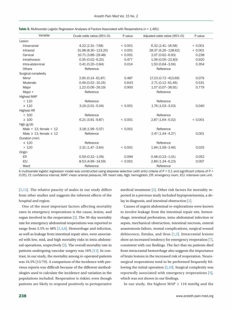
[3,12]. The relative paucity of males in our study differs
from other studies and suggests the inherent effects of the
hospital and region.
One of the most important factors affecting mortality
rates in emergency reoperations is the cause, lesion, and
organ involved in the reoperation [2]. The 30-day mortality
rate for emergency abdominal reoperations was reported to
range from 5.5% to 48% [1,3,6]. Hemorrhage and infection,
as well as leakage from intestinal repair sites, were associat-
ed with low, mid, and high mortality risks in intra-abdomi-
nal operations, respectively [2]. The overall mortality rate in
patients undergoing vascular surgery was 16% [11]. In con-
trast, in our study, the mortality among re-operated patients
was 16.5% (13/79). A comparison of the incidence with pre-
vious reports was difficult because of the different method-
ologies used to calculate the incidence and variation in the
populations included. Reoperation is riskier, even though
patients are likely to respond positively to perioperative
medical treatment [2]. Other risk factors for mortality re-
ported in a previous study included hypoproteinemia, a de-
lay in diagnosis, and intestinal obstruction [1].
Causes of urgent abdominal re-explorations were known
to involve leakage from the intestinal repair site, hemor-
rhage, intestinal perforation, intra-abdominal infection or
sepsis, mechanical obstruction, intestinal necrosis, enteral
anastomosis failure, stomal complications, surgical wound
dehiscence, fistulas, and ileus [1,2]. Intracranial lesions
show an increased tendency for emergency reoperation [7],
consistent with our findings. The fact that six patients died
from intracranial hemorrhage also suggests the importance
of brain lesions in the increased risk of reoperation. Neuro-
surgical reoperations tend to be performed frequently fol-
lowing the initial operation [2,10]. Surgical complexity was
reportedly associated with emergency reoperations [3],
which was not shown in our findings.
In our study, the highest MAP ≥ 110 mmHg and the
Table 5. Multivariate Logistic Regression Analyses of Factors Associated with Reoperations (n = 1,481)
Variable Crude odds ratios (95% CI) P value Adjusted odds ratios (95% CI) P value
Lesion Intracranial 4.22 (2.31–7.68) < 0.001 6.32 (2.41–16.58) < 0.001 Intraoral 31.98 (8.30–123.20) < 0.001 28.37 (6.26–128.62) < 0.001 Cervical 10.71 (3.89–29.48) < 0.001 2.07 (0.62–6.93) 0.238 Intrathoracic 0.35 (0.02–6.25) 0.477 1.06 (0.05–22.83) 0.920 Intra-abdominal 0.41 (0.20–0.84) 0.014 1.50 (0.64–3.56) 0.354 Others Reference ReferenceSurgical complexity Minor 2.95 (0.14–61.87) 0.487 17.23 (0.72–413.69) 0.079 Moderate 0.49 (0.02–10.24) 0.643 2.71 (0.12–61.46) 0.531 Major 1.22 (0.06–26.19) 0.900 1.57 (0.07–36.91) 0.779 Major + Reference ReferenceHighest MAP < 110 Reference Reference ≥ 110 3.19 (2.01–5.04) < 0.001 1.76 (1.03–3.03) 0.040Highest HR < 100 Reference Reference ≥ 100 6.21 (3.91–9.87) < 0.001 2.87 (1.64–5.02) < 0.001Hgb (g/dl) Male < 13, female < 12 3.18 (1.99–5.07) < 0.001 Reference Male ≥ 13, female ≥ 12 Reference 2.47 (1.44–4.27) 0.001Duration (min) ≤ 120 Reference Reference > 120 2.31 (1.47–3.64) < 0.001 1.94 (1.09–3.46) 0.025Origin ER 0.59 (0.32–1.09) 0.094 0.48 (0.23–1.01) 0.052 ICU 8.53 (4.99–14.59) < 0.001 2.89 (1.34–6.23) 0.007 Ward Reference Reference
A multivariate logistic regression model was constructed using stepwise selection (with entry criteria of P < 0.1 and significant criteria of P < 0.05). CI: confidence interval, MAP: mean arterial pressure, HR: heart rate, Hgb: hemoglobin, ER: emergency room, ICU: intensive care unit.
238 www.anesth-pain-med.org
Anesth Pain Med Vol. 15 No. 2

General
highest HR ≥ 100 beats/min were related to emergency re-
operations, which indirectly reflected high hemodynamic
instability. Hypertension may be due to Cushing reflex in
intracranial problems, the use of inotropics, and psycho-
logical stress. Shock and hemorrhage requiring blood
transfusion increase patients’ susceptibility to postopera-
tive mortality in emergency surgeries [9]. Tachycardia is
common in these patients and suggests unstable intravas-
cular volume [1,6]. These patients are usually anemic [1]. A
lower hemoglobin concentration is associated with a high-
er risk of reoperation [10], consistent with our findings, in-
directly suggesting coagulation deficiency and poor nutri-
tion. If hemorrhage is associated with severe anemia or se-
vere hypotension, the mortality rate increases proportion-
ately [16].
The possibility of effectively lowering the mortality rate
depends on the success of the first operation [1,2]. There-
fore, a number of these problems can be avoided by taking
all precautions possible at the time of the initial operation
to minimize the occurrence of complications [1]. For ex-
ample, a majority of the hemorrhages were caused by tech-
nical errors, such as inadequate hemostasis during the first
operation [2]. Surgical challenges include inappropriate
judgment, poorly developed skills, and incorrect handling
of surgical devices [16]. Half of the reoperations were mi-
nor surgeries in the present study, suggesting possible
technical errors during the initial operation. In our study,
patients undergoing emergency reoperations were mostly
transferred from the ICU. This reflects the urgency of most
reoperation cases and the grave physical status of these pa-
tients.
This study had several limitations. First, our research was
limited by the size of the study groups. This study was
based on data obtained from a single institution and sur-
geries of short duration (70.0% were less than two hours)
and involved minor-to-moderate types of surgery (83.9%).
Therefore, the results may differ from those involving larg-
er populations and multiple institutions, such as regional
emergency trauma centers. Our institution is a universi-
ty-affiliated community hospital. Institutional factors may
limit generalizability. Second, the variables in our investi-
gation may be too diverse to perform a comprehensive
analysis and exclude the role of unmeasured variables. The
variety of diseases and operations included did not allow a
clear distinction between the effects of reoperation on pa-
tient outcomes. The results should be based on analyses
according to individual departments and procedures in the
future. The selection of abnormal variables, including vital
signs, was defined arbitrarily and these aspects may have
affected our results. Third, we could not analyze the anes-
thetic implications for postoperative mortality as an end-
point due to the small number of mortalities. Lastly, varia-
tions in surgical procedures due to the surgeon’s experi-
ence, operative devices, and methods could not be ana-
lyzed in a meaningful way and were beyond the scope of
the study.
In summary, our results described the clinical features of
emergency reoperations. The factors associated with a
higher proportion of emergency reoperations were intra-
cranial and intraoral lesions, highest MAP ≥ 110 mmHg
and HR ≥ 100 beats/min, anemia, and arrival from the
ICU. Emergency anesthesia is an important segment of
clinical anesthesiology and advances in anesthetic care af-
fect mortality. Particular attention must be paid to emer-
gency reoperation and more time and resources are re-
quired to redefine the clinical features of emergency reop-
eration.
CONFLICTS OF INTEREST
No potential conflict of interest relevant to this article
was reported.
AUTHOR CONTRIBUTIONS
Conceptualization: Jun Rho Yoon. Data acquisition: Jun
Rho Yoon, Kyoung Rim Kim, Taehee Kim. Formal analysis:
Tae Kwan Kim. Funding: Ui Jin Park. Supervision: Jun Rho
Yoon. Writing—original draft: Jun Rho Yoon. Writing—re-
view & editing: Yu Na Choi, Tae Kwan Kim.
ORCID
Tae Kwan Kim, https://orcid.org/0000-0002-0635-3304
Jun Rho Yoon, https://orcid.org/0000-0001-7457-7433
Yu Na Choi, https://orcid.org/0000-0001-9111-8298
Ui Jin Park, https://orcid.org/0000-0003-2114-0195
Kyoung Rim Kim, https://orcid.org/0000-0001-5442-8705
Taehee Kim, https://orcid.org/0000-0002-0476-3254
REFERENCES
1. Wain MO, Sykes PA. Emergency abdominal re-exploration in a
district general hospital. Ann R Coll Surg Engl 1987; 69: 169-74.
www.anesth-pain-med.org 239
Emergency reoperation

2. Unalp HR, Kamer E, Kar H, Bal A, Peskersoy M, Ali Onal M. Ur-
gent abdominal re-explorations. World J Emerg Surg 2006; 1: 10.
3. Guevara OA, Rubio-Romero JA, Ruiz-Parra AI. Unplanned re-
operations: is emergency surgery a risk factor? A cohort study. J
Surg Res 2013; 182: 11-6.
4. Tera H, Aberg C. Relaparotomy. A ten-year series. Acta Chir
Scand 1975; 141: 637-44.
5. O’Leary DP, Hardwick RH, Cosford E, Knox AJ. Does hospital
mortality rate reflect quality of care on a surgical unit? Ann R
Coll Surg Engl 1997; 79: 46-8.
6. Harbrecht PJ, Garrison RN, Fry DE. Early urgent relaparotomy.
Arch Surg 1984; 119: 369-74.
7. Copeland GP. The POSSUM system of surgical audit. Arch Surg
2002; 137: 15-9.
8. Donati A, Ruzzi M, Adrario E, Pelaia P, Coluzzi F, Gabbanelli V,
et al. A new and feasible model for predicting operative risk. Br
J Anaesth 2004; 93: 393-9.
9. Matsuyama T, Iranami H, Fujii K, Inoue M, Nakagawa R,
Kawashima K. Risk factors for postoperative mortality and mor-
bidities in emergency surgeries. J Anesth 2013; 27: 838-43.
10. Kwinta BM, Krzyżewski RM, Kliś KM, Donicz P, Gackowska M,
Polak J, et al. Emergency reoperations in cranial neurosurgery.
World Neurosurg 2017; 105: 749-54.
11. Davies AH, Pope I, Collin J, Morris PJ. Early reoperation after
major vascular surgery: a four-year prospective analysis. Br J
Surg 1992; 79: 76-8.
12. Brant JA, Bur AM, Chai R, Hatten K, Nicolli EA, Fischer JP, et al.
Reoperation following adult tonsillectomy: review of the Ameri-
can College of Surgeons National Surgical Quality Improvement
Program. Otolaryngol Head Neck Surg 2016; 154: 779-84.
13. Ramachandran R, Hegde T. Chronic subdural hemato-
mas--causes of morbidity and mortality. Surg Neurol 2007; 67:
367-72.
14. Lee DI, Lee MH, Shin OY, Shin KY. A clinical study of anesthesia
for emergency surgery. Korean J Anesthesiol 1979; 12: 252-60.
15. Ching SS, Muralikrishnan VP, Whiteley GS. Relaparotomy: a five-
year review of indications and outcome. Int J Clin Pract 2003;
57: 333-7.
16. Irita K. Risk and crisis management in intraoperative hemor-
rhage: human factors in hemorrhagic critical events. Korean J
Anesthesiol 2011; 60: 151-60.
240 www.anesth-pain-med.org
Anesth Pain Med Vol. 15 No. 2

General
Correlation between patient health questionnaire-2 and postoperative pain in laparoscopic cholecystectomy
Yusom Shin, Tae Woo Park, Huiyoung Kim, Dong-jin Shim, Hochul Lee, Joo-Duck Kim, and Donghee Kang
Department of Anesthesiology and Pain Medicine, Kosin University College of Medicine, Busan, Korea
INTRODUCTION
Postoperative pain causes patients discomfort and vaso-
constriction through sympathetic hyperactivity, which may
decelerate wound healing and increase the infection rate
[1,2]. Therefore, appropriate pain control is important.
Proper postoperative pain control reduces morbidity and
Clinical ResearchAnesth Pain Med 2020;15:241-246 https://doi.org/10.17085/apm.2020.15.2.241pISSN 1975-5171 • eISSN 2383-7977
Received July 24, 2019Revised September 25, 2019Accepted October 23, 2019
Corresponding author Donghee Kang, M.D. Department of Anesthesiology and Pain Medicine, Kosin University College of Medicine, 262 Gamcheon-ro, Seo-gu, Busan 49267, Korea Tel: 82-51-990-6281Fax: 82-51-254-2504E-mail: [email protected]
Background: Postoperative pain is affected by preoperative depression. If the risk of post-operative pain associated with depression can be predicted preoperatively, anesthesiolo-gists and/or surgeons can better manage it with personalized care. The objective of this study was to determine the efficacy of Patient Health Questionnaire-2 (PHQ-2) depression screening tool as a predictor of postoperative pain.
Methods: A total of 50 patients scheduled for elective laparoscopic cholecystectomy with an American Society of Anesthesiologists physical status 1 or 2 were enrolled. They an-swered the PHQ-2, which consists of two questions, under the supervision of a researcher on the day before the surgery. The numerical rating scale (NRS) scores were assessed at post-anesthesia care unit (PACU), at 24, and 48 postoperative hours, and the amount of in-travenous patient-controlled analgesia (IV-PCA) administered was documented at 24, 48, and 72 postoperative hours. At 72 h, the IV-PCA device was removed and the final dosage was recorded.
Results: The NRS score in PACU was not significantly associated with the PHQ-2 score (cor-relation coefficients: 0.13 [P = 0.367]). However, the use of analgesics after surgery was higher in patients with PHQ-2 score of 3 or more (correlation coefficients: 0.33 [P = 0.018]).
Conclusions: We observed a correlation between the PHQ-2 score and postoperative pain. Therefore, PHQ-2 could be useful as a screening test for preoperative depression. Particular-ly, when 3 points were used as the cut-off score, the PHQ-2 score was associated with the dosage of analgesics, and the analgesic demand could be expected to be high with higher PHQ-2 scores.
Keywords: Analgesics; Depression; Patient health questionnaire; Postoperative pain.
shortens hospitalization and convalescence [3]. Several
factors influence postoperative pain, but depression may
significantly heighten its intensity [4,5].
The prevalence of depression is increasing in most coun-
tries, including Korea, where the lifetime and annual prev-
alence are 6.7% and 3.0% [6]. To screen patients with de-
pression, many measures, such as the Patient Health Ques-
This is an Open Access article distributed under the terms of the Creative Commons Attribution Non-Commercial License (http://creativecommons.org/licenses/by-nc/4.0) which permits unrestricted non-commercial use, distribution, and reproduction in any medium, provided the original work is properly cited.Copyright © the Korean Society of Anesthesiologists, 2020
241

tionnaire-9 (PHQ-9) [7], Center for Epidemiological Studies
Depression Scale [8], Beck Depression Inventory [9], and
Geriatric Depression Scale [10], have been developed.
However, 30–50% of depressed patients are misdiagnosed
due to difficulties in diagnosis [11,12].
Postoperative pain is associated with depression, there-
fore screening for preoperative depression might help with
better management of postoperative pain [4,5]. However,
screening for depression in pre-anesthetic assessments is
difficult, as it is time-consuming. Moreover, depression
screening is difficult for anesthesiologists, as it generally
requires professional knowledge. To screen for depression
in pre-anesthetic assessments, the screening test should be
short, with high reliability and validity. Therefore, PHQ-2
could be an appropriate option, since it evaluates patients
using only two of the nine questions of PHQ-9, which is the
conventional depression screening test [13].
In this study, we evaluated the usefulness of the PHQ-2
test for preoperative depression evaluation. If there was a
correlation between preoperative PHQ-2 and postopera-
tive pain, we considered that PHQ-2 could be used to pre-
dict postoperative pain without a definite diagnosis of de-
pression.
MATERIALS AND METHODS
This study was approved by our committee for Clinical
Research Ethics (no. KUGH 2017-08-033-006). A total of 55
patients participated in this study. The correlation coeffi-
cient of the study was determined to be 0.4, which can be
interpreted as a fair result between 0.4 and 0.6, referring to
the previous study [14]. The minimum number of samples
was 47 when α = 0.05, α = 0.20, and correlation coefficient
is 0.4, and the study was conducted in 55 patients consid-
ering the losers. Patients identified as American Society of
Anesthesiologist physical status class I–II were scheduled
for laparoscopic cholecystectomy under general anesthe-
sia. Patients with a history of depression, current medica-
tion, or complaint of post-anesthetic nausea and vomiting
in previous surgeries were excluded from this study.
An anesthesiologist, who did not have prior information
regarding the patient, visited the ward to examine the
PHQ-2 scores of the patients who were hospitalized a day
before the operation. Subsequently, he explained the use
of intravenous patient-controlled analgesia (IV-PCA).
There were two types of PHQ-2: Short-answer PHQ-2 and
score-type PHQ-2. In this study, score-type PHQ-2 was
used. The PHQ-2 questionnaire consisted of two items:
“Little interest or pleasure in doing things” and “ Feeling
down, depressed, or hopeless.” The available answers were:
“Not at all,” “Several days,” “More than half the days,” and
“Nearly every day.” Each question was scored on a scale of
0–3, and the scores from both questions were added. The
patient’s medical records (age, height, weight, and history
of psychological disorder) were also obtained.
Pre-anesthetic medication was not administered. Propo-
fol (1 mg/kg) and remifentanil (1 μg/kg) were used to in-
duce anesthesia. Anesthesia was maintained using sevoflu-
rane and remifentanil, and remifentanil was discontinued
during extubation. In the recovery room, fentanyl 50 μg
was administered in the loading dose and IV-PCA was con-
nected. The patient was then told to press the button of the
IV-PCA directly if there was pain. An IV-PCA device was
used with Hospira Gemstar Blue (GemStarTM, Hospira Inc.,
USA). We mixed 1,000 μg of fentanyl (fentanyl citrate, Hana
Pharm, Korea), 50 mg of nefopam (nefopam HCl, Ilsung
Pharm, Korea), and 4 mg of ondansetron to obtain a total
volume of 150 ml. When the bolus button was pressed, 1.5
ml (10 μg of fentanyl) of the drug mixture was injected. The
lock time was 5 min.
The degree of postoperative pain on a numerical rating
scale (NRS) and the usage amount of IV-PCA were as-
sessed. The NRS scores were assessed at PACU, 24, and 48
postoperative hours. Usage amount of IV-PCA was collect-
ed at 24, 48, and 72 postoperative hours. At 72 h, the IV-
PCA device was removed. When a patient complained of
nausea or vomited while using IV-PCA, 4 mg of ondanse-
tron was additionally administered. When the complaint
sustained, IV-PCA was discontinued.
A statistical analysis was performed using the SPSS (ver-
sion 24.0, IBM, USA) statistical program. To analyze the
correlations among the PHQ-2 score, NRS score, and fen-
tanyl use, point-biserial correlation or Pearson correlation
analyses were used. Quantitative variables were expressed
as mean ± SD, and P < 0.05 was the significance level of
the test.
RESULTS
A total of 55 patients who underwent laparoscopic chole-
cystectomy under general anesthesia were enrolled in this
study, of which 8 complained of postoperative nausea and
received 4 mg of ondansetron intravenously. Out of the 8
patients, 5 complained of nausea and discontinued IV-PCA
242 www.anesth-pain-med.org
Anesth Pain Med Vol. 15 No. 2

General
due to uncontrolled symptoms after a single administra-
tion of 4 mg of ondansetron and were excluded from the
study. Finally, we analyzed 50 patients (Fig. 1). Table 1
shows the demographic characteristics. Side effects other
than nausea did not occur. In this study, the PHQ-2 was
conducted for about two minutes, and no patient refused
the survey.
Postoperative NRS in recovery room and total fentanyl
usage were not significantly associated with the patient’s
PHQ-2 score (correlation coefficients: 0.13 [P = 0.367] and
0.24 [P = 0.098], Table 2). When three points on PHQ-2 was
used as the cut-off score, the fentanyl use was significantly
higher in patients who scored ≥ 3 points than in those
Fig. 1. A study flow chart. IV-PCA: intravenous patient-controlled analgesia.
55 patients meetinginclusion criteria
47 patients had no complications
Analyzed (n = 50) Excluded from thestudy
3 patients had improved symptoms
8 patients complained postoperative nausea
4 mg of the ondansetron was
injected intravenously
5 patients discontinued IV-PCA due to
uncontrolled symptom
Table 1. Characteristics of Patients
Variable Value
Sex (M/F) 28/22Age (yr) 50.9 ± 10.8Weight (kg) 66.3 ± 13.6ASA physical status I/II 1938-12-01Preoperative PHQ-2 score 0 24 (48) 1 10 (20) 2 5 (10) 3 6 (12) 4 5 (10) 5 0 (0) 6 0 (0)Remifentanil total dose (μg)* 450.9 ± 57.1Fentanyl total dose (μg)†
PHQ-2 score ≥ 3 371.6 ± 71.5 PHQ-2 score < 3 300.3 ± 88.6Duration of anesthesia (min) 63.3 ± 13.7
Values are presented as number (%) or mean ± SD. ASA: American Society of Anesthesiologists, PHQ-2: Patient Health Questionnaire-2, IV-PCA: intravenous patient-controlled analgesia. *Total amount of remifentanil during surgery. †Total amount of fentanyl in IV-PCA. †Total amount of fentanyl in IV-PCA.
Table 2. Coefficient of Point-biserial Correlation or Pearson Correlations between Demographic Data/PHQ-2 Score and NRS Score/Fentanyl Consumption
Variable NRS scorein PACU
Fentanyl consumption
POD1 POD2 POD3
Sex (M/F) –0.25 –0.69Age (yr) –0.12 –0.34*PHQ-2 score 0.13 0.68† 0.45* 0.24
PHQ-2: Patient Health Questionnaire-2, NRS: numerical rating scale, POD: post-operative day, PACU: post-anesthesia care unit. POD3 means total fentanyl consumption. *P < 0.05, †P < 0.001.
Fig. 2. Relationship between preoperative Patient Health Questionnaire-2 (PHQ-2) score and analgesic consumption on the first day after surgery (correlation coefficient r = 0.68 [P < 0.001]).
scoring < 3 points (371.6 ± 71.5 vs. 300.3 ± 88.6), and total
fentanyl usage correlated with the PHQ-2 score (correla-
tion coefficients: 0.33 [P = 0.018]). Fentanyl usage at 24
and 48 postoperative hours were significantly associated
with the PHQ-2 score (correlation coefficients: 0.68 [P <
0.001] and 0.45 [P = 0.010], Table 2). Figs. 2, 3, and 4 show
the relationship between PHQ-2 score and analgesic con-
sumption on first, second, and third day after surgery. Age
was associated with total fentanyl usage but sex was not
(correlation coefficients: –0.34 [P = 0.016] and –0.69 [P =
PHQ-2
0 1 2 3 4
80
70
60
50
40
30
20
10
0
Anal
gesi
c co
nsum
ptio
n (μ
g)
www.anesth-pain-med.org 243
Correlation between patient health questionnaire-2 and postoperative pain

cases of ear, nose, and neck surgery. These differences are
probably because of differences in the type of surgery and
type of postoperative analgesics used.
Patients who were included in this study underwent lap-
aroscopic cholecystectomy. The surgery was less painful
compared to open cholecystectomy, and IV-PCA with fen-
tanyl controlled postoperative pain appropriately [19]. Im-
mediately after the surgery, patients with higher PHQ-2
score had higher analgesic requirements. However, as the
postoperative pain was controlled with a strong opioid, the
appropriate pain control effect might have been achieved
regardless of the PHQ-2 score, and it is believed that there
was no difference in usage.
PHQ-2 scores did not significantly correlate with total
fentanyl usage. However, after selecting the appropriate
cut-off score for PHQ-2, the PHQ-2 scores were associated
with total analgesic requirement. The sensitivity and speci-
ficity of depression were 91.9% and 100%, respectively,
when the cut-off score on PHQ-2 was 3 [15]. In this study,
when 3 points were used as cut-off score, fentanyl use was
significantly higher in patients who scored 3 or more
points. Therefore, 3 points as a cut-off score on PHQ-2 may
be appropriate for preoperative depression screening.
In this study, sex did not affect analgesic use (correlation
coefficient: –0.69 [P = 0.640]). In another study on ana-
lyzed factors affecting postoperative pain, sex did not affect
the postoperative pain or analgesic use [4]. However, age
was a major predictor of postoperative pain and analgesic
use [4]. In this study, the use of analgesics was higher in
younger patients (correlation coefficient : –0.34 [P =
0.016]).
0.640], Table 2).
DISCUSSION
The purpose of this study was to evaluate the correlation
between preoperative PHQ-2 score and postoperative pain.
PHQ-2 is a simple screening test using two of the nine ques-
tions of PHQ-9, a previously developed depression screening
test [13]. The two questions addressed depressed mood and
anorexia. There is a significant correlation with other screen-
ing tests, and PHQ-2 is known to increase the total score de-
pending on the severity of depression in the patient group
[15].
In this study, the preoperative PHQ-2 scores did not sig-
nificantly correlate with the postoperative NRS scores mea-
sured in the PACU or the total fentanyl usage (correlation
coefficients: 0.13 [P = 0.367] and 0.24 [P = 0.098]). Howev-
er, fentanyl usage during 24 postoperative hours highly
correlated with the preoperative PHQ-2 scores (0.68 [P <
0.001]). Various studies on the association between post-
operative pain and preoperative depression have been
conducted. De Cosmo et al. [14] reported that depression
was associated with tramadol use in patients after laparo-
scopic cholecystectomy. Schade et al. [16] reported that
preoperative depression affects postoperative pain and re-
habilitation in patients with lumbar disc herniation. Oz-
tekin et al. [17] reported that anterior cruciate ligament
surgery was not associated with postoperative pain despite
high preoperative depression scores in the patients. Kavak-
ci et al. [18] found that preoperative anxiety has a greater
impact on postoperative pain compared to depression in
Fig. 3. Relationship between preoperative Patient Health Questionnaire-2 (PHQ-2) score and analgesic consumption on the second day after surgery (correlation coefficient r = 0.45 [P = 0.010]).
Fig. 4. Relationship between preoperative Patient Health Questionnaire-2 (PHQ-2) score and analgesic consumption on the third day after surgery (correlation coefficient r = 0.24 [P = 0.098]).
PHQ-2
0 1 2 3 4
80
70
60
50
40
30
20
10
0
Anal
gesi
c co
nsum
ptio
n (μ
g)
PHQ-2
1 2 3 4
80
70
60
50
40
30
20
10
0
Anal
gesi
c co
nsum
ptio
n (μ
g)
244 www.anesth-pain-med.org
Anesth Pain Med Vol. 15 No. 2

General
This study has a few limitations. First, anxiety was not in-
vestigated. Anxiety plays a major role in acute pain, includ-
ing postoperative pain [18]. Brander et al. [20] reported that
depression had a greater effect on postoperative chronic
pain than anxiety. Patients’ anxiety before surgery should
have been examined to confirm its association with pain.
Second, the diagnosis of depression was not an inclusion
criterion. Therefore, in a controlled study on patients who
were diagnosed or treated for depressive disorders, we in-
tend to assess the efficacy of PHQ-2 as a predictor of post-
operative pain. Marazziti et al. [21] reported that in the
group with depression, the pain threshold was lower than
in the group without depression. Depressed patients who
had lower thresholds for postoperative pain were believed
to have higher analgesic requirements and more negative
effects of pain. Future studies could evaluate pain control
or patient satisfaction when controlling postoperative pain
more aggressively by increasing the amount of pain medi-
cation IV-PCA in patients with high PHQ-2 scores.
In conclusion, the PHQ-2 test is composed of two simple
questions and can be performed in two minutes; therefore,
it could be useful as a screening test for preoperative de-
pression. Despite the simple questions, it highly correlated
with fentanyl consumption in the early postoperative peri-
od. Particularly, when 3 points were used as the cut-off
score, the PHQ-2 score was associated with the dosage of
analgesics, and the analgesic demand could be expected to
be high with higher PHQ-2 scores.
CONFLICTS OF INTEREST
No potential conflict of interest relevant to this article
was reported.
AUTHOR CONTRIBUTIONS
Conceptualization: Yusom Shin. Data acquisition: Tae
Woo Park, Dong-jin Shim. Formal analysis: Huiyoung Kim.
Supervision: Joo-Duck Kim, Donghee Kang. Writing—orig-
inal draft: Yusom Shin, Tae Woo Park. Writing—review &
editing: Hochul Lee, Donghee Kang.
ORCID
Yusom Shin, https://orcid.org/0000-0002-4829-0866
Tae Woo Park, https://orcid.org/0000-0003-4032-4424
Huiyoung Kim, https://orcid.org/0000-0003-2330-1458
Dong-jin Shim, https://orcid.org/0000-0002-8704-529X
Hochul Lee, https://orcid.org/0000-0002-7740-9424
Joo-Duck Kim, https://orcid.org/0000-0002-9236-5183
Donghee Kang, https://orcid.org/0000-0001-6614-9244
REFERENCES
1. Kiecolt-Glaser JK, Page GG, Marucha PT, MacCallum RC, Gla-
ser R. Psychological influences on surgical recovery. Perspec-
tives from psychoneuroimmunology. Am Psychol 1998; 53:
1209-18.
2. Derbyshire DR, Smith G. Sympathoadrenal responses to an-
aesthesia and surgery. Br J Anaesth 1984; 56: 725-39.
3. Kehlet H, Holte K. Effect of postoperative analgesia on surgical
outcome. Br J Anaesth 2001; 87: 62-72.
4. Ip HYV, Abrishami A, Peng PW, Wong J, Chung F. Predictors of
postoperative pain and analgesic consumption: a qualitative
systematic review. Anesthesiology 2009; 111: 657-77.
5. Vaughn F, Wichowski H, Bosworth G. Does preoperative anxi-
ety level predict postoperative pain? AORN J 2007; 85: 589-604.
6. Ahn J. Depression, suicide, and Korean society. J Korean Med
Assoc 2012; 55: 320-1.
7. Spitzer RL, Kroenke K, Williams JBW; Patient Health Question-
naire Primary Care Group. Validation and utility of a self-report
version of PRIME-MD: the PHQ primary care study. JAMA
1999; 282: 1737-44.
8. Radloff LS. The CES-D scale: a self-report depression scale for
research in the general population. Appl Psychol Meas 1977; 1:
385-401.
9. Beck AT, Guth D, Steer RA, Ball R. Screening for major depres-
sion disorders in medical inpatients with the Beck Depression
Inventory for Primary Care. Behav Res Ther 1997; 35: 785-91.
10. Yesavage JA, Brink TL, Rose TL, Lum O, Huang V, Adey M, et al.
Development and validation of a geriatric depression screen-
ing scale: a preliminary report. J Psychiatr Res 1982; 17: 37-49.
11. Gerber PD, Barrett J, Barrett J, Manheimer E, Whiting R, Smith
R. Recognition of depression by internists in primary care: a
comparison of internist and “gold standard” psychiatric as-
sessments. J Gen Intern Med 1989; 4: 7-13.
12. Susman JL, Crabtree BF, Essink G. Depression in rural family
practice: easy to recognize, difficult to diagnose. Arch Fam Med
1995; 4: 427-31.
13. Tuck AN, Scribani MB, Grainger SD, Johns CA, Knight RQ. The
9-Item Patient Health Questionnaire (PHQ-9): an aid to assess-
ment of patient-reported functional outcomes after spinal sur-
gery. Spine J 2018; 18: 1398-405.
14. De Cosmo G, Congedo E, Lai C, Primieri P, Dottarelli A, Aceto P.
Preoperative psychologic and demographic predictors of pain
perception and tramadol consumption using intravenous pa-
www.anesth-pain-med.org 245
Correlation between patient health questionnaire-2 and postoperative pain

tient-controlled analgesia. Clin J Pain 2008; 24: 399-405.
15. Shin JH, Kim HC, Jung CH, Kim JB, Jung SW, Cho HJ, et al. The
Standardization of the Korean Version of the Patient Health
Questionnaire-2. J Korean Neuropsychiatr Assoc 2013; 52: 115-
21.
16. Schade V, Semmer N, Main CJ, Hora J, Boos N. The impact of
clinical, morphological, psychosocial and work-related factors
on the outcome of lumbar discectomy. Pain 1999; 80: 239-49.
17. Oztekin HH, Boya H, Ozcan O, Zeren B, Pinar P. Pain and affec-
tive distress before and after ACL surgery: a comparison of am-
ateur and professional male soccer players in the early postop-
erative period. Knee 2008; 15: 368-72.
18. Kavakci Ö, Altuntas EE, Müderris S, Kugu N. Effects of the pre-
operative anxiety and depression on the postoperative pain in
ear, nose and throat surgery. Indian J Otol 2012; 18: 82-7.
19. Kang DH, Kim DS, Kim JD, Kim JW. A comparison of fentanyl
and morphine for patient controlled analgesia after laparo-
scopic cholecystectomy. Anesth Pain Med 2013; 8: 21-5.
20. Brander VA, Stulberg SD, Adams AD, Harden RN, Bruehl S, Sta-
nos SP, et al. Predicting total knee replacement pain: a pro-
spective, observational study. Clin Orthop Relat Res 2003;
(416): 27-36.
21. Marazziti D, Castrogiovanni P, Rossi A, Rosa C, Ghione S, Di
Muro A, et al. Pain threshold is reduced in depression. Int J
Neuropsychopharmacol 1998; 1: 45-8.
246 www.anesth-pain-med.org
Anesth Pain Med Vol. 15 No. 2

General
Submental intubation is commonly performed during
general anesthesia for maxillofacial surgeries, especially in
cases where nasotracheal intubation can cause potential
complications, including epistaxis, accidental cranial or in-
tracranial intubation, and meningitis [1–5]. Furthermore,
the presence of comminuted midface or nasoethmoidal
complex fractures in maxillofacial injuries may lead to the
luminal occlusion of the nasotracheal tube by nasal turbi-
nates, mucous, blood clots. While nasotracheal intubation
interferes with the surgical approach during maxillofacial
surgeries, submental intubation provides undisturbed and
safe surgical access [4,5]. Moreover, it can be reversed after
surgery, allowing easy extubation of the patient. In addi-
tion, this technique avoids the potentially serious compli-
cations of surgical tracheostomy [6–8]. Therefore, submen-
tal intubation is widely used in maxillofacial surgeries.
Novel alternative for submental intubation - A case report -
Inyoung Jung, Byung Hoon Yoo, Ji Youn Ju, Sijin Choi, Jun Heum Yon, Kye-Min Kim, Yun-Hee Lim, and Woo Yong Lee
Department of Anesthesiology and Pain Medicine, Sanggye Paik Hospital, Inje University
College of Medicine, Seoul, Korea
Case Report Anesth Pain Med 2020;15:247-250 https://doi.org/10.17085/apm.2020.15.2.247pISSN 1975-5171 • eISSN 2383-7977
Received July 24, 2019Revised September 24, 2019Accepted September 25, 2019
Corresponding author Byung Hoon Yoo, M.D., Ph.D. Department of Anesthesiology and Pain Medicine, Sanggye Paik Hospital, Inje University College of Medicine, 1342 Dongil-ro, Nowon-gu, Seoul 01757, Korea Tel: 82-2-950-1170Fax: 82-2-950-1323E-mail: [email protected]
Background: Submental intubation is commonly used during general anesthesia for maxil-lofacial surgeries as it provides a safe unrestricted surgical access compared to tracheosto-my. During submental intubation, soft tissues and blood clots can become lodged in the en-dotracheal tube. To overcome this problem, we used a laparoscopic trocar.
Case: A 52-year-old male with maxillofacial injury was scheduled to undergo an open reduc-tion and internal fixation. We performed submental intubation using laparoscopic trocar, which created sufficient space for the insertion of the endotracheal tube. Unlike convention-al methods, our method did not require any blunt dissection and caused significantly less soft tissue damage and required significantly less time.
Conclusions: Submental intubation with laparoscopic trocar is a one-step method and is quick and easy-to-perform technique with less complications.
Keywords: Airway management; Intubation, intratracheal; Maxillofacial injuries; Trocar.
There are several modifications of this technique, includ-
ing the Seldinger technique and use of percutaneous tra-
cheostomy dilatation kit [6,7]. However, these modifica-
tions are associated with potential problems, such as oc-
clusion of endotracheal tube by soft tissues and blood
clots. Therefore, we attempted to identify a more effective
way of submental intubation. A laparoscopic trocar con-
sists of trocar sleeve and trocar needle. After creation of a
passage with trocar in the desired position, the trocar nee-
dle is removed and the instrument is inserted subsequently
through the trocar sleeve.
We performed submental intubation by using a laparo-
scopic trocar in making a submental passage. The trocar
was then used as a passage for inserting the endotracheal
tube. The use of a laparoscopic trocar reduced trauma and
intubation time.
This is an Open Access article distributed under the terms of the Creative Commons Attribution Non-Commercial License (http://creativecommons.org/licenses/by-nc/4.0) which permits unrestricted non-commercial use, distribution, and reproduction in any medium, provided the original work is properly cited.Copyright © the Korean Society of Anesthesiologists, 2020
247

CASE REPORT
Informed consent was obtained from the patient for the
publication of this report.
A 52-year-old male with fractures of the zygomaticomax-
illary, zygomatic arch, and right inferior orbital floor (Lefort
II fracture) was scheduled to undergo open reduction and
internal fixation. For this patient, instead of conventional
nasotracheal intubation, we planned a submental intuba-
tion because nasotracheal intubation in patients with Lefort II
or III fracture can lead to potential complications, such as intra-
cranial intubation, meningitis, and the luminal occlusion of en-
dotracheal tube by nasal turbinates, mucous, and blood clots.
However, even submental intubation has several complications
associated with its use. To overcome these problems, we used a
12-mm laparoscopic trocar (Laport, Korea) for pulling out the
endotracheal tube from the oral cavity to the submental area
during submental intubation. Before the procedure, we con-
firmed that the armored endotracheal tube (internal diameter,
7.5 mm) could pass through the trocar. After removal of the
connector of the armored tube, the endotracheal tube was
passed through the trocar's 12-mm diameter internal lumen
(Fig. 1).
After standard orotracheal intubation, the tongue was re-
tracted to expose the floor of the mouth. After making a
1.5-cm incision, the 12-mm laparoscopic trocar was passed
in the skin-to-oral direction. After ventilating the patient
with 100% oxygen for several minutes, the tracheal tube
was briefly disconnected from the breathing circuit. The
connector of the armored tube was removed and the proxi-
mal part of the tube was pulled through the passage in the
lumen of the trocar. Thereafter, the trocar was removed.
The connector was reattached and the tube was reconnect-
ed to the breathing circuit (Fig. 2). The position of the tra-
cheal tube was checked using capnography and chest aus-
cultation, and the distance marking on the endotracheal
tube at the exit site on the skin was carefully noted. The
tube was then secured to the skin of the submental region
with a heavy (2⁄0) black silk suture. The surgery was suc-
cessful and without any adverse effects. After completion
of the surgery, the abovementioned procedures were re-
versed. The skin sutures were cut, and the tracheal tube
was briefly disconnected from the breathing circuit. There-
after, the connector was removed and the tube was pulled
back through the passage in the floor of the mouth. The
connection was then reestablished and the tube was se-
cured. The submental incision was closed using three
monofilament skin sutures, which were removed 5–7 days
postoperatively. No attempt was made to close the oral defect.
The use of a laparoscopic trocar for submental intuba-
tion significantly reduced the inflow of soft tissues and
blood into the endotracheal tube. This method was much
easier than the passing the tube from the oral cavity to the
submental incision site.
DISCUSSION
Submental intubation is an effective alternative for air-
way management in select patients undergoing surgery for
maxillofacial injuries. It avoids complications associated
with tracheostomy, such as hemorrhage, emphysema, tube
blockage, tracheal stenosis, and scarring [6,8,9]. In addi-
tion, it provides unrestricted access to the surgical site.
Generally, submental intubation is performed after stan-
dard orotracheal intubation. After submental incision and
dissection towards the mouth floor, the proximal part of
Fig. 1. (A) Removal of the armored tube connector. (B) The armored endotracheal tube (internal diameter, 7.5 mm) passes through the 12-mm diameter lumen of the laparoscopic trocar.
A B
Fig. 2. (A) The 12-mm laparoscopic trocar is inserted in the skin-to-oral direction. (B) The tube is pulled through the passage of the trocar’s lumen.
248 www.anesth-pain-med.org
Anesth Pain Med Vol. 15 No. 2
A B

General
the endotracheal tube is pulled out through the submental
incision. However, this process is difficult because soft tis-
sues and blood clots can cause luminal occlusion of the
endotracheal tube. The outer diameter of the armored en-
dotracheal tube used in our case was 10.3 mm. However,
because of the connection between the E-tube and the pi-
lot line, a trocar with an inner diameter slightly larger than
10.3 mm was required. Before using the trocar, we verified
that the 7.5-mm inner diameter E-tube could pass through
the 12-mm laparoscopic trocar. In our experience, it is ad-
visable to use a trocar with an inner diameter of approxi-
mately 1–2 mm larger than the outer diameter of the endo-
tracheal tube. In our case, a laparoscopic trocar offered
sufficient space for the passage of the endotracheal tube.
Injury to Wharton’s duct, sublingual salivary gland, and
other vital structures, such as the lingual nerve in the floor
of mouth, are potential complications that can arise during
submental intubation due to misplacement of the trocar. In
our case, the direction was set by inserting the trocar into
the initial incision at the passage similar to that applied
during conventional blunt dissection. Therefore, the de-
gree of damage with submental intubation is considered to
be similar with that occurring during the conventional
method [10]. Moreover, we did not perform blunt dissec-
tion, and thus, caused minimal soft tissue damage. Previ-
ously, it took 15–20 min to perform the Kelly's blunt dissec-
tion, mostly because of the prolonged time needed to cre-
ate sufficient space for the passage of the endotracheal
tube through the submental area; however, the percutane-
ous tracheostomy dilatation kit took less than 10 min
[11,12]. However, our technique required significantly less
time ( < 5 min). Additionally, the endotracheal tube did not
occlude because of reduced soft tissue damage and mini-
mal hemorrhage. Moreover, the laparoscopic trocar is a de-
vice which is easily accessible in the operating room. Thus,
the use of a laparoscopic trocar for submental intubation is
a rapid one-step method, which is easy and quick to per-
form with minimal complications.
We therefore recommend performing the submental in-
tubation technique using a laparoscopic trocar.
CONFLICTS OF INTEREST
No potential conflict of interest relevant to this article
was reported.
AUTHOR CONTRIBUTIONS
Conceptualization: Byung Hoon Yoo, Inyoung Jung. Data
acquisition: Ji Young Ju, Sijin Choi. Writing—original draft:
Inyoung Jung. Writing—review & editing: Byung Hoon Yoo,
Jun Heum Yon, Kye-Min Kim, Yun-Hee Lim, Woo Yong Lee.
ORCID
Inyoung Jung, https://orcid.org/0000-0002-6221-9177
Byung Hoon Yoo, https://orcid.org/0000-0002-1958-8380
Ji Youn Ju, https://orcid.org/0000-0001-9069-4189
Sijin Choi, https://orcid.org/0000-0003-1860-3320
Jun Heum Yon, https://orcid.org/0000-0002-2997-2191
Kye-Min Kim, https://orcid.org/0000-0003-1298-7642
Yun-Hee Lim, https://orcid.org/0000-0003-2399-4768
Woo Yong Lee, https://orcid.org/0000-0002-1632-1314
REFERENCES
1. Bähr W, Stoll P, Schilli W, Scheramet R. [Nasal intubation for
frontobasal fractures?]. Dtsch Zahnarztl Z 1992 47: 43-5. Ger-
man.
2. Junsanto T, Chira T. Perimortem intracranial orogastric tube
insertion in a pediatric trauma patient with a basilar skull frac-
ture. J Trauma 1997; 42: 746-7.
3. Schade K, Borzotta A, Michaels A. Intracranial malposition of
nasopharyngeal airway. J Trauma 2000; 49: 967-8.
4. Joo DT, Orser BA. External compression of a nasotracheal tube
due to the displaced bony fragments of multiple LeFort frac-
tures. Anesthesiology 2000; 92: 1830-2.
5. Sprung J, Bourke DL, Harrison C, Barnas GM. Endotracheal
tube and tracheobronchial obstruction as causes of hypoventi-
lation with high inspiratory pressures. Chest 1994; 105: 550-2.
6. Ujam A, Perry M. Minimally traumatic submental intubation: a
novel dilational technique. Eur J Trauma Emerg Surg 2017; 43:
359-62.
7. Walker DG. Complications of tracheostomy: their prevention
and treatment. J Oral Surg 1973; 31: 480-2.
8. Kita R, Kikuta T, Takahashi M, Ootani T, Takaoka M, Matsuda
M, et al. Efficacy and complications of submental tracheal in-
tubation compared with tracheostomy in maxillofacial trauma
patients. J Oral Sci 2016; 58: 23-8.
9. de Toledo GL, Bueno SC, Mesquita RA, Amaral MB. Complica-
tions from submental endotracheal intubation: a prospective
study and literature review. Dent Traumatol 2013; 29: 197-202.
10. Tidke AS, Borle RM, Madan RS, Bhola ND, Jadhav AA, Bhoyar
AG. Transmylohoid/submental endotracheal intubation in
www.anesth-pain-med.org 249
Submental intubation with a trocar

pan-facial trauma: a paradigm shift in airway management
with prospective study of 35 cases. Indian J Otolaryngol Head
Neck Surg 2013; 65: 255-9.
11. Saheb SM, Nath VN, Kumar KP, Padmaja PP. A novel method
using Seldinger's technique for submental intubation in major
craniomaxillofacial fractures: a case series. Indian J Anaesth
2014; 58: 48-50.
12. Biswas BK, Joshi S, Bhattacharyya P, Gupta PK, Baniwal S. Per-
cutaneous dilational tracheostomy kit: an aid to submental
intubation. Anesth Analg 2006; 103: 1055.
250 www.anesth-pain-med.org
Anesth Pain Med Vol. 15 No. 2

General
Abdominal compartment syndrome (ACS) is a disease
that causes multiple organ damage as a consequence of
edema or increased pressure in the abdominal tissue, due
to factors such as sepsis, abdominal trauma, excessive fluid
supply, hematoma, abdominal tumor, or ascites. While
ACS may occur due to various causes that increase the in-
tra-abdominal pressure (IAP), an increase in IAP within a
certain range does not cause organ damage due to abdom-
inal wall compliance. Therefore, cases of ACS due to gas-
trointestinal distension caused by excessive food intake are
not commonly reported.
Along with a literature review, the authors seek to report
their experience with a bulimia nervosa patient, who had
Abdominal compartment syndrome caused by gastric distension in bulimia nervosa and fatal injury following surgical decompression - A case report -
Byeong Hun Eom, Hyun Kyoung Lim, Nayoung Tae, and Helen Ki Shinn
Department of Anesthesiology and Pain Medicine, Inha University Hospital, Inha
University School of Medicine, Incheon, Korea
Case Report Anesth Pain Med 2020;15:251-258 https://doi.org/10.17085/apm.2020.15.2.251pISSN 1975-5171 • eISSN 2383-7977
Received August 20, 2019Revised November 15, 2019 Accepted November 19, 2019
Corresponding author Helen Ki Shinn, M.D., Ph.D. Department of Anesthesiology and Pain Medicine, Inha University Hospital, Inha University School of Medicine, 27 Inhang-ro, Jung-gu, Incheon 22332, KoreaTel: 82-32-890-3968Fax: 82-32-881-2476E-mail: [email protected]
Background: Abdominal compartment syndrome (ACS) occurs due to increased abdominal cavity pressure, causes multiple organ damage, and leads to fatal consequences. Increased intraperitoneal pressure due to different reasons generally does not result in serious dam-age, due to the compliance of the abdominal wall. However, when the pressure exceeds the limit, ACS develops, thereby causing fatal damage to the organs.
Case: A patient presented with rapid stomach swelling due to excessive food intake and was known to have bulimia nervosa, which had now resulted in ACS. Mental changes, ab-dominal distension, color change in the legs, acute kidney injury, and acidosis were seen. The patient expired due to ischemia-reperfusion injury and disseminated intravascular coag-ulation, which occurred after surgical decompression.
Conclusions: Under suspected ACS conditions, we should be aware of various symptoms that can occur. Early attempts for decompression are helpful, and it is important to be pre-pared for reperfusion injury prior to surgical decompression attempts.
Keywords: Bulimia; Intra-abdominal hypertension; Reperfusion injury; Shock, hemorrhagic.
developed ACS due to excessive eating, and in whom isch-
emia-reperfusion injury, and disseminated intravascular
coagulation (DIC) occurred after surgical decompression,
leading to death.
CASE REPORT
The present case report was published with the prior
consent of the patient’s guardians.
A 23-year-old, 168 cm tall female patient, weighing 42 kg,
visited the emergency room with a chief complaint of ab-
dominal pain. The abdominal pain, accompanied by nau-
sea and vomiting, kept increasing gradually for eight hours.
This is an Open Access article distributed under the terms of the Creative Commons Attribution Non-Commercial License (http://creativecommons.org/licenses/by-nc/4.0) which permits unrestricted non-commercial use, distribution, and reproduction in any medium, provided the original work is properly cited.Copyright © the Korean Society of Anesthesiologists, 2020
251

The patient was diagnosed with major depression and bu-
limia nervosa nine years ago. The patient also had a history
of frequent vomiting after episodes of binge eating, along
with numerous suicide attempts. Ten hours before report-
ing to the hospital, the patient ate a very large quantity
food, equivalent to approximately ten portions; however, the
quantity of the vomit was much less, relative to the amount of
food taken. The physical examination revealed abdominal
pain and distension in the entire region, along with tender-
ness, but no rebound tenderness. Subsequent abdominal
computed tomography (CT) revealed clear gastric disten-
sion. The results of various blood tests were normal. In the
joint general surgery consultation that followed, it was de-
cided that an immediate surgical treatment was not neces-
sary; hence, the need for continuous observation of the
condition was explained, and a gastroenterological or psy-
chiatric hospitalization was advised, but both the patient
and her guardians declined hospitalization. Afterward,
they were warned of the possible complications, such as
electrolyte imbalance, aspiration pneumonia, gastrointes-
tinal perforation, ischemic changes, due to the pressure
applied by gastrointestinal distension, and the possibility
of death, in case the condition worsened. The self-dis-
charge form was signed, and they returned home.
Seven hours after returning home, the patient re-visited
the emergency room due to persistent abdominal pain,
and mental confusion. At the time of the visit, the patient
was unconscious. On physical examination, a more severe
abdominal distension, and signs of abdominal rigidity
were observed. Both legs of the patient had turned pale in
color. No auscultation sounds were heard over the abdo-
men, and the dorsalis pedis pulse was not felt on either
foot. Shock was suspected due to a 60/40 mmHg blood
pressure, 160 beats/min heart rate, 22 times/minute aspi-
ration rate, and 36.4°C temperature. Other findings includ-
ed pH 7.076, PaCO2 22.3 mmHg, PaO2 122 mmHg, HCO3-
6.3 mM/L, base excess (BE) 23.7 mM/L, and lactic acid 11.1
mM/L, which were measured in the arterial blood gas anal-
ysis and, thus, metabolic acidosis was confirmed. The electro-
lyte levels Na+ 157 mM/L, K+ 6.2 mM/L, and Cl- 130 mM/L,
indicated that the electrolyte imbalance was severe (Table 1).
In addition, the patient was in a severe hypoglycemic state
with a blood sugar level of 15 mg/dl; because of an elevated
creatinine level of 2.84 mg/dl, acute renal failure was suspect-
ed. First, to treat the metabolic acidosis, sodium bicarbonate
was set at 80 mEq/h. In the abdominal X-ray, the gastrointes-
tinal tract (GIT) was seen to be filled with a large amount of
food but without any bowel gas (Fig. 1). Abdominal CT re-
Table 1. Changes in Arterial Blood Gas Analysis
Time pH pCO2 (mmHg) HCO3- (mM/L) Hb (g/dl) Na+ (mM/L) K+ (mM/L)
Re-visit ER 21:23 7.07 22.3 6.3 14.9 157 6.2Prior to OP 01:03 7.43 21.5 14.1 164 4OP starts 03:20 1st 03:25 7.45 20.8 14.4 6.8 161 3.8 2nd 03:57 6.75 46 6.5 146 7.5 3rd 04:09 7.01 33.9 8.4 4.3 157 6.7 4th 04:28 6.98 22.1 5.2 5.9 154 6.7 5th 04:42 6.94 18.1 3.8 6.7 138 6OP ends 05:05ICU arrival 05:20 7.29 48.8 23.3 3.5 148 6.7Expired* 08:10 6.91 34.4 7 1.8
Hb: hemoglobin, ER: emergency room, OP: operation, ICU: intensive care unit. *5 min before the patient expired.
Fig. 1. The plain abdominal X-ray image shows markedly dilated state in stomach with abundant food materials.
252 www.anesth-pain-med.org
Anesth Pain Med Vol. 15 No. 2

General
the esophagus, which made further insertion impossible.
An abdominal CT performed for follow-up observation re-
vealed that the abdomen was still filled with food, and the
nasogastric tube was twisted at the end of the esophagus
(Fig. 3). It was also suspected that insertion was not possi-
ble due to gastroesophageal junction deviation or esopha-
geal stenosis; however, no such sign was seen.
There was no decompression through the nasogastric
tube, and as time passed, the mental state of the patient
changed into a state of confusion. The patient’s vital signs
became increasingly unstable. Since the symptoms and
follow-up blood test results exacerbated, instead of per-
forming a percutaneous drainage, it was decided to at-
tempt a surgical decompression; subsequently, the patient
was transferred to the operation room six hours after the
re-visit. In the emergency room, a total of 3,170 ml of crys-
talloid fluid, in which a 5% glucose solution was included,
was administered. Urine volume was not measured.
After reaching the operation room, the patient’s blood
pressure was 70/46 mmHg and the heart rate was 128
beats/min. First, in order to improve the vital signs, crystal-
loid fluid was administered at the highest speed via the
right arm’s peripheral venous catheter and via the central
venous catheter of the right internal jugular vein. The in-
Fig. 2. The abdomen-pelvis computed tomography axial (A) and coronal (B) images revealing markedly distended stomach, dilated distal esophagus and proximal duodenum.
A
B
Fig. 3. Axial (A) and coronal (B) images of the abdomen-pelvis computed tomography (CT). CT shows coiling of L-tube tip in distal esophagus.
A
B
vealed a very large dilation of the stomach, esophagus, and
the second part of the duodenum; it was suspected that the
distended organs were pressing against the descending
aorta, and the right kidney was pressed upwards (Fig. 2).
One hour after the emergency room re-visit, the patient's
mental state became lethargic—communication became
impossible and spontaneous respiration decreased; as
SpO2 was measured to be 88%, endotracheal intubation
was performed. A Foley catheter was then inserted, but no
urine was drained; an emergency hemodialysis was pre-
pared, and for enough fluid supply, a central venous cathe-
ter was inserted into the right jugular vein. For continuous
monitoring of changes in the vital signs, a conduit was
placed in the right radial artery. An attempt was made to
insert a nasogastric tube to relieve gastrointestinal pres-
sure, but the food was not drained due to a blockage near
www.anesth-pain-med.org 253
Abdominal compartment syndrome and complications due to bulimia nervosa
stillation of norepinephrine was initiated with sustained
dosing at an additional rate of 0.2 μg/kg/min, and continu-
ous infusion of vasopressin at 4–8 unit/h was also initiated.
Subsequently, general anesthesia was induced using sevo-
flurane 1–2 vol% and rocuronium 50 mg. In the arterial
blood gas analysis conducted after entering the operating
room, pH 7.45, PaCO2 20.8 mmHg, PaO2 335 mmHg, Na+
161 mM/L, K+ 3.8 mM/L, glucose 205 mg/dl, lactate 13
mM/L, hemoglobin 6.8 g/dl, and HCO3-14.4 mM/L were
measured (Table 1). Four units of erythrocytes were pre-
scribed to correct the reduced hemoglobin, and 20 mg of
furosemide was injected due to a continuous lack of urina-
tion. Furthermore, for rapid transfusion, the rapid infusion
system (RIS) was connected to the central venous catheter
of the right internal jugular vein, and the transfusion of
four erythrocyte units was initiated.
The operating surgeon planned and performed a gastrot-
omy, after making a skin incision of approximately 10 cm
and within 20 min of beginning the surgery. The food and
5,000 ml of body fluid present in the stomach were drained
via suction. Thirty minutes after the operation, blood pres-
sure was maintained low at 60/40 mmHg and almost all
food was drained; as a large amount of continuous gastric
bleeding occurred after decompression, a total of 6,000 ml
was drained into the suction bottle. In the follow-up arteri-
al blood gas analysis, results showed: pH 6.75, PaCO2 46
mmHg, and lactate 20 mM/L. Acidosis had worsened, the
blood sugar level had significantly increased to 469 mg/dl,
electrolyte imbalance had exacerbated with K+ 7.5 mM/L,
and hemoglobin had fallen to 4.3 g/dl, suggesting a large
amount of bleeding. To correct this, 60 mEq of sodium bi-
carbonate, and two units of insulin were administered.
Further, 1.2 g of calcium chloride was administered to treat
hyperkalemia. Moreover, additional 20 μg of epinephrine
was injected to correct persistent hypotension.
Bleeding in the stomach continued, and despite initiat-
ing erythrocyte transfusion via RIS, hemoglobin level de-
creased. As a result, a large amount of internal bleeding
was suspected, and additional eight units of erythrocytes,
and eight units of fresh frozen plasma were prescribed. The
operating surgeon tried to determine the cause and area of
bleeding, but as the surgical incision was only 10 cm, ab-
dominal organs other than the stomach could not be iden-
tified with the naked eye. To reduce bleeding, the operating
surgeon considered performing a total gastrectomy or ex-
tending the abdominal incision to confirm the bleeding ar-
eas from, other organs but concluded that the bleeding was
due to ischemia-reperfusion injury after surgical decom-
pression, and DIC. Since the patient’s vital signs steadily
worsened, it was decided that the abdomen be closed ur-
gently, and the patient moved to the intensive care unit
(ICU).
One hour after the start of the operation, suturing after
gastrectomy was completed. When the closing of the abdo-
men started, non-invasive blood pressure was not mea-
sured, and flat waveforms were observed in the catheter of
the right radial artery. There was no pulse detected via pal-
pation, and after confirming pulseless electrical activity
through the observation of electrocardiogram, an addition-
al 1 mg of epinephrine was injected, and cardiopulmonary
resuscitation (CPR) was initiated.
A total of 3 mg of epinephrine was injected, with cardiac
rhythm monitoring at 2 min intervals; afterward, 2 units of
vasopressin were injected at 2 min intervals, totaling an in-
jection of 4 units of vasopressin. After completing the ab-
dominal closure while performing CPR, the patient was
transferred to the intensive care unit, and no urine output
was seen until the patient left the operation room.
The operation was conducted for 1 h 20 min, and the an-
esthesia time was 1 h 50 min. During the operation, a total
of 800 ml, four erythrocyte units, were transfused, and
6,200 ml of crystalloid fluid were administered. A total of
6,000 ml was drained into the suction bottle—5,000 ml of
food and body fluid through the suction unit via gastrecto-
my, and 1000 ml due to bleeding that had occurred after
decompression from the start of the operation until finish;
however, as mentioned earlier, the incision area was small,
and bleeding occurring from other abdominal organs
could not be confirmed, making it difficult to estimate the
exact estimated blood loss. Additionally, prescribed eryth-
rocytes and fresh frozen plasma were transfused through
the RIS as the patient left the operation room.
After entering the ICU, CPR was stopped due to a return
of spontaneous circulation, but the existing usage of epi-
nephrine, norepinephrine, and vasopressin was preserved,
and transfusion continued. However, blood pressure was
still not measured, an additional 1,000 ml of bleeding was
confirmed through the nasogastric tube, and bleeding from
the closed abdominal area continued. Subsequently, as 3.5
g/dl hemoglobin was measured in the follow-up test, trans-
fusion continued, and a 113.1-second prothrombin time,
180-s activated partial thromboplastin time, and platelet
count of 22,000/μl were found, confirming that the patient
was in a state of DIC. Despite continuous drug administra-
254 www.anesth-pain-med.org
Anesth Pain Med Vol. 15 No. 2

General
tion and transfusion, the hemoglobin level decreased to 1.8
g/dl, and after three hours in the ICU, the heartbeat disap-
peared, and the patient was declared dead.
DISCUSSION
Dozens of cases have been reported that required surgi-
cal treatment due to bulimia nervosa, mentioned the prob-
lems caused by gastrointestinal necrosis or perforation, or
addressed the problems associated with the occurrence of
ACS. However, such a case is not commonly reported, in
which ACS occurred due to rapid abdominal distension
without perforation or necrosis, due to binge eating, and
surgical decompression was performed without effective
prior decompression, causing hypovolemic shock due to
massive hemorrhage caused by ischemia-reperfusion inju-
ry and DIC, ultimately leading to death.
Kim et al. [1] and Youm et al. [2] reported similar cases.
In the case report by Kim et al. [1], while gastrointestinal
distension, abdominal pain, and leg color changes due to
overeating were shown before surgical decompression, vi-
tal signs and blood test results were close to normal, and
no signs of other organ failures were seen, thus showing no
clear symptoms of ACS. In the case report by Youm et al.
[2], changes in the vital signs and mental state also oc-
curred two hours after admission. However, in this case,
acute renal failure and acidosis were present since the hos-
pital visit, and severe ACS symptoms were seen even be-
fore decompression, including mental state changes and
respiratory failure.
To understand ACS, it is important to first differentiate
between ACS and intra-abdominal hypertension (IAH).
The common feature of the two is that IAP increases above
the normal level. IAP is defined as the pressure in the ab-
domen, and 5–7 mmHg is considered normal. While IAH is
defined as a state in which IAP is above 12 mmHg, ACS not
only means a state of increased IAP but also a state in
which IAP is above 20 mmHg, accompanied by a new or
ongoing organ dysfunction. When the increase in abdomi-
nal pressure (AP) exceeds the abdominal wall compliance,
the supply of blood and oxygen to the organs becomes re-
stricted, and the increased pressure causes fluids to pene-
trate into the various organs of the abdomen, further in-
creasing the pressure, thereby causing organ failure [3].
IAH, which can be considered as the stage before ACS,
can also cause various severe complications. The World So-
ciety of the Abdominal Compartment Syndrome (WSACS)
recommends measuring IAP in patients who possess risk
factors for IAH or ACS, since IAH is correlated with patients’
morbidity and mortality and, therefore, treating and pre-
venting it are important [3]. The reported risk factors in-
clude abdominal surgery, major trauma, prone position,
and intra-abdominal infection. Among these circumstances
which were included as risk factors, gastrointestinal swell-
ing, acidosis, and sepsis, were present in our case, suggest-
ing a high possibility of ACS [3].
As mentioned above, IAP is important because through
it, the patient´s present state and prognosis can be predict-
ed, and it is correlated with mortality. According to Kirk-
patrick et al. [3], IAP grades can be divided into Grade I =
IAP 12 to 15 mmHg, Grade II = IAP 16 to 20 mmHg, Grade
III = IAP 21 to 25 mmHg, and Grade IV = IAP > 25 mmHg.
According to the study performed by Prasad et al. [4], a
higher IAP grade significantly correlated with a higher
mortality. In a Grade IV case, 71–85% mortality has been
reported [5].
In the IAP measuring method recommended by WSACS,
saline solution is injected into the empty bladder and con-
nects the inserted Foley catheter to the pressure transducer
and monitor [3]. In their study, Flores-Alvarez et al. [6] also
indirectly measured IAP to indirectly obtain an early ACS
diagnosis and noted that anuria and grade III–IV IAH were
the most important factors impacting mortality. In the
present case, however, the IAP could not be measured due
to a lack of time for IAP measurement and a lack of ade-
quate equipment. According to Torquato et al. [7], in-
creased IAP increases the airway pressure. With reference
to this finding regarding the correlation between positive
end expiratory pressure (PEEP) and IAP, given that the air-
way pressure increased to 28 cmH2O immediately after an-
esthesia and dropped to 10 cmH2O after surgical decom-
pression under the same ventilator setting (volume control
ventilation mode, 400 ml tidal volume, 12 times/min respi-
ration rate, 6 cmH2O PEEP), we conjectured that the IAP in
this case was in a significantly elevated state. Furthermore,
Bailey and Shapiro [8] reported that from an IAP of 15–20
mmHg, urine volume starts to decrease, and above 30
mmHg IAP, anuria occurs; taking into consideration that
the patient in the present case was in a state of anuria, a
high possibility of the patient having been in a grade IV
state with an IAP above 30 mmHg can be inferred. The as-
pect that could be considered responsible for causing such
a decrease in kidney function is that in an increased IAP
state, the inferior vena cava and portal vein are compressed
www.anesth-pain-med.org 255
Abdominal compartment syndrome and complications due to bulimia nervosa

due to pressure; venous return decreases and the intra-tho-
racic pressure increases, thereby reducing the flow of the
superior and inferior vena cava and, thus, reducing the
cardiac output. According to the report by Bailey and Sha-
piro [8], even after the restoration of cardiac output in a
state of renal failure due to ACS, the impairment of kidney
function did not recover. Through this observation, they
reported that rather than the cause of renal failure being
influenced by decreased cardiac output, if the parenchyma
of the kidney was compressed, renal vascular resistance
could increase from 500% to 1,500%, and this phenomenon
could increase the secretion of renin, antidiuretic hor-
mones, and aldosterone, further increasing the renal vas-
cular resistance and exacerbating renal dysfunction [8].
Therefore, it is necessary to anticipate that high grade IAH
and high mortality rates may be present in cases of exacer-
bation and to prepare for active treatment.
Saggi et al. [9] reported that intestinal perfusion damage
occurs at mucosal and submucosal levels in IAPs above 20
mmHg, which reduces tissue oxygen tension and increases
acidosis and free radical production. This ischemic dam-
age, increased free radicals, and endotoxins lead to multi-
ple organ failure [10]. Recent studies have revealed a cor-
relation between increased IAPs above 10 mmHg with sep-
sis, mortality, and multiple organ failure [9].
An important factor contributing to the death of patients
with long-term failure due to ACS is ischemia-reperfusion
injury. This means that the tissue damage occurs when tis-
sues are reperfused after a period of ischemia, during
which blood is not supplied or oxygenated for any reason.
During the ischemic period, many changes occur in the
cells; if ischemia persists, the cells die, or if reperfusion oc-
curs in already changed cells, the cells generate free radi-
cals. The resulting oxidative stress causes inflammatory
and oxidative reactions that damage tissues. When this
damage occurs in microvessels, the permeability of the
cells increases, and fluids, including blood, easily escape
the tissues [11,12]. Such tissue damage is likely to worsen
organ failure and simultaneously cause massive hemor-
rhage and induce DIC.
Reperfusion syndrome can frequently occur during de-
compression surgery and be a cause of death. Rapid dila-
tion of the abdominal and pelvic vein after decompression,
and tissue damage and increased blood vessel permeabili-
ty due to ischemia-reperfusion injury cause massive hem-
orrhage, leading to a state of low blood pressure, cardiac
atrophy and ventricular arrhythmias, and resulting in high
mortality [5]. Therefore, in order to prepare for the phe-
nomenon of ischemia- reperfusion, systemic vascular re-
sistance, and a rapid decrease of IAP after decompression,
the administration of sufficient fluid is needed before de-
compression [9].
Since the problems caused by increased IAP are import-
ant, the treatment and prevention of IAH and isch-
emia-perfusion injury are also important. According to the
ACS guideline published by the WSACS in 2013, it was rec-
ommended that anxiety and pain be reduced in patients
through sedation and anesthesia to prevent and treat IAH;
it also reported that temporary use of neuromuscular
blockers can reduce IAP by reducing the abdominal mus-
cle tension, and increasing abdominal compliance [3]. Ma-
calino et al. [13] reported that neuromuscular blockers im-
proved IAP, blood pressure, airway pressure, and urine
output volume before surgery. Moreover, in case intraperi-
toneal fluid is clearly present in the abdominal cavity, per-
cutaneous drainage was deemed to be helpful, and it was
recommended to attempt a decompression via a nasogas-
tric or rectal catheter if the GIT was enlarged. Diuretics, al-
bumin, and dialysis, on the other hand, were not helpful
and therefore, not recommended. While the aforemen-
tioned methods can improve the patients’ state, ultimately
a surgical method should be employed, and decompres-
sion laparotomy should be performed in order to reduce
IAP and improve the organ function. Muresan et al. [14] in-
vestigated how decompression laparotomy could reduce
mortality in patients with ACS. Treatment in this study
used a four-step therapy protocol. The first step was to in-
sert a nasogastric catheter into the patient, give an ade-
quate sedation effect, and not to supply excessive fluids.
Step two was to manage the edema of the tissue using di-
uretics and hypertonic or colloid solutions in a Trelenden-
burg position. Step three was to stop nutrition and insert a
rectosigmoid aspiration catheter or puncture for discharge
using ultrasound or CT to decompress the colon before
mechanical ventilation after endotracheal intubation. In
step four, despite the medical treatment, surgical decom-
pression was attempted if the IAP remained above 20
mmHg after 24 h after IAP elevation. After decompression,
the Vivano® Med Abdominal Kit (Paul Hartmann AG, Ger-
many) was used to continuously lower IAP and reduce the
tissue edema. The Vivano® Med Abdominal Kit used foams
and layers to keep the abdominal wound open without su-
turing and to control the intraperitoneal pressure using a
suction catheter connected to the foam. Several days later,
256 www.anesth-pain-med.org
Anesth Pain Med Vol. 15 No. 2

General
suturing was performed after the IAP, and the wound was
stabilized. The result of the study showed that surgical de-
compression reduced the mortality rate by 8.7%. It was re-
ported that the shorter time between the onset of ACS and
decompression laparotomy, the better the prognosis; prog-
nosis was poor in case the time exceeded 24 h.
As previously mentioned, ischemia-reperfusion injury
and shock due to DIC are more likely to occur after de-
compression laparotomy. In the report by Morris et al.
[15], 1 g of 0.45% normal saline was mixed with 50 g man-
nitol and 50 mEq sodium bicarbonate to minimize dam-
age due to the reperfusion washout effect of the anaerobic
byproducts, and providing sufficient volume prior to sur-
gery, which helped in the reduction of ischemic reperfu-
sion injury.
The unfortunate aspect in this case was that the wound
was closed immediately after decompression laparotomy,
and IAP was not controlled. Muresan et al. [14] reported that
it is important to keep in mind that suturing the IAP with
open wounds can improve patient condition and improve
the survival rate.
In conclusion, we report a case in which bulimia nervosa
increased IAP and progressed to ACS; after surgical de-
compression, a massive hemorrhage occurred due to isch-
emia-reperfusion injury and DIC, leading to a state of hy-
povolemic shock and ultimately death. If it is possible to
measure IAP, the patient’s state and prognosis can be pre-
dicted, but in emergency rooms, measuring IAP is realisti-
cally difficult; thus, evaluating the clinical symptoms that
may occur in the patient, depending on the increase of IAP
and progression of ACS, may be useful for the early detec-
tion and prediction of IAP and, thus, the occurrence of
ACS. If the patient’s condition is critical, AP should be re-
lieved via surgical decompression as soon as possible, and
nasogastric tube insertion and the use of neuromuscular
blockers prior to surgery may be helpful in alleviating the
patient’s state. Prophylactic treatment with mannitol and
sodium bicarbonate prior to decompression to minimize
ischemia-reperfusion injury and suturing after adjusting
IAP with open wound are expected to reduce mortality.
SUPPLEMENTARY MATERIALS
Supplementary data containing Korean version of this
article is available at https://doi.org/10.17085/apm.2020.
15.2.251.
CONFLICTS OF INTEREST
No potential conflict of interest relevant to this article
was reported.
AUTHOR CONTRIBUTIONS
Conceptualization: Hyun Kyoung Lim. Data acquisition:
Nayoung Tae. Supervision: Helen Ki Shinn. Writing—origi-
nal draft: Byeong Hun Eom.
ORCID
Byeong Hun Eom, https://orcid.org/0000-0001-8776-5855
Hyun Kyoung Lim, https://orcid.org/0000-0003-2694-1258
Nayoung Tae, https://orcid.org/0000-0001-7211-9527
Helen Ki Shinn, https://orcid.org/0000-0003-1703-5501
REFERENCES
1. Kim BS, Kwon JW, Kim MJ, Ahn SE, Park HC, Lee BH. Abdominal
compartment syndrome caused by a bulimic attack in a buli-
mia nervosa patient. J Korean Surg Soc 2011; 81 Suppl 1: S1-5.
2. Youm SM, Kim JY, Lee JR. Acute gastric dilatation causing fatal
outcome in a young female with eating disorder: a case report.
Korean J Anesthesiol 2015; 68: 188-92.
3. Kirkpatrick AW, Roberts DJ, De Waele J, Jaeschke R, Malbrain
ML, De Keulenaer B, et al. Intra-abdominal hypertension and
the abdominal compartment syndrome: updated consensus
definitions and clinical practice guidelines from the World So-
ciety of the Abdominal Compartment Syndrome. Intensive
Care Med 2013; 39: 1190-206.
4. Prasad GR, Subba Rao JV, Aziz A, Rashmi TM. The role of rou-
tine measurement of intra-abdominal pressure in preventing
abdominal compartment syndrome. J Indian Assoc Pediatr
Surg 2017; 22: 134-8.
5. Parsak CK, Sakman G. Acute gastric dilatation and abdominal
compartment syndrome as complication of pyloric stenosis.
Internet J Gastroenterol [serial on the Internet]. 2007; 5 [cited
2019 Jul 13]. Available from http://ispub.com/IJGE/5/2/6299/.
6. Flores-Alvarez E, Avila-Cuevas GE, de la Torre-González JC, Ri-
vera-Barragán V, López-Rodríguez JL, Reynoso-Talamantes D.
[Early diagnosis and risk factors associated with abdominal
compartment syndrome]. Cir Cir 2005 73: 179-83. Spanish.
7. Torquato JA, Lucato JJ, Antunes T, Barbas CV. Interaction be-
tween intra-abdominal pressure and positive-end expiratory
pressure. Clinics (Sao Paulo) 2009; 64: 105-12.
8. Bailey J, Shapiro MJ. Abdominal compartment syndrome. Crit
www.anesth-pain-med.org 257
Abdominal compartment syndrome and complications due to bulimia nervosa

Care 2000; 4: 23-9.
9. Saggi BH, Ivatury R, Sugerman HJ. Part XVI. Surgical critical
care issues. In: Surgical treatment: evidence-based and prob-
lem-oriented. Edited by Holzheimer RG, Mannick JA: Munich,
Zuckschwerdt. 2001. Available from https://www.ncbi.nlm.
nih.gov/books/NBK6908/.
10. Yoshikawa T, Kondo M. Free radicals in multiple organ failure.
Jpn J Med 1990; 29: 683-5.
11. Carden DL, Granger DN. Pathophysiology of ischaemia-reper-
fusion injury. J Pathol 2000; 190: 255-66.
12. Sun MS, Jin H, Sun X, Huang S, Zhang FL, Guo ZN, et al. Free
radical damage in ischemia-reperfusion injury: an obstacle in
acute ischemic stroke after revascularization therapy. Oxid
Med Cell Longev 2018; 2018: 3804979.
13. Macalino JU, Goldman RK, Mayberry JC. Medical management
of abdominal compartment syndrome: case report and a cau-
tion. Asian J Surg 2002; 25: 244-6.
14. Muresan M, Muresan S, Brinzaniuc K, Voidazan S, Sala D, Jim-
borean O, et al. How much does decompressive laparotomy
reduce the mortality rate in primary abdominal compartment
syndrome?: a single-center prospective study on 66 patients.
Medicine (Baltimore) 2017; 96: e6006.
15. Morris JA Jr, Eddy VA, Blinman TA, Rutherford EJ, Sharp KW.
The staged celiotomy for trauma. Issues in unpacking and re-
construction. Ann Surg 1993; 217: 576-84.
258 www.anesth-pain-med.org
Anesth Pain Med Vol. 15 No. 2

www.anesth-pain-med.org
1. Online manuscript review process and guidelines for submission
1) All manuscripts must be submitted through the online
submission system (available at http://submit-apm.
org). Any inquiries about the manuscripts should be
posted on the “Contacting Author” page, which is avail-
able to the right upon logging in. Specify the manu-
script number, author, and corresponding author when
posting an inquiry.
2) The corresponding author should be a faculty. The cor-
responding author will be notified by e-mail whenever
there is any change in the status of a submitted manu-
script, and any resubmission can only be made by the
corresponding author.
3) Once the manuscript has been submitted and placed
under review, the content or author information can-
not be changed.
4) Before submitting the reviewed manuscripts, please en-
sure that the title page contains the author names, affil-
iations, and corresponding author information.
APM Notices
5) Failure to submit responses to comments by reviewers
and editors, along with the revised manuscript, within
60 days will be considered as “no intention to publish,”
and the review process will be terminated.
2. Mandatory English editing for Korean authors
APM has made English editing mandatory for manu-
scripts submitted on or after September 1, 2013. Authors are
responsible for the cost of editing. English abstracts of Kore-
an manuscripts are edited by an editing service designated
by the society. For English manuscripts, please submit them
to a professional editing service after acceptance and upload
the edited file and certificate of editing in the manuscript
submission system. Manuscripts edited by an individual and
not through a professional editing service will not be accept-
ed. Manuscripts submitted in Korean will be translated into
English by the Society after acceptance, and Korean version
will be published only on the website (www.anesth-pain-
med.org).

www.anesth-pain-med.org
Anesthesia and Pain Medicine (APM) is the official scientific
journal of Korean Society of Neuroscience in Anesthesiology
and Critical Care (KSNACC), The Korean Society for Anesthetic
Pharmacology (KSAP), The Korean Society of Obstetric Anes-
thesiologists (KSOA), The Korean Society of Pediatric Anesthe-
siologists (KSPA), Korean Neuromuscular Research Society
(KNRS), Korean Society of Cardiothoracic and Vascular Anes-
thesiologists (KSCVA), Korean Society of Transplantation Anes-
thesiologists (KSTA), and The Korean Spinal Pain Society (KSPS)
and Korean Society of Regional Anesthesia (KSRA). The abbre-
viated title is “Anesth Pain Med”. It is published four times a year
on the last day of January, April, July, and October in English.
I. Editorial Policy
The Editor assumes that all authors listed in a manuscript
have agreed with the following policy of the APM on submis-
sion of manuscript. Except for the negotiated secondary pub-
lication, manuscript submitted to the APM must be previously
unpublished and not be under consideration for publication
elsewhere. Under any circumstances, the identities of the ref-
erees will not be revealed. Minimum publication charges and
additional fee for reprints will due on every manuscript. All
published manuscripts become the permanent property of the
Korean Society of Anesthesiologists (KSA) and may not be
published elsewhere without written permission. APM ad-
heres completely to guidelines and best practices published
by professional organizations, including Recommendations
for the Conduct, Reporting, Editing, and Publication of Schol-
arly Work in Medical Journals (http://www.icmje.org/icm-
je-recommendations.pdf ) from ICMJE and Principles of
Transparency and Best Practice in Scholarly Publishing (joint
statement by COPE, DOAJ, WAME, and OASPA; http://doaj.
org/bestpractice) if otherwise not described below.
II. General information
1. Publication types
APM focuses on clinical research, experimental research,
case reports, reviews, and letters to the editor, online images
and various introductions.
Instructions for Authors
Enacted: May 15, 2006
Recently revised (12th): April 30, 2020
2. Language
APM publishes articles in English. Manuscript submitted in
Korean will be translated into English by the society after accep-
tance, Korean version will be published only on the website
(www.anesth-pain-med.org).
Spellings should abide by American spellings. Medical termi-
nology should be written based on the most recent edition of
Dorland’s Illustrated Medical Dictionary. Accepted manuscripts
are requested to be proofread by professional English editors.
3. Submission of manuscripts
In addition to members of the Korean Society of Anesthesiol-
ogists, any researcher throughout the world can submit a man-
uscript if the scope of the manuscript is appropriate. Authors
are requested to submit their papers electronically by using the
online manuscript submission system, available at: http://sub-
mit-apm.org. Authors, reviewers, and editors send and receive
all correspondences through this system. Final revisions by au-
thors should be submitted within 1 week of the request.
4. Peer review process
APM uses double-blind review, which means that both the
reviewer and author identities are concealed from the review-
ers, and vice versa, throughout the review process. To facilitate
this, authors need to ensure that their manuscripts are prepared
in a way that does not give away their identity. If one or more of
edtors are involved as authors, the editor(s) should not be in-
volved in the peer reviewer selection, evaluation, or decision
process. Submitted manuscripts will be reviewed by 2 or more
experts in the corresponding field. The Editorial Board may re-
quest authors to revise the manuscripts according to the re-
viewer’s opinion. After revising the manuscript, the author
should upload the revised files with a reply to each item of the
reviewer’s opinion. Additions and amendments to the revised
manuscript should be highlighted in red. The author’s revi-
sions should be completed within 60 days after the request. If it
is not received by the due date, the Editorial Board will not
consider it for publication. To extend the revision period to
more than 60 days, the author should negotiate with the Edito-

www.anesth-pain-med.org
rial Board. The manuscript review process should be finished
the second review. If the reviewers wish further review, the
Editorial Board may consider it. The Editorial Board will make
a final decision on the approval for publication of the submit-
ted manuscripts and can request any further corrections, revi-
sions, and deletions of the article text if necessary. Statistical
editing is also performed if the data need professional statisti-
cal review by a statistician. The review and publication pro-
cesses that are not described in the Instructions for Authors
will be incorporated into the Editorial Policy Statements ap-
proved by the Council of Science Editors Board of Directors,
available at: www.councilscienceeditors.org/.
5. Article processing charge and publication fee
There is no charge for submitting and processing a paper. But,
the APM charges a publication fee for each printed page of KRW,
except for an invited manuscript. Publication fees are waived if
the affiliation of corresponding author is outside Korea.
6. Copyrights and secondary publication
Copyrights of all published materials are owned by the APM.
On behalf of co-author(s), corresponding author must complete
and submit the journal’s copyright transfer agreement, which
includes a section on the disclosure of potential conflicts of in-
terest based on the recommendations of the International
Committee of Medical Journal Editors, “Uniform Requirements
for Manuscripts Submitted to Biomedical Journals”. A copy of
the form is made available to the submitting author within the
online manuscript submission process. It is possible to repub-
lish manuscripts if ONLY the manuscripts satisfy the condition
of secondary publication of the Uniform Requirements for
Manuscripts Submitted to Biomedical Journals, available at:
http://www.icmje.org
7. Open access
APM is an Open Access journal accessible for free on the In-
ternet. Accepted peer-reviewed articles are freely available on
the journal website for any user, worldwide, immediately upon
publication without additional charge.
III. Research and Publication Ethics Guidelines
For the policies on research and publication ethics, the “Good
Publication Practice Guidelines for Medical Journals” (https://
www.kamje.or.kr/board/view?b_name = bo_publication&bo_
id = 7) or the “Ethical Guidelines on Good Publication” (http://
publicationethics.org/resources/guidelines) or “Ethical Consid-
erations in the International Committee of Medical Journal Edi-
tors” (http://www.icmje.org/recommendations) are applied.
1. Conflict-of-interest statement
The corresponding author is required to summarize all au-
thors’ conflict of interest disclosures. Disclosure form shall be
same with ICMJE Uniform Disclosure Form for Potential Con-
flicts of Interest (www.icmje.org/conflicts-of-interest). A conflict
of interest may exist when an author (or the author’s institution
or employer) has financial or personal relationships or affilia-
tions that could influence (or bias) the author’s decisions, work,
or manuscript. All authors should disclose their conflicts of in-
terest, i.e., (1) financial relationships (such as employment,
consultancies, stock ownership, honoraria, paid expert testimo-
ny), (2) personal relationships, (3) academic competition, and
(4) intellectual passion. These conflicts of interest must be in-
cluded as a footnote on the title page or in the Acknowledge-
ments section.
All sources of funding should be declared on the title page or in
the Acknowledgements section at the end of the text. If an au-
thor’s disclosure of potential conflicts of interest is determined to
be inaccurate or incomplete after publication, a correction will be
published to rectify the original published disclosure statement,
and additional action may be taken as necessary.
If one or more of editors are involved as authors, the authors
should declare conflict of interests.
Ex) AAA has been an editor of the Anesthesia and Pain Medi-
cine since 2017; however, he was not involved in the peer re-
viewer selection, evaluation, or decision process of this arti-
cle. No other potential conflicts of interest relevant to this ar-
ticle were reported.
2. Statement of informed consent
Copies of written informed consents and Institutional Review
Board (IRB) approval for clinical research are recommended
kept. The editor or reviewers may request copies of these docu-
ments to make potential ethical issues clear.
3. Protection of privacy, confidentiality, and written informed consent
Identifying details should not be published in written de-
scriptions, photographs, or pedigrees unless it is essential for

www.anesth-pain-med.org
scientific purposes and the patient (or his/her parents or guard-
ian) provides written informed consent for publication. Addi-
tionally, informed consent should be obtained in the event that
anonymity of the patient is not assured. For example, masking
the eye region of patients in photographs is not adequate to en-
sure anonymity. If identifying characteristics are changed to
protect anonymity, authors should provide assurance that alter-
ations do not distort scientific meaning. When informed con-
sent has been obtained, this should be indicated in the pub-
lished article.
4. Protection of human and animal rights
In the reporting of experiments that involve human subjects,
it should be stated that the study was performed according to
the Helsinki Declaration of 1975 (revised 2008) (Available from
https://www.wma.net/wpcontent/uploads/2018/07/DoH-
Oct2008.pdf) and approved by the Institutional Review Board
(IRB) of the institution where the experiment was performed.
Clinical studies that do not meet the Helsinki Declaration will
not be considered for publication. Identifying details should not
be published (such as name, initial of name, ID numbers, or
date of birth).
In the case of an animal study, a statement should be provid-
ed indicating that the experimental processes, such as the
breeding and the use of laboratory animals, were approved by
the Research Ethics Committee (REC) of the institution where
the experiment was performed or that they did not violate the
rules of the REC of the institution or the NIH Guide for the Care
and Use of Laboratory Animals (Institute of Laboratory Animal
Resources, Commission on Life Sciences, National Research
Council, https://www.nap.edu/catalog/5140/guide-for-the-
care-and-use-oflaboratory-animals). The authors should pre-
serve raw experimental study data for at least 1 year after the
publication of the paper and should present this data if required
by the Editorial Board.
5. Registration of the clinical research
It is recommended that all clinical trials be registered in the
primary registry before submission. APM accepts registration in
any of the primary registries that participate in the World Health
Organization (WHO) International Clinical Trials Portal (http://
www.who.int/ictrp/en), NIH ClinicalTrials.gov (http://www.
clinicaltrials.gov), or Korea Clinical Research Information Ser-
vice (CRiS, http://cris.nih.go.kr).
6. Reporting guidelines
The APM recommends a submitted manuscript to follow re-
porting guidelines appropriate for various study types. Good
sources for reporting guidelines are the EQUATOR Network
(www.equatornetwork.org) and the NLM’s Research Reporting
Guidelines and Initiatives (www.nlm.nih.gov/services/re-
search_report_guide.html).
7. Author and authorship
An author is considered as an individual who has made
substantive intellectual contributions to a published study and
whose authorship continues to have important academic, so-
cial, and financial implications.
Authorship credit should be based on: (1) substantial con-
tributions to the conception or design of the work, or to the
acquisition, analysis, or interpretation of data for the work; (2)
the drafting of the article or revising it critically for important
intellectual content; (3) final approval of the version to be
published; and (4) agreement on taking accountability for the
accuracy or integrity of the work. Authors should meet these
four criteria. These criteria distinguish the authors from other
contributors.
When a large, multicenter group has conducted the work,
the group should identify the individuals who accept direct re-
sponsibility for the manuscript. When submitting a manu-
script authored by a group, the corresponding author should
clearly indicate the preferred citation and identify all individu-
al authors as well as the group name. Acquisition of funding,
collection of data, or general supervision of the research group
alone does not constitute authorship. Journals generally list
other members of the group in the Acknowledgments section.
8. Plagiarism and duplicate publication
Plagiarism is the use of previously published material without
attribution. Prior to peer review, all manuscripts are screened
for plagiarism by the Editor-in-Chief using iThenticate. When
plagiarism is detected at any time before publication, the APM
editorial office will take appropriate action as directed by the
standards set forth by the Committee on Publication Ethics
(COPE). For additional information, please visit http://www.
publicationethics.org. Text copied from previously published
work is interpreted using the following taxonomy:
1) Intellectual theft
Deliberate copying of large blocks of text without attribution

www.anesth-pain-med.org
2) Intellectual sloth
Copying of “generic” text, e.g., a description of a standard
technique, without clear attribution
3) Plagiarism for scientific English
Copying of verbatim text, often from multiple sources
4) Technical plagiarism
Use of verbatim text without identifying it as a direct quota-
tion but citing the source
5) Self-“plagiarism”
Manuscripts are only accepted for publication if they have
not been published elsewhere. Manuscripts published in this
journal should not be submitted for publication elsewhere. Du-
plicate submissions identified during peer review will be imme-
diately rejected. Duplicate submissions that are discovered after
publication will be retracted. It is mandatory for all authors to
resolve any copyright issues when citing a figure or table from a
different journal that is not open access.
When duplicate publication is detected, the APM editorial
office will notify the counterpart journal on this violation. Ad-
ditionally, it will be notified to the authors’ affiliation and pen-
alties will be imposed on the authors. It is possible to repub-
lish manuscripts if the manuscripts satisfy the condition of
secondary publication of the Uniform Requirements for Man-
uscripts Submitted to Biomedical Journals, available at: www.
icmje.org. If the author or authors wish to obtain a duplicate
or secondary publication for reasons such as publication for
readers of a different language, the author(s) should obtain
approval from the Editors-in-Chief of both the first and second
journal.
IV. Manuscript Preparation
APM recommends compliance with some or all of the follow-
ing guidelines (www.equatornetwork.org/library).
CONSORT for reporting of randomized controlled trials
(http://www.consort-statement.org)
STARD for reporting of diagnostic accuracy studies (http://
www.stard-statement.org)
STROBE for reporting of observational studies in epidemiolo-
gy (http://www.strobe-statement.org)
PRISMA for reporting of systematic reviews (http://www.pris-
ma-statement.org)
MOOSE for reporting of observational studies (http://www.
emgo.nl/kc/reporting-your-study-results-in-a-scientific-article)
GLOBAL ADVANCES in Health and Medicine for reporting of
clinical cases (http://www.gahmj.com)
1. Word processors and format of manuscripts
A manuscript must be written in proper and clear English or
Korean. Our preferred file format is DOCX or DOC. Manuscripts
should be typed double-spaced on A4-sized paper, using 12
point font in English, using 10 point font in Korean.
2. Abbreviation of terminology
Abbreviations should be avoided as much as possible. When
they are used, full expression of the abbreviated words should
be provided at the first use, with the abbreviation following in
parentheses.
Ex) target controlled infusion (TCI)
After that, “TCI” can be used instead of “target controlled in-
fusion.” Common abbreviations may be used, however, such
as DNA. Abbreviations can be used if they are listed as a
MeSH subject heading (https://www.ncbi.nlm.nih.gov/
mesh).
3. Word spacing
1) Leave 1 space on each side when using arithmetic marks
such as +,–, × , etc.
Ex) 24 ± 2.5
Leave no space when using hyphen between words.
Ex) intra-operative
2) When using parentheses, leave 1 space on each side in En-
glish, and leave no space in the Korean manuscript.
3) When using brackets in parentheses, apply square brackets.
Ex) ([ ])
4) Manuscripts in Korean should obey the rules of Korean
spelling (www.korean.go.kr).
4. Citations
1) If a citation has 2 authors, write as “Hirota and Lambert”. If
there are more than 3 authors, apply “et al.” at the end of
the first author’s surname.
Ex) Kim et al. [1]
2) Citations should be applied after the last word.
Ex) It is said that hypertension can be induced [1] and the
way to injure the brain [2] is…
Ex) Choi and Kim [1] reported…
3) Apply citations before a comma or period.
Ex) ....is reported [1],
4) Several or coupled superscripts can be written as [1–5] or
[1,3,5].

www.anesth-pain-med.org
5. Arrangement of manuscript
The manuscript should be organized in the order of title, ab-
stract, introduction, materials and methods, results, discussion,
acknowledgments, references, tables, figures, and figure leg-
ends. Figures should be uploaded as separate files. The title of
each new section should begin on a new page. The conclusion
should be included in the discussion section. Number pages
consecutively, beginning with the first page of the manuscript.
Page numbers should be placed at the middle of the bottom of
the page. For survey-based clinical studies, the original survey
document does not need to be included in the body of the man-
uscript but may be included as a supplement in an appendix.
6. Organization of manuscript
1) Clinical or experimental research
(1) Cover page (upload separately)
① Title
Title should be concise and precise. The first word
should be capitalized. Drug names in the title should be
written with generic names, not brand names. For the
title, only the first letter of the first word should be capi-
talized.
Ex) Effect of smoking on bronchial mucus transport
velocity under total intravenous anesthesia ··········
[○]
Ex) Effect of Smoking on Bronchial Mucus Transport
Velocity under Total Intravenous Anesthesia ········ [ × ]
Provide drug names as generic names, not product
names.
Ex) In CPR, Isosorbide Dinitrate is, ·········· [○]
Ex) In CPR, Isosorbide Dinitrate (Isoket®) is, ········ [ × ]
Ex) In CPR, Isoket® is, ·········· [ × ]
② Running title
A running title of no more than 40 characters, includ-
ing letters and spaces in Korean, or 10 words in En-
glish, should be provided. If this title is inappropriate,
the Editorial Board may revise it.
Ex) Kim et al. [1]
③ Author information
First name, middle initial, and last name of each author,
with their highest academic degree(s) (M.D., Ph.D.,
etc.), and institutional affiliations; make sure the names
of and the order of authors as they appear on the Title
Page and entered in the system match exactly.
④ Previous presentation in conferences
Title of the conference, date of presentation, and the
location of the conference may be described.
⑤ Funding statement
Disclosure of all financial support for the work, in-
cluding departmental or institutional funding/sup-
port.
⑥ Conflicts of interest
Any conflicts of interest for any or all authors within
the 36 months of submission. If no competing inter-
ests, please add the following statement: “The au-
thors declare no competing interests.” If any of these
elements are not applicable to your submission,
write “not applicable” after thenumber and topic; for
example, “Prior Presentations: Not applicable.”
(2) Manuscript
① Title and Running title (without author information)
It should be the same as the Cover page.
② Abstract
All manuscripts should contain a structured abstract
that is written only in English. Authors should pro-
vide an abstract of no more than 250 words. It should
contain 4 subsections: Background, Methods, Re-
sults, and Conclusions. Citation of references is not
permitted in the abstract. A list of key words at least 4,
with a maximum of 10 items, should be included at
the end of the abstract. Key words should be selected
from MeSH (https://www.ncbi.nlm.nih.gov/mesh),
and these should be written in small letters with the
first letter capitalized. Separate each word with a
semicolon (;), and include a period (.) at the end of
the last word. Ex) Keywords: Carbon dioxide; Cere-
bral vessels; Oxygen; Spinal analgesia.
③ Introduction
The introduction should address the purpose of the
article concisely and include background informa-
tion that is relevant to the purpose of the paper.
④ Materials and Methods
The materials and methods section should include
sufficient details regarding the design, subjects, and
methods of the research in order, as well as methods
used for data analysis and control of bias in the study.
Sufficient details must be provided in the methodol-
ogy section of an experimental study so that it can be
further replicated by others.
Institute and author names should be avoided.
When reporting experiments with human or animal
subjects, the authors should indicate whether they
received approval from the Institutional Review
Board for the study. When reporting experiments

www.anesth-pain-med.org
with animal subjects, the authors should indicate
whether the handling of the animals was supervised
by the Institutional Board for the Care and Use of
Laboratory Animals. Demographic data should be
included in the materials and methods section if ap-
plicable. As a rule, subsection titles are not recom-
mended. If several study designs were used, then
subtitles can be used without assigning numbers.
Ensure correct use of the terms sex (when reporting bi-
ological factors) and gender (identity, psychosocial or
cultural factors), and, unless inappropriate, report the
sex and/or gender of study participants, the sex of ani-
mals or cells, and describe the methods used to deter-
mine sex and gender. If the study was done involving an
exclusive population, for example in only one sex, au-
thors should justify why, except in obvious cases (e.g.,
prostate cancer). Authors should define how they deter-
mined race or ethnicity and justify their relevance.● Units Laboratory information should be reported
using the International System of Units [SI], available
at : https://www.nist.gov/pml/special-publica-
tion-811
< Exceptions >
A. The unit for volume is “L”, while others should be
written as “dl, ml, μl”.
Ex) 1 L, 5 ml
B. The units for pressure are mmHg or cmH2O.
instead of Pascal.
C. Use Celsius for temperature. °C
D. Units for concentration are M, mM, μM.
Ex) μmol/L; [ × ]
E. When more than 2 items are presented, diago-
nal slashes are acceptable for simple units.
Negative exponents should not be used.
Ex) mg/kg/min [O], mgㆍkg-1ㆍmin-1 [ × ]
F. Leave 1 space between number and units, except
%, oC.
Ex) 5 mmHg
Ex) 5%, 36oC
G. Units of time
Ex) hour: 1 h = 60 min = 3,600 s, day: 1 d = 24 h
= 86,400 s● Machines and equipment
Provide model name and manufacturer’s name, and
country. Do not put “.” between words when writing
the names of countries.
Ex) U.S.A. [ × ], USA [O]
For drug names, use generic names. If a brand name
should be used, insert it in parentheses after the ge-
neric name. Provide® or TM as a superscript and the
manufacturer’s name and country.● Ions
Ex) Na+[○], Mg2+[O], Mg++[ × ], Mg+2[ × ]
Ex) Premedicated magnesium [O]
Ex) Premedicated Mg2+ [O]
⑤ Results
Results should be presented in a logical sequence in
the text, tables, and figures, giving the main or most
important findings first. Do not repeat all of the data
provided in the tables or figures in the text; empha-
size or summarize only the most important observa-
tions. Results can be sectioned by subsection titles
but should not be numbered. Citation of tables and
figures should be provided as Table 1 and Fig. 1.
Type or print each table on a separate page. Figures
should be uploaded as separate tif, jpg, pdf, gif, ppt
files.
⑥ Statistics
Precisely describe the methods of statistical analysis
and computer programs used. Mean and standard
deviation should be described as mean ± SD, and
mean and standard error should be written as mean
± SEM. Median and interquartile should be de-
scribed as median (1Q, 3Q). When displaying P val-
ues, use a capital P and do not put a “-” between “P”
and “value”.
A. Describe the statistical tests employed in the study
with enough detail so that readers can reproduce
the same results if the original data are available.
The name and version of the statistical package
should be provided.
B. Authors should describe the objective of the study
and hypothesis appropriately. The primary/sec-
ondary endpoints are predetermined sensibly ac-
cording to the objective of the study.
C. The characteristics of measured variables should
determine the use of a parametric or nonpara-
metric statistical method. When a parametric
method is used, the authors should describe
whether the basic statistical assumptions are met.
For an analysis of a continuous variable, the nor-
mality of data should be examined. Describe the
name and result of the particular method to test
normality.
D. When analyzing a categorical variable, if the num-
ber of events and sample is small, exact test or as-

www.anesth-pain-med.org
ymptotic method with appropriate adjustments
should be used. The standard chi-squared test or
difference-in-proportions test may be performed
only when the sample size and number of events
are sufficiently large.
E. The APM strongly encourages authors to show
confidence intervals. It is not recommended to
present the P value without showing the confi-
dence interval. In addition, the uncertainty of esti-
mated values, such as the confidence interval,
should be described consistently in figures and
tables.
F. Except for study designs that require a one-tailed
test, for example, non-inferiority trials, the P val-
ues should be two-tailed. A P value should be ex-
pressed up to three decimal places (ex. P = 0.160
not as P = 0.16 or P < 0.05). If the value is less
than 0.001, it should be described as “P < 0.001”
but never as “P = 0.000.” For large P value greater
than 0.1, the values can be rounded off to one
decimal place, for example, P = 0.1, P = 0.9.
G. A priori sample size calculation should be de-
scribed in detail. Sample size calculation must
aim at preventing false negative results pertain-
ing to the primary, instead of secondary, end-
point. Usually, the mean difference and standard
deviation (SD) are typical parameters in estimat-
ing the effect size. The power must be equal to or
greater than 80 percent. In the case of multiple
comparisons, an adjusted level of significance is
acceptable.
H. When reporting a randomized clinical study, a
CONSORT type flow diagram, as well as all the
items in the CONSORT checklist, should be in-
cluded. If limited in terms of the space of the
manuscript, this information should be submitted
as a separate file along with the manuscript.
I. Results must be written in significant figures. The
measured and derived numbers should be round-
ed off to reflect the original degree of precision.
Calculated or estimated numbers (such as mean
and SD) should be expressed in no more than one
significant digit beyond the measured accuracy.
Therefore, the mean (SD) of cardiac indices in pa-
tients measured on a scale that is accurate to 0.1
L/min/m2 should be expressed as 2.42 (0.31) L/
min/m2.
J. Except when otherwise stated herein, authors
should conform to the most recent edition of the
American Medical Association Manual of Style.
⑦ Discussion
The discussion should be described to emphasize
the new and important aspects of the study, includ-
ing the conclusions. Do not repeat in detail the re-
sults or other information that is provided in the in-
troduction or the results section. Describe the con-
clusions according to the purpose of the study but
avoid unqualified statements that are not adequately
supported by the data. Conclusions may be stated
briefly in the last paragraph of the discussion section.
⑧ Acknowledgments
Persons or institutes that contributed to the manu-
script but not sufficiently to be coauthors may be rec-
ognized. Financial support, including foundations,
institutions, pharmaceutical and device manufactur-
ers, private companies, or intramural departmental
sources, or any other support, should be described.
⑨ References● References should be obviously related to docu-
ments and should not exceed 30. References should
be numbered consecutively in the order in which
they are first mentioned in the text. Provide cita-
tions in the body text. All references should be list-
ed in English, including author, title, name of jour-
nal, etc.● The format for references follows the descriptions
below. Otherwise, it follows the NLM Style Guide
for Authors, Editors, and Publishers (Patrias, K. Cit-
ing medicine: the NLM style guide for authors, edi-
tors, and publishers [Internet]. 2nd ed. Wendling,
DL, technical editor. Bethesda (MD): National Li-
brary of Medicine (US); 2007 [updated 2015 Oct 2;
cited Year Month Day]. Available at: www.ncbi.nlm.
nih.gov/books/NBK7256/).● If necessary, the Editorial Board may request origi-
nal documents for the references.● The journal title should be listed according to the
List of Journals Indexed for MEDLINE, available at:
https://www.nlm.nih.gov/archive/20130415/tsd/
serials/lji.html, or the List of KoreaMed Journals
(journal browser of KoreaMed Services), available
at: http://koreamed.org/JournalBrowserNew.php.● Six authors can be listed. If there are more than 6
authors, only list 6 names with “et al.”.● Provide the start and final page numbers of the cit-
ed reference.

www.anesth-pain-med.org
● Abstracts of conferences may not be included in the
references. The American Society of Anesthesiolo-
gists (ASA) refresher course lecture is not accept-
able as a reference.● Description format
A. Regular journal
Author name. Title of article. Name of journal pub-
lished year; volume: start page-final page. Ex) Ros-
enfeld BA, Faraday N, Campbell D, Dorman T,
Clarkson K, Siedler A, et al. Perioperative platelet
activity of the effects of clonidine. Anesthesiology
1992; 79: 256-61.
Ex) Hirota K, Lambert DG. Ketamine: its mecha-
nism(s) of action and unusual clinical uses. Br
J Anaesth 1996; 77: 741-4.
Ex) Kang JG, Lee SM, Lim SW, Chung IS, Hahm TS,
Kim JK, et al. Correlation of AEP, BIS, and OAA/
S scores under stepwise sedation using propo-
fol TCI in orthopedic patients undergoing total
knee replacement arthroplasty under spinal an-
esthesia. Korean J Anesthesiol 2004; 46: 284-92.
Ex) ‘2006; 7(Suppl 1): 64-96’ ‘2007; 76: H 232-8’
B. Monographs
- Author. Book name. Edition. Place, press. Pub-
lished year, pp (start page)-(End page).
- If reference page is only 1 page, mark ‘p’.
- Note if it is beyond the 2nd edition.
Ex) Nuwer MR. Evoked potential monitoring in the
operating room. 2nd ed. New York, Raven
Press. 1986, pp 136- 71.
- Translated documents cannot be used as refer-
ences. The original documents should be provided
as references.
C. Chapter
Any separate author of a chapter should be provid-
ed.
Ex) Blitt C. Monitoring the anesthetized patient. In:
Clinical Anesthesia. 3rd ed. Edited by Barash
PG, Cullen BF, Stoelting RK: Philadelphia, Lip-
pincott -Raven Publishers. 1997, pp 563-85.
D. Electronic documents
Ex) Grainge MJ, Seth R, Guo L, Neal KR, Coupland
C, Vryenhoef P, et al. Cervical human papillo-
mavirus screening among older women.
Emerg Infect Dis [serial on the Internet]. 2005
Nov [2005 Nov 25]. Available from www.cdc.gov/
ncidod/EID/vol11no11/05-0575.htm.
E. Online journal article
Ex) Sampson AL, Singer RF, Walters GD. Uric acid
lowering therapies for preventing or delaying
the progression of chronic kidney disease. Co-
chrane Database Syst Rev 2017; 10: CD009460.
F. Advance access article
Ex) Baumbach P, Gotz T, Gunther A, Weiss T, Meis-
sner W. Chronic intensive care-related pain:
Exploratory analysis on predictors and influ-
ence on health-related quality of life. Eur J Pain
2017. Advance Access published on Nov 5,
2017. doi:10.1002/ejp.1129.
⑩ Tables● Only one table is to be drawn per page in the order
cited in the text.● The title of the table is to be in English and written
at the top of the table in the form of a phrase.● Words in the table excluding the title should use
capital letters for the first word, and the following
words are to be written in small letters.● For demographic data, gender is recorded as M/F,
age as yr, (if necessary, use days or months in chil-
dren) without decimal point. The “ ± ” sign within
the table is to be aligned with the rows above and
below.● Footnotes are to be written in the following order:
“Values are mean ± SD (or SEM) or median (1Q,
3Q)”, the explanations for the groups and the abbre-
viations in order of appearance, and statistics. Ab-
breviations apart from internationally recognized
abbreviations are to be explained with their full
spellings at the bottom of the table. Full spellings
are to be presented even for repeated abbreviations
for each table in order of appearance.● Significance marks are to conform to the Vancouver
style (Uniform Requirements for Manuscripts Sub-
mitted to Biomedical Journals. JAMA 1997; 227:
927-34). In other words, these must be in the order
of *, †, ‡, §, ∥, ¶, **, ††, ‡‡ and written as superscripts.
⑪ Legends for figures and photographs● All of the figures and photographs should be de-
scribed in the text separately.● The description order is the same as in the foot-
notes in tables and should be in recognizable sen-
tences.● Define all abbreviations every time they are repeat-
ed.
(3) Figures and Photographs
① APM encourages authors to use color to increase the

www.anesth-pain-med.org
clarity of figures. Please note that color figures are
used without charge.
② Standard colors should be used (black, red, green,
blue, cyan, magenta, orange, and gray). Avoid colors
that are difficult to see on the printed page (e.g., yel-
low) or are visually distracting (e.g., pink). Figure
backgrounds and plot areas should be white, not
gray. Axis lines and ticks should be black and thick
enough to clearly frame the image. Axis labels should
be large enough to be easily readable, and printed in
black.
③ Figures should be uploaded as separate tif, jpg, pdf,
gif, or ppt files. Width of figure should be 84 mm (one
column). Contrast of photos or graphs should be at
least 600 dpi. Contrast of line drawings should be at
least 1,200 dpi. Number figures as “Fig. (Arabic nu-
meral)” in the order of their citation (ex. Fig. 1).
④ Photographs should be submitted individually. If Fig.
1 is divided into A, B, C, and D, do not combine it
into 1, but submit each of them separately. Authors
should submit line drawings in black and white.
⑤ In horizontal and vertical legends, the letter of the
first English word should be capitalized.
⑥ Connections between numbers should be denoted
by “–”, not “~”. Do not space the numbers (ex. 2–4).
⑦ An individual should not be recognizable in photo-
graphs or X-ray films unless written consent has been
obtained from the subject and is provided at the time
of submission.
⑧ Pathological samples should be pictured with a mea-
suring stick.
(4) Video (movie) clip(s)
The APM publishes supplemental video (movie) clip(s)
that will be available online. Authors should submit vid-
eos according to our video submission guidelines.
① Each video clip should clearly illustrate the primary
findings within an adequate amount of viewing time
and should be discussed in the text. Authors should
provide appropriate labeling (e.g., arrows, abbrevia-
tions of anatomic structures, etc.) in the video clips.
However, all identifying information, including pa-
tient names and/or ID numbers, hospital names, and
dates of the procedures, should be removed.
② Video clips should contain succinct teaching points
that must be supported by the current literature or
standard reference texts, preferably those most ac-
cessible to the general reader. The adequacy of the
teaching points will be evaluated during the review
process and finally confirmed by the Editorial Board
at the end of the review process.
③ Video clips are uploaded as the last file(s) at the time
of manuscript submission and should be marked as
supplementary video files.
④ The video clip(s) should have simple file names (e.g.,
Video 1, Video 2) and should include the appropriate
extension (e.g., .mov, .mpg, .avi).
⑤ The maximum number of video clips is 20.
⑥ The video clip(s) should be playable on Microsoft
Windows OS. The video clip(s) should be tested for
playback before submission, preferably on comput-
ers not used for their creation, to check for any com-
patibility issues.
⑦ Individual video files should be a minimum of 480 ×
320 pixels (smaller clips will not be accepted) and a
maximum of 2 GB. Files of < 15 MB will be rejected
outright unless special arrangements have been
made with the editorial board prior to submission.
Approval of files of > 2 GB will be made at the end of
the review process.
⑧ Supplemental still images that correspond to the re-
spective video clip(s) should be, but are not always
required to be, accompanied by legends. The video
clip file name(s) should refer to the corresponding
figure number(s).
⑨ The author will be able to find additional information
in the Figures and photographs section.
2) Case Reports
A case report is almost never a suitable means to describe
the efficacy of a treatment or a drug; instead, an adequately
powered and well-controlled clinical trial should be per-
formed to demonstrate such efficacy. The only context in
which a case report can be used to describe efficacy is in a
clinical scenario, or population, that is so unusual that a
clinical trial is not feasible. Case reports of humans must
state in the text that informed consent to publication was
obtained from the patient or guardian. Copies of written
informed consents should be kept. If necessary, the editor
or reviewers may request copies of these documents. If
these steps are impossible, Institutional Review Board ap-
proval should be obtained prior to submission. Rarity of a
disease condition is itself not an acceptable justification for
a case report.
(1) Cover page: Same as that for clinical and experimental
studies.
(2) Abstract: All case reports should contain a structured
www.anesth-pain-med.org
abstract that is written only in English. Provide an ab-
stract of no more than 150 words. It should contain 3
subsections: Background, Case, and Conclusions. A list
of keywords, with a minimum of 3 and maximum of 10
items, should be included at the end of the abstract.
(3) Introduction: Should not be separately divided. Briefly
describe the case and background without a title.
(4) Case report: Describe only the clinical information that
is directly related to the diagnosis and anesthetic man-
agement.
(5) Discussion: Briefly discuss the case, and state conclu-
sions at the end of the case. Do not structure the con-
clusion section separately.
(6) References: The number of references should be less
than 15. However, if necessary, the number of reference
can be added in accordance with the decision of the
editorial committee.
(7) Tables and figures: Proportional to those for clinical and
experimental studies.
3) Reviews
Review articles synthesize previously published material
into an integrated presentation of our current understand-
ing of a topic. Review articles should describe aspects of a
topic in which scientific consensus exists, as well as aspects
that remain controversial and are the subject of ongoing
scientific disagreement and research. Review articles
should include unstructured abstracts written in English
equal to or less than 250 words. The organization should be
in order of abstract, introduction, text following each title,
conclusion and references. Figures and tables should be
provided in English. Body text should not exceed 30 A4-
sized pages, and the number of figures and tables should
each be less than 6. However, if necessary, the number of
pages, number of figures and tables can be added in accor-
dance with the decision of the editorial committee.
4) Letters to the Editor
Letters to the Editor should include brief constructive com-
ments that concern previously published articles and inter-
esting cases. Letters to the Editor should be submitted no
more than 3 months after the paper has been published.
(1) Cover pages should be formatted in the same way as
those of clinical research papers. The corresponding au-
thor should be the first author. A maximum of five au-
thors is allowable.
(2) The body text should not exceed 1,000 words and
should have no more than 5 references. A figure or a ta-
ble may be used.
(3) Letters may be edited by the Editorial Board, and if nec-
essary, responses by the author of the subject paper may
be provided.
5) Book reviews and announcements
Book reviews as well as news of scientific societies and sci-
entific meeting dates in Korea or abroad can be included.
Their formats will be same as those of Letters to the Editor.
6) Images and Videos in APM
This feature is intended to capture the sense of visual dis-
covery and variety that anesthesiologists experience.
(1) The title should contain no more than 8 words. No more
than 2 authors should be listed.
(2) The legend should contain no more than 250 words.
(3) If there is more than one panel, please label them Panel
A, Panel B, etc.
(4) The legends to the images and videos should briefly
present relevant clinical information, including a short
description of the patient’s history, relevant physical
and laboratory findings, clinical course, response to
treatment (if any), and condition at the last follow-up.