Review Article - Missouri University of Science and Technology
19
Review Article CAN ANTIOXIDANTS BE BENEFICIAL IN THE TREATMENT OF LEAD POISONING? HANDE GURER* and NURAN ERCAL ² *Department Of Toxicology, Faculty of Pharmacy, University of Hacettepe, Ankara, Turkey; and ² Department of Chemistry, University of Missouri-Rolla, Rolla, MO, USA (Received 28 March 2000; Revised 14 July 2000; Accepted 11 August 2000) Abstract—Recent studies have shown that lead causes oxidative stress by inducing the generation of reactive oxygen species, reducing the antioxidant defense system of cells via depleting glutathione, inhibiting sulfhydryl-dependent enzymes, interfering with some essential metals needed for antioxidant enzyme activities, and/or increasing suscepti- bility of cells to oxidative attack by altering the membrane integrity and fatty acid composition. Consequently, it is plausible that impaired oxidant/antioxidant balance can be partially responsible for the toxic effects of lead. Where enhanced oxidative stress contributes to lead-induced toxicity, restoration of a cell’s antioxidant capacity appears to provide a partial remedy. Several studies are underway to determine the effect of antioxidant supplementation following lead exposure. Data suggest that antioxidants may play an important role in abating some hazards of lead. To explain the importance of using antioxidants in treating lead poisoning the following topics are addressed: (i) Oxidative damage caused by lead poisoning; (ii) conventional treatment of lead poisoning and its side effects; and (iii) possible protective effects of antioxidants in lead toxicity. © 2000 Elsevier Science Inc. Keywords—Free radicals, Lead poisoning, Antioxidants, Oxidative stress, Treatment INTRODUCTION Lead is a ubiquitous environmental toxin that induces a broad range of physiological, biochemical, and behav- ioral dysfunctions. Its toxicity has been known from ancient times and many studies have explored the mech- anisms and symptoms of this toxicity through the years. Because the known mechanisms have not been success- ful in explaining some of the symptoms of lead poison- ing, alternative mechanisms are now being investigated. Recent studies have reported lead’s potential for induc- ing oxidative stress and evidence is accumulating in support of the role for oxidative stress in pathophysiol- ogy of lead poisoning. The currently approved clinical intervention method is to give chelating agents, which bind and remove lead from lead-burdened tissues. Studies indicate, however, that there is a lack of safety and efficacy when conven- tional chelating agents are used. Despite the knowledge that lead can induce oxidative stress, the usefulness of antioxidants alone or in conjunction with chelation ther- apy has not been thoroughly investigated. Considering the fact that some antioxidants can also function as chelators, this dual benefit makes them strong candidates for use in treating lead poisoning. This review summarizes studies involving the mech- anisms of lead-induced oxidative damage, disadvantages of current therapeutic agents, and the beneficial role of antioxidants in treating lead poisoning. THE MECHANISMS FOR LEAD-INDUCED OXIDATIVE DAMAGE A growing amount of evidence indicates that transi- tion metals, especially iron and copper, are able to pro- duce reactive oxygen species (ROS) that result in lipid peroxidation, DNA damage, and depletion of cell anti- oxidant defense systems. This important role of heavy metals in oxidative damage suggested a new mechanism for an old problem, causing scientists to investigate whether lead is involved in the oxidative deterioration of biological macromolecules. Several theories by which Address correspondence to: Nuran Ercal, Department of Chemistry, University of Missouri-Rolla, 142 Schrenk Hall, Rolla, MO 65409- 0010, USA; Tel: (573) 341-6950; Fax: (573) 341-6033; E-Mail: [email protected]. Free Radical Biology & Medicine, Vol. 29, No. 10, pp. 927–945, 2000 Copyright © 2000 Elsevier Science Inc. Printed in the USA. All rights reserved 0891-5849/00/$–see front matter PII S0891-5849(00)00413-5 927
-
Upload
khangminh22 -
Category
Documents
-
view
2 -
download
0
Transcript of Review Article - Missouri University of Science and Technology
Review Article
CAN ANTIOXIDANTS BE BENEFICIAL IN THE TREATMENT OFLEAD POISONING?
HANDE GURER* and NURAN ERCAL†
*Department Of Toxicology, Faculty of Pharmacy, University of Hacettepe, Ankara, Turkey; and†Department of Chemistry,University of Missouri-Rolla, Rolla, MO, USA
(Received28 March 2000;Revised14 July 2000;Accepted11 August2000)
Abstract—Recent studies have shown that lead causes oxidative stress by inducing the generation of reactive oxygenspecies, reducing the antioxidant defense system of cells via depleting glutathione, inhibiting sulfhydryl-dependentenzymes, interfering with some essential metals needed for antioxidant enzyme activities, and/or increasing suscepti-bility of cells to oxidative attack by altering the membrane integrity and fatty acid composition. Consequently, it isplausible that impaired oxidant/antioxidant balance can be partially responsible for the toxic effects of lead. Whereenhanced oxidative stress contributes to lead-induced toxicity, restoration of a cell’s antioxidant capacity appears toprovide a partial remedy. Several studies are underway to determine the effect of antioxidant supplementation followinglead exposure. Data suggest that antioxidants may play an important role in abating some hazards of lead. To explainthe importance of using antioxidants in treating lead poisoning the following topics are addressed: (i) Oxidative damagecaused by lead poisoning; (ii) conventional treatment of lead poisoning and its side effects; and (iii) possible protectiveeffects of antioxidants in lead toxicity. © 2000 Elsevier Science Inc.
Keywords—Free radicals, Lead poisoning, Antioxidants, Oxidative stress, Treatment
INTRODUCTION
Lead is a ubiquitous environmental toxin that induces abroad range of physiological, biochemical, and behav-ioral dysfunctions. Its toxicity has been known fromancient times and many studies have explored the mech-anisms and symptoms of this toxicity through the years.Because the known mechanisms have not been success-ful in explaining some of the symptoms of lead poison-ing, alternative mechanisms are now being investigated.Recent studies have reported lead’s potential for induc-ing oxidative stress and evidence is accumulating insupport of the role for oxidative stress in pathophysiol-ogy of lead poisoning.
The currently approved clinical intervention methodis to give chelating agents, which bind and remove leadfrom lead-burdened tissues. Studies indicate, however,that there is a lack of safety and efficacy when conven-tional chelating agents are used. Despite the knowledge
that lead can induce oxidative stress, the usefulness ofantioxidants alone or in conjunction with chelation ther-apy has not been thoroughly investigated. Consideringthe fact that some antioxidants can also function aschelators, this dual benefit makes them strong candidatesfor use in treating lead poisoning.
This review summarizes studies involving the mech-anisms of lead-induced oxidative damage, disadvantagesof current therapeutic agents, and the beneficial role ofantioxidants in treating lead poisoning.
THE MECHANISMS FOR LEAD-INDUCED OXIDATIVE
DAMAGE
A growing amount of evidence indicates that transi-tion metals, especially iron and copper, are able to pro-duce reactive oxygen species (ROS) that result in lipidperoxidation, DNA damage, and depletion of cell anti-oxidant defense systems. This important role of heavymetals in oxidative damage suggested a new mechanismfor an old problem, causing scientists to investigatewhether lead is involved in the oxidative deterioration ofbiological macromolecules. Several theories by which
Address correspondence to: Nuran Ercal, Department of Chemistry,University of Missouri-Rolla, 142 Schrenk Hall, Rolla, MO 65409-0010, USA; Tel: (573) 341-6950; Fax: (573) 341-6033; E-Mail:[email protected].
Free Radical Biology & Medicine, Vol. 29, No. 10, pp. 927–945, 2000Copyright © 2000 Elsevier Science Inc.Printed in the USA. All rights reserved
0891-5849/00/$–see front matter
PII S0891-5849(00)00413-5
927
transition metals can generate ROS suggest that under-going redox cycling with reducing agents is an importantmechanism. However, lead can not readily undergo val-ance changes, and therefore, the mechanisms underlyingthe ability of lead to induce oxidative stress needs to beclarified.
E.D. Willis published the earliest paper regardinglead-induced oxidative stress in 1965 [1]. Some metalswith two valence electrons (Co21, Mn21, V21, Cu21,Fe21, Fe31), were shown by Willis to catalyze a rapidrate of oxidation of linoleic and linolenic acid emulsion.Lead, however, was found to have no pro-oxidant cata-lytic activity with respect to lipid peroxidation. Thirtyyears later, Yiin and Lin demonstrated marked enhance-ment in malondialdehyde (MDA) as a result of incuba-tion of linoic, linolenic, and arachidonic acid with lead[2]. This finding was proven by many other studies,which have pointed to either elevated lipid peroxidationor decreased intrinsic antioxidant defense in various tis-sues of lead-exposed animals [3–7]. Gerber et al. [3] andShafiq-ur-Rehman et al. [4,5] observed an enhanced rateof lipid peroxidation in brain homogenates of lead-ex-posed rats. Furthermore, Shafiq-ur-Rehman [4] measuredlead concentrations in various brain areas as well as therate of lipid peroxidation. He concluded that the increasein the rate of lipid peroxidation followed a pattern similarto that of lead concentrations in different regions of thebrain. Increased contents of brain thiobarbituric acid-reactive substances accompanied by altered antioxidantdefense systems were confirmed by Adanaylo and Oteiza[8] in a recent study. A similar effect in the liver oflead-exposed rats was reported by Sandhir and Gill [6].Increased peroxidation of hepatic mitochondrial and mi-crosomal membranes in lead-exposed developing chickembryos, observed by Somashekaraiah et al. [7] implypossible involvement of ROS in lead-induced toxicity.Some of these studies underline lead-induced oxidativedamage, and activate other groups to further examine thepossible mechanisms of identified effect.
Although the mechanisms by which lead induces ox-idative stress are not completely understood, evidenceindicates that multiple mechanisms may be involved.Any compound or situation that causes oxidative stressdoes so by accelerating pro-oxidant formation, reducingthe antioxidant defense of cells, or by inducing both. Theproposed mechanism for lead-induced oxidative stresswill be reviewed by addressing its role in the generationof ROS, plus its effect on the antioxidant defense system.
Direct effect of lead on cell membranes
Lead is known to have some toxic effects on mem-brane structure and functions [9]. The effects on redblood cell (RBC) membranes in particular, are intensely
analyzed because RBCs have a high affinity for lead,contain a majority of the lead found in the blood stream,and are more vulnerable to oxidative damage than manyother cells [10–12]. Osmotic and mechanic susceptibil-ities of RBC were reported to be increase in lead toxicity[13] accompanied by decreased deformability and ashortened life span [14,15]. The biochemical basis forthose toxic effects still needs to be answered, however.Activity of some membrane-bound enzymes [16–18]and composition of membrane proteins [19] in RBCwere also found to be altered by lead exposure. It is notclear whether oxidative stress is the cause or the conse-quence of these reported toxic effects of lead, but leadexposure may probably further increase the susceptibilityof membranes by altering their integrity via deterioratingtheir components.
Besides directly inducing the generation of ROS, amolecule can indirectly induce oxidative stress by in-creasing the vulnerability membranes to the attack ofROS. The major constituents of biological membranesare lipids and proteins. The lipid molecules found inmembranes contain hydrophobic, fatty acid side-chains.The “first chain initiation” is the initial step of a peroxi-dation sequence in a membrane or polyunsaturated fattyacids. This refers to the attack of any species with suf-ficient reactivity to abstract a hydrogen atom from amethylene group of the fatty acids. The presence of adouble bond in the fatty acid weakens the C-H bonds onthe carbon atom adjacent to the double bond and there-fore makes H removal easier. Therefore, fatty acids withzero, one, or two double bonds are more resistant tooxidative attack than are the polyunsaturated fatty acidsthat have more than two double bonds [20].
Several studies have focused on the possible toxiceffects of lead on membrane components and identified acorrelation between these effects and lead-induced oxi-dative damage. Yiin and Lin demonstrated a markedenhancement in MDA concentrations following incuba-tion of linoic, linolenic, and arachidonic acid with lead[2]. The concentrations of generated MDA were in-creased with regard to the number of double bonds offatty acids [2], suggesting possible association of a per-oxidation process. Several studies pointed to increasedarachidonic acid and the arachidonate/linoleate ratio inliver, serum, and RBC membranes of lead-exposedchicks [21,22]. A mechanism for those changes in thefatty acid composition of membranes was suggested byKnowles and Donaldson [21]. They observed a decreasein the in vitro capacity of the microsomal enzyme systemof fatty acid elongation that lengthens linoleic acid (18:2)to the 20 carbon precursor of arachidonic acid (20:4).How such an effect can paradoxically lead to increases intissue arachidonic acid was suggested to be partiallyexplained by the relative rates of the fatty acid elongation
928 H. GURER and N. ERCAL
and desaturation steps [21]. Since fatty acid chain lengthand unsaturation are important determinants of mem-brane susceptibility to peroxidation, as mentioned above,the authors suggested that lead-induced arachidonic acidaugmentation might be responsible for the enhancedlipid peroxidation in those membranes [22].
On the other hand, Pb12 is shown to bind strongly tophosphotidylcholine membranes in vitro [23]. Shafig-ur-Rehman and Abdullah reported an alteration of the com-position of RBC membrane phospholipids, indicating adecrease in the levels of phosphotidylcholine [24]. Fur-thermore, in a detailed study which was reported by thesame group, lead, phospholipid, and lipid peroxidationlevels were determined in various regions of the brains oflead-exposed rats [4]. The percentages of increase in therate of lipid peroxidation and decrease in the phospho-lipid level were shown to follow a pattern similar to thatof lead concentrations in the brain areas.
Taken together, these data suggest that altered lipidcomposition of membranes may result in altered mem-brane integrity, permeability, and function. These wouldincrease the susceptibility to lipid peroxidation.
Lead-hemoglobin interactions
Ribarov and Benov investigated the relation betweenthe hemolytic action of heavy metals and lipid peroxida-tion [25]. The previous findings that caused them toconsider lipid peroxidation as a mechanism in metal-induced hemolysis were reported as: (i) A study done byLevander et al. [14], which suggested that peroxidationof membrane unsaturated fatty acids is directly related tothe decreased deformability of RBC from vitamin E-de-ficient lead-poisoned rats. (ii) The observations from thestudy of Kumar et al. [26] suggested that the toxicity ofcopper might be mediated, in part, through the genera-tion of O2
2 in human RBC membranes. According tothese studies, Ribarov and Benov found that strong he-molytic agents such as Ag1, Hg21, Cu21, and Pb21
caused the highest degree of peroxidation [25]. Consid-ering their finding that lead-induced hemolysis is asso-ciated with peroxidation of RBC membranes, and theassumption that Pb21 can not initiate peroxidation bydirect action on the membrane lipids [1], they investi-gated a possible indirect mechanism for initiation of lipidperoxidation by lead [27,28]. The interaction of heavymetals with oxyhemoglobin has already been suggestedas an important source of O2
2 formation in RBC [29].Therefore, Ribarov et al. attempted to determine whetherlead has a similar effect. They found that Pb21 signifi-cantly enhances the autoxidation of hemoglobin in an invitro liposome model [27]. The inhibition of this effectby superoxide dismutase (SOD) and catalase suggestedthat O2
2 and H2O2 are somehow involved in the process.
As a result, they speculated that Pb21 may induce gen-eration of ROS by interacting with oxyhemoglobin, lead-ing to peroxidative damage of RBC membranes [27,28].
d-Aminolevulinic acid (ALA)-induced generation ofreactive oxygen species
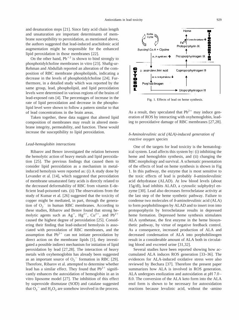
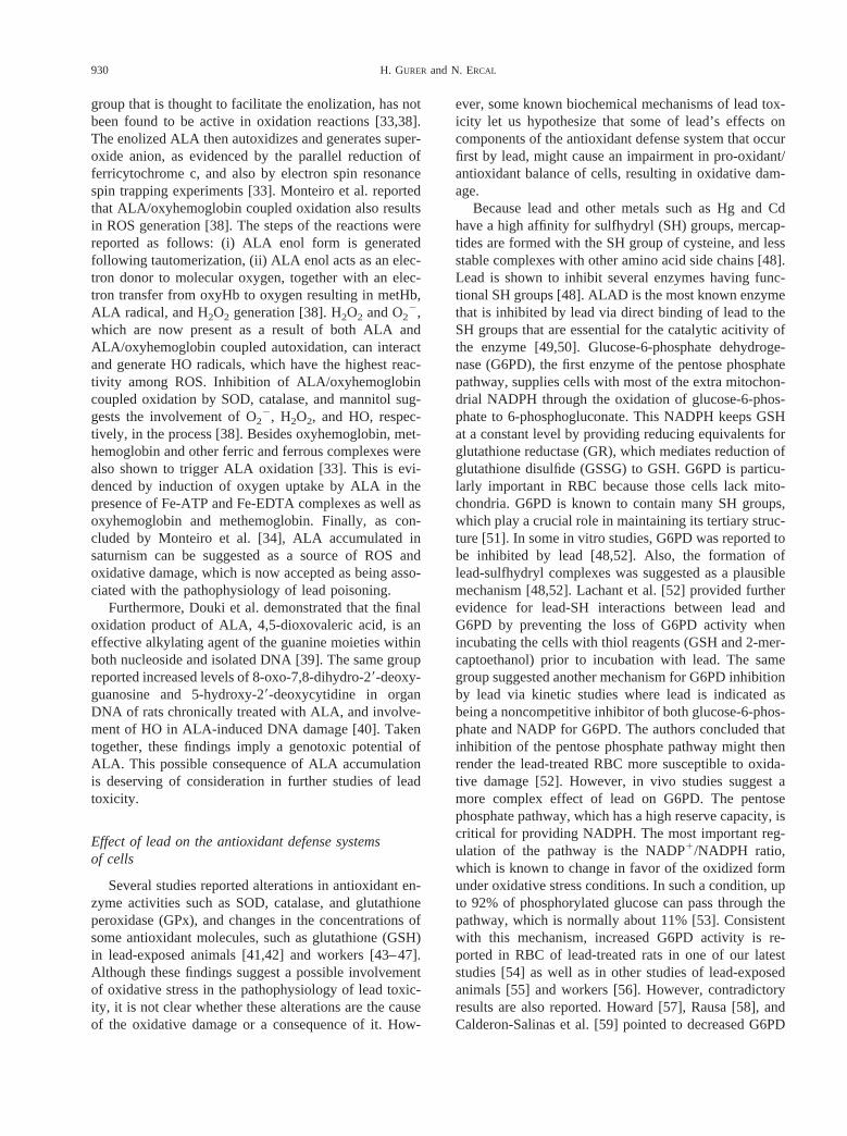
One of the targets for lead toxicity is the hematolog-ical system. Lead affects this system by: (i) inhibiting theheme and hemoglobin synthesis, and (ii) changing theRBC morphology and survival. A schematic presentationof the effects of lead on heme synthesis is shown in Fig1. In this pathway, the enzyme that is most sensitive tothe toxic effects of lead is probablyd-aminolevulinicacid dehydratase (ALAD). At low blood levels (about15g/dl), lead inhibits ALAD, a cytosolic sulphydryl en-zyme [30]. Lead also decreases ferrochelatase activity atthe last step of the heme synthetic pathway. Failure tocondense two molecules ofd-aminolevulinic acid (ALA)to form porphobilinogen by ALAD and to insert iron intoprotoporphyrin by ferrochelatase results in depressedheme formation. Depressed heme synthesis stimulatesALA synthetase, the first enzyme in the heme biosyn-thetic pathway, by virtue of negative feedback control.As a consequence, increased production of ALA anddecreased condensation of ALA into porphobilinogenresult in a considerable amount of ALA both in circulat-ing blood and excreted urine [31,32].
Several studies have been reported showing how ac-cumulated ALA induces ROS generation [33–36]. Theevidences for ALA-induced oxidative stress were alsoreviewed by Bechara [37]. Therefore the present papersummarizes how ALA is involved in ROS generation.ALA undergoes enolization and autoxidation at pH 7.0–8.0. The conversion of the ALA keto form into the ALAenol form is shown to be necessary for autooxidationreactions because levulinic acid, without the -amino
Fig. 1. Effects of lead on heme synthesis.
929Antioxidants in lead toxicity
group that is thought to facilitate the enolization, has notbeen found to be active in oxidation reactions [33,38].The enolized ALA then autoxidizes and generates super-oxide anion, as evidenced by the parallel reduction offerricytochrome c, and also by electron spin resonancespin trapping experiments [33]. Monteiro et al. reportedthat ALA/oxyhemoglobin coupled oxidation also resultsin ROS generation [38]. The steps of the reactions werereported as follows: (i) ALA enol form is generatedfollowing tautomerization, (ii) ALA enol acts as an elec-tron donor to molecular oxygen, together with an elec-tron transfer from oxyHb to oxygen resulting in metHb,ALA radical, and H2O2 generation [38]. H2O2 and O2
2,which are now present as a result of both ALA andALA/oxyhemoglobin coupled autoxidation, can interactand generate HO radicals, which have the highest reac-tivity among ROS. Inhibition of ALA/oxyhemoglobincoupled oxidation by SOD, catalase, and mannitol sug-gests the involvement of O2
2, H2O2, and HO, respec-tively, in the process [38]. Besides oxyhemoglobin, met-hemoglobin and other ferric and ferrous complexes werealso shown to trigger ALA oxidation [33]. This is evi-denced by induction of oxygen uptake by ALA in thepresence of Fe-ATP and Fe-EDTA complexes as well asoxyhemoglobin and methemoglobin. Finally, as con-cluded by Monteiro et al. [34], ALA accumulated insaturnism can be suggested as a source of ROS andoxidative damage, which is now accepted as being asso-ciated with the pathophysiology of lead poisoning.
Furthermore, Douki et al. demonstrated that the finaloxidation product of ALA, 4,5-dioxovaleric acid, is aneffective alkylating agent of the guanine moieties withinboth nucleoside and isolated DNA [39]. The same groupreported increased levels of 8-oxo-7,8-dihydro-29-deoxy-guanosine and 5-hydroxy-29-deoxycytidine in organDNA of rats chronically treated with ALA, and involve-ment of HO in ALA-induced DNA damage [40]. Takentogether, these findings imply a genotoxic potential ofALA. This possible consequence of ALA accumulationis deserving of consideration in further studies of leadtoxicity.
Effect of lead on the antioxidant defense systemsof cells
Several studies reported alterations in antioxidant en-zyme activities such as SOD, catalase, and glutathioneperoxidase (GPx), and changes in the concentrations ofsome antioxidant molecules, such as glutathione (GSH)in lead-exposed animals [41,42] and workers [43–47].Although these findings suggest a possible involvementof oxidative stress in the pathophysiology of lead toxic-ity, it is not clear whether these alterations are the causeof the oxidative damage or a consequence of it. How-
ever, some known biochemical mechanisms of lead tox-icity let us hypothesize that some of lead’s effects oncomponents of the antioxidant defense system that occurfirst by lead, might cause an impairment in pro-oxidant/antioxidant balance of cells, resulting in oxidative dam-age.
Because lead and other metals such as Hg and Cdhave a high affinity for sulfhydryl (SH) groups, mercap-tides are formed with the SH group of cysteine, and lessstable complexes with other amino acid side chains [48].Lead is shown to inhibit several enzymes having func-tional SH groups [48]. ALAD is the most known enzymethat is inhibited by lead via direct binding of lead to theSH groups that are essential for the catalytic acitivity ofthe enzyme [49,50]. Glucose-6-phosphate dehydroge-nase (G6PD), the first enzyme of the pentose phosphatepathway, supplies cells with most of the extra mitochon-drial NADPH through the oxidation of glucose-6-phos-phate to 6-phosphogluconate. This NADPH keeps GSHat a constant level by providing reducing equivalents forglutathione reductase (GR), which mediates reduction ofglutathione disulfide (GSSG) to GSH. G6PD is particu-larly important in RBC because those cells lack mito-chondria. G6PD is known to contain many SH groups,which play a crucial role in maintaining its tertiary struc-ture [51]. In some in vitro studies, G6PD was reported tobe inhibited by lead [48,52]. Also, the formation oflead-sulfhydryl complexes was suggested as a plausiblemechanism [48,52]. Lachant et al. [52] provided furtherevidence for lead-SH interactions between lead andG6PD by preventing the loss of G6PD activity whenincubating the cells with thiol reagents (GSH and 2-mer-captoethanol) prior to incubation with lead. The samegroup suggested another mechanism for G6PD inhibitionby lead via kinetic studies where lead is indicated asbeing a noncompetitive inhibitor of both glucose-6-phos-phate and NADP for G6PD. The authors concluded thatinhibition of the pentose phosphate pathway might thenrender the lead-treated RBC more susceptible to oxida-tive damage [52]. However, in vivo studies suggest amore complex effect of lead on G6PD. The pentosephosphate pathway, which has a high reserve capacity, iscritical for providing NADPH. The most important reg-ulation of the pathway is the NADP1/NADPH ratio,which is known to change in favor of the oxidized formunder oxidative stress conditions. In such a condition, upto 92% of phosphorylated glucose can pass through thepathway, which is normally about 11% [53]. Consistentwith this mechanism, increased G6PD activity is re-ported in RBC of lead-treated rats in one of our lateststudies [54] as well as in other studies of lead-exposedanimals [55] and workers [56]. However, contradictoryresults are also reported. Howard [57], Rausa [58], andCalderon-Salinas et al. [59] pointed to decreased G6PD
930 H. GURER and N. ERCAL
activity, while Rogers et al. [60] indicated unchanged redcell G6PD following lead exposure. Overall, the accu-mulated data suggests that exposure to lead can result inan increase or decrease in G6PD activity depending onthe concentration of exposed lead, duration of lead ex-posure, and magnitude of oxidative stress inside the cell[55].
GSH is a tripeptide containing cysteine that has areactive SH group with reductive potency. Accordingly,GSH plays a vital role in the protection of cells againstoxidative stress. It can act as a nonenzymatic antioxidantby direct interaction of the SH group with ROS, or it canbe involved in the enzymatic detoxification reactions forROS, as a cofactor or a coenzyme [61,62]. It possessescarboxylic acid groups, an amino group, a sulfhydrylgroup, and two peptide linkages as sites for reactions ofmetals [63]. Pb12 binds exclusively to the SH group[63,64], which decreases the GSH levels [65] and caninterfere with the antioxidant activity of GSH.
Another component of the antioxidant defense sys-tem, GR, reduces GSSG back to GSH and thereby sup-ports the antioxidant defense system indirectly. GR pos-sesses a disulfide at its active site [66], which wassuggested as a target for lead, resulting in the inhibitionof the enzyme [6,67]. This inhibition leads to decreasedGSH:GSSG ratios that will render cells more susceptibleto oxidative damage.
On the other hand, GPx, catalase, and SOD are met-alloproteins and accomplish their antioxidant functionsby enzymatically detoxifying peroxides, H2O2 and O2
2,respectively. Since these antioxidant enzymes depend onvarious essential trace elements for proper molecularstructure and enzymatic activity, they are potential tar-gets for lead toxicity [55]. Schrauzer [68] indicated an-tagonistic effects between lead and selenium, resulting inreduced selenium uptake that may affect GPx activity,that requires selenium as a cofactor, and then may in-crease the susceptibility of the cell to oxidative damage.As shown by Othman and El-Missiry [69], administra-tion of selenium, prior to injection of lead into male rats,produced noticeable prophylactic action against lead bymeans of increased SOD, GR activity, and GSH content.Although the protective effect was attributed to the for-mation of inactive selenium-lead complex [70], it wasmentioned that such interactions could not be the solemechanism for the beneficial effects of selenium. Cata-lase is another major antioxidant enzyme having heme asthe prosthetic group. Lead is known to reduce the ab-sorption of iron in the gastrointestinal tract and to inhibitthe heme biosynthesis [71]. Decreased catalase activityobserved in lead-exposed animals was attributed to theinterference of lead by both processes [6,67]. SOD playsan important role in protecting the cells against the toxiceffects of O2
2 by catalyzing its dismutation reactions.
The enzyme requires copper and zinc for its activity.Copper ions appear to have a functional role in thereaction by undergoing alternate oxidation and reduction,where zinc ions seem to stabilize the enzyme instead ofhaving a role in the catalytic cycle [20]. Another type,MnSOD, contains manganese at its active site and is notdetected in mammalian RBC, but is present in humanliver to some extent. Several studies pointed to decreasedRBC SOD activity in lead-exposed rats [55,72]. Mylroieet al. observed: (i) high correlation between decreasedSOD and decreased copper concentrations in the bloodof animals, (ii) no effect on SOD with increased bloodlead levels in the presence of normal copper concentra-tions [72], and (iii) that dietary copper supplementationprevented the Pb-induced decrease in SOD activity [73].Therefore, they have suggested an indirect inhibitoryeffect on SOD in vivo due to the lead-induced copperdeficiency. Inhibition of SOD activity by lead was alsoshown in an in vitro study where the authors indicatedthat this effect of lead can lead to decreased scavengingof ROS and result in oxidative damage [74]. On the otherhand, Ariza et al. [75] demonstrated rapid induction ofcellular H2O2 following treatment of AS52 cells with 1MPb21, which they suggested to be increased by the stim-ulatory effect of lead on the activities of CuZn-SOD andxanthine oxidase, enzymes that produce H2O2.
Overall, these inhibitory effects of lead on variousenzymes would probably result in impaired antioxidantdefenses by cells and render cells more vulnerable tooxidative attacks.
CONVENTIONAL TREATMENT OF LEAD POISONING
AND ITS SIDE EFFECTS
The current therapeutic approach to lead poisoning isto increase the excretion of lead by chelation. Variouschelators are available and are prescribed according tothe blood lead concentrations of the patient (Table 1).Although chelation has been shown to reduce blood leadlevels, the safety and efficacy of the various chelatorsmay be questioned. The efficiency and adverse effects ofeach of the major chelators prescribed in the UnitedStates will be discussed here.
Dimercaprol (BAL in peanut oil)
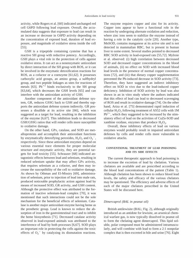
British antilewisite (BAL; Fig. 2), although originallyintroduced as an antidote for lewisite, an arsenical chem-ical warfare gas, is now typically dissolved in peanut oilto form the chelating agent dimercaprol. This lipid sol-uble, polar compound must be administered intramuscu-larly, and will combine with lead to form a 2:1 nonpolarcomplex that is then excreted in bile and urine [76]. Eight
931Antioxidants in lead toxicity
h after treatment, 20% of the BAL is found being ex-creted in the urine as a dithiol [77]. It is distributed inevery tissue because of its high liposolubility and di-rectly chelates the erythrocyte lead [77].
After treatment with BAL, side effects have beenreported in up to 50% of patients, with common prob-lems including fever, tachycardia, nausea, vomiting, sal-vation, watery eyes, sweating, and unpleasant breath
[76–78]. BAL therapy also frequently produces a hista-mine release, which must be countered with antihista-mines [76]. Besides these prevalent adverse effects thataccompany BAL treatment, patients with specialized dis-orders may face more serious risks. Patients with glu-cose-6-phosphate dehydrogenase deficiency have beenshown to experience severe hemolysis [79], while thepeanut oil used to prepare BAL may precipitate a reac-tion in children who are allergic to peanuts [78]. SinceBAL forms a toxic chelate with iron, iron supplementa-tion should be avoided during BAL treatment [80].
BAL is often administered in conjunction with cal-cium disodium ethylenediamine tetraacetic acid(CaNa2EDTA) to patients with blood lead levels greaterthan 70 g/dl [78]. When lead levels are this high, devel-opment of encephelopathy is a risk. BAL has been shownto reduce this risk, while CaNa2EDTA, another majorchelator, has been reported to increase chances of pre-cipitation of encephelopathy and to translocate lead fromsoft tissues to the central nervous system [76]. Althougha combination of BAL and CaNa2EDTA has been re-ported to quickly reduce lead levels and decrease the riskof encephalopathy [77], it has also been accompanied byincreased vomiting andelevatedhepatic enzyme activity[76].
Fig. 2. Chemical structures of chelating agents.
Table 1. Clinical Approach to Chelation Therapy in Childrena
Condition and blood leadconcentrations (mg/dl) Regimen and comments
Encephalopathy(.90–100)
BAL 1 CaNa2EDTA
Symptomatic withoutencephalopaty (70–90)
BAL 1 CaNa2EDTA, CaNa2EDTAor succimer
Symptoms possible(40–69)
CaNa2EDTA or succimer (45mg/dlmay be used as lower tresholdfor chelation)
Biochemical toxicity(25–39)
CaNa2EDTA or succimer (Noconsensus about chelation)
Excessive exposure(10–24)
Chelation therapy not indicated;source identification, education,and nutrition counseling areneeded.
a Modified from [78].
932 H. GURER and N. ERCAL
CaNa2EDTA
CaNa2EDTA (Fig. 2) was introduced in the 1950s asa lead poisoning treatment. When EDTA was first usedtherapeutically as its sodium salt it resulted in cardiovas-cular instability, severe hypocalcemia, and even death insome patients. When it is combined with calcium, so-dium, or zinc it forms less toxic compounds. The water-soluble CaNa2EDTA is recommended for clinical use asa chelator in which the formed lead-EDTA chelate has ahigher stability constant [81,82]. Since the absorption ofCaNa2EDTA from the gastrointestinal tract is onlyaround 5%, the oral administration of the compound isnot recommended [77,78]. Instead, it can be adminis-tered intramuscularly, which is a painful method, orusually by slow intravenous infusion [83]. CaNa2EDTAis distributed mainly in the extracellular compartments ofthe body and does not enter the cells because of its ionicform [77]. Therefore it removes lead from the extracel-lular compartments only [77,84]. In addition, its passageacross the blood-brain barrier is very slow [81].CaNa2EDTA is not excreted in fecal matter, and 90% ofadministered CaNa2EDTA is found not metabolized inthe urine 8 h after treatment [85]. When administered,CaNa2EDTA leads to reduced blood lead levels, a rever-sal in the hematologic effects of lead toxicity, and anincrease in lead levels in urine [76].
However, this chelating agent has many disadvan-tages. It may cause the redistribution of lead from thebones and kidney to the brain and liver, increase the riskof encephalopathy, and cause renal toxicity [76,81,86].Since CaNa2EDTA is not able to cross the blood-brainbarrier, it is doubtful whether it is effective in reducinglead levels in the brain or in relieving the effects of leadon neurodevelopmental functioning [87]. Besides in-creased urinary excretion of lead, because ofCaNa2EDTA’s relative lack of specificity, other essentialmetals such as zinc, copper, iron, cobalt, and manganeseare also reported to be excreted and depleted followingCaNa2EDTA therapy [81,88]. Among them zinc diuresisis the most common and severe side effect. Although itmay be safe to administer zinc while using CaNa2EDTAin order to preclude the effects of long-term chelationtherapy, this may decrease the effectiveness of therapy[89]. Another disadvantage of the CaNa2EDTA treat-ment is its high cost. Since it is administered by slowintravenous infusion, the child needs to be hospitalized.This long treatment protocol may result in a total cost of$30,000 for a child [81].
The adverse effects of CaNa2EDTA therapy are alsoprevalent. Its administration may be responsible for in-creased renal toxicity [82]. Of 130 children receivingBAL and CaNa2EDTA treatment, 13% showed signs ofnephrotoxicity, while 3% developed acute renal failure.
More than 25% of children who were given intramuscu-lar or intravenous administration of CaNa2EDTAshowed increasing serum urea nitrogen [90].
D-penicillamine
D-penicillamine, also referred to asb,b-dimethyl cys-teine, was unintentionally discovered in 1953 as a me-tabolite of penicillin B in the urine of patients with liverdisease. Since then it has been used in the treatment ofWilson’s Disease to reduce serum copper levels [91]. Itis not an FDA-approved drug for lead poisoning. It is,however, used as treatment for low-level lead toxicity inthe blood in the range of 25–40 g/dl, especially inchildren, since 1956 [92]. It is administered orally, and istypically taken by the patient for 4–12 weeks. It leads toreduced blood lead levels and reversal of hematologictoxicity [78]. D-Penicillamine is a sulfhydryl containingamino acid (Fig. 2). One possible mechanism for itschelating ability is the formation of a simple bond be-tween its sulfhydryl group and lead atom. Other possiblemechanisms suggested are as follows: (i) incorporationof lead into a ring structure between the sulfur andadjacent nitrogen atom (Fig. 3), or (ii) a lead atom maybe bound between two penicillamine molecules [93].
Like the two previously discussed chelators, D-peni-cillamine treatment leads to a large number of adverseeffects [92]. A study of children being administeredD-penicillamine showed that most experienced nauseaand vomiting [94]. Eosinophilia was seen in 20% of them[95]. Reversible leukopenia or mild thrombocytopeniawas observed in about 10% of children being treated insome other studies [96], while 0.5–1.0% developed an-gioedema, urticaria, or maculopapular eruptions, requir-ing a cessation of the therapy [94,97]. Cases of protein-uria, microscopic hematuria, and incontinence have alsobeen reported, although they are less common and maybe resolved by reducing the D-penicillamine dosage [96,98]. D-penicillamine also has the potential for eliminat-ing essential nutrients as pyridoxine, zinc, and iron, aswell as lead. Furthermore, its absorption is affected bythe diet; it can be reduced 35% when treatment is com-bined with food or ferrous sulfate, and up to 66% whentaken in conjunction with antacids [99,100].
D-penicillamine is a less effective chelator thanCaNa2EDTA, and its overall toxicity profile allows it tobe considered as a third choice for lead toxicity treatmentafter CaNa2EDTA and succimer [92].
Succimer
Succimer, or 2,3-meso-dimercaptosucccinic acid, is awater soluble analog of dimercaprol. Much less data
933Antioxidants in lead toxicity

exists on this agent as compared to the three previouslydiscussed chelators, because administration of succimerhas been limited. Succimer is the only chelator in theUnited States that has been solely approved for pediatricuse by FDA; thus no appropriate and large clinical trialhas been published with regard to adult treatment withsuccimer [78,84]. Consequently, relatively little isknown about succimer’s therapeutic effects on lead poi-soning.
Succimer is a water-soluble derivative of BAL andcan be administered orally. It has two sulfhydryl groups(Fig. 2), but Pb is reported to be coordinated with thesulfur and oxygen atom. The structure of the chelateformed between meso-succimer and Pb ions in a test tubeis shown in Fig. 3 [81]. With regard to the absorption andfunction of succimer within the body, about 95% of theadministered succimer binds to plasma proteins and isprimarily distributed in the extracellular compartment[101]. With an elimination half-life of about 48 h, a peakblood concentration of succimer is reached within 2 h[102]. The succimer is metabolized rapidly and exten-sively excreted through the urine as mixed disulfides[92].
Succimer is most commonly used to treat childrenwho have blood lead levels greater than 45 g/dl and whoare not at risk of encephalopathy [78]. Several advan-
tages of succimer make it a good candidate for leadpoisoning treatment: (i) Lead doesn’t happen to be re-distributed to the brain following succimer treatment,which is a major disadvantage of other chelators. (ii) Itsoral availability makes it a preferred agent for the ther-apy of childhood lead poisoning and allows for admin-istration in an outpatient setting that will decrease thecost of the therapy. (iii) Unlike CaNa2EDTA, succimerhas specific affinity for heavy metals such as lead, ar-senic, and mercury, but causes little increase in theexcretion of iron, zinc, or calcium [78,81,92]. (iv) Suc-cimer has been shown to decrease the levels of lead in thebrain and blood more effectively than CaNa2EDTA treat-ment. It mobilizes lead from the soft tissue, brain, liver,kidney, and blood, although it has not consistently re-duced lead levels in bone [88,103]. Succimer is moreeffective than CaNa2EDTA in producing plumburesis[104], and is not likely to precipitate encephalopathy. (v)Thus far, succimer toxicity has been minimal, with onlyoccasional reports of nausea, vomiting, diarrhea, loosestool, appetite loss, and foul-smelling urine or stools[92,102]. Some children receiving succimer treatmentexperienced hypersensitivity reactions such as chills, fe-ver, rash, or urticaria [92]. A study of children beingtreated showed that 12% had mild gastrointestinal symp-toms, 5% experienced general malaise, and 4% showed
Fig. 3. Chelates formed between lead and d-penicillamine, BAL, CaNa2EDTA, and succimer.
934 H. GURER and N. ERCAL
transient elevation of liver enzymes. Also, some werestruck with reversible neutropenia [105]. Because 92–95% of succimer binds to plasma proteins, there is apotential for interactions with other protein-bound drugs[78]. Nevertheless, from what is known, succimer seemsto be one of the “best” chelating agents.
Besides its potent chelating effect, succimer appearsto have antioxidant activity because of two SH groups inits structure. Our data demonstrated a visible protectiveeffect of succimer on lead-induced oxidative stress inrats. However, since blood lead levels returned to controllevels in succimer-treated rats, it remains unclearwhether the observed effect of succimer can be attributedto chelation or the potential to act as a thiol antioxidant.Mechanistic studies are missing and needed to determinewhether succimer has a potential ROS scavenging activ-ity via its SH groups.
POSSIBLE PROTECTIVE EFFECTS OF ANTIOXIDANTS
AGAINST LEAD-INDUCED OXIDATIVE STRESS
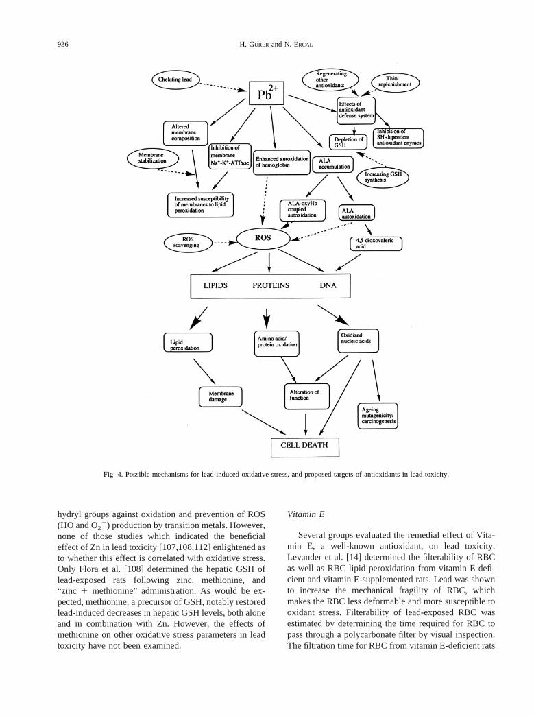
As discussed in the first section, induction of ROS bylead and subsequent depletion of antioxidant cell de-fenses can result in generalized disruption of the pro-oxidant/antioxidant balance in lead-burdened tissues.This could contribute to tissue injury via oxidative dam-age to critical biomolecules (Fig. 4). In the event thatoxidative stress can be partially implicated in lead tox-icity, a therapeutic strategy to increase the antioxidantcapacity of cells may fortify the long-term effectivetreatment of lead poisoning. This may be accomplishedby either reducing blood and tissue lead levels via che-lation, thereby reducing the possibility of lead interactingwith critical biomolecules and inducing oxidative dam-age, or by bolstering the cell’s antioxidant defensesthrough exogenous supplementation of antioxidant mol-ecules (Fig. 4).
Although many investigators have confirmed lead-induced oxidative stress, the usefulness of antioxidantsalone or in conjunction with chelation therapy has notbeen extensively investigated yet. Some groups [4,106–110] investigated the ability of some molecules withantioxidant activity to prevent or treat experimental leadtoxicity in animals. Although some of the agents werefound to be capable of abating some toxic effects of lead,none of them were discussed as being effective viarebalancing the impaired pro-oxidant/antioxidant ratiofollowing lead exposure.
Vitamin B6
In 1987, Tandon et al. investigated the effect of Vi-tamin B6 in lead intoxication [111]. These authors re-
ported significantly reduced inhibition of ALAD activityand zinc protoporphyrine levels as a result of simulta-neous supplementation of vitamin B6 and lead. De-creased blood, kidney and liver lead levels were alsoshown in vitamin B6-supplemented rats, while no effectwas observed in their brain lead levels. These beneficialeffects of vitamin B6 on lead toxicity were suggested asbeing due to the participation of the ring nitrogen atom inthe chelation of lead or to a possible interaction betweenlead and vitamin B6 at the absorption level [111]. In1989, McGowan examined the GSH metabolism of lead-exposed rats fed a vitamin B6-deficient diet [106]. GSHlevels in the experimental group were found to be lowerthan the control values. This involvement of vitamin B6
in GSH metabolism was explained by the cofactor role ofvitamin B6 for several enzymes in the transsulfurationpathway. Most of the cysteine, the bioprecursor of GSH,is synthesized from dietary methionine in that pathway.Therefore, it was suggested that a lack of vitamin B6
prevented the involvement of methionine in GSH bio-synthesis by limiting the availability of cysteine [106].The results indicate an indirect antioxidant role for vita-min B6 in lead-exposed rats via supporting their antiox-idant defense systems by inducing GSH biosynthesis.This possible role for vitamin B6 in lead toxicity was notdiscussed, however, and was not investigated further.
Zinc
Zinc, on the other hand, was reported to be able toprevent and treat lead intoxication in rats, either aloneand/or in combination with methionine or thiamine [107,108]. In both studies, simultaneous dietary supplemen-tation with “Zn1 methionine/thiamine” was found to bemost effective in reducing lead-induced inhibition ofALAD activity in the blood and urinary excretion ofALA. Those studies also suggest that supplementation ofthe combination therapy concurrently with exposure tolead was more effective than treatment after lead expo-sure. This protective effect for Zn was attributed to adecrease in lead absorption in the gastrointestinal track.In another study [112], Zn was administered to lead-intoxicated rats along with chelating agentsCaNa2EDTA, succimer, and D-penicillamine. Zn wasshown to increase the efficacy of chelating agents bypotentiating the depletion of blood, hepatic and renallead, and reversing inhibited blood ALAD activity. Ad-ditionally, Zn supplementation was effective in restoringthe Zn levels in tissues which were depleted followingtreatment with chelating agents [112]. Zn has beenshown to have an antioxidant effect which was reviewedby Bray and Bettger [113]. Besides some proposedmechanisms for the antioxidant function of Zn, twomechanisms have been elucidated: the protection of sulf-
935Antioxidants in lead toxicity
hydryl groups against oxidation and prevention of ROS(HO and O2
2) production by transition metals. However,none of those studies which indicated the beneficialeffect of Zn in lead toxicity [107,108,112] enlightened asto whether this effect is correlated with oxidative stress.Only Flora et al. [108] determined the hepatic GSH oflead-exposed rats following zinc, methionine, and“zinc 1 methionine” administration. As would be ex-pected, methionine, a precursor of GSH, notably restoredlead-induced decreases in hepatic GSH levels, both aloneand in combination with Zn. However, the effects ofmethionine on other oxidative stress parameters in leadtoxicity have not been examined.
Vitamin E
Several groups evaluated the remedial effect of Vita-min E, a well-known antioxidant, on lead toxicity.Levander et al. [14] determined the filterability of RBCas well as RBC lipid peroxidation from vitamin E-defi-cient and vitamin E-supplemented rats. Lead was shownto increase the mechanical fragility of RBC, whichmakes the RBC less deformable and more susceptible tooxidant stress. Filterability of lead-exposed RBC wasestimated by determining the time required for RBC topass through a polycarbonate filter by visual inspection.The filtration time for RBC from vitamin E-deficient rats
Fig. 4. Possible mechanisms for lead-induced oxidative stress, and proposed targets of antioxidants in lead toxicity.
936 H. GURER and N. ERCAL
was much greater than that of RBC from vitamin E-sup-plemented rats. Furthermore, a strong correlation wasfound between increased lipid peroxidation and de-creased filterability of red cells from vitamin E-deficient,lead-poisoned rats. These results indicate that the highvitamin E status of humans may ameliorate lead-inducedchanges in the deformability of RBC, which makes themmore vulnerable to oxidative damage [14]. This preven-tive role for vitamin E in lead toxicity, implied byLevander et al., is confirmed by another group [110] whofound simultaneous supplementation of vitamin E moreeffective than treatment of lead-exposed animals withvitamin E. This preventive effect for vitamin E wasreported to be due to the inhibition of lead absorption.
Ascorbic acid (Vitamin C)
Another antioxidant molecule, ascorbic acid, admin-istered alone or in combination with thiamine to lead-exposed rats and their effect on the efficacy of two thiolmetal chelators, succimer and -mercapto—(2-furyl)ac-rylic acid, were investigated [109]. Both ascorbic acidalone and in combination with thiamine were found to beeffective by means of increasing urinary elimination oflead, reducing hepatic and renal lead burden and revers-ing lead-induced inhibition of the activity of bloodALAD. Thiamine alone, however, did not show anybeneficial effects. This beneficial role of ascorbic acidwas attributed to its ability to complex with lead [109]. In1999 Simon and Hudes reported a population-basedstudy that indicates an inverse relation between serumascorbic acid and blood lead levels among Americans[114]. The authors suggest that higher intake of ascorbicacid may be effective in preventing lead toxicity if acausal relationship is confirmed.
Although improvement by several means of antioxi-dant administration to lead-exposed animals was re-ported, there are not many studies in the literature wherethe effectiveness of an antioxidant in counteracting lead-induced oxidative damage is extensively investigated. Itis only recently that the correlation between those ben-eficial effects of antioxidants and other oxidative stress-related parameters has been investigated.
Ethoxyquin
One of those studies was reported by Donaldson, whodetermined whether systemic effects of lead, attributed totissue peroxidation, can be reversed by the dietary anti-oxidant, ethoxyquin [115]. A peroxidative mechanismfor lead toxicity was suggested and ethoxyquin wasobserved to ameliorate lead toxicity, as assessed bygrowth inhibition [115].
Selenium
Another study came in 1998 from Egypt, where therole of selenium in lead toxicity was investigated [69].Selenium is an essential element known to be requiredfor the activity of glutathione peroxidase, thereby havinga key role in the antioxidant defense systems of cells. Itsefficacy in treatment of free radical-associated diseaseshas been shown by many studies [116,117]. Seleniumadministration, prior to lead injection, resulted in pro-nounced prophylactic action against lead effects in termsof acid and alkaline phosphatases, transaminases (GOT,GPT), total protein, triglycerides, and cholesterol in se-rum [69]. In addition, oxidative stress-related parametersin two major target organs, the liver and kidney, wereanalyzed following intramuscular injection of 10 mol/kgsodium selenite, 2 h before administration of 100 mol/kgof lead acetate to male albino rats. Selenium was foundto enhance the antioxidant capacity of cells by increasingthe activities of SOD and GR, and augmenting the GSHcontent. Three possible mechanisms were proposed forthe protective effect of selenium: (i) formation of aninactive selenium-lead complex; (ii) stimulating radicalscavenging by increasing the activity of SOD, therebyincreasing the removal of the superoxide radical; and (iii)increasing the antioxidant capacity of cells indirectly byincreasing the activity of glutathione reductase, whichhas a major role in maintaining a sufficient content ofGSH in the reduced form [69].
S-adenosyl-L-methionine (SAM)
Since ethanol was reported to potentiate lead-inducedinhibition of rat brain antioxidant defense systems [118],benefits of supplementation of SAM, the precursor ofGSH, to mice exposed to “lead1 ethanol” was investi-gated by Flora and Seth [119]. SAM was shown toprevent the alterations in some biochemical parameters(blood ALAD, GSH, brain and liver lipid peroxidation,and GSH content) and accumulation of lead in blood,liver, and brain during acute “lead1 ethanol” exposure.The results seem promising and the authors suggestedthat SAM could have a possible therapeutic potentialeither as a sole (or as an “adjuvant”) agent during che-lation therapy by depleting the brain’s lead burden andmitigating the “lead1 ethanol”-induced oxidative stress[119].
N-acetylcysteine (NAC)
In 1996 our group began studying the role of antioxi-dants in lead toxicity in both in vitro and in vivo systems.Our goal was to explore whether lead induces oxidativestress and if so, to investigate the dual benefits of some
937Antioxidants in lead toxicity
thiol compounds in lead toxicity, as antioxidants andthiol chelators. In an in vitro model where Chinesehamster ovary (CHO) cells were used, Ercal et al. ob-served increased oxidative damage in lead-exposed cellsby means of a decreased GSH/GSSG ratio, increasedMDA levels and catalase activity [120]. A possible rem-edy for the oxidative imbalance occurring in lead toxicitywas suggested by the authors as being replenishment ofthe GSH supply of the cells. Since direct GSH supple-mentation has been demonstrated to be ineffective [121],a well-known bioprecursor of GSH—NAC—was used asa supplement for lead-exposed CHO cells and its capac-ity to minimize lead-induced oxidative stress was inves-tigated [120]. NAC is a thiol-containing antioxidant (Fig.5) that has been used under several clinical conditionswith few adverse side effects [122,123]. Its high toxicitythreshold and wide therapeutic window enhances its util-ity. The antioxidant action is believed to be due to itsdirect interaction with ROS and/or its ability to stimulateGSH synthesis [124–126]. All of these advantages ofNAC imply that a favorable outcome may be obtained byinclusion of NAC in lead poisoning therapy protocol. Inour study, NAC supplementation resulted in an increasedGSH/GSSG ratio and decreased MDA and catalase ac-tivity [120]. Besides this antioxidant function, NAC also
exerted a positive effect on cell survival, which had beendramatically decreased by lead exposure. When takentogether, the results support the involvement of ROS inlead toxicity and a possible beneficial role for NAC intherapeutic implications of lead poisoning. These find-ings were further supported by the same group [120] inan in vitro system. Treatment with NAC caused a reduc-tion in indices of oxidative stress in both the brains andlivers of lead-exposed C57BL/6 mice. This implies that athiol-containing antioxidant is capable of mitigatinglead-induced oxidative stress.
On the other hand, it has been indicated that NAC hasa chelating activity against several heavy metals such asboron, chromium [127], cobalt [128], cadmium, gold,and lead [129]. However, it is suggested that NAC maybe ineffective in chelating metals when given orally[130]. Consistent with these results, Ercal et al. found nochelating effect of NAC when orally administered tomice in 5.5 mmol/kg dosages for a week [120]. NACtreatment did not cause any decrease in their blood, liver,or brain lead levels. This finding suggests that NACadministration counteracted in vivo oxidative stresswithout removing lead from target tissues. Therefore, theantioxidant role for NAC appears to be related solely toits free thiol group. In another in vivo study, where 5
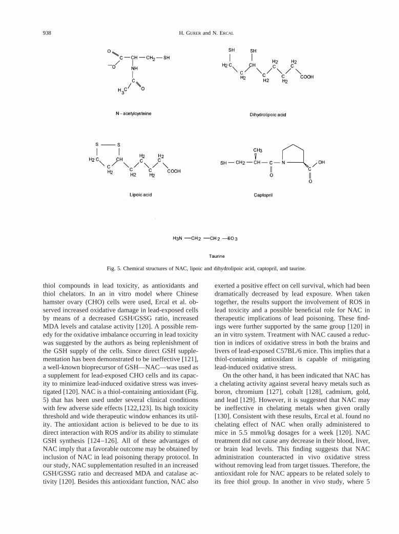
Fig. 5. Chemical structures of NAC, lipoic and dihydrolipoic acid, captopril, and taurine.
938 H. GURER and N. ERCAL
mmol/kg/d NAC was administered orally to Fisher 344(F344) rats for a week following exposure to 2000 ppmlead acetate in drinking water for 5 weeks, Gurer et al.observed a slight decrease in blood lead levels (27.3%decrease) [54]. A well-known chelator, succimer, in-cluded in the same study to compare and evaluate thechelating efficacy of NAC, dramatically enhanced theclearance of lead from the blood stream (92.8% de-crease). Results support the hypothesis that the antioxi-dant action of NAC could provide some beneficial ef-fects in lead poisoning treatment independent ofchelation. Therefore, inclusion of NAC in a chelation-oriented treatment protocol for plumbism seems to bemuch more effective than using it as a sole agent. Similareffects of NAC were also shown in lenses of lead-exposed F344 rats, where lead induced an oxidativemodification of protein sulfhydryl residues and lipids anddecreased GSH levels [131].
Neal et al. in 1997 [132] reported other evidence ofthe beneficial role of NAC in treating lead poisoning.d-ALA-induced oxidative stress via generation of ROS issuggested to be, in part, responsible for the lead-induceddamage [33,35]. Therefore, a study was undertaken totest the hypothesis that ALA accumulation in CHO cellscontributes to the cumulative oxidative challenge of leadpoisoning, and also to examine whether NAC treatmentas an antioxidant can reverse ALA-induced oxidativedamage. The results indicate a pro-oxidant effect forALA by means of a decreased GSH:GSSG ratio, in-creased MDA levels, and inhibited colony formation[132]. Furthermore, a protective role of NAC in ALA-exposed cells was evidenced by an increase in GSH:GSSG ratios and increased cell survival. This studyconfirms a therapeutic role for NAC in plumbism that isindependent of chelation, and could be solely attributedto NAC’s free sulfhydryl group [132].
a-Lipoic acid (LA)
Another antioxidant, LA, was also suggested as beingable to abate some of the toxic effects of lead [133]. LAcan be synthesized by animals and humans [134], andfunctions as a cofactor in several multi-enzyme com-plexes [135]. Its reduced form, dihydrolipoic acid(DHLA), has two free sulfhydryl groups (Fig. 5), and theLA/DHLA redox couple has received great attention inrecent studies with regard to its antioxidant potential[134]. Mechanisms of their antioxidant activity are re-viewed by Packer et al. [136] and Biewenga et al. [137]in detail. Both LA and DHLA (i) have the ability toscavenge some reactive species, (ii) can regenerate otherantioxidants (i.e., vitamins E and C, and GSH) from theirradical or inactive forms, and (iii) have metal chelatingactivity. LA also seems to have a notable advantage over
NAC in opposing GSH loss, since LA is effective in amicromolar range while millimolar NAC is needed for asimilar effect [138]. Other criteria, which are importantwhen considering therapeutic applications of an agent,are its absorption and bioavailability, as well as its con-centration in target tissues. The capability of LA to crossthe blood-brain barrier [139] appears to be an extraadvantage because the brain is an important target in leadpoisoning.
Incubating lead-treated CHO cells with LA wasshown to result in considerably increased cell survivalalong with attenuated oxidative stress [133]. This is interms of decreased MDA levels, increased GSH content,and decreased catalase activity. Similar results werefound in an in vivo model where F344 rats were exposedto 2000 ppm lead acetate in drinking water for 5 weeks.GSH levels in RBC and brains were found to be elevatedfollowing 1 week of 25 mg/kg/d LA administration,whereas RBC, brain, and kidney MDA levels were di-minished and catalase, G6PD activities in RBC werereturned to the control levels [133]. No chelating actionof LA against lead was observed in that study design interms of no changes in blood, brain, and kidney leadconcentrations. Therefore, the beneficial effects of LA onoxidative stress-related parameters do not appear to berelated to its ability to remove lead from target cells butare associated with LA’s potential for bolstering thiolantioxidant capacity. However, LA was found to havealmost no effect on the lenticular redox status followinglead exposure [140]. LA administration to lead-exposedrats resulted in significant increases in lenticular cysteinelevels with insignificant changes in GSH and MDA[140].
Captopril
Captopril, an angiotensin-converting enzyme inhibi-tor, was suggested as having antioxidant potential be-sides its well-known anti-hypertensive action. Its termi-nal sulfhydryl group (Fig. 5) was suggested as playing arole in scavenging ROS, which was indicated as a mech-anism for the antioxidant action of captopril [141–143].The thiol group in the structure of captopril raised thepossibility that it might chelate heavy metals, therebyincreasing their excretion [144]. These properties sug-gested a possible role for captopril in lead toxicity whereoxidative stress is thought to be involved. Therefore,Gurer et al. investigated in vivo effects of captopril onlead-induced oxidative stress [145]. The captopril-treatedsamples showed higher GSH:GSSG ratios in the liver,brain, and kidneys of rats, as well as slightly decreasedMDA concentrations. The catalase activity was not sig-nificantly affected. Unaltered blood lead concentrationswere detected after 1 week of captopril (10mg/d) admin-
939Antioxidants in lead toxicity
istration to lead-exposed F344 rats, which indicates thatit can only be used along with a chelating agent to treatlead poisoning [145].
Lead-induced hypertension is reported in other studies[146,147], which indicated that increased ROS generatedby lead exposure may contribute to increased blood pres-sure by enhancing inactivation of endothelium-derived ni-tric oxide [148,149]. A possible role of ROS in lead-in-duced hypertension was further evidenced by decreased
MDA levels and reduced blood pressure with concomitantadministration of vitamin E to lead-exposed animals [150,151]. Finally, Ding et al. [152] reported that hydroxylradical may be the radical to blame for the shown endothe-lial dysfunction in lead toxicity. Therefore captopril’s mul-tiple benefits as an antioxidant and antihypertensive agentdeserve to be explored to elucidate whether antioxidanteffect of captopril can be a possible alternative mechanismfor its known antihypertensive effect.
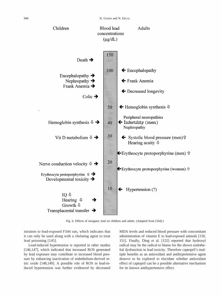
Fig. 6. Effects of inorganic lead on children and adults. (Adapted from [164].)
940 H. GURER and N. ERCAL
Taurine
Taurine, a semi-essential amino acid has been shownto have a role in maintaining calcium homeostasis, os-moregulation, removal of hypochlorous acid, and stabi-lizing the membranes [153,154]. Some of the recent dataindicate that taurine can act as a direct antioxidant byscavenging ROS [155–158] and/or as an indirect antiox-idant by preventing changes in membrane permeabilitydue to oxidant injury [153]. However, some contrarydata have also been reported [159]. In our recent studiestaurine was shown to have beneficial effects in lead-induced oxidative stress in CHO cells and F344 rats(unpublished data). Dramatically increased cell survivalwas established in taurine-treated, lead-exposed CHOcells while MDA levels were diminished and GSH levelswere increased. Similar effects were found in RBC andthe brains and livers of lead-exposed F344 rats. Nochelating effect of taurine (1.2 g/kg/d) was indicated byany change in lead concentrations in the blood, brains,livers, and kidneys after taurine treatment. An antioxi-dant mechanism(s), rather than a chelating activity,seems to underlie this observed effect of taurine againstlead-induced oxidative stress. Further studies are neededto understand the antioxidant properties of taurine.
CONCLUSION
Lead poisoning is an old but persistent public healthproblem throughout the world. Although guidelines forthe management of childhood lead poisoning were re-leased by the Centers for Disease Control in 1985 [160]and 1991 [84], a nationwide survey of pediatric lead-poisoning treatment programs indicated that no commonapproach for the treatment of low-level lead poisoningappears to exist within the lead clinics [161]. Reportedadverse effects of conventional chelators and the uncer-tainty in their efficacy in reversing or preventing theneurotoxic effects of lead (believed to occur in childrenwith 25 mg/dl blood lead concentrations) caused someclinicians to ignore pharmacological intervention in chil-dren with low blood lead levels [83] (Table 1). There isan overall consensus on the adverse neurodevelopmentaloutcomes of low-level (25mg/dl) lead poisoning duringchildhood [162,163] (Fig. 6). Investigations pointed toinduced oxidative damage even with low blood leadlevels.
These facts present a novel approach to strategies fortreating lead poisoning by supplementation with antioxi-dants, either individually or in a combined therapy, withchelating agents. Studies so far suggest that antioxidantscan play an extremely important role in abating sometoxic effects of lead. Some antioxidants, such as NAC,appear to have a potential for chelating lead and remov-
ing it from the blood stream [54]. Further studies shouldfocus on exploring the dual benefits of these antioxidantsas possible chelating and antioxidant agents in treatinglead toxicity.
Use of antioxidants brings another option to the ther-apy: the possibility of therapeutic intervention withoutremoving the patient from the source of lead. Because ofthe rebound effect of chelators, chelation therapy couldnot be started when the subject was near lead. Antioxi-dants, however, are recognized as safe molecules andmay be given to subjects with low lead concentrations intheir blood even when it is not possible to remove themfrom exposure to lead. Consequently, experiments areneeded to show the effects of antioxidants on the cells oranimals that are treated concomitantly for lead exposure.Detailed mechanistic studies are also required to under-stand the mechanisms underlying the beneficial effects ofsome antioxidants and to explore the optimum dosageand duration of treatment to obtain better clinical recov-eries in lead intoxication cases.
Acknowledgements— Nuran Ercal was supported by 1R15ES08016-01from the NIEHS, NIH. Hande Gurer was supported by the TurkishScientific and Technical Research Council.
REFERENCES
[1] Willis, E. D. Mechanisms of lipid peroxide formation in tissues.Role of metals and haematin proteins in the catalysis of theoxidation of unsaturated fatty acids.Biochem. Biophys. Acta98:238–251; 1965.
[2] Yiin, S. J.; Lin, T. H. Lead-catalyzed peroxidation of essentialunsaturated fatty acid.Biol. Trace Elem. Res.50:167–172; 1995.
[3] Gerber, G. B.; Maes, J.; Gilliavod, N.; Casale, G. Brain biochem-istry of infants and rats exposed to lead.Toxicol. Lett.2:51–63;1978.
[4] Shafiq-ur-Rehman, S. Lead-induced regional lipid peroxidation inbrain.Toxicol. Lett.21:333–337; 1984.
[5] Shafiq-ur-Rehman, S.; Rehman, S.; Chandra, O.; Abdulla, M.Evaluation of malondialdehyde as an index of lead damage in ratbrain homogenates.Biometals8:275–279; 1995.
[6] Sandhir, R.; Gill, K. D. Effect of lead on lipid peroxidation inliver of rats.Biol.Trace Elem. Res.48:91–97; 1995.
[7] Somashekaraiah, B.; Padmaja, K.; Prasad, A. R. K. Lead-inducedlipid peroxidation and antioxidant defense components of devel-oping chick embryos.Free Radic. Biol. Med.13:107–114; 1992.
[8] Adanaylo, V. N.; Oteiza, P. I. Lead intoxication: antioxidantdefenses and oxidative damage in rat brain.Toxicology135:77–85; 1999.
[9] Donaldson, W. E.; Knowles, S. O. Is lead toxicosis a reflection ofaltered fatty acid composition of membranes?Comp. Biochem.Physiol. C104:377–379; 1993.
[10] De Silva, P. E. Determination of lead in plasma and studies onits relationship to lead in erythrocytes.Br. J. Ind. Med.38:209–217; 1981.
[11] Leggett, R. W. An age-specific kinetic model of lead metabolismin humans.Environ. Health Perspect.101:598–616; 1993.
[12] Rice-Evans, C. Iron-mediated oxidative stress and erythrocytes.In: Harris, J. R., ed.Blood cell biochemistry(Vol. 1). New York:Plenum Press; 1990:429–453.
[13] Waldron, H. A. The anemia of lead poisoning: a review.Br. J.Ind. Med.23:83–100; 1966.
[14] Levander, O. A.; Morris, V. C.; Ferretti, R. J. Filterability of
941Antioxidants in lead toxicity
erythrocytes from vitamin E-deficient lead-poisoned rats.J.Nutr. 107:363–372; 1977.
[15] Hernberg, S.; Nurminen, M.; Hasan, J. Nonrandom shortening ofred cell survival times in men exposed to lead.Environ. Res.1:247–261; 1967.
[16] Bonting, S. L.; Caravaggio, L. L. Studies on sodium-potassiumactivated ATPase. V. Correlation of enzyme activity with cationflux in six tissues.Arch. Biochem. Biophys.101:37; 1963.
[17] Hasan, J.; Vihko, V.; Hernberg, S. Deficient red cell membrane(Na1 2 K1)-ATPase in lead poisoning.Arch. Environ. Health14:313–318; 1971.
[18] Raghavan, S. R. Erythrocyte lead-binding protein after occupa-tional exposure. II. Influence on lead inhibition of membraneNa1 2 K1-ATPase.J. Toxicol. Environ. Health7:561–568;1981.
[19] Fukumoto, K.; Karai, I.; Horiguchi, S. Effect of lead on eryth-rocyte membranes.Br. J. Ind. Med.40:220–223; 1983.
[20] Halliwell, B.; Gutteridge, J. M. C., eds.Free radicals in biologyand medicine(2nd ed.). Oxford, UK: Clarendon Press; 1989.
[21] Knowles, S. O.; Donaldson, W. E. Dietary modification of leadtoxicity: effects on fatty acid and eicosanoid metabolism inchicks.Comp. Biochem. Physiol. C.95:99–104; 1990.
[22] Lawton, L. J.; Donaldson, W. E. Lead-induced tissue fatty acidalterations and lipid peroxidation.Biol. Trace Elem. Res.28:83–97; 1991.
[23] Hoogeveen, J. T. Thermoconductometric investigation of phos-phatidylcholine in aqueous tertiary butanol solutions in the ab-sence and presence of metal ions. In: Maniloff, J.; Coleman,J. R.; Miller, M. W., eds.Effects of metals on cells, subcellularelements and macromolecules.Springfield, IL: Thomas; 1970:207–269.
[24] Shafiq-ur-Rehman, S.; Abdulla, M. Impacted lead on phospho-lipid metabolism in human erythrocyte membranes.Bull. J.Toxicol. Occup. Environ. Health2:35; 1993.
[25] Ribarov, S. R.; Benov, L. C. Relationship between the hemolyticaction of heavy metals and lipid peroxidation.Biochim. Biophys.Acta 640:721–726; 1981.
[26] Kumar, S. K.; Rowse, C.; Hochstein, P. Copper-induced gener-ation of superoxide in human red cell membrane.Biochem.Biophys. Res. Commun.83:587–592; 1978.
[27] Ribarov, S. R.; Benov, L. C.; Benchev, I. C. The effect of leadon hemoglobin-catalyzed lipid peroxidation.Biochim. Biophys.Acta 664:453–459; 1981.
[28] Ribarov, S. R.; Bochev, P. G. Lead-hemoglobin interaction as apossible source of reactive oxygen species—A chemilumines-cent study.Arch. Biochem. Biophys.213:288–292; 1982.
[29] Carrell, R. W.; Winterbourn, C. C.; Rachmillewitz, E. A. Acti-vated oxygen and haemolysis.Br. J. Haematol.30:259–264;1975.
[30] Farant, J. P.; Wigfield, D. C. Biomonitoring lead exposure withALAD activity ratios. Int. Arch. Occup. Environ. Health51:15–24; 1982.
[31] Klaassen, C. D.; Amdur, M. O.; Doull, J., eds.Casarett andDoull’s toxicology. New York: McGraw-Hill; 1996.
[32] Ratcliffe, J. M., ed. Health effects of lead: II. In:Lead in manand the environment.Chichester, UK: Ellis Horwood Ltd.; 1981:34–47.
[33] Monteiro, H. P.; Abdalla, D. S. P.; Augusto, O.; Bechara, E. J. H.Free radical generation duringd-aminolevulinic acid autoxida-tion: induction by hemoglobin and connections with porphyrin-pathies.Arch. Biochem. Biophys.271:206–216; 1989.
[34] Monteiro, H. P.; Bechara, E. J. H.; Abdalla, D. S. P. Free radicalsinvolvement in neurological porphyrias and lead poisoning.Mol.Cell. Biochem.103:73–83; 1991.
[35] Hermes-Lima, M.; Valle, V. G. R.; Vercesi, A. E.; Bechara,E. J. H. Damage to rat liver mitochondria promoted byd-ami-nolevulinic acid-generated reactive oxygen species: connectionswith acute intermittent porphria and lead poisoning.Biochim.Biophys. Acta1056:57–63; 1991.
[36] Hermes-Lima, M. How do Ca21 and 5-aminolevulinic acid-
derived oxyradicals promote injury to isolated mitochondria?Free Radic. Biol. Med.19:381–390; 1995.
[37] Bechara, E. J. H. Oxidative stress in acute intermittent porphyriaand lead poisoning may be triggered by 5-aminolevulinic acid.Braz. J. Med. Biol. Res.29:841–851; 1996.
[38] Monteiro, H. P.; Abdalla, D. S. P.; Faljoni-Alario, A.; Bechara,E. J. H. Generation of active oxygen species during coupledautoxidation of oxyhemoglobin andd-aminolevulinic acid.Bio-chim. Biophys. Acta881:100–106; 1986.
[39] Douki, T.; Onuki, J.; Medeiros, M. H.; Bechara, E. J.; Cadet, J.;Di Mascio, P. DNA alkylation by 4,5-dioxovaleric acid, the finaloxidation product of 5-aminolevulinic acid.Chem. Res. Toxicol.11:150–157; 1998.
[40] Douki, T.; Onuki, J.; Medeiros, M. H.; Bechara, E. J.; Cadet, J.;Di Mascio, P. Hydroxyl radicals are involved in the oxidation ofisolated and cellular DNA bases by 5-aminolevulinic acid.FEBSLett. 428:93–96; 1998.
[41] McGowan, C.; Donaldson, W. E. Changes in organ nonproteinsulfhydryl and glutathione concentrations during acute andchronic administration of inorganic lead to chicks.Biol. TraceElem. Res.10:37–46; 1986.
[42] Hsu, J. M. Lead toxicity related to glutathione metabolism.J.Nutr. 111:26–33; 1981.
[43] Monteiro, H. P.; Abdalla, D. S. P.; Arcuri, A. S.; Bechara,E. J. H. Oxygen toxicity related to exposure to lead.Clin. Chem.31:1673–1676; 1985.
[44] Ito, Y.; Niiya, Y.; Kurita, H.; Shima, S.; Sarai, S. Serum lipidperoxide level and blood superoxide dismutase activity in work-ers with occupational exposure to lead.Int. Arch. Occup. Envi-ron. Health56:119–127; 1985.
[45] Sugawara, E.; Nakamura, K.; Miyake, T.; Fukumura, A.; Seki,Y. Lipid peroxidation and concentration of glutathione in eryth-rocytes from workers exposed to lead.Br. J. Ind. Med.48:239–242; 1991.
[46] Chiba, M.; Shinohara, A.; Matsushita, K.; Watanabe, H.; Inaba,Y. Indices of lead-exposure in blood and urine of lead-exposedworkers and concentrations of major and trace elements andactivities of SOD, GSH-Px and catalase in their blood.Tohoku J.Exp. Med.178:49–62; 1996.
[47] Solliway, B. M.; Schaffer, A.; Pratt, H.; Yannai, S. Effects ofexposure to lead on selected biochemical and hematologicalvariables.Pharmacol. Toxicol.78:18–22; 1996.
[48] Valle, B. L.; Ulmer, D. D. Biochemical effects of mercury,cadmium and lead.Annu. Rev. Biochem.41:91–128; 1972.
[49] Bernard, A.; Lauweys, R. Metal-induced alterations ofd-amin-olevulinic acid dehydratase.Ann. N.Y. Acad. Sci.514:41–47;1987.
[50] Haeger-Aronsen, B.; Abdulla, M.; Fristedt, B. I. Effect of leadon d-aminolevulinic acid dehydrase activity in red blood cells.Arch. Environ. Health23:440–445; 1971.
[51] Yoshida, A.; Huang, I. Y. Structure of human glucose-6-phos-phate dehydrogenase. In: Yoshida, A.; Beutler, E., eds.Glucose-6-phosphate dehydrogenase.New York: Academic Press; 1986:473–482.
[52] Lachant, N. A.; Tomoda, A.; Tanaka, K. R. Inhibition of thepentose phosphate shunt by lead: a potential mechanism forhemolysis in lead poisoning.Blood 63:518–524; 1984.
[53] Albrecht, V.; Roigas, H.; Schultze, M.; Jacobasch, G.; Rapoport,S. The influence of pH and methylene blue on the pathways ofglucose utilization and lactate formation in erythrocytes of man.Eur. J. Biochem.20:44–50; 1971.
[54] Gurer, H.; Ozgunes, H.; Neal, R.; Spitz, D. R.; Ercal, N. Anti-oxidant effects of N-acetylcysteine and succimer in red bloodcells from lead-exposed rats.Toxicology128:181–189; 1998.
[55] Gelman, B. B.; Michaelson, I. A.; Bus, J. S. The effect of leadon oxidative hemolysis and erythrocyte defense mechanisms inthe rat.Toxicol. Appl. Pharmacol.45:119–129; 1978.
[56] Cocco, P.; Salis, S.; Anni, M.; Cocco, M. E.; Flore, C.; Ibba, A.Effects of short- term occupational exposure to lead on erythro-cyte glucose-6-phosphate dehyrogenase activity and serum cho-lesterol.J. Appl. Toxicol.15:375–378; 1995.
942 H. GURER and N. ERCAL
[57] Howard, J. K. Human erythrocyte glutathione reductase andglucose 6-phosphate dehydrogenase activities in normal subjectsand in persons exposed to lead.Clin. Sci. Mol. Med.47:515–520; 1974.
[58] Rausa, G. Behavior of erythrocyte glucose-6-phosphate dehy-drogenase in rats treated subcutaneously with lead acetate.Chem. Abstr.71:125; 1969.
[59] Calderon-Salinas, V.; Hernandez-Luna, C.; Maldonado, M. V.;Saenz, D. R. Mechanisms of the toxic effect of lead. I. Free leadin erythrocyte.J. Expo. Anal. Environ. Epidemiol.3:153–164;1993.
[60] Rogers, L. E.; Battles, N. D.; Reimold, E. W.; Sartain, P.Erythrocyte enzymes in experimental lead poisoning.Arch. Tox-ikol. 28:202–207; 1971.
[61] Ishikawa, T.; Sies, H. Glutathione as an antioxidant: toxicolog-ical aspects. In: Dolphin, D.; Poulson, R.; Avramovic, O., eds.Glutathione chemical, biochemical and medical aspects(PartB). New York: Wiley-Interscience Publication; 1989:85–111.
[62] Meister, A.; Anderson, M. E. Glutathione.Annu. Rev. Biochem.52:711–760; 1983.
[63] Christie, N. T.; Costa, M. In vitro assessment of the toxicity ofmetal compounds. IV. Disposition of metals in cells: interactionwith membranes, glutathione, metallothionein, and DNA.Biol.Trace Elem. Res.6:139–158; 1984.
[64] Fuhr, B. J.; Rabenstein, D. L. Nuclear magnetic resonancestudies of the solution chemistry of metal complexes. IX. Thebinding of cadmium, zinc, lead and mercury by glutathione.J.Am. Chem. Soc.95:6944–6950; 1973.
[65] Korsrud, G. O.; Meldrum, J. B. Effect of diet on the response inrats to lead acetate given orally or in the drinking water.Biol.Trace Elem. Res.17:167–173; 1988.
[66] Fahey, R. C.; Sundquist, A. R. Evolution of glutathione metab-olism. Adv. Enzymol. Relat. Areas Mol. Biol.64:1–53; 1991.
[67] Sandhir, R.; Julka, D.; Gill, K. D. Lipoperoxidative damage onlead exposure in rat brain and its implications on membranebound enzymes.Pharmacol. Toxicol.74:66–71; 1994.
[68] Schrauzer, G. N. Effects of selenium antagonists on cancersusceptibility: new aspects of chronic heavy metal toxicity.Sangyo Ika Daigaku Zasshi9(Suppl.):208–215; 1987.
[69] Othman, A. I.; El-Missiry, M. A. Role of selenium against leadtoxicity in male rats.J. Biochem. Mol. Toxicol.12:345–349;1998.
[70] Whanger, P. D. Selenium in the treatment of heavy metalspoisoning and chemical carcinogenesis.J. Trace Elem. Electro-lytes Health Dis.6:209–221; 1992.
[71] Dresel, E. I.; Falk, J. E. Studies on the biosynthesis of bloodpigment. Haem synthesis in hemolysed erythrocytes of chickenblood.Biochem. J.56:156–163; 1954.
[72] Mylroie, A. A.; Collins, H.; Umbles, C.; Kyle, J. Erythrocytesuperoxide dismutase activity and other parameters of copperstatus in rats ingesting lead acetate.Toxicol. Appl. Pharmacol.82:512–520; 1986.
[73] Mylroie, A. A.; Umbles, C.; Kyle, J. Effects of dietary coppersupplementation on erythrocyte superoxide dismutase activity,ceruloplasmin and related parameters in rats ingesting lead ac-etate. In: Hemphill, D. D., ed.Trace substances in environmen-tal health (Vol. 18). Columbia, MO: University of MissouriPress; 1984:497–504.
[74] Adler, A. J.; Barth, R. H.; Berlyne, G. M. Effect of lead onoxygen free radical metabolism: inhibition of superoxide dis-mutase activity.Trace Elem. Med.10:93–96; 1993.
[75] Ariza, M. E.; Bijur, G. N.; Williams, M. V. Lead and mercurymetagenesis: role of H2O2, superoxide dismutase, and xanthineoxidase.Environ. Mol. Mutagen.31:352–361; 1998.
[76] Treatment guidelines for lead exposure in children. AmericanAcademy of Pediatrics Committee on Drugs.Pediatrics96:155–160; 1995.
[77] Porru, S.; Alessio, L. The use of chelating agents in occupationallead poisoning.Occup. Med.46:41–48; 1996.
[78] Mortensen, M. E.; Walson, P. D. Chelation therapy for child-hood lead poisoning.Clin. Pediatr. (Phila.)32:284–291; 1993.
[79] Janakiraman, N.; Seeler, R. A.; Royal, J. E.; Chen, M. F.Hemolysis during BAL chelation therapy for high blood leadlevels in two glucose-6-phosphate dehydrogenase deficient chil-dren.Clin. Pediatr. (Phila.)17:485–487; 1978.
[80] Piomelli, S.; Rosen, J. F.; Chisolm, J. J. Jr.; Graef, J. W.Management of childhood lead poisoning.J. Pediatr.105:523–532; 1984.
[81] Aposhian, H. V.; Maiorino, R. M.; Gonzales-Ramirez, D.;Zuniga-Charles, M.; Xu, Z.; Hurlbut, K. M.; Junco-Munoz, P.;Dart, R. C.; Aposhian, M. M. Mobilization of heavy metals bynewer, therapeutically useful chelating agents.Toxicology97:23–38; 1995.
[82] Piomelli, S. Lead poisoning. In: Nathan, D. G.; Oski, F. A., eds.Hematology of infancy and childhood. Philadelphia: WB Saun-ders; 1993:472–494.
[83] Klaassen, C. D. Heavy metals and heavy metal antagonists. In:Gilman, A. G.; Rall, T. W.; Nies, A. S.; Taylor, P., eds.Good-man & Gilman’s the pharmacological basis of therapeutics.New York: Pergamon Press; 1990.
[84] Centers for Disease Control.Preventing lead poisoning in youngchildren. Atlanta, GA: U.S. Department of Health and HumanServices; 1991.
[85] Foreman, H.; Trujilo, T. T. Metabolism of C14-labeled ethylene-diaminetetra acetic acid in human beings.J. Lab. Clin. Med.43:566–571; 1954.
[86] Cory-Slechta, D. A.; Weiss, B.; Cox, C. Mobilization and redis-tribution of lead over the course of calcium disodium ethylenedi-amine tetraacetate chelation therapy.J. Pharmacol. Exp. Ther.243:804–813; 1987.
[87] Schonfeld, D. J. New developments in pediatric lead poisoning.Curr. Opin. Pediatr.5:537–544; 1993.
[88] Chisolm, J. J. Evaluation of the potential role of chelationtherapy in treatment of low to moderate lead exposure.Environ.Health Perspect.89:67–74; 1990.
[89] Boscolo, P.; Porcelli, G.; Menini, E.; Finalli, V. N. EDTA pluszinc as therapy of lead intoxication: preliminary results.Med.Lav. 74:370–375; 1983.
[90] Moel, D. I.; Kumar, K. Reversible nephrotoxic reactions to acombined 2,3-dimercapto-1-propanol and calcium disodium eth-ylenediaminetetra acetic acid regimen in asymptomatic childrenwith elevated blood lead levels.Pediatrics70:259–262; 1982.
[91] Walshe, J. M. Penicillamine, a new oral therapy for Wilson’sdisease.Am. J. Med.21:487–495; 1956.
[92] Liebelt, E. L.; Shannon, M. W. Oral chelators for childhood leadpoisoning.Pediatr. Ann.23:616–626; 1994.
[93] Chisolm, J. J. The use of chelating agents in the treatment ofacute and chronic lead intoxication in childhood.J. Pediatr.73:1–38; 1968.
[94] Sachs, H. K.; Blanksma, L. A.; Murray, E. F.; O’Connell, M. J.Ambulatory treatment of lead poisoning: report of 1155 cases.Pediatrics46:389–396; 1970.
[95] Vitale, L. F.; Rosalinas-Bailon, A.; Folland, D.; Brennan, J. F.;McCormick, B. Oral penicillamine therapy for chronic leadpoisoning in children.J. Pediatr.83:1041–1045; 1973.
[96] Shannon, M.; Graef, J.; Lovejoy, F. H. Jr. Efficacy and toxicityof D-penicillamine in low-level lead poisoning.J. Pediatr.112:799–804; 1988.
[97] Chisolm, J. J. Jr. Chelation therapy in children with subclinicalplumbism.Pediatrics53:441–443; 1974.
[98] Marcus, S. M. Experience with D-penicillamine in treating leadpoisoning.Vet. Hum. Toxicol.24:18–20; 1982.
[99] Osman, M. A.; Patel, R. B.; Schuna, A.; Sundstrom, W. R.;Welling, P. G. Reduction in oral penicillamine absorbtion byfood, antacid, and ferrous sulfate.Clin. Pharmacol. Ther.33:465–470; 1983.
[100] Ifan, A.; Welling, P. G. Pharmacokinetics of oral 500 mg pen-icillamine: effects of antacids on absorbtion.Biopharm. DrugDispos.7:401–405; 1986.
[101] Maiorino, R. M.; Akins, J. M.; Blaha, K.; Carter, D. E.;Aposhian, H. V. Determination and metabolism of dithiol che-lating agents. X. In humans, meso-2,3- dimercaptosuccinic acid
943Antioxidants in lead toxicity
is bound to plasma proteins via mixed disulfide formation.J. Pharmacol. Exp. Ther.254:570–577; 1990.
[102] Jorgensen, F. M. Succimer: the first approved oral lead chelator.Am. Fam. Physician48:1496–1502; 1993.
[103] Cory-Slechta, D. A. Mobilization of lead over the course ofDMSA chelation therapy and long term efficacy.J. Pharmacol.Exp. Ther.246:84–91; 1988.
[104] Graziano, J. H.; Lolacono, N. J.; Moulton, T.; Mitchell, M. E.;Slavkovich, V.; Zarate, C. Controlled study of meso-2,3-dimer-captosuccinic acid for the management of childhood lead intox-ication.J. Pediatr.120:133–139; 1992.
[105] Mann, K. V.; Travers, J. D. Succimer, an oral chelator.Clin.Pharm.10:914–922; 1991.
[106] McGowan, C. Influence of vitamin B6 status on aspects of leadpoisoning in rats.Toxicol. Lett.47:87–93; 1989.
[107] Flora, S. J. S.; Singh, S.; Tandon, S. K. Thiamine and zinc inprevention or therapy of lead intoxication.J. Int. Med. Res.17:68–75; 1989.
[108] Flora, S. J. S.; Kumar, D.; Das Gupta, S. Interaction of zinc,methionine or their combination with lead at gastrointestinal orpost-absorptive level in rats.Pharmacol. Toxicol.68:3–7; 1991.
[109] Dhawan, M.; Kachru, D. N.; Tandon, S. K. Influence of thiamineand ascorbic acid supplementation on the antidotal efficacy ofthiol chelators in experimental lead intoxication.Arch. Toxicol.62:301–304; 1988.
[110] Dhawan, M.; Flora, S. J. S.; Tandon, S. K. Preventive andtherapeutic role of vitamin E in chronic plumbism.Biomed.Environ. Sci.2:335–340; 1989.
[111] Tandon, S. K.; Flora, S. J. S.; Singh, S. Influence of pyridoxineon lead intoxication in rats.Ind. Health25:93–96; 1987.
[112] Flora, S. J. S.; Tandon, S. K. Beneficial effects of zinc supple-mentation during chelation treatment of lead intoxication in rats.Toxicology64:129–139; 1990.
[113] Bray, T. M.; Bettger, W. J. The physiological role of zinc as anantioxidant.Free Radic. Biol. Med.8:281–291; 1990.
[114] Simon, J. A.; Hudes, E. S. Relationship of ascorbic acid to bloodlead levels.JAMA 281:2289–2293; 1999.
[115] Donaldson, W. E. Interaction of dietary lead with fish oil andantioxidant in chicks.Biol. Trace Elem. Res.31:215–222; 1991.
[116] Kowluru, R. A.; Engerman, R. L.; Kern, T. S. Diabetes-inducedmetabolic abnormalities in myocardium: effect of antioxidanttherapy.Free Radic. Res.32:67–74; 2000.
[117] Atroshi, F.; Rizzo, A.; Biese, I.; Veijalainen, P.; Saloniemi, H.;Sankari, S.; Andersson, K. Fumonisin B1-induced DNA damagein rat liver and spleen: effects of pretreatment with coenzymeQ10, L-carnitine, alpha-tocopherol and selenium.Pharmacol.Res.40:459–467;1999.
[118] Jindal, V.; Gill, K. D. Ethanol potentiates lead-induced inhibi-tion of rat brain antioxidant defense systems.Pharmacol. Toxi-col. 85:16–21; 1999.
[119] Flora, G. J. S.; Seth, P. K. Beneficial effects of S-Adenosyl-L-Methionine on aminolevulinic acid deydratase, glutathione andlipid peroxidation during acute lead-ethanol administration inmice.Alcohol 18:103–108; 1999.
[120] Ercal, N.; Treeratphan, P.; Hammond, T. C.; Matthews, R. H.;Granneman, N. H.; Spitz, D. R. In vivo indices of oxidativestress in lead-exposed C57BL/6 mice are reduced by treatmentwith meso-2,3-dimercaptosuccinic acid or N-acetylcysteine.Free Radic. Biol. Med.21:157–161; 1996.
[121] Moldeus, P. N-acetylcysteine.Methods Enzymol.234:482–492;1994.
[122] Prescott, L. F.; Park, J.; Ballantyne, A.; Adriaenssens, P.; Proud-foot, A. T. Treatment of paracetamol poisoning with N-acetyl-cysteine.Lancet2:432–434; 1977.
[123] Ziment, I. Acetylcysteine: a drug with an interesting past and afascinating future.Respiration50(Suppl. 1):26–30; 1986.
[124] Moldeus, P.; Cotgreave, I. A.; Berggren, M. Lung protection bya thiol-containing antioxidant: N-acetylcysteine.Respiration50:31–42; 1986.
[125] Aruoma, O. I.; Halliwell, B.; Hoey, B. M.; Butler, J. Theantioxidant action of N-acetylcysteine: its reaction with hydro-
gen peroxide, hydroxyl radical, superoxide, and hypochlorousacid.Free Radic. Biol. Med.6:593–597; 1989.
[126] Holdiness, M. R. Clinical pharmacokinetics of N-acetylcysteine.Clin. Pharmacokinet.20:123–134; 1991.
[127] Banner, W. Jr.; Koch, M.; Capin, D. M.; Hopf, S. B.; Chang, S.;Tong, T. G. Experimental chelation therapy in chromium, lead,and boron intoxication with N-acetylcysteine and other com-pounds.Toxicol. Appl. Pharmacol.83:142–147; 1986.
[128] Llobet, J. M.; Domingo, J. L.; Corbella, J. Comparison of theeffectiveness of several chelators after single administration onthe toxicity, excretion and distribution of cobalt.Arch. Toxicol.58:278–281; 1986.
[129] Ottenwalder, H.; Simon, P. Differential effect of N-acetylcyste-ine on excretion of the metals Hg, Cd, Pb, and Au.Arch. Toxicol.60:401–402; 1987.
[130] Hjortso, E.; Fomsgaard, J. S.; Fogh-Andersen, N. Does N-ace-tylcysteine increase the excretion of trace metals (calcium, mag-nesium, iron, zinc and copper) when given orally?Eur. J. Clin.Pharmacol.39:29–31; 1990.
[131] Neal, R.; Cooper, K.; Gurer, H.; Ercal, N. Effects of N-acetyl-cysteine and 2,3-dimercaptosuccinic acid on lead induced oxi-dative stress in rat lenses.Toxicology130:167–174; 1998.
[132] Neal, R.; Yang, P.; Fiechtl, J.; Yildiz, D.; Gurer, H.; Ercal, N.Pro-oxidant effects ofd-aminolevulinic acid on Chinese hamsterovary cells.Toxicol. Lett.91:169–178; 1997.
[133] Gurer, H.; Ozgunes, H.; Oztezcan, S.; Ercal, N. Antioxidant roleof alpha-lipoic acid in lead toxicity.Free Radic. Biol. Med.27:75–81; 1999.
[134] Carreau, J. P. Biosynthesis of lipoic via unsaturated fatty acids.Methods Enzymol.62:152–158; 1979.
[135] Reed, L. J. Multienzyme complexes.Acc. Chem. Res.7:40–46;1974.
[136] Packer, L.; Witt, E. H.; Tritschler, H. J. Alpha-lipoic acid as abiological antioxidant.Free Radic. Biol. Med.19:227–250;1995.
[137] Biewenga, G. P.; Haenen, G. R.; Bast, A. The pharmacology ofthe antioxidant lipoic acid.Gen. Pharmacol.29:315–331; 1997.
[138] Han, D.; Tritschler, H. J.; Packer, L.a-Lipoic acid increasesintracellular glutathione in a human T-lymphocyte jurkat cellline. Biochem. Biophys. Res. Commun.207:258–264; 1995.
[139] Panigrahi, M.; Sadguna, Y.; Shivakumar, B. R.; Kolluri,S. V. R.; Sashwati, R.; Packer, L.; Ravindranath, V.a-Lipoicacid protects against reperfusion injury following cerebral isch-emia in rats.Brain Res.717:184–188; 1996.
[140] Neal, R.; Cooper, K.; Kellogg, G.; Gurer, H.; Ercal, N. Effects ofsome sulfur-containing antioxidants on lead-exposed lenses.Free Radic. Biol. Med.26:239–243; 1999.
[141] Westlin, W.; Mullane, K. Does captopril attenuate reperfusion-induced myocardial dysfunction by scavenging free radicals?Circulation 77:I30–I39; 1988.
[142] Bagchi, D.; Prasad, R.; Das, D. K. Direct scavenging of freeradicals by captopril, an angiotensin converting enzyme inhibi-tor. Biochem. Biophys. Res. Commun.158:52–57; 1989.
[143] Andreoli, S. P. Captopril scavenges hydrogen peroxide andreduces, but does not eliminate, oxidant-induced cell injury.Am. J. Physiol.264:F120–F127; 1993.
[144] Chavez, E.; Zazueta, C.; Osornio, A.; Holguin, J. A.; Miranda,M. E. Protective behavior of captopril on Hg11-induced tox-icity on kidney mitochondria. In vivo and in vitro experiments.J. Pharmacol. Exp. Ther.256:385–390; 1991.
[145] Gurer, H.; Neal, R.; Yang, P.; Oztezcan, S.; Ercal, N. Captoprilas an antioxidant in lead-exposed Fisher 344 rats.Hum. Exp.Toxicol.18:27–32; 1999.
[146] Lal, B.; Murthy, R. C.; Anand, M.; Chandra, S. V.; Kumar, R.;Tripathi, O.; Srimal, R. C. Cardiotoxicity and hypertension inrats after oral lead exposure.Drug Chem. Toxicol.14:305–318;1991.
[147] Selbst, S. M.; Sokas, R. K.; Henretig, F. M.; Weller, S. C.;Tershakovec, A. M. The effect of blood lead on blood pressurein children. J. Environ. Pathol. Toxicol. Oncol.12:213–218;1993.
944 H. GURER and N. ERCAL
[148] Gonick, H. C.; Ding, Y.; Bondy, S. C.; Ni, Z.; Vaziri, N. D.Lead-induced hypertension: interplay of nitric oxide and reactiveoxygen species.Hypertension30:1487–1492; 1997.
[149] Ding, Y.; Vaziri, N. D.; Gonick, H. C. Lead-induced hyperten-sion. II. Response to sequential infusions of L-arginine, super-oxide dismutase, and nitroprusside.Environ. Res.76:107–113;1998.
[150] Vaziri, N. D.; Ding, Y.; Ni, Z. Nitric oxide synthase expressionin the course of lead-induced hypertension.Hypertension34:558–562; 1999.
[151] Vaziri, N. D.; Liang, K.; Ding, Y. Increased nitric oxide inacti-vation by reactive oxygen species in lead-induced hypertension.Kidney Int.56:1492–1498; 1999.
[152] Ding, Y.; Gonick, H. C.; Vaziri, N. D. Lead promotes hydroxylradical generation and lipid peroxidation in cultured aortic en-dothelial cells.Am. J. Hypertens.13:552–555; 2000.
[153] Wright, C. E.; Tallan, H. H.; Lin, Y. Y. Taurine: biologicalupdate.Annu. Rev. Biochem.55:427–453; 1986.
[154] Huxtable, R. J. Physiological actions of taurine.Physiol. Rev.72:101–144; 1992.
[155] Wright, C. E.; Lin, T. T.; Lin, Y. Y.; Sturman, J. A.; Gaull, G. E.Taurine scavenges oxidized chlorine in biological systems. In:Oja, S. S.; Ahtee, L.; Kontro, P.; Paasonen, M. K., eds.Taurine:biological actions and clinical perspectives. New York: Alan R.Liss; 1985:137–147.
[156] Cozzi, R.; Ricordy, R.; Bartolini, F.; Ramadori, L.; Perticone, P.;De Salvia, R. Taurine and ellagic acid: two differently-actingnatural antioxidants.Environ. Mol. Mutagen.26:248–254;1995.
[157] Redmond, H. P.; Wang, J. H.; Bouchier-Hayes, D. Taurineattenuates nitric oxide- and reactive oxygen intermediate-depen-dent hepatocyte injury.Arch. Surg.131:1287–1288; 1996.
[158] Timbrell, J. A.; Seabra, V.; Waterfield, C. J. The in vivo and invitro protective properties of taurine.Gen. Pharmacol.26:453–462; 1995.
[159] Aruoma, O. I.; Halliwell, B.; Hoey, B. M.; Butler, J. Theantioxidant action of taurine, hypotaurine and their metabolicprecursors.Biochem. J.256:251–255; 1988.
[160] Centers for Disease Control.Prevention of lead poisoning inchildren. A statement by the Centers for Disease Control. At-lanta, GA: U.S. Department of Health, Education and Welfare;1985.
[161] Glotzer, D. E.; Bauchner, H. Management of childhood leadpoisoning: a survey.Pediatrics89:614–618; 1992.
[162] Needleman, H. L.; Geiger, S. K.; Frank, R. Lead and IQ scores:a reanalysis.Science227:701–704; 1985.
[163] Needleman, H. L.; Gatsonis, C. A. Low level lead exposure andthe IQ of children: a meta-analysis of modern studies.JAMA263:673–678; 1990.
[164] Royce, S. E.; Needelman, H. L.Case studies in environmentalmedicine: lead toxicity. Atlanta, GA: Agency for Toxic Sub-stances and Disease Registry; June 1990.
ABBREVIATIONS
ALA— d-aminolevulinic acidALAD—Aminolevulinic acid dehydrataseBAL—DimercaprolCaNa2EDTA—Calcium disodiumetilendiamintetraacetic
acidCHO—Chinese hamster ovaryCuZnSOD—Copper-zinc superoxide dismutaseDHLA—Dihydrolipoic acidF344—Fisher 3446PD—Glucose-6-phosphate dehydrogenaseGPx—Glutathione peroxidaseGR—Glutathione reductaseGSH—GlutathioneGSSG—Glutathione disulfideGST—Glutathione S-TransferaseH2O2—Hydrogen peroxideHO—Hydroxyl radicalLA—a-Lipoic acidMDA—MalondialdehydeNAC—N-AcetylcysteineO2
2—Superoxide anionRBC—Red blood cellROS—Reactive oxygen speciesSAM—S-adenosyl-L-methionineSH—Sulfhydryl (thiol)SOD—Superoxide dismutase
945Antioxidants in lead toxicity