REVIEW ARTICLE - JOURNAL OF EVOLUTION OF MEDICAL ...
276
REVIEW ARTICLE Journal of Evolution of Medical and Dental Sciences/Volume1/Issue5/November-2012Page-892 USES OF HYPERBARIC OXYGEN THERAPY: A REVIEW Farheen Ustad, Fareedi Mukram Ali, Tanveer Ustad, Vinit Aher, Prasant M. C, Harshal Suryavanshi 1. Lecturer, Department of Oral and Maxillofacial Surgery. King Khalid University Abha. 2. Reader, Department of Oral and Maxillofacial Surgery. SMBT Dental College, Sangamner Taluka. 3. Senior Resident, Department of Urology, St. Johns Medical College Hospital, Bangalore. 4. Senior Lecturer, Department of Oral and Maxillofacial Surgery. Sangamner Taluka. 5. Professor & HOD, Department of Oral and Maxillofacial Surgery. RKDF Dental College, Bhopal. 6. Senior Lecturer, Department of Oral and Maxillofacial Surgery. YMT Dental College, Nayi Mumbai CORRESPONDING AUTHOR Dr. Fareedi Mukram Ali, Reader, Dept of Oral & Maxillofacial Surgery, Sangamner Taluka, Ahmednagar Dist, Maharashtra, E-mail: [email protected] Ph: 0091 9326325156 ABSTRACT: In the last three decades great strides in Hyperbaric Oxygen research has raised the value of this unique therapy. Studies have expanded the list of conditions usefully treated with compressed oxygen. Despite the promising experimental and clinical data, the major criticism to most HBO studies has been the lack of controlled prospective analysis for its use. This article discusses the use of HBO, including staffing and equipment considerations, side effects and reviews the published experience in this subject. KEYWORDS: hyperbaric oxygen therapy, HBO, uses of HBO. INTRODUCTION: HBO therapy was initially used to treat patients involved in diving accidents or with decompression sickness. However, its indications have increased over the past few decades. Currently, there are twelve indications for HBO therapy approved by the Undersea and Hyperbaric Medical Society (UHMS) in the United States. 1 There are, however, over a hundred indications internationally, although most of them have not been proven by controlled studies. The committee on hyperbaric medicine defines hyperbaric oxygen therapy as “A mode of medical treatment in which the patient is entirely enclosed in a pressure chamber and breathes 100% oxygen at a pressure greater than 1 atmosphere absolute (ATA)”. ATA is the units of pressure and 1 ATA is equal to 760 mm of mercury or pressure at sea level. 1 As with most areas of medicine, in hyperbaric medicine there is a constant struggle to balance enthusiasm for progress in the field with the need to apply it on the basis of established evidence.The lack of sound scientific evidence of the efficacy of HBO has bred uncertainty in the wider medical community regarding its legitimacy. PHYSIOLOGICAL BASIS: The arterial partial pressure of O2 is 100 mm Hg, Hb is 95% saturated and 100 ml of blood carries 19 ml of O2in combination with Hb and 0.32 ml dissolved in plasma. If the inspired O2 concentration is increased to 100%, O2combined with Hb can increase to a maximum of 20 ml when the Hb is 100% saturated and the amount of O2 dissolved in plasma may increase to 2.09 ml. During HBO in addition to the Hb which is 100% saturated the amount of O2 carried in solution will increase to 4.4 ml% at a pressure of 2 ATA to 6.8 ml % at 3 ATA which is almost sufficient to supply the resting total oxygen requirement of many tissues
-
Upload
khangminh22 -
Category
Documents
-
view
2 -
download
0
Transcript of REVIEW ARTICLE - JOURNAL OF EVOLUTION OF MEDICAL ...

REVIEW ARTICLE
Journal of Evolution of Medical and Dental Sciences/Volume1/Issue5/November-2012Page-892
USES OF HYPERBARIC OXYGEN THERAPY: A REVIEW Farheen Ustad, Fareedi Mukram Ali, Tanveer Ustad, Vinit Aher, Prasant M. C,
Harshal Suryavanshi
1. Lecturer, Department of Oral and Maxillofacial Surgery. King Khalid University Abha.
2. Reader, Department of Oral and Maxillofacial Surgery. SMBT Dental College, Sangamner Taluka.
3. Senior Resident, Department of Urology, St. Johns Medical College Hospital, Bangalore.
4. Senior Lecturer, Department of Oral and Maxillofacial Surgery. Sangamner Taluka.
5. Professor & HOD, Department of Oral and Maxillofacial Surgery. RKDF Dental College, Bhopal.
6. Senior Lecturer, Department of Oral and Maxillofacial Surgery. YMT Dental College, Nayi Mumbai
CORRESPONDING AUTHOR
Dr. Fareedi Mukram Ali, Reader,
Dept of Oral & Maxillofacial Surgery,
Sangamner Taluka, Ahmednagar Dist,
Maharashtra,
E-mail: [email protected]
Ph: 0091 9326325156
ABSTRACT: In the last three decades great strides in Hyperbaric Oxygen research has raised
the value of this unique therapy. Studies have expanded the list of conditions usefully treated
with compressed oxygen. Despite the promising experimental and clinical data, the major
criticism to most HBO studies has been the lack of controlled prospective analysis for its use.
This article discusses the use of HBO, including staffing and equipment considerations, side
effects and reviews the published experience in this subject.
KEYWORDS: hyperbaric oxygen therapy, HBO, uses of HBO.
INTRODUCTION: HBO therapy was initially used to treat patients involved in diving accidents
or with decompression sickness. However, its indications have increased over the past few
decades. Currently, there are twelve indications for HBO therapy approved by the Undersea and
Hyperbaric Medical Society (UHMS) in the United States.1 There are, however, over a hundred
indications internationally, although most of them have not been proven by controlled studies.
The committee on hyperbaric medicine defines hyperbaric oxygen therapy as “A mode of
medical treatment in which the patient is entirely enclosed in a pressure chamber and breathes
100% oxygen at a pressure greater than 1 atmosphere absolute (ATA)”. ATA is the units of
pressure and 1 ATA is equal to 760 mm of mercury or pressure at sea level.1As with most areas
of medicine, in hyperbaric medicine there is a constant struggle to balance enthusiasm for
progress in the field with the need to apply it on the basis of established evidence.The lack of
sound scientific evidence of the efficacy of HBO has bred uncertainty in the wider medical
community regarding its legitimacy.
PHYSIOLOGICAL BASIS: The arterial partial pressure of O2 is 100 mm Hg, Hb is 95% saturated
and 100 ml of blood carries 19 ml of O2in combination with Hb and 0.32 ml dissolved in plasma.
If the inspired O2 concentration is increased to 100%, O2combined with Hb can increase to a
maximum of 20 ml when the Hb is 100% saturated and the amount of O2 dissolved in plasma
may increase to 2.09 ml. During HBO in addition to the Hb which is 100% saturated the amount
of O2 carried in solution will increase to 4.4 ml% at a pressure of 2 ATA to 6.8 ml % at 3 ATA
which is almost sufficient to supply the resting total oxygen requirement of many tissues

REVIEW ARTICLE
Journal of Evolution of Medical and Dental Sciences/Volume1/Issue5/November-2012Page-893
without a contribution from oxygen bound to hemoglobin. Itis this increased oxygen in plasma
which is responsible for most of the beneficial effects of hyperbaric oxygen.2
HISTORY OF HYPERBARIC OXYGEN THERAPY: The first pressurized room used to treat
health problems was built by an Englishman named Henshaw in 1662.3in1788; hyperbaric air
was put to large scale use in a diving bell for underwater industrial repairs of an English bridge.
The first deep sea diving suit, invented in 1819 by August Siebe, used compressed air supplied
to the helmet for generous underwater movement.4Dr. John S. developed the first diving tables
for the Royal Navy. His legacy gives him the title "Father of Oxygen Therapy" and physicians
continue in his line of work to this day.4 In 1918 Dr. Orval Cunningham built the world's largest
functional hyperbaric chamber, a 64' steel sphere "hyperbaric medical hotel" with five floors of
living space after he found that denser air helped people fight infection suffering from flu. The
Great Depression in the 1930's ended his project and the giant chamber was scrapped for the
war effort in the 1940's.5 The Hyperbaric Oxygen Committee was developed by the UHMS in
1976 to oversee the ethical practice of hyperbaric medicine.5
THERAPEUTIC EFFECTS OF HBO THERAPY:
Therapeutic effects of HBO can be attributed to its mechanical or hyperoxygenation
effects as shown in the table
Therapeutic effects Mechanism
Reduces bubble size Direct mechanical effect.
Immune stimulation Restores WBC function, enhances phagocytic
capabilities and neutrophil mediated killing of
bacteria.
Neovascularization Augmentation of fibroblastic activity which
promotes capillary growth.
Reduces edema and tissue swelling Hyper oxygenation.
Bactericidal For anaerobic organisms such as
Clostridiwelchii,
and also inhibits the growth of aerobic
bacteria at pressures
greater than 1.3 ATA.
ADMINISTRATION: HBO therapy can be given in a monoplace chamber in which a single
patient is placed in a chamber pressurized with 100% oxygen or it can be given in a multiplace
chamber where many patients can be treated at the same time. To be effective, hyperbaric
oxygen must be inhaled in the atmosphere or through an endotracheal tube in a monoplace
chamber and in multiplace chamber masks, tight-fitting hoods, or endotracheal tubes can be
used. Monoplace chambers are the most common type of chamber used due to their portability,
minimal personnel requirements and low cost.6
Time: The duration of single treatments varies from 45 minutes for carbon monoxide
poisoning to almost 5 hours for some severe decompression disorders. For treatment of wounds
- most protocols average 90 minutes for each of 20 to 30 treatments.
REVIEW ARTICLE
Journal of Evolution of Medical and Dental Sciences/Volume1/Issue5/November-2012Page-894
GENERAL EQUIPMENT CONSIDERATIONS: All equipment used inside hyperbaric chambers
must adhere to the guidelines of the National Fire Protection Association (NFPA) .7 Chamber
fires result in catastrophic consequences.8The primary cause of mishaps is the introduction of
prohibited items into the chambers, specifically when chamber personnel do not adhere to
NFPA fire safety rules. Equipment inside chambers must be intrinsically electrically safe, follow
NFPA guidelines, and be tested for the pressures to which they will be exposed.7
HYPERBARIC CHAMBER SELECTION, LOCATION AND STAFFING: Hyperbaric oxygen can be
offered to critically ill patients inmonoplace and multiplace chamber. Monoplace chambers can
be located inside the intensive care unit, where they can be staffed by ICU personnel and are
then an extension of the ICU.9, 10However; hands-on care cannot be provided to a patient inside a
monoplace chamber. Although multiplace chambers do allow hands-on care, experienced staff
must be available inside the chamber. Because most hyperbaric chambers are not located within
or adjacent to the ICU, the potential benefits of HBO2 to a critically ill patient must be balanced
by the risks from transporting the patient as well as the risks from HBO.11, 12 Personnel working
as inside attendants of multiplace chambers must be medically suitable for hyperbaric exposure
(e.g., able to equalize ears, no claustrophobia, no pulmonary or cardiac disease, etc). Staff
supporting critically ill patients during HBO2 could include Certified Hyperbaric Registered
Nurses, physicians; critical care respiratory therapists, and paramedics.
INDICATIONS: The following indications are approved uses of hyperbaric oxygen therapy as
defined by the Hyperbaric Oxygen Therapy Committee.
1. Air or Gas Embolism
2. Carbon Monoxide Poisoning
Carbon Monoxide Poisoning Complicated By Cyanide Poisoning
3. Clostridial Myositis and Myonecrosis (Gas Gangrene)
4. Crush Injury, Compartment Syndrome and Other Acute Traumatic Ischemias
5. Decompression Sickness
6. Arterial Insufficiencies:
Central Retinal Artery Occlusion
Enhancement of Healing In Selected Problem Wounds
7. Severe Anemia
8. Intracranial Abscess
9. Necrotizing Soft Tissue Infections
10. Osteomyelitis (Refractory)
11. Delayed Radiation Injury (Soft Tissue and Bony Necrosis)
12. Compromised Grafts and Flaps
13. Acute Thermal Burn Injury
14. Idiopathic Sudden Sensorineural Hearing Loss.
USES OF HBO
ARTERIAL GAS EMBOLISM: Arterial gas embolism, occurs when air bubbles in the circulation.
There are many causes, including mechanical ventilation; central line placement, haemodialysis,
severe diving injury and pulmonary barotrauma.13The bubbles cause tissue deformation and
vessel occlusion, impairing tissue perfusion and oxygenation. Biochemical effects at the blood-

REVIEW ARTICLE
Journal of Evolution of Medical and Dental Sciences/Volume1/Issue5/November-2012Page-895
gas interface cause endothelial damage, changes in haemostasis and activation of leukocytes.14
Clinical symptoms include muscle and joint pain, arrhythmias, ischemia, confusion, focal
neurological deficits and loss of consciousness.
RATIONALE OF TREATMENT WITH HBO:
•HBO reduces bubble size in accordance with Boyle's law—at 3ATA, bubble volume is reduced
by about two-thirds.15
•Hyperoxia increases the diffusion gradient with the embolized gas, moving gas into solution
where it can be metabolized.16
HBO is widely accepted as the only life-saving treatment, UHMS suggests maximal benefit with
100% oxygen at 2.8 ATA, and repeated treatments until no further improvement is seen,
typically after no more than 5–10 treatments.1719 patients in the USA with iatrogenic cerebral
arterial gas embolism, showed significant improvement in symptoms with HBO treatment, but
the loophole in the study was the control group and end-points were not clearly defined.18 HBO
is most effective when initiated early, but can be successful after hours or even days.17
CARBON MONOXIDE POISONING: Loss of consciousness (syncope, seizures, and coma),
neurologic deficits, pulmonary edema, myocardial ischemia, and severe metabolic acidosis are
the most common symptoms of carbon monoxide poisoning caused primarily from smoke
inhalation and suicide attempts. Less severely poisoned patients may have headache, nausea,
and other constitutional symptoms. All victims of carbon monoxide poisoning are at risk for
delayed neuropsychological sequelae.CO combines preferentially with hemoglobin to produce
COHb, displacing oxygen and reducing systemic arterial oxygen (O2) content. CO binds
reversibly to hemoglobin with an affinity 200- 230 times that of oxygen.19consequently,
relatively minute concentrations of the gas in the environment can result in toxic concentrations
in human blood. Toxicity includes decrease in the oxygen carrying capacity of blood and
alteration of the dissociation characteristics of oxyhemoglobin. It also causes decrease in
cellular respiration by binding with cytochrome a3 and binding to myoglobin, which leads to
myocardial and skeletal muscle dysfunction.19
The rationale for the use of HBO:
• HBO induces cerebral vasoconstriction, which may reduce intracranial pressure and
cerebral edema.20
• HBO results in more rapid dissociation of CO from respiratory cytochromes.20
• HBO may antagonize the oxidative injury that occurs after CO poisoning.20
Thom has demonstrated that oxygen at 3 ATA, but not at 1 ATA prevents brain lipid
peroxidation when administered to rats beginning 45 minutes subsequent to CO poisoning.21
undersea and Hyperbaric Medical Society recommends HBO for those patients with signs of
serious intoxication regardless of their COHB levels. This includes patients with a history of
unconsciousness, presence of neurological signs, cardiovascular dysfunction or severe acidosis.
Pregnant women should be evaluated with liberal criteria for HBO due to the increased toxicity
risk to the fetus.1

REVIEW ARTICLE
Journal of Evolution of Medical and Dental Sciences/Volume1/Issue5/November-2012Page-896
GAS GANGRENE: Infection with Clostridium Perfringens in devitalized tissue is the most
common cause of gas gangrene. Clostridial microorganisms are anaerobes that produce local
and systemic toxins. Wide surgical debridement and appropriate antibiotic therapy remain the
standard treatment modality. Adjunctive HBO is known to have antibacterial and anti-toxin
effects.22case reports support combined therapy with HBO, antibiotics and surgery in these
conditions, reducing need for drastic surgery and amputation.23
Rationale for treatment:
• HBO therapy induces high oxygen partial pressure in all tissues; achievable tissue oxygen
levels are lethal to some obligate anaerobic bacteria such as Clostridium perfringens.22
•Anti-edema effect, causes activation of fibroblasts and macrophages, and stimulates
angiogenesis.22
The UHMS recommends that three 90-min treatments should be given at 3.0ATA in the first 24h,
followed by twice-daily treatments for 4–5 days, until clinical improvement is seen.1
CRUSH INJURIES, COMPARTMENT SYNDROMES AND OTHER ACUTE TRAUMATIC
PERIPHERAL ISCHEMIA’S: Acute traumatic peripheral ischemia’s (ATPIs) results in
progressive, self-perpetuating ischemia, edema and inadequate healing due to extravasation of
intravascular fluid. There is severe damage centrally, with progressive improvement in adjacent
tissues. Ischemia and edema may continue even when the primary injury is controlled.24
COMPARTMENT SYNDROME: Severe pain on passive stretching of the muscles involved
decreased sensation in branches of the involved peripheral nerves and Elevated intra
compartmental pressures on direct manometry are the symptoms associated with compartment
syndrome.
RATIONALE FOR TREATMENT:
• HBO improves tissue oxygen tensions by increasing plasma-based oxygenation and increasing
erythrocyte deformability.25
• Intermittent hyperoxia stimulates fibroblast and collagen synthesis, enabling angiogenesis,
tissue repair and optimal healing. Hyperoxic vasoconstriction resolves oedema without
impairing oxygen delivery, and reverses the ischaemia-oedema cycle.26
• HBO also antagonizes free-radical-associated lipid peroxidation, reducing reperfusion injury.21
Published research is limited, but a randomized controlled trial in 1996 demonstrated
significant improvement in healing with HBO.27The UHMS recommends treatment within 4–6h
of injury, given at 2.0–2.5ATA at least once daily for several days, although guidelines vary
depending on the type of injury.
DECOMPRESSION SICKNESS: Decompression sickness (DCS) occurs mainly when inert gas
(mainly nitrogen) comes out of solution during ascent and decompression, forming bubbles in
the capillaries and tissues in scuba divers.18 physical distortion, vessel occlusion, clotting and
immune changes lead to symptoms such as fatigue, joint pains, rash, neurological and cardio-
respiratory symptoms, coma and death . Predisposing factors include dehydration, injury,
exertion at depth and cold exposure.28 since 1930, HBO is the only established lifesaving
treatment for DCS.29
Rationale for treatment:

REVIEW ARTICLE
Journal of Evolution of Medical and Dental Sciences/Volume1/Issue5/November-2012Page-897
• HBO recompresses bubbles and forces gas back into solution for a more controlled ascent.
• Inert nitrogen is replaced by rapidly-metabolized oxygen, and bubbles move either to the
lungs where they are excreted, or to smaller vessels where obstruction is less important, and
surface tension forces eventually collapse the bubbles.
• HBO also counteracts platelet and leukocyte activation and endothelial interactions.30
UHMS recommend rapid treatment at 2.8 ATA, repeated up to ten times if symptoms persist.1
HYPERBARIC OXYGEN THERAPY IN NON-HEALING WOUNDS: Diabetic foot ulcers, non-
healing traumatic wounds, and peripheral vascular insufficiency ulcers develop in compromised
hosts with local and systemic factors contributing to impairment of tissue repair. Wound
healing is slowed down due to decreased collagen production, poor capillary angiogenesis, and
impaired oxygen-dependent intracellular leukocyte killing.
Rationale for treatment
• HBO therapy promotes neovascularization and increases endothelial cells, fibroblast
proliferation and collagen deposition.31
• Modifies the cellular functions of the activated neutrophil, resulting in increased oxidative
microbial killing and decreased neutrophil-endothelial adhesion.32
• HBO up-regulates platelet-derived growth factor receptor messenger RNA activity.33
• Has synergistic effect with growth factor.34
Kalani found that the wound-healing rate was 76% in the group with HBO therapy and 48% in
the group without HBO therapy in 38 patients treated for diabetic foot ulcers over a period of
three years. 35Patients are generally treated at 2.0 to 2.5 ATA for 90-120 minutes per day and
receive 20-30 treatments.35
EXCEPTIONAL BLOOD LOSS ANEMIA: Severe hemorrhagic shock and blood loss anemia may
lead to tissue hypoxia and ischemia. Where whole blood transfusion is not possible, for religious
or practical reasons, HBO may compensate for such a hemoglobin deficiency. HBO is used as a
short-term measure, but is inconvenient and expensive, and the risk of oxygen toxicity limits its
treatment.
Rationale for treatment:
HBO increases levels of plasma-dissolved oxygen to enable oxygenation while erythrocyte
regeneration occurs.36
Hart described 70% survival in 26 patients who received HBO after losing >50% of their
circulating volume.37
UHMS recommend treatments at up to 3ATA for 2–4h periods, three or four times a day, until
hypoxic symptoms have resolved and red blood cells have been regenerated.1
INTRACRANIAL ABSCESS: HBO can be used as an adjuvant therapy in patients with severe
infection or immune compromise, who may be unresponsive to standard aspiration and
antibiotic treatment.1
Rationale for treatment
• HBO inhibits the predominantly anaerobic micro-organisms.20
• reduces cerebral oedema.20

REVIEW ARTICLE
Journal of Evolution of Medical and Dental Sciences/Volume1/Issue5/November-2012Page-898
• modifies the immune response.20
Kurschel reported HBO therapy to be safe and effective in treating children with brain
abscesses.37
UHMS recommends HBO for multiple, deep or dominantly-located abscesses, or in
patients with immune compromise, poor surgical risk, or resistance to conventional treatment.1
Treatments are once or twice daily, at 2.0–2.5ATA for 60–90min. The average number of
treatments is thirteen, and a utilization review is recommended after 20treatments.1
NECROTISING SOFT TISSUE INFECTIONS: Necrotising fasciitis is commonly seen in patients
with diabetes mellitus, cirrhosis, and intravenous drug abuse. Reports of mortality range from
30% to 75%.1 It is a rapidly-progressive traumatic bacterial infection of the deep fascia with
secondary subcutaneous and cutaneous involvement. Hemolytic streptococci are typical
pathogens, but polymicrobial infection, host diabetes and vascular disease are all common. An
obliterative endarteritis occurs, causing tissues to become hypoxic, hypovascular and
hypocellular. Leukocytes become sequestered in vessels, impairing local immunity, and
incomplete substrate oxidation results in hydrogen and methane accumulation in the tissues.
Tissue necrosis occurs, with purulent discharge and gas production.1
Rationale for treatment
• In animal studies, HBO has a direct bactericidal effect.
• Improves tissue oxygen tension, leukocyte function and bacterial clearance.38
• Integrin inhibition decreases leukocyte adherence, reducing systemic toxicity.21
HBO has been reported to reduce mortality by up to two-thirds.39 HBO is particularly indicated
in bacterial gangrene and non-clostridial myonecrosis (which have high mortality and
morbidity), and in compromised or unresponsive hosts.40
The UHMS recommends twice-daily treatments for 90–120min at 2.0–2.5ATA, reduced to once
daily when the patient's condition is stabilized. Further treatments may be given to reduce
relapse, and a utilization review is recommended after 30 treatments.1
HYPERBARIC OXYGEN AS ADJUNCTIVE THERAPY FOR OSTEOMYELITIS: Bone infections
that fail to respond to surgical and antibiotic therapy due to systemic host and local immune
compromised factors lead to refractory osteomyelitis. Failure of treatment or recurrence of
osteomyelitis often leads to amputation. Hyperbaric oxygen can be used as an adjunctive
treatment in chronic refractory osteomyelitis along with culture-directed antibiotics, surgical
debridement, and nutritional support.
Rationale for treatment:
• HBO enhances oxygen-dependent leukocyte killing through the production of hydrogen
peroxide and superoxide by providing increased oxygen tension in the hypoxic tissue.
• Transient reversal of hypoxia might increase clearance of bacteria.31
• Optimal tissue oxygen tension enhances osteogenesis and neovascularization.41
• HBO has also been shown to enhance osteoaclastic activity on necrotic dead bone to remove
bony debris.42
• Synergistic effects of HBO on bone healing with bone morphogenic protein were also
demonstrated .43

REVIEW ARTICLE
Journal of Evolution of Medical and Dental Sciences/Volume1/Issue5/November-2012Page-899
• potentiate the antimicrobial effects of aminoglycosides, and possibly sulpha drugs and
vancomycin, in the killing of susceptible bacteria.38
The results of multiple clinical studies have suggested a beneficial effect with the
addition of adjunctive hyperbaric oxygen therapy to conventional treatment regimens for
osteomyelitis in terms of reduced hospital stay and amputation rates. 44whereas other clinical
studies have failed to demonstrate such a benefit.45Patients with osteomyelitis are usually
treated at 2.0 to 2.5 ATA for 90-120 minutes per day and typically receive 20-40 treatments.1
SKIN FLAPS AND GRAFTS (COMPROMISED): Skin grafts survive as oxygen and nutrients
diffuse into them from the underlying wound bed. Long-term survival depends on a new blood
supply forming from the wound to the graft. When the wound bed does not have enough
oxygen supplied to it, the skin graft will at least partially fail. Common causes for this are
previous radiation to the wound area, diabetes mellitus, and certain infections. Significant
improvements with HBO in skin grafts and flaps have been reported since 1967.46
Rationale for treatment:
• Increased angiogenesis, healing and increased microvasculature.47
• Reduce endothelial leukocyte adherence.25
• Prevents progressive vasoconstriction of reperfusion injury.25
• Fibroblast stimulation and collagen synthesis.
Nemiroff reported significantly increased microvasculature in animals treated with HBO.47 The
UHMS recommends twice-daily treatment at 2.0–2.5 ATA for 90–120min, reducing to once-daily
when the graft or flap has stabilized. A utilization review is recommended after 20 treatments,
whether preparing a site for grafting, or maximizing survival of a new graft.1
ACUTE THERMAL BURN INJURY: Insufficient oxygen and nutrient supply from the
surrounding tissues lead to a central zone of coagulation in cases of severe burn. Burn therapy
comprises respiratory care, antibiotics, debridement, and parenteral nutrition, with the aims of
reducing oedema, preserving borderline tissue and enhancing host defenses.
Rationale for treatment:
• Reduces haemoconcentration, coagulability and vascular damage in thermal burns.48
• Hyperoxic vasoconstriction decreases edema, and increases collagen formation and
angiogenesis.25
• Phagocytic bacterial killing is also improved, and white cell endothelial adherence is inhibited,
preventing capillary damage.25
• HBO maintains ATP levels and microvascular integrity, and reduces infection.25
HBO has been proved effective in such cases and decreases healing time and reduces need for
grafting.1 However, some studies have found no benefit from HBO in thermal burns and stated
that HBO could worsen pulmonary damage in thermal burns.49 however clinical data is needed
to further confirm the benefits. The UHMS recommends three sessions within 24h of injury, and
90-min treatments twice-daily thereafter, at 2.0–2.4 ATA.1
IDIOPATHIC SUDDEN SENSORINEURAL HEARING LOSS: Idiopathic sudden sensorineural
hearing loss leads to Tinnitus and a feeling of increased pressure; vertigo is less commonly

REVIEW ARTICLE
Journal of Evolution of Medical and Dental Sciences/Volume1/Issue5/November-2012Page-900
associated with the syndrome.50Etiology could be a viral infection, such as mumps, trauma,
Ménière’s disease, acoustic neurinoma, ototoxic medication, multiple sclerosis vascular
occlusion, viral infections, labyrinthine membrane breaks, immune associated disease,
abnormal cochlear stress response, trauma, abnormal tissue growth, toxins, ototoxic drugs and
cochlear membrane damage.
Rationale for treatment:
• Increase of oxygen partial pressure in the inner ear.51
•HBO restores the arterial-perilymphatic oxygen concentration difference.
A meta-analysis by Lamm showed a positive effect of HBO in approximately 50% of cases, after
failure of classical drug therapy. 52The UHMS recommends 100% O2 at 2.0 to 2.5 atmospheres
absolute for 90 minutes daily for10 to 20 treatments.1
OTHER IMPLICATIONS FOR HBO
NEUROLOGICAL ILLNESSES
CEREBROVASCULAR ACCIDENTS (STROKE): Hyperbaric oxygen therapy shows a striking
reduction in spasticity possibly due to improved function of neurons in affected areas of the
brain and secondly to rise of PO2 in the spastic inactive and hypoxic muscle. Additionally there
is an improvement in the cognitive and mental performance.53
Potential benefits:
• Increased oxygen delivery.
• decreased cerebral edema.
• decreased lipid peroxidation.
• Inhibition of leukocyte activation.
• Maintenance of blood-brain barrier integrity.21, 54.
ACUTE TRAUMATIC BRAIN INJURIES: Acute traumatic brain injury causes a cycle of ischemia,
hypoxia, edema and enzymatic derangements. HBO tends to break this vicious cycle. However
there has to be a responsive cerebral circulation.
Rationale for treatment
• Improved aerobic metabolism,
• Reduction in lactate levels,
• Increase in creatinine phosphate and ATP levels.
• Elevation of partial pressure of Oxygen increases the diffusion distance, and O2 delivery in
abnormal areas is enhanced.21
Wang demonstrated that multiple HBOT (3 ATA hourly for 3 or 5 days), delivered 2 days post-
injury resulted in significantly reduced overall neurological deficit scores and neuronal
apoptosis within brain tissue. 55
CEREBRAL PALSY: Studies show that HBO therapy can improve some cerebral Palsy symptoms
like spasticity, vision, hearing, and speech.56 however there is a lack of clinical evidence for its
use and hence HBO can be used as an adjuvant with other treatment modalities, but it is not a
cure.

REVIEW ARTICLE
Journal of Evolution of Medical and Dental Sciences/Volume1/Issue5/November-2012Page-901
The rationale for treatment:
• Increased oxygenation of the cerebral ischaemic penumbra.57
A study conducted by Collett showed improvement in gross motor function, improved
performance in activities of daily living, attention, working memory, and speech in 111
children’s with cerebral palsy aged 3-12 years treated with 100% oxygen at 1.75 atmospheres
absolute (ATA).58
BELL’S PALSY: Steroids and surgical decompression are the only treatment used currently but
results are inconclusive as to their benefit. HBO added to other treatment increases the efficacy
of the treatment and reduces the period needed for restoration of complete function of the
damaged nerve.59
Rationale for treatment:
The antioedematous effect and additional oxygenation of hypoxic cranial nerve VII with
dissolved oxygen.60
In the double blind study, Raeiae in 95.2% cases had full recovery in the average period of 22
days at experimental group of 42 patients treated only with HBO2 under 2.8 ATA and placebo
tablets.
ONCOLOGY: A dominant feature of post-radiation change is the obliteration of small blood
vessels leading to hypoxia. The oxygen tension inside a tumor drops lower as the tumor
enlarges and may drop to zero in the necrotic center of the tumor. Hypoxia increases the
resistance of cancer to radiotherapy. With oxygen tension at zero, the amount of radiation
required to be effective is three times that required with normal oxygen tension. When
irradiation is done immediately after HBO therapy, the well-oxygenated cells will be damaged
lethally.
Rationale:
• Stimulates angiogenesis, increases neovascularization, fibroblast and osteoblast
proliferation.
• It stimulates collagen formation at wound edges and thus helps in re-epithelialization of
ulcers and provides a better nutritive bed to support grafts and pedicle flaps.61
AUTISM: Studies have shown that there is cerebral hypo perfusion in approximately 86% of
autistic patients.62 Hypoperfusion could be the result of inflammation around the blood vessels
in the brain. HBO is used in successful treatment of vasculitis.
Rationale for treatment:
HBO attenuates the production of proinflammatory cytokines including TNF α, 62 IL-1β
and IL-6 and increase the production of anti-inflammatory IL-10.63In one case report Heuser
treated a 4 year patient with autism using HBO at 1.3 atm and 24% oxygen and reported
striking improvement in behavior including memory and cognitive functions.64
SIDE EFFECTS OF HBO2:While HBO2 has an admirable safety record, those recommending
HBO2 in wound care should be aware of potential side effects and complications.

REVIEW ARTICLE
Journal of Evolution of Medical and Dental Sciences/Volume1/Issue5/November-2012Page-902
EAR AND SINUS BAROTRAUMA: Middle ear barotrauma is the most common side effect of
HBO2. Patients with a cold, upper respiratory tract infection or allergic rhinitis are not suitable
candidates for HBO2. 65
MYOPIA: Some patients receiving HBO2 will develop reversible myopia. 66 HBO2 may lead to
oxidative change of the lens proteins. After cessation of therapy, the refraction usually returns
to the pretreatment state within a few weeks.67
AGGRAVATION OF CONGESTIVE HEART FAILURE: HBO2 causes increased peripheral
vascular resistance from its vasoconstrictive effects. Blood flow to the left ventricle has been
noted to decrease during HBO. Patients with a cardiac ejection fraction of less than 35% are
generally not treated by HBO.68
PULMONARY BAROTRAUMA: injury is related to pressure changes and occurs only on ascent.
For the lungs to be injured there must be an obstruction such as a closed glottis or bronchial
obstruction. An untreated pneumothorax is an absolute contradiction to HBO2 therapy and
patients with a pneumothorax must have a chest tube inserted prior to the treatment dive. If a
pneumothorax occurs during the treatment, a chest tube must be inserted prior to ascent to
prevent a marked deterioration in the condition of the patient.69
COST: analysis has shown that the addition of hyperbaric oxygen to conventional treatment
results in significant cost savings due to lesser stay in hospital and shorter course of illness.53
CONCLUSION: The Hyperbaric Committee of the Undersea and Hyperbaric Medical Society in
the US (UHMS) reviews and publishes once in 2-3 years the indications for HBO, which are
supported by adequate medical literature. The Committee usually looks for three kinds of
evidence: physiological, animal studies and human studies preferably double blinded, and
publishes this list of "approved" indications. In addition to the use of HBO for the "Approved"
indications, growth of Hyperbaric Medicine over the past two decades has also led to its
popularity and use for some "unapproved" or the so called “Off Label” indications. In diseases
for which the use of hyperbaric oxygen is not well supported, the potential benefits must be
carefully weighed against the risks of treating. Patient selection for HBOT should be executed
carefully and according to accepted guidelines. If safety guidelines are strictly followed, HBO
therapy is a modality with an acceptable rate of complications. Doctors in all fields must
familiarize themselves with recent evidence on this mode of therapy, so that their patients are
not denied the gains of this modern treatment.
REFERENCE:
1. Hampson NB, ed. Hyperbaric Oxygen Therapy: 1999 Committee report.
Kensington MD, Undersea and Hyperbaric Medical Society, 1999.
2. Leach RM, Rees PJ, Wilmshurst P. Hyperbaric oxygen therapy.BMJ 1998;
317:1140-3.
3. Simpson A. Compressed air as a therapeutic agent in the treatment of
consumption, asthma, chronic bronchitis and other diseases. Edinburgh:
Sutherland and Knox, 1857.

REVIEW ARTICLE
Journal of Evolution of Medical and Dental Sciences/Volume1/Issue5/November-2012Page-903
4. Acott, C. (1999). "JS Haldane, JBS Haldane, L Hill, and A Siebe: A brief resume of
their lives."South Pacific Underwater Medicine Society Journal 29 (3). ISSN 0813-
1988. OCLC 16986801.Retrieved 2008-07-12.
5. Kindwall EP, Gottlieb FJ, Larson DL. Hyperbaric oxygen therapy in plastic
surgery, plastic and reconstructive therapy.1991; 888:898-908.
6. Hart GB. The monoplace chamber. In: Kindwall EP, ed. hyperbaric medicine
practice. Flagstaff, Ariz.: Best,1994:57-82.
7. National Fire Protection Association: NFPA 99: Standard for Health Care
Facilities. 2005 Edition.Quincy, MA, NFPA, 2005.
8. Sheffield PJ, Desautels DA: Hyperbaric and hypobaric chamber fires: A 73-year
analysis. Undersea Hyperb Med 1997; 24:153–164.
9. Gossett WA, Rockswold GL, Rockswold SB: The safe treatment, monitoring and
management of severe traumatic brain injury patients in a monoplace chamber.
Undersea Hyperb Med 2010; 37:35–48
10. Weaver LK, Churchill SC, Bell JE: Taking the chamber to the critically ill patient.
Abstr. Undersea Hyperb Med 2010; 37:347
11. Waydhas C, Schneck G, Duswald KH: Deterioration of respiratory function after
intrahospital transport of critically ill surgical patients. Intensive Care Med 1995;
21:784–789.
12. Shirley PJ, Bion JF: Intra-hospital transport of critically ill patients: Minimising
risk. Intensive Care Med 2004; 30:1508–1510.
13. Murphy BP, Harford FJ, Cramer FS. Cerebral air embolism resulting from invasive
medical procedures: treatment with hyperbaric oxygen. Ann Surg 1985;
201:242–5.
14. Muth CM, Shank ES. Gas embolism. N Engl J Med 2000; 342:476–82.
15. Branger AB, Lambertsen CJ, Eckmann DM. Cerebral gas embolism absorption
during hyperbaric therapy: theory. J ApplPhysiol 2001; 90:593–600.
16. Dexter F, Hindman BJ. Recommendations for hyperbaric oxygen therapy of
cerebral air embolism based on a mathematical model of bubble
absorption.AnesthAnalg 1997; 84:1203–7.
17. Moon RE, de Lisle Dear G, Stolp BW. Treatment of decompression illness and
iatrogenic gas embolism.Respir Care Clin N Am 1999; 5:93–135.
18. Benson J, Adkinson C, Collier R. Hyperbaric oxygen therapy of iatrogenic cerebral
arterial gas embolism. Undersea Hyperb Med 2003; 30:117–26.
19. Gosselin RE, Smith RP, Hodge HC, eds. Clinical Toxicology of Commercial
Products, 4th edn, Baltimore, MD: Williams and Wilkins, 1984
20. Tibbles PM, Edelsberg JS. Hyperbaric oxygen therapy (Reviewarticle). NEJM
1996:1642-8.
21. Thom SR. Dehydrogenase conversion to oxidase and lipid peroxidation in brain
after carbon monoxide poisoning. J ApplPhysiol 1992;73:1584-9.
22. Kaye D. Effect of hyperbaric oxygen on Clostridia in vitro and in vivo.
ProcSocExpBiol Med 1967; 124:360–6.
23. Hirn M. Hyperbaric oxygen in the treatment of gas gangrene and perineal
necrotizing fasciitis.Eur J Surg 1993; 570:1–36.
24. Wattel F, Mathieu D. Proceedings of the 1st European Consensus Conference on
Hyperbaric Medicine. Lille (France), 1994:377–82.
25. Zamboni WA, Roth AC, Russell RC, Graham B, Suchy H, Kucan JO et al.
Morphological analysis of the microcirculation during reperfusion of ischaemic

REVIEW ARTICLE
Journal of Evolution of Medical and Dental Sciences/Volume1/Issue5/November-2012Page-904
skeletal muscle and the effect of hyperbaric oxygen. Plastic ReconstrSurg 1993;
91:1110–23.
26. Nylander G, Lewis D, Lewis D. Reduction of post-ischaemicoedema with
hyperbaric oxygen. PlastReconstrSurg 1985; 76:596–601.
27. Bouachour G, Cronier P, Gouello JP. Hyperbaric oxygen therapy in the
management of crush injuries: a randomised double-blind placebo-controlled
trial. J Trauma 1996; 41:333–9.
28. Hagberg M, Ornhagen H. Incidence and risk factors for symptoms of
decompression sickness among male and female dive masters and instructors: a
retrospective cohort study. Undersea Hyperb Med 2003; 30:93–102.
29. Yarbrough OD, Behnke AR. Treatment of compressed air illness utilizing oxygen.
J IndustHygToxicol 1939; 21:213–18.
30. Ishii Y, Miyanaga Y, Shimojo H, Ushida T, Tateishi T. Effects of hyperbaric oxygen
on procollagen messenger RNA levels and collagen synthesis in the healing of rat
tendon laceration. Tissue Eng 1999;5: 279-86.
31. Knighton DR, Halliday B, Hunt TK. Oxygen as an antibiotic. The effect of inspired
oxygen on infection. Arch Surg 1984;119:199-204
32. Bonomo SR, Davidson JD, Tyrone JW, Lin X, Mustoe TA.Enhancement of wound
healing by hyperbaric oxygen and transforming growth factor beta3 in a new
chronic wound model in aged rabbits. Arch Surg 2000; 135:1148-53.
33. Zhao LL, Davidson JD, Wee SC, Roth SI, Mustoe TA.Effect of hyperbaric oxygen
and growth factors on rabbit ear ischemic ulcers. Arch Surg 1994;129:1043-9.
34. Kalani M, Jorneskog G, Naderi N, Lind F, Brismar K. Hyperbaric oxygen therapy in
treatment of diabetic foot ulcers. Long-term follow-up. J Diabetes 2002; 16:153-
8.
35. Gill, Bell CN. Hyperbaric oxygen: its uses, mechanisms of action and outcomes.
QJM. 2004 Jul;97(7):385-95.
36. Hart GB, Lamb RC, Strauss MB. Gas gangrene. J Trauma 1983; 23: 991-100.
37. Kurschel S, Mohia A, Weigl V, Eder HG. Hyperbaric oxygen therapy for the
treatment of brain abscess in children. Childs Nerv Syst. 2006 Jan;22(1):38-42.
38. Mader JT, Guckian JC, Glass DL, Reinarz JA.Therapy with hyperbaric oxygen for
experimental osteomyelitis due to Staphylococcus aureus in rabbits. J Infect Dis
1978; 138:312–18.
39. Riseman JA, Zamboni WA, Curtis A, Graham DR, Konrad HR, Ross DS et al.
Hyperbaric oxygen therapy for necrotizing fasciitis reduces mortality and the
need for debridements. Surgery 1990; 108:847–50.
40. Hirn M, Niinikoski J, Lehtonen OP. Effect of hyperbaric oxygen and surgery on
experimental multimicrobial gas gangrene. EurSurg Res 1993; 25:265–9.
41. Sheikh AY, Gibson JJ, Rollins MD, Hopf HW, Hussain Z, Hunt TK et al. Effect of
hyperoxia on vascular endothelial growth factor levels in a wound model. Arch
Surg 2000; 135:1293-7.
42. Barth E, Sullivan T, Berg E. Animal model for evaluating bone repair with and
without adjunctive hyperbaric oxygen therapy (HBO): comparing dose
schedules. J Invest Surg 1990;3:387-92.
43. Okubo Y, Bessho K, Fujimura K, Kusumoto K, Ogawa Y, Iizuka T et al. Effect of
hyperbaric oxygenation on bone induced by recombinant human bone
morphogenetic protein-2. Br J Oral MaxillofacSurg 2001;39:91-5

REVIEW ARTICLE
Journal of Evolution of Medical and Dental Sciences/Volume1/Issue5/November-2012Page-905
44. Davis JC, Heckman JD, DeLee JC, Buckwold FJ. Chronic non-haematogenous
osteomyelitis treated with adjuvant hyperbaric oxygen. J Bone Joint Surg
1986;68A:1210-7.
45. Esterhai JL Jr, Pisarello J, Brighton CT, Heppenstall RB, Gellman H, Goldstein G et
al. Adjunctive hyperbaric oxygen therapy in the treatment of chronic refractory
osteomyelitis. J Trauma 1987;27:763-8.
46. Perrins DJD. Influence of hyperbaric oxygen on the survival of split skin grafts.
Lancet 1967; 7495:868–71.
47. Nemiroff PM, Lungu AL. The influence of hyperbaric oxygen and irradiation on
vascularity in skin flaps: a controlled study. Surg Forum 1987; 38:565–7.
48. Grossman AR, Grossman AJ. Update on hyperbaric oxygen and treatment of
burns. Burns 1982; 8:176–9.
49. Brannen AL, Still J, Haynes M, Orlet H, Rosenblum F, Law E, et al. A randomised
prospective trial of hyperbaric oxygen in a referral burn centre population. Ann
Surg 1997; 63:205–8.
50. Lehnhardt, R.E. Die akuteInnenohrschwerhörigkeit. HNO 1991; 39: 378-385
51. Murata, K. and U. Fisch. Effect of CO2 on the perilymphatic oxygen tension in cats.
Ann OtolRhinolLaryngol 1977; 86: 164-171
52. Lamm, K., Lamm, H. and W. Arnold. Effect of hyperbaric oxygen therapy in
comparison to conventional or placebo therapy or no treatment in idiopathic
sudden hearing loss, acoustic trauma, noise-induced hearing loss and tinnitus.A
literature survey.AdvOtorhinolaryngol. 1998;54:86-99.
53. Sahni T, Jain M. Status of Status of Hyperbaric Oxygen Therapy in Neurological
Illnesses – Review of International and Indian experience. Neurosciences Today
2003; 7:126-134.
54. Mink RB, Dutka AJ. Hyperbaric oxygen after global cerebral ischemia in rabbits
reduces brain vascular permeability and blood flow. Stroke. 1995; 26: 2307–
2312
55. Wang GH, Zhang XG, Jiang ZL, Li X, Peng LL, Li YC et al.: Neuroprotective Effects
of Hyperbaric Oxygen Treatment on Traumatic Brain Injury in the Rat. J
Neurotrauma 2010, 27:1733-1743
56. Montgomery D, Goldberg J, Amar M. Effects of hyperbaric oxygen therapy on
children with spastic diplegic cerebral palsy: A pilot project. Undersea Hyperb
Med 1999;26:235-42.
57. Neubauer RA. Hyperbaric oxygen for treatment of closed head injury. South Med
J 1994;87:933-6.
58. Collet JP, Vanasse M. Hyperbaric oxygen for children with cerebral palsy; a
randomized multicentre trial. HBO-CP Research Group. Lancet 2001;357:582-86.
59. Heiden CH, Biesinger E, Hoing R. Idiopathic facial palsy (Bell’s Palsy): an
indication for hyperbaric oxygen therapy? Eur J Underwater Hyper baric Med
2003; 4(1): 20-23.
60. Raèiæ G, Denoble P, Sprem N, Bojiæ L, Bota B. Hyperbaric Oxygen as therapy of
Bell,s palsy. Undersea Hyperb Med 1997; 24(1):35-8.
61. TarunSahni, Madhur Jain, S. Hukku, G.K. Jadhav. Role of hyperbaric oxygen
therapy in oncology and radiation induced tissue damage: Apollo Medicine, Vol.1,
December 2004
62. Weisz G, Lavy A, Adir Y, Melamed Y, Rubin D, Eidelman S, Pollack S et al.
Modification of in vivo and in vitro TNF-alpha, IL-1, and IL-6 secretion by

REVIEW ARTICLE
Journal of Evolution of Medical and Dental Sciences/Volume1/Issue5/November-2012Page-906
circulating monocytes during hyperbaric oxygen treatment in patients with
perianal Crohn's disease. J ClinImmunol. 1997 Mar;17(2):154-9.
63. Benson RM, Minter LM, Osborne BA, Granowitz EV. Hyperbaric oxygen inhibits
stimulus-induced proinflammatory cytokine synthesis by human blood-derived
monocyte-macrophages. ClinExpImmunol. 2003 Oct;134(1):57-62.
64. G Heuser, SA Heuser, D Rodelander Treatment of neurologically impaired adults
and children with mild hyperbaric oxygen (1.38 ATA and 24% oxygen) edited by
Joiner JT Flagstaff Arizona:best publications 2002:109-15.
65. Plafki C, Peters P, Almeling M, Welslau W, Busch R. Complications and side effects
of hyperbaric oxygen therapy. Aviat Space Environ Med 2000; 71(2): 119-24.
66. Palmquist BM, Philipson B, Barr PO. Nuclear cataract and myopia during
hyperbaric oxygen therapy. Br J Ophthalmol 1984; 68(2): 113-7.
67. Lyne AJ. Ocular effects of hyperbaric oxygen. Trans OphthalmolSoc UK 1978;
98(1): 66-8.
68. Bergo GW, Risberg J, Tyssebotn I. Effect of 5 bar oxygen on cardiac output and
organ blood flow in conscious rats. Undersea Biomed Res 1988; 15(6): 457-70.
69. Davis JC, Dunn JM, Heimbach RD. Hyperbaric medicine: Patient selection,
treatment procedures and side effects. In: Davis JC, Hunt TK, editors. Problem
Wounds - the role of oxygen. New York: Elsevier, 1988.

ORIGINAL ARTICLE
Journal of Evolution of Medical and Dental Sciences/Volume1/Issue5/November-2012Page-646
A CORRELATIVE STUDY BETWEEN SERUM URIC ACID AND hs-CRP IN
PATIENTS WITH ISCHAEMIC HEART DISEASE Munty Bhattacharya, Happy Chutia, R. K. Goswami.
1. M & HO in Maternity & Child Health Care Centre. Department of Biochemistry, Dhirenpara, Guwahati, Assam.
2. Senior Resident Doctor, Department of Biochemistry, NEIGRIHMS, Shillong Meghalaya.
3. Professor & Head, Department of Biochemistry, Assam Medical College & Hospital, Dibrugarh, Assam.
CORRESPONDING AUTHOR
Dr. Happy Chutia, C/O Dr Akash Handique, Quarter No Type B 5A, NEIGRIHMS Shillong, Meghalaya, India, PIN: 793018 E-mail: [email protected], Ph: 0091 9435031695. ABSTRACT: INTRODUCTION: Uric acid (UA) contributes to the development of human vascular disease and atherosclerosis through a pro-inflammatory pathway. Highly-sensitive C-reactive protein (hs-CRP) also has emerged as the most exquisitely sensitive systemic marker of inflammation and a powerful predictive marker of future cardiovascular risk. In this study we estimated the levels of serum uric acid and hs- CRP in patients with ischaemic heart disease and association between them. METHODS: Study was carried out among fifty (50) newly diagnosed patients of ischaemic heart disease and fifty (50) age and sex matched healthy controls. Serum uric acid, hs-CRP, random glucose, urea, creatinine and cardiac troponin-I were measured.
RESULTS: The mean serum uric acid level in the control group was 4.66 ± 0.97 mg/dl and in
case group was 6.86 ± 0.90 mg/dl. Mean hs-CRP level in control group was 0.19 ± 0.09 mg/dl, in
case group was 5.14 ± 2.11 mg/dl. There was a positive association between UA and hs-CRP but the correlation was statistically not significant (p > 0.05). CONCLUSION: Our study indicated that there was a statistically significant association between ischaemic heart disease patients and elevated serum UA and serum hs-CRP and hence a strong role in the pathophysiology of ischaemic heart disease. KEY WORDS: Ischaemic heart disease, hs-CRP, uric acid. INTRODUCTION: With urbanization in the developing world, the prevalence of risk factors for Ischemic heart disease (IHD) is increasing rapidly in these regions such that a majority of the global burden of IHD is now occurring in low-income and middle-income countries. Population subgroups that appear to be particularly affected are men in South Asian countries, especially India (1).
The most common cause of IHD is atherosclerosis disease of an epicardial coronary artery (or arteries) sufficient to cause a regional reduction in myocardial blood flow and inadequate perfusion of the myocardium supplied by the involved coronary artery (1). It leads to increased uric acid generation (from adenosine breakdown) and reduced excretion (due to lactate competing with urate transporter in the proximal tubule of kidney) (2). Hyperuricaemia is most commonly defined by plasma uric acid concentration greater than 7.0 mg/dl in men or greater than 6.0 mg/dl in women (3). The association of hyperuricaemia with cardiovascular disease remains controversial. There has been no test of
ORIGINAL ARTICLE
Journal of Evolution of Medical and Dental Sciences/Volume1/Issue5/November-2012Page-647
hypothesis that a reduction in serum uric acid would prevent cardiovascular disease. Serum uric acid probably reflects and integrates different risk factors and their possible interaction. (4) C-reactive protein is an acute phase reactant initially developed to, evaluate patients with infection. It was originally discovered by Tillet and Francis in 1930 as a substance in the serum of patients with acute inflammation that reacted with the C-polysaccharide of Pneumococcus. A high-sensitivity CRP (hs-CRP) test measures low levels of CRP (5).CRP has been shown to actively participate in both atheromatous lesions formation and plaque disruption (6, 7). CRP increases the expression of endothelial adhesion molecules and monocyte chemoattractant protein-1 (MCP-1), facilitates native LDL uptake into macrophages, promotes monocyte activation and a procoagulant effect by inducing monocytes to synthesize tissue factor (8, 9). CRP can also activate the classic pathway of complement activation and has been demonstrated to co-localize with terminal complement complexes in established coronary plaques (10).
Many recent studies have proved higher serum level of uric acid and high sensitivity C-reactive protein (hs-CRP) associated with ischemic heart disease. But very few comprehensive studies have yet been done to determine the correlation between serum uric acid level and hs-CRP level in ischemic heart disease. This study was therefore undertaken to determine the concentrations of serum uric acid and serum hs-CRP in IHD patients and to evaluate the association and correlation between them if there is any.
MATERIALS AND METHODS: This case-control study was carried out among fifty (50) newly
diagnosed patients of ischaemic heart disease ≥ 20 years of age, admitted in Cardiology department of Assam Medical College and Hospital, Dibrugarh. Patients with inflammatory diseases like gout, rheumatoid arthritis, inflammatory bowel disease, renal disease, hypothyroid, diabetes, stroke, malignancy, bacterial infections were excluded from the study. Fifty age and sex matched healthy individuals were taken as control .Blood samples were collected from the subjects and estimation of serum uric acid , hs-CRP, random glucose, urea, creatinine and cardiac troponin-I were measured. Estimation of serum uric acid, random glucose, urea, and creatinine were done in Dimension RxL Max autoanalyzer. Serum UA was estimated by PAP /uricase method. Normal Reference Values with this method were 3.4–7.0 mg/dl for male and 2.5–6.0 mg/dl for female (11).Serum hs-CRP, was analysed in the Dimension RxL Max autoanalyzer using Particle Enhanced Turbidimetric Immunoassay (PETIA) Technique. Normal Reference Value for hs-CRP according to this method was 0.0–0.5 mg/dl (12). Qualitative estimation of cardiac troponin-I was done by using membrane-based immunoassay to confirm the diagnosis of ischaemic heart disease with ready to use Trop I Scan Test Device manufactured by Zydus Pathline.
A database was constructed on Microsoft Excel 2007, and statistical analysis was done using statistical package for social sciences (SPSS) Windows version 14. Student’s t-test , and Pearson correlation were done to analyze the data.
RESULTS: The mean levels of serum glucose, urea and creatinine were within reference range in both cases and controls.
The mean serum uric acid level in the control group was 4.66 ± 0.97 mg/dl (in males :
4.98 ± 0.79 mg/dl and in females : 3.53 ± 0.63 mg/dl) and in case group was 6.86 ± 0.90 mg/dl(
in males : 7.07 ± 0.86 mg/dl and in females, 6.12 ± 0.58 mg/dl). A highly significant increase in serum uric acid level was found in IHD subjects as compared to that in normal healthy

ORIGINAL ARTICLE
Journal of Evolution of Medical and Dental Sciences/Volume1/Issue5/November-2012Page-648
individuals with p < 0.001. Higher values of serum uric acid level was found in the age groups 60–<70 years and 40–<50 years in the control group. The higher values of serum uric acid in
the cases were in the age group of 60–<70 yrs and ≥ 80 years. In both the study groups, the mean value of serum uric acid had no relation with age (Table 1).
The mean hs-CRP level in control group was 0.19 ± 0.09 mg/dl(in males:0.18 ± 0.08
mg/dl and in females : 0.25 ± 0.07 mg/dl) and in case group was 5.14 ± 2.11 mg/dl(in
males:5.06 ± 2.17 mg/dl and in females : 5.44 ± 1.98 mg/dl). A highly significant increase in serum hs-CRP level was found in IHD subjects as compared to that in normal healthy individuals (p < 0.001). Mean value of serum hs-CRP level in controls increased with the increase in age.
The highest value was in the age group of ≥ 80 years. In the cases, the mean value of hs-CRP
level was high in the age groups of 60–<70 years and ≥ 80 years (Table2). The correlation coefficient ‘r’ between serum uric acid and serum hs-CRP was found to
be 0.124 with p-value> 0.05 which means that statistically, the correlation is not significant.
DISCUSSION: In the present study, the minimum age of presentation of IHD was found to be thirty-eight (38) years and the maximum was of 85 years. The age-wise distribution revealed the highest percentage of cases in the age group 60–<70 years (26%) followed by 24% in 50–<60 years and 20% in 40–<50 years. Also, there were 2% cases in the age group of < 40 years. Similar findings were also observed by Krisnaswami et al (1970) and Jhatakia KU et al (1967) (13, 14). Yusuf et al (2004) reported that mean age of presentation on with new MI was 52 years in South Indians, 9.7% of these cases were younger than 40 years of age (15). In the present study, there were 39 (78%) males and 11 (12%) females with male to female ratio of 3.5:1. Almost similar finding was found by Singh et al (14). They reported a ratio of 3.8:1 between males and females. The lower incidence of IHD in women, especially in premenopausal age, was probably due to high levels of oestrogens and high density lipoprotein, both of which have anti-atherogenic influence.
In our study, the mean serum uric acid level was found to be significantly higher than
in ischaemic heart disease patients (6.86 ± 0.90 mg/dl) that in the control group (4.66 ± 0.97 mg/dl). Similar results were also found by Morris London et al (1967) and M. Torun et al (1998) (16, 17). The role of uric acid as risk factor for myocardial infarction is controversial. The Rotterdam Study concluded that uric acid is a strong risk factor for myocardial infarction and stroke (18). Hong Evy Lim et al (2010) has also found that serum uric acid was higher in patients with coronary artery disease as compared to normal healthy individuals (19).
In the present study, the mean serum hs-CRP level in ischaemic heart disease patients
(5.14 ± 2.11 mg/dl) was found to be significantly higher than in control group (0.19 ± 0.09 mg/dl). Similar results were found by Arroyo et al (2004), N. Yilmaz et al (2007) and Suman B Sharma et al (2008) (20,21,22). Significantly higher hs-CRP levels were found in angiographically proven CAD patients with acute coronary syndrome as compared to patients with normal coronary angiography; and the levels of hs-CRP correlated well with the
angiographic severity of the CAD (23). Also mean hs-CRP level in females (5.44 ± 1.98 mg/dl) is
slightly higher as compared with males (5.06 ± 2.17 mg/dl) with IHD. Ross Arena et al (2006) also found a significantly higher hs-CRP level in females as compared with males (24).
These findings may indicate that serum uric acid and serum hs-CRP may have some role in the pathophysiology of ischaemic heart disease. The present study is consistent with earlier studies wherein it was concluded that higher levels of serum uric acid and hs-CRP were associated with poor prognosis of the ischaemic heart disease patients.

ORIGINAL ARTICLE
Journal of Evolution of Medical and Dental Sciences/Volume1/Issue5/November-2012Page-649
In the present study, serum uric acid and serum hs-CRP were found to be positively associated (r = 0.124) but the correlation was not found to be significant. This finding is consistent with the study of Zhang Y H et al in 2009 (25). However a statistically significant correlation was found between serum CRP and serum UA in studies done by N. Yilmaz et al (2007) and Christa Meisinger et al (21, 26).
So, for a definitive conclusion regarding the usefulness of these two parameters as markers of cardiovascular risk factors and to know the mechanism by which uric acid and hs-CRP play role in the pathophysiology of ischaemic heart disease, further studies comprising of large number of cases and longer duration of study are required.
REFERENCES:
1. Anthony S. Fauci , Eugene Braunwald , Dennis L. Kasper ,Stephen L. Hauser, Dan L. Longo , J. Larry Jameson ,et al; Harrison’s Principle of Internal Medicine, 17th Edition:1381-3.
2. Pluig JG, Ruilope LM. Uric acid as a cardiovascular risk factor in arterial hypertension. J Hypertens 1999 Jul;17(7):869-72
3. Carl A. Burtis, Edward R. Ashwood, David E. Bruns; Teitz’s textbook of Clinical Chemistry and Molecular Diagnostics, 4th Edition:803-5.
4. Culleton BF. Uric acid and cardiovascular disease a renal cardiac relationship; Curr. Opin. Nephrol. Hypertens May 2001:10(3):371-5.
5. Leo K. Niskanen, David E. Laaksonen, , Kristiina Nyyssönen, Georg Alfthan, Hanna-Maaria Lakka, et al . Uric acid level as a risk factor for cardiovascular and all-cause mortality in middle-aged man-A prospective cohort study; Arch Intern. Med. 2004 164(14):1546-1551
6. Tenzin Nyandak, Arun Gogna, Sandeep Bansal, Manorama Deb.High Sensitive C-Reactive Protein (hs-CRP) and its Correlation with Angiographic Severity of Coronary Artery Disease (CAD); JIACM 2007; 8(3): 217-21
7. Vincenzo Pasceri, James T. Willerson, Edward T. H. Yeh . Direct proinflammatory effect of CRP on human endothelial cells. Circulation 2000; 102: 2165–8.
8. Thomas P. Zwaka, Vinzenz Hombach, Jan Torzewski, . CRP-mediated LDL uptake by macrophages: implicates for atherosclerosis. Circulation 2001; 103: 1194–7.
9. SP Ballou, Gerard Lozanski. Induction of inflammatory cytokine release from cultured human monocytes to synthesize tissue factor. Blood 1993; 82: 513–20
10. Carolyn Mold, Henry Gewurz, Terry W Du Clos. Regulation of complement activation by CRP. Immunopharmacology 1999; 42: 23–30
11. Town M.H.Gehm, S. Hammer, B. Ziegenhorn; J.Clin. Clin. Biochem(1985):23:591 12. Tie Q. Wei, Steve Kramer, Victor P. Chu, Dave,Hudson, Daniel Kilgore, Sue Salyer,
et al. An improved automated immunoassay for C-reactive protein on the DimensionR clinical chemistry system; Journal of Automated Methods & Management in Chemistry, Vol.22, No. 5 (September – October 2000): 125-131.
13. Krishnaswami V, Radhakrishnan T, Michael Johns B and Mathew A. Pattern of ischaemic heart disease: a clinical study; J Indian Med Asso 1970 ;55:153-57.
14. Jhatakia KU, Thakore PB, Vasavada PJ Observations on some facets of coronary artery disease.J Assoc Physicians India. 1967 Mar; 15(3):109-15.
15. Salim Yusuf, Steven Hawken, Stephanie Ôunpuu , Tony Dans , Alvaro Avezum, Fernando Lanas, Matthew McQueen et al. Effect of potentially modifiable risk factors

ORIGINAL ARTICLE
Journal of Evolution of Medical and Dental Sciences/Volume1/Issue5/November-2012Page-650
associated with MI in 52 countries (The Interheart Study). Lancet 2004; 364: 937–52.
16. Morris London and Mary Hums. Distribution patterns of uric acid in CAD; Clinical Chemistry, 1967: Vol. 13:132-41.
17. M. Torun, S. Yardim, B. Simsek & S. Burgaz: Serum uric acid levels in cardiovascular diseases; J Clinn Pharm Ther 1998; 23: 25-9
18. Bos MJ, Koudstall PJ, Hotman A, Wittman JC, Breteler MM. Uric acid is a risk factor for myocardial infarction and stroke: The Rotterdam Study. Stroke 2006 June; 37(6): 1503–7.
19. Hong Euy Lim, Seong Hwan Kim, Eung Ju Kim, Jin Won Kim. Clinical value of serum uric acid in patients with suspected coronary artery disease;The Korean Journal of Internal Medicine, March 2010: Vol. 25, No. 1:21-6.
20. Ramón Arroyo-Espliguero, Pablo Avanzas, Juan Cosín-Sales, Guillermo Aldama, Carmine Pizzi,Juan Carlos Kaski . CRP elevation and disease activity in patients with coronary artery disease; Eur Heart J 2004 Mar; 25(5): 401–8.
21. Yilmaz N, Ciçek HK, Celik A, Meram I, Kocabas R, Davutoglu V. Diagnostic value of homocysteine, C-reactive protein and bilirubin for coronary artery disease; Eastern Mediterranean Health Journal. May–June, 2007:Vol. 13(3). 522-35
22. Suman B. Sharma, Seema Garg, Abhinav Veerwal, Sridhar Dwivedi. hs-CRP and oxidative stress in young CAD patients: A Pilot Study; Indian Journal of Clinical Biochemistry 2008; 23(4): 334–36.
23. Tenzin Nyandak, Arun Gogna, Sandeep Bansal, Manorama Deb. hs-CRP and its correlation with Angiographic severity of coronary Artery Disease (CAD); Journal, Indian Academy of Clinical Medicine 2007; 8(3): 217-21.
24. Ross Arena, James A. Arrowood, Ding-Yu Fei, Shirley Helm, Kenneth A. Kraft. The Relationship between C-Reactive Protein and Other Cardiovascular Risk Factors in Men and Women; Journal of Cardiopulmonary Rehabilitation & Prevention September/ October 2006, Volume 26 (5): 323-27.
25. Zhang YH, Lü R, Zhao XY, Kang LM, Yang YJ, Zhang J. Association of serum uric acid, plasma NT-proBNP, hs-C reactive protein and invasive hemodynamic parameters in patients with heart failure ; Zhonghua Xin Xue Guan Bing Za Zhi , Chinese journal of cardiovascular diseases :2009 Feb;37(2):126-9.
26. Christa Meisinger, Wolfgang Koenig, Jens Baumert and Angela Doring. The Uric Acid Levels Are Associated With All-Cause and Cardiovascular Disease Mortality Independent of Systemic Inflammation in Men From the General Population. MONICA/KORA Cohort Study; Arterioscler Thromb Vasc Biol 2008; 28:1186-92.
Table 1: Age-wise distribution of serum uric acid
Age group (in years)
Serum uric acid (mg/dl)
Range Mean ± S.D.
Controls Cases Controls Cases
< 40 4.3 6.2 4.3 6.2
40–<50 2.6–6.9 5.0–8.2 4.83 ± 1.69 6.63 ± 0.97
50–<60 3.2–5.9 5.2–9.2 4.73 ± 0.77 6.93 ± 1.15
60–<70 3.6–6.2 5.5–8.5 4.85 ± 0.86 7.02 ± 1.00

ORIGINAL ARTICLE
Journal of Evolution of Medical and Dental Sciences/Volume1/Issue5/November-2012Page-651
70–<80 3.9–5.10 5.9–7.8 4.33 ± 0.39 6.78 ± 0.63
≥ 80 3.9–5.0 6.8–7.3 4.32 ± 0.50 7.00 ± 0.19
Table2: Age-wise distribution of serum hs-CRP
Age group (in years)
Serum hs-CRP (mg/dl)
Range Mean ± S.D.
Controls Cases Controls Cases
< 40 0.03 3.81 0.03 3.81
40–<50 0.05–0.25 0.8–7.8 0.15 ± 0.07 3.88 ± 1.98
50–<60 0.10–0.29 1.3–9.2 0.17 ± 0.07 4.30 ± 2.68
60–<70 0.10–0.32 3.27–10.0 0.19 ± 0.08 5.05 ± 2.88
70–<80 0.10–0.40 3.72–8.50 0.24± 0.11 4.99 ± 2.90
≥ 80 0.21–0.35 3.9–8.20 0.28 ± 0.06 5.25 ± 2.28
Table–3: Statistical analysis for serum uric acid in the study groups
Table–4: Statistical analysis for serum hs-CRP in the study groups
Study group Number of Observation
Serum
hs-CRP (Mean±S.D.) (mg/dl)
Degree of Freedom
‘t’ Value
‘p’ Value
Controls 50 0.19 ± 0.09 98 9.2 < 0.001
Cases 50 5.14 ± 2.11
Study group Number of Observation
Serum Uric Acid
(Mean±S.D.)
(mg/dl)
Degree of Freedom
‘t’ Value
‘p’ Value
Controls 50 4.66 ± 0.97 98 7.6 < 0.001
Cases 50 6.86 ± 0.90

ORIGINAL ARTICLE
Journal of Evolution of Medical and Dental Sciences/Volume1/Issue5/November-2012Page-652
AN ANALYSIS OF BLOOD USAGE IN AN ELECTIVE SURGERIES AND ITS
WASTAGE AT MEDICAL COLLEGE HOSPITAL Nagarekha Kulkarni.
1. Associate Professor, Department of Pathology, Vijayanagara Institute of Medical Sciences, Bellary,
Karnataka.
CORRESPONDING AUTHOR
Dr. Nagarekha Kulkarni,
Associate Professor,
Vijayanagara Institute of Medical Sciences,
E-mail: [email protected],
Ph: 0091 9449463366.
ABSTRACT: BACKGROUND: The demand for large quantities of blood for elective surgeries, of
which little is utilized results in exhaustion of valuable supplies and resources both in terms of
technician time and reagents. This adds to the financial burden of the patients. The aim of this
study is to analyse the usage of blood in elective surgeries and to prevent excessive wastage of
blood. METHODS: A retrospective study was conducted at Medical College Hospital Blood Bank
during the period from January 2010 to December 2010.The number of patients cross-matched
& transfused were analysed. The different transfusion indices such as Cross match/Transfusion
ratio(C/T), Transfusion Probability (%T), Transfusion Index (TI) were calculated. Maximal
Surgical Blood Ordering System (MSBOS) was estimated for each procedure and the degree of
over transfusion was calculated. RESULTS: A total of 1276 units of blood were cross-matched
for 804 patients, but only 399 units were transfused to 213 patients i.e. 26% of blood cross-
matched was utilized, leaving 74% unutilized. Significant blood utilization was nil in most of the
routine elective cases. The overall C/T was 2.01, MSBOS was 0.465 and in 26% of cases over
transfusion was present. CONCLUSION: This study showed that there was excessive cross
matching of blood. “Type, screen and hold” policy should be implemented. Blood ordering
pattern needs to be revised and over-ordering of blood should be minimized.
KEY WORDS: elective surgeries, transfusion indices, over transfusion
INTRODUCTION: The optimal function of the surgical departments depends on an efficient
round the clock blood dispensing service of the blood bank.[1] Blood transfusion will be liberal
with the ready availability of the blood and blood components. Many units of blood routinely
ordered by the surgeons are not utilized, but are held in reserve and are thus unavailable for the
needy patient.[2] The consequences of such misuse leads to outdating of blood, overburdening
on blood bank personnel, depletion of blood bank resources, and wastage of blood.[3] In South
Africa for example, 7-10% of blood is wasted annually because of over-ordering of blood.[4] It
has become clear from the studies of United States, Australia and Israel that great savings may
be made from rationalizing blood ordering habits.[5] Hence it is quiet necessary to streamline
the blood usage by incorporating blood ordering schedule for such procedures which decreases
over-ordering of blood, unnecessary compatibility testing, returning of unused blood & wastage
due to outdating.
A maximum surgical blood order schedule (MSBOS) provides guidelines for frequently
performed elective surgical procedures by recommending the maximum number of units of
blood to be cross matched preoperatively. The MSBOS has the following advantages [6]:

ORIGINAL ARTICLE
Journal of Evolution of Medical and Dental Sciences/Volume1/Issue5/November-2012Page-653
1. A reduction in cross matching work load of the blood transfusion laboratory (in some cases in
excess of 20%) which allows more time to respond to emergency requests, and also to
investigate complex serological problems.
2. A reduction in the level of stress.
3. More efficient use of blood stocks and a reduction in wastage due to out-dating.
The ratio of the number of units cross matched to the number of units actually transfused, that
is C: T ratio, T ratio should not exceed 2:1. Although MSBOS has improved the efficiency of blood
utilization, there are certain drawbacks, the most significant are being the absence of
accountability for individual, differences in transfusion requirements between different
persons undergoing the same surgical procedure.[3]
The aim of this study is to analyse the usage of blood in elective surgeries and to study the
measures to prevent excessive wastage of blood bank resources .
MATERIALS AND METHODS: A retrospective study was conducted at Medical College Hospital
Blood Bank during the period from January 2010 to December 2010. There are seven surgical
departments which include Obstetrics & Gynaecology, Surgery, Orthopaedic Surgery, Urology,
Neurosurgery, ENT and Ophthalmolgy and perform between 7500- 8000 elective surgeries a
year. The patients included in this study are adult patients who underwent elective surgical
procedures for which requisition of blood was made. Postponed elective surgeries for various
reasons and patients who underwent massive transfusion were excluded from the study to
eliminate bias.(Massive transfusion is defined based on the absolute number of transfused
packed red blood cells (RBCs) either the transfusion of 10 units or more of packed RBCs in 24
hours or the transfusion of 20 units of packed RBC or more in the course of the hospital stay).[3]
The details of the patient who underwent elective surgical procedures was obtained from the
operation theatre records. The number of units cross matched, the number of units issued and
transfused during the procedure were obtained from blood bank records. The pre-operative
haemoglobin (Hb) levels, the post–operative Hb levels of patient between first and fourth post-
operative days were recorded The following transfusion indices were used to determine the
blood utilization for each surgical procedure.[5]
A) CROSS-MATCH TO TRANSFUSION RATIO (C/T RATIO)= No. of units cross-
matched /No. of units transfused A ratio of <2.5 is considered indicative of significant blood usage.
B) TRANSFUSION PROBABILITY (%T) = No. of patients transfused x100 /No. of
patients cross-matched. A value of >30 was considered indicative of significant blood usage.
C) TRANSFUSION INDEX (TI)= No. of units transfused /No. of patients cross-
matched A value of >0.5 was considered indicative of significant blood utilization.
Next Maximal Surgical Blood Order Schedule (MSBOS) and degree of over transfusion was
calculated by using the following formula:
1. MSBOS= 1.5xti
MSBOS estimates the amount of blood that will be needed for the individual procedure.
2. DEGREE OF OVERTRANSFUSION= No. of patients with post-transfusion Hb more than
11g/dl /Total no. of patients transfused
ORIGINAL ARTICLE
Journal of Evolution of Medical and Dental Sciences/Volume1/Issue5/November-2012Page-654
RESULTS: The patients who had elective surgeries for which request for blood (packed red cell
concentrate) was made from six surgical departments, except ophthalmology. Total number of
patients included were 804. Male patients were 228, while female were 576.The age range was
15-65 years. The mean pre-operative Hb in the study group was 10.2+/- 0.8g/dl. On calculation
of all the transfusion indices, a total of 1276 units of blood were cross-matched for 804 patients,
but only 399 units were transfused to 213 patients i.e;26% of blood cross-matched was utilized,
leaving 74% unutilized. The overall C/T was 2.01 (as shown in Table- 1). Maximum number of
patients (61%) who had requested for blood was from the department of obstetrics &
gynaecology with 47% listed for LSCS and 53% being gynaecology cases. The department of
Surgery, Orthopaedic, Urology, Neurosurgery, ENT recorded 14%, 12%, 10%, 2%, 1%
respectively.
1.OBSTETRICS AND GYNAECOLOGY: The blood usage indices for obstetrics & gynaecology
cases (as shown in Table-2). LSCS (general) was the most frequently performed elective
surgery(190 cases) and had the highest total number of units cross-matched (272cases).All the
three transfusion indices (C/T,%T,TI) showed less utilization of blood. In LSCS-placenta praevia
all the 3 indices showed significant utilization and MSBOS is more in this procedure. In
dilatation and curettage (D&C) and total abdominal hysterectomy & bilateral salphingo-
oopherectomy (TAHBS), two indices showed blood utilization. The C/T ratio was significant in
LSCS-placenta praevia and vaginal hysterectomy.
2. GENERAL SURGERY: Blood usage for surgical cases (as shown in Table-3). Modified radical
mastectomy was the most frequently performed elective surgery (26cases) and had the highest
total number of units cross-matched (32 units).The C/T and TI was significant. All the
transfusion indices were nil in patients who have undergone laparoscopic cholecystectomy,
thyroidectomy and oesophagectomy. Blood utilization was more in right hemicolectomy. In
large bowel resection 2 indices were significant. But %T was high in open cholecystectomy and
spleenectomy. In breast mass lumpectomy C/T was high and other indices were insignificant
indicating less utilization of blood. MSOBS was more in large bowel resection.
3. ORTHOPAEDICS: Blood usage for orthopaedic surgical cases (as shown in Table-4). All the
three transfusion indices were significant in hemiarthroplasty, disc surgery, tibial fractures and
total hip replacement indicating more blood utilization. In trochantric fracture and shoulder
repair, all the three transfusion indices were nil, because number of units transfused was nil. In
femur shaft fracture C/T was high and %T &TI was insignificant. MSBOS in tibial fractures was
more.
4. UROLOGY: Blood usage for urology cases (as shown in Table-5). Transurethral resection of
prostate (TURP) was the most frequently performed elective surgery and the three transfusion
indices was nil. In nephrectomy and radical cystectomy the three indices showed significant
blood utilization and MSBOS in nephrectomy was more.
5. NEUROSURGERY: Blood usage for neurosurgery cases (as shown in Table-6). Craniotomy
was the only one elective surgery performed and requested for blood. Fifty units of blood were
cross matched for eighteen patients. All the three indices were significant.
ORIGINAL ARTICLE
Journal of Evolution of Medical and Dental Sciences/Volume1/Issue5/November-2012Page-655
6. ENT: Blood utilization was nil (as shown in Table-7), because no patient received blood
transfusion.
The overall MSBOS was 0.465.Among 804 cases, we were able to trace 121 cases who
had been transfused with red cells with in 24 hours of surgery and had documented post-
transfusion Hb levels. Using the formula stated above, 32 (26%) cases of the patient has been
over transfused and may be the cause of low C/T ratio.
DISCUSSION: Blood is a precious commodity and its proper utilization is the key for efficient
management of blood bank resources. Blood transfusion no doubt plays a major role in the
resuscitation and management of surgical patients, but surgeons most of the time overestimate
the anticipated blood loss thereby over-ordering blood.[7] Many a times blood requisition is
made by ‘force of habit’. Analysis of the data indicated that the majority (77%) of operations
will need no pre-operative preparations of blood.[8] The demand for large quantity of blood for
elective surgeries of which little is utilized causes wastage of valuable supplies and resources
both in terms of technician time and reagents. Hence it is essential that the usage of blood and
blood product be rationalized and in crisis situations only.
A number of studies in many countries of the world have shown over-ordering of blood
by surgeons with utilization ranging from 5-40%. [9] Basnet et al [2] showed 13.6% of utilization.
Present study showed 26% of cross matched blood being utilized. This suggests that a
significant amount of time spent by overworked blood bank technicians as well as reagents used
for cross matching were wasted. In our study significant blood utilization using all three indices
was obtained in placenta praevia, right hemicolectomy, hemiarthroplasty, disc surgery, tibial
fractures, total hip replacement, nephrectomy, radical cystectomy and craniotomy. Various
published studies elsewhere have shown similar findings.[4,10] The study also shown that in ENT
surgeries, laparoscopic cholecystectomy, thyroidectomy, oesophagectomy, trochantric fracture,
shoulder repair, TURP, none of the blood that was cross matched was utilized which was similar
to the findings of Olawuni Ho et al.[4] This indicates that the routine cross matching of blood in
elective surgery is a culture rather than a necessity.[11] The lack of confidence on the part of
surgeons in the ability of the blood bank staff to supply blood immediately if required.[12] The
ordering of blood appeared to be even more indiscriminate. The ordering of blood for surgery
was left to the inexperienced house surgeons who have been known to over order blood due to
lack of communication with the senior doctors.[13]
In the present study, pre-operative cross matching for elective surgery was 1276 units
for 804 patients. The overall C/T ratio was 2.015. A low C/T ratio may represent a low cross
match incidence or alternatively a high transfusion incidence conversely, a high C/T may
represent a high cross match incidence or low transfusion incidence.[14] In the authors study the
overall C/T ratio showed 4.4, which represents only about one fifth of the blood cross matched
for elective surgery is transfused.[1] In order to reduce excessive cross-matching “type, screen
and hold” procedure must be implemented. Here, blood is screened for antibodies by using
internationally accepted techniques and reagents a few days prior to the procedure. If no
antibodies are detected, no blood will be cross-matched. If need does arise for transfusion,
cross matching may be accomplished in 10 minutes using the immediate spin method. If
antibodies are detected in the antibody screening tests, suitable blood units lacking the
corresponding antigen and compatible with the patient will have to be provided prior to
surgery. Several studies have shown the “type, screen & hold” to be safe if done according

ORIGINAL ARTICLE
Journal of Evolution of Medical and Dental Sciences/Volume1/Issue5/November-2012Page-656
recommended technique.[15] This technique proved to be 99.99% efficient in preventing
incompatible blood.[10]
The degree of over transfusion obtained here is similar as compared to that of various
other centers, where over transfusion ranged from 27% to 39%.[13] Dodsworth[16] found 33%
and Maha SA[1] found 45.5%. This study suggests that blood has been further wasted and patient
have been unnecessarily exposed to the very significant risk of blood transfusion.
In the present study MSBOS in placenta praevia, nephrectomy and craniotomy was high.
But the overall MSBOS is 0.465. In a similar study by Vibhute [9] the blood evaluation and
transfusion practices for 500 elective general surgical procedures were evaluated and MSBOS
was put into immediate effect after formulation. As a result, the blood ordering pattern changed
for the next 150 patients.[9] This shows that MSBOS definitely improves the blood utilization
and reduces the wastage rate. However, it does not take into consideration the individual
differences in transfusion needs between different patients undergoing the same surgery.
Surgical blood ordering equation (SBOE) is an extended MSBOS incorporating patient and
surgical variables, such as pre- and postoperative hemoglobin levels of the patient and the
amount of surgical blood loss during each surgical procedure.[3] By establishing such an SBOE,
each surgical team can develop its own transfusion system. They can also audit the operative
blood loss for each procedure.[10] A strong institutional hospital transfusion committee is
required to uplift the profile of blood transfusion.
CONCLUSION: The present study showed 26% of cross matched blood being utilized. The
overall C/T ratio is 2.015 and the over transfusion is 26%. Blood ordering pattern needs to be
revised and over-ordering of blood should be minimized. It is an ideal method in saving hospital
resources and manpower. In order to reduce unnecessary cross matching, “type, screen and
hold” procedure must be implemented. However, one must confirm the availability of blood for
emergency situation before starting the surgery.
ACKNOWLEDGEMENT:
I thank the Director, VIMS, Professor and HOD, Department of Pathology, VIMS for kind
co-operation and I thank all the surgeons , blood bank staff for their help.
REFERENCES:
1. Maha SA, Abdelhadi, Layla AM, Bashawari. Blood conservation in elective surgery.
Kuwait Medical Journal. 2001; 33(3):232-234.
2. Basnet RB, Lamichhane D, Sharma VK. A study of blood requisition and transfusion
practice in surgery at Bir Hospital. PMJN. July-Dec 2009; 9(2):14-19.
3. Arulselvi Subramanian, Kanchana Rangarajan, Sudeep Kumar, Kamran Farooque, Vijay
Sharma, Mahesh Chandra Misra. Reviewing the blood ordering schedule for elective
orthopaedic surgeries at a level one trauma care center. Journal of emergencies, trauma
and shock. 2010; 3(3):225-230.
4. Olawumi Ho, Bolaji Bo. Blood utilization in elective surgical procedures in Ilorin. The
Tropical Journal Of Health Sciences. 2006 ; 13:15-17.
5. Smallwood JA. Use of blood in elective general surgery: an area of wasted resources. BMJl. 1983 ; 286:868-870.
6. Guidelines for implementation of a maximum surgical blood order system schedule. The British committee for standards in haematology Blood Transfusion Task Force. Clin Lab
Haematol. 1990 ; 12:321-7.

ORIGINAL ARTICLE
Journal of Evolution of Medical and Dental Sciences/Volume1/Issue5/November-2012Page-657
7. Efraim K.Blood conservation in South Africa. A vital need .Bloodless Medicine and Surgery. 2001 ; 43:7.
8. Chawla T, Kakepoto GN, Khan MA. An audit of blood cross- match ordering practices at the Aga Khan University Hospital: First step towards a maximum surgical blood
ordering schedule. J Pak Med Assoc. 2001 ; 51:251-4.
9. Vibhute M, Kamath SK, Shetty A. Blood utilization in elective general surgery cases: Requirements, ordering and transfusion practices. J Postgrad Med. 2000 ; 46:13-4.
10. Friedman BA, Oberdman HA, Chandnick AR, Kingdon KI. The maximum surgical Blood order schedule and surgical blood use in United States.Transfusion.1976; 16:380-7.
11. Friedman BA. An analysis of surgical Blood use in United States hospitals with application to the maximum surgical blood order schedule. Transfusion. 1979; 19:268-
78. 12. 12. Davis SP, Barrasso C, Ness PM. Maximizing the benefits of type and screen by
continued surveillance of transfusion practice. Am J Med Technology. 1983 ; 49:579-82.
13. Sarma DP. Use of blood in elective surgery. JAMA. 1980 ; 243:1536-7.
14. Boral L, Henry JB. The type and screen: a safe alternative and supplement in selected
surgical procedures. Transfusion.1977 ; 17:163.
15. Garratty G. The role of compatibility tests. In: Report of a meeting sponsored by the
Bureau of Biologics for the Blood Products Advisory Committee. Transfusion. 1982; 22:169-72.
16. Dodsworth H, Dudley H. Increased efficiency of transfusion practice in routine surgery using pre-operative antibody screening and selective ordering with an abbreviated
crossmatch.Br J Surg. 1985;72:102-104.
ANALYSIS OF TRANSFUSION DATA IN ELECTIVE SURGERIES
Table-1 :Shows blood usage from various surgical departments
Department No.of
patients
cross
matched
No.
of
units
cross
matc
hed
No.of
patients
transfus
ed
No. of
units
transfu
sed
C/T %T TI MSBOS
1.OBG&
Gynaec
2.Surgery
3.Orthpaedics
4.Urology
488 (61%)
112 (14%)
98 (12%)
80 (10%)
838
181
105
86
128
26
24
20
248
54
28
28
3.37
3.35
3.75
3.07
26
23
24
24
0.29
0.29
0.26
0.32
0.43
0.43
0.39
0.48

ORIGINAL ARTICLE
Journal of Evolution of Medical and Dental Sciences/Volume1/Issue5/November-2012Page-658
5.Neurosurge
ry
6.ENT
Total
18 (2%)
8 (1%)
804
(100%)
50
16
1276
15
Nil
213
41
Nil
399
1.2
Nil
2.015
83
Nil
26
2.27
Nil
0.31
3.4
Nil
0.465
Table -2: Department of Obstretics and Gynaecology.
Procedure No.of
patients
cross
matched
(Total=
)
No. of units
cross
matched
(Total= )
No. of
patients
transfused
(Total= )
No. of
units
Transfuse
d
(Total= )
C/T
%
T
TI
MSBOS
1.D&C
2.TAHBS
3.Vaginal
Hystrectomy
4.Partial
Hystrectomy
5.LSCS
-General
-Placenta
praevia
101
98
28
33
190
38
226
190
36
42
272
72
41
28
07
02
28
22
72
49
12
06
46
63
3.13
3.87
2.33
5.50
4.13
0.60
41
39
25
06
15
58
0.71
0.50
0.42
0.18
0.24
1.65
1
0.75
0.6
0.27
0.36
2.47
D&C=Dilatation & Curettage, TAHBS=Total abdominal hysterectomy and bilateral salphingo-
oopherectomy, LSCS=Lower segment caesarean section

ORIGINAL ARTICLE
Journal of Evolution of Medical and Dental Sciences/Volume1/Issue5/November-2012Page-659
Table-3: Department of General Surger
Procedure No. of
patients
cross
matched
No. of
units
cross
matched
No. of
patients
transfused
No. of
units
transfused
C/T
%T
TI
MSBOS
1.Open
Cholecystectomy
2.Laproscopic
Cholecystectomy
3.Thyroidectomy
4.Modified radical
mastectomy
5.Right
hemicolectomy
6.Oesophagectomy
7.Large bowel
resection
8.Spleenectomy
9.Breast mass
lumpectomy
10.Hemorrhoidectomy
06
09
08
26
18
04
12
12
07
10
14
12
09
32
22
10
30
22
12
18
01
Nil
Nil
05
08
Nil
03
05
02
02
02
Nil
Nil
18
11
Nil
14
05
02
02
07
0
0
1.77
02
0
2.14
4.4
06
09
33
0
0
19
44
0
25
41
17
05
0.3
0
0
0.69
0.61
0
1.16
0.41
0.28
0.2
0.45
0
0
1.03
0.91
0
1.74
0.61
0.42
0.3

ORIGINAL ARTICLE
Journal of Evolution of Medical and Dental Sciences/Volume1/Issue5/November-2012Page-660
Table-4: Department of Orthopaedics
Procedure No. of
patients
cross
matched
No. of
units
cross
matched
No. of
patients
transfused
No. of
units
transfused
C/T
%T
TI
MSBOS
1.Hemiarthroplasty
2.Trochantric
fracture
3.Femur shaft
fracture
4.Disc surgery
5.Tibial fractures
6.Total hip
replacement
7.Shoulder repair
24
23
18
09
02
04
18
29
25
11
15
06
07
12
12
Nil
02
05
02
03
Nil
12
Nil
02
07
04
03
Nil
2.4
0
5.5
2.14
1.5
2.3
0
50
0
11
56
100
75
0
0.5
0
0.1
0.7
02
0.75
0
0.75
0
0.15
1.05
3
1.12
0
Table-5: Department of Urology
Procedure No. of
patients
cross
matched
No. of units
cross
matched
No. of
patients
transfused
No. of
units
transfused
C/T
%T
TI
MSBOS
1.TURP
2.Nephrectomy
3.Urethroplasty
4.Radical
cystectomy
41
04
12
23
41
06
06
27
Nil
04
03
13
Nil
06
04
18
0
1.0
1.5
1.5
0
100
25
57
0
1.5
0.3
0.78
0
2.25
0.45
1.17
TURP= Trans – urethral resection of prostate

ORIGINAL ARTICLE
Journal of Evolution of Medical and Dental Sciences/Volume1/Issue5/November-2012Page-661
Table-6: Department of Neurosurgery
Procedure No. of
patients
cross
matched
No. of units
cross
matched
No. of
patients
transfused
No. of
units
transfused
C/T
%T
TI
MSBOS
1.Craniotomy 18 50 15 41 1.2 83 2.27 3.4
Table-7: Department of ENT
Procedure No. of
patients
cross
matched
No. of units
cross
matched
No. of
patients
transfused
No. of
units
transfused
C/T
%T
TI
MSBOS
1.Parotidectomy
2.Laryngectomy
06
02
08
08
Nil
Nil
Nil
Nil
0
0
0
0
0
0
0
0

ORIGINAL ARTICLE
Journal of Evolution of Medical and Dental Sciences/Volume1/Issue5/November-2012Page-662
A HOSPITAL BASED STUDY ON LIPID PROFILE IN SMOKERS AND NON
SMOKERS- A COMPARATIVE STUDY Afroz Afshan, Manjushree Sugoor, I. Amruta Swati, Revansiddappa B Patil
1. Assistant Professor, Department of Physiology, Konaseema Institute of Medical Sciences, Amalapuram.
2. Assistant Professor, Department of Biochemistry, KBNIMS, Gulbarga.
3. Associate Professor, Department of Community Medicine, KBNIMS, Gulbarga.
4. Associate Professor, Department of Physiology, M. R Medical College, Gulbarga.
CORRESPONDING AUTHOR
Dr. Afroz Afshan.
Sofi residence,
H. No 5-993-246/A. Near New Children Park, Mecca Colony
Ring Road, Gulbarga-585104 (Karnataka)
E-mail: [email protected],
Ph: 0091 9019319222.
ABSTRACT: BACKGROUND: Smoking is one of the environmental factors which can alter
normal lipid profile. It is one of the major risk factors in the genesis of coronary atherosclerosis
and development of coronary heart disease. AIMS: To evaluate and compare the lipid profile in
both groups and to evaluate the existence of dose dependent relationship and durational
significance between smoking and lipid profile among smokers. SETTINGS AND DESIGN: Out of
100 apparently healthy male subjects of age group 21-40yrs, 50 were smokers and 50 were non
smokers. All the subjects were non alcoholic, non –obese, normotensives and from same
socioeconomic status. Subjects who smoke more than equal 10 cigarettes for more than 2 years
were considered as smokers group. METHODS AND MATERIAL: The subjects were asked to
fast overnight and early morning blood samples collected and analyzed for lipid profile by
appropriate methods. STATISTICAL ANALYSIS USED: The student’s unpaired “t” test was used
for statistical analysis. P-value of < 0.05 or P value <0.01 was considered statistically significant.
RESULTS: In our study we found serum TC, TG, LDL and VLDL were higher in smokers as
compared to non smokers and the serum HDL level was significantly decreased in smokers
compared to non smokers showing greater risk of these persons to atherosclerosis and
coronary heart disease.
Conclusions: - We conclude our study with the observation that smoking causes alteration in
lipid profile. Increased amount and duration of smoking causes more dyslipidaemia .This
alteration in serum lipid levels increases risk for coronary artery disease.
KEY WORDS: Cigarette Smoking, Coronary Heart Disease, Dyslipidaemia, Lipid profile and
Tobacco.
INTRODUCTION: Smoking is generally considered to be associated with increased risk of a
variety of medical disorders. Diet and environmental factors influence the lipid profile. Smoking
is one of the environmental factors which can alter normal lipid profile and several studies
provide the evidence that tobacco is strongly associated with altering the normal status of the
lipid profile. Tobacco smoking is one of the largest single preventable causes of ill health in the
world.1 People smoke for many reasons like for relieving stress, for enjoyment, social
reinforcement etc. Usually smoking starts with a curiosity to know how it feels at a tender age of
ORIGINAL ARTICLE
Journal of Evolution of Medical and Dental Sciences/Volume1/Issue5/November-2012Page-663
12-14 years. Kessler2 defined smoking as a “Pediatric Disease”. The prevalence of smoking has
reached its peak among teenagers.
Teenagers are not aware of harmful effects of smoking. Smoking parents, attractive
smoking and tobacco chewing advertisements, unhealthy bets on maximum smoking challenges,
inadequate legislation to control smoking have made the teenage smokers to become adult
chronic smokers. All these factors have contributed tobacco smoking as a major factor of
preventable cardiovascular and respiratory morbidity and mortality. Relationship of Coronary
Heart Disease (CHD) and smoking was first developed by White et al.3 and Doll et al.4 Incidence
of developing CHD is directly related to the number of cigarettes smoked. Sudden death is 2-4
times more in heavy smokers than in non smokers.5 It has been suggested that cigarettes when
consumed more than 10 per day on regular constitute a major risk factor for CHD.6
This study was carried out to assess the impact of active tobacco smoking on lipid profile
and to compare the effect of smoking in apparently healthy adult male smokers and non-
smokers.
AIMS:
1. To evaluate and compare the lipid profile in both smokers and non-smokers.
2. To evaluate the existence of dose dependent relationship and durational significance
between smoking and lipid profile among smokers.
MATERIALS AND METHODS: This study was conducted in the Department of Physiology and
Biochemistry, M.R.M.C. and Basaveshwar teaching hospital Gulbarga, after obtaining the
permission of the Ethical committee of our institution. The present study includes apparently
healthy male subjects in the age group of 21–40 years of Gulbarga city. All the subjects gave the
informed consent. The inclusion criteria of study subjects includes Non Smokers [control group,
n=50] non alcoholic, non smokers, non-obese and Smokers group [n=50], subjects who smoke
more than 10 cigarettes for more than 2 years and non-alcoholic subjects. The exclusion criteria
of our study includes subjects below 21yrs and above 40yrs of age, obese, persons with angina,
diabetics, alcoholics, subjects with renal failure, hepatic failure and females excluded from the
study as female smokers are almost non-existent in our area. All subjects were of same
socioeconomic status. Detailed history, name, age, sex, occupation, personal history and
personal habits of the subjects were taken. Smoking history was taken in detail. Family history
of hypertension, diabetes and obesity were enquired. These were noted in a personal Performa
and parameters concerned with the study were recorded. The lipid profile parameters include
serum Total Cholesterol [TC], Triglycerides [TG], Low Density Lipoproteins [LDL], High Density
Lipoproteins [HDL] and Very Low Density Lipoproteins [VLDL].
The subjects were asked to fast overnight and early morning blood sample from the
antecubital vein of each subject [5 ml blood] was collected under all aseptic precautions.
Samples were processed within 1 hour for quantitative lipoprotein cholesterol measurements
using the vertical spin ultracentrifugation technique. Serum was obtained by centrifugation for
4 min at 3000 rpm and was then transferred into properly labeled sterile vials and stored at -
20°C till the performance of lipid profile. Serum TC was measured by CHOD-PAP method7,
serum TG by GPO -TRINDER method8 and HDL-C tests by Phosphotungstic Acid method9
whereas LDL-C and VLDL-C was determined by calculation method.10 All the tests were done on
ERBA Chem-7 semi autoanalyser with in 1 hour after collection of sample in biochemistry
laboratory at Basaveshwar Teaching and General Hospital.

ORIGINAL ARTICLE
Journal of Evolution of Medical and Dental Sciences/Volume1/Issue5/November-2012Page-664
The student’s unpaired “t” test was used for statistical analysis. P-value less than 0.05 or
P value less than 0.01 was considered statistically significant.
RESULTS: In our study out of 50 smokers and 50 non smokers 44% were in the age group of
21to 30 years and 56% were in the age group of 31 to 40 years. Out of 50 smokers 23 (46%)
smokers were smoking 10 cigarettes per day and 27(54%) smoking more than 10 cigarettes per
day. Majority i.e. 32(64%) of smokers were smoking for more than 5 years duration and
18(36%) smoking for less than 5yrs.
Table 1 depicts the age wise distribution of subjects in both smokers group and non
smokers group with mean age distribution of 29.15+ 5.91 and 33.8 + 5.15 years. This table
reveals that majority 68% of smokers were in the age group of 31-40 years.
Table 2 shows the comparison of lipid profile in both smokers group and non smokers
group. A significant (P< 0.005) rise of lipid profile i.e. Total Cholesterol, Serum Triglycerides,
Low density lipoproteins and Very low density lipoproteins was observed among smokers
group than their control group. On the other hand no significant rise of lipid profile was
observed among non smokers. HDL was decreased in smokers group compared to non smokers;
this decrease was statistically significant with p value < 0.005.
Table 3 indicates that the number of total cigarettes smoked per day has a significant
impact on lipid profile as mean Total cholesterol, Triglycerides, LDL, VLDL were higher in
smokers who smoke more than 10 cigarettes per day in comparison to those who smoke 10
cigarettes per day . This observed difference is statistically highly significant with p<0.001 and
<0.005 respectively for LDL and Total cholesterol. HDL level was lower in subjects who were
smoking more than 10 cigarettes per day.
The Table 4 reveals that duration of smoking in years has an impact on lipid profile as
Total cholesterol (243mg/dl), Triglycerides (185mg/dl), and LDL (161mg/dl) were high among
smokers who were smoking for more than 5 years in comparison to smokers of less than 5
years of duration With P < 0.001, 0.01and 0.05 respectively.HDL level was significantly lower
33.5+3 among smokers smoking for more than 5 years in contrast to those smoking for less
than 5years of duration i.e. HDL 39.7+ 2 with P<0.05.
DISCUSSION AND CONCLUSION: Tobacco smoke contains many constituents; nicotine is one of
the main constituents. Nicotine and other toxic substances from tobacco smoke are absorbed
through the lungs into the blood stream and are circulated throughout the body. These
substances narrow or damage the blood vessel walls; hence plaques form at a faster rate in
smokers.11 There is definite relationship between tobacco use and atherosclerosis. Therefore
even modest cigarette smoking during adolescence and early adulthood adversely alters the
serum lipid and lipoprotein levels. (12, 13, 14)
We conducted this study to assess the impact of smoking on lipid profile. In our study all
subjects in both smokers and non smokers group were apparently healthy adult males who
were non-diabetic, non-alcoholic and normotensives. In our study Serum TC, TG, LDL and VLDL
were significantly higher in smokers as compared to non-smokers and the serum HDL level was
significantly lower in smokers as compared to non-smokers.
Our findings are in accordance with the findings of many research workers. The change
in the serum lipoprotein levels became more marked with the number of cigarettes smoked per
day and duration of smoking in years. This finding has been substantiated by N S Neki.15
Contrary to the above findings Diricana M et al did not find significant differences in serum TC,

ORIGINAL ARTICLE
Journal of Evolution of Medical and Dental Sciences/Volume1/Issue5/November-2012Page-665
TG, LDL HDL levels between smokers and nonsmokers.16 Nesje LA et al also found no significant
difference between smokers and non-smokers concerning triglycerides and total cholesterol.17
Dyslipidemia is a well-established risk factor for the development of coronary artery disease.
Our study demonstrated presence of dyslipidemia in chronic smokers.
Increased levels of Triglycerides, Total cholesterol, LDL, VLDL and lower HDL in
smokers points to coronary artery disease. HDL is an anti-atherogenic substance; its fall raises
cardiovascular disease. The rapid reduction of its risk after cessation of smoking implies that
tobacco enhances thrombosis and contributes in formation of atherosclerotic plaque. The health
benefits of smoking cessation occur faster for cardiovascular diseases compared to other
conditions.
We conclude our study with the observation that smoking causes dyslipidemia.
Increased amount and duration of smoking also enhances this condition .This dyslipidemic state
is an increased risk for coronary artery disease.
As statistical analysis were dependent on the accuracy of self reported smoking habits
and age this could limit significance of the result. The result of our study cannot be generalized
at this stage to the entire population because of small sample size and local geographical
representation.
The policies that prevent and reduce smoking will have immediate and large benefits for
reducing cardiovascular mortality.18 Creating awareness regarding health consequences of
smoking in schools and colleges by establishment of mandatory public health education and
other antismoking advice should be made an important part of public health system.
ACKNOWLEDGEMENT: This research paper is made possible by the support from the
participants of our study. We dedicate our acknowledgment of gratitude towards
Dr. Rashmi. C. G as she kindly read our paper and offered valuable detailed advices on grammar,
organization, and theme of the paper. Finally, we sincerely thank almighty, family and friends,
who provided financial support and timely advice.
REFERENCES:
1. (WHO) World Health Organization (1983). Chronicle 37: 86-90.
2. Kessler DA, Natanblut SL, Wilkenfeld JP, Lorraine CC, Mayl SL, Bernstein IBG (1997).
Nicotine addiction: a pediatric disease. J. Pediatr. 130: 518-524.
3. White, P.D; Coronary disease & coronary thrombosis in youth. J.Med. Soc. J. 1935:32: 596-
605
4. Doll, R, & Hill, A.B. Mortality in relation to smoking. Br.Med. J. 1964: ii: 399-440.
5. Stanler, J.Wentworth D. Is the relationship between serum cholesterol & risk of premature
death from coronary heart disease continuous & graded? JAMA 1986: 256:2823-28.
6. Garrison, R.J. Kannel, W.B. Cigarette smoking & HDL cholesterol. Atherosclerosis: 1979:
30,17.
7. Roeschalu P Bernt E and Gruber W A Clin Biochem 1974; 12(226).
8. Mc Gowan, M .W etal.Clin.Chem, 1988 ;29:538.
9. Burstein M. Scholnic HR,Morfin R.J Lipid Res,1970.
10. Carl.A.Burtis, Edward Ashwood, Cardiac function teitz Textbook of clinical Biochemistry and
molecular Diagnostic; 4th edition; Saunders publications: 1619-1660.

ORIGINAL ARTICLE
Journal of Evolution of Medical and Dental Sciences/Volume1/Issue5/November-2012Page-666
11. Mitchell B (1999). Tobacco use and cessation: The adverse health effects of tobacco and
Tobacco-Related Products. Primary Care: Clin. Office Pract., 26: 463-498.
12. Glueck CJ, Heiss G, Morrison JA, Khoury P, Moore M (1981). Alcohol intake, cigarette
smoking and plasma lipids and lipoproteins in 12-19 year children. Circulation 64: 48-56.
13. Freedman DS, Srinivasan SR, Shear CL, Hunter SM, Croft JB, Webber LS (1986). Cigarette
smoking initiation and longitudinal changes in serum lipids and lipoproteins in early
adulthood: The Bogalusa Heart Study. Am. J. Epidemiol. 124: 207-219.
14. Craig WY, Palomaki GE, Johnson AM, Haddow JE(1990). Cigarette smoking-associated
changes in blood lipid and lipoprotein levels in the 8- to 19-year-old age group: a meta-
analysis. Pediatr. 85: 155-158.
15. Neki NS. Lipid profile in chronic smokers-A clinical Study. JIACM; 3:51-4,(2002)
16. Dirican M , Sarandol E , Ulukaya E, Tokullugil HA. Effects of smoking on Serum lipid and
lipoproteinconcentrations and lecithin: cholesterolacyltransferase activity. J. Med. Invest.
46:169-172, (1999)
17. Nesje LA, Mjøs OD. Plasma HDL cholesterol and the subclasses HDL2 ISSN 0975-
6299 Vol.1/Issue-4/Oct-Dec.2010 www.ijpbs.net Biochemistry B 113 and HDL3 in smokers
and non-smokers .Artery. ; 13 (1):7-18, (1985)
18. Ezzati M, Henley SJ, Thun MJ, Lopez AD, . Role of Smoking in Global and Regional
Cardiovascular Mortality. Circulation.; 112: 489-497, (2005).
Table-1: Age distribution of subjects studied
Age in years
Non-Smoking group Smoking group
Number % Number %
21-30 28 56.0 16 32.0
31-40 22 44.0 34 68.0
Total 50 100.0 50 100.0
Mean ± SD 29.14±5.91 33.80±5.15
Table 2:- Comparison of lipid profile between smokers and non smokers
Lipid profile Smokers
n=50
Mean ±SD(mg/dl)
Non – smokers
n=50
Mean ±SD(mg/dl)
P value
Total cholesterol[TC] 220±40.3 162±29.1 0.005
Triglycerides[TG] 182.4±41.1 109.1±25.4 0.005
LDL 142.3±32.4 101.4±20.2 0.005
VLDL 22.7±5.6 21.1±5.4 0.98
HDL 38.8±3.9 45.7±5.8 0.005
Results are presented in Mean ± SD, P value obtained by student t test.

ORIGINAL ARTICLE
Journal of Evolution of Medical and Dental Sciences/Volume1/Issue5/November-2012Page-667
Table 3:- lipid profile among smokers based on number of cigarettes smoked per day
Lipid profile 10 cigarettes per day
n=23
Mean ±SD(mg/dl)
>10 cigarettes per day
n=27
Mean ±SD(mg/dl)
P value
Total cholesterol 191±30 259±32 0.005
Triglycerides 164±30 183±50 0.1
LDL 102±21 166±22 0.001
VLDL 24±18 26±16 0.17
HDL 38±7 35±4 0.1
Results are presented in Mean ± SD, P value obtained by student t test.
Table 4:- lipid profile among smokers based on duration of smoking.
Lipid profile Duration of smoking
<5yrs [n= 18 ]
Mean ±SD(mg/dl)
Duration of smoking
>5yrs [n=32 ]
Mean ±SD(mg/dl)
P value
Total cholesterol 197±24 243±51 <0.001
Triglycerides 169±40 185±48 <0.01
LDL 112±26 161±24 <0.05
VLDL 25±13 28±13 0.132
HDL 39.7±2 33.5±3 <0.05
Results are presented in Mean ± SD, P value obtained by student t test.

ORIGINAL ARTICLE
Journal of Evolution of Medical and Dental Sciences/Volume1/Issue5/November-2012Page-668
PREVELANCE OF ANTI-TPO ANTIBODY IN TYPE-1 DIABETES AND
THYROID DYSFUNCTION IN TPO ANTIBODY POSITIVE DIABETICS. Ganesan Subramanyam, Josephine Latha Pushparaj
1. Assistant Professor, Department of Biochemistry, K. A. P. V. Government Medical College, Tiruchirapalli, Tamilnadu, Dr. M. G. R. Medical University.
2. Assistant Professor, Department of Biochemistry, K. A. P. V. Government Medical College, Tiruchirapalli, Tamilnadu, Dr. M. G. R. Medical University.
CORRESPONDING AUTHOR
Dr. Ganesan Subramanyam, 41, V. O. C. Nagar, Angarai, Lalgudi, Tiruchirappalli-621 703, E-mail: [email protected], [email protected], Ph: 0091 99439 45369, 0091 89036 25415. ABSTRACT: BACKGROUND: The appearance of TPO-Abs precedes thyroid dysfunction and increases in autoimmune diseases like type1diabetes.Thyroid peroxidase (TPO) antibodies are one of the major secondary antibodies associated with autoimmune thyroid disease and can be used as diagnostic marker. The prevalence of thyroid auto antibodies is increased when patients have non-thyroid autoimmune diseases such as type 1 diabetes and pernicious anemia. Thyroid dysfunction is common among diabetic patients and can produce metabolic disturbances. Therefore, regularly screening diabetic patients allows early treatment. OBJECTIVE: The objective of our study is to measure TPO-Abs in young Type-1Diabeticindividuals and to find Thyroid abnormalities in TPO-Abs positive individuals. MATERIALS AND METHODS: This was a cross-sectional study conducted at a rural clinic in Tiruchirappalli, Tamil Nadu. 60 persons in the age group of 10 to 35 years were selected for this study. Fasting blood samples were collected from the study population and glucose, lipid profile, thyroid profile and TPO- Ab were estimated using standard kits by standard methods. RESULTS:16 persons showed high levels of anti TPO-Abs(> 40 IU).In the anti TPO-Ab Positive group, all values were statistically significant according to the Pearson R formula P < 0.001. There was significant correlation between age and anti TPO-Ab level, between weight, BMI and TPO Positive and Negative levels, as per the T-Test P <0.001.56.30% of anti TPO-Ab Positive subjects had high TSH. CONCLUSION: Our results indicate that thyroid dysfunctionis common in Type-1 diabetes but more in anti TPO-Ab positive subjects. Hence all Type-1 diabetic individuals should undergo annual screening of serum anti TPO-Ab and TSH measurement in anti TPO-Ab positive individuals. KEY WORDS: Type-1 diabetes, lipid profile, thyroid profile, TSH and TPO-Ab
INTRODUCTION: There are a number of auto antibodies associated with the autoimmune thyroid diseases, which are characterized as either primary or secondary antibodies. Primary antibodies are directly pathogenic and often directed against cell membrane receptors, whilst secondary antibodies do not appear to be involved in pathogenesis but can serve as useful diagnostic markers for the presence of autoimmune thyroid disease. Thyroid peroxidase (TPO) antibodies are one of the major secondary antibodies associated with autoimmune thyroid disease. TPO was previously known as thyroid microsomal antigen (1). It is a 107 KD enzyme which is involved in thyroid hormone synthesis. TPO is located both on the cell surface and within the

ORIGINAL ARTICLE
Journal of Evolution of Medical and Dental Sciences/Volume1/Issue5/November-2012Page-669
cytoplasm of thyroid acinar cells, bound to the vesicle which transports newly synthesized thyroglobulin, where it is involved in the iodination of thyroglobulin. High affinity antibodies (predominantly IgG) directed against TPO is found at elevated levels in the serum of patients with autoimmune thyroid disease such as Graves’s disease, Hashimoto’s thyroiditis, Diabetes mellitus and myxedema. Autoimmune thyroid disease (AITD) causes cellular damage and alters thyroid gland function by humoral and cell-mediated mechanisms (1).
Cellular damage occurs when sensitized T-lymphocytes and/or auto antibodies bindto thyroid cell membranes causing cell lysis and inflammatory reactions. Alterations in thyroid gland function result from the action of stimulating or blocking auto antibodies on cell membrane receptors. Three principal thyroid auto antigens are involved in AITD. These are thyroperoxidase (TPO), thyroglobulin (Tg) and the TSH receptor (1). Other auto antigens, such as the Sodium iodide Symporter have also been described, but as yet have no diagnostic role in thyroid autoimmunity.
CLINICAL SIGNIFICANCE OF THYROID AUTOANTIBODIES: TPO antibodies (TPO-Abs) appear to be involved in the tissue destructive processes associated with the hypothyroidism observed in Hashimoto’s and atrophic thyroiditis(2). The appearance of TPO-Abs usually precedes the development of thyroid dysfunction. Some studies suggest that TPO-Abs may be cytotoxic to the thyroid. TPO-Ab and/or Tg-Ab are frequently present in the sera of patients with AITD. The prevalence of thyroid auto antibodies is increased when patients have non-thyroid autoimmune diseases such as type 1diabetes and pernicious anemia. Ageing is also associated with the appearance of thyroid auto antibodies and increased prevalence of AITD. THYROID DISEASE AND DIABETES: Thyroid disease is widespread and prevalence increases with advancing age. However, as assessing thyroid function is reliable and inexpensive, certain high risk groups such as neonates, the elderly and diabetics – should undergo regular screening.
Diabetes mellitus (DM) is a multisystem disease and is a chronic disease of carbohydrate, fat, and protein metabolism caused by the lack of insulin. In type 1 diabetes, insulin is functionally absent because of the destruction of the beta cells of the pancreas. Type 1 DM occurs most commonly in juveniles but can occur in adults, especially in those in their late 30s and early 40s.Diabetes mellitus is thought to be, in some cases, an auto-immune disease caused when antibodies attack certain cells of the pancreas, affecting the production of insulin. In patients or families where auto-immune thyroid disease exists, this type of diabetes mellitus may develop, sometimes in younger members of the family. This should be suspected particularly if symptoms of tiredness and weight loss develop together with increased thirst and passing of large volumes of urine. Clinically, thyroid dysfunction may undermine diabetes control. For example, hyperthyroidism may worsen glycemic control and increase insulin requirements. Indeed, thyrotoxicosis may unmask subclinical diabetes. While hypothyroidism markedly alters carbohydrate metabolism, such changes are rarely clinically significant. However, as less insulin is degraded, the exogenous insulin requirement may be lower. Moreover, hypothyroidism often produces dyslipidemias, including elevated triglyceride and low- density lipoprotein (LDL) cholesterol concentrations. Therefore, hypothyroidism can exacerbate coexisting dyslipidemias in type 2 diabetes. Thyroxin reverses these lipid abnormalities. But diagnosing thyroid dysfunction can be difficult. For example, poor glycemic control produces symptoms similar to hyperthyroidism, such as weight loss despite increased appetite as well as fatigue. Clinicians need to be careful not to confuse severe diabetic

ORIGINAL ARTICLE
Journal of Evolution of Medical and Dental Sciences/Volume1/Issue5/November-2012Page-670
nephropathy and hypothyroidism, both producing edema, fatigue, pallor and weight gains. Finally, poorly controlled diabetes may alter thyroid function(3).
Against this background, the serum TSH immunoassay offers the most reliable and sensitive screening test for thyroid dysfunction. However, screening for anti-thyroid peroxidase (TPO) antibodies in people with type 1 diabetes may predict autoimmune thyroid disorders. Management is generally similar to that in the non-diabetic population. However, L-thyroxin therapy may exacerbate angina by increasing myocardial contractility and heart rate. Clinicians should consider treating subclinical hypothyroidism if patients either have elevated serum LDL cholesterol exacerbated by hypothyroidism or detectable serum anti-TPO antibodies. Thyroid dysfunction is common among diabetic patients and can produce metabolic disturbances. Therefore, regularly screening diabetic patients allows early treatment(4). Type 1 patients expressing anti-TPO antibodies should be screened annually(5). In anti-TPO negative patients, a TSH assay every two to three years suffices. Among patients suffering from type 2 diabetes, clinicians should consider a TSH at diagnosis and then at least every five years.
OBJECTIVE OF THE STUDY:
1. To detect and measure TPO-Ab in young Type-1 DM individuals. 2. To find out the Thyroid abnormalities in TPO-Ab positive individuals in IDDM – using Thyroid function tests. MATERIALS AND METHODS: This was a cross-sectional study conducted at a rural clinic in Tiruchirappalli, Tamil Nadu. 60 persons in the age group of 10 to 35 years were selected for this study. Subjects with any anti-thyroid treatment (or) hormone (or) lipid lowering drugs were excluded from the study. All the participants were enquired by a questionnaire about their complaints and physically examined to rule out features suggesting any hypo (or) hyper thyroid status. Fasting (10-12 hrs of fasting) blood specimen was collected from each participant and analyzed for the following parameters. Plasma fasting glucose level was estimated by GOD/POD method in auto analyzer. Serum tri-iodothyronine, serum thyroxin and thyroid stimulating hormone were estimated by enzyme immunoassay method. Serum total cholesterol, serum triglycerides were estimated by enzymatic methods and serum high density lipoprotein cholesterol biphosphotungstate method using standard kits in auto-analyzer in the fasting serum sample. Serum low density lipoprotein cholesterol was estimated using Friedwald’s formula. Estimation of TPO antibodies by accubind ELISA micro wells, a sequential ELISA method. Normal TPO-Ab level is below Forty (40) IU / ml
RESULTS: In our study 60 individuals withType-1 DM in the age group of 10 to 35 years were included. Based on the level of anti TPO antibody, the biochemical parameters T3, T4, TSH and lipid profile were tabulated, statistically analyzed and evaluated. In this study 16 persons showed high levels of anti TPO- Ab greater than 40 IU. (Table-1/ chart-1).
In anti TPO-Ab Positive Individuals, T3 levels were low in 5 persons (31.3%) (Chart 2).T4 levels were low in 6 persons (37.5%) (Chart 3) and TSH levels high in 9 persons (56.3%) (Chart 4) (Table- 2).

ORIGINAL ARTICLE
Journal of Evolution of Medical and Dental Sciences/Volume1/Issue5/November-2012Page-671
The value of high TSH was statistically significant according to the Pearson R formula P < 0.001.There was no correlation between duration of IDDM and anti TPO Ab level (Table -3).There was no statistical correlations between high anti TPO Ab titer and Lipid profile (Table-4).TPO Ab titer was positively correlated with TSH and negatively correlated with T3 and T4 significantly (Table-5).
Table-3 shows overall correlation between TPO, Age, Duration of the Disease, Blood –Sugar level and BMI. There was no significant correlation between Blood-Sugar level and TPO level. Prevalence of TPO-Ab with 95% CI is 23 to 30. In our study that was 26.7 %.
DISCUSSION: Auto immune mechanisms are involved in many cases of Type-1 DM (6). The role of screening for thyroid levels in Type-1 DM is controversial. Substantial prevalence of thyroid abnormalities were noted in patients with Type-1 diabetes mellitus (7).Hypothyroidism is more common in females than males (2). TPO positive patients are more prone to develop thyroid dysfunction (8). This statement correlates with our study. Thyroperoxidase being responsible for iodination of tyrosine moieties is essential for active thyroid hormone T4 and T3 synthesis. When inhibited by anti TPO Abs, the active T4, T3 synthesis decreases, resulting in low T4, T3 level and there is a compensatory increase in TSH level, which with increased anti TPO titer and duration leads to deterioration of thyroid function from sub clinical dysfunction to fully manifest clinical hypothyroidism.
Our results indicate that all Type-1 diabetic individuals with positive TPO-Ab should undergo annual screening of serum TSH measurement to detect asymptomatic thyroid dysfunction. In Type-1 diabetes 21.6 % has high level of antibodies to TPO and TG. Thyroid autoimmunity was more common in girls and this predominance was observed in all age groups. In a study to detect sub-clinically associated AITD, 22 % of patients showed thyropathy with the assessment of thyroid auto antibodies and TSH.
The screening for auto antibodies in Type-1 diabetic patients will reveal sub-clinical cases of AITD. The sub-clinical thyroid dysfunction has no influence on diabetic control. There is a need for regular follow up of patients with positive auto antibodies to detect further deterioration of other organs(9).
Type-1 Diabetic patients with thyroid abnormalities have shown an increase in thyroid volume ultrasonographically (10). The expression of involvement of the thyroid in an auto immune disorder is not limited to the islet cells.
The use of high sensitive immunometric methods in clinical laboratories to assay anti thyroid anti bodies had expanded in recent years. The agreement of qualitative results is close to 97% for anti-TPO (11). In individuals with Type-1 DM, the measurements of anti TPO-Abs and TSH are the most efficient and cost effective combination of screening tests in the early detection of AITD (5 and 10).
The positive predictive value of anti TPO-Abs and TSH is 90%. In our study there was increased level of anti TPO-Abs and increased level of TSH and low T4 and T3 level indicating sub clinical hypo thyroidism (12, 13 and 17).
In a three years follow up study of Type-1 DM the prevalence of thyroid dysfunction increased from 5 to 8 %. The prevalence of TPO-Abs is unchanged. All Type-1 DM patients with increased TSH level has ultra sound abnormalities(14) while ultra sound abnormalities were not always associated with increase in TSH level. Thyroid ultrasound abnormalities were a sensitive but nonspecific marker of auto immune thyroid diseases. This shows that it is unsuitable for screening purposes.

ORIGINAL ARTICLE
Journal of Evolution of Medical and Dental Sciences/Volume1/Issue5/November-2012Page-672
So we recommend regular annual screening of serum TSH in the follow up of young Type-1 DM (15 and 16). Sub clinical phase of thyroid dysfunction in Type-1 diabetes due to auto antibody positivity did not affect control of diabetes while fully manifested hyper, hypothyroidism impair diabetes control. So there is no influence on diabetic control(9).In our study fasting blood glucose level in cases with positive TPO–Abs did not reveal statistical significance which is similar to previous studies.
CONCLUSION: Type-I DM being a chronic auto immune disease is associated with different auto-antibodies to thyroid, viz anti TPO-Abs, anti TG- Abs and anti-thyroid antibodies. The Anti TPO- Abs is the autoimmune parameter measured in Type-1 DM patients. In reference 16, the authors proposed the following screening protocol Thyroid auto – antibodies should be measured at diagnosis of Type-1 DM and should be repeated if TSH level exceeded the reference range and free T4 and TSH level should be measured at diagnosis and annually thereafter.
While the other studies suggest , Type-1 DM young patients with anti TPO Abs develop clinical thyroid disease with a mean latent interval of 10 years from the onset of Type-1 DM, during which sub clinical thyroid dysfunction occurs and it should be suffice to measure TSH level annually in anti TPO positive Type-1 DM patients with which we concur.
REFERENCE:
1. Manorama Swain, Truptirekha Swain, Binoy Kumar Mohanty. Autoimmune Thyroid Disorders - An Update. Indian Journal of Clinical Biochemistry. 2005; 20 (1):9-17
2. SeyedMahmoodGhoraishian, SeyedHosseinHekmatiMoghaddam and Mohammad Afkhami. Relationship Between Anti-Thyroid Peroxidase Antibody and Thyroid Function Tests.W J Med Sci2006 Jan-Jun; 1(1) : 44-47
3. Moayeri H and Rabbani A.Prevalence of autoantibodies to thyroid Peroxidase and autoimmune thyroid disease in Type I diabetes mellitus.ActaMedicaIranica. 2004;42(4): 267-271.
4. Riley WJ, Maclaren NK, Lezotte DC, Spillar RP, Rosenbloom AL. Thyroid autoimmunity in insulin-dependent diabetes mellitus: the case for routine screening. J Pediatr . 1981;99:350–354.
5. Kordonouri O, Klinghammer A, Lang EB, Grüters-Kieslich A, Grabert M, Holl RW. Thyroid autoimmunity in children and adolescents with type 1 diabetes: a multicenter survey. Diabetes Care 2002Aug; 25(8):1346–1350.
6. Shimura.H, Kobayashi.T, Autoimmune Endocrine Diseases. JMAJ. 2004 Sep;47(9): 419–424.
7. Guillermo E. Umpierrez, Md,Kashif A. Latif, Md,Mary Beth Murphy, Rn, Ms, Cde, Mba, Helen C. Lambeth, Rn, Frankie Stentz, Phd, Andrew Bush, Phd, Abbas E. Kitabchi, Phd, Md Thyroid Dysfunction in Patients With Type 1 Diabetes. Diabetes Care 26:1181–1185, 2003
8. Bjoro T, Holmen J, Kruger Oet al. Prevalence of thyroid disease, thyroid dysfunction and thyroid peroxidase antibodies in a large, unselected population. The Health Study of Nord-Trondelag (HUNT). Eur. J. Endocrinol. 2000; 143: 639-647.
9. Prázný M, Škrha J, Límanová Z, Vaníčková Z, HilgertováJ, Prázná J et al. Screening for Associated Autoimmunity in Type 1 Diabetes Mellitus With Respect To Diabetes Control.Physiol. Res. 2005; 54: 41-48.

ORIGINAL
Journal of Evolution of Medical and Dental Sciences/Volume1/
10. BianchiG, MontanariP, FabbriA, Gamberini A, Zoli M,MarchesiniGtype 1 diabetes patients without overt thyroid disease.ActaDiabetologica. 1995March; 32(1): 49-52.
11. Tozzoli R,Bizzaro N, Tonutti E, PradellaM,Manoni F,Vilalta D et al. Immunoassay of AntiThyroid Autoantibodies: Clinical Chemistry and LJun;40(6):568-573.
12. Bilimoria KY, Pescovitz OH, DiMeglio LA. with type 1 diabetes mellitus: screening guidelines based on a retrospective analysis. PediatrEndocrinolMetab. 2003 Oct
13. Sarah J. Glastras, Maria E. Craig, Charles F. Verge,Albert K. Chan, Janine M.Cusumano, 14. Kim C. Donaghue. The Role of Autoimmunity at Diagnosis of Type 1 Diabetes in the 15. Development of Thyroid and Celiac Disease and Microvascular
Care. 2005September; 28(16. HansenD, BennedbaekFN, Hoier
of thyroid function, morphology and autoimmunity in young patients with type 1 diabetes. European Journal of
17. Lorini R, d'Annunzio G, Vitali L, Scaramuzza A. IDDM and autoimmune thyroid disease in the pediatric age group. J PediatrEndocrinolMetab. 1996 Mar; 9(1):89
18. Kordonouri O, Hartmann R, DeissD, WilmsM,Grütersautoimmune thyroiditis in type 1 diabetes: association with gender, age, diabetes duration, and puberty. Archives of Disease in Childhood. 2005;90:411
19. De Graaff L.C.G. , Martín-Martorell P, Baan J, Ballieux B, Smit J.W.A., Radder J.K.term follow-up of organ-diabetes mellitus. Netherlands The Journal of Medicine. 2011 Feb; 69(2)
Chart-1
Table 1 Percentage of TPO
Negative
Positive
Total
73%
Type 1 Diabetic individuals(study
ORIGINAL ARTICLE
al and Dental Sciences/Volume1/Issue5/November-2012
BianchiG, MontanariP, FabbriA, Gamberini A, Zoli M,MarchesiniG. Thyroid volume in type 1 diabetes patients without overt thyroid disease.ActaDiabetologica. 1995March;
Tozzoli R,Bizzaro N, Tonutti E, PradellaM,Manoni F,Vilalta D et al. Immunoassay of AntiThyroid Autoantibodies: Clinical Chemistry and Laboratory Medicine 2002
Bilimoria KY, Pescovitz OH, DiMeglio LA. Autoimmune thyroid dysfunction in children with type 1 diabetes mellitus: screening guidelines based on a retrospective analysis. PediatrEndocrinolMetab. 2003 Oct-Nov; 16(8):1111-1117. Sarah J. Glastras, Maria E. Craig, Charles F. Verge,Albert K. Chan, Janine M.Cusumano,
The Role of Autoimmunity at Diagnosis of Type 1 Diabetes in the Development of Thyroid and Celiac Disease and Microvascular Complications.
28(9):2170-2175
HansenD, BennedbaekFN, Hoier-MadsenM, HegedusL, JacobsenBB. A prospective study of thyroid function, morphology and autoimmunity in young patients with type 1 diabetes. European Journal of Endocrinology. 2003; 148(2): 245-251 Lorini R, d'Annunzio G, Vitali L, Scaramuzza A. IDDM and autoimmune thyroid disease in the pediatric age group. J PediatrEndocrinolMetab. 1996 Mar; 9(1):89-94. Kordonouri O, Hartmann R, DeissD, WilmsM,Grüters-KieslichA.Natural course of autoimmune thyroiditis in type 1 diabetes: association with gender, age, diabetes duration, and puberty. Archives of Disease in Childhood. 2005;90:411-414
Martorell P, Baan J, Ballieux B, Smit J.W.A., Radder J.K.-specific antibodies and related organ dysfunction in type 1
diabetes mellitus. Netherlands The Journal of Medicine. 2011 Feb; 69(2) : 66
Table 1 Percentage of TPO-AbposItive& negative type I diabetics
Frequency Percent
Negative 44 73.3
Positive 16 26.7
Total 60 100.0
27%
Type 1 Diabetic individuals(study group)
TPO positive
TPO negative
2012Page-673
. Thyroid volume in type 1 diabetes patients without overt thyroid disease.ActaDiabetologica. 1995March;
Tozzoli R,Bizzaro N, Tonutti E, PradellaM,Manoni F,Vilalta D et al. Immunoassay of Anti-aboratory Medicine 2002
Autoimmune thyroid dysfunction in children with type 1 diabetes mellitus: screening guidelines based on a retrospective analysis.
Sarah J. Glastras, Maria E. Craig, Charles F. Verge,Albert K. Chan, Janine M.Cusumano, The Role of Autoimmunity at Diagnosis of Type 1 Diabetes in the
Complications.Diabetes
MadsenM, HegedusL, JacobsenBB. A prospective study of thyroid function, morphology and autoimmunity in young patients with type 1
Lorini R, d'Annunzio G, Vitali L, Scaramuzza A. IDDM and autoimmune thyroid disease in
Natural course of autoimmune thyroiditis in type 1 diabetes: association with gender, age, diabetes
Martorell P, Baan J, Ballieux B, Smit J.W.A., Radder J.K. Long-specific antibodies and related organ dysfunction in type 1
: 66-71.
AbposItive& negative type I diabetics

ORIGINAL
Journal of Evolution of Medical and Dental Sciences/Volume1/
Table-2 TPO-Ab and thyroid function tests(T3, T4 and TSH)
Chart-2
Chart-3
TPO-Ab negative
Low Normal
T3 count percent
3 6.8%
41 93.2%
T4 count percent
3 6.8%
41 93.2%
TSH count percent
42 95.5%
0.00%
10.00%
20.00%
30.00%
40.00%
TPO-Ab NEGATIVE
0.00%
10.00%
20.00%
30.00%
40.00%
TPO-Ab NEGATIVE
LOW T4
Axis
Tit
le
ORIGINAL ARTICLE
al and Dental Sciences/Volume1/Issue5/November-2012
Ab and thyroid function tests(T3, T4 and TSH)
Ab negative TPO-Ab positive
Normal High Low Normal
93.2%
5 31.3%
11 68.8%
93.2%
6 37.5%
10 62.5%
95.5%
2 4.5%
1 6.3%
6 37.5%
Ab NEGATIVE TPO-Ab POSITIVE
LOW T3
Ab NEGATIVE TPO-Ab POSITIVE
6.80% 37.50%
LOW T4
2012Page-674
High
9 56.3%
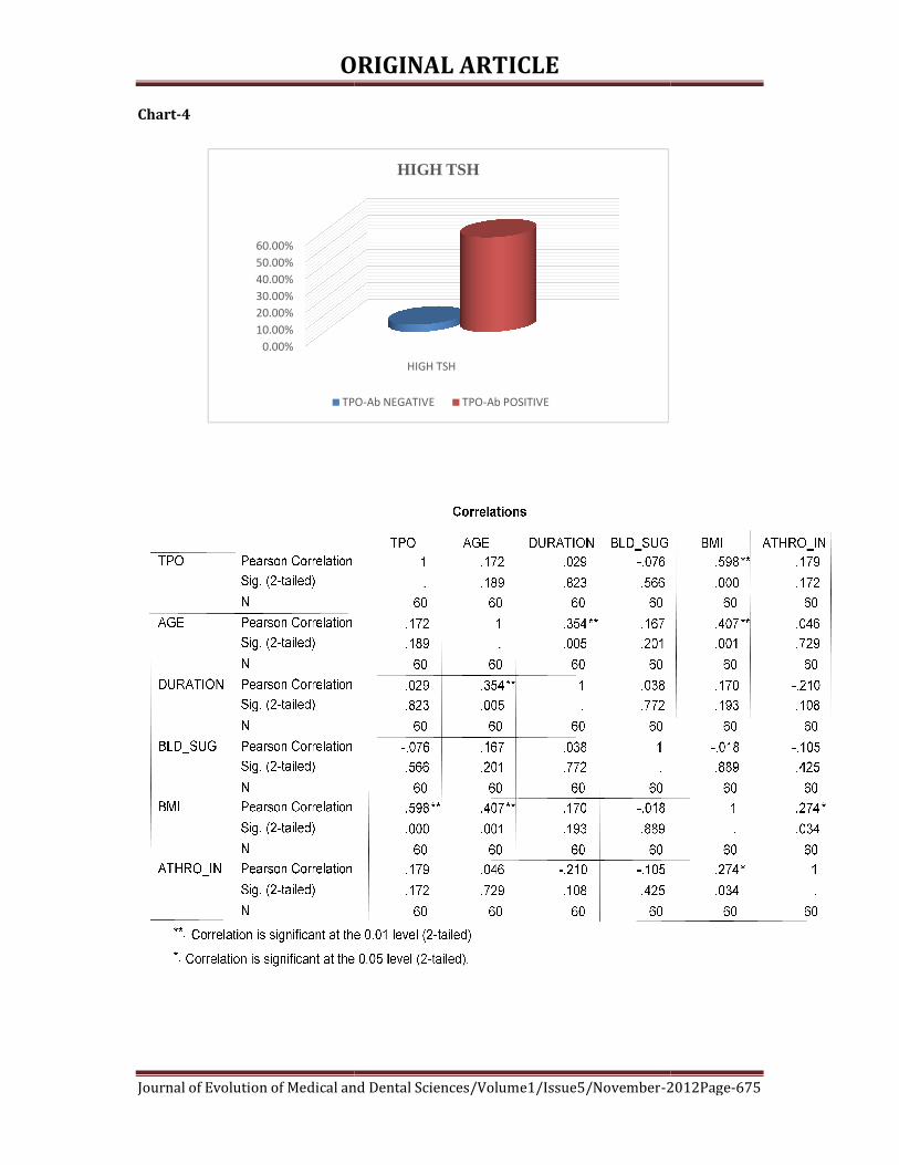
ORIGINAL
Journal of Evolution of Medical and Dental Sciences/Volume1/
Chart-4
0.00%
10.00%
20.00%
30.00%
40.00%
50.00%
60.00%
TPO
ORIGINAL ARTICLE
al and Dental Sciences/Volume1/Issue5/November-2012
HIGH TSH
HIGH TSH
TPO-Ab NEGATIVE TPO-Ab POSITIVE
2012Page-675

ORIGINAL ARTICLE
Journal of Evolution of Medical and Dental Sciences/Volume1/Issue5/November-2012Page-676
Table-4
Table-5
Correlations
1 .032 .043 .057 -.041
. .810 .743 .668 .756
60 60 60 60 60
.032 1 .115 .030 .573**
.810 . .380 .821 .000
60 60 60 60 60
.043 .115 1 .717** .053
.743 .380 . .000 .688
60 60 60 60 60
.057 .030 .717** 1 -.159
.668 .821 .000 . .224
60 60 60 60 60
-.041 .573** .053 -.159 1
.756 .000 .688 .224 .
60 60 60 60 60
Pearson Correlation
Sig. (2-tailed)
N
Pearson Correlation
Sig. (2-tailed)
N
Pearson Correlation
Sig. (2-tailed)
N
Pearson Correlation
Sig. (2-tailed)
N
Pearson Correlation
Sig. (2-tailed)
N
TPO
TGL
HDL
LDL
VLDL
TPO TGL HDL LDL VLDL
Correlation is significant at the 0.01 level (2-tailed).**.
Correlations
1 -.296* -.403** .580** .043
. .022 .001 .000 .746
60 60 60 60 60
-.296* 1 .457** -.585** -.257*
.022 . .000 .000 .048
60 60 60 60 60
-.403** .457** 1 -.768** -.480**
.001 .000 . .000 .000
60 60 60 60 60
.580** -.585** -.768** 1 .368**
.000 .000 .000 . .004
60 60 60 60 60
.043 -.257* -.480** .368** 1
.746 .048 .000 .004 .
60 60 60 60 60
Pearson Correlation
Sig. (2-tailed)
N
Pearson Correlation
Sig. (2-tailed)
N
Pearson Correlation
Sig. (2-tailed)
N
Pearson Correlation
Sig. (2-tailed)
N
Pearson Correlation
Sig. (2-tailed)
N
TPO
T3
T4
TSH
CHOLESTR
TPO T3 T4 TSH CHOLESTR
Correlation is significant at the 0.05 level (2-tailed).*.
Correlation is significant at the 0.01 level (2-tailed).**.

ORIGINAL ARTICLE
Journal of Evolution of Medical and Dental Sciences/Volume1/Issue5/November-2012Page-677
THE PREVALENCE OF CHIKUNGUNYA ARBOVIRAL INFECTION IN AND
AROUND BELLARY DISTRICT, KARNATAKA. Narayan Shrihari, Kumudini T. S, Mariraj. J, Krishna. S
1. Assistant Professor, MD. Department of Microbiology, Vijayanagar Institute of Medical Sciences
(VIMS),Cantonment area, Bellary, Karnataka, India 2. Tutor D Bact, Department of Microbiology, Vijayanagar Institute of Medical Sciences (VIMS),Cantonment
area, Bellary, Karnataka, India 3. Professor, MD. Department of Microbiology, Vijayanagar Institute of Medical Sciences (VIMS),Cantonment
area, Bellary, Karnataka, India 4. Professor & Head, MD. Department of Microbiology, Vijayanagar Institute of Medical Sciences
(VIMS),Cantonment area, Bellary, Karnataka, India
CORRESPONDING AUTHOR
Dr. Narayan Shrihari, Assistant Professor,
Department of Microbiology,
Vijayanagar Institute of Medical Sciences (VIMS),
Cantonment area, Bellary, Karnataka, India.
E-mail: [email protected],
Ph: 0091 9481710655.
ABSTRACT: BACKGROUND: An arbovirus is one that multiplies in a blood sucking arthropod
and is transmitted by the bite to a vertebrate host. Chikungunya fever is a crippling disease
caused by an arbovirus transmitted to human through mosquitoes. The sudden onset of very
high fever along with rash and severe arthralgia are main symptoms. High morbidity with
severe arthralgia persisted for several months made the people both physically and mentally
weak. OBJECTIVES: To know the prevalence of chikungunya arboviral infection in and around
Bellary district. MATERIAL AND METHODS: The laboratory records of clinically suspected
chikungunya patients from January 2009 to December 2011 analyzed retrospectively and
results of Ig M anti chikungunya antibodies tested by Ig M capture enzyme linked
immunosorbant assay (Mac ELISA). RESULTS AND CONCLUSION: A total of 1386 chikungunya
suspected serum samples were analyzed, out of which 343 (24.75%) samples were fond
positive for chikungunya virus infection. Maximum number of positive cases was seen in 2010
(28.40%). The present study emphasizes the continuous sero- epidemiological surveillance for
the effective chikungunya arboviral infection control programme.
KEY WORDS: Chikungunya and Ig M antibody capture ELISA
INTRODUCTION: The arboviruses are transmitted by blood sucking arthropods from one
vertebrate host to another. The vector acquires a lifelong infection through the ingestion of
blood from a viremic vertebrate host. The viruses multiply in the tissues of the arthropod
without evidence of disease or damage. Some arboviruses are maintained in nature by
transovarian transmission in arthropods [Figure No: 1]. The major arboviral diseases
distributed worldwide are yellow fever, dengue , Japanese B encephalitis, chikungunya , St.
Louis encephalitis, western equine encephalitis, eastern equine encephalitis, Russian spring
summer encephalitis, westnile fever and sand fly fever[1]. Chikungunya virus (CHIK V) is a RNA
virus belonging to family Togaviridae, genus Alphavirus. The disease caused by bite of Aedes
aegypti mosquito (Man-Mosquito-Man). The incubation period one to seven days, characterized
by abrupt onset of fever, severe arthralgia and disease is almost self limiting. The literal

ORIGINAL ARTICLE
Journal of Evolution of Medical and Dental Sciences/Volume1/Issue5/November-2012Page-678
meaning of chikungunya is “that which bends up” that stooped posture developed due to
arthritic symptoms of the disease [2]. The disease is generally non-fatal and the acute phase
resolves within three to four days leaving the arthralgic syndrome persisting for some more
time. The serological studies have repeatedly demonstrated the presence of antibodies in
human [2,3]. Incubation period one to seven days, CHIK V produces disease about 48 hours after
mosquito bite. Viraemia declines within three to four days [2,3]. Neutralizing antibodies can
usually be detected after five days fading viraemia[2]. All age groups were affected including
newborns. Recovery from the disease varies by age and longer for elders. Younger patients
recover within 5-15days and middle agers recover in one to three months [2]. Chikungunya fever
is an acute illness characterized by a sudden onset of high fever, rash and joint pain. The most
significant symptom of CHIK V related disease consists of a painful arthralgia that occurs in
almost 100% of patients. Most infections completely resolve within weeks but there are
reported cases of CHIK V induced arthralgia lasting for months or even for years in the form of
recurrent or persistent episodes [2,4].The clinically suspected cases screened using
immunochromatography test kits (Rapid tests) and followed by ELISA detection of Ig M
antibodies and confirmed by virus isolation and virus nucleic acid in sera by RT-PCR [5] The
vector-borne disease and mosquitoes breeding sites are playing an important role in the
transmission and propagation of chikungunya. The present study briefly describes the disease
in general, laboratory diagnosis and the prevalence of chikungunya in and around Bellary.
MATERIAL AND METHODS: The study was conducted at a tertiary care Hospital from January
2009 to December 2011. A total of 1386 serum samples from suspected chikungunya cases
included in our study. Aseptic precautions, two to five ml of blood samples were collected by
venipuncture for chikungunya suspected cases. The samples transported to the Microbiology
laboratory in vaccine carriers with duly filled requisition forms. The serum was separated by
centrifugation of the whole blood sample and labeled with the particulars of the patient and
stored in the refrigerator at -20oC [6]. The test kits used chikungunya Ig M antibody capture
ELISA supplied by Group leader, Arbovirus Diagnostics, National Institute of Virology, Pune,
India. The tests performed strictly as per the manufacturers’ instructions.
RESULTS: During the three years of study period, 1386 Chikungunya suspected serum samples
were analyzed, out of these 343 (24.75%) samples were positive for Chikungunya virus
infection [Table No: 1]. The prevalence of Chikungunya is high in 2010 (28.40%) [Table No: 2].
Male to Female ratio of fever diagnosis in suspected cases is 0.98 [Table No: 3] and majority of
cases belong to age group more than 15 years [Table No: 4].
DISCUSSION: The 2005-2006 epidemics in Indian Ocean island was the most devastating and
had very complicated clinical manifestations associated with encephalopathy and hemorrhagic
fever [7]. Arthralgia persisted for months and years with excruciating pain in joints and ankles [7,8]. The most affected were the aged adults and suffering from diabetes, alcoholic hepatopathy
and impaired renal functions [7,9]. In India, Ahmedabad city, Gujarat and Kerala states
experienced large scale out breaks with morbidity and extensive incapacitation [7]. The Kerala
state had worst epidemic as the infection run through 2006 to 2008 affecting the whole state.
Unique complications such as swollen limbs with painful arthralgia which persisted for long
periods were witnessed among the patients [7]. Karnataka state also more affected during 2006
outbreak, 27 Districts of the state reported over (54.74%) suspected cases. Several districts of

ORIGINAL ARTICLE
Journal of Evolution of Medical and Dental Sciences/Volume1/Issue5/November-2012Page-679
the state such as Bellary, Gulbarga, Tumkur, Bidar, Raichur, Dharwad, Chitradurga, Davangere,
Kolar and Bijapur have reported more number of chikungunya cases during epidemic outbreak
[10]. More than 64% of the entire suspected cases in reported in India during 2008 [10]. We have
noticed that chikungunya outbreak in and around Bellary. The number of confirmed cases are
more in 2010 (28.40%) than in 2009 (23.07%) and subsequently decreased in 2011(19.05%)
[Table No: 2]. All three years statistics revealed that majority of the cases belong to age group of
15 years and above [Table No: 4]. The serological study indicated that arthropod-borne virus
Chikungunya was prevalent in and around Bellary district, although the prevalence differed
according to age, sex, geographic location and the individual virus. The geographical
distribution had a significant influence on the prevalence of antibodies to the virus. This might
be explained by the possible impact of ecological characteristics of the areas on the natural
cycles of the arthropod-borne viruses under consideration [11]. Chikungunya disease recently
emerged as an important public health problem in India. The epidemic occurs in the Indian
Ocean islands during 2006[12]. More than 64% of the entire suspected cases reported in India
during 2008 [13]. We have noticed that Chikungunya outbreak in and around Bellary district. The
number of confirmed cases were more in 2010 (28.40%) than in 2009 (23.07%) and
subsequently decreased in 2011 (19.05%).
CONCLUSION: The arboviral infections mainly dengue, chikungunya and Japanese B
Encephalitis are most common in tropical and subtropical regions. Until the recent epidemic,
CHIK V did not receive much attention due to low mortality, infrequent occurrence and absence
in the developed countries. The serological results (Ig M antibody capture ELISA) clearly
establish the etiology.
KEY MESSAGE: Prevention is better than cure.
REFERENCES:
1. Jawetz, Melnick, Adelberg, Arthropod borne and Rodent borne viral diseases In: Medical
Microbiology, Chapter 38, 23rd edition, Singapore, The Mc Graw Hill Companies;
2004:514.
2. Chhabra M, Mittal V, Bhattacharya D, Rana UVS, Lal S, Chikungunya fever: A re-emerging
viral infection, Indian Journal of Medical Microbiology, 2008; 26(1): 5-12.
3. Srikanth P, Sarangan G, Mallilankaraman, Nayar SA, Barani R, Mattew T, et al, Molecular
Characterization of Chikungunya virus during an outbreak in South India, Indian Journal
of Medical Microbiology, 2010; 28(4): 299-302.
4. Cavrini F, Gaibani P, Pierro AM, Rossini G, Landini MP, Sambri V, Chikungunya: an
emerging and spreading arthropod-borne viral disease, Journal of Infection in
Developing Countries, 2009;3(10): 744-752.
5. Alyson Kelvin A, Outbreak of Chikungunya in the Republic of Congo and the Global
picture, Journal of Infection in Developing Countries, 2011; 5(6): 441-444.
6. Anuradha SK, SurekhaYA, Sathyanarayan MS, Suresh S, Krishna S, Satish SP, Mariraj J,
Ravikumar R , Japanese Encephalitis virus; common cause of viral encephalitis in
paediatric age group in Bellary, Karnataka, India, Journal of Clinical and Diagnostic
Research, 2011; 5(3): 480-482.
7. Sudeep AB, Parashar D, Chikungunya: an overview, Journal of Bioscience, 2008; 33(4):
443-449.

ORIGINAL ARTICLE
Journal of Evolution of Medical and Dental Sciences/Volume1/Issue5/November-2012Page-680
8. Pialoux G, Gauzere BA, Jaureguiberry S, Strobel M, Chikungunya : an epidemic
arboviruses, Lancet Infectious Diseases, 2007; 7:319-327.
9. Couderc T, Chretien F, Schilte C, Disson O, Brigitte M, Guivel BF, Touret Y, Barau G, et al,
A mouse model for Chikungunya: young age and inefficient type-I interferon signaling
are risk factors for severe disease, PLoS Pathogens, 2008; 4:29.
10. Talawar AS, Pujar HS, An outbreak of Chikungunya epidemic in South India-Karnataka,
International Journal of Research and Reviews in Applied Sciences, 2010; 5(3): 229-234.
11. Padbidri VS, Wairagkar NS, Joshi GD, Umarani UB, Risbud AR, Gaikwad DL, Bedekar SS,
Divekar AD, Rodrigues FM, A serological survey of Arboviral diseases among the Human
population of the Andaman and Nicobar islands, India, Southeast Asian Journal of
Tropical Medicine and Public Health, 2002; 33 (4): 749-800.
12. Ukey PM, Bondade SA, Paunipagar PV, Powar RM, Akulwar SL, Study of seroprevalence
of dengue fever in Central India, Indian Journal of Community Medicine, 2010; 35: 517-
519.
13. Kalantri SP, Joshi R, Riley LW, Chikungunya epidemic: An Indian Perspective, National
Medical Journal of India, 2006; 19: 315-322.
Table No: 1 Distribution of suspected and confirmed Chikungunya cases
Suspected Cases Confirmed Cases Percentage 1386 343 24.75
Table No: 2 Distribution of Chikungunya suspected and confirmed cases according to
year
Year Suspected Cases Confirmed Cases Percentag
e
2009 802 185 23.07
2010 500 142 28.40
2011 84 16 19.05
Total 1386 343 70.52
Table No: 3 Distribution of clinically suspected cases according to gender
Gender Suspected Cases
Male 689
Female 697
Total 1386
Table No: 4 Distribution of clinically suspected cases according to age
Age in Years Suspected Cases
Less than 5 12
5 to 10 38
10 to 15 261
More than 15 1075
Total 1386

ORIGINAL ARTICLE
Journal of Evolution of Medical and Dental Sciences/Volume1/Issue5/November-2012Page-681
Figure No: 1
Arbovirus life cycle

CASE REPORT
Journal of Evolution of Medical and Dental Sciences/Volume1/Issue5/November-2012Page-682
CO-INCIDENCE OF MICROFILARIA & TOXOPLASMA IN LYMPH NODE -A
RARE DIAGNOSIS BY FINE NEEDLE ASPIRATION CYTOLOGY (FNAC) Dilip Chandra Barman, Sanjay Kumar Mallick, Bidyut Krishna Goswami, Sweta Sarawagi,
1. Assistant Professor, Department of Pathology, North Bengal Medical College, Darjeeling, West Bengal.
2. Assistant Professor, Department of Microbiology, North Bengal Medical College, Darjeeling, West Bengal.
3. Professor, Department of Pathology, N. R. S. Medical College, Kolkata, West Bengal.
4. Pathologist, B. S. Diagnostic Centre, Siliguri, West Bengal, India.
CORRESPONDING AUTHOR
Dr. Dilip Chandra Barman
Department of Pathology,
North Bengal Medical College and Hospital,
Darjeeling, India.
E-mail: [email protected],
Ph: 0091 9474327035.
ABSTRACT: Toxoplasma lymphadenitis is a rare disease entity, microfilaria also rarely found in
lymph node aspirate but coincidence of toxoplasma gondii and microfilaria in lymph node is
very rare. We report here one such case of a four year old child presented with cervical
lymphadenopathy, fine needle aspiration cytology showed microfilaria and bradyzoides of
toxoplasma gondii, granuloma in a background of reactive lymphoid cells. On serology, serum
IgM anti toxoplasma antibody was positive and IgG was negative.
KEY WORDS: Fine needle aspiration cytology (FNAC), Lymph node, Toxoplasma, Microfilaria.
INTRODUCTION: Filariasis is a major public health problem and it is endemic all over India. [1, 2]
About 120 million people infected in this region and in need of treatment including 40 million
people with overt disease. [3] Despite its high incidence, it is infrequent to find microfilaria in
FNAC smears and only incidentally detected in various sites such as lymph node, breast lump,
bone marrow, bronchial aspirate, pleural and pericardial fluid, ovarian cyst fluid in clinically
unsuspected cases of filariasis with absence of microfilaria in the peripheral blood. [4]
Microfilaria has been observed as a coincidental finding with other infective, inflammatory
conditions and neoplastic lesions. We present here a very rare case in which microfilaria and
Toxoplasma gondi (bradyzoides) together were observed in the fine needle aspirates of cervical
lymph node. No such case report has been documented till date.
CASE REPORT:A 4-year-old male child belongs to a Nepali tribal community of low
socioeconomic condition in Dooars region, presented with multiple enlarged right cervical
lymph nodes since two months with flu like symptoms. On examination, the lymph nodes were
at the right posterior triangle of neck, mildly tender, firm; together measuring 3.5cm × 3.2cm.
Axillary and inguinal lymph nodes were not palpable. He had no visual or neurological
symptom. FNAC of the lymph nodes were done. Aspirate material were whitish granular and
microscopic examination of the air dried Leishman stained and alcohol fixed Hematoxylin &
Eosin (H&E) stained smears showed small cluster of epithelioid histiocytes without necrosis
associated with sheathed microfilaria of Wuchereria bancrofti and cyst of Toxoplasma gondii
(bradyzoides) in a background of heterogeneous population of reactive lymphoid cells [Figure 1
& 2]. Aggregates of activated large ovoid lymphoid cells also seen in the background [Figure 3].

CASE REPORT
Journal of Evolution of Medical and Dental Sciences/Volume1/Issue5/November-2012Page-683
Peripheral blood smear were prepared from blood sample taken for three consecutive
nights showed no microfilaria but eosinophilia was noted (12%). In urine examination, there
was no evidence of chyluria and microfilaria in the centrifuged deposit. Serological study by
Enzyme Linked Immunosorbent Assay (ELISA) for serum IgM anti toxoplasma antibody was
positive but IgG was negative. Patient was non-reactive for HIV-1 & 2. All the test results were
negative in patient’s mother.
DISCUSSION: Microfilaria is transmitted by the Culex mosquito and is caused by two closely
related nematodes, Wuchereria bancrofti and Brugia malayi. About 95% of cases lymphatic
filariasis is caused by infection with Wuchereria bancrofti. [1] Infective larvae penetrate through
the skin wound, enter the lymphatics and travel to the lymph nodes. Adult worms are thread
like creamy white. Females are viviparous, once fertilized they discharge as many as 50,000
microfilaria per day. [5] The sheathed microfilaria of Wuchereria bancrofti having multiple,
coarse, discrete nuclei extending from the head to the tail, except in the small terminal portion
of the caudal end. Thus species are identified by seeing the larval form. [2] Microfilaria displays a
nocturnal periodicity; this is basically a biological adaptation to the nocturnal biting habit of
vector mosquitoes. That is why at least three consecutive night blood sample were examined.
The clinical manifestations varies, may be asymptomatic, or may have acute or chronic
manifestations e.g. lymphangitis, lymphadenitis, lymphedema, and elephantiasis. [1]
Toxoplasmosis is cosmopolitan zoonotic infection caused by a single-celled obligate
intracellular parasite called Toxoplasma gondii. It exists in three form e.g. oocysts, tachyzoites
and bradyzoides (tissue cysts) and transmitted by eating vegetables or drinking water
contaminated with the oocysts commonly passed by cats or from ingestion of mutton, pork,
chicken etc. Early maternal infection (first and second trimester) results in spontaneous
abortion and intrauterine foetal death (IUFD).
Toxoplasma is an infection which is highly variable in its clinical manifestation, these
include myocarditis, encephalomyelitis, chorioretinitis especially in the immunocompromised
host and the healthy host who harbour the infection usually remains asymptomatic.
Superficial enlarged lymph nodes are the most common presenting sign of acquired
toxoplasmosis. [6] Usually involves the posterior cervical lymph nodes. [7] In cytology smear,
some lymphoid cells with relatively large, ovoid, pale nuclei may be seen. These cells probably
correspond to the pale monocytoid B-cells in histological sections. Microcysts and organisms of
Toxoplasma gondii are rarely seen. Diagnosis is usually done by identifying tissue cysts and
serological tests. [6], [8],[9], [10]
Here in our case also, right posterior cervical lymph nodes were involved and the
smears showed loose aggregates of activated large ovoid lymphoid cells [Figure 3].
The objective of the study is to document the value FNAC in the diagnosis of filariasis and
toxoplasmosis in lymph node and thus demonstration and identification of these parasites in
cytological smears played a significant role in the prompt recognition of the disease and
initiation of specific treatment, thus avoiding the more severe manifestations of lymphatic
filariasis and toxoplasmosis. Secondly to highlight such a rare coincidence of two infection in
lymph node.
CONCLUSION: Although microfilaria and toxoplasma cysts in cytological smears are considered
incidental findings, this case illustrates the value of FNAC in the detection of asymptomatic and
clinically unsuspected cases of filariasis & toxoplasmosis. Absence of microfilaria in the

CASE REPORT
Journal of Evolution of Medical and Dental Sciences/Volume1/Issue5/November-2012Page-684
peripheral blood does not exclude filarial infection. Filariasis may be detected in a clinically
unsuspected case, especially in an endemic zone. The spectrum of host response may vary from
no reaction to a marked inflammatory response. [1] The entire spectrum of changes should be
kept in mind while practicing cytopathology in an endemic area. In such situations, a strong
clinical suspicion and meticulous screening of cytology smears may lead to a correct diagnosis.
At the same time, the cytopathologist should search for a coexisting pathology thoroughly. In the
present case, we feel that the presence of microfilaria in association with Toxoplasma gondii is a
very rare coincidental finding. The patient was harboring subclinical filariasis when he was
suffering from toxoplasmosis. Microfilaria was not detected in the peripheral blood smears. This
also highlights the importance of screening smears for parasites even in the absence of clinical
symptoms, particularly in highly endemic areas.
REFERENCES:
1. Park K. Park's Textbook of Preventive and Social Medicine. 21st ed. Jabalpur, India:
Banarasidas Bhanot publisher; 2011.Chapter 5; page: 244.
2. Chatterjee KD. Parasitology (Protozoology and Helminthology) 12th ed. Calcutta, India:
Chatterjee Medical Publishers; 1980; 190–8.
3. WHO (2009), Weekly Epidemiological Record, No. 11/12, 13th March, 2009.
4. Varghese R, Raghuveer CV, Pai MR, Bansal R. Microfilariae in cytologic smears: A report
of six cases. Acta Cytol 1996; 40 (2): 299-30.
5. Nelson GS. Filarial infections. Med Int 1981; 1: 77–82.
6. Stansfelt. AG. Histological diagnosis of Toxoplasmic lymphadenitis. J. clin. Path 1961; 14:
565.
7. Leopold GK. Koss’ Diagnostic Cytology and its Histopathologic Bases: 5th ed, Lippincott
Williams & Wilkins Publisher; 2006. Vol-II, Chapter 31, Page: 1195.
8. Argyle JC, Schumann GB, Kjeldsberg CR, Anthens JW. Identification of a Toxoplasma cyst
by fine needle aspiration. Am J Clin Pathol 1983; 80: 256-258.
9. Christ ML, Feltes-Kennedy M. Fine needle aspiration cytology of Toxoplasmic
lymphadenitis. Acta Cytol 1982; 26: 425-428.
10. Jayaram N, Ramprasad AV, Chatan M, Sujay AR. Toxoplasma lymphadenitis. Analysis of
cytologic and histologic criteria and correlation with serologic tests. Acta Cytol 1997; 41:
653-658.
Figure 1: Tissue cyst of Toxoplasma gondii (Leishman’s stain x 40)

CASE REPORT
Journal of Evolution of Medical and Dental Sciences/Volume1/Issue5/November-2012Page-685
Figure 2: Tissue cysts Toxoplasma gondii & coiled microfilaria of Wuchereria bancrofti
(H/E stain x10)
Figure 3: Activated large ovoid lymphoid cells (Leishman’s stain x 100)

CASE REPORT
Journal of Evolution of Medical and Dental Sciences/Volume1/Issue5/November-2012Page-686
MULTIPLE LIVER ABSCESS BY MIXED BACTERIAL ETIOLOGY: AN
UNUSUAL CASE REPORT
Mannur Sharada, Neelesh Naik Prashanth. B. N.
1. Associate Professor, Department of Microbiology, Sri Siddhartha Medical College, Tumkur 2. Assistant Professor, Department of Microbiology, Sri Siddhartha Medical College, Tumkur
3. Medical Director, Department of Gastroenterology, Vikram Apkon Specialty Polyclinic & Diagnostics.
CORRESPONDING AUTHOR
Dr. Neelesh Naik, Assistant Professor,
Dept. of Microbiology,
Sri Siddhartha Medical College, Tumkur
E-mail: [email protected],
Ph: 0091 9738630329.
ABSTRACT: Pyogenic liver abscess is a serious disease, which is potentially fatal if left
untreated. In developed countries, pyogenic abscesses are the most common but worldwide,
amoebae are the most common cause. We report a 60-year-old diabetic woman with a 2 months
history of pain abdomen, 1 month history of high fever and anorexia who had multiple liver
abscess caused by anaerobic Actinomyces species and Enterococcus faecalis and was
complicated by peritonitis. Treatment included prompt percutaneous drainage coupled with
long-term intravenous administration of Amikacin, Metronidazole and Piperacillin/
Tazobactum. The patient later died due to peritonitis.
KEY WORDS: Liver abscess, Enterococci, anaerobic Actinomyces
INTRODUCTION: Pyogenic liver abscess (PLA) is a serious infrequent disease caused by
bacterial, parasitic or fungal organisms and is potentially fatal if left untreated. In previous years
it was the disease of the young adults but recently the disease has evolved as disease of the
elderly 1. Liver abscess may be solitary or multiple; they may arise from hematogenous spread
of bacteria or from local spread from contiguous sites of infection within the peritoneal cavity.
In the past, appendicitis with rupture and subsequent spread of infection was the most common
source for a liver abscess. Currently, associated disease of the biliary tract is most common.
Pylephlebitis, usually arising from infection elsewhere in the peritoneal cavity is another
common source for bacterial seeding of the liver 2.
CASE HISTORY: A 60 year old female diabetic patient came with the history of pain abdomen in
right upper quadrant since 2 months, associated with on and off fever with chills, malaise and
anorexia since 1 month. Patient gave past history of epigastric pain which was treated with
antacids. On examination she had marked tenderness in the right upper quadrant with
hepatomegaly of 4 cm below the right costal margin. Her temperature was 390C, pulse 96
beats/min and blood pressure 104/80 mm Hg. Laboratory data were as follows: hemoglobin
11.6 gm/dl; white blood cell count 16300/mm3; platelet count 4.80 lakh/mm3; PT 14 seconds;
PCV 33%; MCV 79.7 fl; MCH 28 pg; MCHC 35.2 gm/dl; Red blood cell count 4.14 million/mm3;
Differential count: Polymorphs 90%, Lymphocytes 7%, Monocytes 3%, Basophils & Eosinophils
0%; Total Protein 5.8 gm/dl; Albumin 2.5 gm/dl; Globulin 3.3 gm/dl; A/G ratio 0.8; total
bilirubin 0.7 mg/dl; bilirubin direct 0.0 mg/dl; bilirubin indirect 0.7 mg/dl; Alkaline
phosphatase 114 IU/L; SGOT 19 IU/L; SGPT 18 IU/L; GGT 23 U/L; Glucose (Random) 161

CASE REPORT
Journal of Evolution of Medical and Dental Sciences/Volume1/Issue5/November-2012Page-687
mg/dl; PPBS 200 mg/dl; urine analysis was normal except for presence of 8 – 10 pus cell/ hpf
and traces of albumin. The test for HIV and HBsAg was found to be negative.
Ultrasonography of the abdomen revealed that there was a well defined hypo-echoic
lesion with internal debris in right lobe of liver with sub-capsular collection in the right lobe,
suggesting the presence of abscess with sub-capsular extension. CT scan of the abdomen
confirmed the presence of multiple irregular abscesses varying in size, predominantly in the
right lobe of the liver. Gram stain of the aspirated pus showed the presence of gram positive
filamentous bacilli which was broken into coccoid and bacillary forms, gram positive cocci in
pairs and short chains and scanty of gram negative fusiform bacilli along with plenty of pus
cells. Wet mount of pus did not reveal the presence of trophozoites of Entamoeba histolytica.
The Ziehl- Neelsen stain did not show presence of acid fast bacilli. A definitive diagnosis of
mixed infection was made when an anaerobic Actinomyces species and Enterococcus faecalis
was cultured from aspirated pus by conventional method. The antibiotic sensitivity testing of
Enterococcus faecalis was done by Kirby-Bauer disc diffusion method. The isolate was found to
be sensitive to ampicillin, ampicillin/sulbactum, cotrimoxazole, piperacillin/tazobactum,
vancomycin, ceftriaxone, amikacin, high level gentamicin (HLG) and teicoplanin and was
resistant to ciprofloxacin and linezolid. The patient was treated by percutaneous drainage and
with long-term intravenous administration of amikacin, metronidazole and
piperacillin/tazobactam. Later patient developed peritonitis and succumbed to death.
DISCUSSION: Pyogenic liver abscess is a serious infrequent disease caused by bacterial,
parasitic or fungal organisms and is potentially fatal if left untreated 1. The microorganisms
most often recovered from pyogenic liver abscess are Escherichia coli, anaerobic Streptococci,
gram negative bacilli, Bacteroides fragilis, Staphylococcus aureus, Clostridium species etc., but
the incidence varied considerably among previous reports. Pyogenic liver abscesses were more
commonly seen in elderly patients and in patients with diabetes mellitus. The abscesses were
mostly single and located more frequently in right lobe of the liver 1, 3. Multiple liver abscesses
are not frequently reported in the literature. The overall mortality rate of pyogenic liver
abscess is 11%- 31% and the mortality rate is high in patients with multiple liver abscess.
Patients with diabetes mellitus, immune deficiency, sickle cell anemia, malignancy and liver
transplants are at greater risk for developing liver abscess and in the majority of the cases more
than one organisms have been isolated. Without appropriate diagnosis and treatment, the
pyogenic liver abscesses are almost uniformly fatal. Early diagnosis as well as treatment with
appropriate antibiotics and selective drainage can subsequently reduce the mortality 4.
A high incidence of anaerobic microorganisms (15-46%) as the cause of liver abscess
has been reported recently and because of this high incidence, antibiotics effective against
anaerobes and aerobic microorganisms should be used in all patients 3, 5. CT scan is probably
the most useful aid in diagnosing and localizing hepatic abscesses 6. Biliary tract disease is the
etiology of the abscess in most cases, but sometimes the origin remains unidentified 7. In our
case the cause of multiple liver abscess is probably due to extension from a small perforation
and extension of pus from healed peptic ulcer, as no other aetiology was found in the patient for
the presence of liver abscess. Hepatic actinomycosis should also be considered in the
differential diagnosis of liver abscesses and space-occupying lesions of the liver in
immunocompetent patients 8. The strain of Enterococcus faecalis isolated was resistant to
linezolid, which is relatively rare and appears either due to previous treatment with linezolid or
transmission from other patients 9. The source of linezolid resistant enterococci in our case

CASE REPORT
Journal of Evolution of Medical and Dental Sciences/Volume1/Issue5/November-2012Page-688
could not be determined. Linezolid resistant Enterococcus faecalis is an emerging pathogen and
hence it is important to minimize the emergence of resistance by using it only if the therapeutic
indications exist and keeping the duration of treatment as short as possible. In summary,
multiple liver abscess due to mixed infection with Enterococcus faecalis and an anaerobic
Actinomyces species is a rare entity, but a life threatening disease. Hence such cases requires
timely recognition using CT scan and guided aspiration to start an early and specific therapy to
prevent mortality among high risk and elderly patients.
REFERENCES:
1. Galdfelter T, Brooks M. Liver abscess. JMSMA 1981;7:173-4.
2. Baron MJ, Kasper DL. Intraabdominal infections and abscesses. In: Longo DL, Fauci AS,
Kasper DL, editors. Harrison’s principles of internal medicine. 18th ed. USA: Mc Graw
Hill; 2010. P. 1077-83.
3. Ergas D, Hai AA, Sthoeger Z, Menahem BH, Miller R. Multiple pyogenic liver abscesses
following hemorrhoid banding. IMAJ 2007;9:753-4.
4. Bahloul M, Chaari A, Khlaf NB, Kallel H, Herguefi L, Chelly H, et al. Multiple pyogenic liver
abscess. World J Gastroenterol 2006;12(18):2962-3.
5. Alvarez JA, Baldonedo RF, Bear IG, Jorge JL. Anaerobic liver abscesses as initial
presentation of silent colonic cancer. HPB ( Oxford) 2004;6(1):41-42.
6. Tzur T, Liberman S, Felzenstein I, Cohen R, Rivkind AI, Almogy G . Liver abscesses
caused by Streptococcus milleri: An uncommon presenting sign of silent colonic cancer.
IMAJ 2003;5:206-7.
7. Wang YJ, WenSC, Chien ST, King J, Hsuea CW, Feng NH. Liver abscess secondary to
sigmoid diverticulitis. A case report. J Intern Med Taiwan 2005;16:289-94.
8. Tiftikci A, Vardareli EN, Kaban K, Peker O, Akansel S, Tozun N. Actinomycotic hepatic
abscess. Hepatol Int 2008;2(1):133–5.
9. Burleson BS, Ritchie DJ, Micek ST, Dunne WM. Enterococcus faecalis resistant to
linezolid: case series and review of the literature. Pharmacotherapy 2004;24(9):1225-
31.

CASE REPORT
Journal of Evolution of Medical and Dental Sciences/Volume1/Issue5/November-2012Page-689
RECURRENT MARJOLIN’S ULCER WITH REGIONAL LYMPH NODE
METASTASIS
Anjali Sethi, Deepak Sethi, Vishwas Johri
1. Assistant Professor, Department of Surgery, RNT Medical College and MB Govt. Hospital, Udaipur,
Rajasthan, India.
2. Medical Officer, Department of Surgery, RNT Medical College and MB Govt. Hospital, Udaipur, Rajasthan,
India.
3. Assistant Professor, Department of Surgery, RNT Medical College and MB Govt. Hospital, Udaipur,
Rajasthan, India.
CORRESPONDING AUTHOR
Dr. Deepak Sethi,
303 Akshansh Apartments,
Keshav Nagar, UDAIPUR – 313001
E-mail: [email protected] , [email protected]
Ph: 0091 294 2471892, 0091 9414248134.
ABSTRACT: Marjolin’s ulcer is a malignant tumour developing in a chronic skin lesion (burn
scar, vaccination scar, non-healing wound etc.). The majority of cases reported are squamous
cell carcinoma. Surgery remains the first treatment of choice (resection with 2cms. safety
margin of healthy skin for primary squamous cell carcinoma Marjolin ulcers and 2.5cms. safety
margin for recurrent cases). Recurrence after surgery and regional lymph node metastasis are
not uncommon (17% & 30% respectively).We presents a case report and literature review of
Recurrent Marjolin’s Ulcer with regional Lymph Node Metastasis. Marjolin's ulcer should be
considered as a significant post-burn complication; it should be treated with full emphasis on
adequate local clearance and regular follow up for many years; if not treated adequately, it may
lead to complicated recurrence.
KEY WORDS: Marjolin’s Ulcer, Recurrent, Lymph Node Metastasis.
INTRODUCTION: Marjolin’s ulcer is a malignant tumour developing in a chronic skin lesion
(burn scar, vaccination scar, non-healing wound etc.).(1) The majority of cases reported are
squamous cell carcinoma but other type of malignancy such as basal cell carcinoma, malignant
melanoma, liposarcoma, osteosarcoma, and fibrosarcoma can also be seen although rare.(2) The
incidence of burn scar undergoing malignant transformation has been reported to be 0.77-2 %.(
3)
CASE REPORT: A 22 year old male was presented to our surgical department with exophytic
ulcerated growth of size 7x7 cm. near angle of right scapula. He had a past history of burn over
anterior chest wall, back and left arm 12 years ago that healed completely by conservative
management. Approx. 11 years after initial insult, the patient developed an ulcer on the back
over burn scar. It gradually increased in size to become exophytic growth of size 12x12cms. No
regional lymph node enlargement was noted at that time. Whole of the ulcer with a 2 cm.
margin of healthy skin was excised. The defect in skin was closed by partial thickness skin graft.
Histopathological examination of the excised tissue was suggestive of poorly differentiated
squamous cell carcinoma.
8 months after surgery & skin grafting, the patient came with exophytic ulcerated
growth of size 7x7 cm. near angle of right scapula. It was fixed to underlying muscles of back.
CASE REPORT
Journal of Evolution of Medical and Dental Sciences/Volume1/Issue5/November-2012Page-690
Patient was also having another fungating growth in left axilla which was 6x6 cms. in size, fixed
to underlying structures causing restriction of movements at shoulder. Serous discharge was
present from axillary growth. There was no vascular or neurological impairment present in left
upper limb. Biopsy from axillary growth was positive for squamous cell carcinoma.
LITERATURE REVIEW: The eponym “Marjolin’s ulcer” was derived from French surgeon Jean
Nicholas Marjolin in 1828 who observed and classified cellular change in burn skin and coined
the term “ulcere cancroide”.(4) The skin lesion underlying development is predominantly burn
scar (75%), traumatic non healing wound (8%), venous stasis ulcer (6%), pressure ulcer (3%)
and other e.g. Frost bite, vaccination scar .(1)
The latency period is inversely proportional to patient’s age at the time of skin injury. In
chronic Marjolin’s ulcer it ranges from 20-40 year as transformation from non-healing wound to
malignant disease is slow. The average time lag between the burns and subsequent malignant
ulceration is 19 years. (5) In acute Marjolin’s ulcer it occurs within few weeks to one year.(1)
Average age at diagnosis of Marjolin’s ulcer is in 5th decade of life with a range of 18-84 years.(2)
It is seen more frequently in males as compare to females with ratio of 3:1.(2) Most commonly
it is located on lower extremities (53%), upper extremities (18%), trunk (12%), face and nape
(5%), scalp (9%). (4)
Aetiology of Marjolin’s ulcer is not yet clear. Many hypotheses have been suggested.
Slow healing and scar instability characterized by chronic irritation and the induction of
constantly proliferating epidermal unit have been blamed. Repeated cycle of healing and
breakdown, wound that never healed and application of irritant medication are all result in
reduced ability to withstand carcinogens.(6) As a result of constant breakdown of ulcer, a
nutritional deficiency develops, owing to release of toxins by autolysis and heterolysis of scar.
This yields an epithelium that is unable to withstand the carcinogens produced by skin because
of excessive heat and radiation. (7) Ultraviolet rays are also found to be associated with
Marjolin’s ulcer. On histological examination of sun damaged skin, dyskeratosis and vacuolated
keratinocytes known as sun burn cell are seen. Marjolin’s ulcer least frequently founds on trunk
which is not frequently exposed to sunlight. (8) It has also been suggested that relatively
avascular scar tissue act as immune privileged site that allows the tumour to resist body
defences against foreign cell.(6)
Scar tissue acts as a barrier for the tumours, if we release this barrier, the virulent
spread of the tumour will be permitted. (8) Regional lymph node metastasis and recurrence after
surgery is not uncommon. Metastasis to regional lymph nodes is seen in 30% of cases and local
recurrence occurs in 17% of patients. The median interval to recurrence after surgery is 15
months. Locoregional recurrence is more common in female patients and those with high-grade
tumours. (9) Poorly differentiated squamous cell carcinomas have a tendency to spread to lymph
nodes earlier. Squamous cell carcinomas resulting from the Marjolin's ulcer have a much greater
tendency to metastasize than squamous cell carcinomas resulting from the other causes (10).
Various studies suggested that all chronic wound should be closed surgically either by
skin graft or skin muscle flap. Large wound should not be left for secondary intention healing.
Burn scar from childhood should be carefully monitored; biopsy of any suspected lesion should
not be delayed. At present no standard treatment for Marjolin’s ulcer is suggested. Surgery
remains the first treatment of choice. Marjolin’s ulcer should be excised with a 2 cm. margin of
normal healthy tissue, which may necessitate amputation with lesion involving joint space or
deep local extensive invasion. Although classically 2cm safety margin is still widely used for
resection of primary squamous cell carcinoma Marjolin ulcers, 2.5cm safety margin is better for
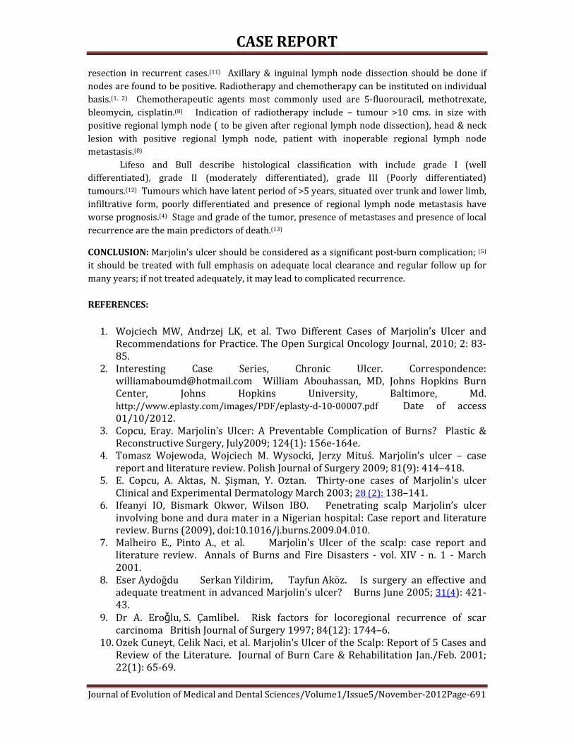
CASE REPORT
Journal of Evolution of Medical and Dental Sciences/Volume1/Issue5/November-2012Page-691
resection in recurrent cases.(11) Axillary & inguinal lymph node dissection should be done if
nodes are found to be positive. Radiotherapy and chemotherapy can be instituted on individual
basis.(1, 2) Chemotherapeutic agents most commonly used are 5-fluorouracil, methotrexate,
bleomycin, cisplatin.(8) Indication of radiotherapy include – tumour >10 cms. in size with
positive regional lymph node ( to be given after regional lymph node dissection), head & neck
lesion with positive regional lymph node, patient with inoperable regional lymph node
metastasis.(8)
Lifeso and Bull describe histological classification with include grade I (well
differentiated), grade II (moderately differentiated), grade III (Poorly differentiated)
tumours.(12) Tumours which have latent period of >5 years, situated over trunk and lower limb,
infiltrative form, poorly differentiated and presence of regional lymph node metastasis have
worse prognosis.(4) Stage and grade of the tumor, presence of metastases and presence of local
recurrence are the main predictors of death.(13)
CONCLUSION: Marjolin's ulcer should be considered as a significant post-burn complication; (5)
it should be treated with full emphasis on adequate local clearance and regular follow up for
many years; if not treated adequately, it may lead to complicated recurrence.
REFERENCES:
1. Wojciech MW, Andrzej LK, et al. Two Different Cases of Marjolin’s Ulcer and
Recommendations for Practice. The Open Surgical Oncology Journal, 2010; 2: 83-
85.
2. Interesting Case Series, Chronic Ulcer. Correspondence:
[email protected] William Abouhassan, MD, Johns Hopkins Burn
Center, Johns Hopkins University, Baltimore, Md.
http://www.eplasty.com/images/PDF/eplasty-d-10-00007.pdf Date of access
01/10/2012.
3. Copcu, Eray. Marjolin's Ulcer: A Preventable Complication of Burns? Plastic &
Reconstructive Surgery, July2009; 124(1): 156e-164e.
4. Tomasz Wojewoda, Wojciech M. Wysocki, Jerzy Mituś. Marjolin’s ulcer – case
report and literature review. Polish Journal of Surgery 2009; 81(9): 414–418.
5. E. Copcu, A. Aktas, N. Şişman, Y. Oztan. Thirty-one cases of Marjolin's ulcer
Clinical and Experimental Dermatology March 2003; 28 (2): 138–141.
6. Ifeanyi IO, Bismark Okwor, Wilson IBO. Penetrating scalp Marjolin’s ulcer
involving bone and dura mater in a Nigerian hospital: Case report and literature
review. Burns (2009), doi:10.1016/j.burns.2009.04.010.
7. Malheiro E., Pinto A., et al. Marjolin's Ulcer of the scalp: case report and
literature review. Annals of Burns and Fire Disasters - vol. XIV - n. 1 - March
2001.
8. Eser Aydoğdu Serkan Yildirim, Tayfun Aköz. Is surgery an effective and
adequate treatment in advanced Marjolin's ulcer? Burns June 2005; 31(4): 421-
43.
9. Dr A. Eroǧlu, S. Çamlibel. Risk factors for locoregional recurrence of scar
carcinoma British Journal of Surgery 1997; 84(12): 1744–6.
10. Ozek Cuneyt, Celik Naci, et al. Marjolin's Ulcer of the Scalp: Report of 5 Cases and
Review of the Literature. Journal of Burn Care & Rehabilitation Jan./Feb. 2001;
22(1): 65-69.
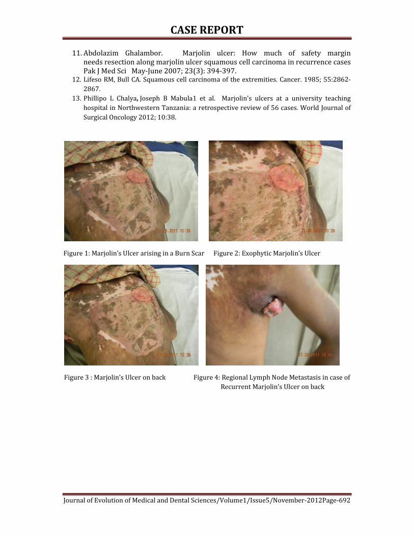
CASE REPORT
Journal of Evolution of Medical and Dental Sciences/Volume1/Issue5/November-2012Page-692
11. Abdolazim Ghalambor. Marjolin ulcer: How much of safety margin
needs resection along marjolin ulcer squamous cell carcinoma in recurrence cases
Pak J Med Sci May-June 2007; 23(3): 394-397. 12. Lifeso RM, Bull CA. Squamous cell carcinoma of the extremities. Cancer. 1985; 55:2862-
2867.
13. Phillipo L Chalya, Joseph B Mabula1 et al. Marjolin's ulcers at a university teaching
hospital in Northwestern Tanzania: a retrospective review of 56 cases. World Journal of
Surgical Oncology 2012; 10:38.
Figure 1: Marjolin’s Ulcer arising in a Burn Scar Figure 2: Exophytic Marjolin’s Ulcer
Figure 3 : Marjolin’s Ulcer on back Figure 4: Regional Lymph Node Metastasis in case of
Recurrent Marjolin’s Ulcer on back

CASE REPORT
Journal of Evolution of Medical and Dental Sciences/Volume1/Issue5/November-2012Page-693
KLIPPEL TRENAUNAY SYNDROME: REPORT OF A RARE, MILD FORM
OF SYNDROME Shubhangi. V. Agale, Bhavana. M. Bharambe, Ravikiransingh Pawar, A. G. Valand
1. Associate Professor, Department of Pathology, Grant Medical College & Sir J. J. Group of Hospitals, Mumbai.
2. Assistant Professor, Department of Pathology, Grant Medical College & Sir J. J. Group of Hospitals, Mumbai.
3. Resident, M. D. Department of Pathology, Grant Medical College & Sir J. J. Group of Hospitals, Mumbai.
4. Professor & Head, Department of Pathology, Grant Medical College & Sir J. J. Group of Hospitals, Mumbai
CORRESPONDING AUTHOR
Dr. Shubhangi V Agale
Associate Professor, Department of Pathology
Grant Medical College & Sir JJ Group of Hospitals, Byculla,
Mumbai- 400008.
E-mail: [email protected],
Ph: 0091 9987533498.
ABSTRACT: Klippel Trenaunay Syndrome (KTS) is a congenital vascular disease characterized
by malformations of capillary, venous and lymphatic vessels with bony and soft tissue
hypertrophy. KTS is a sporadic disease with unknown etiology, and there is no predilection for
gender or any particular ethnicity. The disease appears more frequently at birth, childhood or
adolescence. It generally affects only one extremity and lesions are present at birth or appear
by the age of 12 years. Clinical presentation of this syndrome is protean ranging from minimal
asymptomatic disease to life threatening bleeding and embolism. Management of this syndrome
includes careful diagnosis, prevention and treatment of complications. We report a case of KTS
in a 25 year old female who presented with mild form of disease.
KEYWORDS: Soft tissue hypertrophy, vascular malformation, congenital disease.
INTRODUCTION: Klippel Trenaunay is a rare, congenital disorder characterized by a triad of
capillary vascular malformation, venous malformation and soft tissue or bone hypertrophy.[1-7]
It usually affects lower extremity[6] and lesions are present since birth in approximately 90% of
patients presenting before 12 years of age.[6,8] KTS has a wide spectrum of presentation, from
truncular to extratruncular, from infiltrating to limited forms, containing primarily three
anomalous vascular elements: veins, capillaries and lymphatics.[7] However patients present
with protean manifestations.[2] We report a case of KTS in a 25 year old female who presented
with mild form of disease.
CASE REPORT: A 25 year old married female presented to dermatology department of our
hospital with multiple red colored lesions on the left lower extremity since birth which were
increasing in size with age. She developed difficulty in walking at the age of 6 years. There was
no history of similar symptoms in other family members. On examination, she had multiple,
erythematous, hyper pigmented plaques on lateral side of left lower extremity with largest
plaque of 10x8 cm (Figure 1a).The patient also difficulty in extension of the limb with presence
of hypertrophy and varicose veins (Figure 1b). Hematological investigations showed:
Hemoglobin of 10.2 gm%, WBC of 10,000/mm3, Platelets of 2,10,000/mm3 and microcytic
hypochromic anemia on peripheral smear examination. The vascular anomalies of internal
organs were ruled out using abdominal ultrasonography, Doppler studies and CT scan of

CASE REPORT
Journal of Evolution of Medical and Dental Sciences/Volume1/Issue5/November-2012Page-694
abdomen and pelvic region. The histopathological examination of the skin biopsy showed
ectatic blood vessels in upper dermis with stasis of erythrocytes (Figure 2). The
histopathological diagnosis of nevus flammeus was offered and considering the clinical features,
the case was diagnosed as Klippel Trenaunay Syndrome (mild form).
DISCUSSION: Klippel-Trenaunay Syndrome was first described by two French doctors, Klippel
and Trenaunay in 1900.[6,7] The incidence and genetic predisposition has not been proved
despite various case reports in world literature.[7] KTS occurs sporadically, and shows no
particular racial, sexual or geographical predilection. It affects skin, veins, lymphatic system,
bone and soft tissues of an extremity. Clinical presentation of patients with KTS has a wide
spectrum from incomplete, mild form of port wine stains and few varicose veins causing only
cosmetic deformity to severe disability associated with massive limb overgrowths, chronic pain
syndrome, skin infections, arthritis, thrombo- embolism and life-threatening pelvic or recurrent
rectal bleeding from venous malformations.[7] Capillary malformation is usually a port wine
stain or nevus flammeus. The port-wine stain is usually red to purple in color and is flat. It
usually has an irregular margin and clear, sharp border but rarely crosses the midline. It may or
may not blanch on pressure. It usually occurs on the ipsilateral side as the affected limb.[9]
Varicosities are extensive, atypically very large, take an erratic course and begin to manifest in
early childhood.[9] Varicose veins is the characteristic feature affecting the lower limbs which
was present in our patient.[2] Deep vein anomalies like venous hypoplasia to frank aneurysm,
valve hypoplasia to avalvulia have been described with lymphatic malformations.[7] The bony
abnormalities may affect all bones in an extremity or limited to one or two bones. Single limb
involvement is found in 80-85% patients. Apart from hypertrophy or sometimes hypotrophy of
bones other deformities includes macrodactyly, syndactyly, split hand deformity, phalangeal
agenesis and dislocation of hip joint.[7] At least two of three main symptoms (port-wine stains,
varicosity, and hypertrophy of soft tissues and bones) must be present for the diagnosis Klippel-
Trenaunay syndrome to be accepted.[2] Association with arteriovenous malformation is called as
Klippel Trenaunay Weber syndrome.[6] Various authors have described other systemic
involvements in patients of KTS. Central nervous system abnormalities may include
microcephaly, macrocephaly, cerebral arteriovenous malformations and orbito- frontal
varices.[10] Patients may present with neurological symptoms due to compression of spinal cord
by hemangiomas. Gastrointestinal bleeding and genitourinary lesions like hematuria and
vascular malformations of scrotum, penis, vulva, vagina and bladder may also occur.[10]
Several theories have been proposed which include (1) Servelle's theory of a primary
obstruction of the venous system resulting in venous hypertension and therefore development
of abnormal venous pathways and tissue overgrowth; (2) failure of regression of the lateral limb
bud vein; and (3) alteration of the tight balance between angiogenesis and vasculogenesis,
which is controlled by numerous genes, among other theories.[7] Most recent being mutation in
angiogenic factor VG5Q leading to vascular abnormalities.[3]
The management of KTS has been largely conservative with compression therapy as
mainstay of treatment.[7] Conservative measures for varicose veins like compression stockings,
alleviating pain by painkillers, antibiotics for infections, anticoagulant therapy for
thrombophlebitis, laser for ulcers and port wine stains is indicated. Surgery is indicated for
cosmetic reasons or for complications of venous insufficiency and for bone or soft tissue
overgrowth. The conservative management is sufficient in mild form of KTS but in female
patients of reproductive age group, KTS increases obstetric risk and can exacerbate

CASE REPORT
Journal of Evolution of Medical and Dental Sciences/Volume1/Issue5/November-2012Page-695
complications mainly thromboembolism and hemorrhagic. Probably risk is 10 times higher than
in the normal population.[1,2] The other factors which exacerbate this risk are oral
contraceptives, surgery and pregnancy.[1,2,4,11]
So these patients have to be managed with multidisciplinary approach and must be
counseled for: a) Use of contraceptive measures other than oral contraceptive pills b) Pregnancy
related complications such as deep vein thrombosis, thromboembolism, and coagulopathy c)
Surgery for cosmetic appearance of the leg and d) Risk of abnormalities in the fetus.
The multidisciplinary management with long term, regular follow up is mandatory in female
patients with Klippel-Trenaunay Syndrome.
CONCLUSIONS: Klippel Trenaunay syndrome is a rare condition with protean manifestations
which should be extensively investigated for the reason of being associated with various
systemic manifestations and these patients have to be managed accordingly. The clinicians need
to be aware of mild form of KTS which can be managed conservatively and has better prognosis.
KTS occurring in female patients of reproductive age group has different implications and has to
be managed by a multidisciplinary team of obstetrician, physician and surgeon.
REFERENCES:
1. González-Mesa E, Blasco M, Andérica J, Herrera J. Klippel-Trenaunay syndrome
complicating pregnancy. BMJ Case Rep doi: 10.1136/bcr-2012-006534.
2. Gundogan TG, Jacquemyn Y. Klippel - Trenaunay syndrome and pregnancy. Obstetr &
Gynaecol Int doi: 10, 1155/ogt-2010706850.
3. Tian XL, Kadaba R, You SA, Liu M, Timur AA, and Yang L, et al.: Identification of an
angiogenic factor that when mutated causes susceptibility to Klippel-Trenaunay
syndrome. Nature. 2004; 427:640-5.
4. Martin JR, Pels SG, Paidas M, Seli E. Assisted reproduction in a patient with Klippel-
Trenaunay syndrome: management of thrombophilia and consumptive coagulopathy J
Assist Reprod Genet 2011; 28:217–9.
5. Tanaka R, Fujita Y, Hiasa KI, Yumoto Y, Hidaka N, Fukushima K. Wake N. Successful
management of pregnancy complicated by Klippel Trenaunay syndrome using MR
angiography based evaluation. Case Rep in Obstetr and Gynaecol doi:
0.1155/2011/723467.
6. Leon Cdo A, Braun Filho LR, Ferrari MD, Guidolin BL, Maffessoni BJ. Klippel-Trenaunay
syndrome: case report. An Bras Dermatol. 2010; 85:93-6.
7. Zea MI, Hanif M, Habib M, Ansari A. Klippel-Trenaunay Syndrome: a case report with
brief review of literature. J Dermatol Case Rep 2009; 3:56-9. doi:
10.3315/jdcr.2009.1038
8. Verhelst H, Van Coster R. Neuroradiologic findings in a young patient characteristic of
Sturge Weber syndrome and Klippel Trenaunay Syndrome. J Child Neurol 2005; 20:911-
13.
9. Abdul-Rahman NR, Mohammad KF, Ibrahim S. Gigantism of the lower limb in Klippel-
Trenaunay syndrome: anatomy of the lateral marginal vein. Singapore Med J 2009; 50:
e223-e225.
10. Chhajed M, Pandit S, Dhawan N, Jain A. Klippel- Trenaunay and Sturge- Weber overlap
syndrome with phakomatosis pigmentovascularis. J Pediatr Neurosci 2010; 5:138-40.

CASE REPORT
Journal of Evolution of Medical and Dental Sciences/Volume1/Issue5/November-2012Page-696
11. Billingham R. Klippel Trenaunay Weber syndrome- a case report. J Lymphoedema 2010;
5:99-103.
Figure 1a: Multiple, erythematous, Figure 1b: Multiple tortuous varicosities on the limb.
hyperpigmented plaques on left lower
extremity with soft tissue hypertrophy.
Figure 2: Nevus flammeus showing ectatic blood vessels in upper dermis with erythrocyte
stasis.

CASE REPORT
Journal of Evolution of Medical and Dental Sciences/Volume1/Issue5/November-2012Page-697
CASE REPORT: UNUSAL FOREIGN BODY IN SUBMANDIBULAR SPACE Anjana Agrawal, Rashmi Shukla, Gurchand Singh
1. Associate Professor, Department of E.N.T, R. D. Gardi Medical College, Ujjain ( M.P. )
2. Resident, M.S. Department of E.N.T, R .D. Gardi Medical College, Ujjain ( M.P. )
3. Resident, M.S. Department of E.N.T, R .D. Gardi Medical College, Ujjain ( M.P. )
CORRESPONDING AUTHOR
Dr. Anjana Agrawal
43, Alkapuri Colony
Kothi – Maksi Link Road
Ujjain – 456010 (MP)
E-mail: [email protected],
Ph: 0091 9893138695, 0091 9630691179, 0091 7509696069
ABSTRACT: A rare case report of submandibular space infection secondary to a vegetative
foreign body (stalk of wheat crop ) which penetrated the floor of mouth to reach the
submandibular space and resulted in the formation of abscess. The abscess was drained and the
stalk of wheat measuring 2.5 cms in length, removed in toto.
KEYWORDS : foreign body, submandibular space, Wharton’s duct
INTRODUCTION : Lodgement of a vegetative foreign body in Submandibular space is a rare
case. In chronic and advanced inflammatory process due to foreign body in the gland itself or in
the Wharton’s duct, submandibular gland excision is mandatory. This case is reported as
submandibular abscess due to lodgement of a stalk of wheat crop, in the submandibular space
penetrating into the submandibular gland.
The pathology was attributed to the entry of the straw of wheat crop into the mouth
with subsequent migration of the stalk into the submandibular space through the floor of mouth
as a result of masticatory movements.
CASE REPORT: A 40 years old female, farmer by occupation , presented with the complaints of
a swelling in the left submandibular region of neck of 15 days duration with pain in the swelling
for last 10 days. She gave history of entry of a stalk of wheat crop into the mouth 20 days back
while she was cutting the crops. Patient tried to manually remove the stalk with her finger and
possibly pushed it deeper, as a result of which the stalk penetrated the floor of mouth and
reached the submandibular space. There was no history of increase in the size of swelling
during meals.
On local examination, there was a firm, tender, immobile swelling of size 5 X 4 cms in
the left submandibular region. The swelling had smooth surface, diffuse margins, raised
temperature.
On examination of oral cavity, there was slight edema and congestion in floor of mouth.
No ulcer or entry point seen.
Other relevant ENT examination was normal.
FNAC of the swelling was suggestive of an abscess.
The Ultrasonography report showed a hyperechoic foreign body in the left
submandibular space penetrating the submandibular gland which was surrounded by oedema
and abscess.

CASE REPORT
Journal of Evolution of Medical and Dental Sciences/Volume1/Issue5/November-2012Page-698
Patient was started on intravenous antibiotics and anti inflammatory drugs. The swelling
subsided in 4 days.
Then the patient was planned for surgical removal of the foreign body under general
anaesthesia. A fragment of vegetable crop of length about 2.5 cms was found embedded within
the submandibular space penetrating the submandibular gland surrounded by abscess.
The abscess was drained and the foreign body was removed.
Post-operative antibiotics and anti inflammatory drugs were given for 7 days. Post-
operative period was uneventful and patient was discharged on 7th day.
DISCUSSION:
Submandibular space
o The submandibular space is bounded above by mucous membrane of the floor of mouth and
tongue and below by the deep cervical fascia that extends from the hyoid to the mandible. It is
divided into two by the mylohyoid muscles. The space superior to mylohyoid is called the
sublingual space and contains the sublingual gland, the space inferior to muscle is the
submaxillary space and contains the submandibular gland.
o Sublingual space communicates with the submaxillary space through the posterior margin of
the mylohyoid muscle, around which pus can easily travel.
o Common causes of infection in the submandibular space are oral trauma, dental abscess of
mandibular teeth, submandibular or sublingual sialadenitis. Odontogenic infection in adults is
the most common cause among these, and foreign bodies being one of the rarest.[1-3] Prior to
this, only a single case of deep neck space infection secondary to foreign body in
submandibular space has been reported in literature.[3] The passage of the foreign body, in this
location, can cause local pain. So, surgical removal of the foreign body of the submandibular
space is the treatment left because the spontaneous expulsion is not possible due to its
anatomy. For proper removal of foreign body, preoperative localization is very important. CT
scan is unreliable in detecting unpainted wooden foreign bodies.[4] In our case, USG proved
very useful.
o In the case of foreign body which cannot be accurately localised or is situated posterioly, a
traditional submandibular approach through horizontal cervical skin incision is a preferable
choice as it is simple and safe method for removal of foreign body and complications can be
prevented.[5] In our case there was no complication, except painful swelling of the neck and
trismus.
Initial care of patients with a submandibular abscess includes broad spectrum intravenous
antibiotics to cover for both aerobic and anaerobic organisms. Other treatment modalities for
submandibular abscess include needle aspiration and drainage. Open surgical drainage is
mandatory in the cases of abscess formation with suspected foreign body.
REFERENCES:
1) Maran AG, Mackenzie IJ, Murray JA. The parapharyngeal space. J Laryngol
Otol 1984; 98:371-380. 6325567
2) Abemayor E, Lufkin R. Enhancing accesss to the parapharyngeal space.
Laryngoscope 2002; 112:757-759. 12150536

CASE REPORT
Journal of Evolution of Medical and Dental Sciences/Volume1/Issue5/November-2012Page-699
3) Coales UF, Tandon P, Hinton AF. Limitations of imaging for foreign bodies
in parapharyngeal abscess and the importance of surgical exploration. J
Laryngol Otol 1999; 113:683-685. 10605573
4) Limitations of imaging for foreign bodies in parapharyngeal abscess and
the importance of surgical exploration. J Laryngol Otol 1999; 113:683-685.
10605573
5) Kalsotra PK, Thakar A, Kumar R. Foreign body in the Wharton’s duct. Ind J
Otolaryngol Head Neck Surg, 1995; 47(4): 297-98.
Figure 1 : USG showing FB in submandibular space penetrating the submandibular
gland
Figure 2 : showing F.B. wheat stalk in Figure 3 : showing wheat stalk removed from the left
left submandibular space

CASE REPORT
Journal of Evolution of Medical and Dental Sciences/Volume1/Issue5/November-2012Page-700
ANAESTHETIC MANAGEMENT OF BILATERAL PHEOCHROMOCYTOMA
Joginder Pal Attri, Veena Chatrath, Ranjana Khetarpal, Gurmeet Kaur Anand, Anju Bala
1. Associate Professor, Department of Anaesthesia. Govt. Medical College, Amritsar
2. Professor & Head, Department of Anaesthesia. Govt. Medical College, Amritsar
3. Assistant Professor, Department of Anaesthesia. Govt. Medical College, Amritsar
4. Senior Resident, Department of Anaesthesia. Govt. Medical College, Amritsar
5. Senior Resident, Department of Anaesthesia. Govt. Medical College, Amritsar
CORRESPONDING AUTHOR
Dr. Joginder Pal Attri,
H. No C-13, Medical College Campus,
Guru Nanak Dev Hospital, Circular Road,
Amritsar. (Punjab)-143001
E-mail: [email protected],
Ph: 0091 9501025275.
ABSTRACT: A 27 year old female patient with bilateral pheochromocytoma presented with
hypertension. Confirmation of diagnosis was done by CT scan and raised 24 hour urinary
vanillylmandelic acid levels. Preoperative BP was controlled with prazosin, labetolol,
indapamide and clonidine. The anaesthetic technique used was general anaesthesia with
epidural analgesia.
KEY WORDS : Pheochromocytoma, hypertension, anaesthetic management
INTRODUCTION: Pheochromocytoma is a catecholamine secreting tumour of chromaffin tissue
which can produce excessive amounts of catecholamines. These tumours are most often found
in adrenal glands but can also occur in chromaffin cells in or about sympathetic ganglia. These
tumours are not common, occurring in 0.1% of hypertensive population only.1
Pheochromocytoma typically occurs in a patient who is 30-50 years of age.2 A “rule of 10” has
been applied to these tumours- 10% are bilateral, 10% are malignant and 10% are extra-
adrenal.1,5
CASE REPORT: A 27 year old, 50 kg, female patient presented with confirmed diagnosis of
bilateral adrenal pheochromocytoma on CT scan for removal of tumour. On pre-operative check
up, her BP was 240/126 mmHg. 24 hour urinary vanillylmandelic acid level was 47.70mg. Plain
X-ray KUB showed inferior displacement of right kidney. On echocardiography, LVEF was 60%
at rest with normal global and segmental wall motion. There were occasional premature atrial
contractions on E.C.G. Serum cortisol level was 26.09 µg/dL. FBS was 98.0 mg/dL. Other
investigations like haemoglobin, renal profile, serum electrolytes, serum calcium, thyroid
profile, liver function tests and P.T.I. were normal. Her Blood pressure (BP) was controlled with
prazosin 10mg OD, labetolol 100mg BD, indapamide 1.25mg OD, clonazepam 1mg OD and
clonidine 250µg OD. She was taken up for surgery when her BP was 140/90mmHg with no signs
of postural hypotension. Her haematocrit was 31.7%.
Anaesthetic management
Morning dose of oral antihypertensives and clonazepam were given. Injection
butorphanol 1mg I.V. was given as premedication. Preanaesthetic BP was 140/86 mmHg and
heart rate was 74/min. IV line was maintained with 18G cannula. Monitors were attached for
continuous monitoring of NIBP, IBP, CVP, ECG, SpO2 and temperature. Radial artery cannulation
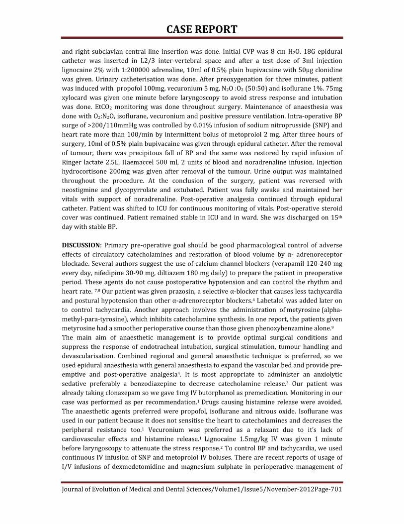
CASE REPORT
Journal of Evolution of Medical and Dental Sciences/Volume1/Issue5/November-2012Page-701
and right subclavian central line insertion was done. Initial CVP was 8 cm H2O. 18G epidural
catheter was inserted in L2/3 inter-vertebral space and after a test dose of 3ml injection
lignocaine 2% with 1:200000 adrenaline, 10ml of 0.5% plain bupivacaine with 50µg clonidine
was given. Urinary catheterisation was done. After preoxygenation for three minutes, patient
was induced with propofol 100mg, vecuronium 5 mg, N2O :O2 (50:50) and isoflurane 1%. 75mg
xylocard was given one minute before laryngoscopy to avoid stress response and intubation
was done. EtCO2 monitoring was done throughout surgery. Maintenance of anaesthesia was
done with O2:N2O, isoflurane, vecuronium and positive pressure ventilation. Intra-operative BP
surge of >200/110mmHg was controlled by 0.01% infusion of sodium nitroprusside (SNP) and
heart rate more than 100/min by intermittent bolus of metoprolol 2 mg. After three hours of
surgery, 10ml of 0.5% plain bupivacaine was given through epidural catheter. After the removal
of tumour, there was precipitous fall of BP and the same was restored by rapid infusion of
Ringer lactate 2.5L, Haemaccel 500 ml, 2 units of blood and noradrenaline infusion. Injection
hydrocortisone 200mg was given after removal of the tumour. Urine output was maintained
throughout the procedure. At the conclusion of the surgery, patient was reversed with
neostigmine and glycopyrrolate and extubated. Patient was fully awake and maintained her
vitals with support of noradrenaline. Post-operative analgesia continued through epidural
catheter. Patient was shifted to ICU for continuous monitoring of vitals. Post-operative steroid
cover was continued. Patient remained stable in ICU and in ward. She was discharged on 15th
day with stable BP.
DISCUSSION: Primary pre-operative goal should be good pharmacological control of adverse
effects of circulatory catecholamines and restoration of blood volume by α- adrenoreceptor
blockade. Several authors suggest the use of calcium channel blockers (verapamil 120-240 mg
every day, nifedipine 30-90 mg, diltiazem 180 mg daily) to prepare the patient in preoperative
period. These agents do not cause postoperative hypotension and can control the rhythm and
heart rate. 7,8 Our patient was given prazosin, a selective α-blocker that causes less tachycardia
and postural hypotension than other α-adrenoreceptor blockers.4 Labetalol was added later on
to control tachycardia. Another approach involves the administration of metyrosine (alpha-
methyl-para-tyrosine), which inhibits catecholamine synthesis. In one report, the patients given
metyrosine had a smoother perioperative course than those given phenoxybenzamine alone.9
The main aim of anaesthetic management is to provide optimal surgical conditions and
suppress the response of endotracheal intubation, surgical stimulation, tumour handling and
devascularisation. Combined regional and general anaesthetic technique is preferred, so we
used epidural anaesthesia with general anaesthesia to expand the vascular bed and provide pre-
emptive and post-operative analgesia4. It is most appropriate to administer an anxiolytic
sedative preferably a benzodiazepine to decrease catecholamine release.3 Our patient was
already taking clonazepam so we gave 1mg IV butorphanol as premedication. Monitoring in our
case was performed as per recommendation.1 Drugs causing histamine release were avoided.
The anaesthetic agents preferred were propofol, isoflurane and nitrous oxide. Isoflurane was
used in our patient because it does not sensitise the heart to catecholamines and decreases the
peripheral resistance too.1 Vecuronium was preferred as a relaxant due to it’s lack of
cardiovascular effects and histamine release.1 Lignocaine 1.5mg/kg IV was given 1 minute
before laryngoscopy to attenuate the stress response.2 To control BP and tachycardia, we used
continuous IV infusion of SNP and metoprolol IV boluses. There are recent reports of usage of
I/V infusions of dexmedetomidine and magnesium sulphate in perioperative management of

CASE REPORT
Journal of Evolution of Medical and Dental Sciences/Volume1/Issue5/November-2012Page-702
pheochromocytomas.6 Steroid cover is mandatory for patients undergoing bilateral
adrenalectomy. We also used hydrocortisone in intra-operative and post-operative period.
In conclusion, the proper anaesthetic management of pheochromocytoma is a truly
rewarding challenge. Mortality has been reduced in recent years due to better knowledge of
these bizarre tumours, particularly the chronic hypovolemia produced, and more adequate pre-
treatment regimens, better intra and post- operative management.
REFERENCES:
1. Brown R. Brunell Jr. Anaesthesia for Pheochromocytoma. In: Robert C Prys and
Brown R. Brunell Jr. Editors International practice of anaesthesia.
2. Stoelting RK, Dierdosrf SF. Endocrine diseases. Anaesthesia and Co-existing
diseases 2002; 430-434.
3. Published – butterworth-Heinemann 1996; 1/83/1-7 G.Singh, P.Kam. An
overview of anaesthetic issues in Pheochromocytoma.Ann acad Singapore
1998;27:843-8.
4. Roberts C. Prys. Pheochromocytoma- Recent progress in it’s management. Br J
Anaesth 2000; 85(1): 44-57.
5. Joet T. Alder, Goswin Y et al. Phaeocchromocytoma: Current approaches and
future directions. The Oncologist 2008; 13:779-793.
6. Bryskin R, Weldon BC. Dexmedetomidine and magnesium sulfate in the
perioperative management of a child undergoing laparoscopic resection of
bilateral pheochromocytomas. J Clin Anesth. 2010 Mar;22(2):126-9.
7. Domi R, Laho H. Management of pheochromocytoma: Old ideas and new drugs.
Niger J Clin Pract 2012;15:253-7.
8. Lebuffe G, Dosseh ED, Tek G, Tytgat H, Moreno S, Tavernier B, et al. The effect of
calcium channel blockers on outcome following the surgical treatment of
pheochromocytomas and paragangliomas. Anaesthesia 2005;60:439-44.
9. Steinsapir J, Carr AA, Prisant LM, Bransome ED Jr. Metyrosine and
pheochromocytoma. Arch Intern Med 1997; 157:901.

CASE REPORT
Journal of Evolution of Medical and Dental Sciences/Volume1/Issue5/November2012 Page-703
CONGENITAL DIAPHRAGMATIC HERNIA ASSOCIATED WITH FRYNS
SYNDROME –AN AUTOPSY STUDY
Surekha Ulhas Arakeri, Jaya Manchanda.
1. Professor, Department of Pathology, BLDEU’S Shri B. M. Patil Medical College & Research Centre, Bijapur.
2. Postgraduate Student, Department of Pathology, BLDEU’S Shri B. M. Patil Medical College & Research
Centre, Bijapur.
CORRESPONDING AUTHOR
Dr. Surekha Ulhas Arakeri, Professor,
Department of Pathology,
BLDEU’s Shri B. M. Patil Medical College
& Research Centre, Bijapur
E-mail: [email protected],
Ph: 0091 98457678444.
ABSTRACT: BACKGROUND: Congenital Diaphragmatic Hernia (CDH) is an anatomical defect
that permits abdominal contents inside the thoracic cavity and affects 1 in 2000 to 5000
children each year and is associated with high morbidity and mortality. CDH may be associated
with other anomalies like dysmorphic features, genitourinary, musculoskeletal, cardiovascular,
neurological and gastrointestinal malformations.CDH is a devastating birth defect that can occur
in isolation or part of complex malformation of various syndromes. Here we report an autopsy
study of CDH with Fryns syndrome. CASE PRESENTATION: An autopsy study of still born
foetus born to a 35-year old, fifth gravida presented with features of CDH with left lung
hypoplasia, hypoplasia of distal parts of digits of upper and lower limbs and dysmorphic
features. CONCLUSION: CDH is a life threatening pathology in infants and a major cause of
death due to pulmonary hypoplasia and pulmonary hypertension. An early diagnosis with
increased understanding of this disease is a crucial factor for a timely approach to manage the
critically ill infant and to offer potential treatment for improved outcome and substantial
reduction in morbidity.
KEY WORDS: Congenital diaphragmatic hernia (CDH), Fryn syndrome, pulmonary hypoplasia,
Hypoplasia of digits.
INTRODUCTION: Congenital Diaphragmatic Hernia (CDH) is a life threatening congenital
anomaly which needs intensive neonatal care and is associated with high morbidity and
mortality with an incidence ranging between 1 in 2000 to 5000 per year. CDH may be
associated with other congenital anomalies like dysmorphic features, genitourinary,
musculoskeletal, cardiovascular, neurological and gastrointestinal malformations in 30% to
40% of the cases. These associated anomalies acts as major factor influencing the outcome of
the affected patients. According to some authors, the most commonly associated major
congenital anomaly was neural tube defect followed by cardiac and chromosomal anomalies.
Severe pulmonary hypoplasia was the major cause of mortality in stillborn babies and in all
infants of CDH dying within one week of birth.1,2 CDH can occur in isolation or as part of a
various syndromes such as Pallister-Killian syndrome(PKS), Ghersoni-Baruch syndrome, WAGR
syndrome, Fryn syndrome, Denys-Dash syndrome, Brachman-De Lange syndrome and Wolf-
Hirschhorn syndrome. Fryns syndrome is a rare autosomal recessive disorder characterized by
diaphragmatic hernia and multiple anomalies2.

CASE REPORT
Journal of Evolution of Medical and Dental Sciences/Volume1/Issue5/November2012 Page-704
CASE SUMMARY: A 35-year old, fifth gravida presented with 24 week of gestation. She gave
past obstetric history of one abortion and history of neonatal death of one child. Antenatal
ultrasound scan was performed which revealed congenital anomaly of diaphragm. Based on
ultrasound findings, radiological diagnosis of CDH was done for which therapeutic abortion was
performed in the present pregnancy at 24 week of gestation, and a still born foetus was
delivered which was sent for post mortem study.
AUTOPSY FINDINGS: Received a male foetus with placenta and attached umbilical cord.
Placenta was 275 gm in weight and measured 16x10x3 cm. Weight of the foetus was 600 gm.
Crown rump and crown heel length was 17 and 29cm respectively. Head circumference, chest
circumference and abdominal girth were 23, 20 and 18 cm respectively. External examination
showed widely spaced protuberant eyes, broad and depressed nasal bridge, extra space
between nose and upper lip, large mouth, small chin and hypoplasia of distal parts of digits of
upper and lower limb (Figure 1). On cut open, it was found that left part of the diaphragm was
deficient & left lobe of liver, spleen and coils of intestine were protruded through this deficiency
in the thoracic cavity (Figure 1). Left lung weighed 4gms and measured 1.5x1.5x0.5cm; Right
lung weighed 6gms and measured 3x2x0.5cm (Figure 2, Inset). Lung weight to body weight
(LW: BW) ratio was 0.01. Heart, liver, spleen, right and left kidneys were normal. Microscopic
examination of right lung showed thick walled lung alveoli lined by cuboidal epithelium. Dense
connective tissue and bronchial cartilage was noted in inter and intralobular connective tissue.
In the left lung features as described above with decrease in number of alveoli were noted.
Based on above findings, a diagnosis of CDH associated with dysmorphic facial features,
hypoplasia of digits and pulmonary hypoplasia suggestive of Fryns syndrome was given.
DISCUSSION: Development of the diaphragm is divided into two phases such as development of
the diaphragmatic pericardium and development of the pleural cavity and closure of the
pleuroperitoneal canal (PPC). General hypothesis is that the defect in CDH may results from
failure of complete closure of the PPC at the embryonic period of 8th to10th gestational week.
Association of CDH with various congenital anomalies, varying pathological patterns and clinical
presentation suggests that it is a result of multiple, complex developmental abnormalities.1 CDH
is a cardinal feature of Fryns syndrome.3 Studies indicate that Fryns syndrome is diagnosed in
1.3% to 10% of all the cases having CDH. This syndrome is generally difficult to diagnose.3,4
Diagnostic criteria for Fryns syndrome reformulated by Lin AE et al5 were 4 out of following 6
symptom groups such as diaphragmatic defect, facial characteristics, distal digital hypoplasia,
pulmonary hypoplasia, parental consanguity and other associated anomalies such as
cardiovascular malformation, renal dysplasia/renal cortical cysts, gastrointestinal
malformation, genital malformation should be present.4,5 In the present case, 4 symptom groups
such as CDH, dysmorphic facial features, pulmonary hypoplasia and distal digital hypoplasia
were seen. Hence this case was concluded as Fryns syndrome. LW: BW ratio is the most widely
used parameter for diagnosis of pulmonary hypoplasia. Pathologic postmortem diagnosis of
pulmonary hypoplasia was suggested when the LW: BW was found to be less than 0.12.6 Based
on this finding, diagnosis of pulmonary hypoplasia was made in the present case. Considerable
phenotypic overlap was noted between Fryns and PKS. In both syndromes CDH, coarse facial
features and minor limb anomalies were seen. Features more common for, Fryns syndrome are
CDH, cleft palate, and distal phalange and/or nail hypoplasia, cardiovascular malformations, and
renal malformations. Features more common for PKS are high forehead, streaky skin hyper

CASE REPORT
Journal of Evolution of Medical and Dental Sciences/Volume1/Issue5/November2012 Page-705
pigmentation, and sparseness of hair bi-temporally.7 In the present case, CDH was associated
with pulmonary hypoplasia, dysmorphic facial features and distal digital hypoplasia hence the
case was concluded as Fryns syndrome. Association of diaphragmatic defects and upper limb
anomalies were not reported commonly.8-10 McCredie and Reid 8 reported 4 single cases of
diaphragmatic defect associated with limb anomalies. The limb defects ranged from a radial
hypoplasia to a transverse deficiency, with a block of bone distal to the hummers. Lerone et al9
reported a case of left congenital diaphragmatic hernia associated with ipsilateral thumb
hypoplasia. A similar case of congenital left diaphragmatic hernia with ipsilateral thumb
hypoplasia and absent radius was reported by Wallerstein et al.10 In the present case, CDH is
associated with hypoplasia of distal parts of digits of upper & lower limbs which is one of the
uncommon finding.
CONCLUSION: Incidence of mortality associated with CDH will become apparent only when the
autopsy study of infant and fetuses that have died with a diagnosis of CDH will be included in
the analysis.1 In this case, abortion of previous pregnancy and neonatal death of sibling may be
due to presence of CDH and associated syndrome. As Fryns syndrome is autosomal recessive
disorder, autopsy study of present case will assist in counseling to the parents about recurrence
of the disease in next pregnancies and advising regular antenatal ultrasound scan and screening
for maternal serum Alpha fetoprotein assay at 13 weeks in next pregnancy.
REFERENCES:
1. Arafah M, Boqari DT, Alsaad KO. Left-sided congenital diaphragmatic hernia with multiple
congenital cardiac anomalies, hernia sac, and microscopic hepatic heterotopia: A Case
Report. Pathology Res Int 2011; 96:710 -7.
2. Tovar JA. Orphanet Journal of Rare Diseases 2012; 7:1 doi: 10.1186/1750-1172-7-1.
Available at: http://www.ojrd.com/content/7/1/1
3. Slavotinek A, Lee SS, Davis R, Shrit A, Leppig KA, Rhim J et al. Fryns syndrome phenotype
caused by chromosome microdeletions at 15q26.2 and 8p23.1. J Med Genet 2005; 42:730–
736.
4. Slavotinek A. Fryns syndrome. In: Pagon RA, Bird TD, Dolan CR, Stephens K, Adam MP
editors. GeneReviews™ [Internet]. Seattle (WA): University of Washington, Seattle; 2007
[cited 2010 Jun 1]. Available from: http://www.ncbi.nlm.nih.gov/books/NBK1459/
5. Lin AE, Pober BR, Mullen MP, Slavotinek AM. Cardiovascular malformations in Fryns
syndrome: is there a pathogenic role for neural crest cells. Am J Med Genet A 2005;
139:186–93.
6. Askenazi SS, Perlman M. Pulmonary hypoplasia: lung weight and radial alveolar count as
criteria of diagnosis. Arch Dis Child 1979; 54:614-618.
7. Pober BR, Russell MK, Ackerman KG. Congenital Diaphragmatic Hernia Overview. In: Pagon
RA, Bird TD, Dolan CR, Stephens K, Adam MP editors. Gene Reviews™ [Internet]. Seattle
(WA): University of Washington, Seattle; 1993 [cited 2010 Mar 16]. Available from:
http://www.ncbi.nlm.nih.gov/books/NBK1359/
8. Mccredie J, Reid IS. Congenital diaphragmatic hernia associated with homolateral upper
limb malformation. J Pediatr 1978; 92:762-5.

CASE REPORT
Journal of Evolution of Medical and Dental Sciences/Volume1/Issue5/November2012 Page-706
9. Lerone M, Soliani M, Corea D, Romeo G, Martucciello G, Silengo MC. Congenital
diaphragmatic hernia associated with ipsilateral upper limb reduction defects: report of a
case with thumb hypoplasia. Am J Med Genet 1992; 44:827-9.
10. Wallestein R, Wallerstein DF, Trauffer P, Desposto F. Congenital diaphragmatic hernia and
ipsilateral limb reduction defect: a new case, longterm follow-up and review of the
literature. Clin Dysmorphol 1997; 6:257-61.

ORIGINAL ARTICLE
Journal of Evolution of Medical and Dental Sciences/Volume1/Issue5/November-2012Page-707
A CORRELATIVE STUDY OF ADENOSINE DEAMINASE ACTIVITY & T.B.
IgG IN SERUM IN CASES OF TUBERCULOSIS. Ajay Narang, Sanjay Kumar Nigam, Anand Mishra, Nilam Nigam
1. Assistant Professor, Department of Pathology, Rama Medical College Hospital & Research Centre
Mandhana Kanpur, U.P.
2. Associate Professor, Department of Pathology, Rama Medical College Hospital & Research Centre
Mandhana Kanpur, U.P.
3. Associate Professor, Department of Biochemistry, Rama Medical College Hospital & Research Centre
Mandhana Kanpur, U.P.
4. Associate Professor, Department of Pharmacology, Rama Medical College Hospital & Research Centre
Mandhana Kanpur, U.P.
CORRESPONDING AUTHOR
Dr. Sanjay Kumar Nigam,
House No. 308,
Rama Medical College Mandhana, Kanpur,
E-mail: [email protected],
Ph: 0091 9411918206.
ABSTRACT: INTRODUCTION: Tuberculosis is major cause of morbidity and mortality in India
as well in other parts of world. It is caused by mycobacterium tuberculosis which primarily
affects lung and cause pulmonary tuberculosis. Diagnosis of tuberculosis rests upon a positive
history of contact, clinical symptoms, x-ray chest, sputum positivity and AFB culture. Adenosine
deaminase (ADA) is an enzyme which catalyzes the deamination of adenosine into inosine and
ammonia. ADA level is found to be elevated in tuberculosis and typhoid fever where cell
mediated immunity is elevated. The ADA level is significantly elevated in tuberculosis and helps
to differentiate between tubercular and non tubercular diseases. The ADA level is also found to
be elevated in serum and pleural fluid in patients of tubercular pleural effusion, tubercular
ascitis and tubercular pericardial effusion. METHODS: Routine hemogram, Montoux test, X-ray
chest, FNAC of lymph nodes, biopsy of lymph node whenever required, estimation of serum ADA
level and T.B.IgG studies were performed in each case. RESULTS: In the present study a total of
45 cases were selected for the study. There are 30 cases of pulmonary tuberculosis and 15
controls. The values of serum ADA and tubercular IgG in pulmonary tubercular group are
significantly higher as compared to those of controls. None of the control for ADA showed
significant ratio of positivity (≥1.7). One of the 15 cases showed remarkable ratio of positivity
(>1.2-1.6) and 14 (93.3%) cases showed insignificant ratio of positivity. Only 2 (13.33%) of the
15 cases showed positivity for TB IgG and rest 13 (86.66%) were regarded negative.
CONCLUSIONS: Thus it can be concluded that determination of serum adenosine deaminase
levels can effectively diagnose tuberculosis with sensitivity of 96.66% and specificity of 93.33%
as compared to TBIgG showing sensitivity of 90% and specificity of 86.6%. Also cost of ADA
estimation is remarkably less than that of tubercular IgG
KEY WORDS: Serum ADA, T.B.IgG levels, Tuberculosis
INTRODUCTION: Tuberculosis is major cause of morbidity and mortality in India as well in
other parts of world. It is caused by mycobacterium tuberculosis which primarily affects lung
and cause pulmonary tuberculosis. It can also affect intestine, bones, joints, lymph nodes,
genitourinary system, skin and virtually every organ of the body.

ORIGINAL ARTICLE
Journal of Evolution of Medical and Dental Sciences/Volume1/Issue5/November-2012Page-708
Diagnosis of tuberculosis rests upon a positive history of contact, classical symptoms,
lymphocytosis on differential count, lesions in x-ray chest, sputum positivity for AFB and culture
of AFB on L.J. media. But none of these except demonstration of AFB is a sure shot evidence of
diagnosis. Besides this several other tests such as demonstration of tubercular antigen by
Polymerase chain reaction1,2 , rapid culture of tubercular bacilli by Bactec system have come
forward but are costly and not available everywhere except at except at specialised centres.3
Adenosine deaminase (ADA) is an enzyme which catalyzes the deamination of
adenosine into inosine and ammonia. It helps in maturation and proliferation of T cells. ADA
level is found to be elevated in tuberculosis and typhoid fever where cell mediated immunity is
elevated 4, 5. The ADA level is significantly elevated in tuberculosis and helps to differentiate
between tubercular and non tubercular diseases. The ADA level is also found to be elevated in
serum and pleural fluid in patients of tubercular pleural effusion. 6, 7,8,9,10,11, tubercular ascitis 12,
13 and tubercular pericardial effusion.8
Besides this there are several other tests which detect the antibody load against tubercular
antigen in patient’s serum. Enzyme linked immunosorbant assay is most frequently used and it
detects the quantity and quality of circulating antibodies against tubercular antigen. Usually
three antibodies are detected which include-
1. Tubercular IgA (TbIgA)
2. Tubercular IgM (TbIgM)
3. Tubercular IgG (TbIgG)
Tubercular IgA antibodies are detected in serum of some apparently healthy individuals
at risk, but being the secretary antibody its detection in body fluid is of more value in patients
having tubercular effusion.
Tubercular IgM is detected in initial phase of infection i.e. 1-2 months.14
Tubercular IgG is detected in patient’s serum in slightly late stages when disease is
properly settled (after 2 months). Its high titre is more specific as compared to tubercular IgA
and IgM in patient’s serum for diagnosis of active tuberculosis.15
Single detection of these antibodies alone cannot confirm the diagnosis of tuberculosis
unless matched or correlated with other clinical Koch’s parameters. Thus the rising antibody
titres are more significant than a single high titre, but because of the cost this is practically
impossible.
So the present study was undertaken to evaluate serum adenosine deaminase activity in
tubercular patients.
MATERIAL AND METHODS: The present study was carried out in the department of pathology,
Biochemistry, Tubercular and chest disease and Medicine Rama Medical College and Research
Centre Mandhana, Kanpur.
The clinical features and detailed history were recorded in a standard proforma. Routine
hemogram, Mantoux test, X-ray chest, FNAC of lymph nodes, biopsy of lymph node whenever
required, estimation of serum ADA level and T.B.IgG studies were performed in each case.
Estimation of ADA activity-
We have utilized the method of GLUSEPPE GIUSTI and BRUNO GALANTI for ADA
estimation. The instrument used for ADA estimation were spectrophotometer or simple

ORIGINAL ARTICLE
Journal of Evolution of Medical and Dental Sciences/Volume1/Issue5/November-2012Page-709
photometer for accurate measurement between 620 and 650 nm, water bath 37oC, centrifuge,
test tube and auto pipette.
Evaluation of T.B. IgG-
For evaluation of T.B.IgG specimen collected was patient’s blood from which sera was
separated and stored in a refrigerator until performance of test. Reagents used were supplied
by TRANSASIA biomedical Ltd, under the name of ERBA ELISA test tuberculosis.
OBSERVATIONS: In the present study a total of 45 cases were selected for the study. There are
30 cases of pulmonary tuberculosis and 15 controls. Normal healthy individuals or patients,
who were suffering from non tubercular pulmonary disease, were taken as control. The serum
ADA levels and tubercular IgG of controls are shown in table I and II. Serum ADA activity and
tubercular IgG of pulmonary tubercular group are shown in table III and IV.
The values in pulmonary tubercular group are significantly higher as compared to those
of controls. Table V shows the comparative ratio of positivity of serum ADA and Tubercular IgG.
Table I- ADA levels in control (Mean = 25.83 U/L).
S.No. ADA levels (U/L) Ratio of positivity
1. 26.65 1.03
2. 31.10 1.20
3. 30.0 1.16
4. 21.65 0.83
5. 26.65 1.03
6. 16.65 0.64
7. 18.30 0.70
8. 31.65 1.20
9. 20.0 0.77
10. 26.65 1.03
11. 28.30 1.09
12. 36.0 1.39
13. 33.30 1.20
14. 16.65 0.64
15. 23.38 0.90
None of the control showed significant ratio of positivity (≥1.7). One of the 15 cases showed
remarkable ratio of positivity (>1.2-1.6) and 14 (93.3%) cases showed insignificant ratio of
positivity.
Table – II TB IgG in controls –
S.No. Observed value
(O.D.)
Positive cut off
(O.D.)
Ratio of
positivity
Interpretation
1. 113 250 0.45 -
2. 226 250 0.90 -
3. 142 250 0.56 -
4. 157 250 0.67 -

ORIGINAL ARTICLE
Journal of Evolution of Medical and Dental Sciences/Volume1/Issue5/November-2012Page-710
5. 440 250 1.76 +
6. 410 650 0.63 -
7. 398 650 0.61 -
8. 357 650 0.54 -
9. 401 650 0.61 -
10. 312 650 0.48 -
11. 475 450 1.05 +
12. 210 450 0.46 -
13. 317 500 0.7 -
14. 416 500 0.83 -
15. 217 500 0.43 -
Only 2 (13.33%) of the 15 cases showed positivity for TB IgG and rest 13 (86.66%) were
regarded negative.
Table III- ADA and ratio of positivity in patients of pulmonary tuberculosis.
S.No. ADA activity in serum Cut off level Ratio of positivity
1. 58.33 35 1.66
2. 56.66 35 1.61
3. 68.33 35 1.95
4. 35.00 35 1.00
5. 70.00 35 2.00
6. 91.66 35 2.61
7. 68.33 35 1.95
8. 73.33 35 2.09
9. 65.00 35 1.85
10. 60.00 35 1.71
11. 63.33 35 1.80
12. 60.00 35 1.71
13. 61.66 35 1.76
14. 63.33 35 1.80
15. 56.66 35 1.61
16. 53.33 35 1.52
17. 68.33 35 1.95
18. 60.00 35 1.71
19. 68.33 35 1.95
20. 61.66 35 1.76
21. 60.00 35 1.71
22. 56.66 35 1.61
23. 58.33 35 1.66
24. 63.33 35 1.80
25. 61.66 35 1.76
26. 66.66 35 1.90
27. 68.33 35 1.95

ORIGINAL ARTICLE
Journal of Evolution of Medical and Dental Sciences/Volume1/Issue5/November-2012Page-711
28. 63.33 35 1.80
29. 55.00 35 1.57
30. 70 35 2.00
22 (73.3%) cases showed significant ratio of positivity (≥1.7).
7 (23.3%) cases were out of remarkable ratio of positivity (>1.2-1.6).
1 (3.3%) case showed insignificant ratio of positivity (<1.2).
Table IV- TB IgG in serum of patients of pulmonary tuberculosis
S.No. Observed value
(O.D.)
Positive cut off
(O.D.)
Ratio of
positivity
Interpretation
1. 383 250 1.53 ++
2. 940 250 3.76 +++
3. 516 400 1.29 ++
4. 207 400 0.51 -
5. 1096 600 1.82 ++
6. 661 600 1.10 +
7. 2001 600 3.33 +++
8. 623 600 1.03 +
9. 666 600 1.11 +
10. 696 650 1.07 +
11. 744 650 1.14 +
12. 535 500 1.07 +
13. 2046 500 4.09 +++
14. 677 500 1.35 ++
15. 923 500 1.84 ++
16. 616 500 1.23 ++
17. 814 500 1.62 ++
18. 490 500 0.98 -
19. 894 500 1.78 ++
20. 774 500 1.54 ++
21. 575 500 1.15 +
22. 750 500 1.50 ++
23. 2136 500 4.23 +++
24. 558 500 1.11 +
25. 1231 500 2.46 +++
26. 525 500 1.05 +
27. 878 500 1.75 ++
28. 632 500 1.22 ++
29. 319 500 0.63 -
30. 714 400 1.78 ++

ORIGINAL ARTICLE
Journal of Evolution of Medical and Dental Sciences/Volume1/Issue5/November-2012Page-712
Of these total 30 cases, 2 were clearly negative (false negative) and one was borderline negative
(+++->2) times positive cut off, ++ ->1.2-2.0 times the positive cut off, + - >1.0-1.2 times the
positive cut off, -<1 times the positive cut off.
Table V- Comparison of ratio of positivity of ADA and Tubercular IgG in pulmonary
tuberculosis.
S.No. Ratio of positivity of ADA Ratio of positivity of
IgG
Interpretation
1. 1.66 1.53 ++
2. 1.61 3.76 ++
3. 1.95 1.29 ++
4. 1.07 0.51 +-
5. 2.00 1.82 ++
6. 2.61 1.10 ++
7. 1.79 3.33 ++
8. 2.09 1.03 ++
9. 1.85 1.11 ++
10. 1.71 1.07 ++
11. 1.80 1.14 ++
12. 1.71 1.07 ++
13. 1.76 4.09 ++
14. 1.80 1.35 ++
15. 1.61 1.84 ++
16. 1.52 1.23 ++
17. 1.95 1.62 ++
18. 1.71 0.98 +-
19. 1.95 1.78 ++
20. 1.76 1.54 ++
21. 1.71 1.15 ++
22. 1.61 1.50 ++
23. 1.66 4.23 ++
24. 1.80 1.11 ++
25. 1.76 2.46 ++
26. 1.90 1.05 ++
27. 1.95 1.75 ++
28. 1.80 1.22 ++
29. 1.57 0.63 +-
30. 2.00 1.78 ++
(++) - Parallel rise of both the parameters.
(+-) - Rise of only one parameter.
DISCUSSION: Tuberculosis continues to be a major cause of mortality and morbidity in
developing countries. Although lung is the most frequent organ to be involved, inflammation of
serous membranes is also very common. The definitive diagnosis is established when typical

ORIGINAL ARTICLE
Journal of Evolution of Medical and Dental Sciences/Volume1/Issue5/November-2012Page-713
histological features can be demonstrated or mycobacteria can be isolated from the body fluids
or sputum or on gastric lavage and various other methods such as gel electrophoresis,
radiometric assay and polymerase chain reaction. It is well documented that isolation of
mycobacteria and culture is very difficult and time consuming (shah et al 1990).
Recently most simple techniques which are most feasible less costly and giving quick
results are now available and include demonstration of antibodies by ELISA method (humoral
immune response)and ADA activity (cell mediated immune response) in serum and body fluids.
So the present study was undertaken to evaluate adenosine deaminase enzyme activity
and tubercular IgG together in patients of pulmonary tuberculosis. Normal healthy individuals
or patients, who were suffering from non tubercular pulmonary disease, were taken as control.
In this study the overall mean serum ADA in control was 25.83 U/L (16.65 – 36 U/L).
There was no significant variation in serum ADA levels in relation to age and sex in controls16.
Only one healthy control showed increase activity of ADA in serum and may possibly be
resulting from any disease involving increase cell mediated immunity e.g. typhoid 4,5.
Mean serum ADA activity in patients of pulmonary tuberculosis in this study was 58.40 U/L
(Table III) which was significantly elevated as compared to that of controls (Mean 25.83 U/L).
Cut off values of serum ADA >35 U/L was diagnostic of tuberculosis with 100% sensitivity.
Similar observations were given other researchers who reported higher levels of serum ADA in
patients with pulmonary tuberculosis as compared to healthy controls16. The level was also
significantly higher in pulmonary tuberculosis as compared to non tubercular pulmonary
diseases (suppurative and malignant) and using a cut off of 33 U/L. Sensitivity and specificity
was claimed to be 98% and 100%. Significantly higher (p<0.0005) serum ADA in tubercular
group (29.8±10 U/L) was reported than in neoplastic group (14.5±4.0 U/L) 17. Similar data was
provided by other observers who reported ADA activity of 27.38 U/L in tuberculosis as
compared to malignancy (7.29) and non tubercular pulmonary diseases (12.71)18.
Tubercular immunoglobulin IgG depicting humoral immune response was also
evaluated in pulmonary tubercular patients by using ELISA method against antigen A60. In
control group 2 cases (13.33%) out of total 15 cases showed a false positivity and may be
because of previous exposure to disease15. The value of optical density obtained in pulmonary
tuberculosis patients are show in table IV. Three (10%) of the total 30 cases studied showed
false negative values. This could be attributed to ANERGY because of initial tubercular toxaemia
or because of immunocompromised status19. The sensitivity and specificity of the test came out
to be 90% and 86.6% respectively. Similar were the observations formulated by Gupta S et al
1995, who reported a specificity of 92.3% and sensitivity of 80%.
Thus it can be concluded that determination of serum adenosine deaminase levels can
effectively diagnose tuberculosis with sensitivity of 96.66% and specificity of 93.33% as
compared to TBIgG showing sensitivity of 90% and specificity of 86.6%. Also cost of ADA
estimation is remarkably less than that of tubercular IgG 9. However using both tests in
combination increases the specificity and sensitivity for diagnosis of tuberculosis.
BIBLIOGRAPHY:
1. 1.Brisson-Noël A, Gicquel B, Lecossier D, Lévy-Frébault V, Nassif X, Hance AJ
Rapid diagnosis of tuberculosis by amplification of mycobacterial DNA in clinical
samples. Lancet. 1989 Nov 4;2(8671):1069-71

ORIGINAL ARTICLE
Journal of Evolution of Medical and Dental Sciences/Volume1/Issue5/November-2012Page-714
2. Tiwari V, Jain A, Verma RK. Application of enzyme amplified mycobacterial DNA
detection in the diagnosis of pulmonary & extra-pulmonary tuberculosis. Indian J
Med Res. 2003 Dec;118:224-8.
3. Middlebrook G, Reggiardo Z, Tigertt WD. Automatable radiometric detection of
growth of Mycobacterium tuberculosis in selective media. Am Rev Respir Dis.
1977 Jun;115(6):1066-9.
4. Galanti B, Nardiello S, Russo M, Fiorentino F. Increased lymphocyte adenosine
deaminase in typhoid fever. Scand J Infect Dis. 1981;13(1):47-50
5. Mishra OP, Gupta BL, Ali Z, Nath G, Chandra L. Adenosine deaminase activity in
typhoid fever. Indian Pediatr. 1994 Nov;31(11):1379-84.
6. Piras MA, Gakis C, Budroni M, Andreoni G. Adenosine deaminase activity in
pleural effusions: an aid to differential diagnosis. Br Med J. 1978 Dec 23-
30;2(6154):1751-2.
7. Nagaraja MV, Ashokan PK, Hande HM. Adenosine deaminase in pleural effusions.
J Assoc Physicians India. 1992 Mar;40(3):157-9
8. 8 Segura RM, Pascual C, Ocaña I, Martínez-Vázquez JM, Ribera E, Ruiz I, Pelegrí
MD. Adenosine deaminase in body fluids: a useful diagnostic tool in tuberculosis.
Clin Biochem. 1989 Apr;22(2):141-9. Gaki C.The low cost of the adenosine
deaminase assay. Chest. 1996 Nov;110(5):1376-7.
9. Sharma SK, Mohan A. Adenosine deaminase in the diagnosis of tuberculosis
pleural effusion. Indian J Chest Dis Allied Sci. 1996 Apr-Jun;38(2):69-71.
10. Shibagaki T, Hasegawa Y, Saito H, Yamori S, Shimokata K. Adenosine deaminase
isozymes in tuberculous pleural effusion. J Lab Clin Med. 1996 Apr;127(4):348-
52.
11. Gupta BK, Bharat V, Bandyopadhyay D. Sensitivity, specificity, negative and
positive predictive values of adenosine deaminase in patients of tubercular and
non-tubercular serosal effusion in India. J Clin Med Res. 2010 May 19;2(3):121-6.
12. 13Sathar MA, Simjee AE, Coovadia YM, Soni PN, Moola SA, Insam B, Makumbi F.
Ascitic fluid gamma interferon concentrations and adenosine deaminase activity
in tuberculous peritonitis. Gut. 1995 Mar;36(3):419-21.
13. Gevaudan MJ, Bollet C, Charpin D, Mallet MN, De Micco P. Serological response of
tuberculosis patients to antigen 60 of BCG. Eur J Epidemiol. 1992 Sep;8(5):666-
76.
14. Gupta S, Kumari S, Banwalikar JN, Gupta SK. Diagnostic utility of the estimation
of mycobacterial Antigen A60 specific immunoglobulins IgM, IgA and IgG in the
sera of cases of adult human tuberculosis. Tuber Lung Dis. 1995 Oct;76(5):418-
24.
15. jhamaria JP, Jenaw RK, Ludhada SK, Mathur Dk, Parihar HL, Sharma SK. Serum
adenosine deaminase in the differential diagnosis of pulmonary tuberculosis and
common non-tubercular respiratory diseases. Ind J Tub. 1988;35:25-7.
16. Baganha MF, Pêgo A, Lima MA, Gaspar EV, Cordeiro AR. Serum and pleural
adenosine deaminase. Correlation with lymphocytic populations. Chest. 1990
Mar;97(3):605-10.
17. 18 Bansal SK, Singh RP, Narang RK, Joshi LD, Bansal A, Agrawal AK. Serum
adenosine deaminase in pulmonary tuberculosis, malignancy and non-tubercular
respiratory diseases. Indian J Chest Dis Allied Sci. 1991 Oct-Dec;33(4):189-93.
18. Fadda G, Grillo R, Ginesu F, Santoru L, Zanetti S, Dettori G. Serodiagnosis and
follow up of patients with pulmonary tuberculosis by enzyme-linked
immunosorbent assay. Eur J Epidemiol. 1992 Jan;8(1):81-7.

ORIGINAL ARTICLE
Journal of Evolution of Medical and Dental Sciences/Volume1/Issue5/November-2012Page-715
EFFICACY OF AIR DISINFECTION DEVICES IN CONTROLLING
ATMOSPHERIC MICROFLORA IN ENCLOSED HEALTH CARE SETTINGS. Ravi Kumar R, Jyothi E. K, Nanda Kumar B. S, Madhava Murthy M. R.
1. Professor & Head, Department of Neuromicrobiology, NIMHANS, Bangalore.
2. PhD Scholar, Department of Neuromicrobiology, NIMHANS, Bangalore.
3. Associate Professor cum Epidemiologist, Department of Community Medicine, MSRMC.
4. Chief Executive Officer, Innova Technology Solutions Mysore Pvt Ltd., Bangalore.
CORRESPONDING AUTHOR
Dr. R. Ravi Kumar MD,
Professor and Head,
Department of Neuromicrobiology,
NIMHANS, Bangalore,
E-mail: [email protected],
Ph: 0091 9448073965.
ABSTRACT: INTRODUCTION: There is increasing concern with the prevalence of hospital
infection. This is causing a significant economic and logistical burden on the health services.
OBJECTIVES: To assess the efficacy of hydroxyl radical based air disinfection devices in
reducing the atmospheric pathogen count in enclosed health care settings. MATERIALS AND
METHODS: Sequential trial with repeated readings was designed before and after installation of
the Air Disinfection devices. Study was conducted at 52 points in different locations which
included sections from out Patient Department, Intensive Care Units, Operation Theatre,
Surgical Ward, Radiology Department and cubicles in routine microbiology. The samples
collected with air sampler were processed in Neuromicrobiology Laboratory. RESULT: It was
observed that the mean (±SE) colony forming units (CFU/m3) count after 24 and 48 hours
before and after use of air disinfectant devices at the above locations were 159.75 (±35.42),
210.31 (±65.15) and 44.04 (±14.15), 55.31 (±15.06) respectively. The differences in the mean
CFU concentrations were found to be statistically significant (p= 0.006). Additionally, a high
degree of variance in the CFU counts was noted before using the devices as compared to that
after using the same. CONCLUSION: The results indicate that there is considerable reduction in
the number of colony forming units subsequent to the introduction of the Air Disinfection
devices. These devices supplement and compliment the general hygienic practices. However
owing to a lot of compounding factors affecting the colony forming units in a given location,
sufficient care must be taken for ascribing the reduction in the counts only to the device.
KEYWORDS: Air Disinfection, Environmental Flora, Disinfection Devices, Air Sampling,
Hospital Infection.
INTRODUCTION: Hospitals and associated areas are high risk areas for acquiring infections1.
Therefore, it is imperative that strict policies be implemented and maintained so as to achieve
the goal of infection control. One of the modes to achieve the same is by air disinfection process
wherein devices are used to reduce the number of microorganisms in the air. This concept in
disease prevention is pivotal in certain high risk areas within the hospital like Intensive care
units, operation theatres, and other sites where virulent and resistant organisms, high risk
patients and invasive interventions are present. Prevention of infections by means of air
disinfection will enhance and greatly synergise with other infection control measures and

ORIGINAL ARTICLE
Journal of Evolution of Medical and Dental Sciences/Volume1/Issue5/November-2012Page-716
reduce the incidence of nosocomial infections(hospital acquired infections), suspected to arise
by the air borne route.
There has been increasing concern with the prevalence of superbugs and hospital
related infections in the past decade. Environmental dissemination of pathogens is an important
issue associated with hospital and community acquired infections and therefore their control
plays a vital role in the formulation of hospital infection surveillance system and antibiotic
policies2. Different technologies are coming up for atmospheric disinfection which employs
various principles for disinfection. The use of hydroxyl radicals is a very efficient mode of
decontamination. The hydroxyl radical is a transient piece of energy. These radicals are highly
reactive and consequently short lived. These are also called the detergent of the troposphere as
these are often the first step to removal of pollutants. The attack process essentially leads to a
cascade of reactions that may induce damage to nucleic acids, structural and functional changes
in proteins, as well as oxidation of lipids. For example, lipids in a bacterial cell wall react with
hydroxyl radicals by losing hydrogen and forming a lipid radical. Oxidation of proteins are far
more complicated where a protein radical will lead many possible reactions, one of which
resulting in a non-protein radical that can be a further hydroxyl radical3,4. In a microbial cell,
due to interactions between proteins and lipids, the oxidative reactions can transfer from lipids
to proteins and so on, causing destruction to main cellular structures which will render the
bacteria non viable. Another study found that hydroxyl radicals are involved in cell killing by the
bacterial Topoisomerase I Cleavage Complex5. The reactions between these hydroxyl radicals
and bacteria are non-strain specific, therefore resistant and non-resistant bacterial strains will
be equally susceptible to hydroxyl radicals’ attack6.
The AD unit used in the study takes the surrounding air within an enclosed space,
processes it and combines it with olefin – the natural scents of flowers and plants – creating
hydroxyl radicals that react with the molecules in airborne pathogens and removes them7,8.
Through this process the AD unit disinfects the air continuously.
OBJECTIVES: To assess the efficacy of hydroxyl radical based air disinfectant devices in
reducing the atmospheric pathogen count in enclosed health care settings.
METHODOLOGY: Sequential air sampling before and after installation of air disinfectant
devices were carried out. The air sampling device used was of the model LA030 Air Petri from
Hi- Media. The volume of air sampled was 1,000L (1m3 ). Time taken for sampling was 3
minutes with a flow rate of 300 litres/min. The machine uses a sieve impaction particle capture
mechanism.
The sampled air was cultured on culture media namely sheep blood agar for bacterial
isolation and Sabouraud's dextrose agar for fungal isolation. The plates were incubated at 370C
for 24-48 hours. First reading was taken after 24hours of incubation, and the second reading
after 48 hours of incubation. Quantitative evaluation of CFU/m3 was performed by observing the
culture and counting the colonies.
Study was conducted at 52 points in different locations of the hospital which included
sections from outpatient department, intensive care units, operation theatre, surgical ward,
radiology department and cubicles in the department of microbiology. Further processing was
performed in the Neuromicrobiology laboratory as per standard guidelines. The air disinfectant
device used was from Inov8 Air Disinfection Company, United Kingdom. Specific identification
of the isolates was not performed during the study.

ORIGINAL ARTICLE
Journal of Evolution of Medical and Dental Sciences/Volume1/Issue5/November-2012Page-717
STATISTICAL ANALYSIS: Continuous data were summarized by mean (± SD) and Categorical
data as proportions and percentages. Paired t-test was used to compare the statistical
significance of differences in mean CFU counts before and after use of AD devices.
RESULTS: It was observed that the mean (±SE) colony forming units (CFU)count after 24 and
48 hours before and after use of air disinfectant devices were 159.75 (±35.42), 210.31 (±65.15)
and 44.04 (±14.15), 55.31 (±15.06) for Digital Subtraction Procedure centre, Neurology OPD,
Neurosurgery OT and Spiral CT centre, respectively. Refer Table 1 to 4.
DISCUSSION:Pre AD CFU counts showed considerable variance and high counts viz a viz the
post AD CFU counts. Subsequent to introduction of the AD device the counts reduced
significantly.
The Hydroxyl radicals travel short distances and when it encounters oxidizable
substrates this triggers a free radical cascade leading to cell injury at sites distant from where
the initial free radical reaction occurred9. Previous studies10 show that the device significantly
reduces the concentration of airborne microbes like S. epidermidis and MS-2 Coliphage by a
factor of 5 log10 within 1 hr. Independent tests have shown a kill rate of 99.999% in a typical
space in less than 60 minutes. It has a slower effect on spores.
The air sampling showed a wide variation in the microbial counts in all the locations.
The overall differences in the mean CFU concentrations were found to be statistically significant
(p= 0.006). These devices supplement and compliment the general hygienic practices. However,
owing to a lot of confounding factors affecting the colony forming units in a given location,
sufficient care must be taken for ascribing the reduction in the counts only to the device.
The hydroxyl radical AD units may have a role in reducing the environmental burden of
microorganisms in a high risk clinical setting. However, more data is required to demonstrate
the efficacy of the system.
ACKNOWLEDGEMENTS:
The entire faculty, residents, technicians and other staff of Department of Neuromicrobiology,
NIMHANS, Bangalore. All the staff of the Departments of Neurology, Neurosurgery,
Neuroanaesthesia and Neuro Imaging and Interventional Radiology.
REFERENCES:
1. World Health Organization. Guidelines on Prevention and Control of Hospital
Associated Infections. WHO, Geneva, 2002.
2. Wong V, Staniforth K, Boswell TC. Environmental contamination and airborne
microbial counts: a role for hydroxyl radical disinfection units? J. Hosp. Infect.
2011;1-6. 3. Garrison WM. Reaction mechanisms in the radiolysis of peptides, polypeptides, and
proteins. Chem Rev 1987;87:381-98
4. Singh J & Thornton J M. Atlas of Protein Side-Chain Interactions, Vols. I & II, 1992. IRL
press, Oxford.
5. Liu IF, Annamalai T, Sutherland JH, Tse-Dinh YC. Hydroxyl radicals are involved
in cell killing by the bacterial topoisomerase I cleavage complex. J. Bacteriol.
2001; 191:5315-19.

ORIGINAL ARTICLE
Journal of Evolution of Medical and Dental Sciences/Volume1/Issue5/November-2012Page-718
6. Inov8 Air Disinfection Combats Antibiotic Resistant Bacteria. In . Air disinfection
solutions: effective against antibiotic resistant microorganisms. Aug 2010.
7. W e s c h l e r CJ, S h i e l d s HS. Production of the hydroxyl radical in indoor air.
Environ. Sci. Technol. 1996; 30:3250-58.
8. Donahue NM, Kroll JH, Anderson JG, Demerjian KL. Direct observation of OH
production from the ozonolysis of olefins. Geophysical Research Letters, 1998;
25: 59-62.
9. Miller RA, Britigan BE. Role of oxidants in microbial pathophysiology. Clin Microbiol Rev
1997;10:1-18.
10. 10 Ezbiri A, Elen R. Review of current evidence on the reduction of infection
rates in NHS hospitals using the Inov8 AD (air disinfection) technology. Hospital
Infection Society Conference; 2010: Liverpool, UK.
TABLE 1: CFU count after 24 and 48 hours before and after use of air disinfectant devices at
location 1- Digital Subtraction Procedure centre.
Location = Digital Subtraction Procedure
Status N Mean
Std.
Deviation Std. Error Mean
24_CFU
Without
AD 4 66.25 55.13 27.57
With
AD 16 50.94 123.32 30.83
48_CFU
Without
AD 8 70.62 66.63 23.56
With
AD 16 58.75 122.37 30.59
a. Location = Digital Subtraction Procedure, P>0.05
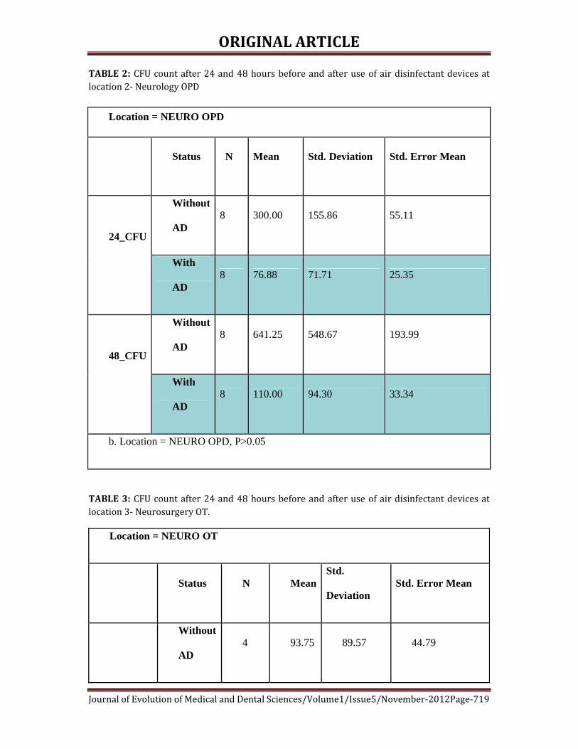
ORIGINAL ARTICLE
Journal of Evolution of Medical and Dental Sciences/Volume1/Issue5/November-2012Page-719
TABLE 2: CFU count after 24 and 48 hours before and after use of air disinfectant devices at
location 2- Neurology OPD
Location = NEURO OPD
Status N Mean Std. Deviation Std. Error Mean
24_CFU
Without
AD 8 300.00 155.86 55.11
With
AD 8 76.88 71.71 25.35
48_CFU
Without
AD 8 641.25 548.67 193.99
With
AD 8 110.00 94.30 33.34
b. Location = NEURO OPD, P>0.05
TABLE 3: CFU count after 24 and 48 hours before and after use of air disinfectant devices at
location 3- Neurosurgery OT.
Location = NEURO OT
Status N Mean
Std.
Deviation Std. Error Mean
Without
AD 4 93.75 89.57 44.79

ORIGINAL ARTICLE
Journal of Evolution of Medical and Dental Sciences/Volume1/Issue5/November-2012Page-720
24_CFU With
AD 14 6.79 15.64 4.18
48_CFU
Without
AD 8 62.50 75.02 26.53
With
AD 14 8.93 15.21 4.07
c. Location = NEURO OT, P>0.05
TABLE 4: CFU count after 24 and 48 hours before and after use of air disinfectant devices at
location 4- Spiral CT centre
Location = SPIRAL CT
Status N Mean
Std.
Deviation Std. Error Mean
24_CFU
Without
AD 4 38.75 41.11 20.55
With AD 14 54.64 134.06 35.83
48_CFU
Without
AD 8 66.88 85.90 30.37
With AD 14 66.50 140.23 37.48
d. Location = SPIRAL CT, P>0.05

ORIGINAL ARTICLE
Journal of Evolution of Medical and Dental Sciences/Volume1/Issue5/November-2012Page-721
FIGURE 1: Box plot depicting the Mean (SD) CFU counts at DSP Room
FIGURE 2: Box plot depicting the Mean (SD) CFU counts at Neuro OPD
Device OFF Device ON
Device OFF Device ON

ORIGINAL ARTICLE
Journal of Evolution of Medical and Dental Sciences/Volume1/Issue5/November-2012Page-722
FIGURE 3 : Box plot depicting the Mean (SD) CFU counts at Neuro OT
FIGURE 4: Box plot depicting the Mean (SD) CFU counts at Spiral CT center
Device ON Device OFF
Device ON Device OFF

ORIGINAL ARTICLE
Journal of Evolution of Medical and Dental Sciences/Volume1/Issue5/November-2012Page-723
PREVALENCE OF PERINATAL INFECTION IN SOUTHERN ODISHA,
INDIA. Basanti Kumari Pathi, Priya Ranjan Lenka, Kumudini Panigrahi, Dipti Pattnaik, Priti Lata Panda,
Banojini Parida
1. Assistant Professor, Department of Microbiology, KIMS, MCH, ODISHA.
2. Tutor, Department of Microbiology, KIMS, MCH, ODISHA.
3. Assistant Professor, Department of Microbiology, KIMS, MCH, ODISHA.
4. Professor, Department of Microbiology, KIMS, MCH, ODISHA.
5. Associate Professor, Department of Microbiology, M. K. C. G, MCH, ODISHA.
6. Professor, Department of Microbiology, M. K. C. G, MCH, ODISHA.
CORRESPONDING AUTHOR
Basanti Kumari Pathi,
Assistant Professor,
Dept. of Microbiology
KIMS, Patia, Bhubaneswar, Odisha,
E-mail: [email protected],
Ph: 0091 7735285610.
ABSTRACT: AIM: This study was undertaken to evaluate the prevalence of perinatal bacterial
infection in southern part of Odisha. MATERIALS AND METHODS: Pregnant women
presenting with premature rupture of membrane (PROM), preterm labour, fever, vaginal
discharge (VD), urinary tract infections and previous bad obstetric history were included in the
study groups. Besides newborns < 7 days old admitted to Paediatrics Department were also
included. High vaginal swab and blood sample were collected from mother whereas from
neonate cord blood and umbilical swab were taken. METHODS: Microscopy, culture for aerobic
bacteria and serology for C. trachomatis were done. RESULT: Total number of cases proven to
have microbiological infections based upon laboratory results. Maximum percentage of PNI
cases were primigravida belonging to the age group of 20-30years and most common bacterial
infections were caused by S. aureus & C. trachomatis. Ampicillin, Cefuroxime & Gentamicin were
found to be the most effective antibiotics for bacterial isolates.
KEYWORDS: PROM – premature rupture of membrane, VD – vaginal discharge, PNI – Perinatal
infection
INTRODUCTON: Perinatal infection (PNI) means the infection transferred from the mother to
the baby after 28th week of gestation and up to 7 days after delivery. [1, 2] These infection leads to
suffering, incapacitation, occasional permanent disability of child and also maternal and
neonatal morbidity and mortality. Still it is one of the most neglected aspects of women and
child health care. The global incidence of neonatal sepsis is 10-20 per 1000 live births. [3] In
India the incidences of sepsis is 2-10 cases per 1000 live births and have a high risk of mortality
approximately 25-30% i.e. about up to 7, 50,000 deaths every year. [3] Data from national
neonatal and perinatal data base 2000 suggests that Escherichia coli, Klebsiella spp. and
Staphylococcus aureus are the common causes of neonatal sepsis in India. The perinatal
infection affects neonates widely in various ways. Infection is the most important cause of
premature rupture of membrane, preterm labour and pPROM all of which lead to low birth
weight baby. Due to low immune status, a newborn is highly vulnerable to infections. The PNMR

ORIGINAL ARTICLE
Journal of Evolution of Medical and Dental Sciences/Volume1/Issue5/November-2012Page-724
(Perinatal mortality rate) reflects the quality of medical care. The PNMR in developing countries
is three fold to five fold higher than in the developed countries.
MATERIALS & METHODS: The prospective study was carried out, in the Department of
Microbiology, MKCG Medical College Hospital, Southern Odisha, India over a period of 22
months, in collaboration with departments of Obstetrics/Gynaecology & Paediatrics.
CASE SELECTION: 150 pregnant women admitted to the labour room, belonging to the age
group of 15 to 40 years. The study group included females with premature rupture of
membrane, preterm labour, fever, vaginal discharge, UTI and previous bad obstetric history. A
matching group of 100 patients without such complaints admitted to labour room for safe
confinement were included as controls. The newborns (<7 days) born to the pregnant women &
subsequently admitted to Paediatrics Dept. and NICU were also included in the study.
SPECIMEN COLLECTION AND PROCESSING: MOTHER: Three high vaginal
swabs/endocervical swabs were collected from the patients at the time of admission to labour
room. One swab was used for direct microscopic examination, another swab was put in 0.9%
saline for direct wet mount microscopy and yet another swab was used for aerobic isolation of
micro-organism. A blood sample of 3 ml was collected from patients for serological study of C.
trachomatis, and 5 ml blood for culture in puerperal sepsis.
NEONATE: Two umbilical swabs were collected for direct microscopy and aerobic isolation of
organisms by culture after 24 hrs of birth. 2ml of blood collected for culture in neonatal
septicaemia.
ANTIBIOTIC SUSCEPTIBILITY TESTING: Antibiotic susceptibility testing was performed by
Kirby and Bauers standard disc diffusion method.
SEROLOGY: Out of 150 cases 60 cases with bad obstetric history were subjected for IgM ELISA
test (By Novatec C. trachomatis IgM ELISA test kit) for C. trachomatis.
RESULT: Maximum cases were primigravida belonging to the age group 26 – 30 years followed
by 21-25 years. Least number of cases was primigravida belonging to the age group 36-40
years. Maximum numbers of neonates were born from primigravida belonging to the age group
26-30 years (TABLE-I).
PROM, 43 (28.6%) was the single most common presentation in these patients (Fig: 1).
Out of 150 cases, gram’s staining revealed gram positive cocci in 50 (33.3%), gram negative
bacilli in 35 (23.3%), clue cells in 28 (18.6%), and gram positive budding yeast cell in 10 (6.6%).
Among 100 controls, gram positive cocci, gram negative bacilli, clue cells and gram positive
budding yeast cell were found to be 18%,5%, 5% & 2% cases respectively (Fig: 2).
S. aureus (28%) was found to be the most common pathogen on culture isolation,
followed by E. coli (20%) in the cases. In control group S. epidermidis (25%) was the most
commonly isolated bacteria (TABLE-II).
Gentamicin is the most sensitive drug for gram positive isolates in new born where as
ampicillin was the most effective drug followed by azithromycin & penicillin in mother (TABLE-
III).

ORIGINAL ARTICLE
Journal of Evolution of Medical and Dental Sciences/Volume1/Issue5/November-2012Page-725
All gram-negative bacilli showed 100% resistance to ampicillin in mother. Cefuroxime was the
most sensitive drug followed by cephalexin and cefepime. Gentamicin is the most sensitive drug
for gram negative isolates in new born (TABLE-IV).
16 (26.6%) out of 60 cases were positive for C. trachomatis by Ig M ELISA test and only
1 (5%) out of 20 controls were showing seropositivity (Fig.3).
DISCUSSION: Microbial flora of the female genital tract presents as an extreme and diversified
spectrum of pathogenic and non-pathogenic organisms. While Chlamydia trachomatis and
Group B β- hemolytic Streptococcus are associated with PNI, bacterial vaginosis caused by
various other pathogens, has emerged as another risk factor for PNI.[4] In this study association
of selected bacterial pathogens present in the mother and transferred to baby in the causation
of PNI has been studied. In our study, the proportion of antenatal mothers attending labour
room were high in the primigravida belong to the age of group 26-30 years (Table-1). This is
comparable with the study of I. Jehan et al. where participants were predominantly in the age
group of 19-24 years. The age distribution of control was the same as that in antenatal mother
with PNI.[5]
Lower age incurs an increased risk of PNI because of biological and behavioral risk
factors. Adolescents tend to have cervical ectopy, which provides large zones of columnar
epithelium for the targeted attachment of C. trachomatis and N. gonorrchea [4]. The present
study showed the comparable observation (Table-1).
As in our study maximum no. of cases and controls were primigravida, the numbers of
neonates were born maximum from primigravida belong to age group 26-30 years (Table-1).
Premature rupture of membrane was the most common symptom in cases (28.6%) found in our
study (Fig-1). The other symptoms and signs were preterm labour, pPROM, vaginal discharge,
puerperal sepsis etc.
In our study, direct microscopic observation by Gram’s stain revealed gram positive
cocci in cases 50 (33.3%), gram negative bacilli 35 (23.3%) & gram negative cocci 4 (2.6%) (Fig-
2). As per Amsel’s criteria for the diagnosis of G. vaginalis, clue cell were found in 28 (18.6%)
cases out of 150 (Fig-2). [5, 6] They found that sexually transmitted cofactor may strengthen, the
relation between bacterial vaginosis associated microorganisms and development of PNI. In
some other studies the prevalence of bacterial vaginosis was reported to be 6-32%.[7] In our
study bacterial vaginosis associated bacteria G. vaginalis was identified as gram variable
pleomorphic coccobacilli mostly in clumps. Deborah Money [8] also used gram stain of vaginal
fluid for the diagnosis of bacterial vaginosis in lieu of clinical criteria, given its greater reliability
for diagnosis of bacterial vaginosis. A number of studies proved that apart from C. trachomatis,
bacterial vaginosis has been a risk factor for preterm labour, premature rupture of membrane,
preterm premature of membrane. [5, 6]
Gram – negative diplococci were found to be 4 (2.6%). They were diagnosed to be N.
gonorrhoeae morphologically by gram stain, as the culture was very cumbersome and not done.
Bacterial culture findings of this study revealed isolation of S. aureus (28%) followed by E. coli
(20%), Group B β- hemolytic streptococci (2.6%) and Enterococcus spp. (2.6%), klebsiella sp.
(1.3%) and Pseudomonas aeruginosa (1.3%). Least was Proteus mirabilis (0.6%), but in control
group maximum isolates were S. epidermidis (25%) (Table–2). As compare in neonates the
isolates were of S aureus (17%), Group B β- hemolytic streptococci (1.3%), E. coli (8.8%),
Klebsiella spp. (1.3%) and Pseudomonas aeruginosa (0.6%) (Table-2).
ORIGINAL ARTICLE
Journal of Evolution of Medical and Dental Sciences/Volume1/Issue5/November-2012Page-726
Serological tests were generally not useful in the diagnosis of genital tract infection
caused by C. trachomatis and that the presence of immunoglobulin M (Ig M) antibodies was an
unreliable marker of acute infection in adolescents and adults. Numazaki K et al reported that
the rates of Ig M seropositivity for C. trachomatis during pregnancy were significantly higher in
mothers who had given birth to infants with complications than in matched controls.[9] The
frequency of chorioamnionitis and meconium stained amniotic fluid was also higher in the anti
C. trachomatis Ig M antibody-positive pregnant women. Several investigations have reported
that 2-20% of pregnant women harbour C. trachomatis in the endocervix. Ig M antibodies on the
other hand may be short-lived. ELISA tests of patients suspected with C. trachomatis infection
have recently been used as a diagnostic tool. Hence, we used this test to demonstrate Ig M
antibodies in cases of clinically diagnosed PNI. Out of 60 cases, 16 cases (26.6%) were found to
be seropositive for C. trachomatis (Fig-3). Only 1 (5%) seropositivity was found in control group
(Fig-3).
Our data confirms the relationship between maternal infection and perinatal outcome
and support the hypothesis that infections might be the cause of these conditions and not
simply an association. For preventing perinatal infections, antenatal mothers should be
screened and treated for these infections. From our study most of the gram positive cocci
isolated were found sensitive to ampicillin, penicillin and azithromycin (Table-III). All the gram
negative bacilli show 100% resistance to ampicillin. Most sensitive drug for gram negative
bacilli is cefuroxime (Table- IV). Gentamicin was the most effective antibiotic for both gram
positive cocci and gram negative bacilli in neonates (Table-III & IV).
CONCLUSION: The study was carried out over a period of 22 months in the Depart. Of
Microbiology, M.K.C.G. Medical College, Southern Odisha, India. All the samples collected from
both the cases and controls were subjected for bacteriological & serological analysis. In view of
the above finding available from results in our study we could derive the following conclusions:
incidence of PNI attending labour room was highest in primigravida belonging to the age group
26 – 30 years. Staphylococcus aureus and C. trachomatis were the most common aetiological
agent of PNI. Other bacterial pathogens isolated in the culture were Gr. B β hemolytic
Streptococci, Enterococci, E. coli and Klebsiella spp. Ampicillin, azithromycin, cefuroxime and
gentamicin were found to be effective on the bacterial isolates.
ACKNOWLEDGMENT:
Special thanks to Dept. of Microbiology, M.K.C.G.MCH for funding to this reasearch work.
REFFERENCES:
1. K. Park, “Textbook of Preventive and social medicine”, 19th edition, 1167, Prem nagar,
Jabalpur, M.P. India, m/s Banarsidas bhanot Publisher, 2007.
2. D.C. Dutta, “Text Book of Obstetrics”; 4th Edn, 8/1 Chintamoni Das Lane, Kolkata, New
Central book agency (P) Ltd., 2000.
3. Altha Roterti Edgren, Teresa G. Odle et al, Gale, Detrait, Gale Encyclopedia of Children’s
Health, 2006.
4. Basavaraj M. Kerur, B Vishnu Bhat, B.N. Harish, S. Habeebullah and C. Uday Kumar,
“Maternal Genital Bacteria and Surface Colonization in Early Neonatal Sepsis”, Indian J
Pediatr, 2006; 73 (1): 29-32.

ORIGINAL ARTICLE
Journal of Evolution of Medical and Dental Sciences/Volume1/Issue5/November-2012Page-727
5. I. Jehan, H. Harris, S. Salat, A. Zeb, N. Mobeen, O. Pasha, E.M. McClure, J. Moore, L.L wright
& R.L Goldenberg, “Neonatal mortality, risk factors and causes: a prosepective
population based cohort study in urban Pakistan”, bulletin of the WHO Volume 87,
Number 2, February 2009, 130-138.
6. Gibbs R.S., “Severe infections in pregnancy”, Med Clin North Am. 1989; 73:713-21.
7. Amsel R, “Non specific vaginitis, Diagnostic criteria and microbial and epidemiological
associations”, Am J Med 1983 Jan; 74 (1):14-22.
8. Deborah Money, “The laboratory diagnosis of bacterial vaginosis”, Can J Infect Dis Med
Microbial, 2005, Mar-Apr; 16 (2): 77-79.
9. Numazaki K, Wainberg MA, Mc Donald J, “Chalmydia trachomatis infections in infants”,
CMAJ 1989, 140: 615-22.
TABLE-I: Age distribution of antenatal mother and neonate of study group
Age
(years)
Cases
Mother Neonate
Primigravida Multigravida Primigravida Multigravida
15-20 14 6 14 7
21-25 20 10 18 10
26-30 28 16 27 16
31-35 16 22 16 23
36-40 4 12 4 12
TABLE-II: Culture isolation of different bacteria in antenatal mother and neonate.
Bacteria Mother Neonate
Cases Control
GPC
S. aureus 42(28%) 15(15%) 25(17%)
S. epidermidis 12(8%) 25(25%) 4(2.6%)
GBS 4(2.6%) 0 2(1.3%)
Enterococci spp. 4(2.6%) 0 0
GNB
E. coli 29(20%) 5(5%) 13(8.8%)
Klebsiella spp. 2(1.3%) 0 2(1.3%)
Pseudomonas aueroginosa 2(1.3%) 1(1%) 1(0.6%)
Proteus spp. 1(0.6%) 0 0
TABLE-III: Sensitivity pattern of GPC to various antimicrobials.
BACTERIA MOTHR NEONATE
Total
no
P A Ox Ch Cp Cu At Cd Total
no
A Ak G Cf Co E Cu Ci
S. aureus 42 14 25 12 2 16 13 26 13 25 5 3 15 7 8 2 11 10
GBS 4 3 4 3 0 1 0 0 0 2 2 0 0 0 2 2 1 0

ORIGINAL ARTICLE
Journal of Evolution of Medical and Dental Sciences/Volume1/Issue5/November-2012Page-728
Enterococcus 4 2 1 0 2 0 2 3 2 1 0 0 0 0 1 0 1 1
Total no 50 19 30 15 4 17 15 29 15 28 7 3 15 7 11 4 13 11
(P – Penicillin, A – Ampicillin, Ox – Oxacillin, Ch – Cephalothin, Cp – Cephalexin, Cu- Cefuroxime,
At – Azithromycin, Cd – Clindamycin, Ak – Amikacin, G – Gentamicin, Cf – Ciprofloxacin, Co –
Cotrimoxazole, E – Erythromycin, Ci – Ceftriaxone)
TABLE-IV: Sensitivity pattern of GNB to various antimicrobials
BACTERIA MOTHER NEONATE
Total
no
A Cp C
u
Cp
m
At Tota
l
no
A A
k
G Cf C
o
C
e
Cu Ci
E. coli 29 0 22 1
3
11 1
1
13 2 8 9 3 4 2 5 1
Klebsiella 2 0 1 2 1 0 2 0 1 2 1 2 1 0 0
P.aeuroginos
a
2 0 0 2 1 1 1 0 0 1 0 0 1 1 0
P.mirabilis 1 0 1 1 0 o 0 2 0 0 0 0 0 0 0
Total no. 34 0 24 1
8
13 1
2
16 2 9 1
2
4 6 4 6 1
(A – Ampicillin, Cp – Cephalexin, Cu – cefuroxime, Cpm – Cefepime, At – Azithromycin, Ak –
Amikacin, G – Gentamicin, Cf – Ciprofloxacin, Co – Cotrimoxazole, Ce - Ceftazidime, Ci –
Ceftriaxone)
Fig-1: clinical features of the patients
(*VD: Vaginal discharge, **PROM: Premature rupture of membrane, ***pPROM: Preterm premature rupture of membrane.)
0
5
10
15
20
25
30
Per
cen
tag
e
VD PROM pPROM PretermLabour
PuerperalSepsis
Other
Clinical features

ORIGINAL ARTICLE
Journal of Evolution of Medical and Dental Sciences/Volume1/Issue5/November-2012Page-729
Fig-2: Microscopic observation by gram’s stain method.
Fig. 3: Ig M ELISA test for Chlamydia trachomatis
0
5
10
15
20
25
30
35
Per
centa
ge
GPC GNC GNB Clue cells Gram +vebuddingyeast
Organisms
Cases Controls
26.6
5
%
Cases (n=60) Control (n=20) others

CASE REPORT
Journal of Evolution of Medical and Dental Sciences/Volume1/Issue5/November-2012Page-730
MALIGNANCY IN AN AMPUTATED LEG STUMP – A RARE CASE
Karigalan. K, Haripriya Reddy
1. Assistant Professor, M. S. Department of General Surgery, P. E. S. I. M. S. R, Kuppam, Andhra Pradesh.
2. Assistant Professor, M. S. Department of General Surgery, P. E. S. I. M. S. R, Kuppam, Andhra Pradesh.
CORRESPONDING AUTHOR
Dr. Karigalan K.
No. 202, II Floor, Staff Quarters,
P. E. S. I. M. S. R.
E-mail: [email protected],
Ph: 0091 900767648.
ABSTRACT: Amputation of lower extremity followed by use of leg prosthesis is very common.
However, malignancy arising in the amputation stump is extremely rare. We are reporting such
a case in a 52-year-old man. Review of the English literature reveals only five additional cases
occurring in men with an average age of 65 years and after a mean lag period of 40 years
between the amputation and development of a low-grade squamous cell carcinoma.
KEYWORDS: Malignancy in amputation stump; Squamous cell carcinoma; Leg prosthesis.
MESH TERMS: Amputation Stumps; Carcinoma, squamous cell; Artificial Limbs
INTRODUCTION: Amputation of the leg followed by the use of artificial leg is very common.
However, a malignant tumor arising in an amputation stump remains a very rare occurrence.
The purpose of this paper is to report a case of squamous cell carcinoma arising in an
amputation stump with a review of all other 5cases of malignancy arising in stumps published
in the English literature.
CASE REPORT: A 52year old man sustained crush injury to left lower limb in a train accident
25 years back.
He underwent left above knee amputation and was fitted with an artificial limb. He had
continued difficulty with proper fitting of the prosthesis over the years. Almost 25 years later,
the patient started experiencing increased pain at the amputation site and noted foul smelling
discharge from the area. The skin over the distal stump was macerated, ulcerated with purulent
foul smelling exudate. He was managed in the wound care clinic with wound cleaning and
antibiotics over the last 6months. During that period, the distal stump area developed a
fungating tissue that bleeds on touch. He was unable to use the leg prosthesis. He is a known
case of diabetes, taking treatment.
The amputated stump showed an irregular ulcer of 10cmx10cm with an exophytic
growth of 6cm in its largest dimension. Ulcer had raised, everted edges that bleed easily on
touch and an indurated base with foul smelling discharge (Fig.1). No inguinal lymphadenopathy,
no evidence of metastatic spread seen.
Patient underwent a left above knee revision amputation with wide local excision of
tumour margins. Specimen was sent for histopathological examination. Postoperative period
was uneventful and patient was discharged on 10th postop day after suture removal.
Microscopically, well formed keratin pearls, stromal infiltration with lymphocytes and
resected margins free of tumour cells were noted (Fig.2). Hence histopathology report notified
features suggestive of well-differentiated type of squamous cell carcinoma.

CASE REPORT
Journal of Evolution of Medical and Dental Sciences/Volume1/Issue5/November-2012Page-731
DISCUSSION: Cutaneous carcinomata of the lower extremities seem to be never a primary
condition [1]. About 60,000 leg amputations are performed each year in the United States.
These patients are usually fitted with leg prosthesis. The distal aspect of the stump has the scar
from surgical closure. Whereas malignant tumor, commonly a squamous cell carcinoma is
known to occur in scarred tissue from burn, chronic ulcers, wounds, sinus and fistulous tracts
[2,3], the occurrence of cancer in amputated leg stump is very rare.
The earliest report of a stump carcinoma that we could find in the English literature was
in 1965 describing a 75-year –old white man who had undergone a left above the knee
amputation following a railroad accident [4]. He was fitted with leg prosthesis. Forty-seven
years later, the patient developed a well-differentiated squamous cell carcinoma at the
amputation stump scar. He underwent a high thigh amputation and inguinal lymph node
dissection. There were no metastases.
In 1985, a case of a 60-year-old man who developed a low-grade squamous cell
carcinoma on the below-the-knee amputation stump was reported [5]. The patient had
undergone amputation of his leg 30 years earlier because of traumatic injury and burn suffered
in a dynamite explosion. He used an artificial leg since then. The patient developed the cancer in
the burn scar rather than in the scar of stump closure. He was treated with above-knee
amputation. There was no evidence of metastatic spread.
In 1991, there were two reports of carcinoma arising in leg amputation stump [6,7] In
one report [6], a 56 –year –old man developed a rapidly growing squamous cell carcinoma at
the end of his right below-knee amputation stump 27 years after his leg was amputated for
gangrene from a snake-bite injury. He was using a poorly constructed prosthesis. The tumor
was widely excised. The carcinoma arose in the scar tissue of the stump. There was no distant
spread. The other case [7] was that of a 65-year- old man who developed a verrucous squamous
cell carcinoma at the stump about 45 years after he underwent a traumatic below-the-knee
amputation followed by use of leg prosthesis. The tumor was locally excised with a revision of
the amputation.
The latest report of a carcinoma arising in an amputation stump was that of a 62-year-
old man who developed a well-differentiated squamous cell carcinoma on his right thigh
amputation stump 52 years after the procedure [8]. The amputation was performed because of
a land mine injury. He was using a leg prosthesis ever since. The patient underwent a surgical
revision of the amputation stump and inguinal lymph node dissection. There was no metastatic
spread.
Malignancy occurring in leg amputation stump remains a rare event as evidenced by
only six such reported cases (including the present case) in the English literature since 1965.All
the patients were male, aged 56 to 75 years with a mean age of 63 years. Amputation was done
mostly for trauma [4,5,7,8]. In two cases [5,6], burn from dynamite and mine explosion was an
additional factor. The time between the amputation and the development of malignancy at the
stump ranged from 27 to 52 years with a mean lag period of 40 years. All patients developed
low-grade squamous cell carcinoma, of which two patients showed a verrucous type of
squamous cell carcinoma.
The factors that may lead to malignancy at a stump may include long-standing scar
tissue and burn injury. Patients who underwent amputation at an older age may not live long
enough to develop stump carcinoma because the average lag period of developing such a lesion
is 40 years. The patients who have undergone amputation at a younger age may have a small
risk of developing malignancy at the stump over their lifetime.

CASE REPORT
Journal of Evolution of Medical and Dental Sciences/Volume1/Issue5/November-2012Page-732
Neoplasms associated with scars and chronic ulcers are well documented for humans. These
lesions comprise approximately 2% of all human epidermal neoplasms. Squamous cell
carcinoma is the most common tumor and typically occurs 20 to 40 y after the initial injury.
Rarely, the original wound does not heal, and a neoplasm appears within a relatively short time
(3 to 12 mo). These neoplasms are termed "acute wound cancers," since they appear relatively
rapidly compared with other scar-associated neoplasms [9].
Carcinoma arising from scar, accounts for 0.78% of all inpatients with scars and 17% of
all skin carcinoma patients. Scar carcinoma is a highly differentiated squamous cell carcinoma in
most of the cases. Because of scar fibrosis and thrombosis of blood vessel and lymphatic vessel,
the growth of scar carcinoma is slow. The metastasis is restricted. If the carcinoma can be
diagnosed and treated early, the prognosis is favourable. The principle of operation is wide
excision of the lesion [10].
The clinical appearance of skin metastases varies over a wide morphologic spectrum,
cutaneous metastases mimicking herpes zoster being rare. zosteriform cutaneous metastases
secondary to a squamous cell carcinoma (SCC) which developed in the stump of an amputated
arm is reported. The pathogenesis is speculative, but the zosteriform distribution might well be
explained by perineural lymphatic invasion and spread [11].
Marjolin's ulcers are uncommon malignancies arising from previously traumatized,
chronically inflamed or scarred skin. They are usually squamous cell carcinomas and they may
present decades after the original insult. Marjolin's ulcers may occur on stumps in patients
using prostheses, in this situation the diagnosis may be delayed because of the false assumption
that the ulcer is caused by an ill fitting prosthesis [12]. A high index of suspicion is required in
the management of chronic non-healing ulcers and all suspected lesions should be biopsied
[13]. The squamous cell carcinoma of Marjolin's ulcer has the worst prognosis in comparison
with other squamous cell carcinomas and it requires an aggressive treatment [14].
Epithelioma cuniculatum (EC) belongs to the category of verrucous carcinomas.
Invasiveness and rate of metastasis are low, but there is a high risk of local recurrence. In cases
of long-standing processes with formation of exophytic, malodorous tumors with jagged edges
that do not respond to conventional therapy, consideration should already be given to EC upon
visual inspection. The diagnosis is always established by histological examination. The standard
treatment of EC is extensive excision of the tumor with micrographic margin control [15].
REFERENCES:
1. De Asis C. CUTANEOUS CARCINOMA OF THE LOWER EXTREMITIES: A STUDY OF CASES
AT THE BARNES AND THE BARNARD FREE SKIN AND CANCER HOSPITALS OF ST.
LOUIS, MO. Ann Surg. 1926 May;83(5):663-81.
2. Arons MS, Lynch JB, Lewis SR, Blocker TG. Scar tissue carcinoma. Part 1. A clinical study
with special reference to burn scar carcinoma. Ann Surg 1965; 161:170-188.
3. Bartle EJ, Sun JH, Wang XW, Schneider BK. Cancers arising from burn areas. A literature
review of twenty-one cases. J Burn Care Rehab 1990; 11:46-49.
4. Wilhite JL. Carcinoma developing in a pre-existing scar. A case report of carcinoma of
amputation stump. Va Med Mon 1964; 91: 292-294.
5. Sarma D, Weilbaecher TG. Carcinoma arising in burn scar. J Surg Oncol 1985; 29:89-90.
6. Mahaisavariya B, Mahaisavariya. Marjolin's ulcer complicating a poorly fabricated
prosthesis. Injury 1991; 22: 423-424.
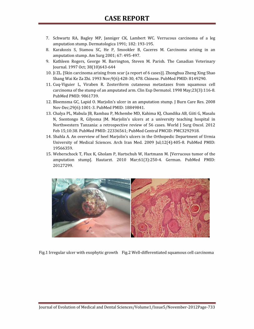
CASE REPORT
Journal of Evolution of Medical and Dental Sciences/Volume1/Issue5/November-2012Page-733
7. Schwartz RA, Bagley MP, Janniger CK, Lambert WC. Verrucous carcinoma of a leg
amputation stump. Dermatologica 1991; 182: 193-195.
8. Karakozis S, Stamou SC, He P, Smookler B, Caceres M. Carcinoma arising in an
amputation stump. Am Surg 2001; 67: 495-497.
9. Kathleen Rogers, George M. Barrington, Steven M. Parish. The Canadian Veterinary
Journal. 1997 Oct; 38(10)643-644
10. Ji ZL. [Skin carcinoma arising from scar (a report of 6 cases)]. Zhonghua Zheng Xing Shao
Shang Wai Ke Za Zhi. 1993 Nov;9(6):428-30, 478. Chinese. PubMed PMID: 8149290.
11. Cuq-Viguier L, Viraben R. Zosteriform cutaneous metastases from squamous cell
carcinoma of the stump of an amputated arm. Clin Exp Dermatol. 1998 May;23(3):116-8.
PubMed PMID: 9861739.
12. Bloemsma GC, Lapid O. Marjolin's ulcer in an amputation stump. J Burn Care Res. 2008
Nov-Dec;29(6):1001-3. PubMed PMID: 18849841.
13. Chalya PL, Mabula JB, Rambau P, Mchembe MD, Kahima KJ, Chandika AB, Giiti G, Masalu
N, Ssentongo R, Gilyoma JM. Marjolin's ulcers at a university teaching hospital in
Northwestern Tanzania: a retrospective review of 56 cases. World J Surg Oncol. 2012
Feb 15;10:38. PubMed PMID: 22336561; PubMed Central PMCID: PMC3292918.
14. Shahla A. An overview of heel Marjolin's ulcers in the Orthopedic Department of Urmia
University of Medical Sciences. Arch Iran Med. 2009 Jul;12(4):405-8. PubMed PMID:
19566359.
15. Weberschock T, Flux K, Gholam P, Hartschuh W, Hartmann M. [Verrucous tumor of the
amputation stump]. Hautarzt. 2010 Mar;61(3):250-4. German. PubMed PMID:
20127299.
Fig.1 Irregular ulcer with exophytic growth Fig.2 Well-differentiated squamous cell carcinoma

CASE REPORT
Journal of Evolution of Medical and Dental Sciences/Volume1/Issue5/November-2012Page-734
JUVENILE IDIOPATHIC ARTHRITIS – A CASE REPORT Paresh H. Bharia, S. D. Birare, S. S. Dantkale, S. A. Chaware, V. B. Dhurve
1. Resident, Department of Pathology, Government Medical College, Latur, Maharashtra.
2. Associate Professor, Department of Pathology, Government Medical College, Latur, Maharashtra. 3. Associate Professor & Head, Department of Pathology, Government Medical College, Latur, Maharashtra. 4. Assistant Professor, Department of Pathology, Government Medical College, Latur, Maharashtra. 5. Resident, Department of Pathology, Government Medical College, Latur, Maharashtra.
CORRESPONDING AUTHOR
Paresh H. Bharia,
Department of Pathology,
Government Medical College, Latur,
E-mail: [email protected],
Ph: 0091 9975188055.
ABSTRACT: The prevalence of Juvenile idiopathic arthritis (JIA) is 0.86 per 1000 children.
Subcutaneous nodules have been reported in 5% to 10% of children with JIA.
Approximately 90% of patients with RA and subcutaneous nodules test positive for
rheumatoid factor (RF), and approximately 40% of all RF-seropositive patients with RA
have subcutaneous nodules, whereas only 6% involvement is seen in seronegative cases.
We hereby report a case of atypical Juvenile idiopathic arthritis (JIA) in a 6 year old,
female child with joint pain & myalgia along with subcutaneous nodules over the dorsum
of feet, hands and elbows. Joint pain initially involving the left ankle, slowly progressed
to involve the knee, shoulder, wrist, metacarpophalangeal and interphalangeal joints over
a period of one year. Joint involvement was not symmetric. RF was Negative. Fundoscopy
examination was normal. Histopathological examination revealed a central zone of
Fibrinoid necrosis surrounded by epithelioid histiocytes and occasional lymphocytes.
Differential diagnosis of Rheumatoid Nodule (RN) or Subcutaneous Granuloma Annulare
(SGA) or Necrobiosis Lipoidica Diabeticorum was made. In light of clinicopathological
findings, both SGA and NLD were ruled out and the diagnosis of Juvenile idiopathic
arthritis presenting as RF-negative polyarthritis was made.
KEYWORDS: juvenile idiopathic arthritis, rheumatoid arthritis, rheumatoid nodule.
MESH TERMS: Nil
INTRODUCTION: Juvenile idiopathic arthritis (JIA), previously known as juvenile
rheumatoid arthritis encompasses all forms of arthritis that develop before 16 years of
age and persist for a minimum of 6 weeks. Rheumatoid nodules (RN) are the most
common cutaneous lesion in adult cases of rheumatoid arthritis (RA) and are present in
25% cases (both oligoarticular & polyarticular) [1]. A much greater incidence (75%) is
observed in those with RA-associated Felty syndrome. [2] Approximately 90% of patients
with RA and subcutaneous nodules test positive for rheumatoid factor (RF), and
approximately 40% of all RF-seropositive patients with RA have subcutaneous nodules,
whereas only 6% involvement is seen in seronegative cases. [3] Rheumatoid nodules are
clinical predictors of more severe arthritis, seropositivity, joint erosions, and rheumatoid
vasculitis. It has been suggested that the presence of RNs often requires more aggressive
treatment of the underlying RA to prevent sequelae. However, in some cases disease

CASE REPORT
Journal of Evolution of Medical and Dental Sciences/Volume1/Issue5/November-2012Page-735
progression is independent of nodular disease activity. The prevalence of JIA is 0.86 per
1000 children. Subcutaneous nodules have been reported in 5% to 10% of children with
JIA. [4] Rheumatoid nodules are almost always confined to those children with
polyarticular arthritis i.e. involving ≥5 joints in first 6 months. [5] Genetics seems to play
a role in the appearance of RNs. The HLA-DR4 haplotype (including the heterogeneous
group of DRB1 alleles) is predictive of the risk of subcutaneous nodules in RA. [6]
CASE HISTORY: We hereby report a case of an atypical Juvenile idiopathic arthritis in a
6 year old, female child with joint pain & myalgia. Joint pain initially involved the left
ankle, slowly progressing to involve the knee, shoulder, wrist, metacarpophalangeal and
interphalangeal joints over a period of one year. Joint involvement was not symmetric.
There was presence of subcutaneous nodules over the dorsum of feet [Figure 1], hands
and elbows. Clinical diagnosis of Rheumatoid arthritis was made.
On investigating, rheumatoid factor was negative. Serum ANA and anti-CCP (cyclic
citrullinated peptide) antibody tests were advised but not performed. Fundoscopy
examination was normal. A skin biopsy was taken from the site of the subcutaneous
nodule from the dorsum of the feet. Histopathological examination of the subcutaneous
nodule revealed a central zone of fibrinoid necrosis surrounded by epithelioid histiocytes
and occasional lymphocytes. [Figures 2 & 3] Differential diagnosis of Rheumatoid Nodule or
Subcutaneous Granuloma Annulare (SGA) or Necrobiosis Lipoidica Diabeticorum (NLD)
was made.
DISCUSSION: A diagnosis of rheumatoid nodules is made in the clinical context of the
disease. Although biopsies of subcutaneous nodules are occasionally done, they are not
useful for diagnosis. [7] The 2010 ACR-EULAR criteria of rheumatoid arthritis has not
taken into consideration the presence of rheumatoid nodules or radiographic joint
damage because these findings occur rarely in early RA. [8] Many different types of
subcutaneous nodules are histologically identical to rheumatoid nodules; mainly NLD and
SGA, which are often misdiagnosed as RNs. [9]
In general, RNs tend to be larger and to be confined to the deep dermis or
subcutis, at the bony prominences, commonly located at the ulnar aspect of the forearm,
elbows, occiput, and lumbosacral area. In our case, the nodules were present on bony
prominences of the dorsum of feet and hands. Microscopically they have a central zone
of fibrinoid necrosis surrounded by a prominent rim of epithelioid histiocytes and
numerous lymphocytes and plasma cells. [1] Fibrin may be deposited in the centre of the
granulomas, as opposed to the mucin deposition of SGA. [10] However, RNs with abundant
mucin may be present. [11] Therefore histopathological differentiation between RNs and
SGA is difficult.
Serum IgM RF has been found in 75–80% of patients with RA [8]; therefore, a
negative result does not exclude the presence of this RA in our patient.
Subcutaneous granuloma annulare (SGA) is one of the most common dermatoses
with the involvement of skin and/or subcutis, usually seen in adults and children, but the
aetiology and pathogenesis are unclear. [12] It most often manifests as a large,
asymptomatic soft tissue mass. Although nodules are usually stable for months, they may
rapidly enlarge over the course of weeks. The typical lesions of SGA are single or
multiple, small, pinkish, nonulcerated nodules in the deep subcutaneous tissue. The most

CASE REPORT
Journal of Evolution of Medical and Dental Sciences/Volume1/Issue5/November-2012Page-736
common lesion location is lower extremity, especially the peri-tibial area, followed by the
hands. On histological evaluation, these nodules are similar to the nodules seen in adults
with rheumatoid arthritis and to the lesion recognized in adult diabetic patients as
necrobiosis lipoidica diabeticorum. [13] Characteristically, the necrobiotic focus is not so
large or deeply situated as those of rheumatoid nodules or as broad and diffuse as those
of necrobiosis lipoidica (diabeticorum). [9] In our case, one year history of joint pain and
migratory polyarthritis along with subcutaneous nodules ruled out SGA and NLD.
Necrobiosis Lipoidica is a well-recognized dermatologic complication of diabetes
mellitus and may develop in both juvenile (Type I) and adult-onset (Type II) diabetes
mellitus. Positive family history of diabetes mellitus has also been one of the risk factors.
[13] The sclerodermatous plaque is round to oval, red-brown with an elevated rim. Like
SGA, the peri-tibial region is the most common site. Clinical correlation must be taken
into consideration before making the diagnosis of NLD. [11] Our patient was not diabetic
nor gave any positive family history for diabetes mellitus. The patient was successfully
treated with steroids and has not shown sustained disease or diabetes mellitus over a
one year follow up.
In light of clinicopathological findings, the diagnosis of Juvenile idiopathic arthritis
presenting as RF-negative polyarthritis was made. We thus believe that histopathology of
rheumatoid nodule presents a special challenge to a pathologist. Relevant
clinicopathological details are essential for diagnosis and appropriate management.
REFERENCES:
1. Kumar V, Abbas A K, Fausto N, Aster J C. Robbins & Cotran Pathologic Basis of
Disease. 8 th Edition. Philadelphia: Saunders Elsevier; 2010.
2. Sayah A, English JC 3rd: Rheumatoid arthritis: A review of the cutaneous
manifestations. J Am Acad Dermatol 53:91-209, 2005
3. Kaye BR, Kaye RL, Bobrove A: Rheumatoid nodules. Review of the spectrum
of associated conditions and proposal of a new classification, with a report
of four seronegative cases. Am J Med 76:279-292, 1984
4. Vicente García-Patos. Rheumatoid Nodule. Semin Cutan Med Surg 26:100-
107 © 2007 Elsevier Inc
5. Sampath Prahalad and David N Glass. Is juvenile rheumatoid
arthritis/juvenile idiopathic arthritis different from rheumatoid arthritis?
6. Ahmed SS, Arnett FC, Smith CA, et al: The HLA-DRB1*0401 allele and the
development of methotrexate-induced accelerated rheumatoid nodulosis: A
follow-up study of 79 Caucasian patients with rheumatoid arthritis.
Medicine 80:271-278, 2001 7. Patricia T. Ting and Benjamin Barankin. Dermacase. VOL 5. JANUARY 2005
Canadian Family Physician.
8. Longo DL, Fauci AS, Kasper DL et al. Harrison’s Principles of Internal
Medicine. Volume 2. 18th edition, New York. McGraw Hill Publications. 2012.
9. Hakan Sabuncuoglu et al. Subcutaneous Granuloma Annulare of the Scalp in
Childhood: A Case Report and Review of the Literature. Turkish
Neurosurgery 2007, Vol: 17, No: 1, 19-22
10. Mills SE, Carter D, Greenson JK et al. Sternberg’s Diagnostic surgical pathology.
5 th Edition. Philadelphia. Lippicott Williams & Wilkins. 2010.

CASE REPORT
Journal of Evolution of Medical and Dental Sciences/Volume1/Issue5/November-2012Page-737
11. Mckee P H, Calonje E, Granter S R. Pathology of the skin with clinical
correlations. Volume 1. 3 rd edition. Philadelphia. Elsevier Mosby. 2005.
12. Requena L, Fernandez- Figueras MT. Subcutaneous granuloma annulare.
Semin Cutan Med Surg 2007; 26: 96-9
13. Grogg KL, Nascimento AG. Subcutaneous granuloma annulare in childhood:
clinicopathologic features in 34 cases. Pediatrics: 107(3):42, 2001
Figure 1. Subcutaneous nodules present on the dorsum of foot.
Figure 2. Low power view through subcutaneous nodule showing a necrobiotic focus
situated in the deeper dermis. (10x; H&E)
Figure 3. Necrobiotic focus showing central fibrinoid necrosis surrounded by histiocytes
and lymphocytes (40x; H&E)

REVIEW ARTICLE
Journal of Evolution of Medical and Dental Sciences/Volume1/Issue5/November-2012Page-738
ANTIBACTERIAL EFFECT OF CALCIUM HYDROXIDE IN DIFFERENT
VEHICLES K. R. Hari, M. R. Ganesh,
1. Professor, M.D. S. Department of Oral Surgery, Adhiparashakti Dental College, Chennai.
2. Reader, M.D. S. Department of Endodontics, Adhiparashakti Dental College, Chennai.
CORRESPONDING AUTHOR
Dr. M. R. Ganesh
C. S.T, back of G. P. O
Mumbai-1, Maharashtra, India,
E-mail: [email protected],
Ph: 0091 8600117037.
ABSTRACT: AIM: This study evaluated the antibacterial effect of calcium hydroxide in different
vehicles in an in vitro model. MATERIAL AND METHODS: Calcium hydroxide paste prepared
with two conventionally used vehicles namely, camphorated monochlophenol, distilled water
and also propylene glycol. The antibacterial activity of these paste were tested against five
micro- organisms that can commonly occur in the infected root canals. RESULTS AND
CONCLUSIONS: The results of the study indicate that a paste of calcium hydroxide made with
propylene glycol exerts significant antibacterial action. Hence, it can be recommended for use as
an intracanal medicament in preference to a paste prepared with a tissue toxic phenolic
compound like camphorated mono chlorophenol.
KEY WORDS: Calcium hydroxide, camphorated monochlorophenol, propylene glycol.
INTRODUCTION: The endodontist today, concentrates more upon instrumentation, irrigation
thorough cleaning and shaping of the root canal for elimination of responsible microbial flora
from the root canal. [12], [15],[17],[21]
With due respect to all norms laid down in the field of endodontics, the review of
literature is suggestive that establishing the complete debridement and disinfection of the root
canal contributes to maximum therapeutic success.
In routine clinical practice, disinfection of the root canal is established by
comprehensive effect of biomechanical preparation, use of intra canal irrigation and intra canal
medicament. [1], [23],[14]
Calcium hydroxide is known to endodontists for its application from many years since
its introduction by Herman in 1930.
In present day, it has become more popular as intra canal medicament. Its bactericidal
or bacteriostatic action is related to its dissociation into calcium and hydroxyl ions thereby
creating alkaline environment in its vicinity, not allowing the growth of acidophilic
microorganisms. In addition to this it also inhibits the enzymatic activity that is essential for
microbial growth. [8],[9]
To facilitate its application in the field of endodontics, it is generally mixed with vehicles
like camphorated mono chloro phenol, distilled water, glycerin and propylene glycol. Various
studies are carried out to identify or to understand the most suitable vehicle and also to find out
the effect of vehicle on the action of the calcium hydroxide as an intra canal medicament.
Observations followed to such study suggest that calcium hydroxide when mixed with water as

REVIEW ARTICLE
Journal of Evolution of Medical and Dental Sciences/Volume1/Issue5/November-2012Page-739
a vehicle promotes the dissociation and initiates the meditational action of the calcium
hydroxide.
Calcium hydroxide when mixed with camphorated mono chlorophenol has dual effect
vehicle as well as additional antibacterial action.
Calcium hydroxide when mixed with propylene glycol also has dual effect like
camphorated mono chlorophenol. However, it exhibits no tissue irritating effect and being
alcoholic in nature, it remains in paste form for longer time.
However there are very few study related to propylene glycol that serves as better
vehicle when mixed with calcium hydroxide to form a paste for the purpose of intracanal
medicament.
It was purposed to comparatively evaluate the antibacterial effect of calcium hydroxide
when mixed with distilled water, camphorated mono chlorophenol and propylene glycol.
MATERIAL AND METHODS: Infected Root Canal Flora Commonly Consist of gram positive
cocci like streptococci, staphylococci, gram positive bacilli like acidophillus, lactobacilus,
actinomycotis fungi like candida albicans, gram negative organism like E-coli, pseudomonas.
More common and more resistant organisms in root canals are streptococci and E.
Faecalis respectively.
Considering infected microbial flora of infected root canal it was purposed to obtained
the five micro organisms samples from the stock culture of the department of microbiology,
grand medical college, Sir. J.J. Group of Hospital Mumbai, namely
1) H. Streptococcus,
2) Enterococcus Faecalis
3) Staphylococcus aureus
4) Pseudomonas aeruginosa
5) Candida albicans
Calcium hydroxide powder was pre-weighed (1 gm.) and stored in autoclaved vials.
Calcium hydroxide paste preparations used in the study are:
1. Calcium Hydroxide and distilled water
2. Calcium Hydroxide and propylene glycol 90%
3. Calcium Hydroxide and propylene glycol 100%
4. Calcium Hydroxide and camphorated monochlorophenol
The ratio used in this study to prepare paste was 1:1 that 1gm calcium hydroxide
powder with 1 CC of appropriate liquid. Sterile instrument were used for each preparation. Such
prepared sample pastes were divided in to 3 different groups namely
Group 1
Calcium hydroxide & distill water (control group), 1gm of calcium hydroxide powder
mixed with 1cc of distilled water.
Group 2
Calcium hydroxide & propylene glycol (Experimental group).
Group two was experimental group, which was further subdivided in to two sub groups

REVIEW ARTICLE
Journal of Evolution of Medical and Dental Sciences/Volume1/Issue5/November-2012Page-740
Group 2 A: - Calcium hydroxide & propylene glycol 100%, 1gm. of calcium hydroxide powder
mixed with 1 cc 100% propylene glycol.
Group 2 B: - Calcium hydroxide & propylene glycol 90%, 1gm. of calcium hydroxide powder
mixed with 1 cc 100% propylene glycol.
Group 3 :- Calcium hydroxide & Camphorated monochlorophenol 1gm. of calcium hydroxide
powder mixed with 1 cc camphorated monochlorophenol.
Solid media were used for propagating the organisms, which were tested for sensitivity
by ditch plate. Glucose broth, peptone water, nutrient broth for aerobes and Sabouraud’s
dextrose broth for fungal organisms.
Four wells of 5 mm diameter were cut on each solid media using a sterile metallic punch
fitted with bulbed teat. A standard loop with an internal diameter of 4 mm, which could deliver
0.01 ml of the suspension of culture of test organism, then spread a cross the plate using sterile
cotton swab.
The wells were then filled with the 4 different preparation of calcium hydroxide and the
culture plates were then incubated.
The plates of aerobic organisms were incubated aerobically at 37oC and the zone of
inhibition measured after 24, 48 and 72 hours accordingly.
The antibacterial sensitivity pattern represented as the zone of inhibition at its maximum
diameter was measured around each well using a caliper and results observed were tabulated.
RESULTS: In the present study, in group one (1) was control group where one gram of calcium
hydroxide powder was mixed with one cc of distilled water to form a paste. The result showed
that calcium hydroxide distilled water paste produced zone of inhibition that was significantly
smaller than other groups, only Candida albicans exhibited a large zone of inhibition. It requires
more incubation period. It is, well-established fact that calcium hydroxide has antibacterial
property. When calcium hydroxide paste prepared with distilled water, water acts as a vehicle
only. It does not possess antibacterial property.
In-group 2 where 1 gram of calcium hydroxide powder was mixed with 1cc of
propylene glycol to form paste
Group 2 further divided into two sub groups
Group 2A, where 1gm. of calcium hydroxide powder mixed with 1cc of 100% of
propylene glycol.
Group 2 B, where 1 gm. of calcium hydroxide powder mixed with 1cc of 90% propylene
glycol.
It was observed in the present study that among all the calcium hydroxide paste, only
calcium hydroxide propylene glycol paste remain in the paste form for longer period of time
which indicate his good handling qualities and exerts significant antibacterial action. The
average diameter of the zone of inhibition for both groups calcium hydroxide and 90 %
propylene glycol and calcium hydroxide and 100% propylene glycol combination were similar.
In group-III, where 1gm of calcium hydroxide powder was mixed with 1cc of
camphorated Mono Chloro Phenol (CMCP) to form a paste. These pastes gave the best results as
a maximum zone of inhibition.
Different preparation of calcium hydroxide used in this study produced some degree of
inhibition of growth of the test organisms including enterococci. The average diameter of the
zones of inhibition for aerobic organisms is shown in table 1. The inhibition of growth was

REVIEW ARTICLE
Journal of Evolution of Medical and Dental Sciences/Volume1/Issue5/November-2012Page-741
exhibited as clear zone, around the well. It had the appearance of a fresh agar surface and was
smooth and clear. Besides the zone of inhibition, there was another distinct zone, which was
white in color. The zones sometimes extended even beyond the inhibition zone and appeared to
be the diffusion of Calcium hydroxide through the agar medium. There was also a slight
brownish discoloration around the immediate periphery of the phosphates due to the increased
alkaline PH in this area of the agar.
Among the various preparation of Calcium hydroxide, Calcium hydroxide camphorated
monochlorophenol combination demonstrated the highest degree of growth inhibition; the
entire organisms exhibited a fairly high sensitive to this paste.
The average diameter of the zone of inhibition for both Experimental group Calcium
hydroxide with 90% propylene glycol & Calcium hydroxide with 100% propylene glycol
combinations were equal. In case of Calcium hydroxide and distilled water paste, as a control
group, only Candida albicans exhibited a large zone of inhibition.
There was distinct growth inhibition observed with enterococcus on 3 days of
incubation with Calcium hydroxide camphorated monochlorophenol paste. Candida albicans
showed no growth on the first day of incubation in all specimens. It required more incubation
period.
Antibacterial Activity of Calcium Hydroxide with Various Vehicles
Sr.N
o.
ORGANISM
(Fungal and Aerobic)
Zone Of Inhibition In MM
Days Of
Incubation
CH+
Distilled
Water
CH+
90%
PG
CH+
100%
PG
CH+
CMCP
Candida Albicans
1st(no growth) Candida required more incubation period
2nd 22 24 24 34
3rd 22 25 24 36
Haemolytic treptococcus 1st 15 18 18 30
2nd 16 19 19 32
3rd 16 19 19 32
Enterococcus
1st R R R 16
2nd R R R 16
3rd R R R 16
Staphylococcus
Aureus
1st 13 13 14 30
2nd 13 14 14 30
3rd 13.5 16 16 32
Pseudomonas Aeroginosa
1st 12 12 14 26
2nd 12 12 14 29
3rd 12 12 14 29

REVIEW ARTICLE
Journal of Evolution of Medical and Dental Sciences/Volume1/Issue5/November-2012Page-742
DISCUSSION: It is, well-known fact that success in endodontic therapy depends upon adhering
strictly to various norms laid down namely diagnostic phase, preparatory phase and phase of
obturation. [7], [13],[24]
The endodontist today concentrates more upon instrumentation, irrigation thorough
cleaning and shaping of the root canal for elimination of responsible microbial flora from the
infected root canal.
Calcium hydroxide is effective in eliminating microorganisms from the root canal space.
[11]
Calcium hydroxide has bactericidal effect and its capacity to neutralize the bacterial
endotoxins.[20]
In the present study, in group one as control group where one gram of calcium
hydroxide powder was mixed with one cc of distilled water to form a paste. The result showed
that calcium hydroxide distilled water paste produced zone of inhibition that was significantly
smaller than other groups, only Candida albicans exhibited a large zone of inhibition. It requires
more incubation period. It is, well-established fact that calcium hydroxide has antibacterial
property. When calcium hydroxide paste prepared with distilled water, water acts as a vehicle
only. It does not possess antibacterial property. [4], [6],[10],[18]
Paul K. evaluated the antibacterial efficacy of calcium hydroxide with different vehicles
like distilled water, camphorated monochloro phenol, and propylene glycol. The results showed
that calcium hydroxide paste with camphorated monochloro phenol, and propylene glycol was
more effective as compare to the calcium hydroxide distilled water paste. [16]
Antony B studied the antibacterial property of calcium hydroxide with four different
vehicles like distilled water, glycerin, camphorated monochloro phenol and propylene glycol.
The result of the study showed that all the different preparation of calcium hydroxide paste
with glycerin, camphorated monochloro phenol and propylene glycol was more effective in
controlling the microorganisms as compared to calcium hydroxide distilled water paste. [3]
In-group II where 1 gram of calcium hydroxide powder was mixed with 1cc of
propylene glycol to form paste
Group II further divided into two sub groups
Group II A, where 1gm. of Calcium Hydroxide powder mixed with 1cc of 100% of
propylene glycol.
Group II B, where 1 gm. Of Calcium Hydroxide powder mixed with 1cc of 90% propylene
glycol.
It was observed in the present study that among all the Calcium Hydroxide paste, only
Calcium Hydroxide propylene glycol paste remain in the paste form for longer period of time
which indicate his good handling qualities and exerts significant antibacterial action. The
average diameter of the zone of inhibition for both groups Calcium Hydroxide + 90 % propylene
glycol and Calcium Hydroxide + 100% propylene glycol combination were similar.
Antibacterial property of Calcium Hydroxide in different vehicles an in vitro evaluated
by the Paulk 100% and 90% propylene glycol as vehicle used to make the paste with Calcium
Hydroxide. The result of the study showed that the paste of the Calcium Hydroxide made with
propylene glycol exert significant antibacterial action and it can be considered for use as an
intra canal medicament in preference to the paste prepared with tissue irritating phenolic
compound like camphorated monochloro phenol. [2]
Propylene glycol which was suggested by Laws in (1962) has been evaluated for its
possible use as a vehicle in the field of endodontics. [5] It permits the release of Calcium and
REVIEW ARTICLE
Journal of Evolution of Medical and Dental Sciences/Volume1/Issue5/November-2012Page-743
Hydroxyl ions essential for the therapeutic action. It has been found to be antibacterial, not
irritating to the periapical tissue. Both 90 and 100% propylene glycol Calcium Hydroxide paste
showed antibacterial properties, which might be due to the slow diffusion of the Calcium and
Hydroxyl ions as compared to the paste prepared with distilled water. The addition of 10%
water was done in group II B with view of facilitating immediate ionization of the Calcium
Hydroxide which is necessary for its action. The addition of water did not facilitate the
enhancement of antibacterial action.
In group-III, where 1gm of calcium hydroxide powder was mixed with 1cc of
camphorated Mono Chloro Phenol (CMCP) to form a paste. These pastes gave the best results as
a maximum zone of inhibition. Its clinical use is objectionable due to its tissue irritating
property. The size of the zone of bacterial inhibition does not necessarily reflect the strength of
antibacterial agent. The zone size may be influenced by the molecular size of the chemical and
its diffusion. An agent that diffuses more easily will exhibit a large zone and camphorated
monochloro phenol has been shown to diffuse readily through blood agar medium. Since dis-
infection of the root canal is established by comprehensive effect of Biomechanical Preparation,
irrigation and Intra canal Medicament.
The loss of long-term activity can be attributed to the insoluble weak salt formed in
combination with the hydroxyl ions dissociation, thus affecting the biological activity of calcium
hydroxide. [19]
Calcium hydroxide Camphorated Mono Chloro Phenol paste has a tendency to become
sticky and set fast.
Besides, camphorated monochloro phenol in volatile liquid can be lost from the paste
rapidly.
The toxicity of phenol, camphorated phenol and camphorate mono chloro phenol was
evaluated by Saekonto and confirmed the cytotoxicity of these antiseptic. They are irritants to
the Periapical tissue. [22]
Even camphorated monochloro phenol alone or with calcium hydroxide is effective
more than other two pastes but it is not prefer as it causes irritation to the periapical tissue.
Considering the result of the study and revive of literature, it can be said that: -
1. Both 90% and 100% propylene glycol paste of calcium hydroxide are bactericidal
agents against microorganisms commonly found in infected root canals.
2. Camphorated monochlorophenol calcium hydroxide paste is most effective bactericidal
agent as compared to other two pastes.
3. The routine use of propylene glycol in place of camphorated monochlorophenol can be
recommended as vehicle for calcium hydroxide in view of its efficacy and non-irritating
property to the periapical tissues.
CONCLUSIONS:
The results of this study are suggestive of: -
1. Both 90% and 100% propylene glycol paste of calcium hydroxide are bactericidal agents
against microorganisms commonly found in root canals.
2. Calcium hydroxide camphorated monochlorophenol paste is more effective bactericidal
agent as compared to the other two pastes.
REVIEW ARTICLE
Journal of Evolution of Medical and Dental Sciences/Volume1/Issue5/November-2012Page-744
3. The routine use of propylene glycol in place of camphorated monochlorophenol can be
recommended as vehicle for calcium hydroxide in view of its efficacy and better
handling properties.
4. The diffusion of calcium hydroxide propylene glycol paste along with tissue irritation
and its efficacy in vivo, require further investigation.
REFERENCES:
1. Amanda Law, An evidenced based analysis of the anti bacterial effectiveness of
intracanal medicaments. JOE,2004;30; 689-93.
2. Angky S. Shonei K., Shiro M., Kerichi O, Hideaki O. Toxicity of camphorated phenol and
camphorated parachlorophenol in dental pulp cell culture. J. Endod 1996; 6 : 284-86.
3. Antony B, Comparitive evaluation of antibacterial efficacy of calcium hydroxide with
four different vehicles. Ind JE 1997 : 9; 50 -55.
4. Behnen, Antimicrobial activity of several calcium hydroxide preparation in root canal
dentin. J Endod 2000;27(12):765-67.
5. Bhat KS, Walvekar SV. Evalution of bactericidal property of propylene glycol for its
possible use in endodontics. Arogya J. Health Sci 1975; 1:54-0.
6. Bystrom, The antimicrobial effect of camphorated monochlorophenol, camphorated
phenol and calcium hydroxide in the treatment of infected root canals. Endod Dent
Traumatol1985;1:170-75
7. Cohen, Cleaning and shaping of root canal,Textbook of Pathways of pulp.
8. Estrela C. Control of micro-organisms invitro by calcium hydroxide paste. Int Endodon J
2001;34:341-345.
9. Estrela C, In vitro determination of direct antimicrobial effect of calcium hydroxide
JOE,1998;24; 15-17.
10. Estrela C, Mechanism of action of calcium and hydroxyl ions of calcium hydroxide on
tissue and bacteria. Brazil Dent J. 1995;6:85-19.
11. Evans M, Sundqvist G, Mechanism in the resistance of E. faecalis to calcium hydroxide Int
Endodont J, 2002;35:221-8.
12. Gianuck G, Shaping and cleaning the root canal system SEM study evaluation of new
instrumentation and irrigation technique. JOE 1999;25; 800-03.
13. Grossman LI, Endodontic practice. 10th Ed. Philadelphia Lea and Febiger
1982;279(Ingle).
14. Gomes, In vitro antimicrobial activity of several concentrations of sodium hypochlorite
and chlorhexidine gluconate in the elimination of E. faecalis. JOE 2000;34; 424 – 28.
15. Keniolime, Chemomechanical reduction of the bacterial population in root canal after
instrumentation and irrigation. JOE 2000.
16. Paul K, Antibacterial property of calcium hydroxide using different vehicles. Ind EJ
1997; 9 ; 43-49.
17. Peters LB, Effect of instrumentation, irrigation and dressing with calcium hydroxide on
infection in pulpless teeth with periapical lesion. Int Endod 2002;35:13-21.
18. Roy HS, Grossman LI. Evaluation of the antimicrobial potential of calcium hydroxide as
an intracanal medicament J. Endod 1983; 9 :372-74.
19. Safavi K, Influence of mixing vehicles on dissociation of calcium hydroxide in
solution. J Endodon 2000;26:649-51.

REVIEW ARTICLE
Journal of Evolution of Medical and Dental Sciences/Volume1/Issue5/November-2012Page-745
20. Safavi and Nichols, Alteration of biological properties of bacterial lipopolysaccharides by
calcium hydroxide treatment. J Endodon 1994;20:127-29.
21. Siqueira JF, Mechanical reduction of bacterial population in the root canal by in
strumentation technique. JOE 1999;25; 332-35.
22. Soekanto , Toxicity of camphorated phenol and camphorated monochlorophenol in
dental pulp cell culture. JOE 1996;22; 284-86.
23. Tu Chawchany, Effect of camphorated monochlorophenol on human periodontal cells in
vitro. JOE 1999;25.
24. Wein, Basis of successful endodontic therapy, Textbook of endodontics. 1996 ; 05 ; 01-
27.
ORIGINAL ARTICLE
Journal of Evolution of Medical and Dental Sciences/Volume1/Issue5/November-2012Page-746
A COMPARATIVE STUDY OF PRELOADING VERSUS COLOADING OF
CRYSTALLOID TO PREVENT SPINAL ANAESTHESIA INDUCED
HYPOTENSION Siddharthkumar B. Parmar, Abdul Nasir A. Sheikh, Priyanka Sunil Shalu
1. Assistant Professor, Department of Anaesthesia, B. J. Medical College, Ahmedabad, Gujarat. 2. Assistant Professor, Department of Anaesthesia, B. J. Medical College, Ahmedabad, Gujarat.
3. Third Year Resident, Department of Anaesthesia, B. J. Medical College, Ahmedabad, Gujarat.
CORRESPONDING AUTHOR
Dr. Siddharthkumar B. Parmar,
Tenament no. 516, Gayatri society, Sector- 27,
Gandhinagar, Gujarat. Pin 382028.
E-mail: [email protected],
Ph: 0091 9426332701.
ABSTRACT: CONTEXT: Preloading of crystalloid is a traditional practice to prevent spinal
anaesthesia induced hypotension. But coloading seems to be more physiological and rational
approach as effect was achieved during the time of spinal anaesthesia. AIMS: To compare
crystalloid preload and coload for the prevention of spinal block induced hypotension in lower
limb surgeries. Secondary outcomes included no. of dose of mephenteramine & atropine,
bradycardia, nausea, vomiting, total volume of infusion, blood loss & urine output. SETTINGS
AND DESIGN: Tertiary level, teaching hospital. Prospective, randomized study. MATERIALS
AND METHODS: Total sixty patients of either sex, aged 20 to 60 years, scheduled for lower limb
surgery under spinal anaesthesia were randomized into preload and coload group, 30 patients
in each group. In preload group, 20 ml/kg of Ringer lactate was preloaded 20 minutes before
commencement of spinal anaesthesia. In group Coload, 20 ml/kg of Ringer lactate was coloaded
in 20 minutes just after lumbar puncture. STATISTICAL ANALYSIS USED: Quantitative data
were compared with student’s t test and qualitative data with chi-square or fisher exact test.
P<0.05 was considered significant. RESULTS: patient characteristics were comparable in both
groups (p>0.05). Mean baseline value and trends at various time intervals of heart rate, systolic
blood pressure, diastolic blood pressure, mean blood pressure were comparable in both groups.
Total incidence of hypotension in group Preload and group Coload were 13.33% and 10%
respectively (p=0.463). Total incidence of bradycardia in group Preload and group Coload were
6.67% and 10% respectively (p=0.394). No. of dose of atropine to treat bradycardia was 2 out
of 30 patients (6.67%) and 3 out of 30 patients (10%) for group P and C respectively (p=0.394).
No. of dose of mephenteramine to treat hypotension was 4 out of 30 patients (13.33%) for each
group (p=1). Incidence of nausea was 6.67% for group Preload and 10% for group Coload
(p=0.394). Incidence of vomiting was 3.33% for group Preload and 6.67 % for group Coload
(p=0.278). Total volume used for preloading was about 1474 ± 206 ml and for coloading was
1386 ± 176 ml (p=0.0805). Urine output in preloading and coloading groups was 223 ± 100 and
173 ± 89 ml respectively (p=0.0453). CONCLUSIONS: Coloading with 20 ml/kg of Ringer’s
lactate was as effective as preloading of same amount 20 minute before lumber puncture to
prevent spinal induced hypotension.
KEY WORDS: preload, coload, ringer lactate, hypotension, spinal anaesthesia.

ORIGINAL ARTICLE
Journal of Evolution of Medical and Dental Sciences/Volume1/Issue5/November-2012Page-747
INTRODUCTION: Spinal anaesthesia is a popular and well-accepted technique for surgery
below umbilicus in adult patients1.
Hypotension following spinal anaesthesia (SA) is a common and troublesome
complication2. Fluid administration before SA (preload) for the prevention of SA induced
hypotension is a common and traditional practice in anaesthesia.
But, preloading of crystalloid is rapidly redistributed, and may induce atrial natriuretic
peptide secretion, resulting in peripheral vasodilatation followed by an increased rate of
excretion of the preloaded fluid. A more rational approach might be to apply fluid loading at the
time that SA is starting to take effect. This might maximize intravascular volume expansion
during vasodilatation from the sympathetic blockade and limit fluid redistribution and
excretion3.
The present study was undertaken to compare the efficacy of crystalloid (Ringer lactate)
preload versus coload for prevention of spinal induced hypotension in patients scheduled for
lower limb surgeries under SA.
MATERIALS AND METHODS: The prospective, randomized study was conducted in tertiary
level hospital and teaching institute. Approval of institutional ethical committee and informed
written consent of patients was obtained. Total sixty patients of either sex, aged 18 to 60 years,
American Society of Anaesthesiologist (ASA) physical status 1 or 2, with weight < 90 kg and
height >150 cm recruited for lower limb surgery under spinal anaesthesia. Exclusion criteria
included obstetrical case, anatomically abnormal spine, hypertension, known cardiovascular
disease, Hb less than 8gm/dl, bradycardia and any contraindications of SA. The patients were
randomized using computer generated random numbers into group P (preload) and group C
(coload) including 30 patients each.
All patients were fasted for 6 hours before surgery. On arrival to operation theatre,
standard monitoring was instituted, including ECG, non-invasive blood pressure, pulse oximetry
and baseline value were recorded. Baseline vitals of patient were recorded. 18 Gauge
intravenous cannula was inserted in forearm.
In group P, 20 ml/kg Ringer lactate (RL) fluid was preloaded 20 minutes before
commencement of surgery. In group C, 20 ml/kg RL fluid was coloaded in 20 minutes just after
lumbar puncture (LP). Thereafter, all patients were infused 2 ml/kg of RL intraoperatively.
All patients received 3 ml of 0.5% hyperbaric bupivacaine intrathecally, in L3-L4 inter-
vertebral space with 23 Gauge spinal needle. After spinal injection, the patients were put in
supine position. A sensory level T8 was achieved using pin prick to 25 Gauge needle. Urinary
catheterisation was done. Heart rate (HR), systolic blood pressure (SBP), diastolic blood
pressure (DBP), Mean blood pressure (MBP) and pulse oximetry (SPO2) were recorded every 5
minutes. Observations were recorded 50 minutes after administration of SA and the patients
were handed over to the respective anaesthesia team for further management.
Hypotension was defined as fall in systolic blood pressure to less than 80% of the
baseline value or systolic blood pressure less than 90 mm of Hg. One dose of Mephenteramine 3
mg was injected intravenously to treat each incidence of hypotension along with 200 ml ringer
lactate bolus. Bradycardia was defined as HR less than 55 beats per minutes and each incidence
was treated with one dose of atropine 0.6 mg intravenously. Incidence of hypotension,
bradycardia, nausea and vomiting were noted. The number of doses atropine and
mephenteramine drug was recorded. Total intravenous fluid, urine output (UO) and surgical
blood loss were also noted.

ORIGINAL ARTICLE
Journal of Evolution of Medical and Dental Sciences/Volume1/Issue5/November-2012Page-748
Data were analysed using Graphpad prism software version 5.1. We found that a sample
size of 25 patients per group will be required (by a priori power analysis) to provide an 80%
power of detecting an absolute difference of 25% in the incidence of spinal induced hypotension
between the treatment groups, with and α= 0.05. accounting for possible dropouts, 60 patients
were included in study. Student’s t test was applied for quantitative data and Chi square for
qualitative data. P value < 0.05 was taken as significant.
RESULTS: Patients in both groups were well matched in terms of age, weight and sex. No
statistical difference was seen in both groups.
The mean baseline of HR, SBP, DBP, MBP were comparable in both groups. The trends of
HR, SBP, DBP, MBP at various time intervals were also comparable in both groups. Total volume
used for preloading was about 1474 ± 206 ml and for coloading was 1386 ± 176 ml (p=0.0805).
Urine output in preloading and coloading groups was 223 ± 100 and 173 ± 89 ml respectively
(p=0.0453).
Total incidence of hypotension in group Preload and group Coload were 13.33% and
10% respectively (p=0.463). Total incidence of bradycardia in group Preload and group Coload
were 6.67% and 10% respectively (p=0.394). No. of dose of atropine to treat bradycardia was 2
out of 30 patients (6.67%) and 3 out of 30 patients (10%) for group P and C respectively
(p=0.394). No. of dose of mephenteramine to treat hypotension was 4 out of 30 patients
(13.33%) for each group (p=1). Incidence of nausea was 6.67% for group Preload and 10% for
group Coload (p=0.394). Incidence of vomiting was 3.33% for group Preload and 6.67 % for
group Coload (p=0.278).
DISCUSSION: Ever since its introduction by august Bier4 in 1898, the popularity of SA has seen
periods of waxing and waning in anaesthetist practice. With the introduction of safe local
anaesthetic drugs and consequent reduction in the incidence of neurological complications, SA
is still widely used in clinical practice.
Due to its advantage such as rapid onset of action, uniformly distributed analgesia,
profound muscle relaxation, maintenance of clear mentation intraoperatively, blunting of stress
response, good post-operative recovery, SA has replaced GA for lower abdominal and lower
limb surgeries. SA has proved to be extremely safe when managed well; however, there is still a
risk of complications. Some of the complications of SA are hypotension, bradycardia, total spinal
anaesthesia and accidental intravascular injection. Hypotension is an important complication
which may be preventable or avoidable.
Various techniques have been used to prevent SA induced hypotension. Some of these
are preloading with IV fluids, low dose local anaesthetics in SA with or without additives and
use of vasopressors prophylactically. Of these preloading with IV fluids has been considered
safe and effective method. But, studies did not consistently prove the efficacy of preloading5 and
preloading before commencement of SA is time consuming. It is not possible to deliver preload
in all the time with heavy routine work schedules and large no. of emergency surgeries in the
second most populous country India. Attempts were done to use fluids after LP (coloading)
instead of previous practice of preloading by various investigators with different success rate.
In the present study, we have attempted to the study the efficacy of preloading versus coloading
with RL in SA induced hypotension in lower limb surgeries.
In present study, we have used RL for preloading and coloading for SA as RL is the most
commonly used fluid as a crystalloid in anaesthetic practice7. However, the best method of

ORIGINAL ARTICLE
Journal of Evolution of Medical and Dental Sciences/Volume1/Issue5/November-2012Page-749
preloading, rate of administration, total volume of fluid remained controversial. Patients’
characteristics were comparable in both groups. Mean baseline value and trends of HR, SBP,
MBP, DBP were also comparable in both groups. Incidence of hypotension and bradycardia
were comparable in both groups and thereby no. of dose of atropine and mephenteramine were
also comparable in both groups. Incidence of nausea and vomiting were similar. Even though
total IV fluids used were comparable in both groups but urine output were not comparable.
Urine output was more in preload group which was suggestive of more redistribution and
excretion associated with preloading. Nausea and vomiting were less in the preload group in
our study, even though, the difference is not statistically significant.
Concept of coloading can be explained by the timing of hemodynamic events after SA.
Sympathetic nerve blockade is completed within the first 10 minutes after administration of
bupivacaine in subarachnoid space. There are high chance of hemodynamic changes like
hypotension and bradycardia in this period. Preloading before commencement of SA may be
effective but with considerable risk of volume overload. But, coloading makes available extra
fluids in intravascular space during period of the highest risk of hemodynamic changes due to
SA. So, it leads to timely compensatory changes in cardiovascular system and limits fluid
redistribution and excretion with reduced risk of fluid overload. So, Coloading is physiologically
more appropriate and rational approach.
Few studies are available in literature, which compares the effects of preloading and
coloading in SA induced hypotension.
Kamenik M et al6 studied the effects of RL solution coload compared with preload, or no
load on cardiac output after SA. They found that a sustained rise above baseline in coload
group, whereas it returned to baseline in preload group and decreased in the group that
received no fluid in general surgical population.
Ewaldsson CA et al7 found in their kinetic analysis of an IV infusion of RL as preload that
a rapid fluid administration over two minutes after induction of spinal or general anaesthesia
for non-obstetric surgery might prevent hypotension caused by central hypovolaemia.
Jose L. Mojica et al8 conducted a randomized clinical trial to evaluate the efficacy of
crystalloids in preventing spinal-induced hypotension and cardiovascular side effects (CVSE) in
total 404 surgical patients. Crystalloid administration at the time of spinal block resulted in an
incidence of SIH almost identical to that seen in the Placebo group, but led to a significant
reduction in the risk of CVSE as compared with placebo (RR, 0.23; P =0.019; number needed to
treat,13) or with crystalloids administered before spinal block (RR, 0.26; P = 0.014; number
needed to treat, 14). Administering crystalloids at the time of spinal block had a beneficial effect
in preventing CVSE in general and specialty surgery patients undergoing spinal anaesthesia as
compared with administering crystalloids before spinal block or administering no crystalloids.
This study favours the results of our study. But, we did not include any placebo or control group
for ethical reasons in our study. So, the efficacy of crystalloid preloading in preventing SA
induced hypotension could not be evaluated per se.
Our study confirms the finding of Manu Bose, et al9. They conducted a randomized study
to compare the effect of preloading against coloading with 15 ml/kg ringer lactate in preventing
hypotension and bradycardia following spinal anaesthesia in total 54 patients undergoing
arthroscopies of lower limb. They found that trend of HR and MBP at various time intervals was
comparable for both preloading and coloading groups. No. of incidence of bradycardia was
48.15% for both groups (p value = 1). No. of incidence of hypotension was 18.52% for
ORIGINAL ARTICLE
Journal of Evolution of Medical and Dental Sciences/Volume1/Issue5/November-2012Page-750
preloading group and 11.11% for coloading group (p value =0.140 ). Incidence of nausea and
giddiness were comparable between two groups (p=0.239 and 0.491 respectively).
Jacob, et al10 Conducted a study of crystalloid preload versus coload for hypotension in
100 parturient scheduled for caesarean section under SA and found that incidence of
hypotension was 30 in preload and 23 in coload group. High incidence of nausea (19 versus 10,
p=0.0473) and vomiting (14 versus 6, p=0.0455) in preloading group as compared to coloading
group. The number of doses of ephedrine required (p=0) and the total dose of ephedrine used
(p=0.1528) in the groups were comparable. They concluded that both preloading and coloading
with 15 ml/kg of RL were ineffective for SA induced hypotension. Frequent monitoring and
prompt treatment with vasopressors were recommended. This study differs to our study by
three ways: (1) They used 15 ml/kg RL solution (2) They included parturient (3) The patients
were scheduled for caesarean section.
The drawbacks of our study are lack of a control group (no load) and non-blinding
method. It is concluded that preloading before commencement of SA is not essential and
coloading is equally effective for the prevention of SA induced hypotension. So, we believe that
one should not spend valuable time to deliver preload before SA to prevent hypotension
specifically in patients with ASA I & II risk and unnecessarily delay surgery. Though care must
be taken for parturient and patients with ASA >III risk.
CONCLUSION : Coloading with 20 ml/kg of ringer lactate is as effective as preloading with
same volume over 20 minutes in the context of prevention of spinal induced hypotension in
lower limb surgery. So, we believe that it is unnecessary to spend time to deliver preload and
delay surgery for the prevention of SA induced hypotension.
REFERENCES:
1. Spinal anaesthesia - a practical guide
http://update.anaesthesiologists.org/wp-content/uploads/2009/10/Spinal-
Anaesthesia-a-Practical-Guide.pdf
2. Bernd Hartmann, Axel Junger, Joachim Klasen, Matthias Benson, Andreas Jost, Anne
Banzhaf, et al. The incidence and risk factors for hypotension after spinal anesthesia
induction: an analysis with automated data collection. Anesth Analg 2002;94:1521–9.
3. RA Dyer, Z Farina, IA Joubert, P du Toit, M Meyer, G Torr, et al. Crystalloid preload
versus rapid crystalloid administration after induction of spinal anaesthesia (coload) for
elective caesarean section. Anaesth Intensive Care 2004; 32: 351-7.
4. K. Drasner. Spinal anaesthesia: a century of refinement, and failure is still an option. Br J
Anaesth 2009;102 (6):729-730.
5. Singh J, Ranjit S, Shrestha S, Sharma R, Marahatta SB. Effect of preloading on
haemodynamic of the patient undergoing surgery under spinal anaesthesia. Kathmandu
university medical journal 2010;8(2): 216-221.
6. Kamenik M, Paver-Erzen V. The effect of lactated Ringers solution infusion on cardiac
output changes after spinal anesthesia. Anesth Analg 2001; 92: 710-14.
7. Ewaldsson CA, Hahn RG. Volume kinetics of Ringers solution during induction of spinal
and general anaesthesia. Br J Anaesth 2001; 87:406-14.

ORIGINAL ARTICLE
Journal of Evolution of Medical and Dental Sciences/Volume1/Issue5/November-2012Page-751
8. Mojica JL, Melendez HJ, Bautista LE. The timing of intravenous crystalloid
administration and incidence of cardiovascular side effects during spinal anesthesia: the
results from a randomized controlled trial. Anesth Analg 2002; 94: 432-37.
9. Manu Bose, Gurudas Kini, Krishna H.M. Crystalloid preload versus rapid crystalloid
administration after induction of spinal anaesthesia (coload) for elective caesarean
section J Anaaesth Clin P
10. Jevel J. Jacob, Aparna williams, Mary verghese, Lalita Afzal. Crystalloid preload versus
crystalloid coload for parturients undergoing cesarean section under spinal anesthesia.
Journal of Obstetric Anaesthesia and Critical Care 2012; 2(1): 10-15.
Table : 1 Patients’s characteristics
Patient’s
characteristics
Group P
(Mean ± SD)
Group C
(Mean ± SD)
P value
(student”s t test)
Age (years) 32.63±10.73 34.63±10.80 0.4747
weight (kg) 54.4±8.20 57.83±7.78 0.1019
Sex (ratio) 1:1 1:1 -
Total IV fluid (ml) 1205±187 1214±163 0.8432
Urine output (ml) 235±57 203±46 0.0166
Table : 2 Perioperative adverse effects and use of drug.
Table : 3 Trends of HR and SBP among study groups.
TIME HR SBP
PRELOAD COLOAD PRELOAD COLOAD
MEAN SD MEAN SD MEAN SD MEAN SD
PREOP 93.13 14.19 90.20 13.23 128.37 10.48 124.87 12.52
ON TABLE 89.60 12.41 91.87 12.32 127.53 10.80 123.40 12.43
PRELOAD 0
MIN 86.93 12.94 89.20 11.74 126.43 11.11 124.90 14.17
5 MIN 84.97 12.27 89.20 11.74 126.80 13.17 124.90 14.17
10 MIN 85.63 12.67 89.93 13.54 126.90 12.55 122.93 13.55
15 MIN 85.53 12.20 87.53 14.32 123.73 10.31 124.63 14.67
20 MIN 84.43 12.77 91.87 12.32 124.53 10.16 123.40 12.43
LP + COLOAD 97.43 12.73 102.43 12.70 127.07 9.99 121.63 12.65
5 MIN 83.77 15.42 89.07 12.10 115.67 12.39 116.13 12.40
Parameters Group P Group C P Value
No. % No. %
Hypotension 4 13.33 3 10 0.463
Bradycardia 2 6.67 3 10 0.394
Atropin dose 2 6.67 3 10 0.394
Mephenteramine dose 4 13.33 4 13.33 1
Nausea 2 6.67 3 10 0.394
Vomiting 1 3.33 2 6.67 0.278

ORIGINAL ARTICLE
Journal of Evolution of Medical and Dental Sciences/Volume1/Issue5/November-2012Page-752
10 MIN 85.63 12.67 89.93 13.54 114.53 10.84 118.60 9.40
15 MIN 82.83 13.75 88.17 13.41 117.57 10.12 116.03 10.62
20 MIN 83.93 12.04 82.83 13.75 118.13 9.28 117.57 10.12
25 MIN 82.20 12.05 83.93 12.04 118.43 7.62 118.13 9.28
30 MIN 80.57 11.57 82.20 12.05 117.97 8.65 118.43 7.62
40 MIN 80.83 9.99 80.57 11.57 118.63 8.19 117.97 8.65
50 MIN 81.37 9.58 81.93 11.86 116.67 9.17 115.63 10.52
0
20
40
60
80
100
120
PR
EO
P
ON
TA
BLE
PR
ELO
AD
0 M
IN
5 M
IN
10
MIN
15
MIN
20
MIN
LP
+ C
OLO
AD
5 M
IN
10
MIN
15
MIN
20
MIN
25
MIN
30
MIN
40
MIN
50
MIN
Mea
n H
R ()
bea
ts/m
inu
te)
Time (minutes)
Chart : 1 Mean HR trends with time
PRELOAD
COLOAD
105
110
115
120
125
130
PR
EO
P
ON
TA
BLE
PR
ELO
AD
0 M
IN
5 M
IN
10
MIN
15
MIN
20
MIN
LP
+ C
OLO
AD
5 M
IN
10
MIN
15
MIN
20
MIN
25
MIN
30
MIN
40
MIN
50
MINM
ean
SB
P (m
m o
f H
g)
Time (minutes)
Chart: 2 Mean SBP trends with time
PRELOAD
COLOAD

ORIGINAL ARTICLE
Journal of Evolution of Medical and Dental Sciences/Volume1/Issue5/November-2012Page-753
58
60
62
64
66
68
70
72
74
76
78
PR
EO
P
ON
TA
BLE
PR
ELO
AD
0 M
IN
5 M
IN
10
MIN
15
MIN
20
MIN
LP
+ C
OLO
AD
5 M
IN
10
MIN
15
MIN
20
MIN
25
MIN
30
MIN
40
MIN
50
MIN
Mea
n D
BP
(mm
of
Hg
)
Time (minutes)
Chart : 3 Mean DBP trends with time
PRELOAD
COLOAD
767880828486889092949698
PR
EO
P
ON
TA
BLE
PR
ELO
AD
0 M
IN
5 M
IN
10
MIN
15
MIN
20
MIN
LP
+ C
OLO
AD
5 M
IN
10
MIN
15
MIN
20
MIN
25
MIN
30
MIN
40
MIN
50
MIN
Mea
n M
BP
(mm
of H
g)
Time (minutes)
Chart : 4 Mean MBP trends with time
PRELOAD
COLOAD

ORIGINAL ARTICLE
Journal of Evolution of Medical and Dental Sciences/Volume1/Issue5/November-2012Page-754
EARLY DIAGNOSIS OF TUBERCULOUS PERITONITIS BY NESTED PCR
AND AUTOMATED CULTURE TECHNIQUE Apurba Apurba Sankar Sastry, Sandhya Bhat K, Anand Sankar Sastry, Kumudavathi M S,
1. Assistant Professor, MD, DNB, Department of Microbiology, Meenakshi Medical College, Kanchipuram, Tamilnadu.
2. Assistant Professor, MD, DNB, Department of Microbiology, Meenakshi Medical College, Kanchipuram, Tamilnadu
3. Senior Resident, MD, Department of Medicine, Karpaga Vinayaga Medical College, Kanchipuram,Tamilnadu.
4. Tutor, D (Bact), Department of Microbiology, Meenakshi Medical College, Kanchipuram, Tamilnadu.
CORRESPONDING AUTHOR
Dr. Apurba Sankar Sastry, Department of Microbiology,
Meenakshi Medical College and Research Institute,
Kanchipuram, Near Chennai, India
E-mail: [email protected]
Ph: 0091 444 327 314.
ABSTRACT: INTRODUCTION: Early laboratory diagnosis of tuberculous peritonitis (TBP) is
crucial to start antitubercular chemotherapy and to prevent its complications. However
conventional methods are either less sensitive or time consuming. Hence the diagnostic
potential of BacT/ALERT and polymerase chain reaction (PCR) was evaluated in this study.
MATERIAL AND METHOD: The study group comprised of 21 cases and nine controls. The
cases were divided into confirmed sub group (seven cases) – smear or culture or
histopathologically proven and probable subgroup (fourteen cases) - clinically suspected cases.
Ziehl Neelsen (ZN), Auramine Phenol (AP) staining, Lowenstein Jensen (LJ) culture,
BacT/ALERT and nested Polymerase chain reaction (PCR) targeting IS6110 were carried out on
all the patients. RESULTS: Sensitivity of ZN, AP staining and LJ culture were found to be 23.8%,
28.5% and 57.1% respectively. Whereas BacT/ALERT and nested PCR showed a sensitivity of
76.1% and 90.4% respectively. The mean detection time of growth by LJ culture was 32.10 days
where as that of BacT/ALERT was 21.28 days. The contamination rates in LJ culture and
BacT/ALERT were 5.0%% and 10.0% respectively. CONCLUSION: Nested PCR and BacT/ALERT
found to be more sensitive compared to LJ culture and smear microscopy. BacT/ALERT detects
mycobacterial growth at a faster rate with less contamination rate compared to LJ culture. As
both false negative and false positive results are reported on nested PCR, so alone it should not
be used as a criterion for initiating or terminating the therapy but should be supported by
clinical, radiological, cytological and other microbiological finding.
KEY WORDS – Tuberculous peritonitis, nested PCR, BacT/ALERT
INTRODUCTION: Tuberculous peritonitis (TBP) accounts for 10-12 % of all cases of extra-
pulmonary tuberculosis(1). Although it is uncommon in western world, it remains a serious
problem in Asia especially in HIV positive patients. Laboratory methods play a crucial role in
establishing the diagnosis, early starting and monitoring the chemotherapy, preventing the
transmission, identifying the changing pattern of epidemiology and detection of resistance to
drug. Diagnosis of TBP is often difficult due to paucibacillary nature of the disease. Though ZN
staining is rapid and cheap but it is neither sensitive nor specific requiring a minimum bacillary
load of 104/ml(2). Culture is more sensitive than ZN staining with detection limit of 10-100

ORIGINAL ARTICLE
Journal of Evolution of Medical and Dental Sciences/Volume1/Issue5/November-2012Page-755
bacilli/ml but requires prolonged incubation time of 6-8 weeks because of the long generation
time of tubercle bacilli(3). Even histology is also at times not conclusive.
Many newer diagnostic modalities like automated microbial detecting systems and
molecular methods have come up with increase isolation rate and early detection of the
organism. BacT/ALERT Microbial Detecting System is based upon colorimetric detection of pH
change which occurs due to CO2 production by Mycobacteria (3). Not much studies are there
evaluating diagnostic potential of BacT/ALERT for tuberculosis (TB) especially for TBP.
Polymerase chain reaction (PCR) is another alternate for the early diagnosis of TBP. IS6110 is a
transposone present as multiple repetitive elements in the genome of M. tuberculosis complex
with variable copy numbers ranging from 0 to 26 (3). Due its repetitive nature, tests using
IS6110 as primer yield higher sensitivity. Nested polymerase chain reaction (PCR) using IS6110
though has been widely evaluated in the diagnosis of TB but fewer studies are only available for
the diagnosis of TBP. Hence we have evaluated the potential of BacT/ALERT and nested PCR for
the diagnosis of TBP.
MATERIAL AND METHODS: The present study was carried out in the department of
Microbiology of a tertiary care centre and was approved by the institute ethical committee.
CASES: Samples were collected from 21 cases of TBP admitted to the hospital, during July
2010 to September 2012 and divided to the following subgroups.
a) Confirmed TBP subgroup: comprised of seven case confirmed by smear, culture or
histopathology.
b) Probable TBP subgroup: comprised of fourteen clinically suspected cases having
features like pyrexia of unknown origin, abdominal tenderness, weight loss, night sweat and /
or positive Mantoux test and / or elevated ESR and / or positive peritoneal fluid cytology like
pleocytosis, elevated protein level and reduced sugar level and / or evidence of pulmonary or
any other organ tuberculosis and patient improving on antitubercular drug (ATT) (Table 1).
CONTROLS: Nine peritoneal fluids were collected from known cases of bacterial peritonitis.
Approximately 2ml of sample was collected aseptically. One ml of the sample was used for
both smear and culture, 0.5 ml was used each for BacT/ALERT & PCR. Smears were made from
the samples and were stained by both Ziehl–Neelsen technique and auramine phenol method
as per the standard methods (3). Cultures were carried out on Lowenstein – Jensen (L-J) egg
based medium as per standard method (4).
BACT/ALERT: The processing of peritoneal fluid was done as per as the manufacturer’s
instruction (5). Inoculated BacT/ALERT MP bottles were loaded in MB/BacT instrument for the
incubation and the growth was monitored. BacT/ALERT MP bottle with mycobacterial growth
produced a color change of sensor from dark green to yellow, changing the screen color to
yellow. The positive BacT/ALERT MP bottles were unloaded, vortexed heavily to make the
large clumps to break and suspend uniformly. The mycobacterial growth was confirmed by
performing ZN staining from it.
POLYMERASE CHAIN REACTION: The PCR was carried out as per the method described
elsewhere (6). The peritoneal fluid was centrifuged at 3000 rpm for 15 minutes, and then the
supernatant was discarded. To the sediment, 50 µl lysozyme was added followed by incubation
at 370C in water bath overnight. 70 µl 14% SDS and 6 µl proteinase K were added and the

ORIGINAL ARTICLE
Journal of Evolution of Medical and Dental Sciences/Volume1/Issue5/November-2012Page-756
mixture was incubated at 650C for 15 minutes. Subsequently, 10 µl of 5M NaCl and 80 µl of 10%
CTAB (activated at 550C) were added and the final mixture was incubated at 650C for 10
minutes. Then, 800 µl of phenol, chloroform and iso amyl alcohol was added in ratio of 25: 24: 1.
The mixture was spun at 10000 RPM for 10 minutes at 40C. Supernatant was collected, 600 µl of
ice cold isopropanol was added and was incubated at -200C overnight. Next day it was spun for
10 minutes at 12000 RPM at 40C. After the fluid was drained out, it was kept in the incubator for
drying. Finally, 20 µl Tris buffer was added and it was stored at -200C.
Primers were validated by blasting the primer sequence used for detection of
M.tuberculosis in the genome database of all the organisms in the web site (
http://www.ncbi.nlm.nih.gov/blast/) and were found to be specific for the organism. The
sequence of the TB PCR Primers were: first set conventional PCR round- forward primer (FL): 5’
CTC AAG TGA AGG AGG CAA CC – 3’ and reverse primer (FR): 5’ TGG GCT AGG GTG TTG ATC TC
– 3’ where as for the nested PCR: forward primer (NFL): 5’ CGT CTG GAG CGT GAC CTA CT – 3’
and reverse primer (NFR): 5’ GAC ATC TCG ACG GTC AGT CA – 3’ respectively.
PCR mix consisted of 4µl of extracted DNA, 10 µl of 2X readymade master mix, 2µl of 10µM
Primer (FL and FR for 1st round PCR and NFL and NFR for nested PCR) and 10µl of milliQ water.
The settings of the thermocycler programmed were similar for both first round and nested PCR
except for the primers used. Total 30 cycles were carried out in each round, each cycle
comprised of initial denaturation at 94°C for 5 minutes, denaturation occurred at 94°C for 60s,
annealing occurred at 56 °C for 60s, amplification occurred at 72 °C for 60s followed by final
extension at 72 °C for 7 min . Agar gel electrophoresis was carried out and bands are visualized
under UV rays. The MTB specific nested PCR product size is 219 bp length. The bands were
separated in the agarose gel according to the molecular weight which then checked by
comparing the bands with standard molecular weight marker.
RESULT: The microbiological findings of various tests were depicted in table 1. ZN staining and
AP staining have shown 23.8% (5 out of 21) and 28.5% (6 out of 21) sensitivity respectively
whereas LJ culture was found to be 57.1% sensitive (12 out of 21 cases). BacT/ALERT microbial
detecting system detected sixteen out of 21 cases (76.1%). There was one case (in probable
group) which was detected by BacT/ALERT but was negative by nested PCR. The contamination
rate of BacT/ALERT and LJ culture in the present study was found to be 5.0% and 10.0%
respectively. Mean detection time of BacT/ALERT in all the isolates was 21.28 days compared to
32.10 days taken by LJ culture. Gel image after nested PCR targeting IS6110 of M.tuberculosis
was shown in figure 1. Nested PCR could detect nineteen out of 21 (90.4%) cases of TBP where
as one out of nine (11.1%) in control group was also detected positive by nested PCR. There
were four cases detected by nested PCR but was negative by BacT/ALERT. Both the cases and
controls were age and gender matched.
DISCUSSION: Tuberculous peritonitis (TBP) is the sixth most frequent site of extrapulmonary
involvement (7). Although an infrequent disease, TBP with its non-specific symptoms and sinister
clinical course can be easily confused with other intraabdominal diseases. Both the incidence
and severity of TBP are expected to increase with increasing incidence of HIV infection. Hence,
early and prompt diagnosis of the condition contributes to early start of anti tubercular therapy,
thus preventing thereby the complications due to the condition. However, accurate laboratory
diagnosis of TBP still continues to be a challenge.

ORIGINAL ARTICLE
Journal of Evolution of Medical and Dental Sciences/Volume1/Issue5/November-2012Page-757
In the present study, acid fast microscopy by ZN and AP staining showed low sensitivity
which can be explained because of the paucibacillary nature of peritoneal fluid. A high bacterial
load (>104 – 105 bacilli / ml) is needed in the specimen to render an AFB microscopy result
positive(2). Various studies documented similar sensitivities of smear microscopy for TBP of <
20 % (6). AP staining is found to be more sensitive than ZN staining as the smears can be
examined by AP staining at 250x or 450x, covering larger area in the same time (12).
Culture by LJ media showed an overall sensitivity of 57.1%. Similar sensitivities had been
documented in different studies ranging from 20-75% (6). Low sensitivity of LJ culture could be
due paucibacillary nature of the peritoneal fluid as minimum detection threshold being 100-
1000 bacilli per ml (3). Other reasons could be due to presence of dead bacilli, intake of other
broad spectrum antibiotics inhibiting Mycobacteria and prior starting of ATT.
The BacT/ALERT microbial detecting system showed an overall sensitivity higher than
the LJ culture and specificity of 100%. In confirmed & probable sub group the sensitivity was
100% and 64.2% respectively. Out of sixteen mycobacterial isolates were recovered in our
study, all (100%) were recovered by BacT/ALERT compared to 75.0 % (twelve isolates)
recovery rate of LJ culture. Other studies also have shown recovery rate of 91% to 94% by
BacT/ALERT (7,8). The contamination rate of BacT/ALERT and LJ culture in our study was found
to be 5.0% and 10.0% respectively. Similar reports were documented in different studies(7).
Mean detection time of BacT/ALERT in all seven isolates was 21.28 days compared to 32.10
days taken by LJ. The mean detection time of BacT/ALERT in smear positive specimen was
17days where as that in smear negative specimens were 22.8 days. Various studies documented
mean detection time of 15-16 days by BacT/ALERT (7-8). Early detection of the bacilli in these
studies could be due to inclusion of both pulmonary and extrapulmonary specimens in these
studies.
In the present study, nested PCR showed an overall sensitivity of 90.4 % which was
higher than culture but differed in the confirmed (100%) and probable (85.7%) subgroup. In an
European study by T.zoanopoulos et al, all the three suspected cases of TBP were found
positive by PCR targeting IS6110 (9). As the sample size was very less and response to ATT was
considered as final inclusion criterion for TBP, so the sensitivity was found higher in this study
which might be misleading.
In our study, there were two cases which could not be detected by nested PCR (false
negative results). The reasons of lower sensitivity might be due to many reasons: First, presence
of PCR inhibitors which are found to be more associated with extrapulmonary specimen
compared to pulmonary specimen (10-11). A better extraction procedure like immunomagnetic
separation technique should be used which could capture all M.tuberculosis DNA, but not
inhibitors (10). Various resin matrixes like ‘Gene releaser preparations’ which absorb inhibitors
without entailing further loss of DNA may also be used (10). Second, poor lysis of Mycobacteria
in the extraction procedure due to the complexity of the cell wall (9,12). Third, some strains of
M.tuberculosis in Asia lack the IS6110 sequence (13-15). Hence few cases might have got
undetected if Mycobacteria present in the samples lack the IS6110 sequence Fourth, low
number of bacilli present in the peritoneal fluid.
The overall specificity of nested PCR in our study was 88.8%. There was one out of nine
controls which was detected positive (false positive) by our study where actually Escherichia
coli was isolated and patient responded to the antibiotics. The reasons of false positive results
could be due to cross contamination during initial handling or due to amplicon carry over

ORIGINAL ARTICLE
Journal of Evolution of Medical and Dental Sciences/Volume1/Issue5/November-2012Page-758
contamination which can be overcome by using a single tube nested PCR (10). It is also found that
use of dUTP/Uracil-N-glycosylase could decrease the amplicon contamination(10).
There were seven cases which were detected by nested PCR but were rendered negative
by LJ culture. The gain in the sensitivity (40%) over the so called “gold standard” can be
explained by low detection limit of nested PCR of as few as 10 bacilli per ml . The sensitivity
gain in the etiological diagnosis by nested PCR substantiates the diagnostic utility of this
method. Moreover nested PCR offers the advantages like speed in obtaining the result, option of
referring the sample rather than the patient to a specialized centre.
There was one case (in probable group) which was detected by BacT/ALERT but was
negative by nested PCR whereas, there were four cases which was detected by nested PCR but
was negative by BacT/ALERT. So the combined sensitivity of BacT/AERT and nested PCR in the
probable group (92.8%, 13 out of 14 cases) is more than individual sensitivity of BacT/AERT
(64.2%) and nested PCR(85.7%).
In conclusion, diagnosis of TBP is often difficult due to the atypical clinical presentation
and paucibacillary nature of the sample. Both Nested PCR and BacT/ALERT found to be more
sensitive compared to conventional LJ culture and smear microscopy. BacT/ALERT takes lesser
time to detect growth and associated with less contamination rate compared to conventional LJ
culture. We suggest that nested PCR should deserve a place in laboratory diagnosis of TBP but
careful adherence to the test protocol is mandatory. As both false negative and false positive
results are reported on PCR, hence PCR alone should not be used as a criterion for initiating or
terminating the therapy. This should be supported by clinical, radiological, cytological and other
microbiological finding (smear microscopy, culture by conventional and automated system) to
guide the clinician in decision making for appropriate therapy whenever possible.
The limitations of the present study are as follows. 1) It was based on clinical criteria which
though had been validated well before but cannot be considered as gold standard. 2) Internal
controls were not used for nested PCR. We believe that use of internal control could have helped
in eliminating the possibility of PCR inhibitors. 3) We have not evaluated the diagnostic
potential of these tests in HIV positive patients, where the sensitivity would have been more. 4)
Only IS6110 is targeted in our study which might be absent in some Indian isolates of
M.tuberculosis. 5) The sample size is small. Since TBP is relatively rare, it is difficult to get more
patients. If the sample size can be increased then it will provide a clear picture about the
parameters.
REFERENCES:
1. Basu S, Ganguly S, Chandra PK, Basu S. Clinical profile and outcome of abdominal
tuberculosis in Indian children. Singapore Med J 2007 Oct;48(10):900-5.
2. Survashi B.S, Bhanu V, Seth P, Suresh N, Arora J, Rana T. Utility of polymerase chain
reaction in diagnosis of tuberculosis from samples of bone marrow aspirate. Am J Trop
Med Hyg 2006;75(5):960-3.
3. Murray PR. Manual of Clinical Microbiology. 9th ed. Washington DC.: Asm press; 2007.
4. Washington CW, Allen SD, Koneman EW, Procop GW, Janda WM, Wood GL. Koneman's
color atlas and textbook of diagnostic microbiology. 6th ed. NewYork: Lippincott
Williams & Wilkins; 2009:1232-35.
5. BioMerieux India (Pvt)Ltd. BacT/ALERT manual. 2009.
Ref Type: Catalog.

ORIGINAL ARTICLE
Journal of Evolution of Medical and Dental Sciences/Volume1/Issue5/November-2012Page-759
6. Sharma MP, Bhatia V. Abdominal tuberculosis. Indian J Med Res 2004 Oct;120 (4):305-
15.
7. Piersimoni C, Scarparo C, Callegaro A, Tosi CP, Nista D, Bornigia S, et al. Comparison of
MB/Bact alert 3D system with radiometric BACTEC system and Lowenstein-Jensen
medium for recovery and identification of mycobacteria from clinical specimens: a
multicenter study. J Clin Microbiol 2001 Feb;39(2):651-7.
8. Mattei R, Savarino A, Fabbri M, Moneta S, Tortoli E. Use of the BacT/Alert MB
mycobacterial blood culture system for detection of Mycobacteria in sterile body fluids
other than blood. J Clin Microbiol 2009 Mar;47(3):711-4.
9. Tzoanopoulos D, Mimidis K, Giaglis S, Ritis K, Kartalis G: The usefulness of PCR
amplification of the IS6110 insertion element of M. tuberculosis complex in ascitic fluid
of patients with peritoneal tuberculosis. Eur J Intern Med 2003, 14(6):367-371.
10. Parija SC, Gireesh AR. Early diagnosis of tuberculous meningitis: A comparison of nested
polymerase chain reaction and BacT/ALERT. Indian J Pathol Microbiol April-June
2011;(54):421-2.
11. Khan R, Abid S, Jafri W, Abbas Z, Hameed K, Ahmad Z: Diagnostic dilemma of abdominal
tuberculosis in non-HIV patients: an ongoing challenge for physicians. World J
Gastroenterol, 2006, 12(39):6371-5.
12. Honore-Bouakline S, Vincensini JP, Giacuzzo V, Lagrange PH, Herrmann JL. Rapid
diagnosis of extrapulmonary tuberculosis by PCR: impact of sample preparation and
DNA extraction. J Clin Microbiol 2003 Jun;41(6):2323-9.
13. Das S, Paramasivan CN, Lowrie DB, Prabhakar R, Narayanan PR. IS6110 restriction
fragment length polymorphism typing of clinical isolates of Mycobacterium tuberculosis
from patients with pulmonary tuberculosis in Madras, south India. Tuber Lung Dis 1995
Dec;76(6):550-4.
14. Sahadevan R, Narayanan S, Paramasivan CN, Prabhakar R, Narayanan PR. Restriction
fragment length polymorphism typing of clinical isolates of Mycobacterium tuberculosis
from patients with pulmonary tuberculosis in Madras, India, by use of direct-repeat
probe. J Clin Microbiol 1995 Nov;33(11):3037-9.
15. Radhakrishnan I, MY K, Kumar RA, Mundayoor S. Implications of low frequency of
IS6110 in fingerprinting field isolates of Mycobacterium tuberculosis from Kerala, India.
J Clin Microbiol 2001 Apr;39(4):1683.
16. Chakravorty S, Sen MK, Tyagi JS. Diagnosis of extrapulmonary tuberculosis by smear,
culture, and PCR using universal sample processing technology. J Clin Microbiol 2005
Sep;43(9):4357-62.
Table- 1 : Various clinical, radiological, cytological and histopathological findings among
the cases
Findings % of patients
Fever 81.5
Abdominal tenderness 67.1
Weight loss 74.6
Night sweat 65.8
Abnormal peritoneal fluid cytology 57.14
Raised ESR 53.1

ORIGINAL ARTICLE
Journal of Evolution of Medical and Dental Sciences/Volume1/Issue5/November-2012Page-760
Positive Mantoux test 100
Caeseating granuloma in histopathology 14.2
Past history of pulmonary tuberculosis 21.4
Table – 2: Microbiological findings among various subgroups of tuberculous peritonitis
(TBP)
Sub groups No of
cases
Number of cases positive for M. tuberculosis by
Zn staining AP
staining
LJ
culture
BacT/
ALERT
Nested
PCR
Combined
BacT/ALERT &
Nested PCR
Total cases 21 05 06 12 16 19 20
Confirmed
TBP 07 03 03 07 07 07
07
Probable
TBP 14 02 03 05 09 12
13
Control(Non
TBP) 09 0 0 0 0 01
0
Figure-1: Gel image after nested PCR targeting IS6110 of M.tuberculosis
Lane M : 100bp DNA ladder, Lane 2 : 219bp PCR product of positive control,
Lane2&3: 219bp PCR product of clinical control,
Lane 4 to 6 : Clinical samples negative for IS6110 of M.tuberculosis

CASE REPORT
Journal of Evolution of Medical and Dental Sciences/Volume1/Issue5/November-2012Page-761
IF THE MIND KNOWS, THE EYES CAN SEE – THE FIRST CASE OF
NEONATAL LISTEROSIS REPORTED FROM EASTERN INDIA
Raja Ray, Jayanta Bikash Dey, Indrani Bhattacharyya, Sanat Kumar Ghosh, Soumitra Roy
1. Professor, Department of Microbiology, West Bengal Medical Education Service, Bankura Sammilani
Medical College, Bankura. 2. Associate Professor, Department of Microbiology, Bankura Sammilani Medical College, Bankura. 3. Assistant Professor, Department of Microbiology, Bankura Sammilani Medical College, Bankura. 4. Demonstrator, Department of Microbiology, Bankura Sammilani Medical College, Bankura. 5. Medical Technologist, Department of Microbiology, Bankura Sammilani Medical College, Bankura.
CORRESPONDING AUTHOR
Prof Raja Ray,
A/11, Survey Park.
Kolkata - 75
E-mail: [email protected],
Ph: 0091 9433323454
ABSTRACT: A three day old baby delivered at B. S. Medical College Hospital, Bankura developed
features of neonatal septicaemia two days after normal delivery. Patient’s blood culture sample
was processed in BacT/Alert 3D Automated blood culture system. Positive growth signal was
obtained at 48 hrs. Subculture on solid non-selective and selective media, morphological studies
including typical motilities and standard recommended bio-chemical tests revealed the isolate
to be Listeria monocytogenes. Antibiogram studies further corroborated the identity of the
isolate. This case report underlines the importance of Automated blood culture detection
system in the identification of fastidious but clinically dangerous pathogens. This is most
probably the first report of neonatal septicemia due to Listeria monocytogenes from eastern
India.
KEY WORDS: Listeria monocytogenes, Neonatal Septicemia, Automated Blood Culture System
INTRODUCTION: Listeria monocytogenes is a relatively rare aerobic, gram positive non-
sporing bacillus causing human infection most commonly in neonates. It can survive in a wide
range of environmental conditions. The temperature tolerance of this bacteria ranges from 40 C
to 370 C. It can withstand high salt concentration and low pH1. These properties indicate that
they can overcome food preservation procedures and safety barriers, thus becoming an
important and highly fatal food borne pathogen2. It has been found that 20-30% of clinical
infections can result in death3. The first case of human listeriosis was reported in March, 1978
from South Africa4.
CASE HISTORY: This case report is being made from the Department of Microbiology, Bankura
Sammilani Medical College, Bankura which is one of the well known government run teaching
hospitals in West Bengal and has a well equipped Microbiology laboratory. The patient was a
three day old baby whose birth weight was 2.5 kg. Delivery was institutional (in this hospital)
and full term. Two days after normal vaginal delivery the patient presented with frequent
episodes of convulsions. Each episode lasted for 5-10 minutes. The baby was immediately
transferred to the SNCU (Sick New Born Unit) of this hospital. Examination revealed: Anaemia,
Jaundice, Oedema & Clubbing – Nil, Chest- Clear, Heart sounds - S1, S2 audible, Abdomen- Soft,
Liver & Spleen- Not palpable. Patient was diagnosed provisionally as Neonatal Septicaemia.CBC

CASE REPORT
Journal of Evolution of Medical and Dental Sciences/Volume1/Issue5/November-2012Page-762
revealed Neutrophil 7,000/cc, ESR 30 mm first hour , Platelet count 2 ,10,000/cc. 0.5 cc of blood
was sent for culture in BacT/ALERT 3D Select system in appropriate culture bottle. CSF sample
could not be collected as the baby was suffering from repeated convulsions. Treatment was
started with Inj. Cefotaxime & Amikacin, Inj. Gardenal, Inj. Vit K, I.V fluid with Isolyte P.
Culture media provided in BacT/Alert 3D Select blood culture bottle contains Soyabean
Caesin Digest, Sodium Polyanethol Sulfonate, Pyridoxine, Menadione, Hemin, L-Cysteine Broth
with Brain Heart infusion. The culture bottle was incubated at 370 C in an atmosphere of CO2 in
oxygen and nitrogen under vacuum in the above system and positive growth signal was
obtained in 48 hours. Direct smear was made from Blood Culture bottle and stained by Gram
stain. Smear showed small non-sporing gram positive bacilli. Arrangement was mostly single,
few pairs, few short chains, V and L shaped diphtheroid arrangements were found also. There
were no granules. Typical pallisades were not seen. Subculture was made in 5% Sheep Blood
Agar, Nutrient Agar & MacConkey Agar. The plates were incubated at 370 C for 24 hrs.
CULTURE FINDINGS: After 24 hrs in 5% Sheep Blood Agar, small zone of haemolysis was seen
around the discrete colonies. In Nutrient Agar, small translucent colonies and in MacConkey
agar no growth occurred. Growth on selective medium, Palcam Listeria Selective Supplement
Agar was also checked. Smear was made from the colonies on Blood Agar and Nutrient Agar and
Listeria selective medium and stained with Gram stain. Gram positive non-sporing bacilli having
the previous characteristics were detected. One inoculated peptone water tube was incubated
for 2 hrs at 370 C and another was incubated at 240 C for 2 hrs. Hanging drop preparations were
made from both tubes. Most of the bacilli incubated at 370 C were non-motile and most bacilli
incubated at 240 C showed typical tumbling motility. Biochemical reactions: Catalase test –
positive, Oxidase test – Negative, Glucose & Maltose fermented with production of acid only and
aesculin hydrolysis were positive. MR and VP – Positive, CAMP Reaction was also positive.5
FINAL DIAGNOSIS: So the patient, a new born baby following vaginal delivery developed
neonatal septicemia with convulsion whose aerobic blood culture yielded positive result
showing Gram positive bacilli with characteristic arrangement. This is followed by the culture
finding of growth in 5% Sheep Blood Agar with haemolysis & growth in Nutrient Agar, but no
growth in Mac Conkey Agar. Tumbling motility at 240C and no motility at 370C with the
characteristic biochemical test findings clearly pointing towards a diagnosis of Listeria
monocytogenes. Antibiotic susceptibility testing was done by Kirby-Bauer disc diffusion
method. The organism was sensitive to Ampicillin, Co-trimoxazole and Gentamycin and it was
resistant to Cephalosporin and Fluoroquinolones.
Listeriosis in man may show diverse clinical features. Meningitis is a very important
outcome of L. monocytogenes infection in newborn, elderly and immunocompromised patients.
Asymptomatic infection of the female reproductive canal may cause intranatal infection of the
newborn, as was most probably the case in this patient. The patient was discharged early after
delivery.
As neonatal listeriosis has never been reported from this region of West Bengal, this
case definitely needs to be reported. Scanning of scientific literature revealed four previously
reported cases of neonatal listeriosis from India (Mokta KK et al 20166, Gogate AA et al 19817,
Khan Sadia et al 20118, and Gupta Ritu et al 199799), but none of these cases were from eastern
India. This should be an eye-opener to clinicians who need to be more vigilant. This case also
authenticates the great diagnostic sensitivity of automated blood culture systems in the
detection of fastidious blood pathogens.

CASE REPORT
Journal of Evolution of Medical and Dental Sciences/Volume1/Issue5/November-2012Page-763
BIBLIOGRAPHY:
1. Human Listeriosis caused by Listeria Ivanovi. Emerg Inf Diseases.2012; 168(1): 16-18.
2. Bortollusi R, Mailman TM: Listerosis, in Infectious Disease of the Fetus and Newborn
Infant.6th ed, S Remington et al. Philadelphia.2005 .Elsevier Saunders : 465
3. Kraft Jurgen,Goebel Werner et al.Listeria, Pathogenesis and Molecular Virulence
Determinants. Clinical Microbiology ReviewJuly,2001; 14(3): 584-640.
4. Adams Hilde,Toxopeus H. Neonatal Listeria monocytogenes Meningitis. South African
Medical Journal.March, 1978; 53: 463 .
5. Mahy Brian W J,Murray Patricket al..Topley & Wilson’s Microbiology & Microbial
Infections.
John Wiley & Sons,Inc.,UK,10th Edition. 2005. Vol 2: 959-961.
6. Mokta KK, Kanga AK, Kaushal RK. Neonatal listerosis : a case report from Sub-Himalayas.
.Indian Journal of Medical Microbiology .October 2010;Vol 28. Issue 4: 385.
7. Gogate AA, Deodha LP. Meningitis due to Listeria monocytogenes
Journal of Post Graduate Medicine. 1981. Vol 27. Issue 4 : 240-2
8. Khan Sadia, Sujatha S, Harish B N, Praharaj I, Parija S.C. Neonatal meningitis due
to Listeria monocytogenes : A case report from Southern India.Journal of Clinical and
Diagnostic Research .2011 June. 5(3): 608-609
9. Ramachandran V.G, Gupta Piyush.Gupta Ritu Listerosis : An Opportunistic
Infection. Indian Pediactrics .August 1997;34:732-734

BRIEF COMMUNICATION
Journal of Evolution of Medical and Dental Sciences/Volume1/Issue5/November-2012Page-764
UTILITY OF KIRBY BAUER DISC DIFFUSION METHOD FOR
VANCOMYCIN SUSCEPTIBILITY TESTING OF STAPHYLOCOCCAL
ISOLATES: PRACTICABILITY, ACCEPTABILITY, AND QUALITY ASPECTS. Nandan T. M, Vidyasagar K, Ravikumar R.
1. Non PG Senior Resident, Department of Neuromicrobiology, NIMHANS, Bangalore.
2. Non PG Senior Resident, Department of Neuromicrobiology, NIMHANS, Bangalore.
3. Professor & Head, Department of Neuromicrobiology, NIMHANS, Bangalore.
CORRESPONDING AUTHOR
Dr. R. Ravi Kumar MD,
Professor and Head,
Department of Neuromicrobiology, NIMHANS, Bangalore,
E-mail: [email protected]
Ph: +91 9448073965.
ABSTRACT: Testing MIC of Vancomycin for all staphylococcal isolates is mandatory according
to the current CLSI guidelines and this will considerably increase the cost of culture and
sensitivity testing. This study is an attempt to re-consider the utility of the conventional disc
diffusion method for cost-effective testing in resource poor settings. MATERIALS AND
METHODS: Thirty coagulase positive and twenty eight coagulase negative staphylococci from
various clinical samples have been randomly tested for minimum inhibitory concentration
along with the Kirby Bauer disc diffusion method for Vancomycin over a period of five months.
RESULTS: Susceptibility results of all the 58 isolates tested have been identical by both disc
diffusion and HiComb MIC methods. Out of 58 isolates, 57 (98.26%) staphylococci have been
sensitive to vancomycin by Kirby Bauer disc diffusion method as well as by the MIC testing
method. The MICs of the susceptible strains have been <2 µg/mL. One isolate, a coagulase
negative staphylococcus, has been tested to be resistant to vancomycin by both the methods
with an MIC of 32 µg/mL SUMMARY: In our study of staphylococcal isolates from various
clinical samples, there is overt significant concordance between disc diffusion and MIC testing
methods in the routine susceptibility testing of vancomycin. CONCLUSION: Kirby Bauer disc
diffusion method may still be of utility for routine testing of vancomycin susceptibility except
for few cases especially in resource poor settings.
KEYWORDS: Staphylococci, Vancomycin, disc diffusion, minimum inhibitory concentration,
quality control.
INTRODUCTION: Updating Clinical Laboratory Standards Institute (CLSI) zone diameter
interpretive standards for vancomycin susceptibility testing of any staphylococcal isolate no
longer recommends disc diffusion methods.1 Testing MIC for all staphylococcal isolates is
mandatory according to the current CLSI guidelines right from Jan 2010 and this will
considerably increase the cost and time of culture and sensitivity testing procedures. This study
is an attempt to reconsider the utility of the disc diffusion method for cost-effective testing
methods in resource poor settings. This study is also an attempt to stress the need for framing
indigenous performance and interpretive standards for antimicrobial susceptibility testing for
validation of quality control criteria. The study has been performed in a tertiary care hospital in
Bangalore, for a sample of randomly selected isolates between June and October 2011 over a
period of 5 months on coagulase positive and coagulase negative staphylococci.

BRIEF COMMUNICATION
Journal of Evolution of Medical and Dental Sciences/Volume1/Issue5/November-2012Page-765
MATERIALS & METHODS: Randomly chosen 30 coagulase positive and 28 coagulase negative
staphylococci isolated from various clinical samples have been tested by the Kirby Bauer disc
diffusion method along with HiComb MIC test for minimum inhibitory concentration for
Vancomycin by standard methods and the results compared and evaluated. Previous2 CLSI
guidelines for disc diffusion zone diameter interpretive standards has been used for the disc
diffusion method and the current3 minimum inhibitory concentration interpretive standards for
the MIC method. Zone diameters ≥15mm has been taken as susceptible to vancomycin for the
disc diffusion method and, the MICs ≤2 µg/mL as “susceptible,” 4–8 µg/mL as “intermediate,”
and ≥16 µg/mL as “resistant” have been taken for the MIC method. All the plates were incubated
for the complete 24 hr period, carefully checked for heteroresistant subpopulation. Media,
Vancomycin(30µg) discs and HiComb MIC strips for vancomycin were procured from HiMedia,
India.
RESULTS: Susceptibility results of all the 58 isolates tested have been identical by both disc
diffusion and HiComb MIC methods. Out of 58 isolates, 57 (98.26%) staphylococci have been
sensitive to vancomycin by Kirby Bauer disc diffusion method as well as by the MIC testing
method. The MICs of the susceptible strains have been <2 µg/mL. One isolate, a coagulase
negative staphylococcus, has been tested to be resistant to vancomycin by both the methods
with an MIC of 32 µg/mL. Table 1, lists the randomly selected sample types and the number of
isolates. Table 2, lists the vancomycin zone diameter8 and MIC means and ranges. Table 3, lists
the distribution of Staphylococci in the present study. Figure 1, shows the vancomycin Kirby
Bauer disc diffusion test. Figure 2, shows the vancomycin HiComb MIC test. Fig 3: Age wise
Distribution of male and female patients.
DISCUSSION: In our study of staphylococcal isolates from various clinical samples, there is
significant concordance between disc diffusion and MIC testing methods in the susceptibility
testing of vancomycin. The MICs tested for vancomycin in a study done in Southern India were
<2 µg/ml.4 Kirby-Bauer disc diffusion method for vancomycin may continue to be of routine
practical utility in developing countries like India. Although vancomycin agar screen method is
recommended for screening for less susceptible isolates, studies show in house vancomycin
screen agar preparations are inferior to commercially prepared ones.5 Moreover, total
prevalence of hVISA according to a meta-analysis supported by National Institute of Health, USA
was < 2% 6 and studies from India are sparse. Different countries have been reporting variable
incidence of hVISA attributable to variable MIC interpretation breakpoint criteria.7 So routine
testing of all staphylococci for vancomycin by methods other than simple disc diffusion should
not be made mandatory, in view of the increasing demand for National Accreditation Board for
Testing and Calibration Laboratories (NABL) accreditation of lab testing, quoting CLSI as the
performance and interpretive standards criteria for quality assurance in our country. In keeping
the costs low for resource poor labs, any cost increases in routine testing practices will have
negative impact on quality control practices and general acceptability of the testing itself. This
also opens up a new arena where regulatory indigenous diagnostic interpretive standards for
antibiotic sensitivity testing at the national level are framed in the Indian context with CLSI or
any other international guideline as a possible model.
CONCLUSION: Kirby Bauer disc diffusion method may still be of utility for routine testing of
vancomycin susceptibility except for few cases and there is an urgent need for national level

BRIEF COMMUNICATION
Journal of Evolution of Medical and Dental Sciences/Volume1/Issue5/November-2012Page-766
guidelines pertaining to antimicrobial susceptibility reporting standards in clinical microbiology
labs for standard practice and sustainable development of clinical microbiology in India.
REFERENCES :
1. Clinical and Laboratory Standards Institute. Performance standards for antimicrobial
susceptibility testing; twentieth informational supplement. CLSI document M100-S18.
Wayne, PA: Clinical and Laboratory Standards Institute; 2010.
2. Clinical and Laboratory Standards Institute. Performance standards for antimicrobial
susceptibility testing; sixteenth informational supplement. CLSI document M100-S18.
Wayne, PA: Clinical and Laboratory Standards Institute; 2006.
3. Clinical and Laboratory Standards Institute. Performance standards for antimicrobial
susceptibility testing; twenty-first informational supplement. CLSI document M100-S18.
Wayne, PA: Clinical and Laboratory Standards Institute; 2011.
4. Tenover, F. C., M. V. Lancaster, B. C. Hill, C. D. Steward, S. A. Stocker, G. A. Hancock, C.M.
O'Hara, S. K. McAllister, N. C. Clark, and K. Hiramatsu. Characterization of staphylococci
with reduced susceptibilities to vancomycin and other glycopeptides. J. Clin. Microbiol.
1998, 36:1020-27.
5. Liu, C., and H. F. Chambers. Staphylococcus aureus with heterogeneous resistance to
vancomycin: epidemiology, clinical significance, and critical assessment of diagnostic
methods. Antimicrob. Agents Chemother. 2003, 47:3040-45.
6. Bhateja P, Mathur T, Pandya M, Fatma T, Rattan A. Detection of vancomycin resistant
Staphylococcus aureus: A comparative study of three different phenotypic screening
methods. Indian J Med Microbiol 2005; 23:52-55.
7. G. A. Menezes, B. N. Harish, S. Sujatha, K, Vinothini ,S. C. Parija. Emergence of
vancomycin-intermediate Staphylococcus species in southern India. Journal of Medical
Microbiology. 2008, 57(7): 911–12.
8. D Oosthuizen, TR Walsh, GF Weldhagen. Detection of Staphylococcus aureus with
reduced susceptibility to vancomycin in a South African teaching hospital. The South
African journal of Epidemiology and Infection. 2005, 20 (4): 127-29
Table 1: Sample wise distribution of isolates
Randomly Selected Sample Types
No. of Isolates
from Various
Samples
Abscess Wall 1
Blood 5
Brain Abscess Pus 1
CSF 12
Cystic Fluid 1
Ear Swab 1
EVD Tip 2
Graft Site Swab 1
Periorbital Pus 1

BRIEF COMMUNICATION
Journal of Evolution of Medical and Dental Sciences/Volume1/Issue5/November-2012Page-767
Pleural Fluid 1
Pus Swab 8
Shunt Tip 2
Tracheal 7
Urine 2
Wound Swab 13
Total No. of Isolates from Various
Samples 58
Table 2: Vancomycin zone diameter and MIC mean and range
Mean Range
Disc Diffusion zone
diameter in mm 20.41304 7.0-32.0
MIC in µg/mL 0.678902 0.016-32
Table 3: Distribution of staphylococci.
Isolates Number
MSSA 6
MRSA 24
CONS 10
MRCONS 18
Total no. of Isolates 58
Fig. 1: Vancomycin testing by disc diffusion method

BRIEF COMMUNICATION
Journal of Evolution of Medical and Dental Sciences/Volume1/
Fig. 2: Vancomycin MIC by HiComb method
Fig 3: Age wise Distribution of male and female patients.
0
1
2
3
4
5
6
7
8
10 20
Nu
mb
er
BRIEF COMMUNICATION
al and Dental Sciences/Volume1/Issue5/November-2012
Fig. 2: Vancomycin MIC by HiComb method
Fig 3: Age wise Distribution of male and female patients.
20 30 40 50 60 70 80
Male
Female
Age Group of Patients in years
2012Page-768

ORIGINAL ARTICLE
Journal of Evolution of Medical and Dental Sciences/Volume1/Issue5/November-2012Page-769
MARKERS OF OXIDATIVE STRESS AND SERUM LIPIDS IN PATIENTS
WITH POLYCYSTIC OVARIAN SYNDROME M. Madhu Latha, M. Vijay Bhaskar, S. S. B. Sharma, A. Sumapreethi
1. Assistant Professor, Department of Biochemistry, Mamatha Medical College and General Hospital,
Khammam.
2. Professor & Head, Department of Biochemistry, Mamatha Medical College and General Hospital,
Khammam.
3. Associate Professor, Department of Biochemistry, Mamatha Medical College and General Hospital,
Khammam.
4. Assistant Professor, Department of Biochemistry, Mamatha Medical College and General Hospital,
Khammam.
CORRESPONDING AUTHOR
Dr. M. Madhu Latha, Assistant Professor, Dept of Biochemistry,
Mamatha Medical College, Khammam.
E-mail: [email protected] Ph: 0091 8106890829
ABSTRACT: Dyslipidemia and oxidative stress were evaluated in patients with polycystic
ovarian syndrome. MATERIALS AND METHODS: Total cholesterol, Triglyceride, HDL
cholesterol, LDL cholesterol, Malondialdehyde (MDA) and Total antioxidant capacity were
measured in serum of PCOS subjects and age matched controls. RESULTS: Study group
comprised of 31 women with PCOS and control group with 31 healthy volunteers. Mean serum
levels of MDA, Cholesterol, Triglycerides and LDL cholesterol were significantly increased and
TAC and HDL cholesterol were significantly decreased in PCOS subjects compared to controls.
CONCLUSION: Our results revealed that PCOS is associated with dyslipidemia and altered
oxidative status.
KEY WORDS: MDA, PCOS, TAC.
INTRODUCTION: Polycystic ovary syndrome (PCOS) is a syndrome of ovarian dysfunction that
is characterized by anovulation, hyperandrogenism, and/or the presence of polycystic ovary
(PCO) morphology [1].Obesity and insulin resistance occur frequently in association with this
syndrome. A wide variety of risk factors have been studied in association with PCOS, including
obesity, insulin resistance, dyslipidemia, endothelial dysfunction, and the presence of the
metabolic syndrome [2,3,4,5].
Women with PCOS would be predicted to be at high risk for dyslipidemia because they
have elevated androgen levels and are frequently obese. Moreover, since they are also often
hyperinsulinemic and insulin resistant, they would also be expected to be at increased risk for
the dyslipidemia associated with insulin resistance. Insulin, rather than androgen, levels
correlate best with lipid abnormalities, and suppressing androgen levels does not alter lipid
profiles in PCOS. Insulin resistance and hyperinsulinemia are also associated with an
atherogenic plasma lipid profile. Elevated plasma insulin concentrations enhance very low
density lipoprotein (VLDL) synthesis, leading to hypertriglyceridemia. Progressive elimination
of lipid and apolipoproteins from the VLDL particle leads to an increased formation of
intermediate-density and low-density lipoproteins, both of which are atherogenic. Last, insulin,
independent of its effects on blood pressure and plasma lipids, is known to be atherogenic. The
hormone enhances cholesterol transport into arteriolar smooth muscle cells and increases

ORIGINAL ARTICLE
Journal of Evolution of Medical and Dental Sciences/Volume1/Issue5/November-2012Page-770
endogenous lipid synthesis by these cells. Insulin also stimulates the proliferation of arteriolar
smooth muscle cells, augments collagen synthesis in the vascular wall, increases the formation of
and decreases the regression of lipid plaques, and stimulates the production of various growth
factors[6,7].
In summary, insulin resistance appears to be a syndrome that is associated with a
clustering of metabolic disorders, including non-insulin-dependent diabetes mellitus, obesity,
hypertension, lipid abnormalities, and atherosclerotic cardiovascular disease. Oxidative stress,
which is generally known to be present in women with PCOS regardless of whether they are lean
or have metabolic abnormalities, has been documented in infertile women[8]. And it is also
reported to affect IR in these patients[9].
Oxidative stress may influence not only cardiovascular system but also female
reproductive system[10].
Products of lipid peroxidation reactions have been widely employed as biomarkers for
oxidative stress. MDA, produced during the decomposition of polyunsaturated fatty acids, is one
of the stable end products of lipid peroxidation that can serve as a good biomarker. Various
studies have measured antioxidant markers to correlate oxidative stress and PCOS and the
diverse clinical manifestations of metabolic syndrome including diabetes, obesity and
cardiovascular diseases. Total antioxidant capacity is the ability of serum to quench free radical
production, protecting the cell structure from molecular damage. Various detection assays for
TAC assay measures the combined antioxidant capacity of all its components including vitamins,
proteins, lipids, glutathione, uric acid, etc[11].
The aim of our study was to investigate the relationship between PCOS, oxidative stress
status and lipid profile in patients with polycystic ovary syndrome.
MATERIAL AND METHODS: In the present study, 31 PCOS patients in the age group of 21-40
years who were admitted to Gynaecology Unit of Mamata General Hospital, Khammam were
recruited for the study after obtaining written informed consent (Study group). 31 healthy
persons in the corresponding age group were selected from the patient’s attendants and
hospital staff were recruited as controls (control group).
PCOS was diagnosed according to the Rotterdam criteria[12]. The patients having two or
more of the following criteria were defined as PCOS:
1. History of oligo and/or anovulation in reproductive age.
2. Clinical and/or biochemical signs of hyperandrogenism: hirsutism score of >6
and/or high total testosterone level.
3. Typical ovarian imaging of polycystic ovaries on ultrasound: multiple follicles in
each ovary measuring 2–9 mm in diameter and/or increased ovarian volume
(>10ml).
Fasting lipid profile [Total cholesterol, High density lipoprotein Cholesterol (HDL-
cholesterol), Triglycerides] was done to all patients by using enzymatic kits on biochemistry
autoanalyser [tulip diagnostics, India]. Low density lipoprotein cholesterol values have long
been estimated using the Friedwald formula:
[Total HDL cholesterol] – 20% of the Triglycerides value = Estimated LDL-cholesterol
[13].
Malonaldehyde (MDA) is determined as Thiobarbituric acid reactive substances
(TBARS)[14,15].
Estimation of Total Antioxidant Capacity using the FRAP (Ferric Reducing Ability of
Plasma) assay.TAC assay measures the combined antioxidant capacity of all its components.(16)

ORIGINAL ARTICLE
Journal of Evolution of Medical and Dental Sciences/Volume1/Issue5/November-2012Page-771
The subjects having Diabetes mellitus, Hypertension, Coronary heart disease, endocrine
disorders, alcohol abuse, and on lipid lowering drugs are excluded from the study.
STATISTICAL ANALYSIS: Mean±S.D values of all biochemical parameters were calculated in
study and control groups and the mean difference was compared by using student‘t’ - Test.
Mean serum cholesterol, LDL-Cholesterol, triglyceride levels were significantly
increased in study group when compared to controls and mean serum HDL-Cholesterol level
was significantly decreased.(TABLE 1)
These observations are consistent with previously mentioned related studies T
Valkenburg O et al17, Djuro Macut et al18, Anuradha Kalra et al19.
Mean MDA level in study group was significantly increased when compared with
controls. The Total antioxidant capacity in study group was significantly decreased when
compared with controls (TABLE 2).
Present results are consistent with the previous studies Zhang D et al20, Palacio JR et al 21
and Fenkci V et al22.
DISCUSSION: In our study we tried to assess lipid profile and oxidative stress in PCOS cases. We
observed that mean serum levels of total cholesterol, TG, LDL-C, MDA in cases of PCOS have
increased significantly, when compared with controls. Mean serum HDL-C & TAC levels are
decreased significantly when compared with controls.
Women with PCOS would be predicted to be at high risk for dyslipidemia because they
have elevated androgen levels and are frequently obese. PCOS women are often
hyperinsulinemic and insulin resistant, which can lead to dyslipidemia . Elevated plasma insulin
concentrations enhance very-low-density lipoprotein (VLDL) synthesis, leading to
hypertriglyceridemia. Progressive elimination of lipid and apolipoproteins from the VLDL
particle leads to an increased formation of intermediate-density and low-density lipoproteins,
both of which are atherogenic (6)
ApoA-1 - apolipoprotein A-1; ApoB - apolipoprotein B; CE - cholesteryl ester, CETP -
cholesteryl ester transfer protein; FFA - free fatty acid; HL - hepatic lipase; LPL - lipoprotein
lipase; SD LDL, - small dense LDL cholesterol; TG - triglyceride.
Insulin resistance initiates the characteristic triad of high triglyceride level, low HDL
cholesterol level and high small dense LDL level. If the concentration of VLDL-transported
triglyceride is high, CETP promotes the transfer of LDL cholesteryl ester or HDL cholesteryl
ester in exchange for triglyceride. Triglyceride-rich HDL cholesterol or LDL cholesterol can
undergo hydrolysis by hepatic lipase or lipoprotein lipase. (22)
CONCLUSION: Dyslipidemia and increased oxidative stress is observed in patients with
polycystic ovary syndrome. PCOS women should be evaluated for status of serum lipids and
oxidative stress, which aids in the management of these cases. Correction of dyslipidemias and
antioxidant supplementation can be beneficial in treatment of PCOS cases. It also reduces the
overall morbidity and enhances the prognosis of PCOS.
ACNOWLEDGEMENT: We are extremely thankful to Dr. K. Koteshwer Rao, Dean and Principal,
and Department of Obstetrics and Gynaecology, Mamatha Medical College and General Hospital,
Khammam for their help.
BIBLIOGRAPH:

ORIGINAL ARTICLE
Journal of Evolution of Medical and Dental Sciences/Volume1/Issue5/November-2012Page-772
1. Wild RA 2002 Long-term health consequences of PCOS. Hum Reprod
Update 8:231–241]
2. Lorenz LB, Wild RA 2007 Polycystic ovarian syndrome: an evidence-based
approach to evaluation and management of diabetes and cardiovascular risks for
today’s clinician. Clin Obstet Gynecol 50:226–243]
3. Talbott E, Guzick D, Clerici A, Berga S, Detre K, Weimer K, Kuller L 1995 Coronary
heart disease risk factors in women with polycystic ovary syndrome. Arterioscler
Thromb Vasc Biol 15:821–826
4. Ehrmann DA, Liljenquist DR, Kasza K, Azziz R, Legro RS, Ghazzi MN 2006
Prevalence and predictors of the metabolic syndrome in women with polycystic
ovary syndrome. J Clin Endocrinol Metab 91:48–53
5. Wild S, Pierpoint T, McKeigue P, Jacobs H 2000 Cardiovascular disease in women
with polycystic ovary syndrome at long-term follow-up: a retrospective cohort
study. Clin Endocrinol (Oxf) 52:595–60
6. DeFronzo RA, Ferrannini E 1991 Insulin resistance . A multifaceted syndrome
responsible for NIDDM, obesity, hypertension, dyslipidemia, and atherosclerotic
cardiovascular disease. Diabetes Care14:173–1947.
7. Reaven GM: Banting lecture 1988: role of insulin resistance in human disease.
Diabetes 37:1595-607,1988
8. Gonzalez F, Rote NS, Minium J, Kirwan JP. Reactive oxygen species-induced
oxidative stress in the development of insulin resistance and hyperandrogenism
in polycystic ovary syndrome. J Clin Endocrinol Metab. 2006;91:336-40.
9. Agarwal A, Gupta S, Sharma RK. Role of oxidative stress in female reproduction.
Reprod Biol Endocrinol. 2005;3:28.
10. Verit FF, Erel O. Oxidative stress in non-obese women with polycystic ovary
syndrome: correlations with endocrine and screening parameters. Gynecol
Obstet Invest. 2008;65:233-9.
11. Mahfouz R, Sharma R, Sharma D, Sabanegh E, Agarwal A. Diagnostic value of the
total antioxidant capacity (TAC) in human seminal plasma. Fertil Steril 2009;
91(3): 805-11.
12. Rotterdam ESHRE/ASRM Sponsored PCOS Consensus Workshop Group 2004
Revised 2003 consensus on diagnostic criteria and long-term health risks related
to polycystic ovary syndrome. Fertil Steril 81:19–25.
13. Friedwald WT, Levy RI, Fredrickson DS. Estimation of the concentration of low
density lipoprotein cholesterol in plasma, without use of the preparative
ultracentrifuge. Clin Chem. 1972; 18:499-502.
14. Sangeetha P, U.N. Das, Koratkar.R, Surya prabha.P, Increase in Free radical lipid
peroxidation following chemotherapy in cancer; Free radical Biol Med 1990
;8:15-19.
15. Iris F.F. Benzie and J.J. Strain .The Ferric Reducing Ability of Plasma (FRAP) as a
Measure of “Antioxidant Power”: The FRAP Assay. Analytical biochemistry 1996;
239:70-76.
16. Mahfouz R, Sharma R, Sharma D, Sabanegh E, Agarwal A. Diagnostic value of the
total antioxidant capacity (TAC) in human seminal plasma. Fertil Steril 2009;
91(3): 805-11.
17. T Valkenburg O ,Steegers –Theunissen RP,Smedts HP,Polycystic ovary syndrome
linked to atherogenic lipid profile J Clin Endocrin Metab 2007.
18. Djuro Macut, Dimitrios Panidis, Biljana Glisic, Nikolaos Spanos, Milan
Petakov,Jelica Bjekic, Ivana Bozic and Svetozar Damjanovic. Relation of

ORIGINAL ARTICLE
Journal of Evolution of Medical and Dental Sciences/Volume1/Issue5/November-2012Page-773
proatherogenic lipid profile and insulin resistance in PCOS Endocrine Abstracts
2008;16 P:646.
19. Anuradha Kalra, Sreekumaran Nair, Lavanya Rai . Association of obesity and
insulin resistance with dyslipidemia in Indian women with polycystic ovarian
syndrome Indian journal of medical sciences 2006 ; 60(11): 447-453.
20. Zhang D, Luo WY, Liao H, Wang CF, Sun Y. The effects of oxidative Stress to
PCOS. Sichuan Da Xue Xue Bao Yi Xue Ban 2008;39(3): 421-3.
21. Palacio JR, Iborra A, Ulcova-Gallova Z, Badia R, Martinez P. The presence of
antibodies to oxidative modified proteins in serum from polycystic ovary
syndrome patients. Clin Exp Immunol 2006; 144(2): 217-22.
22. Fenkci V, Fenkci S, Yilmazer M, Serteser M. Decreased total antioxidant status
and increased oxidative stress in women with polycystic ovary syndrome may
contribute to the risk of cardiovascular disease. Fertil Steril 2003; 80(1): 123-7.
23. Arshag D Mooradian. Dyslipidemia in type 2 diabetes mellitus. Nature Clinical
Practice Endocrinology & Metabolism (2009) 5, 150-159
RESULTS: TABLE 1: Mean Total Cholesterol, HDL, LDL, TG Levels In Control Group &
Study Group
PARAMETERS
(mg/dl)
Control group Study group P value
Mean Std.Dev Mean Std.Dev
T.Cholesterol 159.16 12.87 197.81 37.94 <0.0003
HDL 40.87 3.58 37.13 5.02 <0.0013
LDL 95.13 14.16 125.52 41.29 <0.0003
Triglyceride 113.71 38.36 146.19 57.77 <0.0115
0
50
100
150
200
250
T.cholesterol HDL LDL Triglyceride
cases
controls

ORIGINAL ARTICLE
Journal of Evolution of Medical and Dental Sciences/Volume1/Issue5/November-2012Page-774
Graph- 1
TABLE 2: Mean MDA &TAC Levels In Control & Study Group
PARAMETERS
Control group Study group
Mean Std.Dev Mean Std.Dev P value
MDA(nmol/ml) 1.67 0.53 3.12 1.16 <0.0001
TAC(umol/ml) 1.15 0.33 0.57 0.25 <0.0001
Graph -2
FIG.1:The role of insulin resistance in dyslipidemia
0
0.5
1
1.5
2
2.5
3
3.5
MDA TAC
CASES
CONTROLS

ORIGINAL ARTICLE
Journal of Evolution of Medical and Dental Sciences/Volume1/Issue5/November-2012Page-775
THE CORRELATION BETWEEN NT Pro-BNP LEVELS AND
ECHOCARDIOGRAPHIC FINDINGS IN A PATIENT WITH ACUTE ONSET
DYSPNEA. Sonal Virani, Pavan Kumar. M, Sunil Dalvi
1. Assistant Professor, Department of General Medicine, M. S. Ramaiah Medical College and Teaching
Hospital, Bangalore.
2. Assistant Professor, Department of General Medicine, M. S. Ramaiah Medical College and Teaching
Hospital, Bangalore.
3. Professor, Department of General Medicine, Lilavati, Mumbai.
CORRESPONDING AUTHOR
Dr. Pavan Kumar M, MD (Int-Med)
S/O H. A. Muniraja, No. 573,
Sri Maruthi Mansion, Chikkajala Post,
BB Road, Bangalore North – 562157
E-mail: [email protected],
Ph: 0091 9449240254, 0091 9964777846.
ABSTRACT: BACKGROUND: Cardiac failure is one of the most serious and life threatening
condition which needs an early diagnosis and prompt management to avoid mortality. 2 D echo
serves as a very important tool in confirming the diagnosis of cardiac failure. Many centers in
our country still don’t have the facility of the same and hence we studied the role of NT pro-BNP
as a substitute for 2 D echo. AIM: The aim of this study was to compare the level of serum NT
pro-BNP with the 2 D echo findings in patients with acute onset dyspnea. METHODS: We
studied 100 patients with acute onset dyspnea. Through history and examination was done. 2 d
echo was done in all the patients. We measured the baseline level of serum NT Pro BNP in all the
patients and a cut off level of 1800pg/ml was kept to compensate for the caveats in the
measurement of NT ProBNP. The personnel who did echo were blinded to the result of NT pro-
BNP . We compared the 2 D echo findings of the patients with the levels of NT pro-BNP.
RESULTS: The ejection fraction, regional wall motion abnormality and diastolic dysfunction had
significant correlation with a positive NT-ProBNP level, where as ventricular hypertrophy and
pulmonary artery systolic pressure had no correlation with the same. CONCLUSIONS: NT pro-
BNP level can be used to confirm the diagnosis of cardiac failure state in absence of 2 D echo
facilities.
KEY WORDS: Cardiac failure, NT Pro BNP, 2 D echo, diagnosis
INTRODUCTION: Heart failure is amongst the most common cause of acute onset dyspnoea. It
is a clinical syndrome resulting from the inability of the heart to pump adequate amount of
blood to meet the demands of the body. Main causes include coronary artery disease,
hypertension, and complications of diabetes mellitus and anemia. The important precipitating
factors include chest infection, accelerated hypertension, acute renal failure and other metabolic
derangements. The left ventricular injury due to these causes leads to left ventricular
remodeling, reduced ejection fraction, arrhythmias and sudden cardiac death (1). Hence, it is
very essential to establish the diagnosis of cardiac failure early in the course of the disease to
prevent mortality and morbidity from this condition. Several investigations including X-ray
chest and 2D Echo serve as a useful guide to distinguish between the causes of dyspnoea.

ORIGINAL ARTICLE
Journal of Evolution of Medical and Dental Sciences/Volume1/Issue5/November-2012Page-776
Over a period of years, BNP–brain natriuretic peptide has gained a lot of attention due to
its high negative predictive value in dyspnoea of cardiac origin. The suspicion that the heart may
have an endocrine function was raised approximately 50 years ago. At that time it was shown
that dilatation of cardiac atria produced natriuresis (2). Initially BNP was proposed as a simple
diagnostic tool to aid in the clinical assessment of decompensated heart failure; studies have
now shown that this hormone offers enormous diagnostic as well as prognostic information in a
variety of settings including cardiomyopathy, congestive heart syndromes, ischemic heart
disease and even pulmonary thromboembolism(3). The PRIDE study conducted by the Experta
Medica Inc. concluded that “Increased NT-proBNP was the strongest independent predictor of a
final diagnosis of acute congestive heart failure (4)”.
One more study conducted by Résumés Mogelvang et al at Copenhagen Denmark
concluded that -In general population with dyspnoea, plasma pro-BNP concentration are
increased in left ventricular dilatation, hypertrophy, systolic dysfunction or diastolic
dysfunction but are unaffected by pulmonary dysfunction(5).
The main aim of our study was to establish the correlation between the serum level of NT
pro-BNP and 2 D echo findings so as to establish its role as a good substitute in the settings
where 2 D echo is unavailable.
MATERIALS AND METHODS:
• This is a prospective study of 100 patients with acute onset breathlessness for three
years. Indoor patients admitted to Emergency Room and/ or intensive care unit of
tertiary centre were taken as a trial population. Patients with acute onset dyspnea
between 18-75 yrs both male and female were included. Patients with or without prior
history of cardiac failure and on anti cardiac failure measures were taken. Patients
presenting with acute onset dyspnea due to traumatic causes, known renal dysfunction
or on dialysis of any form and with serum Creatinine >2mg/dl on day one of
presentation were excluded.
This prospective analysis involved 100 patient presented to Lilavati hospital and research
centre, a tertiary care centre with acute onset dyspnea. In all the patients taken, an initial
history and through physical examination was done. The patients selected for the study were
those who on first presentation to the emergency department needed intensive care support.
The patients underwent routine blood investigations like complete blood count, liver profile,
renal profile, x-ray chest, and electrocardiogram. The personnel doing the echocardiography
were blinded to the result of NT-pro BNP. An echocardiography, cardiac troponin T, and NT-
pro BNP levels were measured in all the patients on presentation.
NT-PROBNP ASSAY:
• Elecsys pro BNP kit was used. It detects two polyclonal antibodies directed
against NT-proBNP amino acid 1-21 and amino acid 39-50 respectively.
• NT-proBNP level was measured in heparinised serum sample of 20 microlitre.
• The test principle is Electrochemiluminescence sandwich immunoassay.
• It measures the level between 5-35,000 pg/ml
• A cross reactivity of 0.001% with ANP, BNP and CNP

ORIGINAL ARTICLE
Journal of Evolution of Medical and Dental Sciences/Volume1/Issue5/November-2012Page-777
The cut-off value to be considered significant in a given patient was kept 1800pg/ml to
compensate for the fluctuation in the NT-proBNP level with respect to age, gender, body mass
index, previous history of heart failure etc. The patients were divided into positive and negative
groups based on this cut-off level.
The levels of NT pro-BNP were compared with the 2D echo findings and all the data
collected was analyzed statistically using chi- square tests which includes the Pearson chi-
square, continuity correction and fisher’s exact test. A p value was calculated using these tests.
The association between all the parameters with the positive and the negative value was made
and conclusions were drawn accordingly.
OBSERVATION AND RESULTS: A total of 100 patients of acute onset breathlessness presenting
to ER of Lilavati hospital were studied. Out of these, 43 were females and 57 were males. The
average age was 56.64 yrs with minimum being 18 and maximum being 75 years.
Amongst the 100 patients, 87 patients had an abnormal ECG and 52 had pulmonary
edema on X-ray chest. 2D-echocardiography showed ejection fraction of less than 40% in 43
patients, diastolic dysfunction in 46 patients, regional wall motion abnormality in 41 patients,
PASP of >25mmhg in 39 patients and ventricular hypertrophy in 40 patients. A cardiac troponin
T level of >0.01 was seen in 33 cases. (Figure: 1)
NT- Pro BNP test was performed in all patients and showed a positive result (>1800pg/ml)
in 49% patients with a negative result in 51% (figure 2).
The 2 D echo findings had a significant correlation with the positive result. The ejection
fraction, regional wall motion abnormality and diastolic dysfunction had significant correlation
with a positive NT-ProBNP level, where as ventricular hypertrophy and pulmonary artery
systolic pressure had no correlation with the same. (Table1,2,3,4,5)
DISCUSSION: Cardiac failure is a burgeoning problem worldwide, with more than 20 million
people affected. It is a clinical syndrome that occurs in patients who, because of an inherited or
acquired abnormality of cardiac structure and or function, develop constellation of clinical
symptoms (dyspnoea and fatigue) and signs (edema and rales) that lead to frequent
hospitalization, a poor quality of life and shortened life expectancy (6). When confronted with a
patient of acute onset dyspnoea in the emergency setting, a quick decision has to be made
regarding the diagnosis of the cause. Heart failure (HF) is one of the most common causes.
Patients with this syndrome often have co morbidities that contribute to their symptoms,
thereby making the diagnosis difficult (7). Delays in diagnosing HF result in increased mortality,
hospital stays, and treatment costs (8,9). However, prompt evaluation of cardiac function with
imaging such as nuclear scans and cardiac catheterization is not feasible in most settings.
Furthermore, echocardiography, if available, can miss HF of diastolic origin(10) Given the
increasing burden of HF and the increased benefits of early goal directed therapy, physicians in
acute care settings require an accurate diagnostic test that will allow them to rapidly determine
whether or not HF is the cause of their patients’ symptoms .Various pulmonary diseases and
other causes mimicking acute heart failure makes it challenging for the emergency department
physician to take a call on the same .The Brain natriuretic peptides have gained a lot of interest
in the recent years as a promising diagnostic and prognostic tool in the setting of acute onset
dyspnoea Initially it was proposed as a simple diagnostic tool to aid in the clinical assessment of

ORIGINAL ARTICLE
Journal of Evolution of Medical and Dental Sciences/Volume1/Issue5/November-2012Page-778
decompensated heart failure; studies have now shown that this hormone now offers enormous
diagnostic as well as prognostic information in a variety of settings including cardiomyopathy,
congestive heart failure syndromes, ischemic heart disease and even pulmonary
thromboembolism. The first study done was by David and colleagues who measured the levels
of the natriuretic hormones ANP and BNP in 52 patients presenting with acute dyspnoea. They
found that admission plasma BNP concentration more accurately reflected the final diagnosis
than did left ventricular ejection fraction or ANP plasma concentration.(11)
The first use of the point-of-care BNP assay was used by Dao and colleagues who
evaluated 250 patients presenting to an urgent care centre with the chief complaint of
dyspnoea. The end result of the study was that BNP levels were the strongest predictor of those
who had heart failure. (12) This particular finding spurred the international Breathing Not
Properly study which was a large-scale prospective study using BNP levels to evaluate the cause
of dyspnoea. This trail prospectively evaluated 1586 patients who presented to the ED with
acute dyspnoea. A BNP cutoff value of 100pg/ml had a sensitivity of 90% and a specificity of
76% for differentiating heart failure from other causes of dyspnoea . (13) A study on the lines of
Breathing Not Properly trail was conducted using the NT pro-BNP level in patients with
dyspnoea. The proBNP Investigation of Dyspnoea in the Emergency Department (PRIDE) study
(4) evaluated 600 patients presenting to the ED with acute onset dyspnoea. NT-proBNP levels
were compared with the clinical assessment of managing physicians for identifying CHF. The
authors suggested two different, age-based cut-points for diagnosing acute heart failure: NT pro
BNP above 450 pg/ ml for those younger than 50 yrs old and NT-proBNP above 900 pg/ ml for
those 50 yrs or older. The study found that NT-proBNP was the strongest independent predictor
of a final diagnosis of acute heart failure with an odds ratio of 44. The optimal cut-point for
ruling out heart failure was NT-proBNP less than 300 pg/ ml, which had a 99% negative
predictive value. This study helped establish the clinical utility of NT-proBNP for diagnosing or
excluding heart failure in the ED setting.
Another study done in the general population with dyspnoea concluded that plasma
proBNP concentrations are increased in left ventricular dilatation, hypertrophy, systolic
dysfunction, or diastolic dysfunction, but are unaffected by pulmonary dysfunction (5) The level
of B-type natriuretic peptides is influenced by a many factors. These factors may play an
important role in the discordance observed in the clinical picture and the given value of NT-
proBNP in the blood. (14) Previous history of heart failure often gives a natriuretic peptide
value above the suggested cut-off points, even though the patient is evoluemic. To minimize this
error, it is worth while having a base line value in such patients in their dry weight, any
significant deviation from this value could then be used for either diagnosing acute exacerbation
of heart failure or much lower value might indicate that the patient is over diurised. (13) The
level of NT-pro BNP is much higher in the elderly age group as compared to the general
population. Defining an age based cut-point for NT-proBNP has helped to over come this short
coming. (14) The levels of natriuretic peptides have found to be higher in females as compared
to males of the corresponding age. Studies have shown that estrogen might have a role to play in
this; however these differences are unimportant while dealing with an acutely dyspneic patient.
(15) The natriuretic peptides level rise with worsening renal function though it is unclear
whether the concomitant heart disease with renal failure or decreased clearance of the
hormone via the kidneys is responsible for this rise.
As there are factors which give a false high value, there are factors which give a value
lower than normal despite clear presence of cardiac failure. The following conditions need to be

ORIGINAL ARTICLE
Journal of Evolution of Medical and Dental Sciences/Volume1/Issue5/November-2012Page-779
kept in mind while dealing with a difference in the clinical scenario and the levels of NT-
proBNP. Obesity is one of the conditions which give rise to a lower value even in the presence of
heart failure. The pathphysiologic base for this is still debated. There is some evidence that
natriuretic peptide receptors in the adipose cells lead to increased clearance of the hormone,
there may also be a component of reduced natriuretic peptide secretion in obesity that may also
contribute to this low level. For patients with acute dyspnoea and a body mass index of more
than 35, a cut-point of BNP 54 pg/ml or higher maintains the same 90% sensitivity for
diagnosing acute CHF as does the standard cut-point of BNP 100pg/ml or higher in non obese
individuals. Flash pulmonary edema can give an in appropriately low level of the natriuretic
peptide. This may be due to the time required for natriuretic peptide up-regulation and
expression in response to ventricular wall stress. CHF resulting due o upstream from the left
ventricle, such as acute mitral regurgitation or mitral stenosis, natriuretic peptide levels can be
low or normal despite severe CHF. This is because the left ventricular function may be
uncompromised, especially in the acute setting. On the similar lines, the patients with
constrictive pericarditis may have symptoms of heart failure and elevated filling pressures, but
tend to have low natriuretic peptide level due to lack of ventricular stretch.(14) A significant
correlation between the higher levels of NT-proBNP and echocardiography findings makes it a
good substitute for echocardiography in the setting of absence of trained personnel. The
excellent correlation between the raised value and diastolic dysfunction helps in better
diagnosis of the condition.
CONCLUSION: NT pro-BNP level can be used to confirm the diagnosis of cardiac failure state in
absence of 2 D echo facilities.
ACKNOWLEGEMENT: I thank Dr. Prakash Jiandani and Dr. Shashank Joshi for their constant
support and guidance during the study.
BIBLIOGRAPHY:
1. Girish B Ramteke, Lincy G Ramteke, Current concepts in management of congestive
heart failure. Postgraduate Medicine (Emergency medicine-Emerging Discipline) 2008
197-211.
2. Henry JP, Gauer OH, Reeves JL. Evidence of the atrial location of receptors influencing
urine flow. Circ Res 1956; 4:85 –90.
3. Jagat Narula, James B. Young editorial Natriuretic peptides in heart failure: Empathizing
with the sobbing heart. Heart failure clinics July 2006 volume 2 xi-xiii
4. James L. Januzzi, Jr., The N-Terminal Pro-BNP Investigation of Dyspnea in the
Emergency Department (PRIDE) Study
5. Rasmus Mogelvang, MD, Discriminating Between Cardiac and Pulmonary Dysfunction in
the General Population with Dyspnoea by Plasma Pro-B-Type Natriuretic Peptide.
Journal of the American College of Cardiology Vol. 50, No. 17, 2007 1694-701.
6. Douglas L. Mann Heart failure and cor pulmonale Harrison’s principles of internal
medicine 1433-45
7. Doust JA, Glasziou PP, Pietrzak E, Dobson AJ. A systematic review of the diagnostic
accuracy of natriuretic peptides for heart failure. Arch Intern Med 2004; 164:1978–84.

ORIGINAL ARTICLE
Journal of Evolution of Medical and Dental Sciences/Volume1/Issue5/November-2012Page-780
8. Bales AC, Sorrentino MJ. Causes of congestive heart failure. Prompt diagnosis may affect
prognosis. Post grad Med 1997; 101:44–6.
9. Wuerz RC, Meador SA. Effects of pre hospital medications on mortality
and length of stay in congestive heart failure. Ann Emerg Med 1992; 21:66974
10. Wang CS, FitzGerald JM, Schulzer M, Mak E, Ayas NT. Does this dyspneic patient in the
emergency department have congestive heart failure? JAMA 2005; 294:1944–56.
11. Davis M, Epiner E, Richards G, et al. Plasma brain natriuretic peptide in assessment of
acute dyspnea. Lancet 1994:343(8995):440-4
12. Dao Q, Krishnaswamy P, Kazanegra R, et al. Utility of B-type natriuretic peptide in the
diagnosis of congestive heart failure in an urgent care setting. J am coll Cardiol
2001;37(2):379-85
13. Maisel AS, Krihnaswamy P, Nowak RM, et al. Rapid measurement of B-type natriuretic
peptide in the emergency diagnosis of heart failure. New England Journal of Medicine
2002;347(3):161-7
14. Loris B. Daniels, MD, Alan S. Maisel, MD Natriuretic peptides as diagnostic test : Lessons
from the first 5 years of clinical application Heart failure clinic 2 (2006) 299-309
15. Redfield MM, Rodeheffer RJ, Jacobsen SJ, et al. Plasma brain natriuretic peptide
concentration: impact of age and gender. J Am Coll Cardiol 2002:40(5):976-82
Table no. 1: Association among the cases between- Ejection Fraction (%) & NT- Pro BNP
(pg/ml)
Ejection Fraction (%) NT- Pro BNP (pg/ml)
Total >= 1800 < 1800
Low No. 34 9 43
% 79.1% 20.9% 100.0%
Normal No. 15 42 57
% 26.3% 73.7% 100.0%
Total No. 49 51 100
% 49.0% 51.0% 100.0%
Chi-square tests Value df p-value Association is-
Pearson Chi-Square 27.2950 1 1.75E-07 Significant
Continuity Correction 25.225 1 5.10E-07 Significant
Fisher's Exact Test 2.30E-07 Significant

ORIGINAL ARTICLE
Journal of Evolution of Medical and Dental Sciences/Volume1/Issue5/November-2012Page-781
Table 2: Association among the cases between- Diastolic Dysfunction & NT- Pro BNP
(pg/ml)
Diastolic Dysfunction NT- Pro BNP (pg/ml)
Total >= 1800 < 1800
Yes No. 28 18 46
% 60.9% 39.1% 100.0%
No No. 21 33 54
% 38.9% 61.1% 100.0%
Total No. 49 51 100
% 49.0% 51.0% 100.0%
Chi-square tests Value df p-value Association is-
Pearson Chi-Square 4.8030 1 0.028 Significant
Continuity Correction 3.963 1 0.047 Significant
Fisher's Exact Test 0.044 Significant
Table 3: Association among the cases between- RVMA & NT- Pro BNP (pg/ml)
RVMA NT- Pro BNP (pg/ml)
Total >= 1800 < 1800
Yes No. 32 9 41
% 78.0% 22.0% 100.0%
No No. 17 42 59
% 28.8% 71.2% 100.0%
Total No. 49 51 100
% 49.0% 51.0% 100.0%
Chi-square tests Value df p-value Association is-
Pearson Chi-Square 23.4650 1 1.27E-06 Significant
Continuity Correction 21.536 1 3.47E-06 Significant
Fisher's Exact Test 1.79E-06 Significant
ORIGINAL ARTICLE
Journal of Evolution of Medical and Dental Sciences/Volume1/Issue5/November-2012Page-782
Table 4: Association among the cases between- Ventricular Hypertrophy &NT- Pro BNP
(pg/ml)
Ventricular Hypertrophy NT- Pro BNP (pg/ml)
Total >= 1800 < 1800
Yes No. 24 16 40
% 60.0% 40.0% 100.0%
No No. 25 35 60
% 41.7% 58.3% 100.0%
Total No. 49 51 100
% 49.0% 51.0% 100.0%
Chi-square tests Value df p-value Association is-
Pearson Chi-Square 3.2280 1 0.072 Not significant
Continuity Correction 2.536 1 0.111 Not significant
Fisher's Exact Test 0.102 Not significant
Table 5: Association among the cases between- PSAP (mm Hg) & NT- Pro BNP (pg/ml)
PSAP (mm Hg) NT- Pro BNP (pg/ml)
Total >= 1800 < 1800
Abnormal No. 23 16 39
% 59.0% 41.0% 100.0%
Normal No. 26 35 61
% 42.6% 57.4% 100.0%
Total No. 49 51 100
% 49.0% 51.0% 100.0%
Chi-square tests Value df p-value Association is-
Pearson Chi-Square 2.5450 1 0.111 Not significant
Continuity Correction 1.933 1 0.164 Not significant
Fisher's Exact Test 0.151 Not significant

ORIGINAL ARTICLE
Journal of Evolution of Medical and Dental Sciences/Volume1/Issue5/November-2012Page-783
Fig: 2 NT- Pro BNP Test
Results of investigations among the cases (Only 'present' percentage shown)
40.0%
33.0%
39.0%
41.0%
46.0%
43.0%
52.0%
87.0%
0% 20% 40% 60% 80% 100%
AbnormalElectrocardiogram
Pulmonary odemaon X-ray chest
Low EjectionFraction(%)
DiastolicDysfunction
RVMA
VentricularHypertrophy
Abnormal PSAP(mm Hg)
Abnormal CardiacTroponin(ng/ml)
Inve
stig
atio
ns
Percentage
Distribution of NT- Pro BNP (pg/ml) among the cases
49.0%
51.0%
NT- Pro BNP >= 1800 pg/ml
NT- Pro BNP< 1800 pg/ml

CASE REPORT
Journal of Evolution of Medical and Dental Sciences/Volume1/Issue5/November-2012Page-784
ANAESTHETIC MANAGEMENT OF A CASE OF HEREDITARY
SPHEROCYTOSIS FOR SPLENECTOMY AND CHOLECYSTECTOMY.
Jyotsna P. Bhosale, Souvik Paul.
1. Assistant Professor, Department of Anaesthesiology, Bharati Vidyapeeth Deemed University Medical
College, Pune.
2. Lecturer, Department of Anaesthesiology, Bharati Vidyapeeth Deemed University Medical College, Pune.
CORRESPONDING AUTHOR
Dr. Jyotsna P. Bhosale
Aishwarya Residency 1, Flat No. 1,
Jagtap Chowk, Wanawadi
Pune -411040,
E-mail: [email protected], [email protected]
Ph: 0091 9850764826.
ABSTRACT: We report successful anaesthetic management of a patient with hereditary
spherocytosis who underwent laproscopic splenectomy, cholecystectomy and appendioectomy.
Hereditary spherocytosis is a familial hemolytic disorder with marked heterogeneity of clinical
features, ranging from asymptomatic condition to a fulminant hemolytic anaemia. Commonly
recommended perioperative management in these patients includes preemptive erythrocyte
transfusion, aggressive hydration and avoidance of hypoxia, aplastic crisis, hypothermia and
acidosis. The management of such a case is challenging from anaesthetic point of view because
of sickling oriented anaesthetic approach. Key words: Hereditary spherocytosis, splenectomy,
cholecystectomy, perioperative management.
KEY WORDS: Hereditary spherocytosis, splenectomy, cholecystectomy, perioperative
management.
INTRODUCTION: Hereditary spherocytosis is a genetically transmitted (autosomal dominant)
form of spherocytosis. It is an auto-hemolytic anemia characterized by the production of red
blood cells that are sphere-shaped rather than biconcave disc shaped and therefore more prone
to hemolysis.1,2,3 An osmotic fragility test can aid in the diagnosis.2
It is characterized by anaemia jaundice and splenomegaly.1,2,3,4 The perioperative
management of a patient with hereditary spherocytosis is challenging because of sickling
oriented anaesthetic approach. The key to successful management of a such a patient is to avoid
triggering factors, vigilant perioperative monitoring and aggressive treatment of aplastic crisis
hypothermia, hypotension.
CASE REPORT: A 32 year old female presented with right upper quadrant pain and nausea. On
physical examination she had pallor, icteric sclera and splenomegaly. She had past history of
jaundice 9 years back for which she had received conservative management. Also patient had
history of icteric sclera since childhood. Though her past medical history was typical of
hereditary spherocytosis, she was diagnosed as a case of hereditary spherocytosis only on
admission on basis of her history and investigations.
Her preoperative investigations were as follows: USG abdomen and pelvis was
suggestive of appendicitis, splenomegaly and cholelithiasis, Hb: 6.2gm%, reticulocyte count:
18.5%, PBS : Hereditary spherocytes (+), osmotic fragility ↑ed, Coomb’s test was negative.

CASE REPORT
Journal of Evolution of Medical and Dental Sciences/Volume1/Issue5/November-2012Page-785
In the liver function tests, total serum bilirubin was 5.8 mg /dl and indirect bilirubin was 5.4
mg/dl and PT/INR was 27/2 (Control 13.5 sec). Platelet count was 1 lakh / mm3. Investigations
were within normal limits.
Patient received five units of packed cell volume, five units fresh frozen plasma and inj
Vit K 30 mg iv preoperatively. Her post-transfusion invest were as follows.
Hb: 9.9 gm%, platelet count: 71,000/mm3 and PT/INR:18.1/1.35. Before surgery, she was given
vaccine against pneumococci, H-influenza and hepatitis B.
Considering preoperative investigations and the need of the surgery, it was decided to
proceed with general anaesthesia. Preoperatively 3 units fresh frozen plasma, 2 units platelet
concentrates and 2 units packed cell volume were keep ready.
In the operation theatre intravenous line was secured with 18 G intracath. The patient
was premedicated with intravenous glycopyrrolate 0.2 mg. Monitoring included ECG pulse
oximetry, NIBP and EtCO2, temperature and urine output. Pre-induction patient was given inj
midazolam 0.5 mg and fentanyl 50µg. After adequate preoxygenation patient was induced with
i.v Inj. Thiopentone sodium 5 mg/kg and muscle relaxation was achieved with succinylcholine 2
mg/kg. Intubation was done gently with 6.5 no cuffed portex tube. Anaesthesia was maintained
with 50% N2O in O2 and isoflurane (0.2-1%) and intermittent top-ups of vecuronium.
Intra-operatively patient received 3 units fresh frozen plasma, 2 units platelet
concentrates. Patient was given 1 unit packed cell volume after clamping splenic vessels. Intra-
op hypothermia was avoided using warm IV fluids, properly covering the patient and
monitoring temperature.
Intra-operative hypoxia was prevented by providing 50% Fi O2, giving IPPV, monitoring
SPO2. Hypoperfusion was prevented maintaining adequate intra vascular volume with
crystalloids, FFP, platelet concentrates and PCV and hypotension was prevented by giving
titrated dose of isoflurane and fentanyl and maintaining adequate intravascular volume.
Intraoperatively the patient was stable. The duration of surgery was 7 hours. At the end
of surgery, the neuromuscular block was reversed with Inj. neostigmine 2.5 mg and Inj
glycopyrrolate 0.4 mg. Patient was extubated and shifted to ICU for monitoring.
DISCUSSION: Hereditary spherocytosis is a rare inherited red cell membrane disorder that is
characterized by spherically shaped red blood corpuscles on peripheral blood smear.2,3,4 It
was described in initially in 1871.5 Spherocytosis results from red cell membrane defect.4
Spectin deficiency is the most common defect. Complications of hereditary spherocytosis
include gallstones, aplastic, hemolytic and megaloblastic crises, poor growth and skeletal
deformities.2,3,4
For practical purpose, the treatment of hereditary spherocytosis involves splenectomy,
pre and post splenectomy care. Commonly recommended perioperative management includes
preemptive erythrocyte transfusion, aggressive hydration, avoidance of hypoxia, hypothermia
and acidosis.6,7
Anaesthesia management of patient of hereditary spherocytosis is challenging.
Avoidance of hypoxaemia is the key goal. Premedication and opioid based analgesia has to be
used with extreme caution because of concern about respiratory depression, hypoxia and
sickling.8 Intraop hypothermia is to be avoided to minimize vasoconstriction and associated
circulatory stasis.
Splenectomy is very effective in reducing hemolysis leading to significant prolongation
of red cell span. The clinical manifestations and complications (anaemia and gallstones) are

CASE REPORT
Journal of Evolution of Medical and Dental Sciences/Volume1/Issue5/November-2012Page-786
much reduced in hereditary spherocytosis but at the price of life threatening sepsis from
encapsulated organism particularly streptococcus pneumonia.9
For pre-splenectomy vaccination and post-splenectomy follow up, our patient was
preoperatively vaccinated and also advised to repeat pneumococcal vaccination at 5 year
intervals.
CONCLUSION: The patient of hereditary spherocytosis is at increased risk for developing
various complications like aplastic or megaloblastic crisis, hemolytic crisis, acute chest
syndrome stroke, etc. The anaesthetist should have better understanding of silent but insidious
end-organ damage (brain, kidney and lungs). This allows for more accurate preoperative
assessment. It also points to the development of potentially effective ways to avoid and treat
complications.
REFERENCES:
1. The Lancet, volume 372, Issue 9647, 1411-26, 18 Oct. 2008.
2. Hoffbrand AV, Petit JE, Moss PAH. Hereditary hemolytic anaemias. In: Essential
haemotology. 4th Ed. Massachusetts Blackwell science, 2001:60-63.
3. Gallager PG, Forget BG. Hereditary spherocytosis, elliptocytosis and related disorders.
In: Williams Haemotology, 6th Ed. Chicago : McGraw Hill, 2000: 1189-1209.
4. Pallister C. Disorders of red cell survival. In: Blood Physiology and pathophysiology.
Oxford : Butterworth-Heinemann, 1994: 33-52.
5. Shafqat S, Roger V, Hereditary Spherocytosis. Paediatrics in Review 2004; 25:168-172.
6. Steinberg MH. Management of sickle cell disease N Eng J. Med 1999; 340:1021-30.
7. Vichinsky EP, Haberkern CM, Neumary L, et al. A comparision of conservative and
aggressive transfusion regimens in the perioperative management of sickle cell disease.
The preoperative transfusion in Sickle Cell Disease Study Group. N Eng J. Med 1995;
333:206-13.
8. Shapiro ND, Poe MF. Sickle Cell disease; an anaesthesiological problem, Anaesthesiology
1995; 16:771-80.
9. P. H. B. Bolton – Maggs, R. F. Stevens et al. Guidelines for diagnosis and management of
hereditary spherocytosis, British Journal of Haemotology, 2004;126, 455-74.

ORIGINAL ARTICLE
Journal of Evolution of Medical and Dental Sciences/Volume1/Issue5/November-2012Page-787
STUDY OF EPIDEMIOLOGICAL FACTORS IN CRYPTOSPORIDIOSIS
IN CHILDREN WITH DIARRHOEA Farhat Tahira, Haris M. Khan, Indu Shukla, Fatima Shujatullah. M. Ashraf Malik, Nazish Fatima
1. Assistant Professor, Department of Microbiology, Career Institute of Medical Sciences and Hospital,
Lucknow, U.P., India. 2. Professor, Department of Microbiology, J. N. Medical College, AMU, Aligarh, U.P., India. 3. Chair Person, Department of Microbiology, J. N. Medical College, AMU, Aligarh, U.P., India. 4. Assistant Professor, Department of Microbiology, J. N. Medical College, AMU, Aligarh, U.P., India. 5. Professor, Department of Paediatrics, J. N. Medical College, AMU, Aligarh, U.P., India. 6. Assistant Professor, Department of Microbiology, J. N. Medical College, AMU, Aligarh, U.P., India.
CORRESPONDING AUTHOR
Dr. Farhat Tahira,
Career Institute of Medical Sciences and Hospital,
Sitapur-Hardoi Bypass Road, Ghaila,
Lucknow, U. P., India. E-mail: [email protected],
Ph: 0091 9936349890.
ABSTRACT: BACKGROUND: Cryptosporidium has emerged as one of the major parasitic agents
as a cause of diarrhoea in children. Various epidemiological factors have been described by
different workers. Aims: This study was done to determine different epidemiological factors
incriminated in cryptosporidiosis. Methods: Stool samples from 240 children with diarrhoea
were examined for presence of Cryptosporidium. Wet mount examination, modified Ziehl-
Neelsen (Z.N.) and Safranine-methylene blue staining methods were performed. For 177
samples, ELISA was also done. Detailed history of patients regarding their socioeconomic status
and various sociodemographic factors was taken. Statistical Analysis: Chi-square and z tests
were used to compare differences between the groups. A p value of ≤0.05 was considered
significant. Results: Majority of patients were in Class IV socioeconomic group. Top-feeding, use
of insanitary wells for drinking purposes, close association with animals, field defaecation and
residence in rural areas were different factors that contributed to the spread of infection.
Oocysts were present in 21 children on different staining procedures and 23 were positive by
ELISA. Conclusion: Different sociodemographic factors like improper sanitation practices,
drinking contaminated water, early withdrawal of breast feeding and close intimacy with
animals are various factors that can enhance the spread of infection in community. Preventive
measures are of great importance in control of spread of infection as there is no specific therapy
for cryptosporidiosis.
KEYWORDS: Cryptosporidium, children, diarrhoea, socioeconomic status, sociodemographic
factors
INTRODUCTION: Diarrhoea accounts for childhood morbidity and mortality in developing
countries like India. In our country, every year about one million cases of diarrhoea are
reported in children1. Besides other causes of bacterial and parasitic infectious diarrhoeas,
Cryptosporidium has emerged as one of the major causes of diarrhoea in immunocompetent
and immunocompromised children2. Cryptosporidium is a coccidian protozoan parasite found
in the brush border of enterocytes of the small intestine in many vertebrates, including
humans3. Cryptosporidiosis can induce self-limiting diarrhoea in immunocompetent persons

ORIGINAL ARTICLE
Journal of Evolution of Medical and Dental Sciences/Volume1/Issue5/November-2012Page-788
but potentially life-threatening diarrhoea in immunocompromised persons, especially those
with AIDS, transplant recipients, those receiving chemotherapy, institutionalized patients and
patients with other immunosuppressive infectious diseases4. Infection by this parasite accounts
for up to 6 percent of all diarrhoeal disease in immunocompetent persons. The infection is also
present in up to 24 percent of persons with both AIDS and diarrhoea worldwide5.
The best-documented routes of transmission are waterborne, food borne, and person-to-
person spread. The majority of the documented outbreaks of waterborne infection in the world
have been attributed to contaminated drinking water supplies, although contaminated water
used for recreational activities has also been implicated4. Diagnosis of infection generally
requires observation of infective stage (oocysts) by microscopic examination of faeces by using
different types of staining methods like modified Z.N. and safranine-methylene blue and
immunoflourescence with monoclonal antibodies6,5,7.
This study was undertaken to determine the relevance of various epidemiological
factors in transmission of cryptosporidial infection in children with diarrhoea. The different
epidemiological factors studied in this study were age, sex, socio-economic status and
sociodemographic characteristics.
MATERIAL AND METHODS: The present study was carried out in the Department of
Microbiology for the detection of Cryptosporidium on stool samples from children attending the
out-patient and in-patient sections of Department of Paediatrics at J. N. Medical College, AMU,
Aligarh over a period of 15 months. A total of 240 children upto the age of 12 years suffering
from acute, persistent and chronic diarrhoea were selected as cases. Fifty age and sex matched
children attending the outpatient department of Paediatrics during the period of study with no
symptoms of gastrointestinal disorder for a period of at least one month were selected as
controls.
A detailed history was taken after taking informed consent from one of the parents or
guardians for socioeconomic status, breast-feeding, locality, source of water supply and types of
latrines used and physical examination was done before collecting the stool specimen. All the
samples were subjected to various diagnostic procedures like wet mount examination8,
modified Z.N.9 and safranine-methylene blue staining methods10 and ELISA for the detection of
Cryptosporidium.
For statistical analysis, Chi-square and z tests were used. A p value of ≤0.05 was
considered significant.
RESULT: The wet mount examination and staining techniques were conducted on all the 240
stool samples. ELISA test was performed for detection of Cryptosporidium antigen in 177 stool
specimens. Out of 240 stool specimens processed for detection of Cryptosporidium species, 16
were positive by the wet mount examination and 21 were positive by both the staining
techniques. Out of 177 samples subjected to ELISA, 23 were positive. Since no ‘gold standard’
for the detection of Cryptosporidium oocysts in human stool specimens has yet been
established 11, we considered cryptosporidiosis to be a definite diagnosis if the organisms were
found in any two of the four techniques employed. There were 21 samples out of 177 for which
confirmed identification was made by any two of the tests. The overall prevalence of
Cryptosporidium was found to be 11.8% in this area.
The highest prevalence of Cryptosporidium was found in the age group 0 – 2 years
(57.14%) and no significant difference was found in the detection rates between the two sexes

ORIGINAL ARTICLE
Journal of Evolution of Medical and Dental Sciences/Volume1/Issue5/November-2012Page-789
(p>0.05) (Table I). The positivity of Cryptosporidium cases increased from 9.41% to 28.57% as
the social class decreased from III to V. Thus the lower socioeconomic classes comprised of the
maximum number of Cryptosporidium positive cases (Table II). Children who were bottle-fed
showed high prevalence of infection (15.4%). Most of the patients who were positive were
residing in rural areas and were using insanitary wells for purpose of drinking water. Majority
of these patients were using insanitary latrines (16.7%) and 18.2% had animals at their
residence (Table III).
DISCUSSION: Cryptosporidium should be regarded as a major public health problem as there
are reports of major outbreaks of cryptosporidiosis in the United States, the United Kingdom,
and Australia due to contamination of drinking water supplies4,13.
In this study, Cryptosporidium oocysts were detected in 21 (11.86%) children out of
177 with diarrhoea and none of the controls. Cryptosporidium was found at a higher frequency
in the lower socioeconomic classes. Saredi et al.2 and Nagamani et al.14 have also shown that
majority of the children in their respective studies belonged to lower socioeconomic group. The
prevalence of Cryptosporidium was higher in those children being or having been bottle-fed
(15.4%) or receiving liquids or foods in addition to breast milk (12.3%) than those children on
exclusive breast feeding (5.1%). This finding is in concurrence with the findings of other
workers15,16,17.
The rate of infection in children who lived in rural areas was 12.5% whereas in children
from urban areas, it was slightly lower (11.2%). Similar rural to urban variations in infection
rates is reported by Mahgoub et al.18 and Urbina et al.19 in their respective studies. This is
attributed to the insanitary living conditions in slum areas – paucity of clean drinking water
supplies, mixed dwelling habits (i.e. domestic/pet animals are kept near or inside the houses),
improper sewage or waste disposal facilities, intake of contaminated food, etc.20. It was found
that the rate of infection among those who drank from wells was 42.9%, compared to those who
had access to hand pump (6.4%) and tap water (7.3%). No case was found in children who
drank boiled or filtered water. Unsafe drinking water as a source of infection was reported by
other workers also2,21,22.
Close contact with animals has been found to be a predisposing factor for infection; the
infection rate was 18.2% among children in close vicinity to animals in comparison with 9.8%
among those who lived in compounds with no animals. Spread of Cryptosporidium from an
animal source was documented in other studies also 18, 21. Among the children who went to open
fields for disposal of excreta, the infection rate was 16% while it was 16.7% for insanitary
latrine users. However, only 7.1% children who used sanitary latrines were infected.
CONCLUSIONS: This study highlights the importance of sociodemographic factors affecting the
rate of Cryptosporidium infection. The poor sanitary conditions prevailing in our country may
contribute to the disease burden. Since there is no effective, specific therapy against infection
with this parasite, preventive measures are of great importance. Such measures include
extensive hand washing, avoiding direct contact with stool from animals or humans, avoiding
the accidental ingestion of water used in recreational activities, and taking measures to ensure
the safety of the drinking water.
REFERENCES:

ORIGINAL ARTICLE
Journal of Evolution of Medical and Dental Sciences/Volume1/Issue5/November-2012Page-790
1. Govt. of India (2006), Health Information of India 2005, Ministry of Health And Family
Welfare, New Delhi.
2. Saredi N, Bava J. Cryptosporidiosis in pediatric patients. Rev Inst Med Trop S Paulo
1998; Vol. 40 No. 3.
3. Tzipori S. Cryptosporidiosis in animals and humans. Microbiol Rev 1983;47:84-96.
4. Fayer R, Morgan U, Upton SJ. Epidemiology of Cryptosporidium: transmission, detection
and identification. Int J Parasitol 2000; 30 (12-13): 1305-22.
5. Guerrant RL. Cryptosporidiosis: an emerging, highly infectious threat. Emerg Infect Dis
1997;3:51-57.
6. Guardis M del V- Cryptosporidiosis. In: Basualdo JA; Coto CE & de Torres RA.
Microbiologia biomedica. Buenos Aires, Editorial Atlante SRL, 1996; 943-947.
7. Hendrickson DA and Krenz HM – Reagents and stains. In: Ballows A; Husler WJ;
Hermann KL; Isemberg HD & Shadowy HJ. Manual of clinical microbiology. 5. ed.
Washington, American Society for Microbiology, 1991;1289-1314.
8. Basic Laboratory Methods in Medical Parasitology. WHO, Geneva 1991;9-33.
9. Henriksen SA, Pohlenz JFL. Staining of cryptosporidia by a modified Ziehl-Neelsen
technique. Acta Vet Scand 1981;22:594-596.
10. Baxby D, Blundell N. Sensitive, rapid, simple methods for detecting Cryptosporidium in
faeces. Lancet 1983;ii:1149.
11. Weber R, Bryan RT, Bishop HS, Wahlquist SP, Sullivan JJ, Juranek DD. Threshold of
detection of Cryptosporidium oocysts in human stool specimens: evidence for low
sensitivity of current diagnostic methods. J Clin Microbiol 1991;29:1323-1327.
12. Bhaskara Rao T. (2002). Sociology of Medicine. 1st Edition, pp.76.
13. Meinhardt PL, Casemore DP, Miller KB. Epidemiologic aspects of human
cryptosporidiosis and the role of waterborne transmission. Epidemiol Rev 1996;18:118-
136.
14. Nagamani K, Rajkumari A, Gyaneshwari. Cryptosporidiosis in a tertiary care hospital in
Andhra Pradesh. Indian J Med Microbiol 2001;19:215-216.
15. Sethi S, Sehgal R, Malla N, Mahajan RC. Cryptosporidiosis in a tertiary care hospital. Natl
Med J India 1999;12:207-9.
16. Abdel-Messih IA, Wierzba TF, Abu-Elyazeed R, Ibrahim A, Ahmed SF, Kamal K, et al.
Diarrhoea associated with Cryptosporidium parvum among young children of the Nile
River Delta in Egypt. J Trop Pediatr 2005;51:154-159.
17. Adamu H, Endeshaw T, Teka T, Kifle A, Petros P. The prevalence of intestinal parasites in
paediatric diarrhoeal and non-diarrhoeal patients in Addis Ababa hospitals, with special
emphasis on opportunistic parasitic infections and with insight into the demographic
and socio-economic factors. Ethiopian J Health Dev 2006;20:39-46.
18. Mahgoub ES, Almahbashi A, Abdulatif B. Cryptosporidiosis in children in a north
Jordanian paediatric hospital. East Mediterr Health J 2004;10:494-501.
19. Urbina A, Mata L, Pizarro D. Cryptosporidium en ninos de Costa Rica: cuadro clinico,
variacion estacional y tratamiento. Acta Med Costarric. 1984;27:191-198.
20. Palit A, Sur D, MitraDhar K, Saha MR. Asymptomatic Cryptosporidiosis in a Periurban
Slum setting in Kolkata, India – a Pilot Study. Jpn J Infect Dis 2005;58:110-111.
21. Gambhir LS, Jaiswal JP, Nath G. Significance of Cryptosporidium as an aetiology of acute
infectious diarrhoea in elderly Indians. Trop Med Int Health 2003;8:415-419.

ORIGINAL ARTICLE
Journal of Evolution of Medical and Dental Sciences/Volume1/Issue5/November-2012Page-791
22. Solorzano-Santos F, Penagos-Paniagua M, Meneses-Esquivel R, Miranda-Novales MG,
Leanos-Miranda B, Angulo-Gonzalez D, et al. Cryptosporidium parvum infection in
malnourished and non malnourished children without diarrhoea in a Mexican rural
population. Rev Invest Clin 2000;52:625-31.
TABLE I AGE AND SEX DISTRIBUTION OF CHILDREN EXCRETING CRYPTOSPORIDIUM (n=177)
Age group (in
yrs)
Specimens examined Cryptosporidium detected
Male (%)
(n=95)
Female (%)
(n=82)
Total
0 – 2 96 7 (58.33) 5 (41.66) 12 (57.14)
2 – 4 37 3 (60) 2 (40) 5 (23.81)
4 – 6 17 1 (50) 1 (50) 2 (9.52)
6 – 8 10 1 (50) 1 (50) 2 (9.52)
8 – 10 8 0 0 0
10 – 12 9 0 0 0
Total 177 12 (57.14) 9 (42.86) 21 (11.86)
Figures in parentheses indicate percentage
TABLE II DISTRIBUTION OF CRYPTOSPORIDIUM POSITIVE CASES IN RELATION TO SOCIO-
ECONOMIC STATUS* (n=177)
Socio-Economic Class Total Cases Cryptosporidium Positive
Cases (%)
Class I (> Rs. 10,000) 13 2 (15.38)
Class II (Rs. 5000 – 9999) 38 3 (7.89)
Class III (Rs. 3000 – 4999) 85 8 (9.41)
Class IV (Rs. 1500 – 2999) 34 6 (17.65)
Class V (< Rs. 1500) 7 2 (28.57)
Total 177 21
Figures in parentheses indicate percentage
*Modified Prasad’s Classification (Bhaskara Rao, 2002)12

ORIGINAL ARTICLE
Journal of Evolution of Medical and Dental Sciences/Volume1/Issue5/November-2012Page-792
TABLE III RATE OF INFECTION BY CRYPTOSPORIDIUM IN RELATION TO SOCIODEMOGRAPHIC
CHARACTERISTICS
Characteristics
Total Cases (%)
1. Diet
i.Exclusively breastfed
ii.Partially breastfed
iii.Bottle fed
2/39 (5.1)
9/73 (12.3)
10/65 (15.4)
2. Locality
i.Urban
ii.Rural
10/89 (11.2)
11/88 (12.5)
3. Source of Water Supply
i.Insanitary well
ii.Handpump
iii.Tap water
iv.Boiled/Filtered water
12/28 (42.9)
3/47 (6.4)
6/82 (7.3)
0/20 (0)
4. Animals at residence
i.With
ii.Without
8/44 (18.2)
13/133 (9.8)
5. Place of defaecation
i.Field defaecation
ii.Insanitary latrines
iii.Sanitary latrines
8/50 (16.0)
7/42 (16.7)
6/85 (7.1)
Figures in parentheses indicate percentage

ORIGINAL ARTICLE
Journal of Evolution of Medical and Dental Sciences/Volume1/Issue5/November-2012Page-793
CORRELATION OF INCREASED FIBRINOGEN & OXIDATIVE STRESS IN
PROGNOSIS OF DIABETIC FOOT ULCER Suvendu Sekhar Acharya, Rasmita Kumari Padhy, Roma Rattan
1. Assistant Professor, Dept. of Medicine, MKCG Medical College & Hospital, Berhampur, Odisha.
2. Assistant Professor, Dept. of Biochemistry, MKCG Medical College & Hospital, Berhampur, Odisha.
3. Assistant Professor, Dept. of Biochemistry, MKCG Medical College & Hospital, Berhampur, Odisha.
CORRESPONDING AUTHOR
Dr. Rasmita Kumari Padhy
Assistant Professor, Department of Biochemistry,
MKCG Medical College & Hospital,
Berhampur, Odisha
E-mail: [email protected],
Ph: 0091 09861093548.
ABSTRACT:BACKGROUND: Diabetic foot ulcer has poor prognosis and is a leading cause of
amputation. Oxidative stress is associated with the pathogenesis of chronic wounds. Fibrinogen
is a prognostic marker of peripheral vascular disease. AIMS: This prospective case-control
study was designed to (i) evaluate and compare oxidative stress, protein carbonyl and
fibrinogen levels, (ii) assess the correlation of the above parameters with prognosis in diabetic
foot ulcer patients and healthy controls and (iii) whether fibrinogen levels can be used as
prognostic markers. MATERIAL AND METHODS: The study included 100 diabetic patients (40
diabetic without complication, 40 diabetic foot ulcer grade 1 and 20 grade 2 patients) and 60
volunteer healthy controls. Oxidative stress was evaluated by estimating the amount of oxidant
load of lipid peroxides by ferrous oxidation products in xylenol orange assay in conjunction
with triphenylphosphine version 2 (FOX2 assay) and protein carbonyl. The antioxidant status
level was estimated by ferric reducing ability of serum (FRAP assay). Fibrinogen and glycated
hemoglobin was measured using commercial kits. SPSS version 17 was used for statistical
analysis. RESULTS: Oxidative stress was higher in diabetic foot ulcer patients compared to non
ulcer (p<0.05) and controls (p<0.01). Increased oxidative stress and plasma fibrinogen
correlated with poor prognosis. CONCLUSION: Increased oxidative stress and plasma
fibrinogen are associated with poor prognosis irrespective of glycemic control.
KEY WORDS: diabetic foot ulcer, fibrinogen, fox2, frap.
INTRODUCTION: Diabetes mellitus type 2 (T2DM) and its vascular complications are a major
worldwide health problem. Access to better health care, use of oral hypoglycemic drugs, insulin
devices has resulted in increased longevity of diabetic patients and increased prevalence of
vascular complications of T2DM (1). The vascular complications of T2DM are either due to
microangiopathy or macroangioathy (2). The macroangiopathy in T2DM is a form of accelerated
atherosclerosis affecting carotid, coronary and peripheral arteries. Thus, increasing the
incidence and prevalence of diabetic foot ulcer (DFU) (2, 3).
DFU is one of the most common causes of nontraumatic lower extremity amputation and
also the the most frequent reason for hospitalisation in T2DM patients (4, 5). In T2DM patients
small injuries from shoes and trivial trauma are not perceived owing to neuropathy and these
injuries subsequently result in nonhealing DFUs. The etiopathology of DFU includes various
causes such as hyperglycemia, neuropathy, endothelial dysfunction and oxidative stress (6). The

ORIGINAL ARTICLE
Journal of Evolution of Medical and Dental Sciences/Volume1/Issue5/November-2012Page-794
deviant metabolic state in T2DM involves chronic hyperglycemia, dyslipedemia and insulin
resistance. This aberrant metabolic condition affects the function of endothelial cell, smooth
muscle cells and cells associated with inflammation. The endothelial cells of the vascular
compartment secrete various bioactive substances, which regulate vascular function and
inflammation (6). In T2DM, the secretion of vascular relaxing factor is impaired and the existing
hyperglycemia induces the production of reactive oxygen species (ROS). In T2DM insulin
resistance causes excessive release of fatty acids from the adipose tissue, which results in
concomitant inhibition of phosphatidylinositol-3 kinase (IP-3) pathway and activation of
protein kinase C, thus increasing the production of ROS. Besides an increased production of
ROS, the diabetic state induces the generation of many vasoactive substances and
vasoconstrictors which lead to vascular smooth muscle hypertrophy. The hyperglycemia,
dyslipidemia and increased ROS levels in T2DM enhances atheromatous plaque formation (6,7).
During normal health ROS and antioxidant levels remain in balance. In T2DM this balance is
disrupted resulting in oxidative stress, as there is an increase in oxidant load and a decrease in
the antioxidant level of the serum (8). In addition to oxidative stress, T2DM also increases the
coagulability of blood (10). T2DM patients have impaired fibrinolysis and an enhanced
production of procoagulants. Thus, an increased oxidative stress, enhanced levels of
procoagulants and impaired fibrinolysis nepotises initiation and persistence of atherosclerosis
and thromboembolic episodes in T2DM patients (10). Many epidemiological studies have
correlated the role of oxidative stress and plasma fibrinogen levels in cardiovascular diseases
(CVD) (11, 12). Hence this study was designed to (1) assess the correlation between oxidative
stress, plasma fibrinogen levels in DFU patients and (2) to compare the above parameters in
T2DM patients with and without foot ulcer with controls and (3) to evaluate the role of these
parameters in the prognosis of DFU, using the rate of limb amputation as the basis for
assessment.
METHODS: The study was approved by the Institute Ethical committee. The study included 100
diabetic patients (40 diabetic without complication, 40 diabetic foot ulcer grade 1 and 20 grade
2 patients) and 60 volunteer healthy controls. The DFU patients were classified as per the
University of Texas Diabetic Wound classification for foot ulcer (13). T2DM patients included in
the study were selected on the basis of the following criteria: no episodes of diabetic
ketoacidosis, no associated CVD, retinopathy, nephropathy; age > 30 years when diagnosed with
DM; not on insulin therapy; not undergone lower limb vascular surgery or amputation. In order
to avoid confounding factors like preexisting atherosclerosis or nephropathy contributing to an
raised plasma fibrinogen level T2DM patients with clinical evidence of CVD and
microalbuminuria were excluded. Patients on hormone replacement therapy were also
excluded. Group 1 included 40 DFU patients with superficial ulcers not involving tendon,
capsule or bone and Group 2 included 20 DFU patients whose ulcer involved the tendon or joint
capsule. The biochemical parameters of the DFU patients were compared with 40 T2DM
patients without any complications and 60 healthy volunteers. Of the T2DM patients 76 were on
oral hypoglycemic drugs (59% on sulphonylurea monotherapy, 12% on metformin and the rest
on a combination of sulphonylurea and metformin).
Fasting venous blood sample was collected for estimating biochemical parameters.
Plasma fibrinogen was estimated by immunoturbimetric method using kits from Tulip
diagnostics adapted to EM360 Erba Transasia Autoanalyser. Glycated hemoglobin was
estimated in whole blood by ion exchange chromatography method using kits from Teco

ORIGINAL ARTICLE
Journal of Evolution of Medical and Dental Sciences/Volume1/Issue5/November-2012Page-795
diagnostics, Germany. Lipid profile parameters such as total cholesterol, triglycerides, HDL-
Cholesterol were measured using kits from Erba diagnostics, Germany. LDL-Cholesterol was
calculated using Freidewalds equation.
The oxidative stress was evaluated by estimating the amount of oxidant load of lipid
peroxides was determined by ferrous oxidation products in xylenol orange assay in conjunction
with triphenylphosphine version 2 (FOX2 assay) (14). The inter assay and intra assay coefficient
of variation for FOX2 were 4.9% and 2.7% respectively. Antioxidant power of serum was
measured by ferric reducing ability of serum (FRAP assay) (15). The inter assay and intra assay
coefficient of variation for FRAP were 3.0% and 1.0%, respectively. Plasma protein
carbonylation (PC) was evaluated by Levine method (its millimolar extinction coefficient is
22.01mmol-1 cm-1) (16).
Data are represented as mean ± standard deviation (SD). The data was analyzed by one-
way ANOVA followed by post Hoc Tukey’s honestly significant differences test. Correlation
coefficient was derived by Pearson’s correlation analysis. A p value <0.05 was considered
significant. Statistical analysis was done using SPSS version 17 software.
RESULTS:Table 1 depicts baseline clinical data of the diabetic patients. The mean age of the
diabetic patients was 58.6 ±6.37 years and that of the controls59.8± 6.28 years. The mean
duration of disease was 7.7 years. Table 2 shows the biochemical data of the study groups and
controls. We observed a significant increase in plasma fibrinogen levels in DFU patients than
diabetic patients without complications and controls. The DFU patients had increased levels of
oxidative stress as observed by increased FOX2 levels, decreased total antioxidant levels, FRAP
and increased protein carbonyl levels. DFU patients also exhibited a higher level of total
cholesterol, triglycerides, LDL cholesterol and lower levels of HDL cholesterol. The plasma
fibrinogen and levels of oxidative stress markers were significantly higher in diabetic patients
without foot ulcer. The above parameters were observed to be increased in Grade 2 DFU
patients as compared to Grade 1 DFU cases.
Table 3 shows the correlation between glycated hemoglobin levels, oxidative stress
markers and plasma fibrinogen. All the diabetic patients plasma fibrinogen correlated
significantly with glycated hemoglobin (r=0.746; p<0.01). A significant positive correlation was
observed between oxidant load, protein carbonyl (PC) and plasma fibrinogen (plasma
fibrinogen vs FOX2 r=0.0778; p<0.01, plasma fibrinogen vs PC r= 0.792; p<0.01). we observed a
significant negative correlation between plasma fibrinogen and total antioxidant status (r= -
0.702; p<0.01). a significant positive correlation existed between glycated hemoglobin, oxidant
load and PC ( FOX2 r=0.812; p<0.01, PC r=0.0836; p<0.01). A negative correlation was seen
between glycated hemoglobin and total antioxidant status (r=-0.860; p<0.01).
The receiver operating curve (ROC) was calculated to assess whether plasma fibrinogen
levels can predict for amputation of foot. The ROC curve (figure 1&2) showed an area under
curve 0.976 and p<0.001. The optimum cutoff value for plasma fibrinogen to predict for foot
amputation was 300.4mg%, with 100% sensitivity and 99.2% specificity.
DISCUSSION: This study was designed with the objective to estimate the association of
oxidative stress, plasma fibrinogen and prognosis of DFU. Oxidative stress is considered to
instigate the development of insulin resistance, β cell dysfunction and impaired tolerance to
glucose in T2DM patients (16). Oxidative stress is also associated with the long term
complications of DM such as microvascular and macrovascular complications (17). Various in
ORIGINAL ARTICLE
Journal of Evolution of Medical and Dental Sciences/Volume1/Issue5/November-2012Page-796
vitro studies have correlated an increase in oxidative stress in cells when exposed to a
hyperglycemic environment (18, 19). Oxidative stress occurs as a consequence to an increased
oxidant load and decreased antioxidant status (18. 19). Oxidative stress causes cell damage by
forming lipid peroxides and protein carbonyl (19). We observed significantly high levels of
oxidant load, FOX2 levels, and protein carbonyl in DFU patients than DM. The above parameters
were increased in DM without complications as compared to controls. These findings are in
concordance with previous studies (16-19). These findings imply a positive correlation of DFU
with oxidative stress.
An increased plasma fibrinogen levels was observed in diabetic patients in our study,
which is similar to the findings of previous studies (22-24). The higher level of plasma
fibrinogen is attributed to increased synthesis and impaired clearance (23-25). We observed
higher levels of plasma fibrinogen in DFU grade 2 case as compared to grade 1; higher levels of
fibrinogen in DFU cases as compared to diabetics without complication and controls. The level
of fibrinogen was increased in diabetics as compared to controls. This implies a positive
association of increased plasma fibrinogen with DFU.
An association between oxidative stress and plasma fibrinogen has been observed in
diabetics (26, 27). Fibrinogen synthesis is regulated by a feedback mechanism by thrombin
activation (28-30). In diabetics thrombin formation is induced by free radicals (31, 32). Thus,
oxidative stress is a link between increased fibrinogen levels in diabetics. We observed a
positive correlation between glycated hemoglobin, oxidative stress and plasma fibrinogen
levels. This is in agreement with previous studies (27-32).
Subsequent to follow up period of 10 months, we observed 18 grade 2 DFU patients
underwent lower limb amputation. In these patients the plasma fibrinogen level was ≥ 300.4
mg%. This indicates that increased oxidative stress and high plasma fibrinogen levels are
markers of poor prognosis in DFU patients.
The limitations of this research work are it is a prospective non randomized study and
the data regarding the vascular status of the patients were not recorded. Nevertheless, the
results are interesting and indicate further research in a controlled study to illustrate whether
plasma fibrinogen levels and oxidative stress markers can be used as prognostic markers to
prevent limb amputation in DFU. The associations with vascular parameters such as ankle
brachial pressure indices, toe pressure and tissue oxygen tension should be assessed to provide
a better prospect to both the patients and the clinical fraternity.
REFERENCES:
1. 1.Wild S, Sicree R, Roglic G, King H, Green A. Global prevalence of diabetes.
Diabetes Care. 2004;27;1047-1053.
2. Diabetes Drafting Group, WHO. Prevalence of small vessel and large vessel
disease in diabetic patients from 14 centers. The World Health Organization
multinational study of vascular diseases in diabetics. Diabetologia.1985;28:615-
640. 3. Peripheral arterial disease in people with diabetes: American Diabetes Association
Consensus Statement. J Vasc Surg. 2004;39:1356-1357.
4. Lavery LA, Ashry HR, van Houtum W, Pugh JA, Harkless LB, Basu S. Variations in the
incidence and proportion of diabetes related amputations in minorities. Diabetes Care.
1996;19:48-52.

ORIGINAL ARTICLE
Journal of Evolution of Medical and Dental Sciences/Volume1/Issue5/November-2012Page-797
5. Armstrong DG, Lavery LA, Ouebedeaux TL, Walker SC. Surgical morbidity and the risk of
amputation due to infected puncture wounds in diabetics versus nondiabetic adults.
South Med J. 1997;90:384-389.
6. Gadepalli R, Dhawan B, Sreenivas V, Kapil A, Ammini AC, Chaudhry R. A clinico-
microbiological study of diabetic foot ulcer in an Indian tertiary care hospital. Diabetes
Care. 2006;29:1727-1732.
7. Beckman JA, Creager MA, Libby P. Diabetes and atherosclerosis epidemiology,
pathophysiology and management. JAMA. 2002;287: 2570-2581.
8. Rosen P, Nawroth PP, King G, Möller W, Tritschler HJ,Packer L. The role of oxidative
stress in the onset and progression of diabetes and its complications. Diabetes Metab Re
Rev. 2001;17:189-212.
9. Inoguchi T, Li P, Umeda F, et al. High glucose level and free fatty acid stimulate reactive
oxygen species production through protein kinase C-dependent activation of NAD(P)H
oxidase in cultured vascular cells. Diabetes.2000;49:1939-1945.
10. Carr ME. Diabetes mellitus: a hypercoagulable state. J Diabetes Complications.
2001;15:44-54.
11. Zuanetti G, Latini R, Maggioni AP, Santoro L, Franzosi MG. Influence of diabetes on
mortality in acute myocardial infarction: data from GISSI-2 study. J Am Coll Cardiol.
1993;22:1788-1794.
12. Gu K, Cowie CC, Harris MI. Diabetes and decline in heart disease mortality in US adults.
JAMA. 1999;281:1291-1297.
13. Lavery LA, Armstrong DG, Harkless LB. Classification of diabetic foot wounds. J Foot
Ankle Surg. 1996;35: 528-531.
14. Halliwell B, Gutteridge JMC, eds. Free radicals in biology and medicine. 3rd ed. New
York: Oxford University Press1999:pp298–299.
15. Benzie FFI, Strain JJ. The ferric reducing ability of plasma as a measure of antioxidant
power: the FRAP assay. Analytical biochemistry 1996; 239: pp70-76.
16. Levine RL, Williams JA, Stadtman ER, Shacter E. Carbonyl assays for determination of
oxidatively modified proteins. Methods Enzymol. 1994;233:346-357.
17. Wright E, Jr, Seism-Bacon JL, Glass LC. Oxidative stress in type 2 diabetes: the role of
fasting and postprandial glycemia. Int J Clin Pract. 2006;60:308-314.
18. Guigliano D, Ceriello A, Paolisso G. Oxidative stress and diabetic vascular complications.
Diabetes Care. 1996; 19:257-267.
19. Ouagliaro L, Piconi L, Assalone R, et al. Intermittent high glucose enhances apoptosis
related to oxidative stress in human umbilical vein endothelial cells: the role of protein
kinase C and NAD(P)H-oxidase activation. Diabetes. 2003;52:2795-2804.
20. Schiekofer S, Andrassy M, Chen J, Rudofsky G, Schnieder J, Wendt T. Acute
hyperglycemia causes intracellular formation of CML and activation of ras, p42/4 MAPK
and nuclear factor kappaB in PBMCs. Diabetes. 2003;52:621-633.
21. Berlett BS, Stadman ER. Protein oxidation in ageing, disease and oxidative stress. J Biol
Chem. 1997;272: 20313-20316.
22. Dubry SW, Reaveley DR, Seed M, et al. Risk factors for cardiovascular disease in IDDM. A
study of identical twins. Diabetes. 1994;43:831-835.
23. Vague P, Juhan-Vague I. Fibrinogen, fibrinolysis and diabetes mellitus: a comment.
Diabetologia. 1997;40:738-740.

ORIGINAL ARTICLE
Journal of Evolution of Medical and Dental Sciences/Volume1/Issue5/November-2012Page-798
24. Jones RL, Jovanovic L, Forman S, Peterson CM. Time course of reversibility of accelerated
fibrinogen disappearance in diabetes mellitus: associations with intravascular volume
shifts. Blood. 1984;63:22-30.
25. Jones RL, Peterson CM. Reduced fibrinogen survival in diabetes mellitus: a reversible
phenomenon. J Clin Invest. 1979;63:485-493.
26. Stucker M, Moll C, Rudolph T, et al. Fibrinogen adsorption a new treatment option for
venous ulcers? Vasa. 2003;32:173-177.
27. Collier A, Rumbley AG, Paterson JR, Leach JP, Lowe GDO, Small M. Free radical activity
and haemostatic factor in NIDDM patients with and without microalbuminuria.
Diabetes. 1992;41:909-913.
28. Knobl P, Schernthaner G, Schack C, et al. Thrombogenic factors are related to urinary
albumin excretion in type 1 and type 2 diabetic patients. Diabetologia. 1993;36: 1045 -
1050
29. Ceriello A, Taboga C, Giacomello R, et al. Fibrinogen plasma levels as marker of thrombin
activation in diabetes. Diabetes. 1994;43:430-432.
30. Ceriello A, Pirisi M, Giacomello R, et al. Fibrinogen plasma levels as marker of thrombin
activation: new insights in the role of fibrinogen as a cardiovascular risk factor. Thromb
Haemost. 1994;71:593-595.
31. Giansante C, Fiotti N, Caattin L, Da Col PG, Calabresse S. Fibrinogen, D-dimer and
thrombin-antithrombin complexes in a random population sample: a relationship with
other cardiovascular risk factors. Thromb Haemost. 1994;71:581-586.
32. Ceriello A, Giacomello R, Stel G, et al. Hyper-glycemia induced thrombin formation in
diabetes: a possible role of oxidative stress. Diabetes. 1995;44:924-928.
Table 1 baseline clinical data of diabetic patients
Number 100
Age (years) 58.6 ±6.37
Sex (M/F) 93/7
Duration of Diabetes (years) 7.7 years
Body Mass Index (BMI) (Kg/m2) 27.4 ±4.3
Systolic blood pressure (mm Hg) 124±14
Diastolic blood pressure (mm Hg) 76±10
Smoking (n) 12
Site of ulcer
Forefoot 26
Mid-foot 4
Hind foot 30
Grade of ulcer
Grade 1 40
Grade 2 20

ORIGINAL ARTICLE
Journal of Evolution of Medical and Dental Sciences/Volume1/Issue5/November-2012Page-799
Table 2 comparision of biochemical parameters in controls, diabetic foot ulcer patients
without foot ulcer and diabetic foot ulcer patients (DFU)
Biochemical parameters Controls Diabetic
patients
without foot
ulcer
DFU
Grade 1
DFU
Grade 2
Glycated hemoglobin (%) 6.53±0.22 7.46±0.54 b 8.22±0.56 b 8.76±0.48 b,d
Total cholesterol (mg/dl) 141.26±5.51 189.36±5.92c 190.78±4.98b 244.42±21.6b,d
Triglycerides (mg/dl) 61.22±5.55 100.40±6.08 c 106.4±8.0 b 116.92±8.92 b,d
HDL cholesterol (mg/dl) 56.76±7.77 48.12±6.22 c 50.02±6.66 b 43.44±4.54 b,d
LDL cholesterol (mg/dl) 100.4±6.06 122.36±5.12b 116.58±5.56 b 131.68±4.4 b,d
Fibrinogen (mg/dl) 230.72±5.14 250.48±6.48 b 274.88±14.2b,e 313.2±5.68b,d,e
FOX2 (µmol/L) 4.33±1.7 8.44±2.2b b 10.96±4.4 b,e 18.0±4.9 b,d,e
FRAP (µmol/L) 423±15.23 388.2±10.22 b 112.66±8.8 b,e 99.87±7.48 b,d,e
Protein carbonylation
(nmol/mg of protein)
0.74±0.008 1.86±0.62 b 2.36±0.14 b,e 2.81±0.20 b,d,e
HDL=high density lipoprotein; LDL= low density lipoprotein
All data is represented as mean ± Standard deviation.
b p<0.001 compared to controls
c p<0.05 compared to controls
d p<0.01 compared to controls
e p<0.005 when mean of DFU Grade 1 and DFU Grade 2 comared with diabetics without
complication.
Table 3 Correlation of Plasma Fibrinogen with Glycated Hemoglobin and Oxidative Stress
Markers in Diabetic foot Ulcer patients
FOX2
r value
FRAP
r value
PC
r value
fibrinogen
r value
Glycated hemoglobin 0.812* -0.860* 0.836* 0.746*
Plasma fibrinogen 0.778* -0.702* 0.792*
r = pearson’s correlation coefficient; * p<0.01

ORIGINAL ARTICLE
Journal of Evolution of Medical and Dental Sciences/Volume1/Issue5/November-2012Page-800
Figure-1: Receiver Operating Curve of plasma fibrinogen
1 Area under the curve
Test Result Variable(s): Fibrinogen
Area
Std.
Error(a)
Asymptotic
Sig.(b)
Asymptotic 95%
Confidence Interval
Lower
Bound
Upper
Bound
.976 .022 .000 .932 1.019
a: Under the nonparametric assumption
b: Null hypothesis: true area = 0.5
1.00.80.60.40.20.0
1 - Specificity
1.0
0.8
0.6
0.4
0.2
0.0
Sen
siti
vity
ROC Curve
Figure-2: Sensitivity & specificity plot for fibrinogen as a marker for risk of amputation
0
20
40
60
80
100
120
220
228
235
240
248.
85 252
254
260
268
280
292
300.
931
1.8
314.
231
8.7
320.
4
fibrinogen level (mg/dL)
per
cen
tag
e va
lue
sensitivity
specificity
ORIGINAL ARTICLE
Journal of Evolution of Medical and Dental Sciences/Volume1/Issue5/November-2012Page-801
STUDY OF OXIDATIVE STRESS & ROLE OF ANTIOXIDANTS IN
SENILE CATARACT
Sri Hari Atti, Saseekala. A, Varunsiri
1. Associate Professor, Department of Ophthalmology, Sarojini Devi Eye Hospital, Osmania Medical College,
Mehdipatnam, Hyderabad.
2. Assistant Professor, Department of Biochemistry, Sri Venkateswara Medical College, Tirupati, Chittor Dist.
Andhrapradesh. 3. III MBBS Student, Sri Venkateswara Medical College, Tirupati.
CORRESPONDING AUTHOR
Dr. Sri Hari Atti,
Flat. No. 15, SBH colony,
Sree nagar colony,
Hyderabad -500073.
E-mail: [email protected],
Ph: 0091 9440876459.
ABSTRACT: OBJECTIVE: Senile cataract is by far commonest cause of visual impairment
and blindness globally and also in developing countries like India .It has been
hypothesized that oxidative damage may be involved in the pathogenesis of cataract . The
aim of our present study is to evaluate the role of oxidative stress in cataract.
METHODS: We tried to assess the role of oxidative stress in patients by estimating the
levels of lipid peroxidation assessing plasma Malondialdehyde(MDA) levels and
antioxidant status by reduced glutathione(GSH), Serum Glutathione-S-Transferase(GST) and
Vitamin-C in blood. For this, we have taken 80 cases of senile cataract patients compared
with 100 age matched controls. RESULTS: A significant increase in the levels of Serum
MDA, whereas significant decrease in the levels of reduced glutathione, Serum GST and
Vit-C were observed. CONCLUSION: This suggests that oxidative stress and reduced anti
oxidant defense mechanism play an important role in the pathogenesis of Senile cataract
which needs further studies.
KEYWORDS: Senile cataract, oxidative stress, antioxidants.
INTRODUCTION: Anatomically, an opacity in the lens or its capsule, whether
developmental or acquired is called a cataract. Clinically, it is opacification of the lens which
interferes with vision and which obstructs the normal red glow on direct or indirect
ophthalmoscope. Cataract can be classified etiologically into congenital and acquired types
whereas Senile or age related cataract is the most common type. According to WHO, there
are about 42-45 million blind suffering from cataract and about 15 million of these are in
developing countries like India and china. Over 50% of all cases of blindness can be
attributed to cataract and more than 20 million people worldwide are affected(1,2).
Oxidative stress is implicated in the pathogenesis of a variety of human
diseases(3). Oxidative damage occurs to biomolecules like lipids, proteins, carbohydrates
and nucleic acids and other extracellular components like collagen and hyaluronic acid
which are very deleterious(4 )resulting in lipid peroxidation, mutagenesis and
carcinogenesis. Free radicals are easily generated in the course of normal metabolic
activities and may also be produced by external agents such as electromagnetic and

ORIGINAL ARTICLE
Journal of Evolution of Medical and Dental Sciences/Volume1/Issue5/November-2012Page-802
particulate radiation, air pollutants, tobacco smoke or through the metabolism of
drugs(doxorubicin)(5).They can be produced and act inside the cell or they can be
generated within the and released to extracellular space. However, the body’s defense
mechanisms play an important role in the form of antioxidants that help to minimize the
damages which are caused by oxidative stress. Antioxidants are compounds that dispose,
scavenge and suppress the formation of free radicals or oppose their actions. Oxidative
stress occurs when there is an imbalance between reactive oxygen species(ROS) and
antioxidants reaction capacity which stimulate the development of a disease such as
cataract. Oxidative damage can result in a number of molecular changes that contribute to
the development of glaucoma, cataract, and other eye diseases(6). If the free radical theory
of aging is applied to the eye, an altered oxidant/antioxidant balance should be evident
for age related ocular diseases, such as age-related macular degeneration, cataract, and
glaucoma.(7).
The present study was planned to evaluate the possible role of oxidative stess
and antioxidant status in the pathobiology of senile cataract patients.
MATERIALS AND METHODS: The present study was conducted in the Departments of
Ophthalmology and Biochemistry , S. V. Medical college, Tirupati, AP. 80 diagnosed senile
cataract cases in the age group of 45-70 years were chosen for study. Out of 80, 60 were
senile cortical cataract cases(30 cases of immature & 30 cases of mature) &20 patients are
of senile nuclear(10 cases of immature nuclear&10 of mature nuclear) cases. 80% of the
cases both female and male included population who are mainly manual labourers
working under the sun.100 age matched controls having visual activity of 6\6 without
any lens opacities in either eyes are taken . The controls included mainly population with
life style where there is less exposure to sunlight. We have excluded the patients having
diabetes mellitus, hypertension and patients having any other systemic illness like thyroid
disorders. Informed consent was obtained from all individuals included in the study.
10ml of fasting blood samples were collected by venipuncture and for the
separation of sera, 5ml of blood was centrifuged at 3000rpm for 5min and the remaining
5ml of blood was taken into a plain vial containing EDTA and was centrifuged at
3000rpm for 10min for the separation of plasma. Aqueous humor is also collected from
each cataract case preoperatively with BCG syringe on same day of blood sample
collection. The oxidation is mainly brought about by free radicals .Direct estimation of
blood oxidant levels is difficult because of the very short half life of free radicals. So, the plasma
MDA levels were estimated by using thiobarbituric acid reacting substances(TBARS) by
the method of Yagi(8) and Sinnhuber et al(9).Reduced glutathione was determined by the
method of Beutler et al(10). Serum GST is measured by following the increase in
absorbance at 340nm using 1,chloro2,4 dinitrobenzene as substrate(11).Plasma ascorbic
acid level is estimated by using 2,4 dinitrophenylhydrazine & reading the absorbance at
520nm(12). The findings were expressed as mean + standard deviation & evaluated for
statistical significance.
RESULTS: In this study significant increase in the levels of serum MDA is observed in
senile cataract when compared with controls.. There is significant decrease in levels of
GSH and serum GST levels in senile cataract and also in its cortical and nuclear types
when compared with controls. There is significant decrease in plasma Vit-C levels in

ORIGINAL ARTICLE
Journal of Evolution of Medical and Dental Sciences/Volume1/Issue5/November-2012Page-803
senile cataract and also in its cortical and nuclear types when compared with control
groups.
DISCUSSION: The current interest is in oxidative stress, nutrient & enzymatic antioxidant
status in pathophysiology of senile cataract which is responsible for the largest number of
blindness cases in the country. Oxidative stress has been implicated in cataractogenesis (13).
In our study, a statistically significant relationship was found between the presence of
cataract and plasma MDA levels. The plasma MDA level, a byproduct of lipid peroxidation,
is a reliable and commonly used biomarker of overall lipid peroxidation. ROS causes
damage to biopolymers including nucleic acids, proteins, PUFA & carbohydrates which are
the basic mechanisms underlying diseases including cataract. The solid mass of the lens is
98% protein. Because these proteins undergo minimal turnover as the lens ages, they are
subjected to chronic stresses of exposure to light and oxygen. So, these proteins are
extensively damaged in aged lenses. Lens opacities develop as the damaged proteins
aggregate and precipitate(14).Lipid damage to the fiber cell membrane is also associated
with lens opacities(15). Smoking and ultraviolet light, which appear to induce oxidative
stress(16), are also associated with elevated cataract risk(17). Photo oxidative stress has
important consequences in the lens because the lens never sheds its cells & there is no
turnover of lens proteins throughout the life. The constituents of the young lens differ
chemically from the older lens. These differences between young and old lenses are a result of
three major processes. First, there are post translational changes in protein in the inner region
of the lens where protein synthesis is insignificant and particular protein macromolecules have
been present for many years. There is posttranslational modification of proteins like
racemization, glycation (18), COOH terminal degradation, deamidation and non-covalent
aggregation. Second, the crystallins the major structural proteins are produced by many
genes and any gene is not necessarily active all throughout life. Finally, with aging
certain key metabolically active components involved in protecting the lens from stress appear
to decrease in activity In the normal, young human lens, there is no oxidation of the cytosolic
protein and no oxidation in the membrane fraction. All thiols are buried in the interior of
the macromolecular structure. In normal lenses, some membrane protein oxidation is apparent
by the age of 60-65, but there is still no oxidation of cytosolic protein. At this stage, only about
50% of the protein thiols remain buried. In cataract, the picture is dramatically different. All
thiols are exposed, and massive oxidation of thiol to both protein and mixed disulfides
(probably with GSH) as well as cysteic acid is observed. . Oxidation of membrane lipids precedes
high molecular weight protein aggregation. This often exposes buried functional groups & lead
to conformational change. Oxidation of membrane lipids also causes polymerization & cross-
links between lens proteins & membranes. If these damaged proteins accumulate, eventual
opacification occurs. Cataracts are thought to result from photo oxidation of lens proteins
that results in protein damage, accumulation, aggregation and precipitation in the lens(19).
Photo oxidative stress results essentially from light absorption by the constituents
of the lens like structural proteins, enzymes, DNA & membranes. Direct protection is offered
mainly by dietary antioxidants like Ascorbic acid, Tocopherols & carotenoids and
antioxidant enzymes. So, the rise in plasma MDA levels indicate that oxidative stress may
be responsible in the pathogenesis of cataract.
We also observed a significant decrease in the levels of reduced glutathione in the
cases as compared to the controls. In young lenses, damaged proteins are usually

ORIGINAL ARTICLE
Journal of Evolution of Medical and Dental Sciences/Volume1/Issue5/November-2012Page-804
maintained at harmless amounts by defense systems. Primary defenses that directly
protect the lens against the initial oxidative insult include small molecule antioxidants (eg,
vitamins C and E and carotenoids) and antioxidant enzyme systems (eg, superoxide dismutase,
catalase, and the glutathione redox cycle).The lens also has secondary defense systems, which
include proteolytic enzymes that selectively identify and remove damaged or obsolete
proteins. Accumulation of photo oxidized (and/or otherwise modified) proteins in older
lenses indicates that protective systems are not keeping pace with the insults that damage
lens proteins. This occurs in part because similar to bulk proteins, enzymes that compose
some of the protective systems are damaged by photooxidation.The intracellular depletion
of reduced glutathione can be either due to the formation of a direct complex with a
electrophilic agent or due to the inhibition of synthesis or due to the subjection of the
cell to oxidative stress(20). When a cell is subjected to oxidative stress, there is increased
utilization of glutathione, thus leading to its depletion. Many enzymes are GSH dependent
and their activity may be regulated by the thiol disulphide exchange. They are thus
dependent on the GSH status. Another possible explanation for reduced GSH levels may be
due to defective intracellular synthesis. The liver is the major site for GSH synthesis. The
precursors necessary for this synthesis are L-glutamate, L-cysteine and L-glycine. Although
both glutamate and glycine are important, it seems that the major determinant of the rate
of GSH synthesis is the availability of the amino acid cysteine.(21).Cysteine results from
the metabolism of homocysteine and any interruption in the homocysteine-cysteine
pathway would result not only in the accumulation of homocysteine but also in less
available quantities from the second amino acid which may affect GSH synthesis. So,
reduced glutathione levels here indicate there is depletion which is because of increased
defense mechanisms.
We also observed a significant decrease in the levels of Serum GST in the cases as
compared to the controls. Glutathione –S- transferase (GST) is reduced which are
dimeric, mainly cytosolic enzymes that have extensive ligand binding properties in
addition to their catalytic role in detoxification(22,23). This reduction is due to the reduced
levels of GSH.
There is also a decrease in the levels of non-enzymatic anti-oxidants such as Vit-C,
which states that there is an increased defense mechanism against oxidative damage in
cataract. The decrease in the levels of these non-enzymatic antioxidant parameters may be
due to an increased turnover for preventing oxidative damage in these patients, thus
suggesting an increased defense against oxidative damage. Our results support the
researchers who reported decreases in the antioxidant level and increases in lipid
peroxidation level(24,25).Several other researchers showed over expression of antioxidants
to be associated with cataract.(26,27).
In conclusion, the present study revealed an increase in the levels of MDA and
decrease in the levels of reduced glutathione ,serum GST and Vit-C in cases compared to
controls suggesting the role of oxidative stress as a pathogenic mechanism in the
development of cataract and further extensive studies are required in future to establish
oxidative stress as a biomarker in the development of cataract.
ACKNOWLEDGEMENTS:
Authors acknowledge the immense help received from the scholars whose articles are
cited and included in references of this manuscript. The authors are also grateful to

ORIGINAL ARTICLE
Journal of Evolution of Medical and Dental Sciences/Volume1/Issue5/November-2012Page-805
authors/editors/publishers of all those articles, journals and books from where the
literature for this article has been reviewed and discussed.
REFERENCES:
1. Bunce GE, Kinoshita J, Horwitz J. Nutritional factors in cataract. Annu Rev
Nutr 1990;10:233-254.
2. Taylor A, Nowell T.Oxidative stress and antioxidant function in relation to
risk for cataract. Adv Pharmacol 1997;35:515-556.
3. Beck MM, Levander OA. Dietary oxidative stress and potentiation of viral
infection. Annu Rev Nutr,1998; 18: 93-116.
4. Frei B. Reactive oxygen species and antioxidant vitamins: mechanisms of
action. Am J Med,1994; 97: S5-S13.
5. Halliwell B, Gutteridge JMC. Free radicals in biology & medicine.3rd edition.
Oxford university press,1999.
6. Berman ER.Biochemistry of the eye.Plenum Press:New York,1991.
7. Webb M. Toxicological significance of metallothionein. Exp Suppl
1987;52:109-134.
8. Yagi K. Lipid peroxides and human diseases. Chem Phys Lipids, 1978; 45: 337-
351
9. Sinnhuber RO, Yu TC. Characterization of red pigment formed in
thiobarbituric acid determination of oxidative rancidity. Food Res,1958; 23:
626-630.
10. Beutler E, Duron O, Kelly BM. Improved method for determination of blood
glutathione. J Lab Clin Med, 1963;61:882-888.
11. Habig WH, Pabst MJ, Jacoby WB. Glutathione-s-transferase, the first enzymatic
step in mercapturic acid formation. J Biochem 1974;249:7130-7139.
12. Kuether CA, Roe HJ. Detection of ascorbic acid in whole blood & urine
through the 2,4 dinitrophenylhydrazine derivative of dehydroascorbic acid.
Journal of Biological chemistry,1943;147:399-407.
13. 13.Halliwell B. Antioxidants: the basics-what they are and how to evaluate
them. Adv Pharmacol 1997;38:3-19.
14. Taylor A, Jacques PF, Dorey CK. Oxidation and aging: impact on vision. J
Toxicol Ind Health 1993;9:349-71.
15. Jacques PF, Chylack LT Jr. Epidemiologic evidence of a role for the
antioxidant vitamins and carotenoids in cataract prevention. Am J Clin Nutr
1991;53(suppl):352S-5S.
16. Schectman G, Byrd JC, Gruchow HW. The influence of smoking on vitamin C
status in adults. Am J Health 1989;79:158-62
17. Christen WG, Manson JE, Seddon JM, et al. A prospective study of cigarette
smoking and risk of cataract in men. JAMA 1992;268:989-93.
18. Patrick J.S, Thorpe S.R, Baynes J.W. Non enzymatic glycosylation of proteins
does not increase with age in normal human lenses. J.Gerontol.1990;45:B18-
B23.
19. Taylor, A. Cataract: relationship between nutrition and oxidation. J. Am. Coll.
Nutr.1993;12:138-146.
20. 20.Deneke SM, Farberg TZ. Regulation of cellular glutathione lung cells.
Mol.Physiol,1994 ;1163-1173.

ORIGINAL ARTICLE
Journal of Evolution of Medical and Dental Sciences/Volume1/Issue5/November-2012Page-806
21. Lu SC.Regulation of hepatic glutathione synthesis:current concepts and
controversies.Faseb J.1999;13:1169-1183.
22. Listowsky I,Abramovitz M, Homma H et al. Intracellular binding and transport
of hormones and Xenobiotics by glutathione-S-transferase. Drug Metab
Res,1988;19:305-318.
23. Ketley JN, Habig WH, Jacoby WB. Binding of non substrate ligands to
glutathione-s-transferases. J Biol Chem,1976;250:8670-8673.
24. Gonenc A, Erten D, Aslan S, et al. Lipid peroxidation andantioxidant status in
blood and tissue of malignant breast tumor and benign breast disease. Cell
Biol Int 2006;30:376-80.
25. Yeh CC, Hou MF, Tsai SM, et al. Superoxide anion radical, lipid peroxides and
antioxidant status in the blood of patients with breast cancer. Clin Chim
Acta 2005;361:104-11.
26. Nourmohammadi I, Ladan G, Mehdi M, Abbas G-J. Evaluation of erythrocyte
glutathione peroxidase, superoxide dismutase and total antioxidants in
cataract patients. Arch Iranian Med 2001;4:123-126.
27. Delcourt C, Carriere I, Delage M, Descomps B, Cristol JP, Papoz L. Associations
of cataract with antioxidant enzymes and other risk factors: the French age
related eye diseases(POLA) prospective study. Ophthalmology 2003;110:2318-
2326.
Table 1
Comparison of levels of MDA, enzymatic and non-enzymatic antioxidants in cases and controls
Parameters Senile cortical
cataract(n=60)
Senile nuclear
cataract(n=20)
Controls(n=100) P value
MDA
(nmol /ml)
8.99±0.13 7.93±0.13 2.18±0.12 <0.001(highly
significant)
GSH
(mg/g Hb)
5.87±0.11 6.43±0.97 14.66±0.13 <0.001
(highly
significant)
Serum GST
(IU/L)
39.01±6.92 40.05±5.67 72.73±9.33 <0.001
(highly
significant)
Vitamin-C
(mg/dl)
2.14±0.19 2.32±0.84 7.58±0.16 <0.05
( significant)

ORIGINAL ARTICLE
Journal of Evolution of Medical and Dental Sciences/Volume1/Issue5/November-2012Page-807
A STUDY OF ARTERIAL SUPPLY OF VERMIFORM APPENDIX IN
HUMANS
Hosmani Veeresh1, Halasagi S. S2 , Shakuntala R. Pai, G. F. Mavishettar
1. Assistant Professor, Dept. of Anatomy, Srinivas Institute of Medical Sciences and Research Center, Mukka,
Mangalore.
2. Associate Professor, Dept. of Anatomy, Srinivas Institute of Medical Sciences and Research Center, Mukka,
Mangalore.
3. Professor & HOD, Dept. of Anatomy, Srinivas Institute of Medical Sciences and Research Center, Mukka,
Mangalore.
4. Professor, Dept. of Anatomy, JJM Medical College.Davanagere
CORRESPONDING AUTHOR
Dr. Hosmani Veeresh,
Assistant professor, Dept. of Anatomy,
Srinivas institute of medical sciences and Research Center.
Mukka, Mangalore E-mail: [email protected],
Ph: 0091 08904390833
ABSTRACT: The surgical procedures like appendicectomy, demands a precise knowledge of
vascular anatomy of ileocolic region. The aim of this study is to study the arterial supply of the
appendix, findings of which may reveal more anatomical facts about the arteries of appendix
and their variations. Total 52 specimens of caecum and appendix with their arteries intact were
collected, cleaned and dissected. The ileocolic artery and its branches to the appendix were
traced carefully and observations were recorded. The ileocolic artery arises independently from
superior mesenteric artery in 96.88% of cases and ends by dividing into superior and inferior
division in 93.76% of cases. The appendicular artery arises from inferior division in 46.88%,
ileal branch 28.13%, ileocolic artery 18.75% and from arterial arcade in 6.25% of cases. 21.87%
of cases showed additional appendicular artery.
KEYWORDS: Caecum, appendix, ileocolic artery, appendicular artery.
INTRODUCTION: Vascular anomalies always pose a great challenge to the anatomists and
surgeons. The surgical trauma to the sustaining blood vessels is irreparable and lead to fatal
necrosis of the part involved.
Surgical procedures like appendicectomy, which is one of the common surgical procedures in
case of appendicitis, appendicular carcinoid tumors etc. require good knowledge of arteries
supplying it and the possible variations to avoid intra and post-operative complications like
hemorrhage.
MATERIALS AND METHODS: The arterial supply of the appendix was studied in 52 human
specimens. The specimens (caecum with appendix and part of ascending colon and ilem) were
collected with their arteries intact from the postmortem centre and dissection hall (Department
of anatomy) of J.J.M medical college. Davangere and S.I.M.S & R.C .Mangalore .
Thus collected specimens were preserved in 5% formalin. After the preservation the specimens
were dissected cleaned and numbered. The ileocolic artery and its branches to the appendix
were traced carefully and observations were recorded.

ORIGINAL ARTICLE
Journal of Evolution of Medical and Dental Sciences/Volume1/Issue5/November-2012Page-808
RESULTS: The arterial supply to the vermiform appendix was studied by dissection method in
52 specimens. The arteries to the appendix were carefully traced from their origin to
termination. The findings are noted down.
In the present study of 52 specimens the appendicular had variable origin as follows,
In the present study of 52 specimens, 12 specimens (23.07%) showed an additional
appendicular artery.
Out of these 12 specimens, the additional appendicular artery was originating from
posterior caecal artery in 11 specimens (21.15%) and in one specimen (1.92%) it was
originating from common caecal artery.
The specimen no.50 showed an anastomosis between appendicular and posterior caecal
arteries. The specimen no.40 showed anastomosis between appendicular and ileal branches.
Specimen no 2 showed anastomosis of appendicular artery with the common caecal artery.
DISCUSSION: 52 specimens were studied for the arteries supplying the vermiform appendix.
The findings of the study have been compared with those of previous workers on the subject.
24 (46.15%) out of 52 specimens studied, showed the origin of appendicular artery
from the inferior division of ileocolic artery.Cunningham1and Michel R B 2 illustrate the origin of
appendicular artery from the descending branch of the ileocolic artery. Susan Standring3 in
Gray’s Anatomy mentions the origin of appendicular artery from the inferior division.
In 16 (30.76%) specimens of the present study, the appendicular artery originated from
the ileal branch. Barry J Anson4 and others5,6 have mentioned the origin of appendicular artery
from ilealbranch.Bergmann7 mentioned the origin of appendicular artery from the ileal branch
in 35% of cases. (Un-published report of Beaton, Anson, Swigart and Jamieson).Schumpelick
Volker et al8 mentioned the origin of appendicular artery from the ileac ramus of the ileocolic
artery in 35% of cases.
In 10 (19.23%) specimens the appendicular artery originated directly from the ileocolic
artery. This type of origin has also been mentioned by Haller9and others(4,6, 10, 11, 12,
13).Luzsa14 state that the appendicular artery arises from the ileocolic artery in 1/3 of
cases.Bergmann7 mentions the origin of appendicular artery from the ileocolic artery in 48.5%
of cases. Schumpelick Volker et al8 state that the appendix gets its blood supply from the
appendicular artery, which originate from the ileocolic artery in 28% of cases.
In the present study one specimen (1.92%) showed the origin of appendicular artery
from the arterial arcade between posterior caecal and ascending colic branch. One more
specimen (1.92%) from the arcade between ileal and common caecal branch. Anson and Mcvey6
have mentioned the origin of appendicular artery from the arcade between colic and ileal
branches. Kozmith et al 15 mentioned the origin from the ileal side of the ileocolic loop. Michel
Simon et al13 mentioned the origin of appendicular artery from the ileocolic arcade.
In the present study of 52 specimens, 12 specimens (23.07%) showed an additional
appendicular artery. The variation is even mentioned by Barry J Anson 4, and others (6, 14, 16).
Katzarski M et al 17,have demonstrated more than one appendicular artery in 39.8% of cases.
Ajmani M L Ajmani18 demonstrated more than one appendicular artery in 39% of cadavers.
In the present study, out of 12 specimens the additional appendicular artery originated
from the posterior caecal artery in 11 specimens(21.15%) and in one specimen(1.92%) from

ORIGINAL ARTICLE
Journal of Evolution of Medical and Dental Sciences/Volume1/Issue5/November-2012Page-809
the common caecal artery.Piersol19,Shah and Shah 20, and other workers (4, 6, 13, 14, 16 )have
mentioned the origin of appendicular artery from the posterior caecal artery. Bergmann 7
mentioned the origin of appendicular artery from posterior caecal artery in 5% of cases.
Schumpelick Volker8 states the origin of appendicular artery from posterior caecal artery in
12% of cases.
Specimen no 50 showed an anastomosis between appendicular and posterior caecal
arteries. This observation is even mentioned by Susan Standring3 in Gray’s anatomy, Mc Minn R
M H 16 in Last’s anatomy.
Specimen no 40 showed anastomosis between appendicular and ileal branches.
Specimen no 2 showed anastomosis of appendicular artery with the common caecal artery.
Michel Simon13 mentioned the anastomosis between the appendicular artery and the ileal
branch of the superior mesenteric artery.
CONCLUSION: The appendicular artery originated from inferior division in 46.15%. Other sites
of origin are ileal branch of inferior division (30.76%) and directly from the ileocolic artery in
19.23% and an arterial arcade in 3.84% of cases.23.07% of specimens showed additional
appendicular artery, which originated from posterior caecal artery (21.15%) or common caecal
artery (1.92%)5.76% of specimens showed anastomosis of the appendicular artery with the
posterior caecal, ileal and common caecal branches.
From the inferior division of ileocolic artery 24 specimens 46.15%
From ileal branch 16 specimens 30.76%
Directly from ileocolic artery 10 specimens 19.23%
From an arterial arcade between posterior caecal and
ascending colic branch
1 specimen 1.92%
From an arcade between ileal and common caecal branch 1 specimen 1.92%
REFERENCES:
1. Romanes GJ. Cunninghams’s Text book of Anatomy 12th ed. Wallan street Oxford. Oxford
University press. 1981; p.926-27.
2. Michael RBK, Norman SW. Surgery of Anus, Rectum and Colon. Vol. 1. Philadelphia. W.B.
Saunders Company. 1993; p.11.
3. Jeremiah CH, Neil RB. Abdomen and Pelvis. Section 7 in: Susan S, Harold E, Jeremiah CH,
David J, Andrew W, Patricia C, et al. Gray’s Anatomy. 39th ed. Edinburgh. Elsevier
Livingstone. 2005; p. 1187-90.
4. Barry JA. Morri’s Anatomy 12th ed. Newyork. McGraw-Hill Book Company. 1966;p.744.
5. Patrick WM, David HL, Martin AL, Anthony JS, Surgery of the colon, rectum and anus. 1st ed.
Philadelphia. W. B. Saunders Company. 199; p.18.
6. Anson BJ, McVey CB. Surgical Anatomy. 5th ed. Philadelphia. W.B. Saunders Company 1971.

ORIGINAL ARTICLE
Journal of Evolution of Medical and Dental Sciences/Volume1/Issue5/November-2012Page-810
7. Bergmann RA, Thompson SA, Afifi AK, Saadeh FA. Compendium of Human Anatomic
Variation. 1st ed. Munich. Urban Schwarzenberg. 1988; p.406.
8. Volker S, Bernhard D, Kerstin O, Andreas P. Appendix and Cecum Embryology, Anatomy and
surgical applications. The surgical clinics of North America. Feb 2000; 80(1): p.301-302.
9. Haller V. Icones of Haller 1756.
10. Hollinshed WH. Anatomy for Surgeons. Vol.2, 1956; p.492-95.
11. Solanke TF. The blood supply of Vermiform Appendix in Nigerians. J Ant 1968;102: p.353-
362.
12. Pitynski K, Skawina A, Gorezyca J, Kitlinski M, Kitlinski Z. Arterial vascularization of the
Vermiform appendix in human fetus. Folia Morphol(Warsz) 1992; 51(2): p.159-64.
13. Simon AM, Birnbaum BA, Jacobs JE. Isolated infarction of the cecum: CT findings in two
patients. Radiology 2000 Feb; 214(2): p.513-16.
14. Luzsa G. X-ray Anatomy of the vascular system London. Butterworth and Co. Ltd. 1974;
p.237.
15. Kornith PK, Scot, Boley, Blain SW. Anatomy of the sphlanchnic Circulation. The surgical
clinics of North America. Feb 1992; 72(1): p.12-15.
16. Mcminn RMH. Last’s Anatomy. 9th ed. Edinburgh. Churchill Livingstone. 1994; p.338-39.
17. Katzarski M, Gopal Rao VK, Brady K. Blood supply and position of the Vermiform appendix
in Zambians. Med J Zambia 1979 Apr-May;13(2); p. 32-34.
18. Ajmani ML, Ajmani K. The position, length and arterial supply of Vermiform appendix.
AnatAnz 1983; 153(4): p.369-74.
19. Piersol. Human Anatomy. Philadelphia. JB Lippincott Company. 1907.
20. Shah MA, Shah M. The arterial supply of Appendix. Anatomical Record. 1946 May-Aug;
95:p.1457-60.

ORIGINAL ARTICLE
Journal of Evolution of Medical and Dental Sciences/Volume1/Issue5/November-2012Page-811
A STUDY OF ARTERIAL SUPPLY OF CAECUM IN HUMANS.
Hosmani Veeresh1, Halasagi S. S2 , Shakuntala R. Pai, G. F. Mavishettar
1. Assistant Professor, Dept. of Anatomy, Srinivas Institute of Medical Sciences and Research Center, Mukka,
Mangalore.
2. Associate Professor, Dept. of Anatomy, Srinivas Institute of Medical Sciences and Research Center, Mukka,
Mangalore.
3. Professor & HOD, Dept. of Anatomy, Srinivas Institute of Medical Sciences and Research Center, Mukka,
Mangalore.
4. Professor, Dept. of Anatomy, JJM Medical College.Davanagere
CORRESPONDING AUTHOR
Dr. Hosmani Veeresh,
Assistant professor, Dept. of Anatomy,
Srinivas institute of medical sciences and Research Center.
Mukka, Mangalore E-mail: [email protected],
Ph: 0091 08904390833
ABSTRACT: The surgical procedures on the caecum demand a precise knowledge of vascular
anatomy of ileocolic region. The aim of this study is to study the arterial supply of the caecum,
findings of which may reveal more anatomical facts about the arteries of caecum and their
variations. Total 52 specimens of caecum and appendix with their arteries intact were collected,
cleaned and dissected. The ileocolic artery and its branches to the caecum, and ileum were
traced carefully and observations were recorded. The ileocolic artery arises independently from
superior mesenteric artery in 96.88% of cases and ends by dividing into superior and inferior
division in 93.76% of cases. The anterior and posterior caecal arteries arise by a common trunk
in 56.25%. The ileocolic artery arises from the superior mesenteric artery independently in
96.88% and terminates into superior and inferior division in 93.76% of cases. Common caecal
artery seen in 56.25% of cases, arises from inferior division (43.75%), superior division
(9.38%) and ileocolic artery (3.12%).Anterior caecal artery arises from superior division
(12.5%), inferior division (15.63%), ileocolic artery (3.12%), ileal branch (6.25%) and arterial
arcade (6.25%).The posterior caecal artery arises from superior division (18.76%), inferior
division (9.38%), ileal branch (3.12%), ileocolic artery (3.12%), arterial arcade (6.25%) and
from ascending colic branch of inferior division (3.12%). 21.87% of cases showed additional
anterior and posterior caecal arteries.
KEYWORDS: Caecum, ileocolic artery, anterior caecal artery, posterior caecal artery.
INTRODUCTION: Holstead, a pioneer American surgeon has said that the best way to avoid
injury to the blood vessel is to know how, when and where to ligate them. The responsibility of
studying the arterial variations lies with the anatomists, the knowledge of which helps the
surgeons.
Caecum has got great importance because it is prone for many pathological conditions.
So surgical procedures on caecum demand a precise knowledge of vascular anatomy of ileocolic
region. Literature pertaining to the vascular anatomy of caecum is vast in western population.
There is need for the study on Indian population. In the present study an attempt is made to
study the variations of arteries supplying caecum.

ORIGINAL ARTICLE
Journal of Evolution of Medical and Dental Sciences/Volume1/Issue5/November-2012Page-812
MATERIALS AND METHODS: The arterial supply of the caecum was studied in 52 specimens.
The specimens (caecum with appendix and part of ascending colon and ileum) were collected
with their arteries intact from the postmortem centre and dissection hall (Department of
anatomy), of J.J.M medical college Davangere and S.I.M.S & R.C Mangalore.
Thus collected specimens were preserved in 5% formalin. After the preservation the
specimens were dissected cleaned and numbered. The ileocolic artery and its branches to the
caecum were traced carefully and observations were recorded.
RESULTS: The arterial supply of human caecum was studied by dissection method in 52
specimens. All the branches of the ileocolic artery have been dissected and traced till their
termination with special attention to the caecal branches and variations have been noted down.
ORIGIN OF ILEOCOLIC ARTERY: In the present study the ileocolic artery originated from the
superior mesenteric artery in 50 specimens (96.15%) and in 2 specimens (3.84%) it was
originating in common with the right colic artery.
TERMINATION OF ILEOCOLIC ARTERY: The ileocolic artery ends by dividing into superior and
inferior divisions in 50 specimens (96.15%); into ascending colic, common caecal and ileal
branches in 1 specimen (1.92%); and into anterior caecal, posterior caecal, appendicular and
ileal branches in 1 specimen (1.92%).
COMMON CAECAL ARTERY: The common caecal artery arises from inferior division in majority
of cases 48 specimens (92.30%); from superior division in 3 specimens (5.76%) and from
ileocolic artery in one specimen (1.92 %).
ANTERIOR CAECAL ARTERY: The anterior caecal artery arises from the common caecal artery
in 30 specimens (57.69%); inferior division in 10 specimens (19.23%); superior division in 7
specimens (13.46%); ileal branch and arterial arcade in 2 specimens each (3.84%) and in one
specimen directly from ileocolic artery (1.92%).
One specimen showed an additional anterior caecal artery (1.92%).
POSTERIOR CAECAL ARTERY: The posterior caecal artery arises from the common caecal
artery in 30 specimens(57.69%);superior division in 12 specimens (23.07%);inferior division in
5 specimens (9.61%); ileocolic artery, ascending colic branch of inferior division and ileal
branch in one specimen each (1.92% each) and from an arterial arcade in 2 specimens (3.84%).
3 specimens showed additional posterior caecal arteries (5.765).
2 specimens showed two anterior and two posterior caecal arteries (3.84%).
DISCUSSION: In the present study, 52 specimens were studied for the arteries supplying the
human caecum. The findings of the study have been compared with those of previous workers
on the subject.
ORIGIN OF ILEOCOLIC ARTERY: In the present study the ileocolic artery originated from the
superior mesenteric artery in 50 specimens (96.15%) and in 2 specimens (3.84%) it was

ORIGINAL ARTICLE
Journal of Evolution of Medical and Dental Sciences/Volume1/Issue5/November-2012Page-813
originating in common with the right colic artery. Barry j.Anson1mentions the origin of ileocolic
artery independently from the superior mesenteric artery in 65% of cases and in 35% of cases it
arises in common with the right colic artery. The origin of ileocolic artery either independently
or in common with the right colic artery has also been mentioned byPiersol2,and others (3, 4 )
TERMINATION OF ILEOCOLIC ARTERY: In our study, the ileocolic artery ends by dividing into
superior and inferior divisions in 50 specimens (96.15%); into ascending colic, common caecal
and ileal branches in 1 specimen (1.92%); and into anterior caecal, posteriorcaecal,
appendicular and ileal branches in 1 specimen (1.92%).
Solanke. T. F5 mentions that the division of ileocolic artery into medial and lateral
branches of unequal caliber is found in only 15% of cases; remaining 85% of the cases it
remains single. Cunnigham3 illustrates the termination of ileocolic artery into ascending and
descending branches.
Michel R B4 also describes the termination of the ileocolic artery into ascending colic and
the descending branch which divides into anterior caecal, posterior caecal, appendicular and
ileal branches. Patrick W 6 states the termination of the ileocolic artery into colic and ileal
branches, which is similar to findings of the present study.
In the present study in one specimen (1.92%) the ileocolic terminates by dividing into
ascending colic, common caecal, and ileal branches. The findings are similar to the same finding
by,Schaffer7,Vandamme J P 8and others (2, 9 )Grant’s 10 states the similar pattern of division of
ileocolic artery into four branches anterior caecal, posterior caecal, appendicular and ileal as in
our present study.
COMMON CAECAL ARTERY: In the present study the anterior and posterior caecal arteries take
their origin by a common trunk in 30 specimens(57.69%).Piersol2, Anson and Mcvey11
mentioned the same in their study that the anterior and posterior caecal arteries arise by a
common trunk (common caecal artery).Michel and co-workers 12 in their study found the
anterior and posterior caecal arteries arising from a common trunk in 36% of cases. Ures et al. 13
mentioned the same in 76.2% of cases and Bergmann 14 in 13.5% of cases (unpublished report
of Beaton, Anson, Swigart, and Jamieson).
ORIGIN OF COMMON CAECAL ARTERY (IN 57.69%): In the present study, the common caecal
artery arises from inferior division in majority of cases 26 specimens (50%); from superior
division in 3 specimens (5.76%) and from ileocolic artery in one specimen (1.92 %).
Michel and co-workers 12 mentioned the origin of the common caecal artery from an
arcade between colic and ileal branches in 76%, less frequently from either ascending colic or
ileocolic trunk. Anson and Mcvey11 have mentioned the origin of common caecal artery from the
arcade between colic and ileal branches or separately from colic and ileal branches. Ures et al. 13
mentioned the origin of common caecal artery from the right colic artery in 15% and from the
ileal branch in 61.2% of cases. Bergmann 14mentioned the origin of common caecal artery from
the ileocolic artery.
ANTERIOR AND POSTERIOR CAECAL ARTERIES
In the present study,

ORIGINAL ARTICLE
Journal of Evolution of Medical and Dental Sciences/Volume1/Issue5/November-2012Page-814
The anterior and posterior caecal arteries arise from a common trunk(common caecal
artery) in 30 specimens(57.69%) The anterior and posterior caecal arteries had separate origin
without a common trunk in 22 specimens (42.30%).
The various sources of anterior and posterior caecal arteries are as follows:
ORIGIN OF ANTERIOR CAECAL ARTERY: The anterior caecal artery arises from the common
caecal artery in 30 specimens (57.69%); inferior division in 10 specimens (19.23%); superior
division in 7 specimens (13.46%); ileal branch and arterial arcade in 2 specimens each (3.84%)
and in one specimen directly from ileocolic artery (1.92%).
ORIGIN OF POSTERIOR CAECAL ARTERY: The posterior caecal artery arises from the common
caecal artery in 30 specimens(57.69%);superior division in 12 specimens (23.07%);inferior
division in 5 specimens (9.61%); ileocolic artery, ascending colic branch of inferior division and
ileal branch in one specimen each (1.92% each) and from an arterial arcade in 2 specimens
(3.84%).
Schaffer7and several other workers (1, 4, 15, 16, 17) have mentioned that the anterior
and posterior caecal arteries originate directly from the ileocolic artery.
Michel and co-workers 12 stated that the anterior and posterior caecal arteries arise
separately in 64% of cases. The most common origin for both arteries is from an arcade in
between colic and ileal branches in 76%, less frequently from colic, ileal or ileocolic artery.
Hamilton 18 has mentioned the origin of anterior and posterior caecal arteries from the
inferior division of the ileocolic artery. Ures et al 13 have mentioned the origin of anterior and
posterior caecal arteries separately in 23.7%. In 8.7% of cases the anterior caecal artery had its
origin from the right colic artery and posterior caecal artery directly from the ileal branch. In
13.8% of cases both caecal arteries originated directly from the ileal branch and in 1.2% of cases
the anterior caecal artery originated from the right colic artery and posterior caecal artery from
the ileocolic artery. Bergmann 14 mentioned the origin of both caecal arteries directly from the
ileocolic artery in 28.5% cases and in 4% of cases from the arcade between right colic and ileal
branches. Kozmithet al.19mention the origin of both caecal arteries from the ileocolic loop
formed between ileal and colic branches. Patrick W 6 states the origin of both the caecal arteries
from the ileal branch.
In the present study some of the specimens showed additional anterior or posterior caecal
artery.
Specimen no.18 showed an additional anterior caecal artery from ileal branch. Specimen
no.1 showed additional posterior caecal artery from an arcade between posterior caecal and
ascending colic branch. Specimen no.42 and 47 showed additional posterior caecal arteries from
superior and inferior divisions respectively.
Anson and Mcvey11 described one anterior caecal and two posterior caecal arteries
originating from an arcade by a common trunk. Bergmann 14 mentioned the origin of three
posterior caecal and one anterior caecal arteries from the ileocolic artery in 6.5% of cases
(unpublished report of Beaton, Anson, Swigart and Jamieson).
CONCLUSION: In the present study on the arterial supply of human caecum shows that the
origin of ileocolic artery is from the right side of the superior mesenteric artery independently
and it terminates by dividing into superior and inferior divisions in majority of cases.

ORIGINAL ARTICLE
Journal of Evolution of Medical and Dental Sciences/Volume1/Issue5/November-2012Page-815
There is common caecal artery in more than half of the cases (57.69%), which originated
from inferior division in 50%; superior division 5.76% ; and ileocolic artery in 1.92% of cases.
In 57.69% of the specimens the anterior and posterior caecal arteries originate from the
common caecal artery. Other sites of origin for anterior caecal artery are superior division
13.46%; inferior division 19.23%; ileocolic artery 1.92%; ileal branch and arterial arcade in 3.84
specimens each.
Other sites of origin of posterior caecal artery are superior division 23.07%; inferior
division 9.6%; from ileocolic artery, ascending colic branch of inferior division and ileal branch
in 1.92% each; from an arterial arcade in 3.84% of cases.
7.69% of specimens showed additional anterior and posterior caecal arteries.
Separately from superior mesenteric artery 50 specimens 96.15%
In common with right colic artery 2 specimens 3.84%
Superior and inferior division 50 specimens 96.15%
Ascending colic, common caecal, &ileal branches 1 specimen 1.92%
Anterior caecal, posterior caecal, appendicular &ileal
branches
1 specimen 1.92%
Superior division 3 specimens 5.76%
Inferior division 26 specimens 50%
Ileocolic artery 1 specimen 1.92%
Common caecal artery 30 specimens 57.69%
Ileocolic artery 1 specimen 1.92%
Superior division 7 specimens 13.46%
Inferior division 10 specimens 19.23%
Ileal branch 2 specimens 3.84%
Arterial arcade 2 specimens 3.84%
Common caecal artery 30 specimens 57.69%
Ileocolic artery 1 specimen 1.92%
Superior division 12 specimens 23.07%
Inferior division 5 specimens 9.61%
Ileal branch 1 specimen 1.92%
Arterial arcade 2 specimens 3.84%
Ascending colic branch of inferior division 1 specimen 1.92%
REFERENCES:
1. Barry JA. Morri’s Anatomy 12th ed. Newyork. McGraw-Hill Book Company. 1966; p.744.
2. Piersol. Human Anatomy. Philadelphia. JB Lippincott Company. 1907.
3. Romanes GJ. Cunninghams’s Text book of Anatomy 12th ed. Wallan street Oxford. Oxford
University press. 1981; p.926-27.

ORIGINAL ARTICLE
Journal of Evolution of Medical and Dental Sciences/Volume1/Issue5/November-2012Page-816
4. Michael RBK, Norman SW. Surgery of Anus, Rectum and Colon. Vol. 1. Philadelphia. W.B.
Saunders Company. 1993; p.11.
5. Solanke TF. The blood supply of Vermiform Appendix in Nigerians. J Ant 1968;102: p.353-
362.
6. Patrick WM, David HL, Martin AL, Anthony JS, Surgery of the colon, rectum and anus. 1st ed.
Philadelphia. W. B. Saunders Company. 199; p.18.
7. Schaffer WJ. Morri’s Human Anatomy. 11th ed. Toranto. 1953; p.709.
8. Vandamme JP, Bonte J. Anew look at the blood supply of the ileocolic angle. Acta
Anat(Basel). 1982; 113(1): p.1-14.
9. Mcminn RMH. Last’s Anatomy. 9th ed. Edinburgh. Churchill Livingstone. 1994; p.338-39.
10. Basmajian JV. Grant’s method of Anatomy. 8th ed. Calcutta scientific Book Agency 1972.
11. Anson BJ, McVey CB. Surgical Anatomy. 5th ed. Philadelphia. W.B. Saunders Company 1971.
12. Michels NA, Siddharth P, Kornblith PL, Parke WW. The variant blood supply to the small and
large intestines, its importance in regional resections. A new anatomic study based on 400
dissections with complete review of literature. J Int Coll Surgeons 1963; 39:p.127.
13. Ures J, Stacchini A, Prates JC, Gatto IM, Ures S. Angiographic anatomy of the cecal artery.
ArqGastroenterol. 1979 Jan-Mar; 16(1):p.8-11.
14. Bergmann RA, Thompson SA, Afifi AK, Saadeh FA. Compendium of Human Anatomic
Variation. 1st ed. Munich. Urban Schwarzenberg. 1988; p.406.
15. Hollinshed WH. Anatomy for Surgeons. Vol.2, 1956; p.492-95.
16. Luzsa G. X-ray Anatomy of the vascular system London. Butterworth and Co. Ltd. 1974;
p.237.
17. Simon AM, Birnbaum BA, Jacobs JE. Isolated infarction of the cecum: CT findings in two
patients. Radiology 2000 Feb; 214(2): p.513-16.
18. Hamilton WJ. Text book of human anatomy. 1st ed. London. Mac Millan and Company Ltd.
1976.
19. Kornith PK, Scot, Boley, Blain SW. Anatomy of the sphlanchnic Circulation. The surgical
clinics of North America. Feb 1992; 72(1): p.12-15.

ORIGINAL ARTICLE
Journal of Evolution of Medical and Dental Sciences/Volume1/Issue5/November-2012Page-817
A STUDY OF SUPRACONDYLAR PROCESS OF HUMERUS Prabahita Baruah, Pradipta Ray Choudhury, K. L. Talukdar.
1. Post Graduate Trainee, Dept. of Anatomy, Gauhati Medical College. Guwahati. Assam.
2. Post Graduate Trainee, Dept. of Anatomy, Gauhati Medical College. Guwahati. Assam.
3. Professor & HOD, Dept. of Anatomy, Gauhati Medical College. Guwahati. Assam.
CORRESPONDING AUTHOR
Dr. Prabahita Baruah,
C/O Binod Chandra Bora,
Surya Enclave, Flat No. 401.
Japorigog Road. Near State Zoo.
Guwahati- 781005, Assam.
E-mail: [email protected], [email protected] ,
Ph: 0091 9401269600, 0091 9401359249
ABSTRACT: BACKGROUND: Supracondylar process, in human, is a rare, anomalous, beak-like bony
process on the anteromedial surface of the humerus. It represents the embryologic vestigial remnant
of climbing animals and seen in many reptiles, most marsupials, cats, lemurs and American monkeys.
Aim is to study the supracondylar process of humerus. MATERIALS AND METHODS: 80 adult dry
humeri were collected from Anatomy Department, Gauhati Medical College and were examined.
RESULTS: Out of 80 humeri, we found one humerus of left side with a bony projection from antero-
medial surface of its distal shaft. The bone was then examined, studied, photographed and its
dimensions were recorded. CONCLUSION: Knowledge of this variation may be of great importance
to anatomists and anthropologists, because of possible link to the origins and relations of the human
races.
KEY WORDS: supracondylar process, humerus, Struther’s ligament. INTRODUCTION: Race estimation from skeletal data has always been a central focus in
anthropology1. Also, knowledge of variations in anatomy which is important to anatomists,
radiologists, anesthesiologists and surgeons, has gained more importance due to wide use and
reliance on computer imaging in diagnostic medicine2. Morphological differences are the tools
being used to find the missing links between the different stages of evolution. One of such
variations is the “supracondylar” processes.
The spur of the humerus or supracondylar process was first reported by Struthers in
1849. It has been referred to as the “supraepitrochlear”, “supracondyloid” “epicondyloid” or “a
supratrochlear spur” by various authors3. It is a normal anatomical structure in climbing
animals4. In human, it is a rare, anomalous, beak-like bony process on the anteromedial surface
of the humerus. It represents the embryologic vestigial remnant of climbing animals and seen in
many reptiles, most marsupials, cats, lemurs and American monkeys5. It is usually found 5 - 7
cm above the medial epicondyle. The process projects anteroinferomedially from the distal
third of the humerus and presents in 0.7 to 2.7% of the population4. A ligament called Struthers’
ligament extends from the apex of the process to the medial epicondyle3.
MATERIALS AND METHODS: The study was conducted on 80 humeri which were collected
from the 1st M.B.B.S students and from the osteology laboratory, Department of Anatomy,
Gauhati Medical College. The bones were examined for any osseous projection from distal part

ORIGINAL ARTICLE
Journal of Evolution of Medical and Dental Sciences/Volume1/Issue5/November-2012Page-818
under day light. Only one humerus of left side was found with an osseous spine on its distal
anteromedial surface. Dimensions of the projection were recorded by a vernier caliper.
RESULTS: The bony projection was extending obliquely, medially and downward from the
anteromedial surface of the distal humeral shaft approximately 4.4 cm above the medial
epicondyle. This spine was reported & referred to as supracondylar process.
Out of 80 humeri, 48 were of left sided and rests were of right sided bone.
DISCUSSION: Gupta RK23 et al. and Oluyemi KA3 et al. reported presence of supracondylar
process in one humerus among 380 and 40 humeri in their study respectively. Measurements
calculated in their study are tabulated below-
The incidence of the supracondylar process of the humerus is very low and the
percentage of incidence, as given by different authors, varies. Gruber17 found the incidence of
supracondylar process as 2.7 %, while Danforth18 found it as 0.5%, Adachi19 as 0.8%, Hrdlicka20
as 1%, Dellon21 as1.15% and Natsis22 as 1.3% in different races.
There is a high incidence of unilateral supracondylar process of the humerus in 'Cornelia
de Lange syndrome', an autosomal recessive trait, occurring in approximately one in every
10,000 live births6.
It is usually clinically silent, but may become symptomatic by presenting as a mass or
can be associated with symptoms of median nerve compression and claudication of the brachial
artery7. The process ends in a roughened point at which a dense fibrous band (ligament of
Struthers) continues to the medial epicondyle5. From embryological point of view, the Struthers
ligament lies between the tendon of the latissimus dorsi and the coracobrachialis and
corresponds to the lower part of the tendon of the vestigial latissimo- condyloideus, a muscle
found in climbing mammals which extends from the tendon of insertion of the latissimus dorsi
muscle to the medial epicondyle8. Rarely, this fibrous band may ossify forming a supracondylar
foramen, a tunnel which transmits the median nerve and the brachial artery and sometimes a
variant ulnar artery9 or the ulnar nerve10. In lower mammals, the osseo-fibrous tunnel formed
by the humerus, supracondylar process and the Struthers’ ligament serves to protect the nerves
and vessels going to the forearm10. In human, the presence of supracondylar process and the
Struthers’ ligament is usually asymptomatic, but also it is an important entrapment site for the
median nerve and brachial artery. Entrapment of brachial artery and median nerve by this
ligament at the level of supracondylar process is known as the supracondylar process syndrome
which can be treated by surgical removal of the process and ligament11. The compression
symptoms include severe paresthesia and hypersthesia of the hand and fingers, ischemic pain of
the forearm, embolization of the distal arm arteries and disappearance of the radial or ulnar
pulse on full extension and supination of the forearm8,10,16. More rarely, ulnar nerve
compression can also occur if the fibromuscular band from the process, instead of being
attached to the medial epicondyle, extends downward as a band which blends with the fibrous
arch between the two heads of the flexor carpi ulnaris13,14,15. The anterior surfaces of the
humerus are also covered by the brachialis muscle. The spine is thus likely to be within the sub-
stance of the brachialis muscle. This could probably impair the function of the muscle3. Terry
(1925) states that supracondylar process gives rise to the pronator teres, and occasionally
affords insertion to a persistent lower part of the coracobrachialis12.
A supracondylar process should be differentiated from osteochondroma. The spur is
oriented distally, towards the elbow joint and there is no discontinuity in the cortex of the

ORIGINAL ARTICLE
Journal of Evolution of Medical and Dental Sciences/Volume1/Issue5/November-2012Page-819
humerus. An osteochondroma points away from the joint. X-ray films of the supracondylar
process show an intact underlying humeral cortex, whereas in an osteochondroma, the cortex of
the tumor is continuous with the humeral cortex. Heterotopic bone such as myositis ossificans
may also mimic a supracondylar process14.
The anteroposterior radiographic view is most important since the lateral view may fail
to show the spur on the anteromedial surface of the humerus14.
Treatment consists of excision of the supracondylar spur and the associated ligament of
Struthers. The spur has been reported to recur, and it is therefore recommended that the spur
be removed together with the overlying periosteum24,25.
CONCLUSION: The supracondylar process is frequently misjudged as a pathological condition of
the bone rather than as a normal anatomical variation. Though the supracondylar process is a
very rare vestigial structure in humans, yet it is known to have racial variations. Along with the
anatomists and anthropologists, the supracondylar process is equally important for clinicians as
it may be overlooked and there may be misdiagnosis.
REFERENCE:
1. William PL, Bannister LH, Berry MM, Collins P, Dyson M, Dussek JE, Ferguson MWJ,
(Ed).Gray’s Anatomy. 38th Ed. Churchill Livingstone. New York,1999:626.
2. Harry WG, Bennett JD, Guha SC. Scalene muscles and the brachial plexus: anatomical
variations and their clinical significance. Clin. Anat. 1997; 10: 252.
3. Oluyemi Kayode A, Okwuonu Uche C, Adesanya Olamide A, Akinola Oluwole B, Ofusori
David A, Ukwenya Victor O and Odion Blessing I. Supracondylar and infratubercular
processes observed in the humeri of Nigerians. African Journal of Biotechnology. 2007
Novemver 5; Vol. 6 (21), pp. 2439-2441.
4. Aydinlioglu A, Cirak B, Akpinar F, Tosun N, Dogan A. Bilateral Median Nerve Compresion
at the Level of Struthers’ Ligament. J. Neurosurg. 2000; 92: 693-696.
5. Parkinson C. The supracondylar process. Radiology. 1954; 62: 556-558.
6. Peters FLM. Radiologic manifestations of the Cornelia de Lange syndrome. Pediatr
Radiol. 1975; 3:41-46.
7. Subasi M, Kesemenli C, Necmioqlu S, Kapukaya A, Demirtas M. Supracondylar process of
the Humerus. Acta Orthop. Belg. 2002; 68 (1):72-75.
8. Kessel L, Ring M. The supracondylar spur of the humerus. J Bone Joint Surg. 1976;
48:765-766.
9. Barnard LB, Mccoy SM. The supracondyloid process of the humerus, J Bone Joint Surg,
1946; 28(4):845–850.
10. Mittal RL, Gupta BR. Median and ulnar-nerve palsy: an unusual presentation of the
supracondylar process. Report of a case, J Bone Joint Surg Am, 1978; 60(4):557–558.
11. Pecina M, Boric I, Anticevic D. Intraoperatively Proven Anomalous Struthers’ Ligament
Diagnosed by MRI. Skeletal Radiol. Sep 2002; 31(9): 532-535.
12. Terry RJ. On the Racial Distribution of the Supracondyloid Variation. Am. J. Phys.
Anthropol., 1930; 14: 459-462.
13. Al-Qattan MM, Husband J.B. Median nerve compression by the supracondylar process:A
case report. J. Hand Surg. 1991; 16B: 101-103.

ORIGINAL ARTICLE
Journal of Evolution of Medical and Dental Sciences/Volume1/Issue5/November-2012Page-820
14. Fragiadakis EG, Lamb DW. An unusual case of ulnar nerve compression. Hand, 1970; 2:
14-16.
15. Thomsen PB. Processus supracondyloidea humeri with concomitant compression of the
median nerve and the ulnar nerve. Acta Orthop. Scand., 1977; 48: 391-393.
16. Talha H, Enon B, Chevalier JM, L’hoste P, Pillet J. Brachial artery entrapment:
compression by the supracondylar process, Ann Vasc Surg, 1987; 1(4):479–482.
17. Gruber, W.; Ein Nachtrag zur Kenntnis des Processus supracondyloideus (internus)
humeri des Menschen. Arch. Anat. Physiol. Wissen. Med. 1865; 267:367-376.
18. Danforth CH. The Heredity Of Unilateral Variations In Man; Genetics; 1924; 9: 199.
19. Adachi B. Das Arteriensystem der Japaner. Verlag der Kaiserliche- Japanische University
zu Kyoto, Kenyusha Press, Tokyo.1928.
20. Hrdlicka, A. Incidence of the supracondyloid process in whites and other races. Am. J.
Phys. Anthropol.1923; 6:405-412.
21. Dellon, Lee. Musculotendinous variations about medial humeral epicondyle. Journal of
Hand Surgery (Br), 1986; 11B: 175- 181.
22. Natsis K. Supracondylar process of the humerus: study on 375 Caucasian subjects in
Cologne, Germany. Clin Anat. 2008 Mar; 21(2):138-41.
23. Gupta RK, Mehta CD. A Study of the Incidence of Supracondylar Process of the Humerus.
J. Anat. Soc. India, 2008. 57 (2):111-115.
24. Ivins G K, Fulton MO. Supracondylar process syndrome : A case report. J. Hand Surg.,
1996, 21-A, 279-281.
25. Spinner RJ, Lins RE, Jacobson SR, Nunley J A, Durham NC. Fractures of the supracondylar
process of the humerus. J. Hand Surg. 1994, 19-A, 1038-1041.
Table1. Showing various measurements of the supracondylar process (bony spine)
Measurement Of Spine (supracondylar
process)
Value(in cm)
Length of spine 1.1
Distance of spine from medial epicondyle 4.4
Distance of spine from nutrient foramen 6.5
Breadth at the base of spine 1.5
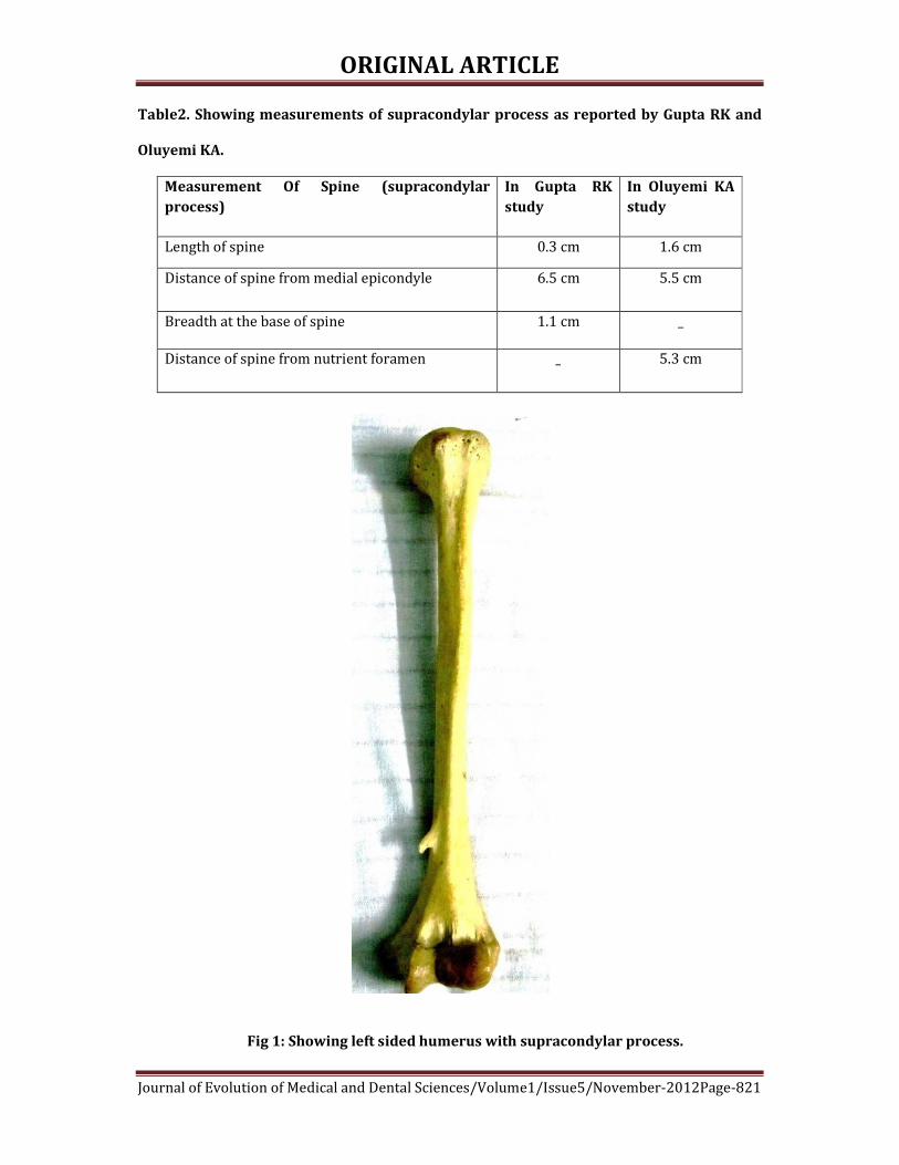
ORIGINAL ARTICLE
Journal of Evolution of Medical and Dental Sciences/Volume1/Issue5/November-2012Page-821
Table2. Showing measurements of supracondylar process as reported by Gupta RK and
Oluyemi KA.
Measurement Of Spine (supracondylar
process)
In Gupta RK
study
In Oluyemi KA
study
Length of spine 0.3 cm 1.6 cm
Distance of spine from medial epicondyle 6.5 cm 5.5 cm
Breadth at the base of spine 1.1 cm _
Distance of spine from nutrient foramen _ 5.3 cm
Fig 1: Showing left sided humerus with supracondylar process.

ORIGINAL ARTICLE
Journal of Evolution of Medical and Dental Sciences/Volume1/Issue5/November-2012Page-822
Fig 2: showing only the distal part of the humerus with supracondylar process
Fig 3: Showing the measurement of distance of supracondylar process from nutrient foramen
with vernier calliper

ORIGINAL ARTICLE
Journal of Evolution of Medical and Dental Sciences/Volume1/Issue5/November-2012Page-823
STUDY OF ONYCHOMYCOSIS AT A TERTIARY CARE HOSPITAL IN
SOUTH INDIA Niranjan H. P., N. Padmaja, Priyanka. B. V.
1. Associate Professor, Department of Microbiology, S. S Institute of Medical Sciences and Research Centre,
Davangere.
2. Assistant Professor, Department of Microbiology, KIMS, Amalapuram, Andhra Pradesh.
3. IIIrd year Student, Department of Microbiology, S. S Institute of Medical Sciences and Research Centre,
Davangere.
CORRESPONDING AUTHOR
Dr. Niranjan H.P.,
B-7, Staff Quarters, SSIMS & RC,
Davangere - 577005.
E-mail: [email protected]
Ph: +91 9844821527.
ABSTRACT:
BACKGROUND: Onychomycosis is common infection in adults and account for 20% of all nail
diseases. The clinical presentation is often confused with other conditions, making laboratory
diagnosis and confirmation necessary. Accurate identification of etiological agent is important,
as the treatment is different for dermatophytes and non dermatophytes. AIMS AND
OBJECTIVES: 1. To isolate and identify the etiological agents of onychomycosis. 2. To study the
occupational status of the study group. 3. Correlation between nail involvement and sex.
METHODOLOGY: To identify the aetiological agents of onychomycosis, the present study was
carried out in the department of Microbiology, S. S. Institute of medical sciences and research
centre. Nail clippings were collected from a total of 120 clinically suspected patients of
onychomycosis attending the outpatient department of Dermatology and Venereology and were
processed and identified by standard laboratory techniques. RESULTS: Fungi was
demonstrated in 66 (55%) either by KOH preparation and/or culture. Sixty (50%) were positive
by direct microscopy and 54 (45%) were culture positive. Out of the 54 culture isolates,32
(59.26%)were dermatophytes followed by yeasts 13 (24.07%) and non dermatophytemoulds 9
(16.67%). Among the dermatophytes,T.rubrum18 (33.33%) was the most common followed by
T.mentagrophyte 8 (14.81%) and T.tonsurans 4 (7.41%).Among the yeasts, C.albicans11
(20.37%) was predominate followed by C.tropicalis 2 (3.7%).Among the non
dermatophytemoulds,Aspergillusflavus3 (5.55%) was the commonest followed by Fusarium
spp 2 (3.7%).The finger nails were more commonly involved 66 (55%) than toe nails 54 (45%).
Most of the patients were farmers 38 (31.67%) followed by housewives 32(26.67%).
CONCLUSION: Dermatophytes remain the predominant cause of onychomycosis, with T.rubrum
as the most common aetiological agent, but less commonly yeasts like C.albicans and non
dermatophytic moulds like Aspergillus spp can also cause nail infection and hence accurate
diagnosis is needed ,since the treatment is different for each group.
KEYWORDS: Onychomycosis, Dermatophytes, Trichophyton rubrum, Candida albicans
INTRODUCTION: Onychomycosis is common infection in adults and account for 20% of all nail
diseases1.The factors that increase the prevalence of onychomycosis include increasing age,
male sex, underlying conditions such as diabetes and immunodeficiency2.Although not life

ORIGINAL ARTICLE
Journal of Evolution of Medical and Dental Sciences/Volume1/Issue5/November-2012Page-824
threatening, this may have significant clinical consequences such as secondary bacterial
infections, chronicity, therapeutic difficulties and disfigurement in addition to acting as a
reservoir of infection.3The symptomatic disease can be a source of embarrassment and potential
cause of morbidity4. Onychomycosis can be classified Into several clinical types: Distal and
lateral subungual onychomycosis, proximal subungual onychomycosis, white superficial
onychomycosis and Total dystrophic onychomycosis1. Most of the cases are due to
dermatophytes, but yeasts and non dermatophytic fungi can also cause nail infection,
particularly after trauma or diseases causing nail dystrophy.1 The clinical presentation may
often be confused with other conditions like psoriasis, lichenplanus, onychodystrophy and nail
trauma1, making laboratory diagnosis and confirmation necessary. Fungal cultures are essential
for accurate identification of the causative organism. This is of paramount importance because
the clinical outcome of antifungal agents varies as to whether the aetiological agent is a
dermatophyte, yeast or a non-dermatophytic mould.5
The present study was undertaken to isolate and identify the aetiological agents of
onychomycosis.
MATERIAL AND METHODS: The present study of onychomycosis was carried out in the
department of Microbiology, S. S. Institute of medical sciences and research centre. A total of
one hundred and twenty clinically diagnosed randomly selected cases of nail infection, of all age
groups and of both sexes, attending Dermatology and Venereology outpatient department of S.
S. hospital, Davangere, were studied. A detailed history of selected cases was taken in relation to
name, age, sex, address, occupation and involvement of more than one site. Patients under
antifungal treatment were excluded from the study group.
The affected area was cleansed with 70% ethyl alcohol and the nail specimen was
collected by taking clippings of the infected part and scrapings beneath thenail1,6. One portion of
the specimen was subjected to 20%potassium hydroxide(KOH) wet preparation for 1 to 2 hours
at room temperature in a moist chamber7-9,and the remaining material was inoculated onto two
sets of test tubes, one containing Sabouraud’s dextrose agar with 0.05% chloramphenicol and
the other containing Sabouraud’s dextrose agar with 0.05% chloramphenicol and 0.5%
cycloheximide and incubated at 280C for up to 4 weeks.6,7,9 Cultures were read initially at 24
to48 hours for non dermatophytes and then on periodically for up to 4 weeks for
dermatophytes. If no growth was found after 4 weeks, it was taken as negative for the growth of
fungi and discarded. Repeat cultures were performed in cases where culture was negative for
dermatophytes but positive for non dermatophytic moulds or yeasts to rule out the possibility
of contamination.7,10,11 The criteria used to report non dermatophytic moulds or yeasts as
pathogens was direct microscopy positive and isolation of same fungus in three consecutive
samples at intervals of 7 days.11
Fungal isolate was identified based on colony morphology, reverse pigmentation,
growth rate, microscopy (LPCB), slide culture and special tests like hair perforation test, urease
test, germ tube test, sugar fermentation and assimilation tests 1,6,8,12.
RESULTS: A total of 120 patients with clinical suspicion of onychomycosis were included in the
study. Most of the patients were farmers 38 (31.67%) followed by house wives 32 (26.67%).
The finger nails were more frequently involved 66 (55%) than toe nails 54 (45%).
Out of 120 clinically suspected cases of onychomycosis, fungi were demonstrated in 66
(55%) either by direct microscopy and / or culture. Forty Eight (40%) were positive by both

ORIGINAL ARTICLE
Journal of Evolution of Medical and Dental Sciences/Volume1/Issue5/November-2012Page-825
microscopy and culture. Twelve (10%) were positive by microscopy and negative by culture. Six
(5%) were negative by microscopy but culture positive. Fifty four (45%) were negative both by
microscopy and culture.
Overall out of the 54 culture isolates, 32 (59.26%) were infected with dermatophytes,
followed by Candida species 13 (24.07%) and non dermatophytemoulds 9 (16.67%).
Various fungi isolated is shown in table 3.
DISCUSSION: In the present study,120 clinically suspected cases of onychomycosis attending
Dermatology and Venereology out patient department of S. S. hospital, Davangere were studied.
In the present study, Males(60%) were more commonly affected than females(40%),
which is comparable with the studies of Adhikari L et al14,NeupaneS et al2, where as Madhuri JT
et al9 (51.96%) and Bokhari et al8(72%) have reported higher prevalence in females. Male
predominance may be due to increased outdoor physical activity and increased opportunity for
exposure. Toe nail infection(52.78%) was commoner in males, while finger nail
infection(66.67%) was common in females. This may be due to increased exposure to wet work
in females, as most of them were house wives.
Onychomycosis was most commonly seen in farmers(31.67%), followed by house
wives(26.67%), where as students were least affected (9.17%), which is comparable with the
study of Veer P et al10, where as NeupaneS et al2 reported that students were more commonly
affected(31.3%) followed by house wives(28.5%).High prevalence in farmers and house wives
may be due to increased outdoor physical activity and increased exposure to wet work
respectively. It is less frequently seen in students due to rapid growth of nails which causes
elimination of dermatophytes.
In the present study, out of 120 clinically suspected cases of onychomycosis, fungus was
demonstrated in 66 (55%) either by direct microscopy and/or culture. Direct microscopy was
positive in 60 (55%) and culture in 54 (45%),which is comparable with the studies of Madhuri
JT et al9 and Das NK et al12, where as Malik NA et al7has reported only 16% culture positivity.
This variation in direct microscopy and culture may be due to non viability of fungal elements in
some cases.
Out of the 54 fungal isolates, dermatophytes 32 (59.26%) were the commonest,
followed by yeasts 13 (24.07%) and non dermatophytic moulds 9 (16.67%).Among the
dermatophytes, T.rubrum was the predominate isolate 18 (33.33%) followed by
T.mentagrophyte 8 (14.81%) ,T.tonsurans 4 (7.41%) and E.floccosum 2 (3.7%).
Among the yeasts, C.albicans11 (20.37%) was the most common followed by
C.tropicalis 2 (3.7%) and most of them were females due to immersion of hands frequently in
water.
Among the non dermatophyticmouldsisolated,A.flavus3 (5.55%) was the most common
followed by Fusariumspp2 (3.7%).A.niger and curvularia were isolated one each(1.85%) and
two isolates(3.7%) could not be identified. This is comparable with the studies of Das NK et al12,
Veer P et al10 and Malik NA et al7, where as Vijaya D et al 11 and Adhikari L et al 14reported yeasts
and T.tonsurans as predominant fungi respectively in their study.
CONCLUSION: Onychomycosis is very common in our country where hot and humid climate in
association with poor hygienic conditions play an important role in the growth of fungi and also
majority of the people are agriculturists. It can no longer be considered as simple cosmetic
problem, but can considerably impair patients quality of life.

ORIGINAL ARTICLE
Journal of Evolution of Medical and Dental Sciences/Volume1/Issue5/November-2012Page-826
Dermatophytes remain the predominant cause of onychomycosis, with T.rubrum as the
most common aetiological agent, but less commonly yeasts like C.albicans and non
dermatophytic moulds like Aspergillus spp can also cause nail infection and hence accurate
diagnosis is needed ,since the treatment is different for each group.
REFERENCES:
1. Chander J(2009).Text book of Medical mycology.3rded.Newdelhi:Mehta
publisher;pg:132-4, 56-63.
2. Neupane S, Pokhrel DB and
PokhrelBM.onychomycosis:Aclinicoepidemiologicalstudy.Nepal Med Coll J
2009;11(2):92-95.
3. ErbaqciZ, TuncelA,Zer Y et al. A prospective epidemiological survey on the
prevalence of onychomycosis and dermatophytosis in male boarding school
residents.Mycopathologica 2005;159:347-52.
4. GuptaAK,TabordaP,etal.Epidemiology and prevalence of onychomycosis in HIV
positive individuals.Int J Dermatol 2000;39:746-53.
5. RamaniR,Srinivas CR, et al.Moulds in onychomycosis.Int J Dermatol
1993;32:877-8.
6. ColleeJG,Fraser AG, et al(1996).Mackie and McCartney practical medical
microbiology.14thed.Edinburgh:Churchhill livingstone:pg:695-714.
7. MalikNA,Raza N, et al.Nondermatophytemoulds and yeasts as causative agents in
onychomycosis.Journal of Pakistan Association of Dermatologists 2009;19:74-78.
8. Bokhari MA et al. onychomycosis in Lahore.PakistanInt J Dermatol
1999;38:5915.
9. MadhuriJT,Rama RGR, et al.Onychomycosis:A significant medical problem.Indian
J DermatolVenereol Leprol2002;68(6):326-9.
10. Veer P et al.A study of onychomycosis:Prevailing fungi and pattern of infection.
Indian J Med Microbiol2007;25:53-6.
11. Vijaya D ,Anandkumar BH, et al. Study of onychomycosis. Indian J
DermatolVenereolLeprol 2004;70(3):185-6.
12. DasNK,Ghosh P, et al.A study on the aetiological agent and clinico-mycological
correlation of finger nail onychomycosis in eastern india. Indian J Dermatol
2008;53:75-9.
13. VelezA,Linares MJ et al.study of onychomycosis in Corboda,Spain:Prevailing fungi
and pattern of infection.Mycopathologica 1997;137:1-8.
14. Adhikari L, Gupta AD et al.Clinico-aetiologic correlates of onychomycosis in
Sikkim.Indian J PatholMicrobiol 2009;52:194-7.
Table 1 shows occupational status of the study group.

ORIGINAL ARTICLE
Journal of Evolution of Medical and Dental Sciences/Volume1/Issue5/November-2012Page-827
Occupation Frequency Percentage
Farmers 38 31.67%
Housewives 32 26.67%
Office workers 13 10.83%
Students 11 9.17%
Miscellaneous 26 21.67%
Table 2 shows nail involvement according to gender.
Gender site
Total fingernail Toe nail
Male 34 (47.22%) 38 (52.68%) 72
Female 32 (66.67%) 16 (33.38%) 48
Total 66 54 120
Various fungi isolated is shown in table 3.
Causative agent Finger nail Toe nail Total percentage
Dermatophytes
T. rubrum
T. mentagrophyte
T. tonsurans
E. floccosum
10
5
2
0
8
3
2
2
18
8
4
2
33.33%
14.81%
7.41%
3.70%
Yeasts
C. albicans
C. tropicalis
8
1
3
1
11
2
20.37%
3.70%
Non-dermatophytes
A. flavus
Fusariumspp
Curvulariaspp
A. niger
unidentified
1
1
1
1
0
2
1
0
0
2
3
2
1
1
2
5.55%
3.70%
1.85%
1.85%
3.70%
Total 30 24 54 100%

ORIGINAL ARTICLE
Journal of Evolution of Medical and Dental Sciences/Volume1/Issue5/November-2012Page-828

ORIGINAL ARTICLE
Journal of Evolution of Medical and Dental Sciences/Volume1/Issue5/November-2012Page-829

ORIGINAL ARTICLE
Journal of Evolution of Medical and Dental Sciences/Volume1/Issue5/November-2012Page-830
SOCIO-DEMOGRAPHIC PROFILE OF HIV PATIENTS ATTENDING ART
CENTRE, VIMS: A HOSPITAL BASED DECRIPTIVE STUDY Ramesh. K, Sangeetha Gandhi, Vishwas Rao, Anusha. C. P, Spoorthi Prakash.
1. Assistant Professor, Department of Community Medicine, Vijayanagara Institute of Medical Sciences,
Bellary, Karnataka.
2. Junior Resident, Department of Community Medicine, Vijayanagara Institute of Medical Sciences, Bellary,
Karnataka.
3. Junior Resident, Department of Community Medicine, Vijayanagara Institute of Medical Sciences, Bellary,
Karnataka.
4. Junior Resident, Department of Community Medicine, Vijayanagara Institute of Medical Sciences, Bellary,
Karnataka.
5. Junior Resident, Department of Community Medicine, Vijayanagara Institute of Medical Sciences, Bellary,
Karnataka.
CORRESPONDING AUTHOR
Dr. Ramesh. K. MBBS, MD,
Assistant Professor, Department of Community Medicine,
Vijayanagara Institute of Medical Sciences (VIMS),
Bellary -583104, Karnataka,
E-mail: [email protected],
Ph: 0091 9481181291
ABSTRACT: BACKGROUND: The human immunodeficiency virus (HIV) infection is a global
pandemic.HIV continues to be a burden globally and presents serious public health
problems in the developing countries, especially in India. According to the UNAIDS and
World Health Organization (WHO) reports of November 2010, there are approximately 33.3
million people living with HIV/Acquired immunodeficiency syndrome (AIDS) worldwide,
with a global prevalence of 0 .8%. It is now the leading cause of adult deaths in the world due
to infectious diseases. HIV has become the first truly international epidemic easily crossing the
oceans & borders. METHODOLOGY: A case-series study was conducted among HIV positive
patients coming to the ART centre for seeking treatment at ART Centre, Vijayanagara Institute
of Medical Sciences Bellary, Karnataka from March 2011 to September 2011. The sample size of
the study was 500 and consecutive sampling technique is used. Permission from respective
authorities and written consent from study participants was obtained. Data was collected using
a pre tested semi structured questionnaire. Data was analyzed using SPSS 15.0.
RESULTS: The study subjects included both males (54%) and females (46%). The educational
status is less than primary schooling in 88% of study subjects and 90% are married. Hetero
sexual route of transmission was most common and only 16.1% of study subjects used
contraceptives. CONCLUSION: Both males and females were equally infected with HIV and
productive age group is more commonly affected. Use of contraception is very low.
KEY WORDS: HIV, ART, Mode of transmission
INTRODUCTION: Since the recognition of AIDS pandemic, many efforts have been concentrated
on the prevention of HIV transmission1. In INDIA as stated by NACO people living with AIDS for
the year 2009 estimates 23.1 lakhs which equates to a prevalence of 0.3%. Well this may seem
low compared to large population of our country, but a mere 0.1% increase in HIV prevalence
would increase the estimated no of people living with HIV by over half a million!!!!2. Since India

ORIGINAL ARTICLE
Journal of Evolution of Medical and Dental Sciences/Volume1/Issue5/November-2012Page-831
has emerged as a major player in the global HIV epidemic, and, the lack of information on
knowledge, perceptions, and behaviors regarding HIV risk and preventive behaviors among
Indian adolescents is alarming!!3.
Nowadays, interventions to stem the spread of the Human immunodeficiency virus
(HIV) throughout the world are as varied as the contexts in which we find them. Not only is the
HIV epidemic dynamic in terms of treatment options, Prevention strategies and disease
progression, but sexual Behavior which remains the primary target of AIDS Prevention efforts
worldwide, is widely diverse and deeply embedded in individual desires, social and cultural
Relationships, and environmental and economic Processes. This makes prevention of HIV,
which could be an essentially simple task, enormously complex involving a Multiplicity of
dimensions .To respond effectively to heterosexual acquired HIV infection, it is important to
understand sexual behavior of persons infected with HIV that facilitate or protect against
transmission4.Behavioral change has been responsible for the prevention successes to date.
Strategies to modify risk behaviors need to remain a main priority for HIV prevention5. By
behavioral strategies we mean an attempt to delay onset of first intercourse, decrease the
number of sexual partners, increase the number of sexual acts that are protected, provide
counseling and testing for HIV, encourage adherence to biomedical strategies preventing HIV
transmission, decrease sharing of needles and syringes, and decrease substance use. Behavioral
strategies to accomplish these goals can focus on individuals, couples, families, peer groups or
networks, institutions, and entire communities 5.
OBJECTIVE: To study the socio demographic profile of HIV patients
METHODOLOGY: A case-series (descriptive) study was conducted among HIV positive patients
coming to the ART centre for seeking treatment at ART Centre, Vijayanagara Institute of Medical
Sciences Bellary, Karnataka from March 2011 to September 2011. Totally 10,000 patients have
registered and among them 5000 patients were on treatment at ART centre. Hence this study
considered patients only who were on treatment. Totally 500 patients were included for the
study. These 500 patients were selected based on non probability purposive sampling
technique.
Data was collected using a pre designed semi structured questionnaire which contained
information regarding socio demographic details and behavior. After obtaining written
informed consent, data was collected by interview technique and after the data collection health
education was given to patients on Prevention & treatment of HIV. Patients who were seriously
ill and those who did not give consent were excluded from the study
Data was entered in Microsoft excel and analyzed in SPSS 15.
RESULTS: Among 500 study subjects, 54% were males and 46% were females. More numbers
of study subjects were in 30 – 39 years of age group (39%) followed by 20 – 29 years(32%), 40
– 49 years (25%) and more than 50 years(4%). It was observed that majority of study subjects
were between 20 to 39 years (71%). Among males, more number of study subjects were in 30 –
39 years of age group(40.8%) and whereas among females, more number of study subjects
were in 20 – 29 years of age group(43.6%) (Table 1)
Among 500 study subjects, 16.4% of them were not known how they might have
acquired HIV infection but it was found that heterosexual route of transmission of HIV was
more common among study subjects i.e. 83%. Blood transfusion and inject able drug abuse
contributed 0.60% collectively (Table.no.2).

ORIGINAL ARTICLE
Journal of Evolution of Medical and Dental Sciences/Volume1/Issue5/November-2012Page-832
Out the 500 subjects taken for our study 45% of them were illiterates, 43% of subjects
had just primary education, 9% of subjects had high school education and 2% of them had
completed their education till pre university level.
In our study about half of the study subjects were unskilled workers primarily daily
wage workers followed by semi skilled and skilled workers who formed 22% and 19%
respectively; 49% were unskilled; 7% were professionals.
Among our study subjects, 62% of them earned an income of about 3000-6000 rupees
per month; 17% of them earned an income less than 3000 per month ; 21% of them earned an
income of more than 6000 per month. In our study it was found that only 10% of the HIV
positive subjects are unmarried (Table.no.3).
Among our study subjects, 99% of the subjects confessed they had indulged in a sexual
intercourse at least once.
Out of the 495 study subjects who had intercourse at least once, 84.8% of them had an
intercourse out of their own interest. Whereas sizeable number of 15.2% of the study subjects
were forced.
Out of the 495 study subjects who had indulged in sexual intercourse, 82.8% of the
subjects had first act of sexual intercourse been 16 to 25 years age group; 14.2% fell below age
15 years & 3% were above 25 years.
In our study, out of 495 study subjects, 62.6% of the study subjects had their first sexual
encounter with their spouse. Sizeable numbers of the study subjects have had their first sexual
encounter with a Commercial Sex Worker (19.1%) and the rest with either friend or relative.
Use of contraceptives was found only among 16.1% of study subjects who had indulged
in sexual intercourse (Table.no.4).
DISCUSSION: In our study out of the 500 subjects, 54% were male subjects and 46% were
female subjects. On a total, maximum subjects fell between age group 30-39 years. Compared to
a similar study in INDIA, 41% were female subjects and 59% were male subjects; majority of
them fell between age groups 30-34 years6. Compared to a similar study done in Goa with only
males as the sample, maximum subjects fell between age group 35- 49 years, similar to our
study7. In another study as well maximum subjects fell between age group 35-49 years4.
Illiteracy is one of the major reasons for the rampant increase in the number of HIV
positive cases. Out of the 500 subjects taken for our study, 45% of them were illiterates.
Compared to the similar study done, 30% were illiterate; 24% had a primary education; 36%
had a high school education & 10% had a higher education; whereas in our study there were
significantly more number of illiterates and less number of subjects with pre-university
education6. Compared to the similar study done in Chennai, 30% were illiterates; 34% had
primary education; 25% had secondary education & 11% were professionals whereas in our
study there was significantly more number of illiterates 8. Compared to another similar study
done in south India, 25% were illiterate; 50% were educated till high school; 25% were
educated till Pre University, whereas in our study there were significantly more number of
illiterates and less number of subjects with high school and pre-university education9.
Ever since the first case of HIV was diagnosed in Chennai in the year 1986, HIV-AIDS
was thought to be, primarily, a disease of Commercial Sex Workers and Truck drivers. But in the
past decade the infection has crept into the richer sections of the society. In our study, about
half of the study subjects were unskilled workers primarily daily wage workers followed by
semi skilled and skilled workers. Compared to a study in INDIA, 19% were unemployed; 81%

ORIGINAL ARTICLE
Journal of Evolution of Medical and Dental Sciences/Volume1/Issue5/November-2012Page-833
were employed6.Whereas in our study we have a large population who are unemployed.
Compared to a study done in Kasturba Medical College Hospital, Mangalore, Karnataka; laborers
and hotel workers constituted 19.9%., housewives constituted 26.97%; beedi rollers, business
and semi-skilled professionals constituted a sizeable number. Drivers constituted only 3.08%10.
Compared to another study done in Goa, 66.3% were employed, 15.7% were unemployed and
18% are students7. Whereas in our study, we have a larger unemployed population.
Poverty is root cause of all the evil in India including the high rates of HIV. Among our
study subjects, a maximum number of study subjects earned an income of about 3000-6000
rupees per month (62%). Compared to a similar study, 15% are poorest; 15% were poorer; 3%
were middle class; 32% were rich; 15% were richest6; whereas in our study 17% have a very
low income & there is no question about rich or richest in our study. Compared to other study,
16% had a low standard of living index; 32% had a medium standard of living index& 52% had
a high standard of living index11.
Marital status of the patient was known to generally access the sexual behavior among
the subjects. Those who are unmarried are assumed to have Pre marital sex whereas those who
are married are assumed to either have sex with either their wives or indulge in extra marital
sex. In our study, it was found that only 10%of the HIV positive subjects are unmarried
therefore it can be assumed that the pre marital sex leading to HIV is quite low. Compared to a
study done in Chennai 55% are married; 35% were single; 7% were separated; 2% were
divorced8. Whereas in our study a significant large number of them are married. Compared to a
study done in south India, 57% are married; 43% were unmarried (43%)9, whereas in our study
a significant large no of them are married and less no of them are unmarried. Compared to a
study done in Goa, 45.3% were single and 54.7% were married7. Whereas in our study
maximum subjects are married.
Among our study subjects, 99% of the study subjects confessed they had indulged in a
sexual intercourse at least once. . Late adolescent age group is at threat age group; teenage
pregnancies are slowly rising as well. Out of 495 subjects, 14% of the subjects had first act of
sexual intercourse before 15 years of age. Compared to a similar study5, about 12% of study
subjects had sexual intercourse before 15 years of age, the findings match with our study .
The scenario of Sexual intercourse before marriage which is very common in western
countries has shown a rampant shift into our country over the years. Shear inquisitiveness
pushes many into this, in our study out of the 495 study subjects who had intercourse at least
once, 420 of them had an intercourse out of their own interest(84%). Whereas sizeable number
of the study subjects was forced, 16% study subjects, it could have been due to peer pressure or
rape. Compared to a study done in Government Medical College & SSG Hospital, Vadodara,
which goes by name “Adolescent HIV/AIDS: Issues and challenges”12, 26% Of the interviewed
female sex workers (FSWs), “Devadasi” FSWs (socially accepted FSWs) had initiated sex work at
a much younger age (mean 15.7 vs. 21.8 years) due to childhood sexual abuse; the finding is
almost similar to our study where 16% were forced. Compared to a similar study “sexual
behavior in India with risk of HIV/AIDS transmission13”, 25% reported experience of premarital
sex out of interest & 25 per cent of male students in a Delhi school and 28 per cent of male
college students in Hyderabad reporting premarital sexual experience .
Heterosexual is the most common mode of transmission worldwide. As such in this
study 83% of seropositive cases accounted for this commonest mode of transmission which is
supported by various studies in India as well as other parts of world14,15,16.

ORIGINAL ARTICLE
Journal of Evolution of Medical and Dental Sciences/Volume1/Issue5/November-2012Page-834
CONCLUSION: Illiteracy and low socio economic status are still remaining as major
determinants of HIV and productive age group of life is more commonly affected so strategies
for tackling HIV infection in our country should be based on profile of the patients.
REFERENCES:
1. B Cribier, M P Schmitt, C Le Coz, and E Grosshans. Changes in sexual behaviour of
patients attending an HIV testing centre: a prospective study 1988-1994.
Genitourin Med. 1996 February; 72(1): 37–42.
2. National AIDS Control Organization. Annual Report. 2008-2009, http://nacoonline.org/upload/Publication/Annual_Report_NACO_2008-09.pdf
accesesed on 07/09/2011 3. Quinn TC, Overbaugh J. HIV/AIDS in women: an expanding epidemic. Science.
2005;308(5728):1582–1583.
4. Journal of Urban Health: Bulletin of New York Academy of Medicine, Vol-83,
No.4. 2006
5. Coates TJ, Richter L, Caceres C. Behavioural strategies to reduce HIV transmission:
how to make them work better. Lancet. 2008 Aug 23;372(9639):669-84.
6. Perkins JM, Khan KT, Subramanian SV (2009) Patterns and Distribution of HIV
among Adult Men and Women in India. PLoS ONE 4(5): e5648.
doi:10.1371/journal.pone.0005648
7. Vaz FS, Ferreira AM, Kulkarni MS, Motghare DD. Sexual risk behaviors and HIV/AIDS
awareness among males in a rural community in Goa. J Commun Dis. 2006
Mar;38(1):74-8.
8. Solomon SS, Celentano DD, Srikrishnan AK, Vasudevan CK, Anand S, Kumar
MS, Solomon S, Lucas GM, Mehta SH. Mortality among injection drug users in
Chennai, India (2005-2008). AIDS. 2009 May 15;23(8):997-1004.
9. Chandra PS, Satyanarayana VA, Satishchandra P, Satish KS, Kumar M. Do men and
women with HIV differ in their quality of life? A study from South India. AIDS
Behav. 2009 Feb;13(1):110-7. Epub 2008 Jul 25.
10. S Jayarama, Shaliny Shenoy, B Unnikrishnan, John Ramapuram, and Manjula Rao. Profiles of Attendees in Voluntary Counseling and Testing Centers of a Medical
College Hospital in Coastal Karnataka. Indian J Community Med. 2008
January; 33(1): 43–46.
11. An Lwin, Thein Z., "An Examination of the Association between HIV Related
Knowledge, Attitudes, and Behaviors and HIV Infection Status in Five High HIV
Prevalence States in India." (2011).Public Health Theses. Paper 168. http://digitalarchive.gsu.edu/iph_theses/168
12. Smriti Naswa and Y. S. Marfatia. Adolescent HIV/AIDS: Issues and challenges. Indian
J Sex Transm Dis. 2010 Jan-Jun; 31(1): 1–10.
13. Moni Nag. Sexual behaviour in India with risk of HIV/AIDS transmission. Health
Transition Review, Supplement to Volume 5, 1995, 293-305.
14. Verma S, Mahajan A, Singh JB, Sharma M. Clinical Profile of HIV/AIDS patients in
Jammu. JK Practitioner. 2007; 14(2):79-83.
15. Destura RV, Berba RP, Mendoza MT et al. Profile of HIV/AIDS Patients at the
Philippine General Hospital: Revisting 9 years of Clinical Experience. Philippine
journal of Microbiology and Infectious Diseases. 2003; 32:111-21.
16. Khopkar U, Raj S, Sukthankar A, Kulka. Clinical profile of HIV Infection. Indian J
Dermatol Ven. 1992; 58(3):155-158.

ORIGINAL ARTICLE
Journal of Evolution of Medical and Dental Sciences/Volume1/Issue5/November-2012Page-835
Table 1: Age sex wise distribution of study subjects
Age group Male Female Total
20 – 29 years 60 (22.3%) 100(43.6%) 160 (32%)
30 – 39 years 110 (40.8%) 85 (36.9%) 195 (39%)
40 – 49 years 80 (29.6%) 45 (19.5%) 125 (25%)
50 – 59 years 15 (05.5%) 00 15 (03%)
>59 years 05 (01.8%) 00 05 (01%)
Total 270 (100%) 230 (100%) 500 (100%)
Table 2: Mode of Transmission of HIV among study subjects
Mode of Transmission Frequency Percentage
Heterosexual 415 83.00%
Blood transfusion 002 00.40%
Inject able drug abuse 001 00.20%
Don’t know 082 16.40%
Total 500 100%

ORIGINAL ARTICLE
Journal of Evolution of Medical and Dental Sciences/Volume1/Issue5/November-2012Page-836
Table 3: Socio demographic characters of study subjects
Socio demographic characters Frequency Percentage
Education
Illiterate
Primary
High school
Pre university
Degree & above
225
215
045
010
005
45.0%
43.0%
09.0%
02.0%
01.0%
Occupation
Unskilled
Semiskilled
Skilled
Professional
Business & others
245
110
095
015
035
49.0%
22.0%
19.0%
03.0%
07.0%
Income (monthly)
<3000 Rs
3000 – 6000 Rs
>6000 Rs
085
310
105
17.0%
62.0%
21.0%
Marital status
Married
Unmarried
450
050
90.0%
10.0%
Table 4: Distribution of study subjects based on sexual behavior
Sexual behaviors Frequency Percentage
Ever indulged in sex?
Yes
No
495
005
99.0%
01.0%
Age at first act of sex
<15 years
15 – 25 years
>25 years
070
410
020
14.2%
82.8%
03.0%
First time sex with
Spouse
Relative
Friend
Commercial sex worker
310
020
075
095
62.6%
04.0%
15.3%
19.1%
Sex before marriage
Out of interest
Out of force
420
075
84.8%
15.2%
Use of contraception
Yes
No
080
415
16.1%
83.9%
REVIEW ARTICLE
Journal of Evolution of Medical and Dental Sciences/Volume1/Issue5/November-2012Page-892
USES OF HYPERBARIC OXYGEN THERAPY: A REVIEW Farheen Ustad, Fareedi Mukram Ali, Tanveer Ustad, Vinit Aher, Prasant M. C,
Harshal Suryavanshi
1. Lecturer, Department of Oral and Maxillofacial Surgery. King Khalid University Abha.
2. Reader, Department of Oral and Maxillofacial Surgery. SMBT Dental College, Sangamner Taluka.
3. Senior Resident, Department of Urology, St. Johns Medical College Hospital, Bangalore.
4. Senior Lecturer, Department of Oral and Maxillofacial Surgery. Sangamner Taluka.
5. Professor & HOD, Department of Oral and Maxillofacial Surgery. RKDF Dental College, Bhopal.
6. Senior Lecturer, Department of Oral and Maxillofacial Surgery. YMT Dental College, Nayi Mumbai
CORRESPONDING AUTHOR
Dr. Fareedi Mukram Ali, Reader,
Dept of Oral & Maxillofacial Surgery,
Sangamner Taluka, Ahmednagar Dist,
Maharashtra,
E-mail: [email protected]
Ph: 0091 9326325156
ABSTRACT: In the last three decades great strides in Hyperbaric Oxygen research has raised
the value of this unique therapy. Studies have expanded the list of conditions usefully treated
with compressed oxygen. Despite the promising experimental and clinical data, the major
criticism to most HBO studies has been the lack of controlled prospective analysis for its use.
This article discusses the use of HBO, including staffing and equipment considerations, side
effects and reviews the published experience in this subject.
KEYWORDS: hyperbaric oxygen therapy, HBO, uses of HBO.
INTRODUCTION: HBO therapy was initially used to treat patients involved in diving accidents
or with decompression sickness. However, its indications have increased over the past few
decades. Currently, there are twelve indications for HBO therapy approved by the Undersea and
Hyperbaric Medical Society (UHMS) in the United States.1 There are, however, over a hundred
indications internationally, although most of them have not been proven by controlled studies.
The committee on hyperbaric medicine defines hyperbaric oxygen therapy as “A mode of
medical treatment in which the patient is entirely enclosed in a pressure chamber and breathes
100% oxygen at a pressure greater than 1 atmosphere absolute (ATA)”. ATA is the units of
pressure and 1 ATA is equal to 760 mm of mercury or pressure at sea level.1As with most areas
of medicine, in hyperbaric medicine there is a constant struggle to balance enthusiasm for
progress in the field with the need to apply it on the basis of established evidence.The lack of
sound scientific evidence of the efficacy of HBO has bred uncertainty in the wider medical
community regarding its legitimacy.
PHYSIOLOGICAL BASIS: The arterial partial pressure of O2 is 100 mm Hg, Hb is 95% saturated
and 100 ml of blood carries 19 ml of O2in combination with Hb and 0.32 ml dissolved in plasma.
If the inspired O2 concentration is increased to 100%, O2combined with Hb can increase to a
maximum of 20 ml when the Hb is 100% saturated and the amount of O2 dissolved in plasma
may increase to 2.09 ml. During HBO in addition to the Hb which is 100% saturated the amount
of O2 carried in solution will increase to 4.4 ml% at a pressure of 2 ATA to 6.8 ml % at 3 ATA
which is almost sufficient to supply the resting total oxygen requirement of many tissues

REVIEW ARTICLE
Journal of Evolution of Medical and Dental Sciences/Volume1/Issue5/November-2012Page-893
without a contribution from oxygen bound to hemoglobin. Itis this increased oxygen in plasma
which is responsible for most of the beneficial effects of hyperbaric oxygen.2
HISTORY OF HYPERBARIC OXYGEN THERAPY: The first pressurized room used to treat
health problems was built by an Englishman named Henshaw in 1662.3in1788; hyperbaric air
was put to large scale use in a diving bell for underwater industrial repairs of an English bridge.
The first deep sea diving suit, invented in 1819 by August Siebe, used compressed air supplied
to the helmet for generous underwater movement.4Dr. John S. developed the first diving tables
for the Royal Navy. His legacy gives him the title "Father of Oxygen Therapy" and physicians
continue in his line of work to this day.4 In 1918 Dr. Orval Cunningham built the world's largest
functional hyperbaric chamber, a 64' steel sphere "hyperbaric medical hotel" with five floors of
living space after he found that denser air helped people fight infection suffering from flu. The
Great Depression in the 1930's ended his project and the giant chamber was scrapped for the
war effort in the 1940's.5 The Hyperbaric Oxygen Committee was developed by the UHMS in
1976 to oversee the ethical practice of hyperbaric medicine.5
THERAPEUTIC EFFECTS OF HBO THERAPY:
Therapeutic effects of HBO can be attributed to its mechanical or hyperoxygenation
effects as shown in the table
Therapeutic effects Mechanism
Reduces bubble size Direct mechanical effect.
Immune stimulation Restores WBC function, enhances phagocytic
capabilities and neutrophil mediated killing of
bacteria.
Neovascularization Augmentation of fibroblastic activity which
promotes capillary growth.
Reduces edema and tissue swelling Hyper oxygenation.
Bactericidal For anaerobic organisms such as
Clostridiwelchii,
and also inhibits the growth of aerobic
bacteria at pressures
greater than 1.3 ATA.
ADMINISTRATION: HBO therapy can be given in a monoplace chamber in which a single
patient is placed in a chamber pressurized with 100% oxygen or it can be given in a multiplace
chamber where many patients can be treated at the same time. To be effective, hyperbaric
oxygen must be inhaled in the atmosphere or through an endotracheal tube in a monoplace
chamber and in multiplace chamber masks, tight-fitting hoods, or endotracheal tubes can be
used. Monoplace chambers are the most common type of chamber used due to their portability,
minimal personnel requirements and low cost.6
Time: The duration of single treatments varies from 45 minutes for carbon monoxide
poisoning to almost 5 hours for some severe decompression disorders. For treatment of wounds
- most protocols average 90 minutes for each of 20 to 30 treatments.

REVIEW ARTICLE
Journal of Evolution of Medical and Dental Sciences/Volume1/Issue5/November-2012Page-894
GENERAL EQUIPMENT CONSIDERATIONS: All equipment used inside hyperbaric chambers
must adhere to the guidelines of the National Fire Protection Association (NFPA) .7 Chamber
fires result in catastrophic consequences.8The primary cause of mishaps is the introduction of
prohibited items into the chambers, specifically when chamber personnel do not adhere to
NFPA fire safety rules. Equipment inside chambers must be intrinsically electrically safe, follow
NFPA guidelines, and be tested for the pressures to which they will be exposed.7
HYPERBARIC CHAMBER SELECTION, LOCATION AND STAFFING: Hyperbaric oxygen can be
offered to critically ill patients inmonoplace and multiplace chamber. Monoplace chambers can
be located inside the intensive care unit, where they can be staffed by ICU personnel and are
then an extension of the ICU.9, 10However; hands-on care cannot be provided to a patient inside a
monoplace chamber. Although multiplace chambers do allow hands-on care, experienced staff
must be available inside the chamber. Because most hyperbaric chambers are not located within
or adjacent to the ICU, the potential benefits of HBO2 to a critically ill patient must be balanced
by the risks from transporting the patient as well as the risks from HBO.11, 12 Personnel working
as inside attendants of multiplace chambers must be medically suitable for hyperbaric exposure
(e.g., able to equalize ears, no claustrophobia, no pulmonary or cardiac disease, etc). Staff
supporting critically ill patients during HBO2 could include Certified Hyperbaric Registered
Nurses, physicians; critical care respiratory therapists, and paramedics.
INDICATIONS: The following indications are approved uses of hyperbaric oxygen therapy as
defined by the Hyperbaric Oxygen Therapy Committee.
1. Air or Gas Embolism
2. Carbon Monoxide Poisoning
Carbon Monoxide Poisoning Complicated By Cyanide Poisoning
3. Clostridial Myositis and Myonecrosis (Gas Gangrene)
4. Crush Injury, Compartment Syndrome and Other Acute Traumatic Ischemias
5. Decompression Sickness
6. Arterial Insufficiencies:
Central Retinal Artery Occlusion
Enhancement of Healing In Selected Problem Wounds
7. Severe Anemia
8. Intracranial Abscess
9. Necrotizing Soft Tissue Infections
10. Osteomyelitis (Refractory)
11. Delayed Radiation Injury (Soft Tissue and Bony Necrosis)
12. Compromised Grafts and Flaps
13. Acute Thermal Burn Injury
14. Idiopathic Sudden Sensorineural Hearing Loss.
USES OF HBO
ARTERIAL GAS EMBOLISM: Arterial gas embolism, occurs when air bubbles in the circulation.
There are many causes, including mechanical ventilation; central line placement, haemodialysis,
severe diving injury and pulmonary barotrauma.13The bubbles cause tissue deformation and
vessel occlusion, impairing tissue perfusion and oxygenation. Biochemical effects at the blood-
REVIEW ARTICLE
Journal of Evolution of Medical and Dental Sciences/Volume1/Issue5/November-2012Page-895
gas interface cause endothelial damage, changes in haemostasis and activation of leukocytes.14
Clinical symptoms include muscle and joint pain, arrhythmias, ischemia, confusion, focal
neurological deficits and loss of consciousness.
RATIONALE OF TREATMENT WITH HBO:
•HBO reduces bubble size in accordance with Boyle's law—at 3ATA, bubble volume is reduced
by about two-thirds.15
•Hyperoxia increases the diffusion gradient with the embolized gas, moving gas into solution
where it can be metabolized.16
HBO is widely accepted as the only life-saving treatment, UHMS suggests maximal benefit with
100% oxygen at 2.8 ATA, and repeated treatments until no further improvement is seen,
typically after no more than 5–10 treatments.1719 patients in the USA with iatrogenic cerebral
arterial gas embolism, showed significant improvement in symptoms with HBO treatment, but
the loophole in the study was the control group and end-points were not clearly defined.18 HBO
is most effective when initiated early, but can be successful after hours or even days.17
CARBON MONOXIDE POISONING: Loss of consciousness (syncope, seizures, and coma),
neurologic deficits, pulmonary edema, myocardial ischemia, and severe metabolic acidosis are
the most common symptoms of carbon monoxide poisoning caused primarily from smoke
inhalation and suicide attempts. Less severely poisoned patients may have headache, nausea,
and other constitutional symptoms. All victims of carbon monoxide poisoning are at risk for
delayed neuropsychological sequelae.CO combines preferentially with hemoglobin to produce
COHb, displacing oxygen and reducing systemic arterial oxygen (O2) content. CO binds
reversibly to hemoglobin with an affinity 200- 230 times that of oxygen.19consequently,
relatively minute concentrations of the gas in the environment can result in toxic concentrations
in human blood. Toxicity includes decrease in the oxygen carrying capacity of blood and
alteration of the dissociation characteristics of oxyhemoglobin. It also causes decrease in
cellular respiration by binding with cytochrome a3 and binding to myoglobin, which leads to
myocardial and skeletal muscle dysfunction.19
The rationale for the use of HBO:
• HBO induces cerebral vasoconstriction, which may reduce intracranial pressure and
cerebral edema.20
• HBO results in more rapid dissociation of CO from respiratory cytochromes.20
• HBO may antagonize the oxidative injury that occurs after CO poisoning.20
Thom has demonstrated that oxygen at 3 ATA, but not at 1 ATA prevents brain lipid
peroxidation when administered to rats beginning 45 minutes subsequent to CO poisoning.21
undersea and Hyperbaric Medical Society recommends HBO for those patients with signs of
serious intoxication regardless of their COHB levels. This includes patients with a history of
unconsciousness, presence of neurological signs, cardiovascular dysfunction or severe acidosis.
Pregnant women should be evaluated with liberal criteria for HBO due to the increased toxicity
risk to the fetus.1

REVIEW ARTICLE
Journal of Evolution of Medical and Dental Sciences/Volume1/Issue5/November-2012Page-896
GAS GANGRENE: Infection with Clostridium Perfringens in devitalized tissue is the most
common cause of gas gangrene. Clostridial microorganisms are anaerobes that produce local
and systemic toxins. Wide surgical debridement and appropriate antibiotic therapy remain the
standard treatment modality. Adjunctive HBO is known to have antibacterial and anti-toxin
effects.22case reports support combined therapy with HBO, antibiotics and surgery in these
conditions, reducing need for drastic surgery and amputation.23
Rationale for treatment:
• HBO therapy induces high oxygen partial pressure in all tissues; achievable tissue oxygen
levels are lethal to some obligate anaerobic bacteria such as Clostridium perfringens.22
•Anti-edema effect, causes activation of fibroblasts and macrophages, and stimulates
angiogenesis.22
The UHMS recommends that three 90-min treatments should be given at 3.0ATA in the first 24h,
followed by twice-daily treatments for 4–5 days, until clinical improvement is seen.1
CRUSH INJURIES, COMPARTMENT SYNDROMES AND OTHER ACUTE TRAUMATIC
PERIPHERAL ISCHEMIA’S: Acute traumatic peripheral ischemia’s (ATPIs) results in
progressive, self-perpetuating ischemia, edema and inadequate healing due to extravasation of
intravascular fluid. There is severe damage centrally, with progressive improvement in adjacent
tissues. Ischemia and edema may continue even when the primary injury is controlled.24
COMPARTMENT SYNDROME: Severe pain on passive stretching of the muscles involved
decreased sensation in branches of the involved peripheral nerves and Elevated intra
compartmental pressures on direct manometry are the symptoms associated with compartment
syndrome.
RATIONALE FOR TREATMENT:
• HBO improves tissue oxygen tensions by increasing plasma-based oxygenation and increasing
erythrocyte deformability.25
• Intermittent hyperoxia stimulates fibroblast and collagen synthesis, enabling angiogenesis,
tissue repair and optimal healing. Hyperoxic vasoconstriction resolves oedema without
impairing oxygen delivery, and reverses the ischaemia-oedema cycle.26
• HBO also antagonizes free-radical-associated lipid peroxidation, reducing reperfusion injury.21
Published research is limited, but a randomized controlled trial in 1996 demonstrated
significant improvement in healing with HBO.27The UHMS recommends treatment within 4–6h
of injury, given at 2.0–2.5ATA at least once daily for several days, although guidelines vary
depending on the type of injury.
DECOMPRESSION SICKNESS: Decompression sickness (DCS) occurs mainly when inert gas
(mainly nitrogen) comes out of solution during ascent and decompression, forming bubbles in
the capillaries and tissues in scuba divers.18 physical distortion, vessel occlusion, clotting and
immune changes lead to symptoms such as fatigue, joint pains, rash, neurological and cardio-
respiratory symptoms, coma and death . Predisposing factors include dehydration, injury,
exertion at depth and cold exposure.28 since 1930, HBO is the only established lifesaving
treatment for DCS.29
Rationale for treatment:

REVIEW ARTICLE
Journal of Evolution of Medical and Dental Sciences/Volume1/Issue5/November-2012Page-897
• HBO recompresses bubbles and forces gas back into solution for a more controlled ascent.
• Inert nitrogen is replaced by rapidly-metabolized oxygen, and bubbles move either to the
lungs where they are excreted, or to smaller vessels where obstruction is less important, and
surface tension forces eventually collapse the bubbles.
• HBO also counteracts platelet and leukocyte activation and endothelial interactions.30
UHMS recommend rapid treatment at 2.8 ATA, repeated up to ten times if symptoms persist.1
HYPERBARIC OXYGEN THERAPY IN NON-HEALING WOUNDS: Diabetic foot ulcers, non-
healing traumatic wounds, and peripheral vascular insufficiency ulcers develop in compromised
hosts with local and systemic factors contributing to impairment of tissue repair. Wound
healing is slowed down due to decreased collagen production, poor capillary angiogenesis, and
impaired oxygen-dependent intracellular leukocyte killing.
Rationale for treatment
• HBO therapy promotes neovascularization and increases endothelial cells, fibroblast
proliferation and collagen deposition.31
• Modifies the cellular functions of the activated neutrophil, resulting in increased oxidative
microbial killing and decreased neutrophil-endothelial adhesion.32
• HBO up-regulates platelet-derived growth factor receptor messenger RNA activity.33
• Has synergistic effect with growth factor.34
Kalani found that the wound-healing rate was 76% in the group with HBO therapy and 48% in
the group without HBO therapy in 38 patients treated for diabetic foot ulcers over a period of
three years. 35Patients are generally treated at 2.0 to 2.5 ATA for 90-120 minutes per day and
receive 20-30 treatments.35
EXCEPTIONAL BLOOD LOSS ANEMIA: Severe hemorrhagic shock and blood loss anemia may
lead to tissue hypoxia and ischemia. Where whole blood transfusion is not possible, for religious
or practical reasons, HBO may compensate for such a hemoglobin deficiency. HBO is used as a
short-term measure, but is inconvenient and expensive, and the risk of oxygen toxicity limits its
treatment.
Rationale for treatment:
HBO increases levels of plasma-dissolved oxygen to enable oxygenation while erythrocyte
regeneration occurs.36
Hart described 70% survival in 26 patients who received HBO after losing >50% of their
circulating volume.37
UHMS recommend treatments at up to 3ATA for 2–4h periods, three or four times a day, until
hypoxic symptoms have resolved and red blood cells have been regenerated.1
INTRACRANIAL ABSCESS: HBO can be used as an adjuvant therapy in patients with severe
infection or immune compromise, who may be unresponsive to standard aspiration and
antibiotic treatment.1
Rationale for treatment
• HBO inhibits the predominantly anaerobic micro-organisms.20
• reduces cerebral oedema.20

REVIEW ARTICLE
Journal of Evolution of Medical and Dental Sciences/Volume1/Issue5/November-2012Page-898
• modifies the immune response.20
Kurschel reported HBO therapy to be safe and effective in treating children with brain
abscesses.37
UHMS recommends HBO for multiple, deep or dominantly-located abscesses, or in
patients with immune compromise, poor surgical risk, or resistance to conventional treatment.1
Treatments are once or twice daily, at 2.0–2.5ATA for 60–90min. The average number of
treatments is thirteen, and a utilization review is recommended after 20treatments.1
NECROTISING SOFT TISSUE INFECTIONS: Necrotising fasciitis is commonly seen in patients
with diabetes mellitus, cirrhosis, and intravenous drug abuse. Reports of mortality range from
30% to 75%.1 It is a rapidly-progressive traumatic bacterial infection of the deep fascia with
secondary subcutaneous and cutaneous involvement. Hemolytic streptococci are typical
pathogens, but polymicrobial infection, host diabetes and vascular disease are all common. An
obliterative endarteritis occurs, causing tissues to become hypoxic, hypovascular and
hypocellular. Leukocytes become sequestered in vessels, impairing local immunity, and
incomplete substrate oxidation results in hydrogen and methane accumulation in the tissues.
Tissue necrosis occurs, with purulent discharge and gas production.1
Rationale for treatment
• In animal studies, HBO has a direct bactericidal effect.
• Improves tissue oxygen tension, leukocyte function and bacterial clearance.38
• Integrin inhibition decreases leukocyte adherence, reducing systemic toxicity.21
HBO has been reported to reduce mortality by up to two-thirds.39 HBO is particularly indicated
in bacterial gangrene and non-clostridial myonecrosis (which have high mortality and
morbidity), and in compromised or unresponsive hosts.40
The UHMS recommends twice-daily treatments for 90–120min at 2.0–2.5ATA, reduced to once
daily when the patient's condition is stabilized. Further treatments may be given to reduce
relapse, and a utilization review is recommended after 30 treatments.1
HYPERBARIC OXYGEN AS ADJUNCTIVE THERAPY FOR OSTEOMYELITIS: Bone infections
that fail to respond to surgical and antibiotic therapy due to systemic host and local immune
compromised factors lead to refractory osteomyelitis. Failure of treatment or recurrence of
osteomyelitis often leads to amputation. Hyperbaric oxygen can be used as an adjunctive
treatment in chronic refractory osteomyelitis along with culture-directed antibiotics, surgical
debridement, and nutritional support.
Rationale for treatment:
• HBO enhances oxygen-dependent leukocyte killing through the production of hydrogen
peroxide and superoxide by providing increased oxygen tension in the hypoxic tissue.
• Transient reversal of hypoxia might increase clearance of bacteria.31
• Optimal tissue oxygen tension enhances osteogenesis and neovascularization.41
• HBO has also been shown to enhance osteoaclastic activity on necrotic dead bone to remove
bony debris.42
• Synergistic effects of HBO on bone healing with bone morphogenic protein were also
demonstrated .43

REVIEW ARTICLE
Journal of Evolution of Medical and Dental Sciences/Volume1/Issue5/November-2012Page-899
• potentiate the antimicrobial effects of aminoglycosides, and possibly sulpha drugs and
vancomycin, in the killing of susceptible bacteria.38
The results of multiple clinical studies have suggested a beneficial effect with the
addition of adjunctive hyperbaric oxygen therapy to conventional treatment regimens for
osteomyelitis in terms of reduced hospital stay and amputation rates. 44whereas other clinical
studies have failed to demonstrate such a benefit.45Patients with osteomyelitis are usually
treated at 2.0 to 2.5 ATA for 90-120 minutes per day and typically receive 20-40 treatments.1
SKIN FLAPS AND GRAFTS (COMPROMISED): Skin grafts survive as oxygen and nutrients
diffuse into them from the underlying wound bed. Long-term survival depends on a new blood
supply forming from the wound to the graft. When the wound bed does not have enough
oxygen supplied to it, the skin graft will at least partially fail. Common causes for this are
previous radiation to the wound area, diabetes mellitus, and certain infections. Significant
improvements with HBO in skin grafts and flaps have been reported since 1967.46
Rationale for treatment:
• Increased angiogenesis, healing and increased microvasculature.47
• Reduce endothelial leukocyte adherence.25
• Prevents progressive vasoconstriction of reperfusion injury.25
• Fibroblast stimulation and collagen synthesis.
Nemiroff reported significantly increased microvasculature in animals treated with HBO.47 The
UHMS recommends twice-daily treatment at 2.0–2.5 ATA for 90–120min, reducing to once-daily
when the graft or flap has stabilized. A utilization review is recommended after 20 treatments,
whether preparing a site for grafting, or maximizing survival of a new graft.1
ACUTE THERMAL BURN INJURY: Insufficient oxygen and nutrient supply from the
surrounding tissues lead to a central zone of coagulation in cases of severe burn. Burn therapy
comprises respiratory care, antibiotics, debridement, and parenteral nutrition, with the aims of
reducing oedema, preserving borderline tissue and enhancing host defenses.
Rationale for treatment:
• Reduces haemoconcentration, coagulability and vascular damage in thermal burns.48
• Hyperoxic vasoconstriction decreases edema, and increases collagen formation and
angiogenesis.25
• Phagocytic bacterial killing is also improved, and white cell endothelial adherence is inhibited,
preventing capillary damage.25
• HBO maintains ATP levels and microvascular integrity, and reduces infection.25
HBO has been proved effective in such cases and decreases healing time and reduces need for
grafting.1 However, some studies have found no benefit from HBO in thermal burns and stated
that HBO could worsen pulmonary damage in thermal burns.49 however clinical data is needed
to further confirm the benefits. The UHMS recommends three sessions within 24h of injury, and
90-min treatments twice-daily thereafter, at 2.0–2.4 ATA.1
IDIOPATHIC SUDDEN SENSORINEURAL HEARING LOSS: Idiopathic sudden sensorineural
hearing loss leads to Tinnitus and a feeling of increased pressure; vertigo is less commonly

REVIEW ARTICLE
Journal of Evolution of Medical and Dental Sciences/Volume1/Issue5/November-2012Page-900
associated with the syndrome.50Etiology could be a viral infection, such as mumps, trauma,
Ménière’s disease, acoustic neurinoma, ototoxic medication, multiple sclerosis vascular
occlusion, viral infections, labyrinthine membrane breaks, immune associated disease,
abnormal cochlear stress response, trauma, abnormal tissue growth, toxins, ototoxic drugs and
cochlear membrane damage.
Rationale for treatment:
• Increase of oxygen partial pressure in the inner ear.51
•HBO restores the arterial-perilymphatic oxygen concentration difference.
A meta-analysis by Lamm showed a positive effect of HBO in approximately 50% of cases, after
failure of classical drug therapy. 52The UHMS recommends 100% O2 at 2.0 to 2.5 atmospheres
absolute for 90 minutes daily for10 to 20 treatments.1
OTHER IMPLICATIONS FOR HBO
NEUROLOGICAL ILLNESSES
CEREBROVASCULAR ACCIDENTS (STROKE): Hyperbaric oxygen therapy shows a striking
reduction in spasticity possibly due to improved function of neurons in affected areas of the
brain and secondly to rise of PO2 in the spastic inactive and hypoxic muscle. Additionally there
is an improvement in the cognitive and mental performance.53
Potential benefits:
• Increased oxygen delivery.
• decreased cerebral edema.
• decreased lipid peroxidation.
• Inhibition of leukocyte activation.
• Maintenance of blood-brain barrier integrity.21, 54.
ACUTE TRAUMATIC BRAIN INJURIES: Acute traumatic brain injury causes a cycle of ischemia,
hypoxia, edema and enzymatic derangements. HBO tends to break this vicious cycle. However
there has to be a responsive cerebral circulation.
Rationale for treatment
• Improved aerobic metabolism,
• Reduction in lactate levels,
• Increase in creatinine phosphate and ATP levels.
• Elevation of partial pressure of Oxygen increases the diffusion distance, and O2 delivery in
abnormal areas is enhanced.21
Wang demonstrated that multiple HBOT (3 ATA hourly for 3 or 5 days), delivered 2 days post-
injury resulted in significantly reduced overall neurological deficit scores and neuronal
apoptosis within brain tissue. 55
CEREBRAL PALSY: Studies show that HBO therapy can improve some cerebral Palsy symptoms
like spasticity, vision, hearing, and speech.56 however there is a lack of clinical evidence for its
use and hence HBO can be used as an adjuvant with other treatment modalities, but it is not a
cure.

REVIEW ARTICLE
Journal of Evolution of Medical and Dental Sciences/Volume1/Issue5/November-2012Page-901
The rationale for treatment:
• Increased oxygenation of the cerebral ischaemic penumbra.57
A study conducted by Collett showed improvement in gross motor function, improved
performance in activities of daily living, attention, working memory, and speech in 111
children’s with cerebral palsy aged 3-12 years treated with 100% oxygen at 1.75 atmospheres
absolute (ATA).58
BELL’S PALSY: Steroids and surgical decompression are the only treatment used currently but
results are inconclusive as to their benefit. HBO added to other treatment increases the efficacy
of the treatment and reduces the period needed for restoration of complete function of the
damaged nerve.59
Rationale for treatment:
The antioedematous effect and additional oxygenation of hypoxic cranial nerve VII with
dissolved oxygen.60
In the double blind study, Raeiae in 95.2% cases had full recovery in the average period of 22
days at experimental group of 42 patients treated only with HBO2 under 2.8 ATA and placebo
tablets.
ONCOLOGY: A dominant feature of post-radiation change is the obliteration of small blood
vessels leading to hypoxia. The oxygen tension inside a tumor drops lower as the tumor
enlarges and may drop to zero in the necrotic center of the tumor. Hypoxia increases the
resistance of cancer to radiotherapy. With oxygen tension at zero, the amount of radiation
required to be effective is three times that required with normal oxygen tension. When
irradiation is done immediately after HBO therapy, the well-oxygenated cells will be damaged
lethally.
Rationale:
• Stimulates angiogenesis, increases neovascularization, fibroblast and osteoblast
proliferation.
• It stimulates collagen formation at wound edges and thus helps in re-epithelialization of
ulcers and provides a better nutritive bed to support grafts and pedicle flaps.61
AUTISM: Studies have shown that there is cerebral hypo perfusion in approximately 86% of
autistic patients.62 Hypoperfusion could be the result of inflammation around the blood vessels
in the brain. HBO is used in successful treatment of vasculitis.
Rationale for treatment:
HBO attenuates the production of proinflammatory cytokines including TNF α, 62 IL-1β
and IL-6 and increase the production of anti-inflammatory IL-10.63In one case report Heuser
treated a 4 year patient with autism using HBO at 1.3 atm and 24% oxygen and reported
striking improvement in behavior including memory and cognitive functions.64
SIDE EFFECTS OF HBO2:While HBO2 has an admirable safety record, those recommending
HBO2 in wound care should be aware of potential side effects and complications.

REVIEW ARTICLE
Journal of Evolution of Medical and Dental Sciences/Volume1/Issue5/November-2012Page-902
EAR AND SINUS BAROTRAUMA: Middle ear barotrauma is the most common side effect of
HBO2. Patients with a cold, upper respiratory tract infection or allergic rhinitis are not suitable
candidates for HBO2. 65
MYOPIA: Some patients receiving HBO2 will develop reversible myopia. 66 HBO2 may lead to
oxidative change of the lens proteins. After cessation of therapy, the refraction usually returns
to the pretreatment state within a few weeks.67
AGGRAVATION OF CONGESTIVE HEART FAILURE: HBO2 causes increased peripheral
vascular resistance from its vasoconstrictive effects. Blood flow to the left ventricle has been
noted to decrease during HBO. Patients with a cardiac ejection fraction of less than 35% are
generally not treated by HBO.68
PULMONARY BAROTRAUMA: injury is related to pressure changes and occurs only on ascent.
For the lungs to be injured there must be an obstruction such as a closed glottis or bronchial
obstruction. An untreated pneumothorax is an absolute contradiction to HBO2 therapy and
patients with a pneumothorax must have a chest tube inserted prior to the treatment dive. If a
pneumothorax occurs during the treatment, a chest tube must be inserted prior to ascent to
prevent a marked deterioration in the condition of the patient.69
COST: analysis has shown that the addition of hyperbaric oxygen to conventional treatment
results in significant cost savings due to lesser stay in hospital and shorter course of illness.53
CONCLUSION: The Hyperbaric Committee of the Undersea and Hyperbaric Medical Society in
the US (UHMS) reviews and publishes once in 2-3 years the indications for HBO, which are
supported by adequate medical literature. The Committee usually looks for three kinds of
evidence: physiological, animal studies and human studies preferably double blinded, and
publishes this list of "approved" indications. In addition to the use of HBO for the "Approved"
indications, growth of Hyperbaric Medicine over the past two decades has also led to its
popularity and use for some "unapproved" or the so called “Off Label” indications. In diseases
for which the use of hyperbaric oxygen is not well supported, the potential benefits must be
carefully weighed against the risks of treating. Patient selection for HBOT should be executed
carefully and according to accepted guidelines. If safety guidelines are strictly followed, HBO
therapy is a modality with an acceptable rate of complications. Doctors in all fields must
familiarize themselves with recent evidence on this mode of therapy, so that their patients are
not denied the gains of this modern treatment.
REFERENCE:
1. Hampson NB, ed. Hyperbaric Oxygen Therapy: 1999 Committee report.
Kensington MD, Undersea and Hyperbaric Medical Society, 1999.
2. Leach RM, Rees PJ, Wilmshurst P. Hyperbaric oxygen therapy.BMJ 1998;
317:1140-3.
3. Simpson A. Compressed air as a therapeutic agent in the treatment of
consumption, asthma, chronic bronchitis and other diseases. Edinburgh:
Sutherland and Knox, 1857.

REVIEW ARTICLE
Journal of Evolution of Medical and Dental Sciences/Volume1/Issue5/November-2012Page-903
4. Acott, C. (1999). "JS Haldane, JBS Haldane, L Hill, and A Siebe: A brief resume of
their lives."South Pacific Underwater Medicine Society Journal 29 (3). ISSN 0813-
1988. OCLC 16986801.Retrieved 2008-07-12.
5. Kindwall EP, Gottlieb FJ, Larson DL. Hyperbaric oxygen therapy in plastic
surgery, plastic and reconstructive therapy.1991; 888:898-908.
6. Hart GB. The monoplace chamber. In: Kindwall EP, ed. hyperbaric medicine
practice. Flagstaff, Ariz.: Best,1994:57-82.
7. National Fire Protection Association: NFPA 99: Standard for Health Care
Facilities. 2005 Edition.Quincy, MA, NFPA, 2005.
8. Sheffield PJ, Desautels DA: Hyperbaric and hypobaric chamber fires: A 73-year
analysis. Undersea Hyperb Med 1997; 24:153–164.
9. Gossett WA, Rockswold GL, Rockswold SB: The safe treatment, monitoring and
management of severe traumatic brain injury patients in a monoplace chamber.
Undersea Hyperb Med 2010; 37:35–48
10. Weaver LK, Churchill SC, Bell JE: Taking the chamber to the critically ill patient.
Abstr. Undersea Hyperb Med 2010; 37:347
11. Waydhas C, Schneck G, Duswald KH: Deterioration of respiratory function after
intrahospital transport of critically ill surgical patients. Intensive Care Med 1995;
21:784–789.
12. Shirley PJ, Bion JF: Intra-hospital transport of critically ill patients: Minimising
risk. Intensive Care Med 2004; 30:1508–1510.
13. Murphy BP, Harford FJ, Cramer FS. Cerebral air embolism resulting from invasive
medical procedures: treatment with hyperbaric oxygen. Ann Surg 1985;
201:242–5.
14. Muth CM, Shank ES. Gas embolism. N Engl J Med 2000; 342:476–82.
15. Branger AB, Lambertsen CJ, Eckmann DM. Cerebral gas embolism absorption
during hyperbaric therapy: theory. J ApplPhysiol 2001; 90:593–600.
16. Dexter F, Hindman BJ. Recommendations for hyperbaric oxygen therapy of
cerebral air embolism based on a mathematical model of bubble
absorption.AnesthAnalg 1997; 84:1203–7.
17. Moon RE, de Lisle Dear G, Stolp BW. Treatment of decompression illness and
iatrogenic gas embolism.Respir Care Clin N Am 1999; 5:93–135.
18. Benson J, Adkinson C, Collier R. Hyperbaric oxygen therapy of iatrogenic cerebral
arterial gas embolism. Undersea Hyperb Med 2003; 30:117–26.
19. Gosselin RE, Smith RP, Hodge HC, eds. Clinical Toxicology of Commercial
Products, 4th edn, Baltimore, MD: Williams and Wilkins, 1984
20. Tibbles PM, Edelsberg JS. Hyperbaric oxygen therapy (Reviewarticle). NEJM
1996:1642-8.
21. Thom SR. Dehydrogenase conversion to oxidase and lipid peroxidation in brain
after carbon monoxide poisoning. J ApplPhysiol 1992;73:1584-9.
22. Kaye D. Effect of hyperbaric oxygen on Clostridia in vitro and in vivo.
ProcSocExpBiol Med 1967; 124:360–6.
23. Hirn M. Hyperbaric oxygen in the treatment of gas gangrene and perineal
necrotizing fasciitis.Eur J Surg 1993; 570:1–36.
24. Wattel F, Mathieu D. Proceedings of the 1st European Consensus Conference on
Hyperbaric Medicine. Lille (France), 1994:377–82.
25. Zamboni WA, Roth AC, Russell RC, Graham B, Suchy H, Kucan JO et al.
Morphological analysis of the microcirculation during reperfusion of ischaemic

REVIEW ARTICLE
Journal of Evolution of Medical and Dental Sciences/Volume1/Issue5/November-2012Page-904
skeletal muscle and the effect of hyperbaric oxygen. Plastic ReconstrSurg 1993;
91:1110–23.
26. Nylander G, Lewis D, Lewis D. Reduction of post-ischaemicoedema with
hyperbaric oxygen. PlastReconstrSurg 1985; 76:596–601.
27. Bouachour G, Cronier P, Gouello JP. Hyperbaric oxygen therapy in the
management of crush injuries: a randomised double-blind placebo-controlled
trial. J Trauma 1996; 41:333–9.
28. Hagberg M, Ornhagen H. Incidence and risk factors for symptoms of
decompression sickness among male and female dive masters and instructors: a
retrospective cohort study. Undersea Hyperb Med 2003; 30:93–102.
29. Yarbrough OD, Behnke AR. Treatment of compressed air illness utilizing oxygen.
J IndustHygToxicol 1939; 21:213–18.
30. Ishii Y, Miyanaga Y, Shimojo H, Ushida T, Tateishi T. Effects of hyperbaric oxygen
on procollagen messenger RNA levels and collagen synthesis in the healing of rat
tendon laceration. Tissue Eng 1999;5: 279-86.
31. Knighton DR, Halliday B, Hunt TK. Oxygen as an antibiotic. The effect of inspired
oxygen on infection. Arch Surg 1984;119:199-204
32. Bonomo SR, Davidson JD, Tyrone JW, Lin X, Mustoe TA.Enhancement of wound
healing by hyperbaric oxygen and transforming growth factor beta3 in a new
chronic wound model in aged rabbits. Arch Surg 2000; 135:1148-53.
33. Zhao LL, Davidson JD, Wee SC, Roth SI, Mustoe TA.Effect of hyperbaric oxygen
and growth factors on rabbit ear ischemic ulcers. Arch Surg 1994;129:1043-9.
34. Kalani M, Jorneskog G, Naderi N, Lind F, Brismar K. Hyperbaric oxygen therapy in
treatment of diabetic foot ulcers. Long-term follow-up. J Diabetes 2002; 16:153-
8.
35. Gill, Bell CN. Hyperbaric oxygen: its uses, mechanisms of action and outcomes.
QJM. 2004 Jul;97(7):385-95.
36. Hart GB, Lamb RC, Strauss MB. Gas gangrene. J Trauma 1983; 23: 991-100.
37. Kurschel S, Mohia A, Weigl V, Eder HG. Hyperbaric oxygen therapy for the
treatment of brain abscess in children. Childs Nerv Syst. 2006 Jan;22(1):38-42.
38. Mader JT, Guckian JC, Glass DL, Reinarz JA.Therapy with hyperbaric oxygen for
experimental osteomyelitis due to Staphylococcus aureus in rabbits. J Infect Dis
1978; 138:312–18.
39. Riseman JA, Zamboni WA, Curtis A, Graham DR, Konrad HR, Ross DS et al.
Hyperbaric oxygen therapy for necrotizing fasciitis reduces mortality and the
need for debridements. Surgery 1990; 108:847–50.
40. Hirn M, Niinikoski J, Lehtonen OP. Effect of hyperbaric oxygen and surgery on
experimental multimicrobial gas gangrene. EurSurg Res 1993; 25:265–9.
41. Sheikh AY, Gibson JJ, Rollins MD, Hopf HW, Hussain Z, Hunt TK et al. Effect of
hyperoxia on vascular endothelial growth factor levels in a wound model. Arch
Surg 2000; 135:1293-7.
42. Barth E, Sullivan T, Berg E. Animal model for evaluating bone repair with and
without adjunctive hyperbaric oxygen therapy (HBO): comparing dose
schedules. J Invest Surg 1990;3:387-92.
43. Okubo Y, Bessho K, Fujimura K, Kusumoto K, Ogawa Y, Iizuka T et al. Effect of
hyperbaric oxygenation on bone induced by recombinant human bone
morphogenetic protein-2. Br J Oral MaxillofacSurg 2001;39:91-5

REVIEW ARTICLE
Journal of Evolution of Medical and Dental Sciences/Volume1/Issue5/November-2012Page-905
44. Davis JC, Heckman JD, DeLee JC, Buckwold FJ. Chronic non-haematogenous
osteomyelitis treated with adjuvant hyperbaric oxygen. J Bone Joint Surg
1986;68A:1210-7.
45. Esterhai JL Jr, Pisarello J, Brighton CT, Heppenstall RB, Gellman H, Goldstein G et
al. Adjunctive hyperbaric oxygen therapy in the treatment of chronic refractory
osteomyelitis. J Trauma 1987;27:763-8.
46. Perrins DJD. Influence of hyperbaric oxygen on the survival of split skin grafts.
Lancet 1967; 7495:868–71.
47. Nemiroff PM, Lungu AL. The influence of hyperbaric oxygen and irradiation on
vascularity in skin flaps: a controlled study. Surg Forum 1987; 38:565–7.
48. Grossman AR, Grossman AJ. Update on hyperbaric oxygen and treatment of
burns. Burns 1982; 8:176–9.
49. Brannen AL, Still J, Haynes M, Orlet H, Rosenblum F, Law E, et al. A randomised
prospective trial of hyperbaric oxygen in a referral burn centre population. Ann
Surg 1997; 63:205–8.
50. Lehnhardt, R.E. Die akuteInnenohrschwerhörigkeit. HNO 1991; 39: 378-385
51. Murata, K. and U. Fisch. Effect of CO2 on the perilymphatic oxygen tension in cats.
Ann OtolRhinolLaryngol 1977; 86: 164-171
52. Lamm, K., Lamm, H. and W. Arnold. Effect of hyperbaric oxygen therapy in
comparison to conventional or placebo therapy or no treatment in idiopathic
sudden hearing loss, acoustic trauma, noise-induced hearing loss and tinnitus.A
literature survey.AdvOtorhinolaryngol. 1998;54:86-99.
53. Sahni T, Jain M. Status of Status of Hyperbaric Oxygen Therapy in Neurological
Illnesses – Review of International and Indian experience. Neurosciences Today
2003; 7:126-134.
54. Mink RB, Dutka AJ. Hyperbaric oxygen after global cerebral ischemia in rabbits
reduces brain vascular permeability and blood flow. Stroke. 1995; 26: 2307–
2312
55. Wang GH, Zhang XG, Jiang ZL, Li X, Peng LL, Li YC et al.: Neuroprotective Effects
of Hyperbaric Oxygen Treatment on Traumatic Brain Injury in the Rat. J
Neurotrauma 2010, 27:1733-1743
56. Montgomery D, Goldberg J, Amar M. Effects of hyperbaric oxygen therapy on
children with spastic diplegic cerebral palsy: A pilot project. Undersea Hyperb
Med 1999;26:235-42.
57. Neubauer RA. Hyperbaric oxygen for treatment of closed head injury. South Med
J 1994;87:933-6.
58. Collet JP, Vanasse M. Hyperbaric oxygen for children with cerebral palsy; a
randomized multicentre trial. HBO-CP Research Group. Lancet 2001;357:582-86.
59. Heiden CH, Biesinger E, Hoing R. Idiopathic facial palsy (Bell’s Palsy): an
indication for hyperbaric oxygen therapy? Eur J Underwater Hyper baric Med
2003; 4(1): 20-23.
60. Raèiæ G, Denoble P, Sprem N, Bojiæ L, Bota B. Hyperbaric Oxygen as therapy of
Bell,s palsy. Undersea Hyperb Med 1997; 24(1):35-8.
61. TarunSahni, Madhur Jain, S. Hukku, G.K. Jadhav. Role of hyperbaric oxygen
therapy in oncology and radiation induced tissue damage: Apollo Medicine, Vol.1,
December 2004
62. Weisz G, Lavy A, Adir Y, Melamed Y, Rubin D, Eidelman S, Pollack S et al.
Modification of in vivo and in vitro TNF-alpha, IL-1, and IL-6 secretion by
REVIEW ARTICLE
Journal of Evolution of Medical and Dental Sciences/Volume1/Issue5/November-2012Page-906
circulating monocytes during hyperbaric oxygen treatment in patients with
perianal Crohn's disease. J ClinImmunol. 1997 Mar;17(2):154-9.
63. Benson RM, Minter LM, Osborne BA, Granowitz EV. Hyperbaric oxygen inhibits
stimulus-induced proinflammatory cytokine synthesis by human blood-derived
monocyte-macrophages. ClinExpImmunol. 2003 Oct;134(1):57-62.
64. G Heuser, SA Heuser, D Rodelander Treatment of neurologically impaired adults
and children with mild hyperbaric oxygen (1.38 ATA and 24% oxygen) edited by
Joiner JT Flagstaff Arizona:best publications 2002:109-15.
65. Plafki C, Peters P, Almeling M, Welslau W, Busch R. Complications and side effects
of hyperbaric oxygen therapy. Aviat Space Environ Med 2000; 71(2): 119-24.
66. Palmquist BM, Philipson B, Barr PO. Nuclear cataract and myopia during
hyperbaric oxygen therapy. Br J Ophthalmol 1984; 68(2): 113-7.
67. Lyne AJ. Ocular effects of hyperbaric oxygen. Trans OphthalmolSoc UK 1978;
98(1): 66-8.
68. Bergo GW, Risberg J, Tyssebotn I. Effect of 5 bar oxygen on cardiac output and
organ blood flow in conscious rats. Undersea Biomed Res 1988; 15(6): 457-70.
69. Davis JC, Dunn JM, Heimbach RD. Hyperbaric medicine: Patient selection,
treatment procedures and side effects. In: Davis JC, Hunt TK, editors. Problem
Wounds - the role of oxygen. New York: Elsevier, 1988.

ORIGINAL ARTICLE
Journal of Evolution of Medical and Dental Sciences/Volume1/Issue5/November-2012Page-837
PAP SMEAR FOR SCREENING T. VAGINALIS Sateesh. K. Malkappa, Saileela. K, Rajendra. B. Surpam, Trinain Kumar Chakraverti
1. Assistant Professor, Department of Microbiology, Kamineni Institute of Medical Sciences. Sreepuram,
Narketpally. Dist. Nalgonda. Andhra Pradesh. India.
2. Professor & HOD, Department of Microbiology, Kamineni Institute of Medical Sciences. Sreepuram,
Narketpally. Dist. Nalgonda. Andhra Pradesh. India.
3. Professor, Department of Microbiology, Kamineni Institute of Medical Sciences. Sreepuram, Narketpally.
Dist. Nalgonda. Andhra Pradesh. India.
4. Assistant Professor, Department of Microbiology, Kamineni Institute of Medical Sciences. Sreepuram,
Narketpally. Dist. Nalgonda. Andhra Pradesh. India.
CORRESPONDING AUTHOR
Dr. Sateesh K Malkappa. Asst Prof.
Department of Microbiology. Kamineni Institute of Medical Sciences.
Sreepuram, Narketpally. Dist. Nalgonda.
Andhra Pradesh. India. 508254
E-mail: [email protected],
Ph: 0091 9885962543
ABSTRACT: BACKGROUND: Trichomonas vaginalis is sometimes seen in Papanicolaou stained
smears, but because emphasis is placed on malignant cells in Papanicolaou stained smears, not
much is done to search for this parasite in smears. In this study, cervical and vaginal specimens
were examined by conventional Papanicolaou method for the presence of Trichomonas
vaginalis microscopically. MATERIALS AND METHODS: Five hundred high vaginal swabs
collected from gynaecology OPD were stained with Papanicolaou stain. RESULTS: One hundred
and fifty (30%) out of 500 Papanicolaou stained smears screened, were positive for
Trichomonas vaginalis. Out of them 76.67% of positive smears were from asymptomatic
females. Presumptive diagnosis based on perinuclear halo and complete T. vaginalis had the
highest sensitivity of 69.33%, while diagnosis based on perinuclear halo alone was 50.66% and
41.33% for diagnosis based on identification of complete organisms in Pap smear.
KEY-WORDS: Papanicolaou smears, Trichomonas vaginalis, perinuclear halo,
INTRODUCTION: Trichomonas vaginalis is a protozoan pathogen of the human urogenital tract.
The prevalence of Trichomonas vaginalis has been reported to be as high as 26% among female
STD clinic patients and 22% among HIV-positive women.1 In infected female patients,
symptoms include: vaginal discharge, vulvar pruritis, dysuria, and dyspareunia. Classical green,
frothy, foul-smelling discharge occurs in 10% of the women. However, up to 50% of the infected
female patients are asymptomatic.2 In women, trichomoniasis may play a role in development of
cervical neoplasia, postoperative infections, and adverse pregnancy outcomes and as a factor in
atypical pelvic inflammatory disease and infertility.3 The presence of T. Vaginalis in the vagina
increases predisposition to HIV seroconversion. Having trichomoniasis may increase the chance
that an HIV infected woman passes HIV to her sex partner(s).4,5
Diagnosis of T. vaginalis is an important public health issue, as asymptomatic patients
may act as a reservoir for its transmission to their sexual partners. Wet mount preparations had
a sensitivity of 40 to 75% while T. vaginalis culture had a sensitivity of 86 to 97%.6,7 However,
expert personnel and laboratory support are required and they are not readily available in a
primary care setting.2 For direct microscopy only fresh specimens are of value.8 Diagnosis by

ORIGINAL ARTICLE
Journal of Evolution of Medical and Dental Sciences/Volume1/Issue5/November-2012Page-838
wet-mount requires visualisation of viable, motile protozoa; therefore, specimens must be
examined immediately. The sensitivity of wet-mount microscopy can be further reduced as a
result of delays between specimen collection and examination,7 and with the large numbers of
patients attending gynaecological clinics, an 'on the spot' examination of a vaginal swab is
virtually impossible.9
Stained smears have the advantage that there can be considerable delay between
preparation and staining and examination of a smear without loss of reliability in diagnosis. In
primary care setting, the Papanicolaou (PAP) smear is a commonly performed screening test for
cervical cancer. Thus, detection of T. vaginalis in PAP smear would mean an additional
advantage provided the result is accurate.2 Trichomonas vaginalis is sometimes seen in PAP
smears where it is reported, but because emphasis is placed on malignant cells in PAP smears,
not much is done to search for this parasite in smears.3 Moreover, studies have shown that PAP
smear may detect Trichomonas vaginalis in a considerable number of culture-negative
women.10 The aim of this study was to determine the suitability of PAP smear in the detection of
Trichomonas vaginalis in cervical and vaginal specimens. 2
MATERIALS AND METHODS: A total of 500 high vaginal swabs were collected from females in
reproductive age group attending at gynaecology OPD. These smears were stained by
conventional Papanicolaou method, 11 and screened microscopically for Trichomonas vaginalis.
The study was carried in department of microbiology, during the period of August 2010 -
August 2011.
PAPANICOLAOU METHOD11 : Each specimen was smeared on a clean grease free slide and fixed
in ether-alcohol for 30 minutes. The specimens were then stained by the Papanicolaou method
as follows: Harris’s haematoxylin without acetic acid for 5 minutes, rinsed in tap water and
differentiated in 1% acid alcohol for 30 seconds and blued in Scott’s water for 2 minutes.
Smears were taken to 95% alcohol and stained in OG6 for 2 minutes, rinsed in 95% alcohol and
stained in EA 35 for 2 minutes. Smears were then taken to two changes of absolute alcohol,
xylene and mounted in DPX. The stained smears were examined under the light microscope at
low and high power objectives for the presence of Trichomonas vaginalis and perinuclear halo.
Identification of one or more of the following morphological characters was considered
to be conclusive of T. vaginalis: a pear-shaped, oval to round, cyanophilic organism that ranges
in size from 15-30 microns; pale nucleus, vesicular and centrally located; cytoplasmic
eosinophilic granules or flagella. The presence of perinuclear halo in the epithelial cells was also
used as a presumptive diagnosis for T. vaginalis. These criteria were also used to distinguish
trichomonads from cytoplasmic fragments.
RESULTS: One hundred and fifty (30%) out of 500 PAP smears screened were positive for
Trichomonas vaginalis. On retrospective analysis 76.67% of positive smears were from
asymptomatic females. Only 23.33% had history of vaginal discharge and pruritis. 25(5%) of
150 positive PAP smears showed Candida infection and all were symptomatic women. See
table1
Presumptive diagnosis based on perinuclear halo alone was 50.66% while diagnosis
based on identification of organisms alone in PAP smear was 41.33%. When both the
morphological characters of Trichomonas vaginalis were given equal consideration for

ORIGINAL ARTICLE
Journal of Evolution of Medical and Dental Sciences/Volume1/Issue5/November-2012Page-839
identification, i.e slide was considered positive when any one or both the characters are present;
diagnosis of Trichomonas vaginalis rose to the sensitivity of 100%. See table 2.
DISCUSSION: Papanicolaou is the best staining method in cytology, because it helps to
effectively differentiate malignant cells from non-malignant cells. It also stains the cytoplasm
and its contents. Its ability to differentiate acidophilic materials from basophilic materials as
well as its ability to stain non-cellular substances such as fibrin, crystals and pigments, makes it
an essential stain in. T. vaginalis, the causative organism for trichomoniasis is the most common
curable sexually transmitted organism worldwide. It parasitizes both males and females where
it is sometimes asymptomatic in the early stages of the infection. T. vaginalis infection is said to
play a role in the development of cervical neoplasia, postoperative infections, and adverse
pregnancy outcomes and as a factor in atypical pelvic inflammatory disease and infertility. In
our study 76.67% of females in reproductive age group were asymptomatic carriers of T.
Vaginalis, and are at risk of developing above said complications until treated in time. There is
also epidemiological and experimental evidence that PAP smears are beneficial in detecting
infections that are risk factors associated with cervical cancer. It can detect certain viral,
bacterial, and fungal infections of the cervix and vagina. Culture is a very sensitive method of
detecting T. vaginalis but it is expensive and time consuming.
In the present study very broad criteria were used for the identification of Trichomonas
vaginalis in PAP smears, to increase the sensitivity. Twenty three percent of the women with
Trichomonas - positive PAP smears had genital symptoms, while 77% of women were
asymptomatic carriers of Trichomonas vaginalis. Thus our data suggest that PAP smear may
detect Trichomonas vaginalis in a considerable number of asymptomatic women. Steven KF
Loo. et al2 reported that Pap smear could diagnose T. vaginalis infection in 42% of
asymptomatic carriers.
The comparison of positive results showed that the highest sensitivity was found when
diagnosis was based on perinuclear halo and/or T. vaginalis in PAP smear followed by
sensitivity of 50.66% when diagnosis was based on perinuclear halo alone. Presumptive
diagnosis based on identification of complete organisms alone in Pap smear was 41.33%.
Avwioro O G3 also reported a similar sensitivity pattern of 65.77%, 52.63% and 42.11% for
diagnosis based on both perinuclear halo and T. vaginalis, perinuclear halo alone and complete
organisms in PAP smear respectively. In our study, sensitivity for the PAP smear was 100 %
which is in comparison with previous studies in the diagnosis of T. vaginalis. The reported
sensitivities ranged from 83 to 99%.12,13
CONCLUSION: Papanicolaou smears used for routine screening of cervical cancers can also be
used for screening T. vaginalis infection in females. Papanicolaou smears can detect
trichomoniasis in asymptomatic patients and is suggested to be one of the best screening tools
for asymptomatic carriers of T.vaginalis in females. This helps in early detection and treatment
of infection.
REFERENCES:
1. Frank Sorvillo, Lisa Smith, Peter Kerndt, Lawrence Ash. Trichomonas vaginalis, HIV and
African-Americans. Emerging Infectious Diseases 2001;1(6):927-32
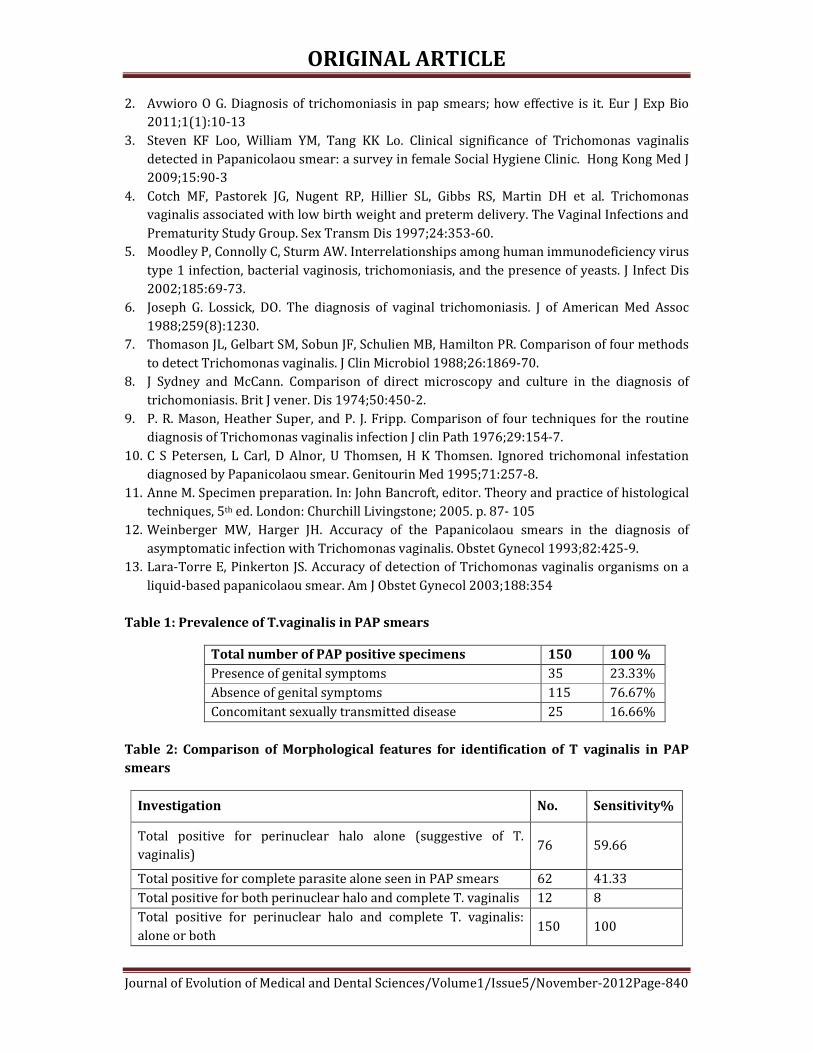
ORIGINAL ARTICLE
Journal of Evolution of Medical and Dental Sciences/Volume1/Issue5/November-2012Page-840
2. Avwioro O G. Diagnosis of trichomoniasis in pap smears; how effective is it. Eur J Exp Bio
2011;1(1):10-13
3. Steven KF Loo, William YM, Tang KK Lo. Clinical significance of Trichomonas vaginalis
detected in Papanicolaou smear: a survey in female Social Hygiene Clinic. Hong Kong Med J
2009;15:90-3
4. Cotch MF, Pastorek JG, Nugent RP, Hillier SL, Gibbs RS, Martin DH et al. Trichomonas
vaginalis associated with low birth weight and preterm delivery. The Vaginal Infections and
Prematurity Study Group. Sex Transm Dis 1997;24:353-60.
5. Moodley P, Connolly C, Sturm AW. Interrelationships among human immunodeficiency virus
type 1 infection, bacterial vaginosis, trichomoniasis, and the presence of yeasts. J Infect Dis
2002;185:69-73.
6. Joseph G. Lossick, DO. The diagnosis of vaginal trichomoniasis. J of American Med Assoc
1988;259(8):1230.
7. Thomason JL, Gelbart SM, Sobun JF, Schulien MB, Hamilton PR. Comparison of four methods
to detect Trichomonas vaginalis. J Clin Microbiol 1988;26:1869-70.
8. J Sydney and McCann. Comparison of direct microscopy and culture in the diagnosis of
trichomoniasis. Brit J vener. Dis 1974;50:450-2.
9. P. R. Mason, Heather Super, and P. J. Fripp. Comparison of four techniques for the routine
diagnosis of Trichomonas vaginalis infection J clin Path 1976;29:154-7.
10. C S Petersen, L Carl, D Alnor, U Thomsen, H K Thomsen. Ignored trichomonal infestation
diagnosed by Papanicolaou smear. Genitourin Med 1995;71:257-8.
11. Anne M. Specimen preparation. In: John Bancroft, editor. Theory and practice of histological
techniques, 5th ed. London: Churchill Livingstone; 2005. p. 87- 105
12. Weinberger MW, Harger JH. Accuracy of the Papanicolaou smears in the diagnosis of
asymptomatic infection with Trichomonas vaginalis. Obstet Gynecol 1993;82:425-9.
13. Lara-Torre E, Pinkerton JS. Accuracy of detection of Trichomonas vaginalis organisms on a
liquid-based papanicolaou smear. Am J Obstet Gynecol 2003;188:354
Table 1: Prevalence of T.vaginalis in PAP smears
Total number of PAP positive specimens 150 100 %
Presence of genital symptoms 35 23.33%
Absence of genital symptoms 115 76.67%
Concomitant sexually transmitted disease 25 16.66%
Table 2: Comparison of Morphological features for identification of T vaginalis in PAP
smears
Investigation No. Sensitivity%
Total positive for perinuclear halo alone (suggestive of T.
vaginalis) 76 59.66
Total positive for complete parasite alone seen in PAP smears 62 41.33
Total positive for both perinuclear halo and complete T. vaginalis 12 8
Total positive for perinuclear halo and complete T. vaginalis:
alone or both 150 100
ORIGINAL ARTICLE
Journal of Evolution of Medical and Dental Sciences/Volume1/Issue5/November-2012Page-841
RANDOMIZED CLINICAL TRIAL IN CHIKUNGUNYA ARTHRITIS CASES Mansoor Ahmed, N. Shantharam, Y. J. Visweswara Reddy.
1. Associate Professor, Department of Community Medicine.MMCRI, Mysore.
2. Formerly Professor & Head, Department of Community Medicine. PESIMSR, Kuppam.
3. Professor, Department of General Medicine, PESIMSR,Kuppam.
CORRESPONDING AUTHOR
Dr. Mansoor Ahmed Associate Professor, Dept of Community Medicine, MMCRI, Mysore. E-mail: [email protected], Ph: 0091 9945939275 ABSTRACT: Chikungunya virus is no stranger to the Indian sub-continent. Since its first
isolation in Calcutta[1] in 1963, there have been several reports of chikungunya virus infection in
different parts of India[2], [3], [4]. The last outbreak of chikungunya virus infection occurred in
India in 1971. Subsequently there has been no active or passive surveillance carried out in the
country and therefore, it ‘seemed’ that the virus had ‘disappeared’ from the subcontinent[5]
However, recent reports of large scale outbreaks of fever caused by chikungunya virus infection
in several parts of Southern India have confirmed the re-emergence of this virus. It has been
estimated that over 1,80,000 cases have occurred in India since December 2005 [6] Andhra
Pradesh (AP) was the first state to report this disease in December 2005, and one of the worst
affected (over 80,000 suspected cases) . Over 12% of patients who contract chikungunya virus
infection develop chronic joint symptoms [7]. OBJECTIVE: To test the efficacy of chloroquine in
reducing the pain of chikungunya induced arthritis as compared to paracetamol.
METHODOLOGY: A Randomized Clinical Trial was carried out in a community attached to
urban health centre of PESIMSR, Kuppam during August 2006. Among the 132 cases of arthritis,
86 persons were selected based on their availability and consent to participate. They were
divided into two randomly assigned groups namely Category–1(Chloroquine group) and
Category–2 ( Paracetamol group). Chloroquine tablet -155 mg and Paracetamol tablet - 500 mg
were administered as a single dose to the two groups respectively. The groups were followed up
for 8 days and the results were analyzed. STATISTICAL ANALYSIS: Analysis was carried out by
using S.P.S.S. package. Asymptoic test statistic and X2 MH (Chi square test ) were used to
evaluate the effect of the drugs. RESULTS OF THE STUDY: The decrease of pain in chikungunya
arthritis cases was significant in the mild and moderate pain categories with ‘p’ values of 0.0117
and 0.0129 respectively.
Asymptoic test statistic was 1.70 for chloroquine group and χ²MH was 2.76 (’P’ value
between 0.05 to 0.1).OR= 48.59.Incidence Risk Ratio for chloroquine was 1.52 with CI ; 1.14 –
1.90.The efficacy of chloroquine in reducing pain in arthritis was 51.83% ( effect size).The
logistic equivalents of odds for chloroquine and paracetamol group were 0.41 and 0.02
respectively in logistic regression analysis. CONCLUSION: Choloroquine is more efficient in
reducing the pain of chikungunya arthritis as compared to paracetamol in both sexes and in all
age groups.
KEY WORDS: Chikungunya, Arthritis, Clinical Trial, Chloroquine.
INTRODUCTION: Chikungunya virus is no stranger to the Indian sub-continent. Since its first
isolation in Calcutta[1] in 1963, there have been several reports of chikungunya virus infection in

ORIGINAL ARTICLE
Journal of Evolution of Medical and Dental Sciences/Volume1/Issue5/November-2012Page-842
different parts of India[2], [3], [4]. The last outbreak of chikungunya virus infection occurred in
India in 1971. Subsequently there has been no active or passive surveillance carried out in the
country and therefore, it ‘seemed’ that the virus had ‘disappeared’ from the subcontinent[5]
However, recent reports of large scale outbreaks of fever caused by chikungunya virus infection
in several parts of Southern India have confirmed the re-emergence of this virus. It has been
estimated that over 1,80,000 cases have occurred in India since December 2005 [6] Andhra
Pradesh (AP) was the first state to report this disease in December 2005, and one of the worst
affected (over 80,000 suspected cases) . Over 12% of patients who contract chikungunya virus
infection develop chronic joint symptoms [7].
Chikungunya arthritis is a crippling and chronic sequel of chikungunya fever causing a
lot of pain and distress to the patients.
Chloroquine is a disease modifying, non steroidal drug known also to have anti-
rheumatic effect[8] . The therapeutic dose of chloroquine is 150 mg. It has a half-life of 4 days and
the therapeutic effect is said to last upto 16 days after cessation of treatment. It is a safe drug in
this dose as it has been extensively used in malaria treatment.
Only one previous study of using chloroquine in patients with chikungunya arthritis has been
documented earlier [9]
The present study was carried out to test the efficacy of chloroquine in reducing the pain
of chikungunya arthritis patients as compared to paracetamol.
MATERIALS AND METHODS: A randomized double blind clinical trial was conducted in a
community attached to the urban health centre of PESIMSR, Kuppam . The study was conducted
in the post epidemic period of chikungunya during August 2006.
STUDY POPULATION: The study population constituted the patients having post chikungunya
arthritis. A door to door survey was done by health workers to check for these cases in the
entire community. The investigators screened all patients and selected 86 arthritis patients with
the following criteria.
INCLUSION CRITERIA: Only those subjects with post chikungunya arthritis.
EXCLUSION CRITERIA:
1. Those subjects not giving their consent to participate in the study.
2. Known cases of rheumatoid arthritis /osteo arthritis /other degenerative diseases.
RANDOMIZATION AND ALLOCATION CONCEALMENT: The drugs chloroquine and
paracetamol were packed in brown envelopes as a single dosage form separately in 86 sealed
covers. These covers were having 43 dosage forms of chloroquine and paracetamol each. The 86
packs were given numbers from a random number table. Thus 86 randomized packs were
prepared. The contents of the covers were not noted on the cover. These covers were taken to
the field and were administered to the patients recruited on a sequential basis. Thus allocation
concealment was done. Thus two groups of patients, one receiving the white capsule and the
other receiving the red capsule were formed.
All patients were followed up by investigators for a period of 8 days after initial clinical
assessment.

ORIGINAL ARTICLE
Journal of Evolution of Medical and Dental Sciences/Volume1/Issue5/November-2012Page-843
MATERIALS:
1) A pretested proforma and follow up record.
2) Visual analogue scale to assess the pain reduction.
3) Drugs : Chloroquine 150 mg base filled in a white gelatin capsule and paracetamol 500
mg filled in a red gelatin capsule of the same type.
DOSAGE ADMINISTRATION: After formation of the groups the white capsule group was given
chloroquine and the red capsule group was given paracetamol as a daily single dose. Directly
observed, supervised treatment was administered.
QUALITY CONTROL: The investigators had several meetings along with the patients and had a
common clinical assessment agreement. Thus inter and intra observer variation was minimized.
The random number allocation for packets was done by the statistician who was not
participating in analysis. The key for the packets were with the same statistician.
EVALUATION OF PATIENTS: Only one end point, viz., the reduction of pain was assessed by the
investigators using visual analogue scale. The monitoring of patients was done by the
investigators who initially assessed them.
Out of 132 cases of arthritis, 86 persons were selected based on their availability and
consent to participate. This study group was divided into 2 randomly assigned groups namely
Category – 1(Chloroquine group) and Category – 2 (Paracetamol group). Informed consent was
taken from all the respondents.
The trial was double blind and the first author had the key. The two groups were similar
in all aspects. Chloroquine tab – 155 mg single dose was given to the chloroquine group and
Paracetamol tab – 500 mg single dose was given to the paracetamol group.
The pain was classified into severe, moderate and mild as per the expression of patients.
The relief of pain was graded into reduction by 25%, 50%, 75% and above 75% as expressed by
patients. The patients were followed up for 8 days and the results were recorded.
STATISTICAL ANALYSIS: Statistical analysis was done by using the SPSS package. Analysis was
done on risk approach basis. Asymptoic test and χ² ( chi square test) with MH treatment were
used to evaluate the effect of the drugs.
RESULTS: Response to treatment was favourable for chloroquine irrespective of the degree of
pain. The decrease of pain in chikungunya arthritis cases was significant in mild and moderate
categories with ‘p’ values of 0.0117 and 0.0129 respectively.
Increasing age had no effect on the relief of the pain (‘r’ was not significant). There was
no difference in the relief of pain in both sexes with increasing age.
It was observed that the Incidence Risk Ratio for chloroquine was 1.52 with CI ; 1.14 –
1.90.
The efficacy of chloroquine in reducing pain in arthritis was 51.83% (effect size).
The decrease of pain in the chloroquine group was 95.34% and in the paracetamol
group it was 62.79% giving a risk difference of 32.55.

ORIGINAL ARTICLE
Journal of Evolution of Medical and Dental Sciences/Volume1/Issue5/November-2012Page-844
In the choloroquine group, the median age was 40.2 and the inter-quartile range was
31.7 to 45.5
In the paracetamol group, the median age was 45.4 and the inter-quartile range was
35.4 to 56.5. No significant difference was seen between the two groups.
The proportion of mild, moderate, and severe pain categories was almost similar in both
the groups. The sex ratio of both the groups was also similar.
The logistic equivalents of odds for chloroquine and paracetamol groups were 0.41 and 0.02
respectively.
Results of standardized analysis : OR= 48.59
χ² MH = 2.76; Asymptoic test statistic = 1.70.
’P’ value lies between 0.05 to 0.1.
DISCUSSION: In this study it has been observed that the drug chloroquine is quite efficient in
reducing the pain of chikungunya arthritis. Though paracetamol also helps in reducing the pain
it is less effective than chloroquine in this regard. These findings are in conformity with an
earlier study [8] where it was observed that the symptoms of chikungunya arthritis improved
significantly following chloroquine treatment.Very few studies have been reported on the use of
chloroquine in chikungunya arthritis cases and these results justify further controlled blind
trials of chloroquine in their treatment .
CONCLUSION: Choloroquine is more efficient in reducing the pain of chikungunya arthritis as
compared to paracetamol in both sexes and all age groups.
LIMITATIONS: The number of severe cases included in the trial was less. Therefore, the
adequacy of chloroquine in reducing the pain in these circumstances cannot be assumed. A
larger number of severe cases need to be included in future trials. The dose of chloroquine to be
given in severe cases also requires further study.
REFERENCES:
1. Shah KV, Gibbs CJ Jr, Banerjee G. Virological investigation of the epidemic of haemorrhagic fever in
Calcutta: isolation of three strains of Chikungunya virus. Indian J Med Res 1964; 52 :676-83.
2. Jadhav M, Namboodripad M, Carman RH, Carey DE, Myers RM. Chikungunya disease in infants and
children in Vellore: a report of clinical and haematological features of virologically proved cases.
Indian J Med Res 1965; 53:764-76.
3. Dandawate CN, Thiruvengadam KV, Kalyanasundaram V, Rajagopal J, Rao TR. Serological survey in
Madras city with special reference to chikungunya. Indian J Med Res 1965; 53 :707-14.
4. Thiruvengadam KV, Kalyanasundaram V, Rajgopal J. Clinical and pathological studies on
chikungunya fever in Madras city. Indian J Med Res 1965; 53 :729-44.
5. Pavri K. Disappearance of Chikungunya virus from India and South East Asia. Trans R Soc Trop Med
Hyg 1986;80:491.
6. Enserink M. "Massive outbreak draws fresh attention to little-known virus". Science 2006; 311
:1085.
7. CDC. Chikungunya Fever in India. Travelers' Health Outbreak Notice April 21,
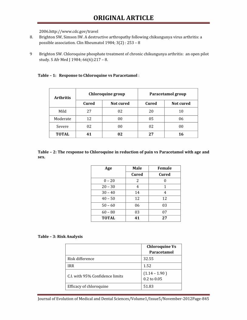
ORIGINAL ARTICLE
Journal of Evolution of Medical and Dental Sciences/Volume1/Issue5/November-2012Page-845
2006.http://www.cdc.gov/travel
8. Brighton SW, Simson IW. A destructive arthropathy following chikungunya virus arthritis: a
possible association. Clin Rheumatol 1984; 3(2) : 253 – 8
9
Brighton SW. Chloroquine phosphate treatment of chronic chikungunya arthritis: an open pilot
study. S Afr Med J 1984; 66(6):217 – 8.
Table – 1: Response to Chloroquine vs Paracetamol :
Arthritis Chloroquine group Paracetamol group
Cured Not cured Cured Not cured
Mild 27 02 20 10
Moderate 12 00 05 06
Severe 02 00 02 00
TOTAL 41 02 27 16
Table – 2: The response to Chloroquine in reduction of pain vs Paracetamol with age and
sex.
Age Male Female
Cured Cured
0 – 20 2 0
20 – 30 4 1
30 – 40 14 4
40 – 50 12 12
50 – 60 06 03
60 – 80 03 07
TOTAL 41 27
Table – 3: Risk Analysis
Chloroquine Vs
Paracetamol
Risk difference 32.55
IRR 1.52
C.I. with 95% Confidence limits (1.14 – 1.90 )
0.2 to 0.05
Efficacy of chloroquine 51.83
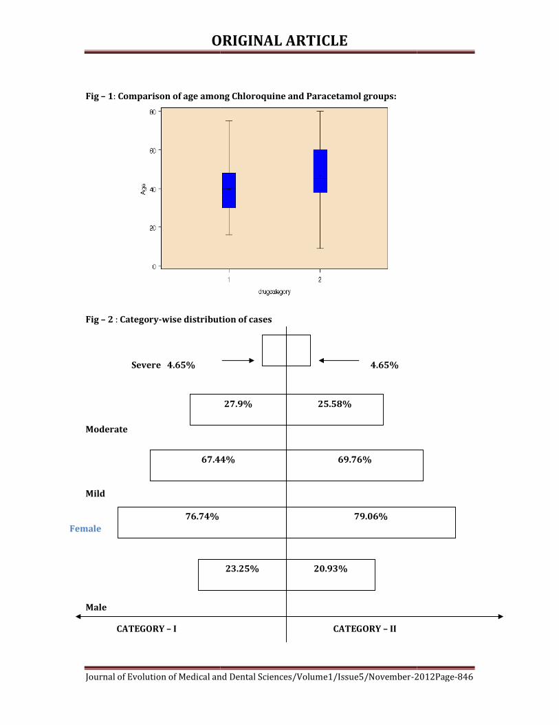
ORIGINAL
Journal of Evolution of Medical and Dental Sciences/Volume1/
Fig – 1: Comparison of age among Chloroquine and Paracetamol groups:
Fig – 2 : Category-wise distribution of cases
Severe 4.65%
Moderate
Mild
Female
Male
CATEGORY – I
67.44%
76.74%
ORIGINAL ARTICLE
al and Dental Sciences/Volume1/Issue5/November-2012
Comparison of age among Chloroquine and Paracetamol groups:
wise distribution of cases
4.65%
CATEGORY – II
27.9% 25.58%
67.44% 69.76%
79.06%
23.25% 20.93%
2012Page-846
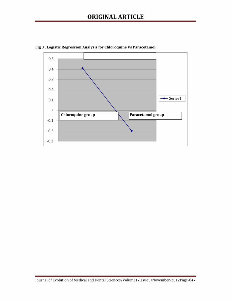
ORIGINAL ARTICLE
Journal of Evolution of Medical and Dental Sciences/Volume1/Issue5/November-2012Page-847
Fig 3 : Logistic Regression Analysis for Chloroquine Vs Paracetamol
-0.3
-0.2
-0.1
0
0.1
0.2
0.3
0.4
0.5
1 2
Series1
Chloroquine group Paracetamol group

CASE REPORT
Journal of Evolution of Medical and Dental Sciences/Volume1/Issue5/November-2012Page-848
PRIMARY LARYNGEAL CANDIDIASIS WITH TUBERCULOSIS MIMICKING
LARYNGEAL NEOPLASM Aditya Prakash Misra. Hari Om Sharan, Ritu Mishra.
1. Associate Professor, Department of Radiodiagnosis, Rama medical College, Hospital and research centre,
Kanpur, UP.
2. Assistant Professor, Department of Microbiology, Rama medical College, Hospital and research centre,
Kanpur, UP.
3. Assistant Professor, Department of Radiodiagnosis, Rama medical College, Hospital and research centre,
Kanpur, UP.
CORRESPONDING AUTHOR
Aditya Prakash Misra,
2A/ 43, Azad Nagar, Kanpur,
Uttar Pradesh. 208002.
E-mail: [email protected]
Ph: 0091 9839188620
ABSTRACT: A 50 -year-old man presented with hoarseness of voice, cough with expectoration
for one month and severe dyspnoea from 15 days. C T Scan of the neck revealed soft tissue
lesion circumferentially involving supra-glottic region with effacement of bilateral pyriform
fossa predominantly on left side. Extralaryngeal spread to adjacent paralaryngeal space was
also seen on left side. Marked airway luminal compromise was seen at the level of false vocal
cord . Primary diagnosis of laryngeal malignancy was considered on findings revealed by CT
scan neck. Direct laryngoscopy revealed erythema and ulcer with white pus discharge on
left false vocal cord. A swab was taken from ulcer on left false vocal cord and sent for
microbiological examination. The microbiological findings confirmed Mycobacterium
tuberculosis with Candida albicans.. CONCLUSION: Primary candidiasis with tuberculosis in
the laryngeal mucosa is a rare entity.
The clinical and pathological presentations of laryngeal candidiasis and tuberculosis might be
confused with those for malignant lesions if extralaryngeal spread is seen. Potential pitfalls in
diagnosis and the importance of microbiological examination in rare case of dual superimposed
primary laryngeal infections has been outlined.
KEYWORDS: Primary Laryngeal Candidiasis, Tuberculosis, mimicking, Laryngeal malignancy
INTRODUCTION: Primary candidiasis with tuberculosis in the laryngeal mucosa is a rare entity.
The clinical and pathological presentations of laryngeal candidiasis and tuberculosis might be
confused with those for benign or malignant lesions. We are presenting a case in which primary
diagnosis of laryngeal malignancy was considered by findings revealed on CT scan neck but
laryngoscopic and microbiological evaluation revealed and confirmed laryngeal infection i.e
Primary Laryngeal Candidiasis with Tuberculosis. We have outlined potential pitfalls in
diagnosis, and highlight the importance of evaluating microbiologically in rare case of dual
superimposed primary laryngeal infections.
CASE REPORT: A 50 -year-old man presented with hoarseness of voice, cough with
expectoration for one month and severe dyspnoea from 15 days. The patient has medical

CASE REPORT
Journal of Evolution of Medical and Dental Sciences/Volume1/Issue5/November-2012Page-849
history of hypertension. Physical examination was otherwise unremarkable. Chest x-ray showed
no evidence of pulmonary nodules, consolidation, or infiltrates.
Soft tissue computed tomography of the neck revealed soft tissue lesion
circumferentially involving supra-glottic region with effacement of bilateral pyriform
fossa predominantly on left side. Extralaryngeal spread to adjacent paralaryngeal space was
also seen on left side(Fig1). Marked airway luminal compromise was seen at the level of false
vocal cord (Fig 2). Few sub centimeter lymph nodes were seen in bilateral submandibular
region.
Direct laryngoscopy revealed erythema and ulcer with white pus discharge on left false
vocal cord(Fig 3). Swab was taken from ulcer on left false vocal cord and sent for
microbiological culture and sensitivity. On Gram staining, Gram positive budding Yeast cells of
Candida albicans were noted. On Ziehl Neelsen staining red coloured acid fast bacilli against
blue background with budding yeast cells were seen with 20% Sulphuric acid (Fig 4). These
microbiological findings confirmed Mycobacterium tuberculosis with Candida albicans.
Patient received a course of 6 weeks fluconazole antifungal with 6 months of
antitubercular therapy. Hoarseness resolved, and there has been no evidence of recurrence
during the subsequent 24 months.
DISCUSSION: Primary laryngeal candidiasis along with tuberculosis is rare entity and can
seldom be diagnosed clinically or radiologically . However, infective differential diagnosis may
be kept if diffuse submucosal involvement is seen on soft tissue CT scan. Mycotic infections of
the larynx are frequently seen in patients with immune insufficiency although they have also
been reported in individuals with normal immune status1,2,3. Candidiasis is frequently seen in
patients with immune deficiency syndromes or diabetes mellitus or those receiving
immunosuppressive treatment3,4 and in individuals whose mucosal barriers are affected by
excessive use of antibiotic or inhaled corticosteroids5,6 radiation therapy, smoking, trauma,
gastro esophageal reflux or chemical–thermal damage4. Laryngeal candidiasis can present with
various symptoms, such as odynophagia, dysphagia, hoarseness, respiratory distress and
stridor7,8.Similarly laryngeal tuberculosis is almost always associated with pulmonary
tuberculosis9,10,11.
In our patient soft tissue computed tomography of the neck showed soft tissue lesion
circumferentially involving supra-glottic region. Effacement of bilateral pyriform fossa with
predominant involvement on left side was noticed. Extralaryngeal spread to adjacent
paralaryngeal space was seen on left side. Marked airway luminal compromise was seen.
Laryngeal malignancy was considered as primary diagnosis as per findings.
The radiological findings of laryngeal tuberculosis depend on the stage and extension of
lesion .In the infiltrative stage, there is focal thickening. In the ulcerative stage, the ulceration is
not deep and rarely reaches the paraglottic spaces and the cartilage. Perichondritis is
sometimes noted (epiglottis, arytenoids), but calcifications are not common and the para-
laryngeal fat spaces are usually spared. The last stage is characterized by sclerosis. Various
radiological findings that have been described include edema alone, an ulcero-infiltrative mass,
infiltrative and pseudo-tumoral appearance (66%); sub-glottic laryngitis (isolated swelling of
the aryepiglottic fold or even massive cartilaginous ulceration and, sometimes, chondritis or
perichondritis); diffuse form; and tuberculoma (enormous ventricular vegetation with a large
base that elevates the ventricular strip)12,13.

CASE REPORT
Journal of Evolution of Medical and Dental Sciences/Volume1/Issue5/November-2012Page-850
Previous studies showed that sparing of paralaryngeal spaces were suggestive of
infective etiology14,15. However , in our case despite of infectious etiology paralaryngeal
involvement was seen .
Laryngeal erythema and ulcer with white pus discharge on left false vocal cord was seen
in our patient on laryngoscopy. The laryngoscopic appearance of candidiasis and tuberculosis
generally includes oedema, erythema, ulceration, white plaque and pseudomembranous
formations5.
SUMMARY: Characteristic CT findings of laryngeal tuberculosis include bilateral involvement,
thickening of the free margin of the epiglottis, and extensive mucosal involvement. By
comparison, laryngeal carcinoma present as unilateral involvement, infiltration of the pre
epiglottic and paralaryngeal fat spaces by a submucosal mass, cartilage destruction, and
extralaryngeal invasion. So due to paralaryngeal fat involvement malignancy was considered
primary diagnosis in our case. Further evaluation is must before reaching to final diagnosis and
starting treatment. In our case provisional diagnosis of malignancy was made as per radiological
findings but confirmatory diagnosis was made after microbiological evaluation and patient was
treated accordingly.
Conflicting Interests: The authors have no conflicting interests.
REFERENCES:
1. Makitie AA, Back L, Aaltonen LM, et al: Fungal infection of the epiglottis
simulating a clinical malignancy. Arch Otolaryngol Head Neck Surg 2003;129:
124 – 126.
2. Pabuççuog˘lu U, Tuncer C, Sengiz S: Histo-Pathology of candidal hyperplastic
lesions of the larynx. Pathol Res Pract 2002; 198: 675 – 678.
3. Haberman RS, Becker ME, Ford CN: Candida epiglottitis. Arch Otolaryngol 1983;
109: 770 – 771.
4. Forrest LA, Weed H: Candida laryngitis appearing as leukoplakia and GERD. J
Voice 1998; 12: 91 – 95.
5. DelGaudio JM: Steroid inhaler laryngitis: Dysphonia caused by inhaled
fluticasone therapy. Arch Otolaryngol Head Neck Surg 2002;128: 677 – 681.
6. Gallivan GJ, Gallivan KH, Gallivan HK: Inhaled corticosteroids: Hazardous effects
on voice—An update. J Voice 2007; 21: 101 – 111.
7. Tashjian LS, Peacock JE: Laryngeal candidiasis. Arch Otolaryngol 1984; 110: 806
– 809.
8. Ganesan S, Harar RP, Dawkins RS, et al: Invasive laryngeal candidiasis: A cause of
stridor in the previously irradiated patient. J Laryngol Otol 1998; 112: 575 – 578.
9. Sá LC, Meirelles RC, Atherino CC, Fernandes JR, Ferraz FR: Laryngo-pharyngeal
Tuberculosis. Braz J Otorhinolaryngol 2007;73:862–866.
10. Kozakiewicz J, Dec M, Gorczyca-Tarnowska J: The rare case of primary isolated
tuberculosis in a 19 year-old patient. Otolaryngol Pol. 2006;60: 607–709.
11. Loehrl TA, Smith TL: Inflammatory and granulomatous lesions of the larynx and
pharynx. Am J Med. 2001;111:113–117.
12. Aspestrand F, Kolbenstvedt A, Boysen M: CT findings in benign expansions of the
larynx. J Comput Assist Tomogr. 1989;13:222–225.
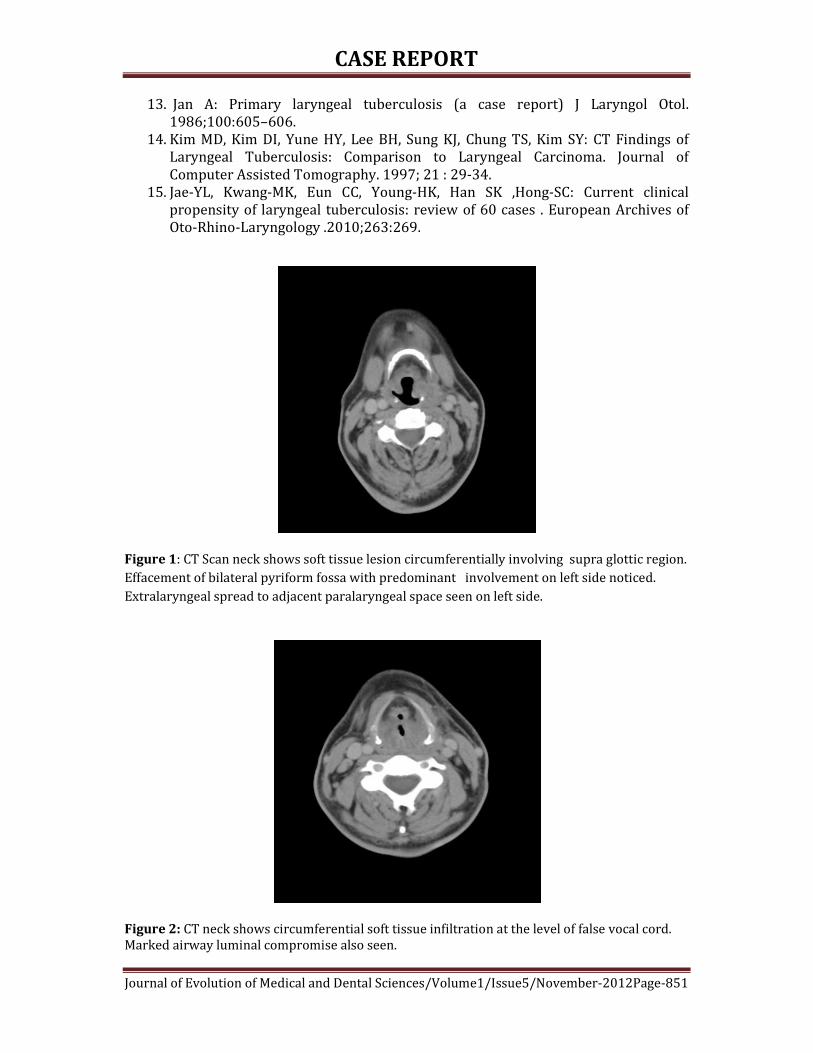
CASE REPORT
Journal of Evolution of Medical and Dental Sciences/Volume1/Issue5/November-2012Page-851
13. Jan A: Primary laryngeal tuberculosis (a case report) J Laryngol Otol.
1986;100:605–606.
14. Kim MD, Kim DI, Yune HY, Lee BH, Sung KJ, Chung TS, Kim SY: CT Findings of
Laryngeal Tuberculosis: Comparison to Laryngeal Carcinoma. Journal of
Computer Assisted Tomography. 1997; 21 : 29-34.
15. Jae-YL, Kwang-MK, Eun CC, Young-HK, Han SK ,Hong-SC: Current clinical
propensity of laryngeal tuberculosis: review of 60 cases . European Archives of
Oto-Rhino-Laryngology .2010;263:269.
Figure 1: CT Scan neck shows soft tissue lesion circumferentially involving supra glottic region.
Effacement of bilateral pyriform fossa with predominant involvement on left side noticed.
Extralaryngeal spread to adjacent paralaryngeal space seen on left side.
Figure 2: CT neck shows circumferential soft tissue infiltration at the level of false vocal cord.
Marked airway luminal compromise also seen.

CASE REPORT
Journal of Evolution of Medical and Dental Sciences/Volume1/Issue5/November-2012Page-852
Figure 3: Direct laryngoscopy showing erythema and ulcer with white pus discharge
on left false vocal cord.
Figure 4: Ziehl Neelsen Staining (100x) : Acid Fast Bacilli with budding yeast cells.

ORIGINAL ARTICLE
Journal of Evolution of Medical and Dental Sciences/Volume1/Issue5/November-2012Page-853
DENGUE IN AND AROUND NAGPUR- CENTRAL INDIA
Sharmila Raut, Ashwini Patil
1. Professor & Head, Department of Microbiology, Dr. Shankarrao Chavan Government Medical College,
Nanded.
2. Assistant Professor, Department of Microbiology, Government Medical College, Nagpur.
CORRESPONDING AUTHOR
Dr. S. S. Raut,
302, Kuber Regency,
Congress Nagar, Near All India Reporter,
Nagpur- 440 012
E-mail: [email protected],
Ph: 0091 9021366222
ABSTRACT: Dengue infection has been known to be endemic in India for over the
centuries as a benign and self limiting disease. Dengue epidemics had been regularly
reported from north India but no reports are available from central India. Here we
report the data of dengue epidemics from March 2008- June 2010 from Nagpur region
(Central India). A total of 289 serum samples from clinically suspected cases of dengue
fever admitted in GMCH, Nagpur, over a period of 3 years (Mar 2008-June 2010) were
tested for dengue specific IgM antibodies using IgM capture ELISA (MAC ELISA). Amongst
a total of 289 serum samples tested for dengue IgM antibodies, 85(29.41%) were positive
for dengue IgM antibodies. Children <10 yrs were most commonly affected with male
predominance. Maximum number of cases occurred in the year 2009.
Although the epidemiology of dengue is changing with dengue serotype 3 as an emerging
serotype, dengue infections are seen every year, thus making it as an epidemic disease in
Central India also.
KEY WORDS: Dengue, IgM capture ELISA, DF (Dengue fever), DHF (Dengue haemorrhagic
fever).
INTRODUCTION: Dengue infection has been known to be endemic in India for over the
centuries as a benign and self limiting disease. 1 In recent years, the disease has changed
its course manifesting in severe form as dengue hemorrhagic fever and with increasing
frequency of outbreaks. 3 Dengue is emerging as a major health problem in India. Since
the first epidemic in Kolkata during 1963-64, many places in India have experienced
dengue infection. Delhi has experienced seven outbreaks of dengue virus since 1967 with
the last reported in 2003. All four serotypes are circulating and cause epidemics. Earlier
serotype 2 was implicated as the etiology of major epidemics in Delhi in the year 1996
and Gwalior in 20012 .The implication of dengue serotype 3 as an etiology of DHF
epidemic recently confirms re-emergence of serotype 3 as a dominant form on the Indian
subcontinent 4. Similar outbreak of dengue in Mumbai in 20035 has been reported.
Dengue epidemics had also occurred in central India. Here we report the data of dengue
epidemics from March 2008- June 2010 from Nagpur region (Central India).
MATERIAL AND METHODS: A total of 289 serum samples from clinically suspected cases
of dengue fever classified as undifferentiated fever/ dengue fever/ DHF (WHO classification
ORIGINAL ARTICLE
Journal of Evolution of Medical and Dental Sciences/Volume1/Issue5/November-2012Page-854
) admitted in GMCH, Nagpur, over a period of 3 years (Mar 2008- June 2010) were
tested / for dengue specific IgM antibodies.
The samples were screened for the presence of IgM antibodies using IgM capture ELISA
(MAC ELISA) supplied by NIV Pune. OD was measured at 450nm using ELISA reader.
RESULTS: During the study period (2008-2010), a total of 289 serum samples were
tested for dengue IgM antibodies, by ELISA test.
Year wise distribution of samples being 52 in 2008, 223 in 2009 and 14 in 2010. Of
these 85(29.41%) were positive for dengue IgM antibodies. Year wise distribution of
dengue IgM positive over 3 years period is shown in table 1.
Maximum number of samples were received in the year 2009. There was an
increase in the number of samples in the monsoon season (August to November). Overall
males predominated over females (1.65:1). Age wise distribution of IgM positive cases in all
three year clearly indicates that children (<10 yrs) were more commonly affected (table 2).
Fever was the commonest presentation (98%). 30 % of the patients had hemorrhages & 4%
had altered sensorium. In 76% of patients, the platelet count was between 5,0000 /mm3-
10,0000/ mm3, while only in 24% of patients it was less than 5,0000/mm3.
DISCUSSION: Dengue is emerging as a great burden in our country. All 4 types of dengue
virus have been isolated from affected Indian population. Cyclical epidemics of dengue are
becoming more frequent. The outbreak in 1996 was the largest one to occur in Delhi 6
following which vigorous steps were taken to prevent and control DF/ DHF. Most of the
patients in the present study were children (<10 years) as compared to the older age
group (>10Yrs). This observation is quite in accordance to the studies reported from
south India5, 7 . However studies from Delhi6,8 found out the adults to be more susceptible to
infection than children. Erythematous rash was found in 8% of the patients. This percentage
is lower than previously reported dengue fever epidemics in Delhi9. Bleeding from various
sites /hemorrhage was found in 30% of cases. A similar high percentage of patients with
bleeding manifestations was found during 1998 and also in the year 2005. The causes of
bleeding in DF are not well established, but could be due to thrombocytopenia,
consumption coagulopathy , capillary fragility or platelet dysfunction. Although 24% of
cases shows significant thrombocytopenia (<50000/mm3). No correlation could be
established between the platelet count or bleeding manifestations and hence it indicated
other features contributing to the bleeding diatheses. Since no platelet function tests or
coagulation profile was available, the exact cause could not be elucidated.
Dengue specific IgM antibodies were found in 29.41% which is comparable with other
studies 2,10. The sensitivity of this test depends on the duration of prodromal illness. The
disadvantage of MAC ELISA is the delayed appearance of antibodies from 5-10 days after
the onset of illness in case of primary dengue virus infection and 4-5 days in secondary
infection. The requirement of paired sera, subsequently in convalescent phase, if negative
in acute phase also delays diagnosis. Detection of NS1 antigen assays hold promise in
early diagnosis in dengue infection 11 .
CONCLUSION: Although the epidemiology of dengue is changing with dengue serotype 3
as emerging serotype, dengue infection are seen every year, thus making it as an
epidemic disease in central India also. Appropriate investigation, strict monitoring and

ORIGINAL ARTICLE
Journal of Evolution of Medical and Dental Sciences/Volume1/Issue5/November-2012Page-855
prompt supportive management can reduce mortality of dengue. Detection of NS1 antigen
assays hold promise. Also prevention of transmission by mosquito control & maintaining
water sanitation is required to effectively control dengue epidemic.
REFERENCES:
1. Broor S, dar L, Sengupta S, Chakraborty M, Wali J P, Biswas A, Kabra S K,
Jain Y, Seth P. Recent dengue epidemic in Delhi, India. In Factors in the
emergence of arbovirus diseases, edited by: saluzzo JE, Dode B, Paris :
Elsevier 1997, 123-27.
2. Bandopadhyay S, Jain D C, Datta K K. Reported incidence of Dengue/ Dengue
hemorraghic fever in India 19991-95. Dengue Bulletin 1996, 20:33-34.
3. Dar l, Broor S, Sengupta S, Xess I, Seth P. The first major outbreak of
dengue hemorrhagic fever in Delhi, India. Emerg Infect Dis 1999, 5:589-90.
4. Dash R K, Saxena P, Abhyankar A, Bhargava R, Jona A M. Emergence of
dengue virus type 3 in northern India. Southeast Asian J Trop Med Public
Health, 2005 36:370-77.
5. Shah I, G C Deshpande, P N Tardeja. Outbreak of dengue in Mumbai and
predictive markers for dengue shock syndrome. Journal of Tropical
Paediatrics 2004, 50:301-05.
6. Anuradha S, Singh N P, Rizvi S N, Agrawal S K, Gur R, Mathur MD. The
1996 outbreak of Dengue hemorraghic fever in India. Southeast Asian J
Trop Med Public Health 1998, 29: 503-06.
7. Vijaykumar T S, Chandy S, Sathish N Abraham M, Abrabham P, Sridharan G.
Is dengue emerging as a major health problem?. Indian J Med Res 2005,
121:100-07.
8. Singh N P, Raja Jhamb, S K Agrawal, M Gaiha, Richa Diwan, M K Daga, anita
Chakravarti, Satish Kumar. The 2005 outbreak of dengue fever in Delhi,
India. Southeast Asian J Trop Med Public Health,2005,Sep;36(5):1174-78.
9. Sharma S, Sharma S K, Mohan. Clinical profile of dengue haemorragic fever in
adults during 1996 outbreak in Delhi ,India, Dengue Bull 1998;22:20-27.
10. Gera C, Singh A, Jose W, Dhanoa J. Dengue outbreak 2006 in Ludhiana,
Punjab. The Indian Practitioner 2 009; 62:703-09.
11. Datta S, Wattal C. Dengue NS1 antigen detection: A useful tool in early
diagnosis of dengue virus infection. IJMM 2010; 28:107-10.
Table 1 YEAR WISE DISTRIBUTION OF DENGUE CASES
Year Total cases Positive
2008 52 (17.99%) 04 (7.69%)
2009 223 (77.16%) 80 (35.87%)
2010 14 (04.85%) 01 (07.14%)
Total 289 85

ORIGINAL ARTICLE
Journal of Evolution of Medical and Dental Sciences/Volume1/Issue5/November-2012Page-856
Table 2 AGE WISE DISTRIBUTION OF MALES AND FEMALES
Age Male Female Positive male Positive female
0-10 yrs 107 83 42 (39.25%) 28 (35.8%)
11-20 yrs 27 17 06 (22.22%) 04 (23.5%)
21-30 yrs 16 03 02 (12.50%) 01 (33.3%)
31-40 yrs 20 03 01 (05%) 00
41-50 yrs 07 02 01 (14.29%) 00
>51 yrs 03 01 00 00
Total 180 109 52 33

ORIGINAL ARTICLE
Journal of Evolution of Medical and Dental Sciences/Volume1/Issue5/November-2012Page-857
EMERGING TRENDS IN THYROID DISEASES IN TSUNAMI HIT COASTAL
AREAS OF PUDUCHERRY AND CUDDALORE, INDIA. Narayanappa Shiroorkar Pradeepkumar, Reecha Singh, Noyal Mariya Joseph
1. Professor & Head, Department of Pathology, Lakshmi Narayana Institute of Medical Sciences, Pondicherry,
India.
2. Assistant Professor, Department of Pathology, Indira Gandhi Medical College and Research Institute,
Pondicherry, India.
3. Assistant Professor, Department of Microbiology, Pondicherry Institute of Medical Sciences, Pondicherry,
India.
CORRESPONDING AUTHOR
Dr. Narayanappa Shiroorkar Pradeepkumar
Professor and Head, Department of Pathology,
Sri Lakshmi Narayana Institute of Medical Sciences,
Osudu, Agaram Village, Villianur Commune,
Kudupakkam Post, Pondicherry – 605 502
E-mail: [email protected]
Ph: 0091 9345476620
ABSTRACT: BACKGROUND: Thyroid diseases are major global health problem but the
incidence and prevalence of this varies from place to place. The government of India has started
universal iodization of salt owing to the huge burden of thyroid diseases in the country. In 2004,
the coastal regions of Puducherry and Cuddalore were flooded by tsunami. AIMS: The aim of
this study is to determine the prevalence of thyroid diseases in coastal regions of Puducherry
and Cuddalore. We also studied the trends in prevalence of thyroid diseases after tsunami.
MATERIALS AND METHODS: All the patients undergoing thyroidectomy in a tertiary care
teaching hospital in South India were included in our study. Histopathological examination of
thyroidectomy specimens was performed. RESULTS: A total of 342 thyroidectomy specimens
were studied. Of the 342 cases, 30 were males and 312 were females with a ratio of 1: 10.4. Out
of 342 cases, 169 (49.42 %) were non neoplastic lesions, 173 (50.58%) were neoplastic. Of the
342 cases, 118 (34.5%) cases were simple goiter, 98 (28.66%) were adenoma, 70 (20.47%)
were papillary carcinoma, 42 (12.28%) were thyroiditis, 9 (2.63%) were toxic goiter and 5
(1.46%) were follicular carcinoma. We observed an increasing trend in the prevalence of
papillary carcinoma and thyroiditis after tsunami in 2004. CONCLUSIONS: There is an urgent
need to establish quality assurance system to monitor iodine content in the soil, food, water and
salt in the tsunami affected coastal areas. It is also necessary to study the other possible causes
for papillary carcinoma and take measures to prevent it.
KEY WORDS: Thyroid, Emerging trends, Histopathology, Tsunami
INTRODUCTION: Thyroid diseases are a major health problem both in developed and
developing countries. Their incidence varies from 4 – 7% of the population.[1-4] They are more
in hilly terrain and away from the sea coast.[2,5-7] The prevalence of thyroid disease is 1 – 7% of
females in UK and 4% of the population in US.[8,9] In India, thyroid diseases are endemic in
many parts of Goa, Gujarat, Kerala, and the Himalayan region. The principal diseases of thyroid
are simple goiter (diffuse and nodular), hyperthyroidism, hypothyroidism, thyroiditis
(Hashimoto thyroiditis, granulomatous, sub-acute lymphocytic thyroiditis) and neoplasms.[2]
Colloid goiter affects more than 200 million individuals throughout the world.[1, 2] FNAC is the

ORIGINAL ARTICLE
Journal of Evolution of Medical and Dental Sciences/Volume1/Issue5/November-2012Page-858
most cost effective method for diagnosis of thyroid disease.[4,9-11] Histopathology is used for the
final diagnosis when FNAC is inconclusive.[4,9]
Thyroid diseases cause concern because 5 – 10% of them are malignant.[1, 3, 9] Exact
causes of thyroid neoplasms are not well known. Exposure to radiation and high dietary intake
of iodine are the risk factors for papillary carcinoma while iodine deficiency is a risk factor for
follicular carcinoma and colloid goiter.[12,13] Viral infections are predisposing factors for
autoimmune thyroiditis. The government of India has started universal iodization of salt owing
to the huge burden of thyroid diseases in the country. However, in view of the recent occurrence
of tsunami in 2004 in the coastal areas of South India which is likely to have increased the
iodine levels in the adjacent regions, there is need to know if the practice of consuming iodized
salt is still necessary in the tsunami hit coastal areas.
We therefore performed this study to determine the prevalence of thyroid disease and
the ongoing trends in the occurrence of various thyroid diseases in the tsunami hit coastal areas
of Puducherry and Cuddalore in South India. This is the first study conducted in the tsunami hit
coastal areas to observe the changing trend in the occurrence of thyroid diseases.
MATERIALS AND METHODS: This study was conducted for a period of 9 years from January
2002 to July 2010 at a tertiary care teaching hospital in Puducherry, which caters to the health
care needs of the people living in the adjacent coastal areas of Puducherry and Cuddalore. We
received a total of 16,979 surgical specimens, of which 354 were thyroid specimens. Of these
354 thyroid specimens received in our lab, 12 had incomplete clinical data and were therefore
excluded from the study. The remaining 342 thyroid specimens obtained by total or partial
thyroidectomy from clinically diagnosed thyroid cases, irrespective of age, gender, race and
demographic habitat were included in the study. This study was approved by the Institute
Ethics Committee.
Clinical history, examination findings and all the investigation results were noted.
Careful gross examination of thyroidectomy specimen and lymph nodes (in malignancy with
secondaries) was performed. Tissue bits were processed after fixation in 10% formalin,
embedded in paraffin wax, thin section of 3-5 microns were taken on albuminized slide, kept at
60° C for 30 minutes and immersed in Xylene. These sections were stained by H & E and
mounted with DPX.
The sections were examined by a team of experienced Pathologists. The slides were
grouped according to the different thyroid lesions, age, sex and other demographic features. The
results were analyzed with available data and compared with other studies. The ongoing trends
in thyroid disease are also analyzed.
Statistical significances were calculated by Chi square test and Fisher’s exact test. P
values < 0.05 were considered statistically significant. Statistics software GraphPad InStat
version 3.06 for Windows, USA was used for calculation of P values.
RESULTS: We received 2.01% (342) thyroidectomy (total as well as subtotal) specimens out of
total 16,979 surgical specimens during study period. The results were analyzed with available
data and compared with other studies.
Of the total 342 cases studied 312 (91.23%) were female and only 30 (8.77%) cases
were male. The distribution of thyroid disease in relation to sex is shown in Table 1. Highest
incidence (19.82%) of the thyroid disease was seen in the age group 26 – 30 years. The age

ORIGINAL ARTICLE
Journal of Evolution of Medical and Dental Sciences/Volume1/Issue5/November-2012Page-859
distribution of the thyroid diseases is shown in Figure 1. Overall 67.54% of thyroid diseases
were in the age group of 21 to 40 years.
Of the 342 cases, 118 (34.5%) were simple colloid goiter and 98 (28.66%) were
adenomas. Malignancy was observed in 75 (21.93%) cases. Thyroiditis and toxic goiter
accounted for 42 (12.28%) and 9 (2.63%) cases respectively. The most common thyroid disease
in female patients was nodular colloid goiter (110/312), while in male patients it was adenoma
(10/30). Year wise follow up shows there is mild decrease in the trend of simple goiter and
adenomas (Figure 2). Similar follow up for inflammatory thyroid diseases shows there is
significant increase (Figure 3). But most important is there is striking increasing in trend of
papillary carcinoma since last 6 years (after 2004 tsunami) (Figure 3). Follicular carcinoma and
toxic goiter show mild change in trend and as their sample size small, it is difficult to give any
opinion on them (Figure 2).
The comparison of the distribution of thyroid diseases in different studies is shown in
Table 2. Of the 42 cases of thyroiditis observed in our study, 25 were identified as Hashimoto
thyroiditis and the remaining 17 were classified as lymphocytic thyroiditis. Among the 75 cases
of malignant neoplasms, 70 were morphologically sub-classified as papillary carcinoma, while
the other 5 were classified as follicular carcinoma. The relative predominance of papillary
carcinoma among thyroid malignancies observed in different studies is summarized in Table 3.
DISCUSSION: Thyroidectomy specimens accounted for 2.08% of the total surgical specimens
received during the study period. This shows a low frequency of thyroid diseases as compared
to Tsegaye B & Ergete W, but is similar to that of Arora R.[2,14] This could be due to the fact that
people from the coastal regions of Puducherry and Goa consume lot of sea food. In addition, the
soil in coastal areas is rich in iodine and consequently the food and water also have a high
content of iodine.
In our study the thyroid diseases showed a very high female preponderance with female
to male ratio of 10.4: 1. This relatively very high female preponderance observed in our study in
comparison to other similar studies is found to be statistically very significant.[1-3, 10,11] However,
the exact reason for this difference between our study and other studies is not known. It could
probably be due to severe nutritional deficiency. There is scope for further study to know the
reason
In the present study, high prevalence of thyroid diseases was seen in relatively younger
age group (26 – 30 years). In a study from Ethiopia, majority of the thyroid diseases occurred in
the age group 30 – 39 yrs.[2] Similarly, in two different studies from Kolkata (India) and
Malaysia, the age group 41 – 60 yrs was observed to be commonly affected by thyroid
diseases.[3, 10] The reason for the increased occurrence of thyroid diseases in younger age group
in our study is not clear.
The presence of goitrogens in foods of coastal places is often considered as a reason for
the predominance of simple colloid goiter in these areas. Although simple colloid goiter was the
most common thyroid disorder in our study, the proportion of patients with this condition was
relatively less in our study compared to other similar studies. This can be attributed to the fact
that the patients from Puducherry and Cuddalore consume iodized salt and sea food rich in
iodine. There is mild decrease in the trend of this simple goiter. This could be due to people’s
awareness about thyroid diseases and practice of preventive measures like using sea foods and
iodized salt.

ORIGINAL ARTICLE
Journal of Evolution of Medical and Dental Sciences/Volume1/Issue5/November-2012Page-860
In a study from Malaysia only 203 of 820 (25%) thyroid specimens were noted to have a
neoplastic condition.[3] Similarly, in a study from Bahrain 43 of 110 (39%) thyroid specimens
had neoplasms.[1] However, in our study 173 of 342 patients (50.59%) had thyroid neoplasms
(adenomas, papillary carcinoma and follicular carcinoma). The increased occurrence of thyroid
malignancies in our study in comparison to the above two studies was statistically very
significant (P value < 0.0001 vs. Malaysia study and P value 0.0275 vs. Bahrain study).
The present study shows an increasing trend in the prevalence of neoplasm especially
thyroid malignancy. The increasing incidence of malignancy is everyone’s concern. It is
necessary to find the cause and take preventive measures for the same. Compared to other
studies, statistically significant increase in the occurrence of papillary carcinoma was observed
in our study. This increasing incidence of papillary carcinoma in the present study is alarming.
There was also a striking increase in the trend of papillary carcinoma in the
present study. The risk factors for papillary carcinoma are radiation, high intake of iodine. The
flooding of the coastal regions of Puducherry and Cuddalore during the tsunami in 2004 could
have increased the iodine content of the soil. Despite the increased iodine levels in the soil due
to tsunami the people still continue to consume iodized salt, which probably could be the reason
for increased occurrence of papillary carcinoma in our population. There is an urgent need to
establish quality assurance system to monitor iodine content in the soil, food, water and salt in
the tsunami affected coastal areas. It is also necessary to study the other possible causes for
papillary carcinoma and take measures to prevent it. Another common lesion encountered in
our study was thyroiditis. It is comparatively higher than the incidence in different studies, but
is less than the study by Richard et al.[1-4,9,15,16] In our study we observed an increase in the
incidence of inflammatory thyroid diseases almost similar to papillary carcinoma. So, we
suspect that the increased occurrence of these two conditions might be interrelated.
Thyrotoxicosis was rare in our study and was observed in only 1.95% of the cases. It is
similar to studies done in Pakistan and Malaysia, but is lower than studies done in Bahrain.[1,4,11]
The incidence of thyrotoxicosis is not showing much change in its trend. The incidence of
follicular carcinoma was decreasing. But the sample sizes of these two conditions were too
small to observe any significant trend.
In conclusion, our Histopathological study emphasizes the fact that there was a higher
predominance of thyroid disease in female as compared with other studies. The thyroid
diseases were seen in relatively younger age group. Neoplasms are the commonest thyroid
lesions necessitating thyroidectomy. There was higher incidence of papillary carcinoma and
thyroiditis as compared with other studies. There was also a striking increase in the trend of
papillary carcinoma after tsunami. However, further studies in the other tsunami affected areas
are necessary to confirm this increased incidence as there is a paucity of similar data from other
tsunami hit coastal areas. There is an urgent need to establish quality assurance system to
monitor iodine content in the soil, food, water and salt in the tsunami affected coastal areas. It is
also necessary to study the other possible causes for papillary carcinoma and take measures to
prevent it.
REFERENCES:
1. Darwish AH, Al-Sindi KA, El-Kafsi J. Pattern of thyroid disease – a histopathological study.
Bahrain Med Bull 2006;28:1-6

ORIGINAL ARTICLE
Journal of Evolution of Medical and Dental Sciences/Volume1/Issue5/November-2012Page-861
2. Tsegaye B, Ergete W. Histopathologic pattern of thyroid disease. East African Medical
journal 2003;80:525-8
3. Htwe TT, Hamdi MM, Swethadri GK, Wong JOL, Soe MM, Abdullah MS. Incidence of thyroid
malignancy among goitrous thyroid lesion from Sarawak General Hospital 2000 – 2004.
Singapore Med J 2009;50:724 -8
4. Nurismah MI, Sharifah NA, Usama AE, Rohaizak M, Ibrahim N. Fine needle aspiration (FNA)
cytology of the thyroid: a cyto-histopathological study of 361 cases in Hospital Universiti
Kebangsaan Malaysia. Med and Health 2007;2:58-65
5. Kumar V, Abbas AK, Fausto N. Robbins and cotran Pathologic basis of disease. 7th ed.
Saunders: Elsevier Inc, 2004
6. Park K. Iodine deficiency disorders. In: Park’s text book of Preventive and social Medicine.
19th ed. Jabalpur: Banarsidas Bhanot, 2007. p. 510-1
7. Das S. The Thyroid and Parathyroids. In: A concise textbook of Surgery. 4th ed. New Delhi:
Das S, 2006. p.644-78
8. Walker BR, Strachan MWJ. Endocrine disease. In: Boon NA, Colledge NR, Walker BR, Hunter
JAA editors. Davidson’s principles and practice of Medicine. 20th ed. Edinburgh: Churchill
Livingstone, 2006. p.744-62
9. Prinz RA, O'Morchoe PJ, Barbato AL, Braithwaite SS, Brooks MH, Emanuele MA, et al. Fine
needle aspiration biopsy of thyroid nodules. Ann Surg 1983;198:70-3
10. Guhamallick M, Sengupta S, Bhattacharya NK, Basu N, Roy S, Ghosh AK, Chowdhury M.
Cytodiagnosis of thyroid lesions – usefulness and pitfalls: a study of 288 cases. J Cytol.
January 2008;25:6-9
11. Kumar S, Aqil S, Dahar A. Role of fine needle aspiration cytology in thyroid diseases. Journal
of Surgery Pakistan (International) 2008;13:22-5
12. Krukowski ZH. The Thyroid gland and the thyroglossal tract. In: Russell RCG, Williams NS,
Bulstrode CJK editors. Bailey and love’s short practice of surgery 23rd ed. London: Hodder
Arnold, 2008. p. 771-806
13. Dorairajan N, Pandiarajan R, Yuvaraja S. A Descriptive study of papillary carcinoma in a
teaching hospital in Chennai, India. Asian Journal of Surgery 2002;25:300-4
14. Arora R, Dias A. Iodine and thyroid cancer in Goa. Online J Health Allied Scs 2006; 5:1-3
15. Perros P. Thyrotoxicosis and Pregnancy. PLoS Med 2005;2:e370
16. Desailloud R, Hober D. Viruses and thyroiditis: an update. Virology Journal 2009;6:5
Table 1. Sex distribution of thyroid disease by different studies
Place
(Reference no.)
Female (%) Male (%) No. of patients
studied
P value*
Puducherry
(Present study)
312 (91.23) 30 (08.7) 342 -----
Kolkata (10) 173 (69.5) 76 (30.5) 249 < 0.0001
Malaysia (3) 677 (82.6) 143 (17.4) 820 0.0002
Bahrain (1) 84 (76.4) 26 (23.6) 110 < 0.0001
Ethiopia (2) 628 (80.5) 152 (19.5) 780 < 0.0001
Pakistan (11) 60 (67.4) 29 (32.6) 89 < 0.0001
* - P value with reference to the present study

ORIGINAL ARTICLE
Journal of Evolution of Medical and Dental Sciences/Volume1/Issue5/November-2012Page-862
Table 2. Comparison of the distribution of thyroid diseases in different studies
Place
(Reference no.)
Simple Colloid
goitre (%)
Toxic
goitre
(%)
Thyroiditis
(%)
Benign
(Adenoma)
(%)
Malignant
(%)
Puducherry
(Present study)
34.5 2.63 12.28 28.65 21.93
Malaysia (4) 71.0 0.3 0.3 11.0 17.4
Malaysia (3) 70.4 4.0 0.9 18.1 6.7
Bahrain (1) 45.5 8.0 7.0 15.5 24.0
Ethiopia (2) 76.9 0 2.1 12.8 8.2
Kolkata (10) 79.1 10.8 10.0
Goa (14) 89.4 10.6
Pakistan (11) 90.0 10
Table 3. Comparison of the predominance of papillary carcinoma among thyroid
malignancies in different studies
S. No. Place of Study
(Reference no.)
No. of
papillary
carcinom
a
Total no. of
thyroid
malignancies
% of
papillary
carcinoma
P value *
1. Puducherry (Present
study) 70 75 93.3 -----
2. Ethiopia (2) 49 64 76.6 0.0103
3. Malaysia (3) 39 55 70.9 0.0014
4. Goa (14) 36 66 54.5 < 0.0001
* - P value with reference to the present study

ORIGINAL ARTICLE
Journal of Evolution of Medical and Dental Sciences/Volume1/Issue5/November-2012Page-863
Figure 1. Age distribution of different thyroid diseases in our study
Figure 2. Trend in occurrence of colloid goiter, toxic goiter, adenomas and follicular
carcinoma
Figure 3. Trend in occurrence of thyroiditis and papillary carcinoma
ORIGINAL ARTICLE
Journal of Evolution of Medical and Dental Sciences/Volume1/Issue5/November-2012Page-864
“EFFECT OF OCIMUM IN CHEMOTHERAPY INDUCED OXIDATIVE
STRESS” A STUDY CONDUCTED IN DAKSHINA KANNADA DISTRICT,
KARNATAKA, INDIA Debkumar Ray, Sumana Chaudhury
1. Assistant Professor, Department of Biochemistry, B. S. Medical College, Bankura, West Bengal.
2. PG Student, Department of Botany, Burdwan Universiy, Burdwan, West Bengal.
CORRESPONDING AUTHOR
Dr. Debkumar Ray,
Assistant Professor, Dept of Biochemistry,
KL-1, Jagatpur, Ganga-Yamuna Apartment,
Block-B, Flat: B/C&D,
Ashwininagar,Kolkata-700059
E-mail: [email protected],
Phone: 0091 9476233354.
ABSTRACT: Adriamycin (ADR) belongs to anthracycline group of antibiotics, which is widely
used for the complete remission of solid tumours. In carcinoma breast, non Hodgkin’s
lymphoma and Hodgkin’s lymphoma, ADR is considered as most useful chemotherapeutic
medication. Anthracycline by virtue of their quinone group generates free radicals in solution in
both normal and malignant cells. The Ocimum leaf extract as well as their flavonoids orientin
and vicenin have strong antioxidant activity in vitro and antilipid peroxidative effect in vivo, and
strongly suggest free radical scavenging as a major mechanism by which Ocimum products
protect against cellular damage and tumour induction. However, most interest has been
devoted to the antioxidant activity of flavonoids, which is due to their ability to reduce free
radical formation and to scavenge free radicals. The capacity of flavonoids to act as antioxidants
in vitro has been a subject of several studies in the past years. The antioxidant efficacy of
flavonoids present in Ocimum sanctum in vivo is less documented, presumably because of the
limited knowledge on their uptake in tumours. Inspite of limitation of the knowledge of exact
mechanism of uptake of flavonoids we have studied antioxidant efficacy of Ocimum sanctum
aqueous extract (140mg) with placebo control in carcinoma breast, non Hodgkin’s lymphoma,
Hodgkin’s lymphoma cases along with chemotherapy particularly during third cycle. Statistical
analysis has been done by Mann Whitney’s test which shows no significant difference of
antioxidant enzymes between study (n=19) and control (n=16) group before chemotherapy.
There is a significant increase of all scavenging enzymes in study group (n=19) compared to
control group (n=16) after chemotherapy. Significant increase in the Haemoglobin content and
enhanced activities of Superoxide dismutase ( SOD) and Catalase (CAT) in the study group
which was given Ocimum.
KEY WORDS: Ocimum sanctum, Adriamycin, Antioxidants, Free radicals, Superoxide dismutase,
Catalase
INTRODUCTION: In last 25 years, a large number of studies have shown that free radical
mediated reactions are responsible for a wide range of chemotherapy induced side effects, and
that antioxidants are able to protect non malignant cells against some of the damaging effect of
cytotoxic drugs(1).Inspite of many available natural and synthetic antioxidants, a few number
have been studied in clinical trials. Beta carotene, Vitamin C and E, azelastine, co-enzyme Q10,

ORIGINAL ARTICLE
Journal of Evolution of Medical and Dental Sciences/Volume1/Issue5/November-2012Page-865
selenium have been studied for their chemotherapeutic effects and found to be effective in
ameliorating the side effects of anticancer drugs. Natural products like silymarin(1), ginseng(2),
ashwagandha(3) are found to be beneficial adjuncts to chemotherapy. There is a concern that
potent antioxidants may interfere with the anticancer efficacy too, which however appears to be
a theoretical concern since many studies done using both chemotherapy and antioxidants have
shown just the opposite. Antioxidant supplements have shown to protect the normal cells while
making the cancer cells more vulnerable to cytotoxicity of chemotherapy and radiation
therapy(1).The unpretentious little backyard plant plays on essential role in the folk medicine of
South Asia named “Tulsi” (Ocimum sanctum) in Sanskrit, is a gentle therapeutic herb belonging
to the family Basil. Conventionally, it is supposed to guard against stress and modern research
now corroborates the concept (4).In laboratory animals, Ocimum sanctum has been shown to
prevent cancer and to protect against radiation damage. Study established that hamsters were
protected from developing cancer of oral cavity by Ocimum leaf extract(5).Mice survived
radiation exposure when they have given with the herb(6,7,8).Its historical use as immuno-
stimulant has now been demonstrated(4).Two flavonoids have been identified in Ocimum
leaves, Orientin and Vicenin(6). In vivo and vitro studies have shown that, in general, flavonoids
have antioxidant (9), antitumour promoter (10), antimetastatic (11) and anti-proliferative (12)
properties. Flavonoids may act as antioxidants either by directly scavenging the free radicals or
by modulating the free radical scavenging enzymes in the body(4).The direct free radical
scavenging action of Ocimum aqueous extract(13) and indirectly by modulating Superoxide
dismutase, Catalase and other antioxidant enzymes like Glutathione peroxidase(14).in its
protective role in radiation induced damage in mice ,have been established .In human beings,
Ocimum has been studied for its efficacy in non Insulin dependent Diabetes Mellitus
(NIDDM)(15) but not for its chemotherapeutic effects. However, Ocimum aqueous extract is
undergoing clinical trial currently for its radiation protective effect at Tata Memorial Hospital,
Mumbai.
Our aim of study is to evaluate the safety and efficacy of Ocimum aqueous extract in modifying
the side effects of Adriamycin (ADR) based chemoschedules in the treatment of post operative
breast cancer, non Hodgkin’s lymphoma and Hodgkin’s lymphoma.
MATERIALS AND METHODS: This is a simple randomized single blind placebo control study,
where the study group was having 19 and control group includes 16 cases of post operative
female breast cancer, non Hodgkin’s lymphoma, Hodgkins lymphoma receiving Adriamycin
based chemoschedules and Statistical analysis has been done by Mann Whitney’s test which
shows no significant difference of antioxidant enzymes between study (n=19) and control
(n=16) group before chemotherapy, but after chemotherapy it was significant . The study was
undertaken in Department of Biochemistry in association with Department of Radiotherapy and
Radiobiology in Kasturba Medical College, Mangalore, India.
INCLUSION CRITERIA: Post operative breast cancer in women, non Hodgkin’s and Hodgkin’s
lymphoma in either gender, cancer diagnosed on both clinical and histological basis; age below
55 years, normal laboratory safety profile range, normal Electrocardiogram(ECG),general well
being as assessed by Karnofsky’s performance status scale more than 70;willing to give the
written informed consent.

ORIGINAL ARTICLE
Journal of Evolution of Medical and Dental Sciences/Volume1/Issue5/November-2012Page-866
EXCLUSION CRITERIA: Haemoglobin less than 10 g/dl, WBC less than 3,000/ml, Platelet count
less than 80,000/ml grossly damaged Liver and renal function tests more than thrice of the
normal range, congestive cardiac failure, hypertension, uncontrolled diabetes mellitus.
STUDY DRUGS: Placebo capsule contains lactose.
Ocimum capsule contains 140mg dried aqueous extract of Ocimum leaves. The fresh leaves of
Ocimum shades dried and powdered. The aqueous extract was prepared by refluxing with
distilled water and was vacuum dried in a speed vac system prepared in Radiobiology
Department, KMC, Manipal. The dose has been selected on the basis of the animal experiments
done in same department.
TREATMENT PLAN AND ASSESSMENT OF ENDPOINT MEASURES: Patients who are willing
to give written informed consent were enrolled for the study, before which the clinical
examination and laboratory assessment of organ function tests were done to rule out the
exclusion criteria. Patients were randomized to either of the groups to receive the placebo or
Ocimum capsules. The study medication was given during 3rd cycle, 24 hours after
chemotherapy for six consecutive days. Superoxide dismutase (SOD), Catalase (CAT) and
Haemoglobin (Hb) was estimated before chemotherapy and 24 hours after the end of Ocimum /
Placebo adjuvant therapy of the same cycle on 7th day. Estimation of Haemoglobin was done by
cyanmethaemoglobin method (16,17).Superoxide dismutase (SOD) was determined by Mccord
and Fridovich method (18) and Catalase (CAT) activity was assessed by Brannan et al (19)
method.
RESULTS: The baseline SOD values of the two groups are not significantly different. The SOD
levels in the study group and control group were 6814.66 ± 3540.04 and 9321.83 ± 5887.23.
However the control group had a higher mean value of SOD than the study group. The values of
SOD levels and the change in SOD levels obtained after 3rd cycle of chemotherapy are given in
Table 3.
The mean value of SOD in the control group was 6422.94 ± 3854.10 U/g Hb while study
groups showed a value of 9892.46 ± 4227.60 U/ g Hb. There was a significant difference in the
change in SOD after chemotherapy between the two groups. The mean change in SOD in the
study group was 3077.79 while the control group showed a fall in SOD value with the mean
change in the value of -2898.89.
Figure 1 and 2 depicts the mean values of SOD obtained before and after 3rd cycle of
chemotherapy in the study and control groups.
The mean values of CAT levels estimated before 3rd cycle of chemotherapy are presented
in table 4
There is no significant difference in the baseline Catalase values of the two groups.
However the study group showed an apparently higher mean value of Catalase in comparison
with with control group (Figure 3).Values of CAT and the change in CAT observed after 3rd cycle
of chemotherapy are given in table 5
A significant increase in the Catalase levels was observed in the study group with the
mean value of 49,727.72 ± 18,264.80. The control group showed a significant decrease and the
mean value was 10,016.83 ± 7547.10.There was a significant difference in the change in
Catalase values after chemotherapy between the two groups. Study group showed a difference
of 28,826.69 and the control group showed a mean value of -3477.19. Figure 3 and 4 depicts the

ORIGINAL ARTICLE
Journal of Evolution of Medical and Dental Sciences/Volume1/Issue5/November-2012Page-867
mean values of CAT obtained before and after 3rd cycle of chemotherapy in the study and control
groups
Table 1 Karnofsky’s Performance Status (KPS)
KPS Description of the general condition of the patients
100% Normal,no complaints,no evidence of disease
90% Able to carry on normal activity;minor signs or symptoms of disease
80% Nornal activity with effort;some signs and symptoms of disease
70% Cares for self;unable to carry on normal activityor to do active work
60% Requires occational assistance,but is mostly able to carefor himself
50% Requires considerable assistance and frequent medical care
40% Disabled,requires special care and assistance
30% Severely disabled,hospitalization indicated;death not imminent
20% Very sick,hospitalization necessary active supportive treatment
10% Moribund,fatal processes,progressing rapidly
0% Dead
Table 2 : SOD levels before 3rd cycle of chemotherapy
n Mean ± SD(Unit/g Hb)
Study Group 19 6814.66 ± 3540.04
Control Group 16 9321.83 ± 5887.23
Table 3 : SOD levels after 3rd cycle of chemotherapy
n Mean ± SD (U/gHb) Change in SOD
Study Group 19 9892.46 ± 4227.60 3077.79 ± 2325.68 **
Control Group 16 6422.94 ± 3854.10 - 2898.89 ± 3183.50
Table 4 : CAT levels before 3rd cycle of chemotherapy
n Mean ± SD(U/g Hb)
Study Group 19 20.901.03 ± 9901.13
Control Group 16 13494.03 ± 7476.94
Table 5 : CAT levels after 3rd cycle of chemotherapy
n Mean ± SD (U/g Hb) Change in Catalase
Study Group 19 49,727.27±18264.80** 28,826.69±25536.24***
Control Group 16 10,016.83±7547.10 -3477.19±2946.16
n = number of subjects
**p value 0.002
***p value 0.001

ORIGINAL ARTICLE
Journal of Evolution of Medical and Dental Sciences/Volume1/Issue5/November-2012Page-868
Figure 1 : Comparison of SOD(Unit/gHb) levels between study and control group before
3rd cycle of chemotherapy
Figure 2 : Comparison of SOD(Unit/gHb) levels between study and control group after 3rd
cycle of chemotherapy
Figure 3 : Comparison of CAT(Unit/gHb) levels between study and control group before
3rd cycle of chemotherapy
0
1000
2000
3000
4000
5000
6000
7000
8000
9000
10000
Study Gr Control Gr
0
2000
4000
6000
8000
10000
12000
Study Gr Control Gr

ORIGINAL ARTICLE
Journal of Evolution of Medical and Dental Sciences/Volume1/Issue5/November-2012Page-869
Figure 4 : Comparison of CAT(Unit/gHb) levels between study and control group after 3rd cycle
of chemotherapy
DISCUSSION: Chemical toxins produce biological damage by forming reactive oxygen species
like singlet oxygen, superoxides, hydroxyl, hydroperoxy, hydrogen peroxides (20).Ikeda et
al.(21) have shown that anthracycline by virtue of their quinone groups generate free radicals in
solution in both normal and malignant cells. In addition generation of lipid peroxide nitric oxide
and other destructive radicals are also produced from semiquinone intermediates of
anthracyclines. Under normal conditions the inherent defence system including SOD,CAT and
other antioxidant enzymes protect against free radical mediated oxidative stress.
Cu-Zn SOD serves as a major antioxidant enzyme in erythrocytes where superoxide
radicals are continuously generated by auto oxidation of Haemoglobin. Several anticancer drugs
lead to a generation of reactive oxygen species which partly explains the cytotoxic effect of
cancer therapy.
0
5000
10000
15000
20000
25000
Study Gr Control Gr
0
10000
20000
30000
40000
50000
60000
Study Gr Control Gr

ORIGINAL ARTICLE
Journal of Evolution of Medical and Dental Sciences/Volume1/Issue5/November-2012Page-870
Increase in SOD activity was 44% in the study group treated with Ocimum extract.
However placebo treated group showed a 31% reduction in SOD activity suggesting an efficient
scavenging of free radicals by Ocimum extract.
Uma devi et al (22) have shown that significant decrease in SOD activity after
radiotherapy,but the activity increases following administration of Ocimum extract.
Our result is in agreement with the above study. Significant increase in SOD observed in
Ocimum treated patients suggests that the Ocimum extract facilitates the removal of superoxide
anions produced by chemotherapy.
Mammalian erythrocytes are endowed with extraordinarily high activities of Catalase.
Cohen et al (23) have suggested that under physiological conditions practically all hydrogen
peroxides encountered by erythrocytes are detoxified by glutathione peroxidase and that
Catalase has a vital role in the clearance of hydrogen peroxide. Jacob et al (24) have reported
that humans lacking erythrocyte Catalase activity are not even susceptible to hemolysis induced
by oxidant drugs. Nihal et al (25) have reported protection by Catalase against exogenous
oxidant challenge. A 2.3 fold increase in Catalase activity was observed in the study group
treated with Ocimum extract where as placebo group showed 1.3 fold reduction in activity. A
significant increase in Catalase activity observed (p value 0.001) suggests a brisk generation of
superoxides and therefore hydrogen peroxide in the present circumstances. Flavinoids orientin
and vicenin in Ocimum seems to have induced the activity of Catalase. Thus providing
protection against the toxicity of anthracyclines.
Doroshow et al 1986(26) have shown that anthracycline induced cytotoxicity is
ameliorated by adding SOD and CAT to the culture medium.
No significant change in Haemoglobin content observed in the two groups after
chemotherapy. However the decrease in SOD, CAT observed in control group without any
appreciable alteration in the Haemoglobin levels.
A significant increase in the SOD,CAT levels after chemotherapy along with Ocimum
extract is an important observation of this study. This suggests that absorbed flavonoids of
Ocimum may play an important role to promote the body enzymatic (SOD, CAT) defence system
to combat the free radicals produced during ADR based chemotherapy. Thus Ocimum sanctum
could be a beneficial adjuvant for the same.
The possibility of developing new chemoprotective for application from Ocimum
sanctum should be further explored. In addition, its impressive antioxidant property and
stimulatory effect on the cellular antioxidants can be exploited for adjuvant therapy against a
number of human ailments such as cancer and other stress related disorders.
CONCLUSION: The results of present study indicate a significant elevation in the antioxidant
enzymes viz.SOD and Catalase after chemotherapy along with Ocimum as an adjuvant.
Increased antioxidant activity brought about by Ocimum may be responsible to combat
the damage in normal healthy tissues with little or no protection to the tumour following
chemotherapy. Hence Ocimum can be a beneficial adjuvant with ADR based chemotherapy.
REFERENCES:
1. Weji NL,Cleton FL,Osanto S.Free radicals and antioxidants in chemotherapy induced
toxicity.Cancer Treat Rev 1997;23:209-240
2. Torosian MH.Metabolic abnormalities and conditionally essential nutrients in the cancer
patient. Adjuvant Nutrition in Cancer Treatment 1992;173-233.

ORIGINAL ARTICLE
Journal of Evolution of Medical and Dental Sciences/Volume1/Issue5/November-2012Page-871
3. Ziauddin N,et al.Studies on the immunomodulatoryeffects of ashwagandha.J of
Ethnopharm, 1996;50(2):69-76
4. Tirtha,Sadashiva,Swami.The Ayurveda Encyclopedia 1998;106.
5. Kartkeyan K,Ravichandran P,Govindaswami S.Chemoprotective effectof ocimum
sanctum on DMBA- Inducedhamster buccal pouch carcinogenesis.Oral Oncl
1999;35(1):112-119
6. Umadevi P,Ganasoundari A,Rao BS,Srinivasan KK.In vivo radioprotection by ocimum
flavonoids:survival of mouse.Radiat res 1999;151(1):74-78
7. Umadevi P,Bisht KS,Vinitha MA. Comparative study of radioprotection by ocimum
flavonoids and Synthetic aminothiol protectors in the mouse.Br.J Radiol
1998;71(847):782-784
8. Ganasoundari A,Mahamood Zare S,Uma Devi.Modification of bone marrow sensitivity by
Medicinal Plant Extracts.The Br.J Radiol 1997;70:599-602
9. Alan L,Muller MD.Antioxidant Flavonoids:Structure,function and clinical usage.Alt Med
Rev 1996;1(2):103-111
10. Fujiki H et al. Inhibition of tumour promotion by flavonoids.In plant flavonoids in
biology and medicine. Biochemical,pharmacologicaland structure activity relationships
Alan R.Liss.New York.1986;429-440
11. Bracke ME et al.Flavonoids inhibit malignant tumours invasion in vitro.In plant
flavonoids in Biology and Medicine.II.Biochemical,Cellular and medicinal
properties.Alan R.Liss.New York 1988;219-233
12. Kandaswamy C et al.Antiproliferative effects of citrus flavonoids on a human squamous
cell carcinoma in vitro.Cancer Lett 1991;56:147-152
13. Ganasoundari A,Uma Devi P,Rao MNA.Protection against radiation induced chromosome
damage in Mouse bone marrow by ocimum sanctum.Mutat Res 1997;373:271-276
14. Uma Devi P,Ganasoundari A.Modulation of glutathione and antioxidant enzymes by
ocimum sanctum and its protection against radiation injury.Indian J Exp Biol 1999;Mar
37:262-268
15. Agarwal P,Rai V,Singh RB.Randomized,placebo controlled,single blind trial of holy
ocimum leaves in Patientswith non insulin dependent diabetes mellitus.Int J Clin
Pharmacol Ther 1996;Sept 34:406-409
16. Drabkin DL et al.J Biol Chem 1932;98:719
17. Zijlstra NC.Clinical Chem Acta 1960;5:719
18. McCord JM,Fridovich I.Superoxide dismutase :An enzymatic function for
erythrocuprein.J.Biol Chem 1969;24:6049-6055
19. Brannan TS,Maker HO,Raes JP.Regional distribution of catalase in the adult rat brain.J
Neurochem 1981; 86:307-309
20. Von Sonntag C.The chemical basis for radiation Biology (Taylor and Francis,London
).1987
21. Ikeda K,Kajiwara K,Tanabi E,Tokumaru S.Kishida E,Masuzawa Y and Kojo S.Involvement
of hydrogen peroxide and hydroxyl radical in chemically induced apoptosis of HL -60
cells.Biochem Pharmacol 1999;57:1361-1365
22. Younes Mand Siegers CP.Planta Medica 1981;43:240
23. Cohen,G and P.Hochstein.Glutathione peroxidase: the primary agent for the elimination
of hydrogen peroxide in erythrocytes.Biochemistry:1963;2:1420-1428
24. Jacob,H.S.,S.H.Ingbar and J.H.Jandl.Oxidative hemolysis and erythrocyte metabolism in
hereditary acatalasia.J.Clin.Invest.1965;44:1187-1199
25. Nihal S Agar,S.M.H.Sadrzadeh,Philip E.Hallaway and John W Eaton.Erythrocyte Catalase
a somatic oxidant defence.J Clin Invest.1985;319-321
26. Doroshow JH,Role of hydrogen peroxide and hydroxyl radical formation in killing of
Ehrlich tumour Cells by anticancer quinines,Proc Natl Acad Sci USA,1986;83:4514-4518

CASE REPORT
Journal of Evolution of Medical and Dental Sciences/Volume1/Issue5/November-2012Page-872
LINGUAL THYROID IN CHILDREN
Mujumdar. V. G, Sanjeev Tadasadmath, Siddaling Changty, Ahmed Abdul Aleem,
1. Assistant Professor, Department of Paediatrics, Khaja Banda Nawaz Institution of Medical Sciences, Roza-
B, Gulbarga, Karnataka.
2. Professor, Department of E.N.T, Khaja Banda Nawaz Institution of Medical Sciences, Roza-B, Gulbarga,
Karnataka.
3. Professor, Department of Paediatrics, Khaja Banda Nawaz Institution of Medical Sciences, Roza-B,
Gulbarga, Karnataka.
4. Junior Resident, Department of Paediatrics, Khaja Banda Nawaz Institution of Medical Sciences, Roza-B,
Gulbarga, Karnataka.
CORRESPONDING AUTHOR
Dr. Mujumdar. V. G,
Plot # 34, N. G. O. S
Housing Colony, Jewargi Road,
Gulbarga,
E-mail: [email protected]
Ph: 0091 9448978170.
INTRODUCTION: Lingual thyroid is a rare embryological anomaly, the incidence being
1/100000 population, that originates from failure of the thyroid gland to descend from the
foramen caecum to its normal prelaryngeal site. The ectopic gland, located at the base of the
tongue is often asymptomatic, but may cause local symptoms such as dysphagia, dysphonia,
stomatologia, upper airway obstruction and haemorrhage, often with hypothyroidism. This
infrequent congenital anomaly is often asymptomatic, until a pathologic stress such as systemic
disease or physiologic stress such as puberty causes enlargement of ectopic tissue, leading to
dysphagia, dysphonia stomatologia. A six year old girl child presented with complaints of mass
over the base of tongue, noticed since six months, with no other symptoms in this case report
the presentation, diagnosis and management of this condition are highlighted.
CASE REPORT: A six year old female child presented with the complaints of swelling over the
base of the tongue, with no other complaints/difficulties such as dysphagia, dysphonia,
stomatologia or difficulty in breathing. The swelling was noticed by the parents six months back.
The swelling is of the same size since then. There was insignificant medical history and no
history of dysphagia, dysphonia, stomatologia, delayed developmental milestones and mental
retardation. She weighed 16 kgs and her height was 90 cms. Physical examination revealed a
solid pink spherical mass covered with intact mucosa, located at the base of the tongue
measuring 1.8cms X1.4cmsX 2cms, obstructing the visualization of larynx, which was
nontender, fixed and firm in consistency (figure-1)
The clinical examination revealed no palpable thyroid gland, in the normal pretrecheal
position and no cervical lymphadenopathy. Ultrasonic scan of the neck showed nonvisualsation
of thyroid in its normal position. X-ray of the chest was normal. CT(computerized tomography)
of the neck revealed hyperdense area of 110-130 HU ,measuring about 1.8X1.4 cms noted at the
base of the tongue in midline. Normal thyroid tissue was not made out in the neck region.
Thyroid function tests revealed thyronormalcy, other laboratory tests also were within normal
range. FNAC of the swelling under general anesthesia was done. Smear prepared showed colloid

CASE REPORT
Journal of Evolution of Medical and Dental Sciences/Volume1/Issue5/November-2012Page-873
and follicular epithelial cells, arranged in small clusters and in follicular pattern. Numerous
pigment laden histiocytes and occasional multi nucleated histiocytes were noted. No malignant
cells were seen. Pathologist gave an opinion that cytological features were suggestive of lingual
thyroid. She was diagnosed as a case of ectopic lingual thyroid with euthyroid state. As the
lingual thyroid was causing no difficulty of whatever sort, to the child and being in euthyroid
state, parents were assured that no intervention was presently required .Parents were also
emphasized about the need for regular follow up in due course.
DISCUSSION: Embryologically, the thyroid gland develops as the first pharyngeal derivative by
an endodermal diverticulum, in the midline of the ventral pharynx between the first and second
pharyngeal pouches. A diverticulum descends caudally into the loose prepharyngeal connective
tissue and passes anterior to the developing hyoid bone and forms most of the thyroid
parenchyma. However, parafollicular C-cells reach the thyroid by ultimobranchial bodies, which
are the product of the fourth and fifth branchial pouches and form 1-30% of the thyroid
weight. [1] Failure of descent of either the medial anlage of the thyroid, or the ultimobranchial
bodies, and the incomplete obliteration of its vertical tract, lead to ectopic thyroid development.
The ectopic thyroids are usually located in the midline from the base of tongue to the
diaphragm, but can be also be present laterally. Lingual thyroid is not a very common lesion;
carcinomatous change in it is very rare. The majority of carcinomas observed in the lingual
thyroid are reported to be follicular. [2] Lingual thyroid is the most frequent ectopic location of
the thyroid gland, although its prevalence varies between 1: 100 000 and 1 : 300 000 and its
clinical incidence is reported to range from 1 : 4000 to 1 : 10000.
Ectopic thyroid tissue can also occur between the geniohyoid and mylohyoid muscles
(sublingual thyroid), above the hyoid bone (prelaryngeal thyroid) and in other rare sites such as
the mediastinum, precardial sac, heart, breast, pharynx, oesophagus, trachea, lung, duodenum,
and mesentery of the small intestine, adrenal gland. [3] CT (computerized tomography) or MR
(magnetic resonance) scans show a midline mass extending from the midline mucosal surface of
the tongue base into the medial sublingual space that can resemble a thyroglossal cyst.
However, nuclear scans are better to demonstrate the location of ectopic glands. [4] Thyroid
scan can also reveal whether there are other sites of thyroid tissue; in approximately 75% of
patients the ectopic tissue is the only functioning thyroid tissue in the body. [5] Management of
lingual thyroid is still controversial. No treatment is required when the lingual thyroid is
asymptomatic and the patient is in a euthyroid state; the patient has to be followed to be aware
of development of complications. Malignant transformation has been described [1],[2] and, for
this reason, some authors consider complete surgical removal of the gland as an appropriate
treatment. [6],[7] For patients with no or only mild clinical symptoms and elevated TSH
concentration, substitutive therapy with thyroid hormone may be successful, producing a slow
reduction of the mass. Ablative radioiodine therapy is an alternative approach recommended in
older patients or patients who are deemed unfit for surgery. This treatment should be avoided
in children and young adults since the systemic doses required have potentially damaging
effects on the gonads or other organs. [8] Surgical excision or radioiodine therapies are effective
treatments for lingual thyroid, but no treatment should be attempted until a radioisotope scan
has determined that there is adequate thyroid tissue in the neck. In patients, those with lacking
thyroid tissue in the neck, the lingual thyroid can be excised and autotransplanted to the
muscles of the neck. If emergency surgery is not necessary, suppression therapy should be tried
first in order to decrease the dimensions of the mass. The general conditions of the patient, the

CASE REPORT
Journal of Evolution of Medical and Dental Sciences/Volume1/Issue5/November-2012Page-874
size of the lesion, and presence of local symptoms or complications, such as hemorrhage, cystic
degeneration, or malignancies, are the most important conditions for planning the choice of
treatment. [9]
REFERENCES:
1. Collins P. Embryology and development. In: Williams PL, Bannister LH, Berry MM, Gray
H. editors. Gray's Anatomy - The Anatomical Basis of Medicine and Surgery. 38 th ed.
New York: Churchill Livingstone; 1995. p. 174-99.
2. Gooder P. Follicular carcinoma in a lingual thyroid. J Laryngol Otol 1980;94:437-9.
3. Di Benedetto V. Ectopic thyroid gland in the submandibular region simulating a
thyroglossal duct cyst: A case report. J Pediatr Surg 1997; 32:1745-6.
4. Elprana D, Manni JJ, Smals AG. Lingual thyroid. ORL J Otorhinolaryngol Relat Spec
1984;46: 147-52.
5. Baik SH, Choi JH, Lee HM. Dual ectopic thyroid. Eur Arch Otorhinolaryngol 2002;259:
105-7.
6. Galizia G, Lieto E, Ferrara A, Castellano P, Pelosio L, Imperatore V, et al. Ectopic thyroid:
Report of a case. G Chir 2001;22:85-8.
7. Shah BC, Ravichand CS, Juluri S, Agarwal A, Pramesh CS, Mistry RC. Ectopic thyroid
cancer. Ann Thorac Cardiovasc Surg 2007;13:122-4.
8. Alderson DJ, Lannigan FJ. Lingual thyroid presenting after previous thyroglossal cyst
excision. J Laryngol Otol 1994;108:341-3.
9. Toso A, Colombani F, Averono G, Aluffi P, Pia F. Lingual thyroid causing dysphagia and
dyspnoea. Case reports and review of the literature. Acta Otorhinolaryngologica ital
2009;29:213-7.
Figure 1

ORIGINAL ARTICLE
Journal of Evolution of Medical and Dental Sciences/Volume1/Issue5/November-2012Page-875
INCREASING PREVALENCE OF HEPATITIS AMONG TRANSFUSION
TRANSMITTED INFECTIONS: A TERTIARY CARE CENTRE EXPERIENCE Rupinder Kaur, Anuradha Singh, Gian Paul Singh
1. Associate Professor, Department of Pathology, Christian Medical College & Hospital, Ludhiyana, Punjab.
2. Junior Lab Technician, Blood Transfusion Unit, Department of Pathology, Christian Medical College &
Hospital, Ludhiyana, Punjab.
3. Technical Supervisor, Blood Transfusion Unit, Department of Pathology, Christian Medical College &
Hospital, Ludhiyana, Punjab.
CORRESPONDING AUTHOR
Dr. Rupinder Kaur, MD
Associate Professor, Pathology &
In charge Blood Transfusion Unit
Christian Medical College & Hospital Brown Road,
Ludhiana- 141 008, Punjab, India
E-mail: [email protected],
Ph: 0091 9888499289
ABSTRACT: Increasing Prevalence of Hepatitis among Transfusion Transmitted Infections: A
tertiary care centre experience. BACKGROUND: Blood transfusion despite being a lifesaving
intervention may result in acute or delayed complications and carries the risk of transfusion–
transmissible infections (TTI’s). There has been an alarming rise of hepatitis (B&C) infection
among blood donors and not much related to prevention or prophylaxis has been done so as to
reduce the risk of transmission. AIMS OF THE STUDY: The study was done to find out the
prevalence of Hepatitis B and Hepatitis C among transfusion transmitted infections in a healthy
blood donor population METHODOLOGY: A six years retrospective study of sero-reactive cases
of TTD’s among the blood donors was done (2004-2009) in Blood Transfusion Unit, Christian
Medical College & Hospital, Ludhiana. The data was retrieved from the blood bank records with
special emphasis on hepatitis infection among the blood donors. RESULTS: Over all 64,528
donors were screened for the Transfusion Transmitted infections (HBV, HCV, HIV1&2,Malarial
Parasite, and VDRL) of which majority were males. Overall seroprevelance for the transfusion
transmitted infections was 2.72%. Seropositivity for Hepatitis per se was 2.4%. Majority of the
donors were reactive for HCV infection (1.4%) followed by HBV (1.0%), HIV1&2(0.2%), VDRL
(0.1%) and Malaria parasite infection (0.02%). CONCLUSION: There has been an alarming rise
of hepatitis cases, especially HCV among the blood donor population which need to be looked
upon and special preventive measures need to be taken at the national level to combat this
problem.
KEY WORDS: Blood donors, Transfusion-transmitted infections, hepatitis
INTRODUCTION: Transfusion of blood and blood products is a life saving measure and saves
millions of lives each year globally, but at the same time it also carries a significant risk of
transmission of many blood transfusion transmitted infections (TTI’s). Despite stringent donor
screening and testing practices, safe blood free from transfusion-transmitted infections (TTI’s)
continues to be a threat to safe transfusion practices. 1

ORIGINAL ARTICLE
Journal of Evolution of Medical and Dental Sciences/Volume1/Issue5/November-2012Page-876
TTI’s can exist as asymptomatic diseases (silent killers) in the hosts, and acquisition of
infections during the window period from such blood donors can be a serious threat to the
safety of the collected donations. 2
Among the various TTI’s, the most dreaded and less sought after infection is by Hepatitis B
(HBV) and Hepatitis C (HCV) viruses and are considered two established causes of post
transfusion hepatitis. Many Blood donors are carriers, not realizing that are infected with this
disease. Individuals with chronic infection have a high risk of developing liver cirrhosis and
hepatocellular carcinoma leading to serious mortality and morbidity. 3 There are about 5.7
million cases of HIV in India, second highest pool of patients in the world. Syphilis is less often
transmitted by blood and the prevalence is low in most studies reported. 4
We did this study to find the prevalence of TTI’s with emphasis on hepatitis infection among
blood donors in our hospital.
MATERIAL & METHODS: This six years retrospective study was done in the Transfusion
Medicine Unit, Christian Medical College & Hospital, Ludhiana, a tertiary care centre in North
India over a period of 6 years from 2004-2009. Average donor numbers is about 10,000-12,000
per year.
The donor’s details were noted down from the donor consent forms kept in the blood
bank records. The donor blood samples were later screened for mandatory tests [Malaria,
Venereal Disease and Research Laboratory (V.D.R.L), Hepatitis B antigen (HBsAg), Anti HCV &
Anti HIV 1&2)].
Testing for malaria parasite was done by card test based on malarial (pLDH) antigen
based principle. VDRL testing was based on TPHA (Treponema Palladium Haemagglutination
Antibody) based principle. Testing for Hepatitis B, Hepatitis C and HIV 1&2 were based on 3rd
generation ELISA techniques (Ortho Clinical Diagnostics, Vitros ECiQ-based on
chemiluminiscense technology). The serological results were retrieved from the records
available in the department
RESULT AND ANALYSIS: A total of 64,528 donors were screened over the 6 year study period.
Majority (94.7%) of them were replacement donors. Male donors (97.8%) outnumbered
females (2.2%). Out of the potential donors screened, 1755 (2.72%) of the blood donors were
reactive for various Transfusion Transmitted Diseases (TTD’s). The details are shown in Fig 1.
Majority (71.8%) of the donor’s positive for TTD’s (HBsAg, HCV and HIV) were in 18-35 years
age group (Fig 2).
Prevalence of hepatitis infection formed the majority ( 2.4%) of the total TTI’s over the 6
year period ( Fig 3) of which HCV reactive donors were (1.4%) and HBsAg were
1%.Prevalence of HIV, VDRL, and MP was 0.2%. 0.1% and .02% respectively. Hepatitis infection
formed majority both among males and females. The detail regarding various TTI’s among
males and females are given in Table 1.
Co-infection was seen in 1.2 % of the total TTD positive donors of which 14 donors had
both HBsAg and HCV positivity. The detail is given in Table-2
DISCUSSION: Blood transfusion even though is a lifesaving procedure is associated with acute
and delayed complications and carries the risk of transmitting TTI’s. Despite stringent donor
screening and testing practices, safe blood free from transfusion-transmitted infections (TTI’s)
remains an elusive goal. 1 Although technological advancements have led to the development of

ORIGINAL ARTICLE
Journal of Evolution of Medical and Dental Sciences/Volume1/Issue5/November-2012Page-877
more sensitive methods to detect markers of TTI’s, the problems of ‘window period’, false-
negative results, prevalence of asymptomatic carriers, genetic variability in viral strains and
technical errors remain. 5
Among the various TTI’s, hepatitis has become an issue of global importance. Hepatitis B
and C are highly infectious and pose major public health problem in developing countries and
are the commonest cause of chronic liver disease in several regions of the world. 2 Hepatitis B is
one of the most common diseases transmitted by blood and has infected two million people
worldwide including an estimated 400 million chronically infected cases. Individuals with
chronic infection have a high risk of developing liver cirrhosis and hepatocellular carcinoma. 6
Hepatitis C virus (HCV) infection is another common chronic blood born infection with an
estimated 3.9 million persons infected by the virus and a high rate of development of liver
cirrhosis. Infection by HBV and HCV causes serious mortality and morbidity. 7
The present study showed higher seroprevelance of HBV and HCV among the blood
donors as compared to other TTI’s, rather it formed the majority of the infections
(2.4% of the total 2.7% of reactive TTI’s).
The prevalence of HBV reactive blood donors is different in various countries. It is as
low as 0.1-0.5% in a healthy population in United States and Western Europe, whereas it ranges
from 5-20% in far Eastern and some Tropical countries.. 8 Seroprevelance of HBsAg in various
Indian studies has shown to range between1.86%-4% 9,10 ,11 ,which was comparable to our
study showing the incidence of HBsAg to be 1%.
HCV is transmitted primarily through blood exposure. In contrast to HBV, about 20 to
40% of HCV cases are acute and majority of them progress to chronic infection. The long term
risk of developing cirrhosis and hepatocellular carcinoma is greater in HCV infected individuals
than in those infected with HBV. 12 There is wide variation globally in the seroprevelance of HCV
with the studies showing lowest prevalence in United States (0.1%)8 and highest in Egypt (
24.8%). [9]Various other Indian studies indicate seroprevelance of HCV ranging from 0.4%-
1.09%. 9,10,13,14
Most of the above mentioned studies have shown increase prevalence of HBV as
compared to HCV. The seroprevelance of HCV was highest (1.4%) among all other TTD’s in the
present study which was comparable to the study done in Pakistan where HCV prevalence was
2.06%. 15
Majority of the sero-positive donors were between 18-35 years age group in the present
study, which is alarming, considering the fact that this is the age during which a person is
considered healthy and can give maximum number of blood donation. Few other studies also
showed increase prevalence of TTI’s in 18-35 years of age group.16, 17
Dual infection was seen in 1.2% of the total TTI’s in our study of which most common
co-infection was HBV and HCV( 14 out of 23 ), which was similar to study done by Rodenas et
al. 17 Few other studies showed HIV to be the commonest co-infection associated with hepatitis
infection.1,18
The results from our study as well as various studies mentioned, showed the endemicity
and the rising prevalence of both HBsAg and HCV in an apparently healthy population as
compared to HIV. There is consequent risk of transmission of these viruses through
blood/blood products, albeit unknowingly. So far, most of the published data worldwide is on
HIV/AIDS and much is being done regarding its prevention and cure by various government
organizations as well as World Health Organization.

ORIGINAL ARTICLE
Journal of Evolution of Medical and Dental Sciences/Volume1/Issue5/November-2012Page-878
Despite various studies suggesting an increase in the hepatitis infection not much has
been done for its prevention. The emphasis must be given to increase the knowledge and to
change the attitude of the people. It was shown that the risk of HIV infection in SanFrancisco
declined substantially, to about 0.2 per cent as a direct result of efforts to educate at risk
individuals to avoid donation 14.
RECOMMENDATIONS: Various measures like strict donor selection criteria, use of sensitive
tests like Nucleic Acid Testing (NAT) and judicious use of blood and blood components have
been taken to reduce the risk of TTI’s per se. Pre-donation counseling, and self-exclusion of
donors is a rare phenomenon seen in our set up. National agencies like National Aids Control
Organization (NACO) and World Health Organization(WHO) should join hands along with
various other voluntary organizations in doing community based study for Hepatitis to identify
at risk and non- risk subjects. At risk subjects should be identified and preventive strategies
should be made at the national level, as the risk factors might vary from one geographical area
to another.
To achieve all this, strong political commitment, multi-sectorial engagement along with
public awareness, educational and motivational programmes and mass immunization ( for
Hepatitis B) should be done to substantially decrease the prevalence of hepatitis in an
otherwise healthy population.
CONCLUSION: From our study it has been clear that prevalence of hepatitis is on rise especially
infection with Hepatitis C in healthy population without them being aware of it (“silent
carriers”) and carries the risk of transmitting infection albeit unknowingly.
BIBLIOGRAPHY:
1. Kaur G, Basu S, Kaur R, Kaur P, Garg S. Patterns of infections among blood
donors in a tertiary care centre: A retrospective study. Natl Med J India
2010; 23:147–9
2. Kalepoto G.N., Bhally H.S., Khaliq G. Epidemiology of blood-borne viruses. A study of
health blood donors in southern Pakistan. J. Pak. Med. Assoc 1996; 27: 703-6.
3. Karki S, Ghimire P, Tiwari BR, Maharjan A, Rajkarnikar M. Trends in hepatitis B and
hepatitis C seroprevalence among Nepalese blood donors. Ind Jl Infec Dis 2008; 61: 324-
326.
4. Bhattacharya P, Chandra PK, Datta S, Banerjee A, Chakraborty S, Rajendran K, Basu SK,
Bhattacharya SK, Chakravarty R. Significant increase in HBV, HCV, HIV and syphilis
infections among blood donors in West Bengal, eastern India, 2004-2005. World Journal
of Gastroenterology 200; 13 :3730-3733.
5. Singh B, Kataria SP, Gupta R. Infectious markers in blood donors of East Delhi:
Prevalence and trends. Indian J Pathol Microbiol 2004;47:477–479.
6. Karki S, Ghimire P, Tiwari BR, Maharjan A, Rajkarnikar M, Trends in hepatitis B and
hepatitis C seroprevalence among Nepalese blood donors. Indian Journal of Infectious
Diseases 2008; 61:324-326.
7. Bhawani Y, Rao PR, Sudhakar V. Seroprevalence of transfusion transmissible infections
among blood donors in a tertiary care hospital of Andhra Pradesh. Biology and Medicine,
2010 ;l 2 : 45-48

ORIGINAL ARTICLE
Journal of Evolution of Medical and Dental Sciences/Volume1/Issue5/November-2012Page-879
8. Dienstag J.L., Isselbacher K.J. Acute viral hepatitis In Braunwald E. Fauji A.S., Kasper D.I.
et al. 15th ed. Harrison’s principles and practice of internal medicine. New York McGraw
Hill 2001: 1721-37.
9. Chandrasekaran S, Palaniappan N, Krishnan V, Mohan G, Chandrasekaran N: Relative
prevalence of hepatitis B viral markers and hepatitis C virus antibodies (antiHCV) in
Madurai, South India. Indian Journal of Medical Sciences 2000;547:270-273.
10. Srikrishna A, Sitalakshmi S, Damodar P: How safe are our safe donors?. Indian Journal of
Pathology and Microbiology, 1999;424:411-416.
11. Choudhury N, Ramesh V, Saraswat S, Naik S: Effectiveness of mandatory transmissible
diseases screening in Indian blood donors. Indian Journal of Medical Research,
1995;101:229-232.
12. WHO hepatitis C. WHO fact sheet No164, October 2000b.Available from
URL.http:/www.who.int/mediacentre/factsheets/fs164/en/print.htm. [Cited on 2007
May 14]
13. Sharma RR, Cheema R, Vajpayee M, Rao U, Kumar S, Marwaha N, Agnihotri SK:
Prevalence of markers of transfusion transmissible diseases in voluntary and
replacement blood donors. The National Medical Journal of India, 2004;171:19-21.
14. Gupta N, Kumar V, Kaur A: Seroprevalence of HIV, HBV, HCV and syphilis in voluntary
blood donors. Ind Jl of Med Sci, 2004;58:255-257.
15. Nazar H, Nadia N, Shazia A, Zulfiqar, Farhat A. Prevalenc of hepatitis B and C in blood
donors in Karachi. Biomedica 2008;24:116-117
16. Shrestha AC, Ghimre P, Tiwari BR, RajKarnikar M. Transfusion- Transmissible infections
among blood donors in Kathmandu, Nepal. J Infect Dev Ctries 2009;3:794-797
17. Rodenas JG, Bacasen LC, Que ER. The prevalence of HBsAg (+) anti HCV(+) among
healthy blood donors at East Avenue Medical Centre, Quezon City. Phil J Gastroenterol
2006;2:64-70
18. Kapur S, Mittal A. Incidence of HIV infection and its predictors in blood donors in Delhi.
Indian J Med Res 1998;108:45–50.
Table 1 showing trend of TTI reactivity among males and females
TTD Reactive( n=1755) Male Female
HCV 879 18
HBV 611 15
HIV 1&2 142 1
VDRL 73 2
MP 14 -
Total 1719 36
Table 2 showing dual reactivity among Blood Donors
TTD.s( Dual reactivity) n=23 Number
HBV+ HCV 14
HIV1&2 + HCV 06
HBV+ HIV1&2 01
HBV + VDRL 01
HCV+ VDRL 01

ORIGINAL ARTICLE
Journal of Evolution of Medical and Dental Sciences/Volume1/
Fig 1 shows distribution of Transfusion Transmitted diseases ( n=1755) among
donors
Fig 2 showing age groups among various TTD reactive blood donors
0
100
200
300
400
500
600
700
800
900
1000
HCV HBsAg
897
(1.4%)
626
(1%)
0
50
100
150
200
250
300
350
400
450
HbsAg HCV
ORIGINAL ARTICLE
al and Dental Sciences/Volume1/Issue5/November-2012
distribution of Transfusion Transmitted diseases ( n=1755) among
age groups among various TTD reactive blood donors
HBsAg HIV 1&2 VDRL MP
626
(1%)
143
(0.2%) 75
(0.1%)14
(.02%)
HCV HIV 1&2 VDRL MP
18-
26-
36-
46-
2012Page-880
distribution of Transfusion Transmitted diseases ( n=1755) among blood
-25
-35
-45
-60

ORIGINAL ARTICLE
Journal of Evolution of Medical and Dental Sciences/Volume1/Issue5/November-2012Page-881
Fig 3 showing prevalence of Hepatitis among blood donors
0
50
100
150
200
250
300
350
Hepatitis
HBsAg
Anti-HCV
2004
2005
2006
2007
20082009
ORIGINAL ARTICLE
Journal of Evolution of Medical and Dental Sciences/Volume1/Issue5/November-2012Page-882
DETECTION OF VANCOMYCIN RESISTANT ENTEROCOCCI (VRE) IN
HOSPITALIZED PATIENTS AND COMPARISION OF KIRBY-BAUER DISC
DIFFUSION AND VANCOMYCIN SCREEN AGAR METHOD. K. Vidyasagar, Nandan. T. M, R. Ravikumar
1. Senior Resident, Department of Neuromicrobiology, National Institute of Mental Health & Neuro Sciences
(NIMHANS), Bangalore.
2. Senior Resident, Department of Neuromicrobiology, National Institute of Mental Health & Neuro Sciences
(NIMHANS), Bangalore.
3. Professor & Head, Department of Neuromicrobiology, National Institute of Mental Health & Neuro Sciences
(NIMHANS), Bangalore.
CORRESPONDING AUTHOR
Dr. R. Ravikumar, MD, Professor & Head,
Department of Neuromicrobiology,
NIMHANS, Hosur Road, Bangalore-560029
E-mail: [email protected]
Ph: +91 9448073965
ABSTRACT: The increasing occurrence of Enterococcus species, worldwide, since late 1980s, is
of particular concern due to the emergence of Vancomycin Resistant Enterococci (VRE). The
appearance of VRE has limited the therapeutic options available for clinicians. VRE infection is
the most common type of infection acquired by patients while hospitalized. Patients at risk for
VRE are those who are already ill, and hospitalized, including individuals with diabetes, elderly,
ICU patients, kidney failure patients, or patients requiring catheters.
Present study was undertaken to detect vancomycin resistance in enterococcal isolates
from hospitalized patients and the comparison of Kirby-Bauer disc diffusion method and
vancomycin agar screen method to screen for vancomycin resistance.
A total of 45 enterococcal isolates from various samples of hospitalized patients were
speciated by standard biochemical reactions and screened to detect vancomycin resistance by
Kirby-Bauer disc diffusion method and vancomycin agar screen method. Out of 45 enterococcal
isolates, 26 (57.77%) were Enterococcus faecalis, 18 (40%) were Enrerococcus faecium and
1(3.33%) was Enterococcus avium. Resistance to vancomycin was 29% (13 isolates) where as
71% (32 isolates) were sensitive. By Kirby-Bauer disc diffusion method, 1 isolate (2%) showed
resistance to vancomycin whereas by vancomycin screen agar, 13 (29%) showed resistance.
The study highlights that vancomycin screen agar method is more sensitive than Kirby-
Bauer disc diffusion method for detecting vancomycin resistance in enterococcal isolates. This
study also signals the emergence of VRE in the hospital and highlights the importance of
screening for VRE in enterococcal isolates from various samples.
KEY-WORDS: Vancomycin resistant Enterococci, Kirby-Bauer disc diffusion, vancomycin screen
agar
INTRODUCTION: The increasing occurrence of Enterococcus species, worldwide, since late
1980s, is of particular concern due to the emergence of Vancomycin Resistant Enterococci
(VRE).1 VRE has also been reported from some parts of India. 2-3 The appearance of VRE has
limited the therapeutic options available for clinicians.VRE infection is the most common type of
infection acquired by patients while they are hospitalized.4 Patients at risk for VRE are those

ORIGINAL ARTICLE
Journal of Evolution of Medical and Dental Sciences/Volume1/Issue5/November-2012Page-883
who are already ill, and hospitalized, including individuals with diabetes, elderly, ICU patients,
kidney failure patients, or patients requiring catheters. 4
Present study was undertaken to detect Vancomycin resistance in enterococcal isolates
from hospitalized patients and comparison of Kirby-Bauer disc diffusion method and
Vancomycin agar screen method for Vancomycin resistance screening.
MATERIALS AND METHODS: A total of 45 enterococcal isolates, 28 from urine, 6 from brain
abscess, 4 from CSF, 2 from blood, 2 from wound swab, 2 from ear swab, and 1 from tracheal
secretion, were included in the study. Refer Table1 for details. They were identified and
speciated by standard biochemical tests.5
Susceptibility to Vancomycin was performed by Kirby-Bauer Disc Diffusion Method
(KBDDM) 6 on Mueller Hinton Agar by using 30µg Vancomycin disc (HiMedia).
Vancomycin resistance was also determined by Vancomycin agar screen method using
6µg/ml of Vancomycin incorporated in Brain Heart Infusion (BHI) agar.6 BHI agar with 6 µg of
Vancomycin was inoculated from a direct colony suspension equivalent to a 0.5 McFarland
standard using a swab. The plates were incubated at 35 ± 2 °C for 24 hours and later examined
carefully for evidence of small colonies (>1 colony) or a film of growth, indicating Vancomycin
resistance.6
RESULTS: Out of the 45 enterococcal isolates, 57.77% (26) were Enterococcus faecalis, 40%
(18) were Enterococcus faecium and 3.33% (1) were Enterococcus avium. Refer Table2 for
details.
29% (13 isolates) were resistant to Vancomycin where as 71% (32 isolates) were
sensitive. Refer Table3 for details.
One of the isolates (2%) showed resistance to Vancomycin by Kirby-Bauer disc diffusion
method (Figure 1). By Vancomycin screen agar, 13 isolates showed growth, giving an overall
VRE positivity of 29% (Figure 2).
In this study, all the 13 VRE isolates were Enterococcus faecium.
In this study, the Vancomycin screen agar method was found to be more sensitive in comparison
to Kirby-Bauer disc diffusion method for detecting Vancomycin resistance in enterococcal
isolates (Figure 3).
DISCUSSION: Vancomycin-resistant Enterococci (VRE) infection is the most common type of
infection acquired by patients while hospitalized.4 Patients at risk for VRE are those who are
already ill, and hospitalized, including individuals with diabetes, elderly, ICU patients, kidney
failure patients, or patients requiring catheters. 4
Enterococci are currently ascendant nosocomial pathogens, having become the second
most common organisms recovered from nosocomial urinary tract and wound infections and
the third most common cause of nosocomial bacteremia in the United States.7
There are the two types of Vancomycin resistance in Enterococci. The first type is
intrinsic resistance. Isolates of Enterococcus gallinarum and E. casseliflavus/E. flavescens
demonstrate an inherent, low-level resistance to Vancomycin. 5
The second type of Vancomycin resistance in Enterococci is acquired resistance.
Enterococci can become resistant to Vancomycin by acquisition of genetic information from
another organism. Most commonly, this resistance is seen in E. faecium and E. faecalis, but also
ORIGINAL ARTICLE
Journal of Evolution of Medical and Dental Sciences/Volume1/Issue5/November-2012Page-884
has been recognized in E. raffinosus, E. avium, E. durans, and several other Enterococcal
species.5
Several genes, including vanA, vanB, vanC, vanD, and vanE, contribute to resistance to
Vancomycin in Enterococci.5
Vancomycin resistance in enterococci has coincided with the increasing incidence of
high-level enterococcal resistance to penicillin and amino glycosides, thus presenting a
challenge for physicians who treat patients who have infections caused by these
microorganisms8,9
Transmission of VRE by health care workers whose hands become transiently
contaminated with the organism while caring for affected patients is probably the most
common mode of nosocomial transmission. Transmission of VRE may also occur by way of
contaminated medical equipments, although this is probably much less important than
transmission by the hands of personnel.10
In this study, out of 45 enterococcal isolates, 57.77% (26) were Enterococcus faecalis,
40% (18) were Enterococcus faecium and 3.33% (1) were Enterococcus avium. This is in
comparison with other Indian studies.2,3
De A.et al reported out of the 200 Enterococcus species, 55% (110) were Enterococcus
faecium, 31% (62) were Enterococcus fecalis and 14% (28) were other Enterococcus species.11
In this study, 29% (13 isolates) were resistance to Vancomycin where as 71%
(32isolate) were sensitive. De A.et al reported 1.5% (3 isolates) were resistance to
Vancomycin.11
In this study, one isolate (2%) was showed resistance to Vancomycin by Kirby-Bauer
disc diffusion method and by Vancomycin screen agar, 13 were showed growth, giving an
overall VRE positivity of 29%. De A.et al reported two isolates (1%) were resistant to
Vancomycin by Kirby-Bauer disc diffusion method and by Vancomycin agar screen method,
three isolates showed growth, giving an overall VRE positivity of 1.5%.
In this study, Enterococcus faecium was the commonest amongst VRE. De A.et al also
reported Enterococcus faecium was the commonest amongst VRE.
This study showed Vancomycin screen agar method is more sensitive in comparison to
Kirby-Bauer disc diffusion method for detecting Vancomycin resistance in enterococcal isolates
CONCLUSION: The study highlights that Vancomycin screen agar method is more sensitive than
Kirby-Bauer disc diffusion method for detecting Vancomycin resistance in enterococcal isolates.
This study also signals the emergence of VRE in the hospital and highlights the importance of
screening for VRE in enterococcal isolates from various samples.
All laboratories should have effective detection methods for Vancomycin resistance,
which will be helpful in reducing the morbidity and mortality due to VRE in hospitalized
patients.
REFERENCES:
1. Zhanel GG, Laing NM, Nichol KA, Palatnick LP, Noreddin A, Hisanaga T, et al .
Antibiotic activity against urinary tract infection (UTI) isolates of Vancomycin
resistant Enterococci. J Antimicrob Chemother 2003; 52:382-88
2. Mathur P, Kapil A, Chandra R, Sharma P, Das B. Antimicrobial resistance in
Enterococcus fecalis at a tertiary care center of Northern India. Indian J Med
Res 2003; 118:25-28.

ORIGINAL ARTICLE
Journal of Evolution of Medical and Dental Sciences/Volume1/Issue5/November-2012Page-885
3. Taneja N, Rani P, Emmanuel R, Sharma M. Significance of Vancomycin resistant
enterococci from urinary specimens at a tertiary care center in Northern India.
Indian J Med Res 2004;119:72-74.
4. Murray BE. Vancomycin-resistant enterococcal infections. N Eng J Med 2000;
342: 710-21.
5. Koneman's Color Atlas and Textbook of Diagnostic Microbiology. 5 th Ed.
Lippincott, Philadelphia. 1997; pp577-650.
6. Performance Standards for Antimicrobial Susceptibility Testing. Seventeenth
Informational Supplement. Clinical Laboratory Standards Institute. 2007;
M100-S17;27(1):p53.
7. Schaberg, D. R., D. H. Culver, and R. P. Gaynes. 1991. Major trends in the
microbial etiology of nosocomial infection. Am. J. Med. 91(Suppl. 3B):72S–75S. 8. 8.Handwerger S, Raucher B, Altarac D, et al. Nosocomial outbreak due to Enterococcus
faecium highly resistant to Vancomycin, penicillin, and gentamicin. Clin Infect Dis
1993; 16:750-55.
9. CDC. Nosocomial enterococci resistant to Vancomycin -- United States, 1989-1993.
MMWR 1993; 42:597-99.
10. Tornieporth, N. G., R. B. Roberts, J. John, A. Hafnier, and L. W. Riley. 1996. Risk
factors associated with Vancomycin-resistant Enterococcus faecium infection
or colonization in 145 matched case patients and control patients. Clin. Infect.
Dis. 23:767–772.
11. De A, Bindlish A, Kumar S, Mathur M. Vancomycin resistant enterococci in a
tertiary care hospital in Mumbai. Indian J Med Microbiol 2009; 27:375-76
Table 1: Enterococcal isolates from various specimens
Sl no. Specimen Number
1 Urine 28
2 Brain abscess 6
3 CSF 4
4 Blood 2
5 Wound swab 2
6 Ear swab 2
7 Tracheal secretions 1
Total 45
Table 2: Distribution of enterococcal isolates
Sl.no Isolate Number of isolates Percentage
1 E. feacalis 26 57.77%
2 E. faecium 18 40%
3 E. avium 1 2.22%
Total 45 100

ORIGINAL
Journal of Evolution of Medical and Dental Sciences/Volume1/
Table 3: Vancomycin susceptibility pattern of
Total
number of
isolates
45
Figure1: Schematic representation of vancomycin
isolates by disc
Figure2: Schematic representation of vancomycin
isolates by vancomycin screen agar
Sensitive
98%
Sensitive
71%
ORIGINAL ARTICLE
al and Dental Sciences/Volume1/Issue5/November-2012
Table 3: Vancomycin susceptibility pattern of enterococcal isolates
Total
number of
isolates
Vancomycin
susceptibility
S R
45 32
(71%)
13
(29%)
Schematic representation of vancomycin susceptibility pattern of enterococcal
isolates by disc diffusion method
representation of vancomycin susceptibility pattern of enterococcal
isolates by vancomycin screen agar
Sensitive Resistance
98%
2%
Sensitive Resistance
71%
29%
2012Page-886
enterococcal isolates
enterococcal
enterococcal

ORIGINAL ARTICLE
Journal of Evolution of Medical and Dental Sciences/Volume1/Issue5/November-2012Page-887
Figure 3: Comparison of enterococcal resistance by disc diffusion method and
vancomycin screen agar
2.39%
29%
Disc diffusion method VSA

CASE REPORT
Journal of Evolution of Medical and Dental Sciences/Volume1/Issue5/November-2012Page-888
CAECAL PERFORATION SECONDARY TO IDIOPATHIC CAECAL
DILATATION– AN INTERESTING CASE
Haripriya Reddy, Karigalan. K.
1. Assistant Professor, M.S. Department of General Surgery, P.E.S.I.M.S.R, Kuppam. Andhra Pradesh.
2. Assistant Professor, M.S. Department of General Surgery, P.E.S.I.M.S.R, Kuppam. Andhra Pradesh.
CORRESPONDING AUTHOR
Dr. Haripriya Reddy
No. 202, second floor, Staff quarters,
P.E.S.I.M.S.R, Kuppam, A.P
E-mail: [email protected],
Ph: 0091 9700765933
ABSTRACT:Acute pseudo-obstruction of the colon(Ogilvie’s syndrome) involves acute colonic
distension without mechanical obstruction or stercoroma in a previously healthy colon. Colonic
pseudo-obstruction can be diffuse or segmental. The cause for idiopathic colon dilatation is not
known. Caecal rupture due to colonic ileus is rare and has a mortality rate of 43 per cent. We
report a case of idiopathic caecal dilatation causing caecal perforation. On emergency
laparotomy, caecal perforation secondary to segmental caecal dilatation without organic
obstruction was found in this patient.
KEYWORDS: Acute pseudo-obstruction of the colon; Idiopathic caecal dilatation; Caecal
perforation; Emergency laparotomy
MESH TERMS: Colonic Pseudo-Obstruction; Intestinal Pseudo-Obstruction, Idiopathic;
Intestinal Perforation; Laparotomy
INTRODUCTION: Colonic pseudo-obstruction is a type of adynamic ileus. It can be "idiopathic"
or can complicate other diseases or surgical procedures (urological and gynaecological
procedures mostly). Conservative treatment is the method of choice but when the caecal
diameter is more than 12 cm (impending perforation), when the colon is perforated or when
medical measures are unsuccessful, surgical procedure is compulsory. Even with a proper
management, the prognosis is severe and the mortality rate is high (3-50%).
We report this case of idiopathic caecal dilatation causing caecal perforation as it can be often
misdiagnosed and has a high mortality rate. Hence, always consider colonic pseudo-obstruction
as a differential diagnosis for any case of subacute or acute large bowel obstruction.
CASE REPORT: A 25 year old female patient was admitted with abdominal pain, abdominal
distension, fever and vomiting of 3days duration. Patient was never admitted for any complaints
in the past nor has undergone any abdominal surgeries.
General examination revealed a pulse rate of 72/min, blood pressure of 120/80mmhg with
normal temperature. On systemic examination, abdomen was soft and distended, shifting
dullness was present. No guarding or rigidity. Bowel sounds were sluggish. All blood
investigations were within normal limits. Erect x ray abdomen showed dilated large bowel
loops with few air fluid levels but no air under diaphragm (Fig.1). Ultrasound abdomen showed
features of subacute intestinal obstruction with ascites.

CASE REPORT
Journal of Evolution of Medical and Dental Sciences/Volume1/Issue5/November-2012Page-889
Patient was put on conservative treatment. She improved symptomatically but the
symptoms recurred after a period of 10days. Clinically, patient became sick with a palpable
mass in the right iliac fossa. CECT abdomen revealed grossly dilated caecum and multiple
loculated collections in pelvis with thickened omentum and peritoneum (Fig.2).
Patient underwent emergency laparotomy. Grossly dilated caecum of >10cm diameter
with 0.5x0.5cm caecal perforation was noted (Fig.3). Caecum was not mobile. No obstruction in
the distal colon noted. Appendix was normal and found adhered to the caecum retrocaecally.
Omental and peritoneal thickening with pelvic exudates were noted.
First caecum was decompressed. Then, Right hemicolectomy with side to side ileo-
transverse anastomosis was done. Peritoneal lavage done and bilateral pelvic drains placed.
Patient was given total parenteral nutrition for 1week and her general condition was improved.
Patient postop recovery was uneventful and was discharged on postop day 14.
Histopathological examination of the specimen revealed no obvious pathology responsible for
the segmental large bowel dilatation and caecal perforation.
DISCUSSION: Pseudoobstruction of the colon is a specific variety of adynamic ileus. The clinical
and radiologic picture closely resembles mechanical obstruction of the large bowel (1).
The pathophysiology of the syndrome is still unknown. Ogilvie, who first described the
syndrome in 1948, suggested an imbalance between the sympathetic and parasympathetic
innervation of the colon (2).
The most relevant clinical finding in Ogilvie's syndrome is abdominal distension, which
arises suddenly, has a progressive course and reaches massive levels. The first-line diagnostic
investigation is plain abdominal radiography which shows extreme colon dilation without air-
fluid levels of the small intestine (3).
The bowel distension in colonic pseudo-obstruction is from swallowed air so stop oral
intake and decompress the stomach with nasogastric tube. Goal is to prevent colonic perforation
as acute pseudo-obstruction of colon is self-limiting (4). Caecal rupture due to colonic ileus is
rare and has a mortality rate of 43 per cent. The disease has always occurred in association with
another illness, has usually afflicted patients over the age of 55 and has only resulted when the
caecum was at least 9 cm in diameter (5).
The acute colonic pseudo-obstruction, Ogilvie's Syndrome, most often appears as a
complication of other clinical conditions like restrictive respiratory dysfunction (6). Principal
associated diseases are cardiopulmonary insufficiencies, postoperative conditions, and systemic
disorders (7).
There are many other conditions that mimic colonic pseudo-obstruction which are
vascular lesions of gut, lead poisoning, porphyria, acute pancreatitis, acute cholecystitis, acute
appendicitis, acute dysentery and morphine overdosage (8).
Pseudo-obstruction of the colon is a potentially lethal condition. The diagnosis should be
suspected in a patient with derangement of a major extra-abdominal organ system in whom
abdominal distension develops (9). The most severe complication is cecal rupture, which may
occur at a diameter of 12 cm or more and which has a lethality rate of 40% (10).
In 1978, authors reported a new case of caecal perforation complicating acute dilatation
of the colon without organic obstruction (Ogilvie's syndrome). They recall the two
characteristics of this syndrome : abdominal distension due to colonic ileus, without any organic
cause, and the constant coexistence of an associated pathological condition (traumatic, post-

CASE REPORT
Journal of Evolution of Medical and Dental Sciences/Volume1/Issue5/November-2012Page-890
operative, infective, cardio-vascular, respiratory or neurological). The pathogenesis of Ogilvie's
syndrome remains mysterious (11).
Diagnosis is generally made on the basis of the plain roentgenogram of the abdomen and
a barium enema of the colon (12). Endoscopic decompression and tube placement is effective
and safe for acute colonic pseudo-obstruction not responding to 24 hour conservative treatment
(13). Surgery should only be envisaged when there are setbacks, due to the seriousness of the
operation and the possibility of postoperative complications (14).
In reviewing 750 cases of acute colonic pseudo-obstruction from the literature, the most
commonly associated disorders are listed and the therapeutical management is critically
discussed. Ogilvie's syndrome has been used synonymously with acute colonic pseudo-
obstruction (ACPO) of the colon, first defined by Sir Heneage Ogilvie in 1948. If inappropriately
managed, the massive colonic dilatation may lead to caecal ischaemia and perforation with a
high mortality rate. Conservative treatment is indicated if the caecum is less than 12 cm in
diameter. If there is a progressive increase in diameter or no improvement is seen, the colon
should be decompressed without further delay. The indications for surgery are failure of
conservative treatment and colonoscopy, signs for ceacal ischaemia or perforation. The choice
of procedure, caecostomy or resection, is dictated by the state of the caecum. Due to a high
mortality rate (up to 50%) if the caecum is perforated, an aggressive therapeutical management
should be applied (15).
REFERENCES:
1. Bachulis BL, Smith PE. Pseudoobstruction of the colon. Am J Surg. 1978 Jul;136(1):66–72.
[PubMed]
2. Nanni G, Garbini A, Luchetti P, Nanni G, Ronconi P, Castagneto M. Ogilvie's syndrome (acute
colonic pseudo-obstruction): review of the literature (October 1948 to March 1980) and
report of four additional cases. Dis Colon Rectum. 1982 Mar; 25(2):157-66. Review. PubMed
PMID: 7039994.
3. Grassi R, Cappabianca S, Porto A, Sacco M, Montemarano E, Quarantelli M, Di Mizio R, De
Rosa R. Ogilvie's syndrome (acute colonic pseudo-obstruction): review of the literature and
report of 6 additional cases. Radiol Med. 2005 Apr; 109(4):370-5. Review. English, Italian.
PubMed PMID: 15883521.
4. Nivatvongs S, Vermeulen FD, Fang DT. Colonoscopic decompression of acute pseudo-
obstruction of the colon. Ann Surg. 1982 Nov;196(5):598–600. [PMC free article] [PubMed]
5. Gierson ED, Storm FK, Shaw W, Coyne SK. Caecal rupture due to colonic ileus. Br J Surg. 1975
May;62(5):383–386. [PubMed]
6. Georgescu S, Dubei L, Zaharia M, Cîrdei C, Crumpei F, Cijevschi-Prelipcean C, Stoian M.
Ogilvie's syndrome--acute colonic pseudo-obstruction. Case report and review of the
literature. Rom J Gastroenterol. 2003 Mar;12(1):51-5. Review. PubMed PMID: 12673381.
7. Wegener M, Börsch G. Acute colonic pseudo-obstruction (Ogilvie's syndrome).Presentation
of 14 of our own cases and analysis of 1027 cases reported in the literature. Surg Endosc.
1987;1(3):169-74. Review. PubMed PMID: 3332478.
8. Stephens FO. Syndrome of Intestinal Pseudo-obstruction. Br Med J. 1962 May
5;1(5287):1248–1238.2. [PMC free article] [PubMed]
9. Melzig EP, Terz JJ. Pseudo-obstruction of the colon. Arch Surg. 1978 Oct;113(10):1186–
1190. [PubMed]

CASE REPORT
Journal of Evolution of Medical and Dental Sciences/Volume1/Issue5/November-2012Page-891
10. Ritschard T, Filippini L. [Acute pseudo-obstruction of the colon (Ogilvie's syndrome).
Internal medicine aspects]. Schweiz Med Wochenschr. 1985 May 4;115(18):614-9. German.
PubMed PMID: 3839093.
11. Leborgne J, Pannier M, Le Neel JC. [Cecum perforation, complication of an acute idiopathic
dilatation of the colon, or Ogilvie's syndrome. Practical interest of various pathogenic data.
Apropos of one case]. Sem Hop. 1978 Jun;54(17-20):665-8. French. PubMed PMID: 211619.
12. Schippers E, Raguse T, Brenner P, Dyballa G. [Pseudo-obstruction of the colon]. Zentralbl
Chir. 1983;108(20):1249-62. Review. German. PubMed PMID: 6359773.
13. Naef M, Maurer CA, Scheurer U, Seidel K, Langen HP, Sell F, Büchler MW. [Idiopathic
dilatation of the large intestine (Ogilvie syndrome—acute pseudo-obstruction)]. Zentralbl
Chir. 1998;123(12):1360-4. German. PubMed PMID: 10063545.
14. Pinta P, Offenstadt G, Guidet B, Senouci M. [Syndrome of acute colonic pseudo-obstruction
(Ogilvie's syndrome). Study of 13 cases]. Ann Gastroenterol Hepatol (Paris). 1986 Mar-
Apr;22(2):69-76. French. PubMed PMID: 3717887
15. Meier C, Di Lazzaro M, Decurtins M. [Ogilvie syndrome with cecal perforation. A rare
complication after isolated thoracic trauma. Case report and current literature review].
Swiss Surg. 2000;6(4):184-91. Review. German. PubMed PMID: 10967947.