Reversible Ca2+‐induced fast‐to‐slow transition in primary skeletal muscle culture cells at...
10
-
Upload
independent -
Category
Documents
-
view
0 -
download
0
Transcript of Reversible Ca2+‐induced fast‐to‐slow transition in primary skeletal muscle culture cells at...
One of the most striking features of the differentiated adultskeletal muscle is its high degree of plasticity. In an adaptiveresponse to altered physiological demands, a switch fromthe fast-glycolytic to the slow-oxidative phenotype, andvice versa, occurs. A fast-to-slow transition inducesmorphological and biochemical alterations resulting in anincreased resistance to fatigue. The expression of isoformsof proteins of the contractile apparatus and enzymes ofenergy metabolism is influenced by the pattern ofinnervation (Buller et al. 1960), by electrical stimulation(Pette & Vbrov�a, 1985; Pette, 1998), by the level of physicalactivity (Salmons & Henriksson, 1981; Pette, 1998), and bypassive stretch (Goldspink et al. 1992; Russell & Dix, 1992).Changes at the level of mRNA and protein expressionduring low frequency stimulation-induced fast-to-slowtransition of skeletal muscle are well documented (Pette &Vrbov�a, 1992; Pette, 1998). Genes encoding slow isoformsof myosin heavy (MyHC) and light chains (MLC), as well asgenes encoding proteins involved in oxidative metabolism,are upregulated, fast myosin isoform genes and thoseencoding glycolytic enzymes are downregulated.
The time course of low frequency stimulation-inducedchanges was studied in detail, pointing to a sequential
transition of MyHC isoforms (Pette & Vrbov�a, 1992; Peukeret al. 1998). Changes in MyHC mRNA levels occur earlyafter the start of electrostimulation of rabbit and rat fast-twitch muscle (Brownson et al. 1988; Kirschbaum et al.1990). The reversibility of low frequency stimulation-inducedchanges has been demonstrated (Brownson et al. 1992a,b;Pette & Vrbov�a, 1992). The reversal of the changes inproteins after cessation of stimulation is relatively slow, butthe reappearance of fast MyHC mRNA is detected after afew days. In contrast to this thorough description of events,little is known about the primary signals that mediate thetransformations. Alterations in intracellular Ca¥concentration, phosphorylation and energy potential havebeen proposed as possible primary trigger events (Shoubridgeet al. 1985; Sreter et al. 1987; Henriksson et al. 1988; Pette,1998). Growing evidence points to the importance of changesin intracellular Ca¥ concentration ([Ca¥]é) for phenotypicadaptations in skeletal muscle (Kubis et al. 1997; Chin et al.1998; Freyssenet et al. 1999).
The expression of developmental MyHC isoforms is a wellknown feature of muscle in vivo during ontogenesis(Buckingham, 1985). In myogenic cell lines, a mixture ofnon-adult and adult MyHC isoforms was found (Weydert et
Journal of Physiology (2000), 523.1, pp.19—28 19
Reversible Ca¥-induced fast-to-slow transition in primary
skeletal muscle culture cells at the mRNA level
Joachim D. Mei³ner, Hans-Peter Kubis, Renate J. Scheibe and Gerolf Gros
Zentrum Physiologie, Medizinische Hochschule Hannover, D_30623 Hannover, Germany
(Received 2 September 1999; accepted after revision 16 November 1999)
1. The adult fast character and a Ca¥-inducible reversible transition from a fast to a slow typeof rabbit myotube in a primary culture were demonstrated at the mRNA level by Northernblot analysis with probes specific for different myosin heavy chain (MyHC) isoforms andenzymes of energy metabolism.
2. No non-adult MyHC isoform mRNA was detected after 22 days of culture. After 4 weeks ofculture the fast MyHCIId mRNA was strongly expressed while MyHCI mRNA wasvirtually absent, indicating the fast adult character of the myotubes in the primary skeletalmuscle culture.
3. The data show that a fast-to-slow transition occurred in the myotubes at the level of MyHCisoform gene expression after treatment with the Ca¥ ionophore A23187. The effects ofionophore treatment were decreased levels of fast MyHCII mRNA and an augmentedexpression of the slow MyHCI gene. Changes in gene expression started very rapidly 1 dayafter the onset of ionophore treatment.
4. Levels of citrate synthase mRNA increased and levels of glyceraldehyde 3-phosphatedehydrogenase mRNA decreased during ionophore treatment. This points to a shift fromanaerobic to oxidative energy metabolism in the primary skeletal muscle culture cells at thelevel of gene expression.
5. Withdrawal of the Ca¥ ionophore led to a return to increased levels of MyHCII mRNA anddecreased levels of MyHCI mRNA, indicating a slow-to-fast transition in the myotubes andthe reversibility of the effect of ionophore on MyHC isoform gene expression.
0050
Keywords:
NOTE: One of the reviewers urged that J. Physiol. should insist upon use of MyHC rather than MHC for myosin light chain to avoid confusionwith major histocompatibility complex. So have left in this paper. OKd with Lynn. [also consistent with fig labels (Emma)]
al. 1987). Recently, a primary skeletal muscle cell culturederived from newborn rabbit hindlimb muscle has beenestablished (Kubis et al. 1997). Growing on gelatin beadmicrocarriers in suspension, the myotubes developed theadult expression pattern of fast MyHC after having beencultured for several weeks. When Ca¥ ionophore A23187was added to the medium, [Ca¥]é increased about 10-foldand a fast-to-slow transformation occurred.
To characterize the adult fast type of myotube in the rabbitprimary culture at the level of gene expression and toestablish changes in gene expression during a Ca¥-inducedfast-to-slow transition in this culture, we performedNorthern blot analysis with probes specific for perinatal, slowand fast isoforms of MyHC (MyHCneo, MyHCI, MyHCII,respectively) and with probes for citrate synthase (CS) andglyceraldehyde 3-phosphate dehydrogenase (GAPDH). CSand GAPDH are enzymes of the aerobic oxidative and theanaerobic glycolytic pathways of energy supply, respectively.For detection of MyHC isoforms, probes derived from3' terminal regions of MyHC genes were used. The hyper-variable 3' untranslated regions of MyHC genes exhibitmuch greater divergence than the coding regions and aretherefore specific for each isoform (Saez & Leinwand, 1986;Schiaffino & Salviati, 1998). The 3' untranslated region of agiven MyHC isoform from one species is very similar to thisregion in other mammalian species, but very different fromthe 3' untranslated region of another MyHC isoform from thesame species. Thus mammalian MyHC isoforms fromdifferent species, with comparable developmental expression,are more similar to each other than they are to other isoformsin the same genome (Moore et al. 1993).
The data presented in this study clearly demonstrate theadult fast character of the myotubes in culture at the level ofgene expression. A fast-to-slow transition occurs in terms ofMyHC isoform gene expression after treatment with a Ca¥ionophore. The observed upregulation of CS mRNA anddownregulation of GAPDH mRNA point to changes inenergy metabolism that are also characteristic of a fast-to-slow transition. Furthermore, the effect of the ionophore onMyHC gene expression proved to be reversible.
METHODS
Materials and chemicals
Cell culture materials were obtained from Nunc (Roskilde,Denmark). Cell culture media, antibiotics and restriction enzymeswere obtained from Gibco Life Technologies. Chemicals wereobtained from Merck and from Sigma. [ÅÂP]dCTP was fromHartmann Analytics (Braunschweig, Germany) or New EnglandNuclear (Boston, MA, USA).
Culture and harvesting of skeletal muscle cells
Newborn New Zealand White rabbits were killed by decapitation.Hindlimb muscles were removed, cut into small pieces andincubated in BSS (composition: 4·56 mÒ KCl, 0·44 mÒ KHµPOÚ,0·42 mÒ NaµHPOÚ, 25 mÒ NaHCO×, 119·8 mÒ NaCl, 50 mg l¢penicillin, and 100 mg l¢ streptomycin, pH 7·0) with 0·125%trypsin at 37°C for 1 h. The suspension was centrifuged at 800 g
for 5 min, the pellet resuspended in Dulbecco’s modified Eagle’smedium (DMEM) supplemented with 10% neonatal calf serum(NCS), and the entire procedure repeated once. The final pellet wassuspended in DMEM—10% NCS and then filtered through a sievewith 0·4 mm pores. The filtrate was transferred into culture bottleswhere the fibroblasts were allowed to settle and attach themselvesto the bottom for 30 min. The supernatant suspension wasdecanted and diluted to a final density of 8 ² 10Ç cells ml¢ inDMEM—10% NCS. A total of 15 ml of this suspension was pouredinto a 260 ml culture flask and 0·04 g cross-linked gelatin beadswith a diameter of 100—300 ìm (CultiSpher-GL; Percell Biolytica,Astorp, Sweden) were added per flask. The flasks were kept at37°C in air with 8% COµ and 95% humidity while being shakengently to ensure adequate Oµ supply to the cells and to prevent cellsand beads from settling down. Twenty-four hours later the cellsuspension was diluted to a cell concentration of 4 ² 10Ç cells ml¢.Myoblasts attached themselves to the gelatin beads and began tofuse after 3 days in culture. After 2 weeks, fusion appeared to becomplete and only myotubes were detectable microscopically. Tocollect the myotubes for protein analysis or for isolation of totalRNA after 1—5 weeks of culture, cell-covered beads were allowedto sediment, washed twice in BSS (pH 7·0) with 0·02% EDTA, andresuspended in prewarmed (37°C) BBS (pH 7·9) containing 0·35%trypsin, 1·8 mÒ CaClµ and 0·8 mÒ MgSOÚ. After incubation for30 min at 37°C on a rotary shaker in an incubator, the isolated cellswere spun down at 800 g for 5 min, washed twice in BSS (pH 7·0)with 0·02% EDTA, and then suspended in BSS (pH 7·0). For theisolation of total RNA, the last two washing steps were omitted.
Animal experiments were carried out according to the guidelines ofthe local Animal Care Committee (Bezirksregierung Hannover).
Northern blot analysis
Total cellular RNA was isolated from cells according to the methodof Chirgwin et al. (1979) including ultracentrifugation of theguanidinium thiocyanate homogenate through a dense cushion ofcaesium chloride. Alternatively, total RNA was isolated in a single-step procedure by acid guanidinium thiocyanate—phenol—chloroformextraction according to Chomczynski & Sacchi (1987), using theUltraspec RNA isolation system (Biotecx Laboratories, Inc.,Houston, TX, USA). The RNA was size fractionated on 1·2%agarose—formaldehyde gels and transferred to a nitrocellulose filter.After restriction enzyme cleavage of cDNA clones (see below),cDNA probes were purified from agarose gels using the GenecleanKit (BIO 101, Inc., Vista, CA, USA). The cDNA probes werelabelled with [
32
P]dCTP using random hexamers as primers(Feinberg & Vogelstein, 1983) with the Prime-a-Gene labellingsystem (Promega Corporation, Madison, WI, USA). Filters wereprehybridized at 42°C overnight in a solution containing 50%formamide, 4 ² saline—sodium phosphate—EDTA buffer (SSPE)(1 ² SSPE = 0·3 Ò NaCl, 0·02 Ò NaHµPOÚ, 0·002 Ò EDTA,pH 7·4), 0·1% Ficoll, 0·1% polyvinylpyrrolidone, 0·1% bovineserum albumin, 0·1% SDS, and 100 ìl salmon testes DNA.Hybridization was performed for 18 h at 42°C in the same solutioncontaining 1 ² 10É to 5 ² 10É c.p.m. ml¢ of labelled DNA probes.To minimize cross-reactivity, blots were washed under highstringency conditions (65°C, 0·2 ² saline—sodium citrate buffer(SSC), 0·5% SDS; 1 ² SSC = 0·15 Ò NaCl, 0·015 Ò sodium citrate,pH 7) twice for 30 min. Autoradiography was performed withintensifying screens at −80°C with exposure times from 1 to5 days.
cDNA probes
To establish muscle type-specific gene expression, we performedNorthern blot analysis with probes specific for MyHC isoforms. All
J. D. Mei³ner, H.-P. Kubis, R. J. Scheibe and G. Gros J. Physiol. 523.120
probes used are fragments from cDNA clones containing only smallparts of the 3' translated and the full sections of the hypervariable3' untranslated regions which exhibit much greater divergence thanthe coding regions (Saez & Leinwand, 1986). For detecting mRNAof neonatal MyHC, the 3' terminal 250 bp PstI fragment of mouseperinatal MyHC cDNA (Weydert et al. 1985) was used. The probefor detecting slow MyHCI mRNA was the 3' terminal 450 bpHinfI fragment from rabbit MyHCI cDNA (Brownson et al. 1992a).The probes for detecting fast MyHCII mRNAs were the 3' terminalPstI fragments from rabbit cDNAs (Maeda et al. 1987), specific forfast MyHC isoforms IIb and IId (Uber & Pette, 1993).
For investigating changes in enzymes of energy metabolism, geneexpression of CS was probed with the 800 bp ClaI—EcoRVfragment of the rabbit cDNA (Annex et al. 1991) and that ofGAPDH with a 1·3 kb PstI fragment encompassing the completerat cDNA (Fort et al. 1985). 18S rRNA was detected with the5·8 kb HindIII fragment of 18S rDNA (Katz et al. 1983).
MyHC electrophoresis
Collected myotubes were homogenized by sonication (6 ² 5 s with60 W at 0°C) and centrifuged at 100000 g for 1 h. Pellets wereextracted with 0·6 Ò KCl, 1 mÒ EGTA, 0·5 mÒ DTT, and 10 mÒpotassium phosphate, pH 6·8. Extracts were centrifuged at 20000 gfor 20 min and supernatants were diluted 1:10 with ice-cold waterto precipitate the actomyosin. After precipitation at 0°C for 12 h,the suspension was centrifuged at 20000 g for 30 min and theactomyosin pellets were solubilized with the extraction buffer. SDSelectrophoresis was performed by the method of Kubis & Gros (1997).
RESULTS
Adult fast character of the primary skeletal muscle
culture cells
To investigate the level of MyHC gene expression in cellsfrom a recently established primary skeletal muscle culturefrom rabbit hindlimb (Kubis et al. 1997) we performedNorthern blot analysis with probes specific for different
MyHC isoforms. For detecting mRNAs of neonatal, adultfast and adult slow MyHC isoforms, we used specific probesas described above. To minimize cross-reactivity, highstringency conditions were used in RNA analysis. Thespecificity of the probe from MyHCI cDNA for slow musclefibres and the specificity of probes from MyHCIIb and IIdcDNA, respectively, for fast muscle fibres was verified byhybridization with total RNA derived from slow soleus andfast extensor digitorum longus muscle of adult rabbits (datanot shown).
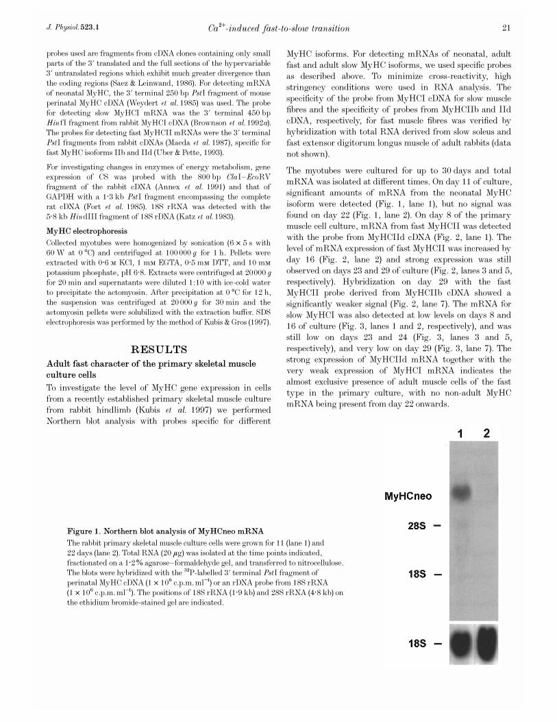
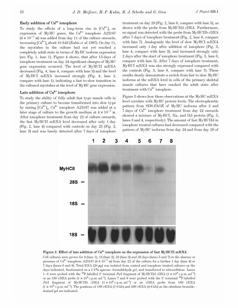
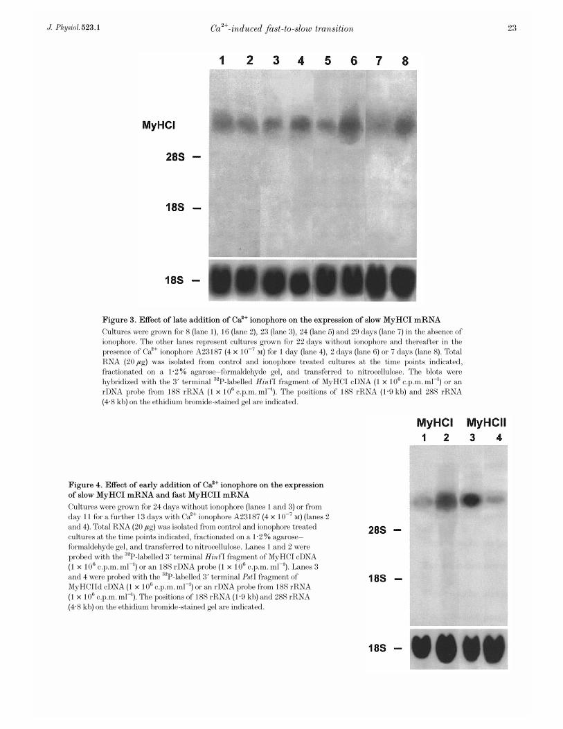
The myotubes were cultured for up to 30 days and totalmRNA was isolated at different times. On day 11 of culture,significant amounts of mRNA from the neonatal MyHCisoform were detected (Fig. 1, lane 1), but no signal wasfound on day 22 (Fig. 1, lane 2). On day 8 of the primarymuscle cell culture, mRNA from fast MyHCII was detectedwith the probe from MyHCIId cDNA (Fig. 2, lane 1). Thelevel of mRNA expression of fast MyHCII was increased byday 16 (Fig. 2, lane 2) and strong expression was stillobserved on days 23 and 29 of culture (Fig. 2, lanes 3 and 5,respectively). Hybridization on day 29 with the fastMyHCII probe derived from MyHCIIb cDNA showed asignificantly weaker signal (Fig. 2, lane 7). The mRNA forslow MyHCI was also detected at low levels on days 8 and16 of culture (Fig. 3, lanes 1 and 2, respectively), and wasstill low on days 23 and 24 (Fig. 3, lanes 3 and 5,respectively), and very low on day 29 (Fig. 3, lane 7). Thestrong expression of MyHCIId mRNA together with thevery weak expression of MyHCI mRNA indicates thealmost exclusive presence of adult muscle cells of the fasttype in the primary culture, with no non-adult MyHCmRNA being present from day 22 onwards.
Ca¥-induced fast-to-slow transitionJ. Physiol. 523.1 21
Figure 1. Northern blot analysis of MyHCneo mRNA
The rabbit primary skeletal muscle culture cells were grown for 11 (lane 1) and22 days (lane 2). Total RNA (20 ìg) was isolated at the time points indicated,fractionated on a 1·2% agarose—formaldehyde gel, and transferred to nitrocellulose.The blots were hybridized with the
32
P-labelled 3' terminal PstI fragment ofperinatal MyHC cDNA (1 ² 10É c.p.m. ml¢) or an rDNA probe from 18S rRNA(1 ² 10É c.p.m. ml¢). The positions of 18S rRNA (1·9 kb) and 28S rRNA (4·8 kb) onthe ethidium bromide-stained gel are indicated.
Early addition of Ca¥ ionophore
To study the effects of a long-term rise in [Ca¥]é onexpression of MyHC genes, the Ca¥ ionophore A23187(4 ² 10¦Ê Ò) was added from day 11 of the culture onwards,increasing [Ca¥]é about 10-fold (Kubis et al. 1997). On day 11,the myotubes in the culture had not yet reached acompletely adult state in terms of MyHC isoform expression(see Fig. 1, lane 1). Figure 4 shows, that after 13 days ofionophore treatment on day 24 significant changes of MyHCgene expression occurred. The level of MyHCII mRNAdecreased (Fig. 4, lane 4, compare with lane 3) and the levelof MyHCI mRNA increased strongly (Fig. 4, lane 2,compare with lane 1), indicating a fast-to-slow transition inthe cultured myotubes at the level of MyHC gene expression.
Late addition of Ca¥ ionophore
To study the ability of fully adult fast type muscle cells inthe primary culture to become transformed into slow typeby raising [Ca¥]é, Ca¥ ionophore A23187 was added at alater stage of culture to the growth medium at 4 ² 10¦Ê Ò.After ionophore treatment from day 22 of culture onwards,the fast MyHCII mRNA level decreased after only 1 day(Fig. 2, lane 4) compared with controls on day 23 (Fig. 2,lane 3) and was barely detected after 7 days of ionophore
treatment on day 29 (Fig. 2, lane 6, compare with lane 5), asshown with the probe from MyHCIId cDNA. Furthermore,no signal was detected with the probe from MyHCIIb cDNAafter 7 days of ionophore treatment (Fig. 2, lane 8, comparewith lane 7). Analogously the level of slow MyHCI mRNAincreased only 1 day after addition of ionophore (Fig. 3,lane 4, compare with lane 3), and increased strongly only2 days after the start of ionophore treatment (Fig. 3, lane 6,compare with lane 5). After 7 days of ionophore treatment,MyHCI mRNA was also strongly expressed compared withthe controls (Fig. 3, lane 8, compare with lane 7). Theseresults clearly demonstrate a switch from fast to slow MyHCisoforms at the mRNA level in cells of the primary skeletalmuscle cultures that have reached the adult state aftertreatment with Ca¥ ionophore.
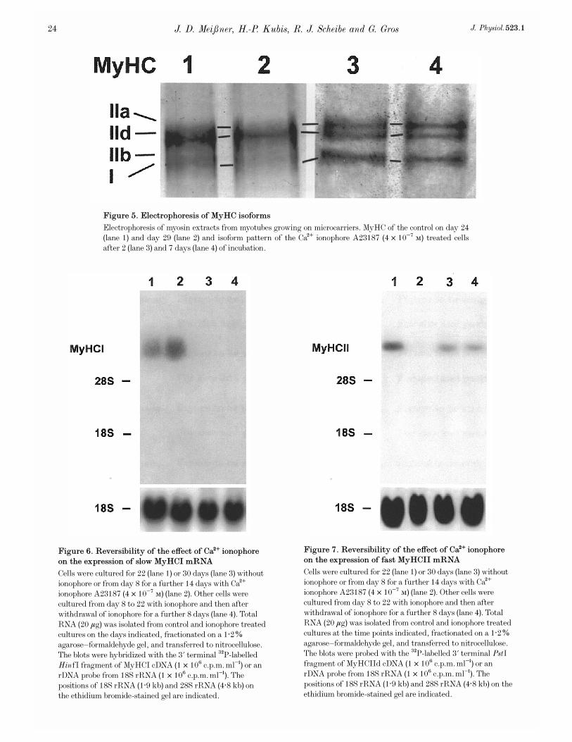
Figure 5 shows how these observations at the MyHC mRNAlevel correlate with MyHC protein levels. The electrophoreticpattern from SDS-PAGE of MyHC isoforms after 2 and7 days of Ca¥ ionophore treatment from day 22 onwardsshowed a mixture of MyHCI, IIa, and IId protein (Fig. 5,lanes 3 and 4, respectively). The amount of fast MyHCIId inionophore treated cultures had decreased compared with thepattern of MyHC isoforms from day 24 and from day 29 of
J. D. Mei³ner, H.-P. Kubis, R. J. Scheibe and G. Gros J. Physiol. 523.122
Figure 2. Effect of late addition of Ca¥ ionophore on the expression of fast MyHCII mRNA
Cell cultures were grown for 8 (lane 1), 16 (lane 2), 23 (lane 3) and 29 days (lanes 5 and 7) in the absence orpresence of Ca¥ ionophore A23187 (4 ² 10¦Ê Ò) from day 22 of the culture for a further 1 day (lane 4) or7 days (lanes 6 and 8). Total RNA (20 ìg) was isolated from control and ionophore treated cultures on thedays indicated, fractionated on a 1·2% agarose—formaldehyde gel, and transferred to nitrocellulose. Lanes1—6 were probed with the
32
P-labelled 3' terminal PstI fragment of MyHCIId cDNA (1 ² 10É c.p.m. ml¢)or an 18S rDNA probe (1 ² 10É c.p.m. ml¢). Lanes 7 and 8 were probed with the 3' terminal
32
P-labelledPstI fragment of MyHCIIb cDNA (1 ² 10É c.p.m. ml¢) or an rDNA probe from 18S rRNA(1 ² 10É c.p.m. ml¢). The positions of 18S rRNA (1·9 kb) and 28S rRNA (4·8 kb) on the ethidium bromide-stained gel are indicated.
Ca¥-induced fast-to-slow transitionJ. Physiol. 523.1 23
Figure 4. Effect of early addition of Ca¥ ionophore on the expression
of slow MyHCI mRNA and fast MyHCII mRNA
Cultures were grown for 24 days without ionophore (lanes 1 and 3) or fromday 11 for a further 13 days with Ca¥ ionophore A23187 (4 ² 10¦Ê Ò) (lanes 2and 4). Total RNA (20 ìg) was isolated from control and ionophore treatedcultures at the time points indicated, fractionated on a 1·2% agarose—formaldehyde gel, and transferred to nitrocellulose. Lanes 1 and 2 wereprobed with the
32
P-labelled 3' terminal HinfI fragment of MyHCI cDNA(1 ² 10É c.p.m. ml¢) or an 18S rDNA probe (1 ² 10É c.p.m. ml¢). Lanes 3and 4 were probed with the
32
P-labelled 3' terminal PstI fragment ofMyHCIId cDNA (1 ² 10É c.p.m. ml¢) or an rDNA probe from 18S rRNA(1 ² 10É c.p.m. ml¢). The positions of 18S rRNA (1·9 kb) and 28S rRNA(4·8 kb) on the ethidium bromide-stained gel are indicated.
Figure 3. Effect of late addition of Ca¥ ionophore on the expression of slow MyHCI mRNA
Cultures were grown for 8 (lane 1), 16 (lane 2), 23 (lane 3), 24 (lane 5) and 29 days (lane 7) in the absence ofionophore. The other lanes represent cultures grown for 22 days without ionophore and thereafter in thepresence of Ca¥ ionophore A23187 (4 ² 10¦Ê Ò) for 1 day (lane 4), 2 days (lane 6) or 7 days (lane 8). TotalRNA (20 ìg) was isolated from control and ionophore treated cultures at the time points indicated,fractionated on a 1·2% agarose—formaldehyde gel, and transferred to nitrocellulose. The blots werehybridized with the 3' terminal
32
P-labelled HinfI fragment of MyHCI cDNA (1 ² 10É c.p.m. ml¢) or anrDNA probe from 18S rRNA (1 ² 10É c.p.m. ml¢). The positions of 18S rRNA (1·9 kb) and 28S rRNA(4·8 kb) on the ethidium bromide-stained gel are indicated.
J. D. Mei³ner, H.-P. Kubis, R. J. Scheibe and G. Gros J. Physiol. 523.124
Figure 6. Reversibility of the effect of Ca¥ ionophore
on the expression of slow MyHCI mRNA
Cells were cultured for 22 (lane 1) or 30 days (lane 3) withoutionophore or from day 8 for a further 14 days with Ca¥ionophore A23187 (4 ² 10¦Ê Ò) (lane 2). Other cells werecultured from day 8 to 22 with ionophore and then afterwithdrawal of ionophore for a further 8 days (lane 4). TotalRNA (20 ìg) was isolated from control and ionophore treatedcultures on the days indicated, fractionated on a 1·2%agarose—formaldehyde gel, and transferred to nitrocellulose.The blots were hybridized with the 3' terminal
32
P-labelledHinfI fragment of MyHCI cDNA (1 ² 10É c.p.m. ml¢) or anrDNA probe from 18S rRNA (1 ² 10É c.p.m. ml¢). Thepositions of 18S rRNA (1·9 kb) and 28S rRNA (4·8 kb) onthe ethidium bromide-stained gel are indicated.
Figure 7. Reversibility of the effect of Ca¥ ionophore
on the expression of fast MyHCII mRNA
Cells were cultured for 22 (lane 1) or 30 days (lane 3) withoutionophore or from day 8 for a further 14 days with Ca¥ionophore A23187 (4 ² 10¦Ê Ò) (lane 2). Other cells werecultured from day 8 to 22 with ionophore and then afterwithdrawal of ionophore for a further 8 days (lane 4). TotalRNA (20 ìg) was isolated from control and ionophore treatedcultures at the time points indicated, fractionated on a 1·2%agarose—formaldehyde gel, and transferred to nitrocellulose.The blots were probed with the
32
P-labelled 3' terminal PstIfragment of MyHCIId cDNA (1 ² 10É c.p.m. ml¢) or anrDNA probe from 18S rRNA (1 ² 10É c.p.m. ml¢). Thepositions of 18S rRNA (1·9 kb) and 28S rRNA (4·8 kb) on theethidium bromide-stained gel are indicated.
Figure 5. Electrophoresis of MyHC isoforms
Electrophoresis of myosin extracts from myotubes growing on microcarriers. MyHC of the control on day 24(lane 1) and day 29 (lane 2) and isoform pattern of the Ca¥ ionophore A23187 (4 ² 10¦Ê Ò) treated cellsafter 2 (lane 3) and 7 days (lane 4) of incubation.
control cultures (Fig. 5, lanes 1 and 2, respectively).MyHCIId was the main MyHC isoform on days 24 and 29 inuntreated cultures. Minor amounts of slow MyHCI were stillpresent on day 24, but had nearly disappeared on day 29.In contrast, no decrease in MyHCI was observed and itsband was much stronger on day 29 with ionophore (Fig. 5,lane 4, compare with lane 2). In addition, significant amountsof MyHCIIa were present, when ionophore was added tothe medium. The appearance of MyHCIIa in the ionophoretreated cultures is in agreement with the fast-to-slowtransition sequence MyHCIId > IIa > I in rabbit muscles(Peuker et al. 1998).
Reversibility of Ca¥ ionophore effect
The reversibility of low frequency stimulation-induced fast-to-slow transition has been demonstrated in vivo at the levelof gene expression (Brownson et al. 1992a,b; Pette & Vrbov�a,1992). To study the reversibility of the Ca¥ ionophore effecton MyHC isoform gene expression in the present culture,cells were treated with ionophore starting on day 8. On day
22 of culture, after 14 days of ionophore treatment, MyHCImRNA was strongly expressed and the level of MyHCIImRNA was low (Figs 6 and 7, respectively, lanes 2). Incontrols on day 22, the reverse was true (Figs 6 and 7,respectively, lanes 1). When, subsequently, the ionophoretreated cells were kept after day 22 for 8 days withoutionophore, the level of mRNA expression of fast MyHCIIincreased and the mRNA for slow MyHCI was no longerdetectable (Figs 7 and 6, respectively, lanes 4). Similarly, thecontrols on day 30 showed no MyHCI but only MyHCIImRNA expression (Figs 6 and 7, respectively, lanes 3).These results clearly demonstrate the reversibility of theionophore effect on the expression of MyHC isoform mRNAsin the myotubes of the primary culture.
Effects of Ca¥ ionophore treatment on metabolic
marker enzymes
It is well documented that a fast-to-slow transition leads tosignificant changes in the enzyme activities of energymetabolism (Pette & Vrbov�a, 1992; Mayne et al. 1996). To
Ca¥-induced fast-to-slow transitionJ. Physiol. 523.1 25
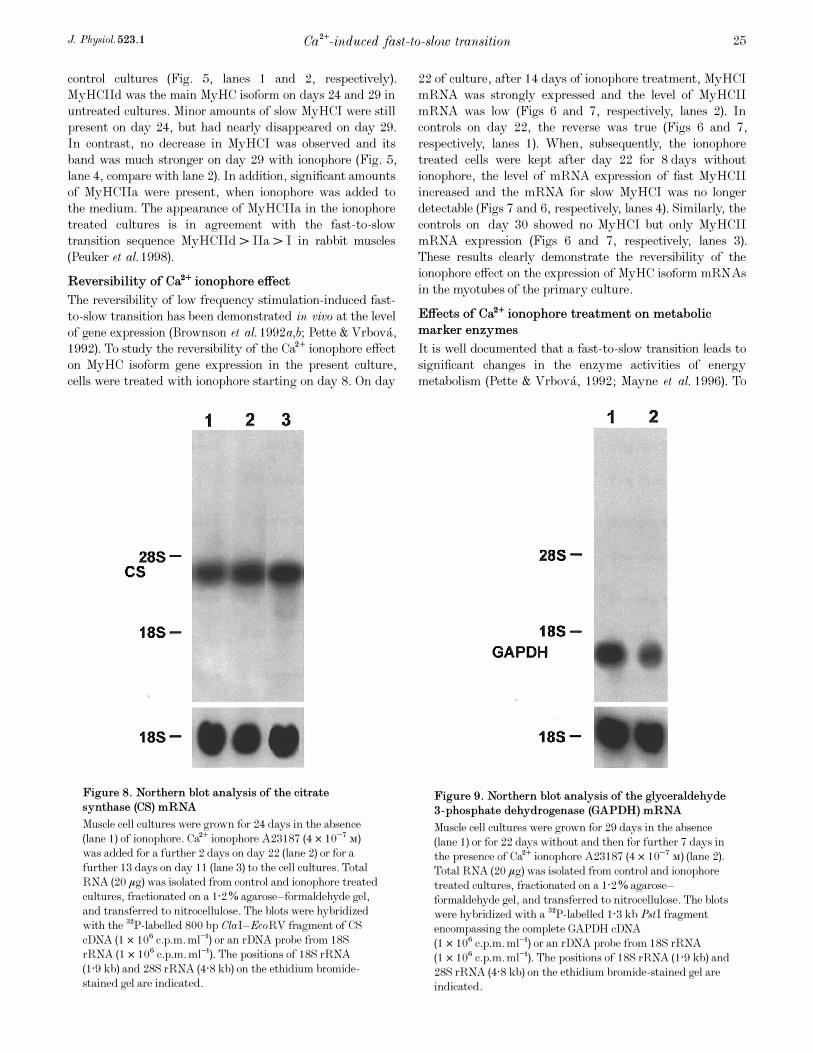
Figure 8. Northern blot analysis of the citrate
synthase (CS) mRNA
Muscle cell cultures were grown for 24 days in the absence(lane 1) of ionophore. Ca¥ ionophore A23187 (4 ² 10¦Ê Ò)was added for a further 2 days on day 22 (lane 2) or for afurther 13 days on day 11 (lane 3) to the cell cultures. TotalRNA (20 ìg) was isolated from control and ionophore treatedcultures, fractionated on a 1·2% agarose—formaldehyde gel,and transferred to nitrocellulose. The blots were hybridizedwith the
32
P-labelled 800 bp ClaI—EcoRV fragment of CScDNA (1 ² 10É c.p.m. ml¢) or an rDNA probe from 18SrRNA (1 ² 10É c.p.m. ml¢). The positions of 18S rRNA(1·9 kb) and 28S rRNA (4·8 kb) on the ethidium bromide-stained gel are indicated.
Figure 9. Northern blot analysis of the glyceraldehyde
3-phosphate dehydrogenase (GAPDH) mRNA
Muscle cell cultures were grown for 29 days in the absence(lane 1) or for 22 days without and then for further 7 days inthe presence of Ca¥ ionophore A23187 (4 ² 10¦Ê Ò) (lane 2).Total RNA (20 ìg) was isolated from control and ionophoretreated cultures, fractionated on a 1·2% agarose—formaldehyde gel, and transferred to nitrocellulose. The blotswere hybridized with a
32
P-labelled 1·3 kb PstI fragmentencompassing the complete GAPDH cDNA(1 ² 10É c.p.m. ml¢) or an rDNA probe from 18S rRNA(1 ² 10É c.p.m. ml¢). The positions of 18S rRNA (1·9 kb) and28S rRNA (4·8 kb) on the ethidium bromide-stained gel areindicated.
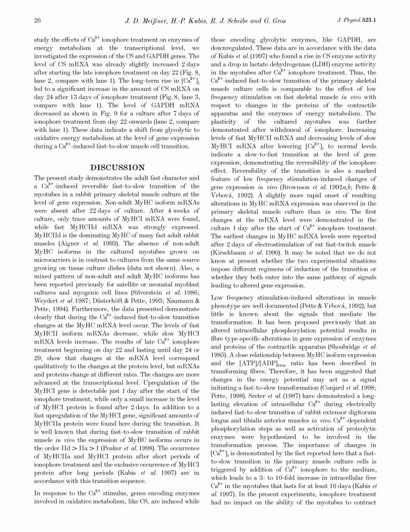
study the effects of Ca¥ ionophore treatment on enzymes ofenergy metabolism at the transcriptional level, weinvestigated the expression of the CS and GAPDH genes. Thelevel of CS mRNA was already slightly increased 2 daysafter starting the late ionophore treatment on day 22 (Fig. 8,lane 2, compare with lane 1). The long-term rise in [Ca¥]éled to a significant increase in the amount of CS mRNA onday 24 after 13 days of ionophore treatment (Fig. 8, lane 3,compare with lane 1). The level of GAPDH mRNAdecreased as shown in Fig. 9 for a culture after 7 days ofionophore treatment from day 22 onwards (lane 2, comparewith lane 1). These data indicate a shift from glycolytic tooxidative energy metabolism at the level of gene expressionduring a Ca¥-induced fast-to-slow muscle cell transition.
DISCUSSION
The present study demonstrates the adult fast character anda Ca¥-induced reversible fast-to-slow transition of themyotubes in a rabbit primary skeletal muscle culture at thelevel of gene expression. Non-adult MyHC isoform mRNAswere absent after 22 days of culture. After 4 weeks ofculture, only trace amounts of MyHCI mRNA were found,while fast MyHCIId mRNA was strongly expressed.MyHCIId is the dominating MyHC of many fast adult rabbitmuscles (Aigner et al. 1993). The absence of non-adultMyHC isoforms in the cultured myotubes grown onmicrocarriers is in contrast to cultures from the same sourcegrowing on tissue culture dishes (data not shown). Also, amixed pattern of non-adult and adult MyHC isoforms hasbeen reported previously for satellite or neonatal myoblastcultures and myogenic cell lines (Silverstein et al. 1986;Weydert et al. 1987; D�usterh�oft & Pette, 1993; Naumann &Pette, 1994). Furthermore, the data presented demonstrateclearly that during the Ca¥-induced fast-to-slow transitionchanges at the MyHC mRNA level occur. The levels of fastMyHCII isoform mRNAs decrease, while slow MyHCImRNA levels increase. The results of late Ca¥ ionophoretreatment beginning on day 22 and lasting until day 24 or29, show that changes at the mRNA level correspondqualitatively to the changes at the protein level, but mRNAsand proteins change at different rates. The changes are moreadvanced at the transcriptional level. Upregulation of theMyHCI gene is detectable just 1 day after the start of theionophore treatment, while only a small increase in the levelof MyHCI protein is found after 2 days. In addition to afast upregulation of the MyHCI gene, significant amounts ofMyHCIIa protein were found here during the transition. Itis well known that during fast-to-slow transition of rabbitmuscle in vivo the expression of MyHC isoforms occurs inthe order IId > IIa > I (Peuker et al. 1998). The occurrenceof MyHCIIa and MyHCI protein after short periods ofionophore treatment and the exclusive occurrence of MyHCIprotein after long periods (Kubis et al. 1997) are inaccordance with this transition sequence.
In response to the Ca¥ stimulus, genes encoding enzymesinvolved in oxidative metabolism, like CS, are induced while
those encoding glycolytic enzymes, like GAPDH, aredownregulated. These data are in accordance with the dataof Kubis et al. (1997) who found a rise in CS enzyme activityand a drop in lactate dehydrogenase (LDH) enzyme activityin the myotubes after Ca¥ ionophore treatment. Thus, theCa¥-induced fast-to-slow transition of the primary skeletalmuscle culture cells is comparable to the effect of lowfrequency stimulation on fast skeletal muscle in vivo withrespect to changes in the proteins of the contractileapparatus and the enzymes of energy metabolism. Theplasticity of the cultured myotubes was furtherdemonstrated after withdrawal of ionophore. Increasinglevels of fast MyHCII mRNA and decreasing levels of slowMyHCI mRNA after lowering [Ca¥]é to normal levelsindicate a slow-to-fast transition at the level of geneexpression, demonstrating the reversibility of the ionophoreeffect. Reversibility of the transition is also a markedfeature of low frequency stimulation-induced changes ofgene expression in vivo (Brownson et al. 1992a,b; Pette &Vrbov�a, 1992). A slightly more rapid onset of resultingalterations in MyHC mRNA expression was observed in theprimary skeletal muscle culture than in vivo. The firstchanges at the mRNA level were demonstrated in theculture 1 day after the start of Ca¥ ionophore treatment.The earliest changes in MyHC mRNA levels were reportedafter 2 days of electrostimulation of rat fast-twitch muscle(Kirschbaum et al. 1990). It may be noted that we do notknow at present whether the two experimental situationsimpose different regimens of induction of the transition orwhether they both enter into the same pathway of signalsleading to altered gene expression.
Low frequency stimulation-induced alterations in musclephenotype are well documented (Pette & Vrbov�a, 1992), butlittle is known about the signals that mediate thetransformation. It has been proposed previously that analtered intracellular phosphorylation potential results infibre type-specific alterations in gene expression of enzymesand proteins of the contractile apparatus (Shoubridge et al.1985). A close relationship between MyHC isoform expressionand the [ATP]Ï[ADP]free ratio has been described intransforming fibres. Therefore, it has been suggested thatchanges in the energy potential may act as a signalinitiating a fast-to-slow transformation (Conjard et al. 1998;Pette, 1998). Sreter et al. (1987) have demonstrated a long-lasting elevation of intracellular Ca¥ during electricallyinduced fast-to-slow transition of rabbit extensor digitorumlongus and tibialis anterior muscles in vivo. Ca¥-dependentphosphorylation steps as well as activation of proteolyticenzymes were hypothesized to be involved in thetransformation process. The importance of changes in[Ca¥]é is demonstrated by the fact reported here that a fast-to-slow transition in the primary muscle culture cells istriggered by addition of Ca¥ ionophore to the medium,which leads to a 3- to 10-fold increase in intracellular freeCa¥ in the myotubes that lasts for at least 16 days (Kubis etal. 1997). In the present experiments, ionophore treatmenthad no impact on the ability of the myotubes to contract
J. D. Mei³ner, H.-P. Kubis, R. J. Scheibe and G. Gros J. Physiol. 523.126
vigorously and spontaneously, indicating integrity of thecell membrane. In addition, an intact morphology of themyotubes could be demonstrated by scanning andtransmission electron microscopy (B. Decker, H.-P. Kubis &G. Gros, unpublished observation). Therefore, we concludethat the fast-to-slow transition of the myotubes as observedhere was induced by changes in [Ca¥]é rather than bymuscle damage, which has been linked to stretch-inducedfibre growth and transformation (McKoy et al. 1999).
Recently, Chin et al. (1998) demonstrated the involvement ofa calcineurin (Ca¥-regulated serineÏthreonine phosphatase)-dependent signalling pathway in controlling fibre type-specific gene expression. Activation of calcineurin selectivelyupregulates slow fibre-specific genes and slow fibre-specifictranscriptional activation appears to be mediated bymembers of the NF-AT (nuclear factor of activatedthymocytes) and MEF2 (myocyte enhancer factor 2)transcription factor families. The importance of Ca¥ forphenotypic adaptations of skeletal muscle was furthershown by the finding of a Ca¥-sensitive protein kinase C-dependent pathway involved in cytochrome c expression(Freyssenet et al. 1999).
The induction of immediate-early genes c-fos, c-jun andegr_1 is one of the earliest responses of rabbit tibialis anteriorsubjected to low frequency stimulation (Michel et al. 1994).The same regimen, or passive stretch, or a combination ofboth, led also to induction of c-jun and c-fos in rabbitlatissimus dorsi and extensor digitorum longus (Osbaldestonet al. 1993, 1995; Dawes et al. 1996). Interestingly, passivestretch, leading to significant growth of muscle, as well asfibre type transformation, was accompanied by increases ininsulin-like growth factor I (IGF-I) protein and mRNA. Incontrast, continuous electrical stimulation-induced fibretransition did not induce muscle growth and did not lead toan increase in IGF-I mRNA (Goldspink et al. 1995; Yang etal. 1997).
A consistent hypothesis for a signalling pathway underlyingfibre type transition has not yet emerged from the availabledata. There might be differences between the differenttransition-inducing regimens. For investigation of thecomplete signalling pathways underlying mechanisms ofmuscle plasticity, electrical stimulation and passive stretchso far have been of restricted value due to the limitationsimposed by the in vivo situation. The primary skeletalmuscle culture offers the possibility to study signallingpathways in vitro in detail and may help to gain moreinsight into the mechanisms, extracellular factors, and intra-cellular signal cascades that lead to fast-to-slow transition.
Aigner, S., Gohlsch, B., H�am�al�ainen, N., Staron, R. S., Uber,A., Wehrle, U. & Pette, D. (1993). Fast myosin heavy chaindiversity in skeletal muscles of the rabbit: heavy chain IId, not IIbpredominates. European Journal of Biochemistry 211, 367—372.
Annex, B. H., Kraus, W. E., Dohm, G. L. & Williams, R. S. (1991).Mitochondrial biogenesis in striated muscles: rapid induction ofcitrate synthase mRNA by nerve stimulation. American Journal ofPhysiology 260, C266—270.
Brownson, C., Isenberg, H., Brown, W., Salmons, S. & Edwards,Y. (1988). Changes in skeletal muscle gene transcription induced bychronic stimulation. Muscle and Nerve 11, 1183—1189.
Brownson, C., Little, P., Jarvis, J. C. & Salmons, S. (1992a).Reciprocal changes in myosin isoform mRNAs of rabbit skeletalmuscle in response to the initiation and cessation of chronicelectrical stimulation. Muscle and Nerve 15, 694—700.
Brownson, C., Little, P., Mayne, C., Jarvis, J. C. & Salmons, S.(1992b). Reciprocal changes in myosin isoform expression in rabbitfast skeletal muscle resulting from the application and removal ofchronic electrical stimulation. Symposium of the Society ofExperimental Biology 46, 301—310.
Buckingham, M. E. (1985). Actin and myosin multigene families:their expression during the formation of skeletal muscle. Essays inBiochemistry 20, 77—109.
Buller, A. J., Eccles, J. C. & Eccles, R. M. (1960). Differentiationof fast and slow muscles in the cat hind limb. Journal of Physiology150, 399—416.
Chin, E. R., Olson, E. N., Richardson, J. A., Yang, Q.,Humphries, C., Shelton, J. M., Wu, H., Zhu, W., Bassel-Duby,R. & Williams, R. S. (1998). A calcineurin-dependenttranscriptional pathway controls skeletal muscle fiber type. Genesand Development 12, 2499—2509.
Chirgwin, J. M., Przybyla, A. E., MacDonald, R. J. & Rutter,W. J (1979). Isolation of biologically active ribonucleic acid fromsources enriched in ribonuclease. Biochemistry 18, 5294—5299.
Chomczynski, P. & Sacchi, N. (1987). Single-step method of RNAisolation by acid guanidinium thiocyanate-phenol-chloroformextraction. Analytical Biochemistry 162, 156—159.
Conjard, A., Peuker, H. & Pette, D. (1998). Energy state andmyosin heavy chain isoforms in single fibres of normal andtransforming rabbit muscles. Pfl�ugers Archiv 436, 962—969.
Dawes, N. J., Cox, V. M., Park, K. S., Nga, H. & Goldspink, D. F.(1996). The induction of c-fos and c-jun in the stretched latissimusdorsi muscle of the rabbit: responses to duration, degree and re-application of the stretch stimulus. Experimental Physiology 81,329—339.
D�usterh�oft, S. & Pette, D. (1993). Satellite cells from slow ratmuscle express slow myosin under appropriate culture conditions.Differentiation 53, 25—33.
Feinberg, A. P. & Vogelstein, B. (1983). A technique forradiolabeling DNA restriction endonuclease fragments to highspecific activity. Analytical Biochemistry 132, 6—13.
Fort, P., Marty, L., Piechaczyk, M., El Sabrouty, S., Dani, C.,Jaenteur, P. & Blanchard, J. M. (1985). Various rat adult tissuesexpress only one major mRNA species from the glyceraldehyde-3-phosphate dehydrogenase multigenic family. Nucleic Acids Research13, 1431—1442.
Freyssenet, D., Di Carlo, M. & Hood, D. A. (1999). Calcium-dependent regulation of cytyochrome c gene expression in skeletalmuscle cells. Identification of a protein kinase C-dependentpathway. Journal of Biological Chemistry 274, 9305—9311.
Goldspink, D. F., Cox, V. M., Smith, S. K., Eaves, L. A.,Osbaldeston, N. J., Lee, D. M. & Mantle, D. (1995). Musclegrowth in response to mechanical stimuli. American Journal ofPhysiology 268, E288—297.
Ca¥-induced fast-to-slow transitionJ. Physiol. 523.1 27
Goldspink, G., Scutt, A., Loughna, P. T., Wells, D. J., Jaenicke,T. & Gerlach, G. F. (1992). Gene expression in skeletal muscle inresponse to stretch and force generation. American Journal ofPhysiology 262, R356—363.
Henriksson, J., Salmons, S., Chi, M. M.-Y., Hintz, C. S. & Lowry,O. H. (1988). Chronic stimulation of mammalian muscle: changes inmetabolite concentrations in individual fibers. American Journal ofPhysiology 255, C543—551.
Katz, R. A., Erlanger, B. F. & Guntaka, R. V. (1983). Evidencefor extensive methylation of ribosomal RNA genes in rat XC cellline. Biochimica et Biophysica Acta 739, 258—264.
Kirschbaum, B. J., Schneider, S., Izumo, S., Mahdavi, V., Nadal-Ginard, B. & Pette, D. (1990). Rapid and reversible changes inmyosin heavy chain expression in response to increased neuro-muscular activity of rat fast-twitch muscle. FEBS Letters 268,75—78.
Kubis, H.-P. & Gros, G. (1997). A rapid electrophoretic method forseparating rabbit skeletal muscle myosin heavy chains at highresolution. Electrophoresis 18, 64—66.
Kubis, H.-P., Haller, E.-A., Wetzel, P. & Gros, G. (1997). Adultfast myosin pattern and Ca¥-induced slow myosin pattern inprimary skeletal muscle cell culture. Proceedings of the NationalAcademy of Sciences of the USA 94, 4205—4210.
McKoy, G., Ashley, W., Mander, J., Yang, S. Y., Williams, N.,Russell, B. & Goldspink, G. (1999). Expression of insulin growthfactor-I splice variants and structural genes in rabbit skeletal muscleinduced by stretch and stimulation Journal of Physiology 516,583—592.
Maeda, K., Sczakiel, G. & Wittinghofer, A. (1987).Characterisation of cDNA coding for the complete light meromyosinportion of a rabbit fast skeletal muscle myosin heavy chain.European Journal of Biochemistry 167, 97—102.
Mayne, C. N., Sutherland, H., Jarvis, J. C., Gilroy, S. J., Craven,A. J. & Salmons, S. (1996). Induction of a fast-oxidative phenotypeby chronic muscle stimulation: histochemical and metabolic studies.American Journal of Physiology 270, C313—320.
Michel, J. B., Ordway, G. A., Richardson, J. A. & Williams, R. S.(1994). Biphasic induction of immediate early gene expressionaccompanies activity-dependent angiogenesis and myofiberremodeling of rabbit skeletal muscle. Journal of ClinicalInvestigation 94, 277—285.
Moore, L. A., Tidyman, W. E., Arrizubieta, M. J. & Bandman, E.(1993). The evolutionary relationship of avian and mammalianmyosin heavy-chain genes. Journal of Molecular Evolution 36,21—30.
Naumann, K. & Pette, D. (1994). Effects of chronic stimulation withdifferent impulse patterns on the expression of myosin isoforms inrat myotube cultures. Differentiation 55, 203—211.
Osbaldeston, N. J., Lee, D. M., Cox, V. M., Eaves, L., Morrison,J. F. J., Hesketh, J. & Goldspink, D. F. (1993). The temporalexpression of cellular oncogenes in mechanically stimulated muscle.Biochemical Society Transactions 21, 367S.
Osbaldeston, N. J., Lee, D. M., Cox, V. M., Hesketh, J. E.,Morrison, J. F. J., Blair, G. E. & Goldspink, D. F. (1995). Thetemporal and cellular expression of c-fos and c-jun in mechanicallystimulated rabbit latissimus dorsi muscle. Biochemical Journal 308,465—471.
Pette, D. (1998). Training effects on the contractile apparatus. ActaPhysiologica Scandinavica 162, 367—376.
Pette, D. & Vbrov�a, G. (1985). Neural control of phenotypicexpression in mammalian muscle fibers. Muscle and Nerve 8,676—689.
Pette, D. & Vrbov �a, G. (1992). Adaption of mammalian skeletalmuscle fibers to chronic electrical stimulation. Reviews of Physiology,Biochemistry and Pharmacology 120, 115—208.
Peuker, H., Conjard, A. & Pette, D. (1998). Alpha-cardiac-likemyosin heavy chain as an intermediate between MyHCIIa andMHCI beta in transforming rabbit muscle. American Journal ofPhysiology 274, C595—602.
Russell, B. & Dix, D. J. (1992). Mechanisms for intracellulardistribution of mRNA: in situ hybridization studies in muscle.American Journal of Physiology 262, C1—8.
Saez, L. & Leinwand, L. A. (1986). Characterisation of diverse formsof myosin heavy chain expressed in adult human skeletal muscle.Nucleic Acids Research 14, 2951—2969.
Salmons, S. & Henriksson, J. (1981). The adaptive response ofskeletal muscle to increased use. Muscle and Nerve 4, 94—105.
Schiaffino, S. & Salviati, G. (1998). Molecular diversity ofmyofibrillar proteins: isoform analysis at the protein and mRNAlevel. Methods in Cell Biology 52, 349—369.
Shoubridge, E. A., Challiss, R. A. J., Hayes, D. J. & Radda, G. K.(1985). Biochemical adaption in the skeletal muscle of rats depletedof creatine with the substrate analogue beta-guanidinopropionicacid. Biochemical Journal 232, 125—131.
Silverstein, L., Webster, S. G., Travis, M. & Blau, H. M. (1986).Developmental progression of myosin gene expression in culturedmuscle cells. Cell 46, 1075—1081.
Sreter, F. A., Lopez, J. R., Alamo, L., Mabuchi, K. & Gergely, J.(1987). Changes in intracellular ionized Ca concentration associatedwith muscle fiber type transformation. American Journal ofPhysiology 253, C296—300.
Uber, A. & Pette, D. (1993). PCR-based assignment of two myosinheavy chain cDNA clones to biochemically and histochemicallydefined single type IIB and IID fibers of rabbit muscle. FEBSLetters 331, 193—197.
Weydert, A., Barton, P., Harris, A. J., Pinset, C. & Buckingham,M. E. (1987). Developmental pattern of mouse skeletal myosinheavy chain gene transcripts in vivo and in vitro. Cell 49, 121—129.
Weydert, A., Daubas, P., Lazarides, I., Barton, P., Garner, I.,Leader, D. P., Bonhomme, F., Catalan, J., Simon, D., Guenet,J. L., Gros, F. & Buckingham, M. E. (1985). Genes for skeletalmuscle myosin heavy chains are clustered and are not located on thesame mouse chromosome as a cardiac myosin heavy chain gene.Proceedings of the National Academy of Sciences of the USA 82,7183—7187.
Yang, S., Alnaqeeb, M., Simpson, H. & Goldspink, G. (1997).Changes in muscle fiber type, muscle mass and IGF-I geneexpression in rabbit skeletal muscle subjected to stretch. Journal ofAnatomy 190, 613—622.
Acknowledgements
We are grateful to Drs C. Brownson, M. Buckingham, R. Guntaka,P. K. Umeda, R. S. Williams, and A. Wittinghofer for their generousgift of plasmids. We thank E.-A. Haller for excellent assistance incell culture and N. Freiberg for technical assistance. We wish tothank Drs W. H. M�uller and A. R�uhlmann for helpful discussions.
Corresponding author
J. D. Mei³ner: Zentrum Physiologie, Medizinische HochschuleHannover, D_30623 Hannover, Germany.
Email: meissner. [email protected]
Author’s email address
G. Gros: [email protected]
J. D. Mei³ner, H.-P. Kubis, R. J. Scheibe and G. Gros J. Physiol. 523.128
![Near-Membrane [Ca2+] Transients Resolved Using the Ca2+ Indicator FFP18](https://static.fdokumen.com/doc/165x107/631286873ed465f0570a4533/near-membrane-ca2-transients-resolved-using-the-ca2-indicator-ffp18.jpg)