Reversal of Muscle Atrophy by Zhimu-Huangbai Herb-Pair via Akt/mTOR/FoxO3 Signal Pathway in...
10
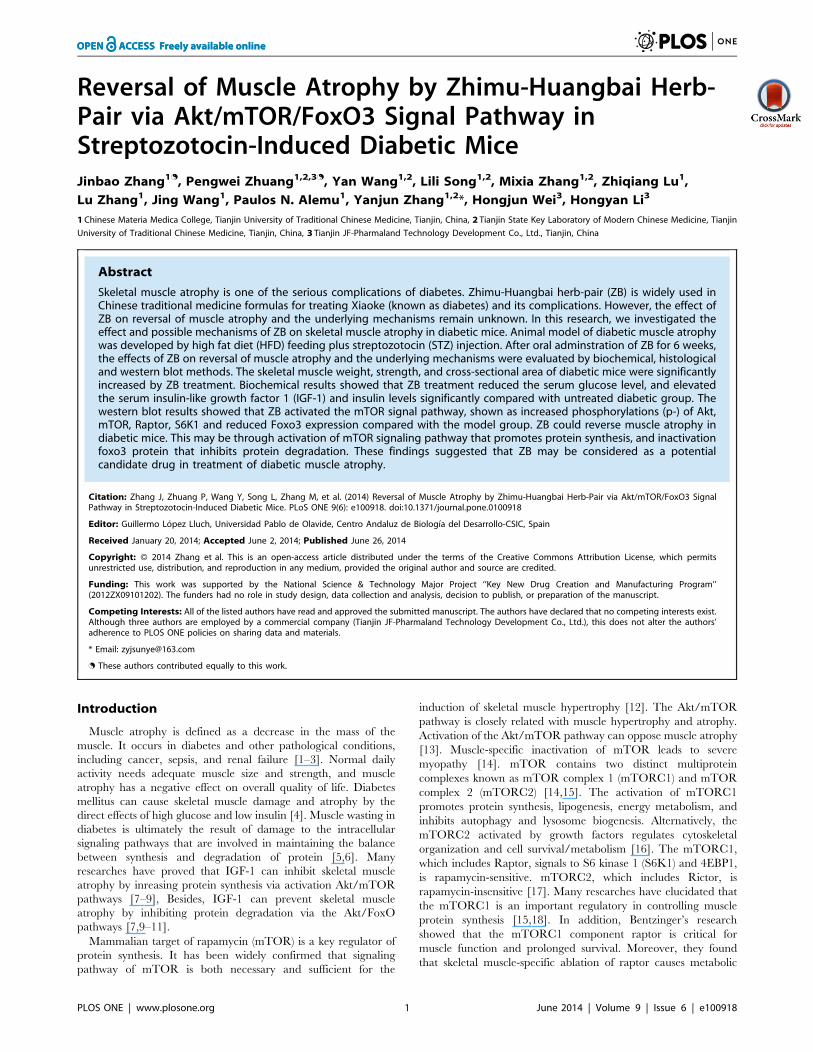
Reversal of Muscle Atrophy by Zhimu-Huangbai Herb- Pair via Akt/mTOR/FoxO3 Signal Pathway in Streptozotocin-Induced Diabetic Mice Jinbao Zhang 1. , Pengwei Zhuang 1,2,3. , Yan Wang 1,2 , Lili Song 1,2 , Mixia Zhang 1,2 , Zhiqiang Lu 1 , Lu Zhang 1 , Jing Wang 1 , Paulos N. Alemu 1 , Yanjun Zhang 1,2 *, Hongjun Wei 3 , Hongyan Li 3 1 Chinese Materia Medica College, Tianjin University of Traditional Chinese Medicine, Tianjin, China, 2 Tianjin State Key Laboratory of Modern Chinese Medicine, Tianjin University of Traditional Chinese Medicine, Tianjin, China, 3 Tianjin JF-Pharmaland Technology Development Co., Ltd., Tianjin, China Abstract Skeletal muscle atrophy is one of the serious complications of diabetes. Zhimu-Huangbai herb-pair (ZB) is widely used in Chinese traditional medicine formulas for treating Xiaoke (known as diabetes) and its complications. However, the effect of ZB on reversal of muscle atrophy and the underlying mechanisms remain unknown. In this research, we investigated the effect and possible mechanisms of ZB on skeletal muscle atrophy in diabetic mice. Animal model of diabetic muscle atrophy was developed by high fat diet (HFD) feeding plus streptozotocin (STZ) injection. After oral adminstration of ZB for 6 weeks, the effects of ZB on reversal of muscle atrophy and the underlying mechanisms were evaluated by biochemical, histological and western blot methods. The skeletal muscle weight, strength, and cross-sectional area of diabetic mice were significantly increased by ZB treatment. Biochemical results showed that ZB treatment reduced the serum glucose level, and elevated the serum insulin-like growth factor 1 (IGF-1) and insulin levels significantly compared with untreated diabetic group. The western blot results showed that ZB activated the mTOR signal pathway, shown as increased phosphorylations (p-) of Akt, mTOR, Raptor, S6K1 and reduced Foxo3 expression compared with the model group. ZB could reverse muscle atrophy in diabetic mice. This may be through activation of mTOR signaling pathway that promotes protein synthesis, and inactivation foxo3 protein that inhibits protein degradation. These findings suggested that ZB may be considered as a potential candidate drug in treatment of diabetic muscle atrophy. Citation: Zhang J, Zhuang P, Wang Y, Song L, Zhang M, et al. (2014) Reversal of Muscle Atrophy by Zhimu-Huangbai Herb-Pair via Akt/mTOR/FoxO3 Signal Pathway in Streptozotocin-Induced Diabetic Mice. PLoS ONE 9(6): e100918. doi:10.1371/journal.pone.0100918 Editor: Guillermo Lo ´ pez Lluch, Universidad Pablo de Olavide, Centro Andaluz de Biologı ´a del Desarrollo-CSIC, Spain Received January 20, 2014; Accepted June 2, 2014; Published June 26, 2014 Copyright: ß 2014 Zhang et al. This is an open-access article distributed under the terms of the Creative Commons Attribution License, which permits unrestricted use, distribution, and reproduction in any medium, provided the original author and source are credited. Funding: This work was supported by the National Science & Technology Major Project ‘‘Key New Drug Creation and Manufacturing Program’’ (2012ZX09101202). The funders had no role in study design, data collection and analysis, decision to publish, or preparation of the manuscript. Competing Interests: All of the listed authors have read and approved the submitted manuscript. The authors have declared that no competing interests exist. Although three authors are employed by a commercial company (Tianjin JF-Pharmaland Technology Development Co., Ltd.), this does not alter the authors’ adherence to PLOS ONE policies on sharing data and materials. * Email: [email protected] . These authors contributed equally to this work. Introduction Muscle atrophy is defined as a decrease in the mass of the muscle. It occurs in diabetes and other pathological conditions, including cancer, sepsis, and renal failure [1–3]. Normal daily activity needs adequate muscle size and strength, and muscle atrophy has a negative effect on overall quality of life. Diabetes mellitus can cause skeletal muscle damage and atrophy by the direct effects of high glucose and low insulin [4]. Muscle wasting in diabetes is ultimately the result of damage to the intracellular signaling pathways that are involved in maintaining the balance between synthesis and degradation of protein [5,6]. Many researches have proved that IGF-1 can inhibit skeletal muscle atrophy by inreasing protein synthesis via activation Akt/mTOR pathways [7–9], Besides, IGF-1 can prevent skeletal muscle atrophy by inhibiting protein degradation via the Akt/FoxO pathways [7,9–11]. Mammalian target of rapamycin (mTOR) is a key regulator of protein synthesis. It has been widely confirmed that signaling pathway of mTOR is both necessary and sufficient for the induction of skeletal muscle hypertrophy [12]. The Akt/mTOR pathway is closely related with muscle hypertrophy and atrophy. Activation of the Akt/mTOR pathway can oppose muscle atrophy [13]. Muscle-specific inactivation of mTOR leads to severe myopathy [14]. mTOR contains two distinct multiprotein complexes known as mTOR complex 1 (mTORC1) and mTOR complex 2 (mTORC2) [14,15]. The activation of mTORC1 promotes protein synthesis, lipogenesis, energy metabolism, and inhibits autophagy and lysosome biogenesis. Alternatively, the mTORC2 activated by growth factors regulates cytoskeletal organization and cell survival/metabolism [16]. The mTORC1, which includes Raptor, signals to S6 kinase 1 (S6K1) and 4EBP1, is rapamycin-sensitive. mTORC2, which includes Rictor, is rapamycin-insensitive [17]. Many researches have elucidated that the mTORC1 is an important regulatory in controlling muscle protein synthesis [15,18]. In addition, Bentzinger’s research showed that the mTORC1 component raptor is critical for muscle function and prolonged survival. Moreover, they found that skeletal muscle-specific ablation of raptor causes metabolic PLOS ONE | www.plosone.org 1 June 2014 | Volume 9 | Issue 6 | e100918
-
Upload
independent -
Category
Documents
-
view
2 -
download
0
Transcript of Reversal of Muscle Atrophy by Zhimu-Huangbai Herb-Pair via Akt/mTOR/FoxO3 Signal Pathway in...
Reversal of Muscle Atrophy by Zhimu-Huangbai Herb-Pair via Akt/mTOR/FoxO3 Signal Pathway inStreptozotocin-Induced Diabetic MiceJinbao Zhang1., Pengwei Zhuang1,2,3., Yan Wang1,2, Lili Song1,2, Mixia Zhang1,2, Zhiqiang Lu1,
Lu Zhang1, Jing Wang1, Paulos N. Alemu1, Yanjun Zhang1,2*, Hongjun Wei3, Hongyan Li3
1Chinese Materia Medica College, Tianjin University of Traditional Chinese Medicine, Tianjin, China, 2 Tianjin State Key Laboratory of Modern Chinese Medicine, Tianjin
University of Traditional Chinese Medicine, Tianjin, China, 3 Tianjin JF-Pharmaland Technology Development Co., Ltd., Tianjin, China
Abstract
Skeletal muscle atrophy is one of the serious complications of diabetes. Zhimu-Huangbai herb-pair (ZB) is widely used inChinese traditional medicine formulas for treating Xiaoke (known as diabetes) and its complications. However, the effect ofZB on reversal of muscle atrophy and the underlying mechanisms remain unknown. In this research, we investigated theeffect and possible mechanisms of ZB on skeletal muscle atrophy in diabetic mice. Animal model of diabetic muscle atrophywas developed by high fat diet (HFD) feeding plus streptozotocin (STZ) injection. After oral adminstration of ZB for 6 weeks,the effects of ZB on reversal of muscle atrophy and the underlying mechanisms were evaluated by biochemical, histologicaland western blot methods. The skeletal muscle weight, strength, and cross-sectional area of diabetic mice were significantlyincreased by ZB treatment. Biochemical results showed that ZB treatment reduced the serum glucose level, and elevatedthe serum insulin-like growth factor 1 (IGF-1) and insulin levels significantly compared with untreated diabetic group. Thewestern blot results showed that ZB activated the mTOR signal pathway, shown as increased phosphorylations (p-) of Akt,mTOR, Raptor, S6K1 and reduced Foxo3 expression compared with the model group. ZB could reverse muscle atrophy indiabetic mice. This may be through activation of mTOR signaling pathway that promotes protein synthesis, and inactivationfoxo3 protein that inhibits protein degradation. These findings suggested that ZB may be considered as a potentialcandidate drug in treatment of diabetic muscle atrophy.
Citation: Zhang J, Zhuang P, Wang Y, Song L, Zhang M, et al. (2014) Reversal of Muscle Atrophy by Zhimu-Huangbai Herb-Pair via Akt/mTOR/FoxO3 SignalPathway in Streptozotocin-Induced Diabetic Mice. PLoS ONE 9(6): e100918. doi:10.1371/journal.pone.0100918
Editor: Guillermo Lopez Lluch, Universidad Pablo de Olavide, Centro Andaluz de Biologıa del Desarrollo-CSIC, Spain
Received January 20, 2014; Accepted June 2, 2014; Published June 26, 2014
Copyright: � 2014 Zhang et al. This is an open-access article distributed under the terms of the Creative Commons Attribution License, which permitsunrestricted use, distribution, and reproduction in any medium, provided the original author and source are credited.
Funding: This work was supported by the National Science & Technology Major Project ‘‘Key New Drug Creation and Manufacturing Program’’(2012ZX09101202). The funders had no role in study design, data collection and analysis, decision to publish, or preparation of the manuscript.
Competing Interests: All of the listed authors have read and approved the submitted manuscript. The authors have declared that no competing interests exist.Although three authors are employed by a commercial company (Tianjin JF-Pharmaland Technology Development Co., Ltd.), this does not alter the authors’adherence to PLOS ONE policies on sharing data and materials.
* Email: [email protected]
. These authors contributed equally to this work.
Introduction
Muscle atrophy is defined as a decrease in the mass of the
muscle. It occurs in diabetes and other pathological conditions,
including cancer, sepsis, and renal failure [1–3]. Normal daily
activity needs adequate muscle size and strength, and muscle
atrophy has a negative effect on overall quality of life. Diabetes
mellitus can cause skeletal muscle damage and atrophy by the
direct effects of high glucose and low insulin [4]. Muscle wasting in
diabetes is ultimately the result of damage to the intracellular
signaling pathways that are involved in maintaining the balance
between synthesis and degradation of protein [5,6]. Many
researches have proved that IGF-1 can inhibit skeletal muscle
atrophy by inreasing protein synthesis via activation Akt/mTOR
pathways [7–9], Besides, IGF-1 can prevent skeletal muscle
atrophy by inhibiting protein degradation via the Akt/FoxO
pathways [7,9–11].
Mammalian target of rapamycin (mTOR) is a key regulator of
protein synthesis. It has been widely confirmed that signaling
pathway of mTOR is both necessary and sufficient for the
induction of skeletal muscle hypertrophy [12]. The Akt/mTOR
pathway is closely related with muscle hypertrophy and atrophy.
Activation of the Akt/mTOR pathway can oppose muscle atrophy
[13]. Muscle-specific inactivation of mTOR leads to severe
myopathy [14]. mTOR contains two distinct multiprotein
complexes known as mTOR complex 1 (mTORC1) and mTOR
complex 2 (mTORC2) [14,15]. The activation of mTORC1
promotes protein synthesis, lipogenesis, energy metabolism, and
inhibits autophagy and lysosome biogenesis. Alternatively, the
mTORC2 activated by growth factors regulates cytoskeletal
organization and cell survival/metabolism [16]. The mTORC1,
which includes Raptor, signals to S6 kinase 1 (S6K1) and 4EBP1,
is rapamycin-sensitive. mTORC2, which includes Rictor, is
rapamycin-insensitive [17]. Many researches have elucidated that
the mTORC1 is an important regulatory in controlling muscle
protein synthesis [15,18]. In addition, Bentzinger’s research
showed that the mTORC1 component raptor is critical for
muscle function and prolonged survival. Moreover, they found
that skeletal muscle-specific ablation of raptor causes metabolic
PLOS ONE | www.plosone.org 1 June 2014 | Volume 9 | Issue 6 | e100918
changes and results in muscle dystrophy [19]. S6K1 is essential for
the control of muscle cytoplasmic volume by Akt/mTOR.
Deletion of S6K1 will reduce myoblast size to the same extent
as that observed with mTOR inhibition by rapamycin [20].
Muscle atrophy occurs when the degradation rate is higher than
the synthesis rate [21,22]. Protein synthesis mediated by mTOR is
activated by Akt, whereas protein degradation mediated by the
forkhead box O (FoxO) transcription factors is suppressed [23].
The transcription factors of the FoxO family are already
recognized as a major regulator of the muscle atrophy program.
FoxO3 is activated during muscle atrophy, and its overexpression
is able to reduce muscle mass, since it activates the expression of
ubiquitin ligase Atrogin-1 [22,24]. Two muscle-specific ubiquitin
ligases, Atrogin1/MAFbx and MuRF1, are induced during
atrophy and are responsible for the loss of muscle mass
[22,25,26]. Thus, FoxO play a critical role in the development
of muscle atrophy, and inhibition of FoxO factors is an attractive
approach to resist muscle wasting [2,22].
For thousands of years, Traditional Chinese Medicine has
played an indispensable role in the prevention and treatment of
diseases in China. Moreover, many traditional medicinal herbs
have been used to treat diabetes and abundant experience has
been accumulated [27,28]. Herb pairs are the most fundamental
and the simplest form of multi-herb formula. Zhimu-Huangbai
herb-pair (ZB) is a famous formula originated from LiaoBen-
ZiShenWan recorded in Lanshimicang written by Li Dongyuan
[29]. ZB is composed of two herbal medicines, Rhizoma
Anemarrhena and Cortex Phellodendri. The main chemical
constituents of ZB have been well investigated in treatment of
diabetes or diabetic complications [30–35]. However, the effect of
ZB on diabetic myopathy is unclear. Hence, we investigated the
effects of ZB on muscle atrophy and the underlying molecular
mechanisms in STZ-induced diabetic mice.
Methods
Preparation of extractsRhizoma Anemarrhena and Cortex Phellodendri were collected
from the regions of Anguo City in Hebei Province, China. The
field studies did not involve endangered or protected species, no
specific permissions were required for these locations/activities.
And the researches were finished in Tianjin University of
Traditional Chinese Medicine, No. 312 Anshanxi Road, Nankai
District, Tianjin 300193, China. The herbs were authenticated as
the dried rhizome of Anemarrhena asphodeloides Bge and dried
rhizome of Phellodendron amurense by senior botanist Dr. Tianxiang
Li. Rhizoma Anemarrhena (100 g) and Cortex Phellodendri
(100 g) were reflux extracted with 50% ethanol (2000 ml) three
times for 1 h each time respectively. The filtered solutions were
combined and concentrated by rotary evaporation to 100 ml (1 ml
equivalent to 1 g of the crude drug). And the main components of
the extracts were detected by UPLC/Q-TOF-MS system (Fig. S1).
AnimalsTwelve-week-old male C57BL/6J mice were purchased from
Vital River Laboratory Animal Technology Co., Ltd. (Beijing,
China). Animals were kept in an environmentally controlled
breeding room (temperature: 2262uC, humidity: 6065%, 12 h
dark/light cycle). Water and food were provided ad libitum. The
study was carried out in strict accordance with the recommenda-
tions in the Guide for the Care and Use of Laboratory Animals of
Institutional Animal Care and Use Committee of Tianjin
University of Traditional Chinese Medicine. The protocol was
approved by the Animal Ethics Committee of Tianjin University
of Traditional Chinese Medicine (No.TCM-2012-010-E01), Chi-
na.
Generation of diabetic model and treatment with drugThe method and procedures are almost the same as previous
reports [36,37]. The mice were placed on HFD D12492 (Research
Diets, New Brunswick, NJ), in which 60% of kilocalories is from
fat. After 3 weeks of HFD feeding, the mice were intraperitoneal
injected once with low-dose STZ (100 mg kg21 body weight in 0.1
M sodium citrate buffer, pH 4.5; Sigma-Aldrich, St Louis, MO,
USA) to induce partial insulin deficiency. The normal diet-fed
mice were injected once with vehicle citrate buffer. Three weeks
after STZ injection, animals with similar degrees of hyperglycemia
and body weight were randomly divided to model and treatment
groups termed as M and ZB group respectively. The normal diet-
fed mice were used as nondiabetic control termed as C group.
ZB was administered to the mice by gavage at a dose of 0.1 ml/
10 g (2.6 g crude drug/kg body weight) per day for six weeks,
whereas the C and M groups were received normal saline. In the
duration of treatment, the M and ZB groups were still fed with the
HFD. Fasting glucose levels and body weight were monitored
weekly.
Oral glucose tolerance testsOral glucose load was administered at 2 g kg21 of body weight
after overnight fasting. Glucose levels were measured from tail
bleeds at the indicated time points after glucose administration.
And the glucose tolerance was evaluated by calculation of the area
under the curves (AUC).
Muscle function testingSkeletal functional performances were assessed by using rotarod
and grip strength measurement. For the rotarod test [38–40], the
mice were acclimated to the rotarod apparatus for two consecutive
days prior to data collection. During acclimation the mice were
placed on the rotarod twice a day for two minutes, at a constant
speed of 20 rpm. If the mice fell off the cylinder before the two
minutes were up, they were placed back on the cylinder. The mice
were then tested three consecutive trials per day for 3 days. The
rotarod was accelerated from 5 to 40 rpm over 5 min with a
Table 1. Characteristics of the mouse model of type 2 diabetes.
Glucose(mmol/l)
Insulin(pmol/l)
Total cholesterol(mmol/l)
Triacylglycerol(mmol/l)
Body weight(g)
Control 4.1760.93 101.04622.65 1.5660.23 0.7460.11 23.5661.21
HFD/STZ 17.4362.12*** 84.68621.83* 3.2160.28** 1.4160.33** 25.6761.37*
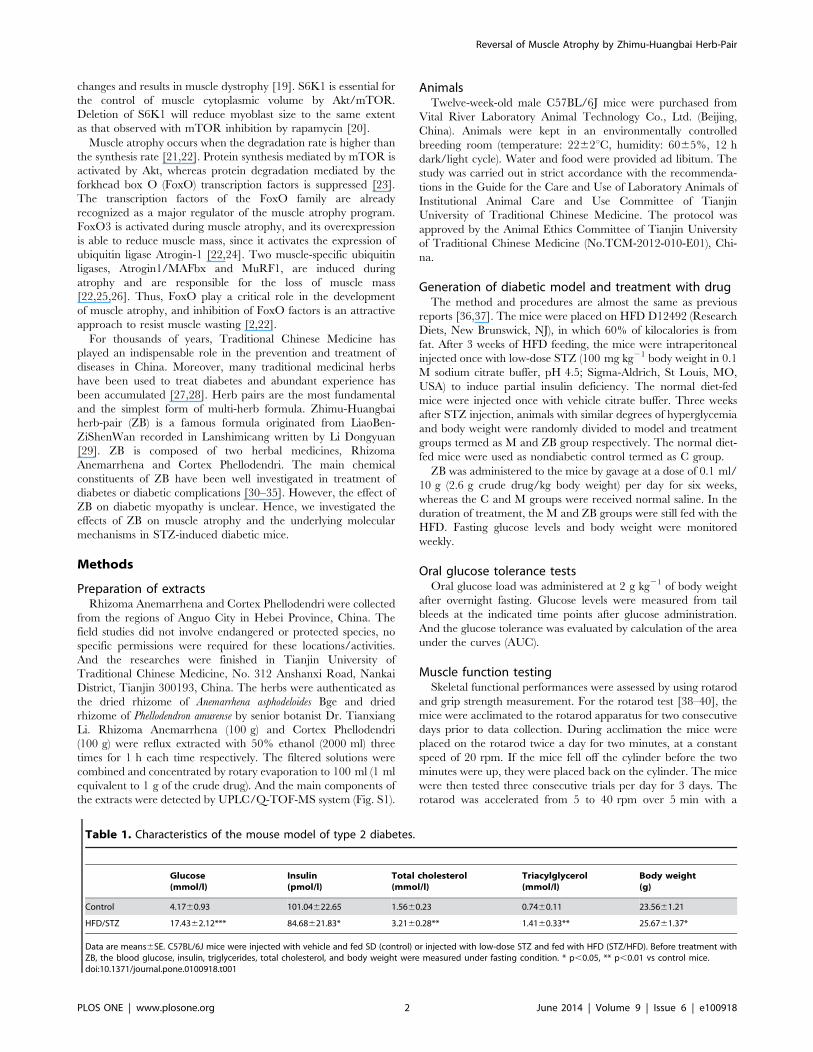
Data are means6SE. C57BL/6J mice were injected with vehicle and fed SD (control) or injected with low-dose STZ and fed with HFD (STZ/HFD). Before treatment withZB, the blood glucose, insulin, triglycerides, total cholesterol, and body weight were measured under fasting condition. * p,0.05, ** p,0.01 vs control mice.doi:10.1371/journal.pone.0100918.t001
Reversal of Muscle Atrophy by Zhimu-Huangbai Herb-Pair
PLOS ONE | www.plosone.org 2 June 2014 | Volume 9 | Issue 6 | e100918

maximum score of 300 sec. The latency to fall was recorded and
the average of three trials per mouse was calculated and analyzed.
For the Grip Strength test [41–43], a grip strength meter (GSM)
was used to measure forelimb and hindlimb grip strength. The
mice were acclimated to the GSM for five minutes one day prior
to data collection. Mice were allowed to grasp the grip with all
limbs. The maximum amount of force exerted was recorded. This
was repeated three times with a 30 sec interval between trials and
the average score (grams) was calculated.
Muscle weight and protein contentAfter 6 weeks of administration, mice were anesthetized by
intraperitoneal injection with 10% Chloral Hydrate (350 mg/kg
body) and the gastrocnemius, quadriceps muscles were quickly
harvested from hind limb, and fresh muscle weights were
recorded. The muscles were homogenized and the protein
contents were determined with a Bradford protein assay kit (Bio-
Rad) according to the manufacturer’s instructions.
Figure 1. Effect of ZB on body weight, blood glucose and oral glucose tolerance of diabetic mice. A: body weight of the mice duringtreatment in each group. B: body weight gain of the mice during treatment in each group. C: food intake of the mice during treatment in each group.D: fasting glucose levels of the mice during treatment in each group. E: glucose tolerance test results for each group. F: area under the curve for eachgroup. Data are expressed as the means6SD. *P,0.05, **P,0.01, ***P,0.001 vs. nondiabetic control. # P,0.05, ## P,0.01 vs. Model group (n= 15 ineach group).doi:10.1371/journal.pone.0100918.g001
Reversal of Muscle Atrophy by Zhimu-Huangbai Herb-Pair
PLOS ONE | www.plosone.org 3 June 2014 | Volume 9 | Issue 6 | e100918

Analysis of myofiber cross-sectional areaAfter the end of the experiment, hind limb muscles (gastrocne-
mius, quadriceps) of mice were collected, fixed in 4% parafor-
maldehyde in 0.1 M phosphate buffer (pH 7.4). To assess muscle
fiber cross-sectional area, transverse paraffinized muscle sections
(6 mm) were stained with hematoxylin and eosin (H&E). Stained
sections were visualized under Olympus IX 70 microscope and
pictures were captured using Olympus MagnaFire digital camera
and software (Olympus America, Melville, New York). Fiber cross-
sectional area was measured for approximately 100 adjacent
muscle fibers in each section for each mouse using Image Pro 6.0
software (Media Cybernetics, Silver Spring, MD) [44,45].
Serum lipid levels, insulin, IGF-1Mice were fasted for 12 hours and collected blood from the
canthus, centrifuged for 15 minutes at 3000 rpm, the serum was
separated. The total cholesterol (TC), high-density lipoprotein
cholesterol (HDL), low-density lipoprotein cholesterol (LDL), and
triglyceride (TG) levels were measured by semi-automatic
biochemical analyzer (Microlab 300, Vital Scientific Inc.). The
levels of insulin, IGF-1 were analysed with enzyme-linked
immunosorbent assay (ELISA). All the steps are operated strictly
in accordance with the instructions.
Western blot analysisThe method and procedures are conducted as previously
reported [46,47]. Quadriceps muscles were lysed with RIPA lysis
buffer. The protein concentrations of the lysates were determined
with a Bradford protein assay kit. An equal amount of protein
(40 ug) was fractionated by SDS-polyacrylamide gel electropho-
resis (PAGE) and transferred onto polyvinylidene difluoride
membranes. Membranes were blocked for 1 h in Tween 20
Tris-base sodium (TBST) containing 5% milk followed by
incubation with the appropriate primary antibody (rabbit anti-
phospho-Akt Ser473, rabbit anti-Akt, rabbit anti-phospho-mTOR
Ser2448, rabbit anti-mTOR, rabbit anti-phospho-rictor Thr1135,
rabbit anti-rictor, rabbit anti-phospho-raptor Ser792, rabbit anti-
raptor, rabbit anti-phospho-p70S6K1 Thr389, rabbit anti-
p70S6K1, rabbit anti-FoxO3, diluted 1:1000 with 5% BSA in
TBST, Cell Signaling Technology, Inc.) overnight at 4uC. After 5times washing in TBST, membranes were incubated with goat
anti-rabbit horse radish peroxidase-conjugated secondary anti-
bodies (1:5000) for 1 h at room temperature. Protein signals were
detected with ECL Western blotting detection reagents (Millipore).
Images were scanned, and band intensities were quantified by
densitometry (Bioquant Image Analysis, Nashville, TN). All the
protein expression data were normalized by b-actin. In addition,
the phospho and total kinase determinations were performed in
separate gels.
Statistical AnalysisAll data were analysed using SPSS version 16 software (SPSS
Inc.) and expressed as means6SD. Statistical comparison between
different treatments was done by one-way ANOVA. Differences
were considered statistically significant for P,0.05.
Results
Generation of a mouse model of type 2 diabetesPrevious studies [36,37] have shown that the combination of
HFD and STZ treatment can lead to disorders in glucose and lipid
metabolism accompanied by impaired insulin secretion and insulin
resistance. In the present study, C57BL/6J mice were fed with
HFD for three weeks and then injected with a single low dose of
STZ followed by continued HFD feeding for an additional three
weeks. As shown in Table 1, the HFD/STZ mouse model
manifested hyperglycemia and hyperlipidemia associated with
insulin resistance and impaired insulin secretion, as described in
previous reports. Therefore, we used this model as type 2 diabetes
to study the effects of ZB on muscle atrophy and the underlying
molecular mechanisms.
ZB increased body weight, reduced glucose levels andimproved glucose toleranceThe ZB extract was administered to diabetic mice, the changes
of body weight and fasting glucose levels were monitored weekly.
Fig. 1A shows the body weights of the mice. The body weight of
diabetic mice was gradually reduced, whereas the normal group
steadily increased over the six weeks. However, treatment with ZB
improved the disadvantaged compared with model group (P,
0.01, Fig. 1B). The amount of food intake of the diabetic mice was
significantly greater than the nondiabetic control, there was no
statistical difference between model group and ZB-treated animals
(Fig. 1C). Blood glucose levels in diabetic mice remained extremely
high throughout the experiment (P,0.05, Fig. 1D). In contrast,
after the fourth week, blood glucose levels of ZB-treated diabetic
mice were gradually descending down until the sixth week. Oral
glucose tolerance test was performed after 6 weeks of the
treatment. Diabetic mice showed impaired glucose tolerance
compared with C group. The ZB group displayed a significant
improvement in glucose clearance, and the area under the glucose
curve were reduced almost 20% compared with M group (p,0.01,
Fig. 1E, F).
Figure 2. ZB increased muscle strength and coordination ofdiabetic mice. A: grip strength of the mice during treatment in eachgroup. B: time to fall from an accelerating rotarod of the mice duringtreatment in each group. Data are expressed as the means6SD. *P,0.05, **P,0.01 vs. nondiabetic control. # P,0.05, ## P,0.01 vs. Modelgroup.doi:10.1371/journal.pone.0100918.g002
Reversal of Muscle Atrophy by Zhimu-Huangbai Herb-Pair
PLOS ONE | www.plosone.org 4 June 2014 | Volume 9 | Issue 6 | e100918
ZB increased muscle strength and coordinationTo explore the effect of ZB on muscle strength and coordination
in diabetic mice, we performed grip strength and rotarod tests
every 2 weeks (Fig. 2A, 2B). The model mice exhibited significant
muscle weakness and tended to fall off the rotarod cylinders sooner
than the control mice. ZB could increase grip strength (P,0.01)
and prolong the time on the rotarod compared with model group
(P,0.05).
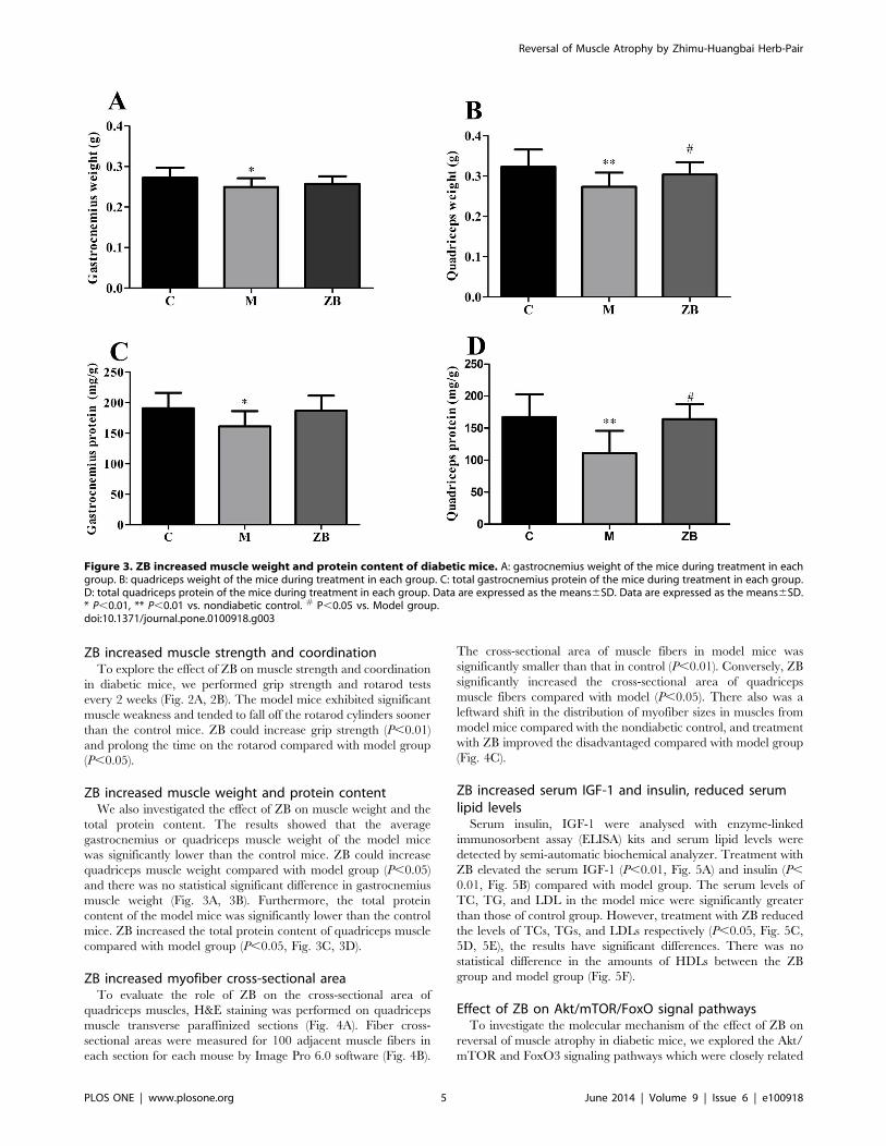
ZB increased muscle weight and protein contentWe also investigated the effect of ZB on muscle weight and the
total protein content. The results showed that the average
gastrocnemius or quadriceps muscle weight of the model mice
was significantly lower than the control mice. ZB could increase
quadriceps muscle weight compared with model group (P,0.05)
and there was no statistical significant difference in gastrocnemius
muscle weight (Fig. 3A, 3B). Furthermore, the total protein
content of the model mice was significantly lower than the control
mice. ZB increased the total protein content of quadriceps muscle
compared with model group (P,0.05, Fig. 3C, 3D).
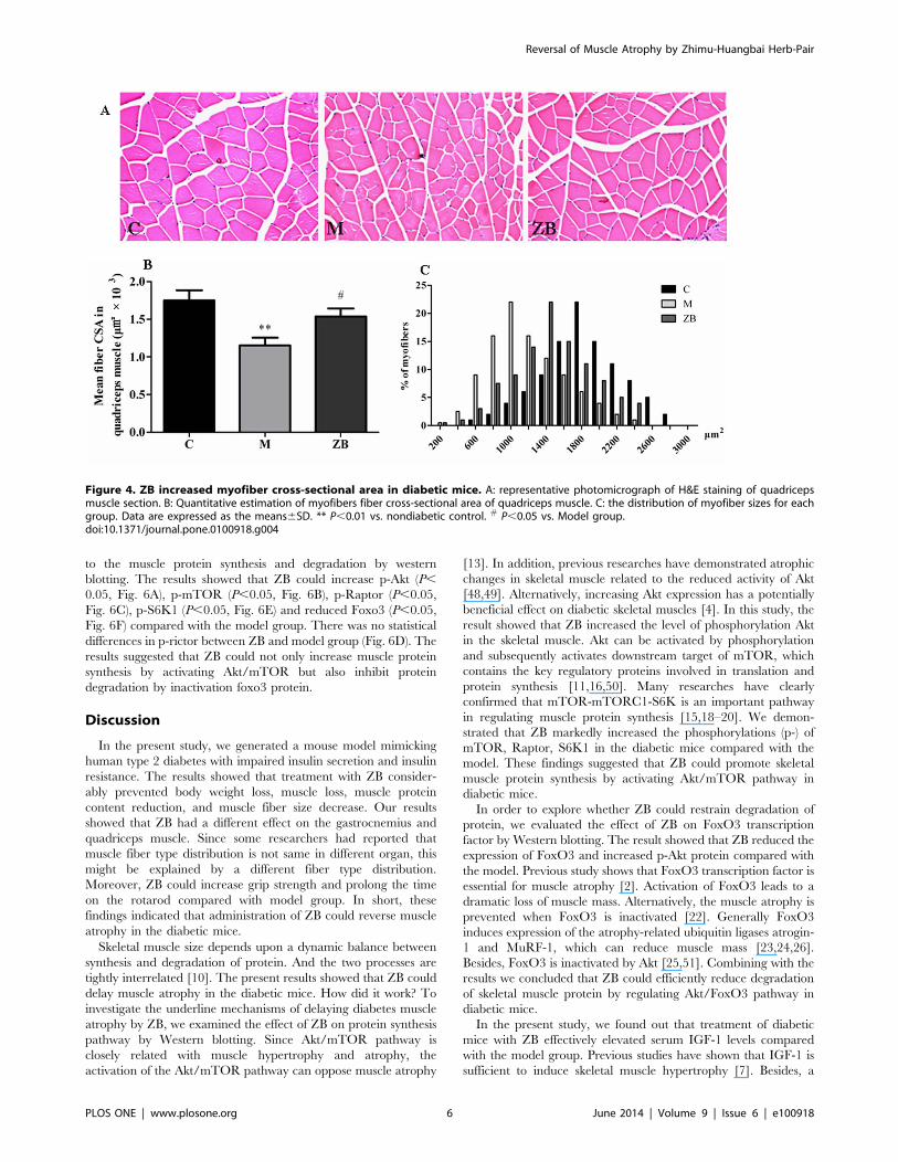
ZB increased myofiber cross-sectional areaTo evaluate the role of ZB on the cross-sectional area of
quadriceps muscles, H&E staining was performed on quadriceps
muscle transverse paraffinized sections (Fig. 4A). Fiber cross-
sectional areas were measured for 100 adjacent muscle fibers in
each section for each mouse by Image Pro 6.0 software (Fig. 4B).
The cross-sectional area of muscle fibers in model mice was
significantly smaller than that in control (P,0.01). Conversely, ZB
significantly increased the cross-sectional area of quadriceps
muscle fibers compared with model (P,0.05). There also was a
leftward shift in the distribution of myofiber sizes in muscles from
model mice compared with the nondiabetic control, and treatment
with ZB improved the disadvantaged compared with model group
(Fig. 4C).
ZB increased serum IGF-1 and insulin, reduced serumlipid levelsSerum insulin, IGF-1 were analysed with enzyme-linked
immunosorbent assay (ELISA) kits and serum lipid levels were
detected by semi-automatic biochemical analyzer. Treatment with
ZB elevated the serum IGF-1 (P,0.01, Fig. 5A) and insulin (P,
0.01, Fig. 5B) compared with model group. The serum levels of
TC, TG, and LDL in the model mice were significantly greater
than those of control group. However, treatment with ZB reduced
the levels of TCs, TGs, and LDLs respectively (P,0.05, Fig. 5C,
5D, 5E), the results have significant differences. There was no
statistical difference in the amounts of HDLs between the ZB
group and model group (Fig. 5F).
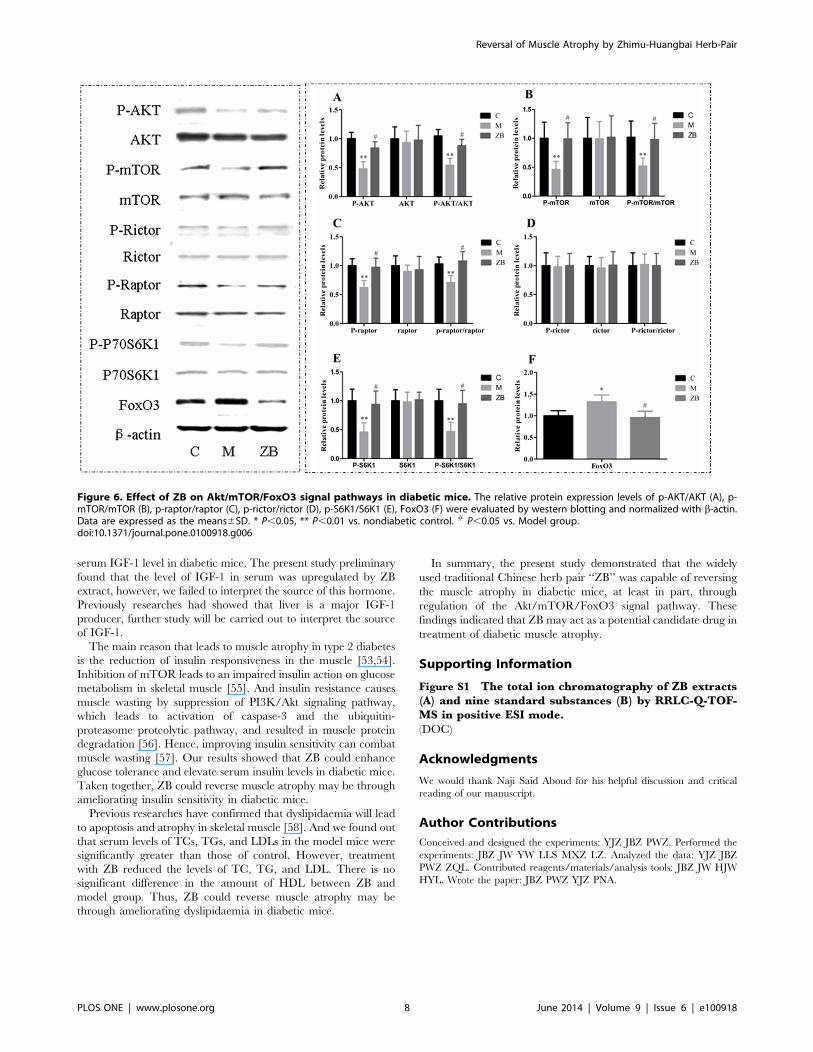
Effect of ZB on Akt/mTOR/FoxO signal pathwaysTo investigate the molecular mechanism of the effect of ZB on
reversal of muscle atrophy in diabetic mice, we explored the Akt/
mTOR and FoxO3 signaling pathways which were closely related
Figure 3. ZB increased muscle weight and protein content of diabetic mice. A: gastrocnemius weight of the mice during treatment in eachgroup. B: quadriceps weight of the mice during treatment in each group. C: total gastrocnemius protein of the mice during treatment in each group.D: total quadriceps protein of the mice during treatment in each group. Data are expressed as the means6SD. Data are expressed as the means6SD.* P,0.01, ** P,0.01 vs. nondiabetic control. # P,0.05 vs. Model group.doi:10.1371/journal.pone.0100918.g003
Reversal of Muscle Atrophy by Zhimu-Huangbai Herb-Pair
PLOS ONE | www.plosone.org 5 June 2014 | Volume 9 | Issue 6 | e100918
to the muscle protein synthesis and degradation by western
blotting. The results showed that ZB could increase p-Akt (P,
0.05, Fig. 6A), p-mTOR (P,0.05, Fig. 6B), p-Raptor (P,0.05,
Fig. 6C), p-S6K1 (P,0.05, Fig. 6E) and reduced Foxo3 (P,0.05,
Fig. 6F) compared with the model group. There was no statistical
differences in p-rictor between ZB and model group (Fig. 6D). The
results suggested that ZB could not only increase muscle protein
synthesis by activating Akt/mTOR but also inhibit protein
degradation by inactivation foxo3 protein.
Discussion
In the present study, we generated a mouse model mimicking
human type 2 diabetes with impaired insulin secretion and insulin
resistance. The results showed that treatment with ZB consider-
ably prevented body weight loss, muscle loss, muscle protein
content reduction, and muscle fiber size decrease. Our results
showed that ZB had a different effect on the gastrocnemius and
quadriceps muscle. Since some researchers had reported that
muscle fiber type distribution is not same in different organ, this
might be explained by a different fiber type distribution.
Moreover, ZB could increase grip strength and prolong the time
on the rotarod compared with model group. In short, these
findings indicated that administration of ZB could reverse muscle
atrophy in the diabetic mice.
Skeletal muscle size depends upon a dynamic balance between
synthesis and degradation of protein. And the two processes are
tightly interrelated [10]. The present results showed that ZB could
delay muscle atrophy in the diabetic mice. How did it work? To
investigate the underline mechanisms of delaying diabetes muscle
atrophy by ZB, we examined the effect of ZB on protein synthesis
pathway by Western blotting. Since Akt/mTOR pathway is
closely related with muscle hypertrophy and atrophy, the
activation of the Akt/mTOR pathway can oppose muscle atrophy
[13]. In addition, previous researches have demonstrated atrophic
changes in skeletal muscle related to the reduced activity of Akt
[48,49]. Alternatively, increasing Akt expression has a potentially
beneficial effect on diabetic skeletal muscles [4]. In this study, the
result showed that ZB increased the level of phosphorylation Akt
in the skeletal muscle. Akt can be activated by phosphorylation
and subsequently activates downstream target of mTOR, which
contains the key regulatory proteins involved in translation and
protein synthesis [11,16,50]. Many researches have clearly
confirmed that mTOR-mTORC1-S6K is an important pathway
in regulating muscle protein synthesis [15,18–20]. We demon-
strated that ZB markedly increased the phosphorylations (p-) of
mTOR, Raptor, S6K1 in the diabetic mice compared with the
model. These findings suggested that ZB could promote skeletal
muscle protein synthesis by activating Akt/mTOR pathway in
diabetic mice.
In order to explore whether ZB could restrain degradation of
protein, we evaluated the effect of ZB on FoxO3 transcription
factor by Western blotting. The result showed that ZB reduced the
expression of FoxO3 and increased p-Akt protein compared with
the model. Previous study shows that FoxO3 transcription factor is
essential for muscle atrophy [2]. Activation of FoxO3 leads to a
dramatic loss of muscle mass. Alternatively, the muscle atrophy is
prevented when FoxO3 is inactivated [22]. Generally FoxO3
induces expression of the atrophy-related ubiquitin ligases atrogin-
1 and MuRF-1, which can reduce muscle mass [23,24,26].
Besides, FoxO3 is inactivated by Akt [25,51]. Combining with the
results we concluded that ZB could efficiently reduce degradation
of skeletal muscle protein by regulating Akt/FoxO3 pathway in
diabetic mice.
In the present study, we found out that treatment of diabetic
mice with ZB effectively elevated serum IGF-1 levels compared
with the model group. Previous studies have shown that IGF-1 is
sufficient to induce skeletal muscle hypertrophy [7]. Besides, a
Figure 4. ZB increased myofiber cross-sectional area in diabetic mice. A: representative photomicrograph of H&E staining of quadricepsmuscle section. B: Quantitative estimation of myofibers fiber cross-sectional area of quadriceps muscle. C: the distribution of myofiber sizes for eachgroup. Data are expressed as the means6SD. ** P,0.01 vs. nondiabetic control. # P,0.05 vs. Model group.doi:10.1371/journal.pone.0100918.g004
Reversal of Muscle Atrophy by Zhimu-Huangbai Herb-Pair
PLOS ONE | www.plosone.org 6 June 2014 | Volume 9 | Issue 6 | e100918

study on transgenic mice shows that muscle-specific overexpres-
sion of an IGF-1 isoform locally expressed in skeletal muscle results
in muscle hypertrophy [8]. Moreover, IGF-1 induces an increase
in muscle mass by stimulating the phosphatidylinositol-3 kinase
(PI3K)/Akt pathway [7,9]. And Akt could induce the activation of
mTOR, whose downstream targets, p70S6K and PHAS-1/4E-
BP1, have been shown to promote protein synthesis [13,50,52].
Therefore, IGF-1 promotes protein synthesis mainly through the
PI3K/Akt/mTOR pathways. On the other hand, IGF-1 can
inhibit protein degradation by activation Akt. Akt represses the
transcription factors of the FoxO family [52] and then block the
upregulation of the ubiquitin-ligases MuRF1 and MAFbx, which
are the key mediators of skeletal muscle atrophy [7,11]. Therefore
both the Akt/FoxO and Akt/mTOR pathways are regulated by
IGF-1 [11]. Hence, stimulation of the IGF1/PI3K/Akt pathway
would not only promote synthesis of protein but also dominantly
suppress degradation of protein [10]. These findings insinuated
that ZB could reverse muscle atrophy may be through increasing
Figure 5. Effect of ZB on Serum IGF-1, insulin and Serum Lipid in diabetic mice. A: Serum IGF-1 levels of each group. B: Serum insulin levelsof each group. C–F: The serum levels of triglyceride (TG), total cholesterol (TC), low-density lipoprotein cholesterol (LDL), high-density lipoproteincholesterol (HDL). Data are expressed as the means6SD. ** P,0.01, *** P,0.001 vs. nondiabetic control. #P,0.05, ##P,0.01 vs. Model group.doi:10.1371/journal.pone.0100918.g005
Reversal of Muscle Atrophy by Zhimu-Huangbai Herb-Pair
PLOS ONE | www.plosone.org 7 June 2014 | Volume 9 | Issue 6 | e100918
serum IGF-1 level in diabetic mice. The present study preliminary
found that the level of IGF-1 in serum was upregulated by ZB
extract, however, we failed to interpret the source of this hormone.
Previously researches had showed that liver is a major IGF-1
producer, further study will be carried out to interpret the source
of IGF-1.
The main reason that leads to muscle atrophy in type 2 diabetes
is the reduction of insulin responsiveness in the muscle [53,54].
Inhibition of mTOR leads to an impaired insulin action on glucose
metabolism in skeletal muscle [55]. And insulin resistance causes
muscle wasting by suppression of PI3K/Akt signaling pathway,
which leads to activation of caspase-3 and the ubiquitin-
proteasome proteolytic pathway, and resulted in muscle protein
degradation [56]. Hence, improving insulin sensitivity can combat
muscle wasting [57]. Our results showed that ZB could enhance
glucose tolerance and elevate serum insulin levels in diabetic mice.
Taken together, ZB could reverse muscle atrophy may be through
ameliorating insulin sensitivity in diabetic mice.
Previous researches have confirmed that dyslipidaemia will lead
to apoptosis and atrophy in skeletal muscle [58]. And we found out
that serum levels of TCs, TGs, and LDLs in the model mice were
significantly greater than those of control. However, treatment
with ZB reduced the levels of TC, TG, and LDL. There is no
significant difference in the amount of HDL between ZB and
model group. Thus, ZB could reverse muscle atrophy may be
through ameliorating dyslipidaemia in diabetic mice.
In summary, the present study demonstrated that the widely
used traditional Chinese herb pair ‘‘ZB’’ was capable of reversing
the muscle atrophy in diabetic mice, at least in part, through
regulation of the Akt/mTOR/FoxO3 signal pathway. These
findings indicated that ZB may act as a potential candidate drug in
treatment of diabetic muscle atrophy.
Supporting Information
Figure S1 The total ion chromatography of ZB extracts(A) and nine standard substances (B) by RRLC-Q-TOF-MS in positive ESI mode.
(DOC)
Acknowledgments
We would thank Naji Said Aboud for his helpful discussion and critical
reading of our manuscript.
Author Contributions
Conceived and designed the experiments: YJZ JBZ PWZ. Performed the
experiments: JBZ JW YW LLS MXZ LZ. Analyzed the data: YJZ JBZ
PWZ ZQL. Contributed reagents/materials/analysis tools: JBZ JW HJW
HYL. Wrote the paper: JBZ PWZ YJZ PNA.
Figure 6. Effect of ZB on Akt/mTOR/FoxO3 signal pathways in diabetic mice. The relative protein expression levels of p-AKT/AKT (A), p-mTOR/mTOR (B), p-raptor/raptor (C), p-rictor/rictor (D), p-S6K1/S6K1 (E), FoxO3 (F) were evaluated by western blotting and normalized with b-actin.Data are expressed as the means6SD. * P,0.05, ** P,0.01 vs. nondiabetic control. # P,0.05 vs. Model group.doi:10.1371/journal.pone.0100918.g006
Reversal of Muscle Atrophy by Zhimu-Huangbai Herb-Pair
PLOS ONE | www.plosone.org 8 June 2014 | Volume 9 | Issue 6 | e100918

References
1. Glass DJ (2003) Signalling pathways that mediate skeletal muscle hypertrophy
and atrophy. Nat Cell Biol 5: 87–90.
2. Clavel S, Siffroi-Fernandez S, Coldefy AS, Boulukos K, Pisani DF, et al. (2010)Regulation of the intracellular localization of Foxo3a by stress-activated protein
kinase signaling pathways in skeletal muscle cells. Mol Cell Biol 30: 470–480.
3. Ito N, Ruegg UT, Kudo A, Miyagoe-Suzuki Y, Takeda S (2013) Activation ofcalcium signaling through Trpv1 by nNOS and peroxynitrite as a key trigger of
skeletal muscle hypertrophy. Nat Med 19: 101–106.
4. Lambertucci AC, Lambertucci RH, Hirabara SM, Curi R, Moriscot AS, et al.(2012) Glutamine supplementation stimulates protein-synthetic and inhibits
protein-degradative signaling pathways in skeletal muscle of diabetic rats. PLoS
One 7: e50390.
5. Hulmi JJ, Silvennoinen M, Lehti M, Kivela R, Kainulainen H (2012) AlteredREDD1, myostatin, and Akt/mTOR/FoxO/MAPK signaling in streptozoto-
cin-induced diabetic muscle atrophy. Am J Physiol Endocrinol Metab 302:E307–315.
6. Newsholme P, Abdulkader F, Rebelato E, Romanatto T, Pinheiro CH, et al.
(2011) Amino acids and diabetes: implications for endocrine, metabolic andimmune function. Front Biosci (Landmark Ed) 16: 315–339.
7. Glass DJ (2005) Skeletal muscle hypertrophy and atrophy signaling pathways.
Int J Biochem Cell Biol 37: 1974–1984.
8. Musaro A, McCullagh K, Paul A, Houghton L, Dobrowolny G, et al. (2001)Localized Igf-1 transgene expression sustains hypertrophy and regeneration in
senescent skeletal muscle. Nat Genet 27: 195–200.
9. Miyazaki M, Esser KA (2009) Cellular mechanisms regulating protein synthesisand skeletal muscle hypertrophy in animals. J Appl Physiol 106: 1367–1373.
10. Stitt TN, Drujan D, Clarke BA, Panaro F, Timofeyva Y, et al. (2004) The IGF-
1/PI3K/Akt pathway prevents expression of muscle atrophy-induced ubiquitinligases by inhibiting FOXO transcription factors. Mol Cell 14: 395–403.
11. Latres E, Amini AR, Amini AA, Griffiths J, Martin FJ, et al. (2005) Insulin-like
growth factor-1 (IGF-1) inversely regulates atrophy-induced genes via the
phosphatidylinositol 3-kinase/Akt/mammalian target of rapamycin (PI3K/Akt/mTOR) pathway. J Biol Chem 280: 2737–2744.
12. Goodman CA, Miu MH, Frey JW, Mabrey DM, Lincoln HC, et al. (2010) A
phosphatidylinositol 3-kinase/protein kinase B-independent activation ofmammalian target of rapamycin signaling is sufficient to induce skeletal muscle
hypertrophy. Mol Biol Cell 21: 3258–3268.
13. Bodine SC, Stitt TN, Gonzalez M, Kline WO, Stover GL, et al. (2001) Akt/mTOR pathway is a crucial regulator of skeletal muscle hypertrophy and can
prevent muscle atrophy in vivo. Nat Cell Biol 3: 1014–1019.
14. Risson V, Mazelin L, Roceri M, Sanchez H, Moncollin V, et al. (2009) Muscleinactivation of mTOR causes metabolic and dystrophin defects leading to severe
myopathy. J Cell Biol 187: 859–874.
15. Drummond MJ, Dreyer HC, Fry CS, Glynn EL, Rasmussen BB (2009)Nutritional and contractile regulation of human skeletal muscle protein synthesis
and mTORC1 signaling. J Appl Physiol (1985) 106: 1374–1384.
16. Laplante M, Sabatini DM (2012) mTOR signaling in growth control and
disease. Cell 149: 274–293.17. Guertin DA, Stevens DM, Thoreen CC, Burds AA, Kalaany NY, et al. (2006)
Ablation in mice of the mTORC components raptor, rictor, or mLST8 reveals
that mTORC2 is required for signaling to Akt-FOXO and PKCalpha, but notS6K1. Dev Cell 11: 859–871.
18. Adegoke OA, Abdullahi A, Tavajohi-Fini P (2012) mTORC1 and the regulation
of skeletal muscle anabolism and mass. Appl Physiol Nutr Metab 37: 395–406.
19. Bentzinger CF, Romanino K, Cloetta D, Lin S, Mascarenhas JB, et al. (2008)Skeletal muscle-specific ablation of raptor, but not of rictor, causes metabolic
changes and results in muscle dystrophy. Cell Metab 8: 411–424.
20. Ohanna M, Sobering AK, Lapointe T, Lorenzo L, Praud C, et al. (2005)Atrophy of S6K1(2/2) skeletal muscle cells reveals distinct mTOR effectors for
cell cycle and size control. Nat Cell Biol 7: 286–294.
21. Zhao J, Brault JJ, Schild A, Cao P, Sandri M, et al. (2007) FoxO3 coordinatelyactivates protein degradation by the autophagic/lysosomal and proteasomal
pathways in atrophying muscle cells. Cell Metab 6: 472–483.
22. Sandri M, Sandri C, Gilbert A, Skurk C, Calabria E, et al. (2004) Foxotranscription factors induce the atrophy-related ubiquitin ligase atrogin-1 and
cause skeletal muscle atrophy. Cell 117: 399–412.
23. Mammucari C, Schiaffino S, Sandri M (2008) Downstream of Akt: FoxO3 andmTOR in the regulation of autophagy in skeletal muscle. Autophagy 4: 524–
526.
24. Sandri M, Lin J, Handschin C, Yang W, Arany ZP, et al. (2006) PGC-1alpha
protects skeletal muscle from atrophy by suppressing FoxO3 action and atrophy-specific gene transcription. Proc Natl Acad Sci U S A 103: 16260–16265.
25. Zheng B, Ohkawa S, Li H, Roberts-Wilson TK, Price SR (2010) FOXO3a
mediates signaling crosstalk that coordinates ubiquitin and atrogin-1/MAFbxexpression during glucocorticoid-induced skeletal muscle atrophy. FASEB J 24:
2660–2669.
26. Bodine SC, Latres E, Baumhueter S, Lai VK, Nunez L, et al. (2001)Identification of ubiquitin ligases required for skeletal muscle atrophy. Science
294: 1704–1708.
27. Jia W, Gao W, Tang L (2003) Antidiabetic herbal drugs officially approved inChina. Phytother Res 17: 1127–1134.
28. Tong XL, Dong L, Chen L, Zhen Z (2012) Treatment of diabetes usingtraditional Chinese medicine: past, present and future. Am J Chin Med 40: 877–
886.
29. Ma C, Fan M, Tang Y, Li Z, Sun Z, et al. (2008) Identification of major
alkaloids and steroidal saponins in rat serum by HPLC-diode array detection-MS/MS following oral administration of Huangbai-Zhimu herb-pair Extract.
Biomed Chromatogr 22: 835–850.
30. Xie W, Zhao Y, Zhang Y (2011) Traditional chinese medicines in treatment of
patients with type 2 diabetes mellitus. Evid Based Complement Alternat Med2011: 726723.
31. Liu YW, Zhu X, Lu Q, Wang JY, Li W, et al. (2012) Total saponins fromRhizoma Anemarrhenae ameliorate diabetes-associated cognitive decline in rats:
involvement of amyloid-beta decrease in brain. J Ethnopharmacol 139: 194–200.
32. Bhutada P, Mundhada Y, Bansod K, Tawari S, Patil S, et al. (2011) Protectionof cholinergic and antioxidant system contributes to the effect of berberine
ameliorating memory dysfunction in rat model of streptozotocin-induceddiabetes. Behav Brain Res 220: 30–41.
33. Zhang X, Zhao Y, Zhang M, Pang X, Xu J, et al. (2012) Structural changes ofgut microbiota during berberine-mediated prevention of obesity and insulin
resistance in high-fat diet-fed rats. PLoS One 7: e42529.
34. He K, Li X, Chen X, Ye X, Huang J, et al. (2011) Evaluation of antidiabetic
potential of selected traditional Chinese medicines in STZ-induced diabeticmice. J Ethnopharmacol 137: 1135–1142.
35. Tang YH, Sun ZL, Fan MS, Li ZX, Huang CG (2012) Anti-diabetic effects of
TongGuanWan, a Chinese traditional herbal formula, in C57BL/KsJ-db/db
mice. Planta Med 78: 18–23.
36. Mu J, Woods J, Zhou YP, Roy RS, Li Z, et al. (2006) Chronic inhibition ofdipeptidyl peptidase-4 with a sitagliptin analog preserves pancreatic beta-cell
mass and function in a rodent model of type 2 diabetes. Diabetes 55: 1695–1704.
37. Kusakabe T, Tanioka H, Ebihara K, Hirata M, Miyamoto L, et al. (2009)
Beneficial effects of leptin on glycaemic and lipid control in a mouse model oftype 2 diabetes with increased adiposity induced by streptozotocin and a high-fat
diet. Diabetologia 52: 675–683.
38. Baur JA, Pearson KJ, Price NL, Jamieson HA, Lerin C, et al. (2006) Resveratrol
improves health and survival of mice on a high-calorie diet. Nature 444: 337–342.
39. Pandey SN, Cabotage J, Shi R, Dixit M, Sutherland M, et al. (2012) Conditional
over-expression of PITX1 causes skeletal muscle dystrophy in mice. Biology
Open 1: 629–639.
40. Wu Y, Lousberg EL, Moldenhauer LM, Hayball JD, Coller JK, et al. (2012)Inhibiting the TLR4-MyD88 signalling cascade by genetic or pharmacological
strategies reduces acute alcohol-induced sedation and motor impairment in
mice. Br J Pharmacol 165: 1319–1329.
41. Chambon C, Duteil D, Vignaud A, Ferry A, Messaddeq N, et al. (2010)Myocytic androgen receptor controls the strength but not the mass of limb
muscles. Proc Natl Acad Sci U S A 107: 14327–14332.
42. Ravenscroft G, Jackaman C, Bringans S, Papadimitriou JM, Griffiths LM, et al.
(2011) Mouse models of dominant ACTA1 disease recapitulate human diseaseand provide insight into therapies. Brain 134: 1101–1115.
43. Hill AJ, Mercier MS, Hill TD, Glyn SE, Jones NA, et al. (2012) Cannabidivarinis anticonvulsant in mouse and rat. Br J Pharmacol 167: 1629–1642.
44. Dogra C, Changotra H, Wedhas N, Qin X, Wergedal JE, et al. (2007) TNF-
related weak inducer of apoptosis (TWEAK) is a potent skeletal muscle-wasting
cytokine. FASEB J 21: 1857–1869.
45. Paul PK, Gupta SK, Bhatnagar S, Panguluri SK, Darnay BG, et al. (2010)Targeted ablation of TRAF6 inhibits skeletal muscle wasting in mice. J Cell Biol
191: 1395–1411.
46. Zhuang P, Zhang Y, Cui G, Bian Y, Zhang M, et al. (2012) Direct stimulation of
adult neural stem/progenitor cells in vitro and neurogenesis in vivo bysalvianolic acid B. PLoS One 7: e35636.
47. Zhuang PW, Cui GZ, Zhang YJ, Zhang MX, Guo H, et al. (2013) Baicalinregulates neuronal fate decision in neural stem/progenitor cells and stimulates
hippocampal neurogenesis in adult rats. CNS Neurosci Ther 19: 154–162.
48. Wu M, Falasca M, Blough ER (2011) Akt/protein kinase B in skeletal muscle
physiology and pathology. J Cell Physiol 226: 29–36.
49. Sugita H, Kaneki M, Sugita M, Yasukawa T, Yasuhara S, et al. (2005) Burninjury impairs insulin-stimulated Akt/PKB activation in skeletal muscle.
Am J Physiol Endocrinol Metab 288: E585–591.
50. Rommel C, Bodine SC, Clarke BA, Rossman R, Nunez L, et al. (2001)
Mediation of IGF-1-induced skeletal myotube hypertrophy by PI(3)K/Akt/mTOR and PI(3)K/Akt/GSK3 pathways. Nat Cell Biol 3: 1009–1013.
51. Mammucari C, Milan G, Romanello V, Masiero E, Rudolf R, et al. (2007)FoxO3 controls autophagy in skeletal muscle in vivo. Cell Metab 6: 458–471.
52. Schiaffino S, Mammucari C (2011) Regulation of skeletal muscle growth by the
IGF1-Akt/PKB pathway: insights from genetic models. Skelet Muscle 1: 4.
53. Workeneh B, Bajaj M (2013) The regulation of muscle protein turnover in
diabetes. Int J Biochem Cell Biol 45: 2239–2244.
54. Pereira S, Marliss EB, Morais JA, Chevalier S, Gougeon R (2008) Insulinresistance of protein metabolism in type 2 diabetes. Diabetes 57: 56–63.
Reversal of Muscle Atrophy by Zhimu-Huangbai Herb-Pair
PLOS ONE | www.plosone.org 9 June 2014 | Volume 9 | Issue 6 | e100918

55. Deblon N, Bourgoin L, Veyrat-Durebex C, Peyrou M, Vinciguerra M, et al.
(2012) Chronic mTOR inhibition by rapamycin induces muscle insulinresistance despite weight loss in rats. Br J Pharmacol 165: 2325–2340.
56. Wang X, Hu Z, Hu J, Du J, Mitch WE (2006) Insulin resistance accelerates
muscle protein degradation: Activation of the ubiquitin-proteasome pathway bydefects in muscle cell signaling. Endocrinology 147: 4160–4168.
57. Bassil MS, Gougeon R (2013) Muscle protein anabolism in type 2 diabetes. Curr
Opin Clin Nutr Metab Care 16: 83–88.
58. Sishi B, Loos B, Ellis B, Smith W, du Toit EF, et al. (2011) Diet-induced obesity
alters signalling pathways and induces atrophy and apoptosis in skeletal muscle
in a prediabetic rat model. Exp Physiol 96: 179–193.
Reversal of Muscle Atrophy by Zhimu-Huangbai Herb-Pair
PLOS ONE | www.plosone.org 10 June 2014 | Volume 9 | Issue 6 | e100918