Restoration of breathing after opioid overdose and spinal cord ...
15
ARTICLE Restoration of breathing after opioid overdose and spinal cord injury using temporal interference stimulation Michael D. Sunshine 1,2,3,4 , Antonino M. Cassarà 5 , Esra Neufeld 5 , Nir Grossman 6,7 , Thomas H. Mareci 8 , Kevin J. Otto 9,10,11,12,13 , Edward S. Boyden 14,15 & David D. Fuller 2,3,4 ✉ Respiratory insufficiency is a leading cause of death due to drug overdose or neuromuscular disease. We hypothesized that a stimulation paradigm using temporal interference (TI) could restore breathing in such conditions. Following opioid overdose in rats, two high frequency (5000 Hz and 5001 Hz), low amplitude waveforms delivered via intramuscular wires in the neck immediately activated the diaphragm and restored ventilation in phase with waveform offset (1 Hz or 60 breaths/min). Following cervical spinal cord injury (SCI), TI stimulation via dorsally placed epidural electrodes uni- or bilaterally activated the diaphragm depending on current and electrode position. In silico modeling indicated that an interferential signal in the ventral spinal cord predicted the evoked response (left versus right diaphragm) and current- ratio-based steering. We conclude that TI stimulation can activate spinal motor neurons after SCI and prevent fatal apnea during drug overdose by restoring ventilation with minimally invasive electrodes. https://doi.org/10.1038/s42003-020-01604-x OPEN 1 Rehabilitation Science PhD Program, University of Florida, Gainesville, FL 32611, USA. 2 Department of Physical Therapy, University of Florida, Gainesville, FL 32611, USA. 3 Breathing Research and Therapeutics Center, University of Florida, Gainesville, FL 32611, USA. 4 McKnight Brain Institute, University of Florida, Gainesville, FL 32611, USA. 5 Foundation for Research on Information Technologies in Society (IT’IS), 8004 Zurich, Switzerland. 6 Division of Brain Sciences, Imperial College London, London SW7 2BU, United Kingdom. 7 United Kingdom Dementia Research Institute, Imperial College London, London SW7 2BU, United Kingdom. 8 Department of Biochemistry and Molecular Biology, University of Florida, Gainesville, FL 32611, USA. 9 J. Crayton Pruitt Family Department of Biomedical Engineering, University of Florida, Gainesville, FL 32611, USA. 10 Department of Neuroscience, University of Florida, Gainesville, FL 32611, USA. 11 Department of Neurology, University of Florida, Gainesville, FL 32611, USA. 12 Department of Materials Science and Engineering, University of Florida, Gainesville, FL 32611, USA. 13 Department of Electrical and Computer Engineering, University of Florida, Gainesville, FL 32611, USA. 14 Departments of Brain and Cognitive Sciences, Media Arts and Sciences, and Biological Engineering, McGovern and Koch Institutes, MIT, Cambridge, MA 02139, USA. 15 Howard Hughes Medical Institute, Cambridge, MA 02138, USA. ✉ email: [email protected]fl.edu COMMUNICATIONS BIOLOGY | (2021)4:107 | https://doi.org/10.1038/s42003-020-01604-x | www.nature.com/commsbio 1 1234567890():,;
-
Upload
khangminh22 -
Category
Documents
-
view
2 -
download
0
Transcript of Restoration of breathing after opioid overdose and spinal cord ...
ARTICLE
Restoration of breathing after opioid overdose andspinal cord injury using temporal interferencestimulationMichael D. Sunshine 1,2,3,4, Antonino M. Cassarà 5, Esra Neufeld5, Nir Grossman6,7, Thomas H. Mareci 8,
Kevin J. Otto9,10,11,12,13, Edward S. Boyden 14,15 & David D. Fuller 2,3,4✉
Respiratory insufficiency is a leading cause of death due to drug overdose or neuromuscular
disease. We hypothesized that a stimulation paradigm using temporal interference (TI) could
restore breathing in such conditions. Following opioid overdose in rats, two high frequency
(5000 Hz and 5001 Hz), low amplitude waveforms delivered via intramuscular wires in the
neck immediately activated the diaphragm and restored ventilation in phase with waveform
offset (1 Hz or 60 breaths/min). Following cervical spinal cord injury (SCI), TI stimulation via
dorsally placed epidural electrodes uni- or bilaterally activated the diaphragm depending on
current and electrode position. In silico modeling indicated that an interferential signal in the
ventral spinal cord predicted the evoked response (left versus right diaphragm) and current-
ratio-based steering. We conclude that TI stimulation can activate spinal motor neurons after
SCI and prevent fatal apnea during drug overdose by restoring ventilation with minimally
invasive electrodes.
https://doi.org/10.1038/s42003-020-01604-x OPEN
1 Rehabilitation Science PhD Program, University of Florida, Gainesville, FL 32611, USA. 2Department of Physical Therapy, University of Florida, Gainesville, FL32611, USA. 3 Breathing Research and Therapeutics Center, University of Florida, Gainesville, FL 32611, USA. 4McKnight Brain Institute, University of Florida,Gainesville, FL 32611, USA. 5 Foundation for Research on Information Technologies in Society (IT’IS), 8004 Zurich, Switzerland. 6 Division of Brain Sciences,Imperial College London, London SW7 2BU, United Kingdom. 7 United Kingdom Dementia Research Institute, Imperial College London, London SW7 2BU,United Kingdom. 8Department of Biochemistry and Molecular Biology, University of Florida, Gainesville, FL 32611, USA. 9 J. Crayton Pruitt Family Departmentof Biomedical Engineering, University of Florida, Gainesville, FL 32611, USA. 10 Department of Neuroscience, University of Florida, Gainesville, FL 32611, USA.11 Department of Neurology, University of Florida, Gainesville, FL 32611, USA. 12 Department of Materials Science and Engineering, University of Florida,Gainesville, FL 32611, USA. 13 Department of Electrical and Computer Engineering, University of Florida, Gainesville, FL 32611, USA. 14 Departments of Brainand Cognitive Sciences, Media Arts and Sciences, and Biological Engineering, McGovern and Koch Institutes, MIT, Cambridge, MA 02139, USA. 15 HowardHughes Medical Institute, Cambridge, MA 02138, USA. ✉email: [email protected]
COMMUNICATIONS BIOLOGY | (2021) 4:107 | https://doi.org/10.1038/s42003-020-01604-x | www.nature.com/commsbio 1
1234
5678
90():,;
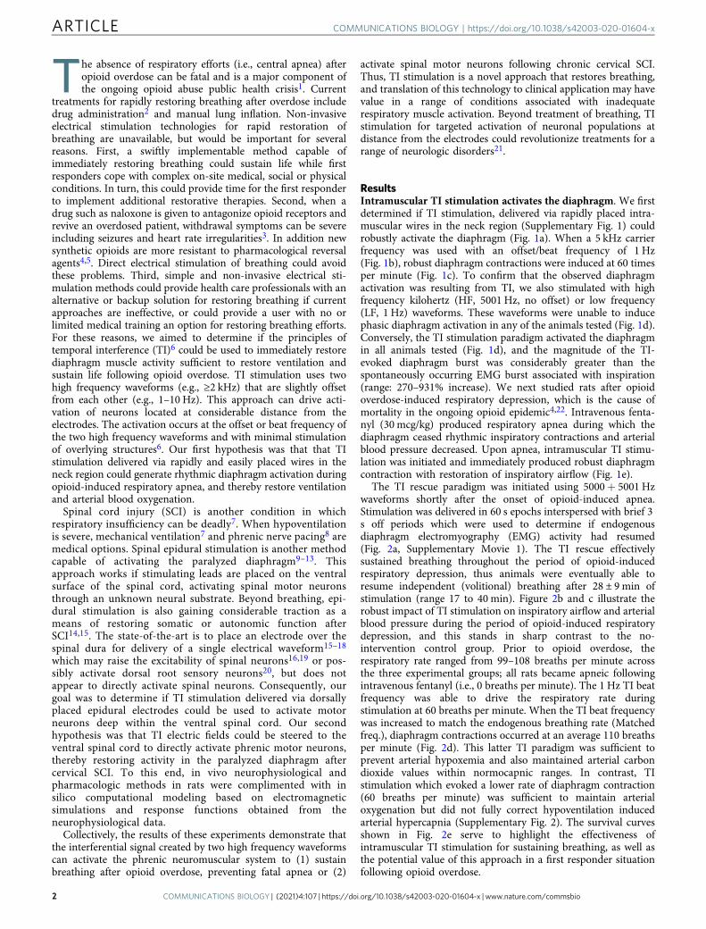
The absence of respiratory efforts (i.e., central apnea) afteropioid overdose can be fatal and is a major component ofthe ongoing opioid abuse public health crisis1. Current
treatments for rapidly restoring breathing after overdose includedrug administration2 and manual lung inflation. Non-invasiveelectrical stimulation technologies for rapid restoration ofbreathing are unavailable, but would be important for severalreasons. First, a swiftly implementable method capable ofimmediately restoring breathing could sustain life while firstresponders cope with complex on-site medical, social or physicalconditions. In turn, this could provide time for the first responderto implement additional restorative therapies. Second, when adrug such as naloxone is given to antagonize opioid receptors andrevive an overdosed patient, withdrawal symptoms can be severeincluding seizures and heart rate irregularities3. In addition newsynthetic opioids are more resistant to pharmacological reversalagents4,5. Direct electrical stimulation of breathing could avoidthese problems. Third, simple and non-invasive electrical sti-mulation methods could provide health care professionals with analternative or backup solution for restoring breathing if currentapproaches are ineffective, or could provide a user with no orlimited medical training an option for restoring breathing efforts.For these reasons, we aimed to determine if the principles oftemporal interference (TI)6 could be used to immediately restorediaphragm muscle activity sufficient to restore ventilation andsustain life following opioid overdose. TI stimulation uses twohigh frequency waveforms (e.g., ≥2 kHz) that are slightly offsetfrom each other (e.g., 1–10 Hz). This approach can drive acti-vation of neurons located at considerable distance from theelectrodes. The activation occurs at the offset or beat frequency ofthe two high frequency waveforms and with minimal stimulationof overlying structures6. Our first hypothesis was that that TIstimulation delivered via rapidly and easily placed wires in theneck region could generate rhythmic diaphragm activation duringopioid-induced respiratory apnea, and thereby restore ventilationand arterial blood oxygenation.
Spinal cord injury (SCI) is another condition in whichrespiratory insufficiency can be deadly7. When hypoventilationis severe, mechanical ventilation7 and phrenic nerve pacing8 aremedical options. Spinal epidural stimulation is another methodcapable of activating the paralyzed diaphragm9–13. Thisapproach works if stimulating leads are placed on the ventralsurface of the spinal cord, activating spinal motor neuronsthrough an unknown neural substrate. Beyond breathing, epi-dural stimulation is also gaining considerable traction as ameans of restoring somatic or autonomic function afterSCI14,15. The state-of-the-art is to place an electrode over thespinal dura for delivery of a single electrical waveform15–18
which may raise the excitability of spinal neurons16,19 or pos-sibly activate dorsal root sensory neurons20, but does notappear to directly activate spinal neurons. Consequently, ourgoal was to determine if TI stimulation delivered via dorsallyplaced epidural electrodes could be used to activate motorneurons deep within the ventral spinal cord. Our secondhypothesis was that TI electric fields could be steered to theventral spinal cord to directly activate phrenic motor neurons,thereby restoring activity in the paralyzed diaphragm aftercervical SCI. To this end, in vivo neurophysiological andpharmacologic methods in rats were complimented with insilico computational modeling based on electromagneticsimulations and response functions obtained from theneurophysiological data.
Collectively, the results of these experiments demonstrate thatthe interferential signal created by two high frequency waveformscan activate the phrenic neuromuscular system to (1) sustainbreathing after opioid overdose, preventing fatal apnea or (2)
activate spinal motor neurons following chronic cervical SCI.Thus, TI stimulation is a novel approach that restores breathing,and translation of this technology to clinical application may havevalue in a range of conditions associated with inadequaterespiratory muscle activation. Beyond treatment of breathing, TIstimulation for targeted activation of neuronal populations atdistance from the electrodes could revolutionize treatments for arange of neurologic disorders21.
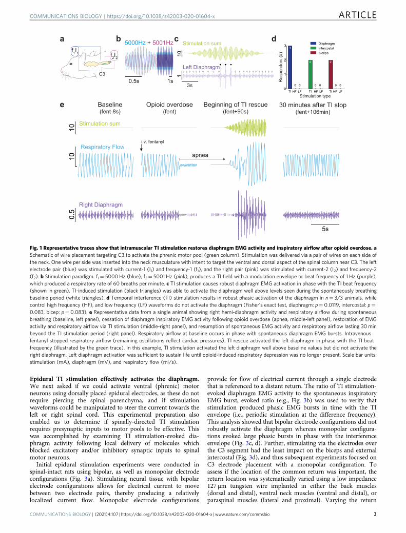
ResultsIntramuscular TI stimulation activates the diaphragm. We firstdetermined if TI stimulation, delivered via rapidly placed intra-muscular wires in the neck region (Supplementary Fig. 1) couldrobustly activate the diaphragm (Fig. 1a). When a 5 kHz carrierfrequency was used with an offset/beat frequency of 1 Hz(Fig. 1b), robust diaphragm contractions were induced at 60 timesper minute (Fig. 1c). To confirm that the observed diaphragmactivation was resulting from TI, we also stimulated with highfrequency kilohertz (HF, 5001 Hz, no offset) or low frequency(LF, 1 Hz) waveforms. These waveforms were unable to inducephasic diaphragm activation in any of the animals tested (Fig. 1d).Conversely, the TI stimulation paradigm activated the diaphragmin all animals tested (Fig. 1d), and the magnitude of the TI-evoked diaphragm burst was considerably greater than thespontaneously occurring EMG burst associated with inspiration(range: 270–931% increase). We next studied rats after opioidoverdose-induced respiratory depression, which is the cause ofmortality in the ongoing opioid epidemic4,22. Intravenous fenta-nyl (30 mcg/kg) produced respiratory apnea during which thediaphragm ceased rhythmic inspiratory contractions and arterialblood pressure decreased. Upon apnea, intramuscular TI stimu-lation was initiated and immediately produced robust diaphragmcontraction with restoration of inspiratory airflow (Fig. 1e).
The TI rescue paradigm was initiated using 5000+ 5001 Hzwaveforms shortly after the onset of opioid-induced apnea.Stimulation was delivered in 60 s epochs interspersed with brief 3s off periods which were used to determine if endogenousdiaphragm electromyography (EMG) activity had resumed(Fig. 2a, Supplementary Movie 1). The TI rescue effectivelysustained breathing throughout the period of opioid-inducedrespiratory depression, thus animals were eventually able toresume independent (volitional) breathing after 28 ± 9 min ofstimulation (range 17 to 40 min). Figure 2b and c illustrate therobust impact of TI stimulation on inspiratory airflow and arterialblood pressure during the period of opioid-induced respiratorydepression, and this stands in sharp contrast to the no-intervention control group. Prior to opioid overdose, therespiratory rate ranged from 99–108 breaths per minute acrossthe three experimental groups; all rats became apneic followingintravenous fentanyl (i.e., 0 breaths per minute). The 1 Hz TI beatfrequency was able to drive the respiratory rate duringstimulation at 60 breaths per minute. When the TI beat frequencywas increased to match the endogenous breathing rate (Matchedfreq.), diaphragm contractions occurred at an average 110 breathsper minute (Fig. 2d). This latter TI paradigm was sufficient toprevent arterial hypoxemia and also maintained arterial carbondioxide values within normocapnic ranges. In contrast, TIstimulation which evoked a lower rate of diaphragm contraction(60 breaths per minute) was sufficient to maintain arterialoxygenation but did not fully correct hypoventilation inducedarterial hypercapnia (Supplementary Fig. 2). The survival curvesshown in Fig. 2e serve to highlight the effectiveness ofintramuscular TI stimulation for sustaining breathing, as well asthe potential value of this approach in a first responder situationfollowing opioid overdose.
ARTICLE COMMUNICATIONS BIOLOGY | https://doi.org/10.1038/s42003-020-01604-x
2 COMMUNICATIONS BIOLOGY | (2021) 4:107 | https://doi.org/10.1038/s42003-020-01604-x | www.nature.com/commsbio
Epidural TI stimulation effectively activates the diaphragm.We next asked if we could activate ventral (phrenic) motorneurons using dorsally placed epidural electrodes, as these do notrequire piercing the spinal parenchyma, and if stimulationwaveforms could be manipulated to steer the current towards theleft or right spinal cord. This experimental preparation alsoenabled us to determine if spinally-directed TI stimulationrequires presynaptic inputs to motor pools to be effective. Thiswas accomplished by examining TI stimulation-evoked dia-phragm activity following local delivery of molecules whichblocked excitatory and/or inhibitory synaptic inputs to spinalmotor neurons.
Initial epidural stimulation experiments were conducted inspinal-intact rats using bipolar, as well as monopolar electrodeconfigurations (Fig. 3a). Stimulating neural tissue with bipolarelectrode configurations allows for electrical current to movebetween two electrode pairs, thereby producing a relativelylocalized current flow. Monopolar electrode configurations
provide for flow of electrical current through a single electrodethat is referenced to a distant return. The ratio of TI stimulation-evoked diaphragm EMG activity to the spontaneous inspiratoryEMG burst, evoked ratio (e.g., Fig. 3b) was used to verify thatstimulation produced phasic EMG bursts in time with the TIenvelope (i.e., periodic stimulation at the difference frequency).This analysis showed that bipolar electrode configurations did notrobustly activate the diaphragm whereas monopolar configura-tions evoked large phasic bursts in phase with the interferenceenvelope (Fig. 3c, d). Further, stimulating via the electrodes overthe C3 segment had the least impact on the biceps and externalintercostal (Fig. 3d), and thus subsequent experiments focused onC3 electrode placement with a monopolar configuration. Toassess if the location of the common return was important, thereturn location was systematically varied using a low impedance127 µm tungsten wire implanted in either the back muscles(dorsal and distal), ventral neck muscles (ventral and distal), orparaspinal muscles (lateral and proximal). Varying the return
e
a
f2I2
f1I1
C30.5s 1s
5000Hz + 5001Hzb
3s
101
Left Diaphragm
Stimulation sumc d
Baseline(fent-8s)
Opioid overdose(fent)
Beginning of TI rescue(fent+90s)
100.
510
5s
Respiratory Flow
Right Diaphragm
Stimulation sum
i.v. fentanyl
apnea
30 minutes after TI stop(fent+106min)
TI HF LF TI HF LF TI HF LF0
1
2
3
Stimulation type
DiaphragmIntercostalBiceps
3
0 0 0 0 0 0
2 2
Res
pond
ers
(#)
Fig. 1 Representative traces show that intramuscular TI stimulation restores diaphragm EMG activity and inspiratory airflow after opioid overdose. aSchematic of wire placement targeting C3 to activate the phrenic motor pool (green column). Stimulation was delivered via a pair of wires on each side ofthe neck. One wire per side was inserted into the neck musculature with intent to target the ventral and dorsal aspect of the spinal column near C3. The leftelectrode pair (blue) was stimulated with current-1 (I1) and frequency-1 (f1), and the right pair (pink) was stimulated with current-2 (I2) and frequency-2(f2). b Stimulation paradigm. f1= 5000Hz (blue), f2= 5001 Hz (pink), produces a TI field with a modulation envelope or beat frequency of 1 Hz (purple),which produced a respiratory rate of 60 breaths per minute. c TI stimulation causes robust diaphragm EMG activation in phase with the TI beat frequency(shown in green). TI-induced stimulation (black triangles) was able to activate the diaphragm well above levels seen during the spontaneously breathingbaseline period (white triangles). d Temporal interference (TI) stimulation results in robust phasic activation of the diaphragm in n= 3/3 animals, whilecontrol high frequency (HF), and low frequency (LF) waveforms do not activate the diaphragm (Fisher’s exact test, diaphragm: p= 0.0119, intercostal: p=0.083, bicep: p= 0.083). e Representative data from a single animal showing right hemi-diaphragm activity and respiratory airflow during spontaneousbreathing (baseline, left panel), cessation of diaphragm inspiratory EMG activity following opioid overdose (apnea, middle-left panel), restoration of EMGactivity and respiratory airflow via TI stimulation (middle-right panel), and resumption of spontaneous EMG activity and respiratory airflow lasting 30minbeyond the TI stimulation period (right panel). Respiratory airflow at baseline occurs in phase with spontaneous diaphragm EMG bursts. Intravenousfentanyl stopped respiratory airflow (remaining oscillations reflect cardiac pressures). TI rescue activated the left diaphragm in phase with the TI beatfrequency (illustrated by the green trace). In this example, TI stimulation activated the left diaphragm well above baseline values but did not activate theright diaphragm. Left diaphragm activation was sufficient to sustain life until opioid-induced respiratory depression was no longer present. Scale bar units:stimulation (mA), diaphragm (mV), and respiratory flow (ml/s).
COMMUNICATIONS BIOLOGY | https://doi.org/10.1038/s42003-020-01604-x ARTICLE
COMMUNICATIONS BIOLOGY | (2021) 4:107 | https://doi.org/10.1038/s42003-020-01604-x | www.nature.com/commsbio 3

location in this manner had no discernable impact on the abilityto activate the diaphragm (Fig. 3e). To verify this effect, wemodeled the impact of the location of the return electrode inaccordance with the experimental data shown in Fig. 3e. For thatpurpose, the return electrode was rotated in six positions aroundthe ventral side of the spinal cord, while maintaining a constantinter-electrode distance. This modeling produced a variability of±3%, and thus the simulation results confirm that the location ofthe return electrode had minimal impact on diaphragmactivation. To confirm that TI was required for in vivo diaphragmactivation with this electrode configuration, we tested the effect of
high frequency kilohertz waveforms with no offset (HF, 5001 Hz)or low frequency (LF, 1 Hz) waveforms on diaphragm activation.Neither of these waveforms were able to cause phasic diaphragmactivation similar to the response to TI (Fig. 3f).
We next tested if epidural TI stimulation could activate theparalyzed diaphragm after cervical spinal cord injury (schematicshown in Fig. 3g). We utilized a high cervical (C2) hemilesionmodel (C2Hx) which results in transient paralysis and chronicparesis of the ipsilateral hemi-diaphragm (Fig. 3h, left panel).Epidural TI stimulation using a C3 monopolar configurationrobustly activated the previously paralyzed hemi-diaphragm as
0
2
4
6
Flow
(ml/s
)
BL PF 5min 10min0
2
4
6
0
50
100
150
MA
BP
(mm
Hg)
BL PF 5min 10min0
50
100
150
MA
BP
(mm
Hg)
b
c
-20
0
20
0
100
200
a
Flow
(ml/s
)A
BP
(mm
Hg)
**
*
No interventionTI rescue (60 bpm)
Flow
(ml/s
)
-5 0 5 10 15Time (min)
BL PF 5min 10min
-5 0 5 10 15Time (min)
BL PF 5min 10min
No interventionTI rescue (60bpm)
-5 0 5 10 15Time (min)
-10 20 25 30 35
Stimulationi.v fentanyl
eNo interventionTI rescue (60 bpm)
-5 0 5 10 15Time (min)
0
20
40
60
80
100
Sur
viva
l (%
)
BL Stim 30 minpost stim
0
40
80
120
160TI rescue (60 bpm)TI rescue (Matched freq)* *
n.s.
Res
pira
tory
rate
(bpm
)
d
No interventionTI rescue (60bpm)
*
Fig. 2 Intramuscular TI stimulation (TI rescue) prevents fatal apnea by restoring diaphragm activation after opioid overdose. Spontaneously breathingrats under urethane anesthesia received an intravenous injection of fentanyl (30mcg/kg) sufficient to cause lasting respiratory suppression. Followingfentanyl dosing, animals received intramuscular TI stimulation or no intervention. Panel a shows an example of sustained restoration of respiratory airflowwhen the stimulation was initiated shortly after opioid overdose-induced apnea, and was delivered with a pattern of 60 s on and 3 s off until spontaneousbreathing resumed. Note: The gaps in the blood pressure traces indicate when arterial blood samples were taken. b Average respiratory airflow (two-wayANOVA: group F(1)= 40.84, p < 0.0001; time F(3)= 41.86, p < 0.0001; interaction F(3)= 21.4, p < 0.0001) and (c) mean arterial blood pressure (MABP)(n= 4 animals/group, two-way ANOVA: group F(1)= 19.81, p= 0.0008; time F(3)= 45.95, p < 0.0001; interaction F(3)= 7.82 p= 0.0037). The graybars indicate time periods selected for quantitative comparison (Baseline–BL, immediately Post Fentanyl–PF, five minutes–5 min, and ten minutes–10minafter fentanyl). The gray dots indicate data points from the animals used for the example traces in panel a, (mean+ 1 standard deviation; asterisk (*)indicates p < 0.05). d TI stimulation restored ventilation and prevented fatal apnea in all animals (TI rescue). TI stimulation with a 1 Hz beat frequencyproduced a respiratory rate of 60 breaths per minute (bpm) during stimulation. When the beat frequency was targeted to the endogenous respiratory rate,there was no difference in the evoked vs. spontaneous respiratory rate (n= 4 animals, two-way ANOVA: group F(1)= 22.64, p= 0.0002; time F(3)=3.85, p= 0.0407; interaction F(3)= 5.03, p= 0.0183). e Survival curve for no-intervention (n= 4 animals) and TI rescue (60 bpm) conditions (n= 4animals).
ARTICLE COMMUNICATIONS BIOLOGY | https://doi.org/10.1038/s42003-020-01604-x
4 COMMUNICATIONS BIOLOGY | (2021) 4:107 | https://doi.org/10.1038/s42003-020-01604-x | www.nature.com/commsbio

evidenced by large EMG bursts occurring in phase with the TIbeat frequency (Fig. 3h, right panel). We systematically evaluatedthis response after acute or chronic (10 month) spinal cord injuryin separate experimental cohorts. The C2Hx lesion immediatelyeliminated spontaneous EMG activity in the ipsilateral diaphragmin the acutely injured rats and caused a substantial reduction inspontaneous EMG output in those with chronic injury (Fig. 3i).
For each condition (e.g., spinal intact, acute and chronic injury),we determined the stimulus current ratio (i.e., ratio of the two TIcurrents) that evoked the largest peak diaphragm EMG output.Figure 3i shows a summary of the evoked diaphragm EMGresponse at these empirically determined optimal currents.Collectively, these data show that epidural TI stimulationeffectively activates the diaphragm after spinal cord injury. The
0.250.5
0.751 1
2345
0.250.5
0.751 5
1015
C3C4C5
Diaphragm(n=2)
Biceps(n=2)
Evo
ked
Rat
io (p
hasi
c ac
tivity
/toni
c)
0.250.5
0.751 1
23456
External Intercostal(n=2)
A1 A2 A3B1 B2 B3
2s
1mV
Integrated LDIA
TI envelope
Left Diaphragm
A1 A2 A3 B1 B2 B3 A1 A2 A3 B1 B2 B3 A1 A2 A3 B1 B2 B3
Intercostal(n=5)
Nor
mal
ized
Evo
ked
Rat
io @
1m
A
Diaphragm(n=5)
Biceps(n=5)
Stimulus current (mA)0 0.2 0.4 0.6 0.8 1 1.2 1.4
Evo
ked
Rat
io
01234567 Lateral and proximal
Ventral and distalDorsal and distal
00.10.20.30.40.50.60.70.80.9
1ANOVA p = 0.014
00.10.20.30.40.50.60.70.80.9
1ANOVA p = 0.003
00.10.20.30.40.50.60.70.80.9
1ANOVA p = 0.001A1
A2
A3
B1
B2 B3
a
b
c d
eStim
ulat
ion
curr
ent (
mA
)C3
C4
C5
0
1
2
3
0
1
2
3
0
1
2
3
Left
diap
hrag
m p
eak
(a.u
.)
0
1
2
32 s
1 m
V
Intact
Acute SCI
Chronic SCI
i
g
*
IntactAcute SCIChronic SCI
Spontaneous breathing(no TI)
TI optimized forintact condition
(Lat 1320:600μA Med)
TI optimized foracute SCI
(Lat 360:10800μA Med)
TI optimized forchronic SCI
(Lat 360:600μA Med)
f1 f2
n.s.n.s.n.s.
f
f2I2f1I1
C3 C5phrenic motor pool
h
TI HF LF TI HF LF TI HF LF0
1
2
3
4
Stimulation type
DiaphragmIntercostalBiceps
4
0 01
3
Res
pond
ers
(#)
0 0 0 0
10.
50.
5
50100
5s
Chronic SCI TI activation
Left Diaphragm
Right Diaphragm
Arterial Blood Pressure
Stimulation sum
01234567
ANOVA p = 0.83
ventr
aldis
taldo
rsal
distal
later
alpr
oxim
al
Avg
Evo
ked
Rat
io@
1mA
COMMUNICATIONS BIOLOGY | https://doi.org/10.1038/s42003-020-01604-x ARTICLE
COMMUNICATIONS BIOLOGY | (2021) 4:107 | https://doi.org/10.1038/s42003-020-01604-x | www.nature.com/commsbio 5

afferent and efferent innervation of the phrenic motor nucleus,and the local tissue architecture, will be different when comparingacute to chronic spinal cord injury. TI stimulation was effective atboth time points, indicating that time-dependent changes in thespinal cord after injury do not impair the effectiveness of thismethod.
Current steering in the spinal cord. One of the theoreticalbenefits of TI stimulation is the ability to shift the focal pointwhere the electric fields overlap (Supplementary Fig. 3) to activateneurons deep within the target tissue and distant from the elec-trodes6. To determine if the focal point in the cervical spinal cordcould be steered during epidural TI stimulation, we varied theratio of the two current sources while maintaining a constant totalcurrent. This experiment was done using electrodes placed lat-erally or spanning the dorsal surface of the spinal cord. Figure 4ashows that TI stimulation delivered using lateral electrode pla-cement (1.5 mm inter-electrode distance) produced rhythmicactivation of the ipsilateral (left) but not contralateral (right)diaphragm at the TI beat frequency. When the total currentacross the two electrodes was held constant at 1800 µA, ratios of1:1, 1:2, and 1:3 produced the largest phasic diaphragm activation.When total current was 2400 µA, ratios of 3:1, 2:1, and 1:1 pro-duced the largest phasic diaphragm activation. Figure 4b showsthe results of the TI current steering experiment using thebilateral electrode configuration (3 mm inter-electrode distance).In contrast to the results with lateral placement, the bilateralconfiguration tended to produce bilateral diaphragm activation.Current steering was still present as ratios of 1:1 and 1:2 weremost likely to produce bilateral diaphragm activation with totalstimulus currents of 1800 and 2400 µA.
Both the lateral and bilateral electrode configurations evokedsustained and non-rhythmic diaphragm activity when the currentratios were 0:1 or 1:0 (i.e., a single high frequency waveform). Theamplitude of this tonic diaphragm activation at 0:1 or 1:0 currentratios was attenuated as the stimulation progressed, and this canbe seen in the example traces shown in Fig. 4a, b.
Blocking synaptic inputs alters the TI stimulation response.Inspiratory-related depolarization of phrenic motor neuronsarises primarily from excitatory bulbospinal pathways in theventrolateral white matter of the mid-cervical spinal cord23.There also exists a complex propriospinal network that can exciteor inhibit phrenic motor neurons24 (Fig. 5a). If the diaphragmactivation resulting from TI stimulation is primarily due todepolarization of excitatory presynaptic inputs, focal delivery ofglutamate receptor antagonists to the phrenic motor pool shouldsubstantially reduce or eliminate the evoked response. Con-versely, if the TI stimulation paradigm is also activating inhibitorysynaptic inputs, then antagonizing inhibitory receptors in thephrenic motor pool should increase the diaphragm response. Totest these possibilities, the epidural electrode configuration wasmodified to enable intraspinal microinjections without movingthe electrode (Fig. 5b). After establishing a baseline TI response,we then intraspinally injected a mixture of glutamatergic receptorantagonists to block NMDA (AP5), and non-NMDA (CNQX)receptors, or a strychnine/bicuculline mixture to block glycineand GABAA receptors, respectively. Each group was then subse-quently injected with the other drug mixture (Fig. 5c). DeliveringCNQX/AP5 to the spinal cord almost completely eliminatedendogenous (spontaneous) inspiratory diaphragm EMG bursts(Fig. 5d). In contrast, strychnine/bicuculline had little to no effecton the amplitude of endogenous bursts (Fig. 5e). Followingapplication of both drug cocktails there was near completeelimination of endogenous diaphragm EMG bursts (Fig. 5f). Theresponse to TI stimulation after the spinal drug injection wasevaluated at the medial stimulus current which evoked an EMGburst that was 25% larger than endogenous activation. Blockinglocal spinal excitatory neurotransmission substantially reduceddiaphragm activation during TI stimulation (Fig. 5gi), whereasblocking inhibitory neurotransmission had minimal impact(Fig. 5gii). The mean data shown in Fig. 5h demonstrate thatfollowing spinal delivery of CNQX/AP5 the evoked diaphragmresponse was decreased by 80, 58, and 54% at lateral currents of100, 200, and 300 µA, respectively. The subsequent delivery of
Fig. 3 Effect of electrode configuration on diaphragm activation and diaphragm response to epidural TI stimulation. a Initial testing to determine leftdiaphragm (LDIA), bicep, and external intercostal activation utilized an epidural electrode grid spanning C3–C5 and with three bipolar (A1, A2, and A3) andthree monopolar configurations (B1, B2, and B3); note: electrode size is exaggerated for schematic, actual wire width is 25 µm. b The ratio of TI stimulation-evoked EMG output to spontaneous EMG activity was used to quantify the response. The EMG activity during the peak of the TI envelope (green shadedboxes) was divided by the activity in the trough of the envelope (gray shaded boxes) for the period of stable stimulation (i.e., excluding the ramp and dampphases). c Heat maps which display the activation of the diaphragm, biceps, and external intercostal muscles as a function of stimulus amplitude andelectrode configuration (waveform current ratio 1:1 in these examples). The particular electrode configuration is shown on the bottom of the panel. d Theaverage evoked diaphragm EMG activity (mean+ 1 standard deviation) during TI stimulation using 1 mA in both waveforms (asterisk (*) indicates p < 0.05tukey’s post-hoc). Electrode configuration B1 (C3 monopolar stimulation) activated the diaphragm with minimal off-target biceps activation (n= 5 animals,Kruskal–Wallis one-way ANOVA on ranks: diaphragm H(5)= 20.73, p < 0.001; biceps H(5)= 22.83, p < 0.001; intercostal H(5)= 23.09, p < 0.001). eEffect of varying the location of the current return on diaphragm activation during C3 monopolar TI stimulation. Three different current return electrodelocations were used (RM two-way ANOVA current F(26)= 8.79, p < 0.001; return location F(2)= 2.51, p= 0.092; n= 3 animals). f Temporal interference(TI) stimulation phasically activated the diaphragm in n= 4/4 animals, while high frequency (HF), and low frequency (LF) waveforms did not phasicallyactivate the diaphragm (Fisher exact test; diaphragm, p= 0.002; intercostal, p= 0.333; bicep, p= 0.018). g Schematic of the mid-cervical spinal cord injury(purple indicates spinal hemilesion) and electrode locations. h Example data from a rat with chronic (10 months) cervical spinal cord injury. At baseline(without stimulation, left panels), the left hemi-diaphragm is inactive while the right hemi-diaphragm shows rhythmic bursting. Electrocardiogram (ECG)activity is present in both traces. TI stimulation (right panel) immediately activates the left hemi-diaphragm (black arrowheads) and produces small burstsin the right diaphragm (gray arrowheads). Spontaneous activity in the right hemi-diaphragm (white arrowheads) is uninterrupted. Units: stimulation (mA),diaphragm (mV), arterial blood pressure (mmHg). Panel i provides additional examples of diaphragm EMG output and mean responses. The left panelshows diaphragm EMG during spontaneous breathing in spinal intact, acute, and chronic spinally injured rats (mean diaphragm bursting is shown in theplots at the bottom of the panel). Proceeding left to right across the figure, evoked EMG responses are shown using stimulus current ratios optimized foreach condition (i.e., spinal intact, acute and chronic injury). Spontaneous bursts (white arrowheads) are present in the spinal intact animal but are absentafter acute and chronic SCI. TI stimulation effectively activates the diaphragm (black arrowheads) in all three conditions. Plots: one way ANOVAspontaneous breathing F(2)= 6.186, p= 0.024; optimized for intact F(2)= 0.863, p= 0.458; optimized for acute SCI F(2)= 0.115, p= 0.893; optimizedfor chronic SCI F(2)= 1.914, p= 0.209; asterisk (*) indicates p < 0.05 tukey’s post-hoc; gray filled dots indicate animals used in example traces (intact, n=4 animals; acute SCI, n= 3 animals; chronic SCI, n= 4 animals).
ARTICLE COMMUNICATIONS BIOLOGY | https://doi.org/10.1038/s42003-020-01604-x
6 COMMUNICATIONS BIOLOGY | (2021) 4:107 | https://doi.org/10.1038/s42003-020-01604-x | www.nature.com/commsbio

strychnine/bicuculline had no discernable additional impact onthe TI response. When inhibitory neurotransmission was blockedfirst (i.e., before the excitatory blockade), there was also littleimpact on the TI-evoked diaphragm activation (Fig. 5i). Sub-sequent delivery of CNQX/AP5, however, caused a considerableattenuation of diaphragm activation during TI stimulation(Fig. 5i). Since local spinal blockade of excitatory synaptic inputsreduced, but did not eliminate, the response to TI stimulation;activation of presynaptic inputs to phrenic motor neurons is
implicated as a component of the mechanism driving TI-induceddiaphragm muscle activation.
To demonstrate that diaphragm EMG activation was not theresult of extracellular electric field propagation directly depolar-izing the diaphragm myofibers, TI stimulation was repeated afterintravenous delivery of a drug to block acetylcholine binding atthe neuromuscular junction (pancuronium bromide, 2.5 mg/kg,i.v., Hospira). The diaphragm could not be activated by TIstimulation after pancuronium bromide, indicating that the
1mA
20s
0.1
Integrated left diaphragm EMG
Integrated right diaphragm EMG
0.1
Left lateral to left medial (1.5mm)
Left lateral to right lateral (3mm)
1800
μA
Left Lat
Left Med
1:0 0:17:1 5:1 4:1 2:1 1:1 1:2 1:4 1:5 1:7
1mA
20s
0.1
0.1
Integrated left diaphragm EMG
Integrated right diaphragm EMG
1:0 0:17:1 5:1 3:1 2:1 1:1 1:2 1:3 1:5 1:7
2400
μA
Left Lat
Right Lat
a
b
Left Lat Right Lat
phrenic motor pool
Left Dia Right Dia
Left MedLeft Lat
phrenic motor pool
Left Dia Right Dia
1:0 5:1 3:1 2:1 1:1 1:2 1:3 1:5 0:10
1
2
3
4
5
6
7
Diap
hrag
m ev
oked
ratio
at 18
00µA
1:0 5:1 3:1 2:1 1:1 1:2 1:3 1:5 0:10
1
2
3
4
5
6
7
Diap
hrag
m ev
oked
ratio
at 24
00µA
1:0 5:1 3:1 2:1 1:1 1:2 1:3 1:5 0:10
1
2
3
4
5
6
1:0 5:1 3:1 2:1 1:1 1:2 1:3 1:5 0:10
1
2
3
4
5
6
Left diaphragm
Right diaphragm
Left diaphragm
Right diaphragm
Diap
hrag
m ev
oked
ratio
at 18
00µA
Diap
hrag
m ev
oked
ratio
at 24
00µA
Ratio p=0.0001
Ratio p=0.0003
Ratio p=0.0001
Ratio p=0.0007
Fig. 4 Current steering during epidural TI stimulation. Using diaphragm EMG as the outcome measure, the steerability of the TI focal point was examinedby varying the ratio of the two stimulus currents using two inter-electrode distance. Stimulating electrodes were placed on the left side of the spinal cord(a, 1.5 mm apart) or spanning the dorsal surface (b; 3 mm apart). The left side of panel a shows an example of rectified and integrated diaphragm activitywhen TI was delivered with lateral electrode placement and a current sum of 1800 µA. In this example it can be appreciated that varying the TI current ratiodramatically alters the magnitude of phasic diaphragm activation, and tonic diaphragm activation occurs at current ratios of 1:0 and 0:1. The mean data(right panels) show a statistical effect of current ratio at currents sums of 1800 µA (Friedman’s two-way ANOVA controlling for diaphragm side effect ofcurrent ratio Chi-squared(8)= 33, p < 0.0001) and 2400 µA (Friedman’s two-way ANOVA controlling for diaphragm side effect of current ratio Chi-squared(8)= 23.96, p= 0.0023). The left side of panel b shows an example of diaphragm activity evoked by stimulating the spinal cord using an inter-electrode distance of 3 mm and a current sum of 2400 µA. In this example, TI stimulation induced a robust bilateral and phasic diaphragm EMG activationat a current ratio of 1:2. The mean data show an effect of current ratio at sum current of 1800 µA (Friedman’s two-way ANOVA controlling for diaphragmside effect of current ratio Chi-squared(8)= 29.33, p= 0.00003) and at 2400 µA (Chi-squared(8)= 26.99, p= 0.00007). Note we lowered inspired CO2
to reduce endogenous diaphragm activity during these trials, small bursts are still present (inset panel a), but at much lower amplitude than the TI-evokedbursts (n= 4 animals).
COMMUNICATIONS BIOLOGY | https://doi.org/10.1038/s42003-020-01604-x ARTICLE
COMMUNICATIONS BIOLOGY | (2021) 4:107 | https://doi.org/10.1038/s42003-020-01604-x | www.nature.com/commsbio 7

response requires activation of cholinergic synaptic inputs to thediaphragm (Supplementary Fig. 4).
Stimulation using alternative waveforms. Stimulation at kilo-hertz frequencies (e.g., 5001 Hz) with a single waveform didnot induce diaphragm activity when applied intramuscularly(Fig. 1d, Supplementary Movie 2) or epidurally (Fig. 3f).Similarly, intramuscular or epidural stimulation with a sinewave at the beat frequency, with no carrier frequency (i.e., 1Hz) did not activate the diaphragm. TI stimulation in thesesame animals elicited a robust diaphragm burst
(Supplementary Fig. 5a). Square wave stimulation at 50 pulsesper second (pps) was able to elicit compound muscle actionpotentials in the diaphragm with concurrent biceps activation.These muscle action potentials did not summate into a sus-tained diaphragm burst as occurs during inspiration (Supple-mentary Fig. 5b). Square wave stimulation at 300 pps evokeda square wave diaphragm burst (Supplementary Fig. 5c) con-current with large amplitude activation of the biceps muscleon the corresponding side. In addition, both the 50 pps and300 pps stimulation caused widespread muscle activationthroughout the rats.
f2I2f1I1
C3 C5
a
SRpre
3μl CNQX/AP5(+)
1μl Strych/Bic(-)
SR+
SR-
1μl Strych/Bic(-)
3μl CNQX/AP5(+)
SRpostE
E
EE
Medial Electrode
LateralElectrode
PhrenicMotor Neuron
inhibitoryinputs
excitatoryinputs
c
b
d
200ms
1au
Pre CNQX/AP5Post CNQX/AP5
e
200ms
1au
Pre Strych/BicPost Strych/Bic f
200ms
1au
Pre AntagonistsPost Antagonists
giMed 2140:300µA Lat
Pre CNQX/AP5Med 2140:300µA Lat
Post CNQX/AP5
2s1au
giiMed 1240:200µA Lat
Pre Strych/BicMed 1240:200µA Lat
Post Strych/Bic
2s
1au
Evo
ked
burs
t (m
V)
0
0.2
0.4
0
0.2
0.4
0.2
0.4
0
0.2
0.4
0.6
0
0.2
0.4
0.6
0
1
2
Pre CNQXAP5
StrychBic
Pre CNQXAP5
StrychBic
Pre CNQXAP5
StrychBic
Pre CNQXAP5
StrychBic
Pre CNQXAP5
StrychBic
Pre CNQXAP5
StrychBic
Evo
ked
burs
t (m
V)h
i
0
**
**
**
**
**
100μA lateral 200μA lateral 300μA lateral
100μA lateral 200μA lateral 300μA lateral
ARTICLE COMMUNICATIONS BIOLOGY | https://doi.org/10.1038/s42003-020-01604-x
8 COMMUNICATIONS BIOLOGY | (2021) 4:107 | https://doi.org/10.1038/s42003-020-01604-x | www.nature.com/commsbio

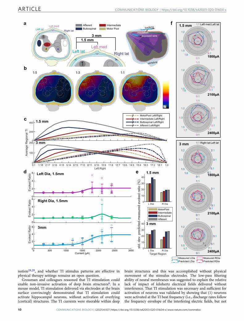
Computational modeling successfully predicts current steeringand respiratory response. Simulation models of the electric tissueexposure have been established (Fig. 6a; see Experiment 5 in the“Methods” section), reproducing the asymmetric, 1.5 mm-distance and the symmetric, 3 mm-distance epidural stimulationexperimental configurations from Fig. 4c, d. Based on the com-puted electric fields (Supplementary Fig. 3b), the exposure ofpotential candidate regions that could affect the respiratoryresponse (‘Motor Pool’, ‘Intermediate Gray Matter’, ‘Bulbospinal’,and ‘Afferent’ pathways) was quantified in terms of the TI metricfrom Grossman et al.6: Simulated TI field distributions for a rangeof current steering ratios, as well as the associated averageexposure of the four regions of interests can be seen in Fig. 6b, c.To translate exposure to evoked respiratory response, experi-mental data obtained by varying the current while maintaining a1:1 current ratio (as shown in Fig. 3e) has been fitted (Fig. 6d; seeExperiment 5 in the “Methods” section). By combining theseresponse functions with the TI metrics extracted from the elec-tromagnetic simulations it becomes possible to predict the evokedrespiratory response for any set of current steering parameters.The differences between the predicted and the experimentallymeasured evoked ratios are in the range of 14–30% (relative to theaverage evoked ratio for a given total current; Fig. 6e). Figure 6fshows a direct comparison of the computational predictions withthe experimental results from Fig. 4a, b. Considering that theexperimental variability related to the biological response, intra-subject anatomical variability, and/or electrode placementreproducibility already accounts for 20%, which does not yetinclude the modeling uncertainty, it is concluded that computa-tional modeling successfully predicts current steering andrespiratory response. The 20% are estimated based on (1) the 21%standard deviation of the evoked responses of the left and rightdiaphragm for corresponding—i.e., mirrored—steering ratios inwhat should be a symmetric setup, see Fig. 4d, and (2) the averagevariability in the experimental data shown in Fig. 3e. The sim-plifications and limitations of the computational model are dis-cussed in more detail in the “Methods” section.
Computational modeling explains the counter-intuitivecurrent-ratio-dependence of the respiratory response, con-firms the relevance of the modulation envelope magnitudemetric. While it can at first be counter-intuitive that shifting the
current ratio towards increased prominence of the right electrode(while maintaining the total current) results in a converse shift ofthe evoked activity to the left diaphragm and vice versa, this is infact predicted by the computational model. It can be understoodin light of the observation from Grossman et al.6 that theamplitude of the modulation envelope predicts TI stimulation.For mostly aligned field orientations, the modulation envelopeamplitude is simply twice the amplitude of the smaller of the twoelectric fields. For field strengths that decay with increasing dis-tance from the electrodes, the maximal modulation envelopeamplitude is therefore found between the electrodes, at locationswhere the two field strengths have a comparable magnitude. Asthe current ratio shifts towards the right electrode, the fieldgenerated by the left electrode decreases (proportionally to the leftelectrode current) and the location where the two fields arecomparable and temporal interference is maximal, shifts towardsthe left side (Fig. 4). The different candidate regions are affectedto a different degree by that shift (Fig. 6c) and it is suggested bythe deviations between computational predictions and experi-mental data (Fig. 6f) that the TI exposure of the phrenic motorpool region is principally responsible for the evoked respiratoryresponse, as it provides the most accurate predictions.
DiscussionBreathing is fundamental to life and can be severely compromisedin many neuromuscular diseases and injuries, as well as drugoverdose. Impaired respiratory muscle activation is a leadingcontributor to morbidity and mortality across a range of condi-tions, and central apnea (i.e., absent neural drive to breathe) canbe fatal if not treated immediately. Here we show the TI stimu-lation is a novel technology for rapidly restoring breathing afteropioid overdose. Further, we demonstrated that TI stimulationprovides a new experimental avenue in the rapidly growing areaof epidural stimulation following spinal cord injury. Thus, TI is apromising new modality for respiratory stimulation.
Interferential current stimulation paradigms have been used inphysical therapy since at least the 1950s25,26. Initially described byNemec27 as a transcutaneous stimulation method, TI stimulationhas been suggested to have less superficial sensory impact com-pared to other stimulation modalities, and to be effective forreducing pain when applied with surface (skin) electrodes25.However, clinical studies have not provided support for this
Fig. 5 Pharmacologically probing the contribution of synaptic input to phrenic motor neurons in TI stimulation-induced diaphragm activation. Todetermine if TI stimulation activates the diaphragm due to direct depolarization of phrenic motor neurons or through activation of synaptic inputs to thesecells, we utilized focal injections of glutamate, glycine, and GABAA receptor antagonists. a Diagram of the phrenic motor neuron pool illustrating excitatory(green) and inhibitory (pink) presynaptic inputs. b Schematic of the epidural stimulation grid and unilateral (left) intraspinal drug injection. c Summary ofthe experimental paradigm. In one cohort (n= 5 animals), excitatory presynaptic inputs were antagonized first, followed by inhibitory antagonists. Inanother cohort (n= 5 animals) the order was reversed. Circles with E indicate when endogenous activity was measured, SR indicate when stimulusresponse curves were performed. Panels d–f provide cycle triggered averages of endogenous diaphragm EMG activity (using the activity in the right(unblocked) hemi-diaphragm as the trigger). d Before (blue) and after (green) intraspinal injection of CNQX/AP5 (n= 5 animals). e Before (blue) and after(pink) intraspinal injection of strychnine/bicuculline (n= 5 animals). f Before (blue) and after (brown) both sets of drugs were injected (n= 10 animals).The solid line is average of breaths over the first 30 s post-injection and the dashed line is the average of the breaths over the last 30 s. gi Examplediaphragm EMG illustrating the magnitude of TI stimulation-evoked responses before (blue, pre CNQX/AP5) and after glutamate receptor antagonism(green, post CNQX/AP5). Endogenous bursts (white arrowheads) were eliminated after CNQX/AP5 indicating that excitatory drive to the phrenic motorpool was effectively blocked. Black arrowheads indicate the peak TI envelope, marking evoked bursts. gii Example diaphragm EMG illustrating themagnitude of evoked responses before (blue, pre-strychnine/bicuculline) and after glycinergic and GABAergic receptor antagonism (pink, post strychnine/bicuculline). Stimulus current on the medial:lateral wires are shown above the traces. h Impact of CNQX/AP5 followed by strychnine/bicuculline on the TI-evoked diaphragm EMG burst. (n= 5 animals). i Impact of strychnine/bicuculline followed by CNQX/AP5 on the TI-evoked diaphragm EMG burst (n= 5animals). In panels h and i, data are shown for 100, 200, and 300 µA lateral currents with medial current standardized at the value which evokeddiaphragm EMG activity 25% above endogenous (pre-drug) baseline bursting. All data presented as mean +1 SD (*p < 0.05 post hoc, panel h at 100 µA:one-way ANOVA F(2)= 44.77, p < 0.001; all other panels: Kruskal–Wallis one-way ANOVA on ranks; panel h at 200 µA H(2)= 6.860, p= 0.032; panel hat 300 µA H(2)= 6.720, p= 0.035; panel i at 100 µA H(2)= 9.380, p= 0.009; panel i at 200 µA H(2)= 9.420, p= 0.009; panel i at 300 µA H(2)=9.380, p= 0.009).
COMMUNICATIONS BIOLOGY | https://doi.org/10.1038/s42003-020-01604-x ARTICLE
COMMUNICATIONS BIOLOGY | (2021) 4:107 | https://doi.org/10.1038/s42003-020-01604-x | www.nature.com/commsbio 9
notion28,29, and whether TI stimulus patterns are effective inphysical therapy settings remains an open question.
Grossman and colleagues reasoned that TI stimulation couldenable non-invasive activation of deep brain structures6. In amouse model, TI stimulation delivered via electrodes at the brainsurface convincingly demonstrated that TI stimulation couldactivate hippocampal neurons, without activation of overlying(cortical) structures. The TI currents were steerable within deep
brain structures and this was accomplished without physicalmovement of the stimulus electrodes. The low-pass filteringability of neural membranes was suggested to explain the relativelack of impact of kilohertz electrical fields delivered withoutinterference. That TI stimulation was necessary and sufficient foractivation of neurons was validated by showing that (1) neuronswere activated at the TI beat frequency (i.e., discharge rates followthe frequency envelope of the interfering electric fields, but not
backing
insulationexposed wire
vertebra
0
200
400 1.5 mmc
0:1 1:18 2:17 3:16 4:15 5:14 6:13 7:12 8:11 9:10 10:9 11:8 12:7 13:6 14:5 15:4 16:3 17:2 18:1 1:0Left:Right
0
100
200
Ave
rage
Reg
iona
l TI
3 mm
d
L Dia R Dia0
10
20
30
40
1.5 mme
L Dia R DiaTarget Region
0
10
20
30
40
std.
dev
. pre
dict
ed-m
easu
red
evok
ed ra
tio (%
)
3 mm
MotorPoolIntermediateBulbospinalAfferent
1
2 34
Motor PoolIntermediate
BulbospinalAfferent
Left latLeft med
Right lat
1
Left latLeft med
Right lat
1.5 mm3 mm
a
1:5 1:3 1:1b
Measured LDiaPredicted LDia
Measured RDiaPredicted RDia
3:1
2:1
1:1 Left med:Left lat
1:2
1:3
1:51:8
0:1 1800µA
1.5 mmf
2:1
1:1
1:2
1:51:6
0:1 2100µA
3:1
2:1
1:1
1:2
1:3
1:51:7
0:1 2400µA
3:1
2:1
1:1 Right lat:Left lat
1:2
1:3
1:51:8
0:1 1800µA
3 mm
2:1
1:1
1:2
1:51:6
0:1 2100µA
3:1
2:1
1:1
1:2
1:3
1:51:7
0:1 2400µA
5:18:1
5:16:1
5:17:1
5:18:1
5:16:1
5:17:1
MotorPool Left/RightIntermediate Left/RightBulbospinal Left/RightAfferent Left/Right
////
12
34
0
2
4
6
Evo
ked
Rat
io
0
2
4
6
Evo
ked
Rat
io
0 500 1000 1500 2000 2500 3000Current (μA)
0
2
4
6
Evo
ked
Rat
io
Left Dia, 1.5mm
Right Dia, 1.5mm
3mm
ARTICLE COMMUNICATIONS BIOLOGY | https://doi.org/10.1038/s42003-020-01604-x
10 COMMUNICATIONS BIOLOGY | (2021) 4:107 | https://doi.org/10.1038/s42003-020-01604-x | www.nature.com/commsbio
the carrier frequency); (2) histological verification of neuronalactivation (c-fos) unique to TI stimulation, and (3) functionalmapping to demonstrate selective movement with stimulation ofspecific brain regions.
The results of Grossman et al. raise the possibility of treatingconditions requiring deep brain stimulation (e.g., Parkinson’sdisease, depression) using electrodes at the surface of the brain todeliver TI stimulation6. The success of such approaches, however,will depend on the ability to use TI stimulation for focal activa-tion of neuronal targets. In this study, we expand the potentialreach of TI technology by showing that TI stimulation caneffectively activate breathing by targeting the spinal cord.Respiratory control neurons in the medulla and pons areembedded within a complex brainstem network involved withmany aspects of autonomic and somatic motor control30–32. Forthis reason, we focused our efforts on targeting spinal respiratorymotor neurons, which comes with less potential for serious offtarget effects (as compared to brainstem targeting) if the inter-ferential currents are not localized to a relatively focal region.
The appeal of the intramuscular TI stimulation approachdescribed here is the rapidity and ease of electrode placement.Adaptation and refinement of this technology for clinical usecould provide, for example, the first responder with another toolfor rapidly/immediately restoring breathing should otherapproaches prove ineffective (e.g., bag-mouth valve breathing,naloxone). Naloxone, when available, can be highly effective atrestoring breathing after opioid overdose, but is much lesseffective during dual overdose conditions (e.g., opioids+ alcohol,a frequent occurrence). Naloxone also immediately reversesanalgesic effects of opioid and can thus leave the first responderwith a patient in acute opioid withdrawal stages. By utilizing non-pharmacological means, TI stimulation can restore breathingwithout the specific need to identify the depressant agent anddoes not reverse the analgesic effects of opioids while naloxonedoes, potentially leaving individuals in pain.
Electrical stimulation of the spinal cord, either using epiduralor intraspinal electrodes, is rapidly advancing as a means ofimproving autonomic and somatic motor function following SCI.Intraspinal electrodes can activate specific motor circuits33–35,and epidural stimulation is often used to provide a non-specificincrease in the excitability of neuronal circuits15,18,36 or targetedactivation of sensory afferent pathways20. Here, we show that TIstimulation via epidurally placed electrodes provides the ability tosteer interferential currents towards regions of interest, similar tothe prior description of deep brain stimulation6. The ability tosteer currents within the spinal cord, as supported by our in vivoand in silico data, represents a potential advantage of TI ascompared to square wave epidural stimulation9. In our study,cervical spinal cord-directed TI stimulation produced substantialdiaphragm activation due to depolarization of phrenic motor
neurons. The phrenic motor neuron pool is in close proximity tothe forelimb motor pools, and we often observed concurrentactivation of the biceps muscles, albeit at much lower levelscompared to the diaphragm. Off-target muscle activation may bemitigated in larger species or with refinements in electrode con-figuration and/or location to improve the accuracy of currentsteering, which could be guided by additional modelingexperiments.
Local synaptic blockade experiments have advanced theunderstanding of how TI stimulation is working and confirm TIfields overlap within the immediate vicinity of the phrenic motorpool. We found that TI stimulation-induced diaphragm activa-tion was considerably attenuated by blocking excitatory synapticinputs in the cervical spinal cord. These results indicate thatactivation of excitatory presynaptic inputs are a fundamentalcomponent of the mechanism by which spinal-directed TI sti-mulation activates motor neurons. However, excitatory synapticinputs are not required since the pharmacological blockadeexperiments show that it is possible for TI stimulation to activatephrenic motor neurons even in the near complete absence ofpresynaptic input. Thus, the field is likely overlapping within thephrenic motor nucleus and this suggests that TI stimulation maybe used as a method to stimulate motor function following SCI inmotor pools lacking presynaptic inputs. In silico models of spinalcord stimulation supported this view, indicating that TI stimu-lation is reaching the ventrolateral spinal cord and the phrenicmotor neurons which activate the diaphragm.
In conclusion, we have expanded the scope of applications forTI technology by showing activation of spinal neuronal pathwaysto restore breathing during hypoventilation or apnea. Furtherrefinement of the technology may provide a new tool for rapidlyrestoring breathing after drug overdose or other conditions whichimpair or eliminate the ability spontaneous respiratory muscleactivation.
MethodsStudy design. The in vivo experiments were performed using adult SpragueDawley rats (n= 47) with procedures that were proved by the University of FloridaIACUC. Both male and female rats were studied (n= 30 males, n= 17 females);the number of each sex is noted in the figure legends. The number of animals foreach cohort was determined prior to initiation of the experiments and was based onpreliminary experiments not included in the manuscript. The preliminary workindicated a robust impact of TI stimulation on diaphragm activation. This enableda power calculation suggesting that n= 4 animals per group would be sufficient tostatistically differentiate the impact of temporal interference for the primary out-come of diaphragm EMG activity. The calculation was done based on an expecteddifference of means (e.g., stimulation vs. no stimulation) of 75% (arbitrary units),expected standard deviation of 25%, power of 0.9 and alpha of 0.05. Experimentswere excluded from analysis if there was an unequivocal mechanical failure (e.g.,broken electrode array, stimulator battery failure, heating pad failure). We did nottest for outliers in the data, and included all data from each set of experiments. Asall in vivo experiments were terminal, the end points were predefined.
Fig. 6 Computational prediction of TI stimulation-induced evoked response and current steering. a Simulation setup reproducing the experimentalsetup. Three electrodes are placed dorsally in the epidural space of the spine (left lateral, right lateral, and ‘Left’ medial), while a shared return electrode isplaced ventrally. TI modulation amplitude maps are generated using two different configurations of channel pairs: the ‘1.5 mm’ configuration using the leftlateral and the medial electrodes, as well as the ‘3mm’ configuration using the left and the right lateral electrodes (naming reflects the electrode spacing).Four candidate target regions have been defined (shown as colored ellipses). b By changing the current ratio, the distribution of the TI modulationamplitude can be steered laterally. c shows for both configurations, how changing the ratio affects the averaged TI field of the left (continuous) and right(dashed) target regions for a given total current (graphs are normalized to 2mA). d To translate TI modulation amplitude field to evoked left and rightdiaphragm responses (evoked ratio) sigmoid/Gaussian curves are fitted to the measured current strength-dependent responses (1:1 current ratio). Thedata points are the experimental data that the model (thick solid line) is derived from. e The deviation (relative standard deviation) between measurementsand simulation predictions of the evoked left and right diaphragm responses have been quantified for predictions based on the simulated TI exposure of thefour candidate target regions. Motor Pool-based predictions provide the best results. Panel f shows, as a function of the current ratio, polar plots comparingthe experimentally measured (solid line) left (blue) and right (red) diaphragm evoked ratio to the simulation predictions (dashed line) for a range of totalcurrents and for the two exposure configurations—here only the superior predictions based on ‘Motor Pool’ exposure are shown.
COMMUNICATIONS BIOLOGY | https://doi.org/10.1038/s42003-020-01604-x ARTICLE
COMMUNICATIONS BIOLOGY | (2021) 4:107 | https://doi.org/10.1038/s42003-020-01604-x | www.nature.com/commsbio 11
Our initial objectives were (1) to determine if intramuscular TI stimulationcould restore ventilation in anesthetized rats, and (2) determine if TI stimulationcould effectively activate the phrenic motor circuit following spinal cord injury. Toanswer those questions we needed to determine electrode locations andconfigurations which would activate the diaphragm with reduced off target (e.g.,forelimb muscle activation). To complement these a priori goals, during datacollection we decided to perform the set of experiments utilizing intraspinalreceptor antagonist to isolate phrenic motor neurons. In addition, we had plannedto perform in silico modeling of current-ratio-based TI stimulation steering toevaluate how the maxima of the induced TI modulation amplitude distribution canmove in the spinal cord. After reviewing the in silico mathematical modeling datawe decided to conduct additional in vivo experiments. Each data point (n)represents a single animal, in a controlled laboratory experiment. The primaryoutcome measures were based on airflow, and diaphragm muscle EMG. Wecollected additional data including: biceps and intercostal EMG, arterial bloodpressure, and arterial blood (partial gas pressures). Further data were collected tomonitor the state of the animals such as: peripheral oxygen saturation, end tidalCO2, body temperature. Experimental conditions were alternated betweenexperimental days (e.g., for the intraspinal receptor antagonist experiments:CNQX/AP5 was administered first on day 1 then strychnine/bicuculline wasadministered first the next day) to reduce variability due to calibration drift orexperimental proficiency.
Statistics and reproducibility. Detailed explanation of the statistical tests used, arelisted in the following sections and within each figure legend. Briefly, the normalityand variance of the data was determined to ensure appropriate assumptions weremet. If data within a particular experiment did not meet these assumptions non-parametric tests were used. Normality (Shapiro-Wilk) and equal variance (Brown-Forsythe) assumptions and statistical tests were performed in SigmaPlot 14, andconfirmed in MATLAB 2019a.
The primary finding of temporal interference-induced activation of thediaphragm muscle to sustain breathing was replicated as follows. We delivered thetemporal interference stimulation using two fundamentally different electrodeconfigurations (intramuscular or epidural), and both methods were shown toactivate the diaphragm. Further, within the epidural stimulation group, the datawere further replicated by testing different electrode configurations. We were ableto reproducibility activate the left vs. right diaphragm based on variations in theelectrode placement and/or current.
Generation of TI waveforms. The kilohertz voltage sinewaves were generated witha Cambridge Electronics Design (CED) Power3 1401 with a DAC conversion rateof 250 kHz, this voltage waveform was logged (100 kS/s) on the 1401 for dataanalysis. Current was generated using two isolated current source devices (AMSystems, model 2200). When higher currents were required, units were stacked inparallel to produce to isolated waveforms using four stimulus isolators. Sinewaveswere ramped and damped at a rate of 0.88 mA/s until the target current wasreached. Each source reliably drove current at 5 kHz as verified by a <2% drop inamplitude at 5 kHz vs. 10 Hz, measured with a digital multimeter (3457 A, HewlettPackard) on 10 kOhm resistor load. When the two current sources were applied toa common conductive load, i.e., a 6 resistor bridge (each 10 kOhm), at 5 kHz and5.001 kHz the power spectrum density (PSD) at the different frequency was 0.001%of the PSD at the applied kHz frequencies, measured using a FFT spectrum ana-lyzer (SR770, Stanford Research).
Terminal electrophysiology. Rats were initially anesthetized with 3% isofluraneand a 24 gauge catheter was placed in the tail vein to enable conversion to urethaneanesthesia (1.7 g/kg body weight, infused at 6 ml/h). After urethane infusion amixture of lactated ringer solution and sodium bicarbonate (4:1) was infused (2 ml/h) to maintain blood pressure and fluid homeostasis. A tracheostomy tube wasplaced to ventilate (PE 240 tubing) the animal during surgical procedure (50–60%inspired oxygen fraction O2 (FiO2), the inspired CO2 fraction (FiCO2) was adjustedto maintain end tidal CO2 42–46 mmHg).All animals were left vagal intact toaccount for the impact of vagal afferents on breathing and cardiovascular regula-tion. An arterial catheter (PE 50 tubing) was inserted in the right femoral artery tomeasure blood pressure and allow for sampling of arterial blood.
Electromyographic activity (EMG) was recorded using pairs of 127 µm tungstenwires. Muscles recorded included the left and right mid-costal diaphragm, left 3rdexternal intercostal, left biceps. EMG activity was amplified (×1000) with adifferential amplifier (A–M systems model 1700), filtered 100–1000 Hz, thendigitized at 10 or 20 kS/s with a Power 3 1401 (CED, Cambridge UK).
Experiment 1: Fentanyl overdose with intramuscular TI rescue. Fentanyloverdose was performed in urethane anesthetized, vagal intact, rats (244–412 g;9 female/3 male) as described above and ventilated during surgical procedures. Toartificially restore ventilation, 127 µm tungsten wires (deinsulated 4 mm from tipand bent into a hook) were inserted intramuscularly. A pair of electrodes wasinserted in the muscles on each side of the neck, one dorsal and one ventral(Supplementary Fig. 1). The wire tips were inserted until they approximated thevertebral column near C3.
After EMG and stimulation wire placement, all rats were placed in a proneposition and disconnected from the ventilator. A custom 3-D printedpneumotachograph (20mm long) was placed at the distal end of the tracheostomytube. The pneumotachograph had a sampling port immediately proximal to the endof the tracheostomy tube which allowed us to sample end tidal CO2 (MicroCapstar,CWE). Two pressure ports separated by a narrow resistor (0.6 mm diameter) wereconnected to a differential pressure transducer (SDP810, Sensirion). The end of thepneumotachograph opened into a cone with a port, which was connected to tube toprovide O2 supplementation. Peripheral capillary oxygen saturation (SPO2) wasmeasured on the left hind foot with a MouseOx Plus (Starr). Oxygen wassupplemented to ensure >90% SPO2 (FiO2 0.5–0.7). We then collected a 10minbaseline period of spontaneous breathing. To model opioid induced respiratorydepression we administered fentanyl citrate (30mcg/kg i.v.) via a tail vein catheter
Three groups (n= 4/group) were studied (1) No stimulation, (2) TI stimulationat 5000 Hz and 5001 Hz to produce a respiratory rate of 60 breaths per minute, (3)TI stimulation with beat frequency matching the endogenous respiratory rate. Toaccomplish this, we measured the spontaneous respiratory rate (96–116 breaths perminute) during the baseline period and set the offset frequency to match (range:1.6–1.933 Hz). Stimulation (8–10 mA) began ~1 min after fentanyl administration.The initial stimulation amplitude was determined while the rats were still on theventilator prior to fentanyl administration and was adjusted to maintain flow ratesat or above baseline levels. The stimulation was delivered for 60 s of stableamplitude (plus ramp and damp phases), and was off for 3 s. Stimulation wasdelivered (28 ± 9min, mean ± SD; 17 to 40 min, range) until spontaneousrespiratory efforts in the off periods produced flow near baseline levels. Animalswere euthanized 30 min after stimulation ended. Samples of arterial blood from thefemoral catheter were taken at (1) baseline (10 min after switching animals topneumotachograph), (2) post fentanyl (30–45 s), (3) every 10 min throughout theduration of the prep.
To verify intramuscular electrode placement relative to the spinal segments, weplaced 4, 127 µm tungsten wires in the neck of a 233 g male rat as described above.We verified that location of these wires was sufficient to activate the diaphragmwith TI stimulation. We then clipped the wires at the surface of the skin andeuthanized the rat. Magnetic resonance images were measured with a 4.7 TeslaOxford magnet, VNMRS console (Agilent Santa Clara, CA), running VnmrJ 3.1,BFG-200/115-S14 gradients (Resonance Research Inc., Billerica, MA), and a 63mm/108 mm (id/od) Agilent quadrature birdcage H-1 coil. High resolution images,with 35 micron isotropic resolution, were acquired in ~33 min at 200MHz with athree-dimensional gradient echo pulse sequence using a repetition time of 30 ms,echo time of 10 ms, with 4 signal averages into a data matrix of 128 × 128 × 128.Images were processed with in-house software written in IDL (Harris GeospatialSolutions, Inc., Herndon, VA).
Experiment 2: Epidural TI stimulation in intact and spinally injured rats. Todetermine how best to activate the diaphragm using epidural electrodes 8, vagalintact, rats (222–385 g; all male) were anesthetized as described above and venti-lated during surgical procedures and throughout experiments. Epidural arraysconsisting of 30 micron platinum/iridium (80/20%) wires insulated with 5 micronspolyimide, glued to a 25 micron polyimide backing were used to deliver the TIfield. Arrays were made with either 2 or 6 electrodes arranged in two rows (onelateral, one medial) spaced 1.5 mm apart. 1 mm of each wire was deinsulated byscraping off the polyimide insulation. Initial experiments used a 6 wire grid with apair of electrodes at C3, C4, and C5 (Fig. 3a–d) in monopolar configurationscurrent was returned to metal retractors in the neck muscles. As C3 monopolarstimulation was determined to be the most optimal later grids only had a pair ofelectrodes at C3 (Fig. 3f). To determine the effect of return location on diaphragmactivation we varied the location of the return (Fig. 3e; ventral, lateral, and dorsal),later experiments used the ventral placement. These initial experiments used a 1:1current ratio (0.2–1.4 mA/waveform).
To determine if TI could activate the paralyzed diaphragm we studied intactrats (n= 4, all male 373–436 g), which then received an acute (<10 min) C2Hx andrats (n= 4, all male 366–540 g) with chronic (10 months) C2Hx injuries. Todetermine the stimulus currents to produce peak diaphragm bursts in the threeconditions (Intact, Acute SCI, Chronic SCI), we produced a stimulus responsecurve by ramping the lateral current (0:60:1800 µA) and stepping up the medialcurrent (0, 60, 180, etc. to 1800 µA). Acute spinal cord injuries were performedimmediately after completing the stimulus response curve in intact animals. Amicro-knife was used to cut the left side of the spinal cord from midline laterally,sufficient to abolish spontaneous activity ipsilateral to injury, the stimulus responsecurve was then repeated within 10 min of acute SCI. Surgeries for the chronicspinal cord injuries were performed 10 month prior to study date. The generalsurgical procedure for the C2Hx surgery has been described37,38. Briefly, rats wereanesthetized with 2% isoflurane and body temperature was maintained with waterrecirculating heating pad. The surgical site was shaved and sanitized withalternating chlorhexidine and alcohol scrubs, the skin and muscles were incised inlayers and a laminectomy was performed over C2. The spinal cord injury wasperformed as per the acutely injured animals using a micro-knife. The muscles andskin were sutured together in layers, and the rats recovered in a heated incubator.Buprenorphine (0.03 mg/kg) was given every 12 h for 60 h following surgery,meloxicam (1 mg/kg) was given daily during this time period.
ARTICLE COMMUNICATIONS BIOLOGY | https://doi.org/10.1038/s42003-020-01604-x
12 COMMUNICATIONS BIOLOGY | (2021) 4:107 | https://doi.org/10.1038/s42003-020-01604-x | www.nature.com/commsbio

Experiment 3: Epidural TI current steering in intact rats. To determine how tosteerably activate the diaphragm without moving electrodes, we tested two staticelectrode configurations in four, vagal intact, rats (275–463 g; 2 female/2 male)which were anesthetized as described above and ventilated during surgical proce-dures and throughout experiments. An epidural array was fabricated as describedabove, but with three electrode contacts; one on each side of the cord and one nearmidline (Fig. 6a). TI was generated on the left side of the spinal cord (lateral andmedial, 1.5 mm configuration) or across the cord (left lateral to right lateral; Fig. 63 mm configuration). Stimulation was produced in specific ratios 0:1, 1:7, 1:5, 1:4,1:3, 1:2, 1:1 and their reciprocals such that the sum of currents equaled 1500 to2400 µA in 300 µA increments, as well as 0 summed current. This allowed us toassess how the focal point (where the fields meet) moved through the tissue as werecorded from the diaphragm, external intercostals, and biceps bilaterally.
Experiment 4: Epidural TI following intraspinal receptor antagonism. To assessif TI stimulation acts through presynaptic mechanisms or directly depolarizesphrenic motor neurons to activate the diaphragm, we injected intraspinal receptorantagonists, in 11 anesthetized, vagal intact, rats (231–336 g; 5 female/6 male). Ratswere ventilated throughout the entire experiment period. We cut out the center ofthe epidural stimulation arrays to allow for antagonist injection without having toremove the epidural electrode. Stimulation was delivered at C3. To verify that thereceptor antagonist reached the motor pool, we monitored endogenous burstactivity prior to each stimulus response curve. The mixture of excitatory receptorantagonists eliminated endogenous bursts on the side of injection (Fig. 5f), withouteliminating spontaneous bursts on the contralateral diaphragm. This suggests theinjections remained within the spinal cord and is unlikely it reached area ofrespiratory control in the brainstem. The experimental protocol was as follows:baseline stimulus response curve, antagonist injection, 10 min wait period fordiffusion, stimulus response curve, alternative antagonist injection, wait 10 min,final stimulus response curve.
Current on the medial electrode was varied from 100 µA to 2580 µA in steps of80 µA, while the current on the lateral electrode was held constant at 100 µA,200 µA, and 300 µA. One group of animals (n= 5 2 f/3 m) were injected withexcitatory receptor antagonists first followed inhibitory receptor antagonists, theother group was injected with the alternate order (n= 5 2 m/3 f). Excitatoryreceptor antagonism was accomplished using a 3 µL injection of a mixture of2-amino-5-phosphonopentanoic acid (AP5; 12.5 mM, #A8054 and Sigma-Aldrich)and cyanquixaline (CNQX; 500 µM, #C127 Sigma-Aldrich) at C3, C4, and C5, total9 µL injected. These antagonists blocked NMDA and non-NMDA glutamatergicreceptors, respectively39,40. Inhibitory receptor antagonism consisted of a 1 µLinjection of a mixture of strychnine (400 µM, #S8753 Sigma-Aldrich) andbicuculline (100 µM, #14343 Sigma-Aldrich) at C3, C4, and C5, total 3 µL injected.These antagonists blocked glycine and GABAA receptors, respectively41.
Alternative stimulus waveforms, that do not produce temporal interference.The primary intent of our work was to evaluate how TI stimulation may be used torestore ventilation during severe hypoventilation, but not to directly compare TIefficacy to other forms of stimulation, however we did examine the impact of somealternative waveforms. The control waveforms consisted a dual (1) high frequency(5001 Hz), (2) low frequency (1 Hz), or (3) 50 pulses per second (pps) 0.2 ms/phasecharge balanced square wave, 0.5 s train duration 1 train/s. The dual high frequencycontrol waveforms (5001+ 5001 Hz) were utilized to verify that the offset (i.e., beatfrequency) was required vs. a non-specific excitability of the spinal cord. The duallow frequency waveforms (1+ 1 Hz) were used determine if a high frequencycarrier wave was required for diaphragm activation. We used 50 pps epiduralsquare-wave stimulation42,43 as our final control waveform. We assessed how thesewaveforms activate the diaphragm, external intercostal and biceps. In urethaneanesthetized ventilated, vagal intact, rats with intramuscular electrodes (n= 3,285–306 g, 1 female/2 male) or lateralized (1.5 mm configuration) epidural elec-trodes (n= 4, same rats as in experiment 3), we found the currents sufficient togenerate diaphragm bursts with TI stimulation. At the current which producedrobust diaphragm activation using TI stimulation, we next generated the controlwaveforms for 6 s each, while recording EMG activity in the diaphragm, intercostal,and biceps muscles ipsilateral to stimulation
During a subset of experiments (n= 3) using epidural stimulating electrodes,we determined the currents to evoked robust diaphragm activation, then weinjected pancuronium bromide (2.5 mg/kg; Hospira) intravenously. This resultedin cessation of endogenous bursting in the diaphragm, following cessation ofendogenous bursting reproduced the TI stimulation, which no longer evokeddiaphragm bursts. This was performed to validate that the TI stimulation wasacting within the nervous system, as pancuronium bromide blocks nicotinicacetylcholine receptors.
Experiment 5: Comparison of simulated and measured TI evoked respiratoryresponse. Simulations mimicking epidural experiments were performed using theSim4Life (ZMT Zurich MedTech AG, Zurich, Switzerland) platform for compu-tational life sciences investigations and were centered on a simplified 2.5D model ofthe C3 spinal cord tract derived from the Watson Spinal Cord atlas (Fig. 6a). Thefinite element method (FEM) Ohmic-Current-Dominated Electro-Quasistatic
(EQSCD) electromagnetic (EM) solver was used to compute solutions to Eq. (1),where σ is the electric conductivity distribution, φ the electric potential.
∇σ∇φ ¼ 0 ð1ÞThe electric field is obtained from Eq. (2).
~E ¼ �∇φ ð2ÞThis solver is suitable for domain sizes much smaller than the wavelength and
when displacement currents are negligible (applicability to the simulated setup wasverified). Tissue electric conductivities were assigned according to the low-frequency section of the IT’IS database (17). Electrode wires were placed inaccordance with the experiments and treated as perfect electric conductors (PEC).The tissue-entities were discretized using tetrahedral elements (finest meshresolution: 0.06 mm) with prismatic shell elements inserted between thecerebrospinal fluid (CSF) and the epidural fat to model the semi-insulating dura.Dirichlet (voltage) boundary conditions were assigned to the electrode surfacesproviding and input current of 1 mA. Two EM simulations were conducted foreach electrode configuration, one for each wire pair. For a given set of steeringparameters (current magnitudes), the fields were correspondingly rescaled andmaps of maximum amplitude modulation (MAM) calculated according to Eq. (3)that is from Grossman et al.6.
TIðw1; w2Þj j ¼2 E2w�!ð~rÞ�
�
�
�
�
�if E2w
�!ð~rÞ�
�
�
�
�
�< E1w�!ð~rÞ�
�
�
�
�
�cosðαÞ
2 E2w�!ð~rÞ ´ E1w
�!~rð Þ � E2w
�!ð~rÞ�
�
�
�
�
�
�= E1w�!
~rð Þ � E2w�!ð~rÞ
�
�
�
�
�
�otherwise
8
>
<
>
:
ð3ÞTo compare simulated and measured evoked ratios of the epidural experiments,
the average MAM in four target regions (elliptic prisms covering the ‘Motor Pool’,‘Intermediate’, ‘Bulbospinal’, and ‘Afferent’ regions—see Fig. 6a—and extending overthe length of the exposed wire) was computed for varying steering parameters (inaccordance to the experiments) and converted to predicted evoked ratios according totransfer functions (evoked ratio as a function of current strength). These transferfunctions have been obtained through fitting of all experimental data available forconfigurations with equal channel currents (1:1 ratios, Fig. 3d) and they permit torelate MAM exposure strength to the evoked response. These same methods wereused to predict the effect of return location (Fig. 3e). Sigmoidal fits were used for theprediction of left diaphragm evoked ratio (one for the ‘1.5mm’ and one for the ‘3mm’electrode condition). For symmetry reasons, the transfer function of the rightdiaphragm in the ‘3mm’ electrode configuration was assumed to be identical to thatof the left diaphragm (accordingly, the measurement data of the left and rightdiaphragm evoked responses were pooled). The right diaphragm response for thatcondition shows an unexpected maximum at 2mA. Therefore, a shifted Gaussian wasused for fitting, instead of a sigmoid. The transfer functions were kept constant,independent of the steering parameters. The differences (standard deviations)between experimental data and simulation predictions can be seen in Fig. 6e.
Assumptions and simplifications: The computational model of epidural stimulationis clearly highly simplified, with important uncertainties and simplifications related toanatomical detail and accuracy, the tissue property assignment, the quality of electrodecontacts, etc. The following are some of the most important ones to keep in mindwhen comparing modeling results to experimental data:
TI metric: The formula from Grossman et al. has been used to quantify exposure6.It is related to the maximum amplitude of the modulation envelope along anyorientation. However, (i) while modulation envelope amplitude explains theobserved steering behavior, it has not strictly shown to be the relevant exposurequantity, (ii) if the relevant neurons show strong preferential orientation, themagnitude of the projection of the modulation envelope could dominate, and (iii)more than one exposed population are likely involved in the response and they arepart of a network, which can prevent simple mapping of local TI exposurestrength to physiological response.Electrode placement: There is a large degree of uncertainty associated with theexact electrode placement, which cannot even be resolved by the availableimaging data.Current ratio: The steering is determined by the ratio of the currents applied to thetwo channels6. Any mismatch between the experimental setup and thecomputational model that affects the impedance (e.g., electrode contact) willtherefore result in an error of the predicted steering.CSF: The high electric conductivity of CSF means that intra-spinal fields arestrongly affected by the distribution of CSF and its ability to route current aroundthe spinal cord. The amount of pressure exerted by the electrode and the resultingCSF displacement can therefore have a major impact.Spinal dorsal roots: The thin and densely packed spinal roots are not considered inthe computational model, but they are frequently a primary target of spinal-cordstimulation44
The variability associated with the measurement data illustrates that there is animportant degree of uncertainty associated with biological response, intra-subjectanatomical variability, and/or electrode placement reproducibility.
Data analysis. Normality (Shapiro-Wilk) and equal variance (Brown-Forsythe)assumptions and statistical tests were performed in SigmaPlot 14. If either test
COMMUNICATIONS BIOLOGY | https://doi.org/10.1038/s42003-020-01604-x ARTICLE
COMMUNICATIONS BIOLOGY | (2021) 4:107 | https://doi.org/10.1038/s42003-020-01604-x | www.nature.com/commsbio 13

failed, the appropriate nonparametric test was used, all tests performed were two-sided. In vivo electrophysiology data was analyzed using MATLAB 2019a.Respiratory airflow signals were rectified and integrated (4 s time constant) to formaverage airflow traces. To assess the impact of TI rescue in fentanyl overdose onairflow and mean arterial pressure we used separate two-way ANOVAs (Groupx Time; Fig. 2).
Evoked muscle activity was band passed filtered (100–1000Hz, 2-poleButterworth), then infinite impulse response (iir)-notch filtered (5000Hz center, 300Hz bandwidth). Filtered EMG signals were then rectified and a moving median filter(50ms time constant) was performed to reduced ECG contamination, finally thesignal was integrated (50ms time constant). To determine if the EMG activity wasevoked during the peak of the TI envelope, we calculated the timing of the TIenvelope by summing the high frequency voltage waveforms (stimulation sum),rectifying and integrating (50ms time constant). We then calculated an evoked ratioof EMG activity during the peak of the TI envelope over the trough of the TI envelope(Fig. 3b). A one-way ANOVA on ranks was performed to assess which electrodeconfiguration produced the largest normalized evoked ratio (Fig. 3d). We alsocalculated peak EMG during TI envelope to assess how spinal cord injury affects theability to activate the diaphragm. We determined the current ratio which evoked thelargest peak diaphragm EMG in the three conditions (Intact–Lat 1320:600 µA Med,Acute SCI–Lat 360:1080 µA Med, Chronic SCI–Lat 360:600 µA Med), then assessedthe peak EMG output in the diaphragm at those ratios for all three conditions. A oneway ANOVA found no differences in evoked burst amplitude (Fig. 3i). To assess theimpact of intraspinal receptor antagonism we performed cycle triggered averaging ofthe ipsilateral diaphragm bursts, using the contralateral diaphragm bursts (Fig. 5D–F).To assess the impact of receptor antagonism we compared evoked burst amplitudesfor three lateral currents (100–300 µA) with medial current standardized at the valuewhich evoked diaphragm EMG activity 25% above endogenous (pre-drug) baselinebursting using a one-way ANOVA or one-way ANOVA on ranks if the data werenon-parametric (Fig. 5h, i).
Reporting summary. Further information on research design is available in the NatureResearch Reporting Summary linked to this article.
Data availabilityThe data that support the findings of this study are available from the correspondingauthor upon reasonable request.
Code availabilityCustom codes written to analyze the data within this manuscript are available from thecorresponding author upon reasonable request.
Received: 28 June 2020; Accepted: 14 December 2020;
References1. Soelberg, C. D., Brown, R. E. Jr., Du Vivier, D., Meyer, J. E. & Ramachandran,
B. K. The US opioid crisis: current federal and state legal issues. Anesth. Analg.125, 1675–1681 (2017).
2. Rzasa Lynn, R. & Galinkin, J. L. Naloxone dosage for opioid reversal:current evidence and clinical implications. Ther. Adv. Drug Saf. 9, 63–88 (2018).
3. Buajordet, I., Naess, A. C., Jacobsen, D. & Brørs, O. Adverse events afternaloxone treatment of episodes of suspected acute opioid overdose. Eur. J.Emerg. Med. 11, 19–23 (2004).
4. Torralva, R. & Janowsky, A. Noradrenergic mechanisms in fentanyl-mediatedrapid death explain failure of naloxone in the opioid crisis. J. Pharm. Exp.Ther. 371, 453–475 (2019).
5. Moss, R. B. & Carlo, D. J. Higher doses of naloxone are needed in the syntheticopiod era. Subst. Abus. Treat. Prev. Policy 14, 6 (2019).
6. Grossman, N. et al. Noninvasive deep brain stimulation via temporallyinterfering electric fields. Cell 169, 1029–1041 (2017). e1016.
7. Berlowitz, D. J., Wadsworth, B. & Ross, J. Respiratory problems andmanagement in people with spinal cord injury. Breathe 12, 328–340 (2016).
8. DiMarco, A. F. Phrenic nerve stimulation in patients with spinal cord injury.Respir. Physiol. Neurobiol. 169, 200–209 (2009).
9. Kowalski, K. E., Hsieh, Y. H., Dick, T. E. & DiMarco, A. F. Diaphragmactivation via high frequency spinal cord stimulation in a rodent model ofspinal cord injury. Exp. Neurol. 247, 689–693 (2013).
10. Dimarco, A. F. & Kowalski, K. E. Spinal pathways mediating phrenicactivation during high frequency spinal cord stimulation. Respiratory Physiol.Neurobiol. 186, 1–6 (2013).
11. DiMarco, A. F. & Kowalski, K. E. Intercostal muscle pacing with highfrequency spinal cord stimulation in dogs. Respiratory Physiol. Neurobiol. 171,218–224 (2010).
12. DiMarco, A. F. & Kowalski, K. E. High-frequency spinal cord stimulation ofinspiratory muscles in dogs: a new method of inspiratory muscle pacing. J.Appl. Physiol. 107, 662–669 (2009).
13. DiMarco, A. F., Kowalski, K. E., Supinski, G. & Romaniuk, J. R. Mechanism ofexpiratory muscle activation during lower thoracic spinal cord stimulation. J.Appl. Physiol. 92, 2341–2346 (2002).
14. Rejc, E., Angeli, C. A., Bryant, N. & Harkema, S. J. Effects of stand and steptraining with epidural stimulation on motor function for standing in chroniccomplete paraplegics. J. Neurotrauma 34, 1787–1802 (2017).
15. Harkema, S. et al. Effect of epidural stimulation of the lumbosacral spinal cordon voluntary movement, standing, and assisted stepping after motor completeparaplegia: a case study. Lancet 377, 1938–1947 (2011).
16. Angeli, C. A., Edgerton, V. R., Gerasimenko, Y. P. & Harkema, S. J. Alteringspinal cord excitability enables voluntary movements after chronic completeparalysis in humans. Brain 137, 1394–1409 (2014).
17. Rejc, E., Angeli, C. & Harkema, S. Effects of lumbosacral spinal cord epiduralstimulation for standing after chronic complete paralysis in humans. PLoSONE 10, e0133998 (2015).
18. Sayenko, D. G., Angeli, C., Harkema, S. J., Edgerton, V. R. & Gerasimenko, Y. P.Neuromodulation of evoked muscle potentials induced by epidural spinal-cordstimulation in paralyzed individuals. J. Neurophysiol. 111, 1088–1099 (2014).
19. Lavrov, I. et al. Epidural stimulation induced modulation of spinal locomotornetworks in adult spinal rats. J. Neurosci. 28, 6022–6029 (2008).
20. Capogrosso, M. et al. A computational model for epidural electrical stimulationof spinal sensorimotor circuits. J. Neurosci. 33, 19326–19340 (2013).
21. Grossman, N., Okun, M. S. & Boyden, E. S. Translating temporal interferencebrain stimulation to treat neurological and psychiatric conditions. JAMANeurol. 75, 1307–1308 (2018).
22. Hill, R., Santhakumar, R., Dewey, W., Kelly, E. & Henderson, G. Fentanyldepression of respiration: comparison with heroin and morphine. Br. J.Pharm. 177, 254–266 (2020).
23. Lee, K. Z. & Fuller, D. D. Neural control of phrenic motoneuron discharge.Respiratory Physiol. Neurobiol. 179, 71–79 (2011).
24. Jensen, V. N., Alilain, W. J. & Crone, S. A. Role of propriospinal neurons incontrol of respiratory muscles and recovery of breathing following injury.Front. Syst. Neurosci. 13, 84 (2019).
25. Ganne, J. M. Interferential therapy. Aust. J. Physiother. 22, 101–110 (1976).26. Ward, A. R. Electrical stimulation using kilohertz-frequency alternating
current. Phys. Ther. 89, 181–190 (2009).27. Nemec, H. Interferential therapy. Br. J. Physiother. 12, 9–12 (1959).28. Ozcan, J., Ward, A. R. & Robertson, V. J. A comparison of true and premodulated
interferential currents. Arch. Phys. Med. Rehabil. 85, 409–415 (2004).29. Beatti, A., Tucker, K. & Chipcase, L. S. A double-blind placebo-controlled
investigation into the effects of interferential therapy on experimentally inducedpain using a cross-over design. Int. Musculoskelet. Med. 23, 115–122 (2013).
30. Nicholls, J. G. & Paton, J. F. Brainstem: neural networks vital for life. Philos.Trans. R. Soc. Lond. B 364, 2447–2451 (2009).
31. Dobbins, E. G. & Feldman, J. L. Brainstem network controlling descendingdrive to phrenic motoneurons in rat. J. Comp. Neurol. 347, 64–86 (1994).
32. Baertsch, N. A., Severs, L. J., Anderson, T. M. & Ramirez, J. M. A spatiallydynamic network underlies the generation of inspiratory behaviors. Proc. NatlAcad. Sci. USA 116, 7493–7502 (2019).
33. Sunshine, M. D. et al. Cervical intraspinal microstimulation evokes robustforelimb movements before and after injury. J. Neural Eng. 10, 036001 (2013).
34. Sunshine, M. D., Ganji, C. N., Reier, P. J., Fuller, D. D. & Moritz, C. T.Intraspinal microstimulation for respiratory muscle activation. Exp. Neurol.302, 93–103 (2018).
35. Mushahwar, V. K. & Horch, K. W. Selective activation and graded recruitmentof functional muscle groups through spinal cord stimulation. Ann. N. Y. Acad.Sci. 860, 531–535 (1998).
36. Gad, P. et al. Neuromodulation of motor-evoked potentials during stepping inspinal rats. J. Neurophysiol. 110, 1311–1322 (2013).
37. Lee, K. Z., Sandhu, M. S., Dougherty, B. J., Reier, P. J. & Fuller, D. D. Hypoxiatriggers short term potentiation of phrenic motoneuron discharge afterchronic cervical spinal cord injury. Exp. Neurol. 263, 314–324 (2015).
38. Alilain, W. J., Horn, K. P., Hu, H., Dick, T. E. & Silver, J. Functionalregeneration of respiratory pathways after spinal cord injury. Nature 475,196–200 (2011).
39. Mantilla, C. B., Gransee, H. M., Zhan, W. Z. & Sieck, G. C. Impact ofglutamatergic and serotonergic neurotransmission on diaphragm muscle activityafter cervical spinal hemisection. J. Neurophysiol. 118, 1732–1738 (2017).
40. Otsuka, H., Lindahl, S. G., Lagercrantz, H. & Yamamoto, Y. Effects of NMDAand non-NMDA receptor antagonists on inspiratory neurons in the in vitrobrainstem-spinal cord preparation of newborn rat. Neurosci. Lett. 171, 94–96(1994).
41. Zimmer, M. B. & Goshgarian, H. G. GABA, not glycine, mediates inhibition oflatent respiratory motor pathways after spinal cord injury. Exp. Neurol. 203,493–501 (2007).
ARTICLE COMMUNICATIONS BIOLOGY | https://doi.org/10.1038/s42003-020-01604-x
14 COMMUNICATIONS BIOLOGY | (2021) 4:107 | https://doi.org/10.1038/s42003-020-01604-x | www.nature.com/commsbio
42. DiMarco, A. F. & Kowalski, K. E. Activation of inspiratory muscles via spinalcord stimulation. Respiratory Physiol. Neurobiol. 189, 438–449(2013).
43. Kowalski, K. E., Romaniuk, J. R., Kowalski, T. & DiMarco, A. F. Effects ofexpiratory muscle activation via high-frequency spinal cord stimulation. J.Appl. Physiol. 123, 1525–1531 (2017).
44. Wenger, N. et al. Spatiotemporal neuromodulation therapies engaging musclesynergies improve motor control after spinal cord injury. Nat. Med. 22,138–145 (2016).
AcknowledgementsThis work was supported by funding from the National Institutes of Health, grantnumbers: F31HL145831 (M.D.S.), R21NS109571 (D.D.F.). A portion of this work wasperformed in the McKnight Brain Institute at the National High Magnetic FieldLaboratory’s Advanced Magnetic Resonance Imaging and Spectroscopy Facility, whichis supported by National Science Foundation Cooperative Agreement No. DMR-1644779 and the State of Florida. We thank Dr. Elisa Gonzalez-Rothi for her assistancewith surgical procedures and Dr. Huadong Zeng for his assistance in MR imaging. Wealso thank Drs. Niels Kuster and Sabine Regel for thoughtful comments on themanuscript.
Author contributionsAll authors interpreted data, reviewed, and approved this work. M.D.S. collected andanalyzed all in vivo data, drafted the manuscript and in collaboration with co-authorsconceived, designed, and interpreted the work. A.M.C. and E.N. developed computa-tional model and suggested complementary in vivo studies N.G., K.J.O., and E.S.B.helped design experiments and interpret data. T.H.M. aided in collecting MRI data andprovided insight on experiments. D.D.F. designed and conceived of experiments, inter-preted data, and provided substantial work on the text.
Competing interestsN.G. and E.S.B. have a patent on TI technology, assigned to MIT. E.N. is a board memberand shareholder of TI Solutions AG, which develops stimulation devices and treatment-planning tools for temporal interference research. M.D.S., K.J.O., and D.D.F. have sub-mitted a patent application related to use of TI for stimulating breathing. T.H.M. andA.M.C. declare no competing interests.
Additional informationSupplementary information The online version contains supplementary materialavailable at https://doi.org/10.1038/s42003-020-01604-x.
Correspondence and requests for materials should be addressed to D.D.F.
Reprints and permission information is available at http://www.nature.com/reprints
Publisher’s note Springer Nature remains neutral with regard to jurisdictional claims inpublished maps and institutional affiliations.
Open Access This article is licensed under a Creative CommonsAttribution 4.0 International License, which permits use, sharing,
adaptation, distribution and reproduction in any medium or format, as long as you giveappropriate credit to the original author(s) and the source, provide a link to the CreativeCommons license, and indicate if changes were made. The images or other third partymaterial in this article are included in the article’s Creative Commons license, unlessindicated otherwise in a credit line to the material. If material is not included in thearticle’s Creative Commons license and your intended use is not permitted by statutoryregulation or exceeds the permitted use, you will need to obtain permission directly fromthe copyright holder. To view a copy of this license, visit http://creativecommons.org/licenses/by/4.0/.
© The Author(s) 2021
COMMUNICATIONS BIOLOGY | https://doi.org/10.1038/s42003-020-01604-x ARTICLE
COMMUNICATIONS BIOLOGY | (2021) 4:107 | https://doi.org/10.1038/s42003-020-01604-x | www.nature.com/commsbio 15