The Trick is to Keep Breathing Janice Galloway - Broughton ...
Upload
khangminh22Category
view
0download
0
Article
Interoception of breathing
and its relationship withanxietyHighlights
d A novel trial-by-trial breathing-related interoception task
during fMRI at 7 T
d Anxiety relates to differences in breathing perception,
metacognition, and learning
d Anterior insula activity reflects respiratory predictions and
prediction errors
d Anterior insula activity during interoceptive predictions
differs with anxiety
Harrison et al., 2021, Neuron 109, 4080–4093December 15, 2021 ª 2021 The Author(s). Published by Elsevierhttps://doi.org/10.1016/j.neuron.2021.09.045
Authors
Olivia K. Harrison, Laura Kochli,
Stephanie Marino, ...,
Frederike H. Petzschner,
Samuel J. Harrison, Klaas E. Stephan
In brief
Measuring brain activity while
manipulating breathing resistance,
Harrison et al. demonstrate that activity in
the anterior insula reflects breathing-
related predictions and prediction errors.
Furthermore, prediction-related brain
activity is altered with anxiety, along with
sensitivity of and insight into breathing
perceptions. These findings elucidate the
link between breathing perception and
anxiety.
Inc.ll

OPEN ACCESS
llArticle
Interoception of breathingand its relationship with anxietyOlivia K. Harrison,1,2,3,8,* Laura Kochli,1 Stephanie Marino,1 Roger Luechinger,4 Franciszek Hennel,4 Katja Brand,2
Alexander J. Hess,1 Stefan Fr€assle,1 Sandra Iglesias,1 Fabien Vinckier,1,5,6 Frederike H. Petzschner,1
Samuel J. Harrison,1,3 and Klaas E. Stephan1,71Translational Neuromodeling Unit, Institute for Biomedical Engineering, University of Zurich and ETH Zurich, Zurich, Switzerland2Department of Psychology, University of Otago, Dunedin, New Zealand3Nuffield Department of Clinical Neurosciences, University of Oxford, Oxford, UK4Institute for Biomedical Engineering, University of Zurich and ETH Zurich, Zurich, Switzerland5Universite de Paris, Paris, France6Department of Psychiatry, Service Hospitalo-Universitaire, GHU Paris Psychiatrie & Neurosciences, Paris, France7Max Planck Institute for Metabolism Research, Cologne, Germany8Lead contact
*Correspondence: [email protected]://doi.org/10.1016/j.neuron.2021.09.045
SUMMARY
Interoception, the perception of internal bodily states, is thought to be inextricably linked to affective qualitiessuch as anxiety. Although interoception spans sensory to metacognitive processing, it is not clear whetheranxiety is differentially related to these processing levels. Here we investigated this question in the domainof breathing, using computational modeling and high-field (7 T) fMRI to assess brain activity relating to dy-namic changes in inspiratory resistance of varying predictability. Notably, the anterior insula was associatedwith both breathing-related prediction certainty and prediction errors, suggesting an important role in repre-senting and updating models of the body. Individuals with low versus moderate anxiety traits showed differ-ential anterior insula activity for prediction certainty. Multi-modal analyses of data from fMRI, computationalassessments of breathing-related metacognition, and questionnaires demonstrated that anxiety-interocep-tion links span all levels from perceptual sensitivity to metacognition, with strong effects seen at higher levelsof interoceptive processes.
INTRODUCTION
We perceive the world through our body. Although questions
regarding how we sense and interpret our external environment
(exteroception) have been highly prominent across centuries of
research, the importance and cognitive mechanisms of moni-
toring our internal environment have only more recently gained
traction within the neuroscience community (Barrett and Sim-
mons, 2015; Craig, 2002; Seth, 2013; Tsakiris and Critchley,
2016). ‘‘Interoception,’’ the perception of our body and inner
physiological condition (Seth, 2013), constitutes a fundamental
component of cerebral processes for maintaining bodily homeo-
stasis (Berntson and Khalsa, 2021; Chen et al., 2021; Pezzulo
et al., 2015; Quigley et al., 2021; Stephan et al., 2016). However,
it has also been suggested to play awider role within the systems
governing emotion, social cognition, and decision making (Adolfi
et al., 2017; Tsakiris and Critchley, 2016). An impaired ability to
monitor bodily signals has also been hypothesized to exist
across a host of psychiatric illnesses (Bonaz et al., 2021; Khalsa
et al., 2018), in particular anxiety (Paulus, 2013; Paulus and Stein,
2010). As sympathetic arousal is a reflexive response to a
4080 Neuron 109, 4080–4093, December 15, 2021 ª 2021 The AuthoThis is an open access article under the CC BY license (http://creative
perceived threat, many symptoms associated with anxiety man-
ifest themselves in the body (such as a racing heart or shortness
of breath). Conversely, perceiving bodily states compatible with
sympathetic arousal in the absence of external triggers can itself
induce anxiety (Paulus, 2013). Miscommunications between the
brain and body are thus thought to represent a key component of
anxiety, where bodily sensationsmay be under-, over-, ormis-in-
terpreted (Paulus and Stein, 2010), which can initiate and perpet-
uate symptoms of anxiety.
Studying interoception is not without significant challenges, as
bodily signals are both noisy and difficult to safely manipulate
(Khalsa et al., 2018). Controlled manipulations of respiratory pro-
cesses represent a promising way to address these challenges:
suitable experimental setups allow dynamic yet safe changes in
visceral aspects of respiration as one interoceptive modality
(Berner et al., 2018; DeVille et al., 2018; Faull and Pattinson,
2017; Faull et al., 2016, 2018; Hayen et al., 2017; Paulus et al.,
2012; Rieger et al., 2020). Furthermore, given the vitally impor-
tant role of breathing for survival, respiratory changes are highly
salient. Indeed, labored, unsatisfied, unexpected, or uncon-
trolled breathing can itself be perceived as a dangerous and
r(s). Published by Elsevier Inc.commons.org/licenses/by/4.0/).

llOPEN ACCESSArticle
debilitating interoceptive threat (Hayen et al., 2013; Herigstad
et al., 2011; Schwartzstein et al., 1990). Beyond respiratory
diseases (Carrieri-Kohlman et al., 2010; Hayen et al., 2013; Her-
igstad et al., 2011; Janssens et al., 2011; Marlow et al., 2019;
Parshall et al., 2012), aversive breathing symptoms have been
noted to be particularly prevalent in individuals suffering from
psychiatric conditions such as anxiety and panic disorder (Giar-
dino et al., 2010; Mallorquı-Bague et al., 2016; McNally and Eke,
1996; Paulus, 2013; Smoller et al., 1996; Woods et al., 1986).
Work toward conceptualizing interoceptive dimensions has
provided us with a framework to integrate the growing body of
interoception research. Instead of treating interoception as a sin-
gle entity, studies now consider both different sensory channels
(e.g., organ-specific and humoral signals) and cognitive layers of
interoceptive processing (Critchley and Garfinkel, 2017). These
layers encompass multiple levels, ranging from metrics of
afferent signal strength at ‘‘lower’’ levels (using techniques
such as heartbeat evoked potentials; Allen et al., 2016; Petzsch-
ner et al., 2019) and psychophysical properties (such as
measuring perceptual sensitivity; Domschke et al., 2010; Kleck-
ner et al., 2015; Petzschner et al., 2017) to psychological and
cognitive components at ‘‘higher’’ levels (Critchley andGarfinkel,
2017). Notable domains within these higher levels include atten-
tion toward bodily signals (Berner et al., 2018; Murphy et al.,
2019; Wang et al., 2019), static and dynamic beliefs and models
of body state (Critchley and Garfinkel, 2017; Seth, 2013; Tsakiris
and Critchley, 2016), and insight into both our interoceptive abil-
ities (Garfinkel et al., 2015, 2016a, 2016b; Harrison et al., 2021a)
and the accuracy of our interoceptive beliefs (‘‘metacognition’’)
(Petzschner et al., 2017; Stephan et al., 2016). Importantly,
research into dynamic models of body state has also connected
the interoceptive literature to that of learning, where influential
(Bayesian) theories of inference about the external world, such
as predictive coding (Behrens et al., 2007; Feldman and Friston,
2010; Friston, 2005; O’Reilly et al., 2012), have been extended to
interoception and used to propose how the brain may build
models of the changing internal environment (Barrett and Sim-
mons, 2015; Gu et al., 2013; Seth, 2013; Seth et al., 2012; Ste-
phan et al., 2016). Although theoretical models have been pro-
posed, realistic synthetic data have been produced (Allen
et al., 2019; Tschantz et al., 2021), and initial learning models
have now been fit to empirical cardiac data (Smith et al., 2020),
concurrent measures of dynamic brain processes during intero-
ceptive learning have not yet been demonstrated.
Here, we build on these conceptual advances and assess the
relationship between anxiety and breathing-related interocep-
tion across the multiple hierarchical levels of processing. Impor-
tantly, although many theoretical proposals have been put
forward as to how anxiety may interrupt the brain’s processing
of dynamic (trial-by-trial) interoceptive predictions and/or pre-
diction errors (Allen, 2020; Barrett and Simmons, 2015; Brewer
et al., 2021; Paulus, 2013; Paulus and Stein, 2006, 2010; Paulus
et al., 2019), these are as yet untested. Therefore, within a
rigorous assessment profile, we included neuroimaging of a
novel breathing-related interoceptive learning paradigm,
providing the first empirical insight into the brain activity associ-
ated with interoceptive predictions and prediction errors.
Furthermore, it is not yet known how alterations in interoceptive
learningmay relate to previously identified relationships between
anxiety and lower level breathing sensitivity (Garfinkel et al.,
2016a; Tiller et al., 1987), higher level beliefs (Ewing et al.,
2017; Garfinkel et al., 2016b; Mehling, 2016; Paulus and Stein,
2010), or metacognition (Harrison et al., 2021c). Therefore, we
aimed to both assess how anxiety is related to dynamic intero-
ceptive learning and additionally provide a unifying perspective
on anxiety and breathing-related interoception across the hierar-
chical levels of interoceptive processing. We adopted a multi-
modal experimental approach to investigate multiple levels of
breathing-related interoceptive processing, including low-level
perceptual sensitivity and related higher level metacognition
via the filter detection task (FDT) (Harrison et al., 2021a), subjec-
tive interoceptive beliefs via questionnaires, and trial-by-trial
interoceptive learning and related brain activity in a novel breath-
ing learning task (BLT). Both the FDT and trial-by-trial behavioral
and functional magnetic resonance imaging (fMRI) data from the
BLT were analyzed with separate computational models. All
tasks were performed by two matched groups of low- and
moderate-anxiety individuals, allowing us to evaluate the rela-
tionship between anxiety and each level of breathing-related
interoceptive processing across the hierarchy, from sensitivity
to metacognition.
RESULTS
Results overviewBelow we present the results from each of our task modalities:
questionnaires, a breathing perception and metacognition task
(the FDT), and a novel interoceptive learning task (the BLT), in
which group-wise comparisons between each of the measures
of interest were conducted. The results from the questionnaires
and FDT are contextualized by previous findings related to anxi-
ety,while the results from theBLTwere validated against an addi-
tional unseen dataset and the relationship with anxiety was as-
sessed. We then present the results of a combined multi-modal
analysis, in which we compared our measures both within and
across task modalities. The principal components (PCs) of these
measures were identified to formalize and asses any shared vari-
ance between measures, which spanned multiple dimensions of
breathing-related interoceptive processing.
Questionnaire resultsThe group summaries and comparisons for each of the affective
and interoceptive questionnaires (excluding the trait anxiety
score thatwasused forgroupallocation) aredisplayed inFigure1.
The group summary values and statistics presented in text are
either mean ± SE when values were normally distributed and
thus compared using unpaired t tests or median ± inter-quartile
range (IQR) when values were not normally distributed and thus
compared using Wilcoxon rank-sum tests. Scores from all ques-
tionnaires of affective symptoms used were found to be highly
significantly different betweengroupswith lowandmoderate trait
anxiety: individuals with moderate levels of trait anxiety demon-
strated higher state anxiety (Spielberger State Anxiety Inventory
[STAI-S] mean ± SE: low anxiety, 25.7 ± 0.7; moderate anxiety,
34.1 ± 1.2; t = �6.1, p < 0.01), higher levels of anxiety
disorder symptoms (Generalized Anxiety Disorder Questionnaire
Neuron 109, 4080–4093, December 15, 2021 4081
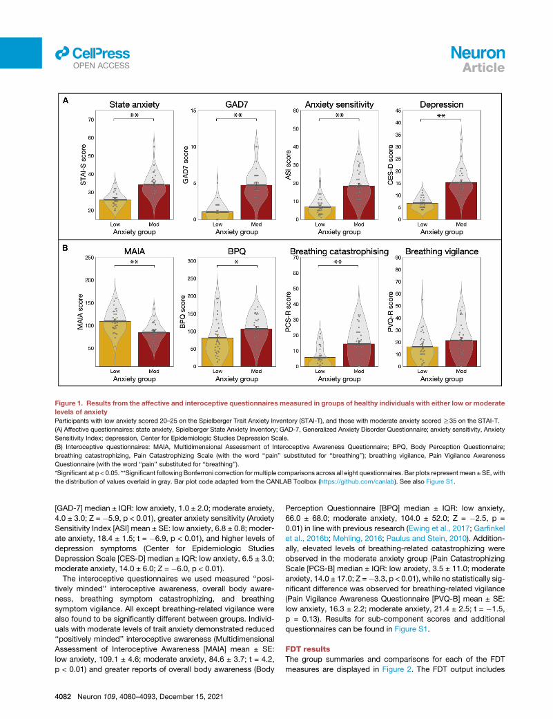
Figure 1. Results from the affective and interoceptive questionnaires measured in groups of healthy individuals with either low or moderate
levels of anxiety
Participants with low anxiety scored 20–25 on the Spielberger Trait Anxiety Inventory (STAI-T), and those with moderate anxiety scored R35 on the STAI-T.
(A) Affective questionnaires: state anxiety, Spielberger State Anxiety Inventory; GAD-7, Generalized Anxiety Disorder Questionnaire; anxiety sensitivity, Anxiety
Sensitivity Index; depression, Center for Epidemiologic Studies Depression Scale.
(B) Interoceptive questionnaires: MAIA, Multidimensional Assessment of Interoceptive Awareness Questionnaire; BPQ, Body Perception Questionnaire;
breathing catastrophizing, Pain Catastrophizing Scale (with the word ‘‘pain’’ substituted for ‘‘breathing’’); breathing vigilance, Pain Vigilance Awareness
Questionnaire (with the word ‘‘pain’’ substituted for ‘‘breathing’’).
*Significant at p < 0.05. **Significant following Bonferroni correction for multiple comparisons across all eight questionnaires. Bar plots represent mean ± SE, with
the distribution of values overlaid in gray. Bar plot code adapted from the CANLAB Toolbox (https://github.com/canlab). See also Figure S1.
llOPEN ACCESS Article
[GAD-7] median ± IQR: low anxiety, 1.0 ± 2.0; moderate anxiety,
4.0 ± 3.0; Z = �5.9, p < 0.01), greater anxiety sensitivity (Anxiety
Sensitivity Index [ASI] mean ± SE: low anxiety, 6.8 ± 0.8; moder-
ate anxiety, 18.4 ± 1.5; t = �6.9, p < 0.01), and higher levels of
depression symptoms (Center for Epidemiologic Studies
Depression Scale [CES-D] median ± IQR: low anxiety, 6.5 ± 3.0;
moderate anxiety, 14.0 ± 6.0; Z = �6.0, p < 0.01).
The interoceptive questionnaires we used measured ‘‘posi-
tively minded’’ interoceptive awareness, overall body aware-
ness, breathing symptom catastrophizing, and breathing
symptom vigilance. All except breathing-related vigilance were
also found to be significantly different between groups. Individ-
uals with moderate levels of trait anxiety demonstrated reduced
‘‘positively minded’’ interoceptive awareness (Multidimensional
Assessment of Interoceptive Awareness [MAIA] mean ± SE:
low anxiety, 109.1 ± 4.6; moderate anxiety, 84.6 ± 3.7; t = 4.2,
p < 0.01) and greater reports of overall body awareness (Body
4082 Neuron 109, 4080–4093, December 15, 2021
Perception Questionnaire [BPQ] median ± IQR: low anxiety,
66.0 ± 68.0; moderate anxiety, 104.0 ± 52.0; Z = �2.5, p =
0.01) in line with previous research (Ewing et al., 2017; Garfinkel
et al., 2016b; Mehling, 2016; Paulus and Stein, 2010). Addition-
ally, elevated levels of breathing-related catastrophizing were
observed in the moderate anxiety group (Pain Catastrophizing
Scale [PCS-B] median ± IQR: low anxiety, 3.5 ± 11.0; moderate
anxiety, 14.0 ± 17.0; Z =�3.3, p < 0.01), while no statistically sig-
nificant difference was observed for breathing-related vigilance
(Pain Vigilance Awareness Questionnaire [PVQ-B] mean ± SE:
low anxiety, 16.3 ± 2.2; moderate anxiety, 21.4 ± 2.5; t = �1.5,
p = 0.13). Results for sub-component scores and additional
questionnaires can be found in Figure S1.
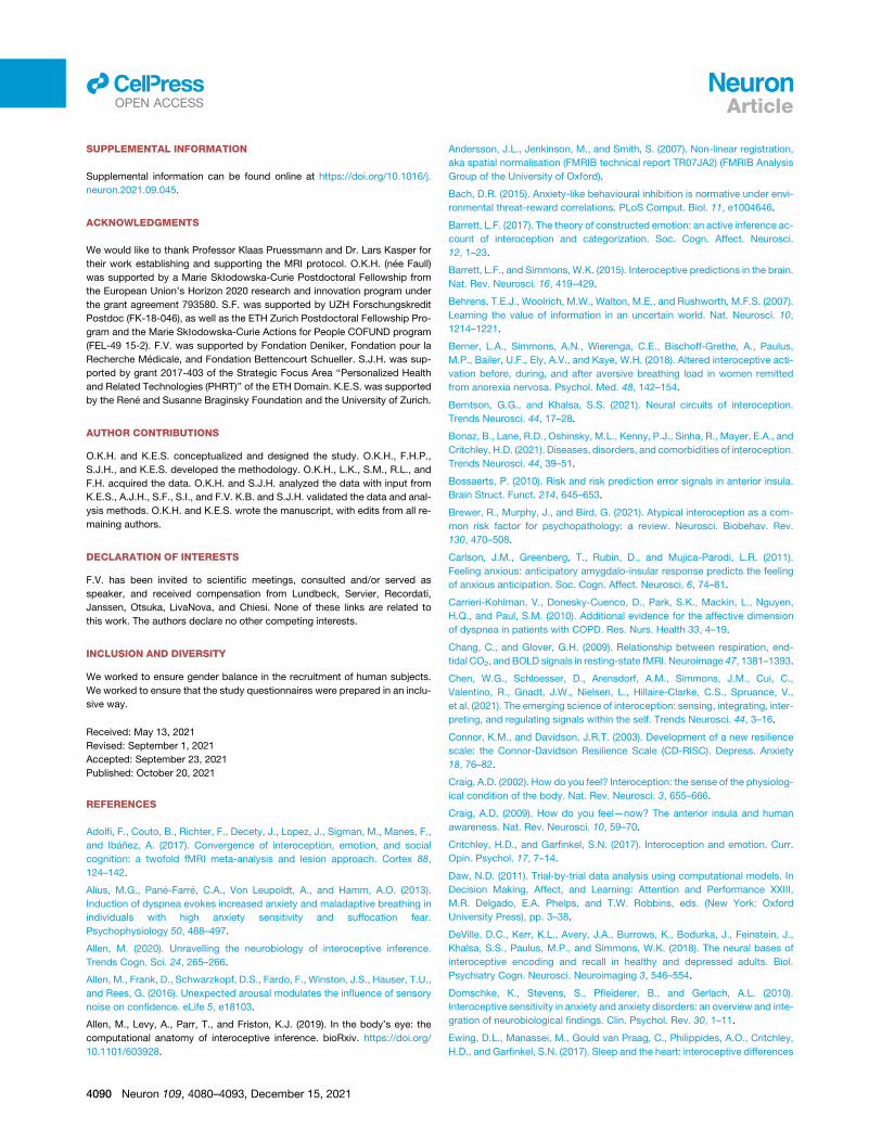
FDT resultsThe group summaries and comparisons for each of the FDT
measures are displayed in Figure 2. The FDT output includes

BASELINE PERIOD RESISTANCE / SHAM PERIOD
INS
PIR
AT
ION
EX
PIR
AT
ION
TRIAL STRUCTURE
APPLICATION OF RESISTANCE OR SHAM
REPORTING PERIOD
(Mouthpiece removed)
Decision: Yes/NoConfidence: 1-10
A
B
** *
Figure 2. Trial structure and results from the filter detection task (FDT)
(A) For each trial participants first took three breaths on the system (baseline period), before either an inspiratory resistance or shamwas applied. Following three
further breaths, participants removed the mouthpiece and reported their decision as to whether a resistance was added (yes or no), and their confidence in their
decision (1–10, where 1 = not at all confident/guessing and 10 = maximally confident). Adapted from Harrison et al. (2021a) under Creative Commons license.
(B) Results from the FDT: individuals with moderate anxiety (scores of R35 on the Spielberger Trait Anxiety Inventory [STAI-T]) demonstrated a higher (less
sensitive) perceptual threshold and lower metacognitive bias (lower average confidence) compared with individuals with low levels of anxiety (scores of 20–25 on
the STAI-T). No difference was found between groups for decision bias (where c values below zero indicate a tendency to report the presence of resistance) or
metacognitive performance (where higher values indicate better metacognitive performance).
*Significant at p < 0.05. **Significant following Bonferroni correction for multiple comparisons across all FDT measures. Bar plots represent mean ± SE, with the
distribution of values overlaid in gray. Bar plot code adapted from the CANLAB Toolbox (https://github.com/canlab).
llOPEN ACCESSArticle
the number of filters at perceptual threshold (indicative of
perceptual sensitivity, where a greater number of filters indicates
lower perceptual sensitivity), decision bias (with c < 0 indicating a
tendency to report the presence of a resistance), metacognitive
bias (calculated from average confidence scores), andmetacog-
nitive performance (reflecting the congruence between confi-
dence scores and performance accuracy). Individuals with
moderate levels of trait anxiety demonstrated both lower
perceptual sensitivity (in line with previous findings; Garfinkel
et al., 2016a; Tiller et al., 1987) (filter number median ± IQR;
low anxiety, 3.0 ± 2.0; moderate anxiety, 4.0 ± 2.0; Z = �2.4,
p = 0.01) and lower metacognitive bias (average confidence
score median ± IQR: low anxiety, 6.7 ± 2.2; moderate anxiety,
6.2 ± 2.1; Z = 2.0, p = 0.02) than those with low levels of anxiety,
with a similar level of metacognitive performance (Mratio
median ± IQR: low anxiety, 0.8 ± 0.0; moderate anxiety, 0.8 ±
0.1; Z = 0.7, p = 0.23). Decision bias was not found to be different
between the groups (decision bias c parameter mean ± SE; low
anxiety, 0.15 ± 0.06; moderate anxiety, 0.05 ± 0.06; t = 1.1, p <
0.14). The relationship between greater anxiety and reduced
confidence is consistent with results previously observed in the
exteroceptive (visual) domain, where decreased confidence
related to individual levels of both anxiety and depression
(Rouault et al., 2018).
BLT resultsBehavioral data modeling
A visual depiction of the BLT and example fitted trajectories for
prediction certainty and prediction error magnitude are provided
in Figure 3. When comparing the plausibility of the three
alternative models (a Rescorla-Wagner [RW] model; a two-level
hierarchical Gaussian filter [HGF2], and a three-level hierarchical
Gaussian filter [HGF3]) using random-effects Bayesian
model selection (Rigoux et al., 2014; Stephan et al., 2009), no
single model was found to have a protected exceedance proba-
bility (PXP) greater than 90% (RW:HGF2:HGF3 PXP =
0.30:0.40:0.30; Table S4). Therefore, as specified in our analysis
plan (https://gitlab.ethz.ch/tnu/analysis-plans/harrison_breathing_
anxiety), we conducted our model-based analysis using the
conceptually most simple model (the RWmodel), in accordance
with Occam’s razor. Importantly, the finding that none of the
models demonstrated a PXP greater than 90% does not provide
Neuron 109, 4080–4093, December 15, 2021 4083

llOPEN ACCESS Article
any absolute statement about the quality of these models.
Rather, this finding indicates that none of the chosen models is
conclusively superior to the others in explaining the data. To
ensure that the chosen model (RW) provided an adequate expla-
nation of the data, we compared it to a ‘‘null model’’ (i.e., where
the choices were due to chance and not related to any associa-
tive learning mechanism) using a likelihood ratio test. We found
that in 58 of the 60 participants, the RW model fit the behavior
significantly better than the null model, demonstrating that the
chosen model captured important aspects of their behavior.
The two participants (one from each anxiety group) who did
not show model fits above chance were excluded from any
further model-based analyses and comparisons.
To further establish the adequacy of our chosen model to
explain learning behavior in this novel interoceptive learning
task, we completed a model validation on 15 additional held-
out datasets for the BLT. These participants were not pre-
selected for any particular level of anxiety (see STAR Methods
for details). A logistic regression was conducted to assess
whether the model prediction trajectory from the original data
was able to significantly explain the prediction decisions made
by the 15 unseen participants in the validation sample. A repre-
sentative prediction trajectory from the original 60 participant
model fits (the trajectory from the participant with the closest
learning rate to the mean) was used in this regression, as well
as an intercept term. The beta estimate for the original prediction
trajectory was 3.1 ± 0.3 (t = 12.1, p = 1.0 3 10�33), denoting a
highly significant ability of the trajectory to predict unseen
data. The beta estimate for the intercept term was �0.2 ± 0.1
(t = �3.1, p = 1.7 3 10�3). For a qualitative representation of
model fits for both the original and validation data, see Figure S3.
Both model-based and behavioral parameter comparisons
are presented in Table 1. For the estimated model parameters,
no difference was observed between the groups for either
learning rate (a) or inverse decision temperature (z) (Table 1). Re-
sults from parameter comparisons between groups including the
excluded participants can be found in Table S5. For the subjec-
tive measures, no difference was observed between the groups
for breathing difficulty ratings, while the task-induced anxiety
ratings were significantly greater in those with moderate anxiety
(Table 1). Additionally, no difference in any physiological mea-
sures were observed (Table S1), nor in relative head motion dur-
ing the task (average root-mean-square displacement ± SD: low
anxiety, 0.17 ± 0.10 mm; moderate anxiety, 0.18 ± 0.07 mm;
Wilcoxon rank-sum p = 0.91).
Computational modeling of brain activity
The overall and between-group BLT brain activity analysis results
are displayed in Figures 4 and 5. In the analysis for the entire field
of view, activations associated with breathing-related prediction
certainty and prediction errors across all participants are shown
in Figure 4. Dorsolateral prefrontal cortex (dlPFC), anterior insula
(aIns), anterior cingulate cortex (ACC), and middle frontal gyrus
(MFG) all demonstrated significant deactivations with overall pre-
diction certainty (i.e., averaged across trials with positive and
negative prediction certainty; Figure 4A). In contrast, aIns, ACC,
MFG, and the periaqueductal gray (PAG) demonstrated signifi-
cant activations with overall prediction error values (i.e., averaged
across trials with positive and negative prediction errors; Fig-
4084 Neuron 109, 4080–4093, December 15, 2021
ure 4B). A small number of differences due to valence (differences
between positive and negative outcomes) were found for predic-
tion errors but not prediction certainty, with negative prediction
errors associated with deactivations of left dlPFC and activations
of left posterior insula (Figure 4B). Although nomain effect of anx-
iety group was observed, an interaction effect was found using
the region-of-interest (ROI) analysis between valence and groups
for predictions in the bilateral aIns (Figure 5). In contrast, no group
or interaction effects were found for prediction errors. Brain activ-
ity associated with inspiratory resistance is provided in Figure S6
for comparison with previously published results (Berner et al.,
2018; DeVille et al., 2018; Faull and Pattinson, 2017; Faull et al.,
2016, 2018; Hayen et al., 2017; Paulus et al., 2012).
Multi-modal analysis resultsFirst, the key measures from each of the different modalities were
combined into a multi-modal correlation matrix. This analysis al-
lowed us to assess the relationships both within and across
task modalities as well as across levels of breathing-related inter-
oceptive processing. The full correlation matrix of all 16 included
measures is displayed in Figure 6A and Table S7. To briefly sum-
marize, the strongest across-task modality correlations were
found between all affective and interoceptive questionnaires (Fig-
ure 6A). Concerning affective questionnaires and the FDT mea-
sures, state anxiety was weakly correlated with the FDT percep-
tual threshold, decision bias, and metacognitive bias, while
anxiety sensitivity was additionally weakly related to metacogni-
tive bias. Depression scores were also weakly related to the
FDT perceptual threshold. Between the interoceptive and the
FDT measures, breathing-related catastrophizing was weakly
related to metacognitive performance on the FDT. Last, between
the FDT and aIns activity, metacognitive performance was
strongly related to the peak aIns activity associated with negative
(i.e., resistance-related) prediction errors, while metacognitive
bias was weakly related to aIns activity associated with negative
(i.e., resistance-related) prediction certainty. Non-parametric cor-
relations (using Spearman’s rho values) produced highly consis-
tent results and are presented in Table S7.
Principal-component analysis
Finally, to assess the extent of shared variance across interocep-
tive measures, the multi-modal data matrix was then subjected
to a principal-component analysis (PCA). This analysis allowed
us to delineate how many underlying dimensions may exist
within the data, as well as which measures were most strongly
associated with anxiety. Two PCs were found to be significant,
where the variance explained with each component was above
the 95%confidence interval of its null distribution. The properties
of each of these significant components are displayed in Figures
6B and 6C. The first PC demonstrated a highly significant (p <
1 3 10�11) difference in scores between the anxiety groups.
Correspondingly, the greatest coefficients within the first PC
were from the affective measures of depression scores, state
anxiety, anxiety sensitivity, and anxiety disorder scores. Addi-
tionally, breathing-related catastrophizing and negative intero-
ceptive awareness also had strong coefficient values, followed
by negative metacognitive bias (i.e., lower confidence scores),
body perception scores (from the BPQ), and negative metacog-
nitive performance (i.e., lower metacognitive performance). In

+How difficult was
it to breathe?
CUE / PREDICT3.5 sec
OR
PAUSE2.5 sec
STIMULUS5 sec
RATE5 sec
REST7-9 sec
Not at alldifficult
Extremely difficult
WILL IT BE DIFFICULTTO BREATHE?
Yes or No
WILL IT BE DIFFICULTTO BREATHE?
+
STIMULUS START
A
B
Figure 3. The breathing learning task (BLT), used to measure dynamic learning of breathing-related stimuli
(A) An overview of the single trial structure, in which one of two cues was presented and participants were asked to predict (on the basis of the cue) whether they
thought that an inspiratory breathing resistance would follow. When the circle appeared on the screen, either an inspiratory resistance or no resistance was
applied for 5 s, with the resistance set to 70%of the individual’s maximal inspiratory resistance. After every trial, participants were asked to rate the intensity of the
previous stimulus. The trace in green is an example of a pressure trace recorded at the mouth.
(B) The 80-trial trajectory structure of the probability that one cue predicts inspiratory resistance (black trace), where the alternative cue has an exactly mirrored
contingency structure, together with example responses (circles). Filled black circles represent stimuli that were correctly predicted, and open black circles
represent stimuli that were not correctly predicted. Example fitted prediction (top) and prediction error (bottom) trajectories are overlaid (red traces). The example
trajectories were taken from the participant with the closest learning rate to the mean value across all participants.
See also Figures S2 and S3 and Tables S1–S4 and S6.
llOPEN ACCESSArticle
contrast, the second PC demonstrated a weak difference (p =
0.05) in scores between the anxiety groups. This component
had the highest coefficient scores from the peak aIns activity
related to positive and negative prediction certainty, as well as
negative coefficients for negative prediction errors, metacogni-
tive performance, and positive prediction errors.
DISCUSSION
Main findingsInteroceptive abilities are thought to be tightly linked to affective
properties such as anxiety. Here we have provided a unifying
analysis by characterizing this relationship across multiple
Neuron 109, 4080–4093, December 15, 2021 4085

Table 1. Behavioral and model-based group comparison results
from the breathing learning task (BLT)
Total Low Moderate p Value Test
Learning
rate (a)
0.25 (0.19) 0.24 (0.15) 0.25 (0.22) 0.58 Wxn
Inverse decision
temperature (z)
2.66 (3.35) 2.71 (3.15) 2.37 (3.65) 0.88 Wxn
Breathing
difficulty
rating (%)
82.6 (18.4) 80.5 (19.9) 83.8 (15.8) 0.61 Wxn
Breathing
anxiety
rating (%)
10.0 (42.0) 0.0 (10.0) 34.0 (48.0) <0.001a Wxn
Response
time (s)
1.28 (0.33) 1.23 (0.36) 1.29 (0.30) 0.73 Wxn
All data are expressed as median (inter-quartile range) and include the
model parameter estimates (learning rate, a; inverse decision tempera-
ture, z), the subjective ratings of breathing difficulty (average of the ratings
provided following each resistance stimulus) and anxiety (rating provided
immediately following the end of the task), and the response times for the
predictions made during the task. Wxn, Wilcoxon rank-sum test. If a Wil-
coxon rank-sum test was used, reported values are median (inter-quar-
tile range).aSignificant difference between groups at p < 0.05 with multiple compar-
ison correction for the number of behavioral parameters. See also
Table S5.
llOPEN ACCESS Article
interoceptive levels in the breathing domain, including novel
findings of altered brain activity within the aIns when processing
dynamic breathing predictions. This study is the first to demon-
strate brain activity related to dynamic interoceptive learning
and, specifically, activity in the insula that is related to breath-
ing-related prediction error and prediction certainty in a trial-by-
trial fashion. Notably, aIns activity related to the certainty of pre-
dictions about breathing resistance was found to differ between
trait anxiety levels. Furthermore, this study is also among the first
to simultaneously tackle multiple levels of interoceptive process-
ing, using breathing as a salient and accessible channel of body
perception. The tasks used reflect the broad range of targeted
processes; not onlywere questionnaires used that spanned affect
and body perceptions, but behavioral data from two different
tasks were assessed by separate computational models. These
analyses allowed formal assessments of both breathing-related
interoceptive learning and metacognition, including the first
computational assessment of trial-by-trial learning in the intero-
ceptive domain, as well as applying state-of-the-art models of
metacognition to interoception of breathing. Our multi-modal
approach revealed that not only is the relationship between
breathing-related interoception and trait anxiety broad, it is
most strongly detected (i.e., greatest PCA weights; Figure 6) at
the higher levels of interoceptive processing, which includes spe-
cific subjective measures of interoceptive beliefs (often termed
‘‘interoceptive sensibility’’; Garfinkel et al., 2015) followed by
metacognitive aspects of breathing perceptions. Notably, the
peak aIns brain activity associated with breathing-related intero-
ceptive learning appeared to be largely independent of other
interoceptivemeasures, with the exception of negative prediction
error-related brain activity and metacognitive performance.
4086 Neuron 109, 4080–4093, December 15, 2021
Affect and levels of breathing-related interoceptionBeyond consequences at single levels of interoceptive process-
ing, here we aimed to assess how the relationship with trait anx-
iety may cross multiple interoceptive levels related to breathing.
Using PCA (with permutation testing) allowed us to identify any
components that share common variance within ourmulti-modal
dataset and additionally assess the relative contribution of our
measures to each dimension (Figure 6B). Here we found that
all affective qualities loaded strongly onto the first principal
component, with the greatest additional contributions from sub-
jective measures of negative interoceptive awareness and
breathing-related catastrophizing. General body awareness
and the breathing-related metacognitive measures (bias and
performance) were the next largest contributors to this shared
variance, followed by the perceptual sensitivity and decision
bias parameters, and last peak aIns activity from the BLT. These
results suggest that the relationship with anxiety is particularly
prominent at the level of subjective interoceptive beliefs in the
breathing domain, which are thought to exist at the higher levels
of interoceptive space (Critchley and Garfinkel, 2017), followed
by metacognitive insight into breathing perception. In compari-
son, the relationship of trait anxiety to lower level properties
such as interoceptive sensitivity (Critchley and Garfinkel, 2017;
Garfinkel et al., 2015, 2016a, 2016b) appear to be present but
less prominent in the breathing domain. However, it must be
noted that quantifying these higher interoceptive levels may be
less noisy in comparison withmeasuring psychophysical proper-
ties such as sensitivity, and thus the relationship with anxiety
might be most easily detected rather than being inherently
stronger.
Although strong relationships were observed between affec-
tive qualities and many of our interoceptive breathing measures,
a sparse number of correlations were found between interocep-
tive measures themselves, and in particular across task modal-
ities (Figure 6A). These findings support the idea that there are
potentially separable levels of breathing-related interoception,
as proposed (Critchley and Garfinkel, 2017). The only notably
strong cross-modal relationship was found between metacogni-
tive performance and aIns activity, for which greater insight into
breathing sensitivity correlated with greater aIns activity during
negative prediction errors. This relationship may reflect a previ-
ously proposed contribution of error processing toward meta-
cognitive awareness, whereby deviations between actual and
predicted bodily inputs are propagated to metacognitive areas
via interoceptive brain structures such as the aIns (Stephan
et al., 2016).
Novel measures of dynamic interoceptive predictionsand prediction errorsMany theories surrounding anxiety have hypothesized an impor-
tant role of altered predictions regarding upcoming threat (Bach,
2015; Mogg et al., 2000; Simmons et al., 2006) and in particular
interoceptive threat (Paulus and Stein, 2010; Paulus et al., 2019)
in the aIns (Allen, 2020; Bossaerts, 2010; Carlson et al., 2011;
Paulus and Stein, 2006; Tan et al., 2018). Although numerous
studies have used inspiratory resistive loads to evoke threat-
ening interoceptive stimuli (Alius et al., 2013; Berner et al.,
2018; Faull and Pattinson, 2017; Faull et al., 2016, 2018; Hayen

A
PAG PAG
PREDICTIONS
0 10ACTIVATION
0
T-STATISTICDEACTIVATION
B PREDICTION ERRORS
0 10ACTIVATION
0
T-STATISTICDEACTIVATION
EDUTINGAM RORRE NOITCIDERPYTNIATREC NOITCIDERP
)SOP > GEN( ECNELAV)SOP > GEN( ECNELAVPAG PAG
Figure 4. Prediction and prediction-error-related trajectories and brain activity
(A and B) Demonstration of how estimated prediction (A) and prediction error (B) trajectories are encoded as positive (i.e., toward no resistance) and negative (i.e.,
toward resistance) prediction certainty values and prediction error magnitudes. The example trajectories were taken from the participant with the closest learning
rate to themean value across all participants. The solid gray lines demonstrate the estimated prediction or prediction error traces (in stimulus space). Positive trial
values are demonstrated in blue and the negative trial values in red, encoded as distance from zero (i.e., absolute values; right axes). The brain images represent
significant activity across both groups for prediction certainty (averaged over trials with positive and negative prediction certainty) and the influence of valence on
prediction certainty (difference between negative and positive predictions), prediction error magnitude (averaged over trials with positive and negative prediction
errors), and the influence of valence on prediction error magnitude (difference between negative and positive prediction errors). The images consist of a color-
rendered statistical map superimposed on a standard (MNI 13 13 1mm) brain. The bright gray region represents the coverage of the coronal-oblique functional
scan. Significant regions are displayed with a cluster threshold of p < 0.05, family-wise error (FWE) corrected for multiple comparisons across all voxels included
in the functional volume. PAG, periaqueductal gray. See also Figures S4–S7.
llOPEN ACCESSArticle
et al., 2017; von Leupoldt and Dahme, 2005; von Leupoldt et al.,
2009; Paulus et al., 2012; Stoeckel et al., 2016; Walter et al.,
2020), the BLT approach presented here is, to our knowledge,
the first investigation of dynamic (trial-by-trial) brain activity
associated with model-based interoceptive predictions and pre-
diction errors for respiration. By fitting an associative learning
model to each participant’s behavioral responses, we could
quantify both the certainty of predictions and magnitude of pre-
diction errors on each trial. We could then compare both the
parameter estimates and the brain activity associated with these
computational quantities, with a particular focus on the aIns and
PAG (Allen, 2020; Grahl et al., 2018; Paulus and Stein, 2006; Roy
et al., 2014; Singer et al., 2009) (Figure 4). Here, we observed ev-
idence for a relationship between anxiety and aIns reactivity to
threat valence in the prediction domain (Figure 5). Specifically,
while individuals with low trait anxiety demonstrated greater
aIns deactivation that scaled with predictions of breathing resis-
tance compared with no resistance, the opposite was true in in-
dividuals with moderate trait anxiety (creating an interaction ef-
fect). This demonstrates a shift in the aIns processing of threat
valence with different levels of anxiety, in line with hypothesized
differences in brain prediction processing (Paulus and Stein,
2006, 2010; Paulus et al., 2019). In comparison, no anxiety group
differences or interactions were found in the breathing prediction
error domain, contrasting with some previously proposed
hypotheses (Barrett and Simmons, 2015; Brewer et al., 2021;
Paulus and Stein, 2006, 2010).
Beyond the aIns and independent of anxiety, multiple (and
largely consistent) proposals have been made regarding which
brain networks might be involved in processing interoceptive
predictions and prediction errors (Allen, 2020; Barrett, 2017;
Barrett and Simmons, 2015; Craig, 2009; Khalsa et al., 2018;
Kleckner et al., 2017; Manjaly and Iglesias, 2020; Marlow
et al., 2019; Owens et al., 2018; Paulus et al., 2019; Pezzulo
et al., 2015, 2018; Quadt et al., 2018; Seth, 2013; Smith
et al., 2017; Stephan et al., 2016). These proposed networks
are loosely hierarchical in structure and typically assign meta-
cognitive processes to higher cortical areas (e.g., prefrontal
cortex [PFC]), while interoceptive predictions are thought to
originate from regions that may serve as an interface between
interoceptive and visceromotor function (e.g., aIns and ACC). In
these concepts, prediction errors have two different roles: on
one hand, they are thought to be sent up the cortical hierarchy
of interoceptive regions in order to update predictions in aIns
and ACC (Barrett and Simmons, 2015; Pezzulo et al., 2015;
Seth et al., 2012); on the other hand, they are thought to deter-
mine regulatory signals, sent from visceromotor regions and
brainstem structures such as the PAG to the autonomic ner-
vous system and bodily organs (Petzschner et al., 2017; Ste-
phan et al., 2016).
Neuron 109, 4080–4093, December 15, 2021 4087

RL
PREDICTION VALENCE
0.00 0.05p-value
**
Figure 5. An interaction effect observed between valence (i.e., trials with positive versus negative predictions) and anxiety group (low versus
moderate) for activity in the anterior insula related to prediction certainty
The images consist of a color-rendered statistical map superimposed on a standard (MNI 13 13 1 mm) brain. Voxel-wise statistics were performed using non-
parametric permutation testing within amask of the anterior insula and periaqueductal gray, with significant results determined by p < 0.05 (corrected for multiple
comparisons within themask). Bar plots represent mean ± SE for the individual contrast estimates within the significant voxels, plotted separately for each side of
the anterior insula. See also Figures S4–S7.
llOPEN ACCESS Article
Although these theories have received considerable attention,
there has been little empirical evidence thus far. In particular, we
are not aware of any studies that have demonstrated, using a
concrete computational model, trial-by-trial prediction and pre-
diction error activity in interoceptive areas. Here, we report evi-
dence of relevant computational quantities being reflected by
activity within several areas of a putative interoceptive breathing
network. Although activity related to trial-wise prediction cer-
tainty was found in higher structures such as dorsolateral PFC,
ACC, and aIns, prominent prediction error responses were not
only found in aIns and ACC but also in the midbrain PAG (Fig-
ure 4). Importantly, concerning predictions, widespread brain
activity was found to be related mainly to prediction uncertainty,
where blood-oxygen-level-dependent (BOLD) activity was
decreased in proportion to increases in the certainty of predic-
tions (Feldman and Friston, 2010; Friston, 2005). Furthermore,
it is notable that the aIns displayed both deactivation for more
certain predictions and activation for greater prediction errors.
This might reflect the proposed critical role of aIns in the repre-
sentation and updating of models of bodily states (Allen, 2020;
Van den Bergh et al., 2017; Manjaly and Iglesias, 2020; Paulus
and Stein, 2006; Paulus et al., 2019; Seth, 2013; Stephan et al.,
2016; Walter et al., 2020), given that greater certainty (precision
of beliefs) reduces and greater prediction errors increase belief
(model) updating (Petzschner et al., 2017).
Our PAG findings are of particular interest. Although the PAG
has been previously noted during anticipation of certain breath-
4088 Neuron 109, 4080–4093, December 15, 2021
ing resistance stimuli (Faull and Pattinson, 2017; Faull et al.,
2016) and has been related to the precision of prior beliefs about
placebo-induced reductions in pain intensity (Grahl et al., 2018),
here we observed that PAG activity did not appear to be related
to the extent of prediction certainty toward upcoming breathing
stimuli (Figure 4). Concerning prediction error activity in the PAG,
this has previously been demonstrated in relation to pain (Roy
et al., 2014); here, we found PAG activity in relation to the mag-
nitudes of trial-wise interoceptive prediction errors (Figure 4B),
consistent with a role of PAG in homeostatic control (Stephan
et al., 2016).
Finally, overall prediction and prediction error-related activity
did not appear to be dissociated between anterior and poste-
rior insula cortices (respectively), as has been previously hy-
pothesized (Allen, 2020; Barrett and Simmons, 2015; Stephan
et al., 2016). However, a small valence difference in prediction
errors was observed, with negative prediction errors (the unex-
pected presence of inspiratory resistance stimuli) producing
greater activity in the right posterior insula than positive predic-
tion errors (the unexpected absence of inspiratory resistance
stimuli; Figure 4). It is therefore possible that the representation
of homeostatically relevant inputs in the posterior insula is
enhanced for events that may negatively affect homeostasis.
However, these results are the first demonstration of brain ac-
tivity related to dynamic interoceptive prediction and prediction
errors; furthermore, the functional images from this study do
not have the resolution required for layer-specific identification

Figure 6. Results from the multi-modal analysis incorporating questionnaires, breathing task data, and peak brain activity in the anterior
insula
(A) Correlation matrix results for the 16 includedmeasures in the multi-modal analysis. Black dots represent significant values at p < 0.05, while white dots denote
significance with correction for multiple comparisons.
(B) The weights and group scores of the first significant principal component, where a strong anxiety group difference in component scores is observed.
(C) The weights and group scores of the second significant principal component, where a weak anxiety group difference in principal component scores is
observed.
*Significant difference between groups at p < 0.05. **Significant difference between groups at p < 0.05 with multiple comparison correction for the two significant
components. Bar plots (rightmost panels) represent mean ± SE, with the distribution of values overlaid in gray. Bar plot code adapted from the CANLAB Toolbox
(https://github.com/canlab). See also Table S7.
llOPEN ACCESSArticle
of prediction and prediction error processing in the insula.
Additionally, the represented prediction error-related activity
may be specific to the breathing domain within interoceptive
processing. This latter caveat of course also applies to the
wider results presented here; as only one interoceptive channel
(i.e., inspiratory resistances within the breathing domain) was
tested, we cannot assume these results would translate to
other interoceptive processes (e.g., related to cardiac or gastric
states).
ConclusionsThe relationship between anxiety and breathing crosses multiple
levels of the interoceptive hierarchy. In particular, anxiety and
associated affective dimensions appear to be most strongly
related to subjective negative body awareness and catastroph-
izing about breathing symptoms, followed by metacognitive
measures related to breathing perception. Furthermore, a novel
interaction between trait anxiety and valence was found within
the aIns, associated with dynamic prediction certainty (but not
prediction errors) of breathing-related interoceptive processing.
More generally, this study provides the first empirical demon-
stration of brain activity associated with dynamic (trial-by-trial)
interoceptive learning. These results provide new and compre-
hensive insights into how anxiety is related to levels of interocep-
tive processing in the breathing domain and provide evidence of
brain activity associated with trial-wise predictions and predic-
tion errors about bodily states in interoceptive breathing
networks.
STAR+METHODS
Detailed methods are provided in the online version of this paper
and include the following:
d KEY RESOURCES TABLE
d RESOURCE AVAILABILITY
B Lead contact
B Materials availability
B Data and code availability
d EXPERIMENTAL MODEL AND SUBJECT DETAILS
B Participants
d METHOD DETAILS
B Procedural overview
B Questionnaires
B Filter detection task
B Breathing learning task
d QUANTIFICATION AND STATISTICAL ANALYSIS
B Statistical analysis overview
B Questionnaire analysis
B FDT analysis
B BLT analysis
B Multi-modal analysis
Neuron 109, 4080–4093, December 15, 2021 4089
llOPEN ACCESS Article
SUPPLEMENTAL INFORMATION
Supplemental information can be found online at https://doi.org/10.1016/j.
neuron.2021.09.045.
ACKNOWLEDGMENTS
We would like to thank Professor Klaas Pruessmann and Dr. Lars Kasper for
their work establishing and supporting the MRI protocol. O.K.H. (nee Faull)
was supported by a Marie Sk1odowska-Curie Postdoctoral Fellowship from
the European Union’s Horizon 2020 research and innovation program under
the grant agreement 793580. S.F. was supported by UZH Forschungskredit
Postdoc (FK-18-046), as well as the ETH Zurich Postdoctoral Fellowship Pro-
gram and the Marie Sk1odowska-Curie Actions for People COFUND program
(FEL-49 15-2). F.V. was supported by Fondation Deniker, Fondation pour la
Recherche Medicale, and Fondation Bettencourt Schueller. S.J.H. was sup-
ported by grant 2017-403 of the Strategic Focus Area ‘‘Personalized Health
and Related Technologies (PHRT)’’ of the ETH Domain. K.E.S. was supported
by the Rene and Susanne Braginsky Foundation and the University of Zurich.
AUTHOR CONTRIBUTIONS
O.K.H. and K.E.S. conceptualized and designed the study. O.K.H., F.H.P.,
S.J.H., and K.E.S. developed the methodology. O.K.H., L.K., S.M., R.L., and
F.H. acquired the data. O.K.H. and S.J.H. analyzed the data with input from
K.E.S., A.J.H., S.F., S.I., and F.V. K.B. and S.J.H. validated the data and anal-
ysis methods. O.K.H. and K.E.S. wrote the manuscript, with edits from all re-
maining authors.
DECLARATION OF INTERESTS
F.V. has been invited to scientific meetings, consulted and/or served as
speaker, and received compensation from Lundbeck, Servier, Recordati,
Janssen, Otsuka, LivaNova, and Chiesi. None of these links are related to
this work. The authors declare no other competing interests.
INCLUSION AND DIVERSITY
We worked to ensure gender balance in the recruitment of human subjects.
We worked to ensure that the study questionnaires were prepared in an inclu-
sive way.
Received: May 13, 2021
Revised: September 1, 2021
Accepted: September 23, 2021
Published: October 20, 2021
REFERENCES
Adolfi, F., Couto, B., Richter, F., Decety, J., Lopez, J., Sigman, M., Manes, F.,
and Ibanez, A. (2017). Convergence of interoception, emotion, and social
cognition: a twofold fMRI meta-analysis and lesion approach. Cortex 88,
124–142.
Alius, M.G., Pane-Farre, C.A., Von Leupoldt, A., and Hamm, A.O. (2013).
Induction of dyspnea evokes increased anxiety and maladaptive breathing in
individuals with high anxiety sensitivity and suffocation fear.
Psychophysiology 50, 488–497.
Allen, M. (2020). Unravelling the neurobiology of interoceptive inference.
Trends Cogn. Sci. 24, 265–266.
Allen, M., Frank, D., Schwarzkopf, D.S., Fardo, F., Winston, J.S., Hauser, T.U.,
and Rees, G. (2016). Unexpected arousal modulates the influence of sensory
noise on confidence. eLife 5, e18103.
Allen, M., Levy, A., Parr, T., and Friston, K.J. (2019). In the body’s eye: the
computational anatomy of interoceptive inference. bioRxiv. https://doi.org/
10.1101/603928.
4090 Neuron 109, 4080–4093, December 15, 2021
Andersson, J.L., Jenkinson, M., and Smith, S. (2007). Non-linear registration,
aka spatial normalisation (FMRIB technical report TR07JA2) (FMRIB Analysis
Group of the University of Oxford).
Bach, D.R. (2015). Anxiety-like behavioural inhibition is normative under envi-
ronmental threat-reward correlations. PLoS Comput. Biol. 11, e1004646.
Barrett, L.F. (2017). The theory of constructed emotion: an active inference ac-
count of interoception and categorization. Soc. Cogn. Affect. Neurosci.
12, 1–23.
Barrett, L.F., and Simmons, W.K. (2015). Interoceptive predictions in the brain.
Nat. Rev. Neurosci. 16, 419–429.
Behrens, T.E.J., Woolrich, M.W., Walton, M.E., and Rushworth, M.F.S. (2007).
Learning the value of information in an uncertain world. Nat. Neurosci. 10,
1214–1221.
Berner, L.A., Simmons, A.N., Wierenga, C.E., Bischoff-Grethe, A., Paulus,
M.P., Bailer, U.F., Ely, A.V., and Kaye, W.H. (2018). Altered interoceptive acti-
vation before, during, and after aversive breathing load in women remitted
from anorexia nervosa. Psychol. Med. 48, 142–154.
Berntson, G.G., and Khalsa, S.S. (2021). Neural circuits of interoception.
Trends Neurosci. 44, 17–28.
Bonaz, B., Lane, R.D., Oshinsky, M.L., Kenny, P.J., Sinha, R., Mayer, E.A., and
Critchley, H.D. (2021). Diseases, disorders, and comorbidities of interoception.
Trends Neurosci. 44, 39–51.
Bossaerts, P. (2010). Risk and risk prediction error signals in anterior insula.
Brain Struct. Funct. 214, 645–653.
Brewer, R., Murphy, J., and Bird, G. (2021). Atypical interoception as a com-
mon risk factor for psychopathology: a review. Neurosci. Biobehav. Rev.
130, 470–508.
Carlson, J.M., Greenberg, T., Rubin, D., and Mujica-Parodi, L.R. (2011).
Feeling anxious: anticipatory amygdalo-insular response predicts the feeling
of anxious anticipation. Soc. Cogn. Affect. Neurosci. 6, 74–81.
Carrieri-Kohlman, V., Donesky-Cuenco, D., Park, S.K., Mackin, L., Nguyen,
H.Q., and Paul, S.M. (2010). Additional evidence for the affective dimension
of dyspnea in patients with COPD. Res. Nurs. Health 33, 4–19.
Chang, C., and Glover, G.H. (2009). Relationship between respiration, end-
tidal CO2, and BOLD signals in resting-state fMRI. Neuroimage 47, 1381–1393.
Chen, W.G., Schloesser, D., Arensdorf, A.M., Simmons, J.M., Cui, C.,
Valentino, R., Gnadt, J.W., Nielsen, L., Hillaire-Clarke, C.S., Spruance, V.,
et al. (2021). The emerging science of interoception: sensing, integrating, inter-
preting, and regulating signals within the self. Trends Neurosci. 44, 3–16.
Connor, K.M., and Davidson, J.R.T. (2003). Development of a new resilience
scale: the Connor-Davidson Resilience Scale (CD-RISC). Depress. Anxiety
18, 76–82.
Craig, A.D. (2002). How do you feel? Interoception: the sense of the physiolog-
ical condition of the body. Nat. Rev. Neurosci. 3, 655–666.
Craig, A.D. (2009). How do you feel—now? The anterior insula and human
awareness. Nat. Rev. Neurosci. 10, 59–70.
Critchley, H.D., and Garfinkel, S.N. (2017). Interoception and emotion. Curr.
Opin. Psychol. 17, 7–14.
Daw, N.D. (2011). Trial-by-trial data analysis using computational models. In
Decision Making, Affect, and Learning: Attention and Performance XXIII,
M.R. Delgado, E.A. Phelps, and T.W. Robbins, eds. (New York: Oxford
University Press), pp. 3–38.
DeVille, D.C., Kerr, K.L., Avery, J.A., Burrows, K., Bodurka, J., Feinstein, J.,
Khalsa, S.S., Paulus, M.P., and Simmons, W.K. (2018). The neural bases of
interoceptive encoding and recall in healthy and depressed adults. Biol.
Psychiatry Cogn. Neurosci. Neuroimaging 3, 546–554.
Domschke, K., Stevens, S., Pfleiderer, B., and Gerlach, A.L. (2010).
Interoceptive sensitivity in anxiety and anxiety disorders: an overview and inte-
gration of neurobiological findings. Clin. Psychol. Rev. 30, 1–11.
Ewing, D.L., Manassei, M., Gould van Praag, C., Philippides, A.O., Critchley,
H.D., and Garfinkel, S.N. (2017). Sleep and the heart: interoceptive differences

llOPEN ACCESSArticle
linked to poor experiential sleep quality in anxiety and depression. Biol.
Psychol. 127, 163–172.
Fan, L., Li, H., Zhuo, J., Zhang, Y.,Wang, J., Chen, L., Yang, Z., Chu, C., Xie, S.,
Laird, A.R., et al. (2016). The human brainnetome atlas: a new brain atlas based
on connectional architecture. Cereb. Cortex 26, 3508–3526.
Faull, O.K., and Pattinson, K.T. (2017). The cortical connectivity of the peria-
queductal gray and the conditioned response to the threat of breathlessness.
eLife 6, e21749–e21767.
Faull, O.K., Jenkinson, M., Clare, S., and Pattinson, K.T.S. (2015). Functional
subdivision of the human periaqueductal grey in respiratory control using 7
tesla fMRI. Neuroimage 113, 356–364.
Faull, O.K., Jenkinson, M., Ezra, M., and Pattinson, K.T.s. (2016). Conditioned
respiratory threat in the subdivisions of the human periaqueductal gray. eLife
5, e12047–e12066.
Faull, O.K., Cox, P.J., and Pattinson, K.T.S. (2018). Cortical processing of
breathing perceptions in the athletic brain. Neuroimage 179, 92–101.
Feldman, H., and Friston, K.J. (2010). Attention, uncertainty, and free-energy.
Front. Hum. Neurosci. 4, 215.
Fleming, S.M. (2017). HMeta-d: hierarchical Bayesian estimation of metacog-
nitive efficiency from confidence ratings. Neurosci. Conscious. 2017, nix007.
Fr€assle, S., Aponte, E.A., Bollmann, S., Brodersen, K.H., Do, C.T., Harrison,
O.K., Harrison, S.J., Heinzle, J., Iglesias, S., Kasper, L., et al. (2021). TAPAS:
an open-source software package for translational neuromodeling and
computational psychiatry. Front. Psychiatry 12, 680811.
Friston, K. (2005). A theory of cortical responses. Philos. Trans. R. Soc. Lond. B
Biol. Sci. 360, 815–836.
Friston, K.J., Ashburner, J.T., Kiebel, S.J., Nichols, T.E., and Penny, W.D.
(2011). Statistical Parametric Mapping: The Analysis of Functional Brain
Images (London: Elsevier).
Garfinkel, S.N., Seth, A.K., Barrett, A.B., Suzuki, K., and Critchley, H.D. (2015).
Knowing your own heart: distinguishing interoceptive accuracy from intero-
ceptive awareness. Biol. Psychol. 104, 65–74.
Garfinkel, S.N., Manassei, M.F., Hamilton-Fletcher, G., In den Bosch, Y.,
Critchley, H.D., and Engels, M. (2016a). Interoceptive dimensions across car-
diac and respiratory axes. Philos. Trans. R. Soc. Lond. B Biol. Sci. 371,
20160014-10.
Garfinkel, S.N., Tiley, C., O’Keeffe, S., Harrison, N.A., Seth, A.K., and Critchley,
H.D. (2016b). Discrepancies between dimensions of interoception in autism:
implications for emotion and anxiety. Biol. Psychol. 114, 117–126.
Giardino, N.D., Curtis, J.L., Abelson, J.L., King, A.P., Pamp, B., Liberzon, I.,
and Martinez, F.J. (2010). The impact of panic disorder on interoception and
dyspnea reports in chronic obstructive pulmonary disease. Biol. Psychol. 84,
142–146.
Grahl, A., Onat, S., and B€uchel, C. (2018). The periaqueductal gray and
Bayesian integration in placebo analgesia. eLife 7, 1–20.
Griffanti, L., Douaud, G., Bijsterbosch, J., Evangelisti, S., Alfaro-Almagro, F.,
Glasser, M.F., Duff, E.P., Fitzgibbon, S., Westphal, R., Carone, D., et al.
(2017). Hand classification of fMRI ICA noise components. Neuroimage 154,
188–205.
Gu, X., Hof, P.R., Friston, K.J., and Fan, J. (2013). Anterior insular cortex and
emotional awareness. J. Comp. Neurol. 521, 3371–3388.
Harrison, O.K., Garfinkel, S.N., Marlow, L., Finnegan, S.L., Marino, S., Kochli,
L., Allen, M., Finnemann, J., Keur-Huizinga, L., Harrison, S.J., et al. (2021a).
The filter detection task for measurement of breathing-related interoception
and metacognition. Biol. Psychol. 165, 108185.
Harrison, S.J., Bianchi, S., Heinzle, J., Stephan, K.E., Iglesias, S., and Kasper,
L. (2021b). A Hilbert-based method for processing respiratory timeseries.
Neuroimage 230, 117787.
Harrison, O.K., Marlow, L.L., Finnegan, S.L., Ainsworth, B., and Pattinson,
K.T.S. (2021c). Heterogeneity in asthma: dissociating symptoms from mood
and their influence on interoception and attention. Biological Psychology
165 (108193). https://doi.org/10.1016/j.biopsycho.2021.108193.
Hayen, A., Herigstad, M., and Pattinson, K.T.S. (2013). Understanding dys-
pnea as a complex individual experience. Maturitas 76, 45–50.
Hayen, A., Wanigasekera, V., Faull, O.K., Campbell, S.F., Garry, P.S., Raby,
S.J.M., Robertson, J., Webster, R., Wise, R.G., Herigstad, M., and
Pattinson, K.T.S. (2017). Opioid suppression of conditioned anticipatory brain
responses to breathlessness. Neuroimage 150, 383–394.
Herigstad, M., Hayen, A., Wiech, K., and Pattinson, K.T.S. (2011). Dyspnoea
and the brain. Respir. Med. 105, 809–817.
Iglesias, S., Mathys, C., Brodersen, K.H., Kasper, L., Piccirelli, M., den Ouden,
H.E.M., and Stephan, K.E. (2013). Hierarchical prediction errors in midbrain
and basal forebrain during sensory learning. Neuron 80, 519–530.
Janssens, T., De Peuter, S., Stans, L., Verleden, G., Troosters, T., Decramer,
M., and Van den Bergh, O. (2011). Dyspnea perception in COPD: association
between anxiety, dyspnea-related fear, and dyspnea in a pulmonary rehabili-
tation program. Chest 140, 618–625.
Jenkinson, M., and Smith, S. (2001). A global optimisation method for robust
affine registration of brain images. Med. Image Anal. 5, 143–156.
Jenkinson, M., Bannister, P., Brady, M., and Smith, S. (2002). Improved opti-
mization for the robust and accurate linear registration and motion correction
of brain images. Neuroimage 17, 825–841.
Jenkinson, M., Beckmann, C.F., Behrens, T.E.J., Woolrich, M.W., and Smith,
S.M. (2012). FSL. Neuroimage 62, 782–790.
Kasper, L., Bollmann, S., Diaconescu, A.O., Hutton, C., Heinzle, J., Iglesias, S.,
Hauser, T.U., Sebold, M., Manjaly, Z.-M., Pruessmann, K.P., and Stephan,
K.E. (2017). The PhysIO Toolbox for modeling physiological noise in fMRI
data. J. Neurosci. Methods 276, 56–72.
Khalsa, S.S., Adolphs, R., Cameron, O.G., Critchley, H.D., Davenport, P.W.,
Feinstein, J.S., Feusner, J.D., Garfinkel, S.N., Lane, R.D., Mehling, W.E.,
et al. (2018). Interoception and mental health: a roadmap. Biol. Psychiatry
Cogn. Neurosci. Neuroimaging 3, 501–513.
Kleckner, I.R., Wormwood, J.B., Simmons, W.K., Barrett, L.F., and Quigley,
K.S. (2015). Methodological recommendations for a heartbeat detection-
based measure of interoceptive sensitivity. Psychophysiology 52, 1432–1440.
Kleckner, I.R., Zhang, J., Touroutoglou, A., Chanes, L., Xia, C., Simmons,
W.K., Quigley, K.S., Dickerson, B.C., and Barrett, L.F. (2017). Evidence for a
large-scale brain system supporting allostasis and interoception in humans.
Nat. Hum. Behav. 1, 0069.
Krupp, L.B., LaRocca, N.G., Muir-Nash, J., and Steinberg, A.D. (1989). The fa-
tigue severity scale. Application to patients with multiple sclerosis and sys-
temic lupus erythematosus. Arch. Neurol. 46, 1121–1123.
Mallorquı-Bague, N., Bulbena, A., Pailhez, G., Garfinkel, S.N., and Critchley,
H.D. (2016). Mind-body interactions in anxiety and somatic symptoms. Harv.
Rev. Psychiatry 24, 53–60.
Manjaly, Z.-M., and Iglesias, S. (2020). A computational theory of mindfulness
based cognitive therapy from the ‘‘Bayesian brain’’ perspective. Front.
Psychiatry 11, 404.
Marlow, L.L., Faull, O.K., Finnegan, S.L., and Pattinson, K.T.S. (2019).
Breathlessness and the brain: the role of expectation. Curr. Opin. Support.
Palliat. Care 13, 200–210.
Mathys, C., Daunizeau, J., Friston, K.J., and Stephan, K.E. (2011). A Bayesian
foundation for individual learning under uncertainty. Front. Hum. Neurosci.
5, 39.
Mathys, C.D., Lomakina, E.I., Daunizeau, J., Iglesias, S., Brodersen, K.H.,
Friston, K.J., and Stephan, K.E. (2014). Uncertainty in perception and the hier-
archical Gaussian filter. Front. Hum. Neurosci. 8, 825.
McCracken, L.M. (1997). ‘‘Attention’’ to pain in persons with chronic pain: A
behavioral approach. Behav. Ther. 28, 271–284.
McNally, R.J., and Eke, M. (1996). Anxiety sensitivity, suffocation fear, and
breath-holding duration as predictors of response to carbon dioxide chal-
lenge. J. Abnorm. Psychol. 105, 146–149.
Neuron 109, 4080–4093, December 15, 2021 4091

llOPEN ACCESS Article
Mehling, W. (2016). Differentiating attention styles and regulatory aspects of
self-reported interoceptive sensibility. Philos. Trans. R. Soc. Lond. B Biol.
Sci. 371, 20160013-11.
Mehling, W.E., Price, C., Daubenmier, J.J., Acree, M., Bartmess, E., and
Stewart, A. (2012). The Multidimensional Assessment of Interoceptive
Awareness (MAIA). PLoS ONE 7, e48230.
Mogg, K., McNamara, J., Powys, M., Rawlinson, H., Seiffer, A., and Bradley,
B.P. (2000). Selective attention to threat: a test of two cognitive models of anx-
iety. Cogn. Emotion 14, 375–399.
Murphy, J., Catmur, C., and Bird, G. (2019). Classifying individual differences
in interoception: implications for themeasurement of interoceptive awareness.
Psychon. Bull. Rev. 26, 1467–1471.
O’Reilly, J.X., Jbabdi, S., and Behrens, T.E. (2012). How can a Bayesian
approach inform neuroscience? Eur. J. Neurosci. 35, 1169–1179.
Owens, A.P., Allen, M., Ondobaka, S., and Friston, K.J. (2018). Interoceptive
inference: from computational neuroscience to clinic. Neurosci. Biobehav.
Rev. 90, 174–183.
Parshall, M.B., Schwartzstein, R.M., Adams, L., Banzett, R.B., Manning, H.L.,
Bourbeau, J., Calverley, P.M., Gift, A.G., Harver, A., Lareau, S.C., et al.;
American Thoracic Society Committee on Dyspnea (2012). An official
American Thoracic Society statement: update on the mechanisms, assess-
ment, and management of dyspnea. Am. J. Respir. Crit. Care Med. 185,
435–452.
Paulus, M.P. (2013). The breathing conundrum-interoceptive sensitivity and
anxiety. Depress. Anxiety 30, 315–320.
Paulus, M.P., and Stein, M.B. (2006). An insular view of anxiety. Biol.
Psychiatry 60, 383–387.
Paulus, M.P., and Stein, M.B. (2010). Interoception in anxiety and depression.
Brain Struct. Funct. 214, 451–463.
Paulus, M.P., Flagan, T., Simmons, A.N., Gillis, K., Kotturi, S., Thom, N.,
Johnson, D.C., Van Orden, K.F., Davenport, P.W., and Swain, J.L. (2012).
Subjecting elite athletes to inspiratory breathing load reveals behavioral and
neural signatures of optimal performers in extreme environments. PLoS ONE
7, e29394.
Paulus, M.P., Feinstein, J.S., and Khalsa, S.S. (2019). An active inference
approach to interoceptive psychopathology. Annu. Rev. Clin. Psychol.
15, 97–122.
Petzschner, F.H., Weber, L.A.E., Gard, T., and Stephan, K.E. (2017).
Computational psychosomatics and computational psychiatry: toward a joint
framework for differential diagnosis. Biol. Psychiatry 82, 421–430.
Petzschner, F.H., Weber, L.A., Wellstein, K.V., Paolini, G., Do, C.T., and
Stephan, K.E. (2019). Focus of attention modulates the heartbeat evoked po-
tential. Neuroimage 186, 595–606.
Pezzulo, G., Rigoli, F., and Friston, K. (2015). Active inference, homeostatic
regulation and adaptive behavioural control. Prog. Neurobiol. 134, 17–35.
Pezzulo, G., Rigoli, F., and Friston, K.J. (2018). Hierarchical active inference: a
theory of motivated control. Trends Cogn. Sci. 22, 294–306.
Porges, S.W. (1995). Orienting in a defensive world: mammalian modifications
of our evolutionary heritage. A polyvagal theory. Psychophysiology 32,
301–318.
Quadt, L., Critchley, H.D., andGarfinkel, S.N. (2018). The neurobiology of inter-
oception in health and disease. Ann. N Y Acad. Sci. 1428, 112–128.
Quigley, K.S., Kanoski, S., Grill, W.M., Barrett, L.F., and Tsakiris, M. (2021).
Functions of interoception: from energy regulation to experience of the self.
Trends Neurosci. 44, 29–38.
Radloff, L.S. (1977). The CES-D scale a self-report depression scale for
research in the general population. Appl. Psychol. Meas. 1, 385–401.
Rescorla, R.A., Wagner, A.R., Black, A.H., and Prokasy, W.F. (1972). Classical
Conditioning II: Current Research and Theory (New York: Appleton-Century-
Crofts).
4092 Neuron 109, 4080–4093, December 15, 2021
Rieger, S.W., Stephan, K.E., and Harrison, O.K. (2020). Remote, automated,
and MRI-compatible administration of interoceptive inspiratory resistive
loading. Front. Hum. Neurosci. 14, 161.
Rigoux, L., Stephan, K.E., Friston, K.J., and Daunizeau, J. (2014). Bayesian
model selection for group studies—revisited. Neuroimage 84, 971–985.
Rouault, M., Seow, T., Gillan, C.M., and Fleming, S.M. (2018). Psychiatric
symptom dimensions are associated with dissociable shifts in metacognition
but not task performance. Biol. Psychiatry 84, 443–451.
Roy, M., Shohamy, D., Daw, N., Jepma, M., Wimmer, G.E., and Wager, T.D.
(2014). Representation of aversive prediction errors in the human periaque-
ductal gray. Nat. Neurosci. 17, 1607–1612.
Schwartzstein, R.M., Manning, H.L., Weiss, J.W., andWeinberger, S.E. (1990).
Dyspnea: a sensory experience. Lung 168, 185–199.
Schwarzer, R., B€aßler, J., Kwiatek, P., Schroder, K., and Zhang, J.X. (1997).
The assessment of optimistic self-beliefs: comparison of the German,
Spanish, and Chinese versions of the General Self-Efficacy Scale. Appl.
Psychol. 46, 69–88.
Seth, A.K. (2013). Interoceptive inference, emotion, and the embodied self.
Trends Cogn. Sci. 17, 565–573.
Seth, A.K., Suzuki, K., and Critchley, H.D. (2012). An interoceptive predictive
coding model of conscious presence. Front. Psychol. 2, 395.
Simmons, A., Strigo, I., Matthews, S.C., Paulus, M.P., and Stein, M.B. (2006).
Anticipation of aversive visual stimuli is associated with increased insula acti-
vation in anxiety-prone subjects. Biol. Psychiatry 60, 402–409.
Singer, T., Critchley, H.D., and Preuschoff, K. (2009). A common role of insula
in feelings, empathy and uncertainty. Trends Cogn. Sci. 13, 334–340.
Smith, S.M. (2002). Fast robust automated brain extraction. Hum. Brain Mapp.
17, 143–155.
Smith, S.M., and Nichols, T.E. (2009). Threshold-free cluster enhancement:
addressing problems of smoothing, threshold dependence and localisation
in cluster inference. Neuroimage 44, 83–98.
Smith, R., Thayer, J.F., Khalsa, S.S., and Lane, R.D. (2017). The hierarchical
basis of neurovisceral integration. Neurosci. Biobehav. Rev. 75, 274–296.
Smith, R., Kuplicki, R., Feinstein, J., Forthman, K.L., Stewart, J.L., Paulus,
M.P., and Khalsa, S.S.; Tulsa 1000 investigators (2020). A Bayesian computa-
tional model reveals a failure to adapt interoceptive precision estimates across
depression, anxiety, eating, and substance use disorders. PLoS Comput. Biol.
16, e1008484.
Smoller, J.W., Pollack, M.H., Otto, M.W., Rosenbaum, J.F., and Kradin, R.L.
(1996). Panic anxiety, dyspnea, and respiratory disease. Theoretical and clin-
ical considerations. Am. J. Respir. Crit. Care Med. 154, 6–17.
Spielberger, C.D., Gorsuch, R.L., and Lushene, R.E. (1970). State-Trait Anxiety
(STAI) Manual (Palo Alto, CA: Consulting Psychologists Press).
Spitzer, R.L., Kroenke, K., Williams, J.B.W., and Lowe, B. (2006). A brief mea-
sure for assessing generalized anxiety disorder: the GAD-7. Arch. Intern. Med.
166, 1092–1097.
Stanislaw, H., and Todorov, N. (1999). Calculation of signal detection theory
measures. Behav. Res. Methods Instrum. Comput. 31, 137–149.
Stephan, K.E., Penny, W.D., Daunizeau, J., Moran, R.J., and Friston, K.J.
(2009). Bayesian model selection for group studies. Neuroimage 46,
1004–1017.
Stephan, K.E., Manjaly, Z.M., Mathys, C.D., Weber, L.A.E., Paliwal, S., Gard,
T., Tittgemeyer, M., Fleming, S.M., Haker, H., Seth, A.K., and Petzschner,
F.H. (2016). Allostatic self-efficacy: a metacognitive theory of dyshomeosta-
sis-induced fatigue and depression. Front. Hum. Neurosci. 10, 550.
Stoeckel, M.C., Esser, R.W., Gamer, M., B€uchel, C., and von Leupoldt, A.
(2016). Brain responses during the anticipation of dyspnea. Neural Plast.
2016, 6434987.
Sullivan, M.J.L., Bishop, S.R., and Pivik, J. (1995). The Pain Catastrophizing
Scale: development and validation. Psychol. Assess. 7, 524–532.

llOPEN ACCESSArticle
Tan, Y., Wei, D., Zhang, M., Yang, J., Jelin�ci�c, V., and Qiu, J. (2018). The role of
mid-insula in the relationship between cardiac interoceptive attention and anx-
iety: evidence from an fMRI study. Sci. Rep. 8, 17280.
Taylor, S., Zvolensky, M.J., Cox, B.J., Deacon, B., Heimberg, R.G., Ledley,
D.R., Abramowitz, J.S., Holaway, R.M., Sandin, B., Stewart, S.H., et al.
(2007). Robust dimensions of anxiety sensitivity: development and initial vali-
dation of the Anxiety Sensitivity Index-3. Psychol. Assess. 19, 176–188.
Tiller, J., Pain, M., and Biddle, N. (1987). Anxiety disorder and perception of
inspiratory resistive loads. Chest 91, 547–551.
Tsakiris, M., and Critchley, H. (2016). Interoception beyond homeostasis:
affect, cognition and mental health. Philos. Trans. R. Soc. Lond. B Biol. Sci.
371, 20160002–20160006.
Tschantz, A., Barca, L., Maisto, D., Buckley, C.L., Seth, A.K., and Pezzulo, G.
(2021). Simulating homeostatic, allostatic and goal-directed forms of intero-
ceptive control using active inference. bioRxiv. https://doi.org/10.1101/2021.
02.16.431365.
Van den Bergh, O., Witthoft, M., Petersen, S., and Brown, R.J. (2017).
Symptoms and the body: taking the inferential leap. Neurosci. Biobehav.
Rev. 74 (Pt A), 185–203.
von Leupoldt, A., and Dahme, B. (2005). Differentiation between the sensory
and affective dimension of dyspnea during resistive load breathing in normal
subjects. Chest 128, 3345–3349.
von Leupoldt, A., Sommer, T., Kegat, S., Eippert, F., Baumann, H.J., Klose, H.,
Dahme, B., and B€uchel, C. (2009). Down-regulation of insular cortex re-
sponses to dyspnea and pain in asthma. Am. J. Respir. Crit. Care Med. 180,
232–238.
Walter, H., Kausch, A., Dorfschmidt, L., Waller, L., Chinichian, N., Veer, I.,
Hilbert, K., L€uken, U., Paulus, M.P., Goschke, T., and Kruschwitz, J.D.
(2020). Self-control and interoception: linking the neural substrates of craving
regulation and the prediction of aversive interoceptive states induced by inspi-
ratory breathing restriction. Neuroimage 215, 116841.
Wang, X., Wu, Q., Egan, L., Gu, X., Liu, P., Gu, H., Yang, Y., Luo, J., Wu, Y.,
Gao, Z., and Fan, J. (2019). Anterior insular cortex plays a critical role in inter-
oceptive attention. eLife 8, e42265.
Watson, D., Clark, L.A., and Tellegen, A. (1988). Development and validation of
brief measures of positive and negative affect: the PANAS scales. J. Pers. Soc.
Psychol. 54, 1063–1070.
Wilson, R.C., and Collins, A.G. (2019). Ten simple rules for the computational
modeling of behavioral data. eLife 8, e49547.
Woods, S.W., Charney, D.S., Loke, J., Goodman, W.K., Redmond, D.E., Jr.,
and Heninger, G.R. (1986). Carbon dioxide sensitivity in panic anxiety.
Ventilatory and anxiogenic response to carbon dioxide in healthy subjects
and patients with panic anxiety before and after alprazolam treatment. Arch.
Gen. Psychiatry 43, 900–909.
Woolrich, M.W., Ripley, B.D., Brady, M., and Smith, S.M. (2001). Temporal
autocorrelation in univariate linear modeling of FMRI data. Neuroimage 14,
1370–1386.
Neuron 109, 4080–4093, December 15, 2021 4093
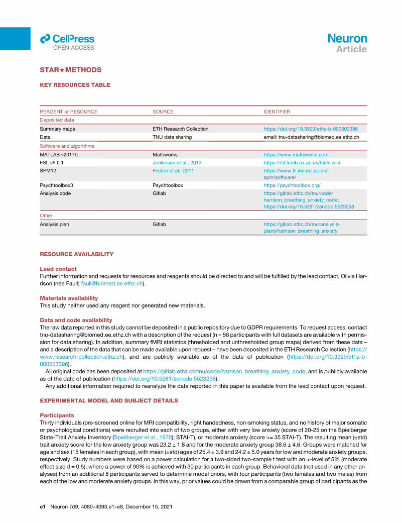
llOPEN ACCESS Article
STAR+METHODS
KEY RESOURCES TABLE
REAGENT or RESOURCE SOURCE IDENTIFIER
Deposited data
Summary maps ETH Research Collection https://doi.org/10.3929/ethz-b-000503396
Data TNU data sharing email: [email protected]
Software and algorithms
MATLAB v2017b Mathworks https://www.mathworks.com
FSL v6.0.1 Jenkinson et al., 2012 https://fsl.fmrib.ox.ac.uk/fsl/fslwiki
SPM12 Friston et al., 2011 https://www.fil.ion.ucl.ac.uk/
spm/software/
Psychtoolbox3 Psychtoolbox https://psychtoolbox.org/
Analysis code Gitlab https://gitlab.ethz.ch/tnu/code/
harrison_breathing_anxiety_code;
https://doi.org/10.5281/zenodo.5523258
Other
Analysis plan Gitlab https://gitlab.ethz.ch/tnu/analysis-
plans/harrison_breathing_anxiety
RESOURCE AVAILABILITY
Lead contactFurther information and requests for resources and reagents should be directed to and will be fulfilled by the lead contact, Olivia Har-
rison (nee Faull; [email protected]).
Materials availabilityThis study neither used any reagent nor generated new materials.
Data and code availabilityThe raw data reported in this study cannot be deposited in a public repository due toGDPR requirements. To request access, contact
[email protected] with a description of the request (n = 58 participants with full datasets are available with permis-
sion for data sharing). In addition, summary fMRI statistics (thresholded and unthresholded group maps) derived from these data –
and a description of the data that can bemade available upon request – have been deposited in the ETHResearch Collection (https://
www.research-collection.ethz.ch), and are publicly available as of the date of publication (https://doi.org/10.3929/ethz-b-
000503396).
All original code has been deposited at https://gitlab.ethz.ch/tnu/code/harrison_breathing_anxiety_code, and is publicly available
as of the date of publication (https://doi.org/10.5281/zenodo.5523258).
Any additional information required to reanalyze the data reported in this paper is available from the lead contact upon request.
EXPERIMENTAL MODEL AND SUBJECT DETAILS
ParticipantsThirty individuals (pre-screened online for MRI compatibility, right handedness, non-smoking status, and no history of major somatic
or psychological conditions) were recruited into each of two groups, either with very low anxiety (score of 20-25 on the Spielberger
State-Trait Anxiety Inventory (Spielberger et al., 1970); STAI-T), or moderate anxiety (score >= 35 STAI-T). The resulting mean (±std)
trait anxiety score for the low anxiety group was 23.2 ± 1.8 and for the moderate anxiety group 38.6 ± 4.6. Groups were matched for
age and sex (15 females in each group), withmean (±std) ages of 25.4 ± 3.9 and 24.2 ± 5.0 years for low andmoderate anxiety groups,
respectively. Study numbers were based on a power calculation for a two-sided two-sample t test with an a-level of 5% (moderate
effect size d = 0.5), where a power of 90% is achieved with 30 participants in each group. Behavioral data (not used in any other an-
alyses) from an additional 8 participants served to determine model priors, with four participants (two females and two males) from
each of the low andmoderate anxiety groups. In this way, prior values could be drawn from a comparable group of participants as the
e1 Neuron 109, 4080–4093.e1–e8, December 15, 2021

llOPEN ACCESSArticle
main study sample. All participants signed a written, informed consent, and the study was approved by the Cantonal Ethics Com-
mittee Zurich (Ethics approval BASEC-No. 2017-02330).
Behavioral data for an additional group of 15 individuals (12 female) were collected for model validation purposes. These partic-
ipants were not pre-selected based on anxiety values (mean ± std trait anxiety = 38.9 ± 12.5), but were screened for non-smoking
status and no history of major somatic or psychological conditions. Participants were aged (mean ± std) 23.1 ± 5.6 years. All partic-
ipants signed a written, informed consent, and the study was approved by the New Zealand Health and Disability Ethics Committee
(HDEC) (Ethics approval 20/CEN/168).
METHOD DETAILS
Procedural overviewEach participant completed three tasks over two testing sessions: a behavioral session that included questionnaires and a task prob-
ing interoceptive sensitivity and metacognition (the Filter Detection Task, or FDT), and a brain imaging session where the Breathing
Learning Task (BLT) was pairedwith fMRI. Each of these tasks and analyses are described below, and all analyses were pre-specified
in time-stamped analysis plans (https://gitlab.ethz.ch/tnu/analysis-plans/harrison_breathing_anxiety). The length of time between
testing sessions (mean ± std) was 4 ± 3 days for all participants. Participants in the validation group completed the BLT in a behavioral
session only.
QuestionnairesThe main questionnaire set employed was designed to first capture subjective affective measures, and second both general and
breathing-specific subjective interoceptive beliefs. The assignment of participants to groups was based on the Spielberger Trait
Anxiety Inventory (STAI-T) (Spielberger et al., 1970). Affective qualities that were additionally assessed included state anxiety (Spiel-
berger State Anxiety Inventory; STAI-S (Spielberger et al., 1970)), symptoms that are part of anxiety disorder (Generalized Anxiety
Disorder Questionnaire; GAD-7 (Spitzer et al., 2006)), anxiety sensitivity (anxiety regarding the symptoms of anxiety; Anxiety Sensi-
tivity Index; ASI-3 (Taylor et al., 2007)), and symptoms of depression (Centre for Epidemiologic Studies Depression Scale; CES-D
(Radloff, 1977)). To obtain self-reports of body awareness we used the Body Perception Questionnaire (BPQ) (Porges, 1995), while
the Multidimensional Assessment of Interoceptive Awareness Questionnaire (MAIA) (Mehling et al., 2012) was used to measure pos-
itive and ‘mindful’ attention toward body symptoms.We alsomeasured breathing-related catastrophising using the Pain Catastroph-
ising Scale (PCS-B) (Sullivan et al., 1995), and breathing-related vigilance using the Pain Vigilance Awareness Questionnaire (PVQ-B)
(McCracken, 1997) (in both questionnaires, the word ‘breathless’ or ‘breathlessness’ was substituted for ‘pain’). Finally, the following
supplementary questionnaires were included to explore possible contributing factors (e.g., general positive and negative affect, re-
silience, self-efficacy and fatigue): Positive Affect Negative Affect Schedule (PANAS-T) (Watson et al., 1988), Connor-Davidson Re-
silience Scale (Connor and Davidson, 2003), General Self-Efficacy Scale (Schwarzer et al., 1997), Fatigue Severity Scale (FSS) (Krupp
et al., 1989). The STAI-T and CES-D were completed online as part of the pre-screening process; all other questionnaires were
completed in the behavioral session at the laboratory.
Filter detection taskTo systematically test properties of breathing perception and relatedmetacognition, we utilized a perceptual threshold breathing task
(the Filter Detection Task; FDT), described in detail elsewhere (Harrison et al., 2021a). The FDT was used to determine interoceptive
perceptual sensitivity, decision bias, metacognitive bias (self-reported confidence) andmetacognitive performance (congruency be-
tween performance and confidence scores) regarding detection of very small variations in an inspiratory load. In this task (outlined in
Figure 2A), following three baseline breaths either an inspiratory load was created via the replacement of an empty filter with com-
binations of clinical breathing filters, or the empty filter was removed and restored onto the system (sham condition) for three further
breaths. All filter changes were performed behind participants, out of their field of view. After each trial of six breaths, participants
were asked to decide whether or not a load had been added, as well as reporting their confidence in their decision on a scale of
1-10 (1 = not at all confident in decision, 10 = extremely confident in decision). An adapted staircase algorithm was utilized to alter
task difficulty until participants were between 60%–85% accuracy (Harrison et al., 2021a), and 60 trials were completed at the cor-
responding level of filter load once the threshold level had been identified (using a ‘constant’ staircase procedure, as described by
(Harrison et al., [2021a]). Respiratory threshold detection (Garfinkel et al., 2016b), metacognitive bias (Rouault et al., 2018) and inter-
oceptive metacognitive performance (Harrison et al., 2021c) have previously been linked to anxiety symptomology.
Breathing learning taskTomeasure behavior and brain activity concerning the dynamic updating of interoceptive beliefs or expectations under uncertainty, a
novel associative learning task was developed and employed during functional magnetic resonance imaging (fMRI). In this Breathing
Learning Task (BLT), 80 trials were performed where on each trial two visual cues were paired with either an 80% or 20% chance of a
subsequent inspiratory resistive load. Participants were explicitly told the probabilities that were being used, as well as that the cues
were paired together – if one cue indicated an 80% chance of resistance then the other must indicate a 20% chance. Participants
were also told that the cues could only swap their contingencies throughout the task, and could not act independently of each other.
Neuron 109, 4080–4093.e1–e8, December 15, 2021 e2

llOPEN ACCESS Article
The number of trials was limited to 80 to ensure feasibility for participants. The number and structure of the cue contingency swaps
(n = 4) within the 80 trials was chosen as simulated data demonstrated both parameter recovery and model identifiability of the three
candidate learning models (see Figure S2 and ‘Quantification and Statistical Analysis’ details below).
The visual information for the task was presented through the VisualStim system (Resonance Technology, Northridge, CA, US). As
outlined in Figure 3, participants were required to predict (via button press) whether they would experience a breathing resistance
following the presentation of one of the cues. The visual cues were counter-balanced such that each was first matched with an
80% chance of resistance for half the participants, as well as counter-balancing of whether the answer ‘yes’ to the prediction ques-
tionwas presented on the left or right of the screen. Following this prediction and a short (2.5 s) pause, a circle appeared on the screen
to indicate the stimulus period (5 s), where participants either experienced inspiratory resistance (70% of their maximal inspiratory
resistance, measured in the laboratory, delivered via a PowerBreathe KH2; PowerBreathe International Ltd, Warwickshire, UK) or no
resistance was applied. Rest periods of 7-9 s were pseudo-randomized between trials.
For the inspiratory resistances we used a mechanical breathing system that allows for remote administration and monitoring of
inspiratory resistive loads (for technical details on resistance administration see previous work (Rieger et al., 2020)). The cue presen-
tations were balanced such that half of all trials delivered the inspiratory resistance. Following an initial stable period of 30 trials, the
stimulus-association pairing was swapped four times during the remainder of the 80 trials (i.e., repeated reversals; Figure 3). The trial
sequence was pseudorandom and fixed across subjects to ensure comparability of the induced learning process. Following every
stimulus, participants were asked to rate ‘How difficult was it to breathe’’?, on a visual analog scale (VAS) from ‘‘Not at all difficult’’ to
‘‘Extremely difficult.’’ Immediately following the final trial of the task, participants were also asked to rate ‘‘How anxious were you
about your breathing’’ on a VAS from ‘‘Not at all anxious’’ to ‘‘Extremely anxious.’’
Two representations of trial-wise quantities were employed for subsequent analyses of data from this task. First, a computational
model (see below) provided dynamic estimates of both predictions and prediction errors on each trial. Second, a standard categor-
ical approach represented trial-by-trial whether the subjects’ prediction decisions indicated the anticipated presence or absence of
an upcoming inspiratory resistance, as well as unsurprising (i.e., following correct predictions) and surprising (i.e., following incorrect
predictions) respiratory stimuli. The latter results are presented in the Supplemental information (Figures S4–S7).
QUANTIFICATION AND STATISTICAL ANALYSIS
Statistical analysis overviewAll analyses and hypotheses were pre-specified in an analysis plan (https://gitlab.ethz.ch/tnu/analysis-plans/harrison_breathing_
anxiety). Each measure within each task modality was first compared between groups, and multiple comparison correction was
applied within modalities. Pre-selection and comparisons across low or moderate anxiety scores allowed us to control for important
factors such as age and sex, with equal numbers of men and women recruited into each group. Finally, a cross-modal analysis was
performed on the key measures from each task. As the trait anxiety measure that was used to recruit participants into the separate
groups was not included in this final analysis, we employed a correlation-based approach as the full spectrum of scores for each task
were available.
Questionnaire analysisGroup differences were tested individually for the 13 scores resulting from the 12 questionnaires, with all questionnaires included
except the trait anxiety score that was used to screen participants and assign them to groups. The data that was used for group com-
parisons were first tested for normality (Anderson-Darling test, with p < 0.05 rejecting the null hypothesis of normally distributed data),
and group differences were determined using either two-tailed independent t tests or Wilcoxon rank sum tests. For the question-
naires, Bonferroni correction for the 13 tests was applied, requiring p < 0.004 for a corrected significant group difference. Results
with p < 0.05 not surviving correction are reported as exploratory for questionnaires as well as all other data. In a secondary explor-
atory step, group difference analyses were then conducted on the questionnaires’ subcomponent scores (22 scores); please see
Figure S1.
FDT analysisBreathing-related interoceptive sensitivity (i.e., perceptual threshold) was taken as the number of filters required to keep task perfor-
mance between �60%–85% accuracy. Both decision bias and metacognitive performance from the FDT were analyzed using the
hierarchical HMeta-d statistical model (Fleming, 2017), as previously described (Harrison et al., 2021a). This model first utilizes signal
detection theory (Stanislaw and Todorov, 1999) to provide single subject parameter estimates for task difficulty (d’; not analyzed as
performance is fixed between 60%–85% by design) and decision bias (c, akin to over- or under-reporting the presence of resistance
with values below and above zero, respectively), as well as using a hierarchical Bayesian formulation of metacognitive performance
(Mratio, calculated by fittingmetacognitive sensitivitymeta-d’, then normalizing by single subject values for d’). Finally, metacognitive
bias was calculated as the average confidence scores across all analyzed trials.
The four FDT parameters that were used for group comparison analyses were: sensitivity (filter number at perceptual threshold),
decision bias (c), metacognitive bias (average confidence scores over threshold trials) and metacognitive performance (Mratio).
These data were first tested for normality (Anderson-Darling test, with p < 0.05 rejecting the null hypothesis of normally distributed
e3 Neuron 109, 4080–4093.e1–e8, December 15, 2021

llOPEN ACCESSArticle
data), and group differences were determined using either two-tailed independent t tests orWilcoxon rank sum tests. Importantly, as
the single subject Mratio parameters were fit using a whole-group hierarchical model (with equal group numbers), standard statistical
comparisons between groups for these parameters can also be performed. Hypothesized group differences were based on previous
findings, where respiratory threshold detection level was hypothesized to be higher (Garfinkel et al., 2016a; Tiller et al., 1987), percep-
tual decisions biased toward ‘yes’ (or deciding the resistance was present; denoted by more negative values for c), metacognitive
bias to be lower (Rouault et al., 2018) and interoceptive metacognitive performance to be lower (Harrison et al., 2021c) with greater
anxiety. Bonferroni correction for the four tests was applied, requiring p < 0.013 for a corrected significant group difference.
BLT analysisModel space
For the trial-by-trial analysis of behavioral data from the BLT, we considered three computational models that are routinely used for
associative learning tasks. This included aRescorlaWagner (RW)model (Equation 1) and 2 variants of theHierarchical Gaussian Filter
(HGF) with 2 or 3 levels (HGF2 and HGF3). While the RWmodel assumes a fixed learning rate, the HGF allows for online adaption of
learning rate as a function of volatility. All learning models were paired with a unit-square sigmoid response model (Equation 2) and
were implemented using the Hierarchical Gaussian Filter Toolbox (Mathys et al., 2011, 2014) (version 5.3) from the open-source
TAPAS software (Fr€assle et al., 2021) (https://www.translationalneuromodeling.org/tapas/).
Prior selection and simulation analyses
Prior means and variances were determined using the distribution of maximum likelihood estimates fit across a holdout dataset (con-
sisting of 8 participants who were distinct from the participants of our study). Individual fits and estimated prior densities of the free
parameters are given in Table S2, as well as values for all remaining parameters of the threemodels. By adopting this procedure, prior
densities were in a regime of the parameter space that is representative of the actual behavioral responses observed when
participants performed the task. At the same time, the arbitrariness inherent to the specification of prior densities in non-hierarchical
inference is reduced to a minimum.
To demonstrate face validity of the three models considered in our model space (each with one parameter free: a in RW, u2 in
HGF2, k2 in HGF3), we assessed both parameter recovery and model identifiability for each (Wilson and Collins, 2019). Data for
60 synthetic subjects were generated for each of the candidate models by randomly sampling values from the prior densities that
were placed over the parameters of the perceptual model. This synthetic data was generated for different noise levels ðzsim =
1;5;10Þ. Subsequently, maximum a posteriori (MAP) parameter estimates were obtained using the Brayden-Fletcher-Goldfarb-
Shanno algorithm, as implemented in the HGF toolbox, to fit the synthetic datasets. This allowed us to quantify parameter recovery
and model identifiability across three different noise levels for each of the candidate models. Parameter recovery of the perceptual
parameters was assessed using Pearson’s correlation coefficient (PCC) and by visual inspection of simulated and recovered param-
eter values (Figure S2). Mean and standard deviation of estimated z values (from the responsemodel) were computed for every noise
level. Model identifiability was quantified by calculating the proportion of correctly identified models using approximate log model
evidence (LME) scores, and assessing whether the former was greater than the upper bound of the 90% confidence interval
when assuming every model is equally likely a priori. For the resulting confusion matrices (Figure S2), we additionally computed
the mean proportion of correctly identified models (balanced accuracy scores).
The outlined procedure for assessing parameter recovery and model identifiability was repeated over 10 iterations with different
seed values, to ensure robustness against any particular setting of the random number generator. The final results (PCCs for the
perceptual parameters and zest values) for every given level of noise were calculated as the average over all iterations, and are pre-
sented in Table S3.
Model comparison and selection
FollowingMAP estimates of the 60 empirical datasets using each of the candidate models, themodels were formally compared using
random effects Bayesian model selection (BMS) as implemented in SPM12 (Friston et al., 2011; Rigoux et al., 2014; Stephan et al.,
2009). BMS utilizes the LME to determine the most likely among a set of competing hypotheses (i.e., models) that may have gener-
ated observed data, and is robust to outliers (Stephan et al., 2009). Our analysis plan had specified that a model would be chosen as
the ‘winning’ model if it demonstrated a protected exceedance probability (PXP) greater than 90%. As explained in the Results sec-
tion, none of our models reached this criterion (although simulations indicated that the proposed models could in principle be differ-
entiated; see Figure S2 for details). We therefore applied the simplest of the models considered (i.e., the RWmodel), as pre-specified
in our analysis plan (https://gitlab.ethz.ch/tnu/analysis-plans/harrison_breathing_anxiety).
In our application of the RWmodel as a perceptual model, the update equation corresponded to a simple delta-learning rule with a
single free parameter, the learning rate (Rescorla et al., 1972):
vðk +1Þ = vðkÞ +adðkÞ (Equation 1)
where vðk + 1Þ is the predicted probability for a specific outcome (encoded as 0 or 1) on trial ðk + 1Þ, vðkÞ is the estimated outcome prob-
ability on the kth trial, a ˛ [0, 1] is a constant learning rate parameter, and dðkÞ is the prediction error magnitude at trial k.
The above perceptual model was paired with a unit-square sigmoid response model (Mathys et al., 2014). This response model
accounts for decision noise by mapping the predicted probability vðkÞ that the next outcome will be 1 onto the probabilities
pðyðkÞ = 1Þ and pðyðkÞ = 0Þ that the agent will choose response 1 or 0, respectively:
Neuron 109, 4080–4093.e1–e8, December 15, 2021 e4

llOPEN ACCESS Article
pðyðkÞjvðkÞ; zÞ =
vzðkÞvzðkÞ + ð1� vðkÞÞz
!yðkÞ ð1� vðkÞÞzvzðkÞ + ð1� vðkÞÞz
!1�yðkÞ
(Equation 2)
Here, yðkÞ represents the expressed decision of a subject given the cue (contingency pairs) on trial k. The parameter z captures how
deterministically y is associatedwith v. The higher z, themore likely the agent is to choose the option that ismore in linewith its current
prediction. The decision model uses the perceptual model indirectly via its inversion (Mathys et al., 2014), given the trajectories of
trial-wise cues and responses (see Figure 3).
In our paradigm, trial-wise outcomes are categorical (resistance versus no resistance), which raises the question of how outcomes
should be coded in the computational model. One way would be to model two trajectories, separately for resistance and no resis-
tance outcomes, and indicate on any given trial whether the respective outcome has occurred (1) or not (0). However, due to the fixed
coupling of contingencies in our paradigm (see above) – which the participants were explicitly instructed about – a computationally
more efficient approach that requires only a singlemodel is to code the outcome in relation to the cue. Here, we adopted this coding in
‘‘contingency space,’’ following the same procedure as in the supplementarymaterial of Iglesias and colleagues (Iglesias et al., 2013).
Specifically, due to the fixed coupling of contingencies in our paradigm (see above), we represented the occurrence of ‘‘no resis-
tance’’ given one cue and the occurrence of ‘‘resistance’’ given the other cue as 1, and both other cue-outcome combinations as
0 (note that under the subsequent transformations described below, the resulting trajectories of predictions and prediction errors
would remain identical if the opposite choice had been made).
Comparison of fitted model parameters
Group differences inmodel parameter estimates of learning rate ðaÞ and inverse decision temperature ðzÞ, as well as perceptionmea-
sures of stimulus intensity (averaged across all trials), breathing-related anxiety (rated immediately following the task) and prediction
response times were compared following tests for data normality. Bonferroni correction for five tests was applied, requiring p < 0.01
for a corrected significant group difference. Results from additional exploratory models encompassing anxiety, depression and
gender are reported in Table S6.
Model validation
Following random effects Bayesian model selection (BMS (Rigoux et al., 2014; Stephan et al., 2009)), the chosen model was exam-
ined in each participant with regard towhether it demonstrated an adequate fit. To this end,model fit in each individual was compared
to the likelihood of obtaining the data by a ‘null model’ (i.e., due to chance) (Daw, 2011) using the likelihood ratio test (lratiotest func-
tion) provided in MATLAB. The final behavioral and brain imaging analyses presented here were run without two subjects in which
non-significant (p > 0.05) differences to randomness were encountered. To demonstrate the extent to which the chosen model
(the Rescorla Wagner) captured important aspects of participant performance, the proportion of incorrect responses across partic-
ipants at each trial was compared to the mean prediction error trajectory (Figure S3B).
To further validate the application of the chosen model to the BLT, we compared the fitted model trajectory to unseen behavioral
data from an additional 15 participants. For qualitative assessment, themean prediction error trajectory from the original dataset was
compared to the proportion of incorrect responses across these held-out participants at each trial (Figure S3C). For quantitative
assessment, a logistic regression was conducted to assess whether the model prediction trajectory from the original data was
able to significantly explain the prediction decisions made by the 15 participants in the validation sample.
Computationally informed regressors
The trajectories of predictions and prediction errors estimated by the RW model were used to construct regressors representing
computational trial-by-trial quantities of interest for subsequent GLM analyses. In order to investigate the salient effects of inspiratory
resistance as an interoceptive stimulus, we separated trials into ‘‘negative’’ (occurrence of resistance) and ‘‘positive’’ (no resistance)
events and represented these events by separate regressors in the GLM (see Figure 4). To achieve this, we first transformed both the
original prediction and prediction error values (estimated in contingency space) back into the stimulus space, according to the cue
presented at each trial:
vstimðkÞ def
(vðkÞ; jif cue type= 11� vðkÞ; jif cue type= 2
(Equation 3)
dstimðkÞ def
(dðkÞ; jif cue type= 1�dðkÞ; jif cue type= 2
(Equation 4)
Here, vstimðkÞ and dstimðkÞ now represent the prediction and prediction error values in stimulus space, with vstimðkÞ = 1 representing maximal
predictions of no resistance and vstimðkÞ = 0maximal predictions of resistance. Similarly, dstimðkÞ = 1 representsmaximal prediction errors of
no resistance and dstimðkÞ = � 1 maximal prediction errors of resistance (see Figure S4 for details).
Second, trial-wise prediction values were then transformed to represent the deviation frommaximally uninformed predictions (i.e.,
guessing), by taking the distance from 0.5 (see Equations 5 and 6). In the RW model prediction values are probabilities bounded by
e5 Neuron 109, 4080–4093.e1–e8, December 15, 2021

llOPEN ACCESSArticle
0 and 1, hence the distance from ‘guessing’ (at 0.5) reflects the ‘certainty’ by which the absence or presence of respiratory resistance
was predicted. This simple transformation enabled us to take into account the role of (un)certainty of predictions – which plays a
crucial role in interoception-oriented theories of anxiety (Paulus and Stein, 2010; Paulus et al., 2019) but, in contrast to Bayesian
models, is not represented explicitly in the RWmodel. Specifically, separately for the two event types, we defined certainty of positive
predictions (no resistance) and of negative predictions (resistance) as the absolute deviation from a prediction with maximum uncer-
tainty (i.e., 0.5):
If vstimðkÞ > 0:5 vposðkÞ def vstimðkÞ � 0:5 (Equation 5)
If vstimðkÞ < 0:5 vnegðkÞ def0:5� vstimðkÞ (Equation 6)
Here, both vposðkÞ and vnegðkÞ exist between 0 and 0.5, with values closer to zero indicating less certain predictions.
Like predictions, prediction errors were also divided between positive (no resistance) and negative events (resistance) values. This
was again determined as the absolute deviation from the mid-point of the prediction errors (i.e., 0):
If dðkÞ > 0 dposðkÞ defdðkÞ (Equation 7)
If dðkÞ < 0 dnegðkÞ def� dðkÞ (Equation 8)
Here, both dposðkÞ and d
negðkÞ exist between 0 and 1, with values closer to zero indicating smaller prediction errors. Note that this derivation
gives prediction error values identical to those that would have been obtained by modeling two separate trajectories for resistance
and no resistance outcomes (see above and (Iglesias et al., 2013)).
Physiological data processing
Physiological data were recorded at a sampling rate of 1000Hz, and included heart rate, chest distension, pressure of expired carbon
dioxide (PETCO2) and oxygen (PETO2), and pressure at the mouth (for equipment details see (Rieger et al., 2020)). In addition to the
task, small boluses of a CO2 gas mixture (20% CO2; 21% O2; balance N2) were administered during some rest periods, allowing for
de-correlation of any changes in PETCO2 from task-related neural activity, as previously described (Faull and Pattinson, 2017; Faull
et al., 2015, 2016, 2018).
Physiological noise regressors were prepared for inclusion into single-subject general linear models (GLMs, described below).
Linear interpolation between PETCO2 peaks was used to form an additional CO2 noise regressor, which was convolved using a
response function based on the haemodynamic response function (HRF) provided by SPM with delays of 10 s and 20 s for the over-
shoot and undershoot, respectively (Chang and Glover, 2009). Temporal and dispersion derivatives of this CO2 noise regressor were
also included. An additional three cardiac- and four respiratory-related waveforms (plus one interaction term) were created using
PhysIO (Kasper et al., 2017). Four respiratory volume per unit time (RVT) regressors (delays: �5, 0, 5, 10) were created using the Hil-
bert-transform estimator in PhysIO (Harrison et al., 2021b), and convolution with a respiratory response function (Kasper et al., 2017).
Magnetic resonance imaging
MRIwas performed using a 7 Tesla scanner (PhilipsMedical Systems: Achieva, Philips Healthcare, Amsterdam, the Netherlands) and
a 32 channel Head Coil (Nova Medical, Wilmington, Massachusetts, United States of America). A T2*-weighted, gradient echo EPI
was used for functional scanning, using a reduced field of view (FOV) with an axial-oblique volume centered over the insula and
midbrain structures. The FOV comprised 32 slices (sequence parameters: TE 30ms; TR 2.3 s; flip angle 75�; voxel size
1.5x1.5x1.5mm; slice gap 0.15mm; SENSE factor 3; ascending slice acquisition), with 860 volumes (scan duration 33 mins 9 s). A
matched whole-brain EPI scan (96 slices) was immediately acquired following the task scan for registration purposes. Additionally,
a whole-brain T1-weighted structural scan with 200 slices was acquired (MPRAGE, sequence parameters: TE 4.6ms; TR 10ms;
segment-TR 3000ms; TI 1000ms; flip angle 8�; voxel size 0.8x0.8x0.8mm; bandwidth; 153.1Hz/Px; sagittal slice orientation). Finally,
a task-free (resting-state) functional scan (250 volumes) was obtained, with participants instructed to keep their eyes open and
fixating a white fixation cross on a black screen.
MRI preprocessing
MRI data analysis was performed using a combination of FSL version 6.0.1 (the Oxford Centre for Functional Magnetic Resonance
Imaging of the Brain Software Library, Oxford, UK) (Jenkinson et al., 2012) and SPM12 (Statistical Parametric Mapping software,
London, UK) (Friston et al., 2011) as prespecified in our analysis plan (https://gitlab.ethz.ch/tnu/analysis-plans/harrison_breathing_
anxiety). Image preprocessing was performed using FSL, including motion correction (MCFLIRT (Jenkinson and Smith, 2001)),
removal of non-brain structures (BET (Smith, 2002)), and high-pass temporal filtering (Gaussian-weighted least-squares straight
line fitting; 100 s cut-off period)(Woolrich et al., 2001). Independent component analysis (ICA) was used to identify noise due to mo-
tion, scanner and cerebrospinal fluid artifacts (Griffanti et al., 2017), and the timeseries of these noise components were entered into
single-subject GLMs (described below) as nuisance regressors. The functional scans were registered to the MNI152 (1x1x1mm)
standard space using a three-step process: 1) Linear registration (FLIRT) with 6 degrees of freedom (DOF) to align the partial FOV
Neuron 109, 4080–4093.e1–e8, December 15, 2021 e6

llOPEN ACCESS Article
scan to the whole-brain EPI image (Jenkinson et al., 2002); 2) Boundary-based registration (BBR; part of the FMRI Expert Analysis
Tool, FEAT) with 12 DOF and a weighting mask of the midbrain and insula cortex to align the whole-brain EPI to T1 structural image;
and 3) Non-linear registration using a combination of FLIRT and FNIRT (Andersson et al., 2007) to align the T1 structural scan to 1mm
standard space. Functional MRI scanswere resampled once into standard spacewith a concatenatedwarp from all three registration
steps, and then spatial smoothing in standard space was performed using a Gaussian kernel with 3mm full-width half-maximum us-
ing the fslmaths tool.
Single-subject general linear model
Single-subject estimates of the general linear model (GLM) were performed using SPM. A GLMwas constructed for each of the par-
ticipants, with a design matrix informed by trial-wise estimates from the RW model of each participant (see above). An additional
analysis, using a more classical (non-computational model-based) design matrix, is presented in the Supplemental information (Fig-
ures S4–S6). Alongside the task regressors described below, rigorous de-noising was performed by the inclusion of the following
regressors (all described above): the convolved end-tidal CO2 regressor plus temporal and dispersion derivatives, six motion regres-
sors trajectories plus their first-order derivatives, physiological noise regressors (provided by the PhysIO toolbox) and ICA compo-
nents identified as noise.
The regressors of interest in the design matrix were as follows (compare Figure 3A and Figure S5):
1) A ‘Cue’ regressor (80 repeats), with onsets and durations (2.5 s) determined by the presentations of visual cues and a magni-
tude of 1;
2) A ‘Positive prediction’ regressor, with onsets given by the presentation of each corresponding visual cue (when no-resistance
was predicted), durations of 0.5 s and magnitudes given by vposðkÞ in Equation 5;
3) A ‘Negative prediction’ regressor, with onsets given by the presentation of each corresponding visual cue (when resistance
was predicted), durations of 0.5 s and magnitudes given by vnegðkÞ in Equation 6;
4) A ‘No resistance’ stimulus regressor, with onset timings according to the first inspiration that occurred after the presentation of
the visual cue, and durations as the remaining time of the potential resistance period (circle in Figure 3), with a magnitude of 1;
5) A ‘Resistance’ stimulus regressor, with onset timings according to the initiation of the inspiratory resistance (identified from the
downward inflection of the inspiratory pressure trace) after the presentation of the visual cue, and durations as the remaining
time of the resistance period (circle in Figure 3), with a magnitude of 1;
6) A ‘Positive prediction error’ regressor, with onsets given by the start of each corresponding no resistance period, durations of
0.5 s and magnitudes given by dposðkÞ in Equation 7;
7) A ‘Negative prediction error’ regressor, with onsets given by the start of each corresponding resistance period, durations of
0.5 s and magnitudes given by dnegðkÞ in Equation 8;
8) A ‘Rating period’ noise regressor, with onsets and durations covering the period where participants were asked to rate the dif-
ficulty of the previous stimulus, and with a magnitude of 1.
Regressors 1-8 were included in the design matrix after convolution with a standard HRF in SPM12, together with their temporal
and dispersion derivatives. Contrasts of interest from this design examined brain activity associated with the average across positive
and negative valence for both predictions and prediction errors, aswell as the difference due to valence (i.e., positive versus negative)
for both predictions and prediction errors.
Group fMRI analysis
First, for the analysis of our entire field of view, contrasts of interest were assessed using random effects group-level GLM analyses
based on the summary statistics approach in SPM12. The group-level GLM consisted of a factorial design with both a group mean
and group difference regressor. The analyses used a significance level of p < 0.05 with family-wise error (FWE) correction at the
cluster-level, with a cluster-defining threshold of p < 0.001. Second, for our region of interest (ROI) analysis, we used FSL’s non-para-
metric threshold-free cluster enhancement (Smith and Nichols, 2009) within a combined mask of the anterior insula and periaque-
ductal gray (PAG), as pre-specified in our analysis plan (section 7.5.5). This analysis employed a significance level of p < 0.05,
with FWE correction across the joint mask. While the anterior insula and PAG have previously been shown to be involved in both
conditioned anticipation and perception of inspiratory resistances (Berner et al., 2018; Faull and Pattinson, 2017; Faull et al.,
2016, 2018; Paulus et al., 2012; Walter et al., 2020) as well as prediction errors (Roy et al., 2014) and precision (Grahl et al., 2018)
toward pain perception, our current analysis considers computational trial-by-trial estimates of interoceptive predictions and predic-
tion errors for the first time. The mask of the anterior insula was taken from the Brainnetome atlas (Fan et al., 2016) (bilateral ventral
and dorsal anterior insula regions), and the PAG incorporated an anatomically-defined mask that has been used in previous fMRI
publications (Faull and Pattinson, 2017; Faull et al., 2016).
Multi-modal analysisMulti-modal data
The different task modalities were then combined into a multi-modal analysis to assess both the relationships between and shared
variance among measures. The data entered into this analysis consisted of:
e7 Neuron 109, 4080–4093.e1–e8, December 15, 2021

llOPEN ACCESSArticle
1) The scores from the four main affective questionnaires that were not used to pre-screen the participants (STAI-S (Spielberger
et al., 1970), GAD-7 (Spitzer et al., 2006), ASI-3 (Taylor et al., 2007) and CES-D (Radloff, 1977));
2) The four interoceptive questionnaires (BPQ (Porges, 1995),MAIA (Mehling et al., 2012), PCS-B (Sullivan et al., 1995) and PVQ-B
(McCracken, 1997));
3) The four FDT measures (breathing sensitivity, decision bias c, metacognitive bias, metacognitive performance Mratio); and
4) The individual peak anterior insula activity associated with both positive and negative predictions, as well as positive and nega-
tive prediction errors. Activity was extracted from a 4mm sphere, centered on each participant’s maximal contrast estimate
within a Brainnetome atlas mask of the anterior insula (Fan et al., 2016), using the first eigenvariate of the data.
Multi-modal correlations and shared variance
A Pearson’s correlationmatrix of all 16 includedmeasures was calculated in order to visualize the relationships between all variables.
The significance values of the correlation coefficients were taken as p < 0.05 (exploratory), and a false discovery rate (FDR) correction
for multiple comparisons was applied (using themafdr function in MATLAB). A supplementary non-parametric correlation matrix was
additionally calculated using Spearman’s rho values, and these results are presented in Table S7.
To assess the shared variance across measures and delineate which measures were most strongly associated with affective qual-
ities, we entered all specified data into a principal component analysis (PCA), following normalization using z-scoring within each
variable. PCA is an orthogonal linear transformation that transforms the n3m data matrix X (participants3measures) into a newma-
trix P, where the dimensions of the variance explained in the data are projected onto the new ‘principal components’ in descending
order. Each principal component consists of a vector of coefficients or weightsw, corresponding to the contribution of eachmeasure
m to each component. The PCA also transforms the original n3m data matrix X to map each row (participant) vector xi of X onto a
new vector of principal component scores ti, given by:
tkðiÞ = xi 3wk for i = 1;.;n; k = 1;.;m (Equation 9)
where tkðiÞ is the score for each participant i within each component k. The number of significant components were then determined
by comparing the variance explained of each component to a null distribution, created by randomly shuffling (n = 1000) the measures
from each variable across participants. Components were considered significant if the variance explained was above the 95% con-
fidence interval of the corresponding component’s null distribution.
To assess the relationship between each of the significant components and anxiety, the component scores for low and moderate
anxiety were compared using either independent t tests or Wilcoxon rank sum tests (following Anderson-Darling tests for normality).
The significance values of the group differences in component scores were taken as p < 0.05 (exploratory), and a false discovery rate
(FDR) correction for multiple comparisons (number of significant components) was applied.
An independent code review was performed on all data analysis procedures, and the analysis code is available on GitLab (https://
gitlab.ethz.ch/tnu/code/harrison_breathing_anxiety_code; https://doi.org/10.5281/zenodo.5523258).
Neuron 109, 4080–4093.e1–e8, December 15, 2021 e8

Neuron, Volume 109
Supplemental information
Interoception of breathing
and its relationship with anxiety
Olivia K. Harrison, Laura Köchli, Stephanie Marino, Roger Luechinger, FranciszekHennel, Katja Brand, Alexander J. Hess, Stefan Frässle, Sandra Iglesias, FabienVinckier, Frederike H. Petzschner, Samuel J. Harrison, and Klaas E. Stephan

Supplementary Figure 1. Results from the additional questionnaires measured in groups of healthy individuals with either low or moderate levels of anxiety, related to Figure 1. Participants with low anxiety scored 20-25 on the Spielberger Trait Anxiety Inventory (STAI-T), and those with moderate anxiety scored 35+ on the STAI-T. Questionnaires: A) ‘Positive affect’ and ‘Negative affect’ from the Positive Affect Negative Affect Schedule (PANAS-T), ‘Fatigue’ from the Fatigue Severity Scale (FSS), ‘Self efficacy’ from the General Self-Efficacy Scale, and ‘Resilience’ from the Connor-Davidson Resilience Scale; B) Sub-scores of the Pain Vigilance Awareness Questionnaire (with the word ‘pain’ substituted for ‘breathing’, PVQ-B); C) Sub-scores of the Anxiety Sensitivity Index (ASI-3) questionnaire; D) Sub-scores of the Pain Catastrophising Scale (with the word ‘pain’ substituted for ‘breathing’, PCS-B); E) Sub-scores of the Body Perception Questionnaire (BPQ); F) Sub-scores of the Multidimensional Assessment of Interoceptive Awareness Questionnaire (MAIA). Bar plots represent mean±standard error values, with the distribution of values overlaid in grey. **Significant at p<0.01, with no correction for multiple comparisons (exploratory results). Bar plots represent mean±standard error values, with the distribution of values overlaid in grey. Bar plot code adapted from the CANLAB Toolbox (https://github.com/canlab).

Supplementary Figure 2. Parameter recovery and model identifiability for the three candidate models, related to Figure 3. A-C) Demonstration of parameter recovery using simulated participants from the prior distributions presented in Supplementary Table 2. 60 simulated participant responses were generated using 10 different seed values, totalling n=600 simulations plotted here. D-E) Demonstration of model identifiability using simulated participants from the prior distributions presented in Supplementary Table 2. 60 simulated participant responses were generated using 10 different seed values, and the confusion matrices, balanced accuracy and zeta estimates are the average values across the 10 simulation runs. Three noise levels were used for the simulations, with an inverse decision temperature (𝜁) of 1 (A,D), 5 (B,E) and 10 (C,E), representing very noisy (𝜁 =1) to very deterministic (𝜁 = 10) settings.
D
E
F
A
B
C

Supplementary Figure 3. Model comparisons and fit quality of the chosen model (the Rescorla Wagner), related to Figure 3. A) Model comparison: Estimated log model evidence (LME) values across all participants. Left and middle: Comparisons between each model pair, where bar plots represent mean±standard error values of the specified differences in LME, with the distribution of values overlaid in grey. Bar plot code adapted from the CANLAB Toolbox (https://github.com/canlab). Right: Proportion of highest LME values across all 60 participants. Dotted lines represent upper and lower 95% confidence intervals for chance, and all proportions of winner classifications lie within the chance range. B) Model validation in original data: Comparison of the mean prediction error trajectory (black) against the proportion of participants giving incorrect responses at each trial (red) for the original dataset (n = 60). The close correlation between these trajectories demonstrates the extent to which the chosen model (the Rescorla Wagner) captures important aspects of participant performance. C) Model validation in unseen data: Comparison of the mean prediction error trajectory from the original dataset (black) against the proportion of participants giving incorrect responses at each trial (red) for a validation dataset (n = 15), who were not preselected for any specific anxiety level. The correlation between these trajectories demonstrates the extent to which the average model fit for the original data is able to capture important aspects of participant performance on unseen data. The prediction error trace in (B) and (C) is the absolute prediction error trajectory from the participant with the closest learning rate to the mean across all participant model fits. The prediction error trace is represented in stimulus space (see Supplementary Figure 4 for transformation procedures).
A
B
C

Supplementary Figure 4. Transformation of the prediction and prediction error trajectories from contingency space to stimulus space, related to Figures 4 and 5. A) The fitted trajectory (in contingency space) is demonstrated by the solid grey line, where the value 1 is assigned when one cue (cue 1) predicts no resistance and the opposing cue (cue 2) predicts a resistance (the value 0 is assigned for the opposing conditions). The trajectories were then transformed into stimulus space, where a value of 1 was assigned when no resistance was delivered, while a value of 0 was assigned when a resistance was delivered. For this transformation, a mirrored trajectory was firstly generated (dashed grey line) to represent the second cue, as the participants were explicitly told that the cues acted as a pair that had opposite probabilities (20% or 80%) of predicting resistance. The solid grey trajectory thus represents the cue that started with an 80% probability of being followed by no resistance in stimulus space (cue 1), while the dashed grey line represents the cue that started with a 20% probability of being followed by no resistance (cue 2). The values at each trial were taken from the trajectory of the cue that was presented at that trial: either cue 1 (trials with a closed grey circle) or cue 2 (trials with an open grey circle). The same transformation was performed on the prediction error trajectories in (B), where the solid grey line represents the prediction error associated with cue 1, while the dashed grey line represents the prediction error associated with cue 2. The example trajectories were taken from the participant with the closest learning rate to the mean value across all participants.
A
B

Supplementary Figure 5. Demonstration of the correlation between regressors for the model-based and decision-based general linear models, related to Figure 4 and 5. A and D) Average correlation matrices (using Fischer’s R-to-Z transformation prior to averaging) from all single subject general linear models used in the model-based (A) and decision-based (D) fMRI analyses (noise regressors not shown). B and E) Targeted correlation matrices to demonstrate the relationship between predictions and errors for A and D, respectively. C) An example general linear model from a single participant model-based fMRI analysis. Each of the main regressors also include a temporal and dispersion derivative. Please see STAR methods for a full description of each regressor. F) An example general linear model from a single participant decision-based fMRI analysis. Each of the main regressors also include a temporal and dispersion derivative, and consist of: 1) ‘cueYes’ – the time periods covering the presentation of cues where the participant predicted an upcoming resistance; 2) ‘cueNo’ – the time periods covering the presentation of cues where the participant predicted no upcoming resistance; 3) ‘resistanceBlock’ – the stimulus periods when resistance was applied, from the onset of the inspiration against the increased inspiratory pressure following the presentation of the circle cue to the end of the circle presentation; 4) ‘resistanceSurprise’ – resistance stimuli that were surprising (i.e. when participant had predicted no resistance), with an onset at the beginning of the corresponding stimulus period and a duration of 0.5 seconds; 5) ‘noResistanceBlock’ – the stimulus periods when no resistance was applied, from the onset of the first inspiration following the presentation of the circle cue to the end of the circle presentation; 6) ‘noResistanceSurprise’ – no-resistance stimuli that were surprising (i.e. when participant had predicted resistance), with an onset at the beginning of the corresponding stimulus period and a duration of 0.5 seconds; 7) ‘noiseRating’ – the periods at the end of each trial where the participant rated the intensity of the previous stimulus, with an onset at the beginning and duration that encompassed the length of the rating period. 8) ‘noisePredict’ – the button press periods during the cue presentation, with an onset given by the response time for the button press on each trial and a duration of 0.5 seconds. Noise regressors not shown: the convolved end-tidal carbon dioxide trace (plus temporal and dispersion derivatives), the RETROICOR and convolved respiratory volume per unit of time (RVT) regressors provided by the PhysIO toolbox, 6 motion regressors and 6 extended motion regressors (derivatives), and the timeseries of all identified noise components from the independent component analysis conducted during preprocessing were also included in the model.

Supplementary Figure 6. BOLD activity related to prediction certainty, prediction errors, resistance and no-resistance stimuli, related to Figures 4 and 5. A) Significant BOLD activity associated with prediction certainty, averaged over positive and negative. B) Significant BOLD activity associated with prediction error magnitude, averaged over positive and negative. C) Significant BOLD activity associated with inspiratory resistance periods. D) Significant BOLD activity associated with no resistance periods. The images consist of a colour-rendered statistical map superimposed on a standard (MNI 1x1x1mm) brain. The bright grey region represents the coverage of the coronal-oblique functional scan. Significant regions are displayed with a cluster threshold of p<0.05, FWE corrected for multiple comparisons across all voxels included in the functional volume. Images in A and B are an expanded view of Figure 4.
A) PREDICTION CERTAINTY
0 10ACTIVATION
0
T-STATISTICDEACTIVATION
B) PREDICTION ERROR
0 10ACTIVATION
0
T-STATISTICDEACTIVATION
C) RESISTANCE STIMULI (NEGATIVE)
0 10ACTIVATION
0
T-STATISTICDEACTIVATION
D) NO RESISTANCE STIMULI (POSITIVE)
0 10ACTIVATION
0
T-STATISTICDEACTIVATION

Supplementary Figure 7. Overall results from the non-computational decision-based analyses of the ‘Breathing Learning Task’ (BLT), related to Figures 4 and 5. The plots in both (A) and (B) demonstrate how prediction decisions (in A) and surprise (in B) trajectories are encoded into positive (i.e. towards no resistance) and negative (i.e. towards resistance, red) values. The grey lines in both plots represent the stimulus at each trial, while the blue (positive) and red (negative) lines in (A) denote the prediction decisions (prior to the stimulus) and in (B) the surprising events (where the prediction decision was incorrect). In both trajectories the dotted black line denotes the boundaries between positive and negative valence, and the distance from the dotted line is taken as the final value (i.e. absolute values). The brain images in (A) represent the influence of valence on prediction decisions (difference between negative and positive decisions), while there is no equivalent representation of overall predictions in a binary decision model compared to the computational model design (the average over positive and negative prediction decisions simply represents the cue presentation). The bottom panel of brain images in (A) demonstrate an interaction effect between valence (i.e. positive vs. negative) and anxiety group (low vs. moderate) for the anterior insula activity related to the valence of the prediction decisions of interoceptive breathing stimuli. Here, voxel-wise statistics were performed using non-parametric permutation testing within a mask of the anterior insula and periaqueductal gray, with significant results determined by p<0.05 (corrected for multiple comparisons within the mask). The brain images in (B) represent the activity associated with average surprise (average over positive and negative surprise trajectories) and the influence of valence on surprise (difference between negative and positive surprise trajectories). The images consist of a colour-rendered statistical map superimposed on a standard (MNI 1x1x1mm) brain. The bright grey region represents the coverage of the coronal-oblique functional scan. Significant regions are displayed with a cluster threshold of p<0.05, FWE corrected for multiple comparisons across all voxels included in the functional volume. Abbreviations: PAG, periaqueductal gray

Supplementary Table 1. Physiological summaries and group comparison results from the stimulus periods of the ‘Breathing Learning Task’ (BLT), related to Figure 3. Abbreviations: Wxn, Wilcoxon rank sum test; Ttest, students independent T-test. If a Wilcoxon rank sum test was utilised, reported values are median ± interquartile range.
Total Low Moderate P-value Test RESISTANCE 70% of maximum inspiratory pressure -58.8 (24.5) -63.4 (23.8) -56.0 (28.0) 0.52 Wxn Avg measured pressure (cmH2O) -4.0 (3.7) -4.2 (3.8) -3.8 (2.9) 0.72 Wxn Max measured pressure (cmH2O) -7.3 (4.9) -7.3 (6.7) -7.3 (3.9) 0.68 Wxn Avg breathing rate (bpm) 13.6 (6.7) 14.6 (5.6) 13.0 (10.1) 0.39 Wxn Avg breathing depth (% of rest depth) 103.9 (25.5) 97.0 (23.7) 105.9 (26.1) 0.29 Ttest Heart rate (bpm) 66.3 (13.5) 66.9 (12.5) 66.0 (15.6) 0.69 Ttest NO RESISTANCE Avg measured pressure (cmH2O) -0.1 (0.1) -0.1 (0.1) -0.1 (0.1) 0.05 Wxn Max measured pressure (cmH2O) -0.8 (0.5) -0.8 (0.8) -0.7 (0.4) 0.59 Wxn Avg breathing rate (bpm) 14.8 (5.1) 14.9 (5.0) 14.7 (5.8) 0.92 Ttest Avg breathing depth (% of rest depth) 106.6 (17.0) 105.0 (17.0) 108.8 (21.7) 0.35 Ttest Heart rate (bpm) 66.7 (12.3) 67.6 (12.1) 66.6 (14.0) 0.84 Wxn
Supplementary Table 2. Parameter configurations and priors for each of the candidate models, related to Figure 3. If the prior variance is set to 0 for a parameter it is not estimated, and the prior mean and variance for the estimated parameters (in bold) were taken from the maximum likelihood fits of the pilot participant data. Prior means are given in native space, prior variances in estimation (transformed) space.
Rescorla Wagner Parameter Prior mean Prior variance Transformation
𝑣( ) 0.5 0 logit 𝜶 0.29 2.54 logit
Observation model 𝜻 2.14 3.33 log Hierarchical Gaussian Filter (2-level)
Parameter Prior mean Prior variance Transformation 𝜇( ) 0 0 none
𝜇( ) 1 0 none
𝜎( ) 0.1 0 log
𝜎( ) 1 0 log 𝜌 0 0 none 𝜌 0 0 none 𝜅 1 0 log 𝜅 0 0 log 흎ퟐ -0.70 1.61 none 𝜔 −∞ 0 none
Observation model 𝜻 2.14 1.17 log Hierarchical Gaussian Filter (3-level)
Parameter Prior mean Prior variance Transformation 𝜇( ) 0 0 none
𝜇( ) 1 0 none
𝜎( ) 0.1 0 log
𝜎( ) 1 0 log 𝜌 0 0 none 𝜌 0 0 none 𝜅 1 0 log 𝜿ퟐ 2.39 0.30 log 𝜔 -3 0 none 𝜔 -6 0 none
Observation model 𝜻 2.14 1.21 log

Supplementary Table 3. Parameter recovery metrics for each of the candidate models, related to Figure 3. R values are Pearson Correlation Coefficients that have been Fisher’s Z-transformed prior to averaging across the 10 simulation runs, and then back-transformed into R values. Each simulation run consisted of 60 simulated synthetic datasets sampled from the prior distribution of values for the perceptual model, repeated at each noise level (𝜁 = [1,5,10]), and recovered using MAP estimation.
Rescorla Wagner 𝜁 = 1 𝜁 = 5 𝜁 = 10
Mean Std Mean Std Mean Std 𝛼 R 0.88 0.07 0.96 0.14 0.97 0.11 𝜁 1.08 1.70 5.82 1.56 10.62 1.61
Hierarchical Gaussian Filter (2-level) 𝜁 = 1 𝜁 = 5 𝜁 = 10
Mean Std Mean Std Mean Std 𝜔 R 0.83 0.10 0.95 0.14 0.96 0.15 𝜁 1.11 1.47 5.05 1.36 7.89 1.57
Hierarchical Gaussian Filter (3-level) 𝜁 = 1 𝜁 = 5 𝜁 = 10
Mean Std Mean Std Mean Std 𝜅 R 0.32 0.31 0.49 0.49 0.56 0.59 𝜁 1.01 1.35 4.92 1.50 8.52 1.75
Supplementary Table 4. Whole and group-wise model comparison results, related to Figure 3. Abbreviations: XP, exceedance probability; PXP, protected exceedance probability.
RW HGF2 HGF3 Whole group XP 0.01 0.99 0.00 PXP 0.30 0.40 0.30 Low anxiety XP 0.30 0.24 0.46 PXP 0.33 0.33 0.34 Moderate anxiety XP 0.01 0.99 0.00 PXP 0.26 0.48 0.26
Supplementary Table 5. Behavioural group comparison results from the ‘Breathing Learning Task’ (BLT) with all participants included, related to Table 1. Fitted perceptual and response model parameters are learning rate (D) and inverse decision temperature (𝜁). Abbreviations: Wxn, Wilcoxon rank sum test; Ttest, students independent T-test. If a Wilcoxon rank sum test was utilised, reported values are median ± interquartile range.
Learning rate (D) 0.25 (0.18) 0.24 (0.14) 0.25 (0.21) 0.64 Wxn Inv. decision temp (𝜁) 2.60 (3.42) 2.70 (3.12) 2.32 (3.65) 0.88 Wxn
Supplementary Table 6. Exploratory regression analysis conducted on the fitted model learning rate parameter and the subjective ratings of breathing difficulty and anxiety, related to Figure 3. Regression parameters consisted of trait anxiety scores (from the STAI-T questionnaire), depression scores (from the CES-D questionnaire) and gender (male=1). **Significant coefficient at p<0.05 with multiple comparison correction for the three exploratory regression models.
Trait anxiety p-value Depression score p-value Gender (male) p-value Learning rate < 0.01 0.99 < 0.01 0.50 -0.14 < 0.01** Breathing anxiety 1.44 0.02 0.16 0.85 -1.77 0.77 Breathing difficulty 0.59 0.05 -0.74 0.08 5.49 0.08

Supplementary Table 7. Correlation matrix across task modalities, with Pearson’s (A) and Spearman’s (B) correlation coefficients given above the diagonal and p values below the diagonal, related to Figure 6. Correlations with a p value<0.05 are represented in bold text, and those p<0.01 are shaded grey. Variables: 1) State anxiety (STAI-S); 2) Anxiety disorder (GAD-7); 3) Anxiety sensitivity (ASI); 4) Depression (CES-D); 5) Body perception (BPQ); 6) Interoceptive awareness (MAIA); 7) Breathing-related catastrophising (PCS-B); 8) Breathing-related vigilance (PVQ-B); 9) Perceptual threshold (from the FDT); 10) Decision bias (from the FDT); 11) Metacognitive bias (average confidence, from the FDT); 12) Metacognitive performance (from the FDT); 13) BOLD activity associated with positive predictions (from the BLT); 14) BOLD activity associated with negative predictions (from the BLT); 15) BOLD activity associated with positive prediction errors (from the BLT); 16) BOLD activity associated with negative prediction errors (from the BLT).
A) CORRELATION MATRIX: PEARSON 1 2 3 4 5 6 7 8 9 10 11 12 13 14 15 16 1 0.70 0.57 0.72 0.22 -0.57 0.42 0.06 0.26 -0.28 -0.30 -0.23 -0.04 0.10 0.01 -0.10 2 <0.01 0.55 0.72 0.35 -0.51 0.39 0.12 0.18 -0.16 -0.25 -0.17 -0.07 0.04 0.12 -0.15 3 <0.01 <0.01 0.68 0.40 -0.40 0.62 0.20 0.20 -0.18 -0.30 -0.21 0.01 0.13 0.16 -0.03 4 <0.01 <0.01 <0.01 0.31 -0.43 0.61 0.27 0.28 -0.14 -0.24 -0.17 -0.01 0.13 0.12 -0.10 5 0.10 0.01 <0.01 0.02 -0.16 0.25 -0.02 0.04 0.19 -0.03 -0.12 0.06 0.06 0.06 -0.08 6 <0.01 <0.01 <0.01 <0.01 0.24 -0.34 0.02 -0.13 0.07 0.23 0.16 0.14 -0.01 -0.12 0.07 7 <0.01 <0.01 <0.01 <0.01 0.05 0.01 0.49 -0.01 0.01 -0.21 -0.30 0.17 0.24 -0.05 -0.10 8 0.66 0.36 0.13 0.04 0.89 0.87 <0.01 -0.10 -0.05 0.10 0.06 0.16 0.13 0.09 -0.08 9 0.05 0.17 0.13 0.03 0.78 0.34 0.97 0.48 0.20 0.05 0.02 -0.12 -0.03 0.03 -0.10 10 0.04 0.23 0.18 0.29 0.16 0.62 0.95 0.69 0.13 0.35 0.02 -0.06 0.01 0.14 0.06 11 0.02 0.06 0.02 0.07 0.85 0.08 0.12 0.46 0.70 0.01 0.35 -0.21 -0.28 -0.00 0.07 12 0.09 0.20 0.11 0.20 0.38 0.22 0.02 0.65 0.87 0.87 0.01 -0.23 -0.23 0.13 0.42 13 0.75 0.60 0.91 0.96 0.64 0.29 0.19 0.22 0.36 0.68 0.12 0.08 0.79 -0.22 -0.30 14 0.44 0.76 0.31 0.34 0.68 0.97 0.07 0.32 0.83 0.94 0.03 0.08 <0.01 -0.10 -0.46 15 0.92 0.39 0.23 0.37 0.64 0.36 0.72 0.52 0.80 0.28 0.98 0.33 0.10 0.45 0.08 16 0.45 0.26 0.84 0.46 0.53 0.58 0.46 0.57 0.45 0.63 0.60 <0.01 0.02 <0.01 0.57
B) CORRELATION MATRIX: SPEARMAN 1 2 3 4 5 6 7 8 9 10 11 12 13 14 15 16 1 0.71 0.55 0.77 0.26 -0.57 0.34 0.05 0.24 -0.18 -0.22 -0.23 -0.18 0.06 0.07 -0.03 2 <0.01 0.53 0.67 0.40 -0.51 0.27 0.10 0.13 -0.18 -0.27 -0.13 -0.12 0.01 0.15 -0.05 3 <0.01 <0.01 0.67 0.47 -0.36 0.58 0.22 0.19 -0.15 -0.29 -0.14 -0.10 0.13 0.22 0.04 4 <0.01 <0.01 <0.01 0.37 -0.40 0.51 0.19 0.24 -0.22 -0.26 -0.13 -0.07 0.11 0.21 0.01 5 0.05 <0.01 <0.01 <0.01 -0.17 0.26 0.03 0.05 0.08 -0.05 -0.01 -0.01 0.01 0.20 0.01 6 <0.01 <0.01 0.01 <0.01 0.20 -0.28 -0.00 -0.11 0.04 0.15 0.08 0.03 -0.07 -0.15 0.03 7 0.01 0.04 <0.01 <0.01 0.05 0.04 0.56 -0.01 0.03 -0.24 -0.29 0.17 0.22 -0.06 0.02 8 0.71 0.47 0.10 0.16 0.82 0.98 <0.01 -0.08 -0.11 0.03 -0.04 0.17 0.09 0.10 -0.06 9 0.07 0.35 0.16 0.07 0.73 0.42 0.96 0.54 0.14 0.13 0.02 -0.10 0.06 -0.03 -0.14 10 0.18 0.17 0.26 0.10 0.57 0.79 0.82 0.40 0.30 0.34 0.07 -0.08 -0.10 0.07 0.12 11 0.10 0.04 0.03 0.04 0.68 0.27 0.07 0.81 0.34 0.01 0.32 -0.10 -0.18 -0.00 0.07 12 0.08 0.32 0.31 0.31 0.92 0.55 0.03 0.77 0.90 0.59 0.01 -0.20 -0.23 0.30 0.47 13 0.18 0.38 0.45 0.63 0.92 0.84 0.19 0.20 0.44 0.55 0.47 0.14 0.62 -0.21 -0.08 14 0.67 0.92 0.33 0.42 0.97 0.60 0.10 0.51 0.67 0.47 0.18 0.08 <0.01 -0.19 -0.32 15 0.59 0.25 0.09 0.12 0.13 0.27 0.66 0.44 0.81 0.63 0.98 0.02 0.11 0.16 0.20 16 0.82 0.69 0.78 0.95 0.97 0.84 0.89 0.66 0.31 0.37 0.62 <0.01 0.54 0.01 0.12
Copyright © 2022 FDOKUMEN