Response of Vestibular Nerve Afferents Innervating Utricle and Saccule During Passive and Active...
39
Response of vestibular-nerve afferents innervating utricle and saccule during passive and active translations Mohsen Jamali, Soroush G. Sadeghi, Kathleen E. Cullen Department of Physiology, Aerospace medical research unit, McIntyre Medical Bldg., McGill University Abbreviated title: Response of otolith afferents to active and passive translations Keywords: otolith, head movement, linear, acceleration, efferent, neck Corresponding author: Kathleen E. Cullen Aerospace Medical Research Unit 3655 Drummond St. Montreal, Quebec H3G 1Y6, Canada Tel: (514) 398-5709 Fax: (514) 398-8241 Email: [email protected] Number of text pages: 34 Number of Figures: 4 Number of Tables: 1 Articles in PresS. J Neurophysiol (October 29, 2008). doi:10.1152/jn.91066.2008 Copyright © 2008 by the American Physiological Society.
Transcript of Response of Vestibular Nerve Afferents Innervating Utricle and Saccule During Passive and Active...

Response of vestibular-nerve afferents innervating utricle and saccule during
passive and active translations
Mohsen Jamali, Soroush G. Sadeghi, Kathleen E. Cullen
Department of Physiology, Aerospace medical research unit, McIntyre Medical Bldg.,
McGill University
Abbreviated title: Response of otolith afferents to active and passive translations
Keywords: otolith, head movement, linear, acceleration, efferent, neck
Corresponding author:
Kathleen E. Cullen
Aerospace Medical Research Unit
3655 Drummond St.
Montreal, Quebec H3G 1Y6, Canada
Tel: (514) 398-5709
Fax: (514) 398-8241
Email: [email protected]
Number of text pages: 34
Number of Figures: 4
Number of Tables: 1
Articles in PresS. J Neurophysiol (October 29, 2008). doi:10.1152/jn.91066.2008
Copyright © 2008 by the American Physiological Society.
2
ABSTRACT
The distinction between sensory inputs that are a consequence of our own actions from
those that result from changes in the external world is essential for perceptual stability
and accurate motor control. In this study, we investigated whether linear translations are
encoded similarly during active and passive translations by the otolith system. Vestibular-
nerve afferents innervating the saccule or utricle were recorded in alert macaques. Single
unit responses were compared during passive whole body, passive head-on-body, and
active head-on-body translations (vertical, fore-aft or lateral) to assess the relative
influence of neck proprioceptive and efference copy related-signals on translational
coding. The response dynamics of utricular and saccular afferents were comparable and
similarly encoded head translation during passive whole-body versus head-on-body
translations. Furthermore, when monkeys produced active head-on-body translations with
comparable dynamics, the responses of both regular and irregular afferents remained
comparable to those recorded during passive movements. Our findings refute the
proposal that neck proprioceptive and/or efference copy inputs coded by the efferent
system function to modulate the responses of the otolith afferents during active
movements. We conclude that the vestibular periphery provides faithful information
about linear movements of the head in the space coordinates, regardless of whether they
are self- or externally-generated.
3
INTRODUCTION
During everyday life, the vestibular system encodes the motion of our head
relative to the world. Linear motion is sensed by the two otolithic organs (the utricle and
the saccule) and rotational motion is sensed by three roughly orthogonal semicircular
canals. The combined activation of receptor cells in the otoliths provide a 3-dimensional
estimate of linear acceleration; hair cells in the utricle, which lies roughly along a plane
delineated by the fore-aft and interaural axes, detect horizontal translations, while hair
cells in the saccule, which is oriented approximately in a plane delineated by the fore-aft
and vertical axes, detect vertical translations (Fernandez and Goldberg 1976a; Lindeman
1969). Similarly by combining the activation of the receptor cells of the three canals, the
brain creates a 3-dimensional representation of instantaneous head rotation. Linear and
rotational motion information from the vestibular periphery is relayed to neurons in the
vestibular nuclei via the afferent fibers innervating the otoliths and canals, respectively.
This information, in turn, is used for a wide range of functions that are crucial for our
daily activities. For example, vestibular information is required to produce reflexes
required to maintain head and body posture (Peterson and Richmond 1988) and stabilize
gaze during orienting head movements, walking, and running (Grossman et al. 1988;
Huterer and Cullen 2002). In addition, vestibular sensory information is critical for higher
level functions such as self-motion perception and spatial orientation (Gu et al. 2007;
Harris et al. 2000; Ohmi 1996; Telford et al. 1995; Tribukait and Eiken 2005)
To date, the processing of linear motion information by the vestibular system has
been characterized exclusively during passive whole-body motion. Previous studies have
4
shown that primary otolith afferents detect net linear acceleration but do not distinguish
translational from gravitational components (Angelaki and Dickman 2000; Purcell et al.
2003; Si et al. 1997). In contrast, many central neurons selectively encode translational
motion and remain relatively insensitive to changes in head orientation relative to gravity
(Angelaki et al. 2004; Shaikh et al. 2005). During daily activities, however, the otoliths
are simultaneously stimulated by both the motion of the head resulting from passively
applied movements as well as that which arises from our own actions. The ability to
distinguish sensory inputs that are a consequence of our own actions from those that
result from changes in the external world is crucial for postural and perceptual stability
and accurate motor control (Cullen 2004). For example, vestibulospinal reflexes are
essential for postural stability. Nevertheless, despite the importance of such innate
reflexes for responding to externally applied perturbations, they can be counterproductive
when the behavioral goal is to make an active movement.
Whether and how the vestibular system distinguishes active from passive linear
motion has yet to be explored. Recent studies have shown that in response to rotational
motion, neurons at the first central stage of vestibular processing (i.e. vestibular nucleus)
can distinguish between self-generated and passive movements (reviewed in Cullen
2004) while vestibular afferents do not (Cullen and Minor 2002; Sadeghi et al. 2007c).
Notably, during active head rotations, a cancellation signal is generated when the
activation of proprioceptors matches the motor-related expectation (Roy and Cullen
2004). This mechanism eliminates information about self-generated rotations from
subsequent computation of angular motion for the estimation of orientation and postural
control. It remains to be determined, however, whether the ability to distinguish actively

5
generated and passive stimuli is a general feature of vestibular processing; no previous
study has explicitly characterized the coding of active versus passive linear motion at
comparable stages of processing.
Vestibular receptors in both the otoliths and canals receive bilateral innervation
from centrifugally projecting efferent fibers (Dickman and Correia 1993; Gacek and Lyon
1974; Myers et al. 1997; Plotnik et al. 2002; Rasmussen and Gacek 1958). The role of
this efferent innervation, however, remains a mystery. The finding that stimulation of the
vestibular efferent system increases the resting discharge and decreases the sensitivities
of afferents in several species (i.e., monkey: Goldberg and Fernandez 1980; toadfish:
Highstein and Baker 1985) has given rise to the idea that activation of the vestibular
efferent system could be used to increase the afferent response range during active head
motion (Goldberg et al. 2000; Purcell and Perachio 1997). While this proposal has been
refuted for semicircular canal afferents (Cullen and Minor 2002; Sadeghi et al. 2007c), it
has not yet been tested for otolith afferents. It is possible that a different strategy is used
for encoding linear and rotation acceleration at the level of the vestibular periphery. First,
although previous studies have shown that individual efferent neurons innervate multiple
end organs (Birinyi et al. 2001; Gleisner and Henriksson 1963), it is possible that some
efferent neurons more strongly target otolith organs and receptors. For example, in
toadfish (Highstein and Baker 1986) and frog (Birinyi et al. 2001), separate groups of
efferent vestibular neurons appear to innervate the semicircular canals and otoliths, albeit
with overlap. Second, electrical stimulation of the cerebellum in frog has been shown to
preferentially affect the background activity of otoliths afferents (Llinas and Precht
1969). The cerebellum is known to play a critical role in predicting the sensory

6
consequences of voluntary actions (Crapse and Sommer 2008; Cullen 2004), and while it
does not project directly to the vestibular periphery, such an effect could potentially be
mediated via a multisynaptic pathway (for example, through the vestibular nuclei) to the
periphery.
Accordingly, in the present study, we tested the hypothesis that the vestibular
efferent system functions to selectively modify the linear motion sensitivities and/or
resting discharges of otolith afferents during active head motion. We recorded from
single otolith afferents during passively applied and self-generated movements.
Responses were compared during passive whole body, passive head-on-body, and active
head-on-body translations in order to assess the relative influence of neck proprioceptive
and efference copy related signals on translational coding.
7
MATERIALS AND METHODS
Surgical preparation
Two macaque monkeys (Macaca fascicularis) were prepared for chronic
extracellular recording under aseptic conditions. All procedures were approved by the
McGill University Animal Care Committee and were in compliance with the guidelines
of the Canadian Council on Animal Care. The surgical preparation was previously
described elsewhere (Sylvestre and Cullen 1999). Briefly, using aseptic techniques and
isoflurane anesthesia (2–3%, to effect), a dental acrylic implant was attached to animal's
skull using stainless steel screws. Within the implant were embedded a stainless steel
post that was used to restrain the animal’s head during the experiment and two stainless
steel recording chambers which were positioned stereotaxically on the skull to allow
recording from the vestibular nerve where it emerges from the internal auditory meatus.
In the same procedure, an 18-19 mm diameter eye coil (three loops of Teflon-coated
stainless steel wire) was implanted in the right eye behind the conjunctiva. After the
surgery, the animals were administered buprenorphine [0.01 mg/kg, intramuscular (IM)]
for postoperative analgesia and the antibiotic cephazolin (Ancef; 25 mg/kg IM, for 5
days). Animals were given at least 2 weeks to recover from the surgery before
experiments began.
Data acquisition
During experiments, the monkey was comfortably seated in a primate chair
mounted on a servomotor. The monkey's head was initially restrained during each
8
experiment, and the room was dimly lit. The vestibular nerve was approached through the
floccular lobe of the cerebellum, as identified by its eye-movement-related activity
(Cullen and Minor 2002; Lisberger and Pavelko 1986); entry to the nerve was preceded
by a silence, indicating that the electrode had left the cerebellum. As has been described
previously (Sadeghi et al. 2007a), extracellular single-unit activity of otolith afferents
was recorded using glass microelectrodes (24–27 MΩ), the depth of which was
controlled using a light weight precision hydraulic microdrive (Narishige, Tokyo, Japan).
Head acceleration was measured in three dimensions using a 3-D linear accelerometer
(ADXL330Z, Analog Devices, Inc, Norwood, MA) that was firmly attached to the
animal’s head post. During experimental sessions, unit activity, horizontal and vertical
eye positions, and head acceleration signals were recorded on digital audiotape for later
playback. The isolation of each unit was later carefully reevaluated off-line. During
playback, action potentials from extracellular recordings were discriminated using a
windowing circuit (BAK Electronics, Mount Airy, MD). Eye position and head
acceleration signals were low-pass filtered at 250 Hz (eight-pole Bessel filter) and
sampled at 1 kHz.
Experimental design
Monkeys were trained to generate voluntary head translational movements to
track a food target, which was presented before them. A small linear head sled mounted
on top of the monkey’s head post allowed the animal to translate its head along one of
three possible directions during each trial (i.e. lateral, fore-aft or vertical). After several
weeks of training monkeys learned to actively move their head in each of the 3 permitted

9
directions in order to receive a food reward. To investigate the responses of each afferent
during passive head translations two different methods were used to produce passive
translations with comparable profiles (i.e. peak head accelerations of 0.1- 0.4 G and
predominant frequencies of up to 8 Hz) to those which were generated during voluntary
head movements: 1) typical acceleration profiles of the animal’s active head movements
were integrated and fed into the vestibular stimulator’s controller to produce ‘natural
passive’ whole body translations and 2) the experimenter manually translated the
animal’s head along the small linear sled, which was mounted above the monkey, to
produce ‘natural passive’ head-on-body translations.
All afferents described in the present report were activated by translational head
movements along at least one of the three major axes tested (i.e., fore-aft (0 degrees),
lateral (90 deg), or vertical) using passive whole body translation (5 Hz, 0.2 G). None of
the afferents responded to yaw rotations. Since, saccular neurons are most sensitive to
vertical translations whereas utricular units are especially sensitive to translations in the
horizontal plane (Fernandez and Goldberg 1976a; Fernandez et al. 1972; Purcell et al.
2003), we classified an afferent as utricular/saccular if it was maximally stimulated by
translation in horizontal/vertical axis. Consistent with Fernandez et al. (1972), all the
units classified as saccular units were encountered in close association with afferents
innervating the posterior canals (i.e., excitatory responses for nose up pitch rotations)
whereas those considered as innervating the utricle were in the same track with afferents
innervating horizontal (i.e., excitatory responses for ipsilateral yaw rotations) or anterior
canals (i.e., excitatory responses for nose down pitch rotations).
10
During the experiments, once the preferred translational direction of a given
afferent was established (i.e., lateral, fore-aft or vertical), 8-10 ‘natural passive’ whole
body translations were also applied along that axis. We next carefully released the
monkey’s head to allow freedom of motion along the preferred direction and manually
applied 10-15 cycles of passive head-on-body sinusoidal translations (~5 Hz, ~0.2 G), as
well as, 10-15 “natural passive” head-on-body translations. Finally, the neuron’s activity
was recorded while the monkey made voluntary head movements along the preferred
direction. Each unit’s spontaneous activity was also recorded in the absence of vestibular
stimulation so that its regularity of discharge could be computed. For the subpopulation
of utricular afferents that remained well isolated (N = 15), we again restrained the head
and applied passive whole body translation (5 Hz, 0.2 G) along two intermediate axes (30
and 60 degrees). During offline analysis, we calculated the optimal axis for maximum
sensitivity of these afferents using a cosine fit (Angelaki and Dickman 2000; Purcell et al.
2003). On average, the calculated maximum sensitivity was only 13% larger than the
average sensitivity measured in response to stimulation along preferred fore-aft/
interaural axes. Notably, this does not affect the main conclusions of the present study
where the main purpose was to compare afferent responses during passive versus active
translations along the preferred axis (i.e., one of the three axes along which the animal
could make voluntary translations).
Data analysis
Data were imported into the Matlab (The MathWorks, Natick MA) programming
environment for analysis. Head acceleration signals were digitally filtered at 20 Hz. The
11
neural discharge was represented using a spike density function in which a Kaiser
window was convolved with the spike train (Cherif et al. 2008). The resting discharge of
each unit (defined as the mean firing rate when stationary with the head in the stereotaxic
position) and coefficient of variation (CV) of the interspike interval were determined. A
normalized coefficient of variation (CV*) was calculated using the method described by
Goldberg et al. (1984) in squirrel monkey. The distribution of CV* was bimodal and
similar to that reported in previous studies of otolith and canal afferents (Angelaki et al.
1992; Goldberg et al. 1984; Hirvonen et al. 2005; Marlinski et al. 2004; Ramachandran
and Lisberger 2006; Sadeghi et al. 2007a; Sadeghi et al. 2007c). Accordingly, neurons
with a CV* < 0.15 were classified as regular whereas those with a CV* ≥ 0.15 were
classified as irregular (Haque et al. 2004; Sadeghi et al. 2007a).
A least-squares regression analysis was used to determine each afferent’s bias
discharge (spikes/sec), phase shift of each unit relative to head acceleration, and head
acceleration sensitivity [(spikes/sec)/G with G = 9.81 m/sec2] in response to passive
sinusoidal translation (Roy and Cullen 2001; Sylvestre and Cullen 1999), using ≥ 10
cycles of the stimulus. The bias, sensitivity, and phase shift of each neuron in response to
sinusoidal translations (5 Hz, 0.2 G) were calculated by estimating the coefficients for the
following model:
)()( θ+×+= tHSbiastFR a&& (equation 1)
where FR is firing rate, aS is the sensitivity to head acceleration, θ is the phase shift,
and H&& is head acceleration. In addition, neuronal sensitivities to active translations as

12
well as passive translations with comparable trajectories (i.e., the ‘natural passive’
stimuli) were calculated by estimating the coefficients of the following equation:
)()()( tHStHSbiastFR ja&&&&& ×+×+= (equation 2)
where FR is firing rate, H&& is head acceleration, aS is the sensitivity to head acceleration,
H&&& is head jerk, and jS is the sensitivity to head jerk. To compare a model’s ability to
predict an afferent’s firing rate, the variance-accounted-for (VAF = 1 − [var (mod −
fr)/var (fr)], where mod represents the modeled firing rate and fr represents the actual
firing rate) was computed (Cullen et al. 1996). Values are expressed as mean ± SEM and
a Student's t-test was used to determine whether the average of two measured parameters
differed significantly from each other. The power spectrums of the passively and actively
generated translations were computed using multitaper estimation techniques with eight
Slepian functions (Jarvis and Mitra 2001) as previously described (Sadeghi et al. 2007a).
13
RESULTS
We recorded from 48 units during active as well as passive translations. When
classified based on the direction of stimulation which led to maximum activation during
passive sinusoidal translations (5 Hz and 0.2 G, see Methods), there were 20, 15, and 13
units sensitive to lateral, fore-aft and vertical translations, respectively. The average head
acceleration sensitivity (i.e., Sa in equation 1) of afferents innervating the saccule (N =
13) was 143.9 ± 64.7 (spikes/sec)/G which was not significantly different from that of the
units innervating the utricle (141.6 ± 22.5 (spikes/sec)/G; p = 0.97, N=35). When
classified based on regularity of discharge, there were 31 regular (CV* = 0.03-0.12), and
17 irregular (CV* = 0.17-0.42) afferents. For both regular and irregular units the
sensitivities were similar for utricular and saccular afferents (p > 0.5). Thus, we pooled
the results obtained for afferents innervating the two otolith organs. The mean resting
discharge rates for the two groups of afferents were 83 ± 5 spikes/sec and 69 ± 5
spikes/sec for regular and irregular units, respectively.
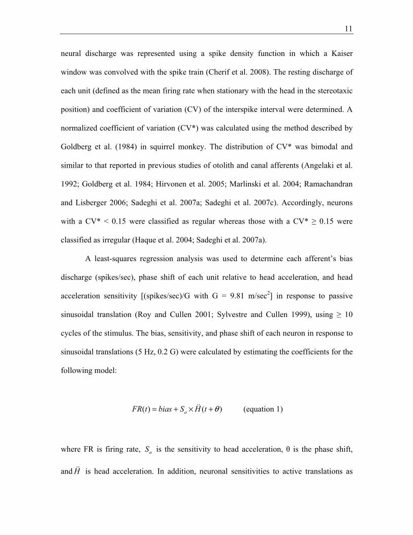
Modulation during passive sinusoidal translation
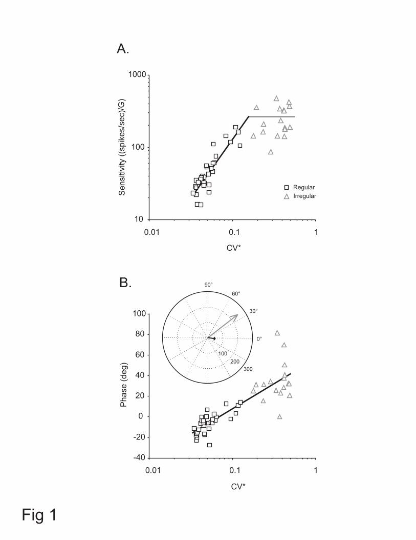
Figure 1 shows the relationship between head acceleration sensitivity (i.e., Sa in
equation 1) and phase, and CV* in response to sinusoidal translations at 5 Hz (0.2 G).
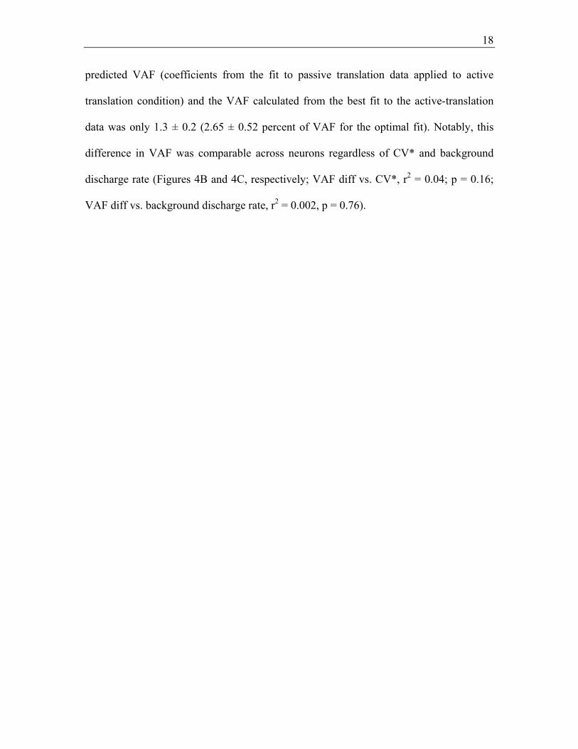
Acceleration sensitivity increased as a function of CV* for regular afferents (N = 31, r2 =
0.8, p < 0.0005), while for irregular units it remained relatively constant as a function of
CV* (N = 17, r2 = 0.06, p > 0.05). In contrast, phase lead increased as a function of CV*
for both regular and irregular afferents (r2 = 0.7, p < 0.0005). These relationships are
consistent with those reported for otolith afferents in chinchilla (Goldberg et al. 1990b).

14
A polar plot illustrating the average acceleration sensitivity and response phase for the
population of the regular (black arrow) and irregular (gray arrow) units is shown in the
inset. The average sensitivity of regular units was 57.1 ± 7.9 (spikes/sec)/G. Moreover,
while the modulation of individual regular afferents could either lag or lead acceleration
(i.e., Fig. 1B, squares), on average their responses lagged linear acceleration by 5.9 ± 1.8
deg. For the population of the irregular units, the average acceleration sensitivity was
249.2 ± 28.0 (spikes/sec)/G, which was higher than sensitivity of regular units (i.e.,
Figure 1B, inset). In contrast to the regular afferents, the modulation of each irregular
afferent in our sample led head acceleration (i.e., Fig. 1B, triangles), and the average
phase lead across the population was 34.1 ± 4.6 deg. Thus, overall both acceleration
sensitivity and phase were significantly larger for irregular units compared to regular
units.
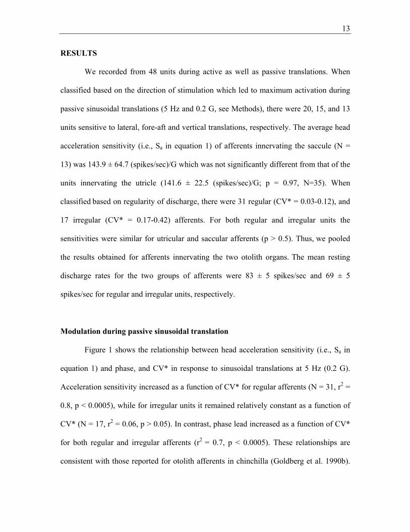
Afferent responses during natural passive and active head movements
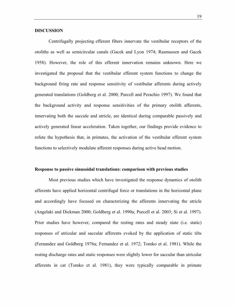
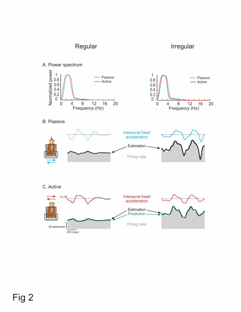
The responses of an example regular (CV* = 0.04) and irregular (CV* = 0.24)
otolith afferent are shown in Figure 2. These afferents were typical in that their response
sensitivities and phases were comparable during passive head-on-body (Figure 2B) and
passive whole-body translations (not shown) along the interaural axis (paired t-test, p >
0.1). Figure 2C shows the responses of the same two afferents during actively generated
head-on-body translations along the same axis. The corresponding power spectra of head
acceleration profiles shown in Figure 2A reveal comparable frequency content (i.e. low
pass up to ~8 Hz) when the monkey’s head was passively translated relative to its body
15
and during actively generated head-on-body movements (compare head acceleration
traces for each afferent between Figure 2B and 2C).
Equation 2 was first used to estimate each afferent’s bias and sensitivity to
passive head-on-body translations. For the example regular afferent, this equation
provided an excellent fit to the response (VAF = 95%) with an estimated bias discharge
rate of 55 spikes/sec, acceleration sensitivity of 27.7 (spikes/sec)/G, and jerk sensitivity
of 0.26 (spikes/sec)/(G/s). The estimated response profile is shown by the heavy black
trace superimposed on the firing rate (gray shaded area) in Figure 2B (left panel). As
expected based on the response dynamics of regular afferents (i.e. Figure 1), the jerk
sensitivity of this unit was negligible. Notably, when the response was estimated using
only the bias and acceleration terms of equation 2, the decrease in VAF was only 3.8%.
In contrast, the irregular unit had higher sensitivities to both jerk and acceleration (2.4
(spikes/sec)/(G/s) and 114.1 (spikes/sec)/G, respectively) as well as a bias of 103
spikes/sec. Accordingly, exclusion of the jerk term from equation 2 resulted in 16%
decrease in VAF (i.e., 87% (Figure 2B, black trace in right panel) vs. 71%). This
difference in the relative weight of a jerk term for fitting the responses of irregular and
regular afferents was consistent across our populations of otolith afferents (22.0 ± 3.4%
vs. 1.8 ± 0.4% decrease in VAF).
Next in order to address whether afferents responded differently to active head
translations, we used this same model (i.e., equation 2, with parameter estimates taken
form the passive condition in 2B) to predict each afferent’s response in the active
condition (Figure 2C, dashed green traces). The example afferents were typical of the
regular and irregular afferents in our sample in that their modulation was similar during

16
passive and active head-on-body translations. This was verified by the good prediction of
the passive model for the responses to active translations (VAF = 91.2% and 83.1% for
the regular and irregular afferent, respectively).
To further assess whether there were any differences in the bias and/or
translational sensitivities of otolith afferents between passive and active movements, we
estimated the parameters of equation 2 to obtain the best fit of each afferent’s modulation
during active head translations. The best fit to each example afferents’ modulation during
active translations is shown by the black trace in Figure 2C. For the regular afferent, we
estimated (VAF = 91.4%) a bias discharge rate, acceleration sensitivity, and jerk
sensitivity of 56 spikes/sec, 28.6 (spikes/sec)/G, and 0.21 (spikes/sec)/(G/s), respectively.
These coefficients corresponded well to those estimated for the passive translation
condition (i.e. Figure 2B). Moreover, there was little improvement in VAF for this
optimal estimation as compared to the passive-based prediction. Similar findings were
obtained for the example irregular afferent (bias = 102 spikes/sec, acceleration sensitivity
= 120 (spikes/sec)/G, jerk sensitivity = 2.1 (spikes/sec)/(G/s), and VAF = 83.5%). Taken
together, these results confirmed that the responses of our two example afferents were
similar during active and passive movements.
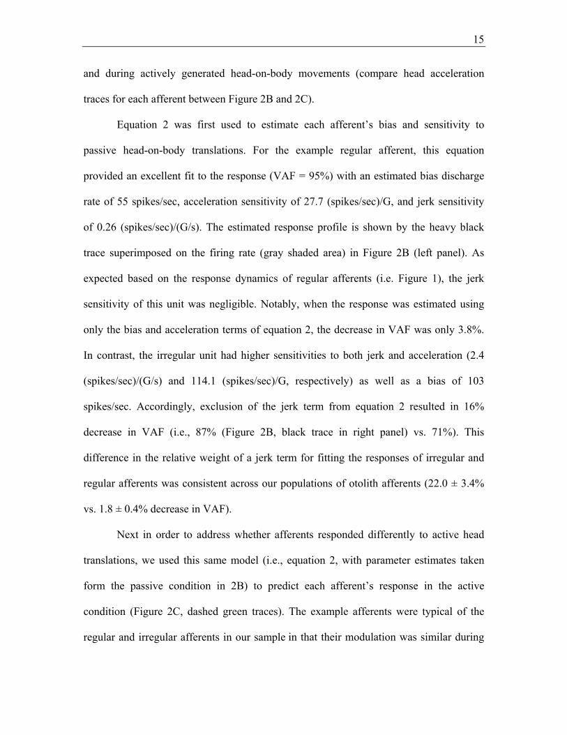
Population analysis: Responses to passive and active translations are comparable
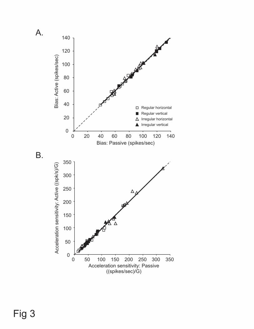
Figure 3 compares the bias discharge rate and acceleration sensitivity in both
conditions for the entire population of afferents. The mean bias discharge measured
across afferents from the fits to the passive and active translations was 84 ± 4 and 85 ± 4
spikes/sec, respectively. The similarity of the bias between the two conditions is shown
17
by the slope of the line fitted to the data in Figure 3A (regression slope = 1.01), which
was not different from one (p = 0.65). Similarly, the acceleration sensitivity for passive
and active translations (mean of 80.6 ± 10.1 and 79.4 ± 10.3 (spikes/sec)/G, respectively)
were identical as shown by the slope of the line fitted to the data points in Figure 3B
(slope = 1.02), which was not different from one (p = 0.29). Thus, taken as a population,
the response dynamics of otolith afferents were similar during active and passive head
translations. Furthermore, as is shown in Table 1, this observation could be extended
when different functional groups of afferents were separately considered. Overall,
regardless of discharge regularity, presumed organ of innervation, and/or direction of
stimulation, response biases and sensitivities (i.e., bias and Sa in equation 2, respectively)
were comparable during active vs. passive translations (paired t-test, p > 0.05).
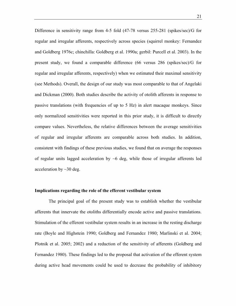
Finally, we evaluated whether differences in discharge bias (or possibly
sensitivities) might be observed in afferents with more irregular resting rate. Prior work
had reported an increase in the resting discharge and a decrease in sensitivity of afferent
responses following electrical stimulation of the brainstem efferents in squirrel monkey
(Goldberg and Fernandez 1980). Moreover, such effects were greater for irregularly than
regularly discharging afferents. Figure 4A shows the bias and sensitivities of regular and
irregular afferents during passive and active translations. Comparison of the bias
discharge rates and translational acceleration and jerk sensitivities revealed no differences
between the two conditions for each group of afferents. This conclusion was further
supported by the finding that the coefficients estimated for equation 2 from the data from
passive translations provided an excellent predictive fit to the data from the active
translations regardless of the afferents regularity. In fact, the difference between the
18
predicted VAF (coefficients from the fit to passive translation data applied to active
translation condition) and the VAF calculated from the best fit to the active-translation
data was only 1.3 ± 0.2 (2.65 ± 0.52 percent of VAF for the optimal fit). Notably, this
difference in VAF was comparable across neurons regardless of CV* and background
discharge rate (Figures 4B and 4C, respectively; VAF diff vs. CV*, r2 = 0.04; p = 0.16;
VAF diff vs. background discharge rate, r2 = 0.002, p = 0.76).
19
DISCUSSION
Centrifugally projecting efferent fibers innervate the vestibular receptors of the
otoliths as well as semicircular canals (Gacek and Lyon 1974; Rasmussen and Gacek
1958). However, the role of this efferent innervation remains unknown. Here we
investigated the proposal that the vestibular efferent system functions to change the
background firing rate and response sensitivity of vestibular afferents during actively
generated translations (Goldberg et al. 2000; Purcell and Perachio 1997). We found that
the background activity and response sensitivities of the primary otolith afferents,
innervating both the saccule and utricle, are identical during comparable passively and
actively generated linear acceleration. Taken together, our findings provide evidence to
refute the hypothesis that, in primates, the activation of the vestibular efferent system
functions to selectively modulate afferent responses during active head motion.
Response to passive sinusoidal translations: comparison with previous studies
Most previous studies which have investigated the response dynamics of otolith
afferents have applied horizontal centrifugal force or translations in the horizontal plane
and accordingly have focused on characterizing the afferents innervating the utricle
(Angelaki and Dickman 2000; Goldberg et al. 1990a; Purcell et al. 2003; Si et al. 1997).
Prior studies have however, compared the resting rates and steady state (i.e. static)
responses of utricular and saccular afferents evoked by the application of static tilts
(Fernandez and Goldberg 1976a; Fernandez et al. 1972; Tomko et al. 1981). While the
resting discharge rates and static responses were slightly lower for saccular than utricular
afferents in cat (Tomko et al. 1981), they were typically comparable in primate
20
(Fernandez and Goldberg 1976a). In addition, off-axis centrifugal forces have been
applied to characterize the dynamic responses of saccular afferents to sinusoidal linear
force in anesthetized squirrel monkeys (Fernandez and Goldberg 1976c). In the present
study we applied comparable pure translations in both the horizontal and vertical planes
and thus were able to compare afferent response dynamics of both end organs.
Unlike utricle receptors, the receptors in the saccule are biased at rest since they
are stimulated by the constant force exerted by gravity. As a result, saccule afferent
responses will modulate around the 1 G or -1 G point of their parabolic force-response
curve (Fernandez and Goldberg 1976b) during vertical linear motion, depending on
whether they innervate receptors that are sensitive to downward or upward translations,
respectively. However, while their force-response curve is inherently non-linear,
relatively vigorous stimulation (i.e. > ~2G) is required to drive afferents out of their
linear range (Fernandez and Goldberg 1976b). In the present study, our stimuli were
designed to test neurons only in the linear portion of their stimulus-response curves.
Accordingly, comparison of the response sensitivity of regular saccular afferents that
were preferentially excited by upward translations (i.e., haircells exposed to downward
linear acceleration) with those excited by downward translations revealed comparable
sensitivities. Note, this comparison was not done for irregular saccular afferents due to
our limited sample. Moreover, we found that, on average, the sensitivity and phase of
saccular afferents were comparable to those of afferents innervating the utricle. Thus, the
two groups are discussed as a pooled population.
Previous studies of utricular afferents have reported significantly greater
sensitivities for irregular than regular units in response to passively applied translations.
21
Difference in sensitivity range from 4-5 fold (47-78 versus 255-281 (spikes/sec)/G for
regular and irregular afferents, respectively across species (squirrel monkey: Fernandez
and Goldberg 1976c; chinchilla: Goldberg et al. 1990a; gerbil: Purcell et al. 2003). In the
present study, we found a comparable difference (66 versus 286 (spikes/sec)/G for
regular and irregular afferents, respectively) when we estimated their maximal sensitivity
(see Methods). Overall, the design of our study was most comparable to that of Angelaki
and Dickman (2000). Both studies describe the activity of otolith afferents in response to
passive translations (with frequencies of up to 5 Hz) in alert macaque monkeys. Since
only normalized sensitivities were reported in this prior study, it is difficult to directly
compare values. Nevertheless, the relative differences between the average sensitivities
of regular and irregular afferents are comparable across both studies. In addition,
consistent with findings of these previous studies, we found that on average the responses
of regular units lagged acceleration by ~6 deg, while those of irregular afferents led
acceleration by ~30 deg.
Implications regarding the role of the efferent vestibular system
The principal goal of the present study was to establish whether the vestibular
afferents that innervate the otoliths differentially encode active and passive translations.
Stimulation of the efferent vestibular system results in an increase in the resting discharge
rate (Boyle and Highstein 1990; Goldberg and Fernandez 1980; Marlinski et al. 2004;
Plotnik et al. 2005; 2002) and a reduction of the sensitivity of afferents (Goldberg and
Fernandez 1980). These findings led to the proposal that activation of the efferent system
during active head movements could be used to decrease the probability of inhibitory

22
cutoff or excitatory saturation of afferents, thereby effectively increasing the dynamic
range available (Goldberg and Fernandez 1980). Indeed there are several lines of
evidence that support a role for the efferent vestibular system in contributing to the
differential processing of active and passive movements. First, experiments in alert
toadfish have demonstrated that efferent activation in this species accompanies the
responses leading up to an escape reaction (Boyle and Highstein 1990; Highstein and
Baker 1985). The behaviorally-induced excitation of efferents, in turn, led to an increase
in the discharge rate and a decrease in the rotational sensitivity of afferents when tested
using passive head rotation. These findings led to the suggestion that the vestibular
efferent system carries a motor efference copy signal that modulates the responses of
vestibular afferents during self-generated movements in toadfish. Second, the
convergence of somatosensory and proprioceptive information with vestibular signals via
efferent projections has been reported in the periphery of frog and fish (Boyle and
Highstein 1990; Caston and Bricout-Berthout 1984; Hartmann and Klinke 1980). These
extravestibular signals could also be used to modulate the sensitivity of afferents during
active movements (Cullen and Minor 2002; Goldberg and Fernandez 1980; Klinke 1970).
Third, it has been shown recently that activation of the vestibular efferent system in alert
macaque monkeys significantly increases background firing rate of afferent fibers
(Sadeghi et al. 2007b). Previous studies however, have shown that the vestibular afferents
innervating semicircular canals, similarly encode active and passive head movements in
normal macaques as well as following contralateral labyrinthectomy (Cullen and Minor
2002; Sadeghi et al. 2007c). Thus, in the case of active rotational movements, activation
of the primate efferent system is not used to increase the available dynamic range.
23
Here we have specifically addressed the possibility that the vestibular efferent
system serves different functional roles regarding the modulation of canal versus otolith
afferent responses. This proposal is consistent with the results of electrical stimulation
studies suggesting that cerebellar stimulation might preferentially alter (via a
multisynaptic pathway) the background activity of otolith afferents, while leaving the
discharges of afferents innervating the semicircular canals largely unaffected (Llinas and
Precht 1969). Preliminary single unit studies in the vestibular system (Brooks and Cullen
2007) and electrosensory system of electric fish (Bell et al. 1999; Mohr et al. 2003;
Sawtell et al. 2007), as well as fMRI studies of tactile processing in humans (Blakemore
et al. 1998; 1999), have shown that the cerebellum is involved in predicting the sensory
consequences of voluntary actions. Double labeling experiments have further shown that
vestibular efferent cells send extensive projections both to the labyrinth as well as the
vestibulo-cerebellum (Shinder et al. 2001). Accordingly, reciprocal connections between
the cerebellum and efferent neurons could be used during intentional head movements to
reduce the likelihood of saturation or silencing (Goldberg et al. 2000) of otolith afferents.
Our findings, however, provide firm evidence that primary otolith afferents are
not differentially influenced by the efferent pathway during active and passive head
translations. Responses during active head translations were well-predicted based on
response during passive head translations. Moreover, responses were comparable during
passive whole-body and head-on-body head translations also refuting the proposal that
the efferent system modulates otolith afferents by encoding neck somatosensory and
proprioceptive information. Overall, our results extend previous findings regarding canal
afferents (Cullen and Minor 2002; Sadeghi et al. 2007c), and show that, in alert macaque

24
monkeys, afferent nerve fibers innervating all of the end organs respond similarly during
self-generated and passive motion.
Notably, vestibular input resulting from active rotational movements (reafference)
is suppressed at the next stage of processing: the vestibular nuclei (McCrea et al. 1999;
Roy and Cullen 2004; 2001). Given that the majority of the neurons in the vestibular
nuclei receive convergent inputs from multiple vestibular end organs (Curthoys and
Markham 1971; Dickman and Angelaki 2002; Kaufman et al. 2000; Markham and
Curthoys 1972; McConville et al. 1996; Straka et al. 2002; Tomlinson et al. 1996; Uchino
et al. 2005; Yakushin et al. 2006; Zakir et al. 2000; Zhang et al. 2001), it seems logical
that comparable strategy is used for encoding both linear and rotation acceleration at the
level of the vestibular periphery. As such, otolith-derived reafference can be suppressed
at the level of vestibular nuclei in a manner comparable to canal-derived inputs (McCrea
et al. 1999; Roy and Cullen 2004; 2001). Further experiments will be required to
determine whether this hypothesis is true or not. In addition, further studies will be
required to understand whether the connectivity between the vestibular efferent system
and cerebellum observed is present in primates and what role it might play in the
processing of vestibular inputs.
25
ACKNOWLEDGEMENTS
We thank W. Kucharski, S. Nuara, and J. Knowles for excellent technical assistance and
C. Massot, J. Brooks, M. Van Horn for critically reading the manuscript. This work was
supported by the Canadian Institutes of Health Research (CIHR) and Canadian Space
Agency (CSA).
26
REFERENCES
Angelaki DE, and Dickman JD. Spatiotemporal processing of linear acceleration:
primary afferent and central vestibular neuron responses. Journal of neurophysiology 84:
2113-2132, 2000.
Angelaki DE, Perachio AA, Mustari MJ, and Strunk CL. Role of irregular otolith
afferents in the steady-state nystagmus during off-vertical axis rotation. Journal of
neurophysiology 68: 1895-1900, 1992.
Angelaki DE, Shaikh AG, Green AM, and Dickman JD. Neurons compute internal
models of the physical laws of motion. Nature 430: 560-564, 2004.
Bell CC, Han VZ, Sugawara Y, and Grant K. Synaptic plasticity in the mormyrid
electrosensory lobe. The Journal of experimental biology 202: 1339-1347, 1999.
Birinyi A, Straka H, Matesz C, and Dieringer N. Location of dye-coupled second
order and of efferent vestibular neurons labeled from individual semicircular canal or
otolith organs in the frog. Brain research 921: 44-59, 2001.
Blakemore SJ, Wolpert DM, and Frith CD. Central cancellation of self-produced
tickle sensation. Nature neuroscience 1: 635-640, 1998.
Blakemore SJ, Wolpert DM, and Frith CD. The cerebellum contributes to
somatosensory cortical activity during self-produced tactile stimulation. NeuroImage 10:
448-459, 1999.
Boyle R, and Highstein SM. Efferent vestibular system in the toadfish: action upon
horizontal semicircular canal afferents. J Neurosci 10: 1570-1582, 1990.
Brooks J, and Cullen KE. Reference frames and reafference in the rostral fastigial
nucleus. Soc Neurosci Abstr 37: 861.2: 2007.
Caston J, and Bricout-Berthout A. Responses to somatosensory input by afferent and
efferent neurons in the vestibular nerve of the frog. Brain, behavior and evolution 24:
135-143, 1984.
Cherif S, Cullen KE, and Galiana HL. An improved method for the estimation of firing
rate dynamics using an optimal digital filter. Journal of neuroscience methods 173: 165-
181, 2008.
27
Crapse TB, and Sommer MA. Corollary discharge across the animal kingdom. Nat Rev
Neurosci 9: 587-600, 2008.
Cullen KE. Sensory signals during active versus passive movement. Current opinion in
neurobiology 14: 698-706, 2004.
Cullen KE, and Minor LB. Semicircular canal afferents similarly encode active and
passive head-on-body rotations: implications for the role of vestibular efference. J
Neurosci 22: RC226, 2002.
Cullen KE, Rey CG, Guitton D, and Galiana HL. The use of system identification
techniques in the analysis of oculomotor burst neuron spike train dynamics. Journal of
computational neuroscience 3: 347-368, 1996.
Curthoys IS, and Markham CH. Convergence of labyrinthine influences on units in the
vestibular nuclei of the cat. I. Natural stimulation. Brain research 35: 469-490, 1971.
Dickman JD, and Angelaki DE. Vestibular convergence patterns in vestibular nuclei
neurons of alert primates. Journal of neurophysiology 88: 3518-3533, 2002.
Dickman JD, and Correia MJ. Bilateral communication between vestibular labyrinths
in pigeons. Neuroscience 57: 1097-1108, 1993.
Fernandez C, and Goldberg JM. Physiology of peripheral neurons innervating otolith
organs of the squirrel monkey. I. Response to static tilts and to long-duration centrifugal
force. Journal of neurophysiology 39: 970-984, 1976a.
Fernandez C, and Goldberg JM. Physiology of peripheral neurons innervating otolith
organs of the squirrel monkey. II. Directional selectivity and force-response relations.
Journal of neurophysiology 39: 985-995, 1976b.
Fernandez C, and Goldberg JM. Physiology of peripheral neurons innervating otolith
organs of the squirrel monkey. III. Response dynamics. Journal of neurophysiology 39:
996-1008, 1976c.
Fernandez C, Goldberg JM, and Abend WK. Response to static tilts of peripheral
neurons innervating otolith organs of the squirrel monkey. Journal of neurophysiology
35: 978-987, 1972.
Gacek RR, and Lyon M. The localization of vestibular efferent neurons in the kitten
with horseradish peroxidase. Acta Otolaryngol 77: 92-101, 1974.

28
Gleisner L, and Henriksson NG. Efferent And Afferent Activity Pattern In The
Vestibular Nerve Of The Frog. Acta Otolaryngol Suppl 192: SUPPL 192:190+, 1963.
Goldberg JM, Brichta AM, and Wackym PA. Efferent vestibular system: anatomy,
physiology and neurochemistry. In: Neurochemistry of the vestibular system. Boca Raton,
FL: CRC, 2000, p. 61-94.
Goldberg JM, Desmadryl G, Baird RA, and Fernandez C. The vestibular nerve of the
chinchilla. IV. Discharge properties of utricular afferents. Journal of neurophysiology 63:
781-790, 1990a.
Goldberg JM, Desmadryl G, Baird RA, and Fernandez C. The vestibular nerve of the
chinchilla. V. Relation between afferent discharge properties and peripheral innervation
patterns in the utricular macula. Journal of neurophysiology 63: 791-804, 1990b.
Goldberg JM, and Fernandez C. Efferent vestibular system in the squirrel monkey:
anatomical location and influence on afferent activity. J Neurophysiol 43: 986-1025,
1980.
Goldberg JM, Smith CE, and Fernandez C. Relation between discharge regularity and
responses to externally applied galvanic currents in vestibular nerve afferents of the
squirrel monkey. Journal of neurophysiology 51: 1236-1256, 1984.
Grossman GE, Leigh RJ, Abel LA, Lanska DJ, and Thurston SE. Frequency and
velocity of rotational head perturbations during locomotion. Exp Brain Res 70: 470-476,
1988.
Gu Y, Deangelis GC, and Angelaki DE. A functional link between area MSTd and
heading perception based on vestibular signals. Nature neuroscience 10: 1038-1047,
2007.
Haque A, Angelaki DE, and Dickman JD. Spatial tuning and dynamics of vestibular
semicircular canal afferents in rhesus monkeys. Exp Brain Res 155: 81-90, 2004.
Harris LR, Jenkin M, and Zikovitz DC. Visual and non-visual cues in the perception
of linear self-motion. Exp Brain Res 135: 12-21, 2000.
Hartmann R, and Klinke R. Efferent activity in the goldfish vestibular nerve and its
influence on afferent activity. Pflugers Arch 388: 123-128, 1980.
Highstein SM, and Baker R. Action of the efferent vestibular system on primary
afferents in the toadfish, Opsanus tau. Journal of neurophysiology 54: 370-384, 1985.
29
Highstein SM, and Baker R. Organization of the efferent vestibular nuclei and nerves of
the toadfish, Opsanus tau. J Comp Neurol 243: 309-325, 1986.
Hirvonen TP, Minor LB, Hullar TE, and Carey JP. Effects of intratympanic
gentamicin on vestibular afferents and hair cells in the chinchilla. Journal of
neurophysiology 93: 643-655, 2005.
Huterer M, and Cullen KE. Vestibuloocular reflex dynamics during high-frequency and
high-acceleration rotations of the head on body in rhesus monkey. J Neurophysiol 88: 13-
28, 2002.
Jarvis MR, and Mitra PP. Sampling properties of the spectrum and coherency of
sequences of action potentials. Neural computation 13: 717-749, 2001.
Kaufman GD, Shinder ME, and Perachio AA. Convergent properties of vestibular-
related brain stem neurons in the gerbil. Journal of neurophysiology 83: 1958-1971,
2000.
Klinke R. Efferent influence on the vestibular organ during active movements of the
body. Pflugers Arch 318: 325-332, 1970.
Lindeman HH. Studies on the morphology of the sensory regions of the vestibular
apparatus with 45 figures. Ergebnisse der Anatomie und Entwicklungsgeschichte 42: 1-
113, 1969.
Lisberger SG, and Pavelko TA. Vestibular signals carried by pathways subserving
plasticity of the vestibulo-ocular reflex in monkeys. J Neurosci 6: 346-354, 1986.
Llinas R, and Precht W. The inhibitory vestibular efferent system and its relation to the
cerebellum in the frog. Exp Brain Res 9: 16-29, 1969.
Markham CH, and Curthoys IS. Convergence of labyrinthine influences on units in the
vestibular nuclei of the cat. II. Electrical stimulation. Brain research 43: 383-396, 1972.
Marlinski V, Plotnik M, and Goldberg JM. Efferent actions in the chinchilla vestibular
labyrinth. J Assoc Res Otolaryngol 5: 126-143, 2004.
McConville KM, Tomlinson RD, and Na EQ. Behavior of eye-movement-related cells
in the vestibular nuclei during combined rotational and translational stimuli. Journal of
neurophysiology 76: 3136-3148, 1996.

30
McCrea RA, Gdowski GT, Boyle R, and Belton T. Firing behavior of vestibular
neurons during active and passive head movements: vestibulo-spinal and other non-eye-
movement related neurons. Journal of neurophysiology 82: 416-428, 1999.
Mohr C, Roberts PD, and Bell CC. The mormyromast region of the mormyrid
electrosensory lobe. I. Responses to corollary discharge and electrosensory stimuli.
Journal of neurophysiology 90: 1193-1210, 2003.
Myers SF, Salem HH, and Kaltenbach JA. Efferent neurons and vestibular cross talk in
the frog. Journal of neurophysiology 77: 2061-2070, 1997.
Ohmi M. Egocentric perception through interaction among many sensory systems. Brain
Res Cogn Brain Res 5: 87-96, 1996.
Peterson BW, and Richmond FJ. Control of head movement. New York: Oxford
University Press, 1988, p. xi, 322 p.
Plotnik M, Marlinski V, and Goldberg JM. Efferent-mediated fluctuations in
vestibular nerve discharge: a novel, positive-feedback mechanism of efferent control. J
Assoc Res Otolaryngol 6: 311-323, 2005.
Plotnik M, Marlinski V, and Goldberg JM. Reflections of efferent activity in rotational
responses of chinchilla vestibular afferents. Journal of neurophysiology 88: 1234-1244,
2002.
Purcell IM, Newlands SD, and Perachio AA. Responses of gerbil utricular afferents to
translational motion. Exp Brain Res 152: 317-322, 2003.
Purcell IM, and Perachio AA. Three-dimensional analysis of vestibular efferent
neurons innervating semicircular canals of the gerbil. Journal of neurophysiology 78:
3234-3248, 1997.
Ramachandran R, and Lisberger SG. Transformation of vestibular signals into motor
commands in the vestibuloocular reflex pathways of monkeys. Journal of
neurophysiology 96: 1061-1074, 2006.
Rasmussen GL, and Gacek RR. Concerning the question of the efferent fiber
component of the vestibular nerve of the cat. Anat Rec 130: 361-362, 1958.
Roy JE, and Cullen KE. Dissociating self-generated from passively applied head
motion: neural mechanisms in the vestibular nuclei. J Neurosci 24: 2102-2111, 2004.

31
Roy JE, and Cullen KE. Selective processing of vestibular reafference during self-
generated head motion. J Neurosci 21: 2131-2142, 2001.
Sadeghi SG, Chacron MJ, Taylor MC, and Cullen KE. Neural variability, detection
thresholds, and information transmission in the vestibular system. J Neurosci 27: 771-
781, 2007a.
Sadeghi SG, Goldberg JM, Minor LB, and Cullen KE. Vestibular-nerve afferents of
alert macaques in normal conditions and following vestibular lesion. Soc Neurosci Abstr
37: 861.9: 2007b.
Sadeghi SG, Minor LB, and Cullen KE. Response of vestibular-nerve afferents to
active and passive rotations under normal conditions and after unilateral labyrinthectomy.
Journal of neurophysiology 97: 1503-1514, 2007c.
Sawtell NB, Williams A, and Bell CC. Central control of dendritic spikes shapes the
responses of Purkinje-like cells through spike timing-dependent synaptic plasticity. J
Neurosci 27: 1552-1565, 2007.
Shaikh AG, Green AM, Ghasia FF, Newlands SD, Dickman JD, and Angelaki DE.
Sensory convergence solves a motion ambiguity problem. Curr Biol 15: 1657-1662,
2005.
Shinder ME, Purcell IM, Kaufman GD, and Perachio AA. Vestibular efferent neurons
project to the flocculus. Brain research 889: 288-294, 2001.
Si X, Angelaki DE, and Dickman JD. Response properties of pigeon otolith afferents to
linear acceleration. Exp Brain Res 117: 242-250, 1997.
Straka H, Holler S, and Goto F. Patterns of canal and otolith afferent input convergence
in frog second-order vestibular neurons. Journal of neurophysiology 88: 2287-2301,
2002.
Sylvestre PA, and Cullen KE. Quantitative analysis of abducens neuron discharge
dynamics during saccadic and slow eye movements. Journal of neurophysiology 82:
2612-2632, 1999.
Telford L, Howard IP, and Ohmi M. Heading judgments during active and passive
self-motion. Exp Brain Res 104: 502-510, 1995.
Tomko DL, Peterka RJ, and Schor RH. Responses to head tilt in cat eighth nerve
afferents. Exp Brain Res 41: 216-221, 1981.
32
Tomlinson RD, McConville KM, and Na EQ. Behavior of cells without eye movement
sensitivity in the vestibular nuclei during combined rotational and translational stimuli. J
Vestib Res 6: 145-158, 1996.
Tribukait A, and Eiken O. On the role of otoliths and semicircular canals in spatial
orientation: Dynamics of the visually perceived eye level during gondola centrifugation.
Perception & psychophysics 67: 1242-1251, 2005.
Uchino Y, Sasaki M, Sato H, Bai R, and Kawamoto E. Otolith and canal integration on
single vestibular neurons in cats. Exp Brain Res 164: 271-285, 2005.
Yakushin SB, Raphan T, and Cohen B. Spatial properties of central vestibular neurons.
Journal of neurophysiology 95: 464-478, 2006.
Zakir M, Kushiro K, Ogawa Y, Sato H, and Uchino Y. Convergence patterns of the
posterior semicircular canal and utricular inputs in single vestibular neurons in cats. Exp
Brain Res 132: 139-148, 2000.
Zhang X, Zakir M, Meng H, Sato H, and Uchino Y. Convergence of the horizontal
semicircular canal and otolith afferents on cat single vestibular neurons. Exp Brain Res
140: 1-11, 2001.
33
FIGURE LEGENDS
Figure 1: Comparison of responses of regular and irregular otolith afferents during
passive sinusoidal (5 Hz, 0.2 G) translations. (A) Acceleration sensitivity plotted as a
function of normalized coefficient of variation (CV*) for regular (squares) and irregular
(triangles) afferents. A power-law was fit to regular units (CV* < 0.15). Horizontal line is
the mean sensitivity for irregular afferents (CV* > 0.15). (B) Response phase relative to
peak linear acceleration plotted as a function of CV* for regular (squares) and irregular
(triangles) afferents. A semilogarithmic relation was fit to all units. Inset: polar plot
showing the average gain and phase of the response of regular (black arrow) and irregular
(gray arrow) afferents innervating otoliths. The length of the arrows represents the
response sensitivity [(spikes/sec)/G] of each neuron.
Figure 2: Activities of an example regular (left panels; CV* = 0.04) and irregular (right
panels; CV* = 0.24) otolith afferent during passive and active interaural translations with
comparable acceleration profiles. (A) Power spectra of head acceleration during passive
(blue) and comparable active (red) translations. Both movements had similar power for
the range of frequencies of 0-10 Hz. (B) Response of afferents to passive translations.
Superimposed on the firing rate (shaded trace) is the model fit (black trace) based on the
bias discharge, the acceleration sensitivity and jerk sensitivity. See Results for details. (C)
Response of afferents to active translations. The estimated response (black trace) based
on the bias discharge, the acceleration sensitivity and jerk sensitivity is superimposed on
the firing rate. To obtain the ‘prediction’ fit (dashed green trace), the bias and the
34
sensitivity values of the passive model were applied. The VAFs of the estimation and
prediction were similar (see Results).
Figure 3: Comparison of parameters of estimations for responses to active and passive
translations for the population of utricular (empty symbols) and saccular (filled symbols)
otolith afferents. (A) Estimated bias for responses in the two conditions. There was no
significant difference between active and passive conditions for regular (squares) and
irregular (triangles) afferents. (B) Estimated acceleration sensitivities were not different
between active and passive translations across otolith afferents. The unity lines (dashed
lines) were superimposed on each plot to facilitate the comparison.
Figure 4: Comparison of responses during active and passive translations. (A) Average
values of bias and sensitivity for the population of regular and irregular afferents during
active (black bars) and passive (gray bars) movements. (B) Difference in VAF between
predicted and estimated responses during active translations as a function of CV*. VAF
difference was less than 4% (dashed line) for regular (squares) and irregular (triangles)
afferents. (C) Difference in VAF between predicted and estimated responses during
active translations as a function of background discharge. VAF difference was less than
4% (dashed line) for most of the afferents.
1
10
100
1000
0.01 0.1
Regular
Irregular
CV*
Se
nsi
tivity
((s
pik
es/
sec)
/G)
A.
-40
-20
0
20
40
60
80
100
0.01 0.1 1
100
200
300
30°
90°
0°
60°
CV*
Ph
ase
(d
eg
)
B.
Fig 1
Regular Irregular
Interaural head acceleration
Firing rate
Estimation
B. Passive
0.2 G Interaural head acceleration
Firing rate
EstimationPrediction
C. Active
200 msec
50 spikes/sec
A. Power spectrum
No
rma
lize
d p
ow
er
PassiveActive
00.20.40.60.81
0 4 8 12 16 20Frequency (Hz)
00.20.40.60.81
0 4 8 12 16 20Frequency (Hz)
PassiveActive
Fig 2
A.
Regular horizontal
Regular vertical
Irregular horizontal
Irregular vertical
0 20 40 60 80 100 120 140
Bias: Passive (spikes/sec)
0
20
40
60
80
100
120
140
Bia
s: A
ctiv
e (
spik
es/
sec)
0 50 100 150 200 250 300 350
Acceleration sensitivity: Passive((spikes/sec)/G)
0
50
100
150
200
250
300
350
Acc
ele
ratio
n s
en
sitiv
ity: A
ctiv
e (
(sp
k/s)
/G)
B.
Fig 3

Table-1: Comparison of bias and head acceleration sensitivities (Sa) during passive and active translations
* Comparison not made due to small data set.
N Passive bias
(spikes/sec) Active bias (spikes/sec)
p value
Passive Sa ((spikes/sec)/G)
Active Sa ((spikes/sec)/G)
p value
Lateral 13 77 ± 9 78 ± 9 0.06 58.8 ± 13.8 55.2 ± 13.8 0.05 Utricular Fore-aft 7 78 ± 9 78 ± 9 0.90 37.4 ± 8.3 38.5 ± 8.7 0.37 Regular Saccular 11 95 ± 8 95 ± 8 0.89 51.0 ± 9.4 49.1 ± 9.3 0.07
Lateral 7 87 ± 9 89 ± 9 0.06 183.6 ± 29.7 188.0 ± 32.2 0.52 Utricular Fore-aft 8 75 ± 6 76 ± 7 0.19 157.1 ± 28.1 161.7 ± 32.2 0.52 Irregular
Saccular 2 109 ± 9 109 ± 6 - * 136.1 ± 16.2 129.7 ± 2.0 - *