Response of antioxidant defense system to chromium (VI)-induced cytotoxicity in human diploid cells
12
Response of antioxidant defense system to chromium (VI)-induced cytotoxicity in human diploid cells Nino Asatiani • Marina Abuladze • Tamar Kartvelishvili • Nina Kulikova • Lali Asanishvili • Hoi-Ying Holman • Nelly Sapojnikova Received: 1 June 2009 / Accepted: 30 October 2009 Ó Springer Science+Business Media, LLC. 2009 Abstract The aim of this study is to establish antioxidant indicators of chromium toxicity in fetal human lung fibroblasts (HLF). The results obtained corroborate and develop our earlier observation of low-dose and long-term action of Cr(VI) on human cells in culture. In the case of a nontoxic chromium dose, temporary oxidative stress is overcome by increased activity of the antioxidant system with correlation to cell cycle re-entry. The toxic concen- trations misbalance the cell antioxidant defense sys- tems and cause irreversible growth arrest and massive cell death by apoptosis. Sub-toxicity is defined as toxicity stretched in time. The activity of GPx (glutathione peroxidase) is proposed as a biomarker of oxidative stress caused by Cr(VI), and the GR (glutathione reductase) inhibition is considered as a marker of the toxicity developed under the complex Cr(VI) action. In HLF cells the glutathione dependent defense system is the first system destroyed in response to toxic chromium action. Only the balance between SOD (superoxide dismutase) and H 2 O 2 degrading enzymes (catalase and GPx), should play an important role in the fate of a cell, not individual enzymes. Keywords Chromium (VI) Antioxidant enzymes Cell cycle ROS Apoptosis Introduction The exposure of cells to environmental oxidants such as UV and ionizing radiation, heavy metals, redox active chemicals, hypoxia and hyperoxia increases reactive oxygen species (ROS) production which shifts cell redox status to a more oxidized state known as an oxidative stress. ROS are capable of causing direct damage effect or acting as critical intermediate signaling molecules leading to diverse biological consequences (Costa and Klein 2006; Dewhirst et al. 2008; Leonard et al. 2004). Toxic stress resistance or adaptation of cells to shift in cell redox status could be characterized by the response of the intracellular antioxidant defense system (Mates et al. 2008). The modulation of antioxidant enzymes N. Asatiani M. Abuladze T. Kartvelishvili L. Asanishvili N. Sapojnikova (&) Andronikashvili Institute of Physics, 6 Tamarashvili St., 0177 Tbilisi, Georgia e-mail: [email protected]; [email protected] N. Kulikova I. Javakhishvili Tbilisi State University, 2 University St., 0143 Tbilisi, Georgia H.-Y. Holman E.O. Lawrence Berkeley National Laboratory, 1 Cyclotron Road, Berkeley, CA 94720, USA 123 Biometals DOI 10.1007/s10534-009-9276-6
Transcript of Response of antioxidant defense system to chromium (VI)-induced cytotoxicity in human diploid cells

Response of antioxidant defense system to chromium(VI)-induced cytotoxicity in human diploid cells
Nino Asatiani • Marina Abuladze • Tamar Kartvelishvili •
Nina Kulikova • Lali Asanishvili • Hoi-Ying Holman •
Nelly Sapojnikova
Received: 1 June 2009 / Accepted: 30 October 2009
� Springer Science+Business Media, LLC. 2009
Abstract The aim of this study is to establish
antioxidant indicators of chromium toxicity in fetal
human lung fibroblasts (HLF). The results obtained
corroborate and develop our earlier observation of
low-dose and long-term action of Cr(VI) on human
cells in culture. In the case of a nontoxic chromium
dose, temporary oxidative stress is overcome by
increased activity of the antioxidant system with
correlation to cell cycle re-entry. The toxic concen-
trations misbalance the cell antioxidant defense sys-
tems and cause irreversible growth arrest and massive
cell death by apoptosis. Sub-toxicity is defined as
toxicity stretched in time. The activity of GPx
(glutathione peroxidase) is proposed as a biomarker
of oxidative stress caused by Cr(VI), and the GR
(glutathione reductase) inhibition is considered as a
marker of the toxicity developed under the complex
Cr(VI) action. In HLF cells the glutathione dependent
defense system is the first system destroyed in
response to toxic chromium action. Only the balance
between SOD (superoxide dismutase) and H2O2
degrading enzymes (catalase and GPx), should play
an important role in the fate of a cell, not individual
enzymes.
Keywords Chromium (VI) � Antioxidant enzymes �Cell cycle � ROS � Apoptosis
Introduction
The exposure of cells to environmental oxidants such
as UV and ionizing radiation, heavy metals, redox
active chemicals, hypoxia and hyperoxia increases
reactive oxygen species (ROS) production which
shifts cell redox status to a more oxidized state
known as an oxidative stress. ROS are capable of
causing direct damage effect or acting as critical
intermediate signaling molecules leading to diverse
biological consequences (Costa and Klein 2006;
Dewhirst et al. 2008; Leonard et al. 2004). Toxic
stress resistance or adaptation of cells to shift in cell
redox status could be characterized by the response of
the intracellular antioxidant defense system (Mates
et al. 2008). The modulation of antioxidant enzymes
N. Asatiani � M. Abuladze � T. Kartvelishvili �L. Asanishvili � N. Sapojnikova (&)
Andronikashvili Institute of Physics, 6 Tamarashvili St.,
0177 Tbilisi, Georgia
e-mail: [email protected];
N. Kulikova
I. Javakhishvili Tbilisi State University, 2 University St.,
0143 Tbilisi, Georgia
H.-Y. Holman
E.O. Lawrence Berkeley National Laboratory,
1 Cyclotron Road, Berkeley, CA 94720, USA
123
Biometals
DOI 10.1007/s10534-009-9276-6
in cultured cells in response to the action of Cr(VI)-
mediated toxic effect could be used as a model to
establish crucial biomarkers of cell adaptation or
resistance to toxicants.
Toxicity of Cr(VI) has been demonstrated for
various human cells, such as gastric mucosa cells,
peripheral blood lymphocytes (Trzeciak et al. 2000),
human lymphoblast cell line (Jurcat cells) (Shi et al.
1999), and human peripheral blood mononuclear
cells (Bagchi et al. 2001). The major cellular targets
of Cr(VI) toxicity are lung epithelial cells and lung
fibroblasts, however human Cr(VI) intoxication is
also associated with hepatotoxicity, nephrotoxicity,
cardiotoxicity and immunotoxicity (Pourahmad and
O’Brien 2001). Many in vitro studies indicate that
when Cr(VI) has contact with biological fluids and
tissues, reduction to Cr(III) occurs rapidly due to the
presence of reducing agents that keep the body in
homeostasis. But in spite of the high reduction
capacity of blood plasma, the toxic effect of Cr(VI)
still takes place, i.e. the elevated concentration of
chromium is observed in peripheral lymphocytes of
chrome-plating workers, which correlates with
increased DNA strand breaks (Gambelunghe et al.
2003).
During the Cr(VI) reduction within a cell, a wide
spectra of ROS, such as superoxide, hydrogen
peroxide and hydroxyl radicals, are produced (Ye
et al. 1999). The cellular toxicity of Cr(VI) is initiated
by oxidative stress, resulting in the excess formation
of ROS.
Cr(VI) inside a cell acts as a multipotent agent,
and any biomacromolecules can be its targets. Cr(VI)
can produce DNA damage from either an oxidative
pathway or a metal-binding pathway that results in a
wide variety of DNA lesions. Cr(VI) can inhibit DNA
replication and repair (Holmes et al. 2008), alter gene
expression (Dubrovskaya and Wetterhahn 1998),
activate stress-response pathways (Chuang et al.
2000; Kim and Yurkow 1996; Leonard et al. 2004;
Ye et al. 1999), trigger transient or terminal growth
arrest and apoptosis (Carlisle et al. 2000; Pritchard
et al. 2001a; Rana 2008; Ye et al. 1999). Cells treated
with Cr(VI) exhibit apoptotic features, depending on
cell line, dose and exposure time: 300 lM Cr(VI)
after 3 h of exposure causes apoptosis in 24 h in
human lung tumor A549 cells (Ye et al. 1999), a 24 h
exposure to 12.5 lM Cr(VI) is apoptogenic for
chronic myelogenous leukemic K562 cells, and for
cultured J774A.1 murine macrophage cells 0.6 lM
Cr(VI) is toxic after 48 h (Bagchi et al. 2001).
In cells, the toxic capacity of Cr(VI) can be
decreased by antioxidant defense systems, poised
against the oxidative assault, which can neutralize the
ROS generated by Cr(VI) action.
The cellular protective mechanisms against ROS
consist of multiple enzymatic [catalase, superoxide
dismutase (SOD), glutathione peroxidase (GPx)] and
non-enzymatic [a-tocopherol, ascorbic acid (AA),
beta-carotene, cysteine and glutathione (GSH)]
antioxidants.
Superoxide generated by the mitochondria and
other sources is converted to H2O2 and O2 by SOD.
Abundant catalase enzyme and peroxidases then
convert H2O2 to H2O and O2. The antioxidant
enzymes can be divided into two types: one that
reacts with ROS and diminishes their level (SOD,
catalase and peroxidases) and one [glutathione
reductase (GR)] that restores reduced forms of non-
enzymatic antioxidants (GSH). The disorder of cell
redox status causes metabolic and cell proliferation
dysfunction and/or cell death.
We have reported (Asatiani et al. 2004) that
Cr(VI)-treated human epithelial-like L-41 cells died
by apoptosis. The dose-dependent involvement of
defense mechanisms in response to low level and
long-term Cr(VI) treatment was analyzed in dynamic.
The nontoxic chromium dose (2 lM) caused transient
cell cycle and growth arrest that correlated with the
increased activity of glutathione peroxidase—gluta-
thione reductase antioxidant system. The toxic apop-
togenic concentration (20 lM) destroyed the cell
antioxidant defense systems, and caused irreversibly
growth arrest and massive cell death via apoptosis.
The particular marker of the toxic Cr(VI) action was
the depletion of glutathione-dependent antioxidant
defense system. Inhibition of GR was an important
aspect of the Cr(VI) toxicity in the L-41 cells.
In the present study we continue to consider the
cell antioxidant responses to Cr(VI) using fetal
human lung fibroblasts (HLF) in culture. We per-
formed a time course study of oxidative stress, cell
cycle distortion and the behavior of the antioxidant
enzymes in Cr(VI)-induced toxicity to determine if
there is an association between oxidative stress, cell
cycle distortion, and antioxidant enzymes, and if
antioxidant enzymes Cu,Zn-SOD, Mn-SOD, GR,
GPx and catalase respond in a coordinated way.
Biometals
123
Materials and methods
Cell culture
The HLF cells (fetal human lung fibroblasts) were
maintained as adherent cells in Dulbecco’s modified
Eagle’s culture supplemented with 15% fetal bovine
serum, 2 mM L-glutamine, 100 units of penicillin/ml,
and 100 lg of streptomycin/ml at 37�C in a 5% CO2
incubator. Cells were harvested with trypsin (0.25%)/
EDTA solution. In all experiments HLF cells were
used in the range of 25–30 cell passages in culture.
Chromium treatment and viability assay
HLF cells were seeded at 3 9 104 cells per well in
200 ll culture medium in 96-well microtiter plates
and cultured to 80% of confluence. Cr(VI) at 2, 5, 10,
15, 20, 25 and 30 lM was added as potassium
chromate at 48 h of growth and the cells continued to
grow for 24 and 48 h. Viability of chromium-exposed
HLF cells was assessed by the ability of viable cells
to convert the tetrazolium dye MTT (3-[4,5-dimeth-
ylthiazol-2-yl]-2,5-diphenyltetrazoliumbromide) to a
water-insoluble formazan dye, which is based on the
activation of succinate dehydrogenase (Carmichael
et al. 1987). The culture medium was removed and
the colored precipitate was solubilized with DMSO
(dimethyl sulfoxide). After 30 min, cell survival and
background was determined by absorbance at 570
and 660 nm, respectively.
For detecting cell viability after 2 and 24 h of
transient chromium action, the exposed cells were
rinsed with phosphate-buffered saline and replaced in
fresh medium without Cr(VI) up to 48 h prior to
determining cell survival by MTT assay.
In these assays, four wells were usually examined
for each concentration and time point.
Measurement of DNA content for cell cycle
analysis
DNA content of propidium iodide (PI) stained cells
was measured by flow cytometry using FACScan
(Becton–Dickinson, USA) and the separation into
phases of the cell cycle was based on the PI
fluorescence according to the accepted method
(Ormerod 2002). The DNA histogram showed the
cell cycle distribution of the viable cells. The
apoptotic cells should be observed as a distinct sub-
G1 peak of the hypodiploid DNA. All results are
expressed as the mean percent cells from each cell
cycle compartment of three experiments.
Measurement of ROS
The detection of ROS in living cells was carried out
by the flow cytometry method using 20-70-dichloro-
fluorescein diacetate (DCFH-DA, Sigma). Cell-per-
meable DCFH-DA is oxidized in live cells to its
fluorescent derivative 20-70-dichlorofluorescein
(DCF) in the presence of ROS (predominantly
hydrogen peroxide and partially •OH and •NO
radicals). Accumulation of DCF was measured by
an increase of fluorescence at 530 nm and the mean
fluorescent intensity (MFI) was used for the estima-
tion of intracellular ROS level. DCFH-DA was added
(10 lM final concentration) to HLF cells (about
1 9 106 cells) and the mixture was incubated for
30 min at 37�C. Hydrogen peroxide (10 mM) was
used as a positive control. After the incubation cells
were subjected to flow cytometry analysis (FACScan,
Becton–Dickinson). The elevated ROS level is pro-
portional to an increase of the basal level of the probe
(Curtin et al. 2002; Esposti 2002).
A flow cytometry method was used for quantita-
tive measurement of superoxide anions with the
fluorescent probe dihydroethidium (DHE). DHE is
taken up by cells and in the presence of superoxide
anion converted to ethidine, which intercalates into
nuclear DNA. The degree of fluorescence is propor-
tional to the superoxide anion amount (Carter et al.
1994; Pritchard et al. 2001b). 10 lM DHE was added
per 0.5 9 106 cells in 1 ml PBS for 15 min at 37�C
and analyzed by FACScan instrument (Becton–
Dickinson, USA). Excitation of DHE was at
490 nm, emission was measured at 610 nm.
Glutathione-dependent antioxidant system
Glutathione reductase (GR) activity was measured by
using the BIOXYTECH GR-340TM
Assay (Oxis,
USA), and glutathione peroxidase (GPx) activity
was determined by using BIOXYTECH GPx-340
colorimetric assay for cellular GPx (Oxis, USA)
according to the manufacturer instructions.
Biometals
123
Cellular superoxide dismutase
The technique of SOD assay involves photoreduction
of nitro blue tetrazolium (NBT) for the determination
of activity of superoxide dismutase following native
polyacrylamide gel electrophoresis. The protein cor-
responding to SOD can be then visualized as
achromatic zones through the inhibition of NBT
(Sigma) reduction via SOD (Steinman 1985). Ach-
romatic bands were visualized for 50 lg protein
equivalent. The positions of the two isozymes of
SOD, Cu, Zn-SOD and Mn-SOD were identified by
incubation of the cell lysate at 378 C with 2% (w/v)
sodium dodecyl sulfate (SDS) for selective inactiva-
tion of Mn-SOD (Geller and Winge 1982).
Catalase activity
Catalase activity in the cell crude extract was
determined by measuring the rate of H2O2 (10 mM)
decomposition in 50 mM potassium phosphate buffer
(pH 7.0), in the presence of the cell crude extract at
240 nm and 25�C, eH2O2¼ 43:6 M�1 cm�1 (Beers
and Sizer 1952).
Preparation of crude cell extract
The catalase and SOD activities were investigated in
cell crude extracts. Cells (*107) were harvested by
centrifugation (3,000 rpm, 5 min, 4�C), rinsed twice
in 50 mM phosphate buffer, pH 7.8. The rinsed cells
were resuspended in a definite volume of above-
mentioned buffer 1:4 (w/v), sonicated five times for
10 s bursts (44 kHz), centrifuged (14,000 rpm,
20 min, 4�C), and the soluble extract was used as a
sample. The cell crude extracts were standardized per
microgram of total protein. Protein concentrations in
the cell extract were determined using BCA (bicinch-
oninic acid) protein assay reagent (Pierce, USA).
Statistical analysis
Experiments were carried out at least in triplicate
unless otherwise stated. All values were expressed as
the mean ± SD and analyzed using one-way
ANOVA with Scheffe’s test. A P-value less than
0.05 were considered statistically significant.
Results and discussion
Inhibition of cell growth by Cr(VI)
Cell viability following chromium treatment depends
on cell line, dose and exposure time, the degree of
cell confluency and the number of cell passages in
culture. The percentage of apoptotic cells in cultured
human normal fibroblasts affected by the same
chromium concentration was enhanced in the early
passage fibroblasts (Pritchard et al. 2001a). The late
passage fibroblasts are characterized by gradually
increasing cell death, DNA fragmentation, mitochon-
drial dysfunction and appearance of apoptotic mark-
ers (caspase-3, cytochrome c) (Mammone et al.
2006). We have limited the passage number range
of the studied cells to 25–30 cell passages.
The dose-dependent response of sub-confluent
HLF cells to chromium action has been observed
using MTT cytotoxicity assay. Cells were exposed to
Cr(VI) in the concentration range from 2 to 30 lM
for 24 and 48 h without medium replenishment. The
character of Cr(VI) toxicity can be described by the
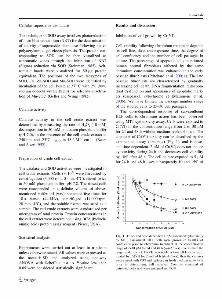
exponential decay (first rate) (Fig. 1), and is dose-
and time-dependent. 2 lM of Cr(VI) does not induce
cytotoxicity during 24 h and decreases cell viability
by 10% after 48 h. The cell culture exposed to 5 lM
for 24 h and 48 h loses subsequently 10 and 15% of
Gro
wth
Inhi
bitio
n (%
of c
ontr
ol)
Concentration of Cr(VI) ( M)
2 h Cr(VI)+46 h
24 h Cr(VI)
48 h Cr(VI)
24 h Cr(VI) +24 h
Fig. 1 Time- and dose-dependent Cr(VI)-induced cytotoxicity
by MTT assessment. HLF cells were grown up to 80% of
confluence prior to chromium treatment at the concentration
range of 2–30 lM for 24 and 48 h (solid lines). To estimate the
range and time of Cr(VI) reversible action HLF cells were
treated by Cr(VI) for 2 and 24 h (dash lines), then the cultures
were rinsed with PBS and replaced in fresh medium up to 48 h
prior to determining cell survival. Controls consisted of
untreated cells and were assigned as 100%
Biometals
123

viable cell population. Incubation of the cells with 10
and 15 lM Cr(VI) decreases cell population by 20–
35% after 24 h of chromium action and by 60–70%
after 48 h. Cr(VI) above 20 lM causes significant
decrease of the cell viability by 50 and 80% after 24
and 48 h, respectively.
To estimate the concentration range and time of
reversible Cr(VI) action the treatment of HLF cells
with Cr(VI) 2–30 lM Cr(VI) for 2 and 24 h was
followed by cell growth in complete medium without
chromium up to 48 h. The cell viability was not
decreased at the early stage (2 h) of Cr(VI) action
even for 30 lM Cr(VI). The effect of 5 lM Cr(VI) at
24 h was entirely reversed in 24 h after replacing the
cultured cells in complete medium without chro-
mium. Under these experimental conditions 10 and
15 lM Cr(VI) decreased in toxic effect by 15 and
20%. The cell exposure with toxic concentrations
(above 20 lM) resulted in progressive cell death,
indicating irreversible chromium action in 24 h.
The results show that chromium action on HLF cell
culture could be separated into groups: nontoxic—2
and 5 lM Cr(VI), sub-toxic—10 and 15 lM Cr(VI)
and toxic—above 20 lM Cr(VI). While entering a
cell low concentration of Cr(VI) has no threshold (Liu
et al. 2001). The cell culture system models all
possibilities of Cr(VI) reduction, which exist under
the physiological conditions, including the presence
of extracellular reductants in the serum complemented
media. The typical natural concentration of chromium
reductant such as ascorbic acid (AA) in bovine serum
is quite high—about 50–70 lM (Kleczkowski et al.
2005) and comparable with ascorbic acid in blood
plasma (Carty et al. 2000). Generally, serum does not
exceed 15% of a complete culture media, and the
concentration of ascorbic acid comprises about
10 lM. On the other hand, serum could not be
omitted from the media as it is a necessary component
to support cell growth, and promote attachment of
cells to the substrate. The question arises whether the
concentration of AA in culture media supplemented
with serum could contribute to the extracellular
reduction of the complemented Cr(VI) and the way
it could affect intracellular ROS level and activities of
antioxidant defense system. As AA is highly perme-
able, one can suppose that the extracellular quantity of
AA decreases during the experimental conditions and
this results in a negligible participation of AA in the
extracellular Cr(VI) reduction. But if we follow the
consideration concerning the influence of AA/Cr(VI)
ratio on the AA pro-oxidant versus antioxidant
properties (Poljsak et al. 2005), we can suppose that
in our case (low AA to Cr(VI) concentrations) the
partial extracellular Cr(VI) reduction to Cr(V) with
the appearance of ROS can not be excluded. However,
it is difficult to separate inputs of intracellular Cr(VI),
Cr(V) from penetrated Cr(V) reduction, and, likewise
differentiating intracellularly generated or penetrated
ROS because Cr(V) as well as Cr(VI), H2O2 and O2.-
are highly permeable, and once in a cell they can
affect the antioxidant defense system and ROS level.
Cr(VI) affects the cell cycle distribution and
induces apoptosis
The cell cycle analysis for a single time-point gives
basically static information detecting only disorders
in the cell cycle phase distribution at stress condi-
tions. The applied dynamic flow cytometry analysis
provides the ability to follow the changes of the cell
cycle phase distribution along with chromium expo-
sure. The studied cell line was affected differently by
the nontoxic (5 lM), sub-toxic (15 lM) and toxic
(30 lM) doses of chromium, respectively, over 24 h
(Fig. 2).
sub-G1 G1 S 2
% o
f C
ell F
ract
ion
5 µM Cr(VI) 15 µM Cr(VI) 30 µM Cr(VI)
sub -G1 G1 S sub-G1 G1 S 2+M0
102030405060708090
100Control 4 h 17 h 24 h
0102030405060708090
100Control 4 h 17 h 24 h
0102030405060708090
100Control 4 h 17 h 24 h
G +M
A B C
G2+MG
Fig. 2 Time- and dose-
dependent effect of Cr(VI)
exposure on cell cycle
progression
Biometals
123
Between 48 and 72 h of growth the control HLF
cell population predominately consists of G1 phase
cells (*70%). The nontoxic 5 lM Cr(VI) had no
influence on DNA histogram up to 17 h and caused
the cell cycle re-entry at 24 h of Cr(VI) action,
lowering the cell fraction in G1 phase to 50% and
increasing the cell fraction in S and G2 ? M phase
(Fig. 2a). Microscope analysis of cells grown on
coverslips demonstrated that the mitotic index did not
decrease by comparison with the control and was
*12–15% Mi (data not shown).
The sub-toxic concentrations of chromium
(15 lM) caused the cell cycle re-entry earlier (at
4 h) compared to the nontoxic concentration
(Fig. 2b). The increase of the G2 ? M cell fraction
with the simultaneous decrease of the G1 cell fraction
at 4 h was followed by growth arrest predominantly
in G1 phase at 17 h, not affecting S and G2 ? M
phases. The growth arrest was also confirmed by the
absence of the mitotic cells in the cell population as
was demonstrated by microscope analysis (data not
shown). The apoptotic sub-G1 fraction increased
insignificantly (not more 20%). The growth inhibition
observed by the MTT assay at 24 h of chromium
treatment (Fig. 1) may partly contribute to the
detected growth arrest.
The toxic 30 lM Cr(VI) did not affect cell cycle
distribution during 17 h of permanent chromium
treatment and sharply increased the apoptotic sub-G1
fraction (about 35%) and lowered the cell number in
G1 phase to 40% only at 24 h. The resultant decrease
of G1 cell fraction is the consequence of the massive
cell apoptosis developed at 24 h of chromium
exposure (Fig. 2c).
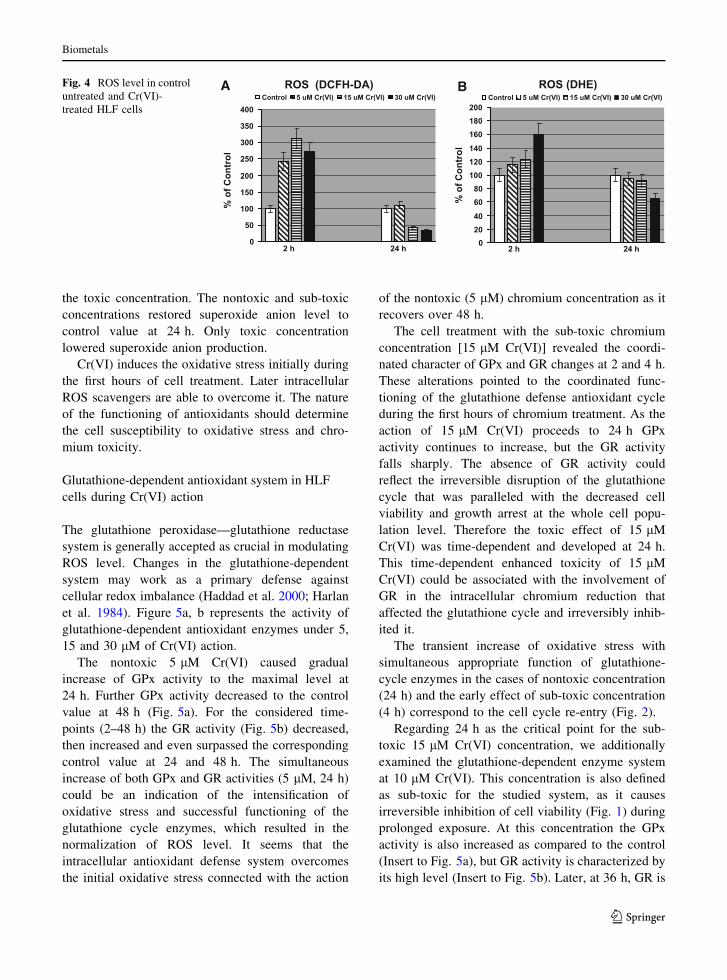
Cell population was analyzed by fluorescent
microscopy after staining with DNA-specific fluores-
cent dyes [acridine orange (AO) and ethidium
bromide (EB)] for the detection of apoptotic cells
(Diaz et al. 1999). Apoptotic nuclei are characterized
by highly condensed chromatin. The data of the
morphological analysis of Cr(VI) on HLF cells in the
range from 5 to 30 lM Cr(VI) action at 24 h are
presented in Fig. 3.
The results show, that a narrow range of chromium
concentrations initiated diverse effects in HLF cell
culture such as, induction of the cell cycle re-entry
(nontoxic concentration of 5 lM), growth arrest (sub-
toxic concentration of 15 lM) and apoptosis (toxic
concentration of 30 lM).
ROS in HLF cells during Cr(VI) action
The intracellular redox environment may influence
cell cycle progression (Conour et al. 2004; Noda et al.
2001). It was hypothesized that ROS could contribute
to cell cycle progression, and a late-G1 phase check-
point was proposed after transition across the growth-
factor-dependent G1 restriction point that was coor-
dinating cellular ROS production with cell population
transition from G1 to S phase (Havens et al. 2006).
There is evidence that fluctuation in the cellular redox
state contributes in the cell cycle regulatory pathways
(Mennon and Goswami 2007; Sarsour et al. 2008).
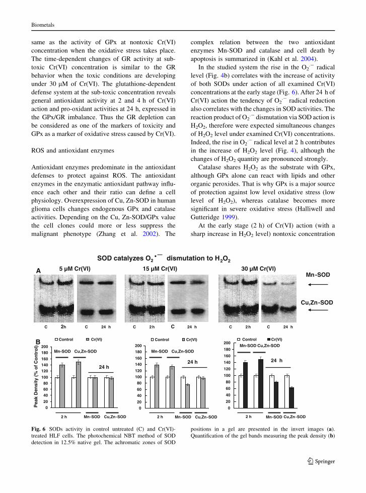
The nontoxic, sub-toxic and toxic doses of Cr(VI)
initially increased ROS production to a comparable
level (above twofold) at 2 h of chromium action
(Fig. 4a). The nontoxic concentration caused the
reestablishment of ROS level to the control value at
24 h. The ROS level had decreased at 24 h under
sub-toxic and toxic concentrations. The decrease of
ROS level under these concentrations correlated with
the greater loss of cell viability and apoptosis. The
decrease of the ROS level at the late stages of
apoptosis was also observed in the case of Fas-
induced apoptosis (Aronis et al. 2003; Shen and
Pervaiz 2006).
Superoxide anion production detected by DHE
(Fig. 4b) was enhanced at 2 h of chromium action.
The extent of the increase was more pronounced for
0
10
20
30
40
50
60
70
80
90
100Apoptotic cells Necrotic cells Viable cells
% o
f Cel
l Num
ber
C
Concentration of Cr(VI) (µM)5 10 15 20 30
Fig. 3 Effect of Cr(VI) on cell survival after treatment of HLF
cells with various chromium concentrations for 24 h deter-
mined by morphological analysis
Biometals
123
the toxic concentration. The nontoxic and sub-toxic
concentrations restored superoxide anion level to
control value at 24 h. Only toxic concentration
lowered superoxide anion production.
Cr(VI) induces the oxidative stress initially during
the first hours of cell treatment. Later intracellular
ROS scavengers are able to overcome it. The nature
of the functioning of antioxidants should determine
the cell susceptibility to oxidative stress and chro-
mium toxicity.
Glutathione-dependent antioxidant system in HLF
cells during Cr(VI) action
The glutathione peroxidase—glutathione reductase
system is generally accepted as crucial in modulating
ROS level. Changes in the glutathione-dependent
system may work as a primary defense against
cellular redox imbalance (Haddad et al. 2000; Harlan
et al. 1984). Figure 5a, b represents the activity of
glutathione-dependent antioxidant enzymes under 5,
15 and 30 lM of Cr(VI) action.
The nontoxic 5 lM Cr(VI) caused gradual
increase of GPx activity to the maximal level at
24 h. Further GPx activity decreased to the control
value at 48 h (Fig. 5a). For the considered time-
points (2–48 h) the GR activity (Fig. 5b) decreased,
then increased and even surpassed the corresponding
control value at 24 and 48 h. The simultaneous
increase of both GPx and GR activities (5 lM, 24 h)
could be an indication of the intensification of
oxidative stress and successful functioning of the
glutathione cycle enzymes, which resulted in the
normalization of ROS level. It seems that the
intracellular antioxidant defense system overcomes
the initial oxidative stress connected with the action
of the nontoxic (5 lM) chromium concentration as it
recovers over 48 h.
The cell treatment with the sub-toxic chromium
concentration [15 lM Cr(VI)] revealed the coordi-
nated character of GPx and GR changes at 2 and 4 h.
These alterations pointed to the coordinated func-
tioning of the glutathione defense antioxidant cycle
during the first hours of chromium treatment. As the
action of 15 lM Cr(VI) proceeds to 24 h GPx
activity continues to increase, but the GR activity
falls sharply. The absence of GR activity could
reflect the irreversible disruption of the glutathione
cycle that was paralleled with the decreased cell
viability and growth arrest at the whole cell popu-
lation level. Therefore the toxic effect of 15 lM
Cr(VI) was time-dependent and developed at 24 h.
This time-dependent enhanced toxicity of 15 lM
Cr(VI) could be associated with the involvement of
GR in the intracellular chromium reduction that
affected the glutathione cycle and irreversibly inhib-
ited it.
The transient increase of oxidative stress with
simultaneous appropriate function of glutathione-
cycle enzymes in the cases of nontoxic concentration
(24 h) and the early effect of sub-toxic concentration
(4 h) correspond to the cell cycle re-entry (Fig. 2).
Regarding 24 h as the critical point for the sub-
toxic 15 lM Cr(VI) concentration, we additionally
examined the glutathione-dependent enzyme system
at 10 lM Cr(VI). This concentration is also defined
as sub-toxic for the studied system, as it causes
irreversible inhibition of cell viability (Fig. 1) during
prolonged exposure. At this concentration the GPx
activity is also increased as compared to the control
(Insert to Fig. 5a), but GR activity is characterized by
its high level (Insert to Fig. 5b). Later, at 36 h, GR is
% o
f Con
trol
% o
f Con
trol
ROS (DCFH-DA) ROS (DHE)
2 h 24 h 2 h 24 h0
50
100
150
200
250
300
350
400Control 5 uM Cr(VI) 15 uM Cr(VI) 30 uM Cr(VI)
020406080
100120140160180200
Control 5 uM Cr(VI) 15 uM Cr(VI) 30 uM Cr(VI)BAFig. 4 ROS level in control
untreated and Cr(VI)-
treated HLF cells
Biometals
123

inhibited and as a result collapse of glutathione cycle
occurs (data not shown).
The changes of GPx and GR activities under the
toxic 30 lM Cr(VI) are similar to the sub-toxic
concentration effect at 2 and 4 h. The prolonged
action of 30 lM Cr(VI) to 24 h inhibits entirely both
GPx and GR activities, thus the disruption of the
glutathione-dependent antioxidant defense system
takes place.
Consideration of GR as the marker of Cr(VI)
toxicity in the studied system expands the number of
the cell types, for which the accompaniment of
Cr(VI) toxicity with GR inhibition has been reported.
The chromate-caused inhibition of GR has been
observed in erythrocytes (Koutras et al. 1965),
fibroblasts (Sugiyama et al. 1991), osteoblasts (Ning
and Grant 2000) and hepatocytes (Gunarantnam and
Grant 2004). Recently the complete inhibition of GR
activity by 10 lM Cr(VI) action has been shown in
the J744.1 murine macrophage cell line (Lalaouni
et al. 2007). The mechanism of the toxic action of
Cr(VI) on GR is proposed as arising from its
participation in one- and/or two-electron transference
and reduction of Cr(V) to Cr(IV) and/or Cr(III) (Bal
and Kasparzak 2002). Generated toxic metabolites
irreversibly inhibit GR activity. Thus GR under
chromium action has a dual function as an antioxi-
dant, restoring the GSH pool, and as a pro-oxidant,
reducing intracellular chromium, and as a result
participating in ROS generation.
The complicated character of the alteration in the
glutathione-dependent defense enzyme activities in
the studied system testifies to their involvement both
in oxidative stress and in chromium toxicity. The
nontoxic concentration initiates the general antioxi-
dant reactions, revealed by the progressive coordi-
nated increase of GPx and GR at the early stage of
Cr(VI) action and reaching the control level at 48 h
of Cr(VI) action. The toxic concentration also causes
the coordinated response of GPx and GR followed by
total inhibition at 24 h. The time-dependent behavior
of GPx at sub-toxic concentration is very much the
0
20
40
60
80
100
120
140
160
Control 5 uM Cr(VI)
15 uM Cr(VI) 30 uM Cr(VI)
0
10
20
30
40
50
60
70
80
Control 5 uM Cr(VI)
15 uM Cr(VI) 30 uM Cr(VI)
GP
x ac
tivi
ty (
mU
/ml)
GR
act
ivit
y (m
U/m
l)
2 h 4 h 24 h 48 h
2 h 4 h 24 h 48 h
0
20
40
60
80
100
120
140
160
Control 10 uM Cr(VI) 15 uM Cr(VI)
0
10
20
30
40
50
60
70
80
Control 10 uM Cr(VI) 15 uM Cr(VI)
A
B
Fig. 5 Glutathione-
dependent antioxidant
system in control untreated
and Cr(VI)-treated HLF
cells. Inserts are GPx and
GR activity at 24 h of sub-
toxic 10 and 15 lM Cr(VI)
action
Biometals
123
same as the activity of GPx at nontoxic Cr(VI)
concentration when the oxidative stress takes place.
The time-dependent changes of GR activity at sub-
toxic Cr(VI) concentration is similar to the GR
behavior when the toxic conditions are developing
under 30 lM of Cr(VI). The glutathione-dependent
defense system at the sub-toxic concentration reveals
general antioxidant activity at 2 and 4 h of Cr(VI)
action and pro-oxidant activities at 24 h, expressed in
the GPx/GR imbalance. Thus the GR depletion can
be considered as one of the markers of toxicity and
GPx as a marker of oxidative stress caused by Cr(VI).
ROS and antioxidant enzymes
Antioxidant enzymes predominate in the antioxidant
defenses to protect against ROS. The antioxidant
enzymes in the enzymatic antioxidant pathway influ-
ence each other and their ratio can define a cell
physiology. Overexpression of Cu, Zn-SOD in human
glioma cells changes endogenous GPx and catalase
activities. Depending on the Cu, Zn-SOD/GPx value
the cell clones could more or less suppress the
malignant phenotype (Zhang et al. 2002). The
complex relation between the two antioxidant
enzymes Mn-SOD and catalase and cell death by
apoptosis is summarized in (Kahl et al. 2004).
In the studied system the rise in the O2.- radical
level (Fig. 4b) correlates with the increase of activity
of both SODs under action of all examined Cr(VI)
concentrations at the early stage (Fig. 6). After 24 h of
Cr(VI) action the tendency of O2.- radical reduction
also correlates with the changes in SOD activities. The
reaction product of O2.- dismutation via SOD action is
H2O2, therefore were expected simultaneous changes
of H2O2 level under examined Cr(VI) concentrations.
Indeed, the rise in O2.- radical level at 2 h contributes
in the increase of H2O2 level (Fig. 4), although the
changes of H2O2 quantity are pronounced strongly.
Catalase shares H2O2 as the substrate with GPx,
although GPx alone can react with lipids and other
organic peroxides. That is why GPx is a major source
of protection against low level oxidative stress (low
level of H2O2), whereas catalase becomes more
significant in severe oxidative stress (Halliwell and
Gutteridge 1999).
At the early stage (2 h) of Cr(VI) action (with a
sharp increase in H2O2 level) nontoxic concentration
SOD catalyzes O2 dismutation to H2O2
15 µM Cr(VI) 30 µM Cr(VI)
C 2h
5 µM Cr(VI)Mn-SOD
Cu,Zn-SOD
C 2 h C 2 hC 24 h C 24 h C 24 h
Pea
k D
Mn-SOD
Mn-SOD
Cu,Zn-SOD
Cu,Zn-SOD2 h
24 h
0
20
40
60
80
100
120
140
160
180
200
Control Cr(VI)
2 h
24 h
Mn-SOD
Mn-SOD
Cu,Zn-SOD
Cu,Zn-SOD
0
20
40
60
80
100
120
140
160
180
200
Control Cr(VI)
0
20
40
60
80
100
120
140
160
180
200Control Cr(VI)
2 h
24 h
Mn-SOD
Mn-SOD
Cu,Zn-SOD
Cu,Zn-SOD
ensi
ty (
% o
f C
on
tro
l)
A
B
Fig. 6 SODs activity in control untreated (C) and Cr(VI)-
treated HLF cells. The photochemical NBT method of SOD
detection in 12.5% native gel. The achromatic zones of SOD
positions in a gel are presented in the invert images (a).
Quantification of the gel bands measuring the peak density (b)
Biometals
123
did not cause an appreciable increase of catalase and
GPx activities compared with corresponding controls,
whereas under sub-toxic concentration increase of
catalase activity corresponds to the sharp reduction of
GPx activity, which becomes stronger at toxic
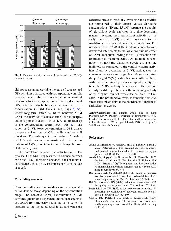
concentration (30 lM Cr(VI), 4 h, Figs. 7, 5a).
Under long-term action (24 h) of nontoxic 5 lM
Cr(VI) the activities of catalase and GPx rise sharply,
that is a probable cause of H2O2 level diminution up
to the corresponding control level (Fig. 4a). The
action of Cr(VI) toxic concentration at 24 h causes
complete exhaustion of GPx, while catalase still
functions. The subsequent examination of catalase
and GPx activities under sub-toxic and toxic concen-
trations of Cr(VI) points to the interchangeable role
of these enzymes.
The correlation between the activities of ROS–
catalase–GPx–SOD, suggests that a balance between
SOD and H2O2 degrading enzymes, but not individ-
ual enzymes, should play an important role in the fate
of a cell.
Concluding remarks
Chromium affects all antioxidants in the enzymatic
antioxidant pathways depending on the concentration
range. The nontoxic Cr(VI) concentration (5 lM)
activates glutathione-dependent antioxidant enzymes
and SODs from the early beginning of its action in
response to the increased ROS level, and when the
oxidative stress is gradually overcome the activities
are normalized to their control values. Sub-toxic
concentrations (10 and 15 lM) separate the activity
of glutathione-cycle enzymes in a time-dependent
manner, revealing their antioxidant activities at the
early stage of Cr(VI) action in response to the
oxidative stress observed under these conditions. The
imbalance of GPx/GR at the sub-toxic concentrations
developed later points to the toxic pro-oxidant effect
of Cr(VI) reduction, leading to Cr(III) formation and
destruction of macromolecules. At the toxic concen-
tration (30 lM) the glutathione-cycle enzymes are
inhibited, as compared to the control enzyme activ-
ities, from the beginning of Cr(VI) action, then the
system activates to an insignificant degree and after
the prolonged Cr(VI) action becomes fully inhibited
with the cells dying by means of apoptosis. By the
time the SODs activity is decreased, the catalase
activity is still high, however the remaining activity
of the enzymes can not reverse the cell fate. Cell re-
entry in the proliferative cycle at transient oxidative
stress takes place only at the coordinated function of
antioxidant enzymes.
Acknowledgments The authors would like to thank
Professor Len W. Poulter (Department of Immunology, UCL,
London) for his kind gift of HLF cell line and Lia Lezhava for
technical assistance. We are grateful to the ISTC for Project G-
349 Grant research funding.
References
Aronis A, Melendez JA, Golan O, Shilo S, Dicter N, Tirosh O
(2003) Potentiation of Fas-mediated apoptosis by attenu-
ated production of mitochondria-derived reactive oxygen
species. Cell Death Differ 10:335–344
Asatiani N, Sapojnikova N, Abuladze M, Kartvelishvili T,
Kulikova N, Kiziria E, Namchevadze E, Holman H-Y
(2004) Effects of Cr(VI) long-term and low-dose action
on mammalian antioxidant enzymes (an in vitro study). J
Inorg Biochem 98:490–496
Bagchi D, Bagchi M, Stohs SJ (2001) Chromium (VI)-induced
oxidative stress, apoptotic cell death and modulation of p53
tumor suppressor gene. Mol Cell Biochem 222:149–158
Bal W, Kasparzak KS (2002) Induction of oxidative DNA
damage by carcinogenic metals. Toxicol Lett 127:55–62
Beers RF, Sizer IW (1952) A spectrophotometric method for
measuring the breakdown of hydrogen peroxide by cata-
lase. J Biol Chem 195:133–140
Carlisle DL, Pritchard DL, Singh J, Patierno SR (2000)
Chromium(VI) induces p53-dependent apoptosis in dip-
loid human lung mouse dermal fibroblasts. Mol Carcinog
28:111–118
0
20
40
60
80
100
120
140
160
Control 5 uM Cr(VI)
15 uM Cr(VI) 30 uM Cr(VI)
2 h
Cat
alas
e ac
tivi
ty (
U/m
l)
4 h 24 h 48 h
Fig. 7 Catalase activity in control untreated and Cr(VI)-
treated HLF cells
Biometals
123
Carmichael J, DeGraff W, Gazdar AF, Minna JD, Mitchell JB
(1987) Evaluation of a tetrazolium-based semiautomated
colorimetric assay: assessment of radiosensitivity. Cancer
Res 47:936–942
Carter WO, Narayanan PK, Robinson JP (1994) Intracellular
hydrogen peroxide and superoxide anion detection in
endothelial cells. J Leukoc Biol 55:253–258
Carty JL, Bevan R, Waller H, Mistry N, Cooke M, Lunec J,
Griffiths HR (2000) The effects of vitamin C supple-
mentation on protein oxidation in healthy volunteers.
Biochem Biophys Res Commun 273:729–735
Chuang S-M, Liou G-Y, Yang J (2000) Activation of JNK, p38
and ERK mitogen-activated protein kinases by chromium
(VI) is mediated through oxidative stress but does not
affect cytotoxicity. Carcinogenesis 21:1491–1500
Conour JE, Graham WV, Gaskins HR (2004) A combined in
vitro/bioinformatic investigation of redox regulatory
mechanisms governing cell cycle progression. Physiol
Genomics 18:196–205
Costa M, Klein CB (2006) Toxicity and carcinogenicity of
chromium compounds in humans. Crit Rev Toxicol
36:155–163
Curtin JF, Donovan M, Cotter TG (2002) Regulation and
measurement of oxidative stress in apoptosis. J Immunol
Methods 265:49–72
Dewhirst M, Cao Y, Moeller B (2008) Cycling hypoxia and
free radicals regulate angiogenesis and radiotherapy
response. Nat Rev Cancer 8:425–437
Diaz G, Setzu MD, Zucca A, Isola R, Diana A, Murru R, Sogos
V, Gremo F (1999) Subcellular heterogeneity of mito-
chondrial membrane potential: relationship with organelle
distribution and intercellular contacts in normal, hypoxic
and apoptotic cells. J Cell Sci 112:1077–1084
Dubrovskaya VA, Wetterhahn K (1998) Effects of Cr(VI) on
the expression of the oxidative stress genes in human lung
cells. Carcinogenesis 19:1401–1407
Esposti MD (2002) Measuring mitochondrial reactive oxygen
species. Methods 26:335–340
Gambelunghe A, Piccinini R, Ambrogi M, Villarini M, Moretti
M, Marchetti C, Abbritti G, Muzi G (2003) Primary DNA
damage in chrome-plating workers. Toxicology 188:187–
195
Geller BL, Winge DR (1982) Rat liver Cu, Zn-superoxide
dismutase. Subcellular location in lysosomes. J Biol Chem
257:8945–8952
Gunarantnam M, Grant MH (2004) Damage to F-actin and cell
death induced by chromium VI and nickel in primary
monolayer cultures of rat hepatocytes. Toxicol In Vitro
18:245–253
Haddad JJE, Oliver RE, Land SC (2000) Antioxidant/Pro-
oxidant equilibrium regulates HIF-1a and NF-kB redox
sensitivity. Evidence for inhibition by glutathione oxida-
tion in alveolar epithelial cells. J Biol Chem 275:21130–
21139
Halliwell B, Gutteridge JMC (1999) Free radicals in biology
and medicine. Oxford Science Publication, Oxford Uni-
versity Press, Oxford
Harlan J, Levine JD, Callahan RS, Schwartz BR, Harker LA
(1984) Glutathione redox cycle protects cultured endo-
thelial cells against lysis by extracellularly generated
hydrogen peroxide. J Clin Invest 73:706–713
Havens CG, Ho A, Yoshika N, Dowdy SF (2006) Regulation of
late G1/S phase transition and APCCdh1 by reactive oxy-
gen species. Mol Cell Biol 26:4701–4711
Holmes AL, Wise SS, Wise JP Sr (2008) Carcinogenicity of
hexavalent chromium. Indian J Med Res 128:353–372
Kahl R, Kampkotter A, Watjen W, Chovolou Y (2004) Anti-
oxidant enzymes and apoptosis. Drug Metab Rev 36:747–
762
Kim J, Yurkow EJ (1996) Chromium induces a persistent
activation of mitogen-activated protein kinases by a
redox-sensitive mechanism in H4 rat hepatoma cells.
Cancer Res 56:2045–2051
Kleczkowski M, Klucinski W, Shaktur A, Sikora J (2005)
Concentration of ascorbic acid in the blood of cows
affected with mastitis. Bull Vet Inst Pulawy 49:203–207
Koutras GA, Scheider AS, Hattori M, Valentine WN (1965)
Studies on chromated erythrocytes. Mechanisms of chro-
mate inhibition of glutathione reductase. Brit J Haematol
11:360–369
Lalaouni A, Henderson C, Kupper C, Grant MH (2007) The
interaction of chromium (VI) with macrophages: deple-
tion of glutathione and inhibition of glutathione reductase.
Toxicology 236:76–81
Leonard SS, Harris GK, Shi X (2004) Metal-induced oxidative
stress and signal transduction. Free Rad Biol Med
37:1921–1942
Liu K, HuslerSurname J, Ye J, Leonard SS, Cutler D, Chen F,
Wang S, Zhang Z, Ding M, Wang L, Shi X (2001) On the
mechanism of Cr(VI)-induced carcinogenesis: dose
dependence of uptake and cellular responses. Mol Cell
Biochem 222:221–229
Mammone T, Gan D, Foyouzi-Youssefi R (2006) Apoptotic
cell death increases with senescence in normal human
dermal fibroblast cultures. Cell Biol Int 30:903–909
Mates JM, Segura JA, Alonso FJ, Marquez J (2008) Intracel-
lular redox status and oxidative stress: implications for
cell proliferative, apoptosis, and carcinogenesis. Arch
Toxicol 82:273–299
Mennon SG, Goswami PC (2007) A redox cycle within the cell
cycle: ring in the old with the new. Oncogene 26:1101–
1109
Ning J, Grant MH (2000) The role of reduced glutathione and
glutathione reductase in the cytotoxicity of chromium(VI)
in osteoblasts. Toxicol In Vitro 14:329–335
Noda T, Iwakiri R, Fujimoto K, Aw TY (2001) Induction of
mild intracellular redox imbalance inhibits proliferation of
CaCo-2 cells. FASEB J 15:2131–2139
Ormerod MG (2002) Investigating the relationship between the
cell cycle and apoptosis using flow cytometry. J Immun
Methods 265:73–80
Poljsak B, Gazdag Z, Jenko-Brinovec S, Fujs S, Pesti M, Be-
lagyi J, Plesnicar S, Raspor P (2005) Pro-oxidative vs
antioxidative properties of ascorbic acid in chro-
mium(VI)-induced damage: an in vivo and in vitro
approach. J Appl Toxicol 25:535–548
Pourahmad J, O’Brien TJ (2001) Biological reactive interme-
diates that mediate Chromium (VI) toxicity. In: Dansette
PM (ed) Biological reactive intermediates VI: chemical
and biological mechanisms in susceptibility to and pre-
vention of environmental diseases: advances in experi-
mental medicine and biology. Springer, Berlin, p 203
Biometals
123
Pritchard DE, Ceryak S, Ha I, Fornsaglio JL, Hartman SK,
O’Brien TJ, Patierno SR (2001a) Mechanism of apoptosis
and determination of cellular fate in Chromium(VI)-
exposed population of telomerase-immortalized human
fibroblasts. Cell Growth Differ 12:487–496
Pritchard KA Jr, Ackerman AW, Gross ER, Stepp DW, Shi Y,
Fontana ST, Baker JE, Sessa WC (2001b) Heat shock
protein 90 mediates the balance of nitric oxide and
superoxide anion from endothelial nitric-oxide synthase. J
Biol Chem 276:17621–17624
Rana SVS (2008) Metals and apoptosis: recent development. J
Trace Elem Med Biol 22:262–284
Sarsour EH, Venkatraman S, Kalen AL, Oberley LW, Gosw-
ami PC (2008) Manganese superoxide dismutase activity
regulates transitions between quiescent and proliferative
growth. Aging Cell 7:405–417
Shen H-M, Pervaiz Sh (2006) TNF receptor superfamily-
induced cell death: redox-dependent execution. FASEB J
20:1589–1598
Shi X, Ding M, Ye J, Wang S, Leonard SS, Zang L, Castranova
V, Vallyathan V, Chiu A, Dala N, Liu K (1999) Cr(VI)
causes activation of nuclear transcription factor kB, DNA
strand breaks and dG hydroxylation via free radical
reactions. J Inorg Biochem 75:37–44
Steinman HM (1985) Bacteriocuprein superoxide dismutases
in pseudomonads. J Bacteriol 162:1255–1260
Sugiyama M, Tsuzuki K, Ogura R (1991) Effect of ascorbic
acid on DNA damage, cytotoxicity, glutathione reductase,
and formation of paramagnetic chromium in Chinese
hamster V-79 cells treated with sodium chromate(VI). J
Biol Chem 266:3383–3386
Trzeciak A, Kowalik J, Malecka-Panas E, Drzewoski J, Wo-
jewodzka M, Iwanenko T, Blasiak J (2000) Genotoxicity
of chromium in human gastric mucosa cells and periph-
eral blood lymphocytes evaluated by single cell gel
electrophoresis (comet assay). Med Sci Monit 6:24–29
Ye J, Wang S, Leonard SS, Yi S, Butterworth L, Antonini J,
Ding M, Yo R, Vallyathan V, Castranov V, Shi X (1999)
Role of reactive oxygen species and p53 in chro-
mium(VI)-induced apoptosis. J Biol Chem 274:34974–
34980
Zhang Y, Zhao W, Zhang HJ, Domann FE, Oberley LW (2002)
Overexpression of copper zinc superoxide dismutase
suppresses human glioma cell growth. Cancer Res
62:1205–1212
Biometals
123