Blast mitigation in a sandwich composite using graded core and polyurea interlayer
Upload
udistritalCategory
view
0download
0
Resonant magnetic reflectivity in the extreme ultraviolet spectral range:Interlayer-coupled Co/Si/Ni/Fe multilayer system
P. Grychtol,1 R. Adam,1 S. Valencia,2 S. Cramm,1 D. E. Bürgler,1 and C. M. Schneider1
1Institute of Solid State Research, IFF-9, Forschungszentrum Jülich, D-52425 Jülich, Germany2Helmholtz-Zentrum-Berlin, BESSY, Albert-Einstein-Strasse 15, D-12489 Berlin, Germany
�Received 18 May 2010; published 30 August 2010�
A polycrystalline test structure comprising a 5 nm cobalt and a 10 nm nickel/iron layer separated by a siliconlayer ranging from 1.5 to 4 nm prepared by thermal evaporation has been investigated by resonant magneticreflectivity measurements of horizontally polarized light in the extreme ultraviolet spectral range. By exploitingthe transversal magneto-optical Kerr effect at the M absorption edges of cobalt and nickel �59.5 eV and 66.5eV� a magnetic contrast as large as 80% for cobalt and 25% for nickel can be obtained near a Brewster angleof about 45°. Angle- and energy-dependent scans of the magnetic asymmetry as well as element-selective,magneto-optical loops of the hysteresis were recorded against the thickness of the interlayer, reflecting theswitching behavior of the individual ferromagnetic layers as a function of the interlayer coupling.
DOI: 10.1103/PhysRevB.82.054433 PACS number�s�: 75.25.�j
I. INTRODUCTION
The interaction of light with a magnetic system provides awealth of experimental information to study various aspectsof magnetism. An example is the magneto-optical Kerr effect�MOKE�, which denotes the elastic scattering of polarizedlight at a magnetic surface within the visible range. Here, achange in the magnetization state in the material usually in-duces a change in the polarization state of the scattered light,except for a transversal Kerr �T-MOKE� geometry, in whichthe magneto-optical response directly translates into an in-tensity modulation. Exploiting MOKE in a reflectivity ex-periment is a well-established technique for investigations ofstatic and dynamic processes in magnetism.1–3 At wave-lengths above 250 nm, however, the effect is weak in mostmaterials—less than a millirad of Kerr rotation and far belowa percent change in intensity upon a full magnetization re-versal. Furthermore, wavelengths employed in conventionalMOKE experiments are large as compared to cutting-edgemagnetic structures on the nanometer scale, thus precludingtheir imaging with an appropriate lateral resolution.4
In the last decades, these shortcomings have been largelyovercome by exploiting much stronger magneto-optical ef-fects in the soft x-ray regime. Although these effects can betreated in a common theoretical framework, it is convenientto distinguish between magnetodichroic phenomena in ab-sorption and transmission, i.e., x-ray magnetic circular di-chroism �XMCD� and linear dichroism, and magnetodichroiceffects in reflection, which are often grouped under the termx-ray resonant magnetic scattering �XRMS�. All of thesephenomena require a resonant excitation by means of lin-early or circularly polarized tunable soft x rays. Access tothis radiation is provided by large synchrotron radiation fa-cilities, which significantly increases the experimental com-plexity as compared to a laboratory approach. By exploitingXRMS and XMCD effects at the L edges of transition metalsat photon energies above 500 eV, not only a superior mag-netodichroic contrast of up to several tens of percent and alateral resolution down to a few nanometers can be attained,but also element and chemical selectivity can be gained in a
resonant excitation.5–7 These x-ray magneto-optical phenom-ena allow for magnetic investigations of individual constitu-ents of heterogeneous ferromagnetic systems on a nanometerand on a femtosecond scale, if suitable light sources such asfree-electron lasers8 or appropriate techniques such asfemtoslicing9 are employed.
So far, only few experiments have addressed resonantmagnetic reflectivity and magnetic dichroism at the M edgesof transition metals at photon energies around 50 eV—in theextreme ultraviolet �XUV� regime. In terms of the magneto-optical response, these XUV phenomena have been foundequally useful, with changes in the dichroic contrast rangingup to almost 100%.10–14 Because the majority of beamlines atthird-generation synchrotron facilities are dedicated to pho-ton energies in the soft x-ray region, the opportunity of XUVmagneto-optical studies has mostly escaped the attention ofthe magnetic community. However, recently substantialprogress has been made in laser-based light sources reachingphoton energies of up to 100 eV with moderate effort justi-fying a closer look into XUV magneto-optics. Advancementsin laser amplifier technology have brought about reliabletable-top light sources, which are able to produce coherentand ultrashort XUV light pulses exploiting a highly nonlinearconversion process �higher harmonic generation—HHG�.15–17 Due to the pulsed and coherent nature of theemitted radiation, HHG-based light sources may serve ascompact tools for element-selective investigations of mag-netic properties on the femtosecond and nanometer scale in alaboratory environment.18,19
For this reason, we present in this contribution magneto-optical studies in the XUV performed with synchrotron ra-diation at the M edges of ferromagnets, which are based ontransition metals in a spin-valve-type layered system. We ex-plore advantages and disadvantages of experiments in thisspectral range and discuss the data as a basis for future stud-ies employing HHG-based table-top systems.
II. XUV VS SOFT X-RAY REGIME
The XUV spectral range offers certain advantages overthe soft x-ray regime with respect to magneto-optical studies.
PHYSICAL REVIEW B 82, 054433 �2010�
1098-0121/2010/82�5�/054433�8� ©2010 The American Physical Society054433-1

Several experiments have already shown that magnetic in-vestigations of single films by resonantly scattered XUVlight at the M absorption edges of transition metals10–14 andthe N absorption edges of the lanthanide series20,21 yield verystrong magnetodichroic signals. Moreover, studies of theFaraday rotation and the Voigt effect at the M and L absorp-tion edges of Fe, Co, and Ni reveal that magneto-opticalconstants and thus responses at the M edges are as large asthose at the L edges. This fact emphasizes the dominant roleof the exchange interaction in comparison to the spin-orbitcoupling in the generation of magnetic contrast at the Medges.14,22,23 It is the high absorbance of most materials, atangles where the magnetodichroic signal in the XUV maxi-mizes, which gives no rise to a magnetic contrast in compa-rable soft x-ray experiments. In a T-MOKE geometry, themaximum of the magnetodichroic response is located aroundthe Brewster angle, i.e., incoming and outgoing light beamenclose an angle of �90°. Under these conditions, there isstill a sizable reflectance on the order of R�10−4, whichensures a reasonable signal-to-noise ratio in the XUV. In thesoft x-ray regime, on the other hand, the light penetratesdeeper into the sample and the reflectivity drops off muchfaster with increasing incidence angle �, reaching values ofR�10−10–10−11 at �=45°.24 Therefore, in the soft x-ray re-gime grazing incidence optics are required adding to thecomplexity of the experimental setup. This also generates aconsiderable problem for imaging experiments in a reflectiongeometry, as the focusing of the photon beam becomes verydifficult and the imaged plane is not well defined. That iswhy a right-angle geometry, generally favored in the XUV, ismuch more suitable for imaging experiments. This approachis sensitive to the in-plane magnetization in contrast to trans-mission techniques in the soft x-ray regime, which are onlysensitive to the magnetic out-of-plane component, thus im-posing certain restrictions on the sample thickness and thegeometry with respect to the probe beam.
Another aspect concerns optical interference effects aris-ing in the sample. Typical structural dimensions of a samplesystem, such as the individual layer thickness in a multilayerstack, and the wavelength corresponding to a transition-metal L edge are of a similar magnitude, i.e., on the order ofa few nanometer �1.59 nm for Co L3 absorption edge at778.1 eV�. In a reflectivity experiment at these photon ener-gies, the multilayer acts as a superlattice, resulting in a rela-tively complex interference pattern mixing structural andmagnetic periodicities. As a consequence, the reflected inten-sity exhibits a strong angular modulation of the magnetod-ichroic signal and structural and magnetic properties cannotbe separated easily.25 In the case of M edges, where thewavelength is one order of magnitude larger �20.6 nm forCo M absorption edge at 60.2 eV�, structurally induced in-terferences in the magnetic signal are less pronounced, sim-plifying the interpretation in this respect. For the same rea-son, surface and interface roughness influence the magneticsignal to a much smaller extent. Moreover, due to a smallerpenetration depth of the XUV radiation, Kiessig fringes—resonances between surface and substrate—do not occur, fur-ther facilitating the interpretation.
Just as the XUV region of the electromagnetic spectrum islocated in between the visible and soft x-ray range, its inter-
action with matter combines the characteristics of both spec-tral regions. This makes XUV light a unique element-selective, ultrafast probe of magnetism on the nanometerscale, which is going to be available in any optical laboratoryin the near future. Since only little attention has been paid tothe XUV region, it is the purpose of this work to explore itspotential as a magnetic contrast mechanism by tuning thephoton energy to the M absorption edges of Co and Ni in aT-MOKE reflectivity experiment and focusing on layer-selective investigations of magnetically coupled multilayersystems.
III. SAMPLE
The sample consisted of a magnetic trilayer structure andwas prepared by thermal evaporation at a base pressure of5�10−11 mbar.26 In a first step, a substrate system for themagnetic multilayer was manufactured comprising a 150-nm-thick Ag�001� buffer layer,27 which was grown on aniron precovered GaAs�001� wafer at a temperature of 380 K.After postannealing at a temperature of 570 K, a 2 nm Fefollowed by a 8 nm Ni layer were deposited onto the bufferat room temperature. A stepped Si layer with a step height of1.5, 2.5, and 4 nm, each 1 mm wide, followed. Finally, a5-nm-thick Co layer was deposited on top.
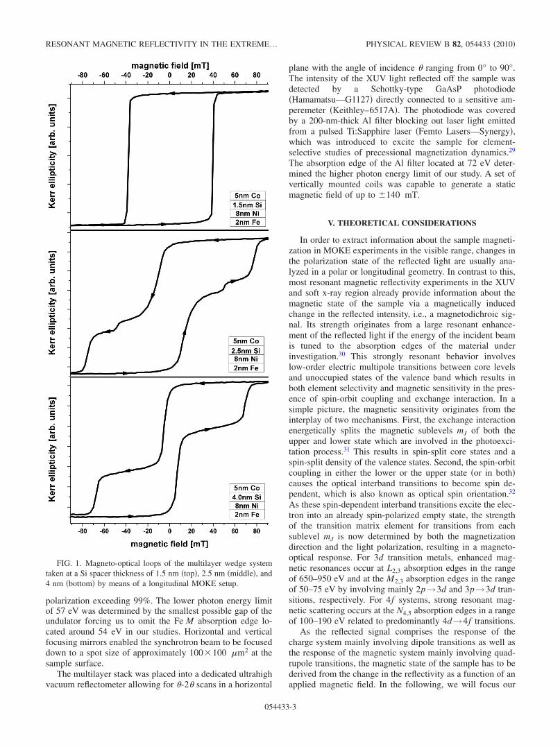
The magnetic switching behavior in the plane of themultilayer system was first characterized by MOKE mea-surements in the visible range.28 Recording hysteresis loopsin a longitudinal MOKE �L-MOKE� setup for various anglesof the sample with respect to the external magnetic fieldrevealed a negligible magnetocrystalline anisotropy. Threeregions of different interlayer coupling between the ferro-magnetic Co and Ni/Fe layers were identified and associatedwith regions of different Si spacer thicknesses, as deducedfrom distinct coercivities of the hysteresis loops, which aredisplayed in Fig. 1.
As can be seen from this figure, at a spacer thickness of1.5 nm the multilayer is ferromagnetically coupled forcingall layers to switch simultaneously. At a spacer thickness of 4nm, the multilayer is entirely decoupled allowing for an in-dependent switching of the top Co and bottom Ni/Fe layers.Independent experiments prove that the Ni/Fe bilayer indeedbehaves like a single magnetic unit. At a spacer thickness of2.5 nm, an intermediate behavior can be observed, in whichthe reversal of one layer is hindered by a weak ferromagneticinterlayer coupling. The hysteresis loops taken in the visiblerange do not reveal directly, which layer switches first,thereby necessitating a layer- and element-selective ap-proach.
IV. EXPERIMENTAL SETUP
In order to further characterize the magnetic multilayer,we performed resonant magnetic reflectivity measurementsin a T-MOKE geometry at the undulator beamline UE56/1-SGM of the synchrotron radiation facility BESSY II. Thereflectivity of linearly p-polarized XUV light was measuredacross the M absorption edges of Co and Ni from 57 to 72eV with an energy resolution of 0.1 eV and a degree of linear
GRYCHTOL et al. PHYSICAL REVIEW B 82, 054433 �2010�
054433-2
polarization exceeding 99%. The lower photon energy limitof 57 eV was determined by the smallest possible gap of theundulator forcing us to omit the Fe M absorption edge lo-cated around 54 eV in our studies. Horizontal and verticalfocusing mirrors enabled the synchrotron beam to be focuseddown to a spot size of approximately 100�100 �m2 at thesample surface.
The multilayer stack was placed into a dedicated ultrahighvacuum reflectometer allowing for �-2� scans in a horizontal
plane with the angle of incidence � ranging from 0° to 90°.The intensity of the XUV light reflected off the sample wasdetected by a Schottky-type GaAsP photodiode�Hamamatsu—G1127� directly connected to a sensitive am-peremeter �Keithley–6517A�. The photodiode was coveredby a 200-nm-thick Al filter blocking out laser light emittedfrom a pulsed Ti:Sapphire laser �Femto Lasers—Synergy�,which was introduced to excite the sample for element-selective studies of precessional magnetization dynamics.29
The absorption edge of the Al filter located at 72 eV deter-mined the higher photon energy limit of our study. A set ofvertically mounted coils was capable to generate a staticmagnetic field of up to �140 mT.
V. THEORETICAL CONSIDERATIONS
In order to extract information about the sample magneti-zation in MOKE experiments in the visible range, changes inthe polarization state of the reflected light are usually ana-lyzed in a polar or longitudinal geometry. In contrast to this,most resonant magnetic reflectivity experiments in the XUVand soft x-ray region already provide information about themagnetic state of the sample via a magnetically inducedchange in the reflected intensity, i.e., a magnetodichroic sig-nal. Its strength originates from a large resonant enhance-ment of the reflected light if the energy of the incident beamis tuned to the absorption edges of the material underinvestigation.30 This strongly resonant behavior involveslow-order electric multipole transitions between core levelsand unoccupied states of the valence band which results inboth element selectivity and magnetic sensitivity in the pres-ence of spin-orbit coupling and exchange interaction. In asimple picture, the magnetic sensitivity originates from theinterplay of two mechanisms. First, the exchange interactionenergetically splits the magnetic sublevels mJ of both theupper and lower state which are involved in the photoexci-tation process.31 This results in spin-split core states and aspin-split density of the valence states. Second, the spin-orbitcoupling in either the lower or the upper state �or in both�causes the optical interband transitions to become spin de-pendent, which is also known as optical spin orientation.32
As these spin-dependent interband transitions excite the elec-tron into an already spin-polarized empty state, the strengthof the transition matrix element for transitions from eachsublevel mJ is now determined by both the magnetizationdirection and the light polarization, resulting in a magneto-optical response. For 3d transition metals, enhanced mag-netic resonances occur at L2,3 absorption edges in the rangeof 650–950 eV and at the M2,3 absorption edges in the rangeof 50–75 eV by involving mainly 2p→3d and 3p→3d tran-sitions, respectively. For 4f systems, strong resonant mag-netic scattering occurs at the N4,5 absorption edges in a rangeof 100–190 eV related to predominantly 4d→4f transitions.
As the reflected signal comprises the response of thecharge system mainly involving dipole transitions as well asthe response of the magnetic system mainly involving quad-rupole transitions, the magnetic state of the sample has to bederived from the change in the reflectivity as a function of anapplied magnetic field. In the following, we will focus our
FIG. 1. Magneto-optical loops of the multilayer wedge systemtaken at a Si spacer thickness of 1.5 nm �top�, 2.5 nm �middle�, and4 nm �bottom� by means of a longitudinal MOKE setup.
RESONANT MAGNETIC REFLECTIVITY IN THE EXTREME… PHYSICAL REVIEW B 82, 054433 �2010�
054433-3

attention to a T-MOKE geometry, which has been adopted inour resonant scattering experiments described below. Thestrength of the T-MOKE signal, being odd in magnetizationM, is commonly denoted as the normalized difference of thereflected intensity I for two inverted directions of the samplemagnetization, here referred to as ↑ and ↓. The latter can beachieved by an external magnetic field applied transverselyto the scattering plane. This so-called magnetic asymmetry Ais related to the Fresnel reflection coefficients within theclassical magneto-optical formalism,33
A =I↑ − I↓I↑ + I↓
=�rpp
↑ �2 − �rpp↓ �2
�rpp↑ �2 + �rpp
↓ �2.
Its maximum is close to the Brewster angle, where in asimplified picture for p-polarized light the oscillation axis ofthe electric dipole coincides with the axis of specular reflec-tion along which no dipole radiation is emitted. Due to thedistinct emission characteristic of the multipole radiation, forthis angle the response of the charge system is suppressed,whereas the magnetic contribution resulting from the quadru-pol transitions prevails. Thus, the magnetic contributiondominates the total signal.
The reflection coefficient rpp, consisting of a nonmagneticrpp
0 and a magnetic �pp response, describes the influence ofmaterials on p-polarized light which is incident at an angle of�i and reflected at an angle of �r, with n0 being the refractiveindex of the nonmagnetic transmitted medium and n̄ beingthe average refractive index of the magnetic reflectivemedium,34
rpp↑↓ = rpp
0 � �pp
=n̄ cos �i − n0 cos �r
n̄ cos �i + n0 cos �r
�2in0n̄ cos �i sin �r
�n̄ cos �i + n0 cos �r�2Qx.
In isotropic media, the Voigt vector Q, being directly re-lated to the dielectric tensor and commonly introduced toaccount for magnetic interactions, is linearly proportional tothe magnetization M and points in its direction, here alongthe x axis, which is perpendicular to the plane of incidence.As the formulation above is directly deduced from Max-well’s equations utilizing boundary conditions for dielectricmedia within a magneto-optical framework, it can be readilyapplied to resonant reflectivity from multilayer structures inthe XUV and soft x-ray region. In order to interpret the ex-perimental data, the magneto-optical response of themultilayer under investigation has also been simulated by acomputer code based on a 4�4 matrix formalism, whichalso takes into account interference effects as well as surfaceand interface roughness.25
VI. RESULTS AND DISCUSSION
A. XUV magneto-optical response
As a first step in the experiment, we determined the con-ditions for a maximum of the magneto-optical signal. Forthis purpose we mapped the magnitude of the dichroism bytaking angular and photon energy scans of the magneticasymmetry. These measurements were performed for regions
of different interlayer coupling clearly identified previouslyin the visible range. For each of these three regions, Fig. 2depicts the measured asymmetry across the Co and Ni edgein an energy range of 57–72 eV with the angle of incidence� varying from 35° to 55° and a magnetic field reversingbetween �100 mT, thereby magnetically saturating thesample in opposite directions.
At a first glance, the overall appearance of the asymmetrydistributions A�� ,h�� for different Si interlayer thicknesses is
FIG. 2. �Color� Angular- and energy-dependent magnetic asym-metry of the multilayer wedge system taken at a Si spacer thicknessof 1.5 nm �top�, 2.5 nm �middle�, and 4 nm �bottom�.
GRYCHTOL et al. PHYSICAL REVIEW B 82, 054433 �2010�
054433-4
quite similar. A closer inspection, however, reveals distinctdifferences between the magneto-optical signal associatedwith the absorption edges of Co and Ni. As the main spectralfeature, a pronounced bipolar peak structure can be identifiedin the asymmetry distribution at an energy of 59.5 eV �61eV� and an angle of 42° �50°� with an amplitude of about80% �−25%� in all three regions of the stepped wedge sys-tem. This feature can be clearly attributed to the resonantexcitation of the Co at the M absorption edge and it is con-sistent with earlier findings.10 A totally different behavior ofthe asymmetry A�� ,h�� can be observed at the M absorptionedge of Ni. At the lowest Si interlayer thickness �top graph�,the magnetic asymmetry has only a dip shape with a negativeextremum of about −25% at an energy of 66.75 eV and anangle of 47°. With increasing spacer thickness, the resonancemoves toward an energy of 65.5 eV �67.5 eV� and an angleof 45° �46°�. At the same time, it also changes its shape to abipolar resonance with an overall amplitude of about �20%.
In an effort to further exemplify and elaborate on thisbehavior, we have calculated magnetic asymmetry spectraA�h�� for all three sample configurations using the formalismmentioned above.25 The calculation has been carried out fora fixed angle � of 45°. This position is marked by a dashedline in Fig. 2, along which corresponding experimental spec-
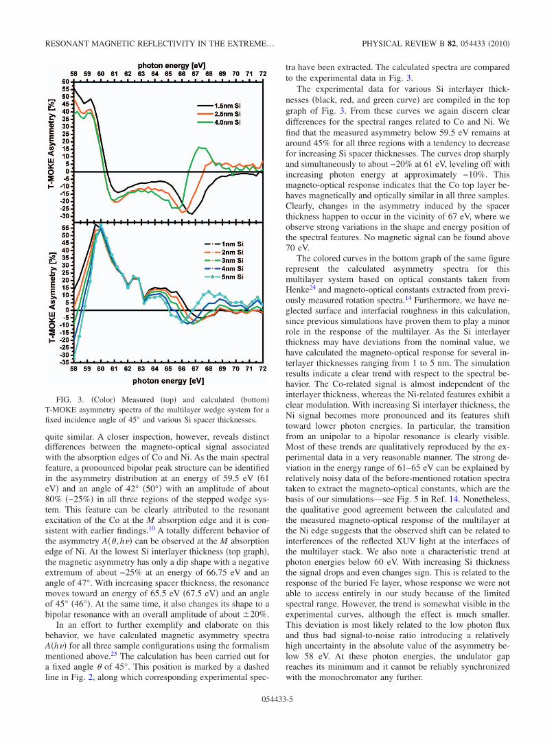
tra have been extracted. The calculated spectra are comparedto the experimental data in Fig. 3.
The experimental data for various Si interlayer thick-nesses �black, red, and green curve� are compiled in the topgraph of Fig. 3. From these curves we again discern cleardifferences for the spectral ranges related to Co and Ni. Wefind that the measured asymmetry below 59.5 eV remains ataround 45% for all three regions with a tendency to decreasefor increasing Si spacer thicknesses. The curves drop sharplyand simultaneously to about −20% at 61 eV, leveling off withincreasing photon energy at approximately −10%. Thismagneto-optical response indicates that the Co top layer be-haves magnetically and optically similar in all three samples.Clearly, changes in the asymmetry induced by the spacerthickness happen to occur in the vicinity of 67 eV, where weobserve strong variations in the shape and energy position ofthe spectral features. No magnetic signal can be found above70 eV.
The colored curves in the bottom graph of the same figurerepresent the calculated asymmetry spectra for thismultilayer system based on optical constants taken fromHenke24 and magneto-optical constants extracted from previ-ously measured rotation spectra.14 Furthermore, we have ne-glected surface and interfacial roughness in this calculation,since previous simulations have proven them to play a minorrole in the response of the multilayer. As the Si interlayerthickness may have deviations from the nominal value, wehave calculated the magneto-optical response for several in-terlayer thicknesses ranging from 1 to 5 nm. The simulationresults indicate a clear trend with respect to the spectral be-havior. The Co-related signal is almost independent of theinterlayer thickness, whereas the Ni-related features exhibit aclear modulation. With increasing Si interlayer thickness, theNi signal becomes more pronounced and its features shifttoward lower photon energies. In particular, the transitionfrom an unipolar to a bipolar resonance is clearly visible.Most of these trends are qualitatively reproduced by the ex-perimental data in a very reasonable manner. The strong de-viation in the energy range of 61–65 eV can be explained byrelatively noisy data of the before-mentioned rotation spectrataken to extract the magneto-optical constants, which are thebasis of our simulations—see Fig. 5 in Ref. 14. Nonetheless,the qualitative good agreement between the calculated andthe measured magneto-optical response of the multilayer atthe Ni edge suggests that the observed shift can be related tointerferences of the reflected XUV light at the interfaces ofthe multilayer stack. We also note a characteristic trend atphoton energies below 60 eV. With increasing Si thicknessthe signal drops and even changes sign. This is related to theresponse of the buried Fe layer, whose response we were notable to access entirely in our study because of the limitedspectral range. However, the trend is somewhat visible in theexperimental curves, although the effect is much smaller.This deviation is most likely related to the low photon fluxand thus bad signal-to-noise ratio introducing a relativelyhigh uncertainty in the absolute value of the asymmetry be-low 58 eV. At these photon energies, the undulator gapreaches its minimum and it cannot be reliably synchronizedwith the monochromator any further.
FIG. 3. �Color� Measured �top� and calculated �bottom�T-MOKE asymmetry spectra of the multilayer wedge system for afixed incidence angle of 45° and various Si spacer thicknesses.
RESONANT MAGNETIC REFLECTIVITY IN THE EXTREME… PHYSICAL REVIEW B 82, 054433 �2010�
054433-5

B. Magnetic switching behavior
Having identified maxima of the magnetic contrast at theM absorption edges of Ni and Co, the incidence angle andphoton energy can be tuned to probe the magnetic switchingbehavior element selectively, which in the case of amultilayer system translates into a layer-specific response.For this purpose, we have recorded the magnetic field depen-dence of the magneto-optical signal A��0H�. This measure-ment results in hysteresislike loops which do not, however,directly reflect the behavior of the magnetization, as the sig-nal comprises a convolution of magnetic and optical contri-butions. We will therefore call them magneto-optical loops.The loops, which are displayed in red and green in Fig. 4,have been recorded at a fixed angle of 41°, where local,element-specific maxima of the magnetic contrast are clearlyseparated, for reasons that are explained further below. Theresponse at the Co edge was taken at a photon energy of 59.5eV for all three regions �red curves�, whereas the response atthe Ni edge was recorded at a photon energy of 67.5 eV fora 1.5 nm Si spacer, at a photon energy of 67 eV for a 2.5 nmSi spacer, and at a photon energy of 66.5 eV for a 4 nm Sispacer �green curves�. These data are compared to the con-ventional Kerr loops in Fig. 1.
The top graph in Fig. 4 shows magneto-optical loops as-sociated with a sample region having a Si spacer thickness of1.5 nm. All three loops exhibit a very similar rectangularshape with a coercivity of about 40 mT. The shape of bothmagneto-optical loops taken at the absorption edges of Coand Ni, reflecting a layer-specific response, closely agreeswith that of the loop taken in the visible range, which reflectsthe collective response of the multilayer system. From thisresult we can conclude that the entire layer stack undergoes amagnetization reversal as a single magnetic unit. All ferro-magnetic layers in the stack are strongly coupled to eachother and the coupling across the Si interlayer is of ferromag-netic nature. This ferromagnetic coupling can be a conse-quence of either intrinsic interlayer exchange coupling, pin-holes or other imperfections in this very thin interlayer,which permit a direct ferromagnetic exchange interaction be-tween the magnetic layers. It remains to be seen whether thecoercive field is determined by the magnetic properties of theconstituent layers or whether it is directly related to the fer-romagnetic interlayer coupling.
The center graph in Fig. 4 displays the data taken from aregion of the sample where top and bottom magnetic layersare separated by a Si spacer of 2.5 nm. Two distinct steps anda third small step at �50 mT can be identified in the pointsymmetric hysteresis loop taken in the visible range. Thisalready points toward a more complex switching behavior,which may be related to a rather individual behavior of theferromagnetic layers and therefore a weakly coupled state inthe multilayer. In the element-selective magneto-opticalloops both steps around �75 mT agree very well with thecoercivity of the loops taken at the Ni edge. The secondswitching event around �50 mT and the third step around�15 mT, coincide with steps of the magneto-optical looptaken at the Co edge. This analysis reveals that the Ni film inthe Ni/Fe bilayer is magnetically harder and switches athigher magnetic fields. Whether this is an intrinsic property
of the Ni film at that particular thickness, or whether thecoercivity is caused by the ferromagnetic coupling to theadjacent Fe layer cannot be distinguished at this point. In anycase, it is interesting to note that the larger coercivity of theNi/Fe bottom layer is not dominating the magnetic responseof the sample with strong ferromagnetic interlayer couplingbut rather moderately increases the coercivity to a value inbetween HC
Co�15 mT and HCNi/Fe�75 mT.
The bottom graph in Fig. 4 shows loops taken at a regionwhere top and bottom ferromagnetic layers are separated bya Si spacer of 4 nm. Here, the conventional MOKE loop
FIG. 4. �Color� Magneto-optical loops of the multilayer wedgesystem taken at a Si spacer thickness of 1.5 nm �top�, 2.5 nm�middle�, and 4 nm �bottom�.
GRYCHTOL et al. PHYSICAL REVIEW B 82, 054433 �2010�
054433-6

reveals only two pronounced steps, one of which can beclearly associated with the rectangular magneto-optical looptaken at the Ni absorption edge having a coercivity ofHC
Ni/Fe�70 mT. The second step at �10 mT in theL-MOKE loop can be clearly related to the switching of theCo layer. This is the typical magnetization loop of apseudospin-valve system with a negligible magnetic cou-pling between the hard �Ni/Fe� and soft �Co� magnetic lay-ers.
Summarizing the findings and arguments given above, wecan clearly map out the switching behavior of this multilayersystem and explore it in detail. For a thin Si spacer, the layerstack is strongly ferromagnetically coupled throughout. Thesystem behaves as one layer, having a coercivity that lies inbetween the coercivities of the individual decoupled layers,HC
Co and HCNi/Fe. The region of the layer stack with a thick Si
interlayer is magnetically decoupled allowing for an inde-pendent switching of the top and bottom layers. For an in-termediate spacer thickness, the top Co layer switches firstbut it is still weakly coupled to the bottom Ni layer, whichgoverns the switching behavior because of a much highervolume magnetization. This also explains a higher coercivityand a gradual rather than abrupt switching of the Co layer ascompared to the decoupled case. Moreover, the system isprobably not homogeneous with respect to the couplingstrength. This is suggested by the small additional steps inthe Co-related loops, which can be found around �50 mT.These may be related to areas with an increased ferromag-netic coupling �for example, due to pinholes�, which switchat higher magnetic field values.
C. Magneto-optical crosstalk and interferences
It is important to note that layer-selective magneto-opticalloops, which can be directly compared with their correspond-ing classical Kerr effect counterparts, as shown in Fig. 4, canusually be obtained only for a specific set of angles andphoton energies. In order to elaborate on this finding, a set ofhysteresis loops taken at a fixed angle of 45°, in a photonenergy range of 57–68 eV and at a Si spacer thickness of 4nm is shown in Fig. 5.
By scanning the photon energy across the Co and Ni ab-sorption edges, the shape of the recorded magneto-opticalloops changes significantly. It is striking that all loops areconsidered to be a linear combination of the pristine loopsassociated with the individual switching of the Co and Nilayers. Even at local extrema of the magnetic asymmetry,namely, at 59, 61, and 66 eV—compare Fig. 3, supposedlylayer-selective magneto-optical loops leave doubt about a de-coupled state of the sample.
But this behavior can be understood by first taking intoaccount the switching of the 2 nm thin Fe layer at the verybottom of the multilayer and by second considering thestrength and character of the magnetic dichroism at the Mabsorption edges. The Fe and Ni layer, having direct contactto each other, are strongly coupled and switch simulta-neously. That is why recording a magneto-optical loop at theFe and Ni edge would yield the same shape. Due to theenergetic proximity of the M absorption edges of Fe �54 eV�,Co �60 eV�, and Ni �67 eV� and the width of the 3d bands,the electrons are excited into for magnetic contrast genera-tion, the magneto-optical response of a single element isspread over several electron volt,14 thereby reaching into theresponse of a neighboring element. Besides this crosstalk,interferences play a role in the magneto-optical response ofthe multilayer. As has been outlined above, changing thethickness of the nonmagnetic interlayer can influence themagneto-optical response of individual layers as well as themultilayer as a whole, resulting in an energetic shift of themagnetodichroic signal. As a consequence, the overallmagneto-optical response of the Co layer is influenced by theresponse of the Fe and Ni layer and vice versa resulting in anenergy-dependent superposition of the measured magneto-optical loops. Figure 5 and basic calculations reveal, for pho-ton energies below 60 eV, that the measured magneto-opticalloops can be reproduced by the weighted difference of tworectangular loops associated with the switching of the Fe andCo layers—compare layer-selective loops extracted fromFig. 4 and inserted into Fig. 5 at 59 eV. Whereas for photonenergies above 60 eV, the measured magneto-optical loopscan be reproduced by the weighted sum of two rectangularloops associated with the switching of the Ni and Colayers—compare layer-selective loops extracted from Fig. 4and inserted into Fig. 5 at 65 eV. Only by recording a loop atthe far end of the photon spectrum at 68 eV, it is possible toobtain a pristine magneto-optical loop proofing that themultilayer is magnetizationally decoupled. The weight andrelative sign of the magneto-optical loop superposition de-pends on the magneto-optical constants which can be calcu-lated for each individual layer as well as the entire multilayersystem on the basis of the magneto-optical formalism out-lined above.25 And that is also how layer selectivity can begained at arbitrary sets of angles and energies. Nevertheless,we have demonstrated in the example above that it is pref-erable to choose the angle of incidence such that themagneto-optical response of individual layers is clearly sepa-rated.
VII. SUMMARY AND CONCLUSION
We have measured resonantly enhanced reflection spectraof a polycrystalline Co/Si/Ni/Fe multilayer wedge system in
FIG. 5. �Color� Magneto-optical loops taken at an angle of 45°,a Si spacer thickness of 4 nm, and in a energy range of 57–68 eV.
RESONANT MAGNETIC REFLECTIVITY IN THE EXTREME… PHYSICAL REVIEW B 82, 054433 �2010�
054433-7

a T-MOKE geometry across the M absorptions edges of Coand Ni. We explored the character of the magnetic dichroismin the XUV and its potential for layer-selective investiga-tions. To this end we studied the magneto-optical response aswell as the switching behavior of the Co �5 nm� and Ni/Fe �8nm, 2 nm� layers as a function of the interlayer coupling byvarying the angle of incidence, the photon energy, and the Sispacer thickness.
We showed that the magnetic contrast at the M absorptionedges of Co and Ni �59.5 eV and 66.5 eV� can be as large as80% and 25%, respectively, near the Brewster angle in acomfortable 90° reflection geometry. By means of magneto-optical calculations, we were able to show that the shift oflocal magnetic contrast maxima is related to interference ef-fects in the multilayer system. By recording hysteresis loopsfor various incidence angles and photon energies, it was pos-sible to characterize the role played by magneto-optical in-terferences and crosstalk with regard to a layer-selective re-sponse of the multilayer stack. This consequently enabled usto understand the switching behavior of individually selected
layers as a function of the ferromagnetic interlayer coupling,whose strength decreases with increasing spacer thickness.
Our work lays the basis for element- and layer-selectivemagnetic investigations in this increasingly interesting spec-tral range. Especially so, as recently developed table-top softx-ray sources manage to produce ultrafast and coherent XUVpulses up to photon energies of 100 eV with moderate effort,thus opening the door for element-selective investigations ofmagnetic properties in heterogeneous systems on the femto-second and nanometer scale in a laboratory environment.
ACKNOWLEDGMENTS
We would like to thank Bernd Küpper, Jürgen Lauer,Heinz Pfeiffer, Norbert Schnitzler, and Reinert Schreiber fortheir relentless technical support, without which this workwould not have been possible. We also are grateful to theBESSY staff for their assistance in running the beamline24/7. This work was financially supported by the BMBF�Project No. 05KS7UK1�.
1 B. Hillebrands, Spin Dynamics in Confined Magnetic Structures�Springer, Berlin, 2004�, Vols. 1–3.
2 I. Žutić, J. Fabian, and S. Das Sarma, Rev. Mod. Phys. 76, 323�2004�.
3 J. Stöhr and H. C. Siegmann, Magnetism: From Fundamentals toNanoscale Dynamics �Springer, Berlin, 2006�.
4 A. Zvezdin and V. Kotov, Modern Magnetooptics and Magne-tooptical Materials �Institute of Physics, Bristol, 1997�.
5 C. Kao, J. B. Hastings, E. D. Johnson, D. P. Siddons, G. C.Smith, and G. A. Prinz, Phys. Rev. Lett. 65, 373 �1990�.
6 H.-C. Mertins, D. Abramsohn, A. Gaupp, F. Schäfers, W. Gudat,O. Zaharko, H. Grimmer, and P. M. Oppeneer, Phys. Rev. B 66,184404 �2002�.
7 F. Nolting et al., Nature �London� 405, 767 �2000�.8 C. Gutt et al., Phys. Rev. B 79, 212406 �2009�.9 C. Stamm et al., Nature Mater. 6, 740 �2007�.
10 F. U. Hillebrecht, T. Kinoshita, D. Spanke, J. Dresselhaus, C.Roth, H. B. Rose, and E. Kisker, Phys. Rev. Lett. 75, 2224�1995�.
11 M. Pretorius et al., Phys. Rev. B 55, 14133 �1997�.12 M. Sacchi, G. Panaccione, J. Vogel, A. Mirone, and G. van der
Laan, Phys. Rev. B 58, 3750 �1998�.13 M. Hecker, P. Oppeneer, S. Valencia, H.-C. Mertins, and C.
Schneider, J. Electron Spectrosc. Relat. Phenom. 144-147, 881�2005�.
14 S. Valencia, A. Gaupp, W. Gudat, H.-C. Mertins, P. M. Oppe-neer, D. Abramsohn, and C. M. Schneider, New J. Phys. 8, 254�2006�.
15 H. C. Kapteyn, M. M. Murnane, and I. P. Christov, Phys. Today58�3�, 39 �2005�.
16 X. Zhou, R. Lock, N. Wagner, W. Li, H. C. Kapteyn, and M. M.Murnane, Phys. Rev. Lett. 102, 073902 �2009�.
17 T. Popmintchev, M. Chen, A. Bahabad, M. Gerrity, P. Sidorenko,O. Cohen, I. P. Christov, M. M. Murnane, and H. C. Kapteyn,Proc. Natl. Acad. Sci. U.S.A. 106, 10516 �2009�.
18 C. La-O-Vorakiat et al., Phys. Rev. Lett. 103, 257402 �2009�.19 R. L. Sandberg, D. A. Raymondson, C. La-O-Vorakiat, A. Paul,
K. S. Raines, J. Miao, M. M. Murnane, H. C. Kapteyn, and W. F.Schlotter, Opt. Lett. 34, 1618 �2009�.
20 K. Starke, F. Heigl, A. Vollmer, M. Weiss, G. Reichardt, and G.Kaindl, Phys. Rev. Lett. 86, 3415 �2001�.
21 J. E. Prieto, F. Heigl, O. Krupin, G. Kaindl, and K. Starke, Phys.Rev. B 68, 134453 �2003�.
22 H.-C. Mertins, F. Schäfers, X. Le Cann, A. Gaupp, and W. Gu-dat, Phys. Rev. B 61, R874 �2000�.
23 S. Valencia, A. Kleibert, A. Gaupp, J. Rusz, D. Legut, J. Bans-mann, W. Gudat, and P. M. Oppeneer, Phys. Rev. Lett. 104,187401 �2010�.
24 B. Henke, E. Gullikson, and J. Davis, At. Data Nucl. Data Tables54, 181 �1993�, released by CXRO �http://henke.lbl.gov/optical_constants/�
25 S. Valencia, A. Gaupp, W. Gudat, L. Abad, L. Balcells, and B.Martinez, J. Appl. Phys. 104, 023903 �2008�.
26 R. R. Gareev, D. E. Bürgler, M. Buchmeier, D. Olligs, R.Schreiber, and P. Grünberg, Phys. Rev. Lett. 87, 157202 �2001�.
27 D. E. Bürgler, C. M. Schmidt, J. A. Wolf, T. M. Schaub, and H.J. Güntherodt, Surf. Sci. 366, 295 �1996�.
28 M. Buchmeier, R. Schreiber, D. E. Bürgler, and C. M. Schneider,Phys. Rev. B 79, 064402 �2009�.
29 R. Adam, P. Grychtol, S. Cramm, S. Valencia, and C. M.Schneider �unpublished�.
30 J. P. Hannon, G. T. Trammell, M. Blume, and D. Gibbs, Phys.Rev. Lett. 61, 1245 �1988�.
31 H. Ebert, L. Baumgarten, C. M. Schneider, and J. Kirschner,Phys. Rev. B 44, 4406 �1991�.
32 F. Meier and B. Zakharchenya, Optical Orientation, ModernProblems in Condensed Matter Sciences Vol. 8 �North-Holland,Amsterdam, 1984�.
33 P. Oppeneer, Handbook of Magnetic Materials �Elsevier, Am-sterdam, 2001�, Vol. 13.
34 O. Zaharko, P. M. Oppeneer, H. Grimmer, M. Horisberger, H.-C.Mertins, D. Abramsohn, F. Schäfers, A. Bill, and H.-B. Braun,Phys. Rev. B 66, 134406 �2002�.
GRYCHTOL et al. PHYSICAL REVIEW B 82, 054433 �2010�
054433-8
Copyright © 2022 FDOKUMEN