Regulation of System XC- and its Contribution to Cell Death
185
Marquee University e-Publications@Marquee Dissertations (2009 -) Dissertations, eses, and Professional Projects Regulation of System XC- and its Contribution to Cell Death XiaoQian Liu Marquee University Recommended Citation Liu, XiaoQian, "Regulation of System XC- and its Contribution to Cell Death" (2012). Dissertations (2009 -). Paper 234. hp://epublications.marquee.edu/dissertations_mu/234
-
Upload
khangminh22 -
Category
Documents
-
view
0 -
download
0
Transcript of Regulation of System XC- and its Contribution to Cell Death
Marquette Universitye-Publications@Marquette
Dissertations (2009 -) Dissertations, Theses, and Professional Projects
Regulation of System XC- and its Contribution toCell DeathXiaoQian LiuMarquette University
Recommended CitationLiu, XiaoQian, "Regulation of System XC- and its Contribution to Cell Death" (2012). Dissertations (2009 -). Paper 234.http://epublications.marquette.edu/dissertations_mu/234
REGULATION OF SYSTEM XC- AND ITS CONTRIBUTION TO CELL DEATH
by
XiaoQian Liu, B.S.
A Dissertation submitted to the Faculty of the Graduate School, Marquette University,
in Partial Fulfillment of the Requirements for the Degree of Doctor of Philosophy
Milwaukee, Wisconsin
December 2012
ABSTRACT REGULATION OF SYSTEM XC- AND ITS CONTRIBUTION TO CELL DEATH
XiaoQian Liu, B.S.
Marquette University, 2012
The main focus of the studies in this thesis involves examining the role of cystine/glutamate exchange (system xC-) in neuronal death in primary cortical cell culture, with an emphasis on how glial function affects neuronal cell death. System xC- is a sodium-independent transporter that mediates cystine uptake and glutamate release. It accounts for most of the cystine uptake in astrocytes in mature cultures, providing the rate limiting substrate for synthesis of the main endogenous antioxidant glutathione. The glutamate released by system xC- may lead to excessive extracellular glutamate and cause excitotoxicity.
β-N-methylamino-L-alanine (BMAA) is a non-protein amino acid that may be involved in neurodegenerative diseases. We found that BMAA induced oxidative stress by competing with cystine at system xC- leading to depletion of glutathione. BMAA also drives system xC- mediated glutamate release, which may contribute to its induction of excitotoxicity.
Fibroblast growth factor-2 (FGF-2) is involved in multiple processes in the central nervous system, including plasticity, neurogenesis, differentiation, and neuronal survival. Also, alterations in FGF-2 and its signaling have been implicated in neurodegenerative diseases and psychiatric disorders. We found that FGF-2 greatly increased cystine uptake through system xC- in astrocyte-enriched primary cultures, but not in neuronal or microglial cultures. Our data showed that FGF-2 increased cystine uptake by upregulating system xC- by acting on FGFR1, and signaling through the PI3K/Akt and MEK/ERK pathways.
FGF-2 treatment for 48 hours caused significant neuronal death only in mixed neuronal and glial cultures, but not in neuronal-enriched or astrocyte-enriched cultures. Blocking system xC-, or AMPA/kainate receptors, eliminated the neuronal death induced by FGF-2 treatment. Therefore, it is likely that 48 hour FGF-2 treatment induces AMPA receptor mediated toxicity through increased glutamate release from astrocytes due to increased system xC- function. However, we cannot exclude the possibility that FGF-2 treatment sensitizes the neurons to normal system xC- mediated glutamate release.
Together the results indicate that 1) competitive substrates of system xC-, such as BMAA, that do not lead to glutathione production are particularly toxic; and 2) upregulation of system xC- on astrocytes may be toxic to surrounding neurons.

i
ACKNOWLEDGMENTS
XiaoQian Liu, B.S.
I would like to offer my sincere appreciation to those who helped me reach this important
milestone. I would especially like to thank my family for their constant support, encouragement,
and understanding. I want to thank my mentor Doug Lobner. I am extremely grateful for your
generous support and patient guidance. Special thanks to my friends and fellow graduate students
Jon Resch, Travis Rush, and Rebecca Albano, and our “pig-lady” Julie Hjelmhaug. Thank you
all for the help throughout the years in and out of the lab. I would also like to thank all my
committee members and the entire Marquette University Biological Sciences and Biomedical
Sciences departments. Thank you all.
ii
TABLE OF CONTENTS
LIST OF FIGURES…………………………………………………………………….. vi
LIST OF ABBREVIATIONS ……………………………………………………………ix
CHAPTER I
GENERAL INTRODUCTION ........................................................................................... 1
Glutamate Neurotransmission ................................................................................. 4
Glutamate Receptors ................................................................................... 4
AMPA/Kainate Receptors .............................................................. 5
NMDA Receptors ........................................................................... 6
mGluRs ........................................................................................... 7
Excitotoxicity .............................................................................................. 7
Regulation of Extracellular Glutamate by Astrocytic Glutamate Transporters .................................................................................... 8
Synaptic and Extrasynaptic Compartments .................................... 9
Neuron-Glia Interaction ........................................................................................ 10
Regulation of Astrocytes by Neurons ....................................................... 11
Regulation of Neurons by Astrocytes ....................................................... 12
Astrocyte and Neuron Metabolic Coupling .............................................. 13
Oxidative Stress and Glutathione in the Brain ...................................................... 14
Oxidative Stress ........................................................................................ 14
GSH in the Brain ....................................................................................... 16
System xC- ............................................................................................................. 18
Structure of System xC- ............................................................................. 18
iii
Function of System xC- ............................................................................. 20
Regulation of System xC- .......................................................................... 21
Nrf2-ARE Pathway ....................................................................... 22
eIF2-ATF4-AARE Pathway ......................................................... 24
NF-κB ........................................................................................... 28
Activator protein 1 (AP-1) ............................................................ 29
Hypoxia inducible Factor (HIF) .................................................... 29
cAMP ............................................................................................ 29
Growth Factors .............................................................................. 30
FGF-2 Receptors and Intracellular Signaling ....................................................... 30
FGF-2 Regulation, Function and Dysfunction in Diseases ...................... 33
FGF-2 in Ischemia/Stroke ............................................................. 33
FGF2 in Major Depression/Anxiety ............................................. 34
FGF-2 and Schizophrenia ............................................................. 35
FGF-2 and Addiction .................................................................... 37
System xC- Contribution to Cell Death/Survival .................................................. 37
Oxidative Glutamate Toxicity ................................................................... 37
Increased System xC- Activity: Protection Against Oxidative Stress ....... 38
Increased System xC- Activity: Enhanced Excitotoxicity ......................... 39
Potential Role of System xC- in Neurological Disorders ...................................... 39
Alzheimer’s disease (AD) ......................................................................... 39
Parkinson’s Disease (PD) ......................................................................... 40
Amyotrophic Lateral Sclerosis–Parkinsonism Dementia Complex (ALS-PDC) ............................................................................................... 41
iv
β-N-methylamino-L-alanine (BMAA) ......................................... 42
Heavy Metals ................................................................................ 44
Ischemia/Stroke ......................................................................................... 44
Glioma ....................................................................................................... 45
Addiction ................................................................................................... 46
General Methods ................................................................................................... 48
Cortical Culture ......................................................................................... 49
Cell Death Assay ....................................................................................... 50
14C-cystine Uptake .................................................................................... 51
CHAPTER II
Β-N-METHYLAMINO-L-ALANINE INDUCES OXIDATIVE STRESS AND GLUTAMATE RELEASE THROUGH ACTION ON SYSTEM XC− .......................... 53
Abstract ................................................................................................................. 54
Introduction ........................................................................................................... 55
Materials and Methods .......................................................................................... 57
Results ................................................................................................................... 61
BMAA Toxicity Involves the NMDA Receptor, mGluR5 Receptor, and Oxidative Stress ........................................................................................ 61
BMAA Induces Oxidative Stress Through Inhibition of System xC- Mediated Cystine Uptake .......................................................................... 63
Discussion ............................................................................................................. 67
CHAPTER III
FUNCTIONAL UPREGULATION OF SYSTEM XC- BY FIBROBLAST GROWTH FACTOR-2 ....................................................................................................................... 72
v
Abstract ................................................................................................................. 73
Introduction ........................................................................................................... 74
Materials and Methods .......................................................................................... 76
Results ................................................................................................................... 79
Discussion ............................................................................................................. 87
CHAPTER IV
FGF-2 INDUCES NEURONAL DEATH ........................................................................ 92
Abstract ................................................................................................................. 93
Introduction ........................................................................................................... 94
Materials and Methods .......................................................................................... 97
Results ................................................................................................................. 100
Discussion ........................................................................................................... 105
Conclusions ......................................................................................................... 110
CHAPTER V
GENERAL DISCUSSION ............................................................................................. 111
Interaction of FGF-2 and BMAA on Neuronal Death ........................................ 112
Controversies Regarding BMAA ........................................................................ 114
Is BMAA a Potent Neurotoxin? .............................................................. 115
Measurement of BMAA ......................................................................... 120
BMAA in Animal Models ...................................................................... 121
Is Action on System xC- an Important Mechanism of BMAA Toxicity? 122
Regulation of System xC- by FGF-2 ................................................................... 122
FGF-2 in the Central Nervous System .................................................... 123
vi
FGF-2 and Oxidative Stress .................................................................... 124
FGF-2 and Glutamate ............................................................................. 125
Toxicity of FGF-2 Mediated by System xC- ....................................................... 127
Is System xC- Activity Toxic to Neurons? .......................................................... 130
Questions Regarding System xC- Studies ........................................................... 131
Molecular Properties of System xC- ........................................................ 131
Relevance and Limitations of Translating in vitro Studies to in vivo: ... 133
CONCLUSION ............................................................................................................... 135
BIBLIOGRAPHY……………………………………………………………………....136
vii
LIST OF FIGURES
FIGURE 1.1. Schematic diagram illustrating the function of system xC- and the
interaction between neuron and astrocyte. .................................................................. 3 FIGURE 1.2. Glutamate receptor subtypes ....................................................................... 5 FIGURE 1.3. A typical glutamate synapse. ..................................................................... 11 FIGURE 1.4. Schematic representation of glutamate cycling and glucose metabolism
coupling. .................................................................................................................... 14 FIGURE 1.5. Diagram illustrating the different pathways for handling O2
-. .................. 15 FIGURE 1.6. GSH synthesis and cycling in the central nervous system. ....................... 17 FIGURE 1.7. The structure of system xC- ....................................................................... 19 FIGURE 1.8. Nrf2-ARE pathway .................................................................................... 23 FIGURE 1.9. Regulation of translation initiation by eIF2 phosphorylation. ................... 26 FIGURE 1.10. Regulation of translation initiation by eIF2B phosphorylation. .............. 27 FIGURE 1.11. Expression of mRNA for FGFR1 and FGFR2 in cerebral cortex,
neuronal-enriched cultures, and glial-enriched cultures. .......................................... 31 FIGURE 1.12. FGF-FGFR1 intracellular signaling pathways. ....................................... 33 FIGURE 1.13. Structural similarities between the neurotransmitter L-glutamate and the
neurotoxic carbonate adduct of BMAA. ................................................................... 43 FIGURE 1.14. 24 hour FGF-2 treatment on 2 min and 20 min cystine uptake ............... 52 FIGURE 2.1. BMAA induced toxicity occurs through multiple mechanisms. ............... 62 FIGURE 2.2. BMAA inhibits system xC- mediated cystine uptake. .............................. 63 FIGURE 2.3. BMAA decreases cellular glutathione levels. ........................................... 64 FIGURE 2.4. BMAA induces oxidative stress. ............................................................... 65 FIGURE 2.5. BMAA and cystine stimulate glutamate release. ...................................... 66 FIGURE 2.6. Cystine-induced neuronal death is attenuated by MK-801 and MPEP. .... 67
viii
FIGURE 3.1. Growth factor effects on cystine uptake in cortical cultures. ................... 79 FIGURE 3.2. Growth factor effects on cystine uptake spinal cord glial cultures. .......... 80 FIGURE 3.3. Effects of FGF-2 on cystine uptake in neuronal-enriched cultures (A),
astrocyte-enriched cultures (B), and microglial cultures (C). ................................... 82 FIGURE 3.4. FGF-2 induced increase in cystine uptake is mediated by system xC-. .... 83 FIGURE 3.4. Time and protein synthesis dependent changes in cystine uptake induced
by FGF-2 treatment on astrocyte-enriched cultures. ................................................. 84 FIGURE 3.6. Kinetic analysis of FGF-2 effects on cystine uptake. ............................... 85 FIGURE 3.7. Time course of changes in xCT mRNA in astrocyte-enriched cultures
assessed by RT-qPCR following different durations of FGF-2 (100 ng/ml) treatment.................................................................................................................................... 85
FIGURE 3.8. FGF-2 stimulation of cystine uptake is mediated by activation of FGFR1
and both the MEK/ERK and PI3-kinase pathways. .................................................. 87 FIGURE 4.1. FGF-2 induced toxicity occurs in mixed neuronal and glial cultures after
48 hour treatment. ................................................................................................... 100 FIGURE 4.2. FGF-2 does not induce toxicity after 48 hour treatment on either neuronal-
enriched cultures or astrocyte-enriched cultures. .................................................... 101 FIGURE 4.3. FGF-2 induced increase in cystine uptake is mediated by system xC-. ... 102 FIGURE 4.4. FGF-2 induced neuronal death is prevented by blocking system xC-. .... 103 FIGURE 4.5. Effects of the AMPA receptor antagonist NBQX and the NMDA receptor
antagonist memantine on neuronal death induced by 48 hour exposure to FGF-2 or glutamate. ................................................................................................................ 104
FIGURE 4.6. FGF-2 induced toxicity is not mediated by calcium-permeable AMPA
receptors. ................................................................................................................. 105 FIGURE 5.1. Concentration response of BMAA induced neuronal death after FGF-2
pretreatment. ........................................................................................................... 113 FIGURE 5.2. Cholinergic, but not dopaminergic neurons are selectively sensitive to
BMAA toxicity. ...................................................................................................... 117
ix
FIGURE 5.3. MK801 (10 µM) and trolox (100 µM) provide protection against BMAA toxicity to total septal neurons (A), while NBQX (20 µM) provides protection of cholinergic neurons (B). .......................................................................................... 118
FIGURE 5.4. FGF-2 induced increase in cystine uptake is not oxidative stress dependent.
................................................................................................................................. 125 FIGURE 5.5. Short-term synaptic glutamate release increases system xC- activity. ..... 126 FIGURE 5.7. 24 hour exposure to neuronal-conditioned media (NCM) increases system
xC- mediated cystine uptake in astrocytes. .............................................................. 134
x
LIST OF ABBREVIATIONS
6-OHDA 6-hydroxydopamine AAR amino acid response AARE amino acid response element AD Alzheimer’s disease ALS-PDC Amyotrophic lateral sclerosis–Parkinsonism dementia complex AMPA 2-amino-3-(5-methyl-3-oxo-1,2- oxazol-4-yl)propanoic acid APP amyloid-β precursor protein ARE antioxidant responsive element ATF activating transcription factor Aβ amyloid-β BDNF brain derived neurotrophic factor BMAA β-N-methylamino-L-alanine BMAA β-N-methylamino-L-alanine BSO buthionine sulphoximine CHX cycloheximide CPG S-4-carboxyphenyl glycine CysGly cysteinylglycine DAG diacyl glycerol DAI double-stranded RNA-activated inhibitor dbcAMP N(6),2'-O-dibutyryladenosine 3':5' cyclic monophosphate DCF dichlorofluorescein EAAT excitatory amino acid transporter eIF2 eukaryotic initiation factor 2 EpRE electrophile response element ER endoplasmic-reticulum ERK extracellular signal-regulated kinase FGF-2 fibroblast growth factor 2, basic fibroblast growth factor FGFR FGF receptor FRS2 fibroblast growth factor receptor substrate 2 GCN2 general control nonderepressible protein 2 GEF guanine nucleotide exchange factor GSH glutathione GSK3β glycogen synthetase 3 β GSSG glutathione disulfide H2O2 hydrogen peroxide HIF hypoxia inducible factor HRI heme-regulated inhibitor IGF-1 Insulin-like growth factor 1 IP3 inositol 1,4,5-trisphosphate LPS lipopolysaccharide MeHg methylmercury
xi
MEK mitogen-activated protein kinase kinase MEM memantine mGluRs metabotropic glutamate receptors MK-801 [5R,10S]-[+]-5-methyl-10,11- dihydro-5H-dibenzo[a,d]cyclohepten-5,10-imine MPEP 6-methyl-2-[phenylethynel]-pyridine MPP+ 1-methyl-4-phenylpyridinium ion NASPM 1-naphthyl acetyl spermine NBQX 2,3-dihydroxy-6-nitro-7-sulfamoyl-benzo[f]quinoxaline-2,3-dione NCM neuronal-conditioned media NMDA N-methyl-D-Aspartate Nrf2 eythroid 2-related factor 2 NRG neuregulin NT-4 neurotrophin-4 O2- superoxide ODAP b-N-oxalyl-L-a,b-diaminopropionic acid PD Parkinson’s disease PERK PKR-like endoplasmic-reticulum-localized eIF2α kinase PI3-kinase phosphatidylinositol 3 kinase PIP2 phosphatidylinositol 4,5-bisphophate PKA protein kinase A PKB, Akt activate protein kinase B PKC protein kinase C PKR protein kinase R PLC phospholipase C ROS reactive oxygen species RT-qPCR reverse transcription quantitative real-time PCR Slc7a11 18 solute carrier family 7, member 11 SOD superoxide dismutase system XAG sodium-dependent glutamate/aspartate/cysteine transporter system xC- cystine/glutamate antiporter TGF transforming growth factor

1
CHAPTER I
GENERAL INTRODUCTION

2
OVERVIEW
The brain is an extremely complex organ with complicated and specific
interactions between multiple cell types and regions. However, neuroscience research
has historically focused primarily on neurons. Glial cells have been traditionally
considered as merely supporting cells. In the last couple of decades, as our knowledge of
the glial cells has dramatically expanded, we now know that the function of glial cells
extends far beyond just supporting neurons. Growing evidence suggests that neuronal
and glial cells communicate through neurotransmitters, neuromodulators, and growth
factors, and this bidirectional communication is critical for normal function of the brain.
The aim of this thesis is to explore the interaction between neurons and glia, and
how that interaction regulates neuronal fate, with a particular emphasis on the
involvement of the cystine/glutamate antiporter (system xC-) (Fig. 1.1). System xC- is
mainly expressed on astrocytes and it is the main route of cystine uptake in these cells.
This cystine uptake is the critical step in synthesizing the major antioxidant glutathione.
Astrocytes then release glutathione, and other cysteine containing molecules, to supply
neurons with cysteine for them to produce glutathione. With every molecule of cystine
uptake into the astrocytes, one molecule of glutamate is exchanged out of the cell. This
extrasynaptic, nonvesicular, release of glutamate not only serves to regulate synaptic
function, but also when excessive, can over-activate glutamatergic receptors on the
neurons to cause excitotoxicity. Both oxidative stress and excitotoxicity have been
implicated in various neurodegenerative diseases, as well as psychiatric disorders. Thus,
it is possible that system xC- plays a role in these diseases.
3
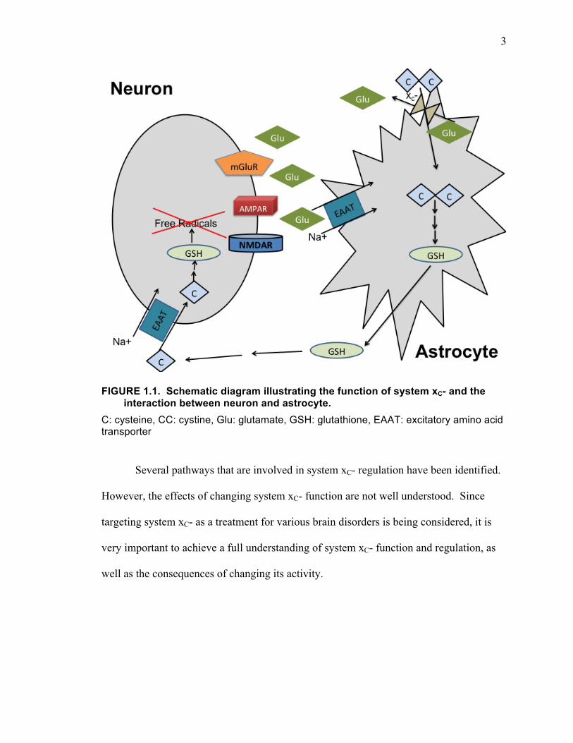
FIGURE 1.1. Schematic diagram illustrating the function of system xC- and the interaction between neuron and astrocyte.
C: cysteine, CC: cystine, Glu: glutamate, GSH: glutathione, EAAT: excitatory amino acid transporter
Several pathways that are involved in system xC- regulation have been identified.
However, the effects of changing system xC- function are not well understood. Since
targeting system xC- as a treatment for various brain disorders is being considered, it is
very important to achieve a full understanding of system xC- function and regulation, as
well as the consequences of changing its activity.
4
GLUTAMATE NEUROTRANSMISSION
Glutamate is the most important excitatory neurotransmitter in the brain (Fonnum,
1984). It is also a precursor for the most important inhibitory neurotransmitter GABA
(Petroff, 2002; Schousboe and Waagepetersen, 2007), and is a component of glutathione,
one of the major antioxidants in the brain (Meister and Anderson, 1983; Dringen et al.,
2000). Glutathione can be synthesized de novo from glucose in astrocytes via the Krebs
cycle, leading to the release of glutamine from astrocytes, which is taken up by neurons
and hydrolyzed into glutamate by glutaminase (Erecińska and Silver, 1990). Glutamate
can activate a large family of receptors existing on neurons and astrocytes leading to
signal transmission.
Glutamate receptors
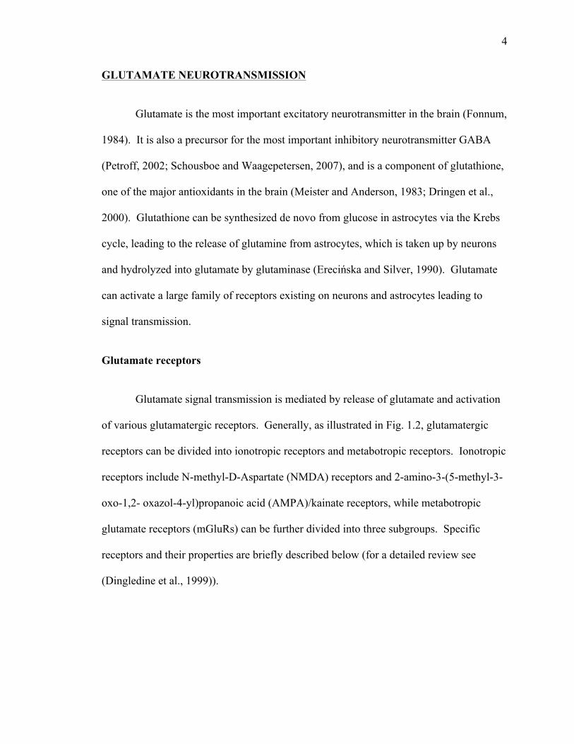
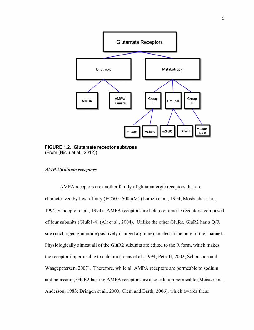
Glutamate signal transmission is mediated by release of glutamate and activation
of various glutamatergic receptors. Generally, as illustrated in Fig. 1.2, glutamatergic
receptors can be divided into ionotropic receptors and metabotropic receptors. Ionotropic
receptors include N-methyl-D-Aspartate (NMDA) receptors and 2-amino-3-(5-methyl-3-
oxo-1,2- oxazol-4-yl)propanoic acid (AMPA)/kainate receptors, while metabotropic
glutamate receptors (mGluRs) can be further divided into three subgroups. Specific
receptors and their properties are briefly described below (for a detailed review see
(Dingledine et al., 1999)).
5
FIGURE 1.2. Glutamate receptor subtypes (From (Niciu et al., 2012))
AMPA/Kainate receptors
AMPA receptors are another family of glutamatergic receptors that are
characterized by low affinity (EC50 ~ 500 µM) (Lomeli et al., 1994; Mosbacher et al.,
1994; Schoepfer et al., 1994). AMPA receptors are heterotetrameric receptors composed
of four subunits (GluR1-4) (Alt et al., 2004). Unlike the other GluRs, GluR2 has a Q/R
site (uncharged glutamine/positively charged arginine) located in the pore of the channel.
Physiologically almost all of the GluR2 subunits are edited to the R form, which makes
the receptor impermeable to calcium (Jonas et al., 1994; Petroff, 2002; Schousboe and
Waagepetersen, 2007). Therefore, while all AMPA receptors are permeable to sodium
and potassium, GluR2 lacking AMPA receptors are also calcium permeable (Meister and
Anderson, 1983; Dringen et al., 2000; Clem and Barth, 2006), which awards these
intriguing features of ionotropic glutamate receptors is their diversity ofchannel properties based on subunit composition and expression profilein the mammalian brain.
5.1.1. NMDA receptorsNMDA receptors have the highest affinity for glutamate (EC50 1 μM).
Three families of NMDA receptor subunits have been identified: (1)NR1, (2) NR2A-D and (3) NR3A-B. Via in situ hybridization studies, NR1expression appears to be ubiquitous and obligatory in the brain; it iscritical for neurodevelopment, as NR1 knockout mice die shortly afterbirth due to respiratory demise. Interestingly, hippocampal CA1-specificNR1-knockout mice display grossly normal development but impairedlong-termpotentiation (LTP), themolecular and electrophysiological cor-relate of learning and memory in CA1 hippocampal pyramidal neuronsand impaired spatial memory in the Morris water maze (Tsien et al.,1996). NR2 mRNA displays differential expression and appears to bedevelopmentally-regulated (Monyer et al., 1994). NR2A expression pre-dominates in the neocortex and hippocampus while NR2B is primarilyexpressed in the forebrain. In contrast, NR2C and NR2D are intenselyexpressed in the cerebellum and diencephalon/lower brain stem(Nakanishi, 1992). NR3A is predominantly expressed in the neocortexand displays neurodevelopmental regulation; dysregulated NR3A devel-opment has been proposed to contribute to the pathogenesis of schizo-phrenia (Das et al., 1998; Henson et al., 2008). Finally, NR3B mRNAexpression is evident in the brainstem and alphamotor neurons of spinalcord (Chatterton et al., 2002; Matsuda et al., 2003; Matsuda et al., 2002;Nishi et al., 2001). More recently, NR3B has been detected in the cerebel-lum and hippocampus (Andersson et al., 2001; Bendel et al., 2005).
NMDA receptors are among the most tightly regulated in themammalian brain and unique in requiring co-agonists for activation. Atleast six binding sites have been identified that regulate theprobability of ion channel opening, viz., sites for two obligatory co-ligands (glutamate and glycine), polyamines and cations (Mg2+, Zn2+
and H+). NMDA receptor ligands are short-chain dicarboxlic aminoacids (NMDA, glutamate, aspartate, etc.). Glutamate, the most potentneurochemical agonist identified in the CNS, and several competitiveantagonists of the NMDA receptor including D-2-amino-5-phosphono-pentanoic acid (D-AP5) and 3-(2-carboxypiperazin-4-yl)1-propeny-1-
phosphonic acid (2R-CPPene) bind to the NR2 subunit of the tetramericreceptor complex. In contrast, glycine binds to a site on the NR1 subunit(Dingledine et al., 1999; Kleckner and Dingledine, 1988). The glycine-binding site on the NR1 subunit has gained clinical significance due toD-cycloserine's binding at the same glycineB site. D-cycloserine is a par-tial agonist that has beenproposed as a novel neuromodulatory agent toenhance the efficacy of evidence-based psychotherapies like exposureand response prevention in anxiety disorders (Danysz and Parsons,1998; Krystal et al., 2009; Sheinin et al., 2001). Glycine transport re-quires the activity of specific glycine transporters (GlyT). Two suchtransporters have been identified to date, GlyT1 andGlyT2. Recent stud-ies suggest GlyT inhibitorsmayprovide an efficacious augmenting strat-egy in treatment-refractory schizophrenia (Lane et al., 2006; Lane et al.,2010).
Extracellular Mg2+ acts as an open-channel, voltage-dependent“pore blocker” to preclude cation flux (Nowak et al., 1984). Interestingly,Zn2+, while also a divalent cation, does not block the pore of the NMDAreceptor. Instead, Zn2+ is an important allosteric modulator of some glu-tamate receptors and colocalizes to synaptic vesicles and is co-releasedwith glutamate in select populations of synaptic vesicles, which possiblyprovides an additionalmechanism to regulate glutamate receptor activa-tion. Several additional NMDA receptor antagonists also exert their influ-ence in an analogous voltage-dependent manner, e.g. phencyclidine(PCP), ketamine and MK-801. These noncompetitive antagonists haverecently garnered significant attention both for their psychotomimetic(Balla et al., 2001; Javitt, 2007; Javitt et al., 2004; Krystal et al., 1994;Moghaddam and Adams, 1998; Patil et al., 2007; Umbricht et al., 2000)and rapidly-acting antidepressant-like properties (aan het Rot et al.,2010; Berman et al., 2000; Diazgranados et al., 2010; Mathew et al.,2009; Price et al., 2009; Valentine et al., 2011; Zarate et al., 2006).
Hydrogen ions (H+) are also critical endogenous allosteric modula-tors of glutamate receptors. At physiological pH, the presence of H+
decreases the frequency of channel opening due to H+ binding toNR2B. The polyamine regulatory sites of ionotropic glutamate receptorsalso play an important pH-dependent modulatory role. The binding ofpolyamines (spermine, spermidine) relieves the H+-mediated blockand increases cation flux; however, the effect of polyamines reversesat higher concentrations (Traynelis et al., 1995). These pH-dependent
Fig. 2. Glutamate receptor subtypes.
659M.J. Niciu et al. / Pharmacology, Biochemistry and Behavior 100 (2012) 656–664
6
receptors the important property of increasing intracellular calcium levels when they are
activated (Erecińska and Silver, 1990; Schneggenburger et al., 1993).
Kainate receptors are tetramers of GluR5, GluR6, GluR7, KA1, and KA2
(Dingledine et al., 1999). Similar to AMPA receptors, they are ion channels that are
permeable to sodium and potassium (Dingledine et al., 1999; Niciu et al., 2012).
However, the functions of kainate receptors are not well defined.
NMDA receptors
NMDA receptors are another important ionotropic receptor subtype that are
usually heteromers of GluN1 and GluN2 (GluN2A, GluN2B) subunits in mature brain
(Béhé et al., 1995; Premkumar and Auerbach, 1997). Compared to AMPA receptors,
NMDA receptors are characterized by a high affinity (EC50 ~1 µM) for glutamate
(Patneau and Mayer, 1990; Burnashev et al., 1995). However, under physiological
conditions, they are normally blocked by magnesium at negative membrane potential
(Nowak et al., 1984). During normal synaptic activity, AMPA receptors must be
activated first to depolarize the cell membrane, which removes the magnesium block
(Nowak et al., 1984; Dingledine et al., 1999). NMDA receptor activation also requires
binding of a co-agonist, such as glycine or D-serine (Johnson and Ascher, 1987; Schell et
al., 1995). NMDA receptors are nonspecific cation channels that are permeable to
sodium, potassium, and calcium (Grienberger and Konnerth, 2012), with the permeability
to calcium distinguishing them from most AMPA/Kainate receptors.
7
mGluRs
mGluRs are G-protein coupled receptors that exist on neurons and glial cells
(Conn and Pin, 1997; Ferraguti and Shigemoto, 2006; Kim et al., 2008). Like all G-
protein coupled receptors, mGluRs have 7 transmembrane spanning domains, an agonist
binding domain (N-terminus), as well as an intracellular domain (C-terminus) that
couples to different G-proteins (Niciu et al., 2012). To date, there are 8 known family
members as illustrated in Fig. 1.2: mGluR1-8. They are divided into three subfamilies:
Group 1 (mGluR1 and 5) that are coupled to Gq, Group II (mGluR2 and 3) and Group III
(mGluR 4, 6, 7 and 8) that are coupled to Gi. Activation of Gq leads to activation of
phospholipase C (PLC), which cleaves phosphatidylinositol 4,5-bisphophate (PIP2) to
secondary messengers diacyl glycerol (DAG) and inositol 1,4,5-trisphosphate (IP3),
which then lead to the increase in intracellular free calcium levels and activation of
calcium dependent protein kinases such as protein kinase C (PKC) (Miller et al., 1995;
Conn and Pin, 1997). On the other hand, activation of Gi leads to inhibition of adenylyl
cyclase, decreased cAMP, and decreased activation of protein kinase A (PKA) (Winder
and Conn, 1993; Niciu et al., 2012). Group I mGluRs are mainly localized to post
synaptic and glial membranes, while the majority of Group II mGluRs are localized to
presynaptic membranes and provide an autoinhibition mechanism for neurotransmitter
release.
Excitotoxicity
Although glutamate transmission is essential to normal brain function, excessive
extracellular glutamate can cause excitotoxicity. Excitotoxicity is typically caused by
8
over-activation of glutamatergic receptors, especially NMDA receptors due to their
permeability to calcium and slower inactivation, leading to excessive calcium influx into
the cells to trigger cell death (Choi, 1987). This type of neuronal death can occur in
conditions such as stroke, traumatic brain injury, and neurodegenerative diseases (Bains
and Shaw, 1997; Choi, 1998).
In certain situations, over-activation of AMPA receptors can cause excitotoxicity
as well. AMPA receptor over-activation by addition of AMPA, a direct agonist for
AMPA receptors, has been shown to be toxic to oligodendrocytes (McDonald et al.,
1998). The general AMPA receptor antagonist 2,3-dihydroxy-6-nitro-7-sulfamoyl-
benzo[f]quinoxaline-2,3-dione (NBQX) has been shown to be protective to
oligodendrocytes in multiple sclerosis models (Pitt et al., 2000; Smith et al., 2000).
Oligodendrocyte toxicity often appears to be mediated by calcium permeable GluR2
lacking AMPA receptors because it is prevented by the selective antagonist 1-naphthyl
acetyl spermine (NASPM) (Yoshioka et al., 1996; Bannerman et al., 2007).
Regulation of extracellular glutamate by astrocytic glutamate transporters
Total glutamate concentration is extremely high in the brain (10 mM) (Erecińska
and Silver, 1990), but the extracellular glutamate concentration is very low (below 10
µM) (Ronne-Engström et al., 1995; Baker et al., 2003; Rodriguez et al., 2012). The
extracellular glutamate is tightly regulated by the excitatory amino acid transporters
(EAATs). EAATs mediate sodium-dependent high-affinity glutamate uptake, which is
driven by the sodium concentration gradients: for every glutamate taken up, three
molecules of sodium enter the cell while one molecule of potassium exits the cell
9
(Barbour et al., 1988; Zerangue and Kavanaugh, 1996a). Astrocytes with abundant
glutamate transporters EAAT1/GLAST and EAAT2/GLT-1 activity-dependently
ensheath glutamatergic synapses (Ventura and Harris, 1999; Witcher et al., 2010), where
they are involved in clearing extracellular glutamate to avoid unwanted prolonged
synaptic activation and excitotoxicity (Amara, 1992; Kanai and Hediger, 1992; Storck et
al., 1992; Rothstein et al., 1994; 1996; Diamond and Jahr, 1997; Lehre and Danbolt,
1998). Glutamate taken up by astrocytes is converted into glutamine by glutamine
synthetase. Glutamine, in turn, can be released and taken up by neurons to synthesize
glutamate. This glutamate-glutamine cycle between neurons and astrocytes ensures
signaling specificity and a fast turnover rate (Sibson et al., 1998).
Synaptic and extrasynaptic compartments
Another important function of astrocytes ensheathing synapses is that they
spatially separate synaptic and extrasynaptic compartments of the extracellular space.
Astrocytic EAATs provide efficient buffering and clearance of glutamate to prevent
spillover and cross-talk between different synapses and compartments (Rothstein et al.,
1996; Asztely et al., 1997; Rusakov and Kullmann, 1998).
Synaptic and extrasynaptic receptors seem to have different functions; the most
studied example are the synaptic and extrasynaptic NMDA receptors. It has become an
increasingly accepted point of view that synaptic NMDA receptor activation is
neuroprotective, while extrasynaptic NMDA receptor activation turns off the
neuroprotective synaptic NMDA receptor activation and also activates intracellular
pathways that lead to neuronal death (Hardingham et al., 2002; Riccio and Ginty, 2002;
10
Ivanov et al., 2006; Léveillé et al., 2008; Xu et al., 2009; Hardingham and Bading, 2010).
Drugs that preferentially block extrasynaptic NMDA receptors have received particular
attention as potential treatments for neurodisorders. For example, memantine (MEM),
which is an FDA approved drug for late stage Alzheimer’s disease, has been shown to
have selectivity for extrasynaptic NMDA receptors (Lipton and Chen, 2004; Xia et al.,
2010). MEM is an open channel blocker at NMDA receptors. At low doses, it does not
accumulate in the synaptic cleft to interfere with synaptic NMDA receptor mediated
signaling. However, it does antagonize extrasynaptic NMDA receptors that are
hyperactive due to increased extrasynaptic glutamate levels in diseased brains (Lipton
and Chen, 2004; Chen and Lipton, 2006; Xia et al., 2010).
NEURON-GLIA INTERACTION
Bidirectional communication between astrocytes and neuronal cells is necessary
for the normal functioning of the nervous system during signal processing. Some
interactions between neurons and glia are discussed below with a focus on a typical
glutamatergic synapse as shown in Fig. 1.3.
11
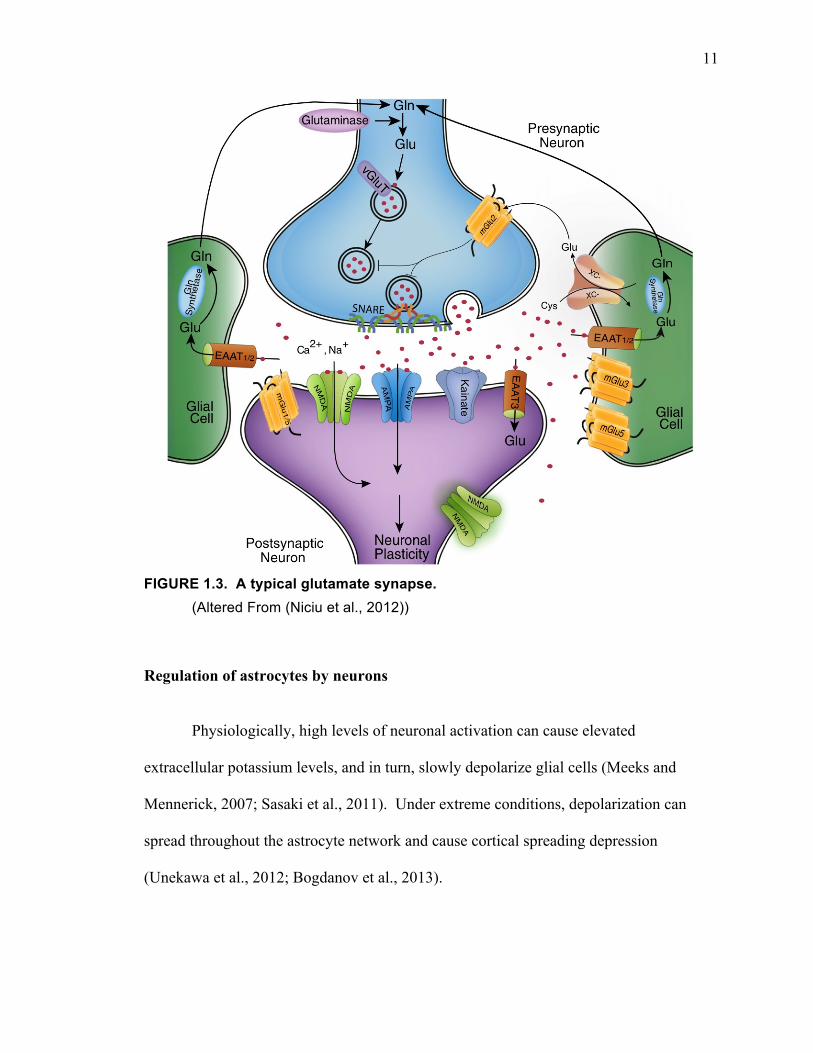
FIGURE 1.3. A typical glutamate synapse.
(Altered From (Niciu et al., 2012))
Regulation of astrocytes by neurons
Physiologically, high levels of neuronal activation can cause elevated
extracellular potassium levels, and in turn, slowly depolarize glial cells (Meeks and
Mennerick, 2007; Sasaki et al., 2011). Under extreme conditions, depolarization can
spread throughout the astrocyte network and cause cortical spreading depression
(Unekawa et al., 2012; Bogdanov et al., 2013).
12
Similar to neurons, astrocytes also express a variety of receptors to respond to
neurotransmitter release, such as mGluRs (Venance et al., 1997), nicotinic acetylcholine
receptors (Oikawa et al., 2005), adrenoceptor , P2 receptors of the P2X (ligand-gated
cationic channels) and P2Y (G-protein coupled receptors) types (Butt, 2011; Köles et al.,
2011). Most of these astrocytic receptors are Gq-protein coupled receptors. Unlike
neurons, astrocytes do not generate action potentials, However, activation of these
astrocytic Gq-protein coupled receptors by neurotransmitters can lead to elevated
intracellular calcium levels (Venance et al., 1997; Agulhon et al., 2008). Astrocytes also
express some ionotropic glutamate receptors (Seifert and Steinhäuser, 2001), although
their functional significance is largely unknown.
Regulation of neurons by astrocytes
Each astrocyte is believed to have its own territory, and within that territory, it
may interact with 140,000, or more, synapses and neuronal processes (Benarroch, 2009).
Therefore, individual astrocytes are potentially capable of coordinating a large amount of
neuronal activity (Poskanzer and Yuste, 2011). Astrocytes are also connected to each
other through gap junctions, which allows fast chemical and electrical communication
among astrocytes, enabling them to function as a network. Thus, activation of a single
astrocyte can spread to an extended surrounding area and potentially regulate the function
of multiple neighboring neurons (Cornell-Bell et al., 1990).
Astrocytes can also release gliotransmitters (such as glutamate, ATP, D-serine
etc.) to regulate neuronal function and synaptic plasticity (Fellin et al., 2006; Butt, 2011;
Parpura et al., 2012). It is known that increased calcium levels in astrocytes, like that in
13
neurons, can trigger the fusion of vesicles containing gliotransmitters with the plasma
membrane (Bezzi et al., 1998; 2004; Kreft et al., 2004). Non-vesicular release of
glutamate has also been suggested. A recent study describes a channel mediated release
of gliotransmitter (<900 Da), such as glutamate (Duan et al., 2003). System xC- mediates
a selective non-vesicular release of glutamate. It is believed that glutamate released from
astrocytes mainly activates the extrasynaptic pool of glutamatergic receptors (Xi et al.,
2002).
Astrocyte and neuron metabolic coupling
As the largest population of cells in the nervous system, astrocytes are crucial in
maintaining normal glutamate transmission (Ye and Sontheimer, 2002; Huang et al.,
2004; López-Bayghen and Ortega, 2011). Glucose is the major substrate for brain energy
production. Astrocytes take up glucose from their endfeet on capillaries (Magistretti and
Pellerin, 1996; Edvinsson and Krause, 2002), metabolize it, and release L-lactate and
pyruvate as energy sources for neurons (Dringen et al., 1993). Metabolites from glucose
can also be further processed to produce essential neurotransmitters such as GABA and
glutamate (Magistretti and Pellerin, 1996). Glutamate uptake and glucose utilization by
astrocytes are tightly coupled as illustrated in Fig 1.4.
14
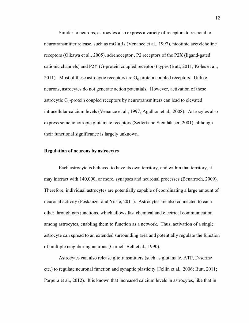
FIGURE 1.4. Schematic representation of glutamate cycling and glucose metabolism coupling.
At glutamatergic synapses, excessive extracellular glutamate is removed by a glutamate uptake system located primarily on astrocytes. Glutamate is cotransported with Na+, resulting in an increase in the intracellular concentration of Na+ in astrocytes leading to activation of the Na+/K+ ATPase pump. The pump utilizes ATP, which is provided by membrane-bound glycolytic enzymes. This demand for ATP activates glycolysis in astrocytes, resulting in the production of lactate. Lactate, once released can be taken up by neurons and serve as an energy substrate. From (Magistretti and Pellerin, 1996)
OXIDATIVE STRESS AND GLUTATHIONE IN THE BRAIN
Oxidative stress
Oxidative stress is an excessive accumulation of free radicals and other reactive
oxygen species (ROS). It can be caused by either an overproduction of free radicals or a
deficit in their clearance. ROS are normal products of cellular metabolism. While some
levels of free radical generation is normal and necessary, overwhelming amounts of free
radicals can lead to oxidative damage to proteins, lipids and DNA, causing dysfunction of
these molecules (Freeman and Crapo, 1982; Simonian and Coyle, 1996). As illustrated in
Fig. 1.5., ROS are mainly generated in the mitochondria: electrons along the electron
15
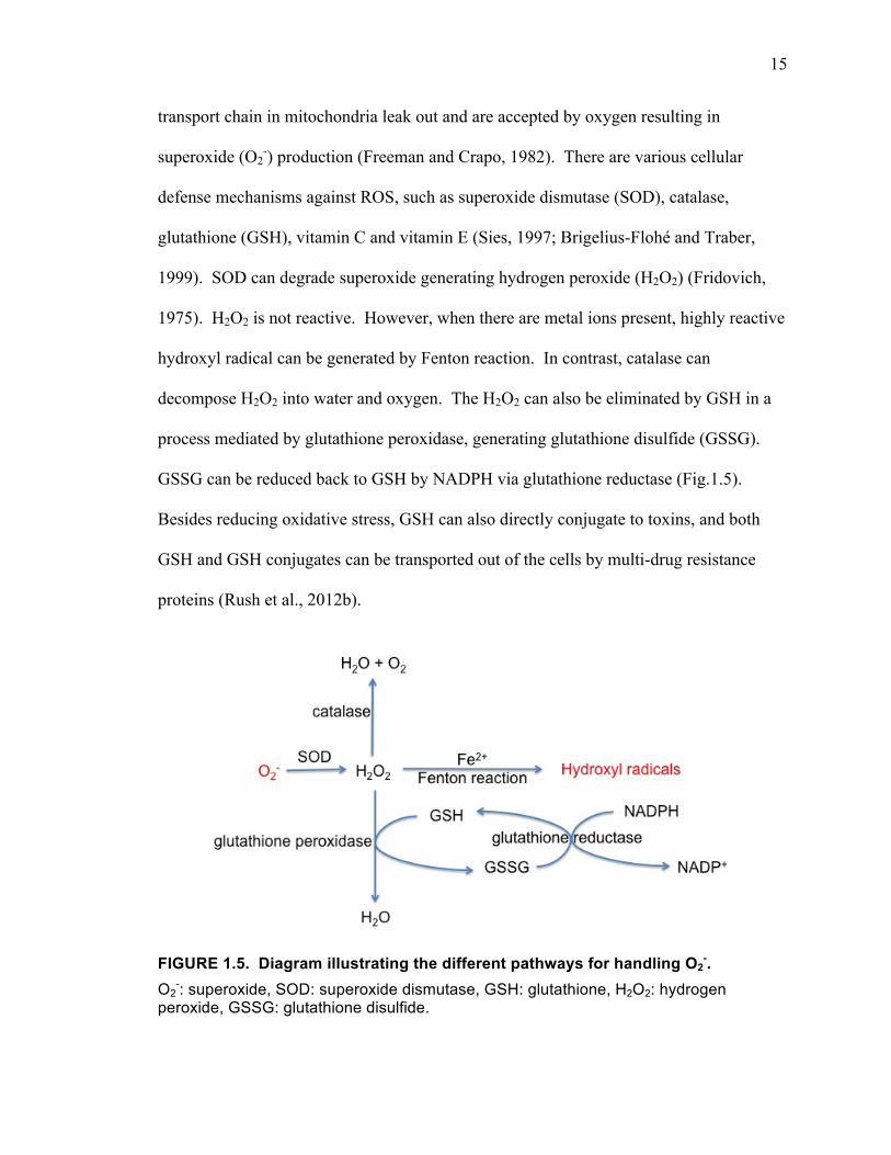
transport chain in mitochondria leak out and are accepted by oxygen resulting in
superoxide (O2-) production (Freeman and Crapo, 1982). There are various cellular
defense mechanisms against ROS, such as superoxide dismutase (SOD), catalase,
glutathione (GSH), vitamin C and vitamin E (Sies, 1997; Brigelius-Flohé and Traber,
1999). SOD can degrade superoxide generating hydrogen peroxide (H2O2) (Fridovich,
1975). H2O2 is not reactive. However, when there are metal ions present, highly reactive
hydroxyl radical can be generated by Fenton reaction. In contrast, catalase can
decompose H2O2 into water and oxygen. The H2O2 can also be eliminated by GSH in a
process mediated by glutathione peroxidase, generating glutathione disulfide (GSSG).
GSSG can be reduced back to GSH by NADPH via glutathione reductase (Fig.1.5).
Besides reducing oxidative stress, GSH can also directly conjugate to toxins, and both
GSH and GSH conjugates can be transported out of the cells by multi-drug resistance
proteins (Rush et al., 2012b).
FIGURE 1.5. Diagram illustrating the different pathways for handling O2-.
O2-: superoxide, SOD: superoxide dismutase, GSH: glutathione, H2O2: hydrogen
peroxide, GSSG: glutathione disulfide.
16
The brain, while only 2% of the body weight, consumes about 20% of the total
oxygen, generating high levels of ROS (Ballatori et al., 2009), and oxidative stress has
been shown to be a possible contributor to the damage occurring in neurodegenerative
diseases (Simonian and Coyle, 1996; Schulz et al., 2000; Ballatori et al., 2009).
GSH in the brain
GSH is a tripeptide consisting of the amino acids glutamate, glycine, and cysteine
(Dringen et al., 2000). It is the most prevalent cellular thiol in the brain, with an
intracellular concentration of ~2.5 mM in neurons and ~ 3.8 mM in astrocytes (Bolaños
et al., 1995; Rice and Russo-Menna, 1998). Normally, the intracellular GSSG/GSH ratio
is tightly regulated, with over 99% of the total cellular GSH present as the reduced form
(Deneke and Fanburg, 1989). The GSSG/GSH ratio is a sensitive indicator of oxidative
stress (Rahman et al., 2005).
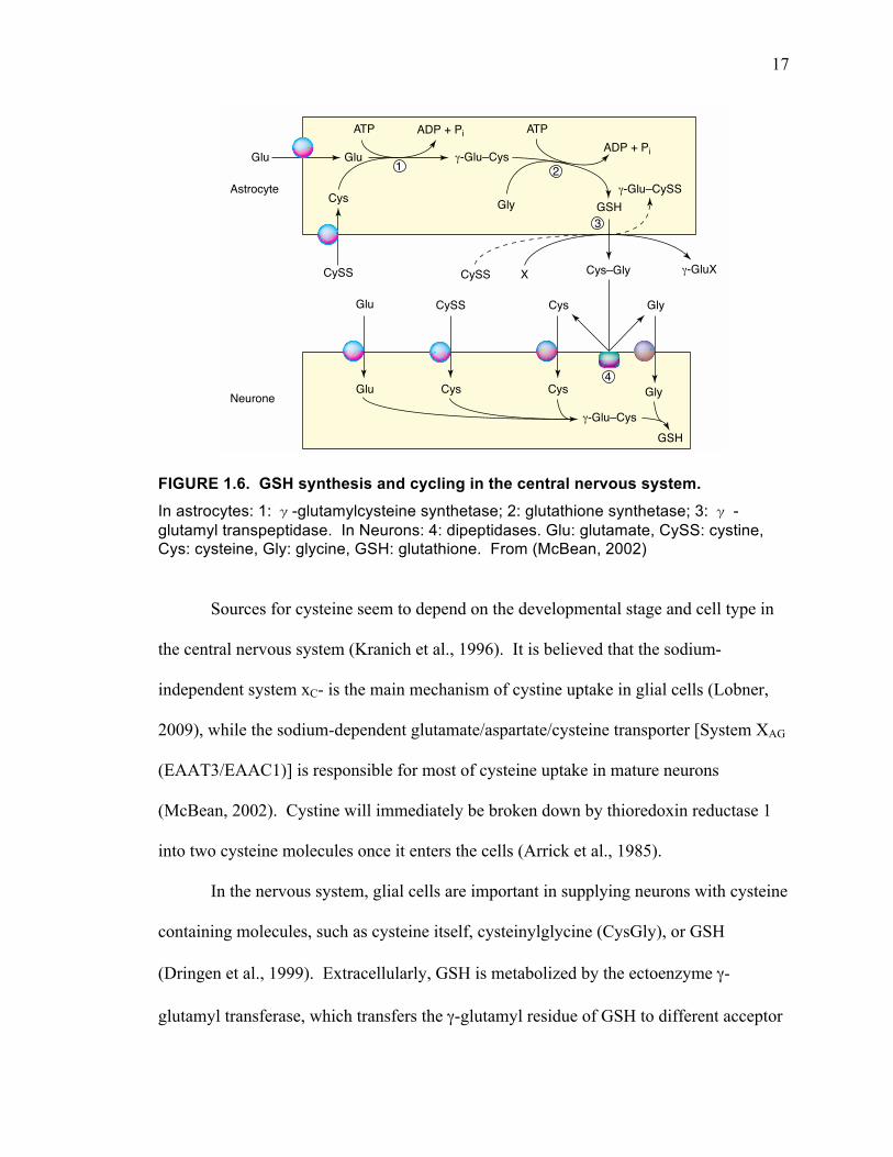
GSH is synthesized via a two-step reaction (Fig. 1.6) (Beutler, 1989; Deneke and
Fanburg, 1989). First, glutamate and cysteine are catalyzed to γ-glutamylcysteine by
glutamate cysteine ligase. Then glycine joins γ-glutamylcysteine mediated by
glutathione synthetase. Both steps require ATP. Both glutamate and glycine are highly
available in the cells, so the rate-limiting factor is cysteine. The rate of this reaction is
based on intracellular cysteine levels and γ-glutamylcysteine synthetase that is feedback-
regulated by GSH. Inhibiting cystine uptake inhibits GSH synthesis because the levels of
intracellular cysteine are dependent on cystine uptake (Bannai and Kitamura, 1980).
17
FIGURE 1.6. GSH synthesis and cycling in the central nervous system. In astrocytes: 1: γ-glutamylcysteine synthetase; 2: glutathione synthetase; 3: γ -glutamyl transpeptidase. In Neurons: 4: dipeptidases. Glu: glutamate, CySS: cystine, Cys: cysteine, Gly: glycine, GSH: glutathione. From (McBean, 2002)
Sources for cysteine seem to depend on the developmental stage and cell type in
the central nervous system (Kranich et al., 1996). It is believed that the sodium-
independent system xC- is the main mechanism of cystine uptake in glial cells (Lobner,
2009), while the sodium-dependent glutamate/aspartate/cysteine transporter [System XAG
(EAAT3/EAAC1)] is responsible for most of cysteine uptake in mature neurons
(McBean, 2002). Cystine will immediately be broken down by thioredoxin reductase 1
into two cysteine molecules once it enters the cells (Arrick et al., 1985).
In the nervous system, glial cells are important in supplying neurons with cysteine
containing molecules, such as cysteine itself, cysteinylglycine (CysGly), or GSH
(Dringen et al., 1999). Extracellularly, GSH is metabolized by the ectoenzyme γ-
glutamyl transferase, which transfers the γ-glutamyl residue of GSH to different acceptor
uptake, reduce cystine transport furtherby indirectly blocking exchange.
Oxidative stress and induction of the xc
−− exchangerDepletion of intracellular GSH infibroblasts triggers induction of xc
−,leading to an increased rate ofNa+-independent uptake of cystine [2],which implies that upregulation of cystineuptake protects cells against depletion ofGSH. In addition, much recent work hasbeen directed towards investigating theresponse of xc
−-mediated uptake of cystineto oxidative stress. Chronic exposure ofendothelial [26] or epithelial [27] cells tothe nitric oxide donor, S-nitroso-N-acetyl-penicillamine (SNAP), which can lead tothe formation of toxic radical species,increases xc
−-mediated cystine uptake.Responses to oxidative stress in gliomacells are linked to increased expression of xCT with no alteration in the expressionof 4F2hc [8]. xc
−-mediated transport ofglutamate in astrocytes is upregulated bydibutyryl cAMP but, in this case, theexpression of both xCT and 4F2hc areincreased [28]. In macrophages, an
increase in intracellular GSH parallelsinduction of xCT in response to bacteriallipopolysaccharide [29].
Although upregulation of the xc
− exchanger will provide more cysteinefor GSH synthesis, glutamate release willalso increase, potentially causing theextracellular concentration to rise [22].This could trigger glutamate-mediatedtoxicity in certain brain pathologies.Indeed, Ye et al. [30] reported increasedglutamate efflux by the xc
− exchanger in glioma cells in which the XAG
− transporters were mislocalized ordysfunctional and therefore not availableto remove glutamate. However, it isarguable that such a situation would nothappen if XAG
− transporters were operatingnormally. In other words, activation of thexc
− exchanger would not be expected tocause toxicity unless XAG
−-mediatedglutamate uptake was disrupted.Conversely, elevation of extracellularglutamate would inhibit both avenues of cystine uptake into the cell, causing GSH levels to fall. Release of glutamate inanoxia, for example, could damage cellsfor this reason.
Concluding remarksMuch new information on the molecularbasis of cystine uptake has come to light inrecent years. Current opinion holds thatxc
−-mediated uptake of cystine might bemore important as a provider of cysteineto synthesize GSH, particularly underconditions of oxidative stress. However,this view remains hypothetical until suchtime as the physiological role of both xc
−- and XAG−-mediated transport of
cystine in vivo is fully resolved.
AcknowledgementsMy research in this field is supported by the Irish Health Research Board and the Irish Motor Neurone Disease Association.
References1 Bannai, S. and Kitamura, E. (1980) Transport
interaction of L-cystine and L-glutamate in humandipliod fibroblasts in culture. J. Biol. Chem. 255,2372–2376
2 Bannai, S. (1984) Induction of cystine andglutamate transport activity in human fibroblastsby diethylmaleate and other electrophilic agents.J. Biol. Chem. 259, 2435–2440
3 Bannai, S. et al. (1984) Amino acid transportsystems. Nature 311, 308
4 Sato, H. et al. (1999) Cloning and expression of aplasma-membrane cystine–glutamate exchangetransporter composed of two distinct proteins.J. Biol. Chem. 274, 11455–11458
5 Sato, H. et al. (2000) Molecular cloning andexpression of human xCT, the light chain of aminoacid transport system xc. Antioxid. Redox Signal.2, 665–671
6 Shih, A.Y. and Murphy, T.H. (2001) xCt cystinetransporter expression in HEK293 cells:pharmacology and localization. Biochem. Biophys.Res. Commun. 282, 1132–1137
7 Bassi, M.T. et al. (2001) Identification andcharacterisation of human xCT that co-expresses, with 4F2 heavy chain, the aminoacid transport activity system xc. Pflugers Arch.442, 286–296
8 Kim, J.Y. et al. (2001) Human cystine/glutamatetransporter: cDNA cloning and upregulation byoxidative stress in glioma cells. Biochim. Biophys.Acta 1512, 335–344
9 Danbolt, N.C. (2001) Glutamate uptake. Prog. Neurobiol. 65, 1–105
10 Chase, L.A. et al. (2001) L-Quisqualic acidtransport into hippocampal neurons by a cystine-sensitive carrier is required for the induction ofquisqualate sensitization. Neuroscience 106,287–301
11 Tsai, M.J. et al. (1996) Characterisation of L-α-aminoadipic acid transport in cultured ratastrocytes. Brain Res. 741, 166–173
12 Johnson, C.L. and Johnson, C.G. (1993)Substance P regulation of glutamate and cystine transport in human astrocytoma cells.Receptors Channels 1, 53–59
13 Sagara, J.I. et al. (1993) Cystine uptake andglutathione level in foetal brain cells in primaryculture and in suspension. J. Neurochem. 61,1667–1671
TRENDS in Pharmacological Sciences Vol.23 No.7 July 2002
http://tips.trends.com
301Research Update
TRENDS in Pharmacological Sciences
Glu Glu
CySS
Cys
γ-Glu–Cys
ATP ATPADP + Pi
Gly GSH
ADP + Pi
1 2
Cys–GlyX γ-GluX
3
Cys GlyGlu
Glu
CySS
Cys Cys
γ-Glu–CysGSH
Gly
CySS
γ-Glu–CySSAstrocyte
Neurone
4
Fig. 3. The relationship between astrocytes and neurones in glutathione (GSH) synthesis. Transport of glutamate(G lu) and cystine (CySS) into astrocytes provides substrates for γ-glutamylcysteine synthetase (1), which catalyzesthe production of γ-glutamylcysteine. GSH synthetase (2) then catalyzes the conversion of γ-glutamylcysteine(γ-G lu–Cys) to GSH. GSH released from astrocytes is a substrate for γ-glutamyltranspeptidase (3), which transfers the γ-glutamyl moiety (γ-G lu) of GSH to an acceptor am ino acid (X). A lso shown is the net translocation of cystine intothe cell (broken line) w ith cystine as the acceptor am ino acid. The di-peptide, cysteinyl–glycine (Cys–G ly) is cleavedinto component am ino acids by an ectopeptidase (4). The identity of the glutamate and cystine transporters in thisdiagram is not specified. There is uncertainty of the relative importance of cystine or cysteine as a precursor forGSH synthesis in neurones (see text for details). Further information on other aspects of GSH metabolism inastrocytes and neurones can be found in [19].
18
amino acids leading to the formation of a γ-glutamyl containing dipeptide and a dipeptide
CysGly. CysGly is then either cleaved by extracellular dipeptidases to generate cysteine
and glycine, or directly taken up by neurons. Knocking out EAAT3 greatly reduces
neuronal cysteine uptake and intracellular GSH levels, resulting in decreased viability of
hippocampal neurons against H2O2 insults (Zerangue and Kavanaugh, 1996b; Chen and
Swanson, 2003; Aoyama et al., 2006). This suggests that EAAT3 plays a critical role in
the ability of neurons to obtain cysteine.
SYSTEM XC-
System xC- is a sodium-independent, chloride-dependent amino acid transport
system located on the plasma membrane. System xC- was first characterized by Bannai
and Kitamura in 1980 when mutual inhibition of glutamate and cystine uptake was
described (Bannai and Kitamura, 1980). It can transport one molecule of cystine into the
cell in exchange for one molecule of intracellular glutamate, with Km values of ~ 80 µM
for cystine uptake and ~ 160 µM for glutamate uptake (Sato et al., 1999). The transport
direction is determined by the high cytosolic glutamate levels and low cytosolic cystine
levels.
Structure of system xC-
System xC- is a heteromeric antiporter, composed of two subunits: a light-chain
subunit xCT and a heavy chain 4F2hc, which are linked together by one disulfide bond
(Torrents et al., 1998; Shih et al., 2006) (Fig. 1.7.). The 4F2hc subunit is a single
transmembrane glycoprotein that is believed to be universally shared among the
19
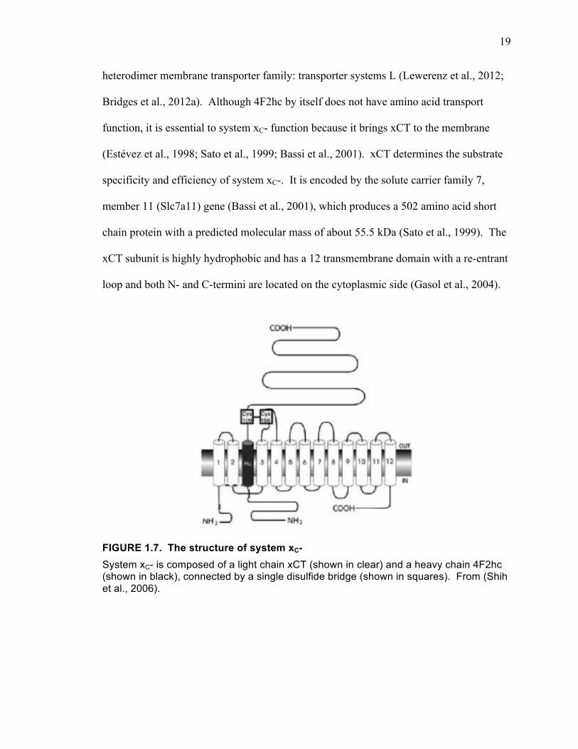
heterodimer membrane transporter family: transporter systems L (Lewerenz et al., 2012;
Bridges et al., 2012a). Although 4F2hc by itself does not have amino acid transport
function, it is essential to system xC- function because it brings xCT to the membrane
(Estévez et al., 1998; Sato et al., 1999; Bassi et al., 2001). xCT determines the substrate
specificity and efficiency of system xC-. It is encoded by the solute carrier family 7,
member 11 (Slc7a11) gene (Bassi et al., 2001), which produces a 502 amino acid short
chain protein with a predicted molecular mass of about 55.5 kDa (Sato et al., 1999). The
xCT subunit is highly hydrophobic and has a 12 transmembrane domain with a re-entrant
loop and both N- and C-termini are located on the cytoplasmic side (Gasol et al., 2004).
FIGURE 1.7. The structure of system xC- System xC- is composed of a light chain xCT (shown in clear) and a heavy chain 4F2hc (shown in black), connected by a single disulfide bridge (shown in squares). From (Shih et al., 2006).
20
Function of system xC-
Because of the dual transport property of system xC-, it can regulate both
intracellular GSH levels and extracellular glutamate levels. Cystine that enters cells
through system xC- can be broken down to cysteine to synthesize GSH, which can reduce
free radicals or be released to regulate the redox state of the extracellular millieu (Wang
and Cynader, 2000; Banjac et al., 2008). System xC- has been shown to be responsible
for 60% of extracellular glutamate in rat striatum (Baker et al., 2002). Deleting the xCT
gene in Drosophila causes a 50% reduction in extracellular glutamate levels (Augustin et
al., 2007). Extracellular glutamate released from system xC- can activate presynaptic
mGluR2/3, which can regulate synaptic release of neurotransmitters, such as dopamine
(Baker et al., 2002).
Sato et al. 2005, developed and characterized xCT null mice with a partial
deletion of the xCT gene. These mutant mice appear healthy and fertile. However, in
plasma, the cystine concentration is doubled compared to wild type, while GSH levels are
half of the wild type (Sato et al., 2005). No difference in cysteine levels was reported.
Microglial cells isolated from these mice showed normal levels of cystine uptake.
However, the uptake was not blocked by glutamate and was not inducible by
lipopolysaccharide (LPS) (Sato et al., 2005). Fibroblasts isolated from these xCT -/-
mice die unless exogenous 2-mercaptoethanol or N-acetylcysteine, which reduces cystine
to cysteine, is present (Sato et al., 2005). In xCT deficient mice, ischemia-reperfusion-
induced acute renal failure is more severe compared to wild type animals (Shibasaki et
al., 2009). No increased oxidative stress or brain atrophy were observed (De Bundel et
al., 2011). This is probably because in xCT deficient animals other cystine/cysteine
21
uptake transporters are upregulated to maintain the normal activity. However, it is not
possible to upregulate in these xCT deficient animals under oxidative stress to help
increase GSH levels (Shibasaki et al., 2009). Therefore, it appears that some degree of
compensation for the lack of system xC- occurs, but it is not entirely effective.
Another mutant mouse line involving altering system xC- function is the sut/sut
mouse, which has a partial deletion of the xCT gene. These animals show changes in fur
color due to a deficiency in the cysteine-dependent yellow/red pigment, pheomelanin
(Chintala et al., 2005), and a large reduction in pheomelanin is also observed in cultured
sut/sut melanocytes. Interestingly, sut/sut mice also exhibit prominent brain atrophy in
the hippocampus (Shih et al., 2006). The mechanisms resulting in different phenotypes
of these two different mutant mouse lines require further studies.
Regulation of system xC-
Despite the involvement of system xC- in both excitotoxicity and oxidative stress,
there is limited knowledge about its regulation. Because xCT is specific to system xC-
and determines specificity of its transport function, the regulation studies have been
mainly focused on xCT regulation (Sato et al., 1999). To date, the best-characterized
pathways are nuclear factor eythroid 2-related factor 2 (Nrf2)-antioxidant responsive
element (ARE) and eukaryotic initiation factor-2 (eIF2) - activating transcription factor
(ATF) 4- amino acid response element (AARE), which are discussed in detail below,
along with other regulatory mechanisms.
22
Nrf2-ARE pathway
There are four ARE-like, also known as electrophile response element (EpRE)-
like, sequences in the 5’ flanking region of the mouse xCT gene (Sasaki et al., 2002), two
of which are completely conserved in the 5’-flanking region of the human xCT gene
(Sato et al., 2000; Sasaki et al., 2002).
The transcription factor Nrf2-ARE pathway was first proposed by Venugopal and
Jaiswal (Venugopal and Jaiswal, 1996), as illustrated in (Fig. 1.8). Nrf2 is normally
cytosolic because of keap-1 binding (Itoh et al., 1999). However, when under oxidative
stress, Nrf2 is freed and translocates to the nucleus and binds to ARE to activate specific
protein transcription (Itoh et al., 1999). ARE is a cis-acting regulatory element located in
the promoter regions of multiple genes encoding phase II detoxification enzymes and
antioxidant proteins (Lee and Johnson, 2004). These proteins include heme oxygenase,
γ-glutamylcysteine synthetase, glutamate-cysteine ligase, glutathione synthetase,
glutathione S-transferase, glutathione reductase, multidrug resistance protein 1, as well as
xCT (Bannai, 1984; Erickson et al., 2002; Sasaki et al., 2002).
23
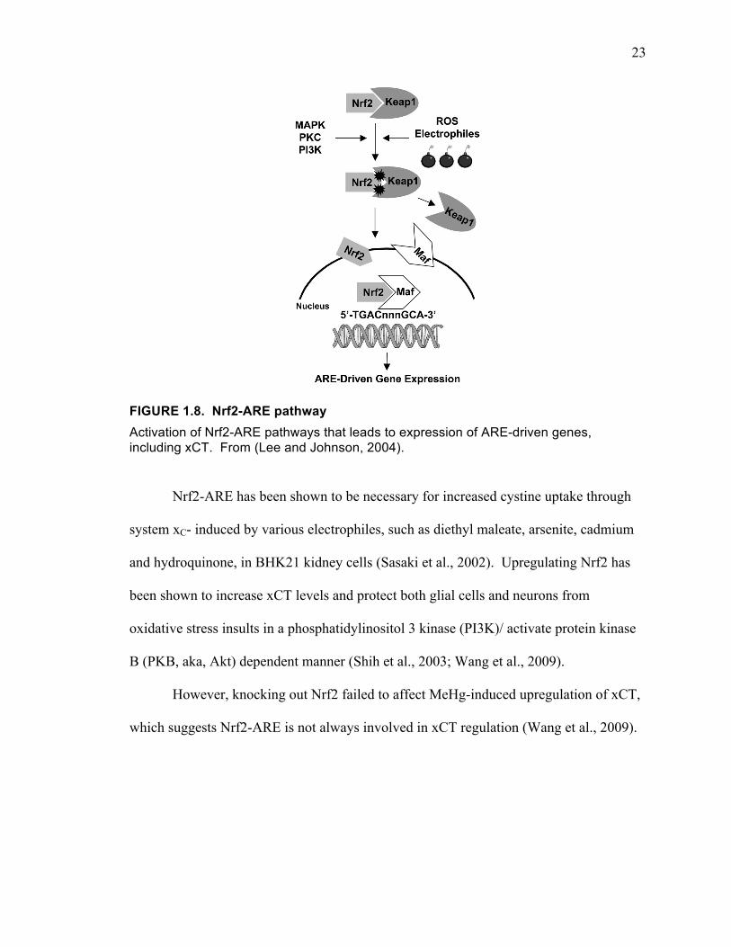
FIGURE 1.8. Nrf2-ARE pathway Activation of Nrf2-ARE pathways that leads to expression of ARE-driven genes, including xCT. From (Lee and Johnson, 2004).
Nrf2-ARE has been shown to be necessary for increased cystine uptake through
system xC- induced by various electrophiles, such as diethyl maleate, arsenite, cadmium
and hydroquinone, in BHK21 kidney cells (Sasaki et al., 2002). Upregulating Nrf2 has
been shown to increase xCT levels and protect both glial cells and neurons from
oxidative stress insults in a phosphatidylinositol 3 kinase (PI3K)/ activate protein kinase
B (PKB, aka, Akt) dependent manner (Shih et al., 2003; Wang et al., 2009).
However, knocking out Nrf2 failed to affect MeHg-induced upregulation of xCT,
which suggests Nrf2-ARE is not always involved in xCT regulation (Wang et al., 2009).
24
eIF2-ATF4-AARE pathway
There are two AARE-like sequences in the 5’-flanking region of the xCT gene
(Sato et al., 2004; Lewerenz and Maher, 2009). Amino acid response (AAR) is usually
triggered by limited uptake of any essential amino acid (Kilberg et al., 2005). It serves as
a self-protective effect by globally slowing down protein production and cellular activity
by limiting protein synthesis. However, transcription and translation of certain proteins,
such as basic leucine zipper and ATF4 is enhanced (Wek et al., 2006). These proteins
then regulate gene expression of membrane transporters and growth factors to cope with
environmental stresses (Kilberg et al., 2005; Wek et al., 2006; Ameri and Harris, 2008;
Kilberg et al., 2009). It is a protective mechanism against a harmful environment, such
as changed pH, nutrient levels, and oxidative stress (Duncan and Hershey, 1985; 1987).
eIF2, a heterotrimer composed of eIF2α, eIF2β, and eIF2γ, is an important
component of the initiating complex for most of the protein synthesis (Fafournoux et al.,
2000). The GDP in eIF2 has to be exchanged for GTP mediated by guanine nucleotide
exchange factor eIF2B to successfully form the initiating complex (Matts and London,
1984; Dholakia and Wahba, 1989). Components of eIF2 can be phosphorylated,
preventing the attached GDP from being replaced with GTP (Kilberg et al., 2009).
Among the three, eIF2α is the most easily phosphorylated (Costa-Mattioli et al., 2007).
The two mechanisms of eIF2B inhibition are described below, both of which can lead to
increased ATF4 production, and then activation of AARE regulated gene transcription.
When there are not enough available essential amino acids in the cell, the
excessive free tRNAs activate the general control nonrepressible protein 2 (GCN2)
kinase, which in turn phosphorylates eIF2 (Zhang et al., 2002). Phosphorylated eIF2 has
25
increased affinity for eIF2B, although it cannot be activated for initiating complex
assembly. Therefore, phosphorylated eIF2 becomes a potent competitive inhibitor for
eIF2B (Rowlands et al., 1988; Kimball, 1999). Normally, the intracellular eIF2 levels are
significantly higher than eIF2B, therefore, phosphorylation of merely 30% of eIF2α is
enough to completely block eIF2B activity (Matts and London, 1984; Duncan and
Hershey, 1987). Besides GCN2, there are other kinases that are sensitive to other
stressors, and become activated leading to eIF2 phosphorylation. These kinases include
heme-regulated inhibitor (HRI) activated by heme-deficiency (Han et al., 2001; Lu et al.,
2001), double-stranded RNA-activated inhibitor (DAI) or dsRNA-dependent
serine/threonine protein kinase R (PKR) activated by viral infection (Hershey, 1989;
Proud, 2005) and PKR-like endoplasmic-reticulum (ER)-localized eIF2α kinase (PERK)
activated by ER stress (Lu et al., 2004) (Fig. 1.9.). All of these cellular stresses have the
potential of activating the same pathways amino acid deprivation activate and inducing
system xC- expression, but this theory has not yet been tested.

26
FIGURE 1.9. Regulation of translation initiation by eIF2 phosphorylation. dsRNA-dependent serine/threonine protein kinase R (PKR), heme-regulated inhibitor (HRI), PKR-like endoplasmic-reticulum (ER)-localized eIF2α kinase (PERK), general control nonrepressible protein 2 (GCN2) are activated in response to various environmental stresses, which leads to phosphorylation of eIF2. Phosphorylated eIF2 inhibits eIF2B mediated guanine nucleotide exchange, in turn, globally slows down translation initiation, but increases ATF4 levels. From (Wek et al., 2006)
Phosphorylation of eIF2B is another mechanism that leads to activation of AARE
regulated gene transcription (Proud, 2005) (Fig. 1.10). Glycogen synthetase 3β
(GSK3β), a constitutively active kinase, can phosphorylate eIF2B, leading to the loss of
its GEF property (Welsh and Proud, 1993) and prevents assembly of the initiation
complex. Hormones, mitogens, and growth factors can activate phosphatidylinositol
3(PI3)-kinase (Welsh et al., 1998) and MEK/ERK (Kleijn and Proud, 2000; Quevedo et
al., 2000), which can lead to phosphorylation of GSK3β to inactivate it, which removes
the GSK3β inhibition effect on eIF2B.
27
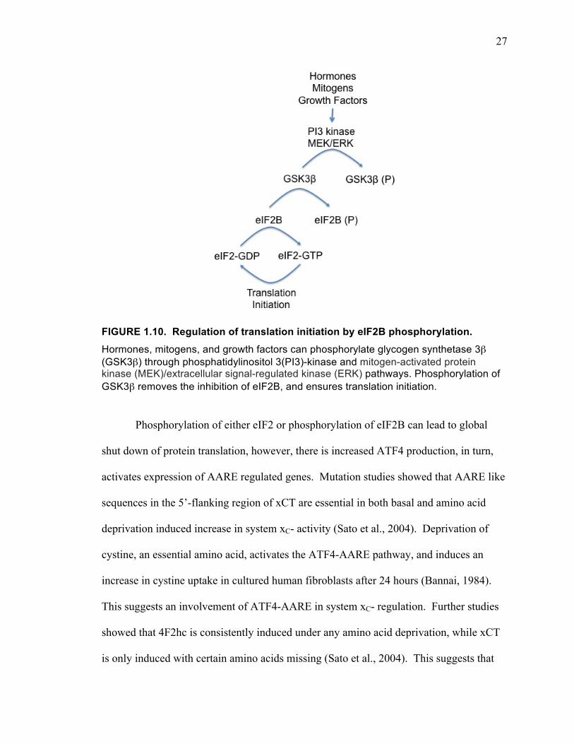
FIGURE 1.10. Regulation of translation initiation by eIF2B phosphorylation. Hormones, mitogens, and growth factors can phosphorylate glycogen synthetase 3β (GSK3β) through phosphatidylinositol 3(PI3)-kinase and mitogen-activated protein kinase (MEK)/extracellular signal-regulated kinase (ERK) pathways. Phosphorylation of GSK3β removes the inhibition of eIF2B, and ensures translation initiation.
Phosphorylation of either eIF2 or phosphorylation of eIF2B can lead to global
shut down of protein translation, however, there is increased ATF4 production, in turn,
activates expression of AARE regulated genes. Mutation studies showed that AARE like
sequences in the 5’-flanking region of xCT are essential in both basal and amino acid
deprivation induced increase in system xC- activity (Sato et al., 2004). Deprivation of
cystine, an essential amino acid, activates the ATF4-AARE pathway, and induces an
increase in cystine uptake in cultured human fibroblasts after 24 hours (Bannai, 1984).
This suggests an involvement of ATF4-AARE in system xC- regulation. Further studies
showed that 4F2hc is consistently induced under any amino acid deprivation, while xCT
is only induced with certain amino acids missing (Sato et al., 2004). This suggests that

28
the two subunits of system xC- have different regulatory mechanisms. Decreasing
intracellular eIF2α levels increases HT22 cells resistance to oxidative glutamate toxicity,
while increasing intracellular eIF2α can render HT22 cells highly sensitive to glutamate
toxicity by decreasing system xC- mediated cystine uptake, depleting GSH, and
increasing ROS (Tan et al., 2001; Lewerenz and Maher, 2009). This effect is mediated
through ATF4 binding to AARE (Lewerenz and Maher, 2009). Therefore, it appears that
the ATF4-AARE pathway is an important regulatory mechanism for system xC-.
NF-κB (nuclear factor kappa-light-chain-enhancer of activated B cells)
NF-κB may be another activator of xCT expression because there is a NF-κB
binding site in the 5’-flanking region of xCT (Sato et al., 2001). NF-κB has long been
known to play a role in rapid response to calcium influx and harmful cellular stimuli,
especially in the immune response (Meffert et al., 2003). LPS is known to activate NF-
κB (Sen and Smale, 2010). In mouse peritoneal macrophages, both xCT and 4F2hc
mRNA increased in a time dependent manner within 12 hours of LPS treatment (Sato et
al., 2001). However, this LPS induced increase in xCT levels is not likely to be mediated
through NF-κB because there was no increased nuclear NF-κB caused by the LPS
treatment (Sato et al., 2001). Since LPS still induces the activity of system xC- in
macrophages prepared from Nrf2-deficient mice, it is not likely that an LPS induced
increase in system xC- activity is mediated through the Nrf2-ARE pathway (Sato et al.,
2001). Therefore, despite an NF-κB binding site in the 5’-flanking region of xCT, there is
no direct evidence of its role in system xC- regulation. The mechanism of LPS inducing
increased system xC- activity remains unclear.
29
Activator protein 1 (AP-1)
There are several putative AP-1 binding sites in the 5’-flanking region of xCT,
one of them overlaps with the ARE sequence that is essential for response to electrophile
reagent activated xCT transcription and translation (Sato et al., 2001). AP-1 transcription
factor mediates gene regulation in response to cytokines, growth factors, stress, and
infections (Hess et al., 2004). However, its role in xCT regulation has not yet been
studied.
Hypoxia inducible factor (HIF)
Hypoxic preconditioning, the protection against a severe hypoxic insult by an
earlier mild hypoxic insult, increases xCT levels both transcriptionally and translationally
in hippocampus in vivo and in mouse neuronal stem cells (Ogunrinu and Sontheimer,
2010; Sims et al., 2012). This is mostly mediated through HIF-1α, but since siHIF-1α
does not completely abolish xCT upregulation in B104 mouse neuronal stem cell cultures
after hypoxic preconditioning, the possibility of other intracellular pathways being
involved cannot be ruled out (Sims et al., 2012). Also, hypoxia did not induce system xC-
function in mouse macrophage cultures (Sato et al., 2001). Determining the mechanism
of system xC- regulation by preconditioning, and when it occurs, requires further study.
cAMP
In rat striatal punches, system xC- activity is decreased by 15 minutes of
mGluR2/3 agonist treatment, and this effect is mimicked by inhibiting cAMP (Baker et
30
al., 2002). Also, a two-fold increase in xCT mRNA levels in rat cortical astrocytes was
observed after a 10-day incubation with N(6),2'-O-dibutyryladenosine 3':5' cyclic
monophosphate (dbcAMP), a cAMP analog (Gochenauer and Robinson, 2001). One
week dbcAMP treatment potentiates buthionine sulfoximine induced increase in system
xC- activity in rat primary astrocytes (Seib et al., 2011). There are two consensus PKA
phosphorylation sites on human xCT (Baker et al., 2002). However, the exact
intracellular pathway by which cAMP is involved in system xC- regulation is yet to be
investigated.
Growth factors
Our lab was the first to report growth factor effects on system xC- function.
Insulin-like growth factor 1 (IGF-1) and transforming growth factor-β (TGF-β) can
upregulate system xC- function in dental pulp cells (Pauly et al., 2011). We also showed
that fibroblast growth factor-2 (FGF-2) upregulates system xC- function selectively in
primary cortical astrocytes (Liu et al., 2011).
FGF-2 RECEPTORS AND INTRACELLULAR SIGNALING PATHWAYS
FGF-2 was the first member isolated and cloned among the FGF family of growth
factors in the 1980s. After decades of research, we now know FGF-2 is involved in many
nervous system functions. During embryonic development, FGF-2 plays an important
role in regulating proliferation, differentiation, and migration; while in adult, FGF-2 plays
a critical role in neuronal death, neurogenesis, learning and memory, and lesion repair
(Reuss and Bohlen und Halbach, 2003; Eswarakumar et al., 2005). FGF-2 expression is
31
found in both neuronal and glial cells, with glial cells as its main source (Eckenstein et
al., 1991a; 1991b).
FGF-2 can activate all members of the FGF receptor (FGFR) family, with FGFR1
and FGFR2 the prominent forms present in the cerebral cortex (Reuss and Bohlen und
Halbach, 2003). Like any typical tyrosine kinase receptor, FGFRs are composed of an
extracellular ligand-binding domain composed of three immunoglobulin-like domains, a
single transmembrane domain, and an intracellular domain with catalytic protein tyrosine
kinase activity (Mohammadi et al., 1996a). Two molecules of FGF-2 bind to FGFRs,
which triggers dimerization and activation of tyrosine kinase activity through
autophosphorylation (Schlessinger et al., 1995; Mohammadi et al., 1996b). Previous
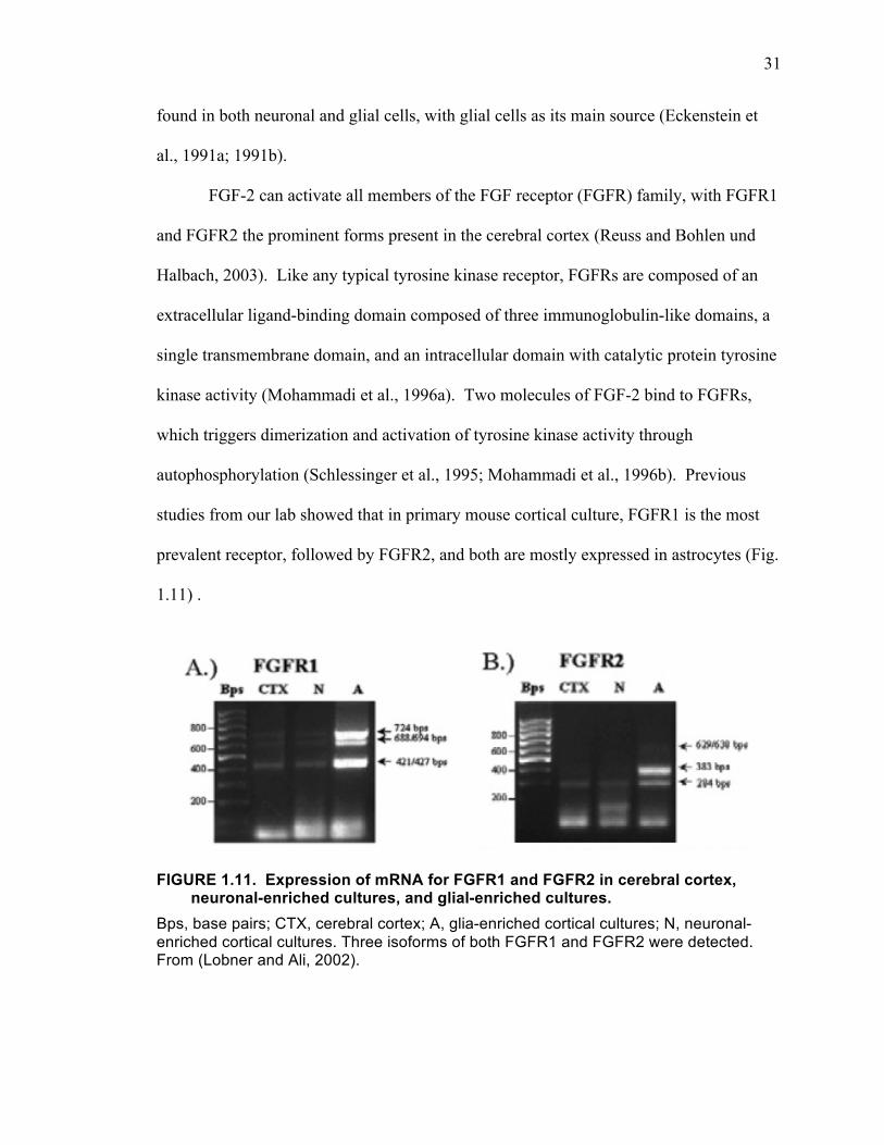
studies from our lab showed that in primary mouse cortical culture, FGFR1 is the most
prevalent receptor, followed by FGFR2, and both are mostly expressed in astrocytes (Fig.
1.11) .
FIGURE 1.11. Expression of mRNA for FGFR1 and FGFR2 in cerebral cortex, neuronal-enriched cultures, and glial-enriched cultures.
Bps, base pairs; CTX, cerebral cortex; A, glia-enriched cortical cultures; N, neuronal-enriched cortical cultures. Three isoforms of both FGFR1 and FGFR2 were detected. From (Lobner and Ali, 2002).

32
Upon activation, at the carboxy terminal tail of the FGFR, autophosphorylation on
a tyrosine residue (Tyr766) creates a specific binding site for PLC (Mohammadi et al.,
1992; 1996a) and activates it to catalyze the hydrolysis of PIP2 to generate two secondary
messengers: IP3 and DAG. Also, fibroblast growth factor receptor substrate 2 (FRS2)
constitutively docks at the juxtamembrane domain of FGFRs (Reuss and Bohlen und
Halbach, 2003; Eswarakumar et al., 2005). In response to FGFR activation, the docking
proteins become tyrosine phosphorylated and recruit protein complexes and activate
multiple intracellular signal transduction pathways, such as PI3K/Akt and mitogen-
activated protein kinase kinase (MEK)/extracellular signal-regulated kinase (ERK)
pathways (Kouhara et al., 1997; Ojeda et al., 2011). The PLC activation mediated effects
are independent from the FRS2 mediated effects (Fig. 1.12.).
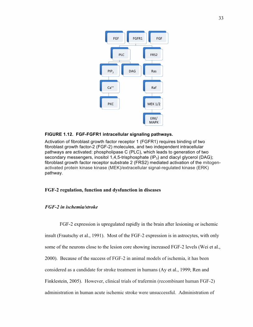
33
FIGURE 1.12. FGF-FGFR1 intracellular signaling pathways. Activation of fibroblast growth factor receptor 1 (FGFR1) requires binding of two fibroblast growth factor-2 (FGF-2) molecules, and two independent intracellular pathways are activated: phospholipase C (PLC), which leads to generation of two secondary messengers, inositol 1,4,5-trisphosphate (IP3) and diacyl glycerol (DAG); fibroblast growth factor receptor substrate 2 (FRS2) mediated activation of the mitogen-activated protein kinase kinase (MEK)/extracellular signal-regulated kinase (ERK) pathway.
FGF-2 regulation, function and dysfunction in diseases
FGF-2 in ischemia/stroke
FGF-2 expression is upregulated rapidly in the brain after lesioning or ischemic
insult (Frautschy et al., 1991). Most of the FGF-2 expression is in astrocytes, with only
some of the neurons close to the lesion core showing increased FGF-2 levels (Wei et al.,
2000). Because of the success of FGF-2 in animal models of ischemia, it has been
considered as a candidate for stroke treatment in humans (Ay et al., 1999; Ren and
Finklestein, 2005). However, clinical trials of trafermin (recombinant human FGF-2)
administration in human acute ischemic stroke were unsuccessful. Administration of
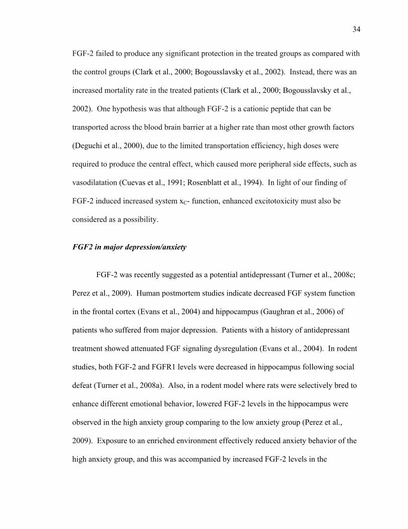
34
FGF-2 failed to produce any significant protection in the treated groups as compared with
the control groups (Clark et al., 2000; Bogousslavsky et al., 2002). Instead, there was an
increased mortality rate in the treated patients (Clark et al., 2000; Bogousslavsky et al.,
2002). One hypothesis was that although FGF-2 is a cationic peptide that can be
transported across the blood brain barrier at a higher rate than most other growth factors
(Deguchi et al., 2000), due to the limited transportation efficiency, high doses were
required to produce the central effect, which caused more peripheral side effects, such as
vasodilatation (Cuevas et al., 1991; Rosenblatt et al., 1994). In light of our finding of
FGF-2 induced increased system xC- function, enhanced excitotoxicity must also be
considered as a possibility.
FGF2 in major depression/anxiety
FGF-2 was recently suggested as a potential antidepressant (Turner et al., 2008c;
Perez et al., 2009). Human postmortem studies indicate decreased FGF system function
in the frontal cortex (Evans et al., 2004) and hippocampus (Gaughran et al., 2006) of
patients who suffered from major depression. Patients with a history of antidepressant
treatment showed attenuated FGF signaling dysregulation (Evans et al., 2004). In rodent
studies, both FGF-2 and FGFR1 levels were decreased in hippocampus following social
defeat (Turner et al., 2008a). Also, in a rodent model where rats were selectively bred to
enhance different emotional behavior, lowered FGF-2 levels in the hippocampus were
observed in the high anxiety group comparing to the low anxiety group (Perez et al.,
2009). Exposure to an enriched environment effectively reduced anxiety behavior of the
high anxiety group, and this was accompanied by increased FGF-2 levels in the
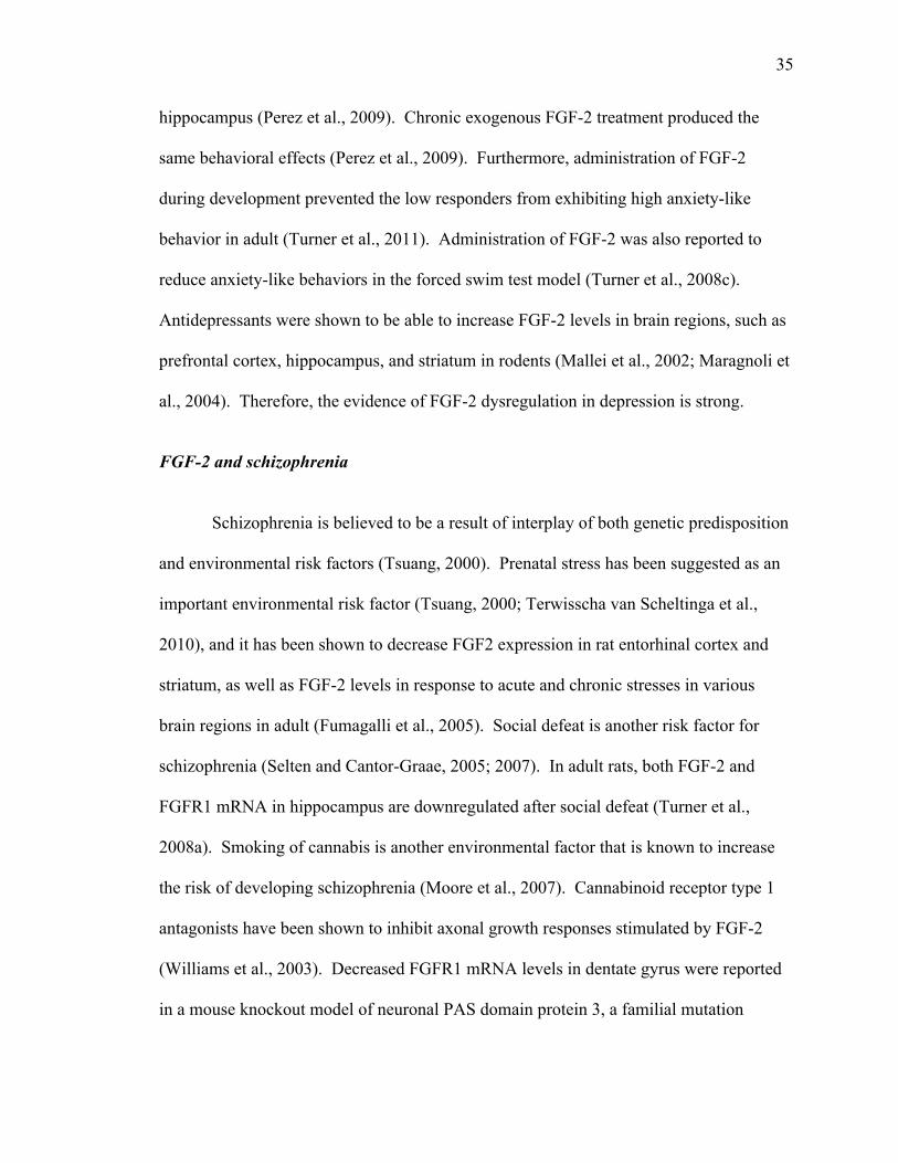
35
hippocampus (Perez et al., 2009). Chronic exogenous FGF-2 treatment produced the
same behavioral effects (Perez et al., 2009). Furthermore, administration of FGF-2
during development prevented the low responders from exhibiting high anxiety-like
behavior in adult (Turner et al., 2011). Administration of FGF-2 was also reported to
reduce anxiety-like behaviors in the forced swim test model (Turner et al., 2008c).
Antidepressants were shown to be able to increase FGF-2 levels in brain regions, such as
prefrontal cortex, hippocampus, and striatum in rodents (Mallei et al., 2002; Maragnoli et
al., 2004). Therefore, the evidence of FGF-2 dysregulation in depression is strong.
FGF-2 and schizophrenia
Schizophrenia is believed to be a result of interplay of both genetic predisposition
and environmental risk factors (Tsuang, 2000). Prenatal stress has been suggested as an
important environmental risk factor (Tsuang, 2000; Terwisscha van Scheltinga et al.,
2010), and it has been shown to decrease FGF2 expression in rat entorhinal cortex and
striatum, as well as FGF-2 levels in response to acute and chronic stresses in various
brain regions in adult (Fumagalli et al., 2005). Social defeat is another risk factor for
schizophrenia (Selten and Cantor-Graae, 2005; 2007). In adult rats, both FGF-2 and
FGFR1 mRNA in hippocampus are downregulated after social defeat (Turner et al.,
2008a). Smoking of cannabis is another environmental factor that is known to increase
the risk of developing schizophrenia (Moore et al., 2007). Cannabinoid receptor type 1
antagonists have been shown to inhibit axonal growth responses stimulated by FGF-2
(Williams et al., 2003). Decreased FGFR1 mRNA levels in dentate gyrus were reported
in a mouse knockout model of neuronal PAS domain protein 3, a familial mutation
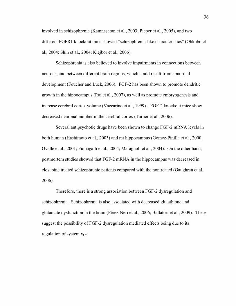
36
involved in schizophrenia (Kamnasaran et al., 2003; Pieper et al., 2005), and two
different FGFR1 knockout mice showed “schizophrenia-like characteristics” (Ohkubo et
al., 2004; Shin et al., 2004; Klejbor et al., 2006).
Schizophrenia is also believed to involve impairments in connections between
neurons, and between different brain regions, which could result from abnormal
development (Foucher and Luck, 2006). FGF-2 has been shown to promote dendritic
growth in the hippocampus (Rai et al., 2007), as well as promote embryogenesis and
increase cerebral cortex volume (Vaccarino et al., 1999). FGF-2 knockout mice show
decreased neuronal number in the cerebral cortex (Turner et al., 2006).
Several antipsychotic drugs have been shown to change FGF-2 mRNA levels in
both human (Hashimoto et al., 2003) and rat hippocampus (Gómez-Pinilla et al., 2000;
Ovalle et al., 2001; Fumagalli et al., 2004; Maragnoli et al., 2004). On the other hand,
postmortem studies showed that FGF-2 mRNA in the hippocampus was decreased in
clozapine treated schizophrenic patients compared with the nontreated (Gaughran et al.,
2006).
Therefore, there is a strong association between FGF-2 dysregulation and
schizophrenia. Schizophrenia is also associated with decreased glutathione and
glutamate dysfunction in the brain (Pérez-Neri et al., 2006; Ballatori et al., 2009). These
suggest the possibility of FGF-2 dysregulation mediated effects being due to its
regulation of system xC-.
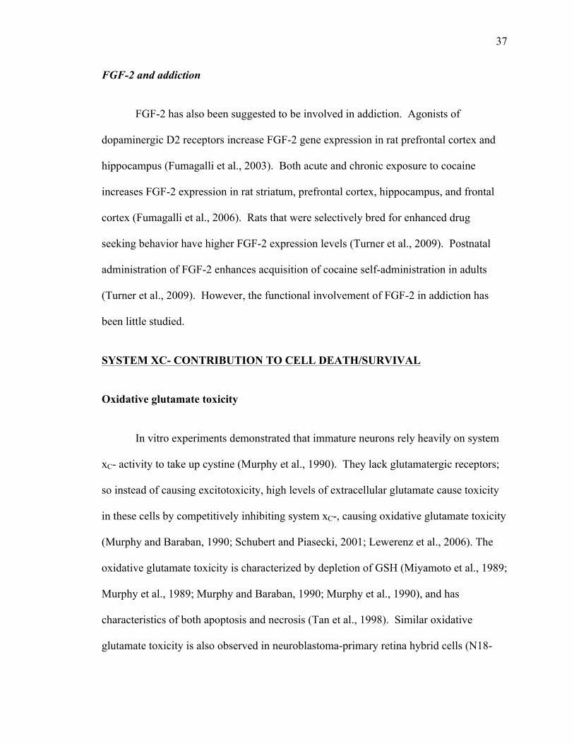
37
FGF-2 and addiction
FGF-2 has also been suggested to be involved in addiction. Agonists of
dopaminergic D2 receptors increase FGF-2 gene expression in rat prefrontal cortex and
hippocampus (Fumagalli et al., 2003). Both acute and chronic exposure to cocaine
increases FGF-2 expression in rat striatum, prefrontal cortex, hippocampus, and frontal
cortex (Fumagalli et al., 2006). Rats that were selectively bred for enhanced drug
seeking behavior have higher FGF-2 expression levels (Turner et al., 2009). Postnatal
administration of FGF-2 enhances acquisition of cocaine self-administration in adults
(Turner et al., 2009). However, the functional involvement of FGF-2 in addiction has
been little studied.
SYSTEM XC- CONTRIBUTION TO CELL DEATH/SURVIVAL
Oxidative glutamate toxicity
In vitro experiments demonstrated that immature neurons rely heavily on system
xC- activity to take up cystine (Murphy et al., 1990). They lack glutamatergic receptors;
so instead of causing excitotoxicity, high levels of extracellular glutamate cause toxicity
in these cells by competitively inhibiting system xC-, causing oxidative glutamate toxicity
(Murphy and Baraban, 1990; Schubert and Piasecki, 2001; Lewerenz et al., 2006). The
oxidative glutamate toxicity is characterized by depletion of GSH (Miyamoto et al., 1989;
Murphy et al., 1989; Murphy and Baraban, 1990; Murphy et al., 1990), and has
characteristics of both apoptosis and necrosis (Tan et al., 1998). Similar oxidative
glutamate toxicity is also observed in neuroblastoma-primary retina hybrid cells (N18-

38
RE105) (Miyamoto et al., 1989), primary rat hippocampal neurons (Murphy et al., 1989),
a mouse hippocampal cell line (HT-22) (Tan et al., 2001), and primary oligodendrocytes
(Oka et al., 1993).
Increased system xC- activity: protection against oxidative stress
Upregulation of system xC- has been shown to be protective against oxidative
stress in multiple systems. Fast proliferating primary brain glial-derived tumor cells
produce high levels of ROS. These cells rely primarily on system xC- to take up cystine
to produce GSH, therefore, inhibiting system xC- decreases the growth of these tumor
cells (Chung et al., 2005). High system xC- activity might contribute to the high tolerance
of human ovarian cancer cells to anticancer treatments (Okuno et al., 2003). In mouse
neural stem cells, hypoxic preconditioning increases system xC- function and strengthens
the cell’s resistance to oxidative glutamate toxicity (Sims et al., 2012). This
preconditioning produced protection is absent when either system xC- is blocked by S-4-
carboxyphenyl glycine (CPG), or GSH synthesis is blocked by buthionine sulphoximine
(Sims et al., 2012). IGF-1 and TGF-β increase cystine uptake through system xC- on
dental pulp cells, which leads to protection against oxidative stress (Pauly et al., 2011).
Retinoic acid increases SH-SY5Y cell (a human dopaminergic neuroblastoma cell line)
tolerance for 6-hydroxydopamine (6-OHDA) and 1-methyl-4-phenylpyridinium ion
(MPP+) compared to untreated cells (Crockett et al., 2011). This is because retinoic acid
increases system xC- activity, which increases GSH levels and protects the cells from
these oxidative insults (Crockett et al., 2011). Upregulating Nrf2, one of the well-
characterized pathways that regulate xCT expression, is also known to protect both glial
39
cells and neurons from oxidative stress insults in a PI3K/Akt dependent manner (Shih et
al., 2003; Wang et al., 2009). Ceftriaxone protects fibroblasts and the hippocampal cell
line HT22 from oxidative glutamate toxicity by upregulating system xC- through the
Nrf2-ARE pathway (Lewerenz et al., 2009). All of this data suggests that increasing
system xC- can increase cystine uptake, which can lead to increased glutathione
production, and provide protection against oxidative stress.
Increased system xC- activity: enhanced excitotoxicity
Increasing system xC- activity is not always protective. Activated microglia have
increased glutamate release via system xC-, which causes oligodendrocyte excitotoxicity
through AMPA receptors (Domercq et al., 2007). Interleukin 1β (IL-1β) upregulates
system xC- activity selectively on astrocytes, but not neurons (Jackman et al., 2010). This
increased system xC- activity is responsible for exacerbating neuronal cell death mediated
by excitotoxicity when the cultures are exposed to hypoxia (Fogal et al., 2007) or glucose
deprivation (Jackman et al., 2012). These studies demonstrate that when system xC-
activity is increased, glutamate release is also increased, which can potentially exacerbate
excitotoxicity.
POTENTIAL ROLE OF SYSTEM XC- IN NEUROLOGICAL DISORDERS
Alzheimer’s disease (AD)
AD is a common neurodegenerative disorder in elderly people. It is characterized
by progressive memory loss and impaired cognitive function. Amyloid-β (Aβ) plaques
and formation of neurofibrillary tangles are the characteristic hallmarks of this disease.
40
Aβ is a normal product of the brain, therefore, the abnormal deposition must be due to an
over production and/or impaired clearance. Astrocytes can degrade Aβ (Koistinaho et al.,
2004; Pihlaja et al., 2011). However, Aβ can make astrocytes reactive, which can
contribute to neuronal degeneration (Olabarria et al., 2010). There are increased levels of
phosphorylated eIF2α (Chang et al., 2002; Unterberger et al., 2006) and ATF4
(Lewerenz and Maher, 2009) in AD patient’s brains, which are shown to be involved in
xCT regulation (Lewerenz and Maher, 2009). In fact, in PC12 cells, induction of eIF2α
phosphorylation using the specific eIF2α phosphatase inhibitor, salubrinal, increased
ATF4 levels, system xC- activity, and resistance to Aβ toxicity (Lewerenz and Maher,
2009). Therefore, it is most likely that the increase of eIF2α and ATF4 is an adaptation
to the increased Aβ accumulation. However, secreted Aβ precursor protein (APP) can
activate microglia and stimulate system xC- mediated glutamate release from microglia,
which leads to decreased synaptic density and neuronal viability (Barger and Basile,
2001). Therefore, it is not clear whether upregulation of system xC- during AD is
beneficial or harmful.
Parkinson’s disease (PD)
PD is a common nervous system disorder in the elderly that is characterized by
degeneration of dopaminergic cells in the substantia nigra, resulting in symptoms such as
difficulties in initiating and finishing voluntary movements, tremors, stiff movements,
and eventually leads to muscle atrophy (Obeso et al., 2010). Extensive postmortem
studies have reported that increased oxidative stress, decreased GSH, and increased
oxidative damage to lipids, protein and DNA are observed in PD (Jenner and Olanow,
41
1996; Simonian and Coyle, 1996). Increased striatal glutamate (Massie et al., 2011) and
decreased nigral glutathione (Schulz et al., 2000) have both been indicated to contribute
to dopaminergic cell degeneration.
Involvement of system xC- in animal models of PD has been studied. xCT is
upregulated in the striatum three weeks after 6-OHDA lesion of the nigrostriatal area
(Massie et al., 2008). Also, 24 hour 6-OHDA treatment increases system xC- mediated
cystine uptake in C6 glioma cells in a dose dependent manner (Massie et al., 2008). In
xCT null mice, striatal glutamate levels are significantly lower compared to control
suggesting that system xC- is responsible for the majority of extracellular glutamate
(Baker et al., 2002; De Bundel et al., 2011; Massie et al., 2011). The absence of xCT
decreased the susceptibility of substantia nigra dopaminergic neurons to 6-OHDA
toxicity (Massie et al., 2011). These data suggest that system xC- function may have a net
damaging effect in PD, but it must be kept in mind that the 6-OHDA lesion model is not
an ideal model of PD.
Amyotrophic lateral sclerosis–Parkinsonism dementia complex (ALS-PDC)
ALS-PDC is a neurological disease that is also known as lytico-bodig to the
Chamorro people on the island of Guam (Reed and Brody, 1975). The disease has
aspects of ALS, PD, and AD: muscular weakness, spasticity, flaccid paralysis and
atrophy, and spinal-cord pathology similar to that are seen in ALS, tremors seen in PD,
neurofibrillary tangles similar to these occurring in brain tissue in AD, and dementia that
is seen both in PD and AD (Spencer et al., 1987; Liu et al., 2009). The disease has been
known to be occurring on Guam for about 200 years (Kurland and Mulder, 1954).

42
However, an outbreak of ALS-PDC in the 1940s to 1960s gained attention because the
incidence in the Chamorro population was 100 times higher than ALS elsewhere
(Kurland and Mulder, 1954; Spencer et al., 1986). The etiology of ALS-PDC remains
uncertain, dietetic, environmental and genetic factors have all been considered (Ince and
Codd, 2005).
At first, genetic factors were considered as the cause since certain villages and
families had higher rates of ALS-PDC. However, no genetic mutation that was found
that could account for the disease, and later studies indicated that adopting the local life
style and traditional diet had a significant association with the disease (Garruto et al.,
1981).
β-N-methylamino-L-alanine (BMAA)
BMAA is a non-protein amino acid that is one of the many potential toxins found
in cycad plants (Spencer et al., 1987; Cox et al., 2003). It was proposed as a possible
environmental trigger for ALS-PDC on Guam (Spencer et al., 1987). The local
Chamorro people collect the cycad seeds for food and medicine (Bradley and Mash,
2009). The local flying foxes (fruit bats) also feed on cycad seeds and it was found that
BMAA accumulates at high levels in these bats (Cox and Sacks, 2002; Cox et al., 2003;
Monson et al., 2003). Flying fox stew was served as a local courtesy dish until its
extinction in the 1960s, after which time the incidence of ALS-PDC declined.
BMAA was found to mimic glutamate and activate glutamatergic receptors when
bicarbonate is present (Weiss and Choi, 1988; Richter and Mena, 1989) (Fig. 1.13), and
BMAA was found to cause excitotoxicity when physiological concentration of
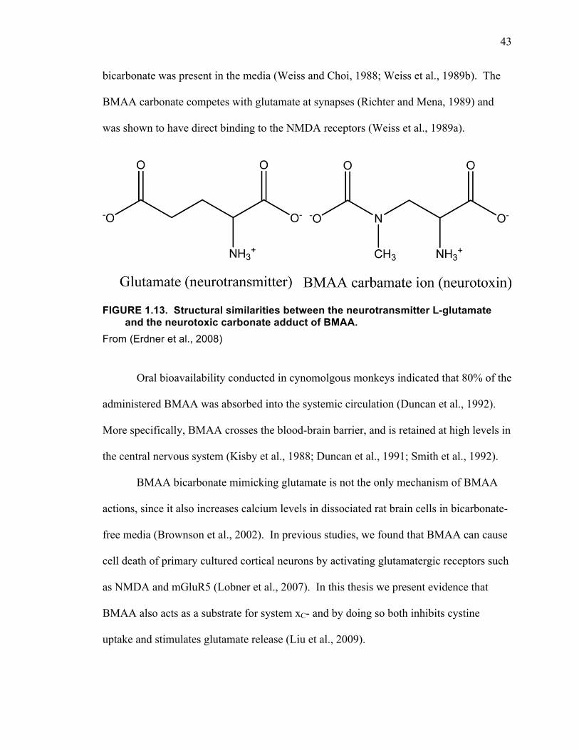
43
bicarbonate was present in the media (Weiss and Choi, 1988; Weiss et al., 1989b). The
BMAA carbonate competes with glutamate at synapses (Richter and Mena, 1989) and
was shown to have direct binding to the NMDA receptors (Weiss et al., 1989a).
FIGURE 1.13. Structural similarities between the neurotransmitter L-glutamate and the neurotoxic carbonate adduct of BMAA.
From (Erdner et al., 2008)
Oral bioavailability conducted in cynomolgous monkeys indicated that 80% of the
administered BMAA was absorbed into the systemic circulation (Duncan et al., 1992).
More specifically, BMAA crosses the blood-brain barrier, and is retained at high levels in
the central nervous system (Kisby et al., 1988; Duncan et al., 1991; Smith et al., 1992).
BMAA bicarbonate mimicking glutamate is not the only mechanism of BMAA
actions, since it also increases calcium levels in dissociated rat brain cells in bicarbonate-
free media (Brownson et al., 2002). In previous studies, we found that BMAA can cause
cell death of primary cultured cortical neurons by activating glutamatergic receptors such
as NMDA and mGluR5 (Lobner et al., 2007). In this thesis we present evidence that
BMAA also acts as a substrate for system xC- and by doing so both inhibits cystine
uptake and stimulates glutamate release (Liu et al., 2009).
44
Heavy metals
Heavy metal toxicity was another hypothesis for ALS-PDC (Johnson and
Atchison, 2009). However, no clear evidence of heavy metal toxicity was discovered.
Since the outbreak of Minamata disease in Japan in 1956, due to an industrial release of
methylmercury (MeHg) (Harada, 1995), MeHg has received attention for its neurotoxic
actions both during development and in adults.
MeHg is an environmental neurotoxin that is most often consumed through
contaminated seafood (Rush et al., 2012a). It is not directly associated with ALS-PDC,
but has been implicated in neurodegenerative diseases (Wang et al., 2008). In vivo
studies suggest that the major localization of MeHg is in glial cells (Charleston et al.,
1995). Further studies showed that in glial cells MeHg decreases GSH levels (Aschner et
al., 1994) and disrupts glutamatergic and GABAergic homeostasis (Schousboe et al.,
1992). In astrocytes, MeHg exposure activates the Nrf2-ARE pathway to upregulate a
series of antioxidative proteins and increases GSH levels (Wang et al., 2008; Niciu et al.,
2012). We have shown that MeHg induces increased system xC- activity in primary
mixed neuronal and glial cortical culture (Fonnum, 1984; Rush et al., 2012b).
Interestingly, we also showed that MeHg and BMAA synergistically deplete cellular
GSH levels and induced toxicity to neuronal cells in primary cultures (Petroff, 2002;
Schousboe and Waagepetersen, 2007; Rush et al., 2012a).
Ischemia/Stroke
Stroke is caused by disturbance in the blood supply to the brain, which leads to
rapid loss of brain functions and often results in permanent neurological damage (Meister
45
and Anderson, 1983; Dringen et al., 2000; Kalra and Ratan, 2008). It is one of the
leading causes for death and disability, especially in the elderly population (Erecińska
and Silver, 1990; Choi, 1998). Injury induced increases in IL-1β from microglia may
exacerbate ischemic injury, because IL-1 receptor type I deficient mice show smaller
infarcts compared to wild type animals following 90 minute reversible middle cerebral
artery occlusion (Dingledine et al., 1999; Fogal et al., 2007). In vitro data shows that IL-
1β selectively upregulates system xC- levels on astrocytes but not neurons, and that
glutamate released from system xC- enhances hypoxic neuronal death (Jackman et al.,
2010; Niciu et al., 2012). Therefore, IL-1β mediated ischemic damage may be mediated
by its upregulation of system xC-. In contrast, erythropoietin has been shown to be
protective against ischemic neuronal injury (Noguchi et al., 2007; Niciu et al., 2012).
Interestingly, erythropoietin increases xCT expression in B104 cells, and this
upregulation can protect the cells from oxidative glutamate toxicity (Dingledine et al.,
1999; Sims et al., 2010). Ischemic preconditioning, which protects cells from further
ischemic stress, has also been shown to upregulate system xC- (Jonas et al., 1994; Sims et
al., 2012). It is possible that the net effect of upregulation of system xC- on ischemia
induced cell death is dependent on the cell type on which it is upregulated.
Glioma
Gliomas are a type of tumor that arise from glial cells and are the most common
type of brain tumor (Sontheimer, 2003; Clem and Barth, 2006). Glioma cells have
increased need for GSH because of their fast dividing rate. When glioma cells
experience oxidative stress, system xC- is rapidly upregulated to maintain the GSH levels
46
(Schneggenburger et al., 1993; Kim et al., 2001). Glioma cells release a large amounts of
glutamate into the extracellular space because of the high system xC- activity (Dingledine
et al., 1999; Ye and Sontheimer, 1999), but they also lack sodium-dependent glutamate
transporters that clear glutamate from extracellular space (Dingledine et al., 1999; Ye et
al., 1999). The combination of these actions cause excitotoxicity to surrounding neuronal
cells and promotes glioma growth at the same time (Béhé et al., 1995; Premkumar and
Auerbach, 1997; Takano et al., 2001). Therefore, glioma cells utilize system xC- to kill
surrounding neuronal cells by releasing glutamate to cause excitotoxicity to make room
for additional growth, and take up sufficient amounts of cystine through system xC- to
make GSH to reduce oxidative stress (Patneau and Mayer, 1990; Burnashev et al., 1995;
Sontheimer, 2003; 2008). The high system xC- activity also makes the tumor cells more
resistant to drugs (Nowak et al., 1984; Lo et al., 2008). In mouse models of glioma
tumors, the increased glutamate release can cause epileptic activity, which is a common
symptom in the majority of glioma patients (Nowak et al., 1984; Dingledine et al., 1999;
Buckingham et al., 2011). Because inhibiting system xC- suppresses glioma growth by
depleting GSH (Johnson and Ascher, 1987; Schell et al., 1995; Chung and Sontheimer,
2009; Guan et al., 2009), it has been suggested as a treatment for gliomas (Chung and
Sontheimer, 2009; Grienberger and Konnerth, 2012).
Addiction
According to the American Society of Addiction Medicine, “addiction is a
primary, chronic disease of brain reward, motivation, memory and related circuitry.”
Decreased levels of basal extracellular glutamate resulting from disrupted system xC-
47
function following chronic cocaine administration has been implicated in the
pathophysiology of drug addiction (Conn and Pin, 1997; Baker et al., 2003; Ferraguti and
Shigemoto, 2006; Kim et al., 2008; Lewerenz et al., 2012). System xC- provides most of
the extrasynaptic glutamate in nucleus accumbens, which can activate group II mGluRs
that are autoreceptors localized on presynaptic terminals (Baker et al., 2002; Niciu et al.,
2012). Intracellularly, group II mGluRs couple to Gi/o to cause decreased cAMP levels
and eventually lead to decreased neurotransmitter release from the presynaptic terminal
(Kilbride et al., 2001; Niciu et al., 2012). Decreased group II mGluRs function has been
reported after chronic cocaine administration (Xi et al., 2002; Niciu et al., 2012).
Interestingly, before reinstatement of cocaine seeking, infusing cystine into the nucleus
accumbens of chronic cocaine administrated animals not only eliminated the decreased
extracellular glutamate levels, but also prevented acute cocaine induced increase in
synaptic glutamate release (Choi, 1987; Baker et al., 2003). Administration of N-
acetylcysteine, a cysteine pro drug, prevented cocaine induced reinstatement (Bains and
Shaw, 1997; Choi, 1998; Baker et al., 2003; Moussawi et al., 2009). A logical
explanation for these results is that chronic cocaine administration decreases glutamate
release from system xC-, as well as decreases presynaptic group II mGluRs functions,
which leads to decreased presynaptic inhibition of neurotransmitter release. After
extinction, following acute cocaine administration, increased neurotransmitter release
elicits reinstatement. However, when extrasynaptic glutamate levels are restored through
increasing glutamate release by driving system xC- with cystine, presynaptic inhibition is
restored, which prevents excessive synaptic glutamate release.
48
The exact mechanism by which cocaine decreases system xC- activity is unknown.
However, injections of N-acetylcysteine before each self-administration prevented
chronic cocaine intake escalation (McDonald et al., 1998; Madayag et al., 2007), as well
as blunted reinstatement (Pitt et al., 2000; Smith et al., 2000; Madayag et al., 2007; Kau
et al., 2008). These studies showed that promotion of system xC- function can prevent
cocaine induced synaptic plasticity to a certain extent. The beneficial effect of N-
acetylcysteine does not seem to be limited to preventing the synaptic plasticity caused by
cocaine administration, but also involves the ability to reverse some plasticity caused by
cocaine administration. Infusion of N-acetylcysteine after drug administration also
restores basal extracellular glutamate levels (Yoshioka et al., 1996; Bannerman et al.,
2007; Moussawi et al., 2009), and blunts reinstatement (Erecińska and Silver, 1990;
Amen et al., 2011). Ceftriaxone, which induces xCT expression and increases system xC-
function (Barbour et al., 1988; Zerangue and Kavanaugh, 1996a; Lewerenz et al., 2009),
was also shown to be able to restore cocaine administration induced decrease in
extracellular glutamate back to normal and prevent relapse to cocaine seeking (Ventura
and Harris, 1999; Knackstedt et al., 2010; Witcher et al., 2010).
GENERAL METHODS
In this dissertation all of the studies utilize primary cell cultures. Following is an
overview of the essential methods used in the studies, with a brief assessment of the
advantages and limitations of these procedures.
49
Cortical Culture
Cortical cell culture is a well-established system for modelling aspects of in vivo
systems. For these studies, the cortex is dissected from mice on gestational day 15.
Cortexes from the same litter are dissociated, then trypsinized before plating in Eagle’s
minimum essential media containing 5% heat-inactivated horse serum and 5% fetal
bovine serum on poly-D-lysine and laminin coated 24 well plates. Cultures are then
incubated in a 5% CO2 incubator at 37°C for two weeks before being used for
experiments. Every six to eight embryos yield one plate. In a mature mixed plate the
neurons (~200,000) extend out axons and dendrites to form complicated networks, below
the neurons, glial cells (~100,000) form a confluent layer. Similarly, we can also make
neuronal-enriched and glia-enriched cultures. By adding cytosine arabinocide, a mitotic
inhibitor, 2-3 days after plating, glial replication is eliminated producing neuronal-
enriched cultures. By delaying the dissection until postnatal day one, neurons do not
survive producing glia-enriched cultures. These procedures provide a complex, but
easily reproducible, in vitro system in which to perform studies.
However, there are some limitations of this culture system. The cells can only be
maintained in vitro for a maximum of about 3 weeks, and even during that time cells are
continually dying, this limits the ability to mimic low-dose, long-term insults. Most of
our experiments are performed on cultures DIV 13-15, with insults lasting 24-48 hours.
Also, as a common limitation of all cell culture studies, the cells are maintained in a
media that is optimized for cell health, and is not exactly the same as that in vivo. These
limitations should always be kept in mind when interpreting cell culture studies.
50
Cell death assay
To quantify the cell death/survival, plate reader based assays are used. Lactate
dehydrogenase (LDH) is a large cytosolic enzyme that is normally retained in the cells.
However, in the event of cell damage/death, the cell membrane is compromised and the
cellular contents, including LDH, leak out into the extracellular culture media. The
amount of LDH being released is proportional to the amount of dead cells. The assay for
LDH involves the fact that it catalyzes the conversion of pyruvate to lactic acid using
NADH. By sampling the extracellular media, then adding pyruvate and NADH, the
amount of LDH present can be determined by measuring the rate of NADH loss using an
absorbance based plate reader. By comparing experimental groups to the control group
and full kill (neuronal full kill is achieved by adding 500 µΜ NMDA to induce
excitotoxicity; glial full kill is achieved by adding 20 µM A23187, a calcium ionophore,
to cause calcium overload to induce cell death), the relative percentage of cell death can
be obtained. Another assay we use to measure neuronal injury is the 3-(4,5-
dimethylthiazol-2-yl)-2,5-diphenyl-tetrazolium bromide (MTT) assay. MTT can be
reduced by mitochondrial reductase enzymes forming a dark purple formazan compound.
By measuring the formazan levels, an indirect measure of cell viability is obtained. The
results of these automated assays are confirmed for key experiments by trypan blue
staining, which is a blue dye that only stains the cells whose membrane is compromised.
The automated assays provide a fast and accurate way of assessing cell death.
However, one should keep in mind that reagents that can potentially alter the function of
the enzymes that are used for the assays may alter the results. For example, LDH
function can be altered by hypoxia (Soñanez-Organis et al., 2012), anoxia (Xiong and
51
Storey, 2012), and growth factors (Matrisian et al., 1985). Also, these assays utilize
changes in absorbance to detect color changes to measure enzyme activity. Therefore,
reagents that interfere with absorbance detection may dramatically change the results.
Because of these confounding factors visual examination and estimation of cell death is
always performed.
14C-cystine uptake
Under physiological conditions, system xC- transports cystine into cells in
exchange for glutamate being transported out. To assess system xC- activity, we take
advantage of the amino acid transport function of system xC- and measure the uptake of
14C-cystine. The advantage of measuring cystine uptake, as opposed to glutamate uptake
or release, is that, in our primary cortical cultures, cystine does not directly interact with
receptors on the cell membrane. In contrast, there are a large amount of glutamatergic
receptors and transporters present on the membrane. However, there are some drawbacks
to using 14C-cystine uptake as measure of system xc- function. Cystine taken up by cells
is quickly broken down into cysteine, and this can happen extracellularly as well.
Therefore, the uptake data may be a mixture of cystine/cysteine uptake. Since system xC-
only transports cystine, not cysteine, this problem can be addressed by blocking system
xC- to dissect out the system xC- mediated uptake activity. Intracellularly, cysteine can be
used to synthesize glutathione, and other cysteine containing molecules, which can be
exported. Therefore, with long-term treatment times the uptake data may be a
compromised by changes in the efflux rate. To address this concern we compared 14C-
cystine uptake for 2 and 20 minutes with or without 24 hour FGF-2 treatment (Fig. 1.14).
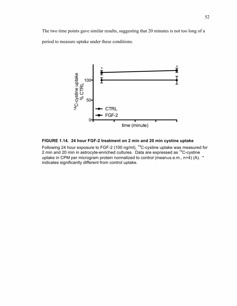
52
The two time points gave similar results, suggesting that 20 minutes is not too long of a
period to measure uptake under these conditions.
FIGURE 1.14. 24 hour FGF-2 treatment on 2 min and 20 min cystine uptake Following 24 hour exposure to FGF-2 (100 ng/ml), 14C-cystine uptake was measured for 2 min and 20 min in astrocyte-enriched cultures. Data are expressed as 14C-cystine uptake in CPM per microgram protein normalized to control (mean±s.e.m., n=4) (A). * indicates significantly different from control uptake.
0
50
100
CTRLFGF-2
* *
time (minute)
14C
-cys
tine
upta
ke%
CTR
L
53
CHAPTER II
β-N-methylamino-L-alanine induces oxidative stress and glutamate
release through action on system xC−
54
ABSTRACT
β-N-methylamino-L-alanine (BMAA) is a non-protein amino acid implicated in
the neurodegenerative disease amyotrophic lateral sclerosis and Parkinson-dementia
complex (ALS-PDC) on Guam. BMAA has recently been discovered in the brains of
Alzheimer’s and ALS patients in North America and is produced by various species of
cyanobacteria around the world. These findings suggest the possibility that BMAA may
be of concern not only for specific groups of Pacific Islanders, but for a much larger
population. Previous studies have indicated that BMAA can act as an excitotoxin by
acting on the NMDA receptor. We have shown that the mechanism of neurotoxicity is
actually three-fold; it involves not only direct action on the NMDA receptor, but also
activation of metabotropic glutamate receptor 5 (mGluR5) and induction of oxidative
stress. We now explore the mechanism by which BMAA activates the mGluR5 receptor
and induces oxidative stress. We found that BMAA inhibits the cystine/glutamate
antiporter (system xC-) mediated cystine uptake, which in turn leads to glutathione
depletion and increased oxidative stress. BMAA also appears to drive glutamate release
via system xC- and this glutamate induces toxicity through activation of the mGluR5
receptor. Therefore, the oxidative stress and mGluR5 activation induced by BMAA are
both mediated through action at system xC-. The multiple mechanisms of BMAA
toxicity, particularly the depletion of glutathione and enhanced oxidative stress, may
account for its ability to induce complex neurodegenerative diseases.

55
INTRODUCTION
In the 1950s it was observed that a substantial number of Chamorros, the native
people of Guam, began developing a disease that showed combined symptoms of
amyotrophic lateral sclerosis and Parkinson’s dementia complex (ALS-PDC). The idea
that BMAA may be involved in this disease began in the 1980s when it was found that
BMAA was present in cycad seeds which were consumed by the Chamorros (Nunn et al.,
1987; Amara, 1992; Kanai and Hediger, 1992; Storck et al., 1992; Rothstein et al., 1994;
1996; Diamond and Jahr, 1997; Lehre and Danbolt, 1998) and that injection of BMAA
into monkey brains induced a Parkinson-like disease (Spencer et al., 1987; Sibson et al.,
1998). Since its initial proposal, the BMAA hypothesis for the development of ALS-
PDC on Guam has been controversial (Rothstein et al., 1996; Asztely et al., 1997;
Rusakov and Kullmann, 1998; Cox and Sacks, 2002; Papapetropoulos, 2007). It was
challenged by findings that the levels of BMAA in cycad seeds are too low to cause
damage to the brain or the spinal cord, particularly because the Chamorros thoroughly
wash the cycad seeds, leading to very low levels of BMAA being consumed (Duncan et
al., 1990; Hardingham et al., 2002; Riccio and Ginty, 2002; Ivanov et al., 2006; Léveillé
et al., 2008; Xu et al., 2009; Hardingham and Bading, 2010). The BMAA hypothesis was
largely abandoned until the last few years.
A number of recent studies have brought the BMAA hypothesis back into
prominence. First, it was shown that BMAA is biomagnified. BMAA is produced by
cyanobacteria that live on cycad plants; it accumulates in the cycad seeds, which are
eaten by fruit bats, which are in turn eaten by the Chamorros (Cox et al., 2003; Lipton
and Chen, 2004; Xia et al., 2010). This system provides a new mechanism by which
56
BMAA can be accumulated to high levels in humans. Second, it was shown that BMAA
could become protein-associated. This property allows for BMAA to build up in tissue
and provides a mechanism for slow release (Lipton and Chen, 2004; Murch et al., 2004b;
Chen and Lipton, 2006; Xia et al., 2010). This slow release may provide a possible
explanation for the delayed onset of ALS-PDC following the time of BMAA
consumption (Ince and Codd, 2005; Niciu et al., 2012). Third, cyanobacteria present
throughout the world have been shown to produce BMAA (Futamachi and Pedley, 1976;
Roitbak and Fanardjian, 1981; Cox et al., 2005; Banack et al., 2007; Esterhuizen and
Downing, 2008; Johnson et al., 2008; Metcalf et al., 2008). Also, BMAA was found not
only in brain samples of ALS-PDC patients from Guam, but also in the brains of ALS
and Alzheimer’s disease patients from North America, but not in patients who died of
other causes (Venance et al., 1997; Murch et al., 2004a; Pablo et al., 2009). These results
suggest that BMAA may be of concern not only for people on select Pacific islands, but
for a much larger population. Fourth, BMAA at lower concentrations than previously
believed is neurotoxic. The original studies in cortical cell culture found that very high
BMAA concentrations (1-3 mM) were required to induce neuronal death (Ross et al.,
1987; Weiss and Choi, 1988; Weiss et al., 1989a; Oikawa et al., 2005). A more recent
study found that BMAA concentrations as low as 30 µM can cause selective death of
motor neurons (Rao et al., 2006; Butt, 2011; Köles et al., 2011) and cholinergic neurons
(Venance et al., 1997; Agulhon et al., 2008; Liu et al., 2010), and we found that BMAA
concentrations as low as 10 µM can enhance neuronal death induced by amyloid-β or 1-
methyl-4-phenylpyridinium ion (MPP+) (Seifert and Steinhäuser, 2001; Lobner et al.,
2007).
57
Given the potential relevance of BMAA consumption to neurodegenerative
diseases it is important to determine the mechanism of BMAA induced neuronal death.
We have previously shown that BMAA induces neuronal death through three distinct
mechanisms: activation of the NMDA and mGluR5 receptors, and induction of oxidative
stress (Lobner et al., 2007; Benarroch, 2009). Through electrophysiological recording it
has been shown that BMAA directly acts on NMDA receptors(Ross et al., 1987; Weiss
and Choi, 1988; Brownson et al., 2002; Lobner et al., 2007; Poskanzer and Yuste, 2011).
The current studies were designed to determine how BMAA activates mGluR5 receptors
and induces oxidative stress. Evidence is presented that the cystine/glutamate antiporter
(system xC-) plays an important role in these effects. System xC- involves the transport of
cystine into the cell in exchange for glutamate being transported out of the cell. Given
the functions of system xC- it seems likely that it plays an important role in neuronal
survival and death. By releasing glutamate it can increase extracellular glutamate levels
and potentially cause excitotoxicity. Through providing cystine uptake, it can regulate
cellular glutathione levels and in this way determine whether oxidative stress induced
neuronal death will occur. We find that the effects of BMAA on neuronal death involve
actions on system xC- to both inhibit cystine uptake and increase glutamate release.
MATERIALS AND METHODS
Materials
Timed pregnant Swiss Webster mice were obtained from Charles River
Laboratories (Wilmington, DE). 35S-cystine was from Perkin Elmer Life and Analytical
Sciences (Boston, MA). 5-(and -6)-carboxy-2′7′-dichlorodihydrofluorescein diacetate
58
(carboxy-H2DCFDA) was from Molecular Probes (Eugene, OR).
Cortical cell cultures
Mixed cortical cell cultures containing neuronal cells were prepared from fetal
(15-16 day gestation) mice as previously described (Cornell-Bell et al., 1990; Lobner,
2000). Dissociated cortical cells were plated on 24 well plates coated with poly-D-lysine
and laminin in Eagles’s Minimal Essential Medium (MEM, Earle’s salts, supplied
glutamine-free) supplemented with 5% heat-inactivated horse serum, 5% fetal bovine
serum, 2 mM glutamine and glucose (total 21 mM). Cultures were maintained in
humidified 5% CO2 incubators at 37°C. Mice were handled in accordance with a
protocol approved by our Institutional Animal Care Committee.
Induction of neuronal death
All experiments were performed on mixed cultures 13-15 days in vitro (DIV).
Toxicity was induced by exposure to the toxic agents for 24 hours in media as described
for plating except without serum. All exposure media contained 26 mM NaHCO3, as it
has been shown previously that HCO3- is required for expression of NMDA receptor
mediated BMAA toxicity (Weiss and Choi, 1988; Fellin et al., 2006; Butt, 2011; Parpura
et al., 2012).
Assay of neuronal death (LDH release)
Cell death was assessed in mixed cultures by the measurement of lactate
dehydrogenase (LDH) release from damaged or destroyed cells, in the extracellular fluid
59
24 hours after the beginning of the insult. Blank LDH levels were subtracted from insult
LDH values and results normalized to 100% neuronal death caused by 500 µM NMDA.
Control experiments have shown previously that the efflux of LDH occurring from either
necrotic or apoptotic cells is proportional to the number of cells damaged or destroyed
(Koh and Choi, 1987; Bezzi et al., 1998; Lobner, 2000; Bezzi et al., 2004; Kreft et al.,
2004) . Glial cell death (assessed by trypan blue staining) was not observed in any of the
current studies. Therefore results are presented as percent neuronal death.
35S-cystine uptake
Uptake of cystine was measured by exposure of cultures to 35S-cystine (2 µCi/ml)
for 20 min in the presence or absence of 3 mM BMAA and/or 1 mM S-4-carboxyphenyl
glycine (CPG). Following the exposure to 35S-cystine, the cultures were washed three
times and dissolved in 1% SDS (250 µl). An aliquot (200 µl) was removed and added to
scintillation fluid for counting. Values were normalized to control 35S-cystine uptake (20
min exposure to 35S-cystine without BMAA or CPG).
Glutathione assay
Total glutathione was assayed using a modification of a previous method (Baker
et al., 1990; Duan et al., 2003; Lobner et al., 2003). Briefly, following exposure to
BMAA for three hours, cells were washed with a HEPES buffered saline solution,
dissolved in 200 µl of 1% salicylic acid, and centrifuged. A 25 µl aliquot of the
supernatant was combined with 150 µl of 0.1 M phosphate/5 mM EDTA buffer, 10 µl of
20 mM dithiobis-2-nitrobenzoic acid, 100 µl of 5 mM NADPH, and 0.2 U of glutathione
60
reductase. Total glutathione was determined by kinetic analysis of absorbance changes at
402 nm for 1.5 min, with concentrations determined by comparison to a standard curve.
Assay of intracellular oxidative stress
Oxidative stress was assayed by measuring dichlorofluorescein oxidation using a
fluorescent plate reader following a modification of a previous method (Wang and
Joseph, 1999; Bridges et al., 2004; Lobner et al., 2007). Cultures were exposed to 3 mM
BMAA for 3 hours in the presence of 5- (and -6)-carboxy-2’7’-
dichlorodihydrofluorescein diacetate (carboxy-H2DFDA) (10 µM). The carboxy-
H2DFDA is de-esterified within cells to form a free acid that can then be oxidized to the
fluorescent 2’7’-dichlorofluorecein (DCF). After the exposure to carboxy-H2DCFDA,
cultures were washed three times with culture media lacking serum. Fluorescence was
then measured using a Fluoroskan Ascent fluorescence plate reader (Thermo lab
systems). The excitation filter was set at 485 nm and emission filter at 538 nm.
Background fluorescence (no carboxy-H2DCFDA added) was subtracted and the results
normalized to control conditions (carboxy-H2DCFDA added, but no BMAA exposure).
Analysis of glutamate release
Glutamate release was measured following exposure to 3 mM BMAA or cystine
for 1 hour. Experiments were performed in the presence of 10 µM MK-801 to block
potential injury induced by glutamate release. Samples of the bathing media from the
cell cultures were assayed for glutamate by using phenylisothiocyanate (PITC)
derivatization, HPLC (Agilent 1100) separation using a Hypersil-ODS reverse phase
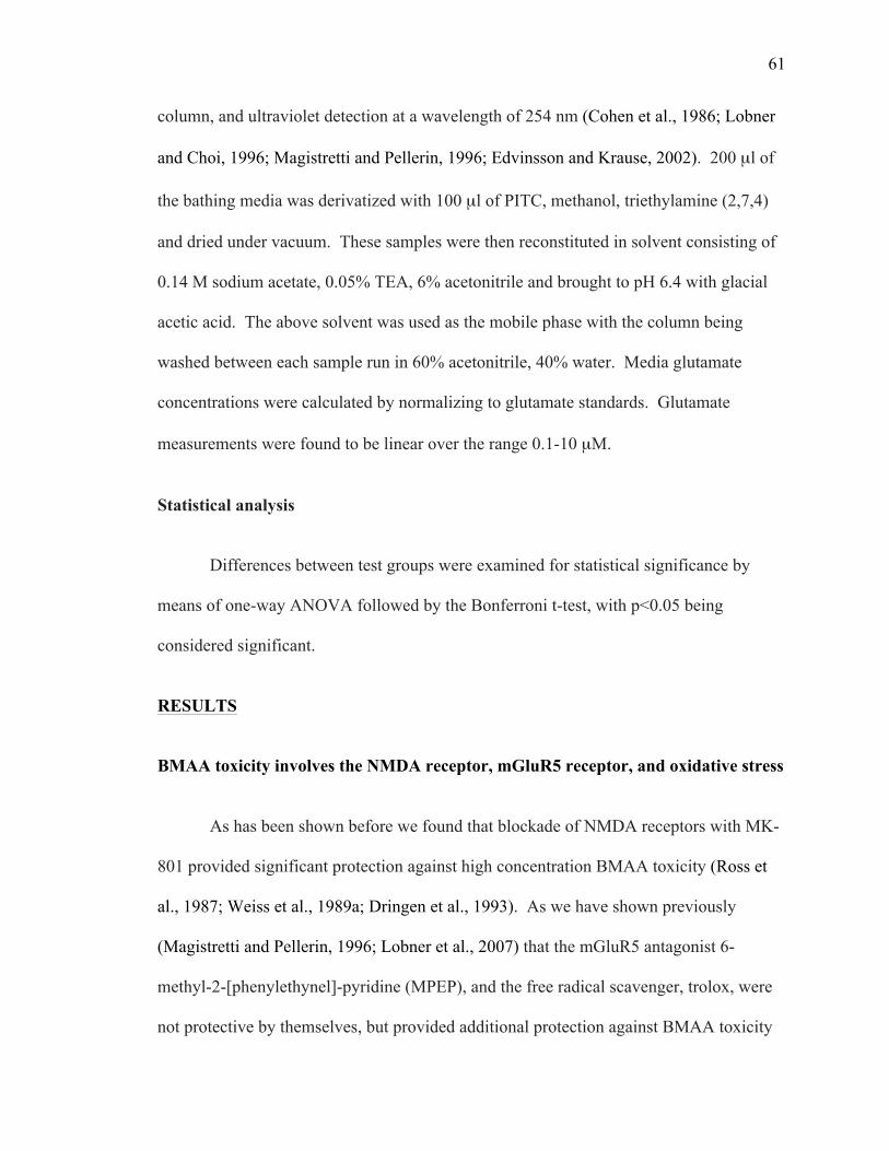
61
column, and ultraviolet detection at a wavelength of 254 nm (Cohen et al., 1986; Lobner
and Choi, 1996; Magistretti and Pellerin, 1996; Edvinsson and Krause, 2002). 200 µl of
the bathing media was derivatized with 100 µl of PITC, methanol, triethylamine (2,7,4)
and dried under vacuum. These samples were then reconstituted in solvent consisting of
0.14 M sodium acetate, 0.05% TEA, 6% acetonitrile and brought to pH 6.4 with glacial
acetic acid. The above solvent was used as the mobile phase with the column being
washed between each sample run in 60% acetonitrile, 40% water. Media glutamate
concentrations were calculated by normalizing to glutamate standards. Glutamate
measurements were found to be linear over the range 0.1-10 µM.
Statistical analysis
Differences between test groups were examined for statistical significance by
means of one-way ANOVA followed by the Bonferroni t-test, with p<0.05 being
considered significant.
RESULTS
BMAA toxicity involves the NMDA receptor, mGluR5 receptor, and oxidative stress
As has been shown before we found that blockade of NMDA receptors with MK-
801 provided significant protection against high concentration BMAA toxicity (Ross et
al., 1987; Weiss et al., 1989a; Dringen et al., 1993). As we have shown previously
(Magistretti and Pellerin, 1996; Lobner et al., 2007) that the mGluR5 antagonist 6-
methyl-2-[phenylethynel]-pyridine (MPEP), and the free radical scavenger, trolox, were
not protective by themselves, but provided additional protection against BMAA toxicity
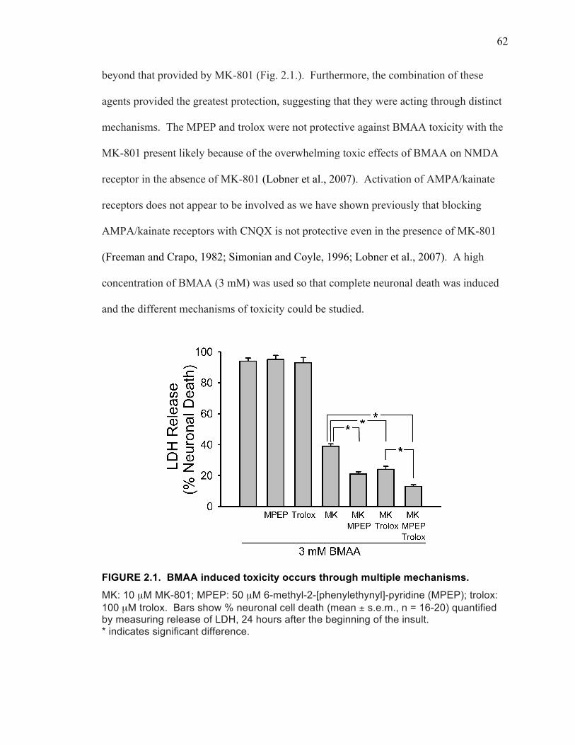
62
beyond that provided by MK-801 (Fig. 2.1.). Furthermore, the combination of these
agents provided the greatest protection, suggesting that they were acting through distinct
mechanisms. The MPEP and trolox were not protective against BMAA toxicity with the
MK-801 present likely because of the overwhelming toxic effects of BMAA on NMDA
receptor in the absence of MK-801 (Lobner et al., 2007). Activation of AMPA/kainate
receptors does not appear to be involved as we have shown previously that blocking
AMPA/kainate receptors with CNQX is not protective even in the presence of MK-801
(Freeman and Crapo, 1982; Simonian and Coyle, 1996; Lobner et al., 2007). A high
concentration of BMAA (3 mM) was used so that complete neuronal death was induced
and the different mechanisms of toxicity could be studied.
FIGURE 2.1. BMAA induced toxicity occurs through multiple mechanisms. MK: 10 µM MK-801; MPEP: 50 µM 6-methyl-2-[phenylethynyl]-pyridine (MPEP); trolox: 100 µM trolox. Bars show % neuronal cell death (mean ± s.e.m., n = 16-20) quantified by measuring release of LDH, 24 hours after the beginning of the insult. * indicates significant difference.
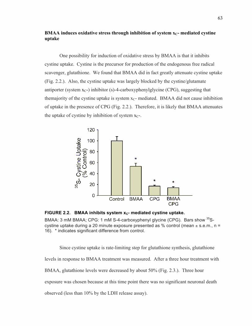
63
BMAA induces oxidative stress through inhibition of system xC- mediated cystine uptake
One possibility for induction of oxidative stress by BMAA is that it inhibits
cystine uptake. Cystine is the precursor for production of the endogenous free radical
scavenger, glutathione. We found that BMAA did in fact greatly attenuate cystine uptake
(Fig. 2.2.). Also, the cystine uptake was largely blocked by the cystine/glutamate
antiporter (system xC-) inhibitor (s)-4-carboxyphenylglycine (CPG), suggesting that
themajority of the cystine uptake is system xC- mediated. BMAA did not cause inhibition
of uptake in the presence of CPG (Fig. 2.2.). Therefore, it is likely that BMAA attenuates
the uptake of cystine by inhibition of system xC-.
FIGURE 2.2. BMAA inhibits system xC- mediated cystine uptake. BMAA: 3 mM BMAA; CPG: 1 mM S-4-carboxyphenyl glycine (CPG). Bars show 35S-cystine uptake during a 20 minute exposure presented as % control (mean ± s.e.m., n = 16). * indicates significant difference from control.
Since cystine uptake is rate-limiting step for glutathione synthesis, glutathione
levels in response to BMAA treatment was measured. After a three hour treatment with
BMAA, glutathione levels were decreased by about 50% (Fig. 2.3.). Three hour
exposure was chosen because at this time point there was no significant neuronal death
observed (less than 10% by the LDH release assay).
64
Since BMAA causes a decrease in the levels of the endogenous free radical
scavenger glutathione, the total oxidative stress of the cells should be increased. We
measured cellular oxidative stress with the fluorescent dye dichlorofluorescein (DCF).
Treatment with BMAA for 3 hours caused a significant increase in oxidative stress,
which was blocked by the free radical scavenger trolox (Fig. 2.4.).
FIGURE 2.3. BMAA decreases cellular glutathione levels. BMAA: 3 mM BMAA. Bars show glutathione levels following three hour BMAA exposure presented as % control (mean ± s.e.m, n = 16). * indicates significant difference from control.
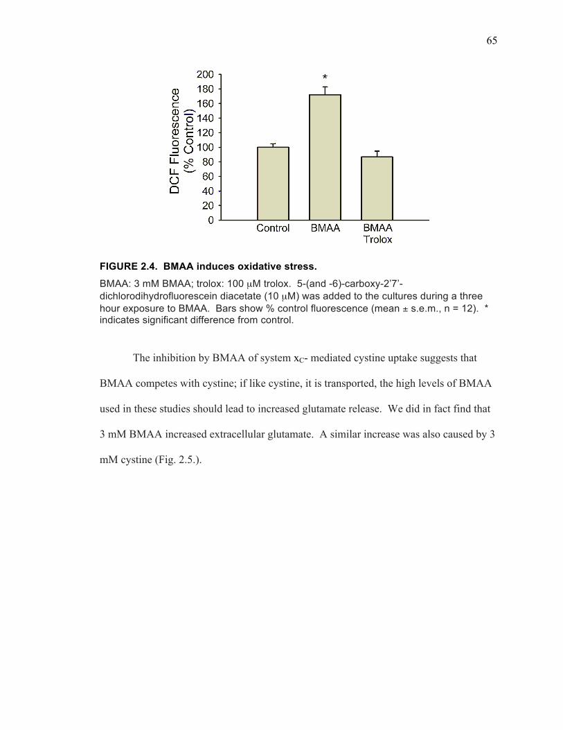
65
FIGURE 2.4. BMAA induces oxidative stress. BMAA: 3 mM BMAA; trolox: 100 µM trolox. 5-(and -6)-carboxy-2’7’-dichlorodihydrofluorescein diacetate (10 µM) was added to the cultures during a three hour exposure to BMAA. Bars show % control fluorescence (mean ± s.e.m., n = 12). * indicates significant difference from control.
The inhibition by BMAA of system xC- mediated cystine uptake suggests that
BMAA competes with cystine; if like cystine, it is transported, the high levels of BMAA
used in these studies should lead to increased glutamate release. We did in fact find that
3 mM BMAA increased extracellular glutamate. A similar increase was also caused by 3
mM cystine (Fig. 2.5.).

66
FIGURE 2.5. BMAA and cystine stimulate glutamate release. BMAA: 3 mM BMAA; cystine: 3 mM cystine. Bars show extracellular glutamate levels following a 1 hour BMAA or cystine exposure (mean ± s.e.m., n = 8). * indicates significant difference from control.
Since BMAA appears to induce glutamate release via system xC-, it is possible
that this glutamate is responsible for at least part of the glutamate receptor mediated
neuronal death. Because BMAA is also a direct agonist for glutamate receptors (Lobner
et al., 2007; Cucchiaroni et al., 2010), and 3 mM cystine caused a similar increase in
extracellular glutamate compared with 3 mM BMAA, we used 3 mM cystine to study the
effect of glutamate released by system xC-. We found that 3 mM cystine did induce
neuronal death (Fig. 2.6.). However, the death was less than that caused by 3 mM
BMAA, likely due to the fact that cystine is not a direct NMDA receptor agonist like
BMAA. However, the cystine toxicity was partially blocked by either MPEP or MK-801.
No protection was afforded by trolox, as would be expected since a high concentration of
cystine should not induce oxidative stress.
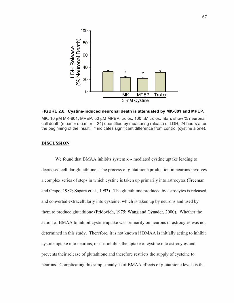
67
FIGURE 2.6. Cystine-induced neuronal death is attenuated by MK-801 and MPEP. MK: 10 µM MK-801; MPEP: 50 µM MPEP; trolox: 100 µM trolox. Bars show % neuronal cell death (mean ± s.e.m, n = 24) quantified by measuring release of LDH, 24 hours after the beginning of the insult. * indicates significant difference from control (cystine alone).
DISCUSSION
We found that BMAA inhibits system xC- mediated cystine uptake leading to
decreased cellular glutathione. The process of glutathione production in neurons involves
a complex series of steps in which cystine is taken up primarily into astrocytes (Freeman
and Crapo, 1982; Sagara et al., 1993). The glutathione produced by astrocytes is released
and converted extracellularly into cysteine, which is taken up by neurons and used by
them to produce glutathione (Fridovich, 1975; Wang and Cynader, 2000). Whether the
action of BMAA to inhibit cystine uptake was primarily on neurons or astrocytes was not
determined in this study. Therefore, it is not known if BMAA is initially acting to inhibit
cystine uptake into neurons, or if it inhibits the uptake of cystine into astrocytes and
prevents their release of glutathione and therefore restricts the supply of cysteine to
neurons. Complicating this simple analysis of BMAA effects of glutathione levels is the
68
finding that blocking system xc- with CPG for as long as 24 hours does not cause a
decrease in cellular glutathione levels (data not shown). The fact that BMAA does cause
a decrease in cellular glutathione indicates that it has actions beyond its effect on system
xc- mediated cystine uptake. Other known actions of BMAA that may play a role in its
effect on glutathione levels include inhibition of the enzymes involved in glutathione
synthesis (Esterhuizen-Londt et al., 2011), activation of NMDA and mGluR5 receptors
(Lobner et al., 2007), and results presented in the current study indicating that it can
cause glutamate release.
The data presented here strongly suggests that BMAA not only inhibits cystine
uptake, but also drives system xC- to release glutamate and that this glutamate induces
neuronal death by acting on mGluR5 receptors and NMDA receptors. The evidence for
this is that BMAA and cystine at the same concentration (3 mM) increase extracellular
glutamate and they both induce NMDA receptor and mGluR5 mediated neuronal death.
There is no evidence that either of these compounds directly activates mGluR5.
Unfortunately, CPG could not be used to block the glutamate release because while it is a
fairly poor substrate for system xC- (Patel et al., 2004; Rush et al., 2012b), at the high
concentrations needed to compete with the high concentrations of BMAA and cystine
used in these studies it stimulated glutamate release by itself (data not shown). Why in
our studies there was some protection against cystine induced toxicity by MK-801 is not
clear. There is no published data indicating that cystine is a direct NMDA receptor
agonist. However, we can not exclude the possibly that some of the cystine may be
converted into cysteine, which is an NMDA receptor agonist (Pullan et al., 1987;
Ballatori et al., 2009). Alternatively, the glutamate release stimulated by cystine via
69
system xC- may act to stimulate NMDA receptors. Furthermore, it has been shown
activation of mGluR5 and NMDA receptor positively enhances each other’s activity
(Alagarsamy et al., 1999; Benquet et al., 2002; Chen et al., 2011), which could
potentially add to the BMAA toxicity.
Both oxidative stress and excitotoxicity have been suggested to be regulated by
system xC- in various cell types. It has been known for many years that inhibition of
cystine uptake mediated by system xC- can induce neuronal death by decreasing
glutathione levels (Murphy et al., 1989; Murphy and Baraban, 1990; Dringen et al., 2000)
In contrast, astrocytes activated by IL-beta were shown to kill cortical neurons by release
glutamate via system xC- (Bolaños et al., 1995; Rice and Russo-Menna, 1998; Fogal et
al., 2007). Also, microglia can release glutamate via system xC- that can kill cerebellar
granule cells (Deneke and Fanburg, 1989; Piani and Fontana, 1994), oligodendrocytes
(Rahman et al., 2005; Domercq et al., 2007), and enhance amyloid-β induced neuronal
death in cortical cultures (Beutler, 1989; Deneke and Fanburg, 1989; Qin et al., 2006a).
Finally, glutamate release from kidney dendritic cells via system xC- can inhibit T cell
activation through actions on mGluR5 receptors (McBean, 2002; Pacheco et al., 2006).
In the current study we find evidence that oxidative stress induced by the inhibition of
cystine uptake and excitotoxicity induced by system xC- mediated glutamate release both
play a role in BMAA induced neuronal death.
Interestingly, another amino acid implicated in neurological diseases, β-N-oxalyl-
L-α,β-diaminopropionic acid (ODAP), also shares some properties with BMAA: it
directly activates glutamatergic receptors and is transported by system xC- (Kranich et al.,
1996; Chase et al., 2007). It is likely that ODAP acts similar to BMAA and causes
70
cytotoxicity through at least some of the same mechanisms.
The finding that BMAA not only inhibited cystine uptake by system xC-, but also
stimulated glutamate release, suggests that it was transported by system xC-. This
transport provides a mechanism by which BMAA can accumulate in cells. There is
evidence that BMAA may be incorporated into proteins (Murch et al., 2004b; Lobner,
2009) and could therefore potentially play a role in the protein misfolding found in
neurodegenerative diseases (McBean, 2002; Uversky, 2008). Whether BMAA can be
incorporated into GSH is unknown. If BMAA does become incorporated into GSH this
would provide another potential mechanism for its ability to cause depletion of GSH and
oxidative stress.
The importance of BMAA acting on system xC- to induce oxidative stress and
excitotoxicity at the same time is that this may be partially account for the different types
of neurological diseases that have been associated with BMAA consumption. It is
possible that the different actions of BMAA, in association with varying underlining
conditions, may lead to the multiple disorders. Most neurodegenerative diseases appear
to involve NMDA receptor mediated excitotoxicity (Arrick et al., 1985; Lipton and Chen,
2004) and oxidative stress (Dringen et al., 1999; Cui et al., 2004), and mGluR5 receptor
activation has been implicated in Parkinson’s disease as well (Zerangue and Kavanaugh,
1996b; Chen and Swanson, 2003; Marino et al., 2003; Aoyama et al., 2006). All of these
mechanisms of neuronal injury may be enhanced by BMAA. The finding that BMAA
causes a depletion of cellular glutathione is of particular importance. There is evidence
that glutathione depletion plays a role in Alzheimer’s disease, Parkinson’s disease, and
ALS (Bannai and Kitamura, 1980; Bains and Shaw, 1997; Liu et al., 2004; Zeevalk et al.,
71
2008). This type of low level, long-term, depletion of glutathione that may be caused by
the inhibition of cystine uptake could cause the long-term oxidative stress that may
underlie aspects of neurodegenerative diseases.
From these results and previous studies, it is obvious that system xC- plays an
important role in neuronal fate. We next want to examine if system xC- can be regulated
by neurotrophic factors, which are naturally occurring molecules, which also regulate
neuronal fate in multiple ways. Neurotrophic factors have wide ranging effects in the
central nervous system, including altering excitotoxicity (Koh et al., 1995; Sato et al.,
1999), oxidative stress (Zhang et al., 1993).
72
CHAPTER III
FUNCTIONAL UPREGULATION OF SYSTEM XC- BY
FIBROBLAST GROWTH FACTOR-2
73
ABSTRACT
The cystine/glutamate antiporter (system xC-) is a sodium-independent amino acid
transport system. Disruption of this system may lead to multiple effects in the central
nervous system including decreased cellular glutathione. Since multiple neurological
diseases involve glutathione depletion, and disruption of growth factor signaling has also
been implicated in these diseases, it is possible that some growth factors effects are
mediated by regulation of system xC-. We tested the growth factors fibroblast growth
factor-2 (FGF-2), insulin-like growth factor-1 (IGF-1), neuregulin-1 (NRG),
neurotrophin-4 (NT-4), and brain derived neurotrophic factor (BDNF) on system xC-
mediated 14C-cystine uptake in mixed neuronal and glial cortical cultures. Only FGF-2
significantly increased cystine uptake. The effect was observed in astrocyte-enriched
cultures, but not in cultures of neurons or microglia. The increase was blocked by the
system xC- inhibitor (s)-4-carboxyphenylglycine (CPG), required at least 12 hour FGF-2
treatment, and was prevented by the protein synthesis inhibitor cycloheximide. Kinetic
analysis indicated FGF-2 treatment increased the Vmax for cystine uptake while the Km
remained the same. Quantitative PCR showed an increase in mRNA for xCT, the
functional subunit of system xC-, beginning at 3 hours of FGF-2 treatment, with a
dramatic increase after 12 hours. Blocking FGFR1 with PD 166866 blocked the FGF-2
effect. Treatment with both a PI3-kinase inhibitor (LY-294002) or a MEK/ERK inhibitor
(U0126) for 1 hour prior to and during the FGF-2 treatment blocked the increased cystine
uptake. The upregulation of system xC- by FGF-2 may be responsible for some of the
known physiological actions of FGF-2.
74
INTRODUCTION
The function of system xC- makes it likely to play an important role in regulating
many processes in the central nervous system. By releasing glutamate, it can increase
extracellular glutamate levels, which may alter neuronal plasticity and potentially cause
excitotoxicity; Through providing cystine uptake, it supplies the substrate for cellular
glutathione synthesis, and in this way may determine whether oxidative stress induced
neuronal death will occur.
There is considerable evidence that glutamate released from system xC- is
involved in both physiological and pathological processes. Physiologically glutamate
release from system xC- plays a role at the photoreceptor synapse (Hu et al., 2008) and in
regulating immune cell function (Xue and Field, 2011). It is known that glutamate
release from system xC- can cause toxicity, and this is observed in many pathological
processes. Activated astrocytes release glutamate via system xC- that can kill cortical
neurons (Fogal et al., 2007). Also activated microglia release glutamate via system xC-
that can kill cerebellar granule cells (Piani and Fontana, 1994) and enhance amyloid-beta
induced neuronal death in cortical cultures (Qin et al., 2006b).
Glutathione is known to detoxify superoxide in a reaction catalyzed by
glutathione peroxidase forming disulfide/oxidized glutathione. This reaction is
accomplished by redox changes at the sulfhydryl group of the cysteine residue of
glutathione (Dringen and Hirrlinger, 2003). The cystine uptake mediated by system xC-
appears to play a critical role in regulating cellular glutathione levels, because de novo
synthesis of glutathione in the brain is rate-limited by the uptake of cystine (Sagara et al.,
1993). Specifically, astrocytes express high levels of system xC- and have high levels of
75
cystine uptake (Dringen et al., 2000; Lewerenz et al., 2006), while neurons of the central
nervous system have limited capacity for cystine uptake, and are reliant primarily on
astrocytes for the provision of cysteine and cysteinylglycine for neuronal glutathione
production (Dringen et al., 1999). The first evidence that system xC- could play a role in
neuronal death was provided when it was shown that high concentrations of glutamate
could competitively inhibit cystine uptake leading to an oxidative stress mediated
neuronal death (Murphy et al., 1990). This type of toxicity was shown to occur before
glutamate receptors developed (Murphy and Baraban, 1990) and to involve depletion of
glutathione (Ratan et al., 1994). In contrast to the enhancement of neurotoxicity caused
by releasing glutamate, system xC- activity on non-neuronal cells can be protective to
neurons by generating and releasing glutathione. Overexpression of xCT, the functional
subunit of system xC-, in astrocytes has been shown to enhance glutathione release and
protect neurons from oxidative stress (Shih et al., 2006). Since neurological diseases
including schizophrenia, depression, Alzheimer’s disease, and Parkinson’s disease have
all been associated with depletion of cellular glutathione (Ballatori et al., 2009; Dean et
al., 2009; Shibasaki et al., 2009; Maes et al., 2011), it is possible that altered system xC-
function plays a role in these diseases.
Neurotrophic factors have wide ranging effects in the central nervous system
including altering excitotoxicity (Koh et al., 1995; Chintala et al., 2005), oxidative stress
(Zhang et al., 1993; Shih et al., 2006), and cellular glutathione levels (Pan and Perez-
Polo, 1993; Sato et al., 1999). The overlapping actions of growth factors and system xC-
suggest the possibility that some neurotrophic factor effects may be mediated by
76
regulation of system xC-. Therefore we tested the effects of a number of well-established
neurotrophic factors on system xC- function.
MATERIALS AND METHODS
Materials
Timed pregnant Swiss Webster mice were obtained from Charles River
Laboratories (Wilmington, DE). Serum was from Atlanta Biologicals (Lawrenceville,
GA). Growth factors were from Alomone Labs (Jerusalem, Israel). 14C-cystine was from
Perkin Elmer (Waltham, MA). All other chemicals were obtained from Sigma (St. Louis,
MO).
Cortical cell cultures
Mixed cortical cell cultures containing glial and neuronal cells were prepared
from fetal (15-16 day gestation) mice as previously described (Lobner, 2000; Sasaki et
al., 2002). Dissociated cortical cells were plated on 24-well plates coated with poly-D-
lysine and laminin in Eagles’ Minimal Essential Medium (MEM, Earle’s salts, supplied
glutamine-free) supplemented with 5% heat-inactivated horse serum, 5% fetal bovine
serum, 2 mM glutamine and glucose (total 21 mM). Neuron-enriched cultures were
prepared exactly as above with the addition of 10 µM cytosine arabinoside 48 hours after
plating to inhibit glial replication. In these cultures <1% of cells are astrocytes (Dugan et
al., 1995; Sato et al., 2000; Sasaki et al., 2002; Rush et al., 2010). Astrocyte-enriched
cultures were prepared as described for mixed cultures except they are from cortical
tissue taken from post-natal day 1-3 mice (Choi et al., 1987; Schwartz and Wilson, 1992;
77
Venugopal and Jaiswal, 1996; Rush et al., 2010). Microglia cultures were prepared by
first growing astrocyte-enriched cultures, then shaking the microglia free and plating
them in media containing 10 ng/ml colony stimulating factor (CSF) (Itoh et al., 1999;
Barger and Basile, 2001). Cultures were maintained in humidified 5% CO2 incubators at
37°C. Mice were handled in accordance with a protocol approved by our institutional
animal care committee and in compliance with the Public Heath Service Policy on
Humane Care and Use of Laboratory Animals. All efforts were made to minimize animal
suffering and reduce the number of animals used.
14C-cystine uptake
System xC- mediated uptake of radiolabeled cystine was assayed as previously
described with modifications (Itoh et al., 1999; Liu et al., 2009). Growth factors were
added to the serum containing media for the indicated durations. Cultures were then
washed into HEPES buffered saline solution and immediately exposed to 14C-cystine
(0.025µCi/ml) for 20 min. Following 14C-cystine exposure, cultures were washed with
ice cold HEPES buffered saline solution and dissolved in 250 µl sodium dodecyl sulfate
(0.1%). An aliquot (200 µl) was removed and added to scintillation fluid for counting.
In some experiments an aliquot was also used to measure cellular protein levels by the
BCA assay. Values were normalized to 14C-cystine uptake in untreated control on the
same experimental plate. Kinetic analysis of cystine uptake was performed as previously
described (Lee and Johnson, 2004; Fogal et al., 2007). Uptake of 14C-cystine was
measured over a range of cystine concentrations: 0.1 µM, 0.3 µM, 1µM, 3 µM, 10 µM,
and 30 µM. Kinetic analysis was performed using the Hanes-Woolf plot where
78
[substrate]/velocity is plotted against [substrate]. The slope corresponds to 1/Vmax, while
the X-intercept corresponds to the Km.
Reverse transcription quantitative real-time PCR (RT-qPCR)
Transcriptional expression of xCT in astrocyte-enriched cultures after stimulation
with FGF-2 was examined using RT-qPCR. Briefly, after various durations of treatment
with FGF-2 cells were washed 3 times with cold PBS. Total RNA was isolated with
TRIzol extraction (Invitrogen; Carlsbad, CA) according to manufacturer’s protocol,
cDNA was synthesized using the Reverse Transcription System (Promega; Madison, WI)
from 1 µg of total RNA following the manufacturer’s protocol. All products were
amplified on a StepOne real-time PCR system (Applied Biosystems; Carlsbad, CA) using
100 ng of cDNA, PerfeCTa SYBR Green FastMix with ROX (Quanta Biosciences;
Gaithersberg, MD), and the following primers: gapdh mouse forward-AAG GGC TCA
TGA CCA CAG TC; gapdh mouse reverse-GGA TAC AGG GAT GAT GTT CT; xCT
mouse forward-AGG GCA TAC TCC AGA ACA CG; xCT mouse reverse-GAC AGG
GCT CCA AAA AGT GA. xCT expression was normalized to the housekeeping gene
glyceraldehyde-3-phosphate dehydrogenase (gapdh). Quantitative PCR was used to
determine fold increase of xCT mRNA using the 2-ΔΔCT method.
Statistical analysis
Differences between test groups were examined for statistical significance by
means of one-way ANOVA followed by the Bonferroni correction post-hoc test, with
p<0.05 being considered significant.
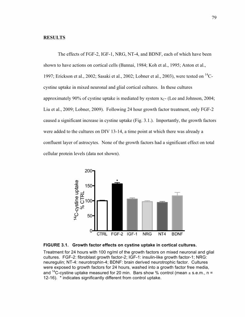
79
RESULTS
The effects of FGF-2, IGF-1, NRG, NT-4, and BDNF, each of which have been
shown to have actions on cortical cells (Bannai, 1984; Koh et al., 1995; Anton et al.,
1997; Erickson et al., 2002; Sasaki et al., 2002; Lobner et al., 2003), were tested on 14C-
cystine uptake in mixed neuronal and glial cortical cultures. In these cultures
approximately 90% of cystine uptake is mediated by system xC- (Lee and Johnson, 2004;
Liu et al., 2009; Lobner, 2009). Following 24 hour growth factor treatment, only FGF-2
caused a significant increase in cystine uptake (Fig. 3.1.). Importantly, the growth factors
were added to the cultures on DIV 13-14, a time point at which there was already a
confluent layer of astrocytes. None of the growth factors had a significant effect on total
cellular protein levels (data not shown).
FIGURE 3.1. Growth factor effects on cystine uptake in cortical cultures. Treatment for 24 hours with 100 ng/ml of the growth factors on mixed neuronal and glial cultures. FGF-2: fibroblast growth factor-2; IGF-1: insulin-like growth factor-1; NRG: neuregulin; NT-4: neurotrophin-4; BDNF: brain derived neurotrophic factor. Cultures were exposed to growth factors for 24 hours, washed into a growth factor free media, and 14C-cystine uptake measured for 20 min. Bars show % control (mean ± s.e.m., n = 12-16). * indicates significantly different from control uptake.
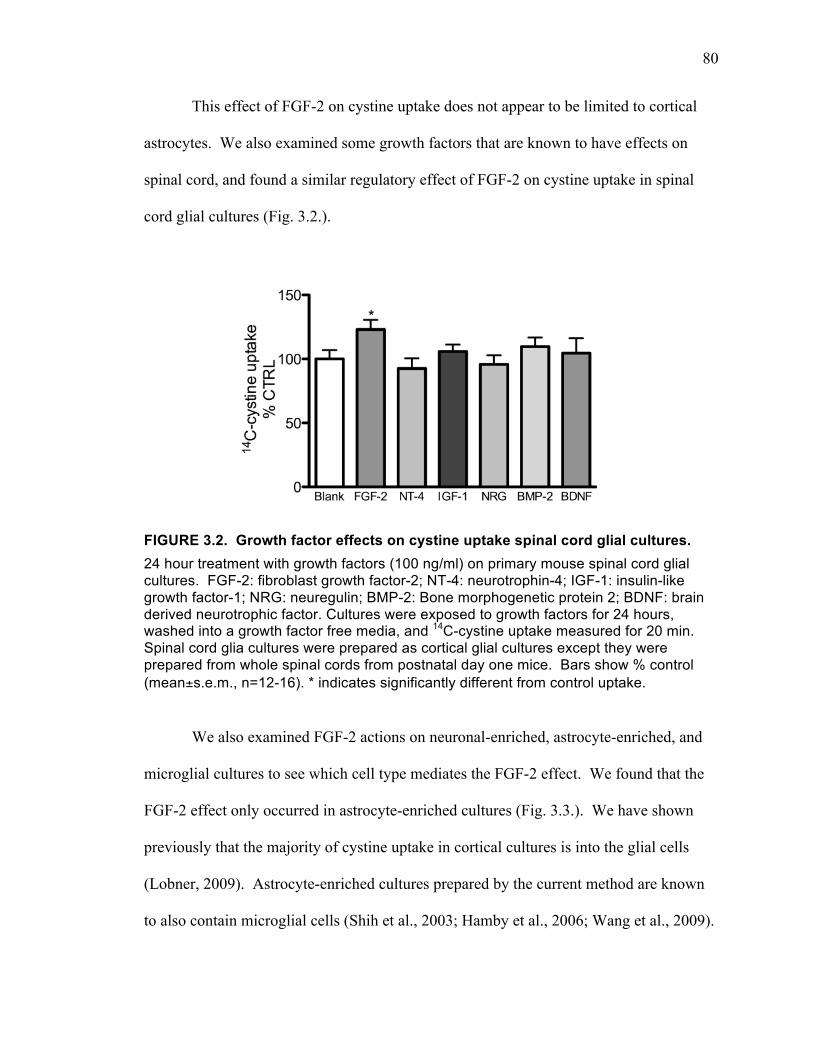
80
This effect of FGF-2 on cystine uptake does not appear to be limited to cortical
astrocytes. We also examined some growth factors that are known to have effects on
spinal cord, and found a similar regulatory effect of FGF-2 on cystine uptake in spinal
cord glial cultures (Fig. 3.2.).
FIGURE 3.2. Growth factor effects on cystine uptake spinal cord glial cultures. 24 hour treatment with growth factors (100 ng/ml) on primary mouse spinal cord glial cultures. FGF-2: fibroblast growth factor-2; NT-4: neurotrophin-4; IGF-1: insulin-like growth factor-1; NRG: neuregulin; BMP-2: Bone morphogenetic protein 2; BDNF: brain derived neurotrophic factor. Cultures were exposed to growth factors for 24 hours, washed into a growth factor free media, and 14C-cystine uptake measured for 20 min. Spinal cord glia cultures were prepared as cortical glial cultures except they were prepared from whole spinal cords from postnatal day one mice. Bars show % control (mean±s.e.m., n=12-16). * indicates significantly different from control uptake.
We also examined FGF-2 actions on neuronal-enriched, astrocyte-enriched, and
microglial cultures to see which cell type mediates the FGF-2 effect. We found that the
FGF-2 effect only occurred in astrocyte-enriched cultures (Fig. 3.3.). We have shown
previously that the majority of cystine uptake in cortical cultures is into the glial cells
(Lobner, 2009). Astrocyte-enriched cultures prepared by the current method are known
to also contain microglial cells (Shih et al., 2003; Hamby et al., 2006; Wang et al., 2009).
81
However, since there was no effect of FGF-2 in the microglial cultures, we believe that
the effect occurs on astrocytes. Therefore, all of the following experiments were
performed using the astrocyte-enriched cultures. We next tested whether the effect of
FGF-2 was on system xC- mediated cystine uptake. More than 90% of cystine uptake
was blocked by the system xC- antagonist (s)-4-carboxyphenylglycine (CPG), and the
increase caused by 24 hour treatment with FGF-2 was completely blocked by CPG (Fig.
3.4.).
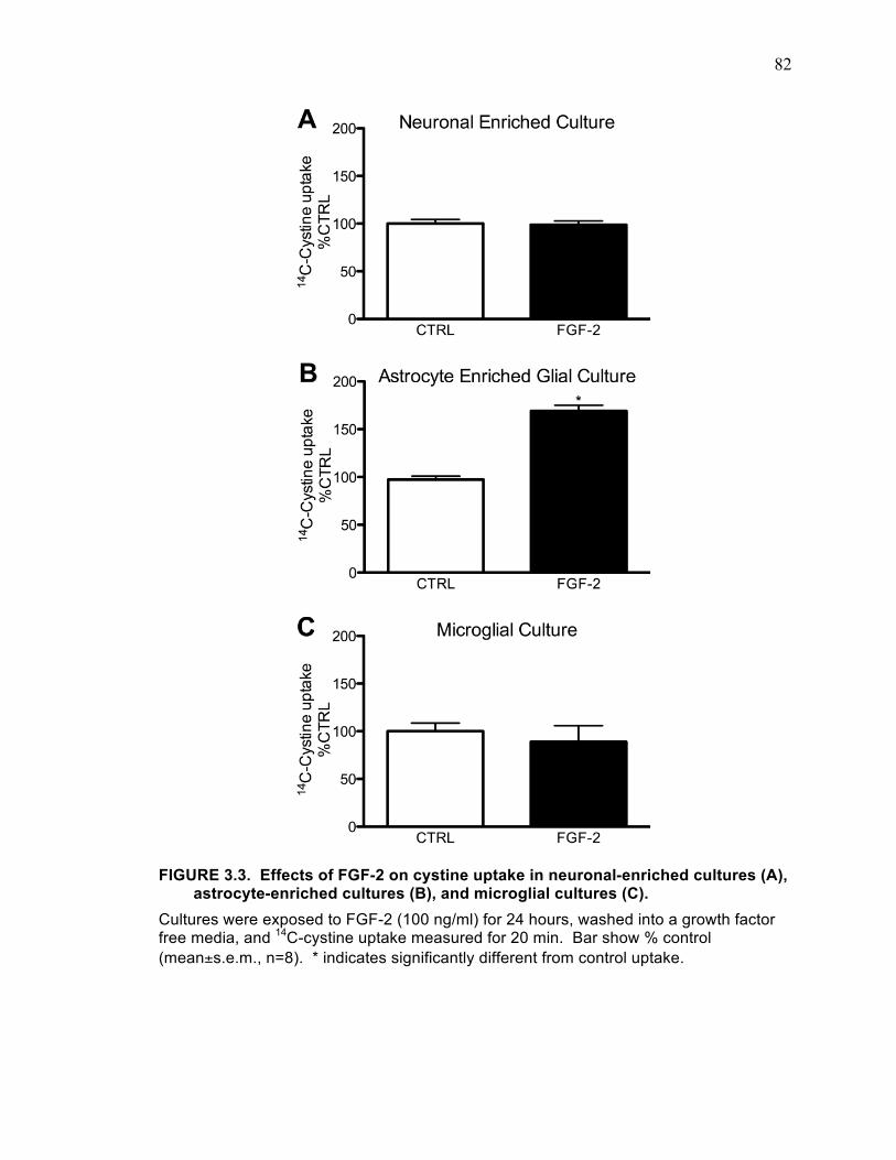
82
FIGURE 3.3. Effects of FGF-2 on cystine uptake in neuronal-enriched cultures (A), astrocyte-enriched cultures (B), and microglial cultures (C).
Cultures were exposed to FGF-2 (100 ng/ml) for 24 hours, washed into a growth factor free media, and 14C-cystine uptake measured for 20 min. Bar show % control (mean±s.e.m., n=8). * indicates significantly different from control uptake.
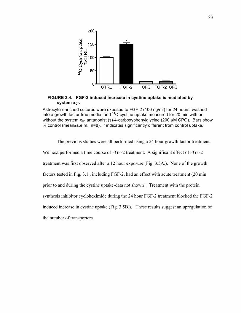
83
FIGURE 3.4. FGF-2 induced increase in cystine uptake is mediated by system xC-.
Astrocyte-enriched cultures were exposed to FGF-2 (100 ng/ml) for 24 hours, washed into a growth factor free media, and 14C-cystine uptake measured for 20 min with or without the system xC- antagonist (s)-4-carboxyphenylglycine (200 µM CPG). Bars show % control (mean±s.e.m., n=8). * indicates significantly different from control uptake.
The previous studies were all performed using a 24 hour growth factor treatment.
We next performed a time course of FGF-2 treatment. A significant effect of FGF-2
treatment was first observed after a 12 hour exposure (Fig. 3.5A.). None of the growth
factors tested in Fig. 3.1., including FGF-2, had an effect with acute treatment (20 min
prior to and during the cystine uptake-data not shown). Treatment with the protein
synthesis inhibitor cycloheximide during the 24 hour FGF-2 treatment blocked the FGF-2
induced increase in cystine uptake (Fig. 3.5B.). These results suggest an upregulation of
the number of transporters.
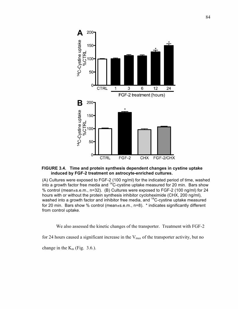
84
FIGURE 3.4. Time and protein synthesis dependent changes in cystine uptake induced by FGF-2 treatment on astrocyte-enriched cultures.
(A) Cultures were exposed to FGF-2 (100 ng/ml) for the indicated period of time, washed into a growth factor free media and 14C-cystine uptake measured for 20 min. Bars show % control (mean±s.e.m., n=32). (B) Cultures were exposed to FGF-2 (100 ng/ml) for 24 hours with or without the protein synthesis inhibitor cycloheximide (CHX, 200 ng/ml), washed into a growth factor and inhibitor free media, and 14C-cystine uptake measured for 20 min. Bars show % control (mean±s.e.m., n=8). * indicates significantly different from control uptake.
We also assessed the kinetic changes of the transporter. Treatment with FGF-2
for 24 hours caused a significant increase in the Vmax of the transporter activity, but no
change in the Km (Fig. 3.6.).
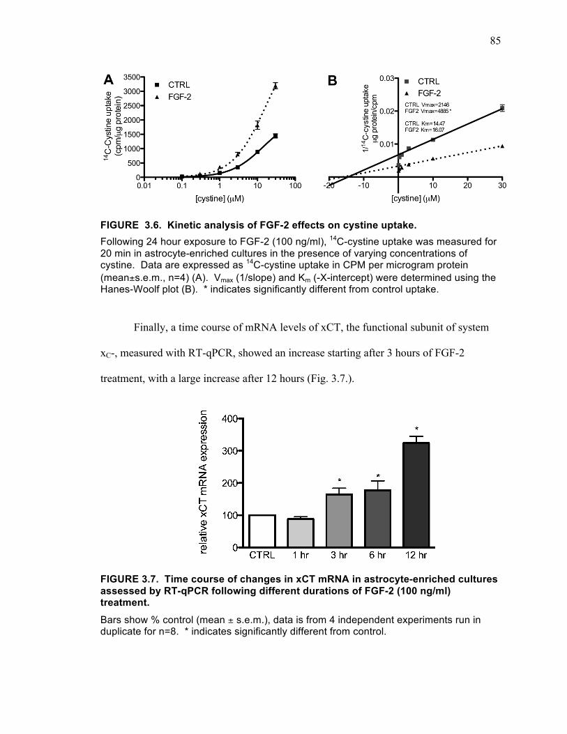
85
FIGURE 3.6. Kinetic analysis of FGF-2 effects on cystine uptake. Following 24 hour exposure to FGF-2 (100 ng/ml), 14C-cystine uptake was measured for 20 min in astrocyte-enriched cultures in the presence of varying concentrations of cystine. Data are expressed as 14C-cystine uptake in CPM per microgram protein (mean±s.e.m., n=4) (A). Vmax (1/slope) and Km (-X-intercept) were determined using the Hanes-Woolf plot (B). * indicates significantly different from control uptake.
Finally, a time course of mRNA levels of xCT, the functional subunit of system
xC-, measured with RT-qPCR, showed an increase starting after 3 hours of FGF-2
treatment, with a large increase after 12 hours (Fig. 3.7.).
FIGURE 3.7. Time course of changes in xCT mRNA in astrocyte-enriched cultures assessed by RT-qPCR following different durations of FGF-2 (100 ng/ml) treatment. Bars show % control (mean ± s.e.m.), data is from 4 independent experiments run in duplicate for n=8. * indicates significantly different from control.
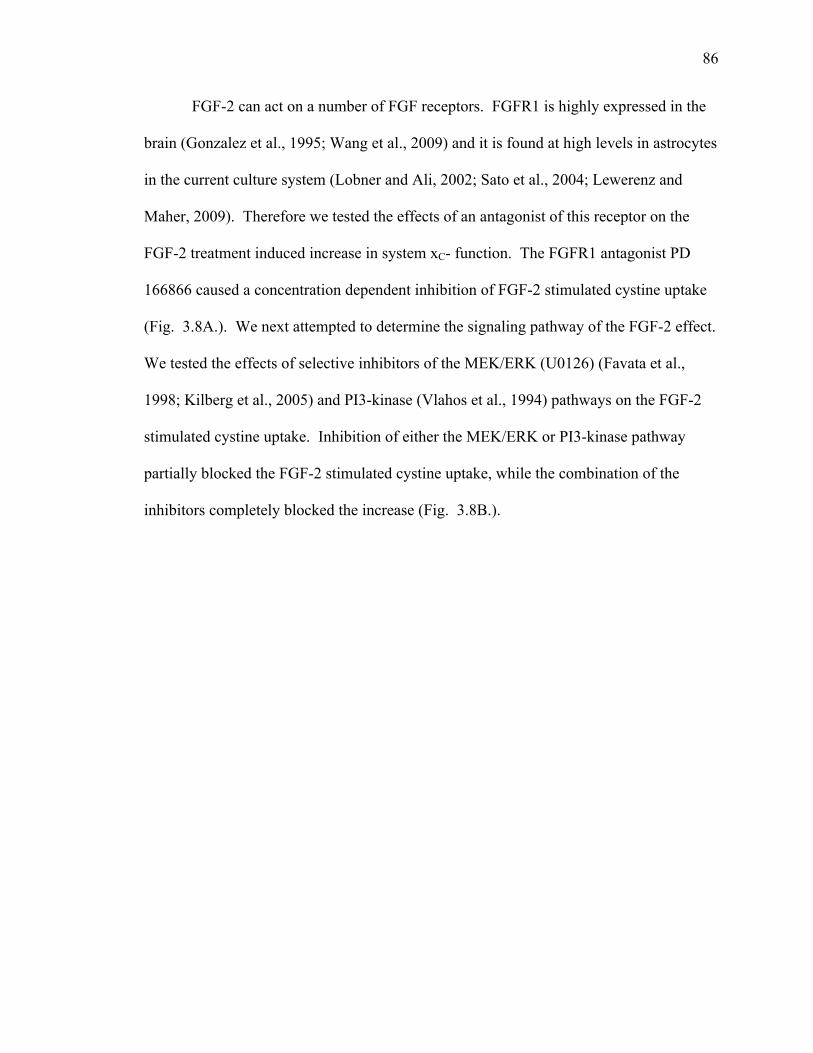
86
FGF-2 can act on a number of FGF receptors. FGFR1 is highly expressed in the
brain (Gonzalez et al., 1995; Wang et al., 2009) and it is found at high levels in astrocytes
in the current culture system (Lobner and Ali, 2002; Sato et al., 2004; Lewerenz and
Maher, 2009). Therefore we tested the effects of an antagonist of this receptor on the
FGF-2 treatment induced increase in system xC- function. The FGFR1 antagonist PD
166866 caused a concentration dependent inhibition of FGF-2 stimulated cystine uptake
(Fig. 3.8A.). We next attempted to determine the signaling pathway of the FGF-2 effect.
We tested the effects of selective inhibitors of the MEK/ERK (U0126) (Favata et al.,
1998; Kilberg et al., 2005) and PI3-kinase (Vlahos et al., 1994) pathways on the FGF-2
stimulated cystine uptake. Inhibition of either the MEK/ERK or PI3-kinase pathway
partially blocked the FGF-2 stimulated cystine uptake, while the combination of the
inhibitors completely blocked the increase (Fig. 3.8B.).
87
FIGURE 3.8. FGF-2 stimulation of cystine uptake is mediated by activation of FGFR1 and both the MEK/ERK and PI3-kinase pathways. (A) Astrocyte-enriched cultures were exposed to FGF-2 (100 ng/ml) for 24 hours with or without the FGFR1 inhibitor (PD 166866), washed into a growth factor and inhibitor free media, and 14C-cystine uptake measured for 20 min. Bars show % control (mean±s.e.m., n=12). (B) Astrocyte-enriched cultures were exposed to FGF-2 (100 ng/ml) for 24 hours with or without the MEK/ERK inhibitor (10 µM U0126) or the PI3-kinase inhibitor (45 µM LY-294002) added 1 hour prior to and during the FGF-2 exposure, washed into a growth factor and inhibitor free media, and 14C-cystine uptake measured for 20 min. Bars show % control (mean ± s.e.m., n = 18). * indicates significantly different from control uptake. # indicates significantly different from FGF-2 stimulated uptake.
DISCUSSION
The upregulation of system xC- induced by the FGF-2 treatment has multiple
implications for understanding the biological effects of FGF-2 and its potential use as a
therapeutic agent. Both dysregulation of FGF-2 signaling and system xC- function have
88
been implicated in psychiatric disorders such as schizophrenia, depression, and addiction.
As FGF-2 and regulators for system xC- have been suggested for therapeutic use, it is
important to understand the mechanism of regulation of system xC-. Also, upregulation
of system xC- must be taken into account when considering the use of FGF-2 for the
treatment of neurological conditions, particularly those that involve excitotoxicity.
The evidence suggesting the involvement of dysregulation of the FGF-2 system in
schizophrenia is substantial (Terwisscha van Scheltinga et al., 2010). FGF-2 is involved
in neurogenesis and normal brain development (Zechel et al., 2010). FGFR1 is widely
expressed in the brain (Gonzalez et al., 1995) and is required for proliferation of
hippocampal stem cells (Ohkubo et al., 2004). Therefore FGF-2 system signaling
dysregulation may be involved in altered development in schizophrenia. Supporting this
idea, in post-mortem brains from schizophrenia patients there was found to be increased
FGFR1 expression in the CA4 region of the hippocampus (Gaughran et al., 2006) and
there is increased expression of FGF-2 in the serum of schizophrenia patients (Hashimoto
et al., 2003). It is interesting to note that disruption of MEK/ERK and PI3-kinase
signaling have also been implicated in developmental diseases, such as schizophrenia
(Waite and Eickholt, 2010; Yuan et al., 2010). We found that the action of FGF-2 to
upregulate system xC- function was partially dependent on both MEK/ERK and PI3-
kinase signaling. The results presented in the current study of FGF-2 effects on system
xC- that are mediated by FGFR1 provide a potential mechanism by which defects in FGF-
2 signaling leads to developmental disorders.
There are decreased levels of glutathione in schizophrenics (Raffa et al., 2011)
and an animal model of schizophrenia involves the post-natal treatment with buthionine
89
sulfoximine, an inhibitor of a glutathione synthesis (Cabungcal et al., 2007). In one study
it was found that in an animal model of schizophrenia there was decreased system xC-
activity, although in the brain of human schizophrenics there was a slight increase in xCT
expression (Baker et al., 2008). Therefore, it is possible that increased FGF-2 signaling is
a compensatory mechanism in schizophrenia to increase xCT expression to increase
system xC- activity. However, a great deal more work determining the time course of
changes in FGF-2 system signaling, system xC- function, glutathione levels, development
changes, and development of the disease is necessary.
Disruption of FGF-2 signaling may also be involved in depression. In the post-
mortem brains of depressed patients there is reduced mRNA for FGF-2, but increased
mRNA for FGFR1 (Gaughran et al., 2006). Treatment with FGF-2 has antidepressant
effects in mouse models of depression and reversed the decrease in neurogenesis in the
hippocampus of these mice (Zechel et al., 2010).
Alterations in both system xC- and FGF-2 have been implicated in cocaine
addiction. In adult rats, following withdrawal from repeated cocaine, there is decreased
function of system xC- in the nucleus accumbens (Baker et al., 2003). Repeated cocaine
administration increases FGF-2 mRNA in the striatum and prefrontal cortex (Fumagalli
et al., 2006), while it decreases FGFR1 mRNA in the hippocampus but increases it in the
prefrontal cortex (Turner et al., 2008b). Due to differences in the areas of the brain
assessed, it is at this time not possible to draw conclusions regarding the interaction
between system xC- and FGF-2 during addiction. However, it is clear that both are
changed.
90
We showed that 24 hour treatment with FGF-2 increases cystine uptake through
system xC- selectively on primary cortical astrocytes, but not neurons or microglia. This
effect does not appear to be limited to cortical astrocytes. We found a similar regulatory
effect of FGF-2 on cystine uptake through system xC- in spinal cord glial cultures. The
result that FGF-2 also increases cystine uptake in spinal cord gial cultures is especially
interesting because of a recent study showing that knocking out FGF-2 can delay disease
progression in the SOD1 ALS mouse model (Thau et al., 2012). One possible
explanation for this result is that the lack of FGF-2 signaling leads to decreased system
xC- activity in the ALS mice, and the protection would then be due to decreased
glutamate release from system xC- and decreased excitotoxicity. However, what role
FGF-2 plays in ALS is unclear. Post mortem studies revealed that there was no change in
FGF-2 or FGFR1 mRNA in ALS patients compared to control (Petri et al., 2009).
Preliminary data from our laboratory on mutant SOD1 mice showed a significant increase
in system xC- function in spinal cord slices even before the animals showed any
symptoms (unpublished data). Future studies on FGF-2 signaling and regulation of
system xC- in the SOD1 model can potentially help clarify the involvement of FGF-2 and
system xC- in ALS.
The function of system xC- makes it likely to play an important role in regulating
neuronal survival and death; by releasing glutamate it can increase extracellular
glutamate levels and potentially cause excitotoxicity, through providing cystine to the cell
it regulates cellular glutathione levels and in this way may protect cells against oxidative
stress. Both excitotoxicity and oxidative stress play a role in neurodegenerative diseases
(ALS) (Bains and Shaw, 1997; Lau and Tymianski, 2010). The dual nature of system xC-
91
means that it may either protect against or enhance neuronal death depending on the
relative importance of excitotoxicity and oxidative stress in each disease.
FGF-2 was tested in clinical trials for the treatment of stroke. It was found that
FGF-2 treatment caused roughly a doubling of the mortality rate (Clark et al., 2000;
Bogousslavsky et al., 2002). While there are many possible reasons for a lack of
protective effects of FGF-2, the increase in mortality indicates that, in this situation, it
was having injury potentiating effects. Could an increase in system xC- be responsible
for the increased mortality following FGF-2 treatment? Certainly excitotoxicity plays a
role in cerebral ischemia (Choi, 1998) and system xC- activity can enhance excitotoxicity
(Fogal et al., 2007). However, FGF-2 has multiple effects on systems that can alter
neuronal death. For example, in the central nervous system, it increases AMPA receptor
expression (Cheng et al., 1995), and decreases NMDA receptor expression (Mattson et
al., 1993). Therefore, the net effect of FGF-2 on neuronal death in various conditions
will depend on many factors. We began to explore the net effect of FGF-2 by testing
whether long-term upregulation of system xC- by FGF-2 directly induces neuronal death.
92
CHAPTER IV
FGF-2 INDUCES NEURONAL DEATH
93
ABSTRACT
FGF-2 upregulates system xC- selectively on astrocytes, which will lead to both
increased cystine uptake, with increased glutathione production, and increased
extracellular glutamate. While the increased intracellular glutathione can contribute to
decreased oxidative stress, the increased glutamate release can potentially lead to
excitotoxicity to neurons. To test this hypothesis, we performed a long-term treatment
(48 hours) of FGF-2 in mature mixed neuronal and glial cultures. Treatment with FGF-2
for 48 hours caused a significant neuronal death in these cultures. This cell death was not
observed in neuronal-enriched cultures, or astrocyte-enriched cultures, suggesting the
toxicity is a result of neuron-glia interaction. Blocking system xC- eliminated the
neuronal death. Since increasing cystine uptake is unlikely to induce toxicity, we
examined the involvement of glutamate release. The general AMPA/kainate receptor
antagonist 2,3-dihydroxy-6-nitro-7-sulfamoyl-benzo[f]quinoxaline-2,3-dione (NBQX)
abolished the neuronal toxicity induced by FGF-2 treatment, and surprisingly, the NMDA
receptor antagonist memantine did not. When cultures were exposed directly to
glutamate, either NBQX or memantine blocked the neuronal toxicity. The mechanism of
this altered profile of glutamate receptor mediated toxicity by FGF-2 is unclear. The
selective calcium permeable AMPA receptor antagonist 1-naphthyl acetyl spermine
(NASPM) failed to offer protection. FGF-2 has been suggested to be able to decrease
NMDA receptor mediated toxicity by both reducing NMDA receptor expression and
causing a faster inactivation. FGF-2 has also been shown to increase GluR1 expression,
as well as increased calcium influx. Therefore, the most logical explanation for the
results is that 48 hour FGF-2 treatment induces AMPA receptor toxicity through

94
increased system xC- function resulting in increased release of glutamate. At the same
time, FGF-2 decreases neuronal sensitivity to NMDA receptor mediated toxicity, and
possibly enhances sensitivity to AMPA receptor mediated toxicity, although we cannot
exclude the possibility that FGF-2 treatment sensitizes the neurons to normal system xC-
mediated glutamate release.
INTRODUCTION
Fibroblast growth factor 2 (FGF-2), despite its discovery in fibroblasts, is widely
expressed throughout the brain (Eckenstein, 1994; Dono, 2003). FGF-2 can activate
members of the FGF receptor family leading to activation of several intracellular
pathways, including PI3K/Akt and MEK/ERK pathways (Reuss and Bohlen und
Halbach, 2003). It has been demonstrated to be involved in development (Ohkubo et al.,
2004), adult neurogenesis (Mudò et al., 2009), and tissue repair (Reuss and Bohlen und
Halbach, 2003). Dysregulation of FGF-2 signaling has been implicated in acute and
chronic neurodegenerative diseases, such as stroke (Alzheimer and Werner, 2002),
traumatic brain injury (Mellergård et al., 2012), and Alzheimer’s disease (Mark et al.,
1997), as well as psychiatric disorders such as schizophrenia (Terwisscha van Scheltinga
et al., 2010), stress (Molteni et al., 2001; Frank et al., 2007), addiction (Flores and
Stewart, 2000), and major depression (Evans et al., 2004). The use of FGF-2 for
treatment for stroke in clinical trials has been attempted but without success (Clark et al.,
2000; Bogousslavsky et al., 2002).
System xC- is a cystine/glutamate antiporter on the cell membrane (Sato et al.,
1995b; Liu et al., 2011). Physiologically, system xC- takes up cystine and releases
95
glutamate in exchange at a 1:1 ratio (Sato et al., 1999). The direction of transport is
determined by the high glutamate levels and low cystine levels intracellularly. Once
cystine is taken up it is rapidly reduced to cysteine, which can then be used to synthesize
protein, or glutathione (GSH), an important endogenous antioxidant in the brain (Meister
and Anderson, 1983). GSH and cysteine can both be released into the extracellular
space, typically by astrocytes, to regulate the redox state of the brain (Wang and Cynader,
2000; Dringen and Hirrlinger, 2003). Also, the extracellular cysteine is taken up by
neurons to synthesize GSH (Wang and Cynader, 2000; Lewerenz et al., 2006; Escartin et
al., 2011). The glutamate released by system xC- can have multiple effects. Glutamate is
the most important excitatory neurotransmitter in the central nervous system. Along with
mediating excitatory neurotransmission at synapses, glutamate released from astrocytes is
also believed to regulate synaptic activity and plasticity through activating parasynaptic
and extrasynaptic receptors (Asztely et al., 1997; Hardingham et al., 2002). However,
high extracellular glutamate levels can cause neuronal death through excitotoxicity,
typically resulting from high NMDA receptor activation leading to excess calcium influx
triggering cell death (Choi et al., 1987).
System xC- has been shown to be widely expressed, especially in the central
nervous system (Sato et al., 2002; Burdo et al., 2006). In vitro cell culture studies have
shown that immature neuronal cells rely on system xC- to take up cystine (Murphy et al.,
1990). When immature neuronal cell cultures are incubated with high levels of
glutamate, glutamate competitively inhibits cystine uptake through system xC- and the
cells die from oxidative stress (Murphy et al., 1989). This mechanism of neuronal death
is called oxidative glutamate toxicity (Schubert and Piasecki, 2001). This type of
96
glutamate toxicity is distinct from glutamate toxicity in mature neuronal cells, which
results from over-stimulation of NMDA receptors leading to excessive calcium influx,
and it is called excitotoxicity (Choi, 1987). Also, in mature cell cultures, neurons show
low levels of system xC- activity, while astrocytes exhibit high levels of system xC-
activity (Lobner, 2009). This shift of function indicates that as cells mature, they take on
more specialized tasks and cooperate as a system (Fellin and Carmignoto, 2004;
Stipursky et al., 2011; Suzuki et al., 2011).
The dual actions of system xC- give it a unique potential for influencing cell fate.
System xC- activity can be either beneficial or destructive depending on the cellular
properties. If cells are susceptible to oxidative stress, increasing system xC- activity
should be protective as it increases cystine uptake, which can contribute to increased
antioxidative defense. However, if the cells express high levels of glutamatergic
receptors, it is likely that they will be sensitive to glutamate induced excitotoxicity.
Thus, increasing system xC- activity might be destructive by increasing extracellular
glutamate, which can potentially over-stimulate extrasynaptic glutamatergic receptors.
In the present study, we used primary mixed neuronal and glial cell cultures
derived from prenatal mouse cortex to investigate the effect of 48 hour FGF-2 treatment
on neuronal survival/death. We have previously demonstrated that 24 hour FGF-2
treatment upregulated system xC- activity on astrocytes, but not neurons or microglia.
We demonstrate here that after system xC- is upregulated for a prolonged period of time,
neuronal death occurs due to system xC- mediated excitotoxicity.

97
MATERIALS AND METHODS
Materials
Timed pregnant Swiss Webster mice were obtained from Charles River
Laboratories (Wilmington, DE). Serum was from Atlanta Biologicals (Lawrenceville,
GA). FGF-2 was from Alomone Labs (Jerusalem, Israel). 14C-cystine was from
PerkinElmer (Waltham, MA). All other chemicals were obtained from Sigma (St. Louis,
MO).
Cortical cell cultures
Mixed cortical cell cultures containing glial and neuronal cells were prepared
from fetal (15-16 day gestation) mice as previously described (Lobner, 2000).
Dissociated cortical cells were plated on 24-well plates coated with poly-D-lysine and
laminin in Eagles’ Minimal Essential Medium (MEM, Earle’s salts, supplied glutamine-
free) supplemented with 5% heat-inactivated horse serum, 5% fetal bovine serum, 2 mM
glutamine and glucose (total 21 mM). Neuron-enriched cultures were prepared exactly as
above with the addition of 10 µM cytosine arabinoside 48 hours after plating to inhibit
glial replication. In these cultures <1% of cells are astrocytes (Dugan et al., 1995; Rush
et al., 2010). Astrocyte-enriched cultures were prepared as described for mixed cultures
except they are from cortical tissue taken from post-natal day 1-3 mice (Choi et al., 1987;
Schwartz and Wilson, 1992; Rush et al., 2010). Cultures were maintained in humidified
5% CO2 incubators at 37°C. Mice were handled in accordance with a protocol approved
by our institutional animal care committee and in compliance with the Public Heath
98
service Policy on Humane Care and Use of Laboratory Animals. All efforts were made
to minimize animal suffering and reduce the number of animals used.
Induction of neuronal death
All experiments were performed on cultures 13 days in vitro (DIV). Cultures
were exposed to different compounds for 48 hours in media as described for plating
except without serum.
Assay of neuronal death
LDH release
Cell death was assessed in mixed cultures by the measurement of lactate
dehydrogenase (LDH) released from damaged or destroyed cells, in the extracellular fluid
48 hours after the beginning of the insult (Koh and Choi, 1987; Lobner, 2000). Blank
LDH levels were subtracted from insult LDH values and results normalized to 100%
neuronal death caused by 500 µM NMDA, or 100% cell death caused by 20 µM of the
calcium ionophore A23187, added 24 hours before the assay. Control experiments have
shown previously that the efflux of LDH occurring from either necrotic or apoptotic cells
is proportional to the number of cells damaged or destroyed. Glial cell death (assessed by
trypan blue staining) was not observed in any of the current studies.
MTT assay
Cell survival was quantified by the measurement of the reduction of 3-(4,5-
dimethylthiazol-2-yl)-2,5-diphenyl-tetrazolium bromide (MTT) to produce a dark blue

99
formazan product (Lobner, 2000). MTT was added to each well 48 hours after the
beginning of the insult to the cells (following removal of media for LDH assay). After
30-minute incubation, the medium was removed, and cells were dissolved in dimethyl
sulfoxide. The formation of formazan was measured as the amount of reaction product by
absorbance change at a wavelength of 590 nm by using a VersaMax tunable microplate
reader (Molecular Devices, Sunnyvale, CA). Levels of formazan formation from cultures
exposed to 500 µM NMDA (100% neuronal death) were subtracted from insult formazan
levels, and results were normalized to a sham wash.
14C-cystine uptake
System xC- mediated uptake of radiolabeled cystine was assayed as previously
described with modifications (Liu et al., 2009). FGF-2 was added to the serum
containing media for the indicated durations. Cultures were then washed into HEPES
buffered saline solution and immediately exposed to 14C-cystine (0.025µCi/ml) for 20
min. Following 14C-cystine exposure, cultures were washed with ice cold HEPES
buffered saline solution and dissolved in 250 µl sodium dodecyl sulfate (0.1%). An
aliquot (200 µl) was removed and added to scintillation fluid for counting. Values were
normalized to 14C-cystine uptake in untreated control on the same experimental plate.
Statistical analysis
Differences between test groups were examined for statistical significance by
means of one-way ANOVA followed by the Bonferroni correction post-hoc test, with
p<0.05 being considered significant.
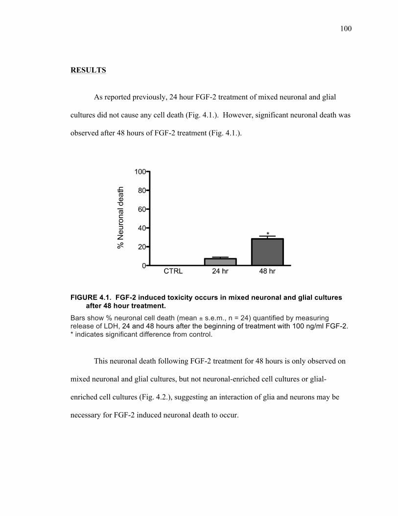
100
RESULTS
As reported previously, 24 hour FGF-2 treatment of mixed neuronal and glial
cultures did not cause any cell death (Fig. 4.1.). However, significant neuronal death was
observed after 48 hours of FGF-2 treatment (Fig. 4.1.).
FIGURE 4.1. FGF-2 induced toxicity occurs in mixed neuronal and glial cultures after 48 hour treatment.
Bars show % neuronal cell death (mean ± s.e.m., n = 24) quantified by measuring release of LDH, 24 and 48 hours after the beginning of treatment with 100 ng/ml FGF-2. * indicates significant difference from control.
This neuronal death following FGF-2 treatment for 48 hours is only observed on
mixed neuronal and glial cultures, but not neuronal-enriched cell cultures or glial-
enriched cell cultures (Fig. 4.2.), suggesting an interaction of glia and neurons may be
necessary for FGF-2 induced neuronal death to occur.
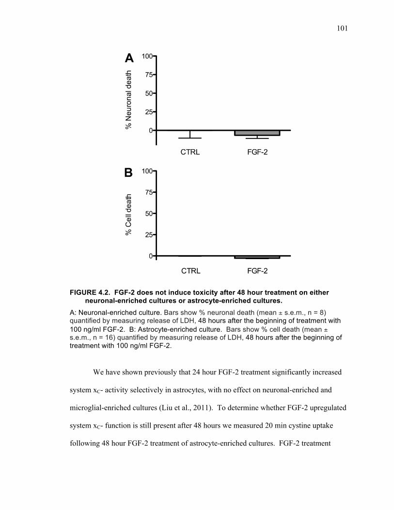
101
FIGURE 4.2. FGF-2 does not induce toxicity after 48 hour treatment on either
neuronal-enriched cultures or astrocyte-enriched cultures. A: Neuronal-enriched culture. Bars show % neuronal death (mean ± s.e.m., n = 8) quantified by measuring release of LDH, 48 hours after the beginning of treatment with 100 ng/ml FGF-2. B: Astrocyte-enriched culture. Bars show % cell death (mean ± s.e.m., n = 16) quantified by measuring release of LDH, 48 hours after the beginning of treatment with 100 ng/ml FGF-2.
We have shown previously that 24 hour FGF-2 treatment significantly increased
system xC- activity selectively in astrocytes, with no effect on neuronal-enriched and
microglial-enriched cultures (Liu et al., 2011). To determine whether FGF-2 upregulated
system xC- function is still present after 48 hours we measured 20 min cystine uptake
following 48 hour FGF-2 treatment of astrocyte-enriched cultures. FGF-2 treatment
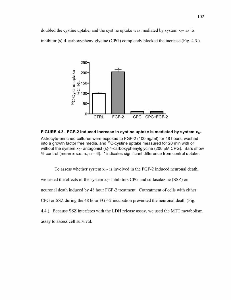
102
doubled the cystine uptake, and the cystine uptake was mediated by system xC- as its
inhibitor (s)-4-carboxyphenylglycine (CPG) completely blocked the increase (Fig. 4.3.).
FIGURE 4.3. FGF-2 induced increase in cystine uptake is mediated by system xC-. Astrocyte-enriched cultures were exposed to FGF-2 (100 ng/ml) for 48 hours, washed into a growth factor free media, and 14C-cystine uptake measured for 20 min with or without the system xC- antagonist (s)-4-carboxyphenylglycine (200 µM CPG). Bars show % control (mean ± s.e.m., n = 6). * indicates significant difference from control uptake.
To assess whether system xC- is involved in the FGF-2 induced neuronal death,
we tested the effects of the system xC- inhibitors CPG and sulfasalazine (SSZ) on
neuronal death induced by 48 hour FGF-2 treatment. Cotreatment of cells with either
CPG or SSZ during the 48 hour FGF-2 incubation prevented the neuronal death (Fig.
4.4.). Because SSZ interferes with the LDH release assay, we used the MTT metabolism
assay to assess cell survival.
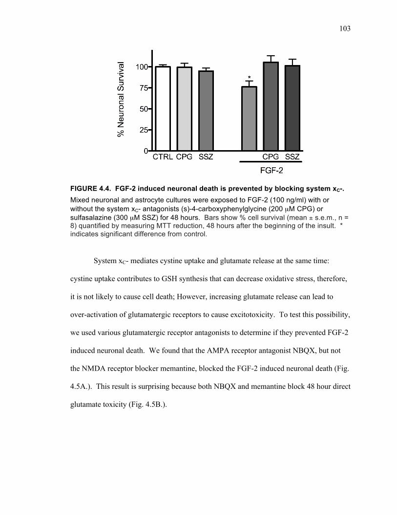
103
FIGURE 4.4. FGF-2 induced neuronal death is prevented by blocking system xC-. Mixed neuronal and astrocyte cultures were exposed to FGF-2 (100 ng/ml) with or without the system xC- antagonists (s)-4-carboxyphenylglycine (200 µM CPG) or sulfasalazine (300 µM SSZ) for 48 hours. Bars show % cell survival (mean ± s.e.m., n = 8) quantified by measuring MTT reduction, 48 hours after the beginning of the insult. * indicates significant difference from control.
System xC- mediates cystine uptake and glutamate release at the same time:
cystine uptake contributes to GSH synthesis that can decrease oxidative stress, therefore,
it is not likely to cause cell death; However, increasing glutamate release can lead to
over-activation of glutamatergic receptors to cause excitotoxicity. To test this possibility,
we used various glutamatergic receptor antagonists to determine if they prevented FGF-2
induced neuronal death. We found that the AMPA receptor antagonist NBQX, but not
the NMDA receptor blocker memantine, blocked the FGF-2 induced neuronal death (Fig.
4.5A.). This result is surprising because both NBQX and memantine block 48 hour direct
glutamate toxicity (Fig. 4.5B.).

104
FIGURE 4.5. Effects of the AMPA receptor antagonist NBQX and the NMDA receptor antagonist memantine on neuronal death induced by 48 hour exposure to FGF-2 or glutamate.
A: FGF-2 (100 ng/ml) treatment induced toxicity is protected by blocking AMPA receptors. B: Glutamate (15 µM) induced toxicity is protected by blocking AMPA or NMDA receptors. NBQX: 7.5 µM 2,3-dihydroxy-6-nitro-7-sulfamoyl-benzo[f]quinoxaline-2,3-dione. MEM: 10 µM memantine. Bars show % neuronal cell death (mean ± s.e.m., n = 12) quantified by measuring release of LDH, 48 hours after the beginning of the insult. * indicates significant difference from control. # indicates significant difference from FGF-2 or glutamate treated.
One possibility for the results is that FGF-2 upregulates calcium permeable
AMPA receptors making the neurons susceptible to AMPA receptor mediated toxicity.
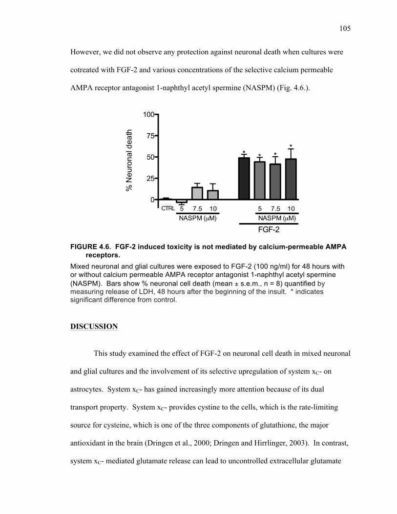
105
However, we did not observe any protection against neuronal death when cultures were
cotreated with FGF-2 and various concentrations of the selective calcium permeable
AMPA receptor antagonist 1-naphthyl acetyl spermine (NASPM) (Fig. 4.6.).
FIGURE 4.6. FGF-2 induced toxicity is not mediated by calcium-permeable AMPA receptors.
Mixed neuronal and glial cultures were exposed to FGF-2 (100 ng/ml) for 48 hours with or without calcium permeable AMPA receptor antagonist 1-naphthyl acetyl spermine (NASPM). Bars show % neuronal cell death (mean ± s.e.m., n = 8) quantified by measuring release of LDH, 48 hours after the beginning of the insult. * indicates significant difference from control.
DISCUSSION
This study examined the effect of FGF-2 on neuronal cell death in mixed neuronal
and glial cultures and the involvement of its selective upregulation of system xC- on
astrocytes. System xC- has gained increasingly more attention because of its dual
transport property. System xC- provides cystine to the cells, which is the rate-limiting
source for cysteine, which is one of the three components of glutathione, the major
antioxidant in the brain (Dringen et al., 2000; Dringen and Hirrlinger, 2003). In contrast,
system xC- mediated glutamate release can lead to uncontrolled extracellular glutamate
106
and cause excitotoxicity (Fogal et al., 2007; Jackman et al., 2010). Both oxidative stress
and excitotoxicity are involved in various neurodegenerative diseases and psychiatric
disorders. Also, FGF-2 dysregulation is observed in acute and chronic neurological
disorders (Gaughran et al., 2006; Terwisscha van Scheltinga et al., 2010; Zechel et al.,
2010). Therefore, it is possible that changes in system xC- function may be responsible
for some of the actions of FGF-2.
Our previous data showed that 24 hour FGF-2 treatment resulted in an increase in
cystine uptake through system xC- on astrocytes, but with no effect on neuronal and
microglial cystine uptake (Liu et al., 2011). FGF-2 treatment mediated increase of
system xC- was mediated through activation of FGFR1 and required both MEK/ERK and
PI3 kinase pathway activations (Liu et al., 2011). All of these cellular events happened
without significant neuronal toxicity. However, after 48 hour FGF-2 treatment,
significant neuronal cell death began to occur. This neuronal death was blocked by the
system xC- antagonists CPG and SSZ.
The role of system xC- in cell death has been investigated in different cell types
and under different conditions. Inhibition of system xC- has been shown to be harmful to
system xC- expressing cells. Oxidative glutamate toxicity was first described on
immature neuronal cultures, in which system xC- is the major route of cystine uptake
(Murphy et al., 1989; 1990). Applying high concentrations of glutamate (millimolar
range) to these immature neurons led to GSH depletion and eventually cell death from
oxidative stress (Miyamoto et al., 1989; Murphy et al., 1989; Murphy and Baraban,
1990). Under these circumstances, increasing system xC- levels would likely be
protective.
107
However, increasing system xC- function is not always protective. It seems that
increasing system xC- function in certain population of cells can be toxic to the
surrounding cells. Primary microglia cultured in physiological extracellular
concentrations of cystine and glutamine have been shown to be able to release enough
glutamate through system xC- to kill surrounding neurons (Piani and Fontana, 1994)
(Domercq et al., 2007). Activation of these microglia by bacterial components enhanced
system xC- mediated glutamate release, which was prevented by blocking protein
synthesis (Piani and Fontana, 1994). Similarly, the increase in xCT expression in
microglia induced by lipopolysaccharide treatment led to increased system xC- mediated
release of glutamate, causing excitotoxicity to cocultured oligodendrocytes (Domercq et
al., 2005). This toxicity was decreased by blocking AMPA receptors with CNQX
(Domercq et al., 2005). This result is similar to what we have observed on our primary
mixed neuronal and glial cultures. The difference is that oligodendrocytes are normally
sensitive to AMPA receptor toxicity (McDonald et al., 1998; Takahashi et al., 2003),
while in cortical neurons, glutamate induced excitotoxicity is normally mediated
primarily by activation of NMDA receptors (Choi et al., 1987; Choi, 1998).
Similar to microglia, selectively increasing system xC- on astrocytes with IL-1β
can also lead to increased glutamate release, which potentiates srounding neuronal death
induced by hypoglycemia and hypoxia (Jackman et al., 2010; 2012), both of which are
well-known to kill neurons in a process that involves over-activation of glutamatergic
receptors (Snider et al., 1998; Czyz et al., 2002). The neuronal death in these conditions
was largely attenuated by blocking NMDA receptors with the selective NMDA receptor
open channel blocker MK-801, and was also blocked by the system xC- inhibitor CPG
108
(Fogal et al., 2007; Jackman et al., 2010; 2012). Our data shows that FGF-2 treatment,
like IL-1β, selectively increases system xC- activity on astrocytes (Liu et al., 2011). But
FGF-2 by itself induced neuronal cell death after system xC- activity was upregulated for
48 hours. Also, the pharmacological profile of the excitotoxicity was different from non-
FGF-2 treated neurons. We showed that, while neuronal death induced by direct
exposure of cultures to glutamate was blocked by either NMDA or AMPA receptor
antagonists, FGF-2 induced neuronal death was only attenuated by the AMPA antagonist.
FGF-2 has been shown to be able to change neuronal expression of both NMDA
and AMPA receptors, and to alter sensitivity to NMDA receptor mediated toxicity both in
vitro and in vivo. A study conducted on cerebellar granule cells reported that FGF-2
treatment caused a time-dependent decrease in expression of NMDA receptor subunits
GluN2A and GluN2C with a decrease of NMDA-evoked calcium influx, while GluN1
and GluN2B levels were not changed (Brandoli et al., 1998). Also, FGF-2 pretreatment
protected striatal neurons from NMDA receptor mediated toxicity (Freese et al., 1992).
Systematic administration of FGF-2 protected against intrastriatal injection of NMDA
(Nozaki et al., 1993a; 1993b). FGF-2 treatment enhanced the rate of NMDA receptor
inactivation in response to calcium influx in hippocampal neurons (Boxer et al., 1999).
FGF-2 treatment decreased NMDA receptor levels in hippocampal cell cultures and
elevations in intracellular calcium levels after glutamate exposure compared to non-FGF-
2 treated cells (Mattson et al., 1993). FGF-2 treatment has been shown to increase
AMPA receptor subunit GluR1 levels in the same cultures, without altering GluR2,
GluR3, and GluR4 levels (Cheng et al., 1995). This suggested the possibility that FGF-2
increased the levels of GluR2 subunit lacking AMPA receptors, which have calcium
109
permeability (Bannerman et al., 2007). However, the selective antagonist of these
channels, NASPM, failed to offer protection in our study. At concentrations above 5 µM,
NASPM began to induce some toxicity by itself. The IC50 of NASPM for calcium-
permeable AMPA receptors is 0.33 µM (Brackley et al., 1993; Koike et al., 1997).
Therefore, the 5 µM concentration that was not toxic should have been an effective
concentration. Therefore, it is possible that the FGF-2 treatment induced increased
glutamate release from system xC- causes an AMPA receptor specific neuronal death
because of attenuated NMDA receptor mediated toxicity induced by FGF-2 treatment.
FGF-2 was a potential candidate for the treatment of stroke as it was shown to
reduce ischemic damage, as well as, promote recovery and regeneration in multiple in
vitro and animal models (Nozaki et al., 1993b; Rosenblatt et al., 1994; Song et al., 2002;
Watanabe et al., 2004). However, in human clinical trials, FGF-2 not only failed to show
any beneficial effect in treated patients compared to control patients, it actually increased
the mortality rate in treated patients (Clark et al., 2000; Bogousslavsky et al., 2002). The
reasons for FGF-2 being beneficial in animal models but not in human trials are not yet
fully understood. Given that FGF-2 can increase system xC- activity, and the dual
functions of system xC-, it is not surprising that FGF-2 can have varying effects on
cerebral ischemia.
FGF-2 administration has been shown to be beneficial in rodent studies in other
disease models, such as depression (Turner et al., 2008c) and Parkinson disease (Hsuan et
al., 2006). It is possible that these beneficial effects may be mediated by the FGF-2
effect on system xC-, which leads to enhanced glutathione synthesis. However, again
110
taking into account the dual actions of system xC-, the effects of long-term treatment with
FGF-2 must be rigorously studied.
CONCLUSIONS
The dual amino acid transport function allows system xC- regulate intracellular
cystine and extracellular glutamate levels at the same time. Cystine taken up is broken
down immediately to cysteine, which is a substrate for glutathione synthesis and protein
synthesis. While increasing cystine uptake can be protective by increasing the cells’
antioxidative defense, glutamate released at the same time can potentially cause toxicity
by over activating glutamatergic receptors. We show here that the net effect of long-term
upregulation of system xC- selectively on astrocytes in mixed neuronal and glial cultures
by FGF-2 treatment is negative. That is, the excitotoxicity component dominates, leading
to neuronal death.

111
CHAPTER V
GENERAL DISCUSSION
112
System xC- mediates cystine and glutamate exchange (Sato et al., 1999), thus it is
involved in both regulating extracellular glutamate and intracellular GSH due to its dual
amino acid transport function. Both abnormal glutamate transmission and chronic
oxidative stress have been indicated to contribute to neurodegenerative diseases, such as
amyotrophic lateral sclerosis, stroke, traumatic spinal cord/brain injury, Alzheimer’s
disease and Parkinson’s disease (Dugan and Choi, 1994; Andersen, 2004), as well as
psychiatric disorders such as addiction (Tsai et al., 1998) and schizophrenia (Pérez-Neri
et al., 2006). Therefore, it seems likely that alterations in system xC- function may play a
role in these diseases. To begin to resolve this question, a series of studies were
conducted to find out how system xC- is involved in brain cell health in primary cortical
cell cultures. We found that the environmental neurotoxin BMAA can compete with
cystine at system xC- leading to both oxidative stress and excitotoxicity, that FGF-2 can
upregulate system xC- selectively on astrocytes, and that the upregulation of system xC-
by FGF-2 treatment may be responsible for AMPA receptor specific neuronal death
caused by 48 hour treatmentFGF-2 . Following is a discussion of some of the issues that
arose during these studies.
INTERACTION OF FGF-2 AND BMAA ON NEURONAL DEATH
Since both BMAA and FGF-2 have actions on system xC-: FGF-2 upregulates
system xC- function (Liu et al., 2011) and BMAA competes with cystine at system xC-
(Liu et al., 2009), we examined whether FGF-2 pretreatment alters BMAA toxicity.
FGF-2 pretreatment for 24 hours changed the concentration response of BMAA toxicity
(Fig. 5.1). At low concentrations, BMAA toxicity was potentiated by FGF-2
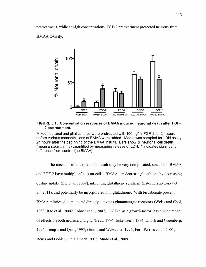
113
pretreatment, while at high concentrations, FGF-2 pretreatment protected neurons from
BMAA toxicity.
FIGURE 5.1. Concentration response of BMAA induced neuronal death after FGF-2 pretreatment.
Mixed neuronal and glial cultures were pretreated with 100 ng/ml FGF-2 for 24 hours before various concentrations of BMAA were added. Media was sampled for LDH assay 24 hours after the beginning of the BMAA insults. Bars show % neuronal cell death (mean ± s.e.m., n= 8) quantified by measuring release of LDH. * indicates significant difference from control (no BMAA).
The mechanism to explain this result may be very complicated, since both BMAA
and FGF-2 have multiple effects on cells. BMAA can decrease glutathione by decreasing
cystine uptake (Liu et al., 2009), inhibiting glutathione synthesis (Esterhuizen-Londt et
al., 2011), and potentially be incorporated into glutathione. With bicarbonate present,
BMAA mimics glutamate and directly activates glutamatergic receptors (Weiss and Choi,
1988; Rao et al., 2006; Lobner et al., 2007). FGF-2, as a growth factor, has a wide range
of effects on both neurons and glia (Beck, 1994; Eckenstein, 1994; Ghosh and Greenberg,
1995; Temple and Qian, 1995; Grothe and Wewetzer, 1996; Ford-Perriss et al., 2001;
Reuss and Bohlen und Halbach, 2003; Mudò et al., 2009).
114
If we consider system xC- as the main mediator of the effects of both compounds,
the most likely explanation for the data is as follows. At low levels of BMAA, when
system xC- is upregulated by FGF-2 pretreatment, BMAA drives increased glutamate
release from system xC-, which exacerbates BMAA toxicity by over activating
glutamatergic receptors. The result could also be explained by a different mechanism:
FGF-2 pretreatment may potentiate low level BMAA toxicity by upregulating system xC-
allowing more BMAA to enter the cells and through inhibition of enzymes or
incorporation into glutathione act to decrease cellular glutathione levels. However, at
high levels of BMAA, excitotoxicity caused by direct activation of glutamatergic
receptors by BMAA is already maximized, and no further excitotoxicity can be induced,
but elevated system xC- function by FGF-2 pretreatment provides cells with more cystine
which strengthens antioxidative defense, thus providing protection against BMAA
toxicity.
Due to the complicated actions of both BMAA and FGF-2, and the lack of direct
scientific implication of their interaction occurring, no further studies were performed
concerning BMAA and FGF-2 interactions.
CONTROVERSIES REGARDING BMAA
In chapter two of this thesis we examined the mechanisms of BMAA toxicity and
found that one novel way in which it kills neurons is through actions on system xC-. It
not only competitively inhibits cystine uptake leading to glutathione depletion, but also
increases glutamate release, causing excitotoxicity. This is potentially a very important
result because it provides a novel mechanism for BMAA toxicity, but it is only important
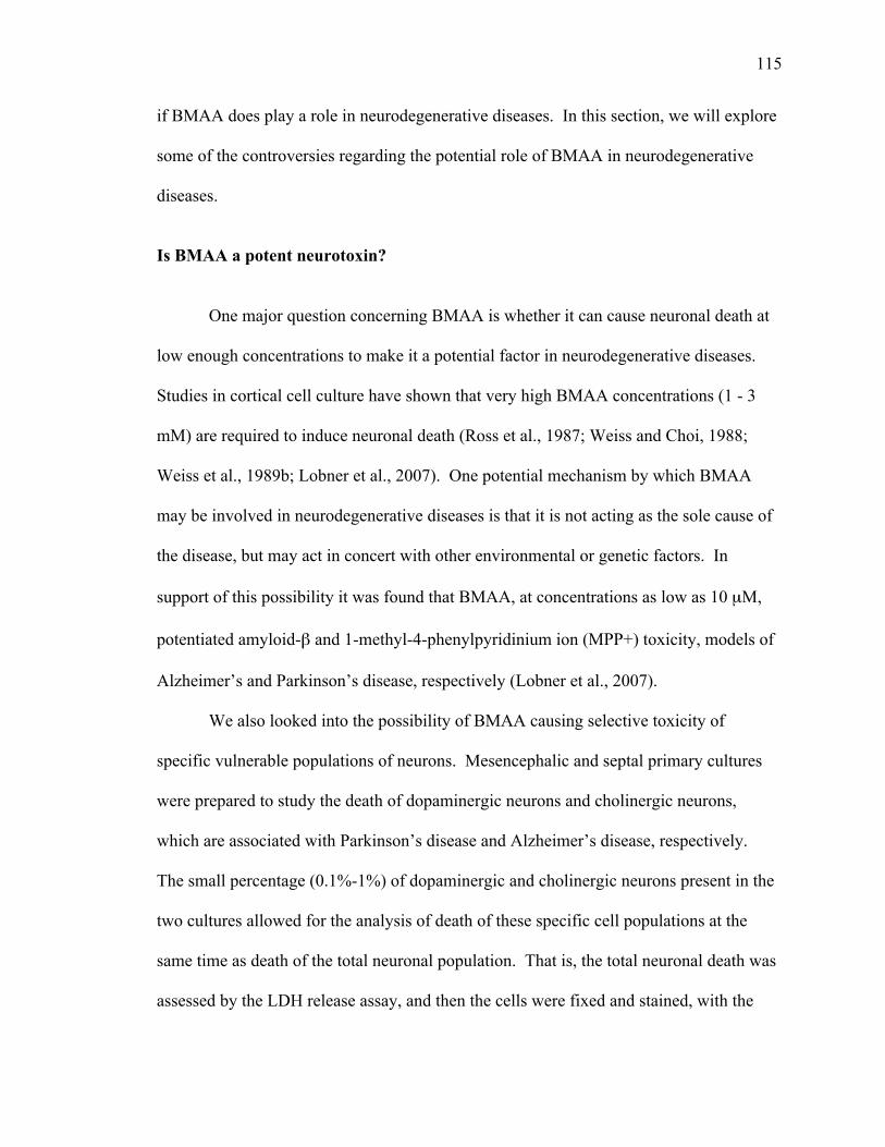
115
if BMAA does play a role in neurodegenerative diseases. In this section, we will explore
some of the controversies regarding the potential role of BMAA in neurodegenerative
diseases.
Is BMAA a potent neurotoxin?
One major question concerning BMAA is whether it can cause neuronal death at
low enough concentrations to make it a potential factor in neurodegenerative diseases.
Studies in cortical cell culture have shown that very high BMAA concentrations (1 - 3
mM) are required to induce neuronal death (Ross et al., 1987; Weiss and Choi, 1988;
Weiss et al., 1989b; Lobner et al., 2007). One potential mechanism by which BMAA
may be involved in neurodegenerative diseases is that it is not acting as the sole cause of
the disease, but may act in concert with other environmental or genetic factors. In
support of this possibility it was found that BMAA, at concentrations as low as 10 µM,
potentiated amyloid-β and 1-methyl-4-phenylpyridinium ion (MPP+) toxicity, models of
Alzheimer’s and Parkinson’s disease, respectively (Lobner et al., 2007).
We also looked into the possibility of BMAA causing selective toxicity of
specific vulnerable populations of neurons. Mesencephalic and septal primary cultures
were prepared to study the death of dopaminergic neurons and cholinergic neurons,
which are associated with Parkinson’s disease and Alzheimer’s disease, respectively.
The small percentage (0.1%-1%) of dopaminergic and cholinergic neurons present in the
two cultures allowed for the analysis of death of these specific cell populations at the
same time as death of the total neuronal population. That is, the total neuronal death was
assessed by the LDH release assay, and then the cells were fixed and stained, with the
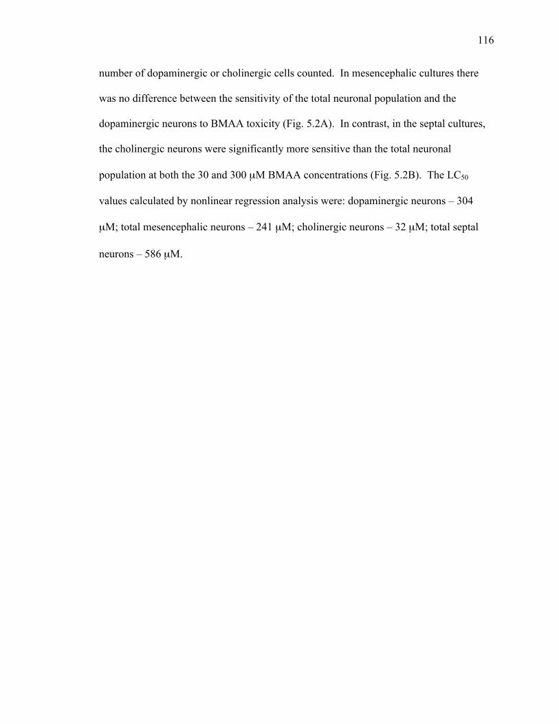
116
number of dopaminergic or cholinergic cells counted. In mesencephalic cultures there
was no difference between the sensitivity of the total neuronal population and the
dopaminergic neurons to BMAA toxicity (Fig. 5.2A). In contrast, in the septal cultures,
the cholinergic neurons were significantly more sensitive than the total neuronal
population at both the 30 and 300 µM BMAA concentrations (Fig. 5.2B). The LC50
values calculated by nonlinear regression analysis were: dopaminergic neurons – 304
µM; total mesencephalic neurons – 241 µM; cholinergic neurons – 32 µM; total septal
neurons – 586 µM.
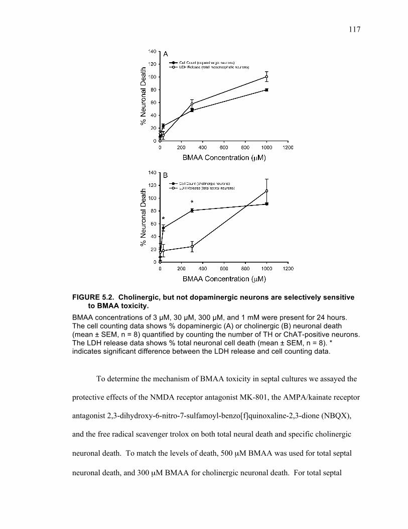
117
FIGURE 5.2. Cholinergic, but not dopaminergic neurons are selectively sensitive to BMAA toxicity.
BMAA concentrations of 3 µM, 30 µM, 300 µM, and 1 mM were present for 24 hours. The cell counting data shows % dopaminergic (A) or cholinergic (B) neuronal death (mean ± SEM, n = 8) quantified by counting the number of TH or ChAT-positive neurons. The LDH release data shows % total neuronal cell death (mean ± SEM, n = 8). * indicates significant difference between the LDH release and cell counting data.
To determine the mechanism of BMAA toxicity in septal cultures we assayed the
protective effects of the NMDA receptor antagonist MK-801, the AMPA/kainate receptor
antagonist 2,3-dihydroxy-6-nitro-7-sulfamoyl-benzo[f]quinoxaline-2,3-dione (NBQX),
and the free radical scavenger trolox on both total neural death and specific cholinergic
neuronal death. To match the levels of death, 500 µM BMAA was used for total septal
neuronal death, and 300 µM BMAA for cholinergic neuronal death. For total septal
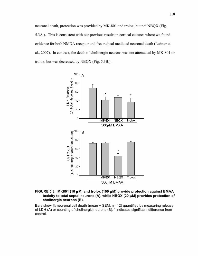
118
neuronal death, protection was provided by MK-801 and trolox, but not NBQX (Fig.
5.3A.). This is consistent with our previous results in cortical cultures where we found
evidence for both NMDA receptor and free radical mediated neuronal death (Lobner et
al., 2007). In contrast, the death of cholinergic neurons was not attenuated by MK-801 or
trolox, but was decreased by NBQX (Fig. 5.3B.).
FIGURE 5.3. MK801 (10 µM) and trolox (100 µM) provide protection against BMAA toxicity to total septal neurons (A), while NBQX (20 µM) provides protection of cholinergic neurons (B).
Bars show % neuronal cell death (mean + SEM, n= 12) quantified by measuring release of LDH (A) or counting of cholinergic neurons (B). * indicates significant difference from control.
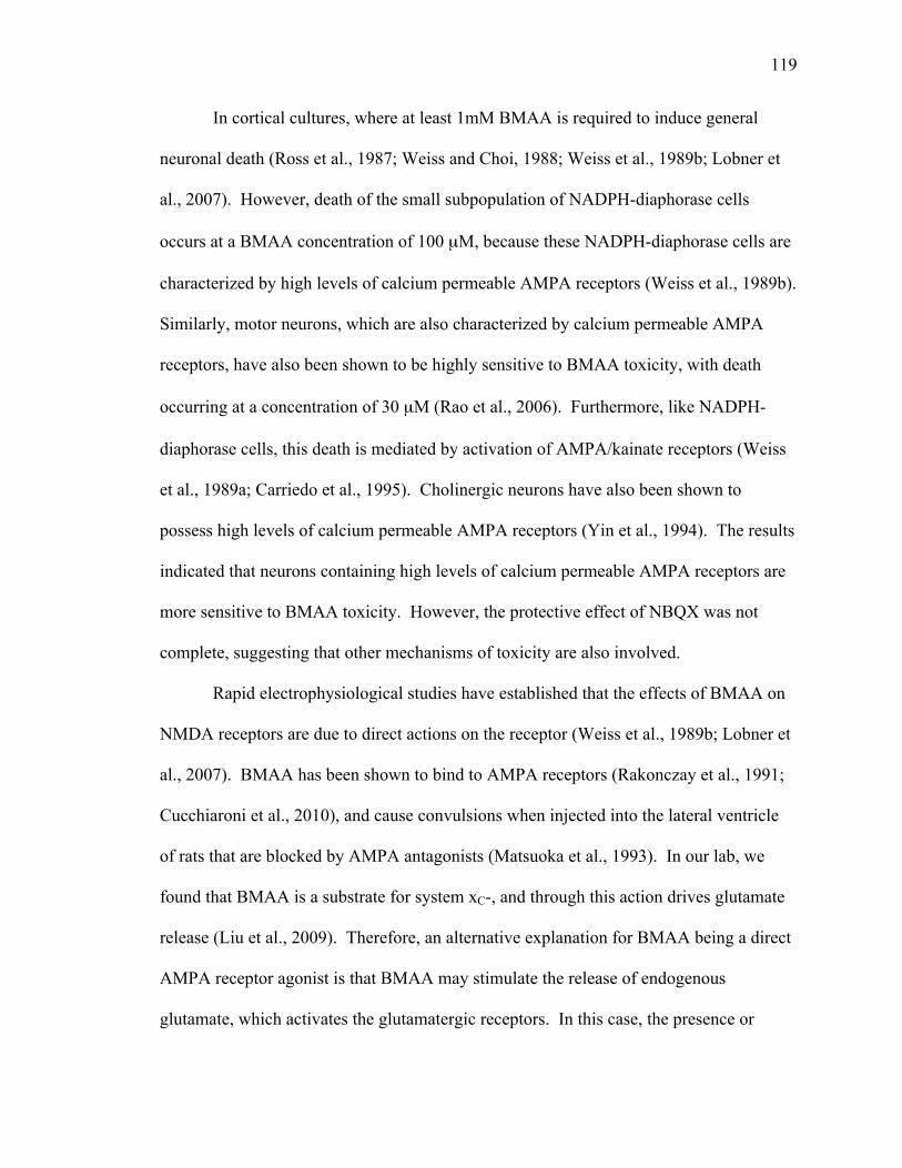
119
In cortical cultures, where at least 1mM BMAA is required to induce general
neuronal death (Ross et al., 1987; Weiss and Choi, 1988; Weiss et al., 1989b; Lobner et
al., 2007). However, death of the small subpopulation of NADPH-diaphorase cells
occurs at a BMAA concentration of 100 µM, because these NADPH-diaphorase cells are
characterized by high levels of calcium permeable AMPA receptors (Weiss et al., 1989b).
Similarly, motor neurons, which are also characterized by calcium permeable AMPA
receptors, have also been shown to be highly sensitive to BMAA toxicity, with death
occurring at a concentration of 30 µM (Rao et al., 2006). Furthermore, like NADPH-
diaphorase cells, this death is mediated by activation of AMPA/kainate receptors (Weiss
et al., 1989a; Carriedo et al., 1995). Cholinergic neurons have also been shown to
possess high levels of calcium permeable AMPA receptors (Yin et al., 1994). The results
indicated that neurons containing high levels of calcium permeable AMPA receptors are
more sensitive to BMAA toxicity. However, the protective effect of NBQX was not
complete, suggesting that other mechanisms of toxicity are also involved.
Rapid electrophysiological studies have established that the effects of BMAA on
NMDA receptors are due to direct actions on the receptor (Weiss et al., 1989b; Lobner et
al., 2007). BMAA has been shown to bind to AMPA receptors (Rakonczay et al., 1991;
Cucchiaroni et al., 2010), and cause convulsions when injected into the lateral ventricle
of rats that are blocked by AMPA antagonists (Matsuoka et al., 1993). In our lab, we
found that BMAA is a substrate for system xC-, and through this action drives glutamate
release (Liu et al., 2009). Therefore, an alternative explanation for BMAA being a direct
AMPA receptor agonist is that BMAA may stimulate the release of endogenous
glutamate, which activates the glutamatergic receptors. In this case, the presence or
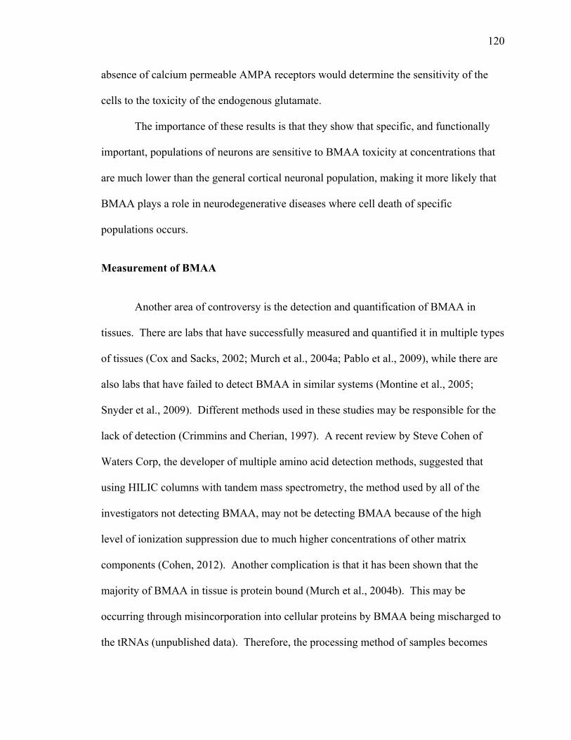
120
absence of calcium permeable AMPA receptors would determine the sensitivity of the
cells to the toxicity of the endogenous glutamate.
The importance of these results is that they show that specific, and functionally
important, populations of neurons are sensitive to BMAA toxicity at concentrations that
are much lower than the general cortical neuronal population, making it more likely that
BMAA plays a role in neurodegenerative diseases where cell death of specific
populations occurs.
Measurement of BMAA
Another area of controversy is the detection and quantification of BMAA in
tissues. There are labs that have successfully measured and quantified it in multiple types
of tissues (Cox and Sacks, 2002; Murch et al., 2004a; Pablo et al., 2009), while there are
also labs that have failed to detect BMAA in similar systems (Montine et al., 2005;
Snyder et al., 2009). Different methods used in these studies may be responsible for the
lack of detection (Crimmins and Cherian, 1997). A recent review by Steve Cohen of
Waters Corp, the developer of multiple amino acid detection methods, suggested that
using HILIC columns with tandem mass spectrometry, the method used by all of the
investigators not detecting BMAA, may not be detecting BMAA because of the high
level of ionization suppression due to much higher concentrations of other matrix
components (Cohen, 2012). Another complication is that it has been shown that the
majority of BMAA in tissue is protein bound (Murch et al., 2004b). This may be
occurring through misincorporation into cellular proteins by BMAA being mischarged to
the tRNAs (unpublished data). Therefore, the processing method of samples becomes

121
important in whether BMAA is detected (Crimmins and Cherian, 1997; Murch et al.,
2004b).
BMAA in animal models
Several studies have been conducted to examine BMAA effects in vivo.
Macaques were fed 100-350 mg/kg BMAA daily for 13 weeks, after which
corticomotoneuronal dysfunction, Parkinsonian features and behavioral abnormalities
were observed (Spencer et al., 1987). However, the study was criticized for the high
levels of BMAA that was tested (Duncan et al., 1990). Intracerebroventricular
administration of 0.5 mg/day of BMAA to rats up to 60 days old caused cholinergic cell
degeneration as early as 16 days after BMAA administration and general cortical
neuronal damage after 40 days (Rakonczay et al., 1991). With the same BMAA
administration, behavioral changes such as splay, jerking movements, and rigidity were
elicited 6 days after administration (Rakonczay et al., 1991; Matsuoka et al., 1993).
Although the symptoms elicited by BMAA administration are not exactly the same as
observed in ALS-PDC, considering the difficulty in modeling the effects of chronic
BMAA exposure, it was encouraging to observe neuronal toxicity and behavior changes
following BMAA administration. However, these studies involve intracerebroventricular
injection, while exposure in humans is through ingestion. There are a number of studies
that failed to observe any behavioral responses following oral intake of BMAA. Force-
feeding a total of 15.5 g/kg (500mg/kg or 1000 mg/kg doses) of BMAA to Cynomolgus
monkeys for 11 weeks did not induce any behavior changes, nor any neurotoxicity in the
cerebral cortex (Perry et al., 1989). In another study, feeding adult mice 28 mg/kg of

122
BMAA per day for 30 days caused no motor, cognitive or neuropathological changes
(Cruz-Aguado et al., 2006). Therefore, whether chronic BMAA administration induces
any behavioral or neuropathological changes is still controversial and requires more
study. One problem is that humans may be exposed to BMAA for decades, which cannot
be mimicked in animal models. Also, animal studies have involved administration of
BMAA to young, healthy animals, which does not mimic the complex changes occurring
in the aging human brain, or interactions with other environmental and genetic factors.
Is action on system xC- an important mechanism of BMAA toxicity?
We showed that along with the already known action of BMAA of activating
glutamatergic receptors, BMAA also acts as a substrate for system xC- decreasing cystine
uptake and increasing glutamate release. Thus, by acting at system xC-, BMAA can
cause both oxidative stress and excitotoxicity, both of which occur during
neurodegenerative diseases. This makes it an attractive mechanism to explain BMAA’s
potential role in neurodegenerative diseases. BMAA producing cyanobacteria exist
ubiquitously, and it can get into the food supply, by accumulation in fish, crabs, and
oysters (Banack et al., 2007; Cox et al., 2009; Brand et al., 2010; Jonasson et al., 2010; Li
et al., 2010). Therefore, it is important to study the potential threats of BMAA and
understand the role of system xC- in its mechanism of toxicity.
REGULATION OF SYSTEM XC- BY FGF-2
In chapter three of this thesis, we presented data showing the novel action of
FGF-2 on astrocytes: increasing system xC- function (Liu et al., 2011). This effect was
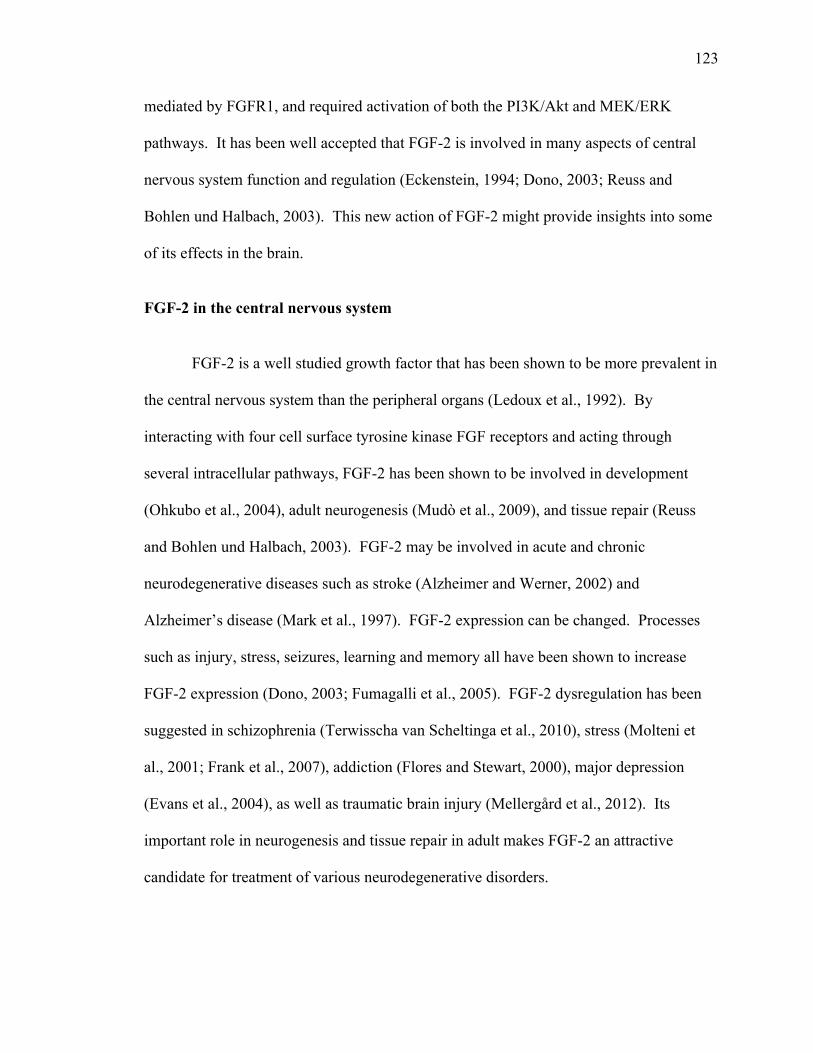
123
mediated by FGFR1, and required activation of both the PI3K/Akt and MEK/ERK
pathways. It has been well accepted that FGF-2 is involved in many aspects of central
nervous system function and regulation (Eckenstein, 1994; Dono, 2003; Reuss and
Bohlen und Halbach, 2003). This new action of FGF-2 might provide insights into some
of its effects in the brain.
FGF-2 in the central nervous system
FGF-2 is a well studied growth factor that has been shown to be more prevalent in
the central nervous system than the peripheral organs (Ledoux et al., 1992). By
interacting with four cell surface tyrosine kinase FGF receptors and acting through
several intracellular pathways, FGF-2 has been shown to be involved in development
(Ohkubo et al., 2004), adult neurogenesis (Mudò et al., 2009), and tissue repair (Reuss
and Bohlen und Halbach, 2003). FGF-2 may be involved in acute and chronic
neurodegenerative diseases such as stroke (Alzheimer and Werner, 2002) and
Alzheimer’s disease (Mark et al., 1997). FGF-2 expression can be changed. Processes
such as injury, stress, seizures, learning and memory all have been shown to increase
FGF-2 expression (Dono, 2003; Fumagalli et al., 2005). FGF-2 dysregulation has been
suggested in schizophrenia (Terwisscha van Scheltinga et al., 2010), stress (Molteni et
al., 2001; Frank et al., 2007), addiction (Flores and Stewart, 2000), major depression
(Evans et al., 2004), as well as traumatic brain injury (Mellergård et al., 2012). Its
important role in neurogenesis and tissue repair in adult makes FGF-2 an attractive
candidate for treatment of various neurodegenerative disorders.
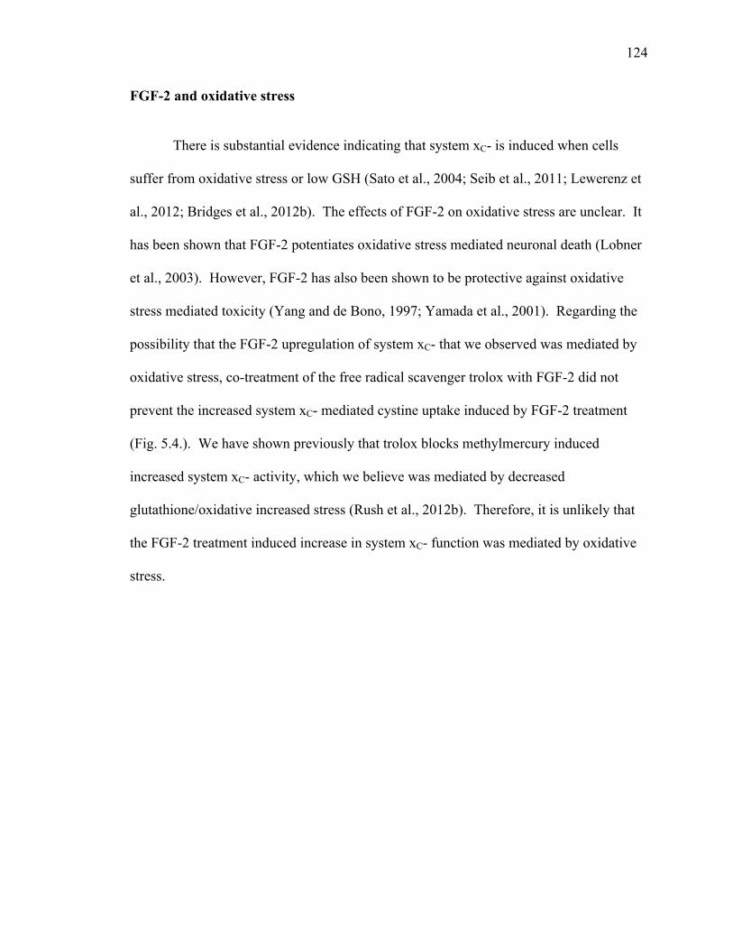
124
FGF-2 and oxidative stress
There is substantial evidence indicating that system xC- is induced when cells
suffer from oxidative stress or low GSH (Sato et al., 2004; Seib et al., 2011; Lewerenz et
al., 2012; Bridges et al., 2012b). The effects of FGF-2 on oxidative stress are unclear. It
has been shown that FGF-2 potentiates oxidative stress mediated neuronal death (Lobner
et al., 2003). However, FGF-2 has also been shown to be protective against oxidative
stress mediated toxicity (Yang and de Bono, 1997; Yamada et al., 2001). Regarding the
possibility that the FGF-2 upregulation of system xC- that we observed was mediated by
oxidative stress, co-treatment of the free radical scavenger trolox with FGF-2 did not
prevent the increased system xC- mediated cystine uptake induced by FGF-2 treatment
(Fig. 5.4.). We have shown previously that trolox blocks methylmercury induced
increased system xC- activity, which we believe was mediated by decreased
glutathione/oxidative increased stress (Rush et al., 2012b). Therefore, it is unlikely that
the FGF-2 treatment induced increase in system xC- function was mediated by oxidative
stress.
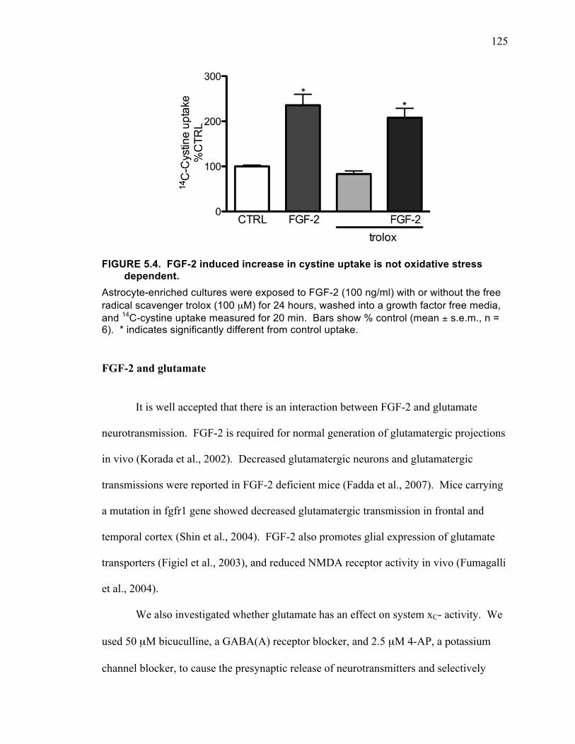
125
FIGURE 5.4. FGF-2 induced increase in cystine uptake is not oxidative stress dependent.
Astrocyte-enriched cultures were exposed to FGF-2 (100 ng/ml) with or without the free radical scavenger trolox (100 µM) for 24 hours, washed into a growth factor free media, and 14C-cystine uptake measured for 20 min. Bars show % control (mean ± s.e.m., n = 6). * indicates significantly different from control uptake.
FGF-2 and glutamate
It is well accepted that there is an interaction between FGF-2 and glutamate
neurotransmission. FGF-2 is required for normal generation of glutamatergic projections
in vivo (Korada et al., 2002). Decreased glutamatergic neurons and glutamatergic
transmissions were reported in FGF-2 deficient mice (Fadda et al., 2007). Mice carrying
a mutation in fgfr1 gene showed decreased glutamatergic transmission in frontal and
temporal cortex (Shin et al., 2004). FGF-2 also promotes glial expression of glutamate
transporters (Figiel et al., 2003), and reduced NMDA receptor activity in vivo (Fumagalli
et al., 2004).
We also investigated whether glutamate has an effect on system xC- activity. We
used 50 µM bicuculline, a GABA(A) receptor blocker, and 2.5 µM 4-AP, a potassium
channel blocker, to cause the presynaptic release of neurotransmitters and selectively
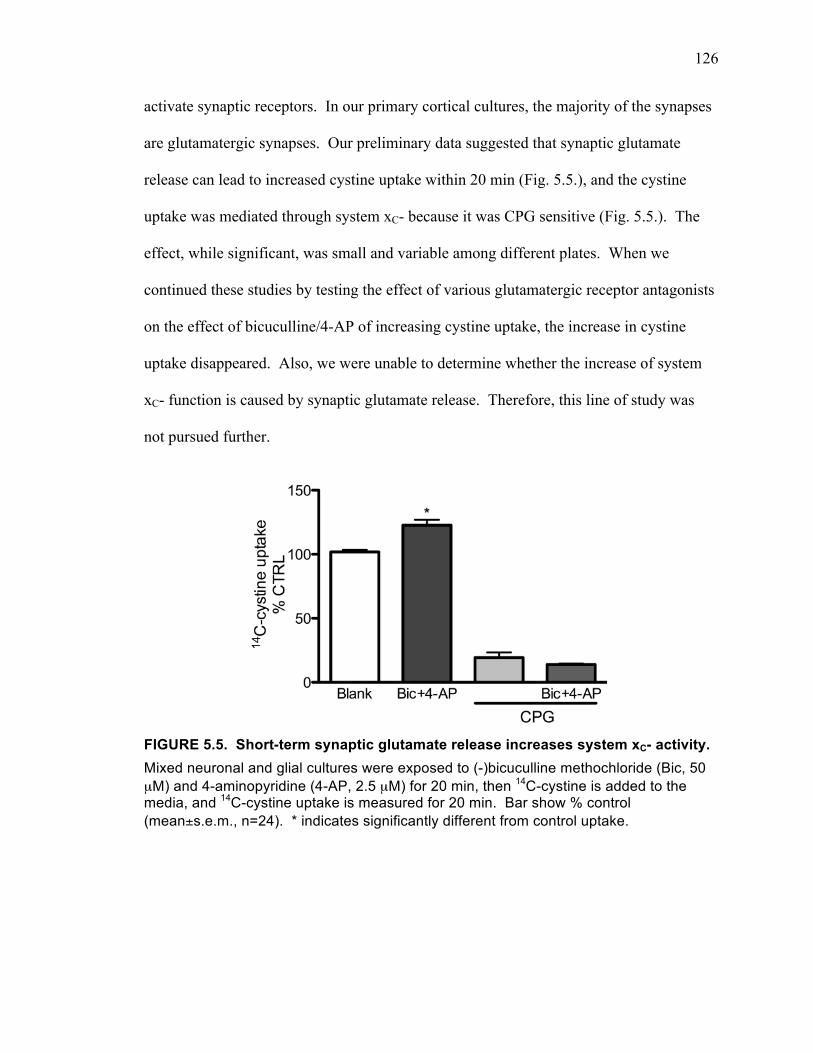
126
activate synaptic receptors. In our primary cortical cultures, the majority of the synapses
are glutamatergic synapses. Our preliminary data suggested that synaptic glutamate
release can lead to increased cystine uptake within 20 min (Fig. 5.5.), and the cystine
uptake was mediated through system xC- because it was CPG sensitive (Fig. 5.5.). The
effect, while significant, was small and variable among different plates. When we
continued these studies by testing the effect of various glutamatergic receptor antagonists
on the effect of bicuculline/4-AP of increasing cystine uptake, the increase in cystine
uptake disappeared. Also, we were unable to determine whether the increase of system
xC- function is caused by synaptic glutamate release. Therefore, this line of study was
not pursued further.
FIGURE 5.5. Short-term synaptic glutamate release increases system xC- activity. Mixed neuronal and glial cultures were exposed to (-)bicuculline methochloride (Bic, 50 µM) and 4-aminopyridine (4-AP, 2.5 µM) for 20 min, then 14C-cystine is added to the media, and 14C-cystine uptake is measured for 20 min. Bar show % control (mean±s.e.m., n=24). * indicates significantly different from control uptake.
127
TOXICITY OF FGF-2 MEDIATED BY SYSTEM XC-
Despite the numerous reports of FGF-2 as a neuroprotective growth factor
(Alzheimer and Werner, 2002), cytotoxic effects of FGF-2 have also been reported.
FGF-2 inhibits growth of cultured breast cancer cells (Johnson et al., 1998; Wang et al.,
1998), Ewing’s sarcoma cells (Schweigerer et al., 1987), and human neuroblastoma cells
(Russo et al., 2004). In Ewing’s sarcoma tumors, the cytotoxic effect of FGF-2 is
mediated through the same pathways that were associated with FGF-2 induced cell
proliferation (Kim et al., 2004; Russo et al., 2004; Williamson et al., 2004; Ma et al.,
2008). 48 hour treatment of FGF-2 inhibits Ewing’s sarcoma growth by inducing cell
death in three different cell lines (Sturla et al., 2000; Ma et al., 2008). FGF-2 also
inhibits Ewing’s sarcoma growth in vivo (Sturla et al., 2000). Overexpressing FGF-2 in
mice reduces chondrocyte proliferation by increasing apoptosis (Sahni et al., 2001).
We also observed significant neuronal death 48 hours after FGF-2 treatment,
which was associated with upregulation of system xC- activity. Blocking either system
xC- or AMPA receptors eliminated the neuronal cell death, suggesting glutamate exported
from astrocytic system xC- contributes to the neuronal death. However, we cannot
exclude the possibility that the neuronal toxicity was due to FGF-2 sensitizing the
neurons to normal glutamate release from system xC-. An AMPA receptor antagonist,
NBQX, but not an NMDA receptor antagonist, MEM, blocked the FGF-2 induced
neuronal death. This profile of toxicity is different from non-FGF-2 treated cells, in
which low level glutamate exposure causes a similar level of neuronal death, but it is
prevented by either AMPA or NMDA receptor antagonists. We could not use the more
potent NMDA receptor antagonists (2R)-amino-5-phosphonovaleric acid (APV) or MK-

128
801 because they cause cell death by themselves, even at low concentrations, after 48
hours (data not shown). Bath incubation of glutamate is believed to activate extracellular
receptors, which is likely similar to glutamate released from system xC-. Therefore, FGF-
2 is in some way altering the type of excitotoxicity that occurs to the neurons. To
determine what FGF-2 changes in terms of response to glutamate, detailed studies would
need to be carried out examining whether the levels of NMDA and AMPA receptors were
changed. This could be done by western blotting, examining the levels of NMDA and
AMPA receptor subunits. Furthermore, electrophysiological studies would be needed to
examine whether functional changes in receptors occurred. Fura-2 studies could also be
carried out to examine whether calcium influx was changed by FGF-2 treatment. It is
also possible that the changes in glutamate receptor mediated toxicity are due to changes
in the intracellular signaling cascade downstream of receptor activation.
We made several attempts to measure whether there was elevated glutamate
accumulating in the culture media following 48 hour FGF-2 treatment. DIV 13 astrocyte-
enriched cultures were washed into fresh media, then following 48 hour exposure to
FGF-2 or not (CTRL), bathing media from the cell cultures was collected and assayed for
glutamate by HPLC. The media used for such long-term exposure was requred to be a
complex media we call MS, which contains more than 100 µM concentrations of 8 amino
acids including cystine. Astrocyte-enriched cultures were used because in mixed cultures
neuronal death would occur at this time point, and the dead neurons would have released
large amounts of glutamate. Since the FGF-2 effect on system xC- occurs on astrocytes,
the effect should be observed in the astrocyte-enriched cultures. Two HPLC methods
were tried to quantitate glutamate: phenylisothiocyanate (PITC) derivatization with
129
ultraviolet detection on an Agilent 1100 HPLC and fluorescence detection with
ophthaldehyde pre-column derivitization on a Shimadzu LC10AD HPLC. However,
because the experiments were done in a complex media, there was an interfering peak
where glutamate was to be measured. Both a Hypersil-ODS reverse phase column and a
Synergi Hydro-RP column were tested with the PITC derivatization with ultraviolet
detection method in an attempt to separate the interfering peak from the glutamate peak.
Also various mobile phases and flow rates were tested. Although the new column, and
using a slower flow rate appeared to give us better separation, it significantly decreased
the sensitivity for detection of glutamate (more than 10 times less sensitive). Since we
are detecting glutamate in a large amount of extracellular media compared to that of the
restricted space among the attached cells (with an estimated 1:1000 dilution), the
glutamate concentration in the extracellular media is very low. Furthermore, it is not
known whether FGF-2 treatment changes the glutamate uptake efficiency. Therefore, we
were unable to determine the extracellular glutamate levels under these conditions.
We cannot exclude the possibility of FGF-2 treatment changing neuronal
susceptibility to normal extracellular glutamate levels. One of the possibilities being that
FGF-2 treatment changes the astrocytes’ ability to release cysteine-containing molecules,
which in turn, selectively decreases glutathione levels in neurons, causing them to be
susceptible to any insult, including glutamate levels that does not cause death to healthy
neurons. This hypothesis could be tested by measuring thiols by HPLC in the culture
media of mixed neuronal and glial cultures.
130
IS SYSTEM XC- ACTIVITY TOXIC TO NEURONS?
In this dissertation, we have shown that BMAA can contribute to neuronal death
by acting at system xC-. Also, that prolonged FGF-2 treatment can upregulate system xC-
and cause a system xC- function dependent neuronal toxicity. As knowledge of the
function of system xC- expands, and the interest in targeting this system as a treatment for
various neurodegenerative diseases and psychiatric disorders increases, it is important to
understand the potential damage that system xC- activity can cause. Currently, it is clear
that for cells that mainly rely on system xC- to take up cystine for the production of
glutathione, blocking system xC- function can be toxic (Murphy et al., 1989). The
question remains whether the glutamate exchanged out of these cells by system xC- is a
threat to surrounding cells. Glioma cells have increased system xC- function, as well as
decreased glutamate uptake, leading to increased extracellular glutamate which can cause
toxicity to the surrounding neurons (Ye and Sontheimer, 1999; Sontheimer, 2003; 2008;
Ogunrinu and Sontheimer, 2010). Both FGF-2 (Liu et al., 2011) and IL-1β (Fogal et al.,
2007) upregulate system xC- function selectively on astrocytes. Long-term FGF-2
treatment causes a system xC- function dependent, and AMPA receptor specific, neuronal
death. While IL-1β treatment potentiates hypoxia and glucose deprivation induced
neuronal death through a system xC- activity dependent mechanism (Fogal et al., 2007;
Jackman et al., 2012). Another example of increased system xC- function is activated
microglia, which release enough glutamate through system xC- to cause toxicity to the
surrounding neurons (Domercq et al., 2007). In all of these studies, along with the
involvement of system xC-, the neurons are under stress: hypoxia, glucose deprivation, or
stimulation by growth factors or cytokines. Therefore, while it is clear that enhanced
131
system xC- activity on non-neuronal cells can potentiate neuronal death when the neurons
are compromised, it is uncertain whether glutamate release from system xC- can cause
neuronal death under normal conditions.
QUESTIONS REGARDING SYSTEM XC- STUDIES
Molecular properties of system xC-
Northern blot has shown three different xCT transcripts (12, 3.5 and 2.5 kb) in
cultured macrophages and HT22 cells, as well as in various mouse organ tissues (Sato et
al., 1999; 2001; 2004; 2005; Lewerenz et al., 2006; Taguchi et al., 2007). They may
represent alternative splicing or alternative polyadenylation (Sato et al., 1999; Lewerenz
et al., 2006). While all of these transcripts are inducible to a similar extent in culture
(Sato et al., 1999), they have a different distribution pattern. The 3.5 and 2.5 kb
transcripts were predominantly seen in macrophages, while the 12 kb RNA transcript was
found in mouse brain, but not in peripheral organs such as heart, lung, liver, and kidney
(Sato et al., 1999; Bassi et al., 2001; Sato et al., 2002). Human ovarian cancer cell lines
(Okuno et al., 2003), hamster kidney cells (Sasaki et al., 2002), and cultured rat
astrocytes (Gochenauer and Robinson, 2001)were shown to exclusively express the 12 kb
transcript of xCT.
The predicted amino acid sequence from cDNA for xCT would be a protein of
502 amino acids with a relative molecular mass of 55.5 kDa (Sato et al., 1999).
However, in reality, xCT with various molecular masses have been reported. The 55kDa
protein is observed both in brain in vivo and in cortical neuronal stem cell cultures (Sims
et al., 2012). 50 kDa xCT was detected in human glioma cells (Kim et al., 2001) and
132
retinal ganglion cell line membranes (Dun et al., 2006). In vitro expression of xCT in
Xenopus oocytes (Sato et al., 1999) and HT22 cells (Burdo et al., 2006) showed a band of
an approximately 40 kDa protein, which could be downregulated by siRNA (Burdo et al.,
2006). This 40 kDa xCT was detected in rat brain tissue, with the expression level
increasing during development and reaching the highest expression as an adult (La Bella
et al., 2007). xCT was also observed in mouse hippocampus at a molecular weight of
~35 kDa, which is absent in xCT -/- mice (Pacchioni et al., 2007; De Bundel et al., 2011).
There are also studies reporting that xCT is detected at multiple molecular masses
in the same tissue, with the same antibody. A study done in primary rat astrocyte cultures
revealed three distinct bands for xCT: 40, 45, and 50 kDa, however, only the 40 kDa was
released from the system xC- heterodimer after reducing agent was added (Seib et al.,
2011). xCT was also detected at 50 kDa and 40 kDa in mouse Muller cells, with the 50
kDa protein as the majority version. However, it was not inducible by oxidative stress,
while the minority 40 kDa form, which is normally predominantly located intracellularly,
was upregulated by oxidative stress and translocated to the membrane (Mysona et al.,
2009). The same 40 kDa form of xCT was also found to be almost exclusively present on
the cellular membrane in rat brain tissue, cultured human fibroblast, rat neurons and
astrocytes (La Bella et al., 2007). xCT was detected at 35 kDa and 55 kDa in mouse
brain and mouse astrocyte cultures, with the 55 kDa form prominently detected only in
mouse meninges (Shih et al., 2006).
Together with these mixed results, there are also a number of system xC- studies
that did not report the band(s) mass in their paper (Bridges et al., 2004; Qiang et al.,
2004; Sakakura et al., 2007). It seems that the use of different antibodies and different
133
tissues can lead to different results. Furthermore, increased expression does not
necessarily mean increased membrane expression and increased function. Therefore,
claims about potential system xC- function by measuring xCT protein expression must be
considered skeptically. In our studies, we have focused on the functional activity of
system xC- measured by 14C-cystine uptake.
Relevance and limitations of translating in vitro studies to in vivo:
It has been suggested that when compared to in vivo, system xC- activity is much
higher in vitro (Lewerenz et al., 2012). This is believed to be because system xC- activity
can be easily induced. Even regular cell culture conditions with ambient O2 of 21%
strongly induces system xC- activity in diverse cell types, including fibroblasts, HEK
cells, hippocampal HT22 cells, astrocytes, and microglial (Lewerenz et al., 2012). In
freshly prepared hepatocytes and macrophages, the activity of system xC- is barely
detectable (Takada and Bannai, 1984; Watanabe and Bannai, 1987), but it is significantly
upregulated after 12 hours of culturing (Watanabe and Bannai, 1987). A much higher
system xC- activity level is measured in macrophages cultured for 8 hours in the presence
of LPS and diethyl maleate (Sato et al., 1995a). Various electrophilic agents and
different pH have been proven to be system xC- activity inducers (Reynolds et al., 1991;
Sasaki et al., 2002). These concerns regarding cell culture studies must be kept in mind
when attempting to extrapolate cell culture studies to the in vivo situation.
Our in vitro data showed that astrocytes have greater system xC- activity than
neurons (Lobner, 2009), in agreement with another lab (Jackman et al., 2012).
Preliminary data from our lab indicates that neuronal conditioned media significantly
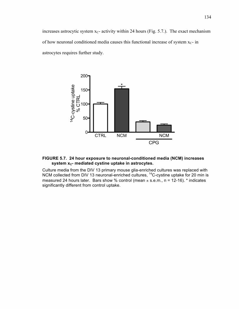
134
increases astrocytic system xC- activity within 24 hours (Fig. 5.7.). The exact mechanism
of how neuronal conditioned media causes this functional increase of system xC- in
astrocytes requires further study.
FIGURE 5.7. 24 hour exposure to neuronal-conditioned media (NCM) increases system xC- mediated cystine uptake in astrocytes.
Culture media from the DIV 13 primary mouse glia-enriched cultures was replaced with NCM collected from DIV 13 neuronal-enriched cultures, 14C-cystine uptake for 20 min is measured 24 hours later. Bars show % control (mean ± s.e.m., n = 12-16). * indicates significantly different from control uptake.
135
CONCLUSION
System xC- dysregulation has been implicated in various neurodegenerative
diseases and psychiatric disorders. The dual function of system xC- awards it a distinct
role in influencing cell fate. It may be protective by promoting cystine uptake and
preventing oxidative stress, but it may be harmful by releasing glutamate and
exacerbating excitotoxicity.
The goal of this thesis was to add to the knowledge regarding the role of system
xC- in neuronal death. Two major advancements to the field were made. First, we
discovered that the environmental neurotoxin BMAA acts as a substrate for system xC-.
Through this action, BMAA both inhibits cystine uptake, leading to oxidative stress and
drives glutamate release causing excitotoxicity. This result is important not only for
understanding the mechanism of BMAA toxicity, but also provides evidence that
compounds that are transported by system xC-, particularly if they do not lead to
increased glutathione production, are potentially highly neurotoxic. Second, the results
involving FGF-2 suggest that increasing system xC- selectively on astrocytes is
potentially harmful to neurons. This finding, in concert with the well known neurotoxic
action of glomas, that also have upregulated system xC-, suggests a general hypothesis
that upregulation of system xC- on non-neuronal cells can be toxic to neurons.
136
BIBLIOGRAPHY
Agulhon C, Petravicz J, Mcmullen A, Sweger E, Minton S, Taves S, Casper K, Fiacco T, Mccarthy K (2008) What Is the Role of Astrocyte Calcium in Neurophysiology? Neuron 59:932–946.
Alagarsamy S, Marino MJ, Rouse ST, Gereau RW, Heinemann SF, Conn PJ (1999) Activation of NMDA receptors reverses desensitization of mGluR5 in native and recombinant systems. Nat Neurosci 2:234–240.
Alt A, Weiss B, Ogden AM, Knauss JL, Oler J, Ho K, Large TH, Bleakman D (2004) Pharmacological characterization of glutamatergic agonists and antagonists at recombinant human homomeric and heteromeric kainate receptors in vitro. Neuropharmacology 46:793–806.
Alzheimer C, Werner S (2002) Fibroblast growth factors and neuroprotection. Adv Exp Med Biol 513:335–351.
Amara SG (1992) Neurotransmitter transporters. A tale of two families. Nature 360:420–421.
Amen SL, Piacentine LB, Ahmad ME, Li S-J, Mantsch JR, Risinger RC, Baker DA (2011) Repeated N-acetyl cysteine reduces cocaine seeking in rodents and craving in cocaine-dependent humans. Neuropsychopharmacology 36:871–878.
Ameri K, Harris AL (2008) Activating transcription factor 4. Int J Biochem Cell Biol 40:14–21.
Andersen JK (2004) Oxidative stress in neurodegeneration: cause or consequence? Nat Med 10 Suppl:S18–S25.
Anton ES, Marchionni MA, Lee KF, Rakic P (1997) Role of GGF/neuregulin signaling in interactions between migrating neurons and radial glia in the developing cerebral cortex. Development 124:3501–3510.
Aoyama K, Suh SW, Hamby AM, Liu J, Chan WY, Chen Y, Swanson RA (2006) Neuronal glutathione deficiency and age-dependent neurodegeneration in the EAAC1 deficient mouse. Nat Neurosci 9:119–126.
Arrick BA, Griffo W, Cohn Z, Nathan C (1985) Hydrogen peroxide from cellular metabolism of cystine. A requirement for lysis of murine tumor cells by vernolepin, a glutathione-depleting antineoplastic. Journal of Clinical Investigation 76:567–574.
Aschner M, Mullaney KJ, Wagoner D, Lash LH, Kimelberg HK (1994) Intracellular glutathione (GSH) levels modulate mercuric chloride (MC)- and methylmercuric chloride (MeHgCl)-induced amino acid release from neonatal rat primary astrocytes cultures. Brain Research 664:133–140.
137
Asztely F, Erdemli G, Kullmann DM (1997) Extrasynaptic glutamate spillover in the hippocampus: dependence on temperature and the role of active glutamate uptake. Neuron 18:281–293.
Augustin H, Grosjean Y, Chen K, Sheng Q, Featherstone DE (2007) Nonvesicular release of glutamate by glial xCT transporters suppresses glutamate receptor clustering in vivo. Journal of Neuroscience 27:111–123.
Ay H, Ay I, Koroshetz WJ, Finklestein SP (1999) Potential usefulness of basic fibroblast growth factor as a treatment for stroke. Cerebrovasc Dis 9:131–135.
Bains JS, Shaw CA (1997) Neurodegenerative disorders in humans: the role of glutathione in oxidative stress-mediated neuronal death. Brain Res Brain Res Rev 25:335–358.
Baker DA, Madayag A, Kristiansen LV, Meador-Woodruff JH, Haroutunian V, Raju I (2008) Contribution of cystine-glutamate antiporters to the psychotomimetic effects of phencyclidine. Neuropsychopharmacology 33:1760–1772.
Baker DA, McFarland K, Lake RW, Shen H, Tang X-C, Toda S, Kalivas PW (2003) Neuroadaptations in cystine-glutamate exchange underlie cocaine relapse. Nat Neurosci 6:743–749.
Baker DA, Xi Z-X, Shen H, Swanson CJ, Kalivas PW (2002) The origin and neuronal function of in vivo nonsynaptic glutamate. Journal of Neuroscience 22:9134–9141.
Baker MA, Cerniglia GJ, Zaman A (1990) Microtiter plate assay for the measurement of glutathione and glutathione disulfide in large numbers of biological samples. Anal Biochem 190:360–365.
Ballatori N, Krance SM, Notenboom S, Shi S, Tieu K, Hammond CL (2009) Glutathione dysregulation and the etiology and progression of human diseases. Biol Chem 390:191–214.
Banack SA, Johnson HE, Cheng R, Cox PA (2007) Production of the neurotoxin BMAA by a marine cyanobacterium. Mar Drugs 5:180–196.
Banjac A, Perisic T, Sato H, Seiler A, Bannai S, Weiss N, Kölle P, Tschoep K, Issels RD, Daniel PT, Conrad M, Bornkamm GW (2008) The cystine/cysteine cycle: a redox cycle regulating susceptibility versus resistance to cell death. Oncogene 27:1618–1628.
Bannai S (1984) Induction of cystine and glutamate transport activity in human fibroblasts by diethyl maleate and other electrophilic agents. The Journal of Biological Chemistry 259:2435–2440.
Bannai S, Kitamura E (1980) Transport interaction of L-cystine and L-glutamate in human diploid fibroblasts in culture. The Journal of Biological Chemistry
138
255:2372–2376.
Bannerman P, Horiuchi M, Feldman D, Hahn A, Itoh A, See J, Jia ZP, Itoh T, Pleasure D (2007) GluR2-free alpha-amino-3-hydroxy-5-methyl-4-isoxazolepropionate receptors intensify demyelination in experimental autoimmune encephalomyelitis. Journal of Neurochemistry 102:1064–1070.
Barbour B, Brew H, Attwell D (1988) Electrogenic glutamate uptake in glial cells is activated by intracellular potassium. Nature 335:433–435.
Barger SW, Basile AS (2001) Activation of microglia by secreted amyloid precursor protein evokes release of glutamate by cystine exchange and attenuates synaptic function. Journal of Neurochemistry 76:846–854.
Bassi MT, Gasol E, Manzoni M, Pineda M, Riboni M, Martín R, Zorzano A, Borsani G, Palacín M (2001) Identification and characterisation of human xCT that co-expresses, with 4F2 heavy chain, the amino acid transport activity system xc-. Pflugers Arch 442:286–296.
Beck KD (1994) Functions of brain-derived neurotrophic factor, insulin-like growth factor-I and basic fibroblast growth factor in the development and maintenance of dopaminergic neurons. Prog Neurobiol 44:497–516.
Benarroch EE (2009) Astrocyte-neuron interactions: implications for epilepsy. Neurology 73:1323–1327.
Benquet P, Gee CE, Gerber U (2002) Two distinct signaling pathways upregulate NMDA receptor responses via two distinct metabotropic glutamate receptor subtypes. Journal of Neuroscience 22:9679–9686.
Beutler E (1989) Nutritional and metabolic aspects of glutathione. Annu Rev Nutr 9:287–302.
Bezzi P, Carmignoto G, Pasti L, Vesce S, Rossi D, Rizzini BL, Pozzan T, Volterra A (1998) Prostaglandins stimulate calcium-dependent glutamate release in astrocytes. Nature 391:281–285.
Bezzi P, Gundersen V, Galbete JL, Seifert G, Steinhäuser C, Pilati E, Volterra A (2004) Astrocytes contain a vesicular compartment that is competent for regulated exocytosis of glutamate. Nat Neurosci 7:613–620.
Béhé P, Stern P, Wyllie DJ, Nassar M, Schoepfer R, Colquhoun D (1995) Determination of NMDA NR1 subunit copy number in recombinant NMDA receptors. Proc Biol Sci 262:205–213.
Bogdanov VB, Bogdanova OV, Koulchitsky SV, Chauvel V, Multon S, Makarchuk MY, Brennan KC, Renshaw PF, Schoenen J (2013) Behavior in the open field predicts the number of KCl-induced cortical spreading depressions in rats. Behav Brain Res
139
236:90–93.
Bogousslavsky J, Victor SJ, Salinas EO, Pallay A, Donnan GA, Fieschi C, Kaste M, Orgogozo J-MM, Chamorro A, Desmet A, European-Australian Fiblast (Trafermin) in Acute Stroke Group (2002) Fiblast (trafermin) in acute stroke: results of the European-Australian phase II/III safety and efficacy trial. Cerebrovasc Dis 14:239–251.
Bolaños JP, Heales SJ, Land JM, Clark JB (1995) Effect of peroxynitrite on the mitochondrial respiratory chain: differential susceptibility of neurones and astrocytes in primary culture. Journal of Neurochemistry 64:1965–1972.
Boxer AL, Moreno H, Rudy B, Ziff EB (1999) FGF-2 potentiates Ca(2+)-dependent inactivation of NMDA receptor currents in hippocampal neurons. Journal of Neurophysiology 82:3367–3377.
Brackley PT, Bell DR, Choi SK, Nakanishi K, Usherwood PN (1993) Selective antagonism of native and cloned kainate and NMDA receptors by polyamine-containing toxins. J Pharmacol Exp Ther 266:1573–1580.
Bradley WG, Mash DC (2009) Beyond Guam: the cyanobacteria/BMAA hypothesis of the cause of ALS and other neurodegenerative diseases. Amyotroph Lateral Scler 10 Suppl 2:7–20.
Brand LE, Pablo J, Compton A, Hammerschlag N, Mash DC (2010) Cyanobacterial Blooms and the Occurrence of the neurotoxin beta-N-methylamino-L-alanine (BMAA) in South Florida Aquatic Food Webs. Harmful Algae 9:620–635.
Brandoli C, Sanna A, De Bernardi MA, Follesa P, Brooker G, Mocchetti I (1998) Brain-derived neurotrophic factor and basic fibroblast growth factor downregulate NMDA receptor function in cerebellar granule cells. J Neurosci 18:7953–7961.
Bridges CC, Hu H, Miyauchi S, Siddaramappa UN, Ganapathy ME, Ignatowicz L, Maddox DM, Smith SB, Ganapathy V (2004) Induction of cystine-glutamate transporter xc- by human immunodeficiency virus type 1 transactivator protein tat in retinal pigment epithelium. Investigative Ophthalmology & Visual Science 45:2906–2914.
Bridges R, Lutgen V, Lobner D, Baker DA (2012a) Thinking Outside the Cleft to Understand Synaptic Activity: Contribution of the Cystine-Glutamate Antiporter (System xc-) to Normal and Pathological Glutamatergic Signaling. Pharmacological reviews 64:780–802.
Bridges RJ, Natale NR, Patel SA (2012b) System xc⁻ cystine/glutamate antiporter: an update on molecular pharmacology and roles within the CNS. Br J Pharmacol 165:20–34.
Brigelius-Flohé R, Traber MG (1999) Vitamin E: function and metabolism. FASEB J
140
13:1145–1155.
Brownson DM, Mabry TJ, Leslie SW (2002) The cycad neurotoxic amino acid, beta-N-methylamino-L-alanine (BMAA), elevates intracellular calcium levels in dissociated rat brain cells. J Ethnopharmacol 82:159–167.
Buckingham SC, Campbell SL, Haas BR, Montana V, Robel S, Ogunrinu T, Sontheimer H (2011) Glutamate release by primary brain tumors induces epileptic activity. Nat Med.
Burdo J, Dargusch R, Schubert D (2006) Distribution of the cystine/glutamate antiporter system xc- in the brain, kidney, and duodenum. J Histochem Cytochem 54:549–557.
Burnashev N, Zhou Z, Neher E, Sakmann B (1995) Fractional calcium currents through recombinant GluR channels of the NMDA, AMPA and kainate receptor subtypes. The Journal of Physiology 485 ( Pt 2):403–418.
Butt AM (2011) ATP: a ubiquitous gliotransmitter integrating neuron-glial networks. Semin Cell Dev Biol 22:205–213.
Cabungcal J-H, Preissmann D, Delseth C, Cuénod M, Do KQ, Schenk F (2007) Transitory glutathione deficit during brain development induces cognitive impairment in juvenile and adult rats: relevance to schizophrenia. Neurobiol Dis 26:634–645.
Carriedo SG, Yin HZ, Lamberta R, Weiss JH (1995) In vitro kainate injury to large, SMI-32(+) spinal neurons is Ca2+ dependent. Neuroreport 6:945–948.
Chang RCC, Wong AKY, Ng H-K, Hugon J (2002) Phosphorylation of eukaryotic initiation factor-2alpha (eIF2alpha) is associated with neuronal degeneration in Alzheimer's disease. Neuroreport 13:2429–2432.
Charleston JS, Body RL, Mottet NK, Vahter ME, Burbacher TM (1995) Autometallographic determination of inorganic mercury distribution in the cortex of the calcarine sulcus of the monkey Macaca fascicularis following long-term subclinical exposure to methylmercury and mercuric chloride. Toxicology and Applied Pharmacology 132:325–333.
Chase LA, Peterson NL, Koerner JF (2007) The lathyrus toxin, beta-N-oxalyl-L-alpha,beta-diaminopropionic acid (ODAP), and homocysteic acid sensitize CA1 pyramidal neurons to cystine and L-2-amino-6-phosphonohexanoic acid. Toxicology and Applied Pharmacology 219:1–9.
Chen H-H, Liao P-F, Chan M-H (2011) mGluR5 positive modulators both potentiate activation and restore inhibition in NMDA receptors by PKC dependent pathway. J Biomed Sci 18:19.
141
Chen H-SV, Lipton SA (2006) The chemical biology of clinically tolerated NMDA receptor antagonists. Journal of Neurochemistry 97:1611–1626.
Chen Y, Swanson RA (2003) Astrocytes and brain injury. J Cereb Blood Flow Metab 23:137–149.
Cheng B, Furukawa K, O'Keefe JA, Goodman Y, Kihiko M, Fabian T, Mattson MP (1995) Basic fibroblast growth factor selectively increases AMPA-receptor subunit GluR1 protein level and differentially modulates Ca2+ responses to AMPA and NMDA in hippocampal neurons. Journal of Neurochemistry 65:2525–2536.
Chintala S, Li W, Lamoreux ML, Ito S, Wakamatsu K, Sviderskaya EV, Bennett DC, Park Y-M, Gahl WA, Huizing M, Spritz RA, Ben S, Novak EK, Tan J, Swank RT (2005) Slc7a11 gene controls production of pheomelanin pigment and proliferation of cultured cells. Proc Natl Acad Sci USA 102:10964–10969.
Choi D (1998) Antagonizing excitotoxicity: a therapeutic strategy for stroke? Mt Sinai J Med 65:133–138.
Choi DW (1987) Ionic dependence of glutamate neurotoxicity. J Neurosci 7:369–379.
Choi DW, Maulucci-Gedde M, Kriegstein AR (1987) Glutamate neurotoxicity in cortical cell culture. J Neurosci 7:357–368.
Chung WJ, Lyons SA, Nelson GM, Hamza H, Gladson CL, Gillespie GY, Sontheimer H (2005) Inhibition of cystine uptake disrupts the growth of primary brain tumors. Journal of Neuroscience 25:7101–7110.
Chung WJ, Sontheimer H (2009) Sulfasalazine inhibits the growth of primary brain tumors independent of nuclear factor-kappaB. Journal of Neurochemistry 110:182–193.
Clark WM, Schim JD, Kasner SE, Victor SJ (2000) Trafermin in acute ischemic stroke: results of a phase II/III randomized efficacy study. Neurology 54:A88.
Clem RL, Barth A (2006) Pathway-specific trafficking of native AMPARs by in vivo experience. Neuron 49:663–670.
Cohen SA (2012) Analytical techniques for the detection of α-amino-β-methylaminopropionic acid. Analyst 137:1991–2005.
Cohen SA, Bidlingmeyer BA, Tarvin TL (1986) PITC derivatives in amino acid analysis. Nature 320:769–770.
Conn PJ, Pin JP (1997) Pharmacology and functions of metabotropic glutamate receptors. Annu Rev Pharmacol Toxicol 37:205–237.
Cornell-Bell AH, Finkbeiner SM, Cooper MS, Smith SJ (1990) Glutamate induces
142
calcium waves in cultured astrocytes: long-range glial signaling. Science 247:470–473.
Costa-Mattioli M, Gobert D, Stern E, Gamache K, Colina R, Cuello C, Sossin W, Kaufman R, Pelletier J, Rosenblum K, Krnjević K, Lacaille J-C, Nader K, Sonenberg N (2007) eIF2alpha phosphorylation bidirectionally regulates the switch from short- to long-term synaptic plasticity and memory. Cell 129:195–206.
Cox PA, Banack SA, Murch SJ (2003) Biomagnification of cyanobacterial neurotoxins and neurodegenerative disease among the Chamorro people of Guam. Proc Natl Acad Sci USA 100:13380–13383.
Cox PA, Banack SA, Murch SJ, Rasmussen U, Tien G, Bidigare RR, Metcalf JS, Morrison LF, Codd GA, Bergman B (2005) Diverse taxa of cyanobacteria produce beta-N-methylamino-L-alanine, a neurotoxic amino acid. Proc Natl Acad Sci USA 102:5074–5078.
Cox PA, Richer R, Metcalf JS, Banack SA, Codd GA, Bradley WG (2009) Cyanobacteria and BMAA exposure from desert dust: a possible link to sporadic ALS among Gulf War veterans. Amyotroph Lateral Scler 10 Suppl 2:109–117.
Cox PA, Sacks OW (2002) Cycad neurotoxins, consumption of flying foxes, and ALS-PDC disease in Guam. Neurology 58:956–959.
Crimmins DL, Cherian R (1997) Increasing the sensitivity of 6-aminoquinolyl-N-hydroxysuccinimidyl carbamate amino acid analysis: a simple solution. Anal Biochem 244:407–410.
Crockett S, Clarke M, Reeves S, Sims B (2011) Cystine glutamate exchanger upregulation by retinoic acid induces neuroprotection in neural stem cells. Neuroreport 22:598–602.
Cruz-Aguado R, Winkler D, Shaw CA (2006) Lack of behavioral and neuropathological effects of dietary beta-methylamino-L-alanine (BMAA) in mice. Pharmacol Biochem Behav 84:294–299.
Cucchiaroni ML, Viscomi MT, Bernardi G, Molinari M, Guatteo E, Mercuri NB (2010) Metabotropic glutamate receptor 1 mediates the electrophysiological and toxic actions of the cycad derivative beta-N-Methylamino-L-alanine on substantia nigra pars compacta DAergic neurons. Journal of Neuroscience 30:5176–5188.
Cuevas P, Carceller F, Ortega S, Zazo M, Nieto I, Giménez-Gallego G (1991) Hypotensive activity of fibroblast growth factor. Science 254:1208–1210.
Cui K, Luo X, Xu K, Ven Murthy MR (2004) Role of oxidative stress in neurodegeneration: recent developments in assay methods for oxidative stress and nutraceutical antioxidants. Progress in neuro-psychopharmacology & biological psychiatry 28:771–799.
143
Czyz A, Baranauskas G, Kiedrowski L (2002) Instrumental role of Na+ in NMDA excitotoxicity in glucose-deprived and depolarized cerebellar granule cells. Journal of Neurochemistry 81:379–389.
De Bundel D, Schallier A, Loyens E, Fernando R, Miyashita H, Van Liefferinge J, Vermoesen K, Bannai S, Sato H, Michotte Y, Smolders I, Massie A (2011) Loss of system x(c)- does not induce oxidative stress but decreases extracellular glutamate in hippocampus and influences spatial working memory and limbic seizure susceptibility. Journal of Neuroscience 31:5792–5803.
Dean OM, van den Buuse M, Bush AI, Copolov DL, Ng F, Dodd S, Berk M (2009) A role for glutathione in the pathophysiology of bipolar disorder and schizophrenia? Animal models and relevance to clinical practice. Curr Med Chem 16:2965–2976.
Deguchi Y, Naito T, Yuge T, Furukawa A, Yamada S, Pardridge WM, Kimura R (2000) Blood-brain barrier transport of 125I-labeled basic fibroblast growth factor. Pharm Res 17:63–69.
Deneke SM, Fanburg BL (1989) Regulation of cellular glutathione. Am J Physiol 257:L163–L173.
Dholakia JN, Wahba AJ (1989) Mechanism of the nucleotide exchange reaction in eukaryotic polypeptide chain initiation. Characterization of the guanine nucleotide exchange factor as a GTP-binding protein. The Journal of Biological Chemistry 264:546–550.
Diamond JS, Jahr CE (1997) Transporters buffer synaptically released glutamate on a submillisecond time scale. J Neurosci 17:4672–4687.
Dingledine R, Borges K, Bowie D, Traynelis SF (1999) The glutamate receptor ion channels. Pharmacological reviews 51:7–61.
Domercq M, Etxebarria E, Pérez-Samartín A, Matute C (2005) Excitotoxic oligodendrocyte death and axonal damage induced by glutamate transporter inhibition. Glia 52:36–46.
Domercq M, Sánchez-Gómez MV, Sherwin C, Etxebarria E, Fern R, Matute C (2007) System xc- and glutamate transporter inhibition mediates microglial toxicity to oligodendrocytes. J Immunol 178:6549–6556.
Dono R (2003) Fibroblast growth factors as regulators of central nervous system development and function. Am J Physiol Regul Integr Comp Physiol 284:R867–R881.
Dringen R, Gutterer JM, Hirrlinger J (2000) Glutathione metabolism in brain metabolic interaction between astrocytes and neurons in the defense against reactive oxygen species. Eur J Biochem 267:4912–4916.
144
Dringen R, Hirrlinger J (2003) Glutathione pathways in the brain. Biol Chem 384:505–516.
Dringen R, Pfeiffer B, Hamprecht B (1999) Synthesis of the antioxidant glutathione in neurons: supply by astrocytes of CysGly as precursor for neuronal glutathione. J Neurosci 19:562–569.
Dringen R, Wiesinger H, Hamprecht B (1993) Uptake of L-lactate by cultured rat brain neurons. Neuroscience Letters 163:5–7.
Duan S, Anderson CM, Keung EC, Chen Y, Chen Y, Swanson RA (2003) P2X7 receptor-mediated release of excitatory amino acids from astrocytes. Journal of Neuroscience 23:1320–1328.
Dugan LL, Bruno VM, Amagasu SM, Giffard RG (1995) Glia modulate the response of murine cortical neurons to excitotoxicity: glia exacerbate AMPA neurotoxicity. J Neurosci 15:4545–4555.
Dugan LL, Choi DW (1994) Excitotoxicity, free radicals, and cell membrane changes. Ann Neurol 35 Suppl:S17–S21.
Dun Y, Mysona B, Van Ells T, Amarnath L, Ola MS, Ganapathy V, Smith SB (2006) Expression of the cystine-glutamate exchanger (xc-) in retinal ganglion cells and regulation by nitric oxide and oxidative stress. Cell Tissue Res 324:189–202.
Duncan MW, Markey SP, Weick BG, Pearson PG, Ziffer H, Hu Y, Kopin IJ (1992) 2-Amino-3-(methylamino)propanoic acid (BMAA) bioavailability in the primate. NBA 13:333–337.
Duncan MW, Steele JC, Kopin IJ, Markey SP (1990) 2-Amino-3-(methylamino)-propanoic acid (BMAA) in cycad flour: an unlikely cause of amyotrophic lateral sclerosis and parkinsonism-dementia of Guam. Neurology 40:767–772.
Duncan MW, Villacreses NE, Pearson PG, Wyatt L, Rapoport SI, Kopin IJ, Markey SP, Smith QR (1991) 2-amino-3-(methylamino)-propanoic acid (BMAA) pharmacokinetics and blood-brain barrier permeability in the rat. J Pharmacol Exp Ther 258:27–35.
Duncan R, Hershey JW (1985) Regulation of initiation factors during translational repression caused by serum depletion. Covalent modification. The Journal of Biological Chemistry 260:5493–5497.
Duncan RF, Hershey JW (1987) Initiation factor protein modifications and inhibition of protein synthesis. Mol Cell Biol 7:1293–1295.
Eckenstein F, Woodward WR, Nishi R (1991a) Differential localization and possible functions of aFGF and bFGF in the central and peripheral nervous systems. Annals of the New York Academy of Sciences 638:348–360.
145
Eckenstein FP (1994) Fibroblast growth factors in the nervous system. J Neurobiol 25:1467–1480.
Eckenstein FP, Shipley GD, Nishi R (1991b) Acidic and basic fibroblast growth factors in the nervous system: distribution and differential alteration of levels after injury of central versus peripheral nerve. J Neurosci 11:412–419.
Edvinsson L, Krause DN (2002) Cerebral blood flow and metabolism.
Erdner DL, Dyble J, Parsons ML, Stevens RC, Hubbard KA, Wrabel ML, Moore SK, Lefebvre KA, Anderson DM, Bienfang P, Bidigare RR, Parker MS, Moeller P, Brand LE, Trainer VL (2008) Centers for Oceans and Human Health: a unified approach to the challenge of harmful algal blooms. Environ Health 7 Suppl 2:S2.
Erecińska M, Silver IA (1990) Metabolism and role of glutamate in mammalian brain. Prog Neurobiol 35:245–296.
Erickson AM, Nevarea Z, Gipp JJ, Mulcahy RT (2002) Identification of a variant antioxidant response element in the promoter of the human glutamate-cysteine ligase modifier subunit gene. Revision of the ARE consensus sequence. The Journal of Biological Chemistry 277:30730–30737.
Escartin C, Won SJ, Malgorn C, Auregan G, Berman AE, Chen P-C, Déglon N, Johnson JA, Suh SW, Swanson RA (2011) Nuclear factor erythroid 2-related factor 2 facilitates neuronal glutathione synthesis by upregulating neuronal excitatory amino acid transporter 3 expression. Journal of Neuroscience 31:7392–7401.
Esterhuizen M, Downing TG (2008) Beta-N-methylamino-L-alanine (BMAA) in novel South African cyanobacterial isolates. Ecotoxicol Environ Saf 71:309–313.
Esterhuizen-Londt M, Pflugmacher S, Downing TG (2011) The effect of β-N-methylamino-L-alanine (BMAA) on oxidative stress response enzymes of the macrophyte Ceratophyllum demersum. Toxicon 57:803–810.
Estévez R, Camps M, Rojas AM, Testar X, Devés R, Hediger MA, Zorzano A, Palacín M (1998) The amino acid transport system y+L/4F2hc is a heteromultimeric complex. FASEB J 12:1319–1329.
Eswarakumar VP, Lax I, Schlessinger J (2005) Cellular signaling by fibroblast growth factor receptors. Cytokine Growth Factor Rev 16:139–149.
Evans SJ, Choudary PV, Neal CR, Li JZ, Vawter MP, Tomita H, Lopez JF, Thompson RC, Meng F, Stead JD, Walsh DM, Myers RM, Bunney WE, Watson SJ, Jones EG, Akil H (2004) Dysregulation of the fibroblast growth factor system in major depression. Proc Natl Acad Sci USA 101:15506–15511.
Fadda P, Bedogni F, Fresu A, Collu M, Racagni G, Riva MA (2007) Reduction of corticostriatal glutamatergic fibers in basic fibroblast growth factor deficient mice is

146
associated with hyperactivity and enhanced dopaminergic transmission. Biol Psychiatry 62:235–242.
Fafournoux P, Bruhat A, Jousse C (2000) Amino acid regulation of gene expression. Biochem J 351:1–12.
Favata MF, Horiuchi KY, Manos EJ, Daulerio AJ, Stradley DA, Feeser WS, Van Dyk DE, Pitts WJ, Earl RA, Hobbs F, Copeland RA, Magolda RL, Scherle PA, Trzaskos JM (1998) Identification of a novel inhibitor of mitogen-activated protein kinase kinase. The Journal of Biological Chemistry 273:18623–18632.
Fellin T, Carmignoto G (2004) Neurone-to-astrocyte signalling in the brain represents a distinct multifunctional unit. The Journal of Physiology 559:3–15.
Fellin T, Sul J-Y, D'Ascenzo M, Takano H, Pascual O, Haydon PG (2006) Bidirectional astrocyte-neuron communication: the many roles of glutamate and ATP. Novartis Found Symp 276:208–17–discussion217–21–233–7–275–81.
Ferraguti F, Shigemoto R (2006) Metabotropic glutamate receptors. Cell Tissue Res 326:483–504.
Figiel M, Maucher T, Rozyczka J, Bayatti N, Engele J (2003) Regulation of glial glutamate transporter expression by growth factors. Experimental Neurology 183:124–135.
Flores C, Stewart J (2000) Basic fibroblast growth factor as a mediator of the effects of glutamate in the development of long-lasting sensitization to stimulant drugs: studies in the rat. Psychopharmacology 151:152–165.
Fogal B, Li J, Lobner D, McCullough LD, Hewett SJ (2007) System x(c)- activity and astrocytes are necessary for interleukin-1 beta-mediated hypoxic neuronal injury. Journal of Neuroscience 27:10094–10105.
Fonnum F (1984) Glutamate: a neurotransmitter in mammalian brain. Journal of Neurochemistry 42:1–11.
Ford-Perriss M, Abud H, Murphy M (2001) Fibroblast growth factors in the developing central nervous system. Clin Exp Pharmacol Physiol 28:493–503.
Foucher JR, Luck D (2006) Psychosis related to neurological conditions: pro and cons of the dis- / mis-connectivity models of schizophrenia. Dialogues Clin Neurosci 8:17–27.
Frank MG, Der-Avakian A, Bland ST, Watkins LR, Maier SF (2007) Stress-induced glucocorticoids suppress the antisense molecular regulation of FGF-2 expression. Psychoneuroendocrinology 32:376–384.
Frautschy SA, Walicke PA, Baird A (1991) Localization of basic fibroblast growth factor
147
and its mRNA after CNS injury. Brain Research 553:291–299.
Freeman BA, Crapo JD (1982) Biology of disease: free radicals and tissue injury. Lab Invest 47:412–426.
Freese A, Finklestein SP, DiFiglia M (1992) Basic fibroblast growth factor protects striatal neurons in vitro from NMDA-receptor mediated excitotoxicity. Brain Research 575:351–355.
Fridovich I (1975) Superoxide dismutases. Annu Rev Biochem 44:147–159.
Fumagalli F, Bedogni F, Maragnoli ME, Gennarelli M, Perez J, Racagni G, Riva MA (2003) Dopaminergic D2 receptor activation modulates FGF-2 gene expression in rat prefrontal cortex and hippocampus. J Neurosci Res 74:74–80.
Fumagalli F, Bedogni F, Slotkin TA, Racagni G, Riva MA (2005) Prenatal stress elicits regionally selective changes in basal FGF-2 gene expression in adulthood and alters the adult response to acute or chronic stress. Neurobiol Dis 20:731–737.
Fumagalli F, Molteni R, Bedogni F, Gennarelli M, Perez J, Racagni G, Riva MA (2004) Quetiapine regulates FGF-2 and BDNF expression in the hippocampus of animals treated with MK-801. Neuroreport 15:2109–2112.
Fumagalli F, Pasquale L, Racagni G, Riva MA (2006) Dynamic regulation of fibroblast growth factor 2 (FGF-2) gene expression in the rat brain following single and repeated cocaine administration. Journal of Neurochemistry 96:996–1004.
Futamachi KJ, Pedley TA (1976) Glial cells and extracellular potassium: their relationship in mammalian cortex. Brain Research 109:311–322.
Garruto RM, Gajdusek DC, Chen KM (1981) Amyotrophic lateral sclerosis and parkinsonism-dementia among Filipino migrants to Guam. Ann Neurol 10:341–350.
Gasol E, Jiménez-Vidal M, Chillarón J, Zorzano A, Palacín M (2004) Membrane topology of system xc- light subunit reveals a re-entrant loop with substrate-restricted accessibility. The Journal of Biological Chemistry 279:31228–31236.
Gaughran F, Payne J, Sedgwick PM, Cotter D, Berry M (2006) Hippocampal FGF-2 and FGFR1 mRNA expression in major depression, schizophrenia and bipolar disorder. Brain Research Bulletin 70:221–227.
Ghosh A, Greenberg ME (1995) Distinct roles for bFGF and NT-3 in the regulation of cortical neurogenesis. Neuron 15:89–103.
Gochenauer GE, Robinson MB (2001) Dibutyryl-cAMP (dbcAMP) up-regulates astrocytic chloride-dependent L-[3H]glutamate transport and expression of both system xc(-) subunits. Journal of Neurochemistry 78:276–286.
148
Gonzalez AM, Berry M, Maher PA, Logan A, Baird A (1995) A comprehensive analysis of the distribution of FGF-2 and FGFR1 in the rat brain. Brain Research 701:201–226.
Gómez-Pinilla F, Dao L, Choi J, Ryba EA (2000) Diazepam induces FGF-2 mRNA in the hippocampus and striatum. Brain Research Bulletin 53:283–289.
Grienberger C, Konnerth A (2012) Imaging calcium in neurons. Neuron 73:862–885.
Grothe C, Wewetzer K (1996) Fibroblast growth factor and its implications for developing and regenerating neurons. Int J Dev Biol 40:403–410.
Guan J, Lo M, Dockery P, Mahon S, Karp CM, Buckley AR, Lam S, Gout PW, Wang Y-Z (2009) The xc- cystine/glutamate antiporter as a potential therapeutic target for small-cell lung cancer: use of sulfasalazine. Cancer Chemother Pharmacol 64:463–472.
Hamby ME, Uliasz TF, Hewett SJ, Hewett JA (2006) Characterization of an improved procedure for the removal of microglia from confluent monolayers of primary astrocytes. J Neurosci Methods 150:128–137.
Han AP, Yu C, Lu L, Fujiwara Y, Browne C, Chin G, Fleming M, Leboulch P, Orkin SH, Chen JJ (2001) Heme-regulated eIF2alpha kinase (HRI) is required for translational regulation and survival of erythroid precursors in iron deficiency. EMBO J 20:6909–6918.
Harada M (1995) Minamata disease: methylmercury poisoning in Japan caused by environmental pollution. Crit Rev Toxicol 25:1–24.
Hardingham GE, Bading H (2010) Synaptic versus extrasynaptic NMDA receptor signalling: implications for neurodegenerative disorders. Nat Rev Neurosci 11:682–696.
Hardingham GE, Fukunaga Y, Bading H (2002) Extrasynaptic NMDARs oppose synaptic NMDARs by triggering CREB shut-off and cell death pathways. Nat Neurosci 5:405–414.
Hashimoto K, Shimizu E, Komatsu N, Nakazato M, Okamura N, Watanabe H, Kumakiri C, Shinoda N, Okada S-I, Takei N, Iyo M (2003) Increased levels of serum basic fibroblast growth factor in schizophrenia. Psychiatry Res 120:211–218.
Hershey JW (1989) Protein phosphorylation controls translation rates. The Journal of Biological Chemistry 264:20823–20826.
Hess J, Angel P, Schorpp-Kistner M (2004) AP-1 subunits: quarrel and harmony among siblings. Journal of Cell Science 117:5965–5973.
Hsuan S-L, Klintworth HM, Xia Z (2006) Basic fibroblast growth factor protects against
149
rotenone-induced dopaminergic cell death through activation of extracellular signal-regulated kinases 1/2 and phosphatidylinositol-3 kinase pathways. Journal of Neuroscience 26:4481–4491.
Hu RG, Lim J, Donaldson PJ, Kalloniatis M (2008) Characterization of the cystine/glutamate transporter in the outer plexiform layer of the vertebrate retina. Eur J Neurosci 28:1491–1502.
Huang YH, Sinha SR, Tanaka K, Rothstein JD, Bergles DE (2004) Astrocyte glutamate transporters regulate metabotropic glutamate receptor-mediated excitation of hippocampal interneurons. Journal of Neuroscience 24:4551–4559.
Ince PG, Codd GA (2005) Return of the cycad hypothesis - does the amyotrophic lateral sclerosis/parkinsonism dementia complex (ALS/PDC) of Guam have new implications for global health? Neuropathol Appl Neurobiol 31:345–353.
Itoh K, Wakabayashi N, Katoh Y, Ishii T, Igarashi K, Engel JD, Yamamoto M (1999) Keap1 represses nuclear activation of antioxidant responsive elements by Nrf2 through binding to the amino-terminal Neh2 domain. Genes & Development 13:76–86.
Ivanov A, Pellegrino C, Rama S, Dumalska I, Salyha Y, Ben-Ari Y, Medina I (2006) Opposing role of synaptic and extrasynaptic NMDA receptors in regulation of the extracellular signal-regulated kinases (ERK) activity in cultured rat hippocampal neurons. The Journal of Physiology 572:789–798.
Jackman NA, Melchior SE, Hewett JA, Hewett SJ (2012) Non-cell autonomous influence of the astrocyte system xc- on hypoglycaemic neuronal cell death. ASN Neuro 4.
Jackman NA, Uliasz TF, Hewett JA, Hewett SJ (2010) Regulation of system x(c)(-)activity and expression in astrocytes by interleukin-1β: implications for hypoxic neuronal injury. Glia 58:1806–1815.
Jenner P, Olanow CW (1996) Oxidative stress and the pathogenesis of Parkinson's disease. Neurology 47:S161–S170.
Johnson FO, Atchison WD (2009) The role of environmental mercury, lead and pesticide exposure in development of amyotrophic lateral sclerosis. Neurotoxicology 30:761–765.
Johnson HE, King SR, Banack SA, Webster C, Callanaupa WJ, Cox PA (2008) Cyanobacteria (Nostoc commune) used as a dietary item in the Peruvian highlands produce the neurotoxic amino acid BMAA. J Ethnopharmacol 118:159–165.
Johnson JW, Ascher P (1987) Glycine potentiates the NMDA response in cultured mouse brain neurons. Nature 325:529–531.
Johnson MR, Valentine C, Basilico C, Mansukhani A (1998) FGF signaling activates
150
STAT1 and p21 and inhibits the estrogen response and proliferation of MCF-7 cells. Oncogene 16:2647–2656.
Jonas P, Racca C, Sakmann B, Seeburg PH, Monyer H (1994) Differences in Ca2+ permeability of AMPA-type glutamate receptor channels in neocortical neurons caused by differential GluR-B subunit expression. Neuron 12:1281–1289.
Jonasson S, Eriksson J, Berntzon L, Spácil Z, Ilag LL, Ronnevi L-O, Rasmussen U, Bergman B (2010) Transfer of a cyanobacterial neurotoxin within a temperate aquatic ecosystem suggests pathways for human exposure. Proc Natl Acad Sci 107:9252–9257.
Kalra L, Ratan RR (2008) Advances in stroke regenerative medicine 2007. Stroke 39:273–275.
Kamnasaran D, Muir WJ, Ferguson-Smith MA, Cox DW (2003) Disruption of the neuronal PAS3 gene in a family affected with schizophrenia. J Med Genet 40:325–332.
Kanai Y, Hediger MA (1992) Primary structure and functional characterization of a high-affinity glutamate transporter. Nature 360:467–471.
Kau KS, Madayag A, Mantsch JR, Grier MD, Abdulhameed O, Baker DA (2008) Blunted cystine-glutamate antiporter function in the nucleus accumbens promotes cocaine-induced drug seeking. Neuroscience 155:530–537.
Kilberg MS, Pan Y-X, Chen H, Leung-Pineda V (2005) Nutritional control of gene expression: how mammalian cells respond to amino acid limitation. Annu Rev Nutr 25:59–85.
Kilberg MS, Shan J, Su N (2009) ATF4-dependent transcription mediates signaling of amino acid limitation. Trends Endocrinol Metab 20:436–443.
Kilbride J, Rush AM, Rowan MJ, Anwyl R (2001) Presynaptic group II mGluR inhibition of short-term depression in the medial perforant path of the dentate gyrus in vitro. Journal of Neurophysiology 85:2509–2515.
Kim CH, Lee J, Lee J-Y, Roche KW (2008) Metabotropic glutamate receptors: phosphorylation and receptor signaling. J Neurosci Res 86:1–10.
Kim JY, Kanai Y, Chairoungdua A, Cha SH, Matsuo H, Kim DK, Inatomi J, Sawa H, Ida Y, Endou H (2001) Human cystine/glutamate transporter: cDNA cloning and upregulation by oxidative stress in glioma cells. Biochim Biophys Acta 1512:335–344.
Kim M-S, Kim CJ, Jung HS, Seo MR, Juhnn Y-S, Shin HY, Ahn HS, Thiele CJ, Chi JG (2004) Fibroblast growth factor 2 induces differentiation and apoptosis of Askin tumour cells. J Pathol 202:103–112.
151
Kimball SR (1999) Eukaryotic initiation factor eIF2. Int J Biochem Cell Biol 31:25–29.
Kisby GE, Roy DN, Spencer PS (1988) Determination of beta-N-methylamino-L-alanine (BMAA) in plant (Cycas circinalis L.) and animal tissue by precolumn derivatization with 9-fluorenylmethyl chloroformate (FMOC) and reversed-phase high-performance liquid chromatography. J Neurosci Methods 26:45–54.
Kleijn M, Proud CG (2000) The activation of eukaryotic initiation factor (eIF)2B by growth factors in PC12 cells requires MEK/ERK signalling. FEBS Lett 476:262–265.
Klejbor I, Myers JM, Hausknecht K, Corso TD, Gambino AS, Morys J, Maher PA, Hard R, Richards J, Stachowiak EK, Stachowiak MK (2006) Fibroblast growth factor receptor signaling affects development and function of dopamine neurons - inhibition results in a schizophrenia-like syndrome in transgenic mice. Journal of Neurochemistry 97:1243–1258.
Knackstedt LA, Melendez RI, Kalivas PW (2010) Ceftriaxone restores glutamate homeostasis and prevents relapse to cocaine seeking. Biol Psychiatry 67:81–84.
Koh JY, Choi DW (1987) Quantitative determination of glutamate mediated cortical neuronal injury in cell culture by lactate dehydrogenase efflux assay. J Neurosci Methods 20:83–90.
Koh JY, Gwag BJ, Lobner D, Choi DW (1995) Potentiated necrosis of cultured cortical neurons by neurotrophins. Science 268:573–575.
Koike M, Iino M, Ozawa S (1997) Blocking effect of 1-naphthyl acetyl spermine on Ca(2+)-permeable AMPA receptors in cultured rat hippocampal neurons. Neurosci Res 29:27–36.
Koistinaho M, Lin S, Wu X, Esterman M, Koger D, Hanson J, Higgs R, Liu F, Malkani S, Bales KR, Paul SM (2004) Apolipoprotein E promotes astrocyte colocalization and degradation of deposited amyloid-beta peptides. Nat Med 10:719–726.
Korada S, Zheng W, Basilico C, Schwartz ML, Vaccarino FM (2002) Fibroblast growth factor 2 is necessary for the growth of glutamate projection neurons in the anterior neocortex. Journal of Neuroscience 22:863–875.
Kouhara H, Hadari YR, Spivak-Kroizman T, Schilling J, Bar-Sagi D, Lax I, Schlessinger J (1997) A lipid-anchored Grb2-binding protein that links FGF-receptor activation to the Ras/MAPK signaling pathway. Cell 89:693–702.
Köles L, Leichsenring A, Rubini P, Illes P (2011) P2 receptor signaling in neurons and glial cells of the central nervous system. Adv Pharmacol 61:441–493.
Kranich O, Hamprecht B, Dringen R (1996) Different preferences in the utilization of amino acids for glutathione synthesis in cultured neurons and astroglial cells
152
derived from rat brain. Neuroscience Letters 219:211–214.
Kreft M, Stenovec M, Rupnik M, Grilc S, Krzan M, Potokar M, Pangrsic T, Haydon PG, Zorec R (2004) Properties of Ca(2+)-dependent exocytosis in cultured astrocytes. Glia 46:437–445.
Kurland LT, Mulder DW (1954) Epidemiologic investigations of amyotrophic lateral sclerosis. I. Preliminary report on geographic distribution, with special reference to the Mariana Islands, including clinical and pathologic observations. Neurology 4:355–378.
La Bella V, Valentino F, Piccoli T, Piccoli F (2007) Expression and developmental regulation of the cystine/glutamate exchanger (xc-) in the rat. Neurochem Res 32:1081–1090.
Lau A, Tymianski M (2010) Glutamate receptors, neurotoxicity and neurodegeneration. Pflugers Arch 460:525–542.
Ledoux D, Gannoun-Zaki L, Barritault D (1992) Interactions of FGFs with target cells. Prog Growth Factor Res 4:107–120.
Lee J-M, Johnson JA (2004) An important role of Nrf2-ARE pathway in the cellular defense mechanism. J Biochem Mol Biol 37:139–143.
Lehre KP, Danbolt NC (1998) The number of glutamate transporter subtype molecules at glutamatergic synapses: chemical and stereological quantification in young adult rat brain. J Neurosci 18:8751–8757.
Lewerenz J, Albrecht P, Tien M-LT, Henke N, Karumbayaram S, Kornblum HI, Wiedau-Pazos M, Schubert D, Maher P, Methner A (2009) Induction of Nrf2 and xCT are involved in the action of the neuroprotective antibiotic ceftriaxonein vitro. Journal of Neurochemistry 111:332–343.
Lewerenz J, Hewett SJ, Huang Y, Lambros M, Gout PW, Kalivas PW, Massie A, Smolders I, Methner A, Pergande M, Smith SB, Ganapathy V, Maher P (2012) The Cystine/Glutamate Antiporter System x(c)(-) in Health and Disease: From Molecular Mechanisms to Novel Therapeutic Opportunities. Antioxid Redox Signal.
Lewerenz J, Klein M, Methner A (2006) Cooperative action of glutamate transporters and cystine/glutamate antiporter system Xc- protects from oxidative glutamate toxicity. Journal of Neurochemistry 98:916–925.
Lewerenz J, Maher P (2009) Basal levels of eIF2alpha phosphorylation determine cellular antioxidant status by regulating ATF4 and xCT expression. The Journal of Biological Chemistry 284:1106–1115.
Léveillé F, Gaamouch El F, Gouix E, Lecocq M, Lobner D, Nicole O, Buisson A (2008)

153
Neuronal viability is controlled by a functional relation between synaptic and extrasynaptic NMDA receptors. The FASEB Journal 22:4258–4271.
Li A, Tian Z, Li J, Yu R, Banack SA, Wang Z (2010) Detection of the neurotoxin BMAA within cyanobacteria isolated from freshwater in China. Toxicon 55:947–953.
Lipton SA, Chen H-S (2004) Paradigm shift in neuroprotective drug development: clinically tolerated NMDA receptor inhibition by memantine. Cell Death Differ 11:18–20.
Liu H, Wang H, Shenvi S, Hagen TM, Liu R-M (2004) Glutathione metabolism during aging and in Alzheimer disease. Annals of the New York Academy of Sciences 1019:346–349.
Liu X, Resch J, Rush T, Lobner D (2011) Functional upregulation of system xc- by fibroblast growth factor-2. Neuropharmacology.
Liu X, Rush T, Zapata J, Lobner D (2009) beta-N-methylamino-l-alanine induces oxidative stress and glutamate release through action on system Xc(-). Experimental Neurology 217:429–433.
Liu XQ, Rush T, Ciske J, Lobner D (2010) Selective death of cholinergic neurons induced by beta-methylamino-L-alanine. Neuroreport 21:55–58.
Lo M, Ling V, Wang YZ, Gout PW (2008) The xc- cystine/glutamate antiporter: a mediator of pancreatic cancer growth with a role in drug resistance. Br J Cancer 99:464–472.
Lobner D (2000) Comparison of the LDH and MTT assays for quantifying cell death: validity for neuronal apoptosis? J Neurosci Methods 96:147–152.
Lobner D (2009) Mechanisms of beta-N-methylamino-L-alanine induced neurotoxicity. Amyotroph Lateral Scler 10 Suppl 2:56–60.
Lobner D, Ali C (2002) Mechanisms of bFGF and NT-4 potentiation of necrotic neuronal death. Brain Research 954:42–50.
Lobner D, Choi DW (1996) Preincubation with protein synthesis inhibitors protects cortical neurons against oxygen-glucose deprivation-induced death. Neuroscience 72:335–341.
Lobner D, Golner S, Hjelmhaug J (2003) Neurotrophic factor effects on oxidative stress-induced neuronal death. Neurochem Res 28:749–756.
Lobner D, Piana PMT, Salous AK, Peoples RW (2007) Beta-N-methylamino-L-alanine enhances neurotoxicity through multiple mechanisms. Neurobiol Dis 25:360–366.
Lomeli H, Mosbacher J, Melcher T, Höger T, Geiger JR, Kuner T, Monyer H, Higuchi
154
M, Bach A, Seeburg PH (1994) Control of kinetic properties of AMPA receptor channels by nuclear RNA editing. Science 266:1709–1713.
López-Bayghen E, Ortega A (2011) Glial glutamate transporters: New actors in brain signaling. IUBMB Life.
Lu L, Han AP, Chen JJ (2001) Translation initiation control by heme-regulated eukaryotic initiation factor 2alpha kinase in erythroid cells under cytoplasmic stresses. Mol Cell Biol 21:7971–7980.
Lu PD, Jousse C, Marciniak SJ, Zhang Y, Novoa I, Scheuner D, Kaufman RJ, Ron D, Harding HP (2004) Cytoprotection by pre-emptive conditional phosphorylation of translation initiation factor 2. EMBO J 23:169–179.
Ma C, Bower KA, Chen G, Shi X, Ke Z-J, Luo J (2008) Interaction between ERK and GSK3beta mediates basic fibroblast growth factor-induced apoptosis in SK-N-MC neuroblastoma cells. The Journal of Biological Chemistry 283:9248–9256.
Madayag A, Lobner D, Kau KS, Mantsch JR, Abdulhameed O, Hearing M, Grier MD, Baker DA (2007) Repeated N-acetylcysteine administration alters plasticity-dependent effects of cocaine. Journal of Neuroscience 27:13968–13976.
Maes M, Mihaylova I, Kubera M, Uytterhoeven M, Vrydags N, Bosmans E (2011) Lower whole blood glutathione peroxidase (GPX) activity in depression, but not in myalgic encephalomyelitis / chronic fatigue syndrome: another pathway that may be associated with coronary artery disease and neuroprogression in depression. Neuro Endocrinol Lett 32:133–140.
Magistretti PJ, Pellerin L (1996) Cellular bases of brain energy metabolism and their relevance to functional brain imaging: evidence for a prominent role of astrocytes. Cereb Cortex 6:50–61.
Mallei A, Shi B, Mocchetti I (2002) Antidepressant treatments induce the expression of basic fibroblast growth factor in cortical and hippocampal neurons. Molecular Pharmacology 61:1017–1024.
Maragnoli ME, Fumagalli F, Gennarelli M, Racagni G, Riva MA (2004) Fluoxetine and olanzapine have synergistic effects in the modulation of fibroblast growth factor 2 expression within the rat brain. Biol Psychiatry 55:1095–1102.
Marino MJ, Valenti O, Conn PJ (2003) Glutamate receptors and Parkinson's disease: opportunities for intervention. Drugs Aging 20:377–397.
Mark RJ, Keller JN, Kruman I, Mattson MP (1997) Basic FGF attenuates amyloid beta-peptide-induced oxidative stress, mitochondrial dysfunction, and impairment of Na+/K+-ATPase activity in hippocampal neurons. Brain Research 756:205–214.
Massie A, Schallier A, Kim SW, Fernando R, Kobayashi S, Beck H, De Bundel D,
155
Vermoesen K, Bannai S, Smolders I, Conrad M, Plesnila N, Sato H, Michotte Y (2011) Dopaminergic neurons of system x(c)⁻-deficient mice are highly protected against 6-hydroxydopamine-induced toxicity. The FASEB Journal 25:1359–1369.
Massie A, Schallier A, Mertens B, Vermoesen K, Bannai S, Sato H, Smolders I, Michotte Y (2008) Time-dependent changes in striatal xCT protein expression in hemi-Parkinson rats. Neuroreport 19:1589–1592.
Matrisian LM, Rautmann G, Magun BE, Breathnach R (1985) Epidermal growth factor or serum stimulation of rat fibroblasts induces an elevation in mRNA levels for lactate dehydrogenase and other glycolytic enzymes. Nucleic Acids Research 13:711–726.
Matsuoka Y, Rakonczay Z, Giacobini E, Naritoku D (1993) L-beta-methylamino-alanine-induced behavioral changes in rats. Pharmacol Biochem Behav 44:727–734.
Matts RL, London IM (1984) The regulation of initiation of protein synthesis by phosphorylation of eIF-2(alpha) and the role of reversing factor in the recycling of eIF-2. The Journal of Biological Chemistry 259:6708–6711.
Mattson MP, Kumar KN, Wang H, Cheng B, Michaelis EK (1993) Basic FGF regulates the expression of a functional 71 kDa NMDA receptor protein that mediates calcium influx and neurotoxicity in hippocampal neurons. J Neurosci 13:4575–4588.
McBean GJ (2002) Cerebral cystine uptake: a tale of two transporters. Trends Pharmacol Sci 23:299–302.
McDonald JW, Althomsons SP, Hyrc KL, Choi DW, Goldberg MP (1998) Oligodendrocytes from forebrain are highly vulnerable to AMPA/kainate receptor-mediated excitotoxicity. Nat Med 4:291–297.
Meeks JP, Mennerick S (2007) Astrocyte membrane responses and potassium accumulation during neuronal activity. Hippocampus 17:1100–1108.
Meffert MK, Chang JM, Wiltgen BJ, Fanselow MS, Baltimore D (2003) NF-kappa B functions in synaptic signaling and behavior. Nat Neurosci 6:1072–1078.
Meister A, Anderson ME (1983) Glutathione. Annu Rev Biochem 52:711–760.
Mellergård P, Sjögren F, Hillman J (2012) The cerebral extracellular release of glycerol, glutamate, and FGF2 is increased in older patients following severe traumatic brain injury. Journal of Neurotrauma 29:112–118.
Metcalf JS, Banack SA, Lindsay J, Morrison LF, Cox PA, Codd GA (2008) Co-occurrence of beta-N-methylamino-L-alanine, a neurotoxic amino acid with other cyanobacterial toxins in British waterbodies, 1990-2004. Environ Microbiol 10:702–708.
156
Miller S, Romano C, Cotman CW (1995) Growth factor upregulation of a phosphoinositide-coupled metabotropic glutamate receptor in cortical astrocytes. J Neurosci 15:6103–6109.
Miyamoto M, Murphy TH, Schnaar RL, Coyle JT (1989) Antioxidants protect against glutamate-induced cytotoxicity in a neuronal cell line. J Pharmacol Exp Ther 250:1132–1140.
Mohammadi M, Dikic I, Sorokin A, Burgess WH, Jaye M, Schlessinger J (1996a) Identification of six novel autophosphorylation sites on fibroblast growth factor receptor 1 and elucidation of their importance in receptor activation and signal transduction. Mol Cell Biol 16:977–989.
Mohammadi M, Dionne CA, Li W, Li N, Spivak T, Honegger AM, Jaye M, Schlessinger J (1992) Point mutation in FGF receptor eliminates phosphatidylinositol hydrolysis without affecting mitogenesis. Nature 358:681–684.
Mohammadi M, Schlessinger J, Hubbard SR (1996b) Structure of the FGF receptor tyrosine kinase domain reveals a novel autoinhibitory mechanism. Cell 86:577–587.
Molteni R, Fumagalli F, Magnaghi V, Roceri M, Gennarelli M, Racagni G, Melcangi RC, Riva MA (2001) Modulation of fibroblast growth factor-2 by stress and corticosteroids: from developmental events to adult brain plasticity. Brain Res Brain Res Rev 37:249–258.
Monson CS, Banack SA, Cox PA (2003) Conservation Implications of Chamorro Consumption of Flying Foxes as a Possible Cause of Amyotrophic Lateral Sclerosis–Parkinsonism Dementia Complex in Guam. Conserv Biol 17:678–686.
Montine TJ, Li K, Perl DP, Galasko D (2005) Lack of beta-methylamino-l-alanine in brain from controls, AD, or Chamorros with PDC. Neurology 65:768–769.
Moore THM, Zammit S, Lingford-Hughes A, Barnes TRE, Jones PB, Burke M, Lewis G (2007) Cannabis use and risk of psychotic or affective mental health outcomes: a systematic review. Lancet 370:319–328.
Mosbacher J, Schoepfer R, Monyer H, Burnashev N, Seeburg PH, Ruppersberg JP (1994) A molecular determinant for submillisecond desensitization in glutamate receptors. Science 266:1059–1062.
Moussawi K, Pacchioni A, Moran M, Olive MF, Gass JT, Lavin A, Kalivas PW (2009) N-Acetylcysteine reverses cocaine-induced metaplasticity. Nat Neurosci 12:182–189.
Mudò G, Bonomo A, Di Liberto V, Frinchi M, Fuxe K, Belluardo N (2009) The FGF-2/FGFRs neurotrophic system promotes neurogenesis in the adult brain. J Neural Transm 116:995–1005.
157
Murch SJ, Cox PA, Banack SA, Steele JC, Sacks OW (2004a) Occurrence of beta-methylamino-l-alanine (BMAA) in ALS/PDC patients from Guam. Acta Neurol Scand 110:267–269.
Murch SJS, Cox PAP, Banack SAS (2004b) A mechanism for slow release of biomagnified cyanobacterial neurotoxins and neurodegenerative disease in Guam. Proc Natl Acad Sci USA 101:12228–12231.
Murphy TH, Baraban JM (1990) Glutamate toxicity in immature cortical neurons precedes development of glutamate receptor currents. Brain Res Dev Brain Res 57:146–150.
Murphy TH, Miyamoto M, Sastre A, Schnaar RL, Coyle JT (1989) Glutamate toxicity in a neuronal cell line involves inhibition of cystine transport leading to oxidative stress. Neuron 2:1547–1558.
Murphy TH, Schnaar RL, Coyle JT (1990) Immature cortical neurons are uniquely sensitive to glutamate toxicity by inhibition of cystine uptake. FASEB J 4:1624–1633.
Mysona B, Dun Y, Duplantier J, Ganapathy V, Smith SB (2009) Effects of hyperglycemia and oxidative stress on the glutamate transporters GLAST and system xc- in mouse retinal Müller glial cells. Cell Tissue Res 335:477–488.
Niciu MJ, Kelmendi B, Sanacora G (2012) Overview of glutamatergic neurotransmission in the nervous system. Pharmacol Biochem Behav 100:656–664.
Noguchi CT, Asavaritikrai P, Teng R, Jia Y (2007) Role of erythropoietin in the brain. Crit Rev Oncol Hematol 64:159–171.
Nowak L, Bregestovski P, Ascher P, Herbet A, Prochiantz A (1984) Magnesium gates glutamate-activated channels in mouse central neurones. Nature 307:462–465.
Nozaki K, Finklestein SP, Beal MF (1993a) Delayed administration of basic fibroblast growth factor protects against N-methyl-D-aspartate neurotoxicity in neonatal rats. Eur J Pharmacol 232:295–297.
Nozaki K, Finklestein SP, Beal MF (1993b) Basic fibroblast growth factor protects against hypoxia-ischemia and NMDA neurotoxicity in neonatal rats. J Cereb Blood Flow Metab 13:221–228.
Nunn PB, Seelig M, Zagoren JC, Spencer PS (1987) Stereospecific acute neuronotoxicity of “uncommon” plant amino acids linked to human motor-system diseases. Brain Research 410:375–379.
Obeso JA, Rodriguez-Oroz MC, Goetz CG, Marin C, Kordower JH, Rodriguez M, Hirsch EC, Farrer M, Schapira AHV, Halliday G (2010) Missing pieces in the Parkinson's disease puzzle. Nat Med 16:653–661.

158
Ogunrinu TA, Sontheimer H (2010) Hypoxia increases the dependence of glioma cells on glutathione. The Journal of Biological Chemistry 285:37716–37724.
Ohkubo Y, Uchida AO, Shin D, Partanen J, Vaccarino FM (2004) Fibroblast growth factor receptor 1 is required for the proliferation of hippocampal progenitor cells and for hippocampal growth in mouse. Journal of Neuroscience 24:6057–6069.
Oikawa H, Nakamichi N, Kambe Y, Ogura M, YONEDA Y (2005) An increase in intracellular free calcium ions by nicotinic acetylcholine receptors in a single cultured rat cortical astrocyte. J Neurosci Res 79:535–544.
Ojeda L, Gao J, Hooten KG, Wang E, Thonhoff JR, Dunn TJ, Gao T, Wu P (2011) Critical role of PI3K/Akt/GSK3β in motoneuron specification from human neural stem cells in response to FGF2 and EGF. PLoS ONE 6:e23414.
Oka A, Belliveau MJ, Rosenberg PA, Volpe JJ (1993) Vulnerability of oligodendroglia to glutamate: pharmacology, mechanisms, and prevention. J Neurosci 13:1441–1453.
Okuno S, Sato H, Kuriyama-Matsumura K, Tamba M, Wang H, Sohda S, Hamada H, Yoshikawa H, Kondo T, Bannai S (2003) Role of cystine transport in intracellular glutathione level and cisplatin resistance in human ovarian cancer cell lines. Br J Cancer 88:951–956.
Olabarria M, Noristani HN, Verkhratsky A, Rodríguez JJ (2010) Concomitant astroglial atrophy and astrogliosis in a triple transgenic animal model of Alzheimer's disease. Glia 58:831–838.
Ovalle S, Zamanillo D, Andreu F, Farré AJ, Guitart X (2001) Fibroblast growth factor-2 is selectively modulated in the rat brain by E-5842, a preferential sigma-1 receptor ligand and putative atypical antipsychotic. Eur J Neurosci 13:909–915.
Pablo J, Banack SA, Cox PA, Johnson TE, Papapetropoulos S, Bradley WG, Buck A, Mash DC (2009) Cyanobacterial neurotoxin BMAA in ALS and Alzheimer's disease. Acta Neurol Scand 120:216–225.
Pacchioni AM, Vallone J, Melendez RI, Shih A, Murphy TH, Kalivas PW (2007) Nrf2 gene deletion fails to alter psychostimulant-induced behavior or neurotoxicity. Brain Research 1127:26–35.
Pacheco R, Oliva H, Martinez-Navío JM, Climent N, Ciruela F, Gatell JM, Gallart T, Mallol J, Lluis C, Franco R (2006) Glutamate released by dendritic cells as a novel modulator of T cell activation. J Immunol 177:6695–6704.
Pan Z, Perez-Polo R (1993) Role of nerve growth factor in oxidant homeostasis: glutathione metabolism. Journal of Neurochemistry 61:1713–1721.
Papapetropoulos S (2007) Is there a role for naturally occurring cyanobacterial toxins in neurodegeneration? The beta-N-methylamino-L-alanine (BMAA) paradigm.
159
Neurochem Int 50:998–1003.
Parpura V, Heneka MT, Montana V, Oliet SHR, Schousboe A, Haydon PG, Stout RF, Spray DC, Reichenbach A, Pannicke T, Pekny M, Pekna M, Zorec R, Verkhratsky A (2012) Glial cells in (patho)physiology. Journal of Neurochemistry.
Patel SA, Warren BA, Rhoderick JF, Bridges RJ (2004) Differentiation of substrate and non-substrate inhibitors of transport system xc(-): an obligate exchanger of L-glutamate and L-cystine. Neuropharmacology 46:273–284.
Patneau DK, Mayer ML (1990) Structure-activity relationships for amino acid transmitter candidates acting at N-methyl-D-aspartate and quisqualate receptors. J Neurosci 10:2385–2399.
Pauly K, Fritz K, Furey A, Lobner D (2011) Insulin-like growth factor 1 and transforming growth factor-β stimulate cystine/glutamate exchange activity in dental pulp cells. J Endod 37:943–947.
Perez JA, Clinton SM, Turner CA, Watson SJ, Akil H (2009) A new role for FGF2 as an endogenous inhibitor of anxiety. Journal of Neuroscience 29:6379–6387.
Perry TL, Bergeron C, Biro AJ, Hansen S (1989) Beta-N-methylamino-L-alanine. Chronic oral administration is not neurotoxic to mice. J Neurol Sci 94:173–180.
Petri S, Krampfl K, Kuhlemann K, Dengler R, Grothe C (2009) Preserved expression of fibroblast growth factor (FGF)-2 and FGF receptor 1 in brain and spinal cord of amyotrophic lateral sclerosis patients. Histochem Cell Biol 131:509–519.
Petroff OAC (2002) GABA and glutamate in the human brain. The Neuroscientist 8:562–573.
Pérez-Neri I, Ramírez-Bermúdez J, Montes S, Ríos C (2006) Possible Mechanisms of Neurodegeneration in Schizophrenia. Neurochem Res 31:1279–1294.
Piani D, Fontana A (1994) Involvement of the cystine transport system xc- in the macrophage-induced glutamate-dependent cytotoxicity to neurons. J Immunol 152:3578–3585.
Pieper AA, Wu X, Han TW, Estill SJ, Dang Q, Wu LC, Reece-Fincanon S, Dudley CA, Richardson JA, Brat DJ, McKnight SL (2005) The neuronal PAS domain protein 3 transcription factor controls FGF-mediated adult hippocampal neurogenesis in mice. Proc Natl Acad Sci USA 102:14052–14057.
Pihlaja R, Koistinaho J, Kauppinen R, Sandholm J, Tanila H, Koistinaho M (2011) Multiple cellular and molecular mechanisms are involved in human Aβ clearance by transplanted adult astrocytes. Glia 59:1643–1657.
Pitt D, Werner P, Raine CS (2000) Glutamate excitotoxicity in a model of multiple
160
sclerosis. Nat Med 6:67–70.
Poskanzer KE, Yuste R (2011) Astrocytic regulation of cortical UP states. Proc Natl Acad Sci 108:18453–18458.
Premkumar LS, Auerbach A (1997) Stoichiometry of recombinant N-methyl-D-aspartate receptor channels inferred from single-channel current patterns. J Gen Physiol 110:485–502.
Proud CG (2005) eIF2 and the control of cell physiology. Semin Cell Dev Biol 16:3–12.
Pullan LM, Olney JW, Price MT, Compton RP, Hood WF, Michel J, Monahan JB (1987) Excitatory amino acid receptor potency and subclass specificity of sulfur-containing amino acids. Journal of Neurochemistry 49:1301–1307.
Qiang W, Cahill JM, Liu J, Kuang X, Liu N, Scofield VL, Voorhees JR, Reid AJ, Yan M, Lynn WS, Wong PKY (2004) Activation of transcription factor Nrf-2 and its downstream targets in response to moloney murine leukemia virus ts1-induced thiol depletion and oxidative stress in astrocytes. Journal of Virology 78:11926–11938.
Qin S, Colin C, Hinners I, Gervais A, Cheret C, Mallat M (2006a) System Xc- and apolipoprotein E expressed by microglia have opposite effects on the neurotoxicity of amyloid-beta peptide 1-40. Journal of Neuroscience 26:3345–3356.
Qin S, Colin C, Hinners I, Gervais A, Cheret C, Mallat M (2006b) System Xc– and Apolipoprotein E Expressed by Microglia Have Opposite Effects on the Neurotoxicity of Amyloid- Peptide 1–40. :1–12.
Quevedo C, Alcázar A, Salinas M (2000) Two different signal transduction pathways are implicated in the regulation of initiation factor 2B activity in insulin-like growth factor-1-stimulated neuronal cells. The Journal of Biological Chemistry 275:19192–19197.
Raffa M, Atig F, Mhalla A, Kerkeni A, Mechri A (2011) Decreased glutathione levels and impaired antioxidant enzyme activities in drug-naive first-episode schizophrenic patients. BMC Psychiatry 11:124.
Rahman I, Biswas SK, Jimenez LA, Torres M, Forman HJ (2005) Glutathione, stress responses, and redox signaling in lung inflammation. Antioxid Redox Signal 7:42–59.
Rai KS, Hattiangady B, Shetty AK (2007) Enhanced production and dendritic growth of new dentate granule cells in the middle-aged hippocampus following intracerebroventricular FGF-2 infusions. Eur J Neurosci 26:1765–1779.
Rakonczay Z, Matsuoka Y, Giacobini E (1991) Effects of L-beta-N-methylamino-L-alanine (L-BMAA) on the cortical cholinergic and glutamatergic systems of the rat. J Neurosci Res 29:121–126.
161
Rao SD, Banack SA, Cox PA, Weiss JH (2006) BMAA selectively injures motor neurons via AMPA/kainate receptor activation. Experimental Neurology 201:244–252.
Ratan RR, Murphy TH, Baraban JM (1994) Macromolecular synthesis inhibitors prevent oxidative stress-induced apoptosis in embryonic cortical neurons by shunting cysteine from protein synthesis to glutathione. J Neurosci 14:4385–4392.
Reed DM, Brody JA (1975) Amyotrophic lateral sclerosis and parkinsonism-dementia on Guam, 1945-1972. I. Descriptive epidemiology. Am J Epidemiol 101:287–301.
Ren JM, Finklestein SP (2005) Growth factor treatment of stroke. Curr Drug Targets CNS Neurol Disord 4:121–125.
Reuss B, Bohlen und Halbach von O (2003) Fibroblast growth factors and their receptors in the central nervous system. Cell Tissue Res 313:139–157.
Reynolds RA, Mahoney SG, McNamara PD, Segal S (1991) The influence of pH on cystine and dibasic amino acid transport by rat renal brushborder membrane vesicles. Biochim Biophys Acta 1074:56–61.
Riccio A, Ginty DD (2002) What a privilege to reside at the synapse: NMDA receptor signaling to CREB. Nat Neurosci 5:389–390.
Rice ME, Russo-Menna I (1998) Differential compartmentalization of brain ascorbate and glutathione between neurons and glia. Neuroscience 82:1213–1223.
Richter KE, Mena EE (1989) L-beta-methylaminoalanine inhibits [3H]glutamate binding in the presence of bicarbonate ions. Brain Research 492:385–388.
Rodriguez M, Sabate M, Rodriguez-Sabate C, Morales I (2012) The role of non-synaptic extracellular glutamate. Brain Research Bulletin.
Roitbak AI, Fanardjian VV (1981) Depolarization of cortical glial cells in response to electrical stimulation of the cortical surface. Neuroscience 6:2529–2537.
Ronne-Engström E, Carlson H, Liu Y, Ungerstedt U, Hillered L (1995) Influence of perfusate glucose concentration on dialysate lactate, pyruvate, aspartate, and glutamate levels under basal and hypoxic conditions: a microdialysis study in rat brain. Journal of Neurochemistry 65:257–262.
Rosenblatt S, Irikura K, Caday CG, Finklestein SP, Moskowitz MA (1994) Basic fibroblast growth factor dilates rat pial arterioles. J Cereb Blood Flow Metab 14:70–74.
Ross SM, Seelig M, Spencer PS (1987) Specific antagonism of excitotoxic action of “uncommon” amino acids assayed in organotypic mouse cortical cultures. Brain Research 425:120–127.
162
Rothstein JD, Dykes-Hoberg M, Pardo CA, Bristol LA, Jin L, Kuncl RW, Kanai Y, Hediger MA, Wang Y, Schielke JP, Welty DF (1996) Knockout of glutamate transporters reveals a major role for astroglial transport in excitotoxicity and clearance of glutamate. Neuron 16:675–686.
Rothstein JD, Martin L, Levey AI, Dykes-Hoberg M, Jin L, Wu D, Nash N, Kuncl RW (1994) Localization of neuronal and glial glutamate transporters. Neuron 13:713–725.
Rowlands AG, Panniers R, Henshaw EC (1988) The catalytic mechanism of guanine nucleotide exchange factor action and competitive inhibition by phosphorylated eukaryotic initiation factor 2. The Journal of Biological Chemistry 263:5526–5533.
Rusakov DA, Kullmann DM (1998) Extrasynaptic glutamate diffusion in the hippocampus: ultrastructural constraints, uptake, and receptor activation. J Neurosci 18:3158–3170.
Rush T, Liu X, Lobner D (2012a) Synergistic toxicity of the environmental neurotoxins methylmercury and β-N-methylamino-L-alanine. Neuroreport 23:216–219.
Rush T, Liu X, Nowakowski AB, Petering DH, Lobner D (2012b) Glutathione-mediated neuroprotection against methylmercury neurotoxicity in cortical culture is dependent on MRP1. Neurotoxicology.
Rush T, Liu XQ, Hjelmhaug J, Lobner D (2010) Mechanisms of chlorpyrifos and diazinon induced neurotoxicity in cortical culture. Neuroscience 166:899–906.
Russo VC, Andaloro E, Fornaro SA, Najdovska S, Newgreen DF, Bach LA, Werther GA (2004) Fibroblast growth factor-2 over-rides insulin-like growth factor-I induced proliferation and cell survival in human neuroblastoma cells. J Cell Physiol 199:371–380.
Sagara JI, Miura K, Bannai S (1993) Maintenance of neuronal glutathione by glial cells. Journal of Neurochemistry 61:1672–1676.
Sahni M, Raz R, Coffin JD, Levy D, Basilico C (2001) STAT1 mediates the increased apoptosis and reduced chondrocyte proliferation in mice overexpressing FGF2. Development 128:2119–2129.
Sakakura Y, Sato H, Shiiya A, Tamba M, Sagara J-I, Matsuda M, Okamura N, Makino N, Bannai S (2007) Expression and function of cystine/glutamate transporter in neutrophils. J Leukoc Biol 81:974–982.
Sasaki H, Sato H, Kuriyama-Matsumura K, Sato K, Maebara K, Wang H, Tamba M, Itoh K, Yamamoto M, Bannai S (2002) Electrophile response element-mediated induction of the cystine/glutamate exchange transporter gene expression. The Journal of Biological Chemistry 277:44765–44771.
163
Sasaki T, Kuga N, Namiki S, Matsuki N, Ikegaya Y (2011) Locally synchronized astrocytes. Cerebral Cortex 21:1889–1900.
Sato H, Fujiwara K, Sagara J, Bannai S (1995a) Induction of cystine transport activity in mouse peritoneal macrophages by bacterial lipopolysaccharide. Biochem J 310 ( Pt 2):547–551.
Sato H, Kuriyama-Matsumura K, Hashimoto T, Sasaki H, Wang H, Ishii T, Mann GE, Bannai S (2001) Effect of oxygen on induction of the cystine transporter by bacterial lipopolysaccharide in mouse peritoneal macrophages. The Journal of Biological Chemistry 276:10407–10412.
Sato H, Nomura S, Maebara K, Sato K, Tamba M, Bannai S (2004) Transcriptional control of cystine/glutamate transporter gene by amino acid deprivation. Biochemical and Biophysical Research Communications 325:109–116.
Sato H, Shiiya A, Kimata M, Maebara K, Tamba M, Sakakura Y, Makino N, Sugiyama F, Yagami K-I, Moriguchi T, Takahashi S, Bannai S (2005) Redox imbalance in cystine/glutamate transporter-deficient mice. The Journal of Biological Chemistry 280:37423–37429.
Sato H, Takenaka Y, Fujiwara K, Yamaguchi M, Abe K, Bannai S (1995b) Increase in cystine transport activity and glutathione level in mouse peritoneal macrophages exposed to oxidized low-density lipoprotein. Biochemical and Biophysical Research Communications 215:154–159.
Sato H, Tamba M, Ishii T, Bannai S (1999) Cloning and expression of a plasma membrane cystine/glutamate exchange transporter composed of two distinct proteins. The Journal of Biological Chemistry 274:11455–11458.
Sato H, Tamba M, Kuriyama-Matsumura K, Okuno S, Bannai S (2000) Molecular cloning and expression of human xCT, the light chain of amino acid transport system xc-. Antioxid Redox Signal 2:665–671.
Sato H, Tamba M, Okuno S, Sato K, Keino-Masu K, Masu M, Bannai S (2002) Distribution of cystine/glutamate exchange transporter, system x(c)-, in the mouse brain. Journal of Neuroscience 22:8028–8033.
Schell MJ, Molliver ME, Snyder SH (1995) D-serine, an endogenous synaptic modulator: localization to astrocytes and glutamate-stimulated release. Proc Natl Acad Sci USA 92:3948–3952.
Schlessinger J, Lax I, Lemmon M (1995) Regulation of growth factor activation by proteoglycans: what is the role of the low affinity receptors? Cell 83:357–360.
Schneggenburger R, Zhou Z, Konnerth A, Neher E (1993) Fractional contribution of calcium to the cation current through glutamate receptor channels. Neuron 11:133–143.
164
Schoepfer R, Monyer H, Sommer B, Wisden W, Sprengel R, Kuner T, Lomeli H, Herb A, Köhler M, Burnashev N (1994) Molecular biology of glutamate receptors. Prog Neurobiol 42:353–357.
Schousboe A, Waagepetersen HS (2007) GABA: homeostatic and pharmacological aspects. Prog Brain Res 160:9–19.
Schousboe A, Westergaard N, Sonnewald U, Petersen SB, Yu AC, Hertz L (1992) Regulatory role of astrocytes for neuronal biosynthesis and homeostasis of glutamate and GABA. Prog Brain Res 94:199–211.
Schubert D, Piasecki D (2001) Oxidative glutamate toxicity can be a component of the excitotoxicity cascade. J Neurosci 21:7455–7462.
Schulz JB, Lindenau J, Seyfried J, Dichgans J (2000) Glutathione, oxidative stress and neurodegeneration. Eur J Biochem 267:4904–4911.
Schwartz JP, Wilson DJ (1992) Preparation and characterization of type 1 astrocytes cultured from adult rat cortex, cerebellum, and striatum. Glia 5:75–80.
Schweigerer L, Neufeld G, Gospodarowicz D (1987) Basic fibroblast growth factor as a growth inhibitor for cultured human tumor cells. Journal of Clinical Investigation 80:1516–1520.
Seib TM, Patel SA, Bridges RJ (2011) Regulation of the system x(C)- cystine/glutamate exchanger by intracellular glutathione levels in rat astrocyte primary cultures. Glia 59:1387–1401.
Seifert G, Steinhäuser C (2001) Ionotropic glutamate receptors in astrocytes. Prog Brain Res 132:287–299.
Selten J-P, Cantor-Graae E (2005) Social defeat: risk factor for schizophrenia? Br J Psychiatry 187:101–102.
Selten J-P, Cantor-Graae E (2007) Hypothesis: social defeat is a risk factor for schizophrenia? Br J Psychiatry Suppl 51:s9–s12.
Sen R, Smale ST (2010) Selectivity of the NF-{kappa}B response. Cold Spring Harb Perspect Biol 2:a000257.
Shibasaki T, Iuchi Y, Okada F, Kuwata K, Yamanobe T, Bannai S, Tomita Y, Sato H, Fujii J (2009) Aggravation of ischemia–reperfusion-triggered acute renal failure in xCT-deficient mice. Archives of Biochemistry and Biophysics 490:63–69.
Shih AY, Erb H, Sun X, Toda S, Kalivas PW, Murphy TH (2006) Cystine/glutamate exchange modulates glutathione supply for neuroprotection from oxidative stress and cell proliferation. Journal of Neuroscience 26:10514–10523.
165
Shih AY, Johnson DA, Wong G, Kraft AD, Jiang L, Erb H, Johnson JA, Murphy TH (2003) Coordinate regulation of glutathione biosynthesis and release by Nrf2-expressing glia potently protects neurons from oxidative stress. Journal of Neuroscience 23:3394–3406.
Shin DM, Korada S, Raballo R, Shashikant CS, Simeone A, Taylor JR, Vaccarino F (2004) Loss of glutamatergic pyramidal neurons in frontal and temporal cortex resulting from attenuation of FGFR1 signaling is associated with spontaneous hyperactivity in mice. Journal of Neuroscience 24:2247–2258.
Sibson NR, Dhankhar A, Mason GF, Rothman DL, Behar KL, Shulman RG (1998) Stoichiometric coupling of brain glucose metabolism and glutamatergic neuronal activity. Proc Natl Acad Sci USA 95:316–321.
Sies H (1997) Oxidative stress: oxidants and antioxidants. Exp Physiol 82:291–295.
Simonian NA, Coyle JT (1996) Oxidative stress in neurodegenerative diseases. Annu Rev Pharmacol Toxicol 36:83–106.
Sims B, Clarke M, Francillion L, Kindred E, Hopkins ES, Sontheimer H (2012) Hypoxic preconditioning involves system Xc- regulation in mouse neural stem cells. Stem Cell Res 8:285–291.
Sims B, Clarke M, Njah W, Hopkins ES, Sontheimer H (2010) Erythropoietin-induced neuroprotection requires cystine glutamate exchanger activity. Brain Research 1321:88–95.
Smith QR, Nagura H, Takada Y, Duncan MW (1992) Facilitated transport of the neurotoxin, beta-N-methylamino-L-alanine, across the blood-brain barrier. Journal of Neurochemistry 58:1330–1337.
Smith T, Groom A, Zhu B, Turski L (2000) Autoimmune encephalomyelitis ameliorated by AMPA antagonists. Nat Med 6:62–66.
Snider BJ, Lobner D, Yamada KA, Choi DW (1998) Conditioning heat stress reduces excitotoxic and apoptotic components of oxygen-glucose deprivation-induced neuronal death in vitro. Journal of Neurochemistry 70:120–129.
Snyder LR, Cruz-Aguado R, Sadilek M, Galasko D, Shaw CA, Montine TJ (2009) Lack of cerebral bmaa in human cerebral cortex. Neurology 72:1360–1361.
Song B-W, Vinters HV, Wu D, Pardridge WM (2002) Enhanced neuroprotective effects of basic fibroblast growth factor in regional brain ischemia after conjugation to a blood-brain barrier delivery vector. J Pharmacol Exp Ther 301:605–610.
Sontheimer H (2003) Malignant gliomas: perverting glutamate and ion homeostasis for selective advantage. Trends in Neurosciences 26:543–549.
166
Sontheimer H (2008) A role for glutamate in growth and invasion of primary brain tumors. Journal of Neurochemistry 105:287–295.
Soñanez-Organis JG, Rodriguez-Armenta M, Leal-Rubio B, Peregrino-Uriarte AB, Gómez-Jiménez S, Yepiz-Plascencia G (2012) Alternative splicing generates two lactate dehydrogenase subunits differentially expressed during hypoxia via HIF-1 in the shrimp Litopenaeus vannamei. Biochimie 94:1250–1260.
Spencer PS, Nunn PB, Hugon J, Ludolph A, Roy DN (1986) Motorneurone disease on Guam: possible role of a food neurotoxin. Lancet 1:965.
Spencer PS, Nunn PB, Hugon J, Ludolph AC, Ross SM, Roy DN, Robertson RC (1987) Guam amyotrophic lateral sclerosis-parkinsonism-dementia linked to a plant excitant neurotoxin. Science 237:517–522.
Stipursky J, Romão L, Tortelli V, Neto VM, Gomes FCA (2011) Neuron-glia signaling: Implications for astrocyte differentiation and synapse formation. Life Sciences 89:524–531.
Storck T, Schulte S, Hofmann K, Stoffel W (1992) Structure, expression, and functional analysis of a Na(+)-dependent glutamate/aspartate transporter from rat brain. Proc Natl Acad Sci USA 89:10955–10959.
Sturla LM, Westwood G, Selby PJ, Lewis IJ, Burchill SA (2000) Induction of cell death by basic fibroblast growth factor in Ewing's sarcoma. Cancer Res 60:6160–6170.
Suzuki A, Stern SA, Bozdagi O, Huntley GW, Walker RH, Magistretti PJ, Alberini CM (2011) Astrocyte-neuron lactate transport is required for long-term memory formation. Cell 144:810–823.
Taguchi K, Tamba M, Bannai S, Sato H (2007) Induction of cystine/glutamate transporter in bacterial lipopolysaccharide induced endotoxemia in mice. J Inflamm (Lond) 4:20.
Takada A, Bannai S (1984) Transport of cystine in isolated rat hepatocytes in primary culture. The Journal of Biological Chemistry 259:2441–2445.
Takahashi JL, Giuliani F, Power C, Imai Y, Yong VW (2003) Interleukin-1beta promotes oligodendrocyte death through glutamate excitotoxicity. Ann Neurol 53:588–595.
Takano T, Lin JH, Arcuino G, Gao Q, Yang J, Nedergaard M (2001) Glutamate release promotes growth of malignant gliomas. Nat Med 7:1010–1015.
Tan S, Somia N, Maher P, Schubert D (2001) Regulation of antioxidant metabolism by translation initiation factor 2alpha. The Journal of Cell Biology 152:997–1006.
Tan S, Wood M, Maher P (1998) Oxidative stress induces a form of programmed cell death with characteristics of both apoptosis and necrosis in neuronal cells. Journal

167
of Neurochemistry 71:95–105.
Temple S, Qian X (1995) bFGF, neurotrophins, and the control or cortical neurogenesis. Neuron 15:249–252.
Terwisscha van Scheltinga AF, Bakker SC, Kahn RS (2010) Fibroblast growth factors in schizophrenia. Schizophrenia Bulletin 36:1157–1166.
Thau N, Jungnickel J, Knippenberg S, Ratzka A, Dengler R, Petri S, Grothe C (2012) Prolonged survival and milder impairment of motor function in the SOD1 ALS mouse model devoid of fibroblast growth factor 2. Neurobiol Dis.
Torrents D, Estévez R, Pineda M, Fernández E, Lloberas J, Shi YB, Zorzano A, Palacín M (1998) Identification and characterization of a membrane protein (y+L amino acid transporter-1) that associates with 4F2hc to encode the amino acid transport activity y+L. A candidate gene for lysinuric protein intolerance. The Journal of Biological Chemistry 273:32437–32445.
Tsai GE, Ragan P, Chang R, Chen S, Linnoila VM, Coyle JT (1998) Increased glutamatergic neurotransmission and oxidative stress after alcohol withdrawal. Am J Psychiatry 155:726–732.
Tsuang M (2000) Schizophrenia: genes and environment. Biol Psychiatry 47:210–220.
Turner CA, Akil H, Watson SJ, Evans SJ (2006) The fibroblast growth factor system and mood disorders. Biol Psychiatry 59:1128–1135.
Turner CA, Calvo N, Frost DO, Akil H, Watson SJ (2008a) The fibroblast growth factor system is downregulated following social defeat. Neuroscience Letters 430:147–150.
Turner CA, Capriles N, Flagel SB, Perez JA, Clinton SM, Watson SJ, Akil H (2009) Neonatal FGF2 alters cocaine self-administration in the adult rat. Pharmacol Biochem Behav 92:100–104.
Turner CA, Clinton SM, Thompson RC, Watson SJ, Akil H (2011) Fibroblast growth factor-2 (FGF2) augmentation early in life alters hippocampal development and rescues the anxiety phenotype in vulnerable animals.
Turner CA, Flagel SB, Clinton SM, Akil H, Watson SJ (2008b) Cocaine interacts with the novelty-seeking trait to modulate FGFR1 gene expression in the rat. Neuroscience Letters 446:105–107.
Turner CA, Gula EL, Taylor LP, Watson SJ, Akil H (2008c) Antidepressant-like effects of intracerebroventricular FGF2 in rats. Brain Research 1224:63–68.
Unekawa M, Tomita Y, Toriumi H, Suzuki N (2012) Suppressive effect of chronic peroral topiramate on potassium-induced cortical spreading depression in rats.
168
Cephalalgia 32:518–527.
Unterberger U, Höftberger R, Gelpi E, Flicker H, Budka H, Voigtländer T (2006) Endoplasmic reticulum stress features are prominent in Alzheimer disease but not in prion diseases in vivo. J Neuropathol Exp Neurol 65:348–357.
Uversky VN (2008) Alpha-synuclein misfolding and neurodegenerative diseases. Curr Protein Pept Sci 9:507–540.
Vaccarino FM, Schwartz ML, Raballo R, Nilsen J, Rhee J, Zhou M, Doetschman T, Coffin JD, Wyland JJ, Hung YT (1999) Changes in cerebral cortex size are governed by fibroblast growth factor during embryogenesis. Nat Neurosci 2:246–253.
Venance L, Stella N, Glowinski J, Giaume C (1997) Mechanism involved in initiation and propagation of receptor-induced intercellular calcium signaling in cultured rat astrocytes. J Neurosci 17:1981–1992.
Ventura R, Harris KM (1999) Three-dimensional relationships between hippocampal synapses and astrocytes. Journal of Neuroscience 19:6897–6906.
Venugopal R, Jaiswal AK (1996) Nrf1 and Nrf2 positively and c-Fos and Fra1 negatively regulate the human antioxidant response element-mediated expression of NAD(P)H:quinone oxidoreductase1 gene. Proc Natl Acad Sci USA 93:14960–14965.
Vlahos CJ, Matter WF, Hui KY, Brown RF (1994) A specific inhibitor of phosphatidylinositol 3-kinase, 2-(4-morpholinyl)-8-phenyl-4H-1-benzopyran-4-one (LY294002). The Journal of Biological Chemistry 269:5241–5248.
Waite K, Eickholt BJ (2010) The neurodevelopmental implications of PI3K signaling. Curr Top Microbiol Immunol 346:245–265.
Wang H, Joseph JA (1999) Quantifying cellular oxidative stress by dichlorofluorescein assay using microplate reader. Free Radic Biol Med 27:612–616.
Wang L, (null), Jiang H, Yin Z, Aschner M, Cai J (2008) Methylmercury Toxicity and Nrf2-dependent Detoxification in Astrocytes. Toxicological Sciences 107:135–143.
Wang Q, Maloof P, Wang H, Fenig E, Stein D, Nichols G, Denny TN, Yahalom J, Wieder R (1998) Basic fibroblast growth factor downregulates Bcl-2 and promotes apoptosis in MCF-7 human breast cancer cells. Exp Cell Res 238:177–187.
Wang XF, Cynader MS (2000) Astrocytes provide cysteine to neurons by releasing glutathione. Journal of Neurochemistry 74:1434–1442.
Wang Z, Li V, Chan GCK, Phan T, Nudelman AS, Xia Z, Storm DR (2009) Adult type 3 adenylyl cyclase-deficient mice are obese. PLoS ONE 4:e6979.
169
Watanabe H, Bannai S (1987) Induction of cystine transport activity in mouse peritoneal macrophages. J Exp Med 165:628–640.
Watanabe T, Okuda Y, Nonoguchi N, Zhao MZ, Kajimoto Y, Furutama D, Yukawa H, Shibata M-A, Otsuki Y, Kuroiwa T, Miyatake S-I (2004) Postischemic intraventricular administration of FGF-2 expressing adenoviral vectors improves neurologic outcome and reduces infarct volume after transient focal cerebral ischemia in rats. J Cereb Blood Flow Metab 24:1205–1213.
Wei OY, Huang YL, Da CD, Cheng JS (2000) Alteration of basic fibroblast growth factor expression in rat during cerebral ischemia. Acta Pharmacol Sin 21:296–300.
Weiss JH, Choi DW (1988) Beta-N-methylamino-L-alanine neurotoxicity: requirement for bicarbonate as a cofactor. Science 241:973–975.
Weiss JH, Christine CW, Choi DW (1989a) Bicarbonate dependence of glutamate receptor activation by beta-N-methylamino-L-alanine: channel recording and study with related compounds. Neuron 3:321–326.
Weiss JH, Koh JY, Choi DW (1989b) Neurotoxicity of beta-N-methylamino-L-alanine (BMAA) and beta-N-oxalylamino-L-alanine (BOAA) on cultured cortical neurons. Brain Research 497:64–71.
Wek RC, Jiang H-Y, Anthony TG (2006) Coping with stress: eIF2 kinases and translational control. Biochemical society transactions 34:7–11.
Welsh GI, Miller CM, Loughlin AJ, Price NT, Proud CG (1998) Regulation of eukaryotic initiation factor eIF2B: glycogen synthase kinase-3 phosphorylates a conserved serine which undergoes dephosphorylation in response to insulin. FEBS Lett 421:125–130.
Welsh GI, Proud CG (1993) Glycogen synthase kinase-3 is rapidly inactivated in response to insulin and phosphorylates eukaryotic initiation factor eIF-2B. Biochem J 294 ( Pt 3):625–629.
Williams E-J, Walsh FS, Doherty P (2003) The FGF receptor uses the endocannabinoid signaling system to couple to an axonal growth response. The Journal of Cell Biology 160:481–486.
Williamson AJK, Dibling BC, Boyne JR, Selby P, Burchill SA (2004) Basic fibroblast growth factor-induced cell death is effected through sustained activation of p38MAPK and up-regulation of the death receptor p75NTR. The Journal of Biological Chemistry 279:47912–47928.
Winder DG, Conn PJ (1993) Activation of metabotropic glutamate receptors increases cAMP accumulation in hippocampus by potentiating responses to endogenous adenosine. J Neurosci 13:38–44.
170
Witcher MR, Park YD, Lee MR, Sharma S, Harris KM, Kirov SA (2010) Three-dimensional relationships between perisynaptic astroglia and human hippocampal synapses. Glia 58:572–587.
Xi Z-X, Ramamoorthy S, Baker DA, Shen H, Samuvel DJ, Kalivas PW (2002) Modulation of group II metabotropic glutamate receptor signaling by chronic cocaine. J Pharmacol Exp Ther 303:608–615.
Xia P, Chen H-SV, Zhang D, Lipton SA (2010) Memantine preferentially blocks extrasynaptic over synaptic NMDA receptor currents in hippocampal autapses. The journal of Neuroscience:1–5.
Xiong ZJ, Storey KB (2012) Regulation of liver lactate dehydrogenase by reversible phosphorylation in response to anoxia in a freshwater turtle. Comp Biochem Physiol B, Biochem Mol Biol 163:221–228.
Xu J, Kurup P, Zhang Y, Goebel-Goody SM, Wu PH, Hawasli AH, Baum ML, Bibb JA, Lombroso PJ (2009) Extrasynaptic NMDA receptors couple preferentially to excitotoxicity via calpain-mediated cleavage of STEP. Journal of Neuroscience 29:9330–9343.
Xue H, Field CJ (2011) New role of glutamate as an immunoregulator via glutamate receptors and transporters. Front Biosci (Schol Ed) 3:1007–1020.
Yamada H, Yamada E, Ando A, Esumi N, Bora N, Saikia J, Sung CH, Zack DJ, Campochiaro PA (2001) Fibroblast growth factor-2 decreases hyperoxia-induced photoreceptor cell death in mice. Am J Pathol 159:1113–1120.
Yang W, de Bono DP (1997) A new role for vascular endothelial growth factor and fibroblast growth factors: increasing endothelial resistance to oxidative stress. FEBS Lett 403:139–142.
Ye Z-C, Sontheimer H (2002) Modulation of glial glutamate transport through cell interactions with the extracellular matrix. Int J Dev Neurosci 20:209–217.
Ye ZC, Rothstein JD, Sontheimer H (1999) Compromised glutamate transport in human glioma cells: reduction-mislocalization of sodium-dependent glutamate transporters and enhanced activity of cystine-glutamate exchange. Journal of Neuroscience 19:10767–10777.
Ye ZC, Sontheimer H (1999) Glioma cells release excitotoxic concentrations of glutamate. Cancer Res 59:4383–4391.
Yin HZ, Lindsay AD, Weiss JH (1994) Kainate injury to cultured basal forebrain cholinergic neurons is Ca2+ dependent. Neuroreport 5:1477–1480.
Yoshioka A, Bacskai B, Pleasure D (1996) Pathophysiology of oligodendroglial excitotoxicity. J Neurosci Res 46:427–437.
171
Yuan P, Zhou R, Wang Y, Li X, Li J, Chen G, Guitart X, Manji HK (2010) Altered levels of extracellular signal-regulated kinase signaling proteins in postmortem frontal cortex of individuals with mood disorders and schizophrenia. J Affect Disord 124:164–169.
Zechel S, Werner S, Unsicker K, Bohlen und Halbach von O (2010) Expression and functions of fibroblast growth factor 2 (FGF-2) in hippocampal formation. Neuroscientist 16:357–373.
Zeevalk GD, Razmpour R, Bernard LP (2008) Glutathione and Parkinson's disease: is this the elephant in the room? Biomed Pharmacother 62:236–249.
Zerangue N, Kavanaugh MP (1996a) Flux coupling in a neuronal glutamate transporter. Nature 383:634–637.
Zerangue N, Kavanaugh MP (1996b) Interaction of L-cysteine with a human excitatory amino acid transporter. The Journal of Physiology 493 ( Pt 2):419–423.
Zhang P, McGrath BC, Reinert J, Olsen DS, Lei L, Gill S, Wek SA, Vattem KM, Wek RC, Kimball SR, Jefferson LS, Cavener DR (2002) The GCN2 eIF2alpha kinase is required for adaptation to amino acid deprivation in mice. Mol Cell Biol 22:6681–6688.
Zhang Y, Tatsuno T, Carney JM, Mattson MP (1993) Basic FGF, NGF, and IGFs protect hippocampal and cortical neurons against iron-induced degeneration. J Cereb Blood Flow Metab 13:378–388.

![AZ]`e SRT\ e` cVT`gVc ]`de Xc`f_U - Daily Pioneer](https://static.fdokumen.com/doc/165x107/631f5aea13819e2fbb0fb243/aze-srt-e-cvtgvc-de-xcfu-daily-pioneer.jpg)