Preparation of lipoproteins containing cation-dependent ATPase
Upload
st-andrewsCategory
view
5download
0
Regulation of Na,K-ATPase by PLMS, the Phospholemman-likeProtein from SharkMOLECULAR CLONING, SEQUENCE, EXPRESSION, CELLULAR DISTRIBUTION,AND FUNCTIONAL EFFECTS OF PLMS*
Received for publication, May 15, 2003, and in revised form, July 14, 2003Published, JBC Papers in Press, July 21, 2003, DOI 10.1074/jbc.M305126200
Yasser A. Mahmmoud‡, Gordon Cramb§, Arvid B. Maunsbach¶, Christopher P. Cutler§,Lara Meischke§, and Flemming Cornelius‡�
From the ‡Department of Biophysics, University of Aarhus, Ole Worms Alle 185, DK-8000 Aarhus C, Denmark, ¶TheWater and Salt Research Centre, Department of Cell Biology, Institute of Anatomy, University of Aarhus, DK-8000 AarhusC, Denmark, and the §School of Biology, Bute Medical Bldgs., University of St-Andrews, Fife KY16 9TS, United Kingdom
In Na,K-ATPase membrane preparations from sharkrectal glands, we have previously identified an FXYDdomain-containing protein, phospholemman-like pro-tein from shark, PLMS. This protein was shown to asso-ciate and modulate shark Na,K-ATPase activity in vitro.Here we describe the complete coding sequence, expres-sion, and cellular localization of PLMS in the rectalgland of the shark Squalus acanthias. The mature pro-tein contained 74 amino acids, including the N-terminalFXYD motif and a C-terminal protein kinase multisitephosphorylation motif. The sequence is preceded by a 20amino acid candidate cleavable signal sequence. Immu-nogold labeling of the Na,K-ATPase �-subunit and PLMSshowed the presence of � and PLMS in the basolateralmembranes of the rectal gland cells and suggested theirpartial colocalization. Furthermore, through controlledproteolysis, the C terminus of PLMS containing the pro-tein kinase phosphorylation domain can be specificallycleaved. Removal of this domain resulted in stimulationof maximal Na,K-ATPase activity, as well as several par-tial reactions. Both the E1�P 3 E2-P reaction, which ispartially rate-limiting in shark, and the K� deocclusionreaction, E2(K) 3 E1, are accelerated. The latter mayexplain the finding that the apparent Na� affinity wasincreased by the specific C-terminal PLMS truncation.Thus, these data are consistent with a model where in-teraction of the phosphorylation domain of PLMS withthe Na,K-ATPase �-subunit is important for the modula-tion of shark Na,K-ATPase activity.
The Na,K-ATPase is the enzyme responsible for active trans-port of Na� and K� across the plasma membranes of animalcells (for recent review, see Ref. 1). It establishes and maintainsthe electrochemical gradients for Na� and K� responsible forgeneration of a resting membrane potential necessary for ex-
citability of muscle and nerve cells, co- and counter-transport ofions and nutrient molecules across the cell membrane, as wellas the regulation of cell volume. The enzyme is composed of twoessential subunits; a catalytic �-subunit, which undergoes con-formational changes that couple ATP hydrolysis to ion trans-port, and the heavily glycosylated �-subunit responsible formaturation, assembly, and membrane targeting of the enzyme.Different isoforms of the �- and �-subunit have been identi-fied, and these have unique kinetic properties and tissuedistributions.
As a housekeeping enzyme the regulation of the Na,K-ATPase is very complex and occurs at many different levels,including both rapid (short term) and sustained (long term)hormonal control. Recently, considerable interest have beendirected at studying the role of protein-protein interactions inthe acute hormonal regulation of Na,K-ATPase activity. In-deed, regulation of transport ATPases by interaction withsmall regulatory proteins is a well known mechanism toachieve modulation of ATPase activity in vivo (for review seeRef. 2). Such interactions are especially well described for theregulation of SERCA1 by phospholamban (PLN) and sarcolipin(3–7).
The small protein called the �-subunit is the first example ofa small single transmembrane protein interacting with andregulating Na,K-ATPase (8–10). The �-subunit has beenshown to modulate Na,K-ATPase activity in the kidney byaffecting the E2/E1 equilibrium toward E1, thus regulating theaffinity for ATP at its low affinity site and the cytoplasmic Na�
and K� competition (11). The �-subunit has a highly distinctdistribution along different parts of the nephron allowing dif-ferential regulation of ion transport along different nephronsegments (12).
The �-subunit is a member of a family of small hydrophobicproteins, now termed the FXYD domain-containing proteinfamily (13). This family includes phospholemman (PLM orFXYD1) (14), the �-subunit (FXYD2) (15), mammary tumorprotein of 8-kDa molecular mass (MAT-8 or FXYD3) (16), chan-nel-inducing factor (CHIF or FXYD4) (17), related to ion chan-nel (RIC or FYXD5) (18), as well as FXYD6 and FXYD7.
Until recently the physiological functions of the FXYD pro-
* This work was supported by the Danish Medical Research Council,Aarhus University Research Foundation, the A. P. Møller foundation,The Novo Nordic Foundation (to F. C. and Y. A. M.), and The DanishNatural Research Foundation (to A. B. M.). The Federation of the Eu-ropean Biochemical Societies (FEBS) is acknowledged for providing anFEBS summer fellowship (to Y. A. M.) to perform the cloning work. Thecosts of publication of this article were defrayed in part by the paymentof page charges. This article must therefore be hereby marked “adver-tisement” in accordance with 18 U.S.C. Section 1734 solely to indicatethis fact.
The nucleotide sequence(s) reported in this paper has been submittedto the GenBankTM/EBI Data Bank with accession number(s) AJ556170.
� To whom correspondence should be addressed. Tel.: 45-8942-2926;Fax: 45-8612-9599; E-mail: [email protected].
1 The abbreviations used are: SERCA, sarcoplasmic-endoplasmic re-ticulum calcium ATPase; PLN, phospholamban; PKA, protein kinase A;PKC, protein kinase C; DTT, dithiothreitol; RACE, rapid amplificationof cDNA ends; MOPS, 4-morpholinepropanesulfonic acid; MES, 4-mor-pholineethanesulfonic acid; PBS, phosphate-buffered saline; Tricine,N-[2-hydroxy-1,1-bis(hydroxymethyl)ethyl]glycine; CDTA, 1,2-cyclo-hexylenedinitrilotetraacetic acid; EST, expressed sequence tag.
THE JOURNAL OF BIOLOGICAL CHEMISTRY Vol. 278, No. 39, Issue of September 26, pp. 37427–37438, 2003© 2003 by The American Society for Biochemistry and Molecular Biology, Inc. Printed in U.S.A.
This paper is available on line at http://www.jbc.org 37427
teins (except FXYD2) were unknown. However, it has nowbecome evident that they are tissue-specific regulators of iontransporters (see recent reviews, Refs. 19 and 20). Originally,this idea was substantiated by the identification of a PLMhomologue (phospholemman-like protein from shark, PLMS)that was shown to specifically interact with and modulateNa,K-ATPase in the shark rectal gland (21). Subsequent inves-tigations by Crambert et al. (22), who used a co-expressionsystem to study the effects of mammalian FXYD1 on Na,K-ATPase activity, indicated that PLM interacts with and regu-lates Na,K-ATPase isoforms. Indeed, both FXYD 4 (CHIF) andFXYD7 have been recently reported to be tissue-specific regu-lators of Na,K-ATPase (23, 24). Therefore, it seems conceivablethat most, if not all, members of the FXYD protein family aretissue-specific regulators of Na,K-ATPase.
The regulatory interaction of Na,K-ATPase with FXYD pro-teins seems to play an important role in cellular physiology andpathophysiology. For instance, the physiological relevance ofthe �-subunit has recently been substantiated by identificationof a point mutation of a glycine residue, which is highly con-served among all FXYD proteins, which correlates with a renalmagnesium deficiency (25). In addition, phenotypic analysis ofCHIF knockout mice indicated that CHIF plays a vital role in thetolerance to high K� loading (26). Thus, characterization andlocalization of FXYD proteins in different tissues represents animportant aim in identifying regulatory mechanisms of ion trans-port under physiological and pathophysiological states.
Little is known about the three-dimensional functional in-teractions leading to regulation of the Na,K-ATPase by FXYDproteins. Spatial localization of the �-subunit has been indi-rectly inferred from cryo-electron microscopy of two-dimen-sional crystals (27) or from thermal denaturation experiments(28). They seem to indicate that the �-subunit is associatedwith the C terminus of the �-subunit being located eitherbetween the M2/M9 or the M9/M10 transmembrane segments.Recently, the kinetic effects of � on Na,K-ATPase were allo-cated to distinct domains within the �-subunit (29). Also, mu-tagenesis studies of both the �-subunit and CHIF indicatedthat the FXYD motif was important for long term and stableassociation with the �-subunit, whereas the basic residueslocated at the C terminus of CHIF are not necessary for asso-ciation but are important determinants for the functional ef-fects of CHIF on Na,K-ATPase (23). Recently it was demon-strated that residues in the transmembrane segment of � andCHIF are important for their association with and regulation ofthe Na,K-ATPase (30).
PLM and its homologue PLMS are the only members of theFXYD family known to be phosphorylated by protein kinases.The C terminus of PLMS is heavily phosphorylated by PKC(21), as is the case for PLM (14). Co-immunoprecipitation ex-periments demonstrated that dephosphorylated PLMS associ-ated more strongly with the �-subunit than PKC-phosphoryl-ated PLMS (21). This suggests that the interaction betweenPLMS and the shark �-subunit could be controlled by proteinkinase-mediated phosphorylation reactions in a similar way tothat proposed for the phospholamban (PLN) regulation of theCa-ATPase in cardiac tissue in response to hormonal stimula-tion (3–7). Furthermore, PKC phosphorylation of the C-termi-nal cytoplasmic domain of PLMS, or disruption of interactionswithin the transmembrane domain by treatment with non-solubilizing concentrations of the non-ionic detergent C12E8
have been shown to result in activation of the shark Na,K-ATPase by relieving the inhibitory effect of PLMS (21). Thisagain emphasizes the implication of multiple domain interac-tion between FXYD regulatory proteins and Na,K-ATPase, asis the case for PLN regulation of Ca-ATPase.
In the present study we aim to further characterize themolecular interactions that result in the regulation of sharkNa,K-ATPase by PLMS. To begin this, we have first clonedPLMS and determined its primary amino acid sequence fromcDNA. In addition, we have characterized the cellular distri-bution of both PLMS and the �-subunit in rectal gland cellsusing immunocytochemical methods. Finally, through con-trolled proteolysis we have been able to preferentially cleave a5-kDa fragment from the C terminus of PLMS, which containsthe protein kinase phosphorylation sites. Using this approach,we have characterized the functional effects of the interactionbetween the C-terminal domain of PLMS and shark Na,K-ATPase. Some results of this study have been previously re-ported (31).
EXPERIMENTAL PROCEDURES
Total RNA Extraction—Total RNA was extracted using a modifica-tion of the Chomczynski and Sacchi method (32) as described previously(33). In brief, tissues were collected and rapidly frozen in liquid nitrogenbefore transfer and storage at �80 °C. The tissue was pulverized usinga mortar and pestle and then homogenized in 10 volumes (w/v) of 4 M
guanidinium isothiocyanate, 25 mM sodium citrate, 0.5% (v/v) Sarkosyl,and 90 mM 2-mercaptoethanol using a Polytron PT 10 homogenizer(Kinematica Ltd.) set at position 5, for 2 � 20–30 s. Following homog-enization, total RNA was extracted by the sequential addition of 0.1volume of 2 M sodium acetate, pH 4.0, 0.5 volume of water-saturatedphenol, and finally 0.2 volume of 1-bromo-3-chloropropane. Tubes werevortexed briefly between the additions of each solution and then cen-trifuged at 3900 � g for 30 min at 4 °C in a Beckman J6-MC centrifuge(Beckman Instruments Inc.). The upper aqueous phase was carefullytransferred to a fresh tube, and then 2.5 volumes of 2-propanol and 0.2volume of 1.2 M NaCl, 0.8 M sodium citrate, pH 7.0, was added sequen-tially with vortexing. The resulting solution was incubated at roomtemperature for 10 min, before centrifugation at 3900 � g for 30 min.The supernatant was aspirated, and the pellet was washed twice in 80%ethanol before drying under vacuum at room temperature for 5 min.After resuspension of the pellet in diethylpyrocarbonate-treated Milli-Qwater, diluted samples (1:100) were prepared and the absorbance meas-ured at 260 and 280 nm (Philips PU 8620 spectrophotometer) to esti-mate both the concentration and purity of the RNA samples. RNAsamples from each extract were also run on denaturing formaldehydegels and stained with ethidium bromide (as detailed below) to ensurethat no degradation of the RNA had occurred.
Cloning and Sequencing—First strand cDNA synthesis was carriedout in a total reaction volume of 20 �l containing 5 �g of total rectalgland RNA, 75 mM Tris-HCl, pH 8.3, 3 mM MgCl2, 10 mM DTT, 10 �M
oligo(dT)12–18, 1 mM each of deoxyribonucleotide triphosphates (dNTPs;dATP, dGTP, dCTP, and dTTP), and 200 units of Superscript II (In-vitrogen, Paisley, UK). The reaction was incubated at 45 °C for 2 h andthen stored frozen at �20 °C for use in PCR. This single strand cDNAtemplate was used for the amplification and isolation of the initial203-bp fragment with the sequences further 5� or 3� to this subse-quently obtained by rapid amplification of cDNA ends using the Mar-athon RACE kit (Clontech, Basingstoke, UK) as described previously(33). All PCR reactions were carried out using 0.5 �l of cDNA templatein a total volume of 20 �l, comprising 10 mM Tris-HCl, pH 9.0, 50 mM
KCl, 1.5 mM MgCl2, 0.2 mM dNTPs, 4 pmol each of sense and antisenseprimers, and 1.25 unit of Taq DNA polymerase (BioGene Ltd., Cambs,UK). Primers used were as follows. Initial amplifications were carriedout using Squ-1 sense (CGXTTCACTTACGACTACTAC) and Squ-1 an-tisense (CCGCCTGCGGGTGGACAGACGGCG) primer pairs (X � anybase). Subsequent nested 5� and 3�-RACE reactions employed 5�-RACE-1 (CACACAGCACTGCGGCCAC), 5�-RACE-2 (CCACAATCAG-TCCGACAACACGC), 3�-RACE-1 (GCGTGTTGTCGGACTGATTGTG-G), and 3�-RACE-2 (GTGGCCGCAGTGCTGTGTG) primers in ampli-fications with the Marathon kit primers AP-1 (CCATCCTAATACGAC-TCACTATAGGGC) and AP-2 (ACTCACTATAGGGCTCGAGCGGC).PCR was performed using a hot start technique with an initial 2-minincubation at 92 °C, followed by 40 cycles of 94 °C for 5 s, 55 °C for 30 s,and 72 °C for 30 s, with a final incubation of 72 °C for 10 min.
DNA fragments within PCR reactions were either purified directlyusing an Edge Biosystems Quick-Step PCR purification kit (VH BioLtd., Gosforth, UK) or separated by Tris acetate-EDTA-agarose gelelectrophoresis (34) and bands of interest were purified using a Gene-clean II DNA purification kit (Anachem Ltd., Luton, UK).
Regulation of Na,K-ATPase by PLMS37428
3�- and 5�-RACE products were produced in nested PCR reactionsusing Squalus PLMS-specific sense and antisense primers in conjunc-tion with the Marathon kit nested primers AP1 and AP2. PCR frag-ments generated using the initial primers or by RACE amplificationwere blunt-ended by incubation for 15 min at 72 °C with 0.025 unit/�lPfu DNA polymerase in 1� Pfu buffer (Stratagene) containing 0.2 mM
dNTPs and then cloned into TOP10 cells using a Zero Blunt TOPO PCRcloning kit (Invitrogen, Leek, The Netherlands). Positive colonies wereidentified by colony PCR, and cDNA fragments from at least fourdifferent clones were sequenced in both directions using a Big DyeTerminator sequencing kit (PerkinElmer Life Sciences Biosystems,Warrington, UK) as described previously (33). Sequences were com-bined and analyzed using the GeneJockey II software package (Biosoft,Cambridge, UK).
Northern Blotting—Northern blotting was performed as describedpreviously (33). The probe used for Northern analysis was a full-lengthcDNA containing the complete sequence shown in Fig. 1. Total RNA (5�g, as measured by absorbance at 260 nm) extracted from variousSqualus tissues was resuspended in MOPS buffer (20 mM MOPS, 8 mM
sodium acetate, 1 mM EDTA, pH 7.8) containing 12.5 M formamide and2.2 M formaldehyde and then denatured at 65 °C for 15 min and snap-cooled on ice before adding 0.1 volume of 5% “Loading Dyes” (0.025%bromphenol blue, 0.025% xylene cyanol, and 50% glycerol; all w/v).Samples (30–100 �l) were then loaded onto the agarose gel (1.2%agarose w/v (Biogene Ltd.), MOPS buffer containing 6.7% (v/v) formal-dehyde) and electrophoresed at 135 V (5 V/cm) for 1.5–2 h in MOPSbuffer. After electrophoresis, gels were stained for 30 min in 0.1 M
ammonium acetate, 5 �g/ml ethidium bromide before destaining for 1–2h in several changes of 0.1 M ammonium acetate before viewing on a UVtransilluminator. The integrity and relative amounts of RNA loadedonto each lane were qualitatively assessed by viewing the sharpnessand intensity levels of ethidium bromide-stained 18 S and 28 S riboso-mal RNA bands as quantified using a gel documentation and analysissystem (Syngene, Cambridge, UK). The staining intensities of the tis-sue rRNA bands were compared with a known standard, and theamount of total RNA loaded on each lane was re-determined. Theseparated RNAs were electroblotted overnight using TAE (40 mM Trisbase, 0.35% (v/v) glacial acetic acid, 10 mM EDTA, pH 8.0) as blottingbuffer (25 V, 0.75 amps) onto a Zeta Probe nylon membrane (Bio-Rad,Hemel Hempstead, UK). RNA blots were prehybridized in 10 ml ofUltraHyb (Ambion, Huntingdon, UK) for 6 h at 47 °C and then hybrid-ized overnight in the same solution with the 32P-labeled Squalus PLMSprobe (Megaprime DNA labeling system, Amersham Biosciences, LittleChalfont, UK). Membranes were washed sequentially at 47 °C in 0.5�SSC, 1% SDS, then 0.2� SSC, 0.1% SDS and finally 0.1� SSC, 0.1%SDS for 15 min before analysis of radioactive intensity by electronicautoradiography (Instant Imager, Canberra Packard Instruments, Me-riden, CT). The blots were finally incubated at �80 °C with x-ray film(Kodak BioMax MS) for autoradiography (1� SSC � 0.15 M NaCl, 15mM sodium citrate, pH 7.0).
Cellular Localization of PLMS and Na,K-ATPase �-Subunit—Saltglands from two sharks were immersion-fixed for 2 h with a solutioncontaining 4% paraformaldehyde, 150 mM NaCl, and 100 mM sodiumcacodylate buffer (pH 7.2). Small tissue blocks from the middle of theglands were cryo-protected with 2.3 M sucrose and frozen in liquidnitrogen. Immunoelectron microscopy was performed on thin (60–80nm) cryosections, which were cut at �120 °C from the frozen tissue ona Reichert Ultracut S cryo-ultramicrotome (Leica, Vienna, Austria).
Immunolabeling and staining was performed as previously described(35). Briefly, the sections were first preincubated in PBS containing0.1% skimmed milk powder and 50 mM glycine. The sections were thenincubated for 1 h at room temperature with rabbit anti-PLMS antibod-ies, rabbit anti-Na,K-ATPase �-subunit antibodies, or pre-immune seradiluted 1:100–1:1600 in PBS containing 0.1% skimmed milk powder.The primary antibodies were visualized using goat anti-rabbit IgGconjugated to 10-nm colloidal gold particles (GAR.EM1O, Bio-Cell Re-search Laboratories, Cardiff, UK) diluted 1:50 in PBS with 0.1%skimmed milk powder and polyethylene glycol (5 mg/ml). The Ultrathincryosections were stained with uranyl acetate in methylcellulose beforeexamination in a Zeiss 912 or a Philips 208 electron microscope. Im-munolabeling controls consisted of substitution of the primary antibodywith rabbit pre-immune IgG or incubation without primary antibody.All controls showed absence of specific labeling.
Na,K-ATPase Preparation and Hydrolytic Activity—In this study,purified Na,K-ATPase-containing membranes from the rectal gland ofSqualus acanthias were used. Purification of membrane fragments wasas previously described (36). Protein concentrations, ranging from 3 to5 mg/ml, were determined using Peterson’s modification of the Lowry
method (37), using bovine serum albumin as a standard. The specificactivity was � 30 units/mg at 37 °C and 10.5 units/mg at 24 °C (1 unit �1 �mol of Pi/min). The ATPase activity was measured in a reactionmixture containing 30 mM histidine, pH 7.4, 3 mM MgCl2, 0.06% bovineserum albumin, 10% glycerol, 10 �M ATP (containing 0.03 �Ci of�-[32P]ATP), and variable concentrations of NaCl, KCl, and ATP asindicated in separate figure legends. The concentration of Pi hydrolyzedfrom ATP was measured as previously described (38).
PKA and PKC Phosphorylation of Na,K-ATPase—PKA phosphoryla-tion was performed in a reaction mixture containing 50 mM Hepes, 10mM MgCl2, 1 mM EGTA, 0.1 mM ATP (Tris salt) containing �-[32P]ATP(3 �Ci/pmol), 0.1% Triton X-100, 4 �g of protein, and 2 milliunits ofPKA. The catalytic subunit of PKA was purchased from Sigma. PKCphosphorylation was performed in a typical assay mixture containing50 mM Hepes, 10 mM MgCl2, 0.5 mM CaCl2, 20 �M L-�-phosphatidyl-serine (Avanti Polar Lipids, Alabaster, AL), 10 �M dioleoyl 1, 2-sn-glycerol (Sigma, St. Louis, MO), 100 �M ATP (Tris-salt) containing 3�Ci/pmol �-[32P]ATP, 4 �g of protein, and 0.13 �g of purified PKC. PKCwas from Calbiochem (La Jolla, CA), and contained the Ca2�-dependent(conventional) isoforms (�, �I, �II, and �). The phosphorylation reactionfor both kinases was initiated by the addition of ATP, allowed to proceedfor 30 min at 24 °C, and terminated by the addition of 16 �l of electro-phoresis sample buffer (39).
Gel Electrophoresis and Immunoblotting—The phosphorylated pro-teins were separated using Tricine-based SDS-PAGE (3% resolutiongel, 9% intermediate, and 16% resolving gels, unless indicated else-where). Molecular weight standards were from Bio-Rad (Hercules, CA).For the detection of 32P-assisted kinase phosphorylation, the gels werestained with Coomassie Blue, destained, dried, and then analyzed byautoradiography overnight at �80 °C. For immunoblotting, proteinswere transferred to polyvinylidene difluoride membranes, then washedfor 1 h with PBS buffer containing 5% Tween 20, and incubated over-night at room temperature with primary antibody. The membraneswere washed again with PBS and incubated with goat anti-rabbitantibody for 2 h. After washing, the proteins were detected using ECLreagents (Amersham Biosciences). For the detection of the �-subunitfrom shark rectal gland and pig kidney, the antibody NKA1002–1016was used (kindly provided by Jesper V. Møller, Department of Biophys-ics, University of Aarhus).
Preparation of Trypsinized PLMS—To obtain cleavage of the C ter-minus of PLMS, membrane-bound enzyme was incubated with trypsin(w/w trypsin to protein 1:1000) for 0–10 min on ice in the presence of130 mM NaCl or 20 mM KCl, plus 1 mM EDTA. The trypsinizationreaction was started by the addition of trypsin and stopped by adding a10-fold excess of soybean trypsin inhibitor. The mixtures was diluted10-fold with imidazole buffer (25 mM) and centrifuged at 170,000 � g for1 h. The membranes were washed with imidazole and centrifugedagain, then finally suspended in a 30 mM histidine buffer, pH 7.4,containing 25% glycerol, and stored at �20 °C. All procedures werecarefully performed on ice.
Production of an Antibody to PLMS—Anti-PLMS antiserum wasprepared by injection of rabbits with PLMS resolved by SDS-PAGEtogether with Freund’s adjuvant as previously described (40). Charac-terization and epitope mapping of this antibody will be describedelsewhere.
RH421 Fluorescence Measurements—Time-resolved RH421 fluores-cence was measured using a rapid mixing stopped-flow spectrofluorom-eter (SX.17MV, Applied Photophysics) as previously described (41). Atall conditions the flow volume was 100–300 �l. The excitation wave-length was 546 nm (using a combined xenon/mercury lamp), and fluo-rescence was measured at emissions � 630 nm using a cut-off filter. Thedead time for the stopped-flow apparatus was about 1.5 ms.
To measure RH421 fluorescence associated with ATP phosphoryla-tion of Na,K-ATPase, one syringe contained 1.6 ml of 25 mM histidinebuffer (containing 20 �g of protein) and 0.4 �g of RH421. The secondsyringe contained 2.0 ml of 10 mM HEPES/MES buffer, or 30 mM
imidazole, adjusted with N-methyl-D-glucamine to pH 7.5 in the pres-ence various amounts of Na, K, Mg2�, or ATP as specified in separatefigure legends.
The E2 to E1 reaction was measured essentially as described byHumphrey et al. (42) at conditions where one syringe contained 25 mM
histidine buffer (containing 20 �g of protein), 0.5 mM CDTA to quenchthe Mg2�, and 0.4 �g of RH421. The second syringe contained 100 mM
NaCl, 2 mM ATP, and 0.5 mM CDTA.K�-supported dephosphorylation was measured by mixing Na,K-
ATPase pre-phosphorylated by ATP (1 mM) in the presence of Na� (100mM) and Mg2� (4 mM) in HEPES/MES buffer in one syringe, with 20 mM
K� in the same buffer in the second syringe.
Regulation of Na,K-ATPase by PLMS 37429
RESULTS
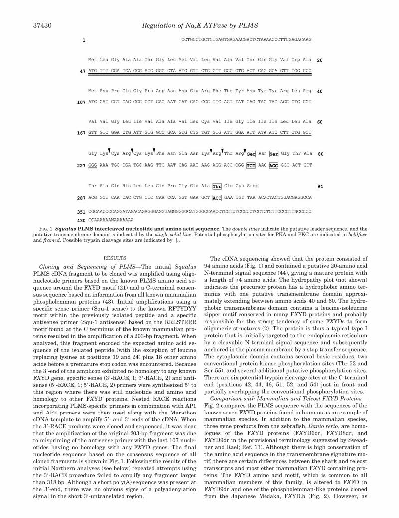
Cloning and Sequencing of PLMS—The initial SqualusPLMS cDNA fragment to be cloned was amplified using oligo-nucleotide primers based on the known PLMS amino acid se-quence around the FXYD motif (21) and a C-terminal consen-sus sequence based on information from all known mammalianphospholemman proteins (43). Initial amplifications using aspecific sense primer (Squ-1 sense) to the known RFTYDYYmotif within the previously isolated peptide and a specificantisense primer (Squ-1 antisense) based on the RRLSTRRRmotif found at the C terminus of the known mammalian pro-teins resulted in the amplification of a 203-bp fragment. Whenanalyzed, this fragment encoded the expected amino acid se-quence of the isolated peptide (with the exception of leucinereplacing lysines at positions 19 and 24) plus 18 other aminoacids before a premature stop codon was encountered. Becausethe 3�-end of the amplicon exhibited no homology to any knownFXYD gene, specific sense (3�-RACE, 1; 3�-RACE, 2) and anti-sense (5�-RACE, 1; 5�-RACE, 2) primers were synthesized 5� tothis region where there was still nucleotide and amino acidhomology to other FXYD proteins. Nested RACE reactionsincorporating PLMS-specific primers in combination with AP1and AP2 primers were then used along with the MarathoncDNA template to amplify 5�- and 3�-ends of the cDNA. Whenthe 3�-RACE products were cloned and sequenced, it was clearthat the amplification of the original 203-bp fragment was dueto mispriming of the antisense primer with the last 107 nucle-otides having no homology with any FXYD genes. The finalnucleotide sequence based on the consensus sequence of allcloned fragments is shown in Fig. 1. Following the results of theinitial Northern analyses (see below) repeated attempts usingthe 3�-RACE procedure failed to amplify any fragment largerthan 318 bp. Although a short poly(A) sequence was present atthe 3�-end, there was no obvious signs of a polyadenylationsignal in the short 3�-untranslated region.
The cDNA sequencing showed that the protein consisted of94 amino acids (Fig. 1) and contained a putative 20-amino acidN-terminal signal sequence (44), giving a mature protein witha length of 74 amino acids. The hydropathy plot (not shown)indicates the precursor protein has a hydrophobic amino ter-minus with one putative transmembrane domain approxi-mately extending between amino acids 40 and 60. The hydro-phobic transmembrane domain contains a leucine-isoleucinezipper motif conserved in many FXYD proteins and probablyresponsible for the strong tendency of some FXYDs to formoligomeric structures (2). The protein is thus a typical type Iprotein that is initially targeted to the endoplasmic reticulumby a cleavable N-terminal signal sequence and subsequentlyanchored in the plasma membrane by a stop-transfer sequence.The cytoplasmic domain contains several basic residues, twoconventional protein kinase phosphorylation sites (Thr-53 andSer-55), and several additional putative phosphorylation sites.There are six potential trypsin cleavage sites at the C-terminalend (positions 42, 44, 46, 51, 52, and 54) just in front andpartially overlapping the conventional phosphorylation sites.
Comparison with Mammalian and Teleost FXYD Proteins—Fig. 2 compares the PLMS sequence with the sequences of theknown seven FXYD proteins found in humans as an example ofmammalian species. In addition to the mammalian species,three gene products from the zebrafish, Danio rerio, are homo-logues of the FXYD proteins (FXYD6dr, FXYD8dr, andFXYD9dr in the provisional terminology suggested by Swead-ner and Rael; Ref. 13). Although there is high conservation ofthe amino acid sequence in the transmembrane signature mo-tif, there are certain differences between the shark and teleosttranscripts and most other mammalian FXYD containing pro-teins. The FXYD amino acid motif, which is common to allmammalian members of this family, is altered to FXFD inFXYD9dr and one of the phospholemman-like proteins clonedfrom the Japanese Medaka, FXYD.b (Fig. 2). However, as
FIG. 1. Squalus PLMS interleaved nucleotide and amino acid sequence. The double lines indicate the putative leader sequence, and theputative transmembrane domain is indicated by the single solid line. Potential phosphorylation sites for PKA and PKC are indicated in boldfaceand framed. Possible trypsin cleavage sites are indicated by 2.
Regulation of Na,K-ATPase by PLMS37430
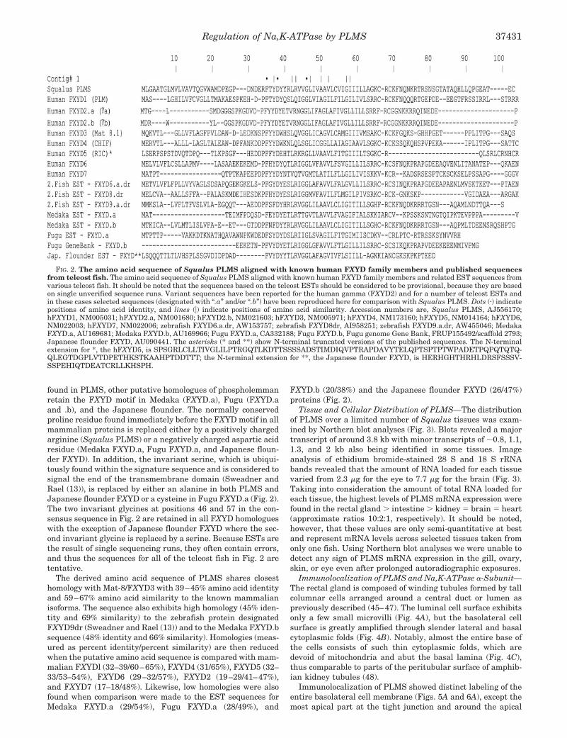
found in PLMS, other putative homologues of phospholemmanretain the FXYD motif in Medaka (FXYD.a), Fugu (FXYD.aand .b), and the Japanese flounder. The normally conservedproline residue found immediately before the FXYD motif in allmammalian proteins is replaced either by a positively chargedarginine (Squalus PLMS) or a negatively charged aspartic acidresidue (Medaka FXYD.a, Fugu FXYD.a, and Japanese floun-der FXYD). In addition, the invariant serine, which is ubiqui-tously found within the signature sequence and is considered tosignal the end of the transmembrane domain (Sweadner andRael (13)), is replaced by either an alanine in both PLMS andJapanese flounder FXYD or a cysteine in Fugu FXYD.a (Fig. 2).The two invariant glycines at positions 46 and 57 in the con-sensus sequence in Fig. 2 are retained in all FXYD homologueswith the exception of Japanese flounder FXYD where the sec-ond invariant glycine is replaced by a serine. Because ESTs arethe result of single sequencing runs, they often contain errors,and thus the sequences for all of the teleost fish in Fig. 2 aretentative.
The derived amino acid sequence of PLMS shares closesthomology with Mat-8/FXYD3 with 39–45% amino acid identityand 59–67% amino acid similarity to the known mammalianisoforms. The sequence also exhibits high homology (45% iden-tity and 69% similarity) to the zebrafish protein designatedFXYD9dr (Sweadner and Rael (13)) and to the Medaka FXYD.bsequence (48% identity and 66% similarity). Homologies (meas-ured as percent identity/percent similarity) are then reducedwhen the putative amino acid sequence is compared with mam-malian FXYDl (32–39/60–65%), FXYD4 (31/65%), FXYD5 (32–33/53–54%), FXYD6 (29–32/57%), FXYD2 (19–29/41–47%),and FXYD7 (17–18/48%). Likewise, low homologies were alsofound when comparison were made to the EST sequences forMedaka FXYD.a (29/54%), Fugu FXYD.a (28/49%), and
FXYD.b (20/38%) and the Japanese flounder FXYD (26/47%)proteins (Fig. 2).
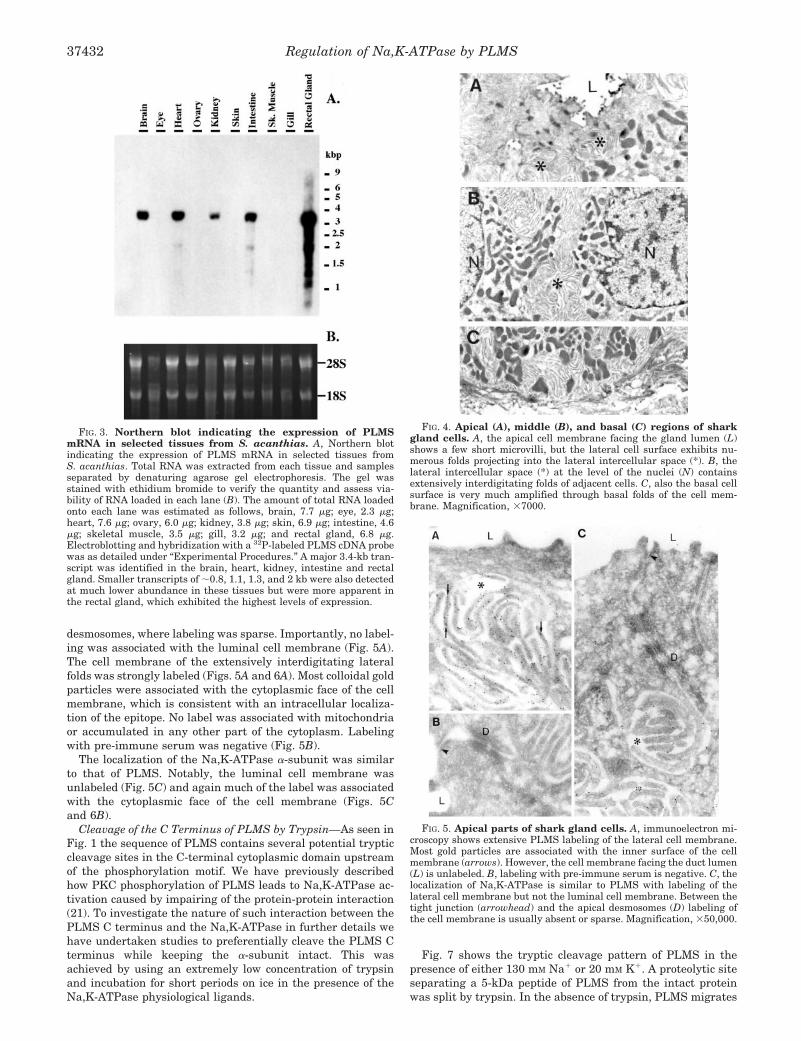
Tissue and Cellular Distribution of PLMS—The distributionof PLMS over a limited number of Squalus tissues was exam-ined by Northern blot analyses (Fig. 3). Blots revealed a majortranscript of around 3.8 kb with minor transcripts of �0.8, 1.1,1.3, and 2 kb also being identified in some tissues. Imageanalysis of ethidium bromide-stained 28 S and 18 S rRNAbands revealed that the amount of RNA loaded for each tissuevaried from 2.3 �g for the eye to 7.7 �g for the brain (Fig. 3).Taking into consideration the amount of total RNA loaded foreach tissue, the highest levels of PLMS mRNA expression werefound in the rectal gland � intestine � kidney � brain � heart(approximate ratios 10:2:1, respectively). It should be noted,however, that these values are only semi-quantitative at bestand represent mRNA levels across selected tissues taken fromonly one fish. Using Northern blot analyses we were unable todetect any sign of PLMS mRNA expression in the gill, ovary,skin, or eye even after prolonged autoradiographic exposures.
Immunolocalization of PLMS and Na,K-ATPase �-Subunit—The rectal gland is composed of winding tubules formed by tallcolumnar cells arranged around a central duct or lumen aspreviously described (45–47). The luminal cell surface exhibitsonly a few small microvilli (Fig. 4A), but the basolateral cellsurface is greatly amplified through slender lateral and basalcytoplasmic folds (Fig. 4B). Notably, almost the entire base ofthe cells consists of such thin cytoplasmic folds, which aredevoid of mitochondria and abut the basal lamina (Fig. 4C),thus comparable to parts of the peritubular surface of amphib-ian kidney tubules (48).
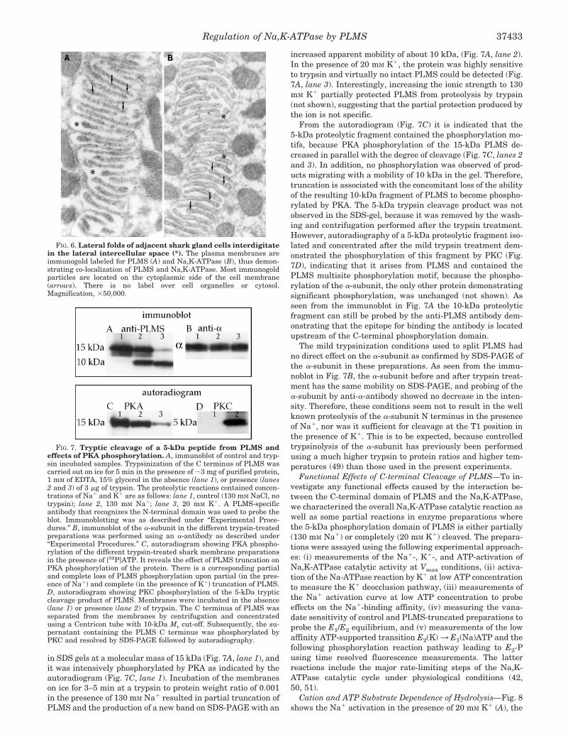
Immunolocalization of PLMS showed distinct labeling of theentire basolateral cell membrane (Figs. 5A and 6A), except themost apical part at the tight junction and around the apical
FIG. 2. The amino acid sequence of Squalus PLMS aligned with known human FXYD family members and published sequencesfrom teleost fish. The amino acid sequence of Squalus PLMS aligned with known human FXYD family members and related EST sequences fromvarious teleost fish. It should be noted that the sequences based on the teleost ESTs should be considered to be provisional, because they are basedon single unverified sequence runs. Variant sequences have been reported for the human gamma (FXYD2) and for a number of teleost ESTs andin these cases selected sequences (designated with “.a” and/or “.b”) have been reproduced here for comparison with Squalus PLMS. Dots (�) indicatepositions of amino acid identity, and lines (�) indicate positions of amino acid similarity. Accession numbers are, Squalus PLMS, AJ556170;hFXYD1, NM005031; hFXYD2.a, NM001680; hFXYD2.b, NM021603; hFXYD3, NM005971; hFXYD4, NM173160; hFXYD5, NM014164; hFXYD6,NM022003; hFXYD7, NM022006; zebrafish FXYD6.a.dr, AW153757; zebrafish FXYD8dr, AI958251; zebrafish FXYD9.a.dr, AW455046; MedakaFXYD.a, AU169681; Medaka FXYD.b, AU169966; Fugu FXYD.a, CA332188; Fugu FXYD.b, Fugu genome Gene Bank, FRUP155492/scaffold 2793;Japanese flounder FXYD, AU090441. The asterisks (* and **) show N-terminal truncated versions of the published sequences. The N-terminalextension for *, the hFXYD5, is SPSGRLCLLTIVGLILPTRGQTLKDTTSSSSADSTIMDIQVPTRAPDAVYTELQPTSPTPTWPADETPQPQTQTQ-QLEGTDGPLVTDPETHKSTKAAHPTDDTTT; the N-terminal extension for **, the Japanese flounder FXYD, is HERHGHTHRHLDRSFSSSV-SSPEHIQTDEATCRLLKHSPH.
Regulation of Na,K-ATPase by PLMS 37431
desmosomes, where labeling was sparse. Importantly, no label-ing was associated with the luminal cell membrane (Fig. 5A).The cell membrane of the extensively interdigitating lateralfolds was strongly labeled (Figs. 5A and 6A). Most colloidal goldparticles were associated with the cytoplasmic face of the cellmembrane, which is consistent with an intracellular localiza-tion of the epitope. No label was associated with mitochondriaor accumulated in any other part of the cytoplasm. Labelingwith pre-immune serum was negative (Fig. 5B).
The localization of the Na,K-ATPase �-subunit was similarto that of PLMS. Notably, the luminal cell membrane wasunlabeled (Fig. 5C) and again much of the label was associatedwith the cytoplasmic face of the cell membrane (Figs. 5Cand 6B).
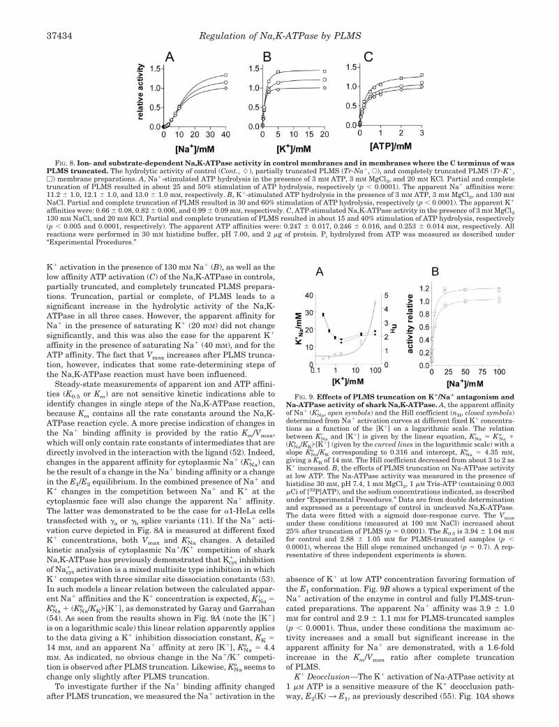
Cleavage of the C Terminus of PLMS by Trypsin—As seen inFig. 1 the sequence of PLMS contains several potential trypticcleavage sites in the C-terminal cytoplasmic domain upstreamof the phosphorylation motif. We have previously describedhow PKC phosphorylation of PLMS leads to Na,K-ATPase ac-tivation caused by impairing of the protein-protein interaction(21). To investigate the nature of such interaction between thePLMS C terminus and the Na,K-ATPase in further details wehave undertaken studies to preferentially cleave the PLMS Cterminus while keeping the �-subunit intact. This wasachieved by using an extremely low concentration of trypsinand incubation for short periods on ice in the presence of theNa,K-ATPase physiological ligands.
Fig. 7 shows the tryptic cleavage pattern of PLMS in thepresence of either 130 mM Na� or 20 mM K�. A proteolytic siteseparating a 5-kDa peptide of PLMS from the intact proteinwas split by trypsin. In the absence of trypsin, PLMS migrates
FIG. 3. Northern blot indicating the expression of PLMSmRNA in selected tissues from S. acanthias. A, Northern blotindicating the expression of PLMS mRNA in selected tissues fromS. acanthias. Total RNA was extracted from each tissue and samplesseparated by denaturing agarose gel electrophoresis. The gel wasstained with ethidium bromide to verify the quantity and assess via-bility of RNA loaded in each lane (B). The amount of total RNA loadedonto each lane was estimated as follows, brain, 7.7 �g; eye, 2.3 �g;heart, 7.6 �g; ovary, 6.0 �g; kidney, 3.8 �g; skin, 6.9 �g; intestine, 4.6�g; skeletal muscle, 3.5 �g; gill, 3.2 �g; and rectal gland, 6.8 �g.Electroblotting and hybridization with a 32P-labeled PLMS cDNA probewas as detailed under “Experimental Procedures.” A major 3.4-kb tran-script was identified in the brain, heart, kidney, intestine and rectalgland. Smaller transcripts of �0.8, 1.1, 1.3, and 2 kb were also detectedat much lower abundance in these tissues but were more apparent inthe rectal gland, which exhibited the highest levels of expression.
FIG. 4. Apical (A), middle (B), and basal (C) regions of sharkgland cells. A, the apical cell membrane facing the gland lumen (L)shows a few short microvilli, but the lateral cell surface exhibits nu-merous folds projecting into the lateral intercellular space (*). B, thelateral intercellular space (*) at the level of the nuclei (N) containsextensively interdigitating folds of adjacent cells. C, also the basal cellsurface is very much amplified through basal folds of the cell mem-brane. Magnification, �7000.
FIG. 5. Apical parts of shark gland cells. A, immunoelectron mi-croscopy shows extensive PLMS labeling of the lateral cell membrane.Most gold particles are associated with the inner surface of the cellmembrane (arrows). However, the cell membrane facing the duct lumen(L) is unlabeled. B, labeling with pre-immune serum is negative. C, thelocalization of Na,K-ATPase is similar to PLMS with labeling of thelateral cell membrane but not the luminal cell membrane. Between thetight junction (arrowhead) and the apical desmosomes (D) labeling ofthe cell membrane is usually absent or sparse. Magnification, �50,000.
Regulation of Na,K-ATPase by PLMS37432
in SDS gels at a molecular mass of 15 kDa (Fig. 7A, lane 1), andit was intensively phosphorylated by PKA as indicated by theautoradiogram (Fig. 7C, lane 1). Incubation of the membraneson ice for 3–5 min at a trypsin to protein weight ratio of 0.001in the presence of 130 mM Na� resulted in partial truncation ofPLMS and the production of a new band on SDS-PAGE with an
increased apparent mobility of about 10 kDa, (Fig. 7A, lane 2).In the presence of 20 mM K�, the protein was highly sensitiveto trypsin and virtually no intact PLMS could be detected (Fig.7A, lane 3). Interestingly, increasing the ionic strength to 130mM K� partially protected PLMS from proteolysis by trypsin(not shown), suggesting that the partial protection produced bythe ion is not specific.
From the autoradiogram (Fig. 7C) it is indicated that the5-kDa proteolytic fragment contained the phosphorylation mo-tifs, because PKA phosphorylation of the 15-kDa PLMS de-creased in parallel with the degree of cleavage (Fig. 7C, lanes 2and 3). In addition, no phosphorylation was observed of prod-ucts migrating with a mobility of 10 kDa in the gel. Therefore,truncation is associated with the concomitant loss of the abilityof the resulting 10-kDa fragment of PLMS to become phospho-rylated by PKA. The 5-kDa trypsin cleavage product was notobserved in the SDS-gel, because it was removed by the wash-ing and centrifugation performed after the trypsin treatment.However, autoradiography of a 5-kDa proteolytic fragment iso-lated and concentrated after the mild trypsin treatment dem-onstrated the phosphorylation of this fragment by PKC (Fig.7D), indicating that it arises from PLMS and contained thePLMS multisite phosphorylation motif, because the phospho-rylation of the �-subunit, the only other protein demonstratingsignificant phosphorylation, was unchanged (not shown). Asseen from the immunoblot in Fig. 7A the 10-kDa proteolyticfragment can still be probed by the anti-PLMS antibody dem-onstrating that the epitope for binding the antibody is locatedupstream of the C-terminal phosphorylation domain.
The mild trypsinization conditions used to split PLMS hadno direct effect on the �-subunit as confirmed by SDS-PAGE ofthe �-subunit in these preparations. As seen from the immu-noblot in Fig. 7B, the �-subunit before and after trypsin treat-ment has the same mobility on SDS-PAGE, and probing of the�-subunit by anti-�-antibody showed no decrease in the inten-sity. Therefore, these conditions seem not to result in the wellknown proteolysis of the �-subunit N terminus in the presenceof Na�, nor was it sufficient for cleavage at the T1 position inthe presence of K�. This is to be expected, because controlledtrypsinolysis of the �-subunit has previously been performedusing a much higher trypsin to protein ratios and higher tem-peratures (49) than those used in the present experiments.
Functional Effects of C-terminal Cleavage of PLMS—To in-vestigate any functional effects caused by the interaction be-tween the C-terminal domain of PLMS and the Na,K-ATPase,we characterized the overall Na,K-ATPase catalytic reaction aswell as some partial reactions in enzyme preparations wherethe 5-kDa phosphorylation domain of PLMS is either partially(130 mM Na�) or completely (20 mM K�) cleaved. The prepara-tions were assayed using the following experimental approach-es: (i) measurements of the Na�-, K�-, and ATP-activation ofNa,K-ATPase catalytic activity at Vmax conditions, (ii) activa-tion of the Na-ATPase reaction by K� at low ATP concentrationto measure the K� deocclusion pathway, (iii) measurements ofthe Na� activation curve at low ATP concentration to probeeffects on the Na�-binding affinity, (iv) measuring the vana-date sensitivity of control and PLMS-truncated preparations toprobe the E1/E2 equilibrium, and (v) measurements of the lowaffinity ATP-supported transition E2(K)3 E1(Na)ATP and thefollowing phosphorylation reaction pathway leading to E2-Pusing time resolved fluorescence measurements. The latterreactions include the major rate-limiting steps of the Na,K-ATPase catalytic cycle under physiological conditions (42,50, 51).
Cation and ATP Substrate Dependence of Hydrolysis—Fig. 8shows the Na� activation in the presence of 20 mM K� (A), the
FIG. 6. Lateral folds of adjacent shark gland cells interdigitatein the lateral intercellular space (*). The plasma membranes areimmunogold labeled for PLMS (A) and Na,K-ATPase (B), thus demon-strating co-localization of PLMS and Na,K-ATPase. Most immunogoldparticles are located on the cytoplasmic side of the cell membrane(arrows). There is no label over cell organelles or cytosol.Magnification, �50,000.
FIG. 7. Tryptic cleavage of a 5-kDa peptide from PLMS andeffects of PKA phosphorylation. A, immunoblot of control and tryp-sin incubated samples. Trypsinization of the C terminus of PLMS wascarried out on ice for 5 min in the presence of �3 mg of purified protein,1 mM of EDTA, 15% glycerol in the absence (lane 1), or presence (lanes2 and 3) of 3 �g of trypsin. The proteolytic reactions contained concen-trations of Na� and K� are as follows: lane 1, control (130 mM NaCl, notrypsin); lane 2, 130 mM Na�; lane 3, 20 mM K�. A PLMS-specificantibody that recognizes the N-terminal domain was used to probe theblot. Immunoblotting was as described under “Experimental Proce-dures.” B, immunoblot of the �-subunit in the different trypsin-treatedpreparations was performed using an �-antibody as described under“Experimental Procedures.” C, autoradiogram showing PKA phospho-rylation of the different trypsin-treated shark membrane preparationsin the presence of [32P]ATP. It reveals the effect of PLMS truncation onPKA phosphorylation of the protein. There is a corresponding partialand complete loss of PLMS phosphorylation upon partial (in the pres-ence of Na�) and complete (in the presence of K�) truncation of PLMS.D, autoradiogram showing PKC phosphorylation of the 5-kDa trypticcleavage product of PLMS. Membranes were incubated in the absence(lane 1) or presence (lane 2) of trypsin. The C terminus of PLMS wasseparated from the membranes by centrifugation and concentratedusing a Centricon tube with 10-kDa Mr cut-off. Subsequently, the su-pernatant containing the PLMS C terminus was phosphorylated byPKC and resolved by SDS-PAGE followed by autoradiography.
Regulation of Na,K-ATPase by PLMS 37433
K� activation in the presence of 130 mM Na� (B), as well as thelow affinity ATP activation (C) of the Na,K-ATPase in controls,partially truncated, and completely truncated PLMS prepara-tions. Truncation, partial or complete, of PLMS leads to asignificant increase in the hydrolytic activity of the Na,K-ATPase in all three cases. However, the apparent affinity forNa� in the presence of saturating K� (20 mM) did not changesignificantly, and this was also the case for the apparent K�
affinity in the presence of saturating Na� (40 mM), and for theATP affinity. The fact that Vmax increases after PLMS trunca-tion, however, indicates that some rate-determining steps ofthe Na,K-ATPase reaction must have been influenced.
Steady-state measurements of apparent ion and ATP affini-ties (K0.5 or Km) are not sensitive kinetic indications able toidentify changes in single steps of the Na,K-ATPase reaction,because Km contains all the rate constants around the Na,K-ATPase reaction cycle. A more precise indication of changes inthe Na� binding affinity is provided by the ratio Km/Vmax,which will only contain rate constants of intermediates that aredirectly involved in the interaction with the ligand (52). Indeed,changes in the apparent affinity for cytoplasmic Na� (K�Na) canbe the result of a change in the Na� binding affinity or a changein the E1/E2 equilibrium. In the combined presence of Na� andK� changes in the competition between Na� and K� at thecytoplasmic face will also change the apparent Na� affinity.The latter was demonstrated to be the case for �1-HeLa cellstransfected with �a or �b splice variants (11). If the Na� acti-vation curve depicted in Fig. 8A is measured at different fixedK� concentrations, both Vmax and K�Na changes. A detailedkinetic analysis of cytoplasmic Na�/K� competition of sharkNa,K-ATPase has previously demonstrated that Kcyt
� inhibitionof Nacyt
� activation is a mixed multisite type inhibition in whichK� competes with three similar site dissociation constants (53).In such models a linear relation between the calculated appar-ent Na� affinities and the K� concentration is expected, K�Na �KNa
o � (KNao /KK)�[K�], as demonstrated by Garay and Garrahan
(54). As seen from the results shown in Fig. 9A (note the [K�]is on a logarithmic scale) this linear relation apparently appliesto the data giving a K� inhibition dissociation constant, KK �14 mM, and an apparent Na� affinity at zero [K�], KNa
o � 4.4mM. As indicated, no obvious change in the Na�/K� competi-tion is observed after PLMS truncation. Likewise, KNa
o seems tochange only slightly after PLMS truncation.
To investigate further if the Na� binding affinity changedafter PLMS truncation, we measured the Na� activation in the
absence of K� at low ATP concentration favoring formation ofthe E1 conformation. Fig. 9B shows a typical experiment of theNa� activation of the enzyme in control and fully PLMS-trun-cated preparations. The apparent Na� affinity was 3.9 � 1.0mM for control and 2.9 � 1.1 mM for PLMS-truncated samples(p 0.0001). Thus, under these conditions the maximum ac-tivity increases and a small but significant increase in theapparent affinity for Na� are demonstrated, with a 1.6-foldincrease in the Km/Vmax ratio after complete truncationof PLMS.
K� Deocclusion—The K� activation of Na-ATPase activity at1 �M ATP is a sensitive measure of the K� deocclusion path-way, E2(K) 3 E1, as previously described (55). Fig. 10A shows
FIG. 9. Effects of PLMS truncation on K�/Na� antagonism andNa-ATPase activity of shark Na,K-ATPase. A, the apparent affinityof Na� (K�Na, open symbols) and the Hill coefficient (nH, closed symbols)determined from Na� activation curves at different fixed K� concentra-tions as a function of the [K�] on a logarithmic scale. The relationbetween K�Na and [K�] is given by the linear equation, K�Na � KNa
o �(KNa
o /KK)�[K�] (given by the curved lines in the logarithmic scale) with aslope KNa
o /KK corresponding to 0.316 and intercept, KNao � 4.35 mM,
giving a KK of 14 mM. The Hill coefficient decreased from about 3 to 2 asK� increased. B, the effects of PLMS truncation on Na-ATPase activityat low ATP. The Na-ATPase activity was measured in the presence ofhistidine 30 mM, pH 7.4, 1 mM MgCl2, 1 �M Tris-ATP (containing 0.003�Ci of [32P]ATP), and the sodium concentrations indicated, as describedunder “Experimental Procedures.” Data are from double determinationand expressed as a percentage of control in uncleaved Na,K-ATPase.The data were fitted with a sigmoid dose-response curve. The Vmaxunder these conditions (measured at 100 mM NaCl) increased about25% after truncation of PLMS (p � 0.0001). The K0.5 is 3.94 � 1.04 mM
for control and 2.88 � 1.05 mM for PLMS-truncated samples (p 0.0001), whereas the Hill slope remained unchanged (p � 0.7). A rep-resentative of three independent experiments is shown.
FIG. 8. Ion- and substrate-dependent Na,K-ATPase activity in control membranes and in membranes where the C terminus of wasPLMS truncated. The hydrolytic activity of control (Cont., �), partially truncated PLMS (Tr-Na�, E), and completely truncated PLMS (Tr-K�,�) membrane preparations. A, Na�-stimulated ATP hydrolysis in the presence of 3 mM ATP, 3 mM MgCl2, and 20 mM KCl. Partial and completetruncation of PLMS resulted in about 25 and 50% stimulation of ATP hydrolysis, respectively (p 0.0001). The apparent Na� affinities were:11.2 � 1.0, 12.1 � 1.0, and 13.0 � 1.0 mM, respectively. B, K�-stimulated ATP hydrolysis in the presence of 3 mM ATP, 3 mM MgCl2, and 130 mM
NaCl. Partial and complete truncation of PLMS resulted in 30 and 60% stimulation of ATP hydrolysis, respectively (p 0.0001). The apparent K�
affinities were: 0.66 � 0.08, 0.82 � 0.006, and 0.99 � 0.09 mM, respectively. C, ATP-stimulated Na,K-ATPase activity in the presence of 3 mM MgCl2130 mM NaCl, and 20 mM KCl. Partial and complete truncation of PLMS resulted in about 15 and 40% stimulation of ATP hydrolysis, respectively(p 0.005 and 0.0001, respectively). The apparent ATP affinities were: 0.247 � 0.017, 0.246 � 0.016, and 0.253 � 0.014 mM, respectively. Allreactions were performed in 30 mM histidine buffer, pH 7.00, and 2 �g of protein. Pi hydrolyzed from ATP was measured as described under“Experimental Procedures.”
Regulation of Na,K-ATPase by PLMS37434
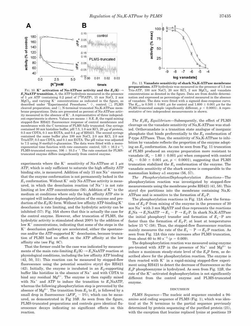
experiments where the K� sensitivity of Na-ATPase at 1 �M
ATP, which is only sufficient to saturate the high affinity ATPbinding site, is measured. Addition of only 15 mM Na� ensuresthat the enzyme conformation is not permanently locked in theE1 conformation. Without K� only Na-ATPase activity is meas-ured, in which the deocclusion reaction (of Na�) is not ratelimiting at low ATP concentrations (56). Addition of K� to themedium at conditions where only the high affinity ATP site isoccupied will induce dephosphorylation of the enzyme and pro-duction of the E2(K) form. Without low affinity ATP-binding K�
deocclusion is rate limiting, and the hydrolytic activity can beinhibited (57). Fig. 10A shows that this is actually the case forthe control enzyme. However, after truncation of PLMS, thehydrolytic activity is stimulated about 200% by the addition oflow K� concentrations, indicating that some of the steps in theK� deocclusion pathway are accelerated, either the spontane-ous and/or the ATP-supported K� deocclusion, because trunca-tion of PLMS had no effect on the ATP affinity at the lowaffinity site (see Fig. 8C).
That the former could be the case was indicated by measure-ments of the main rate-limiting E2(K)3 E1NaATP reaction atphysiological conditions, including the low affinity ATP binding(42, 50, 51). This reaction can be measured by stopped-flowfluorescence using the potential sensitive styryl dye RH421(42). Initially, the enzyme is incubated in an E2-supportingbuffer like histidine in the absence of Na� and with CDTA tobind any residual Mg2�. The enzyme is then rapidly mixedwith Na� and ATP to induce the transition to E1NaATP,whereas the following phosphorylation step is prevented by theabsence of Mg2�. The transition from E2 to E1 is followed by asmall drop in fluorescence (F/Fo � 5%), which can be meas-ured, as demonstrated in Fig 10B. As seen from the figure,PLMS-truncated preparations and controls gave identical flu-orescence decays indicating no significant effects on thisreaction.
The E1/E2 Equilibrium—Subsequently, the effect of PLMScleavage on the vanadate sensitivity of Na,K-ATPase was stud-ied. Orthovanadate is a transition state analogue of inorganicphosphate that binds preferentially to the E2 conformation ofP-type ATPases. Thus, the sensitivity of Na,K-ATPase to inhi-bition by vanadate reflects the proportion of the enzyme adopt-ing an E2 conformation. As can be seen from Fig. 11 truncationof PLMS produced an enzyme preparation more resistant tovanadate (KI � 1.60 � 0.001 �M) when compared with control(KI � 0.50 � 0.001 �M, p 0.0001), suggesting that PLMStruncation stabilized the E1 conformation of the enzyme. Thevanadate sensitivity of the shark enzyme is comparable to themammalian kidney �1 enzyme (56, 57).
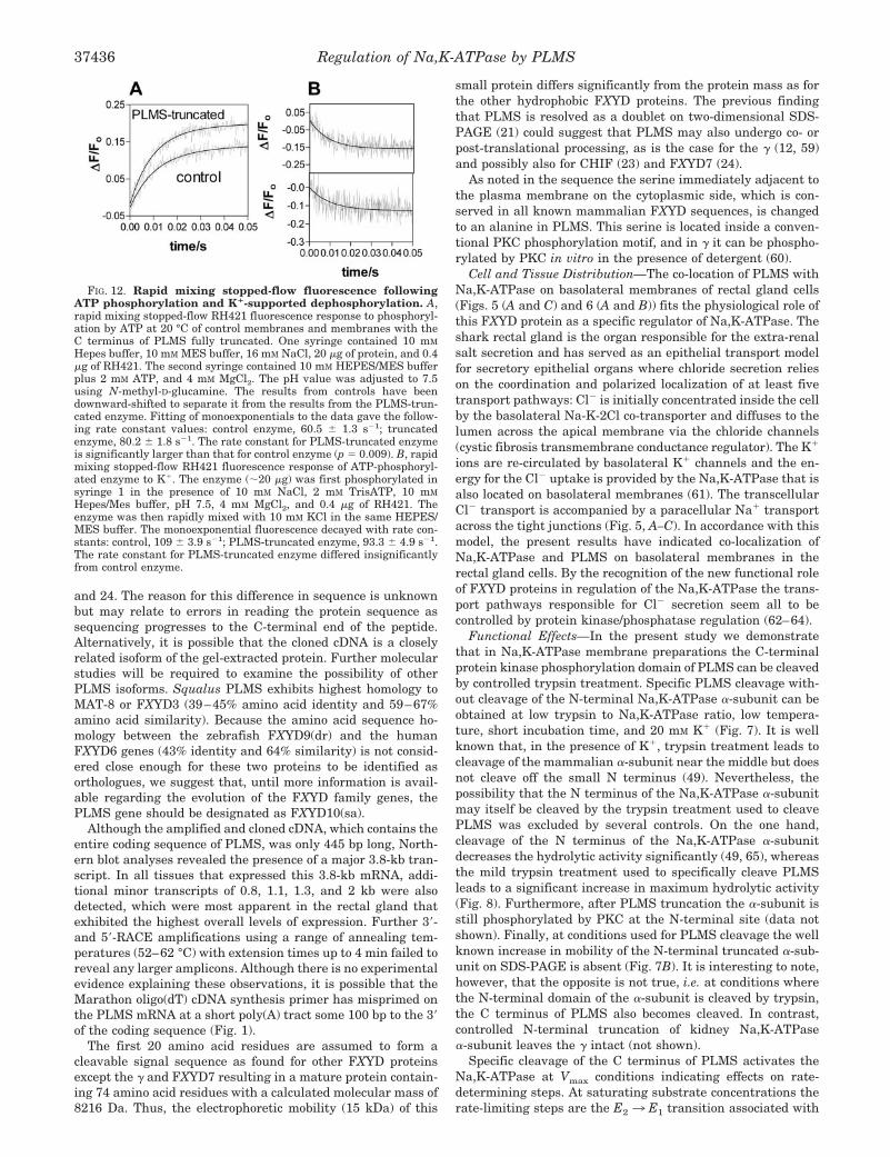
The Phosphorylation/Dephosphorylation Reactions—Thephosphorylation reaction was investigated by stopped-flowmeasurements using the membrane probe RH421 (41, 58). Thisstyryl dye partitions into the membrane containing Na,K-ATPase and is sensitive to the formation of E2-P.
The phosphorylation reactions in Fig. 12A show the forma-tion of E2-P from mixing of the enzyme in the presence of 30mM Na� with 3 mM MgATP and thus represents the reactionsE1Na 3 E1NaATP 3 E1 � P 3 E2-P. In shark Na,K-ATPasethe initial phosphoryl transfer and formation of E1�P arefaster than the formation of E2-P, at least at temperaturesbelow 15 °C (58). Thus, the stopped-flow fluorescence reactionmainly measures the rate of the E1 � P 3 E2-P reaction. Asseen from Fig. 12A this rate increases after PLMS truncation,from about 60 to 80 s�1 (p � 0.009).
The dephosphorylation reaction was measured using enzymepre-treated with ATP in the presence of Na� and Mg2� toinduce a maximum steady-state level of the E2P form, as de-scribed above for the phosphorylation reaction. The enzyme isthen reacted with K� in a rapid-mixing stopped-flow experi-ment using RH421 to detect the decrease of fluorescence as theE2P phosphoenzyme is hydrolyzed. As seen from Fig. 12B, therate of the K�-activated dephosphorylation is not significantlydifferent from the control enzyme and PLMS-truncatedenzyme.
DISCUSSION
PLMS Sequence—The nucleic acid sequence encoded a 94-amino acid coding sequence of PLMS (Fig. 1), which was iden-tical at the N terminus to the partial sequence previouslydetermined by protein sequencing of the purified protein (21),with the exception that leucine replaced lysine at positions 19
FIG. 10. K� activation of Na-ATPase activity and the E2(K) 3E1NaATP transition. A, the ATP hydrolysis measured in the presenceof 1 �M ATP (containing 0.2 pmol of [32P]ATP), 15 mM NaCl, 2 mM
MgCl2, and varying K� concentrations as indicated in the figure, asdescribed under “Experimental Procedures.” �, control; �, PLMScleaved preparation; and E, N-terminal truncated Na,K-ATPase mem-brane preparations. Data are presented as percent of Na-ATPase activ-ity measured in the absence of K�. A representative of three independ-ent experiments is shown. Values are means � S.E. B, the rapid mixingstopped-flow RH421 fluorescence response of control membranes andmembranes with the C terminus of PLMS fully truncated. One syringecontained 30 mM histidine buffer, pH 7.5, 1.0 mM KCl, 20 �g of protein,0.5 mM CDTA, 0.1 mM EGTA, and 0.4 �g of RH421. The second syringecontained the same buffer plus 100 mM NaCl, 2.0 mM KCl, 2.0 mM
TrisATP, 0.5 mM CDTA, and 0.1 mM EGTA. The pH value was adjustedto 7.5 using N-methyl-D-glucamine. The data were fitted with a mono-exponential time function with rate constants: control, 125 � 16.2 s�1;PLMS-truncated enzyme, 106 � 10.3 s�1. The rate constant for PLMS-truncated enzyme differs insignificantly from control enzyme.
FIG. 11. Vanadate sensitivity of shark Na,K-ATPase membranepreparations. ATP hydrolysis was measured in the presence of 1.5 mM
Tris-ATP, 100 mM NaCl, 20 mM KCl, 2 mM MgCl2, and vanadateconcentrations as denoted in the figure. Data are from double determi-nation and expressed as percentage of control measured in the absenceof vanadate. The data were fitted with a sigmoid dose-response curve.The K0.5 is 0.505 � 0.001 �M for control and 1.600 � 0.001 �M for thePLMS-truncated sample (significantly different, p 0.0001). A repre-sentative of two independent measurements is shown.
Regulation of Na,K-ATPase by PLMS 37435
and 24. The reason for this difference in sequence is unknownbut may relate to errors in reading the protein sequence assequencing progresses to the C-terminal end of the peptide.Alternatively, it is possible that the cloned cDNA is a closelyrelated isoform of the gel-extracted protein. Further molecularstudies will be required to examine the possibility of otherPLMS isoforms. Squalus PLMS exhibits highest homology toMAT-8 or FXYD3 (39–45% amino acid identity and 59–67%amino acid similarity). Because the amino acid sequence ho-mology between the zebrafish FXYD9(dr) and the humanFXYD6 genes (43% identity and 64% similarity) is not consid-ered close enough for these two proteins to be identified asorthologues, we suggest that, until more information is avail-able regarding the evolution of the FXYD family genes, thePLMS gene should be designated as FXYD10(sa).
Although the amplified and cloned cDNA, which contains theentire coding sequence of PLMS, was only 445 bp long, North-ern blot analyses revealed the presence of a major 3.8-kb tran-script. In all tissues that expressed this 3.8-kb mRNA, addi-tional minor transcripts of 0.8, 1.1, 1.3, and 2 kb were alsodetected, which were most apparent in the rectal gland thatexhibited the highest overall levels of expression. Further 3�-and 5�-RACE amplifications using a range of annealing tem-peratures (52–62 °C) with extension times up to 4 min failed toreveal any larger amplicons. Although there is no experimentalevidence explaining these observations, it is possible that theMarathon oligo(dT) cDNA synthesis primer has misprimed onthe PLMS mRNA at a short poly(A) tract some 100 bp to the 3�of the coding sequence (Fig. 1).
The first 20 amino acid residues are assumed to form acleavable signal sequence as found for other FXYD proteinsexcept the � and FXYD7 resulting in a mature protein contain-ing 74 amino acid residues with a calculated molecular mass of8216 Da. Thus, the electrophoretic mobility (15 kDa) of this
small protein differs significantly from the protein mass as forthe other hydrophobic FXYD proteins. The previous findingthat PLMS is resolved as a doublet on two-dimensional SDS-PAGE (21) could suggest that PLMS may also undergo co- orpost-translational processing, as is the case for the � (12, 59)and possibly also for CHIF (23) and FXYD7 (24).
As noted in the sequence the serine immediately adjacent tothe plasma membrane on the cytoplasmic side, which is con-served in all known mammalian FXYD sequences, is changedto an alanine in PLMS. This serine is located inside a conven-tional PKC phosphorylation motif, and in � it can be phospho-rylated by PKC in vitro in the presence of detergent (60).
Cell and Tissue Distribution—The co-location of PLMS withNa,K-ATPase on basolateral membranes of rectal gland cells(Figs. 5 (A and C) and 6 (A and B)) fits the physiological role ofthis FXYD protein as a specific regulator of Na,K-ATPase. Theshark rectal gland is the organ responsible for the extra-renalsalt secretion and has served as an epithelial transport modelfor secretory epithelial organs where chloride secretion relieson the coordination and polarized localization of at least fivetransport pathways: Cl� is initially concentrated inside the cellby the basolateral Na-K-2Cl co-transporter and diffuses to thelumen across the apical membrane via the chloride channels(cystic fibrosis transmembrane conductance regulator). The K�
ions are re-circulated by basolateral K� channels and the en-ergy for the Cl� uptake is provided by the Na,K-ATPase that isalso located on basolateral membranes (61). The transcellularCl� transport is accompanied by a paracellular Na� transportacross the tight junctions (Fig. 5, A–C). In accordance with thismodel, the present results have indicated co-localization ofNa,K-ATPase and PLMS on basolateral membranes in therectal gland cells. By the recognition of the new functional roleof FXYD proteins in regulation of the Na,K-ATPase the trans-port pathways responsible for Cl� secretion seem all to becontrolled by protein kinase/phosphatase regulation (62–64).
Functional Effects—In the present study we demonstratethat in Na,K-ATPase membrane preparations the C-terminalprotein kinase phosphorylation domain of PLMS can be cleavedby controlled trypsin treatment. Specific PLMS cleavage with-out cleavage of the N-terminal Na,K-ATPase �-subunit can beobtained at low trypsin to Na,K-ATPase ratio, low tempera-ture, short incubation time, and 20 mM K� (Fig. 7). It is wellknown that, in the presence of K�, trypsin treatment leads tocleavage of the mammalian �-subunit near the middle but doesnot cleave off the small N terminus (49). Nevertheless, thepossibility that the N terminus of the Na,K-ATPase �-subunitmay itself be cleaved by the trypsin treatment used to cleavePLMS was excluded by several controls. On the one hand,cleavage of the N terminus of the Na,K-ATPase �-subunitdecreases the hydrolytic activity significantly (49, 65), whereasthe mild trypsin treatment used to specifically cleave PLMSleads to a significant increase in maximum hydrolytic activity(Fig. 8). Furthermore, after PLMS truncation the �-subunit isstill phosphorylated by PKC at the N-terminal site (data notshown). Finally, at conditions used for PLMS cleavage the wellknown increase in mobility of the N-terminal truncated �-sub-unit on SDS-PAGE is absent (Fig. 7B). It is interesting to note,however, that the opposite is not true, i.e. at conditions wherethe N-terminal domain of the �-subunit is cleaved by trypsin,the C terminus of PLMS also becomes cleaved. In contrast,controlled N-terminal truncation of kidney Na,K-ATPase�-subunit leaves the � intact (not shown).
Specific cleavage of the C terminus of PLMS activates theNa,K-ATPase at Vmax conditions indicating effects on rate-determining steps. At saturating substrate concentrations therate-limiting steps are the E2 3 E1 transition associated with
FIG. 12. Rapid mixing stopped-flow fluorescence followingATP phosphorylation and K�-supported dephosphorylation. A,rapid mixing stopped-flow RH421 fluorescence response to phosphoryl-ation by ATP at 20 °C of control membranes and membranes with theC terminus of PLMS fully truncated. One syringe contained 10 mM
Hepes buffer, 10 mM MES buffer, 16 mM NaCl, 20 �g of protein, and 0.4�g of RH421. The second syringe contained 10 mM HEPES/MES bufferplus 2 mM ATP, and 4 mM MgCl2. The pH value was adjusted to 7.5using N-methyl-D-glucamine. The results from controls have beendownward-shifted to separate it from the results from the PLMS-trun-cated enzyme. Fitting of monoexponentials to the data gave the follow-ing rate constant values: control enzyme, 60.5 � 1.3 s�1; truncatedenzyme, 80.2 � 1.8 s�1. The rate constant for PLMS-truncated enzymeis significantly larger than that for control enzyme (p � 0.009). B, rapidmixing stopped-flow RH421 fluorescence response of ATP-phosphoryl-ated enzyme to K�. The enzyme (�20 �g) was first phosphorylated insyringe 1 in the presence of 10 mM NaCl, 2 mM TrisATP, 10 mM
Hepes/Mes buffer, pH 7.5, 4 mM MgCl2, and 0.4 �g of RH421. Theenzyme was then rapidly mixed with 10 mM KCl in the same HEPES/MES buffer. The monoexponential fluorescence decayed with rate con-stants: control, 109 � 3.9 s�1; PLMS-truncated enzyme, 93.3 � 4.9 s�1.The rate constant for PLMS-truncated enzyme differed insignificantlyfrom control enzyme.
Regulation of Na,K-ATPase by PLMS37436
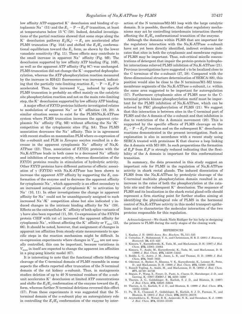
low affinity ATP-supported K� deocclusion and binding of cy-toplasmic Na� (51) and the E1 � P 3 E2-P transition, at leastat temperatures below 15 °C (58). Indeed, detailed investiga-tions of the partial reactions showed that some steps along theK� deocclusion pathway E2(K2) 3 E1 are accelerated afterPLMS truncation (Fig. 10A) and shifted the E1/E2 conforma-tional equilibrium toward the E1 form, as shown by the lowervanadate sensitivity (Fig. 11). These effects may contribute tothe small increase in apparent Na� affinity (Fig. 9B). Thedeocclusion supported by low affinity ATP binding (Fig. 10B),as well as the apparent ATP affinity (Fig 8C) was unchanged.PLMS-truncation did not change the K�-supported dephospho-rylation, whereas the ATP-phosphorylation reaction measuredby the increase in RH421 fluorescence was increased, indicat-ing that the partially rate-limiting reaction E1 � P 3 E2-P isaccelerated. Thus, the increased Vmax induced by specificPLMS truncation is probably an effect mainly on the catalyticsite phosphorylation reaction and not on the main rate-limitingstep, the K� deocclusion supported by low affinity ATP binding.
A major effect of FXYD proteins hitherto investigated relatesto modulation of apparent Na� or K� affinities (19, 20). Asimilar situation seems to exist for the PLMS/Na,K-ATPasesystem where PLMS truncation increases the apparent cyto-plasmic Na� affinity (Fig. 9B) without affecting the Na�/K�
competition at the cytoplasmic sites (Fig. 9A). Thus PLMSassociation decreases the Na� affinity. This is in agreementwith recent studies on mammalian PLM where co-expression ofthe �-subunit and PLM in Xenopus oocytes resulted in a de-crease in the apparent cytoplasmic Na� affinity of Na,K-ATPase (22). Thus, association of FXYD1 proteins with theNa,K-ATPase leads in both cases to a decreased Na� affinityand inhibition of enzyme activity, whereas dissociation of theFXYD1 proteins results in stimulation of hydrolytic activity.Other FXYD proteins have different patterns of effects: associ-ation of � (FXYD2) with Na,K-ATPase has been shown toincrease the apparent ATP affinity by supporting the E1 con-formation of the enzyme and to decrease the apparent affinityfor cytoplasmic Na�, which apparently is an effect secondary toan increased antagonism of cytoplasmic K� to activation byNa� (10, 11). In other investigations the change in apparentaffinity for Na� could not be unambiguously assigned to suchincreased Na�/K� competition alone but also indicated � in-duced changes in the intrinsic binding affinity for Na� (59).Effects on the extracellular K� affinity of both splice variants of� have also been reported (11, 59). Co-expression of the FXYD4protein CHIF with rat �1 increased the apparent affinity forcytoplasmic Na�, without affecting the K� affinity or Vmax (23,66). It should be noted, however, that assignment of changes inapparent ion affinities from steady-state measurements to spe-cific steps in the reaction mechanism might be difficult. Inco-expression experiments where changes in Vmax are not usu-ally controlled, this can be important, because variations inVmax in itself are expected to change the apparent ion affinitiesin a ping-pong kinetic model (67).
It is interesting to note that the functional effects followingcleavage of the C-terminal domain of PLMS resemble in someaspects the effects reported after truncation of the N-terminaldomain of the rat kidney �-subunit. Thus, in mutagenesisstudies deletion of up to 40 N-terminal residues of the �-sub-unit accelerates K� deocclusion at limited ATP concentrationsand shifts the E1/E2 conformation of the enzyme toward the E1
form, whereas further N-terminal deletions reversed this effect(57). From these experiments it was suggested that the N-terminal domain of the �-subunit play an autoregulatory rolein controlling the E1/E2 conformation of the enzyme by inter-
action of the N terminus/M2-M3 loop with the large catalyticdomain. It is possible, therefore, that other regulatory mecha-nisms may act by controlling interdomain interaction therebyaffecting the E1/E2 conformational transition of the enzyme.
Although the domains within PLMS that are important forthe regulatory interaction with the Na,K-ATPase �-subunithave not yet been directly identified, indirect evidence indi-cates that sites in both the cytoplasmic and membrane regionsof PLMS may be important. Thus, sub-critical micelle concen-trations of detergent that impair the protein-protein hydropho-bic interactions relieved PLMS inhibition of Na,K-ATPase (21).Previous investigations have suggested � to be localized close tothe C terminus of the �-subunit (27, 28). Compared with thethree-dimensional structure determination of SERCA (68), thisposition would also be close to the N terminus/M1-M2 trans-membrane segments of the Na,K-ATPase �-subunit, i.e. withinthe same area suggested to be important for autoregulation(57). Furthermore cytoplasmic sites of PLMS near to the C-terminal protein kinase phosphorylation motif must be impor-tant for the PLMS inhibition of Na,K-ATPase, which can berelieved by PKC phosphorylation of PLMS (21). We suggestthat this interaction is between sites in the C-terminal part ofPLMS and the A domain of the �-subunit and that inhibition isdue to restriction of the A domain movement (20). This issupported by the specific effects of PLMS cleavage on theE1 � P3 E2-P reaction and on the subsequent K� deocclusionreactions demonstrated in the present investigation. Such anexplanation is also in accordance with a recent study usingSERCA treated with proteinase K that cleaves a loop linkingthe A domain with M3 (69). In such preparations the formationof E2P from E1P is strongly reduced indicating that the flexi-bility of the A domain is important for this conformationaltransition.
In summary, the data presented in this study suggest animportant role for PLMS in the regulation of Na,K-ATPaseactivity in shark rectal glands. The induced dissociation ofPLMS from the Na,K-ATPase by proteolytic cleavage of theC-terminal multisite phosphorylation domain resulted in anincrease in the rates of both the phosphorylation at the cata-lytic site and the subsequent K� deocclusion. The sequence ofPLMS and its localization in the shark rectal gland cells shouldrepresent a firm starting point for further studies aiming atidentifying the physiological role of PLMS in the hormonalcontrol of Na,K-ATPase activity in this model transport epithe-lium and to characterize the molecular interaction of the twoproteins responsible for this regulation.
Acknowledgment—We thank Niels Rudiger for his help in designingthe primers to PLMS during the early stages of the cloning work.
REFERENCES
1. Kaplan, J. H. (2002) Annu. Rev. Biochem. 71, 511–5352. Cornelius, F., Mahmmoud, Y. A., and Christensen, H. R. Z. (2001) J. Bioenerg.
Biomemb. 33, 415–4233. Kimura, Y., Kurzydlowski, K., Tada, M., and MacLennan, D. H. (1997) J. Biol.
Chem. 272, 15061–150644. Kimura, Y., Asahi, M., Kurzydlowski, K., Tada, M., and MacLennan, D. H.
(1998) J. Biol. Chem. 273, 14238–142415. Reddy, L. G., Autry, J. M., Jones, L. R., and Thomas, D. D. (1999) J. Biol.
Chem. 274, 7649–76556. Odermat, A., Becker, S., Khanna, V. K., Kurzydlowski, K., Leisner, E., Pette,
D., and MacLennan, D. H. (1998) J. Biol. Chem. 273, 12360–123697. Russell Tupling, A., Asahi, M., and MacLennan, D. H. (2002) J. Biol. Chem.
277, 44740–447468. Beguin, P., Wang, X., Firsov, D., Puoti, A., Claeys, D., Horisberger, J.-D., and
Gerring, K. (1997) EMBO J. 16, 4250–42609. Therien, A. G., Goldshleger, R., Karlish, S. J. D., and Blostein, R. (1997)
J. Biol. Chem. 272, 32628–3263410. Therien, A. G., Karlish, S. J. D., and Blostein, R. (1999) J. Biol. Chem. 274,
12252–1225611. Pu, H. X., Cluzeaud, F., Goldshleger, R., Karlish, S. J. D., Farman, N., and
Blostein, R. (2001) J. Biol. Chem. 276, 20370–2037812. Arystarkhova, E., Wetzel, R. K., Asinovski, N. K., and Sweadner, K. J. (1999)
J. Biol. Chem. 274, 33183–33185
Regulation of Na,K-ATPase by PLMS 37437
13. Sweadner, K. J., and Rael, E. (2000) Genomics 68, 41–5614. Palmer, C. J., Scott, D., and Jones, L. R. (1991) J. Biol. Chem. 266,
11126–1113015. Minor, N. T., Sha, Q., Nichols, C. G., and Mercer, R. W. (1998) Proc. Natl. Acad.
Sci. U. S. A. 95, 6521–652516. Morrison, B. W., Moorman, J. R., Kowdley, G. C., Kobayashi, Y. M., Jones,
L. R., and Leder, P. (1995) J. Biol. Chem. 270, 2176–218217. Attali, B., Latter, H., Rachamim, N., and Garty, H. (1995) Proc. Natl. Acad.
Sci. U. S. A. 92, 6092–609618. Fu, X., and Kamps, M. P. (1997) Mol. Cell. Biol. 17, 1503–151219. Crambert, G., and Geering, K. (2003) Science’s STKE http://stke.sciencemag.
org/cgi/content/full/sigtrans;2003/166/re120. Cornelius, F., and Mahmmoud, Y. A. (2003) News. Physiol. Sci. 18, 119–12421. Mahmmoud, Y. A., Vorum, H., and Cornelius, F. (2000) J. Biol. Chem. 274,
35969–3597722. Crambert, G., Fuzesi, M., Garty, H., Karlish, S., and Geering, K. (2002) Proc.
Natl. Acad. Sci. U. S. A. 99, 11476–1148123. Beguin, P., Crambert, G., Guennoun, S., Garty, H., Horisberger, J. D., and
Gerring, K. (2001) EMBO J. 20, 3993–400224. Beguin, P., Crambert, G., Monnet-Tschudi, F., Uldry, M., Horisberger, J. D.,
Garty, H., and Geering, K. (2002) EMBO J. 21, 3264–327325. Meij, I. C., Koenderink, J. B., van Bokhoven, H., Assink, K. F., Groenestege,
W. T., de Pont, J. J., Bindels, R. J., Monnens, L. A., van den Heuvel, L. P.,and Knoers, N. V. (2000) Nat. Genet. 26, 265–266
26. Aizman, R., Asher, C., Fuzesi, M., Latter, H., Lonai, P., Karlish, S. J. D., andGarty, H. (2002) Am. J. Physiol. 283, F569–F577
27. Hebert, H., Purhonen, P., Vorum, H., Thomsen, K., and Maunsbach, A. V.(2001) J. Mol. Biol. 314, 479–494
28. Donnet, C., Arystarkhova, E., and Sweadner, K. J. (2001) J. Biol. Chem. 276,7357–7365
29. Pu, X. P., Scanzano, R., and Blostein, R. (2002) J. Biol. Chem. 277,20270–20276
30. Lindzen, M., Aizman, R., Lifshitz, Y., Lubarski, I., Karlish, S. J., and Garty, H.(2003) J. Biol. Chem. 278, 18738–18743
31. Cornelius, F., and Mahmmoud, Y. A. (2003) Ann. N. Y. Acad. Sci. U. S. A. 986,579–586
32. Chomczynski, P., and Sacchi, N. (1987) Anal. Biochem. 162, 156–15933. Cutler, C. P., Brezillon, S., Bekir, S., Sanders, I. L., Hazon, N., and Cramb, G.
(2000) Am. J. Physiol. 279, R222–R22934. Sambrook, J., and Gething, M. J. (1989) Nature 342, 224–22535. Manusbach, A. B. (1998) in Cell Biology: A Laboratory Handbook, Vol. 3, 2nd
Ed. (Celis, J. E., ed) pp. 268–275, Academic Press, San Diego36. Skou, J. C., and Esmann, M. (1979) Biochim. Biophys. Acta 567, 436–44437. Peterson, G. L. (1977) Anal. Biochem. 83, 346–35638. Lindberg, O., and Ernster, L. (1956) Methods Biochem. Anal. 3, 1–1239. Laemmli, U. K. (1970) Nature 227, 680–68540. Møller, J. V., Ning, G., Maunsbach, A. B., Fujimoto, K., Asai, K., Juul, B, Lee,
Y.-J., de Garcia, A. G., Falson, P., and le Maire, M. (1997) J. Biol. Chem.272, 29015–29032
41. Fedosova, N. U., Cornelius, F., and Klodos, I. (1995) Biochemistry 34,16806–16814
42. Humphrey, P. A., Lupfert, C., Apell, H. J., Cornelius, F., and Clarke, R. J.(2002) Biochemistry 41, 9496–9507
43. Chen, L.-S. K., Lo, C. F., Numann, R., and Cuddy, M. (1997) Genomics 41,435–443
44. Nielsen, H., Engelbrecht, J., Brunak, S., and van Heine, G. (1997) Prot. Eng.10, 1–6
45. Bulger, R. E. (1963) Anat. Rec. 147, 95–12746. Goertmiller, C. C., Jr., and Ellis, R. A. (1976) Cell Tissue Res. 175, 101–11247. Eveloff, J., Karnaky, K. J., Jr., Silva, P., Epstein, F. H., and Kinter, W. B.
(1979) J. Cell Biol. 83, 16–3248. Maunsbach, A. B., and Boulpaep, E. L. (1984) Am. J. Physiol. 246, F710–F72449. Jørgensen, P. L., and Farley, R. A. (1988) Methods Enzymol. 156, 291–30350. Karlish, S. J. D., and Yates, D. (1978) Biochim. Biophys. Acta 527, 115–13051. Lupfert, C., Grell, E., Pintschovius, V., Apell, H. J., Cornelius, F., and Clarke,
R. J. (2001) Biophys J. 81, 2069–208152. Plesner, I. W. (1986) Biochem. J. 239, 175–17853. Cornelius, F. (1992) Biochim. Biophys. Acta 1108, 190–20054. Garay, R. P., and Garrahan, P. J. (1973) J. Physiol. 231, 297–32555. Daly, S. E., Lane, L. K., and Blostein, R. (1996) J. Biol. Chem. 271,
23683–2368956. Cornelius, F., and Skou, J. C. (1991) Biochim. Biophys. Acta 1067, 227–23457. Segall, L., Lane, L. K., and Blostein, R. (2002) J. Biol. Chem. 277, 35202–3520958. Cornelius, F. (1999) Biophys. J. 77, 934–94259. Arystarkhova, E., Donnet, C., Asinovski, N. K., and Sweadner, K. J. (2002)
J. Biol. Chem. 277, 10162–1017260. Mahmmoud, Y. A., and Cornelius, F. (2002) Biophys. J. 82, 1909–191761. Silva, P., Solomon, R. J., and Epstein, F. H. (1996) Kindey Int. 49, 1552–155662. Bear, C. E., Li, C. H., Kartner, N., Bridges, R. J., Jensen, T. J., Ramjeesingh,
M., and Riordan, J. R. (1992) Cell 68, 809–81863. Xu, Z. C., Yang, Y., and Hebert, S. C. (1996) J. Biol. Chem. 271, 9313–931964. Darman, R. B., Flemmer, A., and Forbush, B. (2001) J. Biol. Chem. 276,
34359–3436265. Kruger, A. K., Mahmmoud, Y. A., and Cornelius, F. (2003) Ann. N. Y. Acad.
Sci. U. S. A. 986, 541–54266. Garty, H., Lindzen, M., Scanzano, R., Aizman, R., Fuzesi, M., Goldshleger, R.,
Farman, N., Blostein, R., and Karlish, S. J. D. (2002) Am. J. Physiol. 283,F607–F615
67. Cleland, W. W. (1963) Biochim. Biophys. Acta 944, 188–19668. Toyoshima, C., Nakasako, M., Nomura, H., and Ogawa, H. (2000) Nature 405,
647–65569. Møller, J. V., Lenoir, G., Marchand, C., Montigny, C., le Maire, M., Toyoshima,
C., Juul, B. S., and Champeil, P. (2002) J. Biol. Chem. 277, 38647–38659
Regulation of Na,K-ATPase by PLMS37438
Copyright © 2022 FDOKUMEN