Regulation of COX-2 Expression and IL-6 Release by Particulate Matter in Airway Epithelial Cells
12
Regulation of COX-2 Expression and IL-6 Release by Particulate Matter in Airway Epithelial Cells Yutong Zhao 1 , Peter V. Usatyuk 1 , Irina A. Gorshkova 1 , Donghong He 1 , Ting Wang 1 , Liliana Moreno-Vinasco 1 , Alison S. Geyh 2 , Patrick N. Breysse 2 , Jonathan M. Samet 3 , Ernst Wm. Spannhake 2 , Joe G. N. Garcia 1 , and Viswanathan Natarajan 1 1 Department of Medicine, University of Chicago, Chicago, Illinois; 2 Department of Environmental Health Sciences, and 3 Department of Epidemiology, Bloomberg School of Public Health, Johns Hopkins University, Baltimore, Maryland Particulate matter (PM) in ambient air is a risk factor for human respiratory and cardiovascular diseases. The delivery of PM to airway epithelial cells has been linked to release of proinflammatory cytokines; however, the mechanisms of PM-induced inflammatory responses are not well-characterized. This study demonstrates that PM induces cyclooxygenase (COX)-2 expression and IL-6 release through both a reactive oxygen species (ROS)-dependent NF-kB pathway and an ROS-independent C/EBPb pathway in human bronchial epithelial cells (HBEpCs) in culture. Treatment of HBEpCs with Baltimore PM induced ROS production, COX-2 expression, and IL-6 release. Pretreatment with N-acetylcysteine (NAC) or EUK-134, in a dose-dependent manner, attenuated PM-induced ROS pro- duction, COX-2 expression, and IL-6 release. The PM-induced ROS was significantly of mitochondrial origin, as evidenced by increased oxidation of the mitochondrially targeted hydroethidine to hydroxy- ethidium by reaction with superoxide. Exposure of HBEpCs to PM stimulated phosphorylation of NF-kB and C/EBPb, while the NF-kB inhibitor, Bay11–7082, or C/EBPb siRNA attenuated PM-induced COX-2 expression and IL-6 release. Furthermore, NAC or EUK-134 attenuated PM-induced activation of NF-kB; however, NAC or EUK- 134 had no effect on phosphorylation of C/EBPb. In addition, inhibition of COX-2 partly attenuated PM-induced Prostaglandin E2 and IL-6 release. Keywords: ambient particulate matter; cytokine; reactive oxygen species; transcriptional factors; airway epithelium Recent epidemiologic studies have linked short-term exposure to ambient particulate matter (PM) with increased morbidity and mortality associated with cardiovascular and cardiopulmo- nary illnesses such as myocardial infarction, congestive heart failure, asthma, and bronchitis (1–4).The assessment of the health effects of PM is challenging because the chemical and physical properties of PM vary greatly with time, region, mete- orology, and source category (5–9). Furthermore, sex, age, and pre-existing health conditions seem to influence the morbidity and mortality associated with acute exposure to PM (10). However, mechanism(s) of PM-induced exacerbations of car- diopulmonary diseases are not yet well characterized. Rodents and airway epithelial cells have been widely used as model systems to better understand the pathophysiology of acute ex- posure of PM in exacerbation of cardiovascular/cardiopulmo- nary diseases (7, 8, 11–13). In vivo exposure of rodents to PM increases secretion of cytokines/chemokines (such as IL-1b, IL- 6, IL-8, and TNF-a), generation of reactive oxygen species, and infiltration of neutrophils; induces airway hyperresponsiveness to acetylcholine; and compromises host defense, leading to lung injury and inflammation (9, 14–20). While impaired host de- fense may arise from enhanced apoptosis of alveolar macro- phages and decreased phagocytosis, inflammation seems to result from elevated cytokines/chemokines production in airway epithelial, endothelial, and Th1/Th2 lymphocytes. In vitro, ex- posure of alveolar macrophages, epithelial cells, neutrophils, and dendritic cells to PM stimulates ROS production and cytokine/chemokine secretion, which are consistent with find- ings of in vivo studies (7, 9, 13, 17, 21). In vivo, among the various cytokines, secretion of IL-6 from the lung is significantly higher in response to environmental toxins and stimuli, in- cluding PM (9, 16, 19). Exposure of healthy people and indi- viduals with coronary artery disease to PM increases IL-6 levels in the serum (22), while elevated IL-6 level in the circulation is linked to cardiovascular dysfunction (23–26). A recent study in mice suggests that PM accelerates arterial thrombosis via an IL- 6–dependent mechanism, linking PM exposure to thrombosis (19). IL-6 is a pleotropic cytokine that is involved in proin- flammatory, antiinflammatory, and proliferative responses in lung (22, 27, 28), where different cell types, including alveolar macrophages, epithelial cells, endothelial cells, lymphocytes, and dendritic cells, secrete IL-6 (21, 29–34). A variety of stimuli enhances IL-6 production via transcriptional regulation of the IL-6 gene by NF-kB, AP1, MRE, and C/EBPb regulatory elements (34, 35). Activation of NF-kB by PM and residual oil fly ash induces IL-6 gene expression in the human airway epithelial cell line, BEAS-2B (7, 14), and generation of ROS is associated with PM-induced NF-kB activation (36–38). In developing a human bronchial epithelial cell model to study the biochemical and toxicologic effects of PM exposure, we observed that PM less than 10 mM in aerodynamic diameter collected from Baltimore ambient air elicited increased ROS generation and IL-6 production in primary cultures of human bronchial epithelial cells (HBEpCs). As molecular mechanisms linking PM-induced ROS generation to transcriptional regula- tion of IL-6 gene expression and secretion have not been well characterized, we hypothesized that ROS may provide a link as an intracellular signal, generated in response to PM, in tran- scriptional regulation of IL-6 secretion in HBEpCs. Here we provide evidence for a novel pathway that involves COX-2 CLINICAL RELEVANCE Particulate matter (PM) is a risk factor for human re- spiratory and cardiovascular diseases. Mechanisms linking PM-induced cyclooxygenase-2 expression and IL-6 secre- tion via mitochondrial reactive oxygen species (ROS)- dependent and ROS-independent pathways are provided. (Received in original form March 17, 2008 and in final form June 25, 2008) This work was supported by Environmental Protection Agency/Johns Hopkins Particulate Matter Center Grant # RD83241701 to J.M.S. and J.G.N.G. Correspondence and requests for reprints should be addressed to Viswanathan Natarajan, Ph.D., Section of Pulmonary and Critical Care, Department of Medicine, Center for Integrative Science Building, Room W408B, 929 East 57th Street, Chicago, IL 60637. E-mail: [email protected] Am J Respir Cell Mol Biol Vol 40. pp 19–30, 2009 Originally Published in Press as DOI: 10.1165/rcmb.2008-0105OC on July 10, 2008 Internet address: www.atsjournals.org
-
Upload
independent -
Category
Documents
-
view
4 -
download
0
Transcript of Regulation of COX-2 Expression and IL-6 Release by Particulate Matter in Airway Epithelial Cells

Regulation of COX-2 Expression and IL-6 Release byParticulate Matter in Airway Epithelial Cells
Yutong Zhao1, Peter V. Usatyuk1, Irina A. Gorshkova1, Donghong He1, Ting Wang1, Liliana Moreno-Vinasco1,Alison S. Geyh2, Patrick N. Breysse2, Jonathan M. Samet3, Ernst Wm. Spannhake2, Joe G. N. Garcia1, andViswanathan Natarajan1
1Department of Medicine, University of Chicago, Chicago, Illinois; 2Department of Environmental Health Sciences, and 3Department of
Epidemiology, Bloomberg School of Public Health, Johns Hopkins University, Baltimore, Maryland
Particulate matter (PM) in ambient air is a risk factor for humanrespiratory and cardiovascular diseases. The delivery of PM to airwayepithelial cells has been linked to release of proinflammatorycytokines; however, the mechanisms of PM-induced inflammatoryresponses are not well-characterized. This study demonstrates thatPM induces cyclooxygenase (COX)-2 expression and IL-6 releasethrough both a reactive oxygen species (ROS)-dependent NF-kBpathway and an ROS-independent C/EBPb pathway in humanbronchial epithelial cells (HBEpCs) in culture. Treatment of HBEpCswith Baltimore PM induced ROS production, COX-2 expression, andIL-6 release. Pretreatment with N-acetylcysteine (NAC) or EUK-134,in a dose-dependent manner, attenuated PM-induced ROS pro-duction, COX-2 expression, and IL-6 release. The PM-induced ROSwas significantly of mitochondrial origin, as evidenced by increasedoxidationof themitochondrially targetedhydroethidine tohydroxy-ethidium by reaction with superoxide. Exposure of HBEpCs to PMstimulated phosphorylation of NF-kB and C/EBPb, while the NF-kBinhibitor, Bay11–7082, or C/EBPb siRNA attenuated PM-inducedCOX-2 expression and IL-6 release. Furthermore, NAC or EUK-134attenuated PM-induced activation of NF-kB; however, NAC or EUK-134 had no effect on phosphorylation of C/EBPb. In addition,inhibition of COX-2 partly attenuated PM-induced ProstaglandinE2 and IL-6 release.
Keywords: ambient particulate matter; cytokine; reactive oxygen
species; transcriptional factors; airway epithelium
Recent epidemiologic studies have linked short-term exposureto ambient particulate matter (PM) with increased morbidityand mortality associated with cardiovascular and cardiopulmo-nary illnesses such as myocardial infarction, congestive heartfailure, asthma, and bronchitis (1–4).The assessment of thehealth effects of PM is challenging because the chemical andphysical properties of PM vary greatly with time, region, mete-orology, and source category (5–9). Furthermore, sex, age, andpre-existing health conditions seem to influence the morbidityand mortality associated with acute exposure to PM (10).However, mechanism(s) of PM-induced exacerbations of car-diopulmonary diseases are not yet well characterized. Rodentsand airway epithelial cells have been widely used as modelsystems to better understand the pathophysiology of acute ex-posure of PM in exacerbation of cardiovascular/cardiopulmo-nary diseases (7, 8, 11–13). In vivo exposure of rodents to PM
increases secretion of cytokines/chemokines (such as IL-1b, IL-6, IL-8, and TNF-a), generation of reactive oxygen species, andinfiltration of neutrophils; induces airway hyperresponsivenessto acetylcholine; and compromises host defense, leading to lunginjury and inflammation (9, 14–20). While impaired host de-fense may arise from enhanced apoptosis of alveolar macro-phages and decreased phagocytosis, inflammation seems toresult from elevated cytokines/chemokines production in airwayepithelial, endothelial, and Th1/Th2 lymphocytes. In vitro, ex-posure of alveolar macrophages, epithelial cells, neutrophils,and dendritic cells to PM stimulates ROS production andcytokine/chemokine secretion, which are consistent with find-ings of in vivo studies (7, 9, 13, 17, 21). In vivo, among thevarious cytokines, secretion of IL-6 from the lung is significantlyhigher in response to environmental toxins and stimuli, in-cluding PM (9, 16, 19). Exposure of healthy people and indi-viduals with coronary artery disease to PM increases IL-6 levelsin the serum (22), while elevated IL-6 level in the circulation islinked to cardiovascular dysfunction (23–26). A recent study inmice suggests that PM accelerates arterial thrombosis via an IL-6–dependent mechanism, linking PM exposure to thrombosis(19). IL-6 is a pleotropic cytokine that is involved in proin-flammatory, antiinflammatory, and proliferative responses inlung (22, 27, 28), where different cell types, including alveolarmacrophages, epithelial cells, endothelial cells, lymphocytes,and dendritic cells, secrete IL-6 (21, 29–34). A variety of stimulienhances IL-6 production via transcriptional regulation of theIL-6 gene by NF-kB, AP1, MRE, and C/EBPb regulatoryelements (34, 35). Activation of NF-kB by PM and residualoil fly ash induces IL-6 gene expression in the human airwayepithelial cell line, BEAS-2B (7, 14), and generation of ROS isassociated with PM-induced NF-kB activation (36–38).
In developing a human bronchial epithelial cell model tostudy the biochemical and toxicologic effects of PM exposure,we observed that PM less than 10 mM in aerodynamic diametercollected from Baltimore ambient air elicited increased ROSgeneration and IL-6 production in primary cultures of humanbronchial epithelial cells (HBEpCs). As molecular mechanismslinking PM-induced ROS generation to transcriptional regula-tion of IL-6 gene expression and secretion have not been wellcharacterized, we hypothesized that ROS may provide a link asan intracellular signal, generated in response to PM, in tran-scriptional regulation of IL-6 secretion in HBEpCs. Here weprovide evidence for a novel pathway that involves COX-2
CLINICAL RELEVANCE
Particulate matter (PM) is a risk factor for human re-spiratory and cardiovascular diseases. Mechanisms linkingPM-induced cyclooxygenase-2 expression and IL-6 secre-tion via mitochondrial reactive oxygen species (ROS)-dependent and ROS-independent pathways are provided.
(Received in original form March 17, 2008 and in final form June 25, 2008)
This work was supported by Environmental Protection Agency/Johns Hopkins
Particulate Matter Center Grant # RD83241701 to J.M.S. and J.G.N.G.
Correspondence and requests for reprints should be addressed to Viswanathan
Natarajan, Ph.D., Section of Pulmonary and Critical Care, Department of
Medicine, Center for Integrative Science Building, Room W408B, 929 East 57th
Street, Chicago, IL 60637. E-mail: [email protected]
Am J Respir Cell Mol Biol Vol 40. pp 19–30, 2009
Originally Published in Press as DOI: 10.1165/rcmb.2008-0105OC on July 10, 2008
Internet address: www.atsjournals.org

signaling in IL-6 secretion through ROS-dependent and ROS-independent pathways in HBEpCs. Furthermore, our resultsshow the participation of mitochondrial electron transport, butnot NADPH oxidase, in PM-induced ROS production in thesecells.
MATERIALS AND METHODS
Materials
N-acetylcysteine (NAC) was purchased from Sigma-Aldrich (St. Louis,MO). C/EBPb siRNA and antibodies to phospho-JNK1/2, JNK1, NF-kB(RelA), and C/EBPb were from Santa Cruz Biotechnology, Inc. (SantaCruz, CA). Antibodies to phospho-C/EBPb and phospho-IkB (ser 32)were procured from Cell Signaling Technology (Beverly, MA). Scram-bled siRNA was from Dharmacon (Chicago, IL). Mito-hydroethidine(MitoSOX Red), Dihydroethidium (hydroethidine), 6-carboxy-29,79-dichlorodihydrofluorescein diacetate (DCFDA), Mito Tracker, andAlexa Fluor 488 rabbit secondary antibody were purchased fromMolecular Probes (Eugene, OR). Transmembrane Transfection reagentwas from Qiagen (Valencia, CA). EUK-134, NS-398, prostaglandin(PG)E2, and antibodies for COX-1 and COX-2 were from CaymanChemical, Inc. (Ann Arbor, MI). DCFDA Horseradish peroxidase–conjugated goat anti-rabbit and anti-mouse second antibodies werepurchased from Molecular Probes (Eugene, OR). Rotenone, stigmatel-lin, and apocynin were procured from Sigma. The ECL kit for detectionof proteins by Western blotting was obtained from Amersham Pharma-cia, Inc. (Piscataway, NJ). The EIA kit for PGE2 measurement andantibodies for COX-1 and COX-2 were from Cayman Chemical, Inc.(Ann Arbor, MI). All other reagents were of analytical grade.
Collection and Characterization of PM
The cyclone used for collection of ambient PM in Baltimore, MD wasa commercially available unit (model HVS3; CS3, Inc., Sandpoint, ID),which was designed to collect PM as a part of vacuum carpet dustsampler for lead and pesticide analysis (39). We have used this cyclonefor PM collection in previous studies of ambient PM toxicity (40, 41).The cyclone collector has a theoretical particle size cut point (D50) of0.8 mm when operated at 1 m3/minute. The sampling pump used for thesystem is a BRL-3300M (HI-Q Environmental, San Diego, CA),commonly used for high-volume environmental sampling. Analysisfor elemental metals content was conducted by high-resolution ICP-MS. Sample preparation and analysis were performed at the Lamont-Doherty Earth Observatory, Columbia University. Sample digestionwas conducted in a Milestone MEGA microwave digestion oven(Model Mega MLS 1200; Milestone, Italy). Nitric acid and hydrofluoricacid used for sample digestion were Optima Grade (Fisher Scientific,Columbia, MD). Ultra-high-purity water (Millipore, Billerica, MA)and Teflon round bottom vials (Savillex, Minnetonka, MN) were usedfor all sample digestion processes. Before digestion, the sample wasadded to a Teflon vial along with 20 ml ethanol, 60 ml ultrapure water,and 225 ml nitric acid. The vial was sealed and placed in a Teflonmicrowave digestion bomb containing 10 ml 65% nitric acid to ensureuniform heating. Samples were then irradiated in two steps under anidentical four-phase power cycle. After step 1, 40 ml concentratedhydrofluoric acid were added to the vial. Once step 2 was completed,the digests were diluted with 5 ml ultrapure water, and 1% or 2% nitricacid to achieve a final acid concentration of approximately 4% nitricacid and 0.8% hydrofluoric acid. Samples were sonicated for 20minutes and the membrane was removed. Fifteen to twenty percentof digests in each digest batch were procedural blanks (acids only).Field blanks were treated as samples, and samples and proceduralblanks from digestion batches were analyzed on the same day. Datawere collected for all isotopes of interest at the appropriate resolvingpower (RP) to avoid isobaric interferences. Be, Ag, Cd, Sn, Sb, Cs, La,Pt, Tl, and Pb, for which interferences are not a problem, were run atRP 400; Na, Mg, Al, S, Ca, Sc, Ti, V, Cr, Mn, Fe, Co, Ni, Cu, Zn at RPof 3,000 to 4,300; and K, As, and Se at RP greater than or equal to9,300. Indium was added to all samples, blanks, and standards as aninternal drift corrector and run in all resolving powers. Quantificationwas done by external and internal standardization, and data were drift-corrected using indium. The output was converted to a mass value and
blank corrected. Samples that were below the limit of detection basedon daily procedural blanks were flagged. Indium-corrected elementalsensitivities in either matrix normally differed by less than 5% samplefor all elements. The metal content of the PM used in the present study,derived from the digestion of 0.328 mg of sample, is presented inTable 1.
Cell Culture
Primary human bronchial epithelial cells were isolated from normalhuman lung obtained from lung transplant donors following typicalprocedures as previously described (42–44). The isolated P0 HBEpCswere then seeded, at a density of 1.5 3 104 cells/cm2, onto T-75 flasks inBasal Essential Basal Medium (BEBM; Lonza, Walkersville, MD) thatwas serum free, and supplemented with growth factors. Cells wereincubated at 378C in 5% CO2 and 95% air to approximately 80%confluence and subsequently propagated in 6-well plates. All experi-ments were performed between passages 1 and 4.
Treatment of Cell Cultures with PM
PM was added to BEBM to produce a stock PM suspension ata concentration of 1.0 mg/ml. After sonication in a water bath for 2minutes and shaking at room temperature for 1 hour, BEBM or BEBMcontaining PM was added to the surfaces of the cultures. Afterincubation for the prescribed period, the medium was removed fromthe surfaces of the cultures for analysis by enzyme-linked immunosor-bent assay (ELISA), and protein was extracted from the underlyingcells, as described. The dose of PM used ranged from 10 to 100 mg/ml.After incubation for the indicated time period, the medium wasremoved from the cultures for analysis of cytokines by ELISA, andtotal protein content was determined from the cell lysates using a Pierceprotein assay kit (Pierce Chemical Co., Rockford, IL). Using estimatesof normal breathing volumes over a 24-hour period and airway surfacearea, these doses range from 20 to 200 times the amount of PMpredicted to deposit on epithelial surfaces during exposures to PM lessthan 10 microns in aerodynamic diameter (PM10) at the NationalAmbient Air Quality Standard level of 150 mg/m3. This may representan overestimation of dose in that it assumes that none of the sizefractions of the PM remain in suspension in the overlaying 1.0 ml ofmedium and that the entire dose deposits on the surfaces of the cellcultures during the treatment period.
Preparation of Cell Lysates and Western Blotting
After indicated treatments, HBEpCs were rinsed twice with ice-coldPBS and lysed in 200 ml of lysis buffer containing 20 mM Tris-HCl (pH7.4), 150 mM NaCl, 2 mM EGTA, 5 mM b-glycerophosphate, 1 mMMgCl2, 1% Triton X-100, 1 mM sodium orthovanadate, 10 mg/mlprotease inhibitors, 1 mg/ml aprotinin, 1 mg/ml leupeptin, and 1 mg/mlpepstatin. Cell lysates were incubated at 48C for 15 minutes, sonicatedon ice for 15 seconds, and centrifuged at 5,000 3 g for 5 minutes at 48C.Protein concentration was determined with a BCA protein assay kit(Pierce) using bovine serum albumin (BSA) as standard. Equalamounts of protein (20 mg) were subjected to 10% SDS/PAGE gels,transferred to polyvinylidene difluoride membranes, blocked with 5%(wt/vol) BSA in TBST (25 mM Tris-HCl, pH 7.4, 137 mM NaCl and
TABLE 1. METAL CONTENT OF BALTIMOREPARTICULATE MATTER
Metal mg/g Metal mg/g Metal mg/g
Be 9 0.6 Cr 52 326.0 Sr 88 110.8
Na 23 6222.4 Mn 55 2133.9 Ag 109 1.7
Mg 25 10750.1 Fe 57 28273.9 Cd 111 2.1
Al 27 12439.7 Co 59 9.7 Sn 118 20.8
S 34 6122.7 Ni 60 59.6 Sb 121 15.8
K 39 5824.3 Cu 63 2329.5 Cs 133 0.7
Ca 43 39619.8 Zn 66 645.6 La 139 21.3
Sc 45 3.3 As 75 4.3 Pb 204 119.7
Ti 47 2135.2 Se 78 7.4 Tl 205 0.3
V 51 76.3 Sr 86 111.0 Pb 208 122.1
20 AMERICAN JOURNAL OF RESPIRATORY CELL AND MOLECULAR BIOLOGY VOL 40 2009

0.1% Tween 20) for 1 hour, and incubated with primary antibodies in5% (wt/vol) BSA in TBST for 1 hour at room temperature. Themembranes were washed at least three times with TBST at 15-minuteintervals and then incubated with either mouse or rabbit horseradishperoxidase–conjugated secondary antibody (1:3,000 dilution) for 1 to2 hours at room temperature. The membranes were developed with anenhanced chemiluminescence detection system according to the manu-facturer’s instructions.
Transfection of Small Interfering RNA of C/EBPb
HBEpCs (P1 or P2) were cultured onto 6-well plates. At 50 to 60%confluence, transient transfection of small interfering (si)RNA wasperformed using Transmessenger Transfection Reagent (Qiagen, Chats-worth, CA). Briefly, siRNA (50 nM) was condensed with Enhancer Rand formulated with Transmessenger reagent, according to the manu-facturer’s instructions. The transfection complex was diluted into 900 mlof BEBM medium and added directly to the cells. The medium wasreplaced with complete BEGM medium after 3 hours. Cells wereanalyzed at 72 hours after transfection by Western blotting (44).
ROS Detection and Quantification in Cells by
Epifluorescence Microscopy
PM-induced formation of ROS was quantified by fluorescence micros-copy (45, 46). HBEpCs (z 80% confluent) in chamber slides or glass-bottom 35-mm dishes were loaded with DCFDA (10 mM) in EBM-2basal medium for 30 minutes at 378C in a 95% air, 5% CO2 environment.After 30 minutes of loading, the medium containing DCFDA wasaspirated and the cells were rinsed once with BEBM medium, thenpreincubated with agents for the indicated time periods, followed byexposure to PM. At the end of the incubation, the cells were examinedunder a Nikon Eclipse TE 2000-S fluorescence microscope (Nikon,Tokyo, Japan) with a Hamamatsu digital CCD camera (Hamamatsu,Iwata, Japan), using a 320 or 360 objective lens and MetaVue software(Molecular Devices, Downingtown, PA).
Mitochondrial Superoxide Generation
PM-induced mitochondrial superoxide (O2.-) production was quantified
by MitoSOX Red, a redox-sensitive dye composed of hydroethidinelinked to hexyl carbon chain to triphenylphosphonium group to targetmitochondrial matrix because of the negative membrane potentialacross inner mitochondrial membrane. HBEpCs (z 90% confluence)grown on glass-bottom 35-mm dishes were rinsed with BEBM, thenchallenged with vehicle or vehicle plus Baltimore PM (100 mg/ml). Thencells were loaded with MitoSOX Red (1 mM) for 10 minutes, washed,and used for imaging using immunofluorescence microscopy (47).
Measurement of IL-6 Secretion
HBEpCs grown on 6-well plates were challenged with PM for theindicated times and media were collected and centrifuged at 5,000 3 gfor 10 minutes at 48C. The supernatants were transferred to newEppendorf tubes and frozen at 2808C for later analysis of IL-6 withIL-6 ELISA kit according to the manufacturer’s instructions.
Immunocytochemistry
HBEpCs grown on coverslips to approximately 80% confluence werechallenged with LPA (1 mM) for 15 minutes. Coverslips were rinsedwith PBS and treated with 3.7% formaldehyde in PBS at roomtemperature for 20 minutes. After washing with PBS, coverslips wereincubated in blocking buffer (1% BSA in TBST) for 1 hour, and cellswere subjected to immunostaining with antibody to NF-kB (RelA)(1:200 dilution) for 1 hour and washed four times with TBST followedby staining with a secondary antibody with Alexa Fluor 488 (1:200dilution in blocking buffer) for 1 hour. After washing four times withTBST, the coverslips were mounted using commercial mountingmedium for fluorescent microscopy (Kirkegaard and Perry Laborato-ries, Gaithersburg, MD) and were examined by immunofluorescentmicroscope with Hamamatsu digital camera using a 360 oil immersionobjective and MetaVue software.
Statistical Analyses
All results were subjected to statistical analysis using one-wayANOVA and, where appropriate, analyzed by Student–Newman-Keuls test. Data are expressed as means 6 SD of triplicate samplesfrom at least three independent experiments, and level of significancewas taken as P , 0.05.
RESULTS
Inflammatory Cytokine Production by Baltimore PM
in HBEpCs
An important function of airway epithelial cells is the ability tosecrete proinflammatory cytokines in response to inhaledparticles, allergens, and other environmental pollutants. Todetermine the ability of Baltimore PM to stimulate release ofproinflammatory cytokines, we investigated HBEpCs grownunder submerged or air–liquid interface (ALI) conditions.Consistent with earlier studies from PM collected from differentlocations (9), Baltimore PM (100 mg/ml, 24 h) stimulated GM-CSF, IL-1b, IL-6, and IL-8 secretion in submerged HBEpCs;however, cells grown under ALI showed significant stimulationof IL-6, but not GM-CSF, IL-1b, or IL-8 (Figures 1A and 1B).Furthermore, over 95% of PM-induced IL-6 was secretedtoward the apical side of ALI cells with no significant changein the basolateral side of the ALI cells (data not shown). As ourresults show that Baltimore PM stimulated IL-6, but not GM-CSF, IL-1b, or IL-8, and that the magnitude of IL-6 producedafter exposure to PM was very similar, if not identical, betweenthe submerged and ALI cells, all further experiments wereperformed with primary HBEpCs grown under submergedconditions. The Baltimore PM–induced IL-6 secretion was dosedependent, which was statistically significant at 10, 50, and 100 mg/mlof PM exposure for 24 hours (Figure 2A). Interestingly,exposure of cells to Baltimore PM (100 mg/ml) did not stimulateIL-6 production at 6 hours; however, a significant release of IL-6
Figure 1. Inflammatory cytokine production by Bal-
timore particulate matter (PM) in submerged and air–
liquid interface grown human bronchial epithelial
cells (HBEpCs). HBEpCs grown (A) submerged or (B)at air-liquid interface were challenged with Baltimore
PM (100 mg/ml) for 24 hours. Media were collected
and centrifuged at 2,000 3 g at 48C, and cytokine
levels were quantified by enzyme-linked immunosor-bent assay (ELISA). Values are mean 6 SD of three
independent determinations in triplicate and
expressed as picograms of cytokine released per milli-gram of protein in cell lysates. *Significantly different
from cells exposed to vehicle alone (P , 0.05).
Zhao, Usatyuk, Gorshkova, et al.: Particulate Matter Regulates COX-2 and IL-6 Expression 21
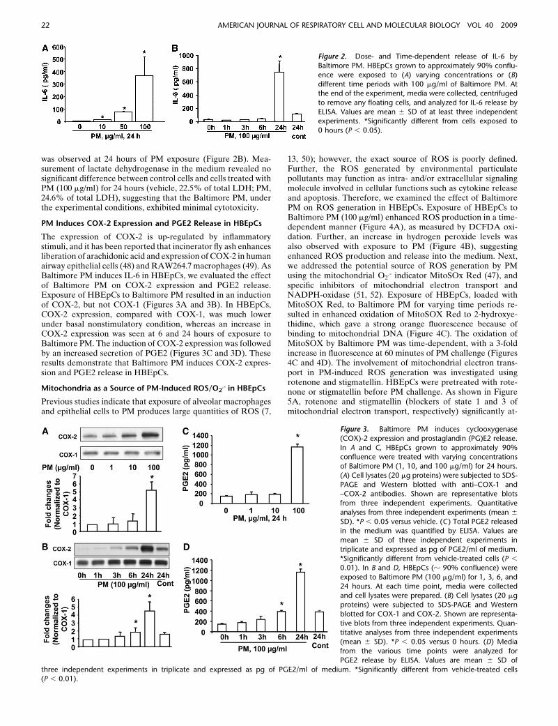
was observed at 24 hours of PM exposure (Figure 2B). Mea-surement of lactate dehydrogenase in the medium revealed nosignificant difference between control cells and cells treated withPM (100 mg/ml) for 24 hours (vehicle, 22.5% of total LDH; PM,24.6% of total LDH), suggesting that the Baltimore PM, underthe experimental conditions, exhibited minimal cytotoxicity.
PM Induces COX-2 Expression and PGE2 Release in HBEpCs
The expression of COX-2 is up-regulated by inflammatorystimuli, and it has been reported that incinerator fly ash enhancesliberation of arachidonic acid and expression of COX-2 in humanairway epithelial cells (48) and RAW264.7 macrophages (49). AsBaltimore PM induces IL-6 in HBEpCs, we evaluated the effectof Baltimore PM on COX-2 expression and PGE2 release.Exposure of HBEpCs to Baltimore PM resulted in an inductionof COX-2, but not COX-1 (Figures 3A and 3B). In HBEpCs,COX-2 expression, compared with COX-1, was much lowerunder basal nonstimulatory condition, whereas an increase inCOX-2 expression was seen at 6 and 24 hours of exposure toBaltimore PM. The induction of COX-2 expression was followedby an increased secretion of PGE2 (Figures 3C and 3D). Theseresults demonstrate that Baltimore PM induces COX-2 expres-sion and PGE2 release in HBEpCs.
Mitochondria as a Source of PM-Induced ROS/O2.- in HBEpCs
Previous studies indicate that exposure of alveolar macrophagesand epithelial cells to PM produces large quantities of ROS (7,
13, 50); however, the exact source of ROS is poorly defined.Further, the ROS generated by environmental particulatepollutants may function as intra- and/or extracellular signalingmolecule involved in cellular functions such as cytokine releaseand apoptosis. Therefore, we examined the effect of BaltimorePM on ROS generation in HBEpCs. Exposure of HBEpCs toBaltimore PM (100 mg/ml) enhanced ROS production in a time-dependent manner (Figure 4A), as measured by DCFDA oxi-dation. Further, an increase in hydrogen peroxide levels wasalso observed with exposure to PM (Figure 4B), suggestingenhanced ROS production and release into the medium. Next,we addressed the potential source of ROS generation by PMusing the mitochondrial O2
.- indicator MitoSOx Red (47), andspecific inhibitors of mitochondrial electron transport andNADPH-oxidase (51, 52). Exposure of HBEpCs, loaded withMitoSOX Red, to Baltimore PM for varying time periods re-sulted in enhanced oxidation of MitoSOX Red to 2-hydroxye-thidine, which gave a strong orange fluorescence because ofbinding to mitochondrial DNA (Figure 4C). The oxidation ofMitoSOX by Baltimore PM was time-dependent, with a 3-foldincrease in fluorescence at 60 minutes of PM challenge (Figures4C and 4D). The involvement of mitochondrial electron trans-port in PM-induced ROS generation was investigated usingrotenone and stigmatellin. HBEpCs were pretreated with rote-none or stigmatellin before PM challenge. As shown in Figure5A, rotenone and stigmatellin (blockers of state 1 and 3 ofmitochondrial electron transport, respectively) significantly at-
Figure 3. Baltimore PM induces cyclooxygenase
(COX)-2 expression and prostaglandin (PG)E2 release.
In A and C, HBEpCs grown to approximately 90%confluence were treated with varying concentrations
of Baltimore PM (1, 10, and 100 mg/ml) for 24 hours.
(A) Cell lysates (20 mg proteins) were subjected to SDS-
PAGE and Western blotted with anti–COX-1 and–COX-2 antibodies. Shown are representative blots
from three independent experiments. Quantitative
analyses from three independent experiments (mean 6
SD). *P , 0.05 versus vehicle. (C ) Total PGE2 releasedin the medium was quantified by ELISA. Values are
mean 6 SD of three independent experiments in
triplicate and expressed as pg of PGE2/ml of medium.
*Significantly different from vehicle-treated cells (P ,
0.01). In B and D, HBEpCs (z 90% confluence) were
exposed to Baltimore PM (100 mg/ml) for 1, 3, 6, and
24 hours. At each time point, media were collectedand cell lysates were prepared. (B) Cell lysates (20 mg
proteins) were subjected to SDS-PAGE and Western
blotted for COX-1 and COX-2. Shown are representa-
tive blots from three independent experiments. Quan-titative analyses from three independent experiments
(mean 6 SD). *P , 0.05 versus 0 hours. (D) Media
from the various time points were analyzed for
PGE2 release by ELISA. Values are mean 6 SD of
three independent experiments in triplicate and expressed as pg of PGE2/ml of medium. *Significantly different from vehicle-treated cells
(P , 0.01).
Figure 2. Dose- and Time-dependent release of IL-6 by
Baltimore PM. HBEpCs grown to approximately 90% conflu-
ence were exposed to (A) varying concentrations or (B)
different time periods with 100 mg/ml of Baltimore PM. Atthe end of the experiment, media were collected, centrifuged
to remove any floating cells, and analyzed for IL-6 release by
ELISA. Values are mean 6 SD of at least three independentexperiments. *Significantly different from cells exposed to
0 hours (P , 0.05).
22 AMERICAN JOURNAL OF RESPIRATORY CELL AND MOLECULAR BIOLOGY VOL 40 2009

tenuated Baltimore PM–induced ROS production. However,apocynin, which blocks flavin-containing enzyme(s), had noeffect on PM-mediated DCFDA oxidation (Figure 5A). Analysisof DCFDA oxidation in living cells by immunofluorescencemicroscopy revealed that oxidation of DCFDA co-localized withMito Tracker, a marker for mitochondria (Figure 5B) andpretreatment of cells with rotenone or stigmatellin, but notapocynin, attenuated the colocalization (yellow) of DCFDAoxidation (green) with fluorescence of Mito Tracker (red) (Figure
5B). These results support the role of mitochondrial electrontransport, but not NADPH oxidase, in PM-induced ROS/O2
.-
production in HBEpCs.
N-Acetylcysteine and EUK-134 Attenuate PM-Induced ROS
Generation, COX-2 Expression, and IL-6 Secretion in HBEpCs
To elucidate the putative role of PM-induced ROS in COX-2expression and IL-6 secretion, we pretreated cells with vary-ing concentration of antioxidants, N-acetylcysteine (NAC), or
Figure 4. Baltimore PM induces reactive oxygen
species (ROS) generation in HBEpCs. (A) HBEpCs
grown on glass bottom-dishes to approximately
90% confluence were loaded with 10 mM DCFDAfor 30 minutes. Cells were rinsed in basal medium
without growth factors and exposed to Baltimore
PM (100 mg/ml) for 15, 30, and 60 minutes. At the
end of exposure, ROS formation was visualizedunder fluorescence microscope and quantified us-
ing image analysis. (B) HBEpCs grown on 35-mm
dishes to approximately 90% confluence werechallenged with 10 or 100 mg/ml of Baltimore PM
for 60 minutes. Media were collected and H2O2
levels were measured using the Amplex Red assay.
Values are mean 6 SD of three experiments.*Significantly different from cells exposed to vehicle
alone (P , 0.05). (C) HBEpCs grown on glass-
bottom dishes to approximately 90% confluence,
challenged with Baltimore PM (100 mg/ml) for 15,30, and 60 minutes. At the end of each time point,
cells were washed with EBM phenol red free media
and were loaded with MitoSOX (1 mM) for 10minutes. Cells were washed three times with EBM
phenol red free media and intracellular MitoSOX
Red-emitted fluorescence was visualized by immu-
nofluorescence microscopy. (D) Intracellular Mito-SOX Red-emitted fluorescence of C quantified by
image analysis using MetaVue software. Values are
mean 6 SD of three independent experiments.
*Significantly different from cells exposed to vehicle(P , 0.05).
Figure 5. PM-induced ROS generation is dependent on mitochondrialelectron transport. (A) HBEpCs grown to approximately 90% conflu-
ence were pretreated with apocynin (50 mM) or rotenone (2 mM) or
stigmatellin (1 mM) for 1 hour before loading with 10 mM DCFDA for30 minutes. Cells were rinsed and challenged with vehicle or vehicle
plus Baltimore PM (100 mg/ml) for 30 minutes. Formation of ROS was
quantified by immunofluorescence microscopy. Values are mean 6 SD
of three independent experiments. *Significantly different from cellsexposed to vehicle (P , 0.05); **significantly different from cells
challenged with Baltimore PM (P , 0.01). (B) HBEpCs grown on glass
coverslips to approximately 90% confluence were pretreated with
apocynin (50 mM), rotenone (2 mM), or stigmatellin (1 mM) for 1 hourbefore loading with 10 mM DCFDA and 50 nM Mito Tracker for
30 minutes. Cells were challenged with vehicle or vehicle plus Baltimore
PM (100 mg/ml) for 30 minutes, and cells were visualized for ROSgeneration (green) and Mito Tracker (red) marker using fluorescence
microscope. Co-localization of ROS (green) in mitochondria with Mito
Tracker (red) is shown as yellow. Shown is a representative immunoflu-
orescence micrograph from three independent experiments.
Zhao, Usatyuk, Gorshkova, et al.: Particulate Matter Regulates COX-2 and IL-6 Expression 23

EUK-134 before challenge with PM (100 mg/ml). NAC (0.5 and5 mM) or EUK-134 (5 and 25 mM) attenuated PM-inducedDCFDA oxidation (Figures 6A and 6D), COX-2 expression(Figures 6B and 6E), and IL-6 production (Figures 6C and 6F)in HBEpCs. These data suggest the involvement ROS/cellularthiol redox status in PM-induced COX-2 expression and IL-6secretion in HBEpCs.
PM Induces COX-2 Expression and IL-6 Secretion via NF-kB
As NF-kB is a key transcriptional activator of COX-2 and IL-6genes (53, 54), we investigated the role of NF-kB in PM-inducedCOX-2 expression and IL-6 generation. Exposure of HBEpCs toBaltimore PM (100 mg/ml) for 15 minutes increased IkB phos-phorylation, and translocation of NF-kB to the nucleus (Figures7A and 7B). To study the role of NF-kB, cells were treated withthe IkB kinase inhibitor, Bay11–7082 (1 and 5 mM). Bay11–7082blocked PM-induced translocation of NF-kB to the nucleus(Figure 7B), COX-2 protein expression (Figure 7C), and IL-6secretion (Figure 7D). These results suggest that PM-inducedactivation of NF-kB partly regulates COX-2 expression and IL-6secretion in HBEpCs.
PM-Induced NF-kB Activation Is Attenuated by NAC and
EUK-134
To determine the role of ROS in PM-induced NF-kB activation,we studied the effect of NAC and EUK-134 on IkB phosphor-ylation. As shown in Figures 8A and 8B, NAC (0.5 and 5.0 mM)or EUK-135 (5 mM) pretreatment for 1 hour attenuated PM-induced IkB phosphorylation. Further, pretreatment with NAC(5 mM) blocked PM-induced translocation of NF-kB to thenucleus of HBEpCs (Figure 8C). These results show that ROSgenerated by PM stimulate NF-kB activation in HBEpCs.
C/EBPb Regulates PM-Induced COX-2 Expression and IL-6
Secretion via ROS-Independent Pathway
Our results with NAC and EUK-134 suggest a role for ROS-dependent activation of NF-kB in PM-induced COX-2 expres-sion and IL-6 secretion. However, NAC and EUK-134 did notcompletely block either COX-2 expression or IL-6 productionmediated by Baltimore PM, suggesting the involvement of othersignaling pathways independent of ROS in COX-2 expressionand IL-6 secretion in HBEpCs. As C/EBPb is another tran-scriptional regulator of COX-2 and IL-6, we investigated therole of C/EBPb in PM-induced COX-2 and IL-6 in HBEpCs. Asshown in Figures 9A and 9B, PM exposure (100 mg/ml) for 15minutes increased serine phosphorylation of C/EBPb, whichwas not blocked by NAC (0.05–5.0 mM) or EUK-134 (5 mM),suggesting ROS-independent activation. To further establishthe involvement of C/EBPb in COX-2 expression and IL-6secretion, we used C/EBPb siRNA to knockdown C/EBPb
(Figure 9C). The effect of C/EBPb siRNA was specific, as it hadno effect on the protein expression of actin or ERK (results notshown). Knockdown of C/EBPb blocked PM-mediated COX-2expression (z 50% compared with scrambled siRNA plus PM)(Figure 9C) and IL-6 secretion (z 45% compared with scram-bled siRNA plus PM) (Figure 9D). These results indicate theinvolvement of an ROS-independent pathway in PM-inducedC/EBPb activation and regulation of COX-2 and IL-6 inHBEpCs.
Involvement of COX-2 and PGE2 in PM-Induced IL-6
Secretion in HBEpCs
To further determine the role of COX-2 in Baltimore PM-induced IL-6 secretion, HBEpCs were transfected with COX-2siRNA (100 nM) for 72 hours or treated with NS-398 (50 mM),
an inhibitor of COX-2, for 1 hour before challenge with PM(100 mg/ml). The efficacy of the siRNA employed was de-termined by Western blotting for the protein expression ofCOX-2. As shown in Figure 10A, transfection of cells withsiRNA for COX-2 blocked the expression of COX-2 withoutaffecting COX-1 level, indicating a high degree of specificity.Furthermore, the PM-induced IL-6 secretion was attenuatedby COX-2 siRNA (z 50% inhibition) (Figure 10B) and NS-398(z 60% inhibition) (Figure 10C). Induction of COX-2 results inconversion of AA to PGE2 and PGD2 in mammalian cells.Having established a role for COX-2 in PM-mediated IL-6release, we next determined if PGE2 is involved in IL-6secretion via PGE2 receptors. Exposure of HBEpCs to PGE2(10 and 100 ng/ml) for 24 hours resulted in increased IL-6production (Figure 10D), suggesting PGE2-dependent IL-6production. These results show a role for COX-2-mediatedand PGE2-dependent production of IL-6 in HBEpCs.
DISCUSSION
The data presented in this study demonstrate that PM-inducesIL-6 secretion in primary human airway epithelial cells and thatthis secretion is mediated through ROS-dependent activationof NF-kB/COX-2/PGE2 and ROS-independent activation ofC/EBPb/COX-2/PGE2 signaling pathways. Further, evidence isprovided for the participation of the mitochondrial electrontransport chain, but not NADPH oxidase, in Baltimore PM–induced ROS generation. Our finding that PM-induces IL-6secretion via ROS-dependent and ROS-independent activationof COX-2/PGE2 pathways implies a novel signaling mechanismlinking air pollution particles to the inflammatory response inthe epithelium (Figure 11).
IL-6 is a multifunctional cytokine that is central to innateand acquired immune responses. In the airway, epithelial cellssecrete IL-6 as a primary response to an external stimulus or asa secondary response to inflammatory mediators such as TNF-aor IL-1b (14, 15, 17, 27, 28). The increases in IL-6 levelsobserved in this study with Baltimore PM are similar to thosedescribed earlier using ROFA collected from the cyclone ofa power plant in Florida (14); however, they differ from thosedescribed using ambient PM2.5 collected from Cache Valleylocations in Utah (20). The earlier studies were performed inthe BEAS-2B transformed cell line (14), whereas primarycultures of HBEpCs were employed in the present investiga-tion. ROFA stimulated IL-6/IL-8 gene expressions; however, inthat study IL-6 secretion was not measured (14). Secretion ofIL-6 in BEAS-2B cells exposed to PM2.5 from Cache Valley,Utah was approximately 1.3- to 1.5-fold higher compared withcontrol cells (20), while the Baltimore PM–induced IL-6 se-cretion in HBEpCs was at least 4- to 5-fold higher (Figure 2).This variation in IL-6 secretion between Cache Valley PM andBaltimore PM could be due to differences in the particle size,variation in particle composition, time, and collection location.It appears that the size of the particle PM from Baltimoreversus PM2.5 from Utah may contribute to the observed differ-ences in IL-6 secretion. Endotoxin is a natural constituent ofPM, which may contribute to the cytokine production in theepithelium (55); however, pretreatment of Baltimore PM withpolymyxin B had no appreciable effect on IL-6 secretion (datanot shown), suggesting no significant role of endotoxins asa contaminant in IL-6 production. In addition to IL-6, PM10
(EHC-93), obtained from the Environmental Health Director-ate, Health Canada (Ottawa, ON, Canada) induced mRNA andprotein expression of LIF, an IL-6–related glycoprotein, inHBEpCs (56). Similar to Baltimore PM, exposure of HBEpCs
24 AMERICAN JOURNAL OF RESPIRATORY CELL AND MOLECULAR BIOLOGY VOL 40 2009

Figure 6. N-Acetylcysteine and EUK-134 attenuate
Baltimore PM–induced ROS generation, COX-2
expression, and IL-6 secretion. HBEpCs grown in35-mm dishes to approximately 90% confluence
were pretreated with varying concentrations of N-
acetylcysteine (NAC) (0.05, 0.5, and 5 mM) or
EUK-134 (0.5, 5, and 25 mM) for 1 hour. In A and D,cells were loaded with 10 mM DCFDA for 30
minutes before addition of Baltimore PM (100
mg/ml) and ROS generation was quantified after
60 minutes using immunofluorescence microscopy.Values are mean 6 SD of three independent experi-
ments. *Significantly different from cells treated
with vehicle (P , 0.01); **significantly differentfrom cells exposed to Baltimore PM (P , 0.05). In B
and E, cells after pretreatment with vehicle or NAC
or EUK-134 were challenged with Baltimore PM
(100 mM) for 24 hours, and cell lysates (20 mgproteins) were subjected to SDS-PAGE and Western
blotted with anti–COX-2 and actin antibodies.
Shown are representative blots from three inde-
pendent experiments. Quantitative analyses fromthree independent experiments (mean 6 SD). *P ,
0.05 versus vehicle; **P , 0.05 versus PM chal-
lenge. In C and F, media were collected from cells(B and E), and analyzed for IL-6 by ELISA. Values are
mean 6 SD from three independent experiments in
triplicate and represented as pg/ml of medium.
*Significantly different from vehicle treated cells(P , 0.05); **significantly different from cells
challenged with Baltimore PM (P , 0.001).
Figure 7. Baltimore PM-induced COX-2 expression
and IL-6 secretion via NF-kB. In A, HBEpCs grown in35-mm dishes to approximately 95% confluence were
starved in basal EBM medium without any growth
factors for 3 hours, and then challenged with Baltimore
PM (10 and 100 mg/ml) for 15 minutes. Cell lysates(20 mg proteins) were subjected to SDS-PAGE and Western
blotted with phospho-IkB and ERK2 antibodies. Shown
are representative blots and quantitative analyses from
three independent experiments (mean 6 SD). *P ,
0.05 versus vehicle. **P , 0.05 versus PM challenge. In
B, HBEpCs in glass coverslips (z 95% confluence) were
pretreated with Bay compound (5 mM) for 60 minutes,cells were challenged with vehicle or vehicle plus
Baltimore PM (100 mg/ml) for 15 minutes. Cells were
washed, fixed, permeabilized, probed with anti-p65
antibody, and examined by immunofluorescencemicroscopy using a 360 oil objective. Shown is
a representative image from several independent
experiments. In C, HBEpCs grown on 35-mm dishes
were pretreated with varying concentrations of Baycompound (1 and 5 mM) for 60 minutes. Cells were
challenged with vehicle or vehicle plus Baltimore PM
(100 mg/ml) for 24 hours, cell lysates (20 mg proteins)were subjected to SDS-PAGE, and Western blotted
with anti–COX-2 and actin antibodies. Shown are
representative blots and quantitative analyses from
three independent experiments (mean 6 SD). *P ,
0.05 versus vehicle; **P , 0.05 versus PM challenge. In
D, media from C were analyzed by ELISA for IL-6.
Values are mean 6 SD from three independent experiments in triplicate. *Significantly different from cells exposed to vehicle (P , 0.01);
**significantly different from cells exposed to Baltimore PM (P , 0.05).
Zhao, Usatyuk, Gorshkova, et al.: Particulate Matter Regulates COX-2 and IL-6 Expression 25

to a standard PM, SRM 1648, also increased ROS production,COX-2 expression, PGE2 release, IkB phosphorylation, andIL-6 release (data not shown). These earlier studies and ourpresent results demonstrate the ability of different sources ofPM to stimulate IL-6 in immortalized and primary cultures ofHBEpCs, in vitro.
Expression of the IL-6 gene is regulated by binding ofa number of transcription factors, including NF-kB, CREB,AP-1, and NF–IL-6 (C/EBP), to the 59 region of the IL-6 gene(17). Transcriptional regulation of the IL-6 gene could bestimulus and/or cell specific, and activation of NF-kB has been
shown to control ROFA-induced IL-6 expression in cells of theBEAS-2B line (14). The results from this study show thatBaltimore PM–induced IL-6 secretion is dependent on NF-kB,in that blocking NF-kB activation with Bay 117082 antagonizedPM-induced IkB phosphorylation and IL-6 secretion. Further,the PM-induced NF-kB activation and IL-6 secretion wereattenuated by NAC, suggesting a link between ROS generationto NF-kB activation and IL-6 expression. Although an earlierstudy with ROFA suggests a potential link between ROS andIL-6 expression in BEAS-2B cell line (14), the current studyestablishes a direct link between PM-dependent ROS genera-tion in NF-kB activation and IL-6 secretion in primaryHBEpCs. It has been shown that redox-active transition metals,redox-cycling quinones, and polycyclic aromatic hydrocarbonspresent in diesel exhaust particles (DEP) and PM generatesROS and oxygen free radicals in cultured macrophages, epithe-lial cells, and suspensions (13, 17). The source of ROS afterexposure to DEP or PM is unclear, but may involve activationof NADPH oxidase (50) and/or leak from the mitochondrialelectron transport chain (50, 57–59). In the present study, wehave demonstrated that the mitochondrial inhibitors stigmatel-lin and rotenone, but not apocynin, attenuated Baltimore PM–stimulated ROS production, suggesting participation of themitochondrial electron transport chain at complex I and III asthe major source of ROS. While the nature of the redox-activemetal(s) present in Baltimore PM that induces ROS productionin primary HBEpCs has not been established, earlier studiesindicate that vanadium present in ROFA may mediate ROSproduction (50), cytokine gene expression, and airway epithelialinjury (60). Analyses of the Baltimore PM revealed the pres-ence of vanadium (Table 1).
In addition to NF-kB, our results show a regulatory role forCOX-2 in PM-induced IL-6 secretion in HBEpCs, as evidencedby COX-2 siRNA and COX-2 inhibitor studies. In accordancewith the human COX-2 promoter having two NF-kB–bindingsites, blocking NF-kB with Bay compound attenuated PM-mediated IL-6 secretion in HBEpCs. Interestingly, we haveidentified ROS-dependent and ROS-independent mechanismsof COX-2 expression in HBEpCs, demonstrating multiple sig-naling pathways of IL-6 regulation by PM. The ROS-dependentstimulation of COX-2 expression is via transcriptional regula-tion by NF-kB, while the ROS-independent expression ofCOX-2 requires activation of C/EBPb in HBEpCs (Figure 9).Induction of COX-2 results in subsequent production of PGE2,and in the present study we show that exposure of HBEpCs toPM stimulated PGE2 secretion. Further, exogenous addition of
Figure 8. N-Acetylcysteine and EUK-134 attenuate Baltimore PM-
induced NF-kB activation. HBEpCs grown in 35-mm dishes to approx-
imately 90% confluence were pretreated with varying concentrationsof N-acetylcysteine (NAC) (0.05, 0.5, and 5 mM) or EUK-134 (5 mM)
for 1 hour. In A and B, cells were challenged with vehicle or vehicle plus
Baltimore PM (100 mg/ml) for 15 minutes, and cell lysates (20 mg
proteins) were subjected to SDS-PAGE and Western blotted with anti–phospho-IkB and actin antibodies. Shown are representative blots and
quantitative analyses from three independent experiments (mean 6
SD). *P , 0.05 versus vehicle; **P , 0.05 versus PM challenge. In C,HBEpCs grown on glass coverslips (z 95% confluence) were pretreated
with NAC (5 mM) for 60 minutes, cells were challenged with vehicle or
vehicle plus Baltimore PM (100 mg/ml) for 15 minutes, and cells were
washed, fixed, permeabilized, probed with anti-p65 antibody, andexamined by immunofluorescence microscopy using a 360 oil objec-
tive. Shown is a representative image from several independent
experiments.
b
26 AMERICAN JOURNAL OF RESPIRATORY CELL AND MOLECULAR BIOLOGY VOL 40 2009

Figure 9. C/EBPb regulates Balti-
more PM–induced COX-2 expres-
sion and IL-6 secretion via ROS-
independent pathway. In A and B,HBEpCs grown on 35-mm dishes
to approximately 90% confluence
were pretreated with varying con-
centrations of NAC (0.05, 0.5,and 5 mM) or EUK-134 (5 mM)
for 1 hour. Cells were challenged
with vehicle or vehicle plus Balti-more PM (100 mg/ml) for 15
minutes, and cell lysates (20 mg
proteins) were subjected to SDS-
PAGE and Western blotted withanti–phospho-C/EBPb or C/EBPb
antibodies. Shown are representa-
tive blots and quantitative an-
alyses from three independentexperiments (mean 6 SD). *P ,
0.05 versus vehicle. In C, HBEpCs
grown on 35-mm dishes to ap-
proximately 50% confluence weretransfected with scrambled siRNA
or C/EBPb siRNA (100 nM) for
72 hours. Cells were challenged withvehicle or vehicle plus Baltimore
PM (100 mg/ml) for 24 hours, and
cell lysates (20 mg proteins) were
subjected to SDS-PAGE and West-
ern blotted with anti–COX-2 or C/EBPb antibodies as described in MATERIALS AND METHODS. Shown are representative blots and quantitative analyses
from three independent experiments (mean 6 SD). *P , 0.05 versus vehicle; **P , 0.05 versus PM challenge. In D, media from C were analyzed forIL-6 by ELISA. Values are mean 6 SD from three independent experiments. *Significantly different from vehicle-challenged cells (P , 0.05);
**significantly different from cells challenged with Baltimore PM (P , 0.01).
Figure 10. Involvement of COX-2 and
PGE2 in Baltimore PM–induced IL-6 secre-tion. In A and B, HBEpCs grown on 35-mm
dishes to approximately 50% confluence
were transfected with scrambled siRNA
(50 nM) or COX-2 siRNA (50 nM) for 72hours before challenge with vehicle or
vehicle plus Baltimore PM (100 mg/ml)
for 24 hours. In A, cell lysates (20 mg
proteins) were subjected to SDS-PAGE,and Western blotted with anti–COX-2 or
–COX-1 antibodies. Shown are represen-
tative blots and quantitative analyses fromthree independent experiments. * and
** significantly different from scrambled
siRNA-transfected cells exposed to vehicle
(P , 0.01); *** significantly different fromCOX-2 siRNA-transfected cells exposed to
PM (P , 0.01). In B, media from A were
collected and analyzed for IL-6 by ELISA.
Values are mean 6 SD from three inde-pendent experiments. *Significantly dif-
ferent from scrambled siRNA-transfected
cells exposed to vehicle (P , 0.01); **sig-nificantly different from scrambled siRNA-
transfected cells exposed to Baltimore PM
(P , 0.01). In C, HBEpCs grown to ap-
proximately 90% confluence were pre-treated with COX-2 inhibitor, NS-398
(50 mM) for 1 hour, cells were challengedwith vehicle or vehicle plus Baltimore PM (100 mg/ml) for 24 hours, and media were analyzed for IL-6 by ELISA. Values are mean 6 SD from threeindependent experiments. *Significantly different from cells challenged with vehicle (P , 0.05); **significantly different from cells exposed to Baltimore
PM (P , 0.01). In D, PGE2 (10, and 100 ng/ml)was added to HBEpCs grownon 35-mm dishes for 6 hours,media were collected, andanalyzed for IL-6 by
ELISA. Values are mean 6 SD from three independent experiments. *Significantly different from cells exposed to vehicle (P , 0.05).
Zhao, Usatyuk, Gorshkova, et al.: Particulate Matter Regulates COX-2 and IL-6 Expression 27

PGE2 to HBEpCs resulted in enhanced IL-6 production sug-gesting a role for PM-induced secretion of PGE2 in ROS-dependent and ROS-independent release of IL-6. Our resultsshow that at 10 mg/ml of PM exposure, there was no significantincrease in COX-2 expression (Figure 3) and phosphorylationof IkB (Figure 7). Although PM (10 mg/ml)-induced generationof ROS (Figure 4B) and IL-6 production (Figure 2A) arestatistically significant, the changes are much smaller comparedwith exposure of cells to 100 mg/ml of PM (Figures 2 and 4). AsROS production via the mitochondrial electron transportsystem is upstream of NF-kB and COX-2 pathways, some ofthe observed effects of varying PM concentrations may be dueto the downstream signaling involved in IL-6 production. Whilethe ability of the coarse PM fraction and incinerator fly ash toinduce the expression of COX-2 has been described earlier (17,49), we report for the first time an ROS-dependent expressionof COX-2 via NF-kB and ROS-independent activation of COX-2via C/EBPb by PM in HBEpCs. Although the mechanism(s) ofPGE2-induced IL-6 release was not investigated in HBEpCs,addition of PGE2 stimulated IL-6 production via PGE2 recep-tors EP-2/4 linked to adenylate cyclase in BEAS-2B cell line(T59). A role for nickel, a component of PM also present inBaltimore PM, was demonstrated in Toll-like receptor-2–de-pendent chemokines released from human lung fibroblaststhrough a COX-2–mediated pathway (61). Nickel has beenlinked to human cardiac dysfunction and daily deaths in re-sponse to inhalation of fine PM (62). Future studies in murinemodels of asthma and cardiac hypertrophy will address the roleof metal ions such as nickel and vanadium in ROS generation,COX-2 expression, PGE2 release, and IL-6 secretion in theairway and lung, which could contribute to the cardiopulmonarydysfunctions associated with inhalation of PM. Instillation ofBaltimore PM to mice induced airway inflammation, increasedinflux of activated PMNs and eosinophils in alveolar space, and
stimulated IL-6 secretion in BAL fluid (data not shown)indicating the utility of this model for in vivo studies relatedto PM inhalation, ROS generation, and lung inflammation.
The present study has identified that PM-mediated activa-tion of the transcriptional factor, C/EBPb, regulates ROS-independent activation of COX-2 in HBEpCs. Several proin-flammatory mediators stimulate COX-2 expression via C/EBPb,and recently lysophosphatidic acid–mediated transactivation ofEGF-R was shown to regulate COX-2 expression and PGE2release via C/EBPb in HBEpCs (63). Interestingly, recent studiesshow up-regulation of several EGF-R ligands in 16HBE14oairway epithelial cells exposed to PM with an aerodynamicdiameter less than 2.5 mm (64). Intratracheal instillation ofROFA into perfused rabbit lungs activated EGF-R and mediatedpulmonary vasoconstriction (65), and Zn21,also present in Balti-more PM, induced EGF-R activation and signaling in A431cells (66). It is hypothesized that activation of C/EBPb by PMmight involve activation of EGF-R either by EGF-ligands and/or transactivation mechanism(s) involving G protein–coupledreceptors in HBEpCs and which are currently under investiga-tion.
Epidemiologic studies suggest an association between acuteexposure to PM and increased hospitalization of patients withcardiovascular events (1–4). Exposure of normal individuals andpeople with cardiovascular disease to PM increases serum levelsof IL-6 and C-reactive protein (22), which are independentlyassociated with increased incidence of cardiovascular disease(23–26). These PM effects on humans have been partly sup-ported by studies in murine models, which suggest that exposureto PM can modulate IL-6 production by alveolar macrophagesand lead to reduced clotting times, intravascular thrombinformation, and accelerated arterial thrombosis (19). Therefore,PM-induced IL-6 generation, either independently or in associ-ation with other mediators, may be responsible for the devel-opment of cardiovascular events. As ROS play a key role in thedevelopment of cardiovascular diseases (67), and regulateCOX-2 expression, PGE2 release, and IL-6 production inHBEpCs and other cell types, the targeting of excess ROSgeneration may represent a reasonable approach to alleviatePM-induced cardiopulmonary complications and reduce hospi-talizations. The relationship between the doses used in thepresent studies and in vivo dose levels is difficult to accuratelydetermine, but were, as indicated, many fold higher than thoseexperienced in the Baltimore environment. While it is possiblethat variation in pathway activities may differ somewhat atlower concentrations, the present data identify PM-responsivetargets within respiratory epithelial cells that may play crucialmodulatory roles in respiratory signaling.
In summary, this study demonstrates ROS-dependent andROS-independent stimulation of COX-2 expression, PGE2release, and IL-6 secretion by PM in primary HBEpCs. WhileROS-dependent activation of NF-kB regulates IL-6 secretionindependent of COX-2, enhanced COX-2 expression by ROSresulted in PGE2 release, which also triggered IL-6. Further,ROS-independent activation of COX-2 was mediated by acti-vation of the transcriptional factor C/EBPb, with subsequentrelease of IL-6 via PGE2 signaling. We also show that themitochondrial electron transport chain, but not NADPH oxi-dase, regulates PM-induced ROS production in HBEpCs. Thesenovel ROS-dependent and ROS-independent pathways thatregulate COX-2 expression, PGE2 release, and IL-6 secretionpresent potential targets to minimize the pro-inflammatoryresponses and cardiopulmonary events associated with inhala-tion of environmental air pollutants.
Conflict of Interest Statement: None of the authors has a financial relationshipwith a commercial entity that has an interest in the subject of this manuscript.
Figure 11. Proposed signaling pathways of ROS-dependent and -
independent activation of COX-2 in PM-induced IL-6 secretion in
HBEpCs. Exposure of epithelium to PM results in ROS generation and
activation of NF-kB, which regulates COX-2 and IL-6 secretion. PM-induced expression of COX-2 enhances PGE2 that stimulates IL-6 in
HBEpCs. Independent of ROS generation, PM also stimulates COX-2
expression via C/EBPb signaling. These results suggest multiple path-ways involved in ambient PM–mediated IL-6 generation in airway
epithelium, which involve ROS-dependent and -independent activation
of COX-2 and PGE2 signal transduction.
28 AMERICAN JOURNAL OF RESPIRATORY CELL AND MOLECULAR BIOLOGY VOL 40 2009

References
1. Oberdorster G, Gelein RM, Ferin J, Weiss B. Association of particulateair pollution and acute mortality: involvement of ultrafine particles?Inhal Toxicol 1995;7:111–124.
2. Samet JM, Dominici F, Curriero FC, Coursac I, Zeger SL. Fineparticulate air pollution and mortality in 20 US cities, 1987–1994.N Engl J Med 2000;343:1742–1749.
3. Dominici F, McDermott A, Zeger SL, Samet JM. Airborne particulatematter and mortality: timescale effects in four US cities. Am JEpidemiol 2003;157:1055–1065.
4. Samet JM, Krewski D. Health effects associated with exposure toambient air pollution. J Toxicol Environ Health A 2007;70:227–242.
5. Sandstrom T. Respiratory effects of air pollutants: experimental studiesin humans. Eur Respir J 1995;8:976–995.
6. Dreher KL, Jaskot RH, Lehmann JR, Richards JH, McGee JK, GhioAJ, Costa DL. Soluble transition metals mediate residual oil fly ashinduced acute lung injury. J Toxicol Environ Health 1997;50:285–305.
7. Gonzalez-Flecha B. Oxidant mechanisms in response to ambient airparticles. Mol Aspects Med 2004;25:169–182.
8. Schlesinger RB. The health impact of common inorganic components offine particulate matter (PM2.5) in ambient air: a critical review. InhalToxicol 2007;19:811–832.
9. Becker S, Dailey LA, Soukup JM, Grambow SC, Devlin RB, Huang YC.Seasonal variations in air pollution particle-induced inflammatorymediator release and oxidative stress. Environ Health Perspect 2005;113:1032–1038.
10. Franklin M, Zeka A, Schwartz J. Association between PM2.5 and all-cause and specific-cause mortality in 27 US communities. J Expo SciEnviron Epidemiol 2007;17:279–287.
11. Handzel ZT. Effects of environmental pollutants on airways, allergicinflammation, and the immune response. Rev Environ Health 2000;15:325–336.
12. Peden DB. Air pollution in asthma: effect of pollutants on airwayinflammation. Ann Allergy Asthma Immunol 2001;87:12–17.
13. Tao F, Gonzalez-Flecha B, Kobzik L. Reactive oxygen species in pulmo-nary inflammation by ambient particulates. Free Radic Biol Med 2003;35:327–340.
14. Quay JL, Reed W, Samet J, Devlin RB. Air pollution particles induceIL-6 gene expression in human airway epithelial cells via NF-kappaBactivation. Am J Respir Cell Mol Biol 1998;19:98–106.
15. Fujii T, Hayashi S, Hogg JC, Vincent R, Van Eeden SF. Particulatematter induces cytokine expression in human bronchial epithelialcells. Am J Respir Cell Mol Biol 2001;25:265–271.
16. Yu M, Zheng X, Witschi H, Pinkerton KE. The role of interleukin-6 inpulmonary inflammation and injury induced by exposure to environ-mental air pollutants. Toxicol Sci 2002;68:488–497.
17. Becker S, Mundandhara S, Devlin RB, Madden M. Regulation ofcytokine production in human alveolar macrophages and airwayepithelial cells in response to ambient air pollution particles: Furthermechanistic studies. Toxicol Appl Pharmacol 2005;207:269–275.
18. Dagher Z, Garcon G, Billet S, Gosset P, Ledoux F, Courcot D,Aboukais A, Shirali P. Activation of different pathways of apoptosisby air pollution particulate matter (PM2.5) in human epithelial lungcells (L132) in culture. Toxicology 2006;225:12–24.
19. Mutlu GM, Green D, Bellmeyer A, Baker CM, Burgess Z, RajamannanN, Christman JW, Foiles N, Kamp DW, Ghio AJ, et al. Ambientparticulate matter accelerates coagulation via an IL-6-dependentpathway. J Clin Invest 2007;117:2952–2961.
20. Watterson TL, Sorensen J, Martin R, Coulombe RA Jr. Effects of PM2.5collected from Cache Valley Utah on genes associated with theinflammatory response in human lung cells. J Toxicol Environ Health2007;70:1731–1744.
21. Porter M, Karp M, Killedar S, Bauer SM, Guo J, Williams D, Breysse P,Georas SN, Williams MA. Diesel-enriched particulate matter func-tionally activates human dendritic cells. Am J Respir Cell Mol Biol2007;37:706–719.
22. Ruckerl R, Ibald-Mulli A, Koenig W, Schneider A, Woelke G, Cyrys J,Heinrich J, Marder V, Frampton M, Wichmann HE, et al. Air pollutionand markers of inflammation and coagulation in patients with coro-nary heart disease. Am J Respir Crit Care Med 2006;173:432–441.
23. Tracy RP. Epidemiological evidence for inflammation in cardiovasculardisease. Thromb Haemost 1999;82:826–831.
24. Yudkin JS, Stehouwer CD, Emeis JJ, Coppack SW. C-reactive protein inhealthy subjects: associations with obesity, insulin resistance, andendothelial dysfunction: a potential role for cytokines originatingfrom adipose tissue? Arterioscler Thromb Vasc Biol 1999;19:972–978.
25. Harris TB, Ferrucci L, Tracy RP, Corti MC, Wacholder S, Ettinger WH
Jr, Heimovitz H, Cohen HJ, Wallace R. Associations of elevatedinterleukin-6 and C-reactive protein levels with mortality in theelderly. Am J Med 1999;106:506–512.
26. Yeh ET. CRP as a mediator of disease. Circulation 2004;109:II11–II14.27. Park CS, Chung SW, Ki SY, Lim GI, Uh ST, Kim YH, Choi DI, Park JS,
Lee DW, Kitaichi M. Increased levels of interleukin-6 are associatedwith lymphocytosis in bronchoalveolar lavage fluids of idiopathicnonspecific interstitial pneumonia. Am J Respir Crit Care Med 2000;162:1162–1168.
28. Inoue K, Takano H, Yanagisawa R, Sakurai M, Shimada A, Morita T,
Sato M, Yoshino S, Yoshikawa T. Role of interleukin-6 in toll-likereceptor 4 and 2 expressions induced by lipopolysaccharide in thelung. Immunopharmacol Immunotoxicol 2007;29:63–68.
29. Arcangeli G, Cupelli V, Giuliano G. Effects of silica on human lung
fibroblast in culture. Sci Total Environ 2001;270:135–139.30. Bankey PE, Williams JG, Guice KS, Taylor SN. Interleukin-6 pro-
duction after thermal injury: evidence for nonmacrophage sources inthe lung and liver. Surgery 1995;118:431–438. (discussion 438–439).
31. Frampton MW, Ghio AJ, Samet JM, Carson JL, Carter JD, Devlin RB.
Effects of aqueous extracts of PM(10) filters from the Utah valley onhuman airway epithelial cells. Am J Physiol 1999;277:L960–L967.
32. Koyama S, Sato E, Nomura H, Kubo K, Miura M, Yamashita T, Nagai S,
Izumi T. Bradykinin stimulates type II alveolar cells to releaseneutrophil and monocyte chemotactic activity and inflammatorycytokines. Am J Pathol 1998;153:1885–1893.
33. Mosmann TR, Sad S. The expanding universe of T-cell subsets: Th1, Th2
and more. Immunol Today 1996;17:138–146.34. Yu M, Pinkerton KE, Witschi H. Short-term exposure to aged and
diluted sidestream cigarette smoke enhances ozone-induced lunginjury in B6C3F1 mice. Toxicol Sci 2002;65:99–106.
35. Grassl C, Luckow B, Schlondorff D, Dendorfer U. Transcriptional
regulation of the interleukin-6 gene in mesangial cells. J Am SocNephrol 1999;10:1466–1477.
36. Matsusaka T, Fujikawa K, Nishio Y, Mukaida N, Matsushima K,
Kishimoto T, Akira S. Transcription factors NF-IL6 and NF-kappaB synergistically activate transcription of the inflammatory cytokines,interleukin 6 and interleukin 8. Proc Natl Acad Sci USA 1993;90:10193–10197.
37. Shukla A, Timblin C, BeruBe K, Gordon T, McKinney W, Driscoll K,
Vacek P, Mossman BT. Inhaled particulate matter causes expressionof nuclear factor (NF)-kB-related genes and oxidant-dependent NF-kB activation in vitro. Am J Respir Cell Mol Biol 2000;23:182–187.
38. Nam HY, Choi BH, Lee JY, Lee SG, Kim YH, Lee KH, Yoon HK, Song
JS, Kim HJ, Lim Y. The role of nitric oxide in the particulate matter(PM2.5)-induced NFkappaB activation in lung epithelial cells. Tox-icol Lett 2004;148:95–102.
39. Roberts JW, Budd WT, Ruvy MG, Camann DE, Fortmann RC, Lewis
RG, Wallace LA, Spittler TM. Human exposure to pollutants in thefloor dust of homes and offices. J Expo Anal Environ Epidemiol 1992;2:127–146.
40. Walters DM, Breysse PN, Schofield B, Wills-Karp M. Complement
factor 3 mediates particulate matter-induced airway hyperresponsive-ness. Am J Respir Cell Mol Biol 2002;27:413–418.
41. Walters DM, Breysee PN, Wills-Karp M. Ambient urban Baltimore
particulate-induced airway hyperresponsiveness and inflammation inmice. Am J Respir Crit Care Med 2001;164:1438–1443.
42. Saatian B, Yu XY, Lane AP, Doyle T, Casolaro V, Spannhake EW.
Expression of genes for B7–H3 and other T cell ligands by nasalepithelial cells during differentiation and activation. Am J PhysiolLung Cell Mol Physiol 2004;287:217–225.
43. Bernacki SH, Nelson A, Abdullah L, Sheehan JK, Harris A, Davis CW,
Randell SH. Mucin gene expression during differentiation of humanairway epithelia in vitro. Muc4 and muc5b are strongly induced. Am JRespir Cell Mol Biol 1999;20:595–604.
44. Zhao Y, He D, Saatian B, Watkins T, Spannhake EWm, Pyne NJ,
Natarajan V. Regulation of lysophosphatidic acid-induced epidermalgrowth factor receptor transactivation and interleukin-8 secretion inhuman bronchial epithelial cells by protein kinase Cd, lyn kinase, andmatrix metalloproteinases. J Biol Chem 2006;281:19501–19511.
45. Usatyuk PV, Parinandi NL, Natarajan V. Redox regulation of 4-
hydroxy-2-nonenal-mediated endothelial barrier dysfunction by focaladhesion, adherens, and tight junction proteins. J Biol Chem 2006;281:35554–35566.
46. Usatyuk PV, Romer LH, He D, Parinandi NL, Kleinberg ME, Zhan S,
Jacobson JR, Dudek SM, Pendyala S, Garica JGN, et al. Regulation
Zhao, Usatyuk, Gorshkova, et al.: Particulate Matter Regulates COX-2 and IL-6 Expression 29

of hyperoxia-induced NADPH oxidase activation in human lungendothelial cells by the actin cytoskeleton and cortactin. J Biol Chem2007;282:23284–23295.
47. Mukhopadhyay P, Rajesh M, Hasko G, Hawkins BJ, Madesh M, PacherP. Simultaneous detection of apoptosis and mitochondrial superoxideproduction in live cells by flow cytometry and confocal microscopy.Nat Protocols 2007;2:2295–2301.
48. Samet JM, Ghio AJ, Costa DL, Madden MC. Increased expression ofcyclooxygenase 2 mediates oil fly ash-induced lung injury. Exp LungRes 2000;26:57–69.
49. Fritsch S, Diabate S, Krug HF. Incinerator fly ash provokes alteration ofredox equilibrium and liberation of arachidonic acid in vitro. BiolChem 2006;387:1421–1428.
50. Huang YT, Soukup J, Harder S, Becker S. Mitochondrial oxidantproduction by a pollutant dust and NO-mediated apoptosis in humanalveolar macrophage. Am J Physiol Cell Physiol 2003;284:C24–C32.
51. Li N, Ragheb K, Lawler G, Sturgis J, Rajwa B, Melendez JA, RobinsonJP. Mitochondrial complex I inhibitor rotenone induces apoptosisthrough enhancing mitochondrial reactive oxygen species production.J Biol Chem 2003;278:8516–8525.
52. Lee JY, Jung GY, Heo HJ, Yun MR, Park JY, Bae SS, Hong KW, LeeWS, Kim CD. 4-Hydroxynonenal induces vascular smooth muscle cellapoptosis through mitochondrial generation of reactive oxygenspecies. Toxicol Lett 2006;166:212–221.
53. Cheng DS, Han W, Chen SM, Sherrill TP, Chont M, Park GY, ShellerJR, Plosukhin VV, Christman JW, Yull FE, et al. E3 ubiquitin ligaseCblb regulates the acute inflammatory response underlying lunginjury. Nat Med 2007;13:920–926.
54. Park GY, Christman JW. Nuclear factor kappa B is a promising therapeutictarget in inflammatory lung disease. Curr Drug Targets 2006;7:661–668.
55. Becker S, Fenton MJ, Soukup JM. Involvement of microbial compo-nents and toll-like receptors 2 and 4 in cytokine responses to airpollution particles. Am J Respir Cell Mol Biol 2002;27:611–618.
56. Fujii T, Hayashi S, Hogg JC, Mukae H, Suwa T, Goto Y, Vincent R, vanEden SF. Interaction of alveolar macrophages and airway epithelialcells following exposure to particulate matter produces mediators thatstimulate the bone marrow. Am J Respir Cell Mol Biol 2002;27:34–41.
57. Becker S, Soukup JM, Gallagher JE. Differential particulate airpollution induced oxidant stress in human granulocytes, monocytesand alveolar macrophages. Toxicol In Vitro 2002;16:209–218.
58. Xia T, Korge P, Weiss JN, Li N, Venkatesen MI, Sioutas C, Nel A.Quinones and aromatic chemical compounds in particulate matterinduce mitochondrial dysfunction: implications for ultrafine particletoxicity. Environ Health Perspect 2004;112:1347–1358.
59. Soberanes S, Panduri V, Mutlu GM, Ghio A, Bundinger GR, KampDW. p53 mediates particulate matter-induced alveolar epithelial cellmitochondria-regulated apoptosis. Am J Respir Crit Care Med 2006;174:1229–1238.
60. Dye JA, Adler KB, Richards JH, Dreher KL. Role of soluble metals inoil fly ash-induced airway epithelial injury and cytokine gene expres-sion. Am J Physiol 1999;277:L498–L510.
61. Brant KA, Fabisiak JP. Nickel alteration of TLR2-dependent chemo-kine profiles in lung fibroblasts are mediated by COX-2. Am J RespirCell Mol Biol 2008;38:591–599.
62. Laden F, Neas LM, Dockery DW, Schwartz J. Association of fineparticulate matter from different sources with daily mortality in sixUS cities. Environ Health Perspect 2000;108:941–947.
63. He D, Natarajan V, Stern R, Gorshkova IA, Solway J, Spannhake EWm,Zhao Y. Lysophosphatidic acid-induced transactivation of epidermalgrowth factor receptor regulates cyclooxygenase-2 expression andprostaglandin E2 release via C/EBPb in human bronchial epithelialcells. Biochem J 2008;412:153–162.
64. Rumelhard M, Ramgolam K, Hamel R, Marono F, Baeza-Squiban A.Expression and role of EGFR ligands induced in airway cells byPM2.5 and its components. Eur Respir J 2007;30:1064–1073.
65. Huang YC, Wu W, Ghio AJ, Carter JD, Silbajoris R, Devlin RB,Samet JM. Activation of EGF receptors mediates pulmonaryvasoconstriction induced by residual oil fly ash. Exp Lung Res2002;28:19–38.
66. Samet JM, Dewar BJ, Wu W, Graves LM. Mechanisms of Zn(21)-induced signal initiation through the epidermal growth factor re-ceptor. Toxicol Appl Pharmacol 2003;191:86–93.
67. Shah AM, Channon KM. Free radicals and redox signaling in cardio-vascular disease. Heart 2004;90:486–487.
30 AMERICAN JOURNAL OF RESPIRATORY CELL AND MOLECULAR BIOLOGY VOL 40 2009