Regulation of Calcium Fluxes and Their Regulatory Roles in Pancreatic Islets
21
REGULATION OF CALCIUM FLUXES AND THEIR REGULATORY ROLES IN PANCREATIC ISLETS* Willy J. Malaisse, Andre Herchuelz, Ghislain Devis, Guido Somers, A. Carlos Boschero, John C. Hutton, Shoji Kawazu, and Abdullah Sener La boratoirc de Mhdrc.int, ExpCrimc.ntalc Brussds Univcrsity School of MrdicinP B-1000 Brussc4s Bclgiuin Illani J. Atwater, George Duncan, Bernard Ribalet, and Eduardo Rojas Dcpartmrnt of Biophysics Univrrsity ofEust Anglia School of Biological Scirncc. Noru.ich, England Introduction It is today considered as crystal clear that calcium plays an essential role in the regulation of insulin release by the pancreatic B-cell. Some of the major issues concerning such a role are as follows: (i) what is the detailed mechanism by which secretagogues are susceptible to influence the handling of calcium in the B-cell; (ii) what is the nature and location of the critical pool of calcium that controls insulin release; (iii) what is the relative and respective contribution of calcium influx, efflux, and subcellar distribution in the regulation of such a pool; and (iv) how does calcium influence the process by which secretory granules migrate to the cell boundary and are extruded via exocytosis in the interstitial fluid.’ In the present report, for the sake of clarity, we will restrict the discussion of these questions to the process of glucose-induced insulin release, with the main emphasis on the possible significance of passive ionophoretic movements. The process by which glucose provokes insulin release can be viewed as a sequence of three major events, namely: (i) the recognition or identification of glucose by the B-cell; (ii) the subsequent remodelling of cationic fluxes; and (iii) the activation by calcium of an effector system controlling the migration and exocytosis of secre- tory granules.2 The role of calcium in insulin release is here considered within the framework of such a sequential view. “This work was supported in part by grants from the Belgian Foundation for Scientific Medical Research. A. C. B. is a Research Fellow of the Fundagao de Amparo a Pesquisa (Sao Pauio, Brazil) on leave from the Department of Physiology. University of Campinas (Campinas, Brazil). J. C. H. was recipient of a Pfizer Travel Award through the European Association for the Study of Diabetes. 562 0077-892317810307-0562 $01.7510 0 1978, NYAS
Transcript of Regulation of Calcium Fluxes and Their Regulatory Roles in Pancreatic Islets

REGULATION OF CALCIUM FLUXES AND THEIR REGULATORY ROLES
IN PANCREATIC ISLETS*
Willy J. Malaisse, Andre Herchuelz, Ghislain Devis, Guido Somers, A. Carlos Boschero, John C. Hutton, Shoji Kawazu,
and Abdullah Sener
La boratoirc de Mhdrc.int, ExpCrimc.ntalc Brussds Univcrsity School of MrdicinP
B-1000 Brussc4s Bclgiuin
Illani J . Atwater, George Duncan, Bernard Ribalet, and Eduardo Rojas
Dcpartmrnt o f Biophysics Univrrsity ofEust Anglia
School o f Biological Scirncc. Noru.ich, England
Introduction
It is today considered as crystal clear that calcium plays an essential role in the regulation of insulin release by the pancreatic B-cell. Some of the major issues concerning such a role are as follows: ( i ) what is the detailed mechanism by which secretagogues are susceptible to influence the handling of calcium in the B-cell; (ii) what is the nature and location of the critical pool of calcium that controls insulin release; (iii) what is the relative and respective contribution of calcium influx, efflux, and subcellar distribution in the regulation of such a pool; and (iv) how does calcium influence the process by which secretory granules migrate to the cell boundary and are extruded via exocytosis in the interstitial fluid.’
In the present report, for the sake of clarity, we will restrict the discussion of these questions to the process of glucose-induced insulin release, with the main emphasis on the possible significance of passive ionophoretic movements. The process by which glucose provokes insulin release can be viewed as a sequence of three major events, namely: ( i ) the recognition or identification of glucose by the B-cell; (ii) the subsequent remodelling of cationic fluxes; and (iii) the activation by calcium of an effector system controlling the migration and exocytosis of secre- tory granules.2 The role of calcium in insulin release is here considered within the framework of such a sequential view.
“This work was supported in part by grants from the Belgian Foundation for Scientific Medical Research. A. C. B. is a Research Fellow of the Fundagao de Amparo a Pesquisa (Sao Pauio, Brazil) on leave from the Department of Physiology. University of Campinas (Campinas, Brazil). J . C . H. was recipient of a Pfizer Travel Award through the European Association for the Study of Diabetes.
562 0077-892317810307-0562 $01.7510 0 1978, NYAS

Malaise et al: Ca Fluxes in Pancreatic Islets 563
Effect of Glucose. on Culcium Accumulation in Isk t Cells
Several lines of evidence indicate that glucose provokes an accumulation of calcium in the B-cell. It was first shown that islets incubated in the presence of “Ca and then submitted to repeated washes in order to remove extracellular ra- dioactivity contain more 4sCa after incubation in the presence ofglucose than in its a b ~ e n c e . ~ The dose-action relationship for such an effect of glucose displays a sigmoidal appearance, which is reminiscent of the curve defining the rate of in- sulin secretion at increasing glucose concentrations.,
It was later established that the effect of glucose to increase the net uptake of 45Ca is not an artifact attributable to the washing procedure. Indeed, when islets incubated in the presence of 45Ca are suddenly separated from the incubation me- dium by centrifugation through a layer of oil, glucose again increased the amount of 45Ca present in the islets in excess of that attributable to contamination by ex- tracellular fluid.4 This situation is confirmed by the data given in TABLE I , the lat- ter data having been obtained by a modified procedure in which the incubation
TABLE I EFFECT OF GLUCOSE UPON C A L C I U M - 4 5 N E T U P T A K E BY I S O L A T E D ISLETS. U S I N G
T H E O I L S E P A R A T I O N P R O C E D U R E FOR T H E MEASUREMENT OF C A L C I U M - 4 5
ACCUMULATION I N THE ISLETS’
Incubation Time 2 min 15 min 90 min
No glucose 1.6 i- 0.2 (8) 6.3 i- 0.6 (7) 8.3 2 0.3 ( 1 14) Glucose 2.8 mM Glucose 8.3 mM Glucose 16.7 mM 1.8 ? 0.2 (9) 9.0 + 0.7 (18) 15.2 +0.6(115)
8.3 2 1.3 (6) 12.8 + 0.8 (33)
”Mean values ( + S E ) are expressed as pmoliislet and shown together with the number of individual observations (in parentheses).
medium, instead of the islets. is passed through the oil phase at the end of the in- cubation p e r i ~ d . ~
Relative to appropriate control values, comparable results were obtained with the washing procedure and oil separation technique, respectively, as far as the influence of environmental factors, such as the length of incubation in the presence of 45Ca and the concentration of either glucose or calcium in the incuba- tion medium, is ~ o n c e r n e d . ~ However, the absolute values for 45Ca net uptake are 2-3 times higher in the oil separation technique as distinct from washing procedure.
Increased net uptake of 45Ca is also observed when the islets, after incubation with radioactive calcium, are exposed for 60 min to La3+ in order to remove the so-called lanthanum-displaceable calcium p o ~ l . ~ , ~
In a more recent series of experiments.s we have attempted to document the effect of glucose upon the 40Ca content of the islets, as measured by atomic absorption. The Na, K , and Mg content of the islets was simultaneously de- termined. For such a purpose, groups of 2000 islets each were incubated for 90 min in the absence or presence of glucose (16.7 mM), and then washed 3 times

564 Annals New York Academy of Sciences
with 1 .0 ml of a Tris buffer (150 mM, pH 7.3). In most experiments the Na content of the last washing medium (range 70-1400 pM) largely exceeded that of the Tris buffer (20 * 3 pM), and was likely to represent a significant source of contamina- tion of the islets pellet. On the contrary, the concentration of Mg (10 * 4 pM), K (30 ? 1 I pM), and Ca (8 t- 2 pM) in the last washing media was usually not vastly different from that of the Tris buffer (Mg, 6 c 3 p M ; K, 6 k 2 pM; and Ca , 8 2 I pM; n = 6 in each case) and low enough not to cause any marked contamination. Indeed, the amount of these cations present in the small volume of washing me- dium (less than 0. I ml) left on the islets represented less than 5% of the cationic content measured in the islets sample. In two out of eight samples, however, a significant contamination of the islets was evidenced by abnormally high readings for all cationic species.’ These data were excluded before calculation of the final results.
In those few samples in which the small volume (taken as 0. I ml) of the last washing medium left on the islets did not cause more than a 10% contamination. relative to the amount of Na recovered in the islet pellet, the islet content in Na averaged 87 * 12 pmol per islet (n = 3). Glucose failed to significantly affect the
TABLE 2 EFFECT OF GI UCOSE UPON T H E CONCENTRATION OF POTASSIUM, CALCIUM. A N D
MAGNESIUM I N PANCREATIC ISLETS. A S M t A S U R E D B Y ArobllC ABSORPTION*
No Glucose Glucose 16.7 m M D ~~ ~~ ~ ~
K (pmoliislet) 121 t 19 246 i 39 10.05 K/Mg (molimol) 3.87 t 0.37 5.88 t- 0.56 10.05 Ca (pmol/islet) 8.86 t- 0.58 19.43 i 3.85 <0.06 CaiMg (molimol) 0.29 ? 0.04 0.50 k 0.13 <0. I6
*Mean values ( t S E ) refer to 3 individual measurements in each case. Also shown is the significance (p) of the glucose-induced changes in cationic concentration.
Mg content of the islets, which, for the group as a whole, amounted to 37.2 ? 5.2 pmol per islet ( n = 6). As shohn in T A B L E 2 , glucose significantly increased the amount of both K and Ca recovered in the islets. The mean KiMg and CaiMg ratios were also increased after incubation in the presence of glucose. I n compar- ing the present results to those obtained by radioisotopic procedure (TABLE 1) . one should take into account both the washing procedure used in the present ex- periments, and the fact that the mean protein content of the 2000 islets collected for each sample (0.5-0.6 pg/islet)5 was lower than that of islets collected in smaller numbers for the purpose of routine experiments (0.8-1.0 pgiislet).8,9 I t then ap- pears that the calcium content of the islets, as assayed by atomic absorption, slightly exceeds that of the calcium exchangeable pool(s) as measured by the radioisotopic procedure.
Ultrastructural have also indicated that glucose increases the amount of calcium in the B-cell. Subcellular calcium localization by the pyroanti- monate technique combined with x-ray microanalysis indicated that, in the B-cell. the largest accumulation of precipitates was observed within the secretory granules and along the plasma membrane.” The deposits in the beta granules were

Malaisse et al: Ca Fluxes in Pancreatic Islets 565
in the clear halo surrounding the dense core. The cell surface was outlined by a layer of dense deposits, the precipitates appearing predominantly located at the extracellular site, in close apposition with the membrane's outer leaflet. Inci- dentally in A-cells, the precipitates associated with the cell membrane were preferentially found at the membrane's inner leaflet. After I hr of incubation. the proportion of insulin granules containing precipitates increased from 48.8 & 5 . 5 to 67.6 * S.0 percent as the glucose concentration of the incubation medium was raised from 2.8 to 16.7 mM."
These converging observations clearly demonstrate that glucose indeed aug- ments the amount of calcium present in the B-cell.
Effc>ct ojGlucose on Calcium Entry in the B-Cell
No direct evidence has yet been produced to demonstrate that glucose increases the influx of calcium into the B-cell. Certain observations, however. are compatible with such an idea.
The entry of calcium into the B-cell is thought to be driven by the elec- trochemical gradient for this cation across the plasma membrane and to be mediated. in part a t least, by channels characterized by their sensitivity to organic calcium-antagonists."R Various divalent cations (e.g., Mg2+, Ba2+, Co2+ 3 N"+ 1 )
appear to inhibit calcium entry by blocking such channels or by competing with calcium. '+** All calcium-antagonists so far examined for this purpose inhibit both basal and glucose-stimulated 4sCa net uptake by the islets. None of them, however. suppresses the ability of glucose to increase the uptake of '5Ca relative to the control values found i n the absence of glucose but presence of the an- t agoni s t. I 4 e l9
The influence of extracellular Ca'+ upon glucose-stimulated 45Ca net uptake by the islets (washing procedure) is compatible with the view that the entry of cal- cium into the islet cells occurs at the intervention of a limited number of chan- n e l ~ . ~ On fitting the data for 45Ca uptake obtained at the four lower Ca2+ concentrations on an exponential curve, the extrapolation of the corresponding representative line in semilogarithmic coordinates gave a value of approximately 20 pgiislet in a calcium-free medium (FIGURE I , left panel). The latter value should theoretically be representative of the size of the calcium exchangeable pool(s) in the absence of extracellular Ca2+ ( U o ) , and was subtracted from all measurements ( U ) performed in the presence of Ca'+. in order to estimate the changes in 45Ca uptake at increasing extracellular Ca2+ concentrations. The values obtained for U - Uo, when plotted as a function of the extracellular Ca2+ concentration in a dou- ble reciprocal representation (Lineweaver-Burk plot), yielded a straight line which is illustrated in FIGURE 1 (right panel). Within the range of concentrations here explored, the lack of distortion from linearity suggests that the proper effect of glucose on calcium handling was not altered. Moreover, since the transfer rate constant for calcium outward transport is unaffected by a partial reduction in the extracellular Ca2+ concentration,' the increase in size of the islets' exchangeable calcium pool(s). as illustrated in FIGURE I , is probably due and proportional to the increasing rate of calcium inward transport. Our experimental data suggest, therefore, that such an inward transport of calcium in islet cells is mediated

566 Annals New York Academy of Sciences
.03
through a finite number of carriers, so that its regulation by extracellular Ca2+ follows the Michaelis-Menten equation.
The native ionophoretic system mediating the entry of calcium in the B-cell displays several analogies with the antibiotic ionophore A23 187, the insulinotropic action of which is also suppressed by organic ca lc ium-antagonis t~ .~~ We recently reported that verapamil also inhibits A23 187-mediated calcium translocation in an artificial system, as if the organic calcium-antagonist were to interfere with the calcium-binding sites of the i o t ~ o p h o r e . ~ ~ Such an artificial system may thus con-
-
2.4 1 . - . . . . . . . . -Urn
c 3 I E 3 v
P m 0 -
0 0.12 0.24 0.36 0.60
lCa2.1
.04 1 r' /
- .02 t
/
. 01 c /
-1 0 1 2 3 4
11 [ca2+1
FIGURE I . Effect of increasing extracellular Ca'+ concentrations (mEqil) upon 45Ca net uptake (pghslet) by islets exposed for 90 min to glucose 16.7 mM (see Reference 4). In the left panel, the experimental values (U) for 45Ca net uptake were substracted from a maximal value ( U , J , the difference so obtained (U,? , ~ U ) being plotted as a function of the ex- tracellular Ca2+ concentration in semilogarithmic coordinates. The maximal value (U ,J was reached by successive approximations to obtain the linear relationship shown in the left panel. The theoretical value for the exchangeable calcium pool(s) in the absence of ex- tracellular Ca'+(U,) was derived by extrapolation. In the right panel, the values for ( U - U J are shown as a function of the extracellular Ca2+ concentration in a double reciprocal representation.
ceivably be used to explore the influence of other potential modulators of calcium entry in the B-cell.
If glucose indeed increases the influx of calcium i n the B-cell, this could be due to either a voltage-dependent opening of the calcium channels as a result of B-cell depolarizationZ5 or a modification in the properties of the native ionophoretic system located in the B-cell membrane. For instance, glucose could increase the affinity for calcium of the ionophoretic system and, by doing so, increase the net influx of calcium via such a system. Later in this report, we will consider which factor in the secretory sequence could indeed exert such a modulatory role.

Malaisse et al: Ca Fluxes in Pancreatic Islets 567
It was recently proposed that, at least under certain experimental conditions, a fraction of the calcium entering the B-cell may d o so at the intervention of a second channel, i.e., the veratridine-sensitive Na+ ~ h a n n e l . ' ~
Major methodological difficulties are encountered in trying to document the ef- fect of glucose on calcium influx into the B-cell. Theoretically, short-term ex- posure of the islets to 45Ca should be able to answer this question by deriving from the experimental data the true rate of 45Ca entry.26 However, in view of the likely existence of several pools of calcium (e.g., the membrane-associated pool, the cytosolic pool, the vacuolar pool) with, in each case, a different fractional turn- over rate, it is improbable that the latter approach will yield unambigous informa- tion. The alternative approach, based on the measurement of both steady-state concentrations of 45Ca in the islets and fractional outflow rate, meets with a com- parable difficulty in that no single exponential line can be drawn to define the rate of fall in effluent radioactivity from prelabeled islets.
Thus, so far, most of the available information on T a influx into the B-cell de- pends on indirect methods such as the study of 45Ca efflux and examination of biolectrical activity in the islets (see below). Incidentally, the fact that exposure of the endocrine pancreas to the ionophore A23 187 results in the stimulation of in- sulin can be considered, when the experiments were adequately designed for such a purpose, as an indication that a sudden increase in the influx of calcium into the B-cell may be sufficient to cause at least a transient release of in- sulin.
EffL.ct of Glucose oti Calciiitn Effliix
Glucose exerts a biphasic effect upon 45Ca efflux from perifused islets.32 After about 60 seconds of exposure to glucose, a rapid fall in 4sCa efflux is noticed (FIGURE 2). A few minutes later, a dramatic increase in 4 5 C ~ efflux occurs. This secondary rise coincides with the onset of glucose-stimulated insulin release. We have previously assumed that it corresponded mainly to a release of 4sCa taking place at the time and site of exocytosis of secretory granules. The latter are known to contain large amounts of calcium. as judged from both rad io i~otopic" ,~~ and ul- trastructural" criteria.
The data illustrated in FIGURE 2 indicate that the secondary rise in T a efflux cannot be attributed solely to a process of exocytotic r e l e a ~ e . ~ ~ . ~ ~ When verapamil is used in a concentration sufficient to completely abolish glucose-induced insulin release, a secondary rise in efflux is nevertheless observed. Compared with the control experiments, the secondary rise seen in the presence of verapamil displays at least two abnormalities. First, it is much less elevated. The verapamil-induced reduction in the amplitude of the secondary rise in "Ca efflux could correspond. in part a t least, to the suppression of exocytosis-associated 4sCa release. Second, the secondary rise takes place at a later time than in the control experiments. This second abnormality could be of considerable interest, since it may indicate that the delay between exposure to glucose and reascension of "Ca efflux is somehow related to the rate of 40Ca influx in the islet cells. We have indeed exposed elsewhere our reasons to believe that the major effect of veraparnil upon B-cell function is to diminish the inward transport of calcium across the plasma- membrane.14 Incidentally, findings comparable to those seen in the presence of

568
200 1 180 1 -
e C P)
160 L 0 n
W 140
X
3 120 L U UJ
100
2 80 0
2
In d
60
Annals New York Academy of Sciences
G G 0 300
c___ c
1
I 1 1 1
0
z d 3 u) 0.1
0 z
n : 4 n : 4
I
-*d I I I ' I I
I
31 4 0 50 60 7 0 31 4 0 50 60 70
T I M E (min)
FIGURE 2. Effect of glucose (16.7 mM : G,,,) upon 45Ca efflux and insulin output in islets perifused in the absence or presence of verapamil(O.02 mM : V2J. The vertical dotted lines correspond to the time at which glucose was introduced. The efflux of 45Ca is expressed in percent of the mean value recorded in the same experiment over the 5 min period preceding the introduction of glucose. Mean values ( 5 SE) are shown together with the number of in- dividual observations ( n ) .

Malaisse et al: Ca Fluxes in Pancreatic Islets 569
verapamil were obtained when high concentrations of extracellular Mg2+ were used to inhibit calcium entry and suppress glucose-induced insulin release.34
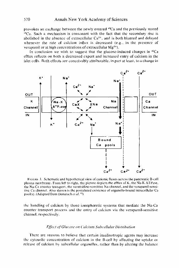
We now wish to discuss the mechanisms underlying the biphasic changes evoked by glucose in the efflux of 4sCa. Prior to such a discussion, several points ought to be stressed. First, we will not further consider the exocytotic modality for 45Ca release, a s defined above. Second, the efflux of 4sCa from the perfused islets is totally suppressed by La3+ and. therefore, originates from intracellular pool(s) (the so-called lanthanum-nondisplaceable pool6) Third, there are several reasons to believe that the effect of glucose on calcium handling is tightly de- pendent on the integrity of glucose metabolism.3fi For instance, the glucose- induced changes in 4sCa efflux are suppressed by both mannoheptulose and iodo- acetate.37 And last. we have to confess our ignorance concerning the modality by which calcium is extruded from the B-cell against its electrochemical gradient. At least two hypotheses should be considered. It is conceivable that calcium is transported across the plasma membrane from the cytosol into the interstitial fluid by an active ATP-consuming pumping process. The islets contain a Ca2+-activated A T P ~ S ~ . ~ ~ However, a s in other tissues. there is no compelling reason to believe that such a pump is located in the plasma membrane. A more likely modality for calcium extrusion is the Na-Ca counter transport mechanism, similar to that described by Bakerzs and Blaustein4"in the squid axons (FIGURE 3) .
The reduction in Y a efflux, which is the predominant effect ofglucose during the first min of exposure to this sugar. cannot be attributed to an isotopic dilution of the pool from which calcium is exported, since i t also takes place in calcium- deprived media enriched with EGTA.32 We cannot rule out the hypothesis that such a reduction in efflux is the consequence of a sudden uptake of cytosolic cal- cium by intracellular organelles, even if it seems unattractive to postulate that glu- cose initially decreases the cytosolic concentration of calcium. It is also conceiv- able that a change occurs in the affinity for calcium of some constituent of the cell boundary, leading to the local accumulation of calcium with a transient decrease in effluent radioactivity. However, since the effect ofglucose on 45Ca efflux is also observed under steady-state conditions. we favor the view that glucose somehow reduces the outward transport of calcium across the cell membrane. At this point. it should be mentioned that no effect of glucose o n 4sCa efflux is detected when the T a movement is judged from the residual amount of radioactivity recovered in the islets after various periods of incubation in the absence of radioactive cal- cium.6
The secondary rise in 4sCa efflux could also have several explanations. For instance, it could be the consequence of a change in the concentration of cyclic AMP in islet cells, since glucose may provoke a modest increase in such a concentration and since the cyclic nucleotide may, by mobilizing calcium from organelles of the vacuolar system, increase 45Ca efflux.41 However, both theophylline, a phosphodiesterase inhibitor, and imidazole, a phosphodiesterase activator, when administered throughout the period of perifusion, fail to affect the glucose-induced secondary rise in 45Ca efflux.37
Although one can conceive of other mechanisms by which glucose could eventually cause calcium mobilization from the vacuolar pool (e.g., by increasing Na+ influx), we consider that the most likely explanation for the secondary rise in 45Ca efflux is that glucose, by increasing the influx of calcium into the B-cell,
570 Annals New York Academy of Sciences
provokes an exchange between the newly entered 40Ca and the previously stored 45Ca. Such a mechanism is consonant with the fact that the secondary rise is abolished in the absence of extracellular Ca’+, and is both blunted and delayed whenever the rate of calcium influx is decreased (e.g., in the presence of verapamil or at high concentrations of extracellular Mg2+).
In conclusion we wish to suggest that the glucose-induced changes in 45Ca efflux reflects on both a decreased export and increased entry of calcium in the islet cells. Both effects are conceivably attributable, in part at least, to a change in
2. Ca 2. C a
B o u n d
I 1 I I
I I I I
+ 4
c Channel
IN
FIGURE 3 . Schematic and hypothetical view of cationic fluxes across the pancreatic 9-cell plasma membrane. From left to right, the picture depicts the efflux of K, the Na-K ATPase, the Na-Ca counter transport, the veratridine-sensitive Na channel, and the verapamil-sensi- tive Ca channel. Also shown is the postulated existence of organelle-bound intracellular Ca pool(s). (Adapted from Donatsch cf a / . 25)
the handling of calcium by those ionophoretic systems that mediate the Na-Ca counter transport process and the entry of calcium via the verapamil-sensitive channel, respectively.
EfJbct of Glucose on Culcium Sirbccllulnr Distribution
There are reasons to believe that certain insulinotropic agents may increase the cytosolic concentration of calcium in the B-cell by affecting the uptake or release of calcium by subcellular organelles, rather than by altering the balance

Malaise et al: Ca Fluxes in Pancreatic Islets 57 1
between calcium influx and efflux across the B-cell membrane. For instance, theophylline is thought to act in such a manner.4'+44 We have also recently reported that the calcium-buffering capacity of the vacuolar system in the B-cell plays an important counterregulatory role in the process by which high concentra- tions of extracellular Ca2+ are able to stimulate insulin release in the absence of any other ~ e c r e t a g o g u e . ~ ~
It is therefore an obvious question whether glucose itself may affect the subcellular distribution of calcium, as occasionally claimed.'6 It is also conceiv- able that glucose modifies the binding of calcium to certain membrane-associated or cytoplasmic proteins. To our knowledge, the sole indication that glucose may modify the handling of calcium in such a manner is the recent finding that, in dis- persed islet cells, the sugar unexpectedly decreases the fluorescent signal produced by the interaction of chloi-otetracycline with calcium presumably located in the plasma m e m b ~ a n e . ~ '
We have already mentioned that. in islets exposed to glucose, the amount of calcium accumulated in some organelles is increased. In our view, this finding does not necessarily imply that glucose modifies the transfer rate constant for calcium uptake or release by such organelles. As a matter of fact, it is possible with a theoretical model4* to reproduce the observed effect of glucose on calcium net uptake, efflux. and accumulation in the organelles. whilst keeping unchanged the transfer rate constant for calcium transport inward and outward in the vacuolar pool.
In conclusion. the possible effect of glucose to alter the subcellular distribution of calcium in the B-cell remains to be adequately explored.
E f f k t of Gli~osr on the Movctnent of Othcr Ions in Puncrccitic Islets
Although beyond the scope of the present review. it is important to note that glucose affects the fate of other ions in the islet cells. For instance, glucose provokes a sudden release of inorganic phosphate.22 decreases the concentration of CI+ (by increasing its fractional outflow rate),49 and. from preliminary data, ap- pears to increase, at least transiently, the efflux of 22Na in perfused i ~ l e t s . ~ " T h e ef- fect of glucose on K + handling is now well documented. Briefly, glucose decreases the fractional outflow rate of K f from the islet^,^^^' and, by doing so, increases its intracellular concentration. However, the inflow-outflow rate of K + is not modified, suggesting that glucose does not affect the activity of the Na+/K+ pump.5 We are impressed by the fact that the effect of glucose on K + handling consists, once again. in the modification of a passive (as distinct from active) cationic movement.
The changes evoked by glucose in the fluxes of various cations can also be evi- denced by recording the bioelectrical activity of the islet cells. The information gained by such investigations is reviewed e l s e ~ h e r e . " ~ ~ ~
In conclusion, within the limits of our present knowledge, glucose affects several cationic movements, which. in most if not all cases, correspond to passive transport processes. The earliest detectable effect of glucose occurs at the same time in all cases. It is tempting, therefore. to suggest that all these cationic changes are consequential to the generation of one unique signal. We now wish to discuss the possible nature of such a signal.

512 Annals New York Academy of Sciences
Timing of Sequential Evcnts in thc Stiinulris-Sec,retion Coupling
We have recently reviewed the evidence suggesting that the rate of glycolysis plays a central role in the identification of glucose by the B - ~ e l l . ~ ~ ~ ~ Briefly, it was shown that ( i ) under a series of experimental conditions (including exposure of islets removed from fed or fasted rats to glucose, mannose, fructose, galactose sorbitol. erythrose, and glyceraldehyde), the net uptake of “Ca increases in pro- portion to the rate of glycolysis;”6 (ii) the more marked insulinotropic action o f & - as distinct from @-D-glucose coincides with a higher rate of glycolysis (due to the stereospecific affinity of phosphoglucose isomerase for a-~-glucose-6-phosphate) and a more marked effect on both calcium efflux and uptake in the isletsg; and ( i i i ) stimulation of glycolysis from endogenous stores of glycogen is sufficient to cause calcium accumulation and insulin release even in glucose-deprived media.56
We have also indicated previously that the chronology for metabolic, cationic and secretory events in the B-cell was compatible with the view that glucose first increases the concentration of substrates (e.g., glucose) and cofactors (e.g.. NADH), then affects the flux of cations (e.g., T efflux), and last provokes insulin release.’
The timing for the stimulus-secretion coupling can also be accurately moni- tored by recording the electrical response of islet cells exposed for short periods to glucose pulses. Previous studies have shown the electrical response of the islet cells to be closely correlated with insulin secretion, suggesting that spike activity and exocytosis are two tightly associated phenomena.5”s~~s7~62 ‘The effect of the short pulses of glucose upon membrane potential is illustrated in FIGURE 4. In this figure, the upper trace shows the characteristic burst pattern recorded in the presence of glucose 1 I . I mM. Upon removal of glucose (indicated by the down- ward deflection of the reference trace), electrical activity ceases and the membrane potential increases to -SO mV. When the same cell is exposed to short pulses (2-5 sec) of glucose ( 5 5 mM), several facts are apparent: ( i ) spike activity is induced in proportion to the duration of the glucose pulse; (ii) the delay of the response is shortened after a longer pulse; ( i i i ) the electrical activity occurs in the absence of glucose, the fluid in thc perfusion chamber being indeed completely ex- changed within 2 sec after switching solutions. At closer examination, both the transient depolarization suggestive of an abortive burst of activity seen 44 sec after the 2 sec glucose pulse and the onset of spike activity after the 3 and 5 sec glucose pulses occurred after respective delays, which were a linear function of the corresponding glucose pulse duration.
The significance of the above results was documented by further investiga- tions. In control experiments. the administration of sucrose (up to 80 mM) for a few seconds up to several minutes failed to affect membrane potential or to cause electrical activity, indicating that the response to glucose cannot be ascribed to a change in osmotic pressure, which could conceivably cause a loosing of the electrically tight seal of the cell membrane around the glass electrode. We also ob- served that the magnitude of the response was related to both the concentration of glucose (for pulses of constant duration) and the duration of the pulse, so that at lower glucose concentrations, pulses of I5 to 30 sec were usually necessary to in- duce spike activity. These findings are in agreement with previous observations“:’ indicating that in the perfused rat pancreas short pulses (30-120 sec) of glucose

Malaise et al: Ca Fluxes in Pancreatic Islets 513
16.7 mM induce a delayed release of insulin in amounts that increase as a function of the pulse duration.
An experiment using short pulses in the opposite sense, i.e.. removal of glu- cose for short times during continuous, steady-state stimulation with glucose, shows that removal of glucose for a s little as 10 sec leads to a delayed inhibition of
1l.lmM Glucose 0- G
2 I
2 s [ 5 5 m M G I -70
-7 0 n 5 5
- 7 0 n - 30 5ec
FIGURE 4. Effect of short pulses of glucose ( 5 5 mM) on the membrane potential and electrical activity of an islet cell. A partially dissected mouse islet was pinned down by the surrounding pancreas into a perfusion chamber (0.2 ml). The perfusate (NaCI I10 mM. N a H C 0 3 25 mM, KCI 5 mM, CaCI, 2.5 m M , MgCI2 1 . 1 mM) was maintained at 37"C, continuously bubbled with a mixture of 0 , and CO, (9515, volivol) and perfused at a rate of 12 ml/min. The glucose pulses were administered at the time shown by the upward deflection of the reference trace, which is drawn below each record at a -70 mV potential. The method used to measure electric potentials in islet cells is described in detail
electrical activity, the inhibition occuring at a time when glucose is again present in the extracellular milieu (FIGURE 5 . ) . The duration of the inhibition is propor- tional to the length of the period of glucose deprivation. If the latter is long enough, a biphasic bioelectrical response is observed 60 sec after returning t o the 1 1 . 1 mM glucose concentration (FIGURE 5 . lower trace). Incidentally, when glu-

574 Annals New York Academy of Sciences
mV 11.1 m M GLucose 7 5 [ 0 - G I
O L U
8 s
U
15 5
O F
30 5
O F - I
I " " 4
FIGURE 5 . Effect of glucose removal for short intervals on the membrane potential and electrical activity of the same cell otherwise exposed to glucose I I . I mM. Same experi- mental design as that described for FIGURE 4. The horizontal calibration bar corresponds to 50 sec. Glucose was removed from the perfusate a t the time indicated by the downward deflections of the reference trace. which is drawn above each record at a zero membrane potential.
cose is removed from the perfusate, an immediate and slight enhancement of the spike activity is seen, either a s a shortening of the silent period between bursts (FIGURE 5 , first trace) or as an increased duration of the last burst with a higher total number of spikes (second trace).
Taken as a whole, the present experiments indicate that the glucose-induced electrical and presumably secretory response of the cell does not require the presence of glucose itself, and that the magnitude of the response depends on the amount of glucose entering the cell. These findings are compatible with the idea

Malaisse et al: Ca Fluxes in Pancreatic Islets 575
that insulin release depends on glucose metabolism. If the activation of a membrane glucoreceptor were responsible for the generation of spike activity, glucose should remain bound for at least IS sec, or several minutes. in order to explain our observations.
The Link Batrcwn Mrtaholic. and Cutionic Evrnts in thc B-Crll
We have recently completed a study of the nature of the link between meta- bolic and subsequent cationic events in the B-cell. In order to explore the possible role of reduced pyridine nucleotides in such a process, we have investigated the influence of menadione upon various parameters of islet
Menadione inhibits insulin release evoked in the rat endocrine pancreas by glu- cose or glyceraldehyde, but fails to affect the secretory response to Ca2+, Ba'+, theophylline, or gliclazide. The inhibitory effect of menadione upon glucose- induced insulin release is a dose-related, rapid, and reversible phenomenon. men- adione and glucose acting apparently as competitive antagonists. Menadione af- fects both the early and late phase of the secretory response to glucose. Men- adione also antagonizes in a dose-related fashion the ability of glucose to reduce *"b efflux, to provoke 86Rb accumulation. to cause biphasic changes in ""Ca efflux, and to stimulate 4sCa net uptake in pancreatic islets. These findings indicate that menadione impairs the insulinotropic action of glucose and other nutrients by impeding the remodeling of cationic fluxes normally provoked by these secretago- gues in islet cells. Menadione, however. does not affect the capacity of divalent cations to activate the effector system that controls the release of secretory granules.
A A
0
2o t 01 I
I I I
0 0.1 0.2 0.3 0.4
[NADH * NADPH) (pmol /islet)
FIGURE 6 . Correlation between the mean values for 4sCa net uptake and total concentra- tion of reduced pyridine nucleotides in islets incubated in the presence of various concentra- tions of glucose and menadione.

516 Annals New York Academy of Sciences
From the metabolic standpoint, the salient finding was that menadione, when used in suitable concentrations, although inhibiting insulin release, fails to affect the concentration of adenine nucleotides. the utilization of glucose, the produc- tion of lactate and pyruvate, the oxidation of [6-L4C]-glucose and the biosynthesis of proinsulin. The metabolism of glucose through the pentose pathway is increased. The sole inhibitory effect of menadione is to reduce the concentration of both NADH and NADPH. A highly significant correlation is found between the concentration of these reduced nucleotides and the net uptake of 4sC by the islets (FIGURE 6). It therefore appears that menadione, by reducing the concentration of such reduced nucleotides, uncouples the metabolism of glucose (which takes place at a normal or increased rate) from the cationic response (which is severely impaired).
A metabolic, cationic, and secretory situation comparable in several respects to that seen in the presence of menadione is encountered when NH4+ (0.5 to 5.0 mM) is added to the incubation medium. Thus, NH,+ also reduces the concentra- tion of reduced pyridine nucleotides. whilst not affecting the rate of glycolysis or the adenylate charge in the islets. The effect of glucose on 4sCa net uptake and in- sulin release is severely impaired by NH4+.6s
These and other converging observations6" raise the question as to the possible mode of action of reduced pyridine nucleotides upon cationic movements. We are presently approaching such a problem by examining the influence of oxidizing and reducing agents upon ionophore-mediated cation transport in an artificial model.24 So far, the results are compatible with the view that passive ionophoretic move- ments are indeed susceptible to be modulated by such agents.
Taken as a whole, these observations suggest that the generation and availability of reduced pyridine nucleotides play a crucial role in the coupling of metabolic to cationic events in the B-cell.
R r p l u t o r y Roles o f Ctrkiurn in Islet Cclls
In our view, the major role of calcium in the secretory sequence is to regulate mechanical events that control the migration and extrusion of secretory granule^.^' This view implies that the accumulation of calcium in the B-cell represents an obligatory step in the release process. Two series of observations support the latter postulate. First, all insulinotropic agents so far investigated for such a purpose affect the movements of calcium i n the islets. The sole exception to such a rule concerns those agents that affect insulin release by altering the func- tional capacity of the microtubular-microfilamentous effector system. In other words, the latter agents seem to modify insulin release by altering the secretory response to calcium, rather than by affecting the provision of the cation to the se- cretory machinery. Second, under a vast series of experimental conditions, the relationship between insulin release and 45Ca net uptake displays an invariable pattern. 3,Ls,68
The exact nature of the pool of calcium controlling insulin release remains open to speculations. On the basis of kinetic studies, it was recently proposed that only the lanthanum-displaceable pool, assumed to be located at the cell membrane, participates in the short-term coupling of the glucose stimulus with

Malaise et al: Ca Fluxes in Pancreatic Islets 517
secretion.” There are at least two reasons to favor the idea that the cytosolic concentration of CaZt represents a critical factor. First, whatever the nature of the primary change evoked by secretagogues in the fluxes of calcium (e.g., enhanced entry, decreased exit, or release from intracellular stores), the cytosolic compart- ment invariably represents the common carrefour for calcium accumulation. Second, i t is precisely in the cytosol that the organelles controlling the movement of secretory granules (i.e., the microtubules and the microfilamentous cell web) are located.
We have reviewed on several occasions the evidence in support of the view that the B-cell microtubular-microfilamentous system plays an essential role in in- sulin r e l e a ~ e . ~ ~ . ~ O - ~ ~ Our claim that such a system controls the movement of granules received more direct support from a recent study in which monolayer cultures of endocrine pancreatic cells were examined by time-lapse ~ inematography. ’~ . ’~ Motion analysis revealed two major types of motile events. On one hand, particles (0.3 p m in diameter). tentatively identified as secretory granules, were found to undergo back-and-forth saltatory movements along oriented pathways. The existence of such pathways was statistically validated by the large prevalence of angles close to either zero or 180 degrees between suc- cessive movements of the same particle. The movements occurred at a mean speed of 0.8 pmisec. Glucose (16.7 mM) increased the frequency of such move- ments. Vincristine (10 pM) caused a progressive inhibition of saltatory move- ments, which may depend therefore on the integrity of the microtubular ap- paratus. On the other hand. areas of the cell boundary displayed contractile-like movements, which were stimulated by insulinotropic agents such as glucose and the ionophore A23187. Cytochalasin B also affected this second type of motile event, which is thought to reflect on the activity of actin-like microfilaments. These findings suggest that the microtubular apparatus serves as a guiding cy- toskeleton for the oriented translocation of secretory granules, whereas the microfilamentous cell web may control the eventual access of the granules to exocytotic sites.
It is tempting to suggest that the regulatory role of calcium in insulin release is indeed mediated by changes in such motile events.
In addition to playing an essential role in the control of motile and secretory events, calcium may also be involved in the regulation of metabolic events in the islet cells and hence participate i n feedback control processes. According to some but not all authors, glucose, in the absence of phosphodiesterase inhibitor. increases the generation and/or concentration of cyclic AMP in islet cells (see Reference 75 for review). This effect was reported to be abolished in the absence of extracellular Ca2+.76 Under the latter experimental condition, the rate of gly- colysis is also possibly as a result of an increase in the cellular concentration of ATP.
Conclusion
The experimental data reviewed in the present report suggest that the me- tabolism of glucose, by generating reduced pyridine nucleotides, affects the affinity for cations of native ionophoretic devices located in the plasma membrane

578 Annals New York Academy of Sciences
and. possibly, other membrane systems in the pancreatic B-cell. This phenomenon apparently results, intPr aliu, in an increased influx and/or decreased efflux of calcium across the cell membrane. The resulting accumulation of calcium in a critical site of the B-cell (e.g., the cytosolic compartment) may eventually trig- ger insulin release by activating motile events involved in the migration and exocytosis of secretory granules.
A i ~ k n o r c ~ l e ~ ~ t n t n t s
The authors are indebted to Misses S. Procureur and A . Vanhaelewijck for skilled secretarial help.
I.
2.
3 .
4.
5.
6.
7.
8.
9.
10.
I I.
12.
13.
MALAISE, W. J . . A. HERCHUELZ, J . LEVY. G . SOMERS, G. DEVIS, M. RAVAZZOLA, F. MALAISE-LAGAE & L. ORCI. 1975. Insulin release and the movements of calcium in pancreatic islets. In Calcium Transport in Contraction and Secretion. E. Carafoli, F. Clementi, W. Drabikowski & A. Margreth, Eds.: 21 1-226. North-Holland Publishing Company. Amsterdam.
MALAISE, W. J . , A. SENER, A . HERCHUELZ, J . C. HUTTON,G. D ~ v 1 s . G . SOMERS. B. BLONDEL. F. MALAISE-LAGAE & L. ORCI. 1977. Sequential events in the process of glucose-induced insulin release. In Diabetes. J. S. Bajaj, Ed.: 95-102. Excerpta Medica. Amsterdam.
MALAISSE-LAGAE, F. & W. J . MALAISE. 1971. The stimulus-secretion coupling of glu- cose-induced insulin release. 111. Uptake of 45Ca by isolated islets of Langerhans. Endocrinology 88: 72-80.
MALAISSE, W. J . , J . C. HUTTON. A. S E N E R , J . LEVY, A. HERCHUELZ. G. DEWS & G. SOMERS. 1977. Calcium antagonists and islet function. VI I . Effect of calcium depriva- tion. J. Membr. Biol. 38: 193-208.
BOSCHERO, A. C.. S. KAWAZU, G. DUNCAN & W. J . MALAISE. 1977. Effect of glu- cose on K f handling by pancreatic islets. FEBS Lett. 83: 151-154.
HELLMAN, B., J . SEHLIN & I.-B. TALJEDAL. 1976. Effectsofglucose on 45Ca'+ uptake by pancreatic islets a s studied with the lanthanum method. J . Physiol. (Lond.)
HELLMAN, B., S. L E N Z E N , J. S E H L I N & I .-B. TALJEDAL. 1977. Effects of various modifiers of insulin release on the lanthanum-nondisplaceable 45Ca2+ uptake by isolated pancreatic islets. Diabetologia 13: 49-53.
MALAISSE. W. J . . A. S E N E R & J . LEVY. 1976. The stimulus-secretion coupling ofglu- cose-induced insulin release. Fasting-induced adaptation of key glycolytic enzymes in isolated islets. J . Biol. Chem. 251: 1731-1737.
MALAISE. W. J. . A. SENER. M. KOSER & A. HERCHUELZ. 1976. The stimulus-secre- tion coupling of glucose-induced insulin release. Metabolism of a- and pD-glucose in isolated islets. J. Biol. Chem. 251: 5936-5943.
H E R M A N , L.. T. SATO & C. N. HALES. 1973. The electron microscopic localization of cations to pancreatic islets of Langerhans and their possible role in insulin secretion. J . Ultrastructure Res. 42: 298-31 I .
RAVAZZOLA, M., F. MALAISE-LAGAE. M. AMHERDT. M. RAVAZZOLA. W. J . MALAISE & L. ORCI. 1976. Patterns of calcium localization in pancreatic endocrine cells. J . Cell Sci. 27: 107-1 17.
DEVIS, G., G. SOMERS, E. V A N OBBERGHEN & W. J . MALAISE. 1975. Calcium an- tagonists and islet function. I . Inhibition of insulin release by verapamil. Diabetes
SOMERS, G., G. DEvIS, E. V A N OBBERGHEN & W. J. MALAISE. 1976. Calcium an- tagonists and islet function. 11. Interaction of theophylline and verapamil. Endocri- nology99: 114-124.
2 5 4 639-656.
24: 546-551.

Malaisse et al: Ca Fluxes in Pancreatic Islets 579
14.
15.
16.
17.
18.
19.
20.
21.
22.
23.
24.
2s.
26.
27.
28
29.
30.
31 .
32.
33.
34.
35.
36.
37.
MALAISSE, w. J., A. HERCHUELZ. J. LEVY & A. SENER. 1977. Calcium antagonists and islet function. 111. The possible site of action of verapamil. Biochem. Pharmacol. 26: 735-740.
MALAISSE. W. J . , G. DEWS. D. G. PiPELI;FRs & G. SOMERS. 1976. Calcium antagonists and islet function. IV . Effect of D600. Diabetologia 12: 77-81.
MALAISSE, W. J., A. SENER, G. DEWS & G. SOMERS. 1976. Calcium antagonists and islet function. V. Effect of R3371 I . Horm. Metab. Res. 8: 434-438.
MALAISSE. W. J . 1977. Calcium antagonists and islet function. X. Effect of suloctidil. Archiv. Int. Pharmacodyn. 228: 339-344.
MALAISSE, W. J . & A. C. BOSCHERO. 1977. Calcium antagonists and islet function. XI. Effect of nifedipine. Horm. Res. 8: 203-209.
MALAISSL, W. J . , G. DEVIS, A. SENER & G. SOMERS. 1976. Calcium antagonists and islet function. VIII. The effect of magnesium. Diab. Metabol. 2: 1-4.
tagonists and islet function. VI. Effect of barium. Pflugers Archiv. 365: 21-28. HENQUIN, J.-C. & A. E. LAMBER’r. 1975. Cobalt inhibition of insulin secretion and cal-
cium uptake by isolated rat islets. Amer. J . Physiol. 228: 1669-1677. FREINKFL. N . , C. E. YouNsi. J . BONNAR & R. M. C. DAWSON. 1974. Rapid transient
efflux of phosphate ions from pancreatic islets a s an early action of insulin secretago- gues. J . Clin. Invest. 54: 1179-1 189.
SOMERS. G.. G. DEWS & W. J. MALAISSE. 1976. Analogy between native and exogenous ionophores in the pancreatic B-cell. FEBS Lett. 66: 20-22.
MALAISSE, W. J . , G. DEWS & G. SOMERS. 1977. Inhibition by verapamil of ionophore- mediated calcium translocation. Experientia. 33: 1035-1036.
DONATSCH. P.. D. A. LOWE, B. P. RICHARDSON & P. TAYLOR. 1977. The functional significance of sodium channels in pancreatic beta-cell membranes. J . Physiol. (London) 257: 367-376.
F R A N K E L . B. J.. J . A . KROMHOUT. H . D. L A N D A H L & G. M. GRODSKY. 1977. 4sCa2+ uptake and insulin release from collagenase isolated rat islets. (Abstract). Diabetes 26 (Suppl. 1):381.
SHARP. 1975. Calcium-induced insulin release in monolayer culture of the endocrine pancreas. J. Biol. Chem. 250: 1354-1360.
CHARLES, M. A,. J. LawEcKr. R. P i c n T Cli G. M. GRODSKY. 1975. Insulin secretion. Interrelationships of glucose. cyclic adenosine-3’:5’-monophosphate. and calcium. J . B i d . Chem. 250: 6134-6140.
K A R L , R. c., w. s. ZAWALICH, J. A. F E R R ~ N D E L L ~ & F. M. MATSCHINSKY. 1975. The role of Caz+ and cyclic adenosine-3’:5’-monophosphate in insulin release induced in vitro by the divalent cation ionophore A23187. J . Biol. Chem. 250: 4575-4579.
HELLMAN. B. 1975. Modifying actions of calcium ionophores on insulin release. Biochim. Biophys. Acta399: 157-169.
ASHBY, J . P. & R. N. SPEAKt. 1975. Insulin and glucagon secretion from isolated islets of Langerhans. The effects of calcium ionophores. Biochem. J . 150: 89-96.
MALAISSE, W. J . , G. R. BRISSON & L. E. BAIRD. 1973. The stimulus-secretion coupl- ing of glucose-induced insulin release. X . Effect of glucose on 45Ca efflux from perifused islets. Amer. J. Physiol. 224: 389-394.
HELLMAN, J . StHLrN & 1.-B. TALJEDAI.. 1971. Calcium uptake by pancreatic B-cells a s measured with the aid of 4sCa and mannitolb3H Amer. J . Physiol. 224: 389-394.
HERCHUELZ. A . & W. J. MALAISE. 1977. The dual action ofglucose on calcium move- ments in the B-cell: Cause or consequence of insulin release? (Abstract) Diabetologia 13: 401.
KIKUCHI, M., G. S. CUENDtT, A. E. RESOLD & G. W. G. SHARP. 1976. Kinetic study of glucose effect on 45Cai+ efflux and insulin release from 46 hour-preloaded rat islets at different temperatures and Ca2+ concentrations. (Abstract). Diabetologia 12: 402.
SENER, A , , J . L E V Y & W. J. MALAISSE. 1976. The stimulus-secretion couplingofglu- cose-induced insulin release. Does glyclolysis control calcium transport in the B-cell? Biochem. J . 156: 521-525.
S O M E R S , G., G. DEWS. E. V A N OBBERGHEN & w . J. MALAISSE. 1976. Calcium an-
WOLLHEIM. C. B.. B. BLONDEL, P. A. TRUEHEART, A. E. RENOLD & G. W. G.
HERCHUELZ. A. & W. J. MALAISE. Unpublished observations.

580 Annals New York Academy of Sciences
38.
39.
40.
41.
42.
43.
44.
45.
46.
47.
48.
49.
50. 51.
52.
53.
54.
55.
56.
57.
58.
59.
60.
61.
FORMBY, B., K. CAPITO, J . EGEBERG & C. J. HEDESKOV. 1976. Ca-activated ATPase activity in subcellular fractions of mouse pancreatic islets. Amer. J . Physiol.
BAKER, P. F. & W. SCHLAEPFER. 1975. Calcium uptake by axoplasm extruded from giant axons of Loligo. J. Physiol. (Lond.) 249: 37-38P.
BLAUSTEIN, M. P. 1974. The interrelationship between sodium and calcium fluxes across cell membranes. Rev. Physiol. Biochem. Pharmac. 70: 33-82.
BRISSON, G. R. & W. J. MALAISE. 1973. The stimulus-secretion coupling of glucose- induced insulin release. XI. Effects of theophytline and epinephrine on 45Ca2+ efHux from perifused islets. Metabolism 22: 445-465.
BRISSON, G. R., F. MALAISSE-LAGAE & W. J. MALAISE. 1972. The stimulus-secretion coupling of glucose-induced insulin release. VII. A proposed site of action for adenosine-3’,5’-cyclic monophosphate. J . Clin. Invest. 51: 232-24 I .
HOWELL, S. L., W. MONTAGUE & M. TYHURST. 1975. Calcium distribution in islets of Langerhans: A study of calcium concentrations and of calcium accumulation in B cell organelles. J . Cell Sci. 19: 395-409.
SEHLIN, J. 1976. Calcium uptake by subcellular fractions of pancreatic islets. Effects of nucleotides and theophylline. Biochem. J . 156: 63-69.
DEVIS, G. , G. SOMERS & W. J . MALAISE. 1977. Dynamics of calcium-induced insulin release. Diabetologia 13: 53 1-536.
SCHAFER. H.-J. & G. KLOPPEL. 1974. The significance of calcium in insulin secretion. Ultrastructural studies on identification and localization of calcium in activated and inactivated B cellsofmice. Virchows Arch. Abt. A Path. Anat. 362: 231-245.
TALJEDAL, I.-B. 1977. Fluorescent probing of calcium in pancreatic islet cells. Abstracts I Ith FEBS Meeting : 10.
MALAISSE, W. J . & D. G. PIPELEERS. 1974. The role of cations in insulin synthesis and release. I n Diabetes. W. J . Malaisse & J . Pirart, Eds.: 95-10, Excerpta Medica. Am- sterdam.
SEHLIN, J . 1977. Chloride Huxes and distribution in isolated pancreatic islets. Abstracts 1 Ith FEBS Meeting : I I .
BOSCHERO, A. C. & W. J. MALAISSE. Unpublished observations. SEHLIN, J . & I.-B. TALJEDAL. 1974. Transport of rubidium and sodium in pancreatic
islets. J . Physiol. (London) 242: 505-515. MATTHEWS, E. K. 1975. Calcium and stimulus-secretion coupling in pancreatic islet
cells. I n Calcium transport in contraction and secretion. E. Carafoli. F. Clementi, W. Drabikowski & A. Margreth. Eds.: 203-210. North-Holland Publishing Company. Amsterdam.
MEISSNER. H. P. 1976. Electrical characteristics of the beta-cells in pancreatic islets. J. Physiol. (Paris) 72: 757-767.
ATWATER, I . & P. M. BEIGELMAN. 1976. Dynamic characteristics of electrical activity in pancreatic B-cells. I. Effects of calcium and magnesium removal. J. Physiol. (Paris) 72: 769-786.
MALAISSE, W. J. 1976. The possible link between glycolysis and insulin release in isolated islets. In Diabetes Research Today. E. Lindenlaub. Ed.: 191-206. F. K. Schattauer Verlag. New York.
MALAISE, W. J., A. SENE.R, M. KOSE.R, M. RAVAZOLLA & F. MALAISSE-LAGAE. 1977. The stimulus-secretion coupling of glucose-induced insulin release. Insulin release due to glycogenolysis in glucose-deprived islets. Biochem. J. 164: 447-454.
DEAN, P. M. & E. K. MATTHEWS. 1970. Glucose-induced electrical activity in pancreatic islet cells. J . Physiol. (Lond.) 210: 255-264.
DEAN, P. M. & E. K. MATTHEWS. 1970. Electrical activity in pancreatic islet cells: Ef- fects of ions. J. Physiol. (Lond.) 210: 265-275.
PACE, C. S. & S. PRICE. 1972. Electrical responses of pancreatic islet cells to secretory stimuli. Biochem. Biophys. Res. Commun. 46: 1557-1563.
MEISSNER, H. P. & H. SCHMELZ. 1974. Membrane potential of @cells in pancreatic islets. PHiigers Arch. 351: 195-206.
MEISSNER. H. P. & I. ATWATER. 1975. Further evidence for the correlation between insulin secretion and electrical activity of single pcells. PHiigers Arch. 355: RS6.
230: 44 1-448.
‘
Malaisse et al: Ca Fluxes in Pancreatic Islets 58 1
62.
63.
64.
65.
66.
67.
68.
69.
70.
71.
72.
73.
74.
7s.
76.
77.
BEIGELMAN. P. M., B. RIBALET & I. ATWATER. 1977. Electrical activity of pancreatic p-cells. Effects of glucose and arginine. J . Physiol. (Paris). In press.
BENNETT, L . L.. D. L . CURRY & K. CURRY. 1973. Differences in insulin release in response to glucose and tolbutamide stimulation. Proc. Soc. Exp. Biol. Med.
MALAISE, W. J. , A. SENER, S. KAWAZU & A. C. BOSCHERO. 1977. Nature of the cou- pling between metabolic and cationic events in pancreatic islets. (Abstract). Dia- betologia 13: 417.
DEWS, G., J . C. HUTTON, A. SENER, A. HERCHUELZ, G. SOMERS & W. J . MALAISSE. 1977. Ammonium accumulation and its inhibitory effect in pancreatic islets. (Abstract). Diabetologia 13: 389.
HELLMAN, B., L.-A. IDAHL, A. LERNMARK, J . SEHLIN & I.-B. TAWEDAL. 1974. Membrane sulphydryl groups and the pancreatic beta cell recognition of insulin secretagogues. In Diabetes. W . J . Malaisse & J . Pirart, Eds.: 65-78 Excerpta Medica. Amsterdam.
MALAISE, W. J., F. MALAISE-LAGAE, E. V A N OBBERGHEN. G. SOMERS. G. DEW, M. RAVAZZOLA & L. ORCI. 1975. Role of microtubules in the phasic pattern of insulin release. Ann. N. Y. Acad. Sci. 253: 630-652.
MALAISSE-LAGAE, F., G. R. BRISSON & W. J . MALAISE. 1971. The stimulus-secretion coupling of glucose-induced insulin release. vI. Analogy between the insulinotropic mechanisms of sugars and amino acids. Horm. Metab. Res. 3: 374-378.
HELLMAN, B.. J . SEHLIN & I.-B. TALJEDAI.. 1976. Calcium and secretion: Distinction between two pools of glucose-sensitive calcium in pancreatic islets. Science
LACY, P. E. & W. J . MALAISSF.. 1973. Microtubules and beta cell secretion. Recent Progr. Horm. Res. 29: 199-221.
MALAISE, W. J . . V . LECLERVQ-MEYER, E. V A N OBBERGHEN. G. SOMERS. G. DEWS, M. RAVAZZOLA, F. MALAISE-LAGAE & L. ORCI. 1975. The role of the microtubular- microfilamentous system in insulin and glucagon release by the endocrine pancreas. In Microtubules and Microtubule Inhibitors. M. Borgers & M. De Brabander. Eds.: 143-152. North-Holland Publishing Company. Amsterdam.
MALAISSE. W. J . , G. DEWS. G. SOMERS, E. V A N OBBERGHEN, F. MALAISE-LAGAE, M. RAVAZZOLA. B. BLONDEL & L. ORCI. 1976. The role of microtubules and microfilaments in secretion. In Stimulus-secretion coupling in the gastrointestinal tract. R. M. Case & H. Goebell. Eds.: 65-76 MTP Press. Lancaster.
SOMERS, G.. B. BLONDEL. L. ORCI & W . J . MALAISE. 1978. Motile events in pancreatic endocrine cells. Endocrinology. In press.
LACY, P. E.. E. H. FINKE & R. C. CODELLA. 1975. Cinematographic studies of p granule movement in monolayer culture of islet cells Lab. Invest. 33: 570-576.
CERASI, E. & V. GRILL. 1976. Islet cyclic AMP and the control ofinsulin secretion. In Diabetes Research Today. E. Lindenlaub, Ed.: 223-240. F. K. Schattauer Verlag. New York.
ZAWALICH, W. S., R. C. KARL, J . A. FERRFNDELLI & F. M. MATSCHINSKY. 1975. Factors governing glucose-induced elevation of 3':5'-AMP levels in pancreatic islets. Diabetologia 11: 231-235.
HELLMAN, B., L.-A. IDAHL, A. LERNMARK, J . SEHLIN & I.-B. TALJEDAL. 1974. The pancreatic p-cell recognition of insulin secretagogues. Effects of calcium and sodium on glucose metabolism and insulin release. Biochem. J . 138: 33-45.
144(2): 436-439.
194: 1421-1423.
- DISCUSSION OF THE PAPER
C . C. ASHLEY (University of Oxford, Oxford, England): I am not certain how you performed your 45Ca experiments. Could , however , your l a g in *"a efflux be caused by diffusion and unstirred layer effects in the extracellular space of your preparation?
582 Annals New York Academy of Sciences
W. J . M.4LAISSE: 1 doubt that such is the case. First, the results are corrected for the dead space of the perfusion system. Second, there is an immediate effect of glucose on “Ca efflux, namely, to decrease it. Last, it is possible to obtain a more rapid secondary rise in 45Ca efflux by using a substimulatory glucose concentra- tion (e.g.. 2.8 mM) prior to introduction of the high glucose concentration (i.e.. 16.7 mM).
E. CARAFOLI (Sli,iss Fcdcral Iristitrrte of Tei,hnology, Zurich, Sii,itzi,rlnnd): Would you please comment on the recent observation by Lowe c’t c r l . (1976. Na- ture) that insulin production can be induced in isolated islets of Langerhans in Ca2+-free media, when Na+ penetration inside the cell is promoted by veratridine.
W. J . MALAISE: The importance of these findings should not be overlooked. They confirm previous indications that sufficient calcium may be mobilized from the organelle-bound pool to stimulate insulin release in the absence of ex- tracellular calcium. It remains to be seen, however. whether a Na+-induced mobi- lization of intracellular Ca’+ plays a role in the normal process of glucose-induced insulin release.
W. R. LOEWENSTEIN (Uniwrsi ty o,f M i c r n i i . Miami, Flm.): Does one get a stimulatory action in your cell system under conditions of prolonged exposure to Ca-free Na medium? The reason for my question is that. provided that the [Ca’+]- dependence of Na efflux shown by Baker. Blaunstein & Hodgkin for nerve membrane applies also to your cell, one may expect an elevation of “a], and, hence. a release of Ca2+ from intracellular stores.
W. J . MALAISE: I am not aware that this kind of experiment has been carried Out.
R . N E H E R (Frirdrick MicJschcr Instituti,, Bciscl, Sli,itzcrltrnd): What is the ef- fect of pyruvate on your islet cell system in absence of glucose’?
W. J . MALAISSE: In islets from adult rats. pyruvate does not stimulate insulin release in the absence of glucose. However, pyruvate causes a dose-related shift to the left of the sigmoidal curve relating insulin output to glucose concentration. Such a behavior coincides with the effect of pyruvate on reduced pyridine nu- cleotides in the islets.
B. FRANKEL (Univrrsity of C a l i f ~ m i c i . Sun Frcinrisco. Calif.): Please restate your ideas with respect to glucose-stimulated changes in potassium or rubidium movements.
W. J . MALAISSE: Glucose decreases the plasma membrane permeability to effluent K f and, by doing s o . increases the concentration of K t in the islets. Glu- cose. however, fails to affect the inflowioutflow rate of K + . We have considered the possible role of Ca2+ in the regulation of Kf permeability. Glucose also decreases the permeability to effluent K’ in the absence of extracellular Ca2+. As a unitarian hypothesis, we like to believe that reduced pyridine nucleotides may affect the affinity for K-of the ionophoretic channel mediating K + efflux.