Regulated Expression of Sodium-dependent Glutamate Transporters and Synthetase: a Neuroprotective...
12
Gabriel Gras 1 ; Fabrice Chrétien 1, 2, 3 ; Anne-Valérie Val- lat-Decouvelaere 3 ; Gwenaelle Le Pavec 1 ; Fabrice Porcheray 1 ; Christophe Bossuet 1 ; Cathie Léone 1 ; Patricia Mialocq 1 ; Nathalie Dereuddre-Bosquet 4 ; Pas- cal Clayette 4 ; Roger Le Grand 1 ; Christophe Crémi- non 5 ; Dominique Dormont 1 ; Anne-Cécile Rimaniol 1 ; Françoise Gray 3 1 CEA, Service de Neurovirologie, DSV/DRM, Centre de Recherches du Service de Santé des Armées, EPHE, IPSC, 92265 Fontenay aux Roses cedex, France. 2 INSERM EMI-00-11, Faculté de Médecine de Créteil, Univer- sité Paris XII, Créteil, France. 3 Laboratoire de Neuropathologie, Hôpital Raymond Poincaré, Faculté de Médecine Paris, Ile de France Ouest, Garches, France. 4 Société de Pharmacologie et Immunologie-BIO, Massy, France. 5 Laboratoire d’Etudes Radio-Immunologiques, CEA, DSV/ DRM/SPI, Saclay, France. It is now widely accepted that neuronal damage in HIV infection results mainly from microglial activation and involves apoptosis, oxidative stress and gluta- mate-mediated neurotoxicity. Glutamate toxicity acts via 2 distinct pathways: an excitotoxic one in which glutamate receptors are hyperactivated, and an oxidative one in which cystine uptake is inhibited, resulting in glutathione depletion and oxidative stress. A number of studies show that astrocytes normally take up glutamate, keeping extracellular glutamate concentration low in the brain and pre- venting excitotoxicity. This action is inhibited in HIV infection, probably due to the effects of inflammato- ry mediators and viral proteins. Other in vitro stud- ies as well as in vivo experiments in rodents follow- ing mechanical stimulation, show that activated microglia and brain macrophages express high affinity glutamate transporters. These data have been confirmed in chronic inflammation of the brain, particularly in SIV infection, where activated microglia and brain macrophages also express glu- tamine synthetase. Recent studies in humans with HIV infection show that activated microglia and brain macrophages express the glutamate transporter EAAT-1 and that expression varies according to the disease stage. This suggests that, besides their rec- ognized neurotoxic properties in HIV infection, these cells also have a neuroprotective function, and may partly make up for the inhibited astrocytic function, at least temporarily. This hypothesis might explain the discrepancy between microglial activation which occurs early in the disease, and neuronal apoptosis and neuronal loss which is a late event. In this review article, we discuss the possible neuro- protective and neurotrophic roles of activated microglia and macrophages that may be generated by the expression of high affinity glutamate trans- porters and glutamine synthetase, 2 major effectors of glial glutamate metabolism, and the implications for HIV-induced neuronal dysfunction, the underlying cause of HIV dementia. Brain Pathol 2003;13:211-222. Introduction The mechanisms underlying neuronal dysfunction in HIV associated dementia are still a matter of study and debate. However, it is widely accepted that infection and activation of macrophages and microglial cells play a key role in HIV-induced neurotoxicity, both directly through viral neurotoxin production and indirectly through immune activation and subsequent release of neu- rotoxic factors. The mechanism of neuronal damage is not fully understood but involves glutamate-related excitotoxicity and oxidative stress resulting in neuronal apoptosis (for review, see 57), a mechanism that is not specific for HIV infection. One paradox of HIV infection is the contrast between the intense microglial activation associated with neurotoxin production found early in the disease, even in the asymptomatic pre-AIDS stage (4, 94), and the neuronal loss that occurs at the end stage of the disease (for review, see 34) and which is not found in asympto- matic HIV-infected individuals (33). Similarly, although occasional apoptotic neurones have been observed in a few pre-AIDS cases (2, 5, 33), apoptotic neuronal death mostly occurs at the late stage of the disease (3). SYMPOSIUM: Neuro-AIDS Part II Regulated Expression of Sodium-dependent Glutamate Transporters and Synthetase: a Neuroprotective Role for Activated Microglia and Macrophages in HIV Infection? Corresponding author: Gabriel Gras, PhD, Laboratoire de Neuro-Immuno-Virologie, Service de Neurovirologie, CEA, DSV/DRM, BP 6, 18 route du Panora- ma, 92265 Fontenay aux Roses cedex, France (e-mail: [email protected])
-
Upload
independent -
Category
Documents
-
view
1 -
download
0
Transcript of Regulated Expression of Sodium-dependent Glutamate Transporters and Synthetase: a Neuroprotective...

Gabriel Gras 1; Fabrice Chrétien 1, 2, 3; Anne-Valérie Val-lat-Decouvelaere 3; Gwenaelle Le Pavec 1; FabricePorcheray 1; Christophe Bossuet 1; Cathie Léone 1;Patricia Mialocq 1; Nathalie Dereuddre-Bosquet 4; Pas-cal Clayette 4; Roger Le Grand 1; Christophe Crémi-non 5; Dominique Dormont 1; Anne-Cécile Rimaniol 1;Françoise Gray 3
1 CEA, Service de Neurovirologie, DSV/DRM, Centre deRecherches du Service de Santé des Armées, EPHE, IPSC,92265 Fontenay aux Roses cedex, France.
2 INSERM EMI-00-11, Faculté de Médecine de Créteil, Univer-sité Paris XII, Créteil, France.
3 Laboratoire de Neuropathologie, Hôpital Raymond Poincaré,Faculté de Médecine Paris, Ile de France Ouest, Garches,France.
4 Société de Pharmacologie et Immunologie-BIO, Massy,France.
5 Laboratoire d’Etudes Radio-Immunologiques, CEA, DSV/DRM/SPI, Saclay, France.
It is now widely accepted that neuronal damage inHIV infection results mainly from microglial activationand involves apoptosis, oxidative stress and gluta-mate-mediated neurotoxicity. Glutamate toxicityacts via 2 distinct pathways: an excitotoxic one inwhich glutamate receptors are hyperactivated, and anoxidative one in which cystine uptake is inhibited,resulting in glutathione depletion and oxidativestress. A number of studies show that astrocytesnormally take up glutamate, keeping extracellularglutamate concentration low in the brain and pre-venting excitotoxicity. This action is inhibited in HIVinfection, probably due to the effects of inflammato-ry mediators and viral proteins. Other in vitro stud-ies as well as in vivo experiments in rodents follow-ing mechanical stimulation, show that activatedmicroglia and brain macrophages express highaffinity glutamate transporters. These data havebeen confirmed in chronic inflammation of thebrain, particularly in SIV infection, where activatedmicroglia and brain macrophages also express glu-tamine synthetase. Recent studies in humans with HIVinfection show that activated microglia and brainmacrophages express the glutamate transporterEAAT-1 and that expression varies according to the
disease stage. This suggests that, besides their rec-ognized neurotoxic properties in HIV infection,these cells also have a neuroprotective function,and may partly make up for the inhibited astrocyticfunction, at least temporarily. This hypothesis mightexplain the discrepancy between microglial activationwhich occurs early in the disease, and neuronalapoptosis and neuronal loss which is a late event. Inthis review article, we discuss the possible neuro-protective and neurotrophic roles of activatedmicroglia and macrophages that may be generated bythe expression of high affinity glutamate trans-porters and glutamine synthetase, 2 major effectorsof glial glutamate metabolism, and the implicationsfor HIV-induced neuronal dysfunction, the underlyingcause of HIV dementia.
Brain Pathol 2003;13:211-222.
IntroductionThe mechanisms underlying neuronal dysfunction in
HIV associated dementia are still a matter of study anddebate. However, it is widely accepted that infectionand activation of macrophages and microglial cells playa key role in HIV-induced neurotoxicity, both directlythrough viral neurotoxin production and indirectlythrough immune activation and subsequent release of neu-rotoxic factors. The mechanism of neuronal damage isnot fully understood but involves glutamate-relatedexcitotoxicity and oxidative stress resulting in neuronalapoptosis (for review, see 57), a mechanism that is notspecific for HIV infection.
One paradox of HIV infection is the contrastbetween the intense microglial activation associatedwith neurotoxin production found early in the disease,even in the asymptomatic pre-AIDS stage (4, 94), and theneuronal loss that occurs at the end stage of the disease(for review, see 34) and which is not found in asympto-matic HIV-infected individuals (33). Similarly,although occasional apoptotic neurones have beenobserved in a few pre-AIDS cases (2, 5, 33), apoptoticneuronal death mostly occurs at the late stage of thedisease (3).
SYMPOSIUM: Neuro-AIDS Part II
Regulated Expression of Sodium-dependentGlutamate Transporters and Synthetase: aNeuroprotective Role for Activated Microglia andMacrophages in HIV Infection?
Corresponding author:Gabriel Gras, PhD, Laboratoire de Neuro-Immuno-Virologie, Service de Neurovirologie, CEA, DSV/DRM, BP 6, 18 route du Panora-ma, 92265 Fontenay aux Roses cedex, France (e-mail: [email protected])

A number of recent in vitro and in vivo studiesshowed that activated macrophages and microglial cells(AMM) may express high affinity glutamate trans-porters and glutamine synthetase in a variety of patho-logical situations, including CNS inflammation. Thissuggests that besides their neurotoxic role, AMM mayexert a neurotrophic role, by producing and providing glu-tamine, and a neuroprotective role, by clearing theextracellular glutamate and counteracting oxidativestress. This hypothesis might explain the absence ofcorrelation between microglial activation and neuronaldamage at different stages of HIV infection. It may alsoexplain the variable course of cognitive dysfunction inpatients receiving highly active antiretroviral therapy(HAART). Some changes may be reversible, as sug-gested by recent reports of improved neuropsychologi-cal performance in patients receiving HAART (90, 91).However, other studies suggest that these treatmentshave a lesser impact on HIV encephalitis (HIVE) than on
other AIDS-defining illnesses (29). The poor CNS pen-etration of many antiretroviral agents is one possibleexplanation. It is also likely that some HIV-inducedCNS changes may not be reversible (49), whereas, in oth-ers, neuroprotection may occur.
The present review considers the different in vitro andin vivo data supporting that hypothesis, focusing on a pos-sible neuroprotective role for activated macrophages/microglia in HIV infection.
The Physiological Glutamate-Glutamine Cycle in theCentral Nervous System
Glutamate is the major excitatory neurotransmitter inthe central nervous system (CNS) (41), and is releasedfrom glutamatergic neurone vesicles by a calcium-dependent mechanism (for review, see 38). Its concen-tration in the synaptic cleft must be kept low because highor sustained activation of glutamate receptor and theresulting calcium influx may induce neuronal death, amechanism called excitotoxicity (86, 87). Extracellularconcentration of glutamate is regulated by a family oftransporter proteins called excitatory amino-acid trans-porters (EAAT). To date these include five cloned sub-types (6, 36, 55, 80, 96, 99). EAAT1 and EAAT2 wereprimarily observed in astrocytes, EAAT3 is a neuronaltransporter with a somatodendritic location (89),EAAT-4 is expressed in the cerebellum (36) and EAAT-5 in the retina (6). These transporters use the Na+ and K+
electrochemical gradients as a driving force to take upextracellular glutamate against a several thousand-foldconcentration gradient, leading to an extracellular con-centration below 1 �M and ensuring a high signal-to-noise ratio for glutamate receptors (for review, see 47).EAAT gene knockout experiments show that theastroglial transporters EAAT1 and EAAT2 are essentialfor protection against excitotoxicity, by clearing extra-cellular glutamate, whereas the neuronal transporterEAAT3 is not (88, 100). Thus, astrocytes are the mainprotectors of neurones from excitotoxicity by extracel-lular glutamate clearance in the normal CNS.
At the same time, the extracellular glutamate level isproportional to intracellular glutamate concentration(for review, see 7) and glutamate metabolism withinglutamate-scavenging cells is vital to prevent excito-toxicity. Glutamate is rapidly converted into glutamineby glutamine synthetase, an astrocyte-expressedenzyme (64, 83) localized in the immediate vicinity ofglutamatergic synapses (26-28, 76, 77). Glutamine syn-thesis is one component of astrocyte neurotrophic prop-erties, and glutamine is provided to neurones through dif-ferent transporter systems, permitting glutamate traffic
212 Sodium-dependent Glutamate Transporters and Glutamine Synthetase—Gras et al
Figure 1. The glutamate-glutamine cycle in the normal CNS.Vesicular glutamate is released from the presynaptic gluta-matergic neurone into the synaptic cleft, by Ca-dependentfusion of vesicle and neurone membranes (1). Extracellularglutamate activates glutamate receptors on the postsynaptic neu-rone (2). Sodium-dependent high affinity glutamate trans-porters (EAATs) clear glutamate from the synapse (3). Astrocyteclearance efficacy is higher than that of neurones. In the astro-cyte, glutamate is a substrate for glutamine production via glu-tamine synthetase (4). The non neuroactive amino-acid gluta-mine is then provided to neurones via different neutralamino-acid transporters (5). The presynaptic neurone restoresits vesicular pool of glutamate by converting glutamine to glu-tamate via the mitonchondrial enzyme glutaminase (6).

in the extracellular compartment in a non-neuroactiveform (glutamine) (for review, see 16). Neurones thenreconstitute their vesicular pool of glutamate byhydrolysing glutamine into glutamate and ammonia viathe mitochondrial phosphate-dependent glutaminase(for review, see 25). This highly simplified scheme isillustrated in Figure 1 but does not take into account eitherthe need for external nitrogen inclusion in the cycle, orthe fact that enzymatic oxidation of glutamate may alsofuel the Krebs cycle in astrocytes and neurones.
Glutamate Metabolism and Oxidative Stress areClosely Related
The tripeptide glutathione GSH (�-glutamyl-cys-teinyl-glycine) is the major cellular antioxidant, and hasa critical activity in brain, which accounts for about20% of oxygen consumption in humans (24). GSH-mediated reduction of radicals and peroxides does not useup GSH (Figure 2), but its role in xenobiotic detoxifi-cation, in cytosolic protein sulhydryl group reduction, andits export from the cell, cause GSH depletion andrequire efficient GSH synthesis for replacement (forreview, see 31).
Glutamate and glutathione (GSH) metabolism have anintricate refined interaction. GSH synthesis is a 2-stepreaction; �-glutamyl-cysteine synthetase first producesthe �-glutamyl-cysteine dipeptide (this enzymatic reac-tion is the limiting one), before glutathione synthetaseuses �-glutamyl-cysteine and glycine as substrates forGSH production. Glutathione metabolism is illustratedin Figure 2. The limiting extracellular substrate forGSH synthesis is cystine. In macrophages cystine ismainly taken up through the sodium independent,CD98/xCT cystine-glutamate antiporter (xc- transport)(85), and not through EAATs as observed by Bender etal (13) in neonatal astrocytes and by Flynn and McBean(40, 65) in synaptosomes. The cystine-glutamateantiporter takes up extracellular cystine in exchange forintracellular glutamate secretion (8), and the reaction canoccur in either direction. Consequently, extracellularcystine and glutamate competitively inhibit each otherfor the use of the cystine-glutamate antiporter (21, 56).The intracellular glutamate pool is large but dynamic (10,53) and is rapidly depleted in EAAT-negative cells in thepresence of cystine (10). Intracellular glutamate istherefore unavailable as a GSH precursor, and secretedglutamate may competitively inhibit cystine uptake(10), further inducing GSH depletion. Also, GSHdepletion due to cystine starvation or oxidative stress(exposure to nitric oxide [NO] or peroxinitrite) inducesan adaptive response with increased cystine uptake (11,
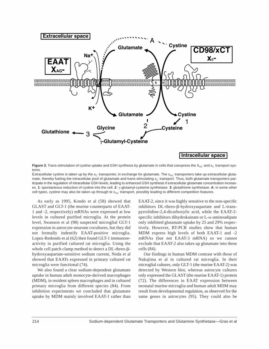
17, 61), a mechanism that may accelerate depletion of theglutamate pool. In macrophages, TNF� is another acti-vator of the xc- transport (84). In many cell types, anexcess of extracellular glutamate inhibits cystineuptake, rapidly depletes GSH, and causes cell degener-ation and death. This non-excitotoxic mechanism ofglutamate toxicity is called oxidative glutamate toxici-ty (9, 20, 21, 42, 50, 52, 56, 63, 71, 92). Another exam-ple of the intimate links between glutamate transport andoxidative stress mechanism is that oxidative metabo-lism directly regulates EAAT function (68, 70, 79, 101).In this context, EAAT-mediated glutamate uptakeenables a high glutamate concentration gradient to bemaintained through the cell membrane in response to anextracellular rise, stimulating the xc- system and leadingto enhanced cystine uptake and GSH synthesis, eventhough competition for uptake occurs (Figure 3). Thismechanism has been demonstrated in both Müller cells(82) and macrophages (85).
Activated Macrophages and Microglia Express Exci-tatory Amino Acid Transporters In Vitro
213Sodium-dependent Glutamate Transporters and Glutamine Synthetase—Gras et al
Figure 2. Glutathione metabolism. GSH is involved in oxidativemetabolism (upper part of the figure), and this activity does notconsume GSH. R°, RH: free radical and reduced form, ROOH,ROH: peroxide and reduced form, A: GSH-peroxidase, B:GSH-reductase.GSH is involved in xenobiotic detoxification and endogeneousprotein thiol groups reduction (lower right part of the figure), aGSH-consuming activity.X: xenobiotic or SH-group-bearing protein. Acc: acceptor for �-glutamyl moiety, 3: S-transferase, 4: �-glutamyl transferase.GSH synthesis is achieved by a 2-step process (lower left partof the figure). Glutamate is not recycled; cysteine and glycinemay be recycled via dipeptidase activity.1: �-glutamyl-cysteine synthetase, 2: glutathione synthetase, 5:dipeptidase.
As early as 1995, Kondo et al (58) showed thatGLAST and GLT-1 (the murine counterparts of EAAT-1 and -2, respectively) mRNAs were expressed at lowlevels in cultured purified microglia. At the proteinlevel, Swanson et al (98) suspected microglial GLT-1expression in astrocyte-neurone cocultures, but they didnot formally indentify EAAT-positive microglia.Lopez-Redondo et al (62) then found GLT-1 immunore-activity in purified cultured rat microglia. Using thewhole cell patch clamp method to detect a DL-threo-�-hydroxyaspartate-sensitive sodium current, Noda et alshowed that EAATs expressed in primary cultured ratmicroglia were functional (74).
We also found a clear sodium-dependent glutamateuptake in human adult monocyte-derived macrophages(MDM), in resident spleen macrophages and in culturedprimary microglia from different species (84). Frominhibition experiments we concluded that glutamateuptake by MDM mainly involved EAAT-1 rather than
EAAT-2, since it was highly sensitive to the non-specificinhibitors DL-threo-�-hydroxyaspartate and L-trans-pyrrolidine-2,4-dicarboxylic acid, while the EAAT-2-specific inhibitors dihydrokainate or L-�-aminoadipateonly inhibited glutamate uptake by 25 and 29% respec-tively. However, RT-PCR studies show that humanMDM express high levels of both EAAT-1 and -2mRNAs (but not EAAT-3 mRNA) so we cannotexclude that EAAT-2 also takes up glutamate into thesecells (84).
Our findings in human MDM contrast with those ofNakajima et al in cultured rat microglia. In theirmicroglial cultures, only GLT-1 (the murine EAAT-2) wasdetected by Western blot, whereas astrocyte culturesonly expressed the GLAST (the murine EAAT-1) protein(72). The differences in EAAT expression betweenneonatal murine microglia and human adult MDM mayresult from developmental regulation, as observed for thesame genes in astrocytes (95). They could also be
214 Sodium-dependent Glutamate Transporters and Glutamine Synthetase—Gras et al
Figure 3. Trans-stimulation of cystine uptake and GSH synthesis by glutamate in cells that coexpress the XAG- and xC
- transport sys-tems.Extracellular cystine is taken up by the xC
- transporter, in exchange for glutamate. The xAG- transporters take up extracellular gluta-
mate, thereby fueling the intracellular pool of glutamate and trans-stimulating xC- transport. Thus, both glutamate transporters par-
ticipate in the regulation of intracellular GSH levels, leading to enhanced GSH synthesis if extracellular glutamate concentration increas-es. 1: spontaneous reduction of cystine into the cell. 2: �-glutamyl-cysteine synthetase. 3: glutathione synthetase. A: in some othercell types, cystine may also be taken up through te xAG
- transport, possibly leading to different competition features.

organ-specific, since our pharmacological inhibitionstudies were only performed using blood-derivedmacrophages and not cultured microglia (84).
A variety of in vitro studies (58, 62, 72, 74, 84, 98)clearly demonstrate that cultured macrophages andmicroglia express functional glial high affinity glutamatetransporters, with pharmacological and electrophysio-logical properties that can be correlated with EAATsexpressed by astrocytes. However, morphological insitu studies of EAAT distribution in normal developingand adult brain did not identify microglial expression ofthese transporters (12, 43, 48, 60, 67, 97, 102). Thismay relate to differential regulation of EAAT genes intheir normal cell locations (astrocytes and neurones)and in macrophages and microglia.
EAAT Expression and Function are Highly Regulat-ed in Macrophages
A striking feature of monocyte/macrophage EAATexpression is the fact that gene expression as well as trans-porter function are highly dependent on cell activationand differentiation.
Freshly elutriated blood monocytes barely expressEAAT-1 and EAAT-2 genes, but clear mRNA levels aredetectable after activation by a one hour culture onplastic (84). This regulation is probably different fromthat already described in astrocytes and neurones. Inastrocytes, EAAT mRNA levels are mainly increasedby neuronal factors (81, 98) or 2�-O-dibutyryl adenosine-3�,5�-cyclic monophosphate (44, 98), a compound thatmimics the effect of neurones in astrocyte cultures.Expression of EAAT-1 and -2 genes are very similar inMDM (84), whereas these genes are differentially reg-ulated in astrocytes (45, 46, 98).
Transporter function kinetics observed in MDM do notparallel expression of mRNAs. Glutamate uptake byEAATs remains undetectable until day 8, when mor-phological differentiation occurs. Full macrophage dif-ferentiation is not enough to induce EAAT function,since freshly sorted resident macrophages from thespleen or brain do not take up glutamate via EAATs, untilthey are activated on plastic for some days (84). The dis-sociation between gene expression and transporterfunction may relate to post transcriptional and/or trans-porter trafficking events, as already shown in astrocytesthat AMPA-kainate receptor activation upregulatesGLAST activity without affecting its mRNA level (44).The recent description of glutamate receptors, includingthe AMPA-kainate subtype, in macrophages (14, 51, 54,75) suggests that a similar mechanism might operate inAMM. However, glutamate transporters themselves
transduce a signal through MEK and ERK1/2 (1), andcause intracellular GLAST to move to the membrane ina F-actin dependent way (32), increasing uptake capac-ity.
Differences in the regulation of sodium-dependent glu-tamate uptake between macrophages/microglia and
215Sodium-dependent Glutamate Transporters and Glutamine Synthetase—Gras et al
Figure 4. EAAT-2 and GS immunostainings in SIV-infectedmacaques. A: EAAT-2 immunostaining in the cerebral neocor-tex of a 3-year SIV-infected monkey without SIVE. Satellitemicroglial cells around neocortical neurones strongly expressEAAT-2. (�1000) B: Glutamine synthetase (GS) immunostain-ing of the cerebral cortex of the same monkey as Figure 4A.Strong GS immunostaining of a microglial looking cell. (�1000)

astrocytes support the theory of a compensatory neuro-protective role for microglia in the setting of impairedastrocytic glutamate uptake, particularly in mechanicalor inflammatory lesions (cf. infra). It has been shown thatTNF� supresses astrocytic glutamate uptake (19, 39)
but induces EAAT function in differentiating mono-cytes that express EAAT genes but do not take up glu-tamate (84). That a major proinflammatory cytokineshould have such an opposite effect on EAAT expressionby macrophages and astrocytes is supported at the
216 Sodium-dependent Glutamate Transporters and Glutamine Synthetase—Gras et al
Figure 5. EAAT-1 immunostainings in HIV-infected patients. A, B : Cerebral cortex in a case with HIV encephalitis. Strong EAAT-1immunostaining of perivascular macrophages and giant cells (A) as well as microglial cells within a microglial nodule (B). Reactiveastrocytes (white arrows) and satellite cells (black arrows) are not stained. (�200). C: White matter in a case with HIV encephali-tis. Strong EAAT-1 expression of perivascular macrophage giant cells and activated microglial cells. Reactive astrocytes (white arrows)are not stained. (�200). D: Cerebral cortex in an AIDS patient without HIV encephalitis. Strong EAAT-1 immunostaining of satellitemicroglial cells. (�500)

molecular level; an EAAT-2 cDNA variant contains a con-sensus site for NF�B binding (66), and NF�B is a majortranscription factor involved in macrophage regulation,induced by TNF�.
In Vivo Experimental Data Support the In VitroResults
There are few experimental in vivo studies of EAATand GS expression by microglial cells. The first studieswere performed in rodents in acute microglial activationfollowing mechanical stimulation. Our recent studiesidentify EAATs and GS expression by activatedmicroglial cells and brain macrophages in subacute andchronic cerebral infection, particularly in experimentalSIV infection.
EAAT expression in microglial activation followingmechanical stimulation. Lopez-Redondo et al (62)studied GLT-1 expression in rats using a facial axotomymodel. Following nerve axotomy, global expressionlevel of GLT-1 assessed by Western blot in the facialnuclei decreased, probably reflecting diminished astro-cytic expression secondary to injury-induced inflam-mation. In contrast, there was strong immunohisto-chemical GLT-1 expression in activated microgliasurrounding motor neurones (62).
Similarly, in the rat controlled cortical impact injurymodel of van Landeghem et al. (103), astrocytic expres-sion of GLAST and GLT-1 was dramatically reduced inthe damaged area as early as 15 minutes post trauma andwas nearly absent 72 hours later. A significant but tran-sient decrease was also detected after 1 to 4 hours in non-traumatized areas including the contralateral cortex.Probably as a consequence of diffuse astrocyte dys-function, glutamate concentration rose in the CSF 8hours post trauma. Induction of EAAT expression inAMM differed depending on whether they were withinor outside the lesion. A subpopulation of activatedmicroglia outside the lesion expressed EAATs, whereaswithin the lesion most activated microglial cells identi-fied by isolectin B4 positivity were negative. Based onmorphology, the authors concluded that EAAT-nega-tive, isolectin B4-positive cells arose from blood bornemacrophages (103). Another possibility is thatmicroglial activation might result from different stimuli,due to discrepancies between neuronal damage in thetraumatized and non-traumatized areas. Microenviron-mental conditions are probably not the same within andoutside the lesion. In particular, apoptotic cell bodiesinduce alternative activation of phagocytes (35) thatmight account for the observed differences.
Studies of microglial activation in rodents followingmechanical stimulation show that microglial cells,which normally do not express detectable levels ofEAATs, become positive when activated, when glutamateuptake by astrocytes is impaired. This suggests thatmicroglial EAAT expression is an early compensatoryneuroprotective mechanism in response to a rise inextracellular glutamate, induced by altered astrocytefunction. They also suggest that microglial activationmight lower excitotoxicity through extracellular gluta-mate clearance in the vicinity of EAAT-positivemicroglia. In 2 rat model studies (62, 103), EAAT-expressing microglia were perineuronal. Finally, in thecontext of trauma, they also show that only some acti-vated microglia express EAATs.
EAATs and GS expression by AMM in experimen-tal SIV infection. To look for expression of EAATs andGS in chronic/subacute inflammation, we examinedthree SIVmac251 infected cynomolgus macaques at theearly asymptomatic stage of the disease, and 2 non-infected controls (22). Neuropathological examination inthe 3 SIV positive monkeys showed mild astrocytosis inthe subcortical white matter, and infiltration of brainparenchyma by activated microglia, and rare perivascu-lar macrophages in the monkey with longer survival.There were no opportunistic infections or focal lesions,no evidence of SIV encephalitis, no multinucleatedgiant cells, no SIV leukoencephalopathy and no obviousneuronal loss. Both microglia and brain macrophagesexpressed EAAT-2 and GS (Figure 4A, B) in infected pri-mates but not in controls. There was definite EAAT-2expression in both perivascular macrophages andparenchymal ramified and non-ramified microglia.Most perineuronal satellite microglia stronglyexpressed EAAT-2. These findings are comparable tothose in rat models, except for infiltrating macrophages,which were positive in our chronic infection model (22)and negative in the rat traumatic cortical lesion (103).
EAAT-1 is Expressed by AMM in HIV Infection inHumans
These in vitro and in vivo studies showing EAATexpression in activated microglia and brainmacrophages, with possible neuroprotective and neu-rotrophic functions in chronic cerebral infection as rep-resented by early SIV infection, raised the question of asimilar role in human HIV infection. We thereforelooked for variation in EAAT expression by glial cellsaccording to the stage of HIV infection (23).
217Sodium-dependent Glutamate Transporters and Glutamine Synthetase—Gras et al

Brain samples from 12 HIV-infected cases at differ-ent stages of the disease and 3 HIV-negative controls wereimmunostained for EAAT-1, the main transporter impli-cated in glutamate uptake in human MDM in vitro (84).EAAT-1 was expressed by AMM in all HIV-infectedcases but not in HIV-negative controls. In controls,there was only mild immunopositivity in the cytoplasmof occasional astrocytes. In HIV-positive cases, EAAT-1 expression did not correlate topographically with thatof GFAP but clearly correlated with HLA-DR andCD68 expression although this differed according tothe disease stage. In 5 cases with acute HIV encephali-tis (HIVE), EAAT-1 expression by AMM was strong,identical to that of HLA-DR and CD68 in the whitematter, particularly in microglial nodules (MGN) andmultinucleated giant cells. Reactive astrocytes were notstained (Figure 5A). It was weaker in the cortex,involving perivascular microglia/macrophages andMGN. Satellite microglial cells were not stained (Figure5B). In 3 AIDS cases without HIVE, EAAT-1 expressionin the white matter was significantly weaker than that ofHLA-DR and CD68. There was a much better correla-tion in the grey matter predominantly in satellitemicroglial cells (Figure 5C). The pattern of EAAT-1immunostaining was comparable, although less intense,in pre-AIDS cases. It was almost negative in the whitematter, whereas CD68 and HLA-DR clearly showedactivated microglial cells and perivascularmacrophages. In the grey matter, a few perivascular andsatellite cells were EAAT-1 positive, comparable withCD68 and HLA-DR immunostains. In one case withpresumed HIVE, cured following HAART, neuropatho-logical examination showed only diffuse poliodystrophyand leukoencephalopathy without MGN, MGCs or pro-ductive HIV infection. In this case regarded as “burnt out”HIVE, EAAT-1 expression in the white matter wasanalogous to that of HLA-DR and CD68, involving dif-fuse scattered microglia. In the grey matter, EAAT-1was weaker than HLA-DR and CD68, but involved a sig-nificant number of satellite cells.
These data are in keeping with the previous in vitroand in vivo findings that AMM may express EAATs incircumstances when glutamate uptake by astrocytes isimpaired. Several in vitro studies show that EAATexpression and function in astrocytes is reduced byHIV, probably due to the effects of inflammatory medi-ators and viral proteins (30, 39, 59, 78, 104). In thepresent study, EAAT-1 expression by astrocytes wasonly found in HIV-negative controls; reactive astro-cytes were clearly negative in HIV-infected patients.
Not all AMM expressed EAAT-1 and expression var-ied according to the disease stage and site. This is con-sistent with the results of van Landeghem et al (103).Satellite cells did not express EAAT-1 in HIVE cases whowere all demented and in whom apoptotic neurons werefrequent. This is similar to the results of van Lan-deghem et al (103) who found numerous perineuronalEAAT-positive microglia only at a distance from thetraumatized area. We can speculate that microglia trans-form into phagocytes around apoptotic cells, and suppressEAAT expression. In contrast, in AIDS cases withoutHIVE and in pre-AIDS cases without HIV dementiaand little or no neuronal apoptosis, EAAT-1 expressionwas prominent in satellite cells. Interestingly, in thecase with treated “burnt out” HIVE, a number of satel-lite cells expressed EAAT-1.
These findings (23) support the hypothesis that inHIV infection, besides their classical neurotoxic prop-erties involving glutamate-related excitotoxicity (54), andoxidative stress (69, 93), activated microglia play acounterbalancing neuroprotective role by clearingextracellular glutamate and producing the anti-oxidantglutathione. In early infection, EAAT-1 expression,mainly by satellite cells, may be sufficient to overcomethe impaired glutamate control in astrocytes, and preventthe neuronal dysfunction underlying HIV-dementia. Interminal HIVE with HIV dementia, EAAT-1 expressionin satellite cells is lost, and glutamate increases in CSFand plasma (37). In these cases, EAAT-1 is stronglyexpressed by AMM in the deep white matter and basalganglia, where productive HIV infection and microglialactivation are prominent. This expression is topograph-ically identical to that of HIV proteins, proinflammato-ry cytokines and enzymes involved in oxidative stress(superoxide dismutase and inducible nitric oxide syn-thase) (2) suggesting a possible balance between theexpression of neurotoxic and neuroprotective factors.Dysregulation of the glutamatergic system may con-tribute to the neuronal dysfunction underlying HIVdementia (73). In patients treated with HAART inwhom productive HIVE in the deep white matter andbasal ganglia has resolved, from our findings in a singlecase, one can speculate that the clinically improvedcognitive function (90, 91) may be associated with re-expression of EAAT-1 by satellite cells.
Biological Meaning: Neuroprotection or Self Protec-tion?
The expression of EAATs by macrophages andmicroglia does not necessarily mean that activatedphagocytes have neuroprotective capacities. It may
218 Sodium-dependent Glutamate Transporters and Glutamine Synthetase—Gras et al

simply reflect a self-protective mechanism againstoxidative stress.
There is a need for high affinity glutamate uptake forself-protection against oxidative stress in a context ofmicroglial activation. This is especially true in neuro-AIDS, where oxidative stress may be critical to braininjury (69). There are no data as to whether or not GSHis secreted by EAAT-expressing microglia to preventoxidative neuronal damage.Nevertheless, it is known that macrophages can clearextracellular glutamate efficiently, and be neuroprotec-tive in vitro (84) ; this would reduce excitotoxic gluta-mate toxicity, particularly in HIV infection where astro-cytic glutamate uptake is impaired (18, 19, 30, 39, 59, 78,104). Macrophages and microglial cells also expressglutamine synthetase in vitro (15, unpublished data)and in SIV infection (22). Co-expression of EAATs andglutamine synthetase is striking and makes sense, sincethe neuroprotective (glutamate clearance) and neu-rotrophic (glutamine synthesis and supply to neurones)properties of astrocytes are based on the same expressionpattern. It seems very likely that EAAT and glutaminesynthetase expression in brain macrophages and activatedmicroglia in vivo, in retroviral infections, reflects trueneuroprotective and neurotrophic properties of thesecells. Variation in the expression of EAAT according tothe site and stage of the disease also strongly supports thisview.
AcknowledgmentsThe authors wish to thank Dr Catherine Keohane for
kindly reviewing the manuscript.Our work is supported by grants and fellowships
from the Agence Nationale de Recherche sur le SIDA(ANRS) and Ensemble contre le SIDA (SIDACTION).
References
1. Abe K, Saito H (2001) Possible linkage between glutamatetransporter and mitogen-activated protein kinase cas-cade in cultured rat cortical astrocytes. J Neurochem76:217-223.
2. Adle-Biassette H, Chretien F, Wingertsmann L, Hery C,Ereau T, Scaravilli F, Tardieu M, Gray F (1999) Neuronalapoptosis does not correlate with dementia in HIV infec-tion but is related to microglial activation and axonaldamage. Neuropathol Appl Neurobiol 25:123-133.
3. Adle-Biassette H, Levy Y, Colombel M, Poron F, NatchevS, Keohane C, Gray F (1995) Neuronal apoptosis in HIVinfection in adults. Neuropathol Appl Neurobiol 21:218-227.
4. An SF, Ciardi A, Giometto B, Scaravilli T, Gray F, ScaravilliF (1996) Investigation on the expression of major histo-compatibility complex class II and cytokines and detectionof HIV-1 DNA within brains of asymptomatic and sympto-matic HIV-1-positive patients. Acta Neuropathol (Berl)91:494-503.
5. An SF, Gray F, Scaravilli F (1995) Programmed cell deathin brains of HIV-1-positive pre-AIDS patients. Lancet346:911-912.
6. Arriza JL, Eliasof S, Kavanaugh MP, Amara SG (1997)Excitatory amino acid transporter 5, a retinal glutamatetransporter coupled to a chloride conductance. Proc NatlAcad Sci U S A 94:4155-4160.
7. Attwell D, Barbour B, Szatkowski M (1993) Nonvesicularrelease of neurotransmitter. Neuron 11:401-407.
8. Bannai S (1986) Exchange of cystine and glutamateacross plasma membrane of human fibroblasts. J BiolChem 261:2256-2263.
9. Bannai S, Ishii T (1982) Transport of cystine and cysteineand cell growth in cultured human diploid fibroblasts:effect of glutamate and homocysteate. J Cell Physiol112:265-272.
10. Bannai S, Ishii T (1988) A novel function of glutamine incell culture: utilization of glutamine for the uptake of cys-tine in human fibroblasts. J Cell Physiol 137:360-366.
11. Bannai S, Kitamura E (1982) Adaptive enhancement of cys-tine and glutamate uptake in human diploid fibroblasts inculture. Biochim Biophys Acta 721:1-10.
12. Bar-Peled O, Ben-Hur H, Biegon A, Groner Y, DewhurstS, Furuta A, Rothstein JD (1997) Distribution of gluta-mate transporter subtypes during human brain develop-ment. J Neurochem 69:2571-2580.
13. Bender AS, Reichelt W, Norenberg MD (2000) Charac-terization of cystine uptake in cultured astrocytes. Neu-rochem Int 37:269-276.
14. Biber K, Laurie DJ, Berthele A, Sommer B, Tolle TR,Gebicke-Harter PJ, van Calker D, Boddeke HW (1999)Expression and signaling of group I metabotropic glutamatereceptors in astrocytes and microglia. J Neurochem72:1671-1680.
15. Bode JG, Peters-Regehr T, Kubitz R, Haussinger D(2000) Expression of glutamine synthetase inmacrophages. J Histochem Cytochem 48:415-422.
16. Broer S, Brookes N (2001) Transfer of glutaminebetween astrocytes and neurons. J Neurochem 77:705-719.
17. Buckley BJ, Whorton AR (2000) Adaptive responses to per-oxynitrite: increased glutathione levels and cystineuptake in vascular cells. Am J Physiol Cell Physiol279:C1168-1176.
18. Chao CC, Hu S (1994) Tumor necrosis factor-alphapotentiates glutamate neurotoxicity in human fetal brain cellcultures. Dev Neurosci 16:172-179.
19. Chao CC, Hu S, Ehrlich L, Peterson PK (1995) Inter-leukin-1 and tumor necrosis factor-alpha synergisticallymediate neurotoxicity: involvement of nitric oxide and ofN-methyl-D-aspartate receptors. Brain Behav Immun9:355-365.
219Sodium-dependent Glutamate Transporters and Glutamine Synthetase—Gras et al

20. Chen CJ, Liao SL, Kuo JS (2000) Gliotoxic action of glu-tamate on cultured astrocytes. J Neurochem 75:1557-1565.
21. Cho Y, Bannai S (1990) Uptake of glutamate and cysteinein C-6 glioma cells and in cultured astrocytes. J Neu-rochem 55:2091-2097.
22. Chrétien F, Vallat-Decouvelaere AV, Bossuet C, RimaniolAC, Le Grand R, Le Pavec G, Créminon C, Dormont D,Gray F, Gras G (2002) Expression of excitatory amino-acidtransporter-2 and glutamine synthetase in brainmacrophages and microglia of SIVmac251-infectedmacaques. Neuropathol Appl Neurobiol 28:410-417.
23. Chrétien F, Vallat-Decouvelaere AV, Le Pavec G, LeManer I, Dormont D, Gras G, Gray F (2002) Expressionof glutamate transporter EAAT-1 in the brain oh HIVinfected patients (Abstr 55). 10th Conference on Neuro-science of HIV infection. J Neurovirol 8:27.
24. Clarke DD, Sokoloff L (1999) Circulation and energymetabolism of the brain. In: Basic neurochemistry:molecular, cellular and medical aspects, GJ Siegel, BWAgranoff, RW Albers, SK Fisher and M D Uhler (eds.), 637-669, Lippincott-Raven, Philadelphia.
25. Daikhin Y, Yudkoff M (2000) Compartmentation of brain glu-tamate metabolism in neurons and glia. J Nutr130:1026S-1031S.
26. Derouiche A, Frotscher M (1991) Astroglial processesaround identified glutamatergic synapses contain glutaminesynthetase: evidence for transmitter degradation. Brain Res552:346-350.
27. Derouiche A, Hartig W, Brauer K, Bruckner G (1996)Spatial relationship of lectin-labelled extracellular matrixand glutamine synthetase-immunoreactive astrocytes inrat cortical forebrain regions. J Anat 189:363-372.
28. Derouiche A, Rauen T (1995) Coincidence of L-gluta-mate/L-aspartate transporter (GLAST) and glutaminesynthetase (GS) immunoreactions in retinal glia: evi-dence for coupling of GLAST and GS in transmitter clear-ance. J Neurosci Res 42:131-143.
29. Dore G, Correll PK, Ly Y, Kaldor JM, Cooper DA, Brew BJ(1999) Changes to AIDS dementia complex in the era ofhighly active antiretroviral therapy. AIDS 13:1249-1253.
30. Dreyer EB, Lipton SA (1995) The coat protein gp120 of HIV-1 inhibits astrocyte uptake of excitatory amino acids viamacrophage arachidonic acid. Eur J Neurosci 7:2502-2507.
31. Dringen R, Gutterer JM, Hirrlinger J (2000) Glutathionemetabolism in brain metabolic interaction between astro-cytes and neurons in the defense against reactive oxygenspecies. Eur J Biochem 267:4912-4916.
32. Duan S, Anderson CM, Stein BA, Swanson RA (1999) Glu-tamate induces rapid upregulation of astrocyte glutamatetransport and cell-surface expression of GLAST. J Neurosci19:10193-10200.
33. Everall I, Gray F, Barnes H, Durigon M, Luthert P, LantosP (1992) Neuronal loss in symptom-free HIV infection.Lancet 340:1413.
34. Everall I, Luthert P, Lantos P (1993) A review of neuronaldamage in human immunodeficiency virus infection: itsassessment, possible mechanism and relationship todementia. J Neuropathol Exp Neurol 52:561-566.
35. Fadok VA, Bratton DL, Konowal A, Freed PW, Westcott JY,Henson PM (1998) Macrophages that have ingestedapoptotic cells in vitro inhibit proinflammatory cytokineproduction through autocrine/paracrine mechanismsinvolving TGF-beta, PGE2, and PAF. J Clin Invest101:890-898.
36. Fairman WA, Vandenberg RJ, Arriza JL, Kavanaugh MP,Amara SG (1995) An excitatory amino-acid transporter withproperties of a ligand-gated chloride channel. Nature375:599-603.
37. Ferrarese C, Aliprandi A, Tremolizzo L, Stanzani L, DeMicheli A, Dolara A, Frattola L (2001) Increased glutamatein CSF and plasma of patients with HIV dementia. Neu-rology 57:671-675.
38. Fillenz M (1995) Physiological release of excitatoryamino acids. Behav Brain Res 71:51-67.
39. Fine SM, Angel RA, Perry SW, Epstein LG, Rothstein JD,Dewhurst S, Gelbard HA (1996) Tumor necrosis factoralpha inhibits glutamate uptake by primary human astro-cytes. Implications for pathogenesis of HIV-1 dementia. JBiol Chem 271:15303-15306.
40. Flynn J, McBean GJ (2000) Kinetic and pharmacologicalanalysis of L-(35S)cystine transport into rat brain synap-tosomes. Neurochem Int 36:513-521.
41. Fonnum F (1984) Glutamate: a neurotransmitter in mam-malian brain. J Neurochem 42:1-11.
42. Froissard P, Monrocq H, Duval D (1997) Role of glu-tathione metabolism in the glutamate-induced pro-grammed cell death of neuronal-like PC12 cells. Eur JPharmacol 326:93-99.
43. Furuta A, Rothstein JD, Martin LJ (1997) Glutamatetransporter protein subtypes are expressed differentiallyduring rat CNS development. J Neurosci 17:8363-8375.
44. Gegelashvili G, Civenni G, Racagni G, Danbolt NC,Schousboe I, Schousboe A (1996) Glutamate receptoragonists up-regulate glutamate transporter GLAST inastrocytes. Neuroreport 8:261-265.
45. Gegelashvili G, Danbolt NC, Schousboe A (1997) Neuronalsoluble factors differentially regulate the expression ofthe GLT1 and GLAST glutamate transporters in culturedastroglia. J Neurochem 69:2612-2615.
46. Gegelashvili G, Dehnes Y, Danbolt NC, Schousboe A(2000) The high-affinity glutamate transporters GLT1,GLAST, and EAAT4 are regulated via different signallingmechanisms. Neurochem Int 37:163-170.
47. Gegelashvili G, Schousboe A (1997) High affinity glutamatetransporters: regulation of expression and activity. MolPharmacol 52:6-15.
48. Gegelashvili G, Schousboe A (1998) Cellular distributionand kinetic properties of high-affinity glutamate trans-porters. Brain Res Bull 45:233-238.
49. Gray F, Adle-Biassette H, Chrétien F, Lorin de la Grand-maison G, Force G, Keohane C (2001) Neuropathologyand neurodegeneration in human immunodeficiencyvirus infection. Clin Neuropathol 20:145-155.
220 Sodium-dependent Glutamate Transporters and Glutamine Synthetase—Gras et al

50. Han D, Sen CK, Roy S, Kobayashi MS, Tritschler HJ,Packer L (1997) Protection against glutamate-inducedcytotoxicity in C6 glial cells by thiol antioxidants. Am J Phys-iol 273:R1771-1778.
51. Hirayama M, Kuriyama M (2001) MK-801 is cytotoxic tomicroglia in vitro and its cytotoxicity is attenuated by glu-tamate, other excitotoxic agents and atropine. Possiblepresence of glutamate receptor and muscarinic receptoron microglia. Brain Res 897:204-206.
52. Igo RP, Jr, Ash JF (1998) The Na+-dependent glutamateand aspartate transporter supports glutathione mainte-nance and survival of CHO-K1 cells. Somat Cell MolGenet 24:341-352.
53. Jabaudon D, Shimamoto K, Yasuda-Kamatani Y,Scanziani M, Gahwiler BH, Gerber U (1999) Inhibition ofuptake unmasks rapid extracellular turnover of glutamateof nonvesicular origin. Proc Natl Acad Sci U S A 96:8733-8738.
54. Jiang ZG, Piggee C, Heyes MP, Murphy C, Quearry B,Bauer M, Zheng J, Gendelman HE, Markey SP (2001) Glu-tamate is a mediator of neurotoxicity in secretions of acti-vated HIV-1-infected macrophages. J Neuroimmunol117:97-107.
55. Kanai Y, Hediger MA (1992) Primary structure and func-tional characterization of a high-affinity glutamate trans-porter. Nature 360:467-471.
56. Kato S, Negishi K, Mawatari K, Kuo CH (1992) A mecha-nism for glutamate toxicity in the C6 glioma cells involv-ing inhibition of cystine uptake leading to glutathionedepletion. Neuroscience 48:903-914.
57. Kaul M, Garden GA, Lipton SA (2001) Pathways to neu-ronal injury and apoptosis in HIV-associated dementia.Nature 410:988-994.
58. Kondo K, Hashimoto H, Kitanaka J, Sawada M, SuzumuraA, Marunouchi T, Baba A (1995) Expression of glutamatetransporters in cultured glial cells. Neurosci Lett 188:140-142.
59. Kort JJ (1998) Impairment of excitatory amino acid trans-port in astroglial cells infected with the human immunod-eficiency virus type 1. AIDS Res Hum Retroviruses14:1329-1339.
60. Lehre KP, Levy LM, Ottersen OP, Storm-Mathisen J,Danbolt NC (1995) Differential expression of two glialglutamate transporters in the rat brain: quantitative andimmunocytochemical observations. J Neurosci 15:1835-1853.
61. Li H, Marshall ZM, Whorton AR (1999) Stimulation of cys-tine uptake by nitric oxide: regulation of endothelial cell glu-tathione levels. Am J Physiol 276:C803-811.
62. Lopez-Redondo F, Nakajima K, Honda S, Kohsaka S(2000) Glutamate transporter GLT-1 is highly expressedin activated microglia following facial nerve axotomy.Brain Res Mol Brain Res 76:429-435.
63. Maher P, Davis JB (1996) The role of monoamine metab-olism in oxidative glutamate toxicity. J Neurosci 16:6394-6401.
64. Martinez-Hernandez A, Bell KP, Norenberg MD (1977)Glutamine synthetase: glial localization in brain. Science195:1356-1358.
65. McBean GJ, Flynn J (2001) Molecular mechanisms ofcystine transport. Biochem Soc Trans 29:717-722.
66. Meyer T, Speer A, Meyer B, Sitte W, Kuther G, Ludolph AC(1996) The glial glutamate transporter complementaryDNA in patients with amyotrophic lateral sclerosis. AnnNeurol 40:456-459.
67. Milton ID, Banner SJ, Ince PG, Piggott NH, Fray AE,Thatcher N, Horne CH, Shaw PJ (1997) Expression of theglial glutamate transporter EAAT2 in the human CNS: animmunohistochemical study. Brain Res Mol Brain Res52:17-31.
68. Miralles VJ, Martinez-Lopez I, Zaragoza R, Borras E,Garcia C, Pallardo FV, Vina JR (2001) Na+ dependent glu-tamate transporters (EAAT1, EAAT2, and EAAT3) in pri-mary astrocyte cultures: effect of oxidative stress. Brain Res922:21-29.
69. Mollace V, Nottet HS, Clayette P, Turco MC, Muscoli C,Salvemini D, Perno CF (2001) Oxidative stress and neu-roAIDS: triggers, modulators and novel antioxidants.Trends Neurosci 24:411-416.
70. Muller A, Maurin L, Bonne C (1998) Free radicals and glu-tamate uptake in the retina. Gen Pharmacol 30:315-318.
71. Murphy TH, Miyamoto M, Sastre A, Schnaar RL, Coyle JT(1989) Glutamate toxicity in a neuronal cell line involvesinhibition of cystine transport leading to oxidative stress.Neuron 2:1547-1558.
72. Nakajima K, Tohyama Y, Kohsaka S, Kurihara T (2001) Abil-ity of rat microglia to uptake extracellular glutamate. Neu-rosci Lett 307:171-174.
73. Neuen-Jacob E, Kuckert S, Kümmel T (2002) Dysregula-tion of the glutamatergic system plays an important rolein the pathogenesis of HIV dementia (Abstr 56). 10thConference on Neuroscience of HIV infection. J Neurovi-rol 8:27.
74. Noda M, Nakanishi H, Akaike N (1999) Glutamaterelease from microglia via glutamate transporter isenhanced by amyloid-beta peptide. Neuroscience92:1465-1474.
75. Noda M, Nakanishi H, Nabekura J, Akaike N (2000)AMPA-kainate subtypes of glutamate receptor in ratcerebral microglia. J Neurosci 20:251-258.
76. Norenberg MD (1979) Distribution of glutamine syn-thetase in the rat central nervous system. J HistochemCytochem 27:756-762.
77. Norenberg MD, Martinez-Hernandez A (1979) Fine struc-tural localization of glutamine synthetase in astrocytes ofrat brain. Brain Res 161:303-310.
78. Patton HK, Zhou ZH, Bubien JK, Benveniste EN, BenosDJ (2000) gp120-induced alterations of human astrocytefunction: Na(+)/H(+) exchange, K(+) conductance, andglutamate flux. Am J Physiol Cell Physiol 279:C700-708.
79. Peng L, Swanson RA, Hertz L (2001) Effects of L-gluta-mate, D-aspartate, and monensin on glycolytic andoxidative glucose metabolism in mouse astrocyte cul-tures: further evidence that glutamate uptake is metabol-ically driven by oxidative metabolism. Neurochem Int38:437-443.
221Sodium-dependent Glutamate Transporters and Glutamine Synthetase—Gras et al
80. Pines G, Danbolt NC, Bjoras M, Zhang Y, Bendahan A,Eide L, Koepsell H, Storm-Mathisen J, Seeberg E, Kan-ner BI (1992) Cloning and expression of a rat brain L-glu-tamate transporter. Nature 360:464-467.
81. Ramachandran B, Houben K, Rozenberg YY, Haigh JR,Varpetian A, Howard BD (1993) Differential expression oftransporters for norepinephrine and glutamate in wildtype, variant, and WNT1-expressing PC12 cells. J BiolChem 268:23891-23897.
82. Reichelt W, Stabel-Burow J, Pannicke T, Weichert H,Heinemann U (1997) The glutathione level of retinalMuller glial cells is dependent on the high-affinity sodium-dependent uptake of glutamate. Neuroscience 77:1213-1224.
83. Riepe RE, Norenburg MD (1977) Muller cell localisationof glutamine synthetase in rat retina. Nature 268:654-655.
84. Rimaniol AC, Haik S, Martin M, Le Grand R, Boussin FD,Dereuddre-Bosquet N, Gras G, Dormont D (2000) Na+-dependent high-affinity glutamate transport inmacrophages. J Immunol 164:5430-5438.
85. Rimaniol AC, Mialocq P, Clayette P, Dormont D, Gras G(2001) Role of glutamate transporters in the regulation ofglutathione levels in human macrophages. Am J PhysiolCell Physiol 281:C1964-1970.
86. Rothman S (1984) Synaptic release of excitatory aminoacid neurotransmitter mediates anoxic neuronal death. JNeurosci 4:1884-1891.
87. Rothman SM (1985) The neurotoxicity of excitatoryamino acids is produced by passive chloride influx. JNeurosci 5:1483-1489.
88. Rothstein JD, Dykes-Hoberg M, Pardo CA, Bristol LA,Jin L, Kuncl RW, Kanai Y, Hediger MA, Wang Y, SchielkeJP, Welty DF (1996) Knockout of glutamate transportersreveals a major role for astroglial transport in excitotoxi-city and clearance of glutamate. Neuron 16:675-686.
89. Rothstein JD, Martin L, Levey AI, Dykes-Hoberg M, Jin L,Wu D, Nash N, Kuncl RW (1994) Localization of neuronaland glial glutamate transporters. Neuron 13:713-725.
90. Sacktor NC, Lyles RH, Skolasky RL, Anderson DE,McArthur JC, McFarlane G, Selnes OA, Becker JT,Cohen B, Wesch J, Miller EN (1999) Combination anti-retroviral therapy improves psychomotor speed perform-ance in HIV-seropositive homosexual men. MulticenterAIDS Cohort Study (MACS). Neurology 52:1640-1647.
91. Sacktor NC, Skolasky RL, Lyles R, Esposito D, Selnes OA,McArthur JC (2000) Improvement in HIV-associatedmotor slowing after antiretroviral therapy including proteaseinhibitors. J Neurovirol 6:84-88.
92. Schubert D, Piasecki D (2001) Oxidative glutamate toxi-city can be a component of the excitotoxicity cascade. JNeurosci 21:7455-7462.
93. Shi B, Jaina J, Lorenzo A, Busciglio J, Gabduza D (1998)Neuronal apoptosis induced by HIV-1 Tat protein andTNF-�: potentiation of neurotoxicity mediated by oxidativestress and implication for HIV-1 dementia. J Neurovirol4:281-290.
94. Sinclair E, Gray F, Ciardi A, Scaravilli F (1994) Immuno-histochemical changes and PCR detection of HIVprovirus DNA in brains of asymptomatic HIV-positivepatients. J Neuropathol Exp Neurol 53:43-50.
95. Stanimirovic DB, Ball R, Small DL, Muruganandam A(1999) Developmental regulation of glutamate trans-porters and glutamine synthetase activity in astrocytecultures differentiated in vitro. Int J Dev Neurosci 17:173-184.
96. Storck T, Schulte S, Hofmann K, Stoffel W (1992) Struc-ture, expression, and functional analysis of a Na(+)-dependent glutamate/aspartate transporter from ratbrain. Proc Natl Acad Sci U S A 89:10955-10959.
97. Sutherland ML, Delaney TA, Noebels JL (1996) Gluta-mate transporter mRNA expression in proliferative zonesof the developing and adult murine CNS. J Neurosci16:2191-2207.
98. Swanson RA, Liu J, Miller JW, Rothstein JD, Farrell K, SteinBA, Longuemare MC (1997) Neuronal regulation of glu-tamate transporter subtype expression in astrocytes. J Neu-rosci 17:932-940.
99. Tanaka K (1993) Expression cloning of a rat glutamatetransporter. Neurosci Res 16:149-153.
100. Tanaka K, Watase K, Manabe T, Yamada K, Watanabe M,Takahashi K, Iwama H, Nishikawa T, Ichihara N, KikuchiT, Okuyama S, Kawashima N, Hori S, Takimoto M, WadaK (1997) Epilepsy and exacerbation of brain injury inmice lacking the glutamate transporter GLT-1. Science276:1699-1702.
101. Trotti D, Rizzini BL, Rossi D, Haugeto O, Racagni G,Danbolt NC, Volterra A (1997) Neuronal and glial glutamatetransporters possess an SH-based redox regulatorymechanism. Eur J Neurosci 9:1236-1243.
102. Ullensvang K, Lehre KP, Storm-Mathisen J, Danbolt NC(1997) Differential developmental expression of the two ratbrain glutamate transporter proteins GLAST and GLT.Eur J Neurosci 9:1646-1655.
103. van Landeghem FK, Stover JF, Bechmann I, Bruck W,Unterberg A, Buhrer C, von Deimling A (2001) Earlyexpression of glutamate transporter proteins in ramifiedmicroglia after controlled cortical impact injury in the rat.Glia 35:167-179.
104. Vesce S, Bezzi P, Rossi D, Meldolesi J, Volterra A (1997)HIV-1 gp120 glycoprotein affects the astrocyte control ofextracellular glutamate by both inhibiting the uptake andstimulating the release of the amino acid. FEBS Lett411:107-109.
222 Sodium-dependent Glutamate Transporters and Glutamine Synthetase—Gras et al