In vitro cytocidal effect of novel lytic peptides on Plasmodium falciparum and Trypanosoma cruzi1
Refractive index maps and membrane dynamicsof human red blood cells parasitizedby Plasmodium falciparumYongKeun Park*†, Monica Diez-Silva†‡, Gabriel Popescu*§, George Lykotrafitis‡, Wonshik Choi*, Michael S. Feld*,and Subra Suresh¶�
*G. R. Harrison Spectroscopy Laboratory, ‡Department of Materials Science and Engineering, and ¶School of Engineering and Harvard-MIT Division ofHealth Science and Technology, Massachusetts Institute of Technology, Cambridge, MA 02139
Communicated by Zdenek P. Bazant, Northwestern University, Evanston, IL, July 25, 2008 (received for review May 18, 2008)
Parasitization by malaria-inducing Plasmodium falciparum leads tostructural, biochemical, and mechanical modifications to the hostred blood cells (RBCs). To study these modifications, we investigatetwo intrinsic indicators: the refractive index and membrane fluc-tuations in P. falciparum-invaded human RBCs (Pf-RBCs). We reportexperimental connections between these intrinsic indicators andpathological states. By employing two noninvasive optical tech-niques, tomographic phase microscopy and diffraction phase mi-croscopy, we extract three-dimensional maps of refractive indexand nanoscale cell membrane fluctuations in isolated RBCs. Oursystematic experiments cover all intraerythrocytic stages of para-site development under physiological and febrile temperatures.These findings offer potential, and sufficiently general, avenuesfor identifying, through cell membrane dynamics, pathologicalstates that cause or accompany human diseases.
erythrocyte � febrile temperature � malaria � mechanical properties �optical techniques
During the intraerythrocytic development, the malaria parasitePlasmodium falciparum causes structural, biochemical, and
mechanical changes to host red blood cells (RBCs). Major struc-tural changes include the formation of parasitophorus vacuoles thatsurround the growing parasite in their host RBCs, loss of cellvolume, and the appearance of small, nanoscale protrusions or‘‘knobs,’’ on the membrane surface (1). From the biochemicalstandpoint, a considerable amount of hemoglobin (Hb) is digestedby parasites during intraerythrocytic development and convertedinto insoluble polymerized forms of heme, known as hemozoin (2,3). Hemozoin appears as brown crystals in the vacuole of parasitein later maturation stages of P. falciparum-invaded human RBCs(Pf-RBCs).
Two major mechanical modifications are loss of RBC deform-ability (4–6) and increased cytoadherence of the invaded RBCmembrane to vascular endothelium and other RBCs (7). Thesechanges lead to sequestration of RBCs in microvasculature in thelater stages of parasite development, which is linked to vital organdysfunction in severe malaria. In the earlier stage, where some lossof deformability occurs, Pf-RBCs continue to circulate in thebloodstream.
Membrane dynamics of RBCs can be influenced by humandisease states. Fluctuations in phospholipid bilayer and attachedspectrin network are known to be altered by cytoskeletal defects,stress, and actin-spectrin dissociations arising from metabolic ac-tivity linked to adenosine 5�-triphosphate (ATP) concentration(8–12). Proteins transported from invading organisms, such as thevirulent malaria-inducing parasite P. falciparum, to specific bindingsites in the spectrin network are considered to introduce significantalterations to RBC membrane dynamics and mechanical response(13, 14). These changes could provide insights into possible mech-anistic pathways in the pathogenesis of malaria, because the par-asite alters biophysical properties of RBCs during its intraerythro-cyte stage that lasts up to 48 h. Despite the broad realization that
membrane fluctuations provide information on critical interactionsamong subcellular structure, mechanical stress, and biochemicallinks between the cell interior and the external environment,systematic experiments of cell membrane dynamics, over the phys-iologically relevant temperature range, have not been performed.
A clinical feature of symptomatic P. falciparum malaria is thepresence of periodic episodes of high fever. Previous studies reportthat fever influences P. falciparum survival rate and deformabilityof Pf-RBCs (14, 15). Specifically, loss of deformability at ring stagewas found to be more severe at febrile temperature (41°C) com-pared with physiological temperature (37°C). However, in vitroexperiments at febrile and physiological temperature only reveal apartial story of what is experienced in vivo. When physiologicaltemperature is restored after a fever episode, infected RBCs incirculation might not fully recover properties typically observed atphysiological temperature. If the changes to deformability observedat fever conditions are irreversible, prior effects of fever on Pf-RBCs would remain. As a consequence, Pf-RBCs in circulation atphysiological temperature may actually display deformability closerto those measured at febrile temperature. To explore this possibil-ity, we measure the deformability of Pf-RBCs at physiologicaltemperature after a transient exposure to febrile temperature tosimulate the in vivo behavior after a febrile episode. When com-bined with deformability measurements at static physiological andfebrile temperatures, these results give useful insights into howPf-RBCs behave in vivo.
In this article, we present two intrinsic indicators that quantita-tively and noninvasively elucidate the consequences on cell biome-chanics of P. falciparum malaria: three-dimensional distributions ofrefractive index and the membrane fluctuations in Pf-RBCs. Therefractive index maps of Pf-RBCs show the morphological alter-ations of host RBCs and the structures of vacuoles of parasites. Inaddition, the refractive index is translated into quantitative infor-mation about Hb content of individual Pf-RBCs. During theintraerythrocytic stages of P. falciparum, we show the decrease ofboth the total amount and the concentration of Hb in the cytoplasmof Pf-RBCs. Thermally driven membrane fluctuations in Pf-RBCsare strongly correlated with the material properties of cell mem-
Author contributions: Y.P., M.D.-S., G.P., G.L., W.C., M.S.F., and S.S. designed research; Y.P.,M.D.-S., and W.C. performed research; G.P., W.C., and M.S.F. contributed new reagents/analytic tools; Y.P., M.D.-S., and G.L. analyzed data; and Y.P., M.D.-S., G.L., W.C., and S.S.wrote the paper.
The authors declare no conflict of interest.
Freely available online through the PNAS open access option.
†These authors contributed equally to this work.
§Present address: Quantitative Light Imaging Laboratory, Department of Electrical andComputer Engineering, Beckman Institute for Advanced Science and Technology, Univer-sity of Illinois at Urbana-Champaign, Urbana, IL 61801.
�To whom correspondence should be addressed. E-mail: [email protected].
This article contains supporting information online at www.pnas.org/cgi/content/full/0806100105/DCSupplemental.
© 2008 by The National Academy of Sciences of the USA
13730–13735 � PNAS � September 16, 2008 � vol. 105 � no. 37 www.pnas.org�cgi�doi�10.1073�pnas.0806100105
branes, which are significantly modified by the specific proteinsexported by parasites during developmental stages. Fluctuations inPf-RBCs membrane are used to characterize the membrane stiff-ness by determining the in-plane shear modulus. We presentexperimental results of membrane fluctuations in Pf-RBCs over thefull range of intraerythrocyte stages at both physiological andfebrile temperatures, representative of malaria fever episodes.Parasite interactions with host RBCs strongly correlate with tem-perature-dependent and stage-specific alterations to membranedynamics. We critically assess the hypothesis that after exposure tofebrile temperature, Pf-RBCs at physiological temperature displaydeformability closer to those at febrile temperature.
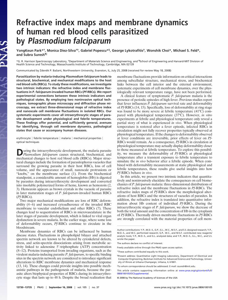
ResultsThree-dimensional Refractive Index Maps of Pf-RBCs. To quantita-tively investigate the refractive index distribution of Pf-RBCs, weused tomographic phase microscopy (TPM) (16). TPM uses laserinterferometry combined with rotating the incident beam, analo-gous to computed tomography (CT) in x-ray. TPM quantitativelyprovides the three-dimensional distribution of refractive index, n(x,y, z). As shown in Fig. 1, we measured the refractive index maps ofPf-RBCs during all intraerythrocytic stages: healthy RBC (Fig. 1A),ring (Fig. 1B), trophozoite (Fig. 1C), and schizont stage (Fig. 1D).Images in the horizontal rows show refractive index maps at threedifferent cross-sections: 0.6 �m above the focused plane (Top), atthe focused plane (Middle), and 0.6 �m below the focused plane(Bottom). Whereas healthy RBCs show homogeneous distributionof refractive index, Pf-RBCs are not optically homogeneous. Manyfactors contribute to refractive index change: the vacuole of parasiteoccupies a fraction of volume in cytoplasm of RBC; Hb is metab-olized and converted into hemozoin crystal in the parasite mem-brane; and various parasite proteins are exported from parasite intocytoplasm of Pf-RBCs (17). Regions of low refractive index indicatethe vacuole of P. falciparum (Fig. 1 B–D, black arrows) and regionsof high refractive index suggest the position of hemozoin (Fig. 1 Cand D, gray arrows).
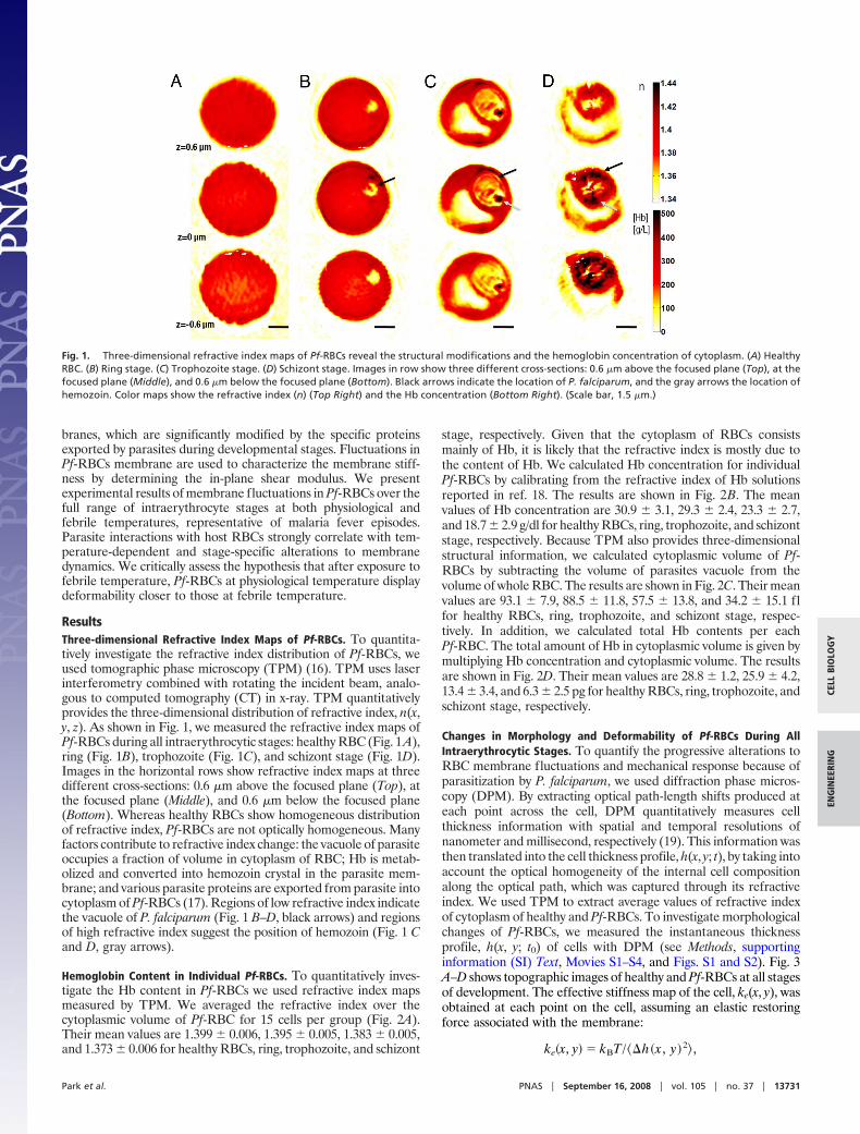
Hemoglobin Content in Individual Pf-RBCs. To quantitatively inves-tigate the Hb content in Pf-RBCs we used refractive index mapsmeasured by TPM. We averaged the refractive index over thecytoplasmic volume of Pf-RBC for 15 cells per group (Fig. 2A).Their mean values are 1.399 � 0.006, 1.395 � 0.005, 1.383 � 0.005,and 1.373 � 0.006 for healthy RBCs, ring, trophozoite, and schizont
stage, respectively. Given that the cytoplasm of RBCs consistsmainly of Hb, it is likely that the refractive index is mostly due tothe content of Hb. We calculated Hb concentration for individualPf-RBCs by calibrating from the refractive index of Hb solutionsreported in ref. 18. The results are shown in Fig. 2B. The meanvalues of Hb concentration are 30.9 � 3.1, 29.3 � 2.4, 23.3 � 2.7,and 18.7 � 2.9 g/dl for healthy RBCs, ring, trophozoite, and schizontstage, respectively. Because TPM also provides three-dimensionalstructural information, we calculated cytoplasmic volume of Pf-RBCs by subtracting the volume of parasites vacuole from thevolume of whole RBC. The results are shown in Fig. 2C. Their meanvalues are 93.1 � 7.9, 88.5 � 11.8, 57.5 � 13.8, and 34.2 � 15.1 flfor healthy RBCs, ring, trophozoite, and schizont stage, respec-tively. In addition, we calculated total Hb contents per eachPf-RBC. The total amount of Hb in cytoplasmic volume is given bymultiplying Hb concentration and cytoplasmic volume. The resultsare shown in Fig. 2D. Their mean values are 28.8 � 1.2, 25.9 � 4.2,13.4 � 3.4, and 6.3 � 2.5 pg for healthy RBCs, ring, trophozoite, andschizont stage, respectively.
Changes in Morphology and Deformability of Pf-RBCs During AllIntraerythrocytic Stages. To quantify the progressive alterations toRBC membrane fluctuations and mechanical response because ofparasitization by P. falciparum, we used diffraction phase micros-copy (DPM). By extracting optical path-length shifts produced ateach point across the cell, DPM quantitatively measures cellthickness information with spatial and temporal resolutions ofnanometer and millisecond, respectively (19). This information wasthen translated into the cell thickness profile, h(x, y; t), by taking intoaccount the optical homogeneity of the internal cell compositionalong the optical path, which was captured through its refractiveindex. We used TPM to extract average values of refractive indexof cytoplasm of healthy and Pf-RBCs. To investigate morphologicalchanges of Pf-RBCs, we measured the instantaneous thicknessprofile, h(x, y; t0) of cells with DPM (see Methods, supportinginformation (SI) Text, Movies S1–S4, and Figs. S1 and S2). Fig. 3A–D shows topographic images of healthy and Pf-RBCs at all stagesof development. The effective stiffness map of the cell, ke(x, y), wasobtained at each point on the cell, assuming an elastic restoringforce associated with the membrane:
ke�x, y� � kBT /��h�x , y�2� ,
Fig. 1. Three-dimensional refractive index maps of Pf-RBCs reveal the structural modifications and the hemoglobin concentration of cytoplasm. (A) HealthyRBC. (B) Ring stage. (C) Trophozoite stage. (D) Schizont stage. Images in row show three different cross-sections: 0.6 �m above the focused plane (Top), at thefocused plane (Middle), and 0.6 �m below the focused plane (Bottom). Black arrows indicate the location of P. falciparum, and the gray arrows the location ofhemozoin. Color maps show the refractive index (n) (Top Right) and the Hb concentration (Bottom Right). (Scale bar, 1.5 �m.)
Park et al. PNAS � September 16, 2008 � vol. 105 � no. 37 � 13731
CELL
BIO
LOG
YEN
GIN
EERI
NG
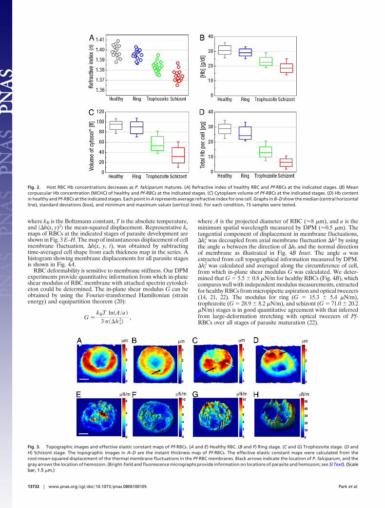
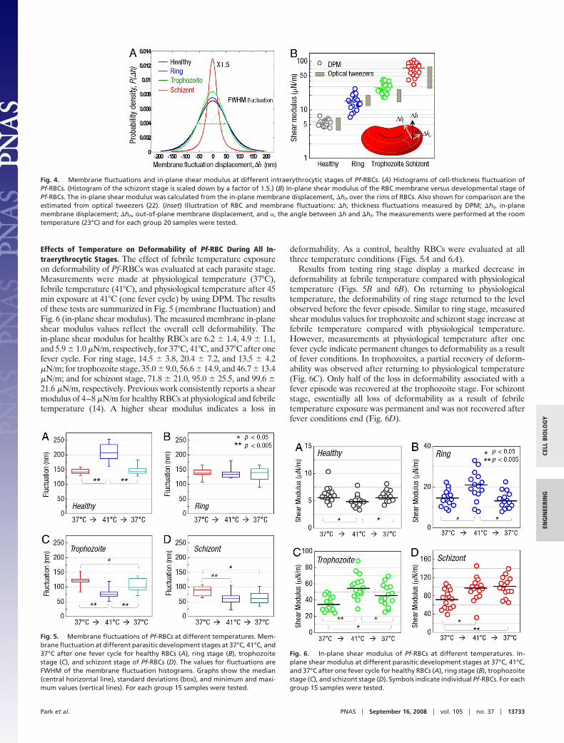
where kB is the Boltzmann constant, T is the absolute temperature,and ��h(x, y)2� the mean-squared displacement. Representative kemaps of RBCs at the indicated stages of parasite development areshown in Fig. 3 E–H. The map of instantaneous displacement of cellmembrane fluctuation, �h(x, y, t), was obtained by subtractingtime-averaged cell shape from each thickness map in the series. Ahistogram showing membrane displacements for all parasite stagesis shown in Fig. 4A.
RBC deformability is sensitive to membrane stiffness. Our DPMexperiments provide quantitative information from which in-planeshear modulus of RBC membrane with attached spectrin cytoskel-eton could be determined. The in-plane shear modulus G can beobtained by using the Fourier-transformed Hamiltonian (strainenergy) and equipartition theorem (20):
G �kBT ln�A /a�
3���h12�
,
where A is the projected diameter of RBC (�8 �m), and a is theminimum spatial wavelength measured by DPM (�0.5 �m). Thetangential component of displacement in membrane fluctuations,�ht
2 was decoupled from axial membrane fluctuation �h2 by usingthe angle � between the direction of �ht and the normal directionof membrane as illustrated in Fig. 4B Inset. The angle � wasextracted from cell topographical information measured by DPM.�ht
2 was calculated and averaged along the circumference of cell,from which in-plane shear modulus G was calculated. We deter-mined that G 5.5 � 0.8 �N/m for healthy RBCs (Fig. 4B), whichcompares well with independent modulus measurements, extractedfor healthy RBCs from micropipette aspiration and optical tweezers(14, 21, 22). The modulus for ring (G 15.3 � 5.4 �N/m),trophozoite (G 28.9 � 8.2 �N/m), and schizont (G 71.0 � 20.2�N/m) stages is in good quantitative agreement with that inferredfrom large-deformation stretching with optical tweezers of Pf-RBCs over all stages of parasite maturation (22).
Fig. 2. Host RBC Hb concentrations decreases as P. falciparum matures. (A) Refractive index of healthy RBC and Pf-RBCs at the indicated stages. (B) Meancorpuscular Hb concentration (MCHC) of healthy and Pf-RBCs at the indicated stages. (C) Cytoplasm volume of Pf-RBCs at the indicated stages. (D) Hb contentin healthy and Pf-RBCs at the indicated stages. Each point in A represents average refractive index for one cell. Graphs in B–D show the median (central horizontalline), standard deviations (box), and minimum and maximum values (vertical lines). For each condition, 15 samples were tested.
Fig. 3. Topographic images and effective elastic constant maps of Pf-RBCs. (A and E) Healthy RBC. (B and F) Ring stage. (C and G) Trophozoite stage. (D andH) Schizont stage. The topographic images in A–D are the instant thickness map of Pf-RBCs. The effective elastic constant maps were calculated from theroot-mean-squared displacement of the thermal membrane fluctuations in the Pf-RBC membranes. Black arrows indicate the location of P. falciparum, and thegray arrows the location of hemozoin. (Bright-field and fluorescence micrographs provide information on locations of parasite and hemozoin; see SI Text). (Scalebar, 1.5 �m.)
13732 � www.pnas.org�cgi�doi�10.1073�pnas.0806100105 Park et al.
Effects of Temperature on Deformability of Pf-RBC During All In-traerythrocytic Stages. The effect of febrile temperature exposureon deformability of Pf-RBCs was evaluated at each parasite stage.Measurements were made at physiological temperature (37°C),febrile temperature (41°C), and physiological temperature after 45min exposure at 41°C (one fever cycle) by using DPM. The resultsof these tests are summarized in Fig. 5 (membrane fluctuation) andFig. 6 (in-plane shear modulus). The measured membrane in-planeshear modulus values reflect the overall cell deformability. Thein-plane shear modulus for healthy RBCs are 6.2 � 1.4, 4.9 � 1.1,and 5.9 � 1.0 �N/m, respectively, for 37°C, 41°C, and 37°C after onefever cycle. For ring stage, 14.5 � 3.8, 20.4 � 7.2, and 13.5 � 4.2�N/m; for trophozoite stage, 35.0 � 9.0, 56.6 � 14.9, and 46.7 � 13.4�N/m; and for schizont stage, 71.8 � 21.0, 95.0 � 25.5, and 99.6 �21.6 �N/m, respectively. Previous work consistently reports a shearmodulus of 4–8 �N/m for healthy RBCs at physiological and febriletemperature (14). A higher shear modulus indicates a loss in
deformability. As a control, healthy RBCs were evaluated at allthree temperature conditions (Figs. 5A and 6A).
Results from testing ring stage display a marked decrease indeformability at febrile temperature compared with physiologicaltemperature (Figs. 5B and 6B). On returning to physiologicaltemperature, the deformability of ring stage returned to the levelobserved before the fever episode. Similar to ring stage, measuredshear modulus values for trophozoite and schizont stage increase atfebrile temperature compared with physiological temperature.However, measurements at physiological temperature after onefever cycle indicate permanent changes to deformability as a resultof fever conditions. In trophozoites, a partial recovery of deform-ability was observed after returning to physiological temperature(Fig. 6C). Only half of the loss in deformability associated with afever episode was recovered at the trophozoite stage. For schizontstage, essentially all loss of deformability as a result of febriletemperature exposure was permanent and was not recovered afterfever conditions end (Fig. 6D).
Fig. 4. Membrane fluctuations and in-plane shear modulus at different intraerythrocytic stages of Pf-RBCs. (A) Histograms of cell-thickness fluctuation ofPf-RBCs. (Histogram of the schizont stage is scaled down by a factor of 1.5.) (B) In-plane shear modulus of the RBC membrane versus developmental stage ofPf-RBCs. The in-plane shear modulus was calculated from the in-plane membrane displacement, �hn over the rims of RBCs. Also shown for comparison are theestimated from optical tweezers (22). (Inset) Illustration of RBC and membrane fluctuations: �h, thickness fluctuations measured by DPM; �ht, in-planemembrane displacement; �hn, out-of-plane membrane displacement, and �, the angle between �h and �ht. The measurements were performed at the roomtemperature (23°C) and for each group 20 samples were tested.
Fig. 5. Membrane fluctuations of Pf-RBCs at different temperatures. Mem-brane fluctuation at different parasitic development stages at 37°C, 41°C, and37°C after one fever cycle for healthy RBCs (A), ring stage (B), trophozoitestage (C), and schizont stage of Pf-RBCs (D). The values for fluctuations areFWHM of the membrane fluctuation histograms. Graphs show the median(central horizontal line), standard deviations (box), and minimum and maxi-mum values (vertical lines). For each group 15 samples were tested.
Fig. 6. In-plane shear modulus of Pf-RBCs at different temperatures. In-plane shear modulus at different parasitic development stages at 37°C, 41°C,and 37°C after one fever cycle for healthy RBCs (A), ring stage (B), trophozoitestage (C), and schizont stage (D). Symbols indicate individual Pf-RBCs. For eachgroup 15 samples were tested.
Park et al. PNAS � September 16, 2008 � vol. 105 � no. 37 � 13733
CELL
BIO
LOG
YEN
GIN
EERI
NG
DiscussionWith P. falciparum parasitic development into the schizont stage,the normal discocyte shape is lost. Parasite modifications to theinternal and membrane structure of invaded RBCs are implicatedin the observed morphological changes (Fig. 3 A–D). Membranestiffness also increases progressively with parasite development. Inparticular, the spatially averaged effective stiffness, �ke(x, y)�, at theschizont stage is up to an order of magnitude higher than that forhealthy RBCs (Fig. 3 E–H). These results indicate that parasitedevelopment stage directly correlates with the amplitude of mem-brane fluctuations, and that the distribution of membrane fluctu-ations becomes much sharper with parasite development.
Interestingly, there is significant increase in membrane fluctua-tions [53% in the full-width half maximum (FWHM) value of thefluctuation displacement histogram] from physiological to febriletemperature (P 0.01), which is only a 7.5% increase in absolutetemperature. Evidently, such enhancement in the fluctuation can-not be explained simply by equilibrium thermodynamics alone, thatis, by the increase in the Boltzmann factor kBT. The substantialincrease in membrane fluctuations indicates that RBC phospho-lipid membrane and spectrin network undergo structural changesthat alter elastic properties, which is reflected in changes tomembrane in-plane shear modulus, G (Fig. 4). One possibleexplanation is that a transitional structural change in spectrinmolecular architecture occurs between physiological and febriletemperature, causing altered and reorganized cytoskeleton net-work. Both �- and �-spectrin molecules have a significant structuraltransition near 40°C (23). It has been shown that membrane shearmodulus of healthy RBCs decreases by �20% when the tempera-ture increases from 23 to 41°C (21). From physiological to febriletemperature, membrane fluctuations decrease significantly (P 0.01) for all infected test conditions (Fig. 3), with trophozoite andschizont stages showing the strongest temperature dependence.Based on comparison with results from healthy RBC and associatedchanges of in-plane shear modulus (Fig. 4B), we postulate thatintraerythrocytic parasite development causes an opposite effect.Parasite-exported proteins that target RBC membrane can alterthermally induced spectrin-folding transitions involved in stabiliz-ing erythrocyte cytoskeleton (24). Expression of P. falciparumgenes is known to be affected by exposure to 41°C (25). Underfebrile conditions, increased levels of parasite-exported proteinscould contribute to large changes in membrane fluctuations ob-served by DPM between physiological and febrile temperatures.One example is the ring-infected erythrocyte surface antigen(RESA). When RESA binds to spectrin, normal balance betweenspectrin dimers and tetramers in the cytoskeletal network is skewedtoward tetramer formation, which results in increased membranestability (26). As a result, RESA enhances RBC resistance tomechanical and thermal damage. Moreover, elevated thermalstability conferred by RESA plays a protective role for Pf-RBCsagainst damage at febrile temperature (27). The possible specificrole of other membrane proteins and histidine-rich proteins ininfluencing thermal and mechanical stability of RBCs over thetemperature range of interest is presently not fully understood. Thedevelopment of adhesion properties at the trophozoite and schizontstages can also contribute to significant decrease in fluctuationobserved by DPM at these stages, especially at febrile temperature.For the same febrile temperature, our results indicate that fluctu-ation amplitude progressively decreases as a parasite matures fromthe ring to the schizont stage.
The findings presented in Figs. 5 and 6 reveal that effects of feverepisodes cause irreversible changes to deformability of Pf-RBCs.These experiments mimic in vivo conditions that Pf-RBCs experi-ence and provide information that cannot be extracted frommechanical experiments performed at a constant temperature.Plastic behavior caused by a febrile episode is not observed at thering stage. Decreased deformability observed under febrile condi-
tions is recovered on returning to physiological temperature. Thedecrease in deformability of Pf-RBCs may be attributed to exportedparasite proteins that interact with the host RBC membrane. Theability of ring stage to regain deformability could play an importantrole in their ability to avoid splenic clearance when circulating.Trophozoite and schizont stages display significant plastic changesto deformability. Again, decreased deformability for mature stagesmay be also attributed largely to parasite-exported proteins thatassociate with the membrane. Parasite protein exportation is likelyaccelerated by thermal energy. In turn, febrile episodes may beaccelerating parasite protein exportation to the membrane thatresults in advancing the loss of deformability. Both reduced de-formability and the development of adhesion properties contributeto sequestration of trophozoite and schizont stage. The tempera-ture-related permanent loss of deformability found in our experi-ments is greater than anticipated and can be viewed as an additionaldriving force to impede microcirculation and promote sequestra-tion. The plastic behavior of trophozoite and schizont stages inresponse to fever could also have implications when treatments areused to suppress fever episodes. Additional studies would be neededto explore this possibility.
Finally, the optical experimental techniques (TPM and DPM)used to measure the refractive index maps and the membranefluctuations in this study offer unique advantages over other morecommonly used techniques, such as micropipette aspiration, opticaltweezers, laminar shear flow, and magnetic twisting cytometry.TPM quantitatively and noninvasively measures the three-dimensional maps of refractive index, which provides a measure ofHb content of single Pf-RBCs. Indeed, TPM does not require anyspecial sample preparation because the refractive index is anintrinsic optical property. DPM is able to measure nanometer levelmembrane fluctuations without any direct, and potentially invasive,contact with a cell. The optical layout of DPM is such that thesample temperature can be regulated easily without affecting of therest of the apparatus, a common limitation of other techniques. Inaddition, deformability of living cells, measured by thermally drivenmembrane fluctuations, ensures mechanical properties in the linearregime. Also, measurements are made within seconds once aspecific cell is identified. Thus, the technique provides the flexibilityto experiment on a large number of samples under a variety of wellcontrolled test conditions in a reasonable time span.
ConclusionsWe have presented three-dimensional maps of refractive index ofPf-RBCs during all intererythrocytic stages, from which we deter-mine the Hb concentration of Pf-RBCs. We have also presentedsystematic measurements of nanoscale fluctuations associated withRBCs parasitized at all stages by P. falciparum at physiological andfebrile temperatures. Our approach to studying Pf-RBCs uniquelycombines optical interferometry, biophysics, and cell nanomechan-ics. A method has been presented to extract elastic properties frommembrane fluctuation results, and this technique is validated byquantitative comparisons of elastic moduli of RBCs over all parasitematuration stages with prior, independent experimental data ob-tained with laser tweezers (14, 22). Compared with other tech-niques for assessing RBC mechanical properties, such as electricfield deformation, micropipette aspiration, optical tweezers, andmagnetic bead excitation, the method presented here has distinctadvantages of being spatially resolved and noncontact. We envisionthat this methodology could offer potentially powerful means tolink cell membrane fluctuations with pathological conditions thatlead to human disease states by providing quantitative informationthat could not be extracted through other experimental techniques.
These results point to major new avenues for exploiting mem-brane fluctuations as quantitative indicators of the onset andprogression of pathological states that could lead to diseases. Inaddition, the information provided by diffraction phase microscopycan guide theory and computational simulations that address a
13734 � www.pnas.org�cgi�doi�10.1073�pnas.0806100105 Park et al.
broader range of cell biology problems concerning human health,disease diagnostics, therapeutics, and drug efficacy assays.
Materials and MethodsPreparation of Pf-RBCs and Parasite Culture. P. falciparum 3D7A were main-tained in leukocyte-free human O� erythrocytes (Research Blood Components)under an atmosphere of 3% O2, 5% CO2, and 92% N2 in RPMI medium 1640(Gibco Life Technologies) supplemented with 25 mM Hepes (Sigma), 200 mMhypoxanthine (Sigma), 0.209% NaHCO3 (Sigma), and 0.25% albumax I (Gibco LifeTechnologies). Cultures were synchronized successively by concentration of ma-ture schizonts by using plasmagel flotation (28) and sorbitol lysis 2 h aftermerozoite invasion to remove residual schizonts (29).
Measurements were performed at 14–20 h (ring stage), 20–36 h (trophozoitestage), and 36–48 h (schizont stage) after merozoite invasion. To identify theinfected RBCs, we treated the parasites with DAPI staining. Before the DPMdynamic measurements, we recorded epi-fluorescence images, as described inref. 19.
Healthy RBC control samples and Pf-RBCs samples were diluted in PBS to �106
RBC per ml before membrane fluctuation experiments. Both healthy and Pf-RBCsadhere to the glass substrate rapidly and strongly. However, adhesion of thebottom membrane of RBCs does not significantly affect to the fluctuation of theupper membrane. DPM measurement shows no significant difference in mem-brane fluctuation between RBCs positioned on the glass substrate and RBCsfirmly attached to the glass substrate by using poly-L-lysine followed the protocolindicated in ref. 30.
Diffraction Phase Microscopy. AnAr2� laser (�514nm)wasusedas illuminationsourceforan invertedmicroscope(IX71,Olympus).Themicroscopewasequippedwith a 40� objective (0.65 N.A.), which facilitates a diffraction-limited transverseresolution of 400 nm. With the additional relay optics used outside the micro-scope, the overall magnification of the system was �200�. EMCCD (Photonmax512B,Princeton Instruments)wasusedto imageinterferogram.DPMemploys theprinciple of laser interferometry in a common path geometry and thus providesfull-field quantitative phase images of RBCs with unprecedented optical path-lengthstability (19).Theinstantaneouscell thicknessmapisobtainedash(x,y, t)�/2��n �(x, y, t), with � the quantitative phase image measured by DPM. Therefractive index contrast �n between the RBC and the surrounding PBS is mainlycontributed to the Hb, which is optically homogeneous in cytosol. We usedtomographic phase microscopy (TPM) to retrieve three-dimensional refractiveindex for all of the stages of Pf-RBCs and healthy RBCs. The DPM optical path-
length stability is 2.4 mrad, which corresponds to a membrane displacement of3.3 nm (19).
Tomographic Phase Microscopy. Tomographic phase microscopy (TPM) is atechnique that can map the three-dimensional distribution of refractive index inlive cells and tissues (16). In TPM, the sample-induced optical phase shift is imagedby using a phase-shifting heterodyne interferometer. Phase images are recordedby varying the directions of illumination. The angle of illumination ranges from 60 to 60° and angular step is 0.2°. It takes �10 s to scan the entire angular range.Phase image at each step of angle corresponds to angular projection of refractiveindex at the illumination angle. The custom built microscopy and CMOS camera(FASTCAM 1024 PCI, Photron) are used to measure interferograms. With the setof angular projection phase images, a filtered back-projection algorithm is usedto calculate a three-dimensional refractive index. The transverse and axial reso-lutions are 0.3 and 0.6 �m, respectively, and the accuracy of index measurementis 0.001.
Temperature Control. The microscopic stage was equipped with a temperaturecontroller (TC-202A, Warner Instruments), which uses a thermistor to set thetemperatureofthesampletowithin�0.2°C.Thewell containingRBCswasplacedin contact with the controller chamber, such that heat transfer and thermalequilibriumbetweenthetwosystemswereattainedrelatively fast,after3–4min.However, we measured the individual RBC at multiple temperature points bywaiting �10 min for each new temperature, which ensured thermal equilibrium.
Statistical Analysis. P values were calculated by two-tailed Mann–Whitney ranksum tests comparing the FWHM fluctuations and shear modulus values betweenvarious test conditions. All of the numbers following the � sign in the text arestandard deviations.
ACKNOWLEDGMENTS. We thank K. Badizadegan, R. R. Dasari, and M. Dao fortechnical discussions, J. P. Mills for helpful suggestions on experimental designand data analysis, and Malaria Research and Reference Reagent Resource, Amer-ican Type Culture Collection, for providing us with 3D7A parasite. This work wassupported by National Institutes of Health Grants P41-RR02594-18 and(1-R01-GM076689-01, National Science Foundation Grant DBI-0754339, and the Inter-disciplinary Research Group on Infectious Diseases, which is funded by the Sin-gapore-MIT Alliance for Research and Technology Center. Y.P. was supported byCambridge Foundation Fellowship, Samsung Scholarship, and Whitaker HealthScienceFellowship.M.D.-S.andG.L.werepartially supportedbyGlobalEnterprisefor MicroMechanics and Molecular Medicine (GEM4) postdoctoral fellowships.
1. Kilejian A (1979) Characterization of a protein correlated with the production ofknob-like protrusions on membranes of erythrocytes infected with Plasmodium falci-parum. Proc Natl Acad Sci USA 76:4650–4653.
2. Sherman IW (1979) Biochemistry of Plasmodium (malarial parasites). Microbiol Rev43(4):453.
3. Goldberg DE, Slater AFG, Cerami A, Henderson GB (1990) Hemoglobin degradation inthe malaria parasite Plasmodium falciparum: An ordered process in a unique or-ganelle. Proc Natl Acad Sci USA 87:2931–2935.
4. Nash GB, O’Brien E, Gordon-Smith EC, Dormandy JA (1989) Abnormalities in themechanical properties of red blood cells caused by Plasmodium falciparum. Blood74(2):855–861.
5. Cranston HA, et al. (1984) Plasmodium falciparum maturation abolishes physiologicred cell deformability. Science 223(4634):400–403.
6. Paulitschke M, Nash GB (1993) Membrane rigidity of red blood cells parasitized bydifferent strains of Plasmodium falciparum. J Lab Clin Med 122(5):581–589.
7. Miller LH, Baruch DI, Marsh K, Doumbo OK (2002) The pathogenic basis of malaria.Nature 415(6872):673–679.
8. Discher DE, Mohandas N, Evans EA (1994) Molecular maps of red cell deformation:Hidden elasticity and in situ connectivity. Science 266(5187):1032.
9. Gov NS, Safran SA (2005) Red blood cell membrane fluctuations and shape controlledby ATP-induced cytoskeletal defects. Biophys J 88(3):1859.
10. Lawrence CLL, Gov N, Brown FLH (2006) Nonequilibrium membrane fluctuations drivenby active proteins. J Chem Phys 124:074903.
11. Tuvia S, Levin S, Bitler A, Korenstein R (1998) Mechanical fluctuations of the mem-brane-skeleton are dependent on F-actin ATPase in human erythrocytes. J Cell Biol141(7):1551–1561.
12. Li J, Dao M, Lim CT, Suresh S (2005) Spectrin-level modeling of the cytoskeleton andoptical tweezers stretching of the erythrocyte. Biophys J 88(5):3707–3719.
13. Glenister FK, Coppel RL, Cowman AF, Mohandas N, Cooke BM (2002) Contribution ofparasite proteins to altered mechanical properties of malaria-infected red blood cells.Blood 99(3):1060–1063.
14. Mills JP, et al. (2007) Effect of plasmodial RESA protein on deformability of human redblood cells harboring Plasmodium falciparum Proc Natl Acad Sci USA 104:9213–9217.
15. Kwiatkowski D (1989) Febrile temperatures can synchronize the growth of Plasmo-dium falciparum in vitro. J Exp Med 169(1):357–361.
16. Choi W, et al. (2007) Tomographic phase microscopy. Nat Methods 4:717–719.
17. Tilley L, McFadden G, Cowman A, Klonis N (2007) Illuminating Plasmodium falciparum-infected red blood cells. Trends Parasitol 23:268–277.
18. Friebel M, Meinke M (2006) Model function to calculate the refractive index of nativehemoglobin in the wavelength range of 250–1100 nm dependent on concentration.Appl Opt 45(12):2838–2842.
19. Park YK, Popescu G, Badizadegan K, Dasari RR, Feld MS (2006) Diffraction phase andfluorescence microscopy. Opt Exp 14(18):8263–8268.
20. Lee JCM, Discher DE (2001) Deformation-enhanced fluctuations in the red cell skeletonwith theoretical relations to elasticity, connectivity, and spectrin unfolding. Biophys J81(6):3178–3192.
21. Waugh R, Evans EA (1979) Thermoelasticity of red blood cell membrane. Biophys J26(1):115–131.
22. Suresh S, et al. (2005) Connections between single-cell biomechanics and humandisease states: Gastrointestinal cancer and malaria. Acta Biomater 1(1):15–30.
23. Minetti M, Ceccarini M, Di Stasi AMM, Petrucci TC, Marchesi VT (1986) Spectrininvolvement in a 40°C structural transition of the red blood cell membrane. J CellBiochem 30:361–370.
24. Da Silva E, et al. (1994) The Plasmodium falciparum protein RESA interacts with theerythrocyte cytoskeleton and modifies erythrocyte thermal stability. Mol BiochemParasitol 66(1):59–69.
25. Oakley MSM, et al. (2007) Molecular factors and biochemical pathways induced byfebrile temperature in intraerythrocytic Plasmodium falciparum parasites. Infect Im-mun 75(4):2012–2025.
26. Pei X, et al. (2007) The ring-infected erythrocyte surface antigen (RESA) of. Plasmodiumfalciparum stabilizes spectrin tetramers and suppresses further invasion. Blood110(3):1036–1042.
27. Silva MD, et al. (2005) A role for the. Plasmodium falciparum RESA protein in resistanceagainst heat shock demonstrated using gene disruption. Mol Microbiol 56(4):990–1003.
28. Pasvol G, Wilson RJ, Smalley ME, Brown J (1978) Separation of viable schizont-infectedred cells of. Plasmodium falciparum from human blood. Ann Trop Med Parasitol72(1):87–88.
29. Lambros C, Vanderberg JP (1979) Synchronization of. Plasmodium falciparum eryth-rocytic stages in culture. J Parasitol 65(3):418–420.
30. Hategan A, Sengupta K, Kahn S, Sackmann E, Discher DE (2004) Topographical patterndynamics in passive adhesion of cell membranes. Biophys J 87(5):3547–3560.
Park et al. PNAS � September 16, 2008 � vol. 105 � no. 37 � 13735
CELL
BIO
LOG
YEN
GIN
EERI
NG
Copyright © 2022 FDOKUMEN