The role of mitochondria in pharmacotoxicology: a reevaluation of an old, newly emerging topic
Upload
independentCategory
view
5download
0
~ Pergamon Neuroscience Vol. 76, No. 2, pp. 335 343, 1997
Copyright C~ 1996 IBRO. Published by Elsevier Science Ltd Printed in Great Britain
PII: S0306-4522(96)00409-5 0306M522/97 $17.00+0.00
COMMENTARY
RE-EVALUATION OF THE FUNCTIONAL ANATOMY OF THE BASAL GANGLIA IN NORMAL AND PARKINSONIAN
STATES
R. LEVY,* L.-N. H A Z R A T I , t M.-T. H E R R E R O , * $ M. VILA,* O.-K. HASSANI,§ M. M O U R O U X , § M. R U B E R G , * H. ASENSI,:~ Y. AGID,* J. FEGER,§ J. A. OBESO,¶
A. P A R E N T t and E. C. H I R S C H * I
*INSERM U. 289, H6pital de la Salp~tri+re, 47 bd de l'H6pital, 75651, Paris, Cedex 13, France
tLaboratoire de Neurobiologie, LocalF-7500, Centre de Recherche Universit6 Larval Robert-Giffard, 2601 de la Canardibre, Beaufort, Quebec, Canada, G13 124
++Departamento de Ciencias Morfol6gicas, University of Murcia, 30150 Murcia, Spain
§Laboratoire de Pharmacologie, Universitd Rend Descartes, 4 avenue de l'observatoire, 75270 Paris Cedex 6, France
¶Centro de Neurologia y Neurocirurgia Funcional, Clinica Quiron, Parque Acolea, Sln 20012 San Sebastian, Spain
Abstraet--ln the late 1980s, a functional and anatomical model of basal ganglia organization was proposed in order to explain the clinical syndrome of Parkinson's disease. According to this model, the pathological overactivity observed in the subthalamic nucleus and the output station of the basal ganglia plays a crucial role in the pathophysiology of the motor signs of Parkinson's disease. The hyperactivity of subthalamic neurons in Parkinsonism is viewed as a direct consequence of a pathological hypoactivity of the external segment of the pallidum. This article reviews recent data from different experimental approaches that challenge the established model of basal ganglia organization by reinterpreting the functional interaction between the external segment of the pallidum and the subthalamic nucleus in both the normal and pathological state. Indeed, recent neurobiochemical studies have rather unexpectedly shown that the GABAergic and metabolic activities of the external pallidum are not decreased in human and non-human primates with Parkinsonism. This absence of any decrease in activity might be explained by the functionally antagonistic influences of the striatal and subthalamic afferences within the external pallidum, as suggested by several anatomical studies. In addition, there are clues from electrophysiological studies to suggest that the hyperactivity found in the subthalamic neurons in Parkinsonism may not depend solely on the level of activity in the external pallidum.
In such a framework, the hyperactivity of the subthalamic neurons would have to be explained, at least in part, by other sources of excitation or disinhibition. However, any explanation for the origin of the subthalamic overactivity in Parkinsonism remains speculative. Copyright ~ 1996 IBRO. Published by Elsevier Science Ltd.
Key words: Parkinson's disease, 1-methyl-4-phenyl-l,2,3,6-tetrahydropyridine, glutamate decarboxylase, cytochrome oxidase, pallidum, subthalamic nucleus.
An accurate knowledge of the anatomical and func- tional organization of the basal ganglia is an essential step in improving our understanding of how motor and cognitive functions operate in the normal brain.
ITo whom correspondence should be addressed. Abbreviations: CO, cytochrome oxidase; 2-DG,
2-deoxyglucose: GAD, glutamate decarboxylase; GPe, external segrnfa~ of the pallidum; GPi, internal segment of the pallidum; MPTP, 1-methyl-4-phenyl-l,2,3,6- tetrahydropyridine; 6-OHDA, 6-hydroxydopamine; PD, Parkinson's disease; Pf nucleus, parafascicular nucleus; QISH, quantitative in situ hybridization; SNpc, substan- tia nigra pars compacta; SNpr, substantia nigra pars reticulata; STN, subthalamic nucleus.
There is also a fundamental need to clarify the consequences of the pathophysiological charrges that occur in Parkinson's disease (PD) and other disorders of the basal ganglia. In the late 1980s an elegant model was developed which proposed a framework for the anatomical organization of the basal ganglia and provided consistent explanations for the clinical syndrome of PD. 1,9~66 Subsequently, new therapeutic approaches to basal ganglia disorders were proposed. According to the model, the basal ganglia are organ- ized in such a fashion that nigrostriatal denervation (the hallmark of PD), experimentally produced by intoxication with 1-methyl-4-phenyl- 1,2,3,6- tetrahydropyridine (MPTP) in non-human primate
335

336 R. Levy et al.
or 6-hydroxydopamine (6-OHDA)-induced lesionin- gof midbrain dopaminergic neurons in the rat, leads to an overactivity of the output station of the basal ganglia, namely the internal segment of the pallidum and the substantia nigra pars reticulata (GPi/SNpr). This pathological hyperactivity of the GPi/SNpr has been interpreted as the result of a reduced inhibitory input from the striatum and an increase in the excitatory input from the subthalamic nucleus (STN). The enhanced activity of the STN neurons is thought to be the direct consequence of a release from the tonic inhibitory control normally exerted by the GABA-containing neurons of the external segment of the pallidum (GPe) on STN neurons. 63 Thus, according to the established model, the hypoactivity of the GPe leads to increased STN activity which in turn (in addition to the diminished influence of the direct striatal GABAergic input) induces the patho- logical overactivity of GPi/SNpr.
In the present review, we report evidence ob- tained using GABAergic and metabolic activity markers of an absence of GPe hypoactivity either in MPTP-intoxicated monkeys or in PD patients. These unexpected results have led to a reassessment of the functional and anatomical organization within the basal ganglia, since the normal GPe activity found in this condition could not alone explain the well-documented subthalamic overactiv- ity observed in Parkinsonism. We will attempt to put these new findings in perspective, in terms of GPe connectivity. We will also provide data from recent electrophysiological studies further support- ing the notion that the STN receives other major influences which may lead to its overactivity in Parkinsonism. However, the precise origin of this subthalamic hyperactivity has yet to be confirmed.
NEURONS OF THE EXTERNAL SEGMENT OF THE PALLIDUM ARE NOT HYPOACTIVATED IN
PARKINSONISM
Output GPe neurons exert an inhibitory effect on STN neurons by means of the neurotransmitter GABA. The current pathophysiological model of Parkinsonism assumes a decrease in the GABAergic activity of these GPe neurons, thus releasing the activity of the STN neurons. In order to determine the level of GABAergic activity of GPe neurons in Parkinsonism, 24 using quantitative in situ hybridiz- ation (QISH) we estimated, at the cellular level on pos t -mor tem tissue, the expression of the messenger RNA coding for the human 67,000 mol. wt isoform of glutamate decarboxylase (GAD67 mRNA), the synthetic enzyme of GABA (the QISH method used in this study has fully been described elsewhere26'35). GAD67 mRNA expression was taken as an index of the GABAergic activity in GPe neurons, since GAD67 levels reflect long-term changes in GABA neurotransmission 11"4° and, consequently, could be used to determine the steady-state level of GABAer- gic activity in GPe neurons in semi-chronic or
chronic diseases of the basal ganglia. The QISH method thus enabled us to quantify GAD67 mRNA expression in the GPe neurons of MPTP-intoxicated monkeys and patients with PD. Comparisons be- tween MPTP-intoxicated monkeys or PD patients and their respective controls failed to reveal any significant decrease in the density of GPe output neurons expressing GADs, 7 mRNA or in the amount of GAD6v mRNA per GPe-labeled neuron in the parkinsonian states (Table 1, Fig. 1). This normal GAD67 mRNA expression in GPe neurons probably results in a normal level of GABA neurotransmission between GPe neurons and their targets, in particular the STN neurons. Indeed, in vivo, several observa- tions suggest that the changes in GAD67 mRNA are mirrored by GAD enzyme activity, GABA concen- tration and release at the terminals and GABA turnover. 2'1s'36'39'5~ Moreover, changes in GAD67 mRNA and GAD activity also follow the functional activity of the GABAergic neurons them- selves, m'23"37"38 In MPTP-intoxicated monkeys, the positive correlation between GAD67 mRNA expres- sion and the functional activity of the GABAergic neurons is further supported by the fact that GAD67 mRNA expression was found to be strongly in- creased in the GPi and SNpr (where it is generally accepted that output GABAergic neurons are hyper- activated), whereas it was significantly decreased in the superior colliculus, which receives strong GABAergic projections from the SNpr. 25'64 Con- versely, L-dopa treatment in MPTP-intoxicated mon- keys restored GAD67 mRNA expression to the level observed in control animals in both the GPi and S N p r Y '('4 It is worthy of note that several studies using GABA markers in MPTP-intoxicated monkeys and 6-OHDA-lesioned rats (a rodent model of ni- grostriatal denervation) not only showed no decrease in GAD67 mRNA expression and GAD activity, but found them to be increased in GPe and globus pallidus (the equivalent of GPe in rodents) neurons in animals killed a few days after lesioning and normal in animals killed several weeks later, j7"3°'57"62"63
Taken together, these data strongly suggest that the GABAergic activity of GPe neurons is not decreased in subchronic and chronic Parkinsonism. This would clearly indicate that the inhibitory neurotransmission provided by GPe neurons to STN neurons is not quantitatively altered in Parkinsonism.
Besides GABAergic neurotransmission, a study of cell metabolism could provide further clues as to the level of functional activity of GPe neurons. To estab- lish whether the metabolic activity of GPe neurons was decreased, increased or normal in Parkinsonism, we also analysed the activity of cytochrome oxidase (CO), an enzymatic complex involved in the mitochondrial respiratory chain. 65 Several studies have shown that CO is directly linked to oxidative energy metabolism and neuronal functional activity and can therefore be used as a reliable marker of neuronal metabolic activity. 4"-~7"2~'67 Using enzyme

Basal ganglia organization in Parkinsonism 337
Table I. GABAergic and metabolic activity in the external segment of the pallidum of MPTP monkeys and Parkinson's disease patients as shown by gluatmate decarboxylase67 messenger RNA
expression and cytochrome oxidase histochemistry
GAD67 mRNA expression CO histochemistry (silver grains per lam 2) (optical density)
Control monkeys (n=3) MPTP monkeys (n--6) MPTP monkeys + L-dopa (n=3)
0.44±0.05 0.12±0.01 0.47±0.05 0.14±0.00 0.46±0.10 0.14±0.01
Human controls (n=4) 0.15 ± 0.02 0.13 ± 0.01 PD patients (n=4) 0.13 ± 0.05 0.12 + 0.01
Nine cynomolgus monkeys (Macacafascicularis) received repeated systemic MPTP injections to a total dose of 0.5-5.3 mg/kg in order to obtain a stable parkinsonian syndrome. Two subgroups of MPTP-intoxicated monkeys were compared with a control group: (i) a group of MPTP- intoxicated monkeys with a stable parkinsonian syndrome (MPTP-intoxicated monkeys), and (ii) a group of MPTP-intoxicated monkeys with a parkinsonian syndrome, treated with L-dopa therapy (50 mg/kg) for three months (MPTP monkeys + L-dopa). All the animals were killed 90 days after the last injections of MPTP. All studies in animals were carried out in accordance with the Guide for the Care and Use o f Laboratory Animals, National Institutes of Health, U.S.A.
Neuropathological examination confirmed the diagnosis of idiopathic PD in humans. There was no difference in terms of mean age at death and mean interval from death to freezing between control and parkinsonian subjects. In both monkeys and humans, analyses were performed on 20-~m 2 sections obtained from frozen post-mortem brains.
Human sense and ant±sense probes for GAD67 cRNA were transcribed in vitro according to the method of Fontaine et al. 14 and labeled to high specific activity with 35S-labeled UTP. In situ hybridization experiments were carried out as described by Chesselet et al. ~ The mRNA content in the GPe was studied at the cellular level using as parameter the density of silver grains per labeled neurons (silver grains per lam2).
CO histochemistry was performed according to the protocol of Hevner and Wong-Riley. ~7 Measurements of CO histochemical staining were performed by densitometric analysis using a computer-based image analysis system.
A one-factor analysis of variance, taking as a parameter the different group of subjects, was performed separately for monkey and human subjects and for each marker. Results presented in the table are expressed as mean :t: S.E.M. No statistical difference was observed between control and parkinsonian subjects in either monkeys or humans.
histochemistry and densitometric measurements, we determined CO activity at the macroscopic level in MPTP-intoxicated monkeys, PD patients and their respective control groups. CO activity in the GPe of MPTP monkeys and PD patients was similar to that of their respective control groups (Table 1, Fig. 1). In contrast, in MPTP-intoxicated monkeys, significant increases in CO activity, reversed by e-dopa, were observed in the GPi, SNpr and STN, suggesting that the technique is a reliable way of detecting changes in CO activity in other basal ganglia structures in the parkinsonian state. 6s
The various biochemical data reported here strongly suggest that functional activity (including metabolic and neurotransmitter activity) is not quan- titatively affected in GPe neurons of parkinsonian subjects. This is in sharp contrast to the concept of GPe hypoactivity in parkinsonian monkeys, an im- portant feature of the current theory of basal ganglia organization. The results pointing to the hypoactivity of GPe neurons in Parkinsonism arise from single- cell recording and metabolic studies in MPTP- intoxicated monkeys. Electrophysiological studies have shown the firing rate of GPe neurons to be decreased in MPTP-intoxicated monkeys.13'41 How- ever, interestingly, the decreased firing rate is associ-
ated with a modified pattern of activity including high bursting activity and an increased proport ion of short intervals between spike potentials, indicating that GPe neurons of parkinsonian monkeys receive more excitatory influx than in the normal state. 13 In addition, in one of these studies, a decreased firing rate was observed only during the first few days after the MPTP injections and several weeks later had returned to normal. 41 Moreover, in other studies in monkeys, the destruction of the nigrostriatal path- way did not produce a decrease in the tonic activity of GPe neurons. 12'45 Furthermore, in rodents with 6-OHDA-induced lesions of midbrain dopaminergic neurons, recordings of globus pallidus neurons, three weeks after the 6-OHDA microinjection, showed only a slight decrease ( - 19.7°/o 48 or - 14.6% 19) in the mean discharge rate, paired with bursting activity changes. Although such a reduction in the firing rate cannot be dismissed, this phenomenon may be coun- terbalanced by the added bursting pattern which may have consequences at the level of GAD67 m R N A expression and on GABA release through the pallidal terminals in the STN. This suggestion appears to be reasonable since, in other brain systems, such as the dopaminergic contingent of the medial forebrain bundle projection to the nucleus accumbens and

338 R. Levy et aL
O ~ ~ 17!i ̧¸I; :" • :
.i
.8 w
O
Fig. 1. GPe GAD67 mRNA expression and CO histochemistry in control and MPTP monkeys. (a,b) Photomicrographs of GPe neurons labeled by GAD67 mRNA in situ hybridization viewed under bright-field illumination in (a) control and (b) MPTP monkeys. Scale bar=10 pm. (c,d) Photographs of basal ganglia sections processed with CO histochemistry in (c) control and (d) MPTP monkeys, nc, caudate nucleus; p, putamen; gpe, external segment of the pallidum; gpi, internal segment of the pallidum.
related limbic structures, bursting activity could optimize neurotransmitter release) 6 Moreover, such bursting activity allows an expression of c-Fos-like immunoreactivity within the postsynaptic neurons while regular stimulation does not induce this expres- sion. 7 Data obtained from metabolic activity measurements have also suggested a hypoactivity of GPe neurons in the parkinsonian syndrome. Indeed, several teams have reported an increase in 2-deoxyglucose (2-DG) uptake in GPe. 44"51"56 It is commonly assumed that changes in glucose utiliza- tion rates are related to the increased activity of the afferent terminalsY However, changes in 2-DG up- take also follow the modified activity of the postsyn- aptic element. 68 In that case, it could also be hypothesized that the enhanced glucose consumption
found in the GPe in Parkinsonism reflects the activi- ties of both the afferent fibers and the GPe neurons. Moreover, besides its GABAergic input from the striatum, the GPe receives inputs from other struc- tures, including the glutamate-excitatory projections from the STN. Thus, as discussed by Mitchell e t al. , 44
the high 2-DG uptake of GPe observed in MPTP- intoxicated monkeys may result in the compounding effect of increased activity of both inhibitory striatal and excitatory STN terminals. Consequently, the previously published electrophysiological and 2-DG uptake studies could be interpreted in a different manner to the classical inhibition of GPe neurons. This, combined with the biochemical data presented above, led us to conclude that the GPe neurons are probably not hypoactive in Parkinsonism. This raises

Basal ganglia organization in Parkinsonism 339
two fundamental and related questions. Is there any anatomical or functional evidence that could explain the normal activity of GPe neurons in Parkinsonism? If the GPe is not inhibited, how can the hyperactivity of STN neurons be explained? The following sections attempt to explain in anatomical and physiological terms the normal GPe activity, and hypothesize on the origin of STN hyperactivity in Parkinsonism.
H O W CAN THE ABSENCE OF HYPOACTIVITY OF EXTERNAL PALLIDUM NEURONS IN PARKINSONISM BE
EXPLAINED?
Anatomical and physiological data indicate that STN neurons may have.a profound influence on the activity of GPe neurons. 2°'22"32'33'49 Indeed, in ad- dition to the GABAergic inhibitory projections from the striatum, the GPe receives a dense glutamatergic excitatory input from the STN. 6° By injecting two different anterograde tracers into the monkey stria- turn and STN, respectively, we have shown that subthalamic fibers are more uniformly distributed than striatal fibers within the GPe. 2° 22.50 Axonal branches of a subthalamic fiber expand over groups of widely distributed pallidal neurons, whereas one striatal axon targets more the restricted subsets of GPe cells. Although it is accepted that both projec- tions converge on individual pallidal neurons, there is an ongoing debate to determine whether the striatal and subthalamic terminals innervate the same cellu- lar region 59'61 or contact distinct segments of the GPe neurons.2O 22 Nevertheless, this double innervation pattern may suggest that the activity of GPe neurons results in the counterbalancing of the activity of these two convergent pathways.
Furthermore, from a physiological standpoint, the STN could also be considered as a driving force for GPe cells. Indeed, stimulation of the STN induces a strong activation of GPe neurons, whereas STN lesioning results in a decreased activity of GPe neu- rons. 1s.52 Thus, it is apparent that the activity of GPe neurons is balanced by two antagonistic inputs: the inhibitory pathway arising from the striatum and the excitatory input from the STN. The situation in MPTP-intoxicated monkeys can thus be viewed as a continuous interaction between these two physio- pathologically antagonistic neuronal systems. This interaction is likely to be the cause of the decreased firing rate coupled with bursting activity recorded in pallidal neurons of animals in which the dopaminer- gic neurons have been lesioned. At least, the net outcome of this complex interaction seems to be a normal or increased activity of GPe neurons, which is consistent with our neurobiochemical findings. How- ever, it has to be acknowledged that little is known about the combined physiological interactions of the striatal and subthalamic terminals at the level of individual GPe neurons. In these circumstances and in the absence of experimental evidence, it is difficult to determine whether either of these influences on a GPe neuron overrides the other.
The STN may not be the only excitatory input responsible for the activation of GPe neurons. In- deed, unilateral subthalamotomy in MPTP- intoxicated monkeys resulted in only a partial decrease in GAD67 mRNA expression (10-30% de- crease) in the GPe. 17 Other excitatory inputs could maintain or increase the activity of the GPe in the normal state and in Parkinsonism. One candidate is the parafascicular nucleus of the thalamus (Pf nucleus), which sends a projection to the GPe. 31"54 Although the anatomical contingent of Pf nucleus- Gpe projections is relatively sparse compared to the innervation by the STN, recent, unpublished electro- physiological observations (Mouroux et al.) evidence a strong excitatory effect, probably of glutamatergic origin, of the Pf nucleus on pallidal neurons in rodents.
THE ACTIVITY OF SUBTHALAMIC NUCLEUS NEURONS IS NOT DEPENDENT ONLY UPON THE INFLUENCE OF
THE EXTERNAL SEGMENT OF THE PALLIDUM
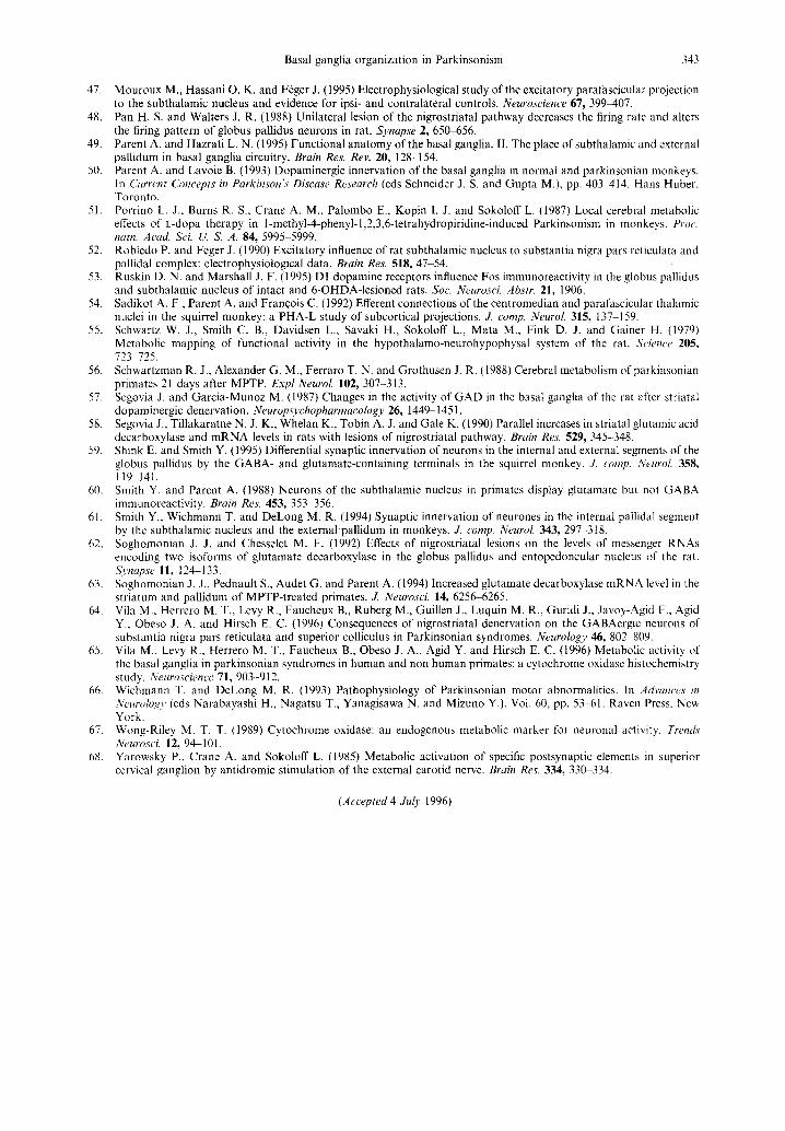
Several lines of evidence, including the biochemical data presented here, show that the STN is hyperac- tive in Parkinsonism, and that the GPi/SNpr which receive direct and strong projections from STN are also overactivated in this pathological condition. According to the current basal ganglia functional model, the inhibition of GPe output neurons is believed to be responsible for the high pathological activity of STN neurons in MPTP-intoxicated mon- keys. As indicated by our biochemical data, the GPe does not appear to be hypoactive in experimental Parkinsonism and thus is unlikely to be responsible for the hyperactivity seen in the STN and GPi/SNpr. In the hypothesis of a strong influence of the GPe on the activity of STN neurons, a normal or increased GPe activity should down-regulate the activity of STN neurons. This powerful influence is further questioned by a recent electrophysiological study in rodents showing that the direct destruction of the globus pallidus does not strongly influence the firing rate of STN neurons (19.5% increase), whereas nigral dopaminergic lesion by injections of 6-OHDA pro- duces a strong increase in the firing rate of STN neurons (105.7% increase) ~9 (Fig. 2). These data suggest that STN hyperactivity in Parkinsonism does not depend solely upon GPe-STN interaction. More- over, in an attempt to relieve the overactivity known to occur in the GPe in MPTP-intoxicated monkeys treated by L-dopa and displaying dyskinesias, Blancher et al. reported a worsening of both motor symptoms such as akinesia and L-dopa-induced dyskinesias after destruction of the GPe. 3 This leads to the notion that the dysfunction of the "indirect pathway" (connecting the striatum to the Gpi/SNpr through the GPe and the STN) in Parkinsonism may have a less important role in the physiopathology of the disease than previously assumed. Consequently, it is necessary to envisage alternative explanations for the overall increase in activity of the STN and

340 R. Levy et al.
I =
i
200ms
m 200 ms
ir IIII II II Illllll I N Illl Ilmgl II III111 It
!:
!ii i l liiiDI ill!hi I!!1!'1 i
200ms
Fig. 2. Samples of spontaneous unit activity of subthalamic neurons. These representative samples of unit activity were recorded in the subthalamic nucleus under urethane an- esthesia (1.2 g/kg, i.p.), in a control rat (top row), a rat with an excitotoxic lesion of the globus pallidus (middle row) and a rat with a 6-OHDA-induced midbrain dopaminergic lesion (bottom row). The mean firing rates were 14.7 + 0.4 spikes per second (sp/s) (n=34), 17±0.6sp/s (n=20) and 30.3± 1 sp/s (n=21), respectively. The mean firing rate of subthalamic neurons was not significantly altered in rats
with pallidal lesion compared with control rats. ~7
GPi /SNpr observed in Parkinsonism. One may ac- knowledge, however, that any at tempt to explain the hyperactivity of the STN on the basis of the current physiological and anatomical knowledge of its other afferent structures remains largely inferential. Thus,
Fig. 3. Schematic representation of a new model of organ- ization of the basal ganglia based on novel anatomical, neurochemical and physiological data. The main findings imply that GPe and STN display broad projections to more structures than those predicted by the "direct/indirect'" model of organization. On the one hand, in the present model, the striatum and STN are both considered as input structures for the basal ganglia. They both distribute corti- cal information, the striatum (from broader cortical areas) and STN (from more restricted cortical areas) to the same targets, namely GPe and GPi/SNpr, where they converge into the same groups of cells. On the other hand, the output structures (GPi/SNpr) are under the antagonistic effect of both input stations and a strong GABAergic input from GPe. Moreover, although it does not appear on the figure, all the above-mentioned structures are modulated by a direct dopaminergic projection from the substantia nigra pars compacta. GPe, external segment of the pallidum; GPi, internal segment of the pallidum; Pf, parafascicular nucleus of the thalamus; STR, striatum, STN, subthalamic nucleus:
SNpr, substantia nigra pars reticulata.
the following paragraph is aimed only at providing some clues for subsequent experiments.
Besides its input from the GPe, the STN receives projections from three afferent systems that may represent potential candidates to explain its hyper- activity in Parkinsonism. These are the glutamate- excitatory projections from the cerebral cortex, the glutamate-excitatory projections from the Pf nucleus and the dopaminergic pathway from the substantia nigra pars compacta (SNpc) and ventral tegmental a r e a . 6'46"47"54 It can be hypothesized that the increase in STN activity is due either to an increase in the excitatory effect produced by some of these structures (cerebral cortex and/or Pf nucleus) or to a decrease in the regulatory control exerted by dopaminergic neu- rons. The cerebral cortex and Pf nucleus are import- ant sources of glutamatergic excitation to the STN. Overactivity of these inputs could lead to STN hyperactivity. However, little is known about the pathological modifications that might occur in these circuits in MPTP-intoxicated monkeys. Anatomical and pharmacological data indicate that STN neurons

Basal ganglia organization in Parkinsonism 341
can be regulated by a direct dopaminergic projection from the SNpc. 29'34'5°'s3 It is now well established that the dopaminergic nigro-STN projection is mark- edly altered in MPTP-intoxicated monkeys. 5° How- ever, the effect of the release from this dopaminergic innervation remains controversial. Although a recent report has suggested that STN neurons may only express dopaminergic D 2 receptors 29 (which may imply an inhibitory effect of dopamine on STN neurons), studies performed with microiontophoretic application of dopamine report either an increase 43 or increase and decrease of the discharge rate of the recorded subthalamic neurons, 5 In summary, GPe does not appear to be responsible for STN hyperac- tivity in Parkinsonism. However, it is still open to speculation whether the release of dopaminergic in- nervation or the hypothetical increased activity of glutamatergic inputs from the cortex and/or Pf nu- cleus plays a crucial role in the hyperactivity of STN neurons.
CONCLUSIONS
In Parkinsonism, biochemical data indicate that metabolic and GABAergic activities in GPe neurons are quantitatively similar to those observed in normal conditions. Therefore, these findings modify the cur- rently accepted model of basal ganglia organization
in which the output structures (GPi/SNpr) are influ- enced by the balance between a direct striato-GPi/ SNpr pathway and an indirect multisynaptic striato- GPe-STN-GPi/SNpr pathway. Although it is too early to be able to propose a sound alternative model of basal ganglia organization, these new findings would appear to shift the weight of evidence in favor of a different arrangement involving the respective positions of the STN and GPe in basal ganglia circuitry. In such a scheme (Fig. 3), the STN is not merely a relay along the indirect pathway but is considered to be an input structure, bringing import- ant excitatory afferent inputs into the basal ganglia. 42 Its excitatory influence is crucial not only for the GPi/SNpr but also for GPe neuronal activity, which is under the antagonistic control of both the striatum and STN. This also highlights the fact that changes in the STN are not related exclusively to GPe activity, but also to its afferents from other brain areas. However, the precise influence of each of these other afferent systems on the STN remains to be deter- mined in normal and parkinsonian brains. Thus, the data reviewed here call for a revised pattern of connectivity that may provide a more satisfactory explanation of basal ganglia organization. Such a model would also help us to understand better the normal functions of the basal ganglia and the patho- logical changes occurring in basal ganglia diseases.
REFERENCES
1. Albin R. L., Young A. B. and Penney J. B. (1989) The functional anatomy of basal ganglia disorders. Trends Neurosci. 12, 366-376.
2. Baran H., Lassman H. and Hornykiewicz O. (1988) Behavioral and neurochemical changes 6 months after kainic acid. Soc. Neurosci, Abstr. 14, 472.
3. Blanchet P. J,. Boucher R. and Bedard P. J. (1994) Excitotoxic lateral pallidotomy does not relieve bdopa-induced dyskinesia in MPTP parkinsonian monkeys. Brain Res. 650, 32-39.
4. Blandini F. and Greenamyre J. T. (1995) Effect of subthalamic nucleus lesion on mitochondrial enzyme activity in rat basal ganglia. Brain Res. 669, 59-66.
5. Campbell G. A., Eckardt M. J. and Weight F. F. (1985) Dopaminergic mechanisms in subthalamic nucleus of the rat: analysis using horseradish peroxidase and microiontophoresis. Brain Res. 333, 261 270.
6. Canteras N. S., Shammah-Lagnado S. J., Silva B. and Ricardo J. A. (1990) Afferent connections of the subthalamic nucleus: a combined retrograde and anterograde horseradish peroxidase study in the rat. Brain Res. 513, 43-59.
7. Chergui K., Nomikos G. G., Math6 J. M., Gonon F. and Svensson T. H. (1996) Burst stimulation of the medial forebrain bundle selectively increases Fos immunoreactivity in the limbic forebrain of the rat. Neuroscience 72, 141 151.
8. Chesselet M. F., Weiss L., Wuenschell C., Tobin A. J. and Affolter H. U. (1987) Comparative distribution of RNAs for glutamic acid decarboxylase, tyrosine hydroxylase and tachykinins in the basal ganglia: an in situ hybridization study in the rodent brain..L comp. Neurol. 262, 125 140.
9. DeLong M. R. (1990) Primate models of movement disorders of basal ganglia origin. Trends' Neurosci. 13, 281 285. 10. Drengler S. M. and Oltmans G. A. (1993) Rapid increases in cerebellar Purkinje cell glutamic acid decarboxylase
(GAD67) mRNA after lesion-induced increases in cell firing. Brain Res. 615, 175 179. 11. Feldblum S., Erlander M. G. and Tobin A. J. (1993) Different distributions of GAD65 and GAD67 mRNAs suggest
that the two glutamate decarboxylases play distinctive functional roles. J. Neurosci. Res. 34, 689-706. 12. Filion M. (1979) Effects of interruption of the nigrostriatal pathway and of dopaminergic agents on the spontaneous
activity of the globus pallidus neurons in awake monkey. Brain Res. 178, 425441. 13, Filion M. and Tremblay L. (1991) Abnormal spontaneous activity of globus pallidus neurons in monkeys with
MPTP-induced Parkinsonism. Brain Res. 547, 142-151. 14, Fontaine B., Sassoon D., Buckingham M. and Changeux J. P. (1988) Detection of the nicotinic acetylcholine receptor
subunit mRNA by in situ hybridization at neuromuscular junction of 15-day old chick striatal muscles. Eur. molec. Biol. Org. J. 7, 603 609.
15. Gale K. and Casu M. (1981) Dynamic utilization of GABA in the substantia nigra: regulation by dopamine and GABA in the striatum and its clinical and behavioral implications. Molec. cell. Biochem. 39, 369405.
16, Gonon F. G. (1988) Nonlinear relationship between impulse flow and dopamine released by rat midbrain dopaminergic neurons as studied by in vivo electrochemistry. Neuroscience 24, 19-28.

342 R. Levy et al.
17. Guridi J., Herrero M.-T., Luquin M. R., Guill6n J., Ruberg M., Vila M., Laguna J., Javoy-Agid F., Agid Y. and Hirsch E. C. and Obeso J. A. (1996) Subthalamotomy in parkinsonian monkeys: behavioral and biochemical analysis. Brain (in press).
18. Hamada I. and DeLong M. R. (1992) Excitotoxic acid lesions of the primate subthalamic nucleus result in reduced pallidal neuronal activity during active holding. J. Neurophysiol. 68, 1859-1866.
19. Hassani O. K., Mouroux M. and F~ger J. (1996) Increased subthalamic neuronal activity after nigral dopaminergic lesion independent of disinhibition via the globus pallidus. Neuroscience 72, 105-115.
20. Hazrati L.-N. and Parent A. (1992) Convergence of subthalamic and striatal efferents at pallidal level in primates. Brain Res. 569, 336 340.
21. Hazrati L.-N. and Parent A. (1992) Differential patterns of arborization of striatal and subthalamic fibers in the two pallidal segments in primates. Brain Res. 598, 311 315.
22. Hazrati L.-N. and Parent A. (1993) The anatomical organization of the striatal and subthalamic afferents at the pallidal level in primates. Prog. Brain Res. 99, 89-104.
23. Hendry S. H. (1991) Delayed reduction in GABA and GAD immunoreactivity in the adult monkey dorsal lateral geniculate nucleus following monocular deprivation or enucleation. Expl Brain Res. 86, 47-59.
24. Herrero M. T., Levy R., Ruberg M., Javoy-Agid F., Luquin M. R., Agid Y., Hirsch E. C. and Obeso J. A. (1996) Glutamic acid decarboxylase mRNA expression in medial and lateral pallidal neurons in the MPTP-treated monkeys and patients with Parkinson's disease. Adv. Neurol. 69, 209 216.
25. Herrero M. T., Levy R., Ruberg M., Luquin M. R., Villares J., Guillen J., Faucheux B., Javoy-Agid F., Guridi J., Agid Y., Obeso J. A. and Hirsch E. C. (1996) Consequence of nigrostriatal denervation and L-dopa therapy on the expression of glutamic acid decarboxylase messenger RNA in the pallidum. Neurology 47, 219 224.
26. Herrero M. T., Ruberg M., Hirsch E. C., Mouatt A., Tobin A. J., Agid Y., Obeso J. A. and Javoy-Agid F. (1993) In situ hybridization of GAD mRNA in monkey and human brain: quantification at both regional and cellular levels. Neurosci. Lett. 157, 57-61.
27. Hevner R. F., Duff R. S. and Wong-Riley M. T. T. (1992) Coordination of ATP production and consumption in brain: parallel regulation of cytochrome oxidase and Na+,K+-ATPase. Neurosci. Len. 138, 188 192.
28. Hevner R. F. and Wong-Riley M. T. T. (1991) Neuronal expression of nuclear and mitochondrial genes for cytochrome oxidase (CO) subunits analyzed by in situ hybridization: comparison with CO activity and protein. ,L Neurosci. 11, 1942 1958.
29. Johnson A. E., Coirini H., K~illstr6m L. and Wiesel F. A. (1994) Characterization of dopamine receptor binding sites in the subthalamic nucleus. NeuroReport 5, 1836-1838.
30. Kincaid A. E., Albin R. L., Newman S. W., Penney J. B. and Young A. B. (1992) 6-Hydroxydopamine lesions of the nigrostriatal pathway alter expression of glutamate decarboxylase messenger RNA in rat globus pallidus projection neurons. Neuroscience 51,705 718.
31. Kincaid A. E., Penney J. B., Young A. B. and Newman S. W. (1991) The globus pallidus receives a projection from the parafascicular nucleus in rat. Brain Res. 553, 18 26.
32. Kita H. and Kitai S. T. (1991) Intracellular study of the rat globus pallidus neurons: membrane properties and responses to neostriatal, subthalamic and nigral stimulation. Brain Res. 564, 296 305.
33. Kitai S. T. and Kita H. (1987) Anatomy and physiology of subthalamic nucleus: a driving force of the basal ganglia. In The Basal Ganglia II: Structure and Function: Current Concepts (eds Carpenter M. B. and Jayaraman A.), pp. 357-373. Plenum Press, New York.
34. Kreiss D. S., Anderson L. A. and Walters J. 13'_ (1995) Subthalamic nucleus neuronal activity is increased by dopamine D1 receptor agonists. Soc. Neurosci. Abstr. 21, 914.
35. Levy R., Herrero M. T., Ruberg M., Villares J., Faucheux B., Guridi J., Guillen J., Luquin M. R., Javoy-Agid F., Obeso J. A., Agid Y. and Hirsch E. C. (1995) Effects of nigrostriatal denervation and L-dopa therapy on the GABAergic neurons of the striatum in MPTP-treated monkeys and Parkinson's disease. Eur. Z Neurosci. 7, 1199-1209.
36. Lindefors N., Brodin E., Tossman U., Segovia J. and Ungerstedt U. (1989) Tissue levels and in vivo release of tachykinins and GABA in striatum and substantia nigra of rat brain after unilateral striatal dopamine denervation. Expl Brain Res. 74, 527-534.
37. Litwak J., Mercugliano M., Oltmans G. A. and Chesselet M. F. (1990) Increased glutamic acid decarboxylase (GAD) mRNA and GAD activity in cerebellar Purkinje cells following lesion-induced increases in cells firing. Neurosci. Lett. 116, 179-183.
38. Lutes J., Lorden J. F., Davis B. J. and Oltmans G. A. (1992) GABA levels and GAD immunoreactivity in the deep cerebellar nuclei of rats with altered olivo-cerebellar function. Brain Res. Bull. 29, 329 336.
39. Marksteiner J. and Sperk G. (1988) Concomitant increase of somatostatin, neuropeptide Y and glutamate decarboxylase in the frontal cortex of rats with decrease seizure threshold. Neuroscience 26, 379-385.
40. Martin D. L. and Rimvall K. (1993) Regulation of gamma-aminobutyric acid synthesis in the brain. J. Neurochem. 60, 395-407.
41. Miller W. C. and DeLong M. R. (1987) Altered tonic activity of neurons in the globus pallidus and subthalamic nucleus in the primate MPTP model of Parkinsonism. In The Basal Ganglia H: Structure and Function." Current Concepts (eds Carpenter M. B. and Jayaraman A.), pp. 415-427. Plenum, New York.
42. Mink J. W. and Thach W. T. (1993) Basal ganglia intrinsic circuits and their role in behavior. Curr. Opin. NeurobioL 3, 950-957.
43. Mintz I., Hammond C. and F6ger J. (1986) Excitatory effect of iontophoretically applied dopamine on identified neurons of the rat subthalamic nucleus. Brain Res. 375, 172-175.
44. Mitchell I. J., Clarke C. E., Boyce S., Robertson R. G., Peggs D., Sambrook M. A. and Crossman A. R. (1989) Neural mechanisms underlying parkinsonian symptoms based upon regional uptake of 2-deoxyglucose in monkeys exposed to 1-methyl-4-phenyl-l,2,3,6-tetrahydropyridine. Neuroscience 32, 213 226.
45. Montgomery E. B., Buchholz S. R., Delitto A. and Collins R. C. (1985) Effects of MPTP on basal ganglia physiology in monkeys. Soc. Neurosci. Abstr. 11, 1161.
46. Mouroux M. and F6ger J. (1993) Evidence that the parafascicular projection to the subthalamic nucleus is glutamatergic. NeuroReport 4, 613 615.
Basal ganglia organization in Parkinsonism 343
47. Mouroux M., Hassani O. K. and F6ger J. (1995) Electrophysiological study of the excitatory parafascicular projection to the subthalamic nucleus and evidence for ipsi- and contralateral controls. Neuroscience 67, 399407.
48. Pan H. S. and Waiters J. R. (1988) Unilateral lesion of the nigrostriatal pathway decreases the firing rate and alters the firing pattern of globus pallidus neurons in rat. Synapse 2, 650-656.
49. Parent A. and Hazrati L. N. (1995) Functional anatomy of the basal ganglia. II. The place of subthalamic and external pallidum in basal ganglia circuitry. Brain Res. Rev. 20, 128-154.
50. Parent A. and Lavoie B. (1993) Dopaminergic innervation of the basal ganglia in normal and parkinsonian monkeys. In Current Concepts in Parkinson's Disease Research (eds Schneider J. S. and Gupta M.), pp. 403414. Hans Huber, Toronto.
51. Porrino L. J., Burns R. S., Crane A. M., Palombo E., Kopin I. J. and Sokoloff L. (1987) Local cerebral metabolic effects of L-dopa therapy in 1-methyl-4-phenyl-l,2,3,6-tetrahydropiridine-induced Parkinsonism in monkeys. Proc. hath. Acad. Sci. ~ S. A. 84, 5995-5999.
52. Robledo P. and Feger J. (1990) Excitatory influence of rat subthalamic nucleus to substantia nigra pars reticulata and pallidal complex: electrophysiological data. Brain Res. 518, 47 54.
53. Ruskin D. N. and Marshall J. F. (1995) D1 dopamine receptors influence Fos immunoreactivity in the globus pallidus and subthalamic nucleus of intact and 6-OHDA-lesioned rats. Soc. Neurosci. Abstr. 21, 1906.
54. Sadikot A. F., Parent A. and Francois C. (1992) Efferent connections of the centromedian and parafascicular thalamic nuclei in the squirrel monkey: a PHA-L study of subcortical projections. J. comp. Neurol. 315, 137 159.
55. Schwartz W. J., Smith C. B., Davidsen L., Savaki H., Sokoloff L., Mata M., Fink D. J. and Gainer H. (1979) Metabolic mapping of functional activity in the hypothalamo-neurohypophysal system of the rat. Scieme 205, 723 725.
56. Schwartzman R. J., Alexander G. M., Ferraro T. N. and Grothusen J. R. (1988) Cerebral metabolism of parkinsonian primates 21 days after MPTP. Expl Neurol. 102, 307-313.
57. Segovia J. and Garcia-Munoz M. (1987) Changes in the activity of GAD in the basal ganglia of the rat after striatal dopaminergic denervation. Neurop,s3,chopharmacology 26, 1449-1451.
58. Segovia J., Tillakaratne N. J. K., Whelan K., Tobin A. J. and Gale K. (1990) Parallel increases in striatal glutamic acid decarboxylase and mRNA levels in rats with lesions of nigrostriatal pathway. Brain Res. 529, 345-348.
59. Shink E. and Smith Y. (1995) Differential synaptic innervation of neurons in the internal and external segments of the globus pallidus by the GABA- and glutamate-containing terminals in the squirrel monkey. J. comp. Neurol. 358, 119 141.
60. Smith Y. and Parent A. (1988) Neurons of the subthalamic nucleus in primates display glutamate but not GABA immunoreactivity. Brain Res. 453, 353 356.
61. Smith Y., Wichmann T. and DeLong M. R. (1994) Synaptic innervation of neurones in the internal pallidal segment by the subthalamic nucleus and the external pallidum in monkeys. J. comp. Neurol. 343, 297-318.
62. Soghomonian J. J. and Chesselet M. F. (1992) Effects of nigrostriatal lesions on the levels of messenger RNAs encoding two isoforms of glutamate decarboxylase in the globus pallidus and entopedoncular nucleus of the rat. Synapse 11, 124-133.
63. Soghomonian J. J., Pednault S., Audet G. and Parent A. (1994) Increased glutamate decarboxylase mRNA level in the striatum and pallidum of MPTP-treated primates. J. Neurosci. 14, 6256-6265.
64. Vila M., Herrero M. T., Levy R., Faucheux B., Ruberg M., Guillen J., Luquin M. R., Guridi J., Javoy-Agid F., Agid Y., Obeso J. A. and Hirsch E. C. (1996) Consequences of nigrostriatal denervation on the GABAergic neurons of substantia nigra pars reticulata and superior colliculus in Parkinsonian syndromes. Neurology 46, 802 809.
65. Vila M.. Levy R., Herrero M. T., Faucheux B., Obeso J. A., Agid Y. and Hirsch E. C. (1996) Metabolic activity of the basal ganglia in parkinsonian syndromes in human and non human primates: a cytochrome oxidase histochemistry study. Neuroscience 71,903--912.
66. Wichmann T. and DeLong M. R. (1993) Pathophysiology of Parkinsonian motor abnormalities. In Advances in Neurology (eds Narabayashi H., Nagatsu T., Yanagisawa N. and Mizuno Y.), Vol. 60, pp. 53 61. Raven Press, New York.
67. Wong-Riley M. T. T. (1989) Cytochrome oxidase: an endogenous metabolic marker for neuronal activity. Trends Neurosci. 12, 94-101.
68. Yarowsky P., Crane A. and Sokoloff L. (1985) Metabolic activation of specific postsynaptic elements in superior cervical ganglion by antidromic stimulation of the external carotid nerve. Brain Res. 334, 330 334.
(Accepted 4 July 1996)
Copyright © 2022 FDOKUMEN