REDESCRlPTION OF IDIOSEPIUS PYGMAEUS STEENSTRUP, 1881 (CEPHALOPODA:IDIOSEPIIDAE), WITH MENTION OF...
10
Phuket mar. bioi. Cent. Res. Bull. 55:33-42 (1991) REDESCRlPfION OF IDIOSEPIUS PYGMAEUS STEENSTRup, 1881 (CEPHALOPODA:IDIOSEPIIDAE), WITH MENTION OF ADDITIONAL MORPHOLOGICAL CHARACTERS By Jorgen Hylleberg and Anuwat Nateewathana Phuket Marine Biolo'gical Center, P. O. Box 60, Phuket 83000, Thailand ABSfRACT Idiosepius pygmaeus Steenstrup, 1881 is redescribed an!! illustrated. Mantle up to 20.5 mm. Fins terminal, small and kidney-shaped. Mantle free from head along the margin, but mid-dorsally completely fused with funnel complex. Cartilagenous locking device with simple longitudinal grooves on funnel; oval ridges on mantle. Nuchal cartilage absent, but we interpret an oval depression in the central neck region as a rudimentary nuchal cartilage. Left and right 4th arms of males hectocoty- lized, normally with 1-3 uniserial suckers, but up to 4 suckers in this material. Suckers in 2 rows on sessile arms and 2-4 rows on tentacular club. Membranous gladius covers the posterior 2/3 of the dorsum. Eyes with opaque, wrinkled secondary· cornea in preserved specimens; anterior pores present. Olfactory crests prominent. Oviducts developed on both sides but functional on the left side only. Penis developed on left side only. We suggest that the genus Idiosepius is more closely related to Teuthida than to Sepiina.l. pygmaeus from mangroves on Phuket, Thailand is a new record for the Andaman Sea. INTRODUCTION When Steenstrup (1881; English transla- tion 1962) described Idiosepius pygmaeus, he found that it differed in so many essential points from the known genera that he had to erect a new genus to accomodate the species. Accord- ing to Steenstrup (1881) I. pygmaeus was a myopsid with the 4 th arm pair hectocotylized in males and with the normal appearance of a Loligo or Loliolus, having the small fins located posteriorly. On the other hand it had short arms like in sepiids and a similar locking apparatus on the mantle and funnel. Steenstrup (1881) found that 1. pygmaeus lacked a dorsal gladius. There was, however, an inner dorsal arch-shaped sinew or hqop, which supported the viscera. He had observed the peculiar sinew after cutting through the thin portion of the dorsal mantle. He found 2 firm strings, starting below the funnel collar, and diverging more and more posteriorly. He illustrated this so-called sinew in dorsal and lateral views (The present paper Figs. 50; 21' & 21"). Our material of Idiosepius pygmaeus agreed well with the general morphology of the species as shown by Steenstrup (1881; Figs. 10- 33). However, when we made a cut as de- scribed by Steenstrup (1962, p. 93), we failed to see anything resembling an arch-shaped sinew, and we found that the species possesed a thin, although very clear gladius. These findings made us re-examine Idiosepius pygmaeus in an attempt to discover what Steenstrup (1881) might have seen and interpreted as a sinew. This unusual supporting structure was the reason for naming the genus Idiosepius. Family IDIOSEPllDAE According to Nesis (1987) the genus Idiosepius comprises 6 valid species: I. pygmae- us Steenstrup, 1881 from the South China Sea, I. paradoxus (Ortmann, 1888) from Japan, I. picte- ti (Joubin, 1894) from Amboina, Indonesia, I. notoides Berry, 1Q21 from South Australia,
-
Upload
independent -
Category
Documents
-
view
1 -
download
0
Transcript of REDESCRlPTION OF IDIOSEPIUS PYGMAEUS STEENSTRUP, 1881 (CEPHALOPODA:IDIOSEPIIDAE), WITH MENTION OF...

Phuket mar. bioi. Cent. Res. Bull. 55:33-42 (1991)
REDESCRlPfION OF IDIOSEPIUS PYGMAEUS STEENSTRup, 1881(CEPHALOPODA:IDIOSEPIIDAE), WITH MENTION OF ADDITIONAL
MORPHOLOGICAL CHARACTERS
By Jorgen Hylleberg and Anuwat NateewathanaPhuket Marine Biolo'gical Center, P. O. Box 60, Phuket 83000, Thailand
ABSfRACT
Idiosepius pygmaeus Steenstrup, 1881 is redescribed an!! illustrated. Mantle up to 20.5 mm.Fins terminal, small and kidney-shaped. Mantle free from head along the margin, but mid-dorsallycompletely fused with funnel complex. Cartilagenous locking device with simple longitudinal grooveson funnel; oval ridges on mantle. Nuchal cartilage absent, but we interpret an oval depression in thecentral neck region as a rudimentary nuchal cartilage. Left and right 4th arms of males hectocotylized, normally with 1-3 uniserial suckers, but up to 4 suckers in this material. Suckers in 2 rows onsessile arms and 2-4 rows on tentacular club. Membranous gladius covers the posterior 2/3 of thedorsum. Eyes with opaque, wrinkled secondary·cornea in preserved specimens; anterior porespresent. Olfactory crests prominent. Oviducts developed on both sides but functional on the left sideonly. Penis developed on left side only. We suggest that the genus Idiosepius is more closely relatedto Teuthida than to Sepiina.l. pygmaeus from mangroves on Phuket, Thailand is a new record for theAndaman Sea.
INTRODUCTION
When Steenstrup (1881; English translation 1962) described Idiosepius pygmaeus, hefound that it differed in so many essential pointsfrom the known genera that he had to erect anew genus to accomodate the species. According to Steenstrup (1881) I. pygmaeus was amyopsid with the 4th arm pair hectocotylized inmales and with the normal appearance of aLoligo or Loliolus, having the small fins locatedposteriorly. On the other hand it had short armslike in sepiids and a similar locking apparatus onthe mantle and funnel. Steenstrup (1881) foundthat 1. pygmaeus lacked a dorsal gladius. Therewas, however, an inner dorsal arch-shaped sinewor hqop, which supported the viscera.
He had observed the peculiar sinew aftercutting through the thin portion of the dorsalmantle. He found 2 firm strings, starting belowthe funnel collar, and diverging more and moreposteriorly. He illustrated this so-called sinew indorsal and lateral views (The present paper Figs.50; 21' & 21").
Our material of Idiosepius pygmaeusagreed well with the general morphology of thespecies as shown by Steenstrup (1881; Figs. 1033). However, when we made a cut as described by Steenstrup (1962, p. 93), we failed tosee anything resembling an arch-shaped sinew,and we found that the species possesed a thin,although very clear gladius. These findingsmade us re-examine Idiosepius pygmaeus in anattempt to discover what Steenstrup (1881) mighthave seen and interpreted as a sinew. Thisunusual supporting structure was the reason fornaming the genus Idiosepius.
Family IDIOSEPllDAE
According to Nesis (1987) the genusIdiosepius comprises 6 valid species: I. pygmaeus Steenstrup, 1881 from the South China Sea, I.paradoxus (Ortmann, 1888) from Japan, I. picteti (Joubin, 1894) from Amboina, Indonesia, I.notoides Berry, 1Q21 from South Australia,
34 Phuket mar. bioi. Cent. Res. Bull.
5
O.5mm
yellow _0 'l::"o..6 " 0 °oqo~
black --.-~ 0 ••
1 mm ."c,o •00
spermatoPtlores
adhesive area
caecum
central nervous system
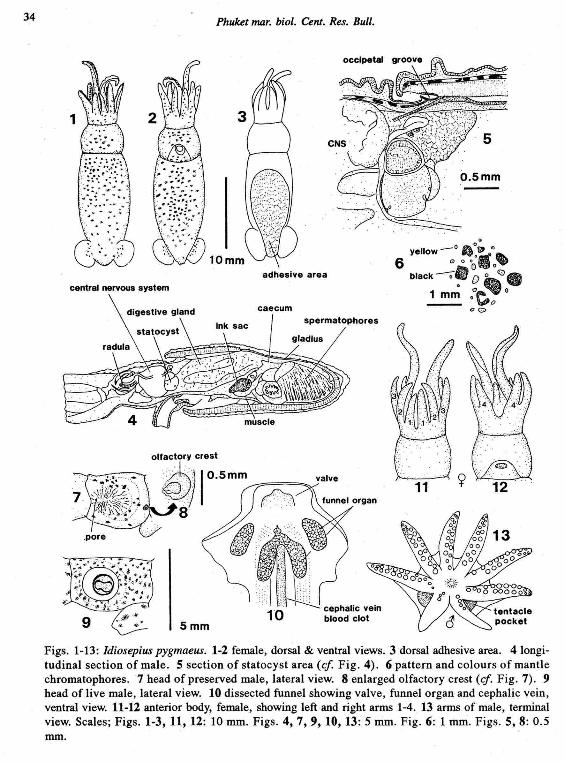
Figs. 1-13: Idiosepius pygmaeus. 1-2 female, dorsal & ventral views. 3 dorsal adhesive area. 4longitudinal section of male. 5 section of statocyst area (cf Fig. 4). 6 pattern and colours of mantlechromatophores. 7 head of preserved male, lateral view. 8 enlarged olfactory crest (cf Fig. 7). 9head of live male, lateral view. 10 dissected funnel showing valve, funnel organ and cephalic vein,ventral view. 11-12 anterior body, female, showing left and right arms 1-4. 13 arms of male, terminalview. Scales; Figs. 1-3,11,12: 10 mm. Figs. 4,7,9,10,13: 5 mm. Fig. 6: 1 mm. Figs. 5, 8: 0.5rom.

Redescription ofldiosepius pygmaeus 35
I. biserialis Voss, 1962 from South Africa, andI. macrocheir Voss, 1962 from South Africa.
Grimpe (1931) divided I. pygmaeus into3 subspecies with I. pygmaeus hebereri in thesouthern Pacific Ocean, I. pygmaeus pygmaeusin the central Indian Ocean, and I. pygmaeusparadoxus in the northern part of the PacificOcean. These subspecies have not been acceptedby cephalopod researchers (Voss, 1963; Nesis,1987).
Idiosepius pygmaeus Steenstrup, 1881Figs. 1-49
Idiosepius pygmaeus Steenstrup, 1881, p. 219;Berry, 1921, p. 357; 1932, p. 46; Grimpe,1931, p. 165-174; Voss, 1963, p. 63-67; Nesis,1987, p. 137-141.
MATERIAL EXAMINED: PMBC 7268,26males and 5 females, Klong Mudong, surfacewater of mangrove channel at Ao Chalong,Phuket Island, 10.7.1988 and 18.7.1990. Handnetting, coll. S. Chantrapornsyl, A. Nateewathana and J. Hylleberg.
DESCRIYflON:
MANTLE: elongate, tapering and rounded atposterior end. Ventrally 0.4-0.6 mm thick;middorsally 0.1 mm in females (Fig. 24); thinner in the smaller males (Figs. 4,5). The dorsalmargin straight (Fig. 1); ventral margin slightlyexcavated (Fig. 2). The posterior 3/4 of thedorsal mantle surface carries a prominent,rugose area which is strongly adhesive in liveanimals due to mucous secretion. The area iseasy to detect in live animals by probing with astick but not always easy to see in preservedanimals (Figs. 1, 3). The ADHESIVE AREA isutilized by the squid to attach to objects in thewater, e.g. prop roots and drifting leaves. Themantle is free around the neck but united to thefunnel complex by complete fusion about 1 mmfrom the dorsal edge of the mantle (Fig. 4).
On the inner surface of the free part ofthe mantle is a small, narrow MUSCULAR"SCAR" located middorsally above a corresponding oval depression on the neck termedoccipetal grove on Fig. 5. We interpret thisstructure as a rudimentary nuchal funnel cartilage.
In the posterior half of the mantle cavity, behind the gills, a VENTRAL MUSCLEconnects the mantle to the body (Figs. 4, 34). Avertical muscular septum connects the mantleand body and divides the mantle cavity into rightand left halves from above the ventral muscle tothe posterior end of the body.
A pair of STELLATE GANGLIA areeasily detached from the mantle in preservedspecimens, and seen as a rounded discs togetherwith the mantle connectives on either side of thedorsal median line after removal of the mantle(Fig. 33). Purple-brown and yellow CHROMATOPHORES cover the body (Fig. 6). Theanimals adapt a pale or black coloration according to their state of exitement. When, disturbedthe body turns black and the squid ejects acoherent cloud of ink.
FINS: small, separate and attached to the base ofthe body at an angle (Figs. 1-3). Fins are kidney-shaped, distally free, projecting slightlybeyond the posterior body. The muscular basecarries many chromatophores; the outer fin ishyaline and very thin (Fig. 1); margin undulatingin preserved specimens (not shown).
HEAD: small and compact, the width less thanthat of the mantle (Figs. 1-3); it extends belowthe mantle in preserved specimens and dorsallythe neck carries an oblong oval depression interpreted as part of a rudimentary nuchal lockingapparatus (Figs. 5, 37). The eyes are hardlyvisible in preserved specimens on account ofwrinkled, opaque secondary cornea (Fig. 7). Inlive animals the eyes are silvery; the iris contractable to an oblong slit (Fig. 9) or completely

36
5mm
27
stellate ganglion
14
funnel cartilage
24
mantle
I0.1 mm
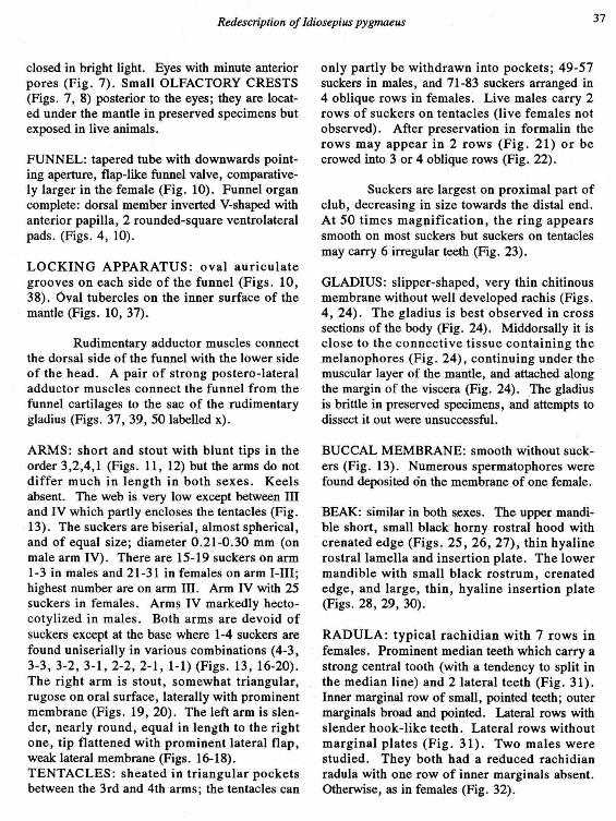
Figs. 14-33. ldiosepius pygmaeus. 14 mantle cartilage, longitudinal section, female. 15 funnel cartilage, longitudinal section, female. 16-18 hectocotylized left arm, lateral, oral a~d dorsal views. 19-20hectocotylized right arm, oral and lateral views. 21 club of male. 22 club of male showing 2 horizontal and 4 oblique transverse rows of suckers, semischematic. 23 sucker from base of male club. 24transverse section of female showing thin dorsal part of mantle and position of gladius. 25-27 uppermandible, male and female. 28-30 lower mandible, male and female. 31-32 teeth of radula, male andfemale. 33 male body, mantle removed, dorsal view. Scales; Fig. 33: 5 mm. Figs. 14, 15,21, 2530: 1 mm. Figs. 16-20,22,24: 0-5 mm. Fig. 23: 0.1 mm. Figs. 31,32: 0.05 mm.
Redescription of Idiosepius pygmaeus 37
closed in bright light. Eyes with minute anteriorpores (Fig. 7). Small OLFACTORY CRESTS(Figs. 7, 8) posterior to the eyes; they are located under the mantle in preserved specimens butexposed in live animals.
FUNNEL: tapered tube with downwards pointing aperture, flap-like funnel valve, comparatively larger in the female (Fig. 10). Funnel organcomplete: dorsal member inverted V-shaped withanterior papilla, 2 rounded-square ventrolateralpads. (Figs. 4, 10).
LOCKING APPARATUS: oval auriculategrooves on each side of the funnel (Figs. 10,38). Oval tubercles on the inner surface of themantle (Figs. 10,37).
Rudimentary adductor muscles connectthe dorsal side of the funnel with the lower sideof the head. A pair of strong postero-Iateraladductor muscles connect the funnel from thefunnel cartilages to the sac of the rudimentarygladius (Figs. 37, 39, 50 labelled x).
ARMS: short and stout with blunt tips in theorder 3,2,4,1 (Figs. 11, 12) but the arms do notdiffer much in length in both sexes. Keelsabsent. The web is very low except between IIIand IV which partly encloses the tentacles (Fig.13). The suckers are biserial, almost spherical,and of equal size; diameter 0.21-0.30 mm (onmale arm IV). There are 15-19 suckers on arm1-3 in males and 21-31 in females on arm I-III;highest number are on arm III. Arm IV with 25suckers in females. Arm~ IV markedly hectocotylized in males. Both arms are devoid ofsuckers except at the base where 1-4 suckers arefound uniserially in various combinations -(4-3,3-3,3-2,3-1,2-2,2-1,1-1) (Figs. 13,16-20).The right arm is stout, somewhat triangular,rugose on oral surface, laterally with prominentmembrane (Figs. 19, 20). The left arm is slender, nearly round, equal in length to the rightone, tip flattened with prominent lateral flap,weak lateral membrane (Figs. 16-18).TENTACLES: sheated in triangular pocketsbetween the 3rd and 4th arms; the tentacles can
only partly be withdrawn into pockets; 49-57suckers in males, and 71-83 suckers arranged in4 oblique rows in females. Live males carry 2rows of suckers on tentacles (live females notobserved). After preservation in formalin therows may appear in 2 rows (Fig. 21) or becrowed into 3 or 4 oblique rows (Fig. 22).
Suckers are largest on proximal part ofclub, decreasing in size towards the distal end.At 50 times magpification, the ring appearssmooth on most suckers but suckers on tentaclesmay carry 6 irregular teeth (Fig. i~).
GLADIUS: slipper-shaped, very thin chitinousmembrane without well developed rachis (Figs.4,24). The gladius is best observed in crosssections of the body (Fig. 24). Middorsally it isclose to the connective tissue containing themelanophores (Fig. 24), continuing under themuscular layer of the mantle, and attached alongthe margin of the viscera (Fig. 24). The gladiusis brittle in preserved specimens, and attempts todissect it out were unsuccessful.
BUCCAL MEMBRANE: smooth without suckers (Fig. 13). Numerous spermatophores werefound deposited on the membrane of one female.
BEAK: similar in both sexes. The upper mandible short, small black horny rostral hood withcrenated edge (Figs. 25, 26, 27), thin hyalinerostral lamella and insertion plate. The lowermandible with small black rostrum, crenatededge, and large, thin, hyaline insertion plate(Figs. 28, 29, 30).
RADULA: typical rachidian with 7 rows infemales. Prominent median teeth which carry astrong central tooth (with a tendency to split inthe median line) and 2 lateral teeth (Fig. 31).Inner marginal row of small, pointed teeth; outermarginals broad and pointed. Lateral rows withslender hook-like teeth. Lateral rows withoutmarginal plates (Fig. 31). Two males werestudied. They both had a reduced rachidianradula with one row of inner marginals absent.Otherwise, as in females (Fig. 32).

38
~4
lip-=:za
lip nidamental gland
occipetal groove
mantle
sperm mass
0.2 mm
cementbody
35
Figs. 34-45. Idiosepius pygmaeus. 34 male body, mantle removed, ventral view. 35 spermatohore.36 enlarged terminal cap (cf Fig. 35). 37 female body, mantle removed, dorsal view. 38 femalebody, mantle removed, ventral view. 39 female body, mantle removed, lateral view. 40 oviduct,gland and gonopore, left side. 41 non-functional gonopore, gland and branchial heart, right side. 42egg, dissected from ovary. 43nidamental gland, showing blood vessel, removed from the left side (cfFig. 38). 44 longitudinal'section of nidamental gland showing lips and vein, left side. 45 nidamentalgland, removed from the right side. Scales; Figs. 34,37-41,43,45: 5 mm. Figs 42-43: 1 mm. Fig.36: 0.1 mm.

Redescription of Idiosepius pygmaeus 39
REPRODUCTIVE SYSTEM: Male. Spermatophoric organ tightly packed with testis, glandsand prostate (Fig. 34) and surrounded with acover forming the genital sac occupying theposterior half of the male body. The exactnumber of glands and their connections was notstudied. All males contained many spermatophores in the voluminous spermatophoric sac(Fig. 33) which on the left hand side (Fig. 34)turned into a fairly short, cylindrical penis notreaching the edge of the funnel.
SPERMATOPHORES: measured about 2.1 mmin length and 0.13 mm in width (Fig. 35). Thegranular sperm mass occupied half of the lengthof the spermatophore. The cement body, supporting cylinder, and ejaculatory apparatusconstituted the other half (Fig. 35). Ejaculatoryapparatus coiled, terminated with a long filament(Fig. 36).
FEMALE REPRODUCTIVE SYSTEM: Largeovary occupy posterior part of body (Figs. 3739). Both oviducts present (Figs. 40, 41) butonly the left one is functional (Fig. 40). Oviductpacked with oval eggs, about 1 mm long (Fig.42). Oviducal glands egg-shaped, joined withoviduct at one end and projecting as a slender lipat the other end (Fig. 40). Large nidamentalglands dominate the ventral body (Figs. 38, 39).The glands are elongated oval with short lips atanterior ends (Figs. 43, 44). The small accessory nidamental glands do not project anterior tothe. nidamental glands (Fig. 45).
INK SAC: pyriform, compact organ with silveryouter coating. In females: hidden under thenidamental glands (Fig. 38); in males: exposedon the dorsal side of the intestine in front of thebranchial hearts (Figs. 4 ,34). The reservoirfilled with dark brown ink; the duct as long asthe ink sac (Figs. 4, 48).
DIGESTIVE SYSTEM: The oesophagus connects the buccal complex (Fig. 4) with thestomach, passing anteriorly through the brain(Fig. 46), continuing through the large midguthepato-pancreatic gland (Fig. 47). The small
stomach is hidden within the visceral mass whilethe prominent spiral part of the caecum dominates the posterior surface of the body (Figs. 33,37). The intestine continues from the stomach tothe ventral surface of the hepato-pancreaticgland, and terminates with the anus providedwith 2 lanceolate flaps (Fig. 48).
INNER CARTILAGENOUS SKELETON:located posterior to the eyes behind the centralnervous system (CNS) transition between headand body. It is a rounded capsule with 3 sections: one dorsal part housing the brain and 2ventro-Iateral statocysts (Figs. 4, 5).
STATOCYSTS: with well developed maculae.Details in maculae, cristae and anticristae werenot studied. Calcified statoliths not present (Fig.5).
CIRCULATORY SYSTEM: Blood of livinganimals colourless. In preserved specimens theblood coagulates into a hard and brittle substanceof brownish-green hue. Clot of blood is particularly prominent in veins: the cephalic vein, venacava, in the branchial hearts, and in gills.
The spacious pericardial coelom contains the heart, branchial hearts (Fig. 45), branchial glands and renal sacs. The branchial heartsare muscular and provided with white, roundedglands. GILLS attached to the inner mantle wallby mesenterium. The free side of each demibranch has about 45 plicate filaments in females(Fig. 49) and about 30 filaments in males.
REMARK: On the basis of dissections of freshand preserved specimens, we suggest that thestructure Steenstrup (1881) referred to as sinewin the diaphragm (Fig. 50; 21 "d) most likely isthe cephalic vein containing coagulated blood(see Fig. 10). Similarly, the structure Steenstrupcalled inner sinus rod is part of the vascularsystem (Fig. 50; 21') and Fig. 50; 21" shows thesame structure seen from the right side. It is erroneously labelled b. One possibility would bethat Steenstrup saw coagulated blood in the

40 Phuket mar. bioi. Cent. Res. Bull.
oesophagus
~~~
5 mmretractor
muscle
digestive gland
branchial heart
1 mm
Figs. 46-50. ldiosepius pygmaeus. 46 transverse section in front of the funnel, posterior view, female. 47 transverse section behind the funnel, anterior, female. 48 part of the digestive system,dissection of the left side, female. 49 gill, branchial heart and branchial heart appendage dissectedfrom right side of female.
50 copy of Fig. 21',22", Steenstrup (1881). The original legend reads: "The inner sinus rod of ldiosep'ius, seen from the dorsal side, 21' , and from the right side 21". a=funnel grooves, b= right retractor infundibuli, c= lens-shaped ink sac, d= sinew in the diaphram."
The correct legend should read: 21': d = funnel groove, also termed funnel cartilage in thepresent text. - 21": a = funnel groove, b = inner sinew rod, c = lens-shaped ink sac, d = sinew inthe diaphragm. Furthermore, to 21" we have added the letter x = right retractor infundibuli. Scale;Figs. 46-48: 5 rom., Fig. 49: 1 rom. Fig. 50: not to scale.
Redescription ofIdiosepius pygmaeus 41
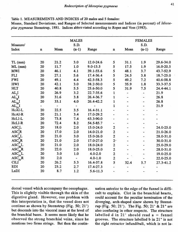
Table 1. MEASUREMENTS AND INDICES of 20 males and 5 females:Means, Standard Deviations, and Ranges of Selected measurements and Indices (in percent) of Idiose-pius pygmaeus Steenstrup, 1881. Indices abbreviated according to Ropes and Voss (1983).
MALES FEMALESMeasure! S.D. S.D.Index n Mean (n-l) Range n Mean (n-l) Range
TL(mm) 20 21.2 3.0 12.0-24.6 5 31.1 1.9 29.6-34.0ML(mm) 20 11.7 1.0 9.0-13.5 5 17.5 1.9 16.0-20.5MWI 20 46.1 4.1 39.1-55.6 5 48.1 3.7 43.7-52.9FLI 20 27.1 5.6 17.4-36.4 5 24.5 3.8 18.7-25.0FWI 20 49.1 4.4 42.5-58.3 5 49.2 7.2 40.6-58.8HWI 20 42.1 3.0 38.2-50.0 5 35.9 1.8 33.3-37.5HLT 20 40.8 5.5 25.6-50.0 5 31.9 7.5 24.4-44.1ALt 20 26.9 3.2 22.7-35.6 1 21.9ALJ 20 31.6 3.8 26.4-36.7 1 26.8ALmI 20 33.1 4.0 26.4-42.2 1 26.8AL~ 1 21.9HcAl-L 20 22.5 3.5 16.4-31.1HcAI-R 20 21.1 3.4 17.0-29.2HcLI-L 20 73.8 7.4 63.3-90.0HcLI-R 20 72.4 8.2 54.5-85.7ASC1L 20 18.0 2.0 15.0-24.0 2 24.0-25.0ASC' 20 17.0 2.0 14.0-21.0 2 21.0-26.0ASCuL 20 21.0 3.0 15.0-26.0 2 28.0-31.0ASC~ 20 21.0 2.0 17.0-27.0 2- 30.0-31.0
. ASCot- 20 21.0 2.0 18.0-24.0 2 25.0-29.0ASC~ 20 22.0 2.0 18.0-25.0 2 28.0-31.0ASC~ 20 3.0 1.0 4.0-2.0 2 19.0-25.0ASC~ 20 2.0 4.0-1.0 2 22.0-25.0C1LI 20 26.2 5.3 16.4-37.8 5 32.4 5.7 27.3-41.2EDI 20 23.2 2.7 17.4-27.3LnDI 20 8.7 1.2 5.6-11.3
dorsal vessel which accompany the oesophagus.This is slightly visible through the skin of thedigestive gland. However, one problem withthis interpretation is, that the vessel does notcontinue as shown by Steenstrup (Fig. 50; 21 ')but descends into the visceral mass at the level ofthe branchial bases. It seems more likely that heobserved the strong branchial veins, since hementions two firms strings. But then the contin-
uation anterior to the edge of the funnel is difficult to explain. Clot in the branchial hearts,could account for the peculiar termination of thediverging, arch-shaped sinew shown by Steenstrup (Fig. 50; 21 '). The Fig. 50; 21' & 21" arealso ·.confusing in other respects. The structurelabelled d in 21' should read a = funnelgrooves. The structure labelled b in 21" is notthe right retractor infundibuli, which is not la-

42Phuket mar. bioi. Cent. Res. Bull.
belled in 21"; we have added the letter x to theFigure. The x shows the right retractor infundibuli while b should read inner sinew rod.
Steenstrup (1881) compared this innersinew rod to the inner ring around the marginsof the spongy substratum (loculomenta) found inSepia brevimana Steenstrup. However, in viewof the absence of such a ring, and the presenceof a thin, but distinct gladius, and an ovalmuscular "scar" in the position of a nuchalfunnel locking cartilage, we suggest that thegenus ldiosepius is more closely related toTeuthida than to Sepiina.
Voss (1953) described Pickfordiateuthispulchella. a myopsid squid which he placed inthe family Pickfordiateuthidae. This squidresembles ldiosepius in several respects, especially in the external morphology, but P. pulchella has nuchal locking apparatus present and afeather-like gladius with a broad fane. P. pulchella is placed in the order Teuthida, suborderMyopsida by Nesis (1987). At present we are
unable to enter into a discussion concerning thesystematics of myopsis squids. We believe thatstudies of ontogenesis are necessary in order tosolve the question of relationships between Idio-
. sepiidae, Pickfordiateuthidae, and Loliginidae.Berry (1932) remarked that much still remainedto be worked out concerning the exact naturalposition are relationships of the genus ldiosepius. According to Berry (1932) its position israther isolated on basis of statocyst structure; theadult organ remaining in a somewhat primitivecondition. In view of the rudimentary gladius,and possibly rudimentary nuchal locking apparatus, we suggest that the relationships of myopsidsquids should be reconsidered by systematists.
ACKNOWLEDGEMENTS
We are most grateful to Dr. J. Knudsen,Zoological Museum, Copenhagen, for commentsto the manuscript, Mrs Marianne Pereira,Zoological Museum, Copenhagen, for providingvaluable references and Mr. Jumroen Keogaeofor typing the manuscript.
REFERENCES
Berry, S.S. 1921. A review of the cephalopod genera Sepioloidea. Sepiadarium. and ldiosepius.Rec. South Australian Mus.• 1:347-364.
Berry, S.S. 1932. Cephalopods of the genera Sepioloidea. Sepiadarium. and ldiosepius. Philippine J.Sci.• 47(1):39-55.
Grimpe, G. 1931. Teuthologische Mitteilungen. XIII. Uber die Cephalopoden der Sunda-ExpeditionRensch. Zool. Anz.• 95:149-174.
Nesis, K.N. 1987. Cephalopods of the world. T.F.H. Publication, Inc. Ltd., New Jersey, 351 pp.Roper, C.F.E. & G.L. Voss. 1983. Guidelines for taxonomic descriptions of cephalopod species.
Memoirs ofNational Museum Victoria No.44:49-63Steenstrup, J. 1881. Sepiadarium. og ldiosepius to nye Slaegter af Sepiernes Familie. Med Be
maerkninger om de to beslaegtede Former Sepioloidea D'Orb. og Spirula Lmk. K. DanskeVidensk. Selsk. Skrift.• ser. 6, 1(3):213-242.
Steenstrup, J. 1962. The cephalopod papers of Japetus Steenstrup. A translation into Eng., Copenhagen: Danish Sci. Press, 330 pp.
Voss, G.L. 1953. A new family, genus, and species of myopsid squid from the Florida Keys. Bull.Mar. Sci. Gulf Carib.• 2(4):602-609.
Voss, G.L. 1963. Cephalopods of the Philippine Islands. Bull. U.S. Nat. Mus.• 234:1-180

![Gebru Abba C̣hequn to Achille Raffray, 16 Jan. [1881]](https://static.fdokumen.com/doc/165x107/6317f85c3394f2252e028a6e/gebru-abba-chequn-to-achille-raffray-16-jan-1881.jpg)