Corneal reshaping patient information brochure - GP Specialists

ARTICLE IN PRESS
0142-9612/$ - se
doi:10.1016/j.bi
�Correspondmental Techno
Montreal Road
3644; fax: +1-6
E-mail addr
Biomaterials 26 (2005) 3093–3104
www.elsevier.com/locate/biomaterials
Recruitment of multiple cell lines by collagen-synthetic copolymermatrices in corneal regeneration
F. Lia,b, M. Griffitha, Z. Lib, S. Tanodekaewb,c, H. Sheardownd, M. Hakima,b,D.J. Carlssona,b,�
aUniversity of Ottawa Eye Inst., 501 Smyth Road, Ottawa, Canada ON K1H 8L6bNational Research Council, 1500 Montreal Road, Ottawa, Canada ON K1A 0R6
cNational Science and Technology Development Center, Pathumthani 12120, ThailanddDepartment of Chemical Engineering, McMaster University, Hamilton, Canada ON L8S 4L7
Received 25 May 2004; accepted 30 July 2004
Available online 15 September 2004
Abstract
Collagen hydrogel matrices with high optical clarity have been developed from collagen I, cross-linked with a copolymer based on
N-isopropylacrylamide, acrylic acid and acryloxysuccinimide. The controlled reaction of collagen amine groups with this copolymer
under neutral pH and aqueous conditions gave robust, optically clear hydrogels and prevented the excessive collagen fibrillogenesis
that can lead to collagen opacity. These sterile, non-cytotoxic hydrogels allowed epithelial cell overgrowth and both stromal cell and
nerve neurite ingrowth from the host tissue. This regenerative ability appeared to result from the high glucose permeability,
nanoporosity and the presence of cell adhesion factors, RGD in collagen and the laminin pentapeptide, YIGSR, grafted onto the
copolymer. Under physiological conditions, optical clarity superior to the human cornea and tensile performance adequate for
suturing were obtained from some formulations.
r 2004 Elsevier Ltd. All rights reserved.
Keywords: Collagen-copolymer hydrogel; Cell recruitment; Permeability; Porosity; Topography; Optical clarity
1. Introduction
Tissue-engineered (TE) matrices designed to emulatekey properties of the natural extracellular matrices oftissues and organs may promote wound healing andtissue repair or regeneration. TE matrices may beimplanted either fully furnished with living cells, or assterile, non-cytotoxic matrices. The latter may becomefunctional by promoting ingrowth of autologous hostcell lines and regeneration of damaged nerves from thesurrounding tissue [1], obviating the need for donor cells
e front matter r 2004 Elsevier Ltd. All rights reserved.
omaterials.2004.07.063
ing author. Institute of Chemical Process and Environ-
logy, National Research Council of Canada, 1200
, Ottawa, Ont., Canada K1A 0R6. Tel.: +1-613-990-
13-991-2384.
ess: [email protected] (D.J. Carlsson).
that could promote immune and rejection reactions.Several three-dimensional (3D) matrices, based oncross-linked biopolymers or hydrophilic synthetic poly-mers have been explored as scaffolds for single cell lines(chondrocytes, osteoblasts, fibroblasts, pancreatic islets,etc.) [2–6]. Tissue engineering a functional, humancorneal implant is particularly demanding because itmay require recruitment of several active cell lines,including stromal (in the bulk), epithelial (anteriorsurface) and endothelial (posterior) cells, as well asnerve networks. Furthermore, the matrix must have lowlight absorption and scattering in the visible region andacceptable refractive power. Finally, it must haveadequate strength to survive both implantation (usuallysuturing) and the wear and tear of normal use.Recently, we have reported the promotion of corneal
cell and nerve growth in vitro by hydrogels, based on

ARTICLE IN PRESSF. Li et al. / Biomaterials 26 (2005) 3093–31043094
collagen I with N-isopropylacrylamide (NiPAAm)copolymers [7]. When implanted into pigs’ corneas,these sterile matrices rapidly recruited epithelial andstromal cells from the hosts’ tissue to reconstitute thecornea. Furthermore, functional (touch sensitive) nervesquickly regenerated in these implants. Although othercorneal substitutes had been reported [8–12], our workappeared to be the first report of an implantable matrixthat performed as a physiologically functional, tissuesubstitute and not simply as a prosthesis. In this paper,we describe in detail the preparation of the copolymersand their collagen hydrogels and discus the physical andchemical properties leading to functional cornealimplants.
2. Materials and methods
2.1. Materials
Chemicals were obtained from Sigma-Aldrich (Oak-ville, Canada) except for poly(N-isopropylacrylamide)(PNiPAAm, Mn ¼ 4:0� 104 Da) and acryloxysuccini-mide (ASI) (from PolySciences, Warington, PA). Solidmonomers and the initiator were recrystallized andacrylic acid (AAc) was distilled before use. Type Icollagen (4wt/vol%) was concentrated from acidic,bovine atelocollagen solutions (Becton Dickinson, Oak-ville, Canada) or purchased directly from Koken(Tokyo, Japan). Both 4% solutions gave identicalperformance. MilliQ water was used throughout.
2.2. Characterization and synthesis of polymers
Proton nuclear magnetic resonance spectra (1HNMR,Varian 400MHz spectrometer) were recorded in deutero-tetrahydrofuran (THF-d8) or acetone-d6. Gel permea-tion chromatography (GPC, Waters chromatographysystem, Model 410, refractive index detector) was run at
C
O
H3C
OH
CH3
O
O
N
NH
O OC C
CH
C
O
H3C
OH
CH3
O
O
N
NH
O OC OC
CH
O
+
+ m
(TERP)
(TERPX)
NH2
NH
X
X
CO O
CH2 )x CH2 CH CH2CH CH( ( )y ( )z
CH2 )x CH2 CH CH2 CH CH2CH CH( ( )y ( )z-m )m(
Fig. 1. Reaction of ASI groups of TERP copolymer with amine groups. TE
YIGSR or glycine. When X-NH2 was YIGSR, TERPX became TERP5 wit
30 1C with water as eluent after calibration withpolyethylene oxide standards (Polymer LaboratoriesLtd., Amherst, MA). Transmission, Fourier transforminfrared (FTIR) spectra were recorded with a Midac Mspectrometer equipped with a diamond compression cell(Graseby-Specac Diasqueeze).The copolymer, poly(N-isopropylacrylamide-co-
acrylic acid) (PNiPAAm-co-AAc, designated COP),was prepared by free-radical polymerization of Ni-PAAm (10.75 g, 95mmol) and AAc (0.36 g, 5mmol) in1,4-dioxane (100ml) with 2,20-azobis-isobutyronitrileinitiator (AIBN, 0.12 g, 7.216� 10�3mol/mol mono-mer) under nitrogen at 70 1C for 24 h. The product waspurified by repeated precipitation from THF into etherand vacuum dried (78% yield). The terpolymer, poly(N-isopropylacrylamide-co-acrylic acid-co-acryloxysuccini-mide (PNiPAAm-co-AAc-co-ASI, designated TERP,Fig. 1) was synthesized by free radical copolymerizationof the three monomers with the feed molar ratio of85:10:5 (NiPAAm: AAc: ASI) in 1,4-dioxane at 70 1C,initiated and purified (82% yield) as for COP. Polymercompositions were established by 1HNMR (Table 1). Abio-active terpolymer (designated TERP5) was synthe-sized by reacting TERP (1.0 g) with the lamininpentapeptide motif [YIGSR, H-tyrosine, isoleucine,glycine, serine, arginine–NH2 (carboxyamide-termi-nated), 2.8 mg from Nova Biochem, Laufelfingen,Switzerland] in N,N-dimethylformamide for 48 h at20 1C (Fig. 1). If all of the YIGSR pentapeptide wasassumed to be grafted to reactive ASI groups, then thefinal composition of the recovered polymer wasx:y:z:m=84.2:9.8:6.0:5� 10�5 (all in molar equiva-lents). The ASI groups remaining after reaction withthe pentapeptide were available for hydrogel formationby subsequent reaction with collagen amine groups.TERP and TERP5 were stored under dry nitrogen [13].To measure the polymers’ lower critical solution
temperatures (LCST), a solution of each polymer (2mg/ml in phosphate buffered saline, PBS) was aliquoted
0
5
10
15
20
25
30
35
40
0 2 3 4 5ASI/-NH2 (Molar Equivalent)
Col
lage
n -N
H2
Loss
(%
)
ON
OH
1
RP: x:y:z=84.2:9.8:6.0. X-NH2 includes collagen (see kinetic curve),
h x:y:z:m=84.1:9.8:6.0:5� 10�5.

ARTICLE IN PRESS
Table 1
Polymer characterization and properties
Polymer Monomers in feed (molar ratio) Monomer units found (molar ratio) Mn (Da) Mw (Da) Mw/Mn LCSTa
COP 95:5b 95.3:4.7b,c 4.1� 104 7.1� 104 1.73 54 1C
93.6:6.4b,d
TERP 85:10:5e 84.2:9.8:6.0e,f 5.6� 104 9.0� 104 1.58 455 oC
TERP5 84.2:9.8:6.0: 5� 10�5g,h 5.6� 104 9.0� 104 1.58 455 oC
PNiPAAm Homopolymer 4.0� 104 30 1C
aThe temperature of the onset of opalescence of polymer solutions in PBS at 2mg/ml.bNiPAAm: AAc.cBy 1HNMR after complete carboxyl methylation with BF3-methanol.dBy aqueous titration with NaOH.eNiPAAm: AAc: ASI.fBy 1HNMR.gNiPAAm: AAc: ASI: YIGSR.hGrafted YIGSR concentration from radiolabelling.
F. Li et al. / Biomaterials 26 (2005) 3093–3104 3095
(0.5ml) into a thin-walled glass tube, which was thenheated from 21 to 60 1C at 0.2 1C/min. The temperatureof the onset of opalescence was taken as the LCST.LCST values for PBS equilibrated, collagen-copolymerhydrogels were similarly measured. Differential scan-ning calorimetry (DSC, TA Instruments DSC 2920,indium calibration) was performed on collagen solutionsand hydrogels in hermetically sealed pans at 2 1C/min.
2.3. Preparation of collagen-copolymer hydrogels
As an example, a gel was prepared by neutralizing 4%acidic, bovine atelocollagen I (1.2ml) with smallaliquots of aqueous sodium hydroxide (1.0 N) and thenthoroughly mixing this clear solution with a TERPsolution (0.34ml, 100mg/ml in PBS) at 0 1C without airbubble entrapment. Aliquots of each homogeneous,neutral solution were immediately dispensed into con-tact lens moulds and cured at 100% humidity, first atroom temperature for 5–24 h and then at 37 1C for 24 h.The final samples were domed (8mm radius ofcurvature), cornea-shaped gels (12mm diameter,200750 and 550750 mm thickness). These cross-linkedgels were then immersed in glycine solution (0.5% inPBS, containing 1% chloroform) at 20 1C to react withany residual ASI groups and to extract out the N-hydroxysuccinimide reaction product. These sterile gelswere thoroughly rinsed in PBS before implantation andcontained (on a wt/wt basis) 96% water, 2.0% collagenand 1.4% TERP or TERP5.For comparison purposes, collagen alone and col-
lagen with the non-reactive COP (1:3wt/wt) were alsoneutralized and cast in the same moulds, then thermo-gelled at 37 1C. To quantify the chemically boundYIGSR content in the collagen/TERP5 gels, the tyrosineresidual of YIGSR was labelled with 125I by the Iodogenmethod [14]. 125I-YIGSR was grafted onto TERP as forYIGSR (Fig. 1). Gels composed of radiolabelled TERP5and collagen were prepared according to the procedure
for TERP5 and collagen. Incubated gels were washedwith PBS for 2 h, then overnight with water. g-Activityof dried gels were measured (Beckman, Gamma 5500counter). The 125I-YIGSR content in all extracted, fullyhydrated hydrogels was 60% of theoretical throughoutthe studied YIGSR concentration range (2–29� 10�6 gYIGSR/g of dried gel).
2.4. Measurement of accessible-NH2 content of collagen
hydrogels
Pendant e-amine groups of lysine repeats in collagenwere measured colorimetrically by reaction with 2,4,6-trinitrobenzene sulfonic acid (TNBS) followed byhydrolysis with HCl (6 N) [15,16]. The free amine contentwas calculated from the ultraviolet (UV) absorbance at345 nm of the trinitrophenyllysine adduct [Hewlet-Packard 8453 UV-Visible spectrophotometer, molarextinction coefficient 14 600 l/mol/cm] [16]. Samplespectra were ratioed to a background spectrum ofTNBS solution.
2.5. Glucose diffusive permeability coefficient
The rates of diffusive permeation of glucose in PBS(pH 7.4) through hydrogels mounted as the diaphragmof a permeation cell were measured at 21 1C by periodiccolorimetric quantification of glucose in the permeatewith a glucose assay kit (Sigma Aldrich) [17] over�8500min.
2.6. Kinetics of terpolymer hydrolysis and reaction with
glycine
TERP was dissolved in PBS (pH 7.4) at 4 1C over20min at 0.13mg/ml. After quickly warming to 21 1C,hydrolysis was monitored for release of N-hydroxysuc-cinimide from the N-oxysuccinimide groups in TERP,quantified by the UV absorption at 260 nm resulting

ARTICLE IN PRESSF. Li et al. / Biomaterials 26 (2005) 3093–31043096
from deprotonation to give the anion at pH 7.4 [13].COP (0.13mg/ml in PBS) was used as the background.The N-hydroxysuccinimide anion extinction coefficient(8600M/cm at pH 7.4) was estimated by adding TERP inPBS (0.13mg/ml) to excess aqueous ammonium hydro-xide (0.05M). The reaction of TERP (0.13mg/ml) withglycine (0.1M) in PBS at 20 1C was carried out in asimilar way.
2.7. Tensile testing of hydrogels
Direct tensile testing of jaw-clamped samples led toextensive jaw damage of the highly hydrated gels. Asmore practical alternative, tensile properties weredetermined by suture pull out, similar to the methodrecommended for vascular prostheses and triflate heartvalves [18]. Briefly, fully hydrated, moulded gels (700 mmthickness) were suspended between two diametricallyopposite nylon 10/0 sutures (33 mm diameter monofila-ments), penetrating through the gel at 2mm in from theedge. Paired free ends of each suture were clamped inone of the two micro-clamps on an Instron TensileTesting Machine (Model 1123) and each sample drawnto break at 10mm/min.
2.8. Surface characterization of hydrogels by atomic
force microscopy
Surfaces of gels pre-hydrated with and immersed inPBS solution were imaged with two tapping modes:magnetic actuated (MAC) and acoustic actuated canti-levers (AAC) (Electrochemical AFM, Molecular Ima-ging Inc. USA) [19]. In MAC mode, the magneticallycoated cantilever is driven by a magnetic field, allowingprecise control over oscillation amplitude and forceregulation to give non-destructive imaging. Because only
Table 2
Collagen-synthetic copolymer matrices: composition and properties
Polymer
composition
[ASI] to [–NH2] in
gel
Final polymer
contenta: total wt/
wt% (collagen/
copolymer ratio)
R
Collagen only
(Thermogel)
NAd 2.870.1 (NA) 1
Collagen/TERP 0.5e 3.270.1 (2.9) 1
Collagen/TERP 1.0e 2.770.1 (1.4) 1
Collagen/TERP5 1.0e,f 3.470.1 (1.4) 1
Collagen/TERP 1.0e 2.770.1 (1.4) 1
Collagen/TERP 1.5d 2.370.1 (0.91) 1
Collagen/TERP 2.0d 1.770.1 (0.71)
aFor gels at equilibrium hydration in PBS; standard deviations from n=3bFor comparison, other relevant refractive indices are water: 1.3330, tearcFrom permeability measurements.dOpaque.eClear.fUsed for in vivo implants.
the tip is driven, the signal-to-noise ratio is improved,important in imaging hydrogels. AAC mode uses apiezo-electric oscillating technique, somewhat less sensi-tive than MAC mode, but still gentler and much lessdestructive than contact mode. Both modes gave similarimages of each surface. Sizes of surface pores wereestimated from raster scanning with fast Fourier trans-form filtering [20,21].
2.9. Flow permeation of hydrogels: average pore size
The hydraulic permeability to PBS of gels pre-equilibrated in PBS was measured [22]. In brief, gelsamples were supported under a column of PBS and theflow through the gel measured at various PBS heads.Average pore sizes (Table 2) were calculated from thepermeability constant of each gel and its specific watercontent [22].
2.10. In vitro and in vivo measurements of optical
properties
Transmission and back scattering measurements weremade in vitro both for white light (quartz-halogen lampsource) and for narrow spectral regions ( Du1=2 of 40 nmcentred at 450, 500, 550, 600 and 650 nm) for hydrogelsand human corneas from the Eye Bank of Canada(Toronto). A custom-built instrument measured boththe percentage transmission of samples as compared toopen beam intensity and back scattered radiation [23].The relative percentage of radiation back scattered fromthe collimated beam by the sample was measured with acircular array of 8 photodiodes, 301 off axis. Overallimplant clarity and thickness were followed in vivo byconfocal microscopy (Nidek ConfoScan, Erlangen,Germany).
efractive indexa,b Hydraulic
permeability
coefficienta (for PBS
flow, cm2)
Average pore diam.
(nm)a,c
.354470.0005 (203728)� 10�14 8375
.341370.0003
.342070.0003 (112717)� 10�14 6174
.342770.0004
.341270.0004 (139724)� 10�14 6875
.349270.0003
.
film: 1.336, PBS: 1.3349, epithelium: 1.401, and stroma: 1.376.

ARTICLE IN PRESSF. Li et al. / Biomaterials 26 (2005) 3093–3104 3097
2.11. Refractive indexes
PBS equilibrated gels were placed between the prismsof an Abbe refractometer (Bellingham and Stanley, UK)at 21 1C, illuminated with the sodium D-line.
2.12. Post-implantation analysis
Collagen-synthetic matrices (200 mm thickness) wereused for lamellar keratoplasty to replace the anteriorlayer of pig corneas and were recovered for FTIRanalysis after dissection of cornea samples from pigseuthanized 6 weeks after implantation [7]. After removalof the epithelial layer, each recovered implant samplewas mounted in the diamond cell, followed by vacuumdrying. FTIR transmission spectra were compared tospectra measured before implantation and to samples ofthe surrounding pig tissue.
2.13. Matrix enzymatic biodegradation
The weights of fully hydrated, epithelium-free cornealstroma (�15wt/vol% collagen) and collagen-TERP gels(2.0 wt/vol% atelocollagen) were periodically measuredafter exposure to separate collagenase solutions [0.2mlof 100U/ml of bacterial, clostridium collagenase (SigmaAldrich) in Tris-HCl with CaCl2] at 37 1C.
3. Results
3.1. Polymer, collagen and hydrogel properties
Monomer feeds, polymer compositions from1HNMR, molecular weights from GPC and LCSTvalues are collected in Table 1. Compositions of purifiedcopolymers were established by 1HNMR based upon
Wavenumber (cm-1)
6001100160021002600310036000.0
0.5
1.0
1.5
2.0
2.5
3.0
a
b
c
d
1713
1736
1235
2876
2970
1450
Abs
orba
nce
Abs
orba
nce
Fig. 2. FTIR spectra of copolymers and collagen-copolymer hydrogels: (a)
TERP5 before pig implantation. (f) Collagen-TERP5 matrix in (e) recovere
adjacent to the implant in (f).
the assigned chemical shifts (in ppm): 2.85 [ASI,CH2C(=O)], 3.98 [NiPAAm, CH(CH3)2] and 11.44(AAC, COOH,). From FTIR spectra (Fig. 2), the broad,weak 1713 cm�1 absorption is consistent with thecarboxyl stretching vibration of AAc repeats in COPand (less visible) in TERP but absent in PNiPAAm (Fig.2a). In Fig 2c, the peak at 1736 and weaker peaks at1781 and 1811 cm�1 (not visible) are succinimide-relatedbands in TERP5 [24], absent in PNiPAAm and COP.The 1736 cm�1 peak was used to measure succinimidegroup concentrations. In Fig. 2e, the succinimide bandswere undetectable in the final collagen/TERP hydrogels,consistent with their complete reaction. FTIR spectra ofTERP and TERP5 (not shown) were identical. DSC ofcollagen solutions and hydrogels allowed the measure-ment of both the denaturing (triple helix to random coiltransition) temperature (Td) and enthalpy of denaturing(DHd; expressed as J/g of dry collagen). The 4wt/vol%(concentrated from the 0.3 wt/vol%) and the commer-cial 4 wt/vol% atelocollagen solutions had Td’s of 33and 40 1C and DHd’s of 60 and 66 J/g, respectively. Td isknown to vary with pH and salt content whereas DHd’sdo not vary and the maximum reported and theoreticalDHd of hydrated mammalian collagen tissues are�60–70 J/g [25–27]. Thus, both atelocollagen solutionshad very high triple helix contents. Td of the TERP/laboratory concentrated collagen (4wt/vol%) hydrogelwas increased by 9 1C and DHd was reduced by over60% compared to the starting collagen solution values,consistent with cross-linking [28].PNiPAAm homopolymer in PBS became an opaque
gel upon heating to above 31 1C, its LCST transition,but reverted to a clear liquid upon cooling to roomtemperature [29]. For cornea applications, opacificationis unacceptable and LCST must be above at least 42 1Cto maintain optical clarity in the human body.The copolymers COP, TERP and TERP5 and their
Wavenumber (cm-1)
600110016002100260031003600
0.0
0.5
1.0
1.5
2.0
2.5
3.0
e
f
g
29702876
14501235
PNiPAAm. (b) COP. (c) TERP5. (d) Bovine collagen. (e) Collagen-
d after 6 weeks implantation in a pig’s eye. (g) Natural porcine tissue
ARTICLE IN PRESS
0
10
20
30
40
50
60
70
80
0 2 3 6 7
Time (h)
Dec
om
po
siti
on
of
AS
I (%
)
Aminolysis
Hydrolysis
1 54
Fig. 4. Hydrolysis and aminolysis of ASI units in terpolymers at 21 1C.
’: Aminolysis of TERP or TERP5 (20 g/l) with glycine (87mM in
PBS). m: Hydrolysis of TERP or TERP5 (10 g/l in PBS).
F. Li et al. / Biomaterials 26 (2005) 3093–31043098
hydrogels with collagen had LCSTs well above 37 1C(Table 2).
3.2. Collagen-TERP reactivity
Without any cross-linker present, collagen I sponta-neously self-gelled at neutral pH and 37 1C to give‘‘thermogels’’. However, these thermogels scatteredlight (Fig. 3), probably because of fibrillogenesis,and were quite frail, easy to crush or break duringhandling. Collagen-COP and collagen-PNiPAAmblends (1.0:0.7–1.0:2.0wt/wt) were slightly more robustthan thermogels, consistent with weak PNiPAAm—protein complexation, but the blends were usuallytranslucent or opaque. COP and PNiPAAm slowlyextracted out into PBS as indicated by IR spectroscopy(not shown).Quite rapid, controlled reaction between atelocolla-
gen and the ASI groups in TERP or TERP5 occurred atroom temperature. ASI groups react selectively withamine groups such as the e-amino groups of lysineresidues in collagen [30]. Hydrolysis of TERP andTERP5 at 4 1C was quite slow, allowing time forintimate mixing with the very viscous, neutralizedcollagen before the onset of gelation. Upon raising thetemperature of the mixture to 21 1C, the terpolymersreacted quite rapidly with –NH2 groups but stillhydrolysed slowly (Fig. 4). We propose that thecollagen-TERP reaction prevented opacification of thecollagen gels by locking the collagen into a disorganized,cross-linked matrix before extensive collagen microfibrilaggregation could occur. Reaction of any remaining ASIgroups with excess glycine (see below) formed stableamide side groups and prevented subsequent reactionsof the hydrogel with cells and nerve ganglia. Reaction ofthe –NH2 groups of glycine with TERP and TERP5 at
0
10
20
30
40
50
60
70
80
90
100
400 450 500 550 600 650 700
Light Source (nm)
Tran
smis
sion
%
Thermo0.5:11:11:1(TERP5)1.5:1human cornea
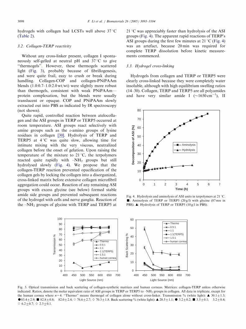
Fig. 3. Optical transmission and back scattering of collagen-synthetic mat
indicated. Ratios, denote the molar equivalent ratio of ASI groups in TERP o
the human cornea where n=4. ‘‘Thermo’’ means thermogel of collagen alo
K83.472.9;’ 82.870.8; � 82.072.4; J 76.672.7;} 70.371.8. Back scatteJ 6.270.7; } 2.370.1.
21 1C was appreciably faster than hydrolysis of the ASIgroups (Fig. 4). The apparent rapid reactions of TERP’sASI groups during the first few minutes at 21 1C (Fig. 4)was an artefact, because 20min was required forcomplete TERP dissolution before kinetic measure-ments commenced.
3.3. Hydrogel cross-linking
Hydrogels from collagen and TERP or TERP5 wereclearly cross-linked because they were completely waterinsoluble, although with high equilibrium swelling ratios(14–30). Collagen, TERP and TERP5 are all polyamidesand have very similar amide I (�1650 cm�1), II
0
10
20
30
40
50
60
400 450 500 550 600 650 700
Light Source (nm)
Bac
k S
catte
ring
%
Thermo
0.5:11:1
1:1(TERP5)1.5:1
human cornea
rices and human corneas. Matrices: collagen-TERP unless otherwise
r TERP5 to –NH2 groups in collagen. All data in triplicate, except for
ne without cross-linker. Transmission % (white light): m 30.171.3;
ring % (white light):m 20.571.1;K 3.270.2;’ 3.570.1; � 3.270.4;

ARTICLE IN PRESSF. Li et al. / Biomaterials 26 (2005) 3093–3104 3099
(�1560 cm�1) and III (�1250 cm�1) absorptions in theIR. Consequently, it was difficult to discriminatebetween pure bovine collagen, TERP5-collagen gelsand natural stromal tissue by FTIR (Fig. 2d, e and g).However, absorptions at 2970 and 2876 cm�1 (isopropylCH3 asymmetrical and symmetrical stretching frequen-cies in PNiPAAm) were visible in TERP5 and TERP5-collagen gels, but not in bovine collagen or nativestromal tissue (Fig. 2c–e and g) [24]. Matrices recoveredby dissection of corneas after 6 weeks of implantation inpigs had FTIR spectra (Fig. 2f) virtually identical tospectra measured on equivalent samples before implan-tation (Fig. 2e).Before reaction, the e-NH2 content of the bovine
atelocollagen was 36.071.2mol/mol of single chaincollagen (molecular weight 1.0� 105Da), as reportedfor other mammalian collagens [15,28,31]. Measurementof –NH2 groups in collagen (4wt/vol%) before andafter reaction with TERP or TERP5 (Fig. 1) showedtheir consumption, consistent with cross-link formation.However, almost 60% of the initial e-NH2 groupsremained even when a 4-fold excess of ASI to–NH2 groups was used. This low extent of reactionprobably resulted from the difficult reaction betweenmacromolecular reagents; especially, when one ofthe macromolecules was beginning to self-aggregateinto fibrils. In contrast, dilute collagen solutions(0.3 wt/vol%) reacted almost quantitatively withTERP (480%) at very high ASI/–NH2 ratios (450mo-lar equivalents ratio, not shown), ratios well beyondthe level at which 4% collagen/TERP formulationsare opaque from phase separation (ASI/–NH2�1.5/1,Table 2).
3.4. Optical and refractive index analyses
The absence of a detectable LCST opacity at �30 1Cfor the copolymers (Table 1) indicated that all wereessentially free of PNiPAAm impurities. In vitrotransmission and back scatter of all hydrogel matricesand human corneas were determined under identicalconditions (Fig. 3). The transmission and backscattering(non-specular reflectance) of gels with ASI:Collagen-NH2=0.5:1–1.5:1 were superior to the human cornealvalues. As the ratio increased to 1.5 and above however,the gels became increasingly opaque (Table 2). Thetransmission and scattering by thermogels were quitedifferent from the collagen-coploymer gels, with ther-mogel transmission being very low and the backscattering very high (Fig. 3). Thermogels consistentlyhad higher refractive indices than optically clearcollagen-TERP gels at the same collagen concentration(Table 2), which were in turn higher than the tear film.In vivo confocal images indicated that clarity andthickness of implants were unchanged over the longerterm (16 week) pig study.
3.5. Glucose permeability coefficient
The permeability coefficient for glucose diffusionthrough PBS equilibrated hydrogels (collagen andTERP 2.0 and 1.4wt/vol%, respectively, used for invivo implantations) was 2.7� 10�6 cm2/s (n=3,R240:95) at 21 1C. This was corrected to 4� 10�6 cm2/s at 37 1C [32], greater than the permeation coefficient ofthe corneal stroma at 37 1C (0.7� 10�6 cm2/s, calculatedfrom its diffusion coefficient of 2.4� 10�6 cm2/s at 37 1C[33] and the solubility coefficient of 0.3 for partitioningbetween PBS and stroma [34]).
3.6. Hydrogel strength and biodegradation
Collagen-TERP and collagen-TERP5 gels were toughenough to support handling and surgical manipulationwith suture thread and needle [7]. In suture pullouttensile testing, stress at failure and apparent modulusduring tensile testing both increased by over a factor oftwo on going from ASI/–NH2 equivalent ratio of0.5–1.5, but decreased at higher ASI/–NH2 ratios (Fig.5). In contrast, human corneas in suture pull out testsdid not fail; the sutures themselves broke at 56 g load,without visible damage to the cornea. Hydrogel failurecorresponded to tearing of the gel adjacent to the multi-axial stress point caused by each suture. Our reportedapparent stiffness (slope of the linear stress–strain curve)is not comparable with normal, stress–strain moduli, butis a very practical indicator of gel performance duringsurgical suturing.During bacterial collagenase digestion, the bio-syn-
thetic matrix lost �90% of its initial weight in 60min,whereas the human corneal stroma samples lost 10% byweight in this time. On a dry weight basis, the bio-synthetic matrix degraded slightly faster than thenatural stroma, which dissolved completely in �19 h at37 1C. However, clostridium collagenase is much morepotent than the human collagenase found in tears [35].
3.7. Topography and porosity
The surface topographies of a fully hydrated collagen-TERP hydrogels and collagen thermogel immersed inPBS solution were imaged by AFM (Fig. 6). There wasno evidence of the macro-fibrilar (200–500 nm diameter)morphology reported in the sclera (white of the eye) byAFM or of the highly ordered arrays of parallel fibrils(60–100 nm) observed in the human cornea [36]. FromFig. 6 and from more highly magnified images (notshown), both gels were disordered, with short filamentsections visible before they submerge in the hydrogel. Ifthe black areas of the images (Fig. 6) were assumed to besolution-filled voids linked to an internal pore structureof the gel, then the apparent pore diameter range in theimplanted collagen-TERP5 samples was somewhat

ARTICLE IN PRESS
0.0
1.0
2.0
3.0
4.0
5.0
1.0 3.0 5.0 7.0Strain (mm)
Str
ess
(g fo
rce)
0
1
2
3
4
5
6
0.5 1.5 2
Terpolymer/Collagen Ratio (ASI/-NH2)
Str
ess
(g fo
rce)
0
0.5
1
1.5
2
0.5 1 1.5 2
Terpolymer/Collagen Ratio (ASI/-NH2)
App
aren
t Stif
fnes
s (g
forc
e/m
m)
1
(A)
(B) (C)
Fig. 5. Stress/strain data for hydrated collagen-TERP matrices. Suture pull out data on hydrated gels made from 4% collagen with the indicated
ASI/–NH2 ratios: (A) Typical stress–strain curve. (B) Stress at gel rupture. (C) Apparent stiffness from the slope of the linear part of stress/strain
curves.
F. Li et al. / Biomaterials 26 (2005) 3093–31043100
narrower (64–420 nm) than in thermogels (110–440 nm).The roughness of the moulded surface of the former wasclearly less than that of the thermogel (Fig. 6, 2Dimages). The average pore diameters from the PBShydraulic permeability coefficients of the implants usedin vivo were 60–70 nm (Table 2).
4. Discussion
The human cornea is extremely tough, tear resistantand long lived, yet susceptible to diseases or damagethat can result in blindness. Corneal toughness dependsupon a highly organized array of collagen I filaments,arranged in �300 offset layers, parallel to the cornea’ssurface, bound with glucosaminoglycans which controlhydration [36,37]. Perfect reproduction of these proper-ties in a matrix for implantation may not be necessary,provided that the implant is functionally comparable incritical areas [1]. Critical factors for the corneal implantinclude both optical properties and cell function. Opticalproperties are based on curvature, refractive index andhigh optical clarity that we have controlled by formula-tion and fabrication conditions. Cell function resultedfrom cell recruitment by our sterile collagen-TERP5implants, leading to integration with the host’s tissue [7].The grafted YIGSR groups of TERP5 accelerated
re-epithelialization and stratification perfection as com-pared to YIGSR-free, collagen-TERP gels, collagenthermogels or collagen-PNiPAAM blends [7,11]. Re-growth of a confluent, stratified epithelial layer over acornea implant is essential as a defense against infectionof the eye, but re-growth has proven to be very difficultfor fully synthetic implants [10,12,38,39 and referencestherein]. Collagen-TERP5 gels also allowed rapidstromal cell recruitment, leading to implant integrationwith the adjacent tissue as well as very rapid, nerveneurite extension into the implants. This restored invivo, touch sensitivity within about 20 days [7]. Theimplant properties leading to this performance arediscussed below.Mammalian corneal clarity is now believed to result
from a combination of refractive index matching andthe presence of structural components with diameterswell below the wavelength of visible light [37]. Wespeculate that our matrices remained transparentbecause of controlled cross-linking to prevent fibrillo-genesis, the spontaneous self-assembly at 37 1C ofcollagen triple helices into opaque, macro-fibrillarassemblies. These self cross-linked collagen matrices(thermogels) are suggested to scatter visible lightbecause the self-assembled macrofibrils have higherrefractive indices than the more highly hydratedmicrofibrils and non-associated, triple helices found in

ARTICLE IN PRESS
Fig. 6. AFM topographies of collagen-TERP and collagen thermogel surfaces. Images obtained in MAC mode (cantilever force constant 1.2–5.5N/
m) for fully hydrated samples in PBS. Gel compositions: collagen:TERP=2.0:1.4wt/wt (upper 2D and 3D images); thermogel 2.8wt% collagen
(lower 2D and 3D images), all values for equilibrium hydrated gels. Raster scan directions vertical in images are shown. For both samples, images
were processed identically with low-pass filter. Note z-axis in A.
F. Li et al. / Biomaterials 26 (2005) 3093–3104 3101
collagen-TERP gels (Table 2, cf. thermogel and col-lagen-TERP refractive indices). Light scattering in-creases as the solvent/solute refractive index differenceincreases [37].Some cells grow well on the surfaces (2D growth) of
synthetic polymers. However, successful ingrowth orgrowth of encapsulated living cells in nano-poroushydrogels (3D growth, the dominant in vivo state) hasonly been demonstrated in a few, fully syntheticpolymers and copolymers such as those based onethylene oxide, propylene oxide and NiPAAm[2,3,6,13]. Stile et al. [2,3] have reported growth ofchondrogenic and osteoblast cells fully encapsulated inopaque hydrogels made from a lightly cross-linkedPNiPAAm-co-AAc copolymer at above its LCST. Cell
growth occurred despite the non-biodegradability of thisgel. For corneal implants, opacification must beprevented by the selection of appropriate comonomerswith NiPAAm to raise the LCST to well above thephysiological range (Table 1). Our TERP and TERP5copolymers were optically clear under physiologicalconditions and were designed to spontaneously cross-link with collagen I.Natural biopolymers are widely used to encapsulate
living cells, but their hydrogels are susceptible to rapidbiodegradation [40,41]. Cell recruitment depends onpermeability, hydration level and appropriate celladhesion and growth factors in encapsulants. Hydrogelsof collagen I are attractive as cell scaffolds because oftheir hydrogel strength at relatively low concentrations,

ARTICLE IN PRESSF. Li et al. / Biomaterials 26 (2005) 3093–31043102
resulting from the virtually rigid rod properties of thecollagen type I triple helix [42]. In addition, they bringseven RGD cell attachment motifs with each atelocolla-gen triple helix [32]. Differences between amino acidcompositions for mammalian atelocollagens are quitesmall [15,28,31]. Because the cornea is immune privi-leged and so not subject to allograft rejection, it may bepossible to use high purity animal collagen I (bovine,porcine, etc.) as a basis for the hydrogel implants [8].For corneal implants, both the biodegradation
resistance and strength of collagen I hydrogels need tobe enhanced by chemical cross-linking. [41]. Watersoluble carbodiimides lead to stable amide links betweenthe –C(=O)OH and –NH2 groups of peptides, includingcollagen, in aqueous solutions especially if side reactionsare reduced by the reaction of the initial cardodiimide-carboxylic acid product with an N-hydroxysuccinimide[15]. These resulting, pendant oxysuccinimide groups, asin our ASI copolymers, react cleanly at room tempera-ture and neutral pH with free –NH2 (Fig. 1) [13]. Atneutral pH both TERP and TERP5 smoothly reactedwith collagen in aqueous solution to give matrices thatremained robust and optically clear under physiologicalconditions yet at a reaction rate which was slow enoughto allow both thorough mixing and moulding of theviscous reagents at �0 1C before gellation.The composite collagen-TERP5 hydrogels had ex-
cellent optical clarity (Fig. 3) and remained transparentduring prolonged incubation in vitro at 37 1C withepithelial and nerve cells and for our longest implanta-tion (16 weeks) in mini-pigs. When molded to thecurvature and dimensions of a human cornea, the gelshad modest tensile strength and elastic modulus (Fig. 5).These collagen-TERP5 implants withstood surgicalhandling and suturing into pigs [7], but were consider-ably weaker than the human cornea.For epithelial cell adhesion and nerve growth,
covalently bound YIGSR (the pentapeptide motif inthe natural laminin macromolecule) is reported to beparticularly important, whereas pentapeptide and lami-nin itself are antagonistic if non-bonded [43,44]. YIGSRwas covalently bonded to collagen through TERP (Fig.1) and was extraction resistant, as measured byradiolabelling assays. Collagen cross-linked withTERP5 containing bound YIGSR appeared to promotestromal cell infiltration, normal re-epithelialization andthe rapid growth of neurites from the surrounding andunderlying corneal tissue [7]. Lamellar keratoplasty(LKP) and deep LKP (DLKP) are surgical proceduresdesigned to remove only the corneal epithelium and partof the stromal thickness and then replace these withnatural or hybrid implants [45]. The remaining stromalbed in LKP and DLKP can act as a vital source ofnerves for re-growth into the implant. In LKP allografttransplants, nerve ingrowth seemed only to occurradially from the patient’s residual corneal rim, with
some touch sensitivity of the central cornea registeredafter �18 months or longer [46]. The restoration oftouch sensitivity over the whole implant surface withinonly 20 days following LKP surgery with our 200 mmimplant indicated that the regenerated nerves arefunctional [7]. Each LKP implantation also resulted inphysically secure implant because of the observedstromal cell ingrowth, integrating the implant with hosttissue.Meaningful measurements of hydrogel pore size are
controversial, often involving vacuum techniques thatrequired complete sample drying and/or freezing (SEM,TEM, cryoTEM, etc.) before characterization[22,47,48]. Techniques that allow pore size estimationfrom measurements on fully hydrated samples may bemore meaningful [22,48]. However, even AFM imagesrepresent a convolution of the actual surface features’dimensions both with the AFM tip shape and withsample softness as compared to the cantilever stiffness[20,21]. For our collagen-TERP matrices, the bandedstructures, usually seen in dried collagen micro-fibrilswere not observed (Fig. 6). This is consistent with Baseltet al. [49] who showed that hydrated (soft and swollen)microfibrils, even on a mica substrate, were featurelessby AFM, as also found in human stroma [36]. Averagebulk pore diameter in the PBS equilibrated, hydrogel ofcollagen-TERP5 was 60–70 nm (from PBS hydraulicpermeability, Table 2) whereas AFM indicated60–400 nm surface pores. Agreement between thesetwo methods on fully hydrated samples is fair, bearingin mind that the hydraulic permeation theory assumedparallel, cylindrical pores [22]. Evans et al. [12] haveconcluded that epithelial stratification requires100–400 nm diameter pores. Average pore diameters of92734 nm were identified in the human epithelialbasement membrane by SEM [49]. Sweeney et al. [32]found that corneal implants must have average porediameters in the 50–100 nm range to allow adequatetransport of key nutrients; especially glucose, to main-tain a healthy epithelium. The collagen/TERP andcollagen/TERP5 hydrogels had a glucose permeabilitycoefficient significantly greater than the human cornealstroma. In addition, the overall surface texture of ourhydrated collagen-TERP hydrogels (Fig. 6) was muchsmoother than found in the epithelial, basementmembrane of humans [47]. Surface texture was pro-posed to influence epithelial cell adhesion and migration[47] but may not be the sole factor.Stile et al. [2] found chondrogenic cell growth in
lightly cross-linked PNiPAAm-co-AAc. From theircopolymer feed composition, their hydrogel had amolecular weight between cross-links (MWXL) of�73,000Da. Based on our measured, initial ASI and–NH2 concentrations in TERP5 and collagen, respec-tively, and on the measured reaction of �16% of these–NH2 at an ASI/–NH2 equivalent ratio of 1/1, our

ARTICLE IN PRESSF. Li et al. / Biomaterials 26 (2005) 3093–3104 3103
MWXL was �17,000 (collagen) and �11,000Da.(TERP5), if all reacted –NH2 are involved in cross-links. This gave an overall MWXL of �28,000Da for thegel used in our in vivo studies, well below MWXL of thegel of Stile et al. [2]. It is surprising that in vivo theneurite growth cone (�2 mm diameter) [41] and chon-drocyte, osteoblast [2,3] and stromal cells (the latter�5 mm diameter in spindle form) penetrated suchmatrices, with only 60–400 nm diameter pores. How-ever, neurite extension into agarose hydrogels with porediameters of 300–600 nm has been found [22]. Therelatively low tensile strength of our gels may compen-sate for their small pore sizes, perhaps yielding to theneurite growth cone or indicating some local biodegra-dation of the collagen component [6].
5. Conclusions
TERP and TERP5 appeared to function by cross-linking collagen and by stabilization the system, so as toprevent collagen fibrillogenesis, control swelling andhydration and increase mechanical properties. Thecollagen-TERP5 network allowed high glucose perme-ability and had both a surface roughness and porositysomewhat lower than reported for the natural basementmembrane. These non-cytotoxic matrices, with natural(RGD) and bound laminin pentapeptide (YIGSR) celladhesion factors, allowed control of the cell and nervecompatibility, toughness and optical properties. SuchTE matrices might eventually address future shortagesof donor corneas and circumvent problems resultingfrom human donor corneal transplants and otherprostheses [9,39,47]. However, refractive power andsurface topography stability by, for example, PAR-CTSmust be established for longer term in vivo implants.
Acknowledgements
This work was supported by the Natural Sciences andEngineering Research Council, Canada, grants 227212-99, 227951-01 and 246418. We gratefully acknowledgeassistance from Gilles Robertson (NMR), John Woods(GPC), Steven Argue (DSC) and David Priest, RejeanMunger and Chris Lohmann (optical properties).
References
[1] Chapekar MS. Tissue engineering: challenges and opportunities. J
Biomed Mater Res: Appl Biomater 2000;53:617–20.
[2] Stile RA, Burghardt WR, Healy KE. Synthesis and characterisa-
tion of injectable poly(N-isopropylacrylamide)-based hydrogels
that support tissue formation in vitro. Macromolecules 1999;
32:7370–9.
[3] Stile RA, Healy KE. Thermo-responsive peptide modified
hydrogels for tissue regeneration. Biomacromolecules 2001;2:
185–94.
[4] Vernon B, Gutowska A, Kim SW, Bae YH. Thermally reversible
polymer gels for biohybrid artificial pancreas. Macromol Symp
1996;109:155–67.
[5] Webb D, An YH, Gutowska A, Mironov VA, Friedman RJ.
Propagation of chondrocytes using a thermosensitive polymer gel
culture. MUSC Orthopaed J 2000;3:18–21.
[6] Pratt AB, Weber FE, Schmoekel HG, Muller R, Hubbell JA.
Synthetic extracellular matrices for in situ tissue engineering.
Biotechnol Bioeng 2004;86:27–36.
[7] Li F, Carlsson DJ, Lohmann CP, Suuronen EJ, Vascotto S,
Kobuch K, Munger R, Doillon CJ, Sheardown H, Griffith M. A
bio-synthetic, corneal implant replicating cellular and nerve
growth in the natural extracellular matrix. Proc Natl Acad Sci
USA 2003;100:15346–51.
[8] Griffith M, Osborne R, Munger R, Xiong X, Doillon CJ, Laycock
NIC, Hakim M, Song Y, Watsky MA. Functional human corneal
equivalents constructed from cell lines. Science 1999;286:2169–72.
[9] Chirila TV. An overview of the development of artificial corneas
with porous skirts and the use of PHEMA for such an
application. Biomaterials 2001;22:3311–7.
[10] Legeais JM, Renard G. A second generation of artificial cornea
(Biokpro II). Biomaterials 1998;19:1517–22.
[11] Shimmura S, Doillon CJ, Griffith M, Nakamura M, Gagnon E,
Usui A, Shinozaki N, Tsubota K. Collagen-poly(N-isopropyla-
crylamide)-based membranes for corneal stroma scaffolds.
Cornea 2003;22(7(Suppl.)):S81–8.
[12] Evans MD, Taylor S, Dalton BA, Lohmann D. Polymer design
for corneal epithelial tissue adhesion: pore density. J Biomed
Mater Res 2003;64A:357–64.
[13] Pollak A, Blumenfeld H, Wax M, Baughn RL, Whitesides GM.
Enzyme immobilization by condensation copolymerisation into
cross-linked polyacrylamide gels. J Am Chem Soc 1980;102:
6324–36.
[14] Aucoin L, Griffith CM, Pleizier G, Deslandes Y, Sheardown H.
Interactions of corneal epithelial cells and surfaces modified with
cell adhesion peptide. J Biomater Sci: Polym Edn 2002;13:447–62.
[15] Olde Damink LHH, Dijkstra PJ, Feijen J, van Luyn MJA, van
Wachem PB, Nieuwenhuis P. Cross-linking of dermal sheep
collagen using a water-soluble carbodiimide. Biomaterials 1996;
17:765–73.
[16] Wang CL, Miyata T, Wesler B, Rubin AL, Stenzel KH. Collagen-
induced platelet aggregation and release. II Critical size and
structural requirements of collagen. Biochim Biophys Act
1978;544:568–77.
[17] Peng T, Cheng Y-L. Temperature-responsive permeability of
porous PNIPAAm-g-PE membranes. J Appl Polm Sci 1998;
70:2133–42.
[18] American National Institute and Association for the Advance-
ment of Medical Instrumentation. ‘‘Cardiovascular Implants—
Vascular Prosthesis, Section 8.8 (Suture Retention Strength)’’,
ANSI/AAMI: 1994;VP20:20–21.
[19] Revenko I, Prosch R. Magnetic and acoustic tapping mode
microscopy of liquid phase phospholipid bilayers and DNA
molecules. J Appl Phys 2000;87:526–33.
[20] Bowen RW, Hall NJ. Properties of microfiltration membranes:
mechanisms of flux loss in the recovery of an enzyme. Biotechnol
Bioeng 1995;46:28–35.
[21] Bowen RW, Doneva TA. Artifacts in AFM studies of mem-
branes: correcting pore images using fast Fourier transform
filtering. J Membrane Sci 2000;171:141–7.
[22] Dillon GP, Yu X, Sridharan A, Ranieri JP, Bellamkonda RV. The
influence of physical structure and charge on neurite extension in
a 3D hydrogel scaffold. J Biomat Sci Polm Ed 1998;9:1049–69.

ARTICLE IN PRESSF. Li et al. / Biomaterials 26 (2005) 3093–31043104
[23] Priest D, Munger R. A new instrument for monitoring the
optical properties of corneas. Invest Ophthalmol Vis Sci 1998;
39:S352.
[24] Percot A, Lafleur M, Zhu XX. New hydrogels based on N-
isopropylacrylamide copolymers crosslinked with polylysine:
membrane immobilization systems. Polymer 2000;41:7231–9.
[25] Privalov PL, Tiktopulo EI, Tischenko VM. Stability and mobility
of the collagen structure. J Mol Biol 1979;127:203–16.
[26] Miles CA, Burjanadze TV, Bailey AJ. The kinetics of the thermal
denaturation of collagen in unrestrained rat tail tendon deter-
mined by differential scanning calorimetry. J Mol Biol
1995;245:437–46.
[27] Miles CA, Bailey AJ. Thermally labile domains in the collagen
molecule. Micron 2001;32:325–32.
[28] Angele P, Abke J, Kujat R, Faltermeier H, Schumann D, Nerlich
M, Kinner B, Englert C, Ruszczak Z, Mehrl R, Mueller R.
Influence of different collagen species on physico-chemical
properties of crosslinked collagen matrices. Biomaterials
2004;25:2831–41.
[29] Heskins M, Guillet JE. Solution properties of poly(N-isopropy-
lacrylamide). J Macromol Sci: Chem 1968;A2:1441–55.
[30] Cline GW, Hanna SB. Kinetics and mechanisms of the aminolysis
of N-hydroxysuccinimide esters in aqueous buffers. J Org Chem
1988;53:3583–6.
[31] Ayed S, Boot-Handford R, Humphries MJ, Kadler KE, Shuttle-
worth A. The extracellular matrix FactsBook. London: Academic
Press; 1994.
[32] Sweeney DF, Xie RZ, O’Leary DJ, Vannas A, Odell R,
Schindhelm K, Cheng HY, Steele JG, Holden BA. Nutritional
requirements of the corneal epithelium and anterior stroma:
clinical findings. Invest Ophthalmol Vis Sci 1998;39:284–91.
[33] Pettit DK, Knight PM. 1985. Glucose permeability of potential
intrastromal implants. Invest Ophthalmol Vis Sci 2000;26:151.
[34] McCarey BE, Schmidt FH. Modeling glucose distribution in the
cornea. Curr Eye Res 1990;9:1025–39.
[35] Holopainen JM, Moilanen JA, Sorsa T, Kivela-Rajamaki M,
Tervahartiala T, Vesaluoma MH, Tervo TM. Activation of
matrix metalloproteinase-8 by membrane type 1-MMP and their
expression in human tears after photorefractive keratectomy.
Invest Ophthalmol Vis Sci 2003;44:2550–6.
[36] Meller D, Peters K, Meller K. Human cornea and sclera studied
by atomic force microscopy. Cell Tissue Res 1997;288:111–8.
[37] Freegard TJ. The physical basis of transparency of the normal
cornea. Eye 1997;11:465–71.
[38] Kornmehl EW, Bredvik BK, Kelman CD, Raizman MB. DeVore
DP In vivo evaluation of a collagen corneal allograft derived from
rabbit dermis. J Refract Surg 1995;11:502–6.
[39] Merrett K, Griffith CM, Deslandes Y, Pleizier G, Sheardown H.
Adhesion of cornea epithelial cells to cell adhesion peptide
modified pHEMA surfaces. J Biomater Sci 2001;12:647–71.
[40] Balgude AP, Yu X, Szymanski A, Bellamkonda RV. Agarose gel
stiffness determines rate of DRG neurite extension in 3D cultures.
Biomaterials 2001;22:1077–84.
[41] Hoffman AS. Hydrogels for biomedical applications. Adv Drug
Deliv Rev 2002;43:3–12.
[42] Amis EJ, Carriere CJ, Ferry JD, Veis A. Effect of pH on collagen
flexibility determined from dilute solution viscoelastic measure-
ments. Int J Biol Macromol 1985;7:130–4.
[43] Graf J, Ogle RC, Robey FA, Sasaki M, Martin GR, Yamada Y,
Kleinman HK. A pentapeptide from the laminin B1 chain
mediates cell adhesion and binds the 67,000 laminin receptor.
Biochemistry 1987;26:6896–900.
[44] Yu X, Dillon GP, Bellamkonda RB. A laminin and nerve growth
factor-laden three-dimensional scaffold for enhanced neurite
extension. Tissue Eng 1999;5:291–304.
[45] Terry MA. The evolution of lamellar grafting techniques over
twenty-five years. Cornea 2000;19:611–6.
[46] Kaminski SL, Biowski R, Lukas JR, Koyuncu D, Grabner G.
Corneal sensitivity 10 years after epikeratoplasty. J Refract Surg
2002;18:731–6.
[47] Abrams GA, Schaus SS, Goodman SL, Nealey PF, Murphy CJ.
Nanoscale topography of the corneal epithelial basement
membrane and Descemet’s membrane of the human. Cornea
2000;19:57–64.
[48] Suzuki A, Yamazaki M, Kobiki Y. Direct observation of polymer
gel surfaces by atomic force microscopy. J Chem Phys
1996;104:1751–7.
[49] Baselt DR, Revel J- P, Baldeschwieler JD. Subfibrillar structure of
type I collagen observed by atomic force microscopy. Biophys J
1993;65:2644–55.
Copyright © 2022 FDOKUMEN