Inhibitory Deficit Theory: Recent Developments in a "New View
Upload
khangminh22Category
view
0download
0
pharmaceutics
Review
Recent Developments in Microfluidic Technologiesfor Central Nervous System Targeted Studies
Maria Inês Teixeira 1,2, Maria Helena Amaral 1 , Paulo C. Costa 1 , Carla M. Lopes 3,* andDimitrios A. Lamprou 2,*
1 UCIBIO-REQUIMTE, MedTech - Laboratory of Pharmaceutical Technology, Department of Drug Sciences,Faculty of Pharmacy, University of Porto, Rua de Jorge Viterbo Ferreira, 228, 4050-313 Porto, Portugal;[email protected] (M.I.T.); [email protected] (M.H.A.); [email protected] (P.C.C.)
2 School of Pharmacy, Queen’s University Belfast, 97 Lisburn Road, Belfast BT9 7BL, UK3 FP-ENAS/CEBIMED, Fernando Pessoa Energy, Environment and Health Research Unit/Biomedical
Research Centre, Faculty of Health Sciences, Fernando Pessoa University, Rua Carlos da Maia, 296,4200-150 Porto, Portugal
* Correspondence: [email protected] (C.M.L.); [email protected] (D.A.L.)
Received: 26 May 2020; Accepted: 9 June 2020; Published: 11 June 2020
Abstract: Neurodegenerative diseases (NDs) bear a lot of weight in public health. By studyingthe properties of the blood-brain barrier (BBB) and its fundamental interactions with the centralnervous system (CNS), it is possible to improve the understanding of the pathological mechanismsbehind these disorders and create new and better strategies to improve bioavailability and therapeuticefficiency, such as nanocarriers. Microfluidics is an intersectional field with many applications.Microfluidic systems can be an invaluable tool to accurately simulate the BBB microenvironment, aswell as develop, in a reproducible manner, drug delivery systems with well-defined physicochemicalcharacteristics. This review provides an overview of the most recent advances on microfluidicdevices for CNS-targeted studies. Firstly, the importance of the BBB will be addressed, and differentexperimental BBB models will be briefly discussed. Subsequently, microfluidic-integrated BBB models(BBB/brain-on-a-chip) are introduced and the state of the art reviewed, with special emphasis on theiruse to study NDs. Additionally, the microfluidic preparation of nanocarriers and other compounds forCNS delivery has been covered. The last section focuses on current challenges and future perspectivesof microfluidic experimentation.
Keywords: Neurodegenerative diseases (NDs); blood-brain barrier (BBB); central nervous system(CNS); microfluidics; organ-on-a-chip; brain-on-a-chip; brain delivery; drug delivery; nanoparticles(NPs); nanocarriers
1. Introduction
Neurodegenerative diseases (NDs) such as Parkinson’s disease (PD), Alzheimer’s disease (AD),Amyotrophic lateral sclerosis (ALS), Multiple sclerosis (MS) or Huntington’s disease (HD) are amongthe disorders with the highest incidence worldwide, with their increase alarming when it comes tothe elderly population [1,2]. In light of their prevalence, they represent a critical public health andeconomic concern [3]. These ailments are marked by different clinical features, depending on theinvolved regions of the central nervous system (CNS), with some causing cognitive and memorydysfunction, and others instigating motor impairment [4]. Several biological mechanisms contribute tothe appearance and chronic progression of NDs, including oxidative stress, mitochondrial dysfunction,neural excitotoxicity, protein aggregation, depletion or deficient synthesis of neurotransmitters ortheir degradation in the synaptic cleft due to higher enzymatic activity, and gross malfunction of theblood-brain barrier (BBB) [5].
Pharmaceutics 2020, 12, 542; doi:10.3390/pharmaceutics12060542 www.mdpi.com/journal/pharmaceutics

Pharmaceutics 2020, 12, 542 2 of 37
The BBB is a highly complex dynamic interface between the bloodstream and the brain,effectively protecting the CNS from potentially harmful external elements and maintaining cerebralhomeostasis [6,7]. BBB disruption, linked with increased permeability, impairment of influx/effluxmechanisms and infiltration of toxins and immune cells in the CNS, is a common factor to many NDs [7].The establishment of in vitro biomimetic platforms that can precisely replicate the functional/structuralproperties of the BBB and enable the monitoring of changes to its integrity, could therefore enhance theunderstanding of NDs, as well as accelerate the research and development of innovative therapies [6,7].Ideally, to faithfully recapitulate the human brain endothelium, in vitro BBB models should possesssome of its essential features, including interactions between different types of cells (e.g., vascularendothelial cells, astrocytes and pericytes), a 3D vessel-like structure of the endothelial cells, thepresence of a basement membrane and flow-induced sheer stress [6,7].
Microfluidics is an intersectional field that deals with systems that process or manipulate nanolitreamounts of fluids, using channels ranging in size from tens to hundreds of micrometres [8]. Advances inmicrofluidic technology have led to the generation of devices (so-called organs-on-a-chip) that can bestandardized at various scales and flow regimes, allowing the simulation of BBB in vivo physiologythrough reconstitution of its multicellular architecture and physicochemical microenvironment [7,9,10].Conventional 2D or 3D systems cannot produce such levels of tissue and organ functionality [10].By providing a highly controlled cellular microenvironment, organs-on-a-chip are suitable toolsto assess the responses of the BBB to physiological and pathological stimuli, including analysisof metabolic, genetic and biochemical activities [7,10]. Furthermore, they also allow for real-time,high-resolution imaging [10]. Thus, these types of models have huge potential for a wide variety ofapplications, including the study of NDs aetiology, biomarker identification, neurotoxicity testing orhigh-throughput screening of CNS drug candidates [7,10].
Currently, none of the available medicines are able to stop or slow the continuous loss of neuronsthat occurs in NDs, with therapy being majorly focused on symptomatic relief [4]. Considering thatthe number of cases is expected to rise, with the World Health Organization (WHO) predicting thatin 20 years’ time NDs will become the second leading cause of death, there is an urgent and unmetneed to come up with more effective therapies [11,12]. However, the treatment of brain diseases isseverely hindered by the BBB [13]. Under normal circumstances, the BBB only allows selective passageof molecules essential to the functioning of the brain, restricting the entrance of 98%, or more, oftherapeutics [14].
In this context, nanotechnology-based approaches such as nanoparticles (NPs) present anopportunity to rethink and retackle the adversities connected to poor BBB permeability when deliveringdrug candidates/diagnostic agents to the CNS. These targeting strategies have the potential to makeuse of receptors and transporters commonly expressed at the luminal side of the brain endotheliumto transport therapeutics across the BBB, leveraging internalization mechanisms that differ fromthe pathways taken by the majority of free drugs [15,16]. NPs can have significant advantagesover traditional pharmaceutical formulations, including biocompatibility and biodegradability oftheir materials, ease of scaling-up, reduced toxicity, controlled and targeted delivery, enhancedbioavailability and dissolution rate, improved pharmacokinetics and therapeutic effects, and protectionof encapsulated biomolecules [17,18].
Conventional methods of NPs preparation are sometimes associated with lack of precise control andhigh batch-to-batch variations. In this area, microfluidics has emerged as a forthcoming and noteworthyalternative, enabling fine tuning of process parameters and optimization. Microfluidic platformsallow for better control and reproducibility over NPs properties, including size distribution, zetapotential or ligand density, which are critical for their clinical translation as prime drug carriers [8,19,20].Importantly, the volume of the fluids used, enables a substantial reduction in the consumption ofreagents, being cost-effective [8]. Furthermore, given that the process of formation of NPs occursin a continuous flow pattern, with preservation of controlled hydrodynamic conditions inside the

Pharmaceutics 2020, 12, 542 3 of 37
microchannels, it is possible to obtain highly monodisperse nanosystems, avoiding batch-to-batchvariability [8,19].
This review provides an overview of the most recent advances in microfluidic devicesfor CNS-targeted studies. Firstly, the importance of the BBB will be addressed, and differentexperimental BBB models will be briefly discussed. Subsequently, microfluidic-integrated BBB models(BBB/brain-on-a-chip) are introduced and the state of the art reviewed, with special emphasis on theiruse to study NDs. Additionally, the microfluidic preparation of nanocarriers and other compounds forCNS delivery have been covered. The last section focuses on current challenges and future perspectivesof microfluidic experimentation.
2. The Blood-Brain Barrier (BBB)
The BBB is a highly specialized capillary wall, primarily comprised of brain endothelial cells andits basement membrane. Adherens and tight junctions (TJs) form cell–cell adhesions that maintainthe integrity of the microvasculature but severely limit the paracellular transport, thus makingtranscellular diffusion the main transport mechanism of molecules between the blood and the brain,and vice-versa [21]. Brain capillaries are encircled by astrocytic perivascular endfeet, which contributeto the regulation of ion concentration and clearance of neurotransmitters, and by pericytes throughthe basal laminal, having a crucial role in the regulation of blood flow, BBB integrity and transport.Neurons and microglia also participate in the formation of the BBB, establishing with the astrocytesand pericytes a dynamic functional unit, called neurovascular unit (NVU) (Figure 1). Functionalsignalling and interaction between these components is the basis for BBB stability and its response topathophysiological stimuli [21,22].
Pharmaceutics 2020, 12, x FOR PEER REVIEW 3 of 38
conditions inside the microchannels, it is possible to obtain highly monodisperse nanosystems, avoiding batch-to-batch variability [8,19].
This review provides an overview of the most recent advances in microfluidic devices for CNS-targeted studies. Firstly, the importance of the BBB will be addressed, and different experimental BBB models will be briefly discussed. Subsequently, microfluidic-integrated BBB models (BBB/brain-on-a-chip) are introduced and the state of the art reviewed, with special emphasis on their use to study NDs. Additionally, the microfluidic preparation of nanocarriers and other compounds for CNS delivery have been covered. The last section focuses on current challenges and future perspectives of microfluidic experimentation.
2. The Blood-Brain Barrier (BBB)
The BBB is a highly specialized capillary wall, primarily comprised of brain endothelial cells and its basement membrane. Adherens and tight junctions (TJs) form cell–cell adhesions that maintain the integrity of the microvasculature but severely limit the paracellular transport, thus making transcellular diffusion the main transport mechanism of molecules between the blood and the brain, and vice-versa [21]. Brain capillaries are encircled by astrocytic perivascular endfeet, which contribute to the regulation of ion concentration and clearance of neurotransmitters, and by pericytes through the basal laminal, having a crucial role in the regulation of blood flow, BBB integrity and transport. Neurons and microglia also participate in the formation of the BBB, establishing with the astrocytes and pericytes a dynamic functional unit, called neurovascular unit (NVU) (Figure 1). Functional signalling and interaction between these components is the basis for BBB stability and its response to pathophysiological stimuli [21,22].
Figure 1. Schematic representation of a neurovascular unit (NVU) cross section in the blood-brain barrier (BBB).
Figure 1. Schematic representation of a neurovascular unit (NVU) cross section in the blood-brainbarrier (BBB).

Pharmaceutics 2020, 12, 542 4 of 37
As a physical and biochemical barrier, the BBB has an essential role in brain homeostasis, limitingthe entry of harmful toxins and pathogens present in the blood, while supplying the fundamentalnutrients and molecules needed for normal cell function [22]. Although the restrictive nature of theBBB is extremely important for the protection of the CNS, it poses a challenge when it comes to themanagement of brain diseases, since therapeutic molecules are not able to penetrate the basementmembrane and reach the intended target site. The entry of therapeutic candidates into the brainparenchyma depends on several factors [21]. As previously mentioned, paracellular transport is verylimited and negligible. Low molecular weight (less than 400–500 Da) lipophilic substances, preferablyneutrally charged, permeate the brain through passive transcellular diffusion [23]. It is also possible totake advantage of some active transport mechanisms in order to assist drug penetration across theBBB, including [22,23]:
• Carrier-mediated transcytosis (binding of a molecule to a protein carrier on the apical side of thecapillary wall, followed by its conformational change and transport of said molecule to the otherside of the membrane).
• Receptor-mediated transcytosis (binding of a particular ligand to a specific receptor, such astransferrin or low-density lipoprotein receptors, with the formation of a ligand-receptor complexwhich is carried through the cytoplasm and released on the basolateral side of the BBB).
• Adsorptive transcytosis (electrostatic interaction between positively charged molecules and thenegatively charged plasma membrane).
Despite the fact that therapeutic molecules might get access to the CNS by passive or activetransport, many are subsequently removed by efflux pumps such as the P-glycoprotein (P-gp) orisoforms of the multidrug resistance protein (MRP), resulting in the extrusion of the drugs backinto the blood circulation and further complicating the effective treatment of brain disorders [22,24].Hence, with the aim to optimize the therapeutic effect of the drugs, some tactics focus on evadingor inhibiting the efflux pumps [23]. An interesting aspect to consider is that, depending on the CNSdisorder, distinct expression profiles of efflux transporters are observed. For instance, in MS, thereis a decrease in endothelial cell P-gp, whereas MRP1 or MRP2 remain unchanged [25]. In turn, inALS, there is an increase in the P-gp but not MRP 1, 2, 4 or 5 expression in the cerebral cortex andspinal cord [26]. This suggests that a rather complex interplay between age-related decrease anddisease-related stimulation, may lead to differences in the levels of such efflux pumps [27].
There is a recognized link concerning pathological impairment/disruption of the BBB and thedevelopment of NDs. Several morphological alterations of cellular elements, as well as changesat a molecular level, lead to BBB breakdown, with ensuing neural inflammation, dysfunction anddegeneration. Numerous factors may be involved in the BBB disruption, including reactive oxygenspecies (ROS), angiogenic factors, inflammatory cytokines, mitochondrial dysfunction, aberrantintracellular signalling, autoantibodies, leukocyte adhesion and pathogens [28]. Degenerative changesin the microvasculature persist as the disease evolves, and may be reflected in reduced expression ofTJs or adherens junctions, defective expression or function of transporters, upregulation of luminaladhesion molecules, astrocyte loss, pericyte detachment, insufficient clearance function or disruptedbasement membrane [28–30]. The implications vary from unregulated molecular and ionic fluxes,extravasation of plasma proteins, entry of pathogens and toxins, to the onset of a central inflammatoryresponse with microglial and astroglial activation, as well as release of cytokines and chemokines [28,31].However, it can be said that the single most important outcome is that the integrity of the BBB isseverely compromised, mainly manifesting as increased barrier permeability [28].
In recent years, exponential focus has been given to alterations of the extracellular matrix (ECM)of the basement membrane as a key contributor to the pathological process of NDs. For instance, ithas been shown that matrix metalloproteinases (MMPs), in cases of chronic neurodegeneration, driveforth the progression of symptomatology in AD and PD [32]. Increasing evidence also indicates that

Pharmaceutics 2020, 12, 542 5 of 37
dysbiosis of gut microbiome may influence and contribute to the onset and progression of a number ofdiseases, including NDs [33].
Overall, it is critical to have a better understanding and translation of the BBB physiological andpathological processes that precede and increase vulnerability to NDs, since it might offer new avenuesto help identify novel strategies and therapeutic targets for drug discovery and development.
3. Experimental BBB Models
3.1. In Vivo Models
BBB research is met with challenging difficulties given its complexities and relative inaccessibility.Reconstituting critical features of the BBB, as well as of NDs, is imperative not only to improvethe understanding of the mechanisms behind these incurable disorders, but also to accelerate drugdiscovery [32].
Currently, there is a heavy reliance on animal models to study in vivo cell behaviour. They areregarded as the gold standard, since they closely mimic the complexity of the BBB microenvironmentand the individual diversity found in humans [34,35]. Nevertheless, it is necessary to consider theoccurrence of species-specific biological differences, and thus, ultimately, their inadequate reproductionof human pathophysiology [32]. Given the existence of immunological, genetic and cellular variancesbetween humans and animals, no in vivo model is capable of faithfully recapitulate all aspects of humanbrain disease [36,37]. This is an important disadvantage, since it becomes problematic to translatethe results obtained using animals to the human context. Indeed, more than 80% of compounds thatwere successfully tested in animal models failed in clinical trials [37]. Poor experimental methodologyand species-to-species variability in protein expression profiles may also contribute to explain thisissue [35]. Furthermore, it is believed that most NDs are polygenetic, comprising multiple, andfrequently unrecognized, genetic alterations, which makes the generation of transgenic animals verychallenging [38]. Other critical drawbacks of using animal models include ethical concerns, elevatedcosts and prolonged, low-throughput, labour-intensive processing [39]. The global implementationof the 3Rs principles (reduction, refinement and replacement) also underlines the requirement toabandon animal testing as much as possible in the future, rerouting even more the investigation ofNDs pathology to in vitro models [32].
3.2. In Vitro Models
To circumvent the issues regarding the use of animals, scientists commonly resort totwo-dimensional (2D) and three-dimensional (3D) in vitro models as an alternative (Figure 2) [34].Cell culture technology is fairly straightforward and can be performed in carefully monitored conditions,thus making it relatively robust and with good reproducibility [34,37]. Additionally, it is generallycheaper to acquire and maintain when compared to animal models [34]. There are different modelsavailable, namely, 2D or 3D static cell cultures, and 2D or 3D perfusion microfluidic systems [40].
Starting with 2D static cell cultures, they are a staple for many applications like toxicity ormetabolism analysis [38,41]. These systems are well known, with extensive literature allowing tocritically analyse the data obtained, and understand the cell behaviour in many conditions [40]. In themost basic 2D models, cells are attached to a medium-containing flat surface such as flasks, dishesor well plates, where they are able to adhere and spread [32,42]. To enable BBB drug transportationstudies, the Transwell® setup was created, with one or more types of BBB cells being cultured onsemi-permeable microporous inserts [35,37]. Transwell® systems are widely utilized, since they areuser friendly and cost-effective, increasing test speed and cutting down reagent consumption [37].Cells of human origin can be used, therefore sidestepping problems with translation of results tothe clinic.
However, even when co-culture is done, 2D cell cultures have significant shortcomings.The production yield is low, and cell growth occurs unnaturally in reaction to external factors [42].

Pharmaceutics 2020, 12, 542 6 of 37
More importantly, there is a failure to properly replicate and reproduce key aspects of the BBBmicroenvironment, including cell–cell and cell–ECM interactions that occur within the NVU [34,41].In addition, they are restricted in supplying suitable electrical, mechanical and chemical cues, whichcan lead to subtle differences in barrier permeability and cell morphology when compared to in vivomodels [41]. Even though the realism of 2D cultures can be somewhat improved by incorporating morecell types or by using human cells, robustness is sacrificed, and thus, these models are not appropriatewhen the intention is to model higher order features and trajectories of the CNS [38].
3D cell systems have garnered attention for decades, being introduced as an attempt to betterrepresent the intricate human physiology and relevant conditions, therefore allowing to improve thesimulation of the in vivo setting of cells inside organs and tissues [32,41]. Traditional 3D culturesare usually achieved by culturing cells on biocompatible extracellular scaffolds (such as hydrogels),either in well plates or on Transwell® membranes [32,34]. By using hydrogels that incorporatenatural molecules of the ECM (e.g., collagen or fibrin) or synthetic polymers (e.g., polylactic acid(PLA) or polyglycolic acid (PGA)), cells are induced to polarize and interact with neighbouringcells, randomly interspersing in the ECM or self-assembling into physiological-like microstructures(organoids) [32,34,43]. Furthermore, culture systems can also self-organize in 3D aggregates in ascaffold-free manner, without the need of a supportive matrix, giving rise to spheroids [44].Pharmaceutics 2020, 12, x FOR PEER REVIEW 7 of 38
Figure 2. Schematic representation of different 2D and 3D in vitro models: (a) culture plates, (b) Transwell inserts, (c) microengineered scaffolds, (d) organ-on-a-chip. Reprinted and adapted with permission from Torras et al. [44].
4. Microfluidic BBB In Vitro Models (BBB/Brain-on-a-Chip)
The rise in microscale technologies has opened avenues for the development of more complex and advanced CNS biological models that are unachievable with conventional cell culture systems [39]. Organs-on-a-chip are microfluidic devices for culturing cells in continuously perfused, micrometre-sized chambers, engineered in such a way to enable them to closely mimic and better replicate the dynamic conditions, and the in vivo microstructure/environment of a tissue or organ (in this case, of the human BBB or brain) [10,35,45]. The goal is not to build a whole living organ but rather to synthesize minimal functional units that recapitulate tissue- and organ-level functions, thus allowing the development of a BBB model on a microdevice in order to study physiological and pathological phenomena [10,32,45]. Early research focused on simpler systems, consisting of a single microfluidic chamber containing one kind of cultured cell [46]. As knowledge advanced, more complex designs were created, generally entailing multiple microchambers connected with microchannels or microgroove arrays, and cultured with distinctive cell types to reproduce interfaces among different tissues (e.g., the BBB) [10,46].
Microfluidics-based models have the ability to regulate primary factors like cell patterning, cell–cell and cell–ECM interactions or transcellular oxygen, molecular and chemical gradients, as well as to provide physiologically relevant levels of fluid flow shear stress and cyclical mechanical forces analogous to those seen in blood vessels (i.e., perfusion culture) [10,41,46]. Accurate control of fluid flow is very useful, since it improves the function, differentiation and long-term survival of many cell types [10]. Such interactions and clear interplay between microenvironmental elements and culture parameters are not achievable with traditional cell systems [47]. Moreover, microfluidic approaches offer a plethora of other advantages that make them a unique platform to replace or at least complement other models for high-throughput drug screening and disease modelling, such as:
Figure 2. Schematic representation of different 2D and 3D in vitro models: (a) culture plates,(b) Transwell inserts, (c) microengineered scaffolds, (d) organ-on-a-chip. Reprinted and adaptedwith permission from Torras et al. [44].
Nonetheless, 3D cell cultures also have their own drawbacks. Firstly, some of these models arenot high-throughput or cost-effective, and often, there are problems regarding the existing protocolsnot being reproducible [32]. The macroscale of 3D leads to challenges in the transportation of nutrientsand oxygen, as well as in cell viability, analysis, sampling and imaging, as its complexity increases [41].Although 3D in vitro cultures are better at emulating certain aspects of the spatial complexity of theCNS and of the BBB microenvironment (including molecular and signalling pathways), many systemsstill lack the level of multiscale architecture and tissue-tissue interfaces that influence the function anddevelopment of organs in both health and disease, including the brain: for instance, just like it happens

Pharmaceutics 2020, 12, 542 7 of 37
with 2D models, cells in traditional static 3D models are not exposed to dynamic stresses like fluidflow from pressure gradients, or tension and compression. Moreover, they also fail to take into accountbiochemical effects that are specific to each cell type within a co-culture, when trying to reconstitutethe NVU [10,32].
Recently, progresses in microfabrication techniques have allowed to the integration of cell/tissueengineering on microfluidic platforms, leading to the development of a new, revolutionary type ofmodel: organ-on-a-chip. Organs-on-a-chip combine the advantages of in vivo and traditional in vitromodels, while potentially overcoming most of their limitations [35,41]. In this sense, in the next sectionswe will discuss the value and the state-of-art of this highly promising approach to mimic BBB/brainphysiology and function, as well as for studying NDs pathology.
4. Microfluidic BBB In Vitro Models (BBB/Brain-on-a-Chip)
The rise in microscale technologies has opened avenues for the development of more complexand advanced CNS biological models that are unachievable with conventional cell culture systems [39].Organs-on-a-chip are microfluidic devices for culturing cells in continuously perfused, micrometre-sizedchambers, engineered in such a way to enable them to closely mimic and better replicate the dynamicconditions, and the in vivo microstructure/environment of a tissue or organ (in this case, of the humanBBB or brain) [10,35,45]. The goal is not to build a whole living organ but rather to synthesize minimalfunctional units that recapitulate tissue- and organ-level functions, thus allowing the development of aBBB model on a microdevice in order to study physiological and pathological phenomena [10,32,45].Early research focused on simpler systems, consisting of a single microfluidic chamber containing onekind of cultured cell [46]. As knowledge advanced, more complex designs were created, generallyentailing multiple microchambers connected with microchannels or microgroove arrays, and culturedwith distinctive cell types to reproduce interfaces among different tissues (e.g., the BBB) [10,46].
Microfluidics-based models have the ability to regulate primary factors like cell patterning,cell–cell and cell–ECM interactions or transcellular oxygen, molecular and chemical gradients, aswell as to provide physiologically relevant levels of fluid flow shear stress and cyclical mechanicalforces analogous to those seen in blood vessels (i.e., perfusion culture) [10,41,46]. Accurate controlof fluid flow is very useful, since it improves the function, differentiation and long-term survival ofmany cell types [10]. Such interactions and clear interplay between microenvironmental elementsand culture parameters are not achievable with traditional cell systems [47]. Moreover, microfluidicapproaches offer a plethora of other advantages that make them a unique platform to replace or atleast complement other models for high-throughput drug screening and disease modelling, such as:
(1) Being highly customizable and having better experimental flexibility and control (such as in-chipdesign or in vitro biochemical and mechanical microenvironment conditions), thus giving replyto specific requirements of each experiment and cellular system [32,39,47].
(2) Significant reduction in chemical cost and consumption, since far less minute amounts of cells,culture medium and other reagents are needed [41,47].
(3) Continuous supply of nutrients and waste removal [40].(4) Schedule flexibility [40].(5) Reduced risk of contamination [47].(6) Possibility of multifunctional integration of analytical biosensors and other electronic apparatus
(e.g., microscopy devices, microelectrode arrays (MEAs), etc.) for real-time/on-chip or downstreammonitorization of cellular behaviour, detection of physiological parameters (e.g., biomarkersor chemokines levels) and in situ analysis of external stimuli, therefore decreasing analysistime [32,40,46,47].
(7) High automation capability [47].(8) Possibility of single-cell handling, compartmentalized culture, co-culture and long-term
culture [39,47].

Pharmaceutics 2020, 12, 542 8 of 37
(9) Possibility to test in the same controlled environment both healthy and disease tissues [35].
The real value of microfluidics is their ability to independently vary many of the previously statedcontrol parameters, while carrying out real-time, high-resolution imaging of the events at a molecularscale in the tissue or organ studied [10]. It is not surprising that the implementation of microplatformshas gained traction, being progressively employed in BBB studies, since they can closely simulate thein vivo microenvironment of the CNS, while using different types of cells (including neurons, glialcells or even brain tissues) [39]. Microfluidic systems replicate organ-level functions, being thereforebetter suited when compared with conventional in vitro models for higher level studying of biologicalinteractions and response to stimuli within the BBB, cellular activities/mechanisms that play a key rolein NDs pathology or even evaluation of CNS drugs [35,37,40].
Different manufacturing processes are available to control the surface topology, and constructorgans-on-a-chip: photolithography, soft lithography, embossing, moulding, etching, 3D printing, laserablation and so forth [38,39,47,48]. Appropriate selection of chip material is pivotal, since it can not onlyimprove sensitivity, accuracy and stability, but also reduce experimental costs [37]. The most prevalentlyused material is polydimethylsiloxane (PDMS). PDMS is a biocompatible polymeric compound, whichis reasonably inexpensive and can be applied in such a way to meet specific experimental requirements.It is robust and non-toxic, gas, vapour and water-permeable, and optically transparent, all importantand convenient features for cell culturing in microfluidic chips. Furthermore, the fabrication andset-up of PDMS-based systems is relatively straightforward, with mass production of chips fromone mould being easily attainable, which is especially advantageous in academia/university settings.However, PDMS devices have some limitations for BBB studies, such as incompatibility with mostorganic solvents or poor affinity for adhesion of living cells, given their hydrophobic nature. Moreover,their softness and elasticity make them less than ideal for industrial scale production. To overcomethese constraints, thermoplastics (e.g., poly(methyl methacrylate), polystyrene, cyclic olefin copolymer)can be employed instead [37,48].
As indicated earlier, microfluidic systems can assume a 2D or 3D configuration [40].Notwithstanding the design or configuration, when implementing a new microfluidic model itis always essential to evaluate that model’s performance [37]. This typically involves assessing barrierintegrity and tightness by measuring transepithelial electric resistance (TEER) values, testing forthe presence of specific BBB markers of TJs (e.g., zona occludens-1, claudin-5, P-gp) and observingthe permeability of model compounds (e.g., fluorescein isothiocyanate (FITC) labelled dextrans orD-mannitol) [37].
Still, hurdles arise when trying to establish a culture on microfluidic devices [49]. Each keycomponent of a BBB- or brain-on-a-chip (manufacturing method, chip material or design, culturebiomaterials, cues and biosensors) can present specific technical challenges [48]. Manufacturing ofthese devices requires specialized micro-engineering skills [10]. The diversity of device designs, thelack of standardized culture protocols and of quantification of BBB parameters, the variety of celllines, as well as the cell-specific responses/behaviour to microfluidic culture, make generalizations andcomparison between platforms difficult [35,49]. Indeed, different cell types respond contrastingly whenmoved from macroscopic to microfluidic culture [49]. Furthermore, operational control is complex [49]:to achieve a consistent and robust cell seeding in the microchambers is tricky, as well as to preciselycontrol the cell–cell and cell–ECM interactions necessary to generate exact tissue structure-functionrelationships [10]. The use of thin ECM coating or simplified ECM gels can lead to contraction ordegradation of the matrix over time, which is obviously problematic for long-term cell survival,despite continuous perfusion. Occurrence of bubbles within the microchannels is another issue.Their complete removal can be difficult, potentially leading to cell damage [10]. Finally, satisfactoryintra- and inter-batch reproducibility of the same organ-on-a-chip still needs to be achieved [48].
Pharmaceutics 2020, 12, 542 9 of 37
4.1. 2D Microfluidic Systems
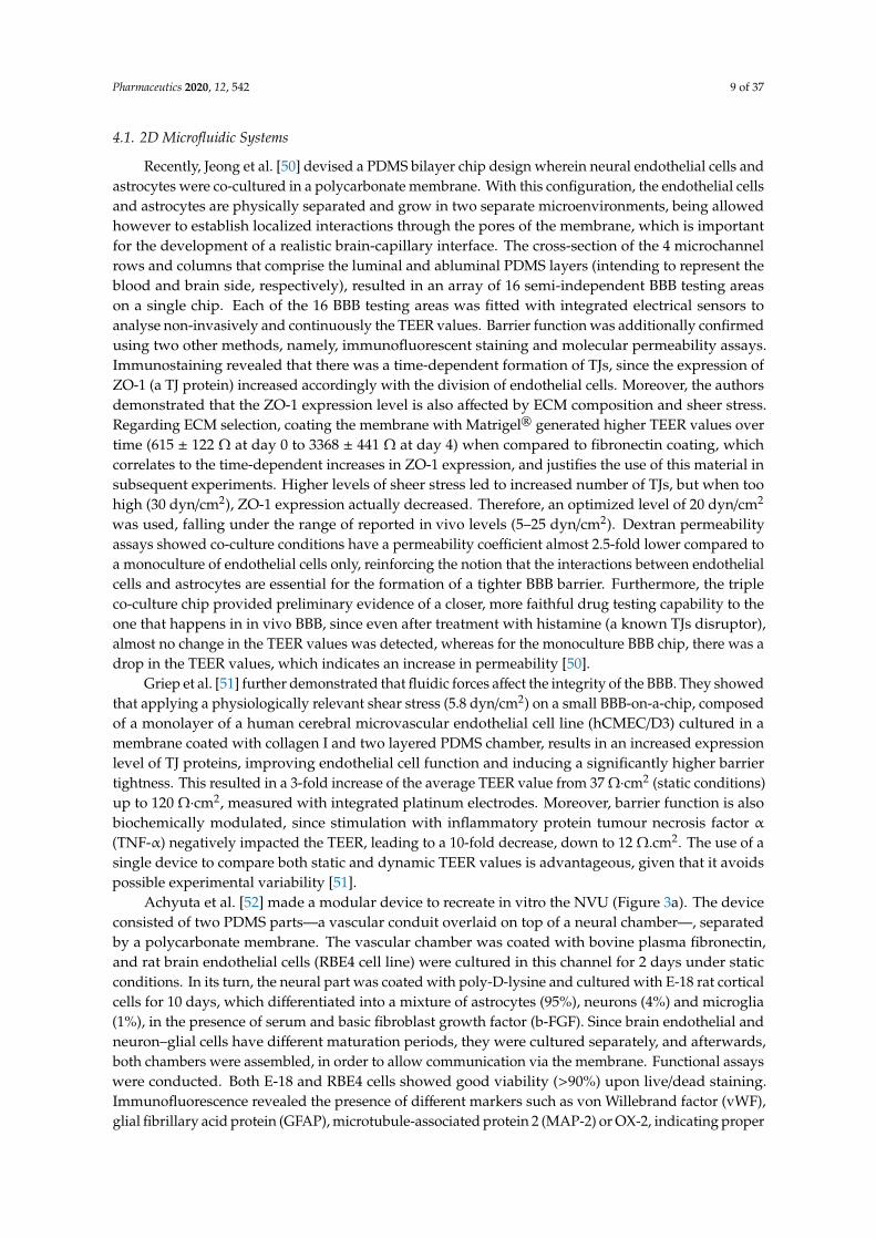
Recently, Jeong et al. [50] devised a PDMS bilayer chip design wherein neural endothelial cells andastrocytes were co-cultured in a polycarbonate membrane. With this configuration, the endothelial cellsand astrocytes are physically separated and grow in two separate microenvironments, being allowedhowever to establish localized interactions through the pores of the membrane, which is importantfor the development of a realistic brain-capillary interface. The cross-section of the 4 microchannelrows and columns that comprise the luminal and abluminal PDMS layers (intending to represent theblood and brain side, respectively), resulted in an array of 16 semi-independent BBB testing areason a single chip. Each of the 16 BBB testing areas was fitted with integrated electrical sensors toanalyse non-invasively and continuously the TEER values. Barrier function was additionally confirmedusing two other methods, namely, immunofluorescent staining and molecular permeability assays.Immunostaining revealed that there was a time-dependent formation of TJs, since the expression ofZO-1 (a TJ protein) increased accordingly with the division of endothelial cells. Moreover, the authorsdemonstrated that the ZO-1 expression level is also affected by ECM composition and sheer stress.Regarding ECM selection, coating the membrane with Matrigel® generated higher TEER values overtime (615 ± 122 Ω at day 0 to 3368 ± 441 Ω at day 4) when compared to fibronectin coating, whichcorrelates to the time-dependent increases in ZO-1 expression, and justifies the use of this material insubsequent experiments. Higher levels of sheer stress led to increased number of TJs, but when toohigh (30 dyn/cm2), ZO-1 expression actually decreased. Therefore, an optimized level of 20 dyn/cm2
was used, falling under the range of reported in vivo levels (5–25 dyn/cm2). Dextran permeabilityassays showed co-culture conditions have a permeability coefficient almost 2.5-fold lower compared toa monoculture of endothelial cells only, reinforcing the notion that the interactions between endothelialcells and astrocytes are essential for the formation of a tighter BBB barrier. Furthermore, the tripleco-culture chip provided preliminary evidence of a closer, more faithful drug testing capability to theone that happens in in vivo BBB, since even after treatment with histamine (a known TJs disruptor),almost no change in the TEER values was detected, whereas for the monoculture BBB chip, there was adrop in the TEER values, which indicates an increase in permeability [50].
Griep et al. [51] further demonstrated that fluidic forces affect the integrity of the BBB. They showedthat applying a physiologically relevant shear stress (5.8 dyn/cm2) on a small BBB-on-a-chip, composedof a monolayer of a human cerebral microvascular endothelial cell line (hCMEC/D3) cultured in amembrane coated with collagen I and two layered PDMS chamber, results in an increased expressionlevel of TJ proteins, improving endothelial cell function and inducing a significantly higher barriertightness. This resulted in a 3-fold increase of the average TEER value from 37 Ω·cm2 (static conditions)up to 120 Ω·cm2, measured with integrated platinum electrodes. Moreover, barrier function is alsobiochemically modulated, since stimulation with inflammatory protein tumour necrosis factor α
(TNF-α) negatively impacted the TEER, leading to a 10-fold decrease, down to 12 Ω.cm2. The use of asingle device to compare both static and dynamic TEER values is advantageous, given that it avoidspossible experimental variability [51].
Achyuta et al. [52] made a modular device to recreate in vitro the NVU (Figure 3a). The deviceconsisted of two PDMS parts—a vascular conduit overlaid on top of a neural chamber—, separatedby a polycarbonate membrane. The vascular chamber was coated with bovine plasma fibronectin,and rat brain endothelial cells (RBE4 cell line) were cultured in this channel for 2 days under staticconditions. In its turn, the neural part was coated with poly-D-lysine and cultured with E-18 rat corticalcells for 10 days, which differentiated into a mixture of astrocytes (95%), neurons (4%) and microglia(1%), in the presence of serum and basic fibroblast growth factor (b-FGF). Since brain endothelial andneuron–glial cells have different maturation periods, they were cultured separately, and afterwards,both chambers were assembled, in order to allow communication via the membrane. Functional assayswere conducted. Both E-18 and RBE4 cells showed good viability (>90%) upon live/dead staining.Immunofluorescence revealed the presence of different markers such as von Willebrand factor (vWF),glial fibrillary acid protein (GFAP), microtubule-associated protein 2 (MAP-2) or OX-2, indicating proper

Pharmaceutics 2020, 12, 542 10 of 37
neural and vascular cell differentiation. Furthermore, western blot showed the presence of TJ proteinZO-1. The integrity and tightness of the barrier was assessed with AlexafluorTM-conjugated dextranleakage, perfused during 1h through the vascular chamber at the rate of 1 mL/h. The models containinga RBE4 cell layer presented significantly decreased leakage into the neural reservoir compared todevices without cells. Conversely, when the vascular layer was exposed to TNF-α for 6h, more dyewas leaked, indicating a pro-inflammatory reaction that disrupted barrier function and triggered BBBhyperpermeability. Moreover, the circulation of the inflammatory stimulus, triggered the activation of75% of astrocytes and microglia in the neural chamber, mimicking neuroinflammation. Several caveatsof the current model were pointed out by the authors, including the use of embryonic stem cells thatdo not reproduce all the features of mature in vivo NVU, the absence of pericytes and shear stress,or the rodent-provenience of the cells, which might make direct correlation with data from humanclinical trials impossible. However, if needed, perfusion could be applied via the vascular channel, inorder to mimic in vivo settings. In addition, control over each module also allows the user to easilymanipulate cellular seeding and cellular:neuroglial ratios, to provide other physiological cues thatimpact the NVU and to observe sub-cellular features through high-content analysis. The system couldbe an opportunity to deliver nutrients, drugs, cells or nanomaterials, and evaluate their impact on theNVU [52].
In a 2017 study, Wang et al. [53] developed a 2D pumpless BBB microfluidic model (Figure 3b).BBB constructs were prepared by co-culturing, up to 10 days, brain microvascular endothelial cells(BMECs) (derived from human-induced pluripotent stem cells (iPSC)) with rat primary astrocytes,on two sides of a collagen IV and fibronectin-coated porous insert. The final assembled device wasachieved by accommodating the insert between a bottom layer with microchannels (perfusion layer)and a middle layer containing a central neuronal chamber and reservoirs filled with culture medium.Recirculation was assured without the need of using tubing and pumps, by means of a rocking platformthat changes the tilting direction. In order to properly mimic and match tissue volume and bloodresidence time of a human adult brain, the neuronal chamber and perfusion flow rate were scaled downproportionally (residence-time based design). Furthermore, to minimize oscillatory fluidic shear stresson the cell surface, a “step chamber” was introduced, increasing the distance between the cell planeand the perfusion layer. This modification enabled the BMECs to survive and maintain their uniqueBBB phenotype, while still providing physiologically relevant flow rates [53]. A TEER measuring probewith costum Ag/AgCl electrodes was integrated into the microfluidic device, facilitating the monitoringof the values and the optimization of culture conditions. This was the first microfluidic model toachieve sustained high TEER values (above 2000 Ω·cm2 for up to 10 days) that fall within the rangeof those recorded in vivo (1500–8000 Ω·cm2), being also the highest reported for any BBB-on-a-chipsystem (peak TEER above 4000 Ω·cm2). Likewise, evaluation of permeability coefficients for severallarge tracer molecules (4, 20 and 70 kDa FITC-dextran) and small drugs (caffeine, cimetidine anddoxorubicin), strongly correlated with in vivo BBB transport data. Doxorubicin disrupted the BBBintegrity after 24h treatment. The authors denote the convenience and simplicity of their setup, alsoindicating that since it closely mimicked BBB barrier functions, the potential and suitability of thisapproach for brain drug screening studies was validated. A possible improvement would be toincorporate astrocytes of human origin instead of rat, in order to create a fully human BBB model [53].
Walter et al. [54] recreated the BBB on a biochip that consisted of a polyethylene terephthalate(PET) membrane, situated between two PDMS channels fixated with a silicone sealant. This core wasplaced between glass slides with gold electrodes. Moreover, attached to the top glass slide, there weretwo PDMS blocks with reservoirs holding culture medium. The upper PDMS channel was definedas the “blood side” and coated with collagen I, while the lower channel, representing the brain, wascoated with collagen IV. The presence of collagen, a natural element of the ECM, facilitates the seedingof cells. This versatile microdevice enabled the formation of two different BBB models: hCMEC/D3human brain endothelial cell line and triple co-culture of primary rat brain endothelial cells withprimary astrocytes and brain pericytes. The endothelial cells were cultured on the top side of the PET

Pharmaceutics 2020, 12, 542 11 of 37
membrane in the top channel, while pericytes and astrocytes were cultured on the bottom channel.For 3 days, the cells were grown under static conditions, after which a peristaltic pump provideddynamic culture conditions at low shear stress. Characterization of cell culture was done by assessingits morphology, TEER values and apparent permeability. As expected, the results demonstrated thatthe primary rat triple co-culture was more efficient in the induction of barrier properties: it exhibitedhigher TEER values (114 ± 37.5 Ω·cm2 for both static and dynamic culture conditions vs. 19 ± 2.8 Ω·cm2
and 28.5 ± 7.2 Ω·cm2 for the hCMEC/D3 under static and dynamic conditions, respectively) and lowertracer permeability coefficients, indicating a tight barrier. Furthermore, immunostaining and confocalmicroscopy confirmed a stronger expression of both β-catenin (adherens junction protein) and ZO-1(TJs protein) for those cells [54].
Likewise, a multi-layered BBB microfluidic model, composed of a triple culture of co-immortalizedmouse brain endothelial cells (bEnd.3), mouse astrocytes (C8D1A) and pericytes, seeded on a 0.4 µmmicropore membrane fabricated using soft litography, was developed (Figure 3c) [55]. The membranewas integrated between two partially overlapping PDMS microchannels, embedded with Ag/AgClelectrodes. The top channel was connected to a pump that provided the culture medium, exposingthe endothelial cells to a fluidic shear stress of 1.6 dyn/cm2. A live/dead cytotoxicity assay proved ahigh viability of all cells up to 21 days, which was further confirmed by fluorescent images of theirmorphology. Interestingly, it was shown that cell alignment is influenced by the microchannel’s sizeand shape, since the spindle of the cells gradually decreased along the longitudinal axis of the channelas a function of days in culture. Compared to a single bEnd.3 monolayer and a double endothelialcells-pericytes co-culture, the triple model had enhanced functional expression and activity of P-gp,given that the basolateral-to-apical permeability and the efflux ratio of dexamethasone (a substratefor P-gp) were superior, gradually increasing with the increase in culture time, thus underlying thefunctionality of the efflux pump in the endothelial cells. Furthermore, it showed higher TEER values.It is known that soluble factors such as TGFβ, bFGF or GDNF, secreted by astrocytes and pericytes, canhave a positive effect in reinforcing the integrity and barrier properties of the BBB model. The authorsdemonstrated that increasing the height of the lower channel from 200 µm to 600 µm and 1000 µm,translated in a progressive decrease of the TEER values. As channel height increases, so does themedium volume, resulting in a dilution of those factors and, therefore, in a reduced restrictiveness ofthe formed barrier. Permeability screening assays with [14C]-urea and [14C]-mannitol showed thatboth the bi- and triple co-cultures have size selectivity, being able to discriminate between the twodifferent markers. Moreover, permeability of [14C]-mannitol after 18 days in the triple culture wassimilar to its reported permeability across the BBB in vivo [55].
Yeon et al. [56] designed a BBB microfluidic model comprised of a PDMS chip with two channels,connected by microholes (Figure 3d). Pressure gradients generated by applying different flow ratesin the microchannels, led to hydrodynamic entrapment of human umbilical cord endothelial cells(HUVECs) in the microholes. Within 2h of incubation with astrocyte-conditioned medium (ACM),a barrier was formed, and the permeability of various FICT-tracer dextrans (4, 40, 70 kDa) anddrugs (antipyrine, carbamazepine, atenolol, verapamil and propranolol) through the HUVEC layerwas evaluated by fluorescence microscopy and high-performance liquid chromatography (HPLC),respectively. Compared with the untreated control, supplementation with ACM significantly decreasedthe permeability of the dextran dyes, as well as of carbamazepine, verapamil, antipyrine andatenolol, which is in good agreement with the enhanced expression of ZO-1 TJ protein detected byimmunofluorescent staining. In the case of propranolol, the difference in permeability was negligible.Moreover, these values highly correlate with the results measured in conventional Transwell® systems,as well as with in vivo permeability data, validating the reliability of this model to predict thepenetrability of new CNS-targeted molecules. The authors also confirmed that exposure to hydrogenperoxide, an inductor of ROS formation, contributed to ZO-1 redistribution to the cytosol, thereforeincreasing BBB permeability. Two of the main concerns of this device are that it lacks cell–cell contact,

Pharmaceutics 2020, 12, 542 12 of 37
a fundamental characteristic of the BBB in vivo, and it does not reproduce the in vivo dimensions ofmicrovasculature [56].
As it is perceptible from the studies described so far in this section, one of the many applicationsof microfluidic platforms is the assessment of the permeability of different compounds, facilitating theearly screening of brain-targeted drug candidates [7,37]. This is not only applicable to conventionaldosage forms, but also to novel biopharmaceuticals and nanomedicines [7]. In regard to this lastcategory, Papademetriou et al. [57] carried on a study wherein the objective was to evaluate theimpact of static and flow conditions on the BBB binding and internalization of angiopep-2 coupledliposomes. The microfluidic device consisted of two S-shaped PDMS microchannels, with the regionof overlap being separated by a polycarbonate membrane treated with a mixture of fibronectin andcollagen IV, wherein mouse brain endothelial cells (b.End3) were cultured in the upper part over3–6 days prior to experiments. The liposomes (mixture of 1,2-dipalmitoyl-sn-glycero-3-phosphocholine(DPPC), 1,2-distearoyl-sn-glycero-3-phosphoethanolamine-N [methoxy(polyethylene glycol)-2000(DSPE-PEG2k) and DSPE-PEG maleimide, MW 3400 (DSPE-PEG-MAL3.4k)) were prepared by lipidfilm hydration method [58], followed by angiopep-2 conjugation. Angiopep-2 (ang-2) is a peptide ofthe LPR1 transferrin receptor, facilitating brain transport and penetration. The formation of a functionalbarrier was confirmed by dextran permeability size selectivity assays, as well as the measured TEERvalue (172 Ω·cm2), which is far superior to baseline values already reported for BBB microfluidicmodels using bEnd.3 cells (30–50 Ω·cm2). Results from fluorescence microscopy and spectroscopyallowed to withdraw two main conclusions: firstly, that the process of functionalization resulted insignificant binding to bEnd.3 cells compared to non-functionalized liposomes, and secondly, the ang-2nanocarriers were less internalized by the endothelial cells via receptor-mediated transcytosis underhigh shear stress (6 dyn/cm2) relative to low flow shear stress (1 dyn/cm2) or in static conditions.This suggests luminal fluid flow impacts the binding levels, and that at higher flow shear stress,detachment forces resulting from the flow are enough to overcome the avidity of ang-2 liposomes,uncoupling them from the endothelial cells. The claudin-5 perinuclear expression hints that flowexposure might have partially disassembled TJs, which may have influenced the penetration of NPsthrough the model. Since physiological flow shear stress in brain capillaries ranges from 5–23 dyn/cm2,which is important to replicate, tuning the NPs characteristics (e.g., ang2 valency) might help to solvethis problem by enabling binding in the presence of flow while maximizing the BBB penetration.As mentioned by the authors, one limitation of this study was the lack of co-culture with other celltypes that form the NVU [57]. In addition, Park et al. [59] characterized the transcellular transportof angiopep-2 quantum dot NPs and monoclonal antibodies directed against the transferrin receptor.The results of this study suggest that hypoxia is needed to better recapitulate the native BBB in terms oftransferrin receptor function, as well as of other BBB transporters, since when compared to normoxicconditions, hypoxic conditions resulted in enhanced differentiation and increased expression of thoseproteins [59].
In similar fashion, the effectiveness of a gH625 (a membranotropic peptide) to direct the transportof polystyrene NPs across a BBB layer under flow conditions that mimic circulation, was tested in amicrofluidic device devised by Falanga et al. [60]. Its design was based on the thermoplastic polymerpoly(methyl methacrylate) (PMMA), which has the advantage of being easily micromachinned bymicromilling. Confocal microscopy and phase contrast demonstrated the formation of a confluentbEnd.3 layer on a porous membrane, at 7 days of culture. Crossing experiments proved thatfunctionalization with gH625 peptide enhanced the adhesion of the NPs to the BBB layer, and at aworking medium flow rate of 5 µL/min, increased 2-fold the transport of NPs (6.13% compared to2.72% of non-functionalized blank NPs). Furthermore, TEER analysis confirmed the maintenance ofbarrier function before and after the passage of the nanocarriers, proving that the bEnd.3 layer is notdisrupted by the NPs. In summary, the system allowed the reproducibility of experiments and thequantification of NPs transport across the BBB in vitro [60].

Pharmaceutics 2020, 12, 542 13 of 37Pharmaceutics 2020, 12, x FOR PEER REVIEW 14 of 38
Figure 3. Examples of 2D BBB microfluidic models: (a) modular polydimethylsiloxane (PDMS) device to recreate the NVU by co-culture of neuroglial and endothelial cells, (b) pumpless microdevice that provides in vivo-like BBB properties for drug permeability screening, (c) multi-layered system composed of a triple culture of co-immortalized mouse brain endothelial cells (bEnd.3), mouse astrocytes (C8D1A) and pericytes, (d) microfluidic device that mimics the cerebral vasculature and is a reliable permeability assay system. Reprinted and adapted with permission from: (a) Achyuta et al. [52], (b) Wang et al. [53], (c) Wang et al. [55]. Copyright (2016) American Chemical Society, (d) Yeon et al. [56].
Figure 3. Examples of 2D BBB microfluidic models: (a) modular polydimethylsiloxane (PDMS) deviceto recreate the NVU by co-culture of neuroglial and endothelial cells, (b) pumpless microdevicethat provides in vivo-like BBB properties for drug permeability screening, (c) multi-layered systemcomposed of a triple culture of co-immortalized mouse brain endothelial cells (bEnd.3), mouse astrocytes(C8D1A) and pericytes, (d) microfluidic device that mimics the cerebral vasculature and is a reliablepermeability assay system. Reprinted and adapted with permission from: (a) Achyuta et al. [52],(b) Wang et al. [53], (c) Wang et al. [55]. Copyright (2016) American Chemical Society, (d) Yeon et al. [56].
Prabhakarpandian et al. [61] introduced a synthetic microvasculature model of the BBB(SyM-BBB). The device consisted of a circular PDMS chip with microchannels, partitioned in atwo-compartment chamber: an outer ring (blood compartment—apical side) and an inner ring (braincompartment—basolateral side), which were separated by an array of micropillars with 3 µm gaps

Pharmaceutics 2020, 12, 542 14 of 37
between them. These gaps allowed communication between the chambers. The outer ring wascoated with fibronectin, and RBE4 rat endothelial cells were cultured under fluidic shear conditions(0.1 µL/min) for 24-48h. ACM was added to the brain compartment, and its influence on barrierpermeability, tight junction formation and P-gp expression/activity was studied. Compared to aconventional Transwell® and a device without ACM, the ACM-perfused model improved the overallBBB function. It had decreased barrier permeability (reflected by lower FITC-dextran intensity levelsin the basolateral chamber), promoted expression of TJ proteins (ZO-1 and claudin) and P-gp, andincreased efflux pump activity (translated in a significantly higher efflux of Rhodamine 123, even inthe presence of verapamil, an inhibitor of P-gp). One thing that differentiates this model from the othertwo-chamber assays that were described earlier is that it does not use a membrane or a filter, with theapical and basolateral compartments being side-by-side, which simplifies fabrication. Furthermore,its design allows the simultaneous, real-time monitoring of both compartments, provides realisticmicrovasculature environment and dimensions, delivers physiological shear stress/fluid flow andallows long-term cell culture [61].
4.2. 3D Microfluidic Systems
Marino et al. [62] resorted to a two-photon lithography technique (3D printing) to fabricate a3D microtubular porous structure that replicates the microcapillaries of the NVU. The core benefit ofthis technique is that capillary diameter, pore size or pore density are easily fine-tuned. The structureconsisted of 50 parallel microtubes with pores on the surface to allow transport toward the externalenvironment, connected to an inlet and an outlet reservoir. The microtubes served as scaffolds for theco-culture of murine brain-derived endothelial cells (bEnd.3) and U87 glioblastoma line, allowingthe cells to maintain their morphology and phenotype, since it mimics the basement membrane ofthe BBB. Culture medium was pumped at 50 µL/h, making the flow rate within each microtube of1 µL/h, a value comparable to that of cerebral microcapillaries. The novelty of this work is that theplatform was constructed according to the actual dimensions of human brain capillaries, leading to thefirst-time development of a real scale, biohybrid and biomimetic BBB model that showed an efficientmaturation of TJs (high expression of ZO-1 by immunofluorescence staining), decreased dextrantranscytosis and higher TEER value (75 ± 2 Ω cm2), with respect to the same microfluidic systemwithout cells. Even after 5 days, TEER value remained stable (71 ± 10 Ω cm2), denoting preserved cellviability and functionality. The investigators pointed out that this model could potentially be used forhigh-throughput screening of drugs or nanomaterials for several brain pathologies [62].
Wevers et al. [63] established a perfused parallel BBB-on-a-chip model, comprised of a two- orthree-lane microfluidic platform that harbours 96 or 40 chips, respectively, in a 384 well plate format(referred to as OrganoPlate). In each chip, a microvessel of brain endothelial cells (TY10 cell line) wasformed and grown against an ECM gel composed of collagen-I. On the other side of the gel, astrocytes(hAst human cells) and pericytes (hBPCT human cells) were added, in order to complete the model. Byplacing the plate on a rocker and infusing it with medium culture on the microvessel channel, perfusionwas generated, creating a bidirectional flow from inlet to outlet and vice-versa. Expression andinterendothelial localization of key BBB TJs (claudin-5) and adherens junction (VE-cadherin, PECAM-1)proteins was detected by immunostaining, indicating barrier formation. Moreover, barrier integritywas assessed at days 5, 7 and 9 by a fluorescent permeability assay. The results showed that the modelwas able to severely restrict the passage of a 20 kDa FICT-dextran dye, which is revealing of a functionalBBB. To study BBB diffusion by receptor-mediated transcytosis of a therapeutic antibody (antibody tohuman transferrin receptor (MEM-189)), the authors infused the microvessel channel with MEM-189 ora control antibody, quantifying afterwards its levels in the basal chamber. The penetration of MEM-189was approximately 2-fold higher than the penetration of the control antibody (apparent permeability of2.9× 10−5 versus 1.6× 10⁻⁵ cm/min, respectively), denoting that the binding of the therapeutic antibodyto the transferrin receptor expressed in the endothelial cells facilitates the transportation across theBBB. All in all, the Organoplate has several advantages such as minimal absorption, low-level passive

Pharmaceutics 2020, 12, 542 15 of 37
permeability and ease of sampling, that make it a valuable and suitable model for high-throughputdrug screening (including of large molecules like antibodies) under physiological conditions withoutthe need of artificial membranes [63].
Maoz and colleagues [64] recently constructed an innovative and sophisticated platform of threeinterconnected microfluidic systems (Figure 4a). The modular structure comprised a brain chip putbetween two BBB chips, thus recapitulating at the same time the brain parenchyma and the influx/effluxacross the BBB. The brain chip contained human neural stem cells (~60% glial cells and 40% ofdopaminergic, glutamatergic, serotonergic and GABAergic neurons) and astrocytes cultured on thelaminin and poly-l-lysine-coated surface of the lower compartment, whereas the BBB chips entailed amonolayer of primary human brain microvascular endothelial cells (hBMVECs) at the vascular (lower)chamber and astrocytes and pericytes at the perivascular (upper) chamber, to mimic the external wallof a brain microvessel. To achieve successive influx and efflux from one chip to the other, and toallow diffusion-mediated molecular transport to dominate (as what happens in vivo in the brain), theshear stress was adjusted to be close to 0 within the brain compartment. The results of proteomicanalysis demonstrated that fluidic coupling modulates the phenotype of the cultured cells via paracrinesignalling, with more effective expression of some proteins (e.g., metabolism-associated proteins) in thecoupled chips compared to uncoupled cultures. Furthermore, the linked system allowed to comparethe individual roles of the different endothelial, neuronal and perivascular cell populations in the NVU,obtaining insights into previously unknown metabolic interactions between them that are significantfor the maintenance of brain function. Of relevance, these metabolic interactions led to an enhancementof the synthesis and secretion of neurotransmitters gamma aminobutyric acid (GABA) and glutamate,suggesting that brain vasculature may have a role in underlying mechanisms of NDs. To investigatethe drug screening capabilities of the model, the neuroactive drug methamphetamine was introducedin the BBB influx vascular channel. Methamphetamine produced a reversible disruption of the barrier,since removing the compound from the system stopped its effects that were leading to BBB breakdown,like reduced expression of cell–cell junction proteins (e.g., VE-cadherin). This microfluidic modelcan, therefore, be used for diverse applications, such as improving the basic knowledge on brainpharmacokinetics, facilitating the evaluation of penetrance, efficacy and toxicity of CNS-targeted drugs,or assessing BBB function and mechanisms in both normal and diseased states [64].
Bang et al. [65] established a 3D BBB microfluidic model, based on a vasculogenesis-like process.The device consisted of a PDMS slab containing one middle chamber and two channels side by side(vascular and neural channel). A vascular network self-assembled in the middle channel on a fibrinhydrogel over a 3-day period, by co-culturing human umbilical vein endothelial cells (HUVEC) andprimary human lung fibroblasts, while in the adjacent micropost traps, astrocytes and rat corticalneurons were grown in direct contact with the capillaries, in order to complete the BBB model.The two channels allowed to independently supply different types of media for the neural andendothelial cells (neurobasal medium and endothelial growth medium, respectively), deliveringthe best combination of medium. Thus, an optimized co-culture environment not only increasedcell viability, but also led to further inducement of relevant BBB characteristics, including greaterexpression of TJs protein (e.g., ZO-1), presence of synaptic structural features and a higher degree ofneurovascular interfacing (establishment of more direct connections between astrocytic end-feet andendothelial cells). Correspondingly, this model had low permeability coefficients for 20 and 70 kDadextran, comparable to reported in vivo BBB values [65]. While the developed platform is great forimproving tissue-level function, presenting substantial potential to majorly accelerate brain diseaseneuropharmacological research, it cannot be considered a complete NVU model, since more cellswould need to be included [65].
Partyka et al. [66] investigated in their 3D model the effects of mechanical stimuli exerted byblood flow on both BBB permeability and waste transport. The device consisted of two parallelmicrochannels with inlets and outlets, and a hydrogel at the centre of the microchannels. The hydrogel,composed of hyaluronan, collagen type I and Matrigel, supported the co-culture of human cerebral

Pharmaceutics 2020, 12, 542 16 of 37
microvascular endothelial cells (hCMEC/D3) with astrocytes, for 3-4 days. As shown in previousstudies [51,67], the findings suggest that both shear stress and cyclic strain increase TJs formation anddecrease transendothelial permeability, significantly improving the overall BBB integrity. Accordingly,compared to static controls, vessels exposed to fluid flow, with or without stretch, had enhancedTEER values and lower permeability coefficients for dextran 4 kDa, thus confirming that these factorsindeed contribute to the maintenance of proper BBB function. It was also demonstrated that vesselwall pulsation provides a convective force that facilitates retrograde transport along the basementmembrane of the cerebral microvasculature, including of metabolic waste products. The authorsconcluded their study by postulating that attenuated pulsatile waste transport as a consequence fromstiffening of the vessel walls, can possibly contribute for the pathogenesis of brain diseases [66].
Adriani and collaborators [68] developed a 3D hydrogel-based NVU system. The PDMS device wascomposed of four distinct parallel channels, where the first provided neural cell culture medium, the twocentral channels were used to co-culture astrocyte and neuron rat cells embedded in adjacent collagenI matrices, and the remaining fourth hosted cerebral endothelial cells (either HUVEC or hCMEC/D3)to mimic the blood vessel wall and provided endothelial cell culture medium. Immunochemistryassays revealed that all three cell types were capable of growing, displaying type-specific markers (e.g.,presence of doublecortin (DCX) for neurons, GFAP for astrocytes and VE-cadherin or ZO-1 for theendothelial cells) and morphological characteristics. Permeability assays for dextran 10 and 70 kDawere done in order to assess barrier functionality. The results showed that co-culture with astrocytesenhanced BBB integrity, and that while both HUVEC and hCMEC/D3 monolayers demonstratedsize-selective penetrability, the hCMEC/D3 line had significantly higher barrier integrity comparedto HUVEC. Non-inclusion of a porous membrane within the system allowed a closer associationbetween astrocytes end-feet and the endothelial cells, leading to a low permeability. The presence ofastrocytes was also important for neuron growth, as demonstrated by the increased number of neuritesegments in co-culture conditions, compared to neurons in monoculture. To best demonstrate themodel’s BBB restrictiveness and its application for compound testing, the authors conducted a testin which glutamate, a neurotransmitter, was injected into the endothelial cell channel and its abilityto trigger neuronal activity was assessed by calcium imaging and c-Fos expression. In contrast to amicrofluidic system without endothelial barrier, the developed model had a significant decreasedcalcium concentration in the neurons and a C-Fos immunopositive staining, implying the presence ofan intact barrier that was able to restrict the passage of the test compound and thus, consequently,restrict neuron activation. It is suggested that this platform will be useful for neurovascular studies,such as assessing drug effects on neural function [68].
Campisi et al. [69] co-cultured human induced pluripotent stem cell-derived endothelial cells(hiPSC-ECs) with human primary astrocytes and pericytes in a microfluidic platform. The mixtureof cells was injected with a fibrin gel to the central microchannel of a PDMS device. The hiPSC-ECsunderwent self-assembly in the 3D matrix into a microvascular network comprised of small perfusablelumens, with pericytes and astrocytes surrounding and adhering to them, thus emulating the in vivoBBB neurovascular organization. Immunochemistry and real time RT-PCR assays, performed after 7days, validated the formation of a functional barrier, with gene expression of several BBB membranetransporters (e.g., P-gp, MRP1 or GLUT-1), TJs proteins (ZO-1, occluding and claudin-5) and ECMproteins (laminin and collagen V) in the triple co-culture being significantly increased comparedwith an iPSC-ECs monoculture or an iPSC-ECs/astrocytes co-culture. This BBB model was robustand physiologically relevant, displaying low permeability and transport selectivity dependent uponmolecular weight. Indeed, permeability values of 2.2 × 10⁻⁷ cm/s and 8.9 × 10⁻⁸ cm/s for 10 kDa and40 kDa FTIC-dextran, respectively, were similar to the levels measured in rat brain, as well as previousin vitro BBB microfluidic systems that also employed iPSC-ECs in co-culture with astrocytes or neurons.Astrocytes and pericytes work together through paracrine signalling and juxtacrine interactions tofacilitate and stabilize endothelial vasculature organization and improve BBB formation/integrity,therefore explaining the fact that the triple co-culture showed the best performance in terms of stability

Pharmaceutics 2020, 12, 542 17 of 37
and permeability. Furthermore, it was observed that the seeding of additional iPSC-ECs on theside channels improved perfusability and decreased permeability, by means of filling eventual gaps,increasing the number of connections in the vascular network and improving the overall coverage onthe boundaries of the gel region. The authors have stated that the developed model could be appliedfor a number of applications, such as the study of angiogenesis and NVU function, investigation of BBBtranscytosis, or evaluation of biological events that occur in NDs and of metastatic cancer extravasationto the brain [69].
More groups have also come up with recent 3D BBB microfluidic models using human iPSC [70–72].Jamieson et al. [70] co-cultured iPSC-derived BMECs and pericytes in a cylindrical channel surroundedby collagen I. They observed the formation of a confluent monolayer without discontinuities,direct BMECs-pericyte contact and abluminal localization of the pericytes. The permeabilityof Lucifer yellow for the co-culture was comparable to that of BMECs microvessels withoutpericytes [70]. This contrasts with other BBB studies, where pericyte co-culture improved thebarrier restriction [69,73,74]. These discrepancies may be due to a series of factors, such as variabilityin the culture and assay conditions, or even iPSC-line background. Linville et al. [71] culturediPSC-derived BMECs within collagen I-coated microchannels in a PDMS microfluidic chip, leading tothe creation of a model that resembles human brain post-capillary venules in terms of their cylindricalgeometry, cell–ECM interactions and shear flow. This 3D perfused microvessel model had similarrestrictive permeability to post-capillary venules in rats. Moreover, in comparison with self-organizingBBB approaches employed to mimic vasculogenesis, this model has the advantage of achievingphysiological barrier functionality after only two days of culture, without the hassle of astrocyte orpericyte co-culture [71]. Motallebnejad et al. [72] created a BBB-on-a-chip that supports flow andwhere a co-culture of hiPSC-derived BMECs with astrocytes embedded in a 3D hydrogel was achieved.Apical addition of TGF-β1 conducted to a reduction of TEER values and astrocyte activation, puttingforth the ability of the system to be used as a BBB disruption model. Furthermore, since all the culturedcells derived from the same hiPSC line, it could also enable genetic and rare disease modelling [72].
To get further insights into the BBB system of efflux transporters, which is generally not themain focus of in vitro brain platforms, Lee et al. [75] established a CNS angiogenesis microfluidicmodel containing a co-culture of human HBMECs, astrocytes and pericytes in a fibrin/hyaluronic acidhydrogel, seeded in the central microchannel (Figure 4b). Fibroblasts were also added to a side channel,as a source of angiogenic factors. Confocal images showed that after 7 days, the microvasculatureresulting from the triple culture was fully mature and perfused, presenting physiologically relevantBBB characteristics, including in vivo 3D-like morphological phenotypes, direct cellular interactions(astrocyte and pericyte covering the vessels), increased adherens/TJs expression and limited vesseldilation, related to the minimized vessel diameter of about 34.64 µm (the smallest engineered vesseldiameter ever reported). Additionally, compared to a monoculture of ECs, the triple co-culture hadlower permeability for both 10 and 70 kDa FITC-dextran, which further confirmed the integrity andbarrier function of the vascular network. To demonstrate the functionality of efflux transporters, theauthors conducted a calcein acetoxymethyl (calcein-AM) assay, again under mono- and triple-cultureconditions. Calcein-AM is a compound that when inside the cells is rapidly hydrolysed to fluorescentcalcein (leading, therefore, to an increase in fluorescence). The developed model had a significantlylower initial calcein intensity, indicating a higher expression of efflux transporters that pumped outmore calcein. Furthermore, fluorescence intensity on the endothelium was tracked over a 10h period,after treatment with P-gp inhibitors (valspodar and elacridar). As expected, for both culture conditions,the remaining fluorescence intensity was higher, since less calcein was being effluxed. However,compared with monoculture conditions, the triple co-culture had a more prominent difference betweentreated and non-treated groups. Under inhibitor treatment, the effect of efflux transport was diminished,leading to a higher remaining calcein intensity than in non-treated groups (8.96- and 2.10-fold higher forvalspodar and elacridar, respectively). This was the first in vitro BBB model that allowed reconstitution

Pharmaceutics 2020, 12, 542 18 of 37
and regulation of the efflux transport system under 3D microvasculature condition, highlighting itspotential for application in CNS drug penetration studies [75].
In an interesting study, Wang et al. [76] converged two approaches–organoids and microfluidicdevices-to develop a 3D brain organoid-on-a-chip that recapitulates the early human brain development(Figure 4c). The culture of hiPSCs-derived embryoid bodies (EBs) in a Matrigel®within the microdevice,under mechanic perfusion flow, led to the in-situ growth and maturation of self-organized organoids.The microfluidic organoids exhibited well-defined neural differentiation, regionalization and corticalorganization. Moreover, compared to brain organoids under static culture conditions in a Petri dish, theon-chip organoids had superior expression of cortical layer markers (TBR1 and CTIP2) and increasedcell viability (<10% apoptosis vs. 40% apoptosis), indicating the importance of perfusion flow and 3DECM in enhancing brain organogenesis and creating a biomimetic microenvironment that supportsprolonged culture. The main disadvantage of this technique is that organoids are not individuallyaddressable for screening purposes. However, it holds promise for fundamental neurodevelopmentand disease modelling studies [76]. In a follow-up paper [77], the same authors demonstrated theapplicability of this platform, by assessing the impact of nicotine in prenatal brain development. It wasfound that nicotine exposure elicited neuronal dysfunction, with the organoids presenting impairedneurogenesis, specifically brain regionalization and cortical development [77].
For in vitro studies of neurovascular pathology, Cho et al. [78] designed a 3D BBB model consistingof an assembly of horizontal parallel microchannels beside a tube-shaped macrochannel in a singlelayered microfluidic chip (Figure 4d). The macrochannel was coated inside with a poly D-lisine (PDL)and a collagen I gel, promoting the adhesion of the RBE4 cell line and leading to the formation after2–3 days of an endothelial cylindric monolayer that resembles the geometry of small blood brain vessels.The formation of a tight BBB was confirmed by confocal imaging of the positive immunostaining of TJsproteins (ZO-1 and VE-cadherin). Permeability assays with 40 kDa dextran were done in order to testthe barrier function. The dye was introduced into the lumen of the macrochannel and the increase influorescence on the side channels was monitored. The presence of the BBB slowed the outward flux ofthe dye, since it took longer to reach gradient saturation in the developed model (7 min), compared to adevice without barrier (less than 4 min). Moreover, it was also able to block and inhibit more efficientlyneutrophil transmigration upon addition of interleukin 8 (IL-8), a chemoattractant. For probingneuroinflammation response, the BBB was exposed to TNF-α. Elevation of several cytokines (e.g.,VEGF, CX3CL1, CINC1, TIMP1, etc.) led to the conclusion that an inflammatory effect was elicitedon the BBB model. The platform was also used to study ischemia, induced by oxygen and glucosedeprivation followed by reoxygenation. An increase in ROS and Rho-associated protein kinase (ROCK)levels was observed, as well as a decrease by more than half in ZO-1 expression. Antioxidant treatmentwith edavarone and Y-27632 (a ROCK inhibitor) had limited protective effects in restoring BBB integrity:ZO-1 levels increased slightly after 3h but decreased again after 6h. This can be due to the fact thatischemia is not only a result of oxidative stress, but also hypoxia and other inflammatory factors [78].

Pharmaceutics 2020, 12, 542 19 of 37Pharmaceutics 2020, 12, x FOR PEER REVIEW 20 of 38
Figure 4. Examples of 3D BBB microfluidic models: (a) three interconnected systems—one brain chip between two BBB chips—to obtain insights into previously unknown metabolic interactions between the different cells of the NVU, (b) 3D brain angiogenesis model for reconstitution of BBB efflux transporter system (P-gp) under perfusion conditions, (c) brain organoid-on-a-chip that recapitulates the early human brain development, (d) microfluidic platform for the study of neurovascular pathology. Reprinted and adapted with permission from: (a) Maoz et al. [64], (b) Lee et al. [75], (c) Wang et al. [76]. Published by The Royal Society of Chemistry, (d) Cho et al. [78].
Figure 4. Examples of 3D BBB microfluidic models: (a) three interconnected systems—one brainchip between two BBB chips—to obtain insights into previously unknown metabolic interactionsbetween the different cells of the NVU, (b) 3D brain angiogenesis model for reconstitution of BBB effluxtransporter system (P-gp) under perfusion conditions, (c) brain organoid-on-a-chip that recapitulatesthe early human brain development, (d) microfluidic platform for the study of neurovascular pathology.Reprinted and adapted with permission from: (a) Maoz et al. [64], (b) Lee et al. [75], (c) Wang et al. [76].Published by The Royal Society of Chemistry, (d) Cho et al. [78].

Pharmaceutics 2020, 12, 542 20 of 37
5. Neurodegenerative Diseases Microfluidic Models
As previously discussed in prior sections, microfluidic devices are able to emulate brainmicrovasculature-related events, for instance, angiogenesis, inflammation or BBB changes [79]. It isknown that BBB dysfunction underlies the development of NDs. By reconstituting such dynamicconditions of the human brain, a validated BBB-on-a-chip can be used to address the critical need toimprove the understanding of the pathological mechanisms of NDs [32,79]. Furthermore, since CNSmodels that mimic NDs typically require controllable fluid delivery and long-term culture duration,microfluidics come forth as an advantageous and reliable platform to study these disorders [39].A summary of the main findings of the studies that will be discussed in this section can be found inSupplementary information (Table S1).
5.1. Alzheimer’s Disease
AD is a neurodegenerative disease that is the most common cause of dementia globally.Pathological hallmarks of AD include the presence of extracellular aggregates of amyloid β
(Aβ) plaques and intracellular neurofibrillary tangles of hyperphosphorylated tau protein [80,81].Anomalous cleavage of the amyloid precursor protein (APP) by β-secretases and γ-secretases leadsto the production of insoluble Aβ fibrils, which suffer oligomerization and aggregation into senileplaques. In turn, Aβ concentration triggers the hyperphosphorylation of the tau protein, followed byits polymerization into neurofibrillary tangles. There appears to be a synergistic neurodegenerativeeffect of these cytotoxic proteins, with the recruitment of microglia cells and formation of a localinflammatory response also potentiating neuronal damage and death [24,81].
AD is associated with three general stages-mild (early), moderate and severe-, with progressivememory deficits and cognitive decline [82]. Neuropsychiatric symptoms may manifest over the courseof the disease, including irritability, diminished insight or restlessness. In nearly 30 to 50% of thecases, psychotic symptoms such as hallucinations or delusions of persecution are also present [82].The plaques and neurofibrillary tangles are mainly detected in cortical and limbic areas of the brain.There is moderate cortical atrophy, accompanied with enlargement of the frontal and temporal hornsof the lateral ventricles, and a decrease in brain weight [80,81]. Given that AD is a long-term process,with a lengthy preclinical asymptomatic phase characterized by pathological changes that begin 20 to30 years prior to the appearance of the first clinical signs, it should not be excluded that cognitivelynormal individuals might also have the disease [80,82].
Clinical diagnosis entails a thorough assessment of previous medical records and family historyto rule out other conditions that may cause similar symptoms, physical and neurological examination,neuroimaging (PET scan, MRI, etc.), and neuropsychological testing, often in combination withdetection of biomarkers like Aβ and τ protein in cerebrospinal fluid (CSF) samples from patients [81,82].To date, accurate identification of AD is still hard, and both clinical diagnosis and post-mortemhistopathological examination of the brain are needed in order to give a conclusive diagnosis. This isespecially true in the early stages of the illness, primarily because the symptoms are dismissed as anormal result of the aging process. The strongest known risk factor for the development of AD is, infact, aging, affecting 10% of people over the age of 65 and about 50% of people older than 85. Familyhistory of AD, existence of one or more apolipoprotein E (ApoE) gene alleles, cardiovascular riskfactors, and moderate or severe brain traumas can also account for the aetiology of the disease [82].
AD research has been majorly committed to the development of therapies that can reduceaggregates of toxic forms of Aβ and tau protein, with several of them having reached Phase IIIclinical trials (e.g., NGP 555, verubecestat, immunotherapy vaccines such as aducanumab, etc.) [81,83].The latest progress is the study by Davtyan et al. [84], wherein they tested in bigenic T5x mice acombinatory therapy that concurrently targeted both Aβ and tau protein, successfully removingplaques and neurofibrillary tangles. Furthermore, the vaccine might also be used as a preventivestrategy, since it can likewise decrease the concentration of those proteins in serum and brain extracts,thus averting their deposition [84]. In fact, avoiding plaque accumulation seems to be a better goal, since

Pharmaceutics 2020, 12, 542 21 of 37
the removal of plaque deposits might merely result in holes in the neuronal synapses, a damage that canbe difficult to overcome. In this field, Novartis has put forth the CAD106 vaccine, presently in PhaseII/III study, designed to stimulate the production of Aβ-antibodies [83]. Nonetheless, therapy failureis often frequent, due to unfavourable pharmacokinetics and pharmacodynamics of the drugs [81].Presently, cholinesterase inhibitors (e.g., donepezil) and memantine, a non-competitive blocker of theNMDA receptor, are available for the treatment [85].
Park et al. [86] were able to show the neuroinflammatory role of microglia in AD pathogenesis,and their influence in neuron death. The investigators developed a 3D human AD triculture system,by culturing neurons and astrocytes expressing mutant APP in a central chamber coated with Matrigel,with human immortalized SV40 microglia being added in surrounding angular chambers at a later time.The neurons and astrocytes expressed late-stage AD hallmarks, including Aβ aggregation, abundanttau protein formation and secretion of pro-inflammatory cytokines and chemokines, thus recapitulatingmany of the features of this disease in a single microfluidic platform. Compared with controls, in thismodel, infiltrating microglia migrated quicker and induced greater neuron toxicity/death by a pathwayinvolving TLR 4 and interferon-γ (IFN-γ). This was demonstrated by the fact that using IFN-γ andTLR4 neutralizing antibodies led to a reduction in microglial activation and, consequently, neuronaltoxicity [86].
Concave microwell arrays in a microfluidic system were used to seed and culture for up to10 days neural progenitor rat cortical cells, with or without fluid flow, evaluating thereafter theneurotoxic effects of Aβ on the formed 3D gel-free neurospheroids [87]. The group demonstratedthat, compared to static cultures, the neurospheroids grown under perfusion conditions similar tothose of the CSF in the brain, had generally larger sizes and greater neurite extension, resultingin a more complex and robust neural network. Furthermore, cell staining for β-III tubulin marker,denoted an enhanced differentiation of the progenitor cells into mature neurons. These differencescould be the result of a better access to oxygen and nutrients, and continuous clearance of metabolicwastes provided by the fluid flow. Afterwards, the neurospheroids were exposed to Aβ, in the sameexperimental settings. The results showed that Aβ was significantly more toxic to neurospheroidsunder flow conditions induced by an osmotic pump, causing greater destruction of neural networksand significantly decreasing cellular viability [87].
Cho et al. [88] elucidated the role of Aβ on microglial accumulation. They cultured humanmicroglial cells in a microfluidic platform and studied their response to chemotaxis of week-longgradients of soluble Aβ and surface-bound (insoluble) Aβ (Figure 5a). Time-lapse microscopy showedthat soluble Aβ drives microglia migration towards the central chamber, explained by Aβ-inducedsecretion of the chemoattractant MCP-1. Furthermore, microglial motility was decreased in areas withAβ fibril-coated surfaces, which correlates to what is seen in vivo in AD brains, where microglia cellsform a stable association with Aβ deposits. All in all, there seems to be a synergistic effect betweensoluble and insoluble Aβ on microglial recruitment and localization during the neurodegenerativeprocess of AD [88].
Song et al. [89], to the best of our knowledge, were the first group to deliver direct experimentalevidence of extracellular Aβ spreading through neuronal connections. They demonstrated retrogradetransport of Aβ from axons to the neuronal cell bodies, being subsequently secreted and transmitted toneighbouring neurons [89]. Deleglise et al. [90], examined the impact of Aβ on neuron degenerationpatterns. By using a microfluidic chip comprised of two chambers separated by microchannels,the scientists were able to culture primary cortical and hippocampal mouse neuronal cells andisolate soma-dendrites from axonal projections, applying afterwards Aβ peptides to the differentcellular compartments. It was found that local somatodendritic Aβ deposits trigger a rapid distalpresynaptic loss and disconnection, long before soma/dendrite abnormalities and death start to occur(a phenomenon referred to as “dying-back process”). On the contrary, when the axons where treatedwith Aβ, the same was not observed. This demonstrates local Aβ deposits are efficient in generatingsignalling pathways (e.g., caspase and NAD+ pathways) that lead to early degeneration in anatomically

Pharmaceutics 2020, 12, 542 22 of 37
distant areas of the neuron [90]. To this day, there are doubts about which molecular entity of Aβ is themost toxic and responsible for causing AD. Choi et al. [91] have looked into this matter. They exposedneurons (differentiated from rat neuronal progenitor cells) adhered onto a microchannel of a PDMSmicrochip to a slow gradient of Aβ oligomeric assemblies for 3 days. Although fibrilization increasedconsiderably over time, cell viability did not statistically change. This suggests that Aβ fibrils do nothave a significant role in neurotoxicity, but rather oligomeric assemblies potentially contribute to theneurodegeneration in AD [91].
To determine the nature of the tau responsible for neuron-to-neuron transmission, Takeda et al. [92]developed a compartmentalized 2D microfluidic platform comprised of three interconnected chamberswherein two sets of neurons where cultured. The authors collected different tau species from corticalextracts and interstitial fluid of human and transgenic mice brains, and their propagation throughthe synaptic connections was observed over the course of 14 days. A soluble high molecular weightphosphorylated tau was identified as the bioactive form, undergoing direct trans-synaptic transport andinitiating the seeding that leads to the formation of aggregates within the cytoplasm of the neurons [92].Wu et al. [93] further demonstrated in a study using a tripartite microfluidic device that cell to cellpropagation is capable of inducing tau aggregation in downstream neurons [93].
5.2. Parkinson’s Disease
First described in 1817 by James Parkinson, PD is after AD the second most prevalentneurodegenerative disease [94]. Estimations point towards 1-3% of the population over the ageof 65 developing this illness, with people under 40 years rarely being affected [95]. PD involvesprogressive disruption and loss of dopaminergic neurons in the substantia nigra pars compacta, withsubstantial depletion of dopamine [94]. Neuronal loss is also extensive in cholinergic (basal nucleusof Meynert), serotonergic (raphe nuclei) and noradrenergic (locus coeruleus nuclei) systems, in thebrainstem (dorsal motor nucleus of the vagus nerve and pedunculopontine nucleus), in the cerebralcortex, optic bulb and autonomic nervous systems [95,96]. Accumulation of cytoplasmic inclusionsconsisting of α-synuclein (αSyn) aggregates, called Lewy bodies, is likewise present in the affectedareas. It is not known how Lewy bodies specifically relate to the progression of the disorder, butnevertheless, the formation of these aggregates induces neurodegeneration [94,95]. Since the disease isheterogeneous and multifactorial, protein aggregations are not the only underlying reason for neuronalloss. Multiple other cellular processes, such as mitochondrial dysfunctions, neuroinflammation,oxidative stress, impaired bioenergetics, altered activity of calcium channels or genetic mutations,equally contribute to PD development of PD [95].
PD manifests itself through motor and nonmotor symptoms. Dysfunctions of the somatomotorsystem comprise resting tremor (usually unilateral, in the beginning), bradykinesia (slowness ofvoluntary movements), impaired gait, postural instability and rigidity. The diagnosis is usuallymade after the onset of one or more of these core motor features [95,96]. Throughout the courseof the disease, nonmotor symptoms also arise: cognitive decline, mood and sleep disorders, anddysfunction of the autonomic nervous system, including constipation, orthostatic hypotension, changesin thermoregulation, incontinence or erectile impotence. These can be as debilitating as the motorsymptoms, increasing disability and reducing quality of life. Some of the key signs are a directresult of neurodegeneration, while other clinical features are suspected to be caused by aberrantactivity patterns within surviving neurons [94,96]. PD is still incurable, but management of thedisease through replacement therapy with dopamine precursor levodopa or dopamine agonists,can be helpful in symptomatic relief [95,96]. Additional options include dopamine agonists,catechol-O-methyltransferase (COMT) inhibitors, anticholinergics and monoamine oxidase-B (MAO-B)inhibitors [97].
Moreno et al. [98] were successful in differentiating human neuroepithelial stem cellsembedded in Matrigel into dopaminergic neurons, using a 3D microfluidic phase-guided bioreactor.Immunofluorescence staining showed a differentiation efficiency of around 19%, which was comparable

Pharmaceutics 2020, 12, 542 23 of 37
to that of a macroscopic culture. The biocompatibility and biological fidelity of this new model werefurther confirmed by the electrophysiological activity of the dopaminergic neurons. This modelcould be an economically efficient route to personalized drug discovery for PD [98]. Kane et al. [99]established the first fully automated 3D brain-on-a-chip (termed “Pelican”) for microfluidic cell cultureand real-time monitorization through image acquisition. Using the Pelican, they differentiated PDpatient stem cells into midbrain dopaminergic neurons. Long-term maintenance was achieved (over100 days) and on day 24, quality control of the culture was assessed. Immunostaining revealedthe presence of fully mature neurons within the microchips and calcium imaging confirmed theirelectrophysiological activity. The authors denote that this is a flexible cost-effective approach to notonly model PD, but also for other diseases, enabling adaptation to various experimental protocols.Integration of new modules could expand even more the array of possible in vitro tests [99]. In a secondstudy, Bolognin et al. [100] also resorted to patient stem cells to generate a 3D microfluidic model withneurons carrying the LRRK2-G2019S mutation (responsible for autosomal-dominant PD). The mutationled to progressive dopaminergic degeneration with mitochondrial defects. When compared to a 2Dsystem, the 3D culture presented more robust PD endophenotypes, thereby demonstrating a higherdegree of differentiation into the intended specific subtype. Treatment with LRRK2 inhibitor 2 waseffective in lessening neurodegeneration and in rescuing some dopaminergic phenotypes, indicatingthat this platform may be useful for drug screening purposes [100].
Fernandes et al. [101] explored the molecular mechanisms involved in PD using a microfluidic chip.The device consisted of two culture chambers interconnected by three channels. Integrated pneumaticvalves allowed fluid flow control and communication between the two chambers. In the first experiment,the transmission of αSyn between two cell populations (naïve human H4 neuroglioma cells and H4cells expressing αSyn tagged with GFP fluorescent marker) was assessed. They permeabilized themembrane of the disease-model cells to release αSyn to the medium, monitoring thereafter the proteindiffusion to the other chamber of the device, where the healthy naïve H4 cells were present. There wasno detectable uptake of αSyn-GFP on the part of naïve H4 cells. Several possible explanations mightjustify this result, such as: too low concentration of αSyn-GFP being released, rapid degradation of themolecule or even the high molecular weight of GFP affecting the spreading of αSyn. Nevertheless,the authors point out that the platform is still valid to study cell–cell communication via solublefactors. The second experiment entailed evaluating the impact of activated N9 microglia cells on theneuron-like H4 population. H4 cells co-cultured with the LPS-activated N9 cells presented almost2-fold higher ROS levels than the controls, confirming the interplay and crosstalk between the two celllines [101].
Freundt et al. [102] seeded and cultured primary neurons for one week in a microfluidic device, inthe presence of αSyn fibrils. Using live cell imaging and immunofluorescence, the group showed thatthe fibrils are internalized and transported anterogradely along the axons until reaching the neuralsoma, following which, they are released and transferred to other neurons. This mechanism can helpexplain the characteristic pattern of neuron-to-neuron spread of Lewy bodies between connected brainareas, such as the neocortex and the limbic system [102].
Mitochondrial trafficking defects are implied in PD pathogenesis. Bearing this in mind,Lu et al. [103] created a compartmentalized microdevice that allowed visualization of mitochondriatransport in dopaminergic axons. Upon application of the PD-mimetic toxin MPP+, a rapid (<1 h) andselective decrease of mitochondrial movement was observed [103].
5.3. Multiple Sclerosis
MS is an autoimmune inflammatory neurodegenerative disorder, characterized by nervedemyelination and axon damage. Myelin proteins start to get recognized as foreign components by theimmune system, resulting in the destruction of the myelin sheath [97]. Apoptosis of oligodendrocytes,the cells responsible for forming and maintaining myelin, instigates further neuronal breakdown andloss of function. Over a dozen of drugs with different mechanisms of action have been approved by

Pharmaceutics 2020, 12, 542 24 of 37
the Food and Drug Administration (FDA) to treat MS attacks (e.g., dimethyl fumarate, interferon-β(IFN-β), fingolimid, teriflunomide, ocrelizumab, etc.) [97].
Given the importance of the possibility of remyelination in MS, Kerman et al. [104] combinedstem cell biology and microfluidic technology to differentiate mouse embryonic stem cells intomyelinating oligodendrocytes, subsequently assessing and quantifying myelin formation over severaldays. Live imaging and confocal analysis led to the observation that oligodendrocytes anchor to thebare axons before wrapping them and forming the myelin sheets. This model is a suitable tool to bettercomprehend the myelination process and to unravel novel treatments for demyelinating diseases likeMS [104].
Hosmane et al. [105] created a co-culture microfluidic system to study axons-microglia interactions(Figure 5b). The group induced axon degeneration and found that there is microglial clearance andphagocytosis of unmyelinated axonal debris through toll/interleukin-1 receptor domain-containingadapter inducing interferon-β (TRIF)—a Toll-like receptor adapter protein—, and through p38mitogen-activated protein kinase (MAPK), thus seemingly showing the importance of these cells incontributing to neural repair after peripheral insult [105].
5.4. Amyotrophic Lateral Sclerosis
In ALS, a motor neuronal disease, there is progressive degeneration of upper and lower motorneurons in the corticospinal tract. In consequence of the deterioration, communication between themotor neuron and the muscle is lost. ALS usually begins in the 40s or 50s with painless, localizedweakness of the arms and legs, gradually evolving to affect most muscles, with patients dying within 3to 5 years of the initial diagnosis. Although the exact mechanism is still unclear, it is thought that severalprocesses contribute to the development of ALS, including genetic factors, glutamate excitotoxicity,inflammation, mitochondrial and enzymatic dysfunction or ROS [32,97]. Two forms of the illness areknown: sporadic ALS, the most prevalent, comprising around 90% of the cases, and familial ALS,where there is a family history of the disease relating to a genetic component. Until now, no effectivetreatment was found to cure ALS. Only two drugs approved by FDA, riluzole and edavarone, providea very limited enhancement in survival [32,97].
Recently, several microfluidic models have allowed to better understand neuromuscular junction(NMJ) connectivity and pathophysiology. For instance, Machado et al. [106] developed a systemthat is designed in such a way that motor neurons in an outer compartment are able to extendtheir axons through an array of microchannels and form NMJ with myofibres seeded in the centralcompartment. The validation of key NMJ features, such as axon outgrowth and proper myofibrecontraction, were verified by immunostaining with β3-tubulin antibody and optogenetic stimulation,respectively. By co-culturing the motor neurons derived from mouse embryonic stem cells withastrocytes expressing superoxide dismutase enzyme 1 (SOD1) (an ALS-linked mutation), and withmyofibers, the authors observed denervation and diminished contractile response, central features ofthis disorder. Furthermore, they verified that through applying the RIPK1 inhibitor Necrostatin, an ALSdrug candidate, the phenotype could be reversed, thus improving motor neuron survival and reducingthe deterioration of motor innervation [106]. Southam et al. [107] demonstrated the importance of gliacells in motor neuron growth and spreading in a physiologically relevant NMJ microfluidic model [107].In a 2013 study, Ionescu et al. [108] also established a microdevice that enabled the efficient monitoringof neuron-muscle formation, maintenance and communication through imaging, calcium transientrecording and muscle contraction assay [108].
Evidence suggests that non-cell autonomous processes have a role in ALS neurodegeneration,with neighbouring glial cells such as astrocytes secreting proinflammatory cytokines and inducingfurther neuronal death [97]. To better understand these indirect interactions and extracellular metaboliccommunication, Kunze et al. [109] co-cultured neurons with astrocytes that overexpress either awild-type (WT) or mutated SOD1, assessing afterwards the response in terms of neuronal cell activity.They showed that the cortical neurons in metabolic contact with SOD-mutant astrocytes had a reduction

Pharmaceutics 2020, 12, 542 25 of 37
in cell density of about 45%, as well as loss in synapsin protein expression. In contrast, SOD-WToverexpressing astrocytes reduced oxidative stress on cortical neurons that were in close metaboliccontact [109].
Spinal motor neurons are considered to be highly vulnerable in ALS. Sances et al. [110] showed thatco-culturing brain microvascular endothelial cells with motor neurons, both derived from iPSC, resultedin vascular-neural interaction and activation of specific spinal cord developmental genes, considerablyenhancing in-vivo like spinal neural tissue maturation, function and signalling. This model couldhelp unravel the mechanisms behind motor neuron-related diseases, provide a platform to infer thepenetrance of prospective ALS therapeutics into the blood-spinal cord barrier and study the modulationof neural activity [110].
Osaki et al. [111] created a human ALS microfluidic model, by co-culturing motor neuronsspheroids derived from iPSC of a patient with sporadic ALS with 3D skeletal muscle fibres surroundedby an endothelial cell layer, in a collagen gel. Compared to a normal motor neuron model (control), theALS system had motor neuron degeneration, increased muscle apoptosis and atrophy, and reducedcontraction force. To study potential drugs that might confer neuroprotection, both the control modeland the ALS model were treated with rapamycin (an mTOR inhibitor) and bosotunib. With thetested therapeutic agents, the disease model exhibited less neurotoxicity, superior motor neuronsurvival and improved muscle contraction. This was attributed to an induction and up-regulation ofautophagic processes, and to the degradation of TAR DNA binding protein 43 in the motor neurons.Although the endothelial cell barrier is known to hinder drug penetration, rapamycin and bosotunibco-treatment reduced the expression of P-gp efflux pump, significantly increasing muscle contractionforce. Furthermore, to mimic glutamate excitotoxicity, the control culture was exposed to an excess ofglutamic acid for 14 days. The results showed that it caused motor neuron dysfunction and death,along with neurite regression and muscle atrophy. Given that other NDs such as AD or PD have alsobeen associated with muscle strength, the authors suggest that the developed microplatform could bea robust model to facilitate the investigation of neurovascular coupling in those cases as well [111].
Tan et al. [112] studied the neurotoxicity of β-methylamino-L-alanine (BMAA), a cyanotoxin thathas been linked with neurodegeneration in ALS. Using a microfluidic device, the group treated maturerodent cortical neurons with BMAA, observing axonal degeneration at sublethal concentrations, as wellas rapid BMAA transcellular forward spreading to other neurons, which could possibly be associatedwith ALS progression [112].
5.5. Huntington’s Disease
HD is an illness with autosomal dominant inheritance caused by an expansion of CAG trinucleotiderepeats in the gene encoding the protein huntingtin, resulting in the generation of a mutant huntingtin(mHTT). mHTT induces dysregulated gene expression, formation of aberrant toxic inclusion bodies,impaired protein folding and degradation, mitochondrial dysfunction, among other things, all of whichlead to neuronal degeneration and death [32,97]. The earliest symptoms are rapid and involuntarymovements that affect mostly facial muscles and distal limbs, termed “chorea”. Currently, no approvedtherapy can delay the advance of HD. However, tetrabenazine is frequently used to supress thechorea [97].
Virlogeaux et al. [113] reconstituted a corticostriatal network in a microdevice, demonstrating thatpresynaptic defects contribute more to the progression of HD than previously thought. Indeed, HDcortical neurons expressing mHTT caused functional and signalling impairments in striatal neurons(e.g., compromised glutamate release), altering the global integrity of the whole network. On the otherhand, WT cortical neurons were sufficient to rescue and restore the circuit in HD striatum, improvingsurvival signalling. It can be concluded therefore that the genetic status of presynaptic neurons plays acrucial role in HD striatum dysfunction and neurodegeneration. Some of the benefits of this systemremarked by the authors are its suitability for spatial and high temporal resolution imaging, testingdrugs for the treatment of HD or getting further insights into pathophysiological mechanisms [113].

Pharmaceutics 2020, 12, 542 26 of 37
Vatine et al. [114] used patient diseased iPSC lines to create an HD-on-a-chip and investigate theBBB dysfunction in this disease (Figure 5c). Permeability for dextran of several molecular sizes wasincreased, suggesting a significant disruption of the integrity of the vascular barrier. The model maythus be used for several aspects of disease modelling, including predict interindividual variability inBBB function and in CNS drug penetrability [114].
Pharmaceutics 2020, 12, x FOR PEER REVIEW 27 of 38
Figure 5. Schematic representation of some microfluidic models for neurodegenerative diseases, namely: (a) Alzheimer’s disease, (b) Multiple sclerosis, (c) Huntington’s disease. Reprinted and adapted with permission from: (a) Cho et al. [88], (b) Hosmane et al. [105], (c) Virlogeux et al. [113].
6. Microfluidic Synthesis of CNS-Targeted Nano/Microcarriers and Other Compounds
Microfluidics production of NPs is a relatively established research field. However, when it comes specifically to the microfluidic synthesis of nano-/microcarriers or other compounds (e.g.,
Figure 5. Schematic representation of some microfluidic models for neurodegenerative diseases,namely: (a) Alzheimer’s disease, (b) Multiple sclerosis, (c) Huntington’s disease. Reprinted andadapted with permission from: (a) Cho et al. [88], (b) Hosmane et al. [105], (c) Virlogeux et al. [113].

Pharmaceutics 2020, 12, 542 27 of 37
6. Microfluidic Synthesis of CNS-Targeted Nano/Microcarriers and Other Compounds
Microfluidics production of NPs is a relatively established research field. However, when it comesspecifically to the microfluidic synthesis of nano-/microcarriers or other compounds (e.g., radiotracers)meant to target the CNS, either with therapeutic or diagnostic purposes, to the best of our knowledge,only a few studies have been published, which we will proceed to describe in this section.
The human immunodeficiency virus (HIV) is able to infect the brain and use it as a reservoirfor replication. Bearing this in mind, Martins et al. [19] prepared transferrin functionalizedpoly(lactic-co-glycolic) acid (PLGA) NPs, loaded with efavirenz (an anti-HIV drug), in order totarget the BBB and treat HIV neuropathology (Figure 6a). Relative to a conventional nanoprecipitationtechnique, the microfluidic approach produced particles with a smaller size (83 nm versus 133 nm),higher drug loading (10.8% versus 3.2%) and association efficiency (80.7% versus 32.7%). A sustainedin vitro release was observed, with the nanosystems releasing 50% of the drug within 24h. Although thefunctionalized NPs had a 1.3-fold higher permeability than the free drug across a hCMEC/D3 in vitromodel, showing their capability to interact more efficiently with BBB cells and leading to a greateranti-HIV effect, in regard to plain PLGA NPs, the transferrin formulation did not potentiate efavirenzmembrane permeation. Furthermore, the NPs were found to be non-haemolytic and non-toxic to BBBendothelial and neuron cells (metabolic activity above 70%), being therefore safe for the intendedintravenous administration and opening new doors for advancements in HIV neuropathology [19].
Precise imaging of cerebrovascular structures can significantly contribute to the timely diagnosticand treatment of NDs, including brain tumours. Several modalities are available, such as near-infraredII (NIR-II) fluorescence or photoacoustic imaging. For this purpose, Guo et al. [115] synthesizeda theranostic nanosystem, by producing conjugated polymer NPs with a uniform size of 50 nm,high imaging contrast and excellent photostability. The NPs were successfully used as contrastingagents in dual NIR-II fluorescence and photoacoustic imaging, to efficiently and noninvasively mapdeep microscopic brain tumours in nude mice after focused ultrasound (FUS) induced BBB opening.The results from cytotoxicity assays in bEnd.3 and C6 cells (> 95% cell viability) and the fact thatno inflammatory lesions or tissue damage were found in organs collected from the treated mice,demonstrated that the nanocarriers are highly biocompatible [115]. In another study performed bythis group in 2019 [116], it was possible to obtain high-resolution 3D images of the cerebral cortexvasculature, ear blood vessels and angiogenic tumour vessels of mice, using the same techniques andnanosystem. By optimizing the microfluidic conditions (320 of Reynolds number for fluid flow and40% ethanol volume fraction), they obtained NPs with the smallest average diameter of 40 nm [116].The authors envision that their polymer NPs can improve diagnostic accuracy of brain tumours andother neurodegenerative processes, as well as enhance real-time imaging guided neurosurgical andphotothermal treatment [115,116].
Jgamadze et al. [117] developed poly(N-isopropylacrylamide) (pNIPAM) hydrogel particlesto facilitate neuronal cell growth and transplantation into the hippocampus of young adult rats.Modification with polylysine coating led to the immobilization and adhesion of progenitor neuronalcells on the external surface of the microcarriers, making expansion and differentiation into neuronssuccessful. Upon injection of the composites into the target site, robust cell implantation was observed.Compared to conventional borosilicate glass beads, the pNIPAM microcarriers provided an increaseof 2.7-fold in the number of implanted cells. This was attributed to the rapid thermoswitching,stimuli-responsive nature of the pNIPAM polymer, which promoted the release of the mature neuronswithout damaging the neuronal processes, thus contributing to their successful long-term survivaland integration for at least 24 weeks in the rats. Furthermore, contrary to what is sometimes observedwith glass beads, no cumpling of pNIPAM particles was noted at the injection site, which indicateslow risk of long-term inflammatory responses. It can be concluded that the properties of pNIPAMmicrocarriers make them a promising and efficient alternative to direct cell transplantation [117].
MicroRNA interference (miRNA) therapy holds great potential to treat many disorders, includingNDs. Samaridou et al. [118] engineered RNA-loaded cationic nanocomplexes for direct nose-to-brain

Pharmaceutics 2020, 12, 542 28 of 37
delivery. The formation of the nanocomplexes was based on electrostatic interactions between amodified octaarginine (C12-r8) and the RNA molecules. Two different polymers, hyaluronic acid(HA) and polyethyleneglycol-polyglutamic acid (PEG-PGA), were used to envelop the nanocomplexes,in order to protect against enzymatic degradation and enhance diffusion across the nasal mucosa.Tweaking the microfluidic conditions (i.e., increasing the concentration or the flow rate ratio ofC12-r8:RNA), enabled the rapid manufacturing of a scalable nanosystem with a reproducible mean sizeof 70 nm and an association efficiency close to 100%. The size of 70 nm is important, since it is knownthat NPs with or less than 100 nm can enable a better transport across the BBB. The nanocarriers wereable to preserve their colloidal stability in biorelevant conditions, preventing the premature release ofthe RNA. Moreover, in vitro assays revealed that the HA and PEG-PGA nanocomplexes were veryefficient in interacting and being internalized by CHO cells, given that a markedly higher cellularuptake was observed when compared with a free miRNA mimic. Following intranasal administrationin an AD mouse model, the NPs successfully increased the therapeutic miRNA in the hippocampusarea. This, in turn, led to a decrease in the expression levels of GATA2 and Rb1 proteins, which play arole in cell cycle and are upregulated in AD. Overall, the findings suggest that the RNA nanocarriersdeveloped by Samaridou et al. are a promising therapeutic strategy for AD [118].
Rungta et al. [119] reported siRNA-loaded lipid NPs effective in silencing neuronal geneexpression, both in vitro and in vivo, after intracranial injection. The NPs (lipid compositionof 3-(dimethylamino)propyl(12Z,15Z)-3-[(9Z,12Z)-octadeca-9,12-dien-1-yl]henicosa-12,15-dienoate(DMAP-BLP), distearoylphosphatidylcholine (DSPC), cholesterol and PEG-DMG) were producedusing a staggered herringbone microfluidic mixer (Figure 6b). The authors demonstrated that thenanocarriers accumulate inside neurons in culture in an ApoE-dependent manner, resulting in a100% efficacy uptake. Delivery of siRNA lipid NPs to the brain of rats was successful, leading towidespread knockdown of neuronal genes that encode for the PTEN and GRIN1 proteins, with asignificant reduction of 72% and 51% in expression levels, respectively, compared to noninjectedcontrols. Furthermore, no significant immunogenicity or toxicity was detected, which is indicative oftheir biocompatibility and improved safety. The results show therefore that the siRNA lipid formulationwas capable of manipulating the expression of molecular targets involved in neuronal processes,potentially facilitating the development of gene therapies for NDs [119].
Yu et al. [120] employed lipopolymeric NPs (LPNPs) to minimize side effects and enhancetherapeutic outcomes in glioblastoma. The LPNPs encapsulated different siRNA to target fourtranscription factors (OLIG2, POU3F2, SOX2 and SALL2) that lead to the formation of braintumour-initiating cells, which in turn drive recurrence, malignant growth and therapy resistance.Different ratios of C15 epoxide-terminated lipids, DSPE-epoxy-PEG 2000 and polyethyleneimine 600were used to obtain LPNPs with a size span from 40 to 135 nm. Infusion of a high dose of LPNPs(1.5 mg/kg), achieved by intratumoral injection and convection enhanced delivery, resulted in anextension of median survival of 19 days in patient-derived xenograft glioblastoma mouse models (50%increased survival). In contrast, while limiting the dose attenuated tumorigenesis, it did not offer anysurvival benefit. This study suggests that multiplexed nanosystems can be a viable option to overcomethe challenges posed by tumour heterogeneity [120].
Radiotracers are chemical compounds used for positron emission tomography (PET),a non-invasive medical imaging method that can be employed for several purposes, such as todiagnose and determine disease stage, evaluate treatment efficacy or research the biological processesinvolved in pathology development, including NDs. Recently, Zhang et al. [121] reported a simplePDMS microfluidic system for the radiosynthesis of [18F]fallypride. The highly integrated configurationof the platform allowed to perform in a short time (~60 min.) most of the necessary reaction processes,including [18F]fallypride concentration and purification, with good radiochemical yield (~87%) andpurity (~99%) of the tracer. It was possible to obtain a sufficient quantity of [18F]fallypride for multiplerat injections for preclinical brain imaging studies, including of the cerebellum, left striatum and rightstriatum. Despite such promising results, improvements are still needed in regards to scale-up, in order

Pharmaceutics 2020, 12, 542 29 of 37
for microfluidics to be widely adopted as a viable alternative to traditional radiotracer productionmethods [121].
Pharmaceutics 2020, 12, x FOR PEER REVIEW 30 of 38
Figure 6. Production of CNS-targeted nanocarriers through microfluidic devices: (a) Tranferrin functionalized poly(lactic-co-glycolic) acid (PLGA) nanoparticles (NPs) for the treatment of HIV neuropathology, (b) siRNA-loaded lipid NPs produced with a staggered herringbone microfluidic mixer, intended to silence neuronal gene expression. Reprinted with permission from: (a) Martins et al. [19], (b) Rungta et al. [119].
7. Challenges, Future Perspectives and Conclusions
Microfluidics has been around for 30 years, having undergone significant advances since its inception in the early 1990s. However, it has yet to achieve its full potential within the fields of chemical and biological experimentation [9,46]. There are several practical problems that need to be overcome if these systems are to be used at a wider, commercial scale in the applications that have been discussed in the prior sections [46].
When it comes to organs-on-a-chip, functionality is currently limited. Perfusion, cell injection and sampling have a low throughput character, despite the existence of automation hardware. The initial preparation of the device and injection of cells are often still a very manual process. Likewise, assurance of sterile handling is critical for proper cell culture and long-term maintenance. Such procedures can be tricky and cumbersome, if not done by a trained operator [122]. Many times, the real usefulness of these models, as is the case of BBB/brain-on-a-chip, is translated in the fact that they allow for simultaneous monitoring and recording of biological responses to a number of stimuli [123]. However, on-chip sample collecting is not very practical nor feasible, since it can interfere with its operation, risking changes in the concentration of biomarkers/metabolites and potentially generating data that cannot be correlated with what happens in vivo [124]. The integration of more sensible and cost-effective sensors that perform the analysis independently in the micro-space of the chip is therefore required [122,123]. Appropriate endpoints for readouts also need to be established [122].
Figure 6. Production of CNS-targeted nanocarriers through microfluidic devices: (a) Tranferrinfunctionalized poly(lactic-co-glycolic) acid (PLGA) nanoparticles (NPs) for the treatment of HIVneuropathology, (b) siRNA-loaded lipid NPs produced with a staggered herringbone microfluidic mixer,intended to silence neuronal gene expression. Reprinted with permission from: (a) Martins et al. [19],(b) Rungta et al. [119].
7. Challenges, Future Perspectives and Conclusions
Microfluidics has been around for 30 years, having undergone significant advances since itsinception in the early 1990s. However, it has yet to achieve its full potential within the fields of chemicaland biological experimentation [9,46]. There are several practical problems that need to be overcome ifthese systems are to be used at a wider, commercial scale in the applications that have been discussedin the prior sections [46].
When it comes to organs-on-a-chip, functionality is currently limited. Perfusion, cell injection andsampling have a low throughput character, despite the existence of automation hardware. The initialpreparation of the device and injection of cells are often still a very manual process. Likewise, assuranceof sterile handling is critical for proper cell culture and long-term maintenance. Such procedures can betricky and cumbersome, if not done by a trained operator [122]. Many times, the real usefulness of thesemodels, as is the case of BBB/brain-on-a-chip, is translated in the fact that they allow for simultaneousmonitoring and recording of biological responses to a number of stimuli [123]. However, on-chipsample collecting is not very practical nor feasible, since it can interfere with its operation, riskingchanges in the concentration of biomarkers/metabolites and potentially generating data that cannot becorrelated with what happens in vivo [124]. The integration of more sensible and cost-effective sensorsthat perform the analysis independently in the micro-space of the chip is therefore required [122,123].Appropriate endpoints for readouts also need to be established [122]. Progresses in each of these
Pharmaceutics 2020, 12, 542 30 of 37
areas will allow the creation of more high-end, sophisticated, but still, nonetheless, practical platforms,that can facilitate the early diagnosis of various NDs and the development of effective therapeuticagents [6].
Another relevant point is the source of the biological tissue [124]. Induced pluripotent stem cells(iPSC) are a very attractive choice for cell culture in microfluidic systems, given their virtually unlimitedability to self-renewal and differentiate into multiple somatic lineages [7,41]. Indeed, extensive workhas already been conducted in regard to the incorporation of stem cells in BBB/brain-on-a-chip forengineering neural networks and brain tissues [41]. iPSC circumvent the ethical controversies associatedwith traditional embryonic stem cells and maintain several BBB physiological attributes, includingthe presence of TJ proteins and similar molecular permeability coefficients [7,124]. Furthermore,they can be obtained from individual patients, which will drive forth not only the development ofpatient-specific brain disease models, but also of personalized drug screening. Accordingly, anddespite several limitations on differentiation and maturation protocols that still subsist, iPSC is likelyto become the main cell source for organs-on-a-chip [7,124].
Regarding the production of NPs, microfluidics could offer an avenue to meet industrial scale-upbenchmarks, by means of parallelization, automation, employment of continuous flow and fastermixing rates. This would result in an increase of the total output, reduce batch variability, anddiminish human error, potentially allowing the marketing of less expensive nanomedicines to agreater patient population [18]. Notwithstanding the abundance of proof-of-concept publications,there are currently no commercial nanopharmaceutics manufactured using microfluidics. Severalreasons might explain the lack of successful translation [18]. Firstly, the cost associated with massproduction and experimental implementation of microfluidic setups is very high. Similarly, there isan absence of standard protocols for large-scale production of the devices. Thus, it is imperative tocome up with robust and transversal manufacturing protocols that facilitate bulk fabrication of cheap,reusable and easy to dispose components [46,124]. Secondly, the conservatism and reluctance of thepharmaceutical industry. This is a business that owing to its strict regulatory requirements, reliesheavily on traditional manufacturing approaches. There would have to be a substantial cost reductionor operational advantage to encourage the use of microfluidic technology beyond the laboratoryscale [18]. In this context, direct collaboration between companies and researchers from different fieldsshould be fostered, in order to truly understand the needs of the industry and adapt the designingprocesses accordingly [46]. Adopting 3D printing as the main strategy for producing microfluidic chipscould also help in this regard, since this technique is low-cost, easy to use, efficient and allows for rapidprototyping and quick translation of new designs [18]. Lastly, broad acceptance by the general publicis a major challenge. Costumers would need to be reassured that quality control standards wouldstill be met, and that the final pharmaceutical product was produced in compliance with regulatorypractices [18]. Nonetheless, the use of microfluidics is paving the way to revolutionize the landscapeof pharmaceutics, and it is envisioned that with continued progress, it will be possible to bridge thegap between the lab and the clinic [8].
Supplementary Materials: The following are available online at http://www.mdpi.com/1999-4923/12/6/542/s1,Table S1: Summary of the main findings of the studies on neurodegenerative diseases using microfluidic devices.
Funding: This work was supported by the Applied Molecular Biosciences Unit - UCIBIO which is financed bynational funds from Fundação para a Ciência e Tecnologia (UIDB/04378/2020). The authors also acknowledgeFundação para a Ciência e Tecnologia (FCT) in the framework of the Strategic Funding [UID/Multi/04546/2019].Furthermore, the authors would like to thank the ERASMUS + programme for the mobile scholarship to M.I.T.
Conflicts of Interest: The authors declare no conflict of interest.

Pharmaceutics 2020, 12, 542 31 of 37
References
1. Barnabas, W. Drug targeting strategies into the brain for treating neurological diseases. J Neurosci. Methods2019, 311, 133–146. [CrossRef]
2. Di Stefano, A.; Iannitelli, A.; Laserra, S.; Sozio, P. Drug delivery strategies for Alzheimer’s disease treatment.Expert Opin. Drug Deliv. 2011, 8, 581–603. [CrossRef]
3. Squillaro, T.; Cimini, A.; Peluso, G.; Giordano, A.; Melone, M.A.B. Nano-delivery systems for encapsulationof dietary polyphenols: An experimental approach for neurodegenerative diseases and brain tumors.Biochem. Pharm. 2018, 154, 303–317. [CrossRef]
4. Yacoubian, T.A. Chapter 1-neurodegenerative disorders: Why do we need new therapies. In Drug DiscoveryApproaches for the Treatment of Neurodegenerative Disorders; Adejare, A., Ed.; Academic Press: Cambridge, MA,USA, 2017; pp. 1–16. [CrossRef]
5. Rasool, M.; Malik, A.; Qureshi, M.S.; Manan, A.; Pushparaj, P.N.; Asif, M.; Qazi, M.H.; Qazi, A.M.;Kamal, M.A.; Gan, S.H.; et al. Recent updates in the treatment of neurodegenerative disorders using naturalcompounds. Evid. Based Complement. Altern. Med. 2014, 2014, 7. [CrossRef]
6. Choi, J.H.; Santhosh, M.; Choi, J.W. In vitro blood-brain barrier-integrated neurological disorder modelsusing a microfluidic device. Micromachines 2019, 11, 21. [CrossRef]
7. Oddo, A.; Peng, B.; Tong, Z.; Wei, Y.; Tong, W.Y.; Thissen, H.; Voelcker, N.H. Advances in microfluidicblood-brain barrier (BBB) models. Trends Biotechnol. 2019, 37, 1295–1314. [CrossRef]
8. Martins, J.P.; Torrieri, G.; Santos, H.A. The importance of microfluidics for the preparation of nanoparticlesas advanced drug delivery systems. Expert Opin. Drug Deliv. 2018, 15, 469–479. [CrossRef]
9. Chiu, D.T.; deMello, A.J.; Di Carlo, D.; Doyle, P.S.; Hansen, C.; Maceiczyk, R.M.; Wootton, R.C.R. Small butperfectly formed? Successes, challenges, and opportunities for microfluidics in the chemical and biologicalsciences. Chem 2017, 2, 201–223. [CrossRef]
10. Bhatia, S.N.; Ingber, D.E. Microfluidic organs-on-chips. Nat. Biotechnol. 2014, 32, 760–772. [CrossRef]11. Durães, F.; Pinto, M.; Sousa, E. Old drugs as new treatments for neurodegenerative diseases. Pharmaceuticals
2018, 11, 44. [CrossRef]12. Gitler, A.D.; Dhillon, P.; Shorter, J. Neurodegenerative disease: Models, mechanisms, and a new hope.
Dis. Models Mech. 2017, 10, 499–502. [CrossRef] [PubMed]13. Liebner, S.; Dijkhuizen, R.M.; Reiss, Y.; Plate, K.H.; Agalliu, D.; Constantin, G. Functional morphology of the
blood-brain barrier in health and disease. Acta Neuropathol. 2018, 135, 311–336. [CrossRef] [PubMed]14. Al-Ahmady, Z.S. Selective drug delivery approaches to lesioned brain through blood brain barrier disruption.
Expert Opin. Drug Deliv. 2018, 15, 335–349. [CrossRef] [PubMed]15. Karthivashan, G.; Ganesan, P.; Park, S.-Y.; Kim, J.-S.; Choi, D.-K. Therapeutic strategies and nano-drug
delivery applications in management of ageing Alzheimer’s disease. Drug Deliv. 2018, 25, 307–320. [CrossRef]16. Moura, R.P.; Martins, C.; Pinto, S.; Sousa, F.; Sarmento, B. Blood-brain barrier receptors and transporters:
An insight on their function and how to exploit them through nanotechnology. Expert Opin. Drug Deliv.2019, 16, 271–285. [CrossRef]
17. Ghasemiyeh, P.; Mohammadi-Samani, S. Solid lipid nanoparticles and nanostructured lipid carriers as noveldrug delivery systems: Applications, advantages and disadvantages. Res. Pharm. Sci. 2018, 13, 288–303.[CrossRef]
18. Bohr, A.; Colombo, S.; Jensen, H. Chapter 15-Future of microfluidics in research and in the market.In Microfluidics for Pharmaceutical Applications; Santos, H.A., Liu, D., Zhang, H., Eds.; William AndrewPublishing: Norwich, NY, USA, 2019; pp. 425–465. [CrossRef]
19. Martins, C.; Araujo, F.; Gomes, M.J.; Fernandes, C.; Nunes, R.; Li, W.; Santos, H.A.; Borges, F.; Sarmento, B.Using microfluidic platforms to develop CNS-targeted polymeric nanoparticles for HIV therapy. Eur. J.Pharm. Biopharm. 2018. [CrossRef]
20. Liu, D.; Zhang, H.; Fontana, F.; Hirvonen, J.T.; Santos, H.A. Current developments and applications ofmicrofluidic technology toward clinical translation of nanomedicines. Adv. Drug Deliv. Rev. 2018, 128, 54–83.[CrossRef]
21. Wong, K.H.; Riaz, M.K.; Xie, Y.; Zhang, X.; Liu, Q.; Chen, H.; Bian, Z.; Chen, X.; Lu, A.; Yang, Z. Review ofcurrent strategies for delivering Alzheimer’s disease drugs across the blood-brain barrier. Int. J. Mol. Sci.2019, 20, 381. [CrossRef]

Pharmaceutics 2020, 12, 542 32 of 37
22. Tang, W.; Fan, W.; Lau, J.; Deng, L.; Shen, Z.; Chen, X. Emerging blood-brain-barrier-crossing nanotechnologyfor brain cancer theranostics. Chem. Soc. Rev. 2019, 48, 2967–3014. [CrossRef]
23. Alexander, A.; Agrawal, M.; Uddin, A.; Siddique, S.; Shehata, A.M.; Shaker, M.A.; Ata-Ur-Rahman, S.;Abdul, M.I.M.; Shaker, M.A. Recent expansions of novel strategies towards the drug targeting into the brain.Int. J. Nanomed. 2019, 14, 5895–5909. [CrossRef]
24. Pan, Y.; Nicolazzo, J.A. Impact of aging, Alzheimer’s disease and Parkinson’s disease on the blood-brainbarrier transport of therapeutics. Adv. Drug Deliv. Rev. 2018, 135, 62–74. [CrossRef]
25. Kooij, G.; Mizee, M.R.; van Horssen, J.; Reijerkerk, A.; Witte, M.E.; Drexhage, J.A.; van der Pol, S.M.;van Het Hof, B.; Scheffer, G.; Scheper, R.; et al. Adenosine triphosphate-binding cassette transportersmediate chemokine (C-C motif) ligand 2 secretion from reactive astrocytes: Relevance to multiple sclerosispathogenesis. Brain 2011, 134, 555–570. [CrossRef]
26. Jablonski, M.R.; Jacob, D.A.; Campos, C.; Miller, D.S.; Maragakis, N.J.; Pasinelli, P.; Trotti, D. Selective increaseof two ABC drug efflux transporters at the blood-spinal cord barrier suggests induced pharmacoresistancein ALS. Neurobiol. Dis. 2012, 47, 194–200. [CrossRef] [PubMed]
27. Erdo, F.; Krajcsi, P. Age-related functional and expressional changes in efflux pathways at the blood-brainbarrier. Front. Aging Neurosci. 2019, 11, 196. [CrossRef] [PubMed]
28. Erdo, F.; Denes, L.; de Lange, E. Age-associated physiological and pathological changes at the blood-brainbarrier: A review. J. Cereb. Blood Flow Metab. 2017, 37, 4–24. [CrossRef]
29. Sweeney, M.D.; Sagare, A.P.; Zlokovic, B.V. Blood-brain barrier breakdown in Alzheimer disease and otherneurodegenerative disorders. Nat. Rev. Neurol. 2018, 14, 133–150. [CrossRef]
30. Abbott, N.J.; Friedman, A. Overview and introduction: The blood-brain barrier in health and disease.Epilepsia 2012, 53, 1–6. [CrossRef]
31. Cenini, G.; Lloret, A.; Cascella, R. Oxidative stress in neurodegenerative diseases: From a mitochondrialpoint of view. Oxidative Med. Cell. Longev. 2019, 2019, 18. [CrossRef] [PubMed]
32. Osaki, T.; Shin, Y.; Sivathanu, V.; Campisi, M.; Kamm, R.D. In vitro microfluidic models for neurodegenerativedisorders. Adv. Healthc. Mater. 2018, 7, 1700489. [CrossRef]
33. Gubert, C.; Kong, G.; Renoir, T.; Hannan, A.J. Exercise, diet and stress as modulators of gut microbiota:Implications for neurodegenerative diseases. Neurobiol. Dis. 2019, 134, 104621. [CrossRef] [PubMed]
34. Chin, E.; Goh, E. Chapter 9-Blood-brain barrier on a chip. In Methods in Cell Biology; Doh, J., Fletcher, D.,Piel, M., Eds.; Academic Press: Cambridge, MA, USA, 2018; Volume 146, pp. 159–182.
35. Van der Helm, M.W.; van der Meer, A.D.; Eijkel, J.C.T.; van den Berg, A.; Segerink, L.I. Microfluidicorgan-on-chip technology for blood-brain barrier research. Tissue Barriers 2016, 4, e1142493. [CrossRef]
36. Ugolini, G.S.; Cruz-Moreira, D.; Visone, R.; Redaelli, A.; Rasponi, M. Microfabricated physiological modelsfor in vitro drug screening applications. Micromachines 2016, 7, 233. [CrossRef] [PubMed]
37. Jiang, L.; Li, S.; Zheng, J.; Li, Y.; Huang, H. Recent progress in microfluidic models of the blood-brain barrier.Micromachines 2019, 10, 375. [CrossRef]
38. Haring, A.P.; Sontheimer, H.; Johnson, B.N. Microphysiological human brain and neural systems-on-a-chip:Potential alternatives to small animal models and emerging platforms for drug discovery and personalizedmedicine. Stem. Cell Rev. Rep. 2017, 13, 381–406. [CrossRef] [PubMed]
39. Yi, Y.; Park, J.; Lim, J.; Lee, C.J.; Lee, S.-H. Central nervous system and its disease models on a chip.Trends Biotechnol. 2015, 33, 762–776. [CrossRef]
40. Coluccio, M.L.; Perozziello, G.; Malara, N.; Parrotta, E.; Zhang, P.; Gentile, F.; Limongi, T.; Raj, P.M.; Cuda, G.;Candeloro, P.; et al. Microfluidic platforms for cell cultures and investigations. Microelectron. Eng. 2019, 208,14–28. [CrossRef]
41. Geraili, A.; Jafari, P.; Hassani, M.S.; Araghi, B.H.; Mohammadi, M.H.; Ghafari, A.M.; Tamrin, S.H.;Modarres, H.P.; Kolahchi, A.R.; Ahadian, S.; et al. Controlling differentiation of stem cells for developingpersonalized organ-on-chip platforms. Adv. Healthc. Mater. 2018, 7, 1700426. [CrossRef]
42. Karimi, M.; Bahrami, S.; Mirshekari, H.; Basri, S.M.; Nik, A.B.; Aref, A.R.; Akbari, M.; Hamblin, M.R.Microfluidic systems for stem cell-based neural tissue engineering. Lab Chip 2016, 16, 2551–2571. [CrossRef]
43. Mendes, B.; Marques, C.; Carvalho, I.; Costa, P.; Martins, S.; Ferreira, D.; Sarmento, B. Influence of glioma cellson a new co-culture in vitro blood-brain barrier model for characterization and validation of permeability.Int. J. Pharm. 2015, 490, 94–101. [CrossRef]

Pharmaceutics 2020, 12, 542 33 of 37
44. Torras, N.; García-Díaz, M.; Fernández-Majada, V.; Martínez, E. Mimicking epithelial tissues inthree-dimensional cell culture models. Front. Bioeng. Biotechnol. 2018, 6, 197. [CrossRef]
45. Xu, H.; Li, Z.; Yu, Y.; Sizdahkhani, S.; Ho, W.S.; Yin, F.; Wang, L.; Zhu, G.; Zhang, M.; Jiang, L.; et al. A dynamicin vivo-like organotypic blood-brain barrier model to probe metastatic brain tumors. Sci. Rep. 2016, 6, 36670.[CrossRef]
46. Convery, N.; Gadegaard, N. 30 years of microfluidics. Micro Nano Eng. 2019, 2, 76–91. [CrossRef]47. Sosa-Hernández, J.E.; Villalba-Rodríguez, A.M.; Romero-Castillo, K.D.; Aguilar-Aguila-Isaías, M.A.;
García-Reyes, I.E.; Hernández-Antonio, A.; Ahmed, I.; Sharma, A.; Parra-Saldívar, R.; Iqbal, H.M.N.Organs-on-a-chip module: A review from the development and applications perspective. Micromachines2018, 9, 536. [CrossRef] [PubMed]
48. Cavero, I.; Guillon, J.-M.; Holzgrefe, H.H. Human organotypic bioconstructs from organ-on-chip devicesfor human-predictive biological insights on drug candidates. Expert Opin. Drug Saf. 2019, 18, 651–677.[CrossRef] [PubMed]
49. Halldorsson, S.; Lucumi, E.; Gómez-Sjöberg, R.; Fleming, R.M.T. Advantages and challenges of microfluidiccell culture in polydimethylsiloxane devices. Biosens. Bioelectron. 2015, 63, 218–231. [CrossRef]
50. Jeong, S.; Kim, S.; Buonocore, J.; Park, J.; Welsh, C.J.; Li, J.; Han, A. A three-dimensional arrayed microfluidicblood-brain barrier model with integrated electrical sensor array. IEEE Trans. Biomed. Eng. 2018, 65, 431–439.[CrossRef] [PubMed]
51. Griep, L.M.; Wolbers, F.; de Wagenaar, B.; ter Braak, P.M.; Weksler, B.B.; Romero, I.A.; Couraud, P.O.;Vermes, I.; van der Meer, A.D.; van den Berg, A. BBB ON CHIP: Microfluidic platform to mechanically andbiochemically modulate blood-brain barrier function. Biomed. Microdevices 2013, 15, 145–150. [CrossRef]
52. Achyuta, A.K.H.; Conway, A.J.; Crouse, R.B.; Bannister, E.C.; Lee, R.N.; Katnik, C.P.; Behensky, A.A.;Cuevas, J.; Sundaram, S.S. A modular approach to create a neurovascular unit-on-a-chip. Lab Chip 2013, 13,542–553. [CrossRef]
53. Wang, Y.I.; Abaci, H.E.; Shuler, M.L. Microfluidic blood-brain barrier model provides in vivo-like barrierproperties for drug permeability screening. Biotechnol. Bioeng. 2017, 114, 184–194. [CrossRef]
54. Walter, F.R.; Valkai, S.; Kincses, A.; Petneházi, A.; Czeller, T.; Veszelka, S.; Ormos, P.; Deli, M.A.; Dér, A.A versatile lab-on-a-chip tool for modeling biological barriers. Sens. Actuators B Chem. 2016, 222, 1209–1219.[CrossRef]
55. Wang, J.D.; Khafagy, E.-S.; Khanafer, K.; Takayama, S.; ElSayed, M.E.H. Organization of endothelial cells,pericytes, and astrocytes into a 3D microfluidic in vitro model of the blood-brain barrier. Mol. Pharm. 2016,13, 895–906. [CrossRef] [PubMed]
56. Yeon, J.H.; Na, D.; Choi, K.; Ryu, S.-W.; Choi, C.; Park, J.-K. Reliable permeability assay system in amicrofluidic device mimicking cerebral vasculatures. Biomed. Microdevices 2012, 14, 1141–1148. [CrossRef][PubMed]
57. Papademetriou, I.; Vedula, E.; Charest, J.; Porter, T. Effect of flow on targeting and penetration ofangiopep-decorated nanoparticles in a microfluidic model blood-brain barrier. PLoS ONE 2018, 13, e0205158.[CrossRef] [PubMed]
58. Briuglia, M.-L.; Rotella, C.; McFarlane, A.; Lamprou, D.A. Influence of cholesterol on liposome stability andon in vitro drug release. Drug Deliv. Transl. Res. 2015, 5, 231–242. [CrossRef]
59. Park, T.-E.; Mustafaoglu, N.; Herland, A.; Hasselkus, R.; Mannix, R.; FitzGerald, E.A.; Prantil-Baun, R.;Watters, A.; Henry, O.; Benz, M.; et al. Hypoxia-enhanced blood-brain barrier chip recapitulates humanbarrier function and shuttling of drugs and antibodies. Nat. Commun. 2019, 10, 2621. [CrossRef]
60. Falanga, A.P.; Pitingolo, G.; Celentano, M.; Cosentino, A.; Melone, P.; Vecchione, R.; Guarnieri, D.; Netti, P.A.Shuttle-mediated nanoparticle transport across an in vitro brain endothelium under flow conditions.Biotechnol. Bioeng. 2017, 114, 1087–1095. [CrossRef]
61. Prabhakarpandian, B.; Shen, M.-C.; Nichols, J.B.; Mills, I.R.; Sidoryk-Wegrzynowicz, M.; Aschner, M.; Pant, K.SyM-BBB: A microfluidic blood brain barrier model. Lab Chip 2013, 13, 1093–1101. [CrossRef]
62. Marino, A.; Tricinci, O.; Battaglini, M.; Filippeschi, C.; Mattoli, V.; Sinibaldi, E.; Ciofani, G. A 3D real-scale,biomimetic, and biohybrid model of the blood-brain barrier fabricated through two-photon lithography.Small 2018, 14, 1702959. [CrossRef]

Pharmaceutics 2020, 12, 542 34 of 37
63. Wevers, N.R.; Kasi, D.G.; Gray, T.; Wilschut, K.J.; Smith, B.; van Vught, R.; Shimizu, F.; Sano, Y.; Kanda, T.;Marsh, G.; et al. A perfused human blood-brain barrier on-a-chip for high-throughput assessment of barrierfunction and antibody transport. Fluids Barriers CNS 2018, 15, 23. [CrossRef]
64. Maoz, B.M.; Herland, A.; FitzGerald, E.A.; Grevesse, T.; Vidoudez, C.; Pacheco, A.R.; Sheehy, S.P.; Park, T.-E.;Dauth, S.; Mannix, R.; et al. A linked organ-on-chip model of the human neurovascular unit reveals themetabolic coupling of endothelial and neuronal cells. Nat. Biotechnol. 2018, 36, 865–874. [CrossRef] [PubMed]
65. Bang, S.; Lee, S.-R.; Ko, J.; Son, K.; Tahk, D.; Ahn, J.; Im, C.; Jeon, N.L. A low permeability microfluidicblood-brain barrier platform with direct contact between perfusable vascular network and astrocytes. Sci. Rep.2017, 7, 8083. [CrossRef] [PubMed]
66. Partyka, P.P.; Godsey, G.A.; Galie, J.R.; Kosciuk, M.C.; Acharya, N.K.; Nagele, R.G.; Galie, P.A. Mechanicalstress regulates transport in a compliant 3D model of the blood-brain barrier. Biomaterials 2017, 115, 30–39.[CrossRef] [PubMed]
67. Deosarkar, S.P.; Prabhakarpandian, B.; Wang, B.; Sheffield, J.B.; Krynska, B.; Kiani, M.F. A novel dynamicneonatal blood-brain barrier on a chip. PLoS ONE 2015, 10, e0142725. [CrossRef]
68. Adriani, G.; Ma, D.; Pavesi, A.; Kamm, R.D.; Goh, E.L.K. A 3D neurovascular microfluidic model consistingof neurons, astrocytes and cerebral endothelial cells as a blood-brain barrier. Lab Chip 2017, 17, 448–459.[CrossRef]
69. Campisi, M.; Shin, Y.; Osaki, T.; Hajal, C.; Chiono, V.; Kamm, R.D. 3D self-organized microvascular modelof the human blood-brain barrier with endothelial cells, pericytes and astrocytes. Biomaterials 2018, 180,117–129. [CrossRef]
70. Jamieson, J.J.; Linville, R.M.; Ding, Y.Y.; Gerecht, S.; Searson, P.C. Role of iPSC-derived pericytes on barrierfunction of iPSC-derived brain microvascular endothelial cells in 2D and 3D. Fluids Barriers CNS 2019, 16, 15.[CrossRef]
71. Linville, R.M.; DeStefano, J.G.; Sklar, M.B.; Xu, Z.; Farrell, A.M.; Bogorad, M.I.; Chu, C.; Walczak, P.; Cheng, L.;Mahairaki, V.; et al. Human iPSC-derived blood-brain barrier microvessels: Validation of barrier functionand endothelial cell behavior. Biomaterials 2019, 190, 24–37. [CrossRef] [PubMed]
72. Motallebnejad, P.; Thomas, A.; Swisher, S.L.; Azarin, S.M. An isogenic hiPSC-derived BBB-on-a-chip.Biomicrofluidics 2019, 13. [CrossRef]
73. Canfield, S.G.; Stebbins, M.J.; Faubion, M.G.; Gastfriend, B.D.; Palecek, S.P.; Shusta, E.V. An isogenicneurovascular unit model comprised of human induced pluripotent stem cell-derived brain microvascularendothelial cells, pericytes, astrocytes, and neurons. Fluids Barriers CNS 2019, 16, 25. [CrossRef]
74. Stebbins, M.J.; Gastfriend, B.D.; Canfield, S.G.; Lee, M.-S.; Richards, D.; Faubion, M.G.; Li, W.-J.; Daneman, R.;Palecek, S.P.; Shusta, E.V. Human pluripotent stem cell-derived brain pericyte-like cells induce blood-brainbarrier properties. Sci. Adv. 2019, 5, eaau7375. [CrossRef] [PubMed]
75. Lee, S.; Chung, M.; Lee, S.-R.; Jeon, N.L. 3D brain angiogenesis model to reconstitute functional humanblood-brain barrier in vitro. Biotechnol. Bioeng. 2020, 117, 748–762. [CrossRef] [PubMed]
76. Wang, Y.; Wang, L.; Guo, Y.; Zhu, Y.; Qin, J. Engineering stem cell-derived 3D brain organoids in a perfusableorgan-on-a-chip system. RSC Adv. 2018, 8, 1677–1685. [CrossRef]
77. Wang, Y.; Wang, L.; Zhu, Y.; Qin, J. Human brain organoid-on-a-chip to model prenatal nicotine exposure.Lab Chip 2018, 18, 851–860. [CrossRef] [PubMed]
78. Cho, H.; Seo, J.H.; Wong, K.H.K.; Terasaki, Y.; Park, J.; Bong, K.; Arai, K.; Lo, E.H.; Irimia, D. Three-dimensionalblood-brain barrier model for in vitro studies of neurovascular pathology. Sci. Rep. 2015, 5, 15222. [CrossRef][PubMed]
79. Parimalam, S.S.; Badilescu, S.; Sonenberg, N.; Bhat, R.; Packirisamy, M. Lab-on-a-chip for the development ofpro-/anti-angiogenic nanomedicines to treat brain diseases. Int. J. Mol. Sci. 2019, 20, 6126. [CrossRef]
80. DeTure, M.A.; Dickson, D.W. The neuropathological diagnosis of Alzheimer’s disease. Mol. Neurodegener.2019, 14, 32. [CrossRef]
81. Tiwari, S.; Atluri, V.; Kaushik, A.; Yndart, A.; Nair, M. Alzheimer’s disease: Pathogenesis, diagnostics, andtherapeutics. Int. J. Nanomed. 2019, 14, 5541–5554. [CrossRef]
82. Zverova, M. Clinical aspects of Alzheimer’s disease. Clin. Biochem. 2019, 72, 3–6. [CrossRef]83. Bachmann, M.F.; Jennings, G.T.; Vogel, M. A vaccine against Alzheimer’s disease: Anything left but faith?
Expert. Opin. Biol. 2019, 19, 73–78. [CrossRef]

Pharmaceutics 2020, 12, 542 35 of 37
84. Davtyan, H.; Hovakimyan, A.; Kiani Shabestari, S.; Antonyan, T.; Coburn, M.A.; Zagorski, K.; Chailyan, G.;Petrushina, I.; Svystun, O.; Danhash, E.; et al. Testing a MultiTEP-based combination vaccine to reduce abetaand tau pathology in Tau22/5xFAD bigenic mice. Alzheimers Res. 2019, 11, 107. [CrossRef] [PubMed]
85. Meldolesi, J. Alzheimer’s disease: Key developments support promising perspectives for therapy. Pharm. Res.2019, 146, 104316. [CrossRef] [PubMed]
86. Park, J.; Wetzel, I.; Marriott, I.; Dréau, D.; D’Avanzo, C.; Kim, D.Y.; Tanzi, R.E.; Cho, H. A 3D human triculturesystem modeling neurodegeneration and neuroinflammation in Alzheimer’s disease. Nat. Neurosci. 2018, 21,941–951. [CrossRef] [PubMed]
87. Park, J.; Lee, B.K.; Jeong, G.S.; Hyun, J.K.; Lee, C.J.; Lee, S.H. Three-dimensional brain-on-a-chip with aninterstitial level of flow and its application as an in vitro model of Alzheimer’s disease. Lab Chip 2015, 15,141–150. [CrossRef] [PubMed]
88. Cho, H.; Hashimoto, T.; Wong, E.; Hori, Y.; Wood, L.B.; Zhao, L.; Haigis, K.M.; Hyman, B.T.; Irimia, D.Microfluidic chemotaxis platform for differentiating the roles of soluble and bound amyloid-β on microglialaccumulation. Sci. Rep. 2013, 3, 1823. [CrossRef] [PubMed]
89. Song, H.L.; Shim, S.; Kim, D.H.; Won, S.H.; Joo, S.; Kim, S.; Jeon, N.L.; Yoon, S.Y. β-Amyloid is transmittedvia neuronal connections along axonal membranes. Ann. Neurol. 2014, 75, 88–97. [CrossRef] [PubMed]
90. Deleglise, B.; Magnifico, S.; Duplus, E.; Vaur, P.; Soubeyre, V.; Belle, M.; Vignes, M.; Viovy, J.-L.; Jacotot, E.;Peyrin, J.-M.; et al. β-amyloid induces a dying-back process and remote trans-synaptic alterations in amicrofluidic-based reconstructed neuronal network. Acta Neuropathol. Commun. 2014, 2, 145. [CrossRef]
91. Choi, Y.J.; Chae, S.; Kim, J.H.; Barald, K.F.; Park, J.Y.; Lee, S.H. Neurotoxic amyloid beta oligomeric assembliesrecreated in microfluidic platform with interstitial level of slow flow. Sci. Rep. 2013, 3, 1921. [CrossRef]
92. Takeda, S.; Wegmann, S.; Cho, H.; DeVos, S.L.; Commins, C.; Roe, A.D.; Nicholls, S.B.; Carlson, G.A.; Pitstick, R.;Nobuhara, C.K.; et al. Neuronal uptake and propagation of a rare phosphorylated high-molecular-weighttau derived from Alzheimer’s disease brain. Nat. Commun. 2015, 6, 8490. [CrossRef]
93. Wu, J.W.; Hussaini, S.A.; Bastille, I.M.; Rodriguez, G.A.; Mrejeru, A.; Rilett, K.; Sanders, D.W.; Cook, C.; Fu, H.;Boonen, R.A.; et al. Neuronal activity enhances tau propagation and tau pathology in vivo. Nat. Neurosci.2016, 19, 1085–1092. [CrossRef]
94. Hayes, M.T. Parkinson’s Disease and Parkinsonism. Am. J. Med. 2019, 132, 802–807. [CrossRef]95. Raza, C.; Anjum, R.; Shakeel, N.U.A. Parkinson’s disease: Mechanisms, translational models and management
strategies. Life Sci. 2019, 226, 77–90. [CrossRef]96. McGregor, M.M.; Nelson, A.B. Circuit mechanisms of Parkinson’s disease. Neuron 2019, 101, 1042–1056.
[CrossRef]97. Teixeira, M.I.; Lopes, C.M.; Amaral, M.H.; Costa, P.C. Current insights on lipid nanocarrier-assisted drug
delivery in the treatment of neurodegenerative diseases. Eur. J. Pharm. Biopharm. 2020, 149, 192–217.[CrossRef]
98. Moreno, E.L.; Hachi, S.; Hemmer, K.; Trietsch, S.J.; Baumuratov, A.S.; Hankemeier, T.; Vulto, P.;Schwamborn, J.C.; Fleming, R.M. Differentiation of neuroepithelial stem cells into functional dopaminergicneurons in 3D microfluidic cell culture. Lab Chip 2015, 15, 2419–2428. [CrossRef] [PubMed]
99. Kane, K.I.W.; Moreno, E.L.; Hachi, S.; Walter, M.; Jarazo, J.; Oliveira, M.A.P.; Hankemeier, T.; Vulto, P.;Schwamborn, J.C.; Thoma, M.; et al. Automated microfluidic cell culture of stem cell derived dopaminergicneurons. Sci. Rep. 2019, 9, 1796. [CrossRef] [PubMed]
100. Bolognin, S.; Fossépré, M.; Qing, X.; Jarazo, J.; Šcancar, J.; Moreno, E.L.; Nickels, S.L.; Wasner, K.; Ouzren, N.;Walter, J.; et al. 3D cultures of Parkinson’s disease-specific dopaminergic neurons for high content phenotypingand drug testing. Adv. Sci. 2019, 6, 1800927. [CrossRef] [PubMed]
101. Fernandes, J.T.; Chutna, O.; Chu, V.; Conde, J.P.; Outeiro, T.F. A novel microfluidic cell co-culture platform forthe study of the molecular mechanisms of Parkinson’s disease and other synucleinopathies. Front. Neurosci.2016, 10, 511. [CrossRef]
102. Freundt, E.C.; Maynard, N.; Clancy, E.K.; Roy, S.; Bousset, L.; Sourigues, Y.; Covert, M.; Melki, R.;Kirkegaard, K.; Brahic, M. Neuron-to-neuron transmission of α-synuclein fibrils through axonal transport.Ann. Neurol. 2012, 72, 517–524. [CrossRef]
103. Lu, X.; Kim-Han, J.S.; O’Malley, K.L.; Sakiyama-Elbert, S.E. A microdevice platform for visualizingmitochondrial transport in aligned dopaminergic axons. J. Neurosci. Methods 2012. [CrossRef]

Pharmaceutics 2020, 12, 542 36 of 37
104. Kerman, B.E.; Kim, H.J.; Padmanabhan, K.; Mei, A.; Georges, S.; Joens, M.S.; Fitzpatrick, J.A.; Jappelli, R.;Chandross, K.J.; August, P.; et al. In vitro myelin formation using embryonic stem cells. Development 2015,142, 2213–2225. [CrossRef]
105. Hosmane, S.; Tegenge, M.A.; Rajbhandari, L.; Uapinyoying, P.; Kumar, G.N.; Thakor, N.; Venkatesan, A.Toll/interleukin-1 receptor domain-containing adapter inducing interferon-β mediates microglialphagocytosis of degenerating axons. J. Neurosci. 2012, 32, 7745–7757. [CrossRef]
106. Machado, C.B.; Pluchon, P.; Harley, P.; Rigby, M.; Sabater, V.G.; Stevenson, D.C.; Hynes, S.; Lowe, A.;Burrone, J.; Viasnoff, V.; et al. In vitro modeling of nerve–muscle connectivity in a compartmentalized tissueculture device. Adv. Biosyst. 2019, 3, 1800307. [CrossRef] [PubMed]
107. Southam, K.A.; King, A.E.; Blizzard, C.A.; McCormack, G.H.; Dickson, T.C. Microfluidic primary culturemodel of the lower motor neuron–neuromuscular junction circuit. J. Neurosci. Methods 2013, 218, 164–169.[CrossRef] [PubMed]
108. Ionescu, A.; Zahavi, E.E.; Gradus, T.; Ben-Yaakov, K.; Perlson, E. Compartmental microfluidic system forstudying muscle–neuron communication and neuromuscular junction maintenance. Eur. J. Cell Biol. 2016,95, 69–88. [CrossRef] [PubMed]
109. Kunze, A.; Lengacher, S.; Dirren, E.; Aebischer, P.; Magistretti, P.J.; Renaud, P. Astrocyte-neuron co-culture onmicrochips based on the model of SOD mutation to mimic ALS. Integr. Biol. 2013, 5, 964–975. [CrossRef][PubMed]
110. Sances, S.; Ho, R.; Vatine, G.; West, D.; Laperle, A.; Meyer, A.; Godoy, M.; Kay, P.S.; Mandefro, B.; Hatata, S.;et al. Human iPSC-derived endothelial cells and microengineered organ-chip enhance neuronal development.Stem Cell Rep. 2018, 10, 1222–1236. [CrossRef]
111. Osaki, T.; Uzel, S.G.M.; Kamm, R.D. Microphysiological 3D model of amyotrophic lateral sclerosis (ALS)from human iPS-derived muscle cells and optogenetic motor neurons. Sci. Adv. 2018, 4, eaat5847. [CrossRef]
112. Tan, V.X.; Lassus, B.; Lim, C.K.; Tixador, P.; Courte, J.; Bessede, A.; Guillemin, G.J.; Peyrin, J.M. Neurotoxicityof the cyanotoxin BMAA through axonal degeneration and intercellular spreading. Neurotox. Res. 2018, 33,62–75. [CrossRef]
113. Virlogeux, A.; Moutaux, E.; Christaller, W.; Genoux, A.; Bruyère, J.; Fino, E.; Charlot, B.; Cazorla, M.;Saudou, F. Reconstituting corticostriatal network on-a-chip reveals the contribution of the presynapticcompartment to Huntington’s disease. Cell Rep. 2018, 22, 110–122. [CrossRef]
114. Vatine, G.D.; Barrile, R.; Workman, M.J.; Sances, S.; Barriga, B.K.; Rahnama, M.; Barthakur, S.; Kasendra, M.;Lucchesi, C.; Kerns, J.; et al. Human iPSC-derived blood-brain barrier chips enable disease modeling andpersonalized medicine applications. Cell Stem. Cell 2019, 24, 995–1005. [CrossRef] [PubMed]
115. Guo, B.; Feng, Z.; Hu, D.; Xu, S.; Middha, E.; Pan, Y.; Liu, C.; Zheng, H.; Qian, J.; Sheng, Z.; et al.Precise deciphering of brain vasculatures and microscopic tumors with dual NIR-II fluorescence andphotoacoustic imaging. Adv. Mater. 2019, 31, 1902504. [CrossRef] [PubMed]
116. Guo, B.; Chen, J.; Chen, N.; Middha, E.; Xu, S.; Pan, Y.; Wu, M.; Li, K.; Liu, C.; Liu, B. High-resolution 3DNIR-II photoacoustic imaging of cerebral and tumor vasculatures using conjugated polymer nanoparticlesas contrast agent. Adv. Mater. 2019, 31, 1808355. [CrossRef] [PubMed]
117. Jgamadze, D.; Liu, L.; Vogler, S.; Chu, L.Y.; Pautot, S. Thermoswitching microgel carriers improve neuronalcell growth and cell release for cell transplantation. Tissue Eng. Part C Methods 2015, 21, 65–76. [CrossRef][PubMed]
118. Samaridou, E.; Walgrave, H.; Salta, E.; Álvarez, D.M.; Castro-López, V.; Loza, M.; Alonso, M.J. Nose-to-braindelivery of enveloped RNA-cell permeating peptide nanocomplexes for the treatment of neurodegenerativediseases. Biomaterials 2020, 230, 119657. [CrossRef] [PubMed]
119. Rungta, R.L.; Choi, H.B.; Lin, P.J.; Ko, R.W.; Ashby, D.; Nair, J.; Manoharan, M.; Cullis, P.R.; Macvicar, B.A.Lipid nanoparticle delivery of siRNA to silence neuronal gene expression in the brain. Mol. Nucleic Acids2013, 2, e136. [CrossRef]
120. Yu, D.; Khan, O.F.; Suvà, M.L.; Dong, B.; Panek, W.K.; Xiao, T.; Wu, M.; Han, Y.; Ahmed, A.U.; Balyasnikova, I.V.;et al. Multiplexed RNAi therapy against brain tumor-initiating cells via lipopolymeric nanoparticle infusiondelays glioblastoma progression. Proc. Natl. Acad. Sci. USA 2017, 114, E6147–E6156. [CrossRef]
121. Zhang, X.; Liu, F.; Knapp, K.A.; Nickels, M.L.; Manning, H.C.; Bellan, L.M. A simple microfluidic platform forrapid and efficient production of the radiotracer [(18)F] fallypride. Lab Chip 2018, 18, 1369–1377. [CrossRef]

Pharmaceutics 2020, 12, 542 37 of 37
122. Probst, C.; Schneider, S.; Loskill, P. High-throughput organ-on-a-chip systems: Current status and remainingchallenges. Curr. Opin. Biomed. Eng. 2018, 6, 33–41. [CrossRef]
123. Kimura, H.; Sakai, Y.; Fujii, T. Organ/body-on-a-chip based on microfluidic technology for drug discovery.Drug Metab. Pharmacokinet. 2018, 33, 43–48. [CrossRef]
124. Wu, Q.; Liu, J.; Wang, X.; Feng, L.; Wu, J.; Zhu, X.; Wen, W.; Gong, X. Organ-on-a-chip: Recent breakthroughsand future prospects. Biomed. Eng. Online 2020, 19, 9. [CrossRef] [PubMed]
© 2020 by the authors. Licensee MDPI, Basel, Switzerland. This article is an open accessarticle distributed under the terms and conditions of the Creative Commons Attribution(CC BY) license (http://creativecommons.org/licenses/by/4.0/).
Copyright © 2022 FDOKUMEN