Verde Connect Project Draft EIS, Public Meeting Reporter's ...

R
ERRa
b
a
ARRAA
KCCCIA
1
auaortph(adraafia
g
0d
Biosensors and Bioelectronics 25 (2010) 2711–2716
Contents lists available at ScienceDirect
Biosensors and Bioelectronics
journa l homepage: www.e lsev ier .com/ locate /b ios
eal-time monitoring of copper ions-induced cytotoxicity by EIS cell chips
lisabetta Primiceri a,∗, Maria Serena Chiriacòa, Eliana D’Amonea, Emanuela Ursob,odica Elena Ionescua, Antonia Rizzellob, Michele Maffiab, Roberto Cingolania,1,oss Rinaldia, Giuseppe Maruccioa,∗∗
Scuola Superiore ISUFI, University of Salento, CNR-Institute of Nanoscience, Via per Arnesano, 73100 Lecce, ItalyLaboratory of General Physiology, Department of Biological and Environmental Science and Technology, University of Salento, S.P. 6, Lecce – Monteroni, 73100 Lecce, Italy
r t i c l e i n f o
rticle history:eceived 8 February 2010eceived in revised form 2 April 2010ccepted 21 April 2010vailable online 29 April 2010
a b s t r a c t
An important goal of biomedical research is the development of tools for high-throughput evaluation ofdrug effects and cytotoxicity tests. Here we demonstrate EIS cell chips able to monitor cell growth, mor-phology, adhesion and their changes as a consequence of treatment with drugs or toxic compounds. As acase study, we investigate the uptake of copper ions and its effect on two cell lines: B104 and HeLa cells.For further understanding, we also carried out in parallel with EIS studies, a complete characterization of
eywords:ell chipell-based assayytotoxicity
mpedance-based sensorsFM on cells
cell morphology and changes induced by copper ions through complementary methodologies (includingstate-of-the-art AFM, viability test and Western blot). Our results reveal a strong correlation between EISdata and both MTT test and AFM characterization so our chip can be used as powerful tools in all biologylab in combination with other standard methods giving additional information that can be useful in acomplete and deep investigation of a biological process. This chip can be used even alone replacing invitro drug tests based on conventional biochemical methods, being very cheap and reusable and allowing
sts w
to perform cytotoxicity te. Introduction
Viability assays and cytotoxicity tests are important to evalu-te possible side effects of potentially new drugs. However, widelysed biochemical methods, such as MTT, neutral red uptake assaysnd adenosine-5′-triphosphate (ATP) measurements, are labori-us and time consuming requiring complex steps with multipleeagents. In addition, these techniques are invasive and destruc-ive against target cells, making impossible to monitor a dynamicrocess. Therefore, the development of new tools for real-time andigh-throughput screening is important for biomedical researchSmith, 2007). On-chip cell-based assays are emerging as a validlternative with the potential to replace in vivo tests on animalsuring the evaluation of side effects of new drug candidates. In thisespect, electrochemical impedance spectroscopy (EIS) represents
suitable transduction technique (Katz and Willner, 2003) enablingnon-invasive, real-time study of cell behaviour as proposedrstly by Giaever and Keese (Giaever and Keese, 1991, 1993; Keesend Giaever, 1994). Indeed, cell attachment and spreading induce
∗ Corresponding author. Tel.: +39 0832 298368; fax: +39 0832 295708.∗∗ Corresponding author. Tel.: +39 0832 298218/5713; fax: +39 0832 295708.
E-mail addresses: [email protected] (E. Primiceri),[email protected] (G. Maruccio).1 Present address: Italian Institute of Technology, Genova-Morego, Italy.
956-5663/$ – see front matter © 2010 Elsevier B.V. All rights reserved.oi:10.1016/j.bios.2010.04.032
ithout using any expensive reagent or equipment.© 2010 Elsevier B.V. All rights reserved.
detectable changes in the impedance, which is correlated to cell via-bility, adhesion and cytoskeleton organization. As a consequence,EIS has been already used to study several cellular processes suchas cell micromotion, cell attachment and spreading, cell concentra-tion and growth or apoptosis (Giaever and Keese, 1991, 1993; Keeseand Giaever, 1994; Xiao et al., 2002; Xiao and Luong, 2003; Arndtet al., 2004; Yeon and Park, 2005; Cheng et al., 2007). Recently, EIShas been also successfully used for cytotoxicity tests on differentcell lines, such as human hepatocellular carcinoma cells (Yeon andPark, 2005; Minseok et al., 2008), fibroblastic cells (Xiao et al., 2002;Xiao and Luong, 2003; Ceriotti et al., 2007a,b), cells from kidney ofmale monkey or human colon adenocarcinoma (Ehret et al., 1997).
Here, we fabricated EIS cell chips able to monitor cell adhesionand morphological changes and, for the first time, we used themin combination with off-chip methodologies (AFM, viability testand Western blot) to demonstrate the biological meaning of thedata obtained by EIS. This approach allowed us to perform a com-plete characterization of the changes induced by copper ions ontwo cell lines (B104 and HeLa cells). Our results revealed a strongcorrelation between EIS data and both MTT test and AFM charac-terization providing further insight on the effects of copper which
is an essential trace metal in human nervous system and has a keyrole in its development. In fact, several diseases, as amyotrophiclateral sclerosis, Menkes and Wilson diseases, are due to disordersof copper metabolism and when its homeostasis is perturbed thismetal reveals a strong toxicity (Ceriotti et al., 2007b). Because of
2712 E. Primiceri et al. / Biosensors and Bioelectronics 25 (2010) 2711–2716
F d eleca uring
ittameTtraa
2
2
dipcmpc(cosdmcmdam
Ptapitf2
ig. 1. Scheme of chip components: pictures of (a) PDMS chambers, (b) interdigitatend cells on the top: cells are optically accessible through an inverted microscope d
ts redox properties, if in excess, copper can catalyze the forma-ion of radical species such as ROS (Reactive Oxygen Species), ableo destroy several cell structures (Valko et al., 2005; Hultberg etl., 1995). Moreover, recent studies also established the role of thisetal in the pathogenesis of Alzheimer’s disease and in other dis-
ases associated with prion formation (Waggoner et al., 1999).2
his approach proves the compatibility and complementarity of EISechnique with the standard technique used in all biological labo-atories and demonstrates that the chip can be easily introduceds rapid and new technique in biological investigation giving newnd dynamic data.
. Materials and methods
.1. Microfabrication of the cell chip
Our chips consist of a cell culture chamber made of PDMS (Poly-imethylsiloxane) incorporating ITO (Indium Tin Oxide) or Cr/Au
nterdigitated electrodes on glass substrates (with a line-spaceeriod of 40 �m and covering a 2 mm × 2.5 mm area). PDMS cellulture chambers were realized by replica molding from a hardaster, while the electrodes were fabricated by optical lithogra-
hy (using a Karl Suss MJB3 mask aligner and AZ5214B resist). In thease of ITO electrodes, glass substrates with a superficial ITO layerVisionteck) were used and the electrode pattern was obtained byhemical etching. Gold electrodes were fabricated by thermal evap-ration of Cr/Au (3 nm/10 nm) on glass substrates (Visionteck) andubsequent lift-off. All materials are biocompatible and the finalevice (Fig. 1) is transparent (or semitransparent) in order to beounted on an inverted microscope for real-time monitoring of
ells during measurements. Each chip includes control and treat-
ent chambers to facilitate an accurate comparison. Moreover,rugs and compounds to be evaluated can be provided in smallmounts and a quick and controllable way by means of microfluidicodules including channels and valves (Minseok et al., 2008).
2 The main proteins associated with Alzheimer’s and prion diseases (APP, Amyloidrecursor Protein and PrPC, Cellular Prion Protein) have binding sites for copper andhey probably play a role in copper metabolism (Waggoner et al., 1999; Inestrosa etl., 2005). In particular, PrPC binds copper ions through its peptide repeats (octare-eat region), a highly conserved region in the N-terminal end. The octarepeat region
s highly selective for Cu2+ and the binding of the metal is pH dependent. In addi-ion, the octarepeat region of PrPC can also reduce copper in vitro from Cu2+ to Cu1+,acilitating its incorporation into the cells (Waggoner et al., 1999; Inestrosa et al.,005).
trodes and (c) of the assembled device. (d) Optical image of interdigitated electrodesthe measurements.
2.2. Cell culture and treatments
B104 neuroblastoma cells derived from rat Central Nervous Sys-tem, represent an appropriate cell model to investigate the linkbetween copper toxicity and PrPC (Cellular Prion Protein) role(Monnet et al., 2003). In fact, other mouse neuronal cell linesexpress low levels of endogenous prion protein or its highly glyco-sylated forms. In contrast, rat B104 neuroblastoma cells abundantlyexpress the N-glycosylated isoform of cellular prion protein. Asterm of comparison, the HeLa human cervix carcinoma cell linewas also investigated.
Cells were cultured under standard conditions at 37 ◦Cand 5% CO2 in Dulbecco’s Modified Eagle Medium (DMEM)(Sigma–Aldrich) supplemented with 10% fetal bovine serum (FBS,Sigma–Aldrich), 1 mM sodium pyruvate and antibiotics. The chipswere then washed in ethanol and cells were seeded in each sensorchamber at a concentration of 105 cells/ml. Then cells were allowedto grow for at least 24 h. Both cell lines were stimulated with CuCl2solutions in DMEM or L-15 media at different concentrations (25,50, 100, 250 and 500 �M) and for different duration time (2, 4, 6,and 24 h) depending on the experiments.
2.3. Cytotoxicity test (MTT test)
MTT (3-[4,5-dimethylthiazol-2-yl]-2,5-diphenyltetrazoliumbromide) is a water soluble tetrazolium salt that is converted toan insoluble purple formazan by active mitochondrial dehydro-genases of living cells. Dead cells do not induce this conversion.Thus, the amount of insoluble purple compound, which can bequantified spectrophotometrically, is proportional to the numberof living cells. This reaction has been largely employed to measurecell proliferation or cytotoxicity.
In our experiments we dissolved MTT (Sigma–Aldrich), 5 mg/ml,in DMEM. This MTT solution was then added in each culture welland incubated for 3 h. Then to dissolve cells and solubilize the con-verted compound, a solution of HCl in isopropanol was added andthe adsorbance was measured at 570 nm. Each experiment has beenrepeated three times and the mean value for each condition hasbeen reported in the graph (Fig. 2).
2.4. EIS measurements on cell chip
An impedance analyzer Autolab PGSTAT30 (EcoChemie)equipped with a FRA2 module was used for measurements.Impedance data were recorded in the frequency range between 1

E. Primiceri et al. / Biosensors and Bioelectronics 25 (2010) 2711–2716 2713
F st afte5 at difft
at
floKmate
2
(sitw(
2
b1icDgltsma
ig. 2. Toxicity of copper ions: viability of B104 and HeLa cells measured by MTT te00 �M and for different duration. Panel d shows the viability after 24 h treatmenthe error bars show the standard deviation on the measurements.
nd 106 Hz using sinusoidal ac voltage with a 15 mV RMS ampli-ude. No dc bias potential was applied (VDC = 0 V).
All the experiments were performed in L-15 medium (Leibovitz)rom Sigma–Aldrich, a conventional cell culture medium formu-ated for use in carbon dioxide free systems to maintain themutside the incubator during the measurement. A redox couple3[Fe(CN)6]/K4[Fe(CN)6] (1:1) (Sigma–Aldrich) was added to theedium to a final concentration of 10 mM. This concentration
llows to increase the conductivity of the solution and the sensi-ivity of the system but it is low enough to avoid any possible toxicffect on the cells (as demonstrated by MTT tests, data not shown).
.5. Atomic force microscopy (AFM)
AFM imaging has been performed using a Bioscope 2 systemVeeco Instruments Inc.) mounted on an inverted optical micro-cope (Zeiss Observer Z1, Carl Zeiss). Fixed and dried cells weremaged in contact mode in air. For contact mode imaging, in ordero avoid cell damage we used V-shaped silicon nitride cantileversith very low force constants, ranging from 0.01 to 0.05 N/m
Microlever, MLCT-AUNM, Veeco).
.6. Western blot analysis
Whole cell extracts were prepared in ice-cold modified RIPAuffer (50 mM Tris–HCl, pH 7.4, 1% NP-40, 0.25% Na-deoxycholate,50 mM NaCl, 1 mM EDTA, 1 mM Na3VO4, 1 mM NaF) contain-
ng a protease inhibitor mixture (Sigma–Aldrich). Samples wereentrifuged at 14,000 × g for 15 min and supernatants collected.eglycosylation of extracts was performed by incubation with N-lycosidase F (Sigma–Aldrich) for 3 h at 37 ◦C. 30 �g proteins were
oaded and run onto 10% SDS polyacrylamide gels. Separated pro-ein bands were transferred onto a nitrocellulose filter and aspecificites were blocked with PBS–0.1% Tween containing 5% non-fatilk. The membrane was incubated with monoclonal primaryntibody against PrPC (Sigma–Aldrich) overnight at 4 ◦C and then
r treatment with copper ions in different concentration: (a) 25 �M; (b) 50 �M; (c)erent concentrations. Each value is the mean of three different measurements and
with peroxidase-labeled secondary antibody (Santa Cruz Biotech-nology, Inc.) at 37 ◦C for 1 h. After washes in PBS–0.1% Tween,band detection was performed by a horseradish peroxidase-basedchemiluminescent detection system (ECL, Amersham).
Blot images were scanned and densitometric analysis per-formed by ImageJ software. Results were normalized against�-actin and expressed as means ± standard error (S.E.) from sixexperiments. Statistical significance in difference between bandintensities was determined by Student’s t-test (*p < 0.05).
3. Results and discussion
In the present study we demonstrate the ability of EIS cell chipsto monitor changes in cell morphology and study the effect of cop-per ions on two different cell lines (B104 and HeLa). Moreover,thanks to the combination of different techniques and the corre-lation of their results, we achieved a deeper understanding of thephenomenon.
Firstly, we tested the effect of copper ions on B104 and HeLa cellviability. The behaviour of the two cell lines is quite different, asshown in the four panels in Fig. 2. B104 cells were found to be verysensitive to copper salt and MTT tests revealed a toxic effect of thiscompound even at low concentration (25 �M; Fig. 2a). Specifically,B104 cell viability starts to decrease already after 2 h treatment,whereas HeLa viability remains unchanged with respect to controlcondition at all times (Fig. 2a). At intermediate copper concentra-tions (50 �M) HeLa cells start to die, whereas for B104 cells the lossof viability drops to about 80% already after 2 h treatment and itkeeps constant for longer exposures (Fig. 2b). At the higher cop-per concentration (250–500 �M) the viability of both cell lines isstrongly reduced and they exhibit the same behaviour (Fig. 2c).
A toxic compound, such as copper salt, typically causes celldeath by inducing molecular pathways. Cytoskeletal proteins arethe mediators or effectors in different signaling pathways and areresponsible for cell structure and shape. So a toxic effect normallyinduces a morphological change before cell death. For this reason,
2714 E. Primiceri et al. / Biosensors and Bioelectronics 25 (2010) 2711–2716
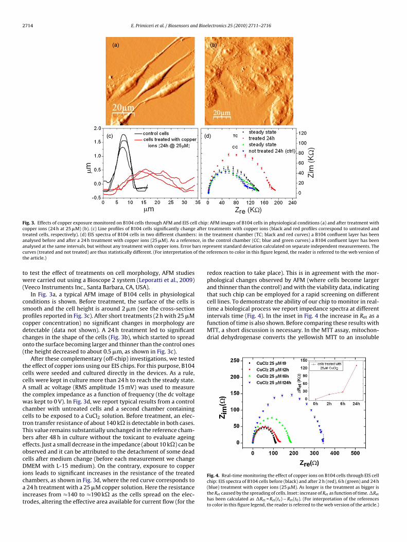
Fig. 3. Effects of copper exposure monitored on B104 cells through AFM and EIS cell chip: AFM images of B104 cells in physiological conditions (a) and after treatment withcopper ions (24 h at 25 �M) (b). (c) Line profiles of B104 cells significantly change after treatments with copper ions (black and red profiles correspond to untreated andtreated cells, respectively). (d) EIS spectra of B104 cells in two different chambers: in the treatment chamber (TC; black and red curves) a B104 confluent layer has beena ce, ina ars repc f the rt
tw(
cspcdco(
tccAtwcctTbeocDicait
intervals time (Fig. 4). In the inset in Fig. 4 the increase in Ret as afunction of time is also shown. Before comparing these results withMTT, a short discussion is necessary. In the MTT assay, mitochon-drial dehydrogenase converts the yellowish MTT to an insoluble
nalysed before and after a 24 h treatment with copper ions (25 �M). As a referennalysed at the same intervals, but without any treatment with copper ions. Error burves (treated and not treated) are thus statistically different. (For interpretation ohe article.)
o test the effect of treatments on cell morphology, AFM studiesere carried out using a Bioscope 2 system (Leporatti et al., 2009)
Veeco Instruments Inc., Santa Barbara, CA, USA).In Fig. 3a, a typical AFM image of B104 cells in physiological
onditions is shown. Before treatment, the surface of the cells ismooth and the cell height is around 2 �m (see the cross-sectionrofiles reported in Fig. 3c). After short treatments (2 h with 25 �Mopper concentration) no significant changes in morphology areetectable (data not shown). A 24 h treatment led to significanthanges in the shape of the cells (Fig. 3b), which started to spreadnto the surface becoming larger and thinner than the control onesthe height decreased to about 0.5 �m, as shown in Fig. 3c).
After these complementary (off-chip) investigations, we testedhe effect of copper ions using our EIS chips. For this purpose, B104ells were seeded and cultured directly in the devices. As a rule,ells were kept in culture more than 24 h to reach the steady state.small ac voltage (RMS amplitude 15 mV) was used to measure
he complex impedance as a function of frequency (the dc voltageas kept to 0 V). In Fig. 3d, we report typical results from a control
hamber with untreated cells and a second chamber containingells to be exposed to a CuCl2 solution. Before treatment, an elec-ron transfer resistance of about 140 k� is detectable in both cases.his value remains substantially unchanged in the reference cham-ers after 48 h in culture without the toxicant to evaluate ageingffects. Just a small decrease in the impedance (about 10 k�) can bebserved and it can be attributed to the detachment of some deadells after medium change (before each measurement we changeMEM with L-15 medium). On the contrary, exposure to copper
ons leads to significant increases in the resistance of the treatedhambers, as shown in Fig. 3d, where the red curve corresponds to24 h treatment with a 25 �M copper solution. Here the resistance
ncreases from ≈140 to ≈190 k� as the cells spread on the elec-rodes, altering the effective area available for current flow (for the
the control chamber (CC; blue and green curves) a B104 confluent layer has beenresent standard deviation calculated on separate independent measurements. Theeferences to color in this figure legend, the reader is referred to the web version of
redox reaction to take place). This is in agreement with the mor-phological changes observed by AFM (where cells become largerand thinner than the control) and with the viability data, indicatingthat such chip can be employed for a rapid screening on differentcell lines. To demonstrate the ability of our chip to monitor in real-time a biological process we report impedance spectra at different
Fig. 4. Real-time monitoring the effect of copper ions on B104 cells through EIS cellchip: EIS spectra of B104 cells before (black) and after 2 h (red), 6 h (green) and 24 h(blue) treatment with copper ions (25 �M). As longer is the treatment as bigger isthe Ret caused by the spreading of cells. Inset: increase of Ret as function of time. �Ret
has been calculated as �Ret = Ret(tx) − Ret(t0). (For interpretation of the referencesto color in this figure legend, the reader is referred to the web version of the article.)

E. Primiceri et al. / Biosensors and Bioe
Fig. 5. Prion protein expression: Western blot analysis in B104 and HeLa cell lines.Panel A: whole lysates (30 �g protein) were separated by SDS-PAGE and probed withmPn
pabcwncs
ggPtPctm(m
larger than the control one because of a slight spreading of the cells
Fit(saa
onoclonal anti-prion protein antibody. Blot is representative of six experiments.anel B: prion protein band intensities were quantified by ImageJ software andormalized to �-actin levels (**p < 0.01).
urple compound. Only living cells with active mitochondria areble to perform this conversion so the MTT assay measures cell via-ility based only on mitochondrial activity. For this reason in thease of MTT only metabolic and energetic alterations are probed,hile through our chip we monitor changes in cytoskeleton orga-ization (as actin fibers formation) that reflect stress and sufferingonditions, which continue to increase also after the MTT signaltabilizes.
To further test our biochips and as a term of comparison toain further insight, we performed the AFM and on-chip investi-ations on HeLa cell line, showing a different expression level ofrpC with respect to B104 cells. This has been demonstrated byhe immunoblotting analysis shown in Fig. 5: the expression ofrpC is 2.6 ± 0.4-fold higher in B104 than in HeLa cells. This findingould at least partly account for the more pronounced sensitivity
o copper exhibited by B104 cells. In fact, a role for prion protein inediating copper uptake has been demonstrated in this cell modelUrso et al., 2009). It follows that copper ions enter in the B104 cells
ore easily than in the HeLa cell model and, when exceeding the
ig. 6. Monitoring the effect of copper ions on HeLa cells: AFM images (40 �m × 40 �m) oons for 2–24 h (b–d). After 2 h exposure (b) copper causes a small spreading of cells, wreatment (24 h, c) most of the cells are detached from the surface or are detaching as thblack) and after 2 h (red) and 24 h (green) treatment with copper ions (500 �M) (each pohort treatment caused a slight spreading of cells, while the longer one induced a detachsignificant decrease of the electron transfer resistance. (For interpretation of the refere
rticle.)
lectronics 25 (2010) 2711–2716 2715
cytosolic buffer capacity, they give rise to more consistent oxida-tive phenomena with consequent cell damage. To further supportthe involvement of prion protein in cell copper transport, PrPC-deficient cells have been shown to exhibit a reduced copper content(Brown et al., 1997). In addition, altered brain copper levels havebeen found in Prp−/− and PrPC-overexpressing mice (Brown, 2003).
In Fig. 2d we can observe that B104 loss of cell viability after24 h treatment with 25 �M copper is higher with respect to HeLacells. In response to higher copper concentrations (except for theextreme values), the decrease in B104 cell viability is less accentu-ated than observed for HeLa cells. In the light of the high level ofPrPC expression by B104 cells, this could be explained by hypoth-esizing that copper toxicity can be partly counterbalanced by thePrPC-dependent activation of anti-oxidant mechanisms (Milhavetet al., 2000; Rachidi et al., 2003). Accordingly, the impairment ofbrain anti-oxidant enzyme activities (i.e. Cu/Zn Superoxide dismu-tase) has been detected in PrPC-deficient cells (Brown et al., 1997)and mice (Klamt et al., 2001; Dal Pizzol et al., 2000; Pereira et al.,2001).
AFM investigations were carried out on HeLa cultures treatedwith 500 �M CuCl2 for 2–24 h (Fig. 6). In physiological conditions,the surface of the cells is quite smooth and their height is around2 �m (Fig. 6a). After 2 h treatment with 500 �M copper ions, cellsappear to spread onto the surface becoming thinner than the con-trol ones (about 1 �m), as observed also for B104 cell line (Fig. 6b).However, longer treatments lead to cell detachment (as suggestedby the round shape some of them assumes, see Fig. 6d). After 24 hthe cell morphology is the same as after 2 h copper exposure. Inaddition, most of cells are detached from the surface (see opticalimages in the insets of Fig. 6e acquired before and after treatment).For this reason we decided to show the results at this concentra-tion to show the ability of our device to detect both morphologicalchanges and detachment phenomena.
So, the same cytotoxic effects were also monitored using thebiochips. Specifically we measured impedance spectra after 2 and24 h treatments. The measured electron transfer resistance in thecase of a 2 h treatment (red curve in Fig. 6e) was found to be slightly
on the electrodes (see the corresponding AFM image in Fig. 6b).Then, after 24 h treatment, the induced cell detachment resulted ina significant decrease of the impedance (green curve) since a largearea of the electrodes became free and available for current flow.
f HeLa cells in physiological conditions (a) and after treatment with 500 �M copperhich become slightly thinner than the control ones (about 1 �m). After a longer
eir round shape suggests (d, 100 �m× 100 �m). (e) EIS spectra of HeLa cells beforeint and the error bars have been calculated on three different measurements). Thement from the surface (as shown in the optical images in the insets) resulting in
nces to color in this figure legend, the reader is referred to the web version of the

2 d Bioe
4
tohcstbtfcpebbicbiphs
A
a
R
A
716 E. Primiceri et al. / Biosensors an
. Conclusion
In conclusion in this work, we report the successful applica-ion of EIS-based devices to monitor the toxicity of copper ionsn cultured cells. Impedance measurements on cellular systemsave been shown to be effective tools for monitoring in real-timeellular behavior and performing cytotoxicity assays, due to thetrong correlation between EIS spectra, MTT and AFM data. Allhe data obtained through these techniques contribute to gain aetter comprehension of the toxic effect of copper salt. Moreover,he comparison between EIS and the other techniques prove theeasibility of on-chip assays which provide additional informationompared to traditional biochemical tests because they allow torobe morphological and structural alterations that correspond toarly events in toxicological process. As a consequence, beyond toecome fundamental tools for drug discovery, on-chip assays cane expected to have applications in numerous studies taking also
nto account that by means of the same cell chips, adherent cellsan be monitored during cultivation and dynamic processes cane non-invasively studied. In particular thanks to multiplexing and
ntegration in more complex fluidic networks, these sensors can bearticularly useful in the field of drug discovery in order to test withigh-throughput a large library of drugs or to test the effect of theame drug on different cell lines.
cknowledgments
This work was supported by Italian Institute of Technology (IIT)nd by MIUR FIRB Project No. BNE03FMCJ 003.
eferences
rndt, S., Seebach, J., Psathaki, K., Galla, H.J., Wegener, J., 2004. Biosens. Bioelectron.19, 583–594.
lectronics 25 (2010) 2711–2716
Brown, D.R., Qin, K., Herms, J.W., Madlung, A., Manson, J., Strome, R., Fraser, P.E.,1997. Nature 390, 684–687.
Brown, D.R., 2003. J. Neurochem. 87, 377–385.Ceriotti, L., Ponti, J., Colpo, P., Sabbioni, E., Rossi, F., 2007a. Biosens. Bioelectron. 22,
3057–3063.Ceriotti, L., Ponti, J., Broggi, F., Kob, A., Drechsler, S., Thedinga, E., Colpo, P., Sabbioni,
E., Ehret, R., Rossi, F., 2007b. Sens. Actuators B: Chem. 123, 769–778.Cheng, X., Liu, Y.S., Irimia, D., Demirci, U., Yang, L.J., Zamir, L., Rodriguez, W.R., Toner,
M., Bashir, R., 2007. Lab Chip 7, 746–755.Dal Pizzol, F., Klamt, F., Vianna, M., Schöder, N., Quevedo, J., Moreira, J.C., Walz, R.,
2000. Neurosci. Lett. 291, 179–182.Ehret, R., Baumann, W., Brischwein, M., Schwinde, A., Stegbauer, K., Wolf, B., 1997.
Biosens. Bioelectron. 12, 29–41.Giaever, I., Keese, C.R., 1991. Proc. Natl. Acad. Sci. U.S.A. 88, 7896–7900.Giaever, I., Keese, C.R., 1993. Nature 366, 591–592.Hultberg, B., Andersson, A., Isaksson, A., 1995. Biochim. Biophys. Acta: Mol. Cell Res.
1269, 6–12.Inestrosa, N.C., Cerpa, W., Varela-Nallar, L., 2005. IUBMB Life 57, 645–650.Katz, E., Willner, I., 2003. Electroanalysis 15, 913–947.Keese, C.R., Giaever, I., 1994. IEEE Eng. Med. Biol. Mag. 13, 402–408.Klamt, F., Dal Pizzol, F., Conte da Frota, M.L., Walz, R., Andrades, M.E., Da Silva, E.G.,
2001. Free Radic. Biol. Med. 30, 1137–1144.Leporatti, S., Vergara, D., Zacheo, A., Vergaro, V., Maruccio, G., Cingolani, R., Rinaldi,
R., 2009. Nanotechnology 20, 055103.Milhavet, O., McMahon, H.E.M., Rachidi, W., Nishida, N., Katamine, S., Mange, A.,
Arlotto, M., Casanova, D., Riondel, J., Favier, A., 2000. Proc. Natl. Acad. Sci. U.S.A.97, 13937–13942.
Minseok, S.K., Wonhye, L., Yu Chang, K., Je-Kyun, P., 2008. Biotechnol. Bioeng. 101,1005–1013.
Monnet, C., Marthiens, V., Enslen, H., Frobert, Y., Sobel, A., Mege, R.M., 2003. Eur. J.Neurosci. 18, 542–548.
Pereira, G.S., Walz, R., Bonan, C.D., Battastini, A.M.O., Izquierdo, I., Martins, V.R.,Brentani, R.R., Sarkis, J.J.F., 2001. Neurosci. Lett. 301, 72–74.
Rachidi, W., Vilette, D., Guiraud, P., Arlotto, M., Riondel, J., Laude, H., Lehmann, S.,Favier, A., 2003. J. Biol. Chem. 278, 9064–9072.
Smith, C., 2007. Nature 446, 219–224.Urso, E., Rizzello, A., Acierno, R., Lionetto, M.G., Salvato, B., Storelli, C., Maffia, M.,
2009. J. Membr. Biol., 10.1007/s00232-009-9219-8.
Valko, M., Morris, H., Cronin, M.T.D., 2005. Curr. Med. Chem. 12, 1161–1208.Waggoner, D.J., Bartnikas, T.B., Gitlin, J.D., 1999. Neurobiol. Dis. 6, 221–230.Xiao, C.D., Lachance, B., Sunahara, G., Luong, J.H.T., 2002. Anal. Chem. 74,5748–5753.Xiao, C., Luong, J.H.T., 2003. Biotechnol. Prog. 19, 1000–1005.Yeon, J.H., Park, J.K., 2005. Anal. Biochem. 341, 308–315.
Copyright © 2022 FDOKUMEN