Development of adherence metrics for caloric restriction interventions
Aging Cell
(2006)
5
, pp247–257 Doi: 10.1111/j.1474-9726.2006.00214.x
© 2006 The Authors
247
Journal compilation © Blackwell Publishing Ltd/Anatomical Society of Great Britain and Ireland 2006
Blackwell Publishing Ltd
Reactive oxygen species, dietary restriction and neurotrophic factors in age-related loss of myenteric neurons
C. Thrasivoulou,
1
V. Soubeyre,
1
* H. Ridha,
1
D. Giuliani,
2
C. Giaroni,
2
G. J. Michael,
3
M. J. Saffrey
4
and T. Cowen
1
1
Department of Anatomy and Developmental Biology, University College London, Royal Free Campus, London NW3 2PF, UK
2
Clinical and Applied Pharmacology Centre, University of Insubria and University of Pavia, Italy
3
Neuroscience Centre, Queen Mary, University of London, London E1 2AT, UK
4
Department of Biological Sciences, Open University, Milton Keynes MK7 6AA, UK
Summary
We have studied the mechanisms underlying nonpatho-logical age-related neuronal cell death. Fifty per cent ofneurons in the rat enteric nervous system are lostbetween 12 and 18 months of age in
ad libitum
(AL) fed rats.Caloric restriction (CR) protects almost entirely againstthis neuron loss. Using the ROS-sensitive dyes, dihy-drorhodamine (DHR) and 2-[6-(4′′′′
-hydroxy)phenoxy-3H-xanthen-3-on-9-yl]benzoic acid (HPF)
in vitro
, weshow that the onset of cell death is linked with elevatedintraneuronal levels of reactive oxygen species (ROS).Treatment with the neurotrophic factors NT3 and GDNFenhances neuronal antioxidant defence in CR rats at 12–15 months and 24 months but not in adult or aged AL-fedanimals. To examine the link between elevated ROS andneuronal cell death, we assessed apoptotic cell deathfollowing
in vitro
treatment with the redox-cycling drug,menadione. Menadione fails to increase apoptosis in6-month neurons. However, in 12–15mAL fed rats, whenage-related cell death begins, menadione induces a 7- to15-fold increase in the proportion of apoptotic neurons. CRprotects age-matched neurons against ROS-induced apop-tosis. Treatment with neurotrophic factors, in particularGDNF, rescues neurons from menadione-induced cell death,
but only in 12–15mCR animals. We hypothesize that CRenhances antioxidant defence through neurotrophicfactor signalling, thereby reducing age-related increasesin neuronal ROS levels and in ROS-induced cell death.Key words: aging; antioxidant defence; apoptosis; celldeath; dietary restriction; myenteric neurons; neurotrophicfactors; reactive oxygen species.
Introduction
Relatively little is known about the mechanisms which underlie
nonpathological age-related neurodegeneration. Neither the
nature nor extent of this phenomenon is well established.
Studies of mammalian central (Morrison & Hof, 1997; Turlejski
& Djavadian, 2002; Baquet
et al
., 2004) and peripheral (Cowen
& Gavazzi, 1998; Cowen
et al
., 2003) neurons demonstrate
that age-associated degenerative changes only occur in select
groups of neurons such as the cholinergic neurons of the basal
forebrain and the sympathetic neurons projecting to cerebral
blood vessels and pineal gland. Previous studies have shown
that around 50% of the neurons of the myenteric plexus in the
gut of rodents (Johnson
et al
., 1998; Cowen
et al
., 2000),
guinea pigs (Gabella, 1989) and humans (Porter
et al
., 1996)
are lost during aging. While in rats neurons expressing choline
acetyl transferase are vulnerable while those expressing nitric
oxide synthase are protected (Cowen
et al
., 2000), the reasons
for this selective vulnerability in different regions of the nervous
system are poorly understood.
Age is the biggest risk factor for several of the more common
neurodegenerative diseases, including Alzheimer, Parkinson
and motor neuron diseases. Damage by reactive oxygen species
(ROS) is implicated in nonpathological brain aging processes
(Poon
et al
., 2004; Lau
et al
., 2005) as well as in all of these
chronic disorders (Schapira, 1998; Jung
et al
., 2001; Nunomura
et al
., 2001; Huang
et al
., 2004). Although there is little concrete
evidence, the implication of these observations is that common
mechanisms involving ROS may underpin pathological as well
as nonpathological age-related neurodegeneration.
Neurons are considered particularly vulnerable to free radical
damage because of their large size, high level of metabolic activity
and relatively poor antioxidant defence (Sohal & Weindruch,
1996). Survival of adult and aged neurons appears to be in some
way related to the capacity to resist ROS-induced damage
(Finkel & Holbrook, 2000). Previous evidence of age-related
vulnerability of enteric neurons (Phillips
et al
., 2003) does not
provide a link with ROS sensitivity. Calcium dyshomeostasis is
a key event in neuronal aging (Bu
et al
., 2003) and in Alzheimer
Correspondence
T. Cowen, PhD, Department of Anatomy and Developmental Biology,
University College London, Royal Free Campus, Rowland Hill St., London NW3
2PF, UK. Tel.: (0)207 830 2181; fax: (0)207 472 6763;
e-mail: [email protected]
*Present address: Neurobiologie des Processus Adaptatifs (UMR 7102),
Université Pierre et Marie Curie (Paris VI), Bât B, 6e étage, Case 12, 9, quai
St Bernard, 75005 Paris, France
Accepted for publication 13 March 2006
Free radicals and diet in neuronal aging, C. Thrasivoulou
et al.
© 2006 The AuthorsJournal compilation © Blackwell Publishing Ltd/Anatomical Society of Great Britain and Ireland 2006
248
disease (LaFerla, 2002) which have been linked to altered
mitochondrial ROS generation in old age (Mattson & Liu, 2002).
The calcium-binding proteins, calretinin and calbindin, present
in many central and peripheral neurons including neurons of
the myenteric plexus (Abalo
et al
., 2005), are thought to protect
neurons against calcium dyshomeostasis and may therefore
allow the identification of neurons that are selectively protected
against ROS-induced damage.
Caloric restriction (CR) extends lifespan in organisms as
diverse as yeasts, worms, flies and mammals by enhancing the
capacity for stress resistance (Sohal & Weindruch, 1996).
Furthermore, CR enhances plasticity in the aging brain (Mattson
et al
., 2002), increases expression of an anti-apoptotic repressor
in the cerebral cortex of aging rats (Shelke & Leeuwenburgh,
2003), and rescues neurons of the rat enteric nervous system
(ENS) from age-related cell death (Cowen
et al
., 2000). We
hypothesize that the effects of diet on neuronal survival are
mediated by altered capacity to regulate ROS.
Neurotrophic factor signalling is crucial to neuronal survival
and differentiation during development (Miller & Kaplan, 2001).
A novel role for neurotrophins in regulating ROS has recently
been demonstrated in developing sympathetic neurons (Dugan
et al
., 1999), in PC12 cells (Mills
et al
., 1998) and in an animal
model of Parkinson’s disease (Salinas
et al
., 2003). Because CR
has been shown to influence neurotrophin-mediated plasticity
in the CNS (Prolla & Mattson, 2001; Nicoletti
et al
., 2005), we
hypothesize that CR might enable neurons to resist age-related
cell death by enhancing neurotrophic factor-mediated antioxidant
defence.
Our aims were therefore (i) to confirm protection by dietary
restriction against age-related loss of myenteric neurons and to
determine whether neurons expressing calcium-binding proteins
were selectively protected against age-related cell death; (ii) to
find out if age- and diet-related alterations in intracellular ROS
levels are associated with neuronal cell death in the ENS; (iii) to
discover whether elevated ROS levels might induce apoptotic
cell death in enteric neurons; and (iv) to demonstrate a possible
role for neurotrophic factors in regulating antioxidant defence
and resistance to ROS-induced neuronal cell death.
Results
Effects of dietary restriction on age-related loss of myenteric neurons
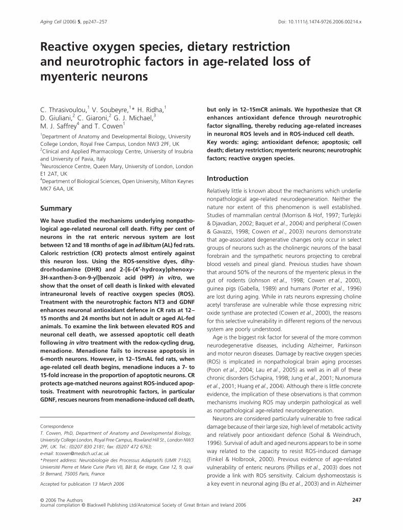
Counts of PGP 9.5-stained neurons confirm previous data
(Cowen
et al
., 2000) by showing an age-related loss of 51%
of neurons (
P <
0.001) in 24mAL animals (Fig. 1A). CR animals
show a much smaller loss of neurons over 24 m (22%;
P
< 0.05)
indicating, like our previous data, substantial protection by
dietary restriction. Examination of the timescale of neuron loss
reveals that at 12 months, numbers of neurons are not signif-
icantly different from those in 6-month rats and there are no
differences between AL and CR groups (Fig. 1A). However, by
13 months there are 24% (
P <
0.05) fewer neurons in the AL
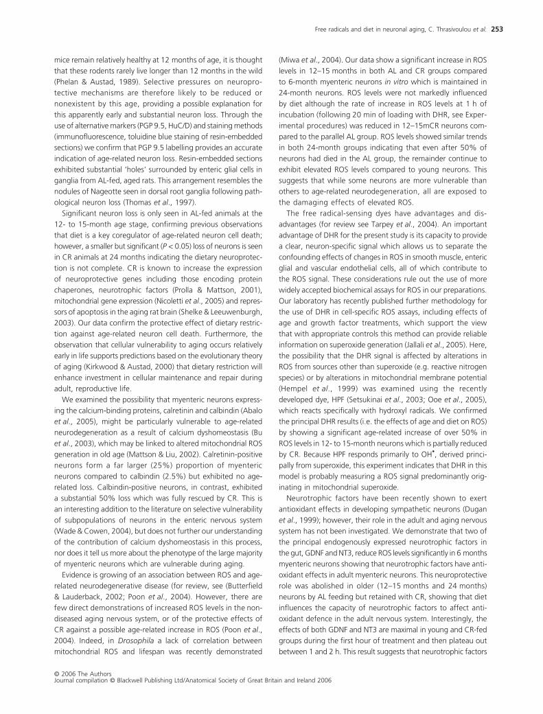
Fig. 1 Total numbers of myenteric neurons. (A) Counts of PGP-immunoreactive neurons. The histogram shows significant losses of neurons, commencing at 12–13 months of age in AL-fed animals and reaching a peak at 24 months (AL; 51%). There is a much smaller but statistically significant (P < 0.05) reduction seen in 24mCR samples, demonstrating substantial protection by CR against age-related cell death in myenteric neurons. (B) Counts of HuCD-immunoreactive neurons. The histogram shows loss of neurons in 17-month AL-fed, confirming that PGP 9.5 labelling gives a reliable estimate of age- and diet-related cell loss. However, total neuron numbers were significantly (P < 0.01) higher in the HuC/D-stained samples.(C–F) Photomicrographs of 6-month (C,E) and 24-month AL (D,F) PGP-immunostained whole-mounts (C,D) and toluidine blue-stained 1-µm resin sections (E,F). Neurons in myenteric ganglia (dotted lines) are indicated by large arrows. Note the large spaces in the ganglia from 24-month AL rats (small arrows) visible in D and F but absent in the 6-month samples (C,E), indicating where neuron cell loss has occurred. n numbers: A: 6 months (n = 15); 12mCR (n = 4); 12mAL (n = 4); 13mCR (n = 8); 13mAL (n = 12); 24mCR (n = 15); 24mAL (n = 14); B: 6 months (n = 6); 17 months (n = 3). Scale bar = 50 µm.
Free radicals and diet in neuronal aging, C. Thrasivoulou
et al.
© 2006 The AuthorsJournal compilation © Blackwell Publishing Ltd/Anatomical Society of Great Britain and Ireland 2006
249
compared to CR groups and this number is significantly lower
than either of the 12 months groups (13mAL vs. 12mCR,
P
< 0.05;
13mAL vs. 12mAL,
P
< 0.01). As reported previously (Cowen
et al
.,
2000), neuron loss is complete by 16–18 months (when cor-
rected for gut size) in AL-fed rats. Whilst these data show sim-
ilar levels of loss of neurons with HuC/D at 17mAL (
∼
50%) with
those shown in Cowen
et al
. (2000), the PGP 9.5 data only show
a partial, but significant loss, at the 13mAL age group studied.
Counts are corrected for changes in gut size to ensure that alter-
ations are due to cell loss rather than redistribution of neurons.
However, our measurements reveal no significant effect of diet
or age on the overall dimensions of the small intestine (Table 1).
As a result of a recent study evaluating pan-neuronal markers
in the rat myenteric plexus (Phillips
et al
., 2004), tissues were
taken from small groups of 3-month and 17mAL animals and
immunolabelled for HuC/D, which was shown to be one of the
most reliable pan-neuronal markers for rodent myenteric neu-
rons. Neuron counts made on HuC/D-labelled preparations
demonstrated significantly (
P <
0.01) higher neuron numbers
compared to PGP 9.5, confirming previous reports that PGP 9.5
does not stain all neurons in the gut (Young
et al
., 2003). HuC/D,
however, demonstrated a comparable loss of neurons (50%;
P
< 0.01; Fig. 1B) in 17mAL fed animals to that shown previously
using PGP 9.5 at 16 and 24 months in AL-fed rats (Cowen
et al
.,
2000). The 50% loss of neurons seen with HuC/D at 17 months
is similar to the extent of loss of PGP 9.5-positive neurons seen
at 24 months (Fig. 1A,B), supporting the view that neuron loss
in aging AL-fed rats is largely complete by 17–18 months.
Loss of immunolabelling may indicate reduced binding of the
antibodies rather than neuron loss. To examine this possibility,
we studied parallel samples from 24mAL and 6-month rats
following PGP staining of whole mounts and toluidine blue
staining of 1
µ
m resin sections (Fig. 1C–F). Both methods reveal
clearly defined spaces surrounded by enteric glia in the ganglia
of 24mAL samples which are not present in 6-month samples.
Overall, these results confirm that age-related neuron loss has
indeed taken place and that dietary restriction protects mye-
nteric neurons against age-related loss.
Selective vulnerability in subpopulations of myenteric neurons
Calcium dyshomeostasis is a key event in neuronal aging
(Bu
et al
., 2003). We therefore investigated whether neurons
containing the calcium-binding proteins, calretinin and calbindin,
were particularly protected compared to the general neuron
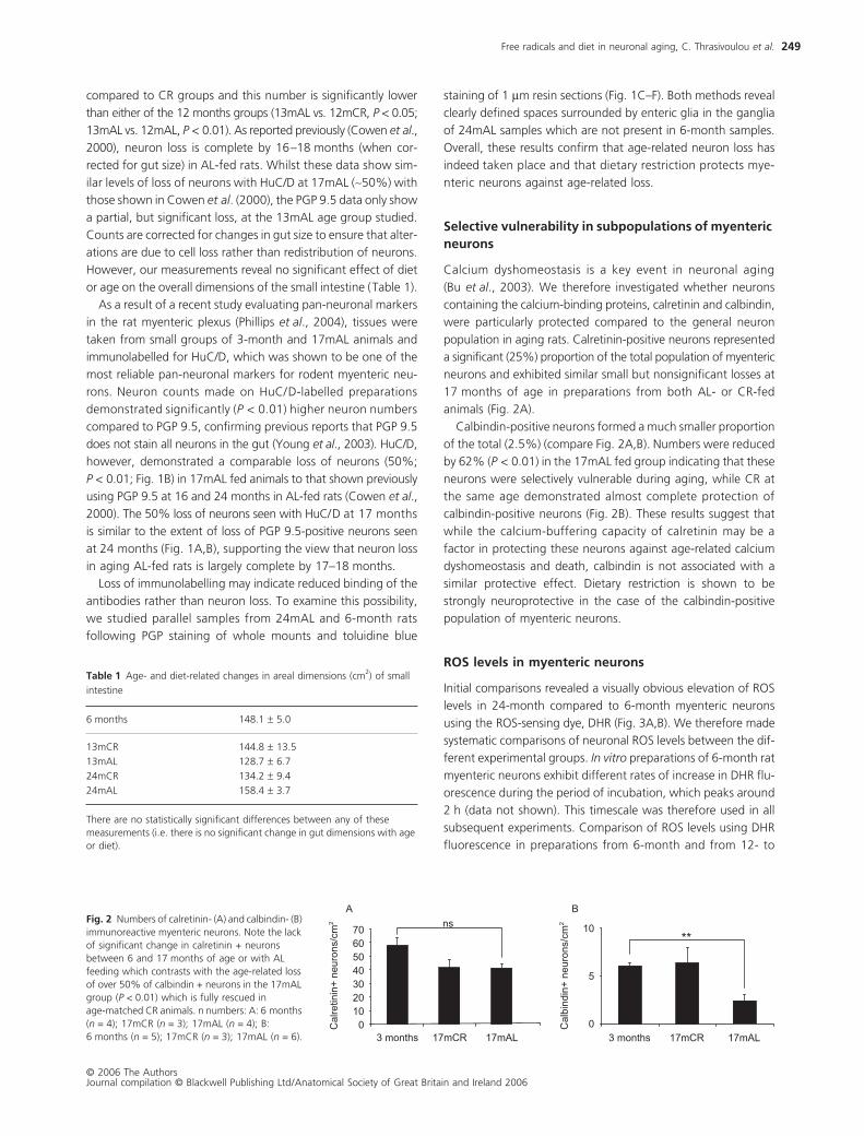
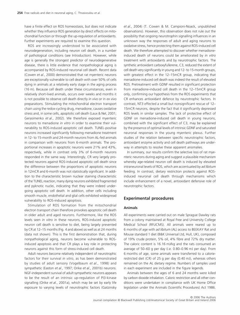
population in aging rats. Calretinin-positive neurons represented
a significant (25%) proportion of the total population of myenteric
neurons and exhibited similar small but nonsignificant losses at
17 months of age in preparations from both AL- or CR-fed
animals (Fig. 2A).
Calbindin-positive neurons formed a much smaller proportion
of the total (2.5%) (compare Fig. 2A,B). Numbers were reduced
by 62% (
P <
0.01) in the 17mAL fed group indicating that these
neurons were selectively vulnerable during aging, while CR at
the same age demonstrated almost complete protection of
calbindin-positive neurons (Fig. 2B). These results suggest that
while the calcium-buffering capacity of calretinin may be a
factor in protecting these neurons against age-related calcium
dyshomeostasis and death, calbindin is not associated with a
similar protective effect. Dietary restriction is shown to be
strongly neuroprotective in the case of the calbindin-positive
population of myenteric neurons.
ROS levels in myenteric neurons
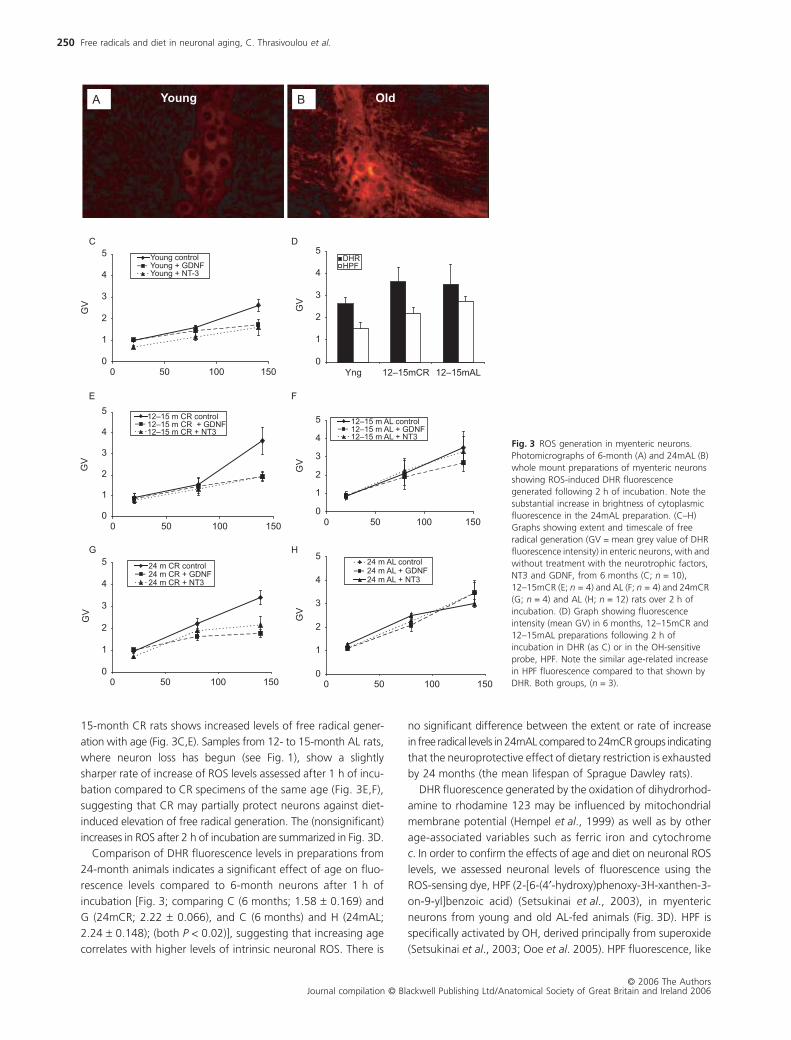
Initial comparisons revealed a visually obvious elevation of ROS
levels in 24-month compared to 6-month myenteric neurons
using the ROS-sensing dye, DHR (Fig. 3A,B). We therefore made
systematic comparisons of neuronal ROS levels between the dif-
ferent experimental groups.
In vitro
preparations of 6-month rat
myenteric neurons exhibit different rates of increase in DHR flu-
orescence during the period of incubation, which peaks around
2 h (data not shown). This timescale was therefore used in all
subsequent experiments. Comparison of ROS levels using DHR
fluorescence in preparations from 6-month and from 12- to
Table 1 Age- and diet-related changes in areal dimensions (cm2) of small
intestine
6 months 148.1 ± 5.0
13mCR 144.8 ± 13.5
13mAL 128.7 ± 6.7
24mCR 134.2 ± 9.4
24mAL 158.4 ± 3.7
There are no statistically significant differences between any of these measurements (i.e. there is no significant change in gut dimensions with age or diet).
Fig. 2 Numbers of calretinin- (A) and calbindin- (B) immunoreactive myenteric neurons. Note the lack of significant change in calretinin + neurons between 6 and 17 months of age or with AL feeding which contrasts with the age-related loss of over 50% of calbindin + neurons in the 17mAL group (P < 0.01) which is fully rescued in age-matched CR animals. n numbers: A: 6 months (n = 4); 17mCR (n = 3); 17mAL (n = 4); B: 6 months (n = 5); 17mCR (n = 3); 17mAL (n = 6).
Free radicals and diet in neuronal aging, C. Thrasivoulou
et al.
© 2006 The AuthorsJournal compilation © Blackwell Publishing Ltd/Anatomical Society of Great Britain and Ireland 2006
250
15-month CR rats shows increased levels of free radical gener-
ation with age (Fig. 3C,E). Samples from 12- to 15-month AL rats,
where neuron loss has begun (see Fig. 1), show a slightly
sharper rate of increase of ROS levels assessed after 1 h of incu-
bation compared to CR specimens of the same age (Fig. 3E,F),
suggesting that CR may partially protect neurons against diet-
induced elevation of free radical generation. The (nonsignificant)
increases in ROS after 2 h of incubation are summarized in Fig. 3D.
Comparison of DHR fluorescence levels in preparations from
24-month animals indicates a significant effect of age on fluo-
rescence levels compared to 6-month neurons after 1 h of
incubation [Fig. 3; comparing C (6 months; 1.58
±
0.169) and
G (24mCR; 2.22
±
0.066), and C (6 months) and H (24mAL;
2.24
±
0.148); (both
P
< 0.02)], suggesting that increasing age
correlates with higher levels of intrinsic neuronal ROS. There is
no significant difference between the extent or rate of increase
in free radical levels in 24mAL compared to 24mCR groups indicating
that the neuroprotective effect of dietary restriction is exhausted
by 24 months (the mean lifespan of Sprague Dawley rats).
DHR fluorescence generated by the oxidation of dihydrorhod-
amine to rhodamine 123 may be influenced by mitochondrial
membrane potential (Hempel
et al
., 1999) as well as by other
age-associated variables such as ferric iron and cytochrome
c
. In order to confirm the effects of age and diet on neuronal ROS
levels, we assessed neuronal levels of fluorescence using the
ROS-sensing dye, HPF (2-[6-(4
′
-hydroxy)phenoxy-3H-xanthen-3-
on-9-yl]benzoic acid) (Setsukinai
et al
., 2003), in myenteric
neurons from young and old AL-fed animals (Fig. 3D). HPF is
specifically activated by OH, derived principally from superoxide
(Setsukinai
et al
., 2003; Ooe
et al
. 2005). HPF fluorescence, like
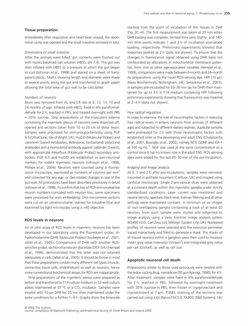
Fig. 3 ROS generation in myenteric neurons. Photomicrographs of 6-month (A) and 24mAL (B) whole mount preparations of myenteric neurons showing ROS-induced DHR fluorescence generated following 2 h of incubation. Note the substantial increase in brightness of cytoplasmic fluorescence in the 24mAL preparation. (C–H) Graphs showing extent and timescale of free radical generation (GV = mean grey value of DHR fluorescence intensity) in enteric neurons, with and without treatment with the neurotrophic factors, NT3 and GDNF, from 6 months (C; n = 10), 12–15mCR (E; n = 4) and AL (F; n = 4) and 24mCR (G; n = 4) and AL (H; n = 12) rats over 2 h of incubation. (D) Graph showing fluorescence intensity (mean GV) in 6 months, 12–15mCR and 12–15mAL preparations following 2 h of incubation in DHR (as C) or in the OH-sensitive probe, HPF. Note the similar age-related increase in HPF fluorescence compared to that shown by DHR. Both groups, (n = 3).
Free radicals and diet in neuronal aging, C. Thrasivoulou
et al.
© 2006 The AuthorsJournal compilation © Blackwell Publishing Ltd/Anatomical Society of Great Britain and Ireland 2006
251
DHR, increases with incubation time, peaking after 2–3 h (data
not shown). HPF demonstrates an age-related increase in
neuronal ROS in the 12- to 15-month samples which is slightly,
but not significantly, greater in the AL group. These differences
are similar to those seen with DHR, confirming that DHR flu-
orescence provides a reliable measure of ROS levels in myenteric
neurons. Because HPF fluorescence is specifically induced by
superoxide-derived OH
•
it is likely that mitochondrial ROS provide
the majority of the free radicals assessed in our experiments.
Neurotrophic factors in antioxidant defence
Because of evidence that neurotrophic factors contribute to
antioxidant defence in early postnatal neurons (Dugan
et al
.,
1999), we tested the capacity of GDNF and NT3, with known
survival-promoting roles in myenteric neurons (Chalazonitis
et al
., 2001; Busciglio
et al
., 2002), to reduce ROS levels in these
neurons from adult rats. Treatment of in vitro preparations of
enteric neurons from 6-month rats with 100 ng mL−1 of GDNF
or NT3 results in significantly lower levels of neuronal ROS
compared to untreated preparations after 2 h of incubation
(Fig. 3C; GDNF: P < 0.01; NT3: P < 0.05). In contrast, treatment
with NGF or IGF-1 [with known antioxidant effects in other
groups of neurons (Dugan et al., 1999) and cell types (Jallali
et al., 2005)], has no effect on ROS levels (data not shown).
Comparisons of the effects of neurotrophic factors on ROS
levels in 12- to 15-month animals (Fig. 3E,F) show that in CR
preparations, ROS are significantly reduced at the 2-h time point
after treatment with NT3 and GDNF (both, P < 0.05). In contrast,
neither factor has a significant effect on ROS levels in prepara-
tions from 12- to 15-month AL rats. Thus CR appears to enhance
neurotrophin-mediated antioxidant mechanisms around the
age at which cell loss occurs in vivo. In the 24mCR group, treatment
with neurotrophic factors suppresses ROS levels significantly
(Fig. 3G,H; GDNF: P < 0.001; NT3: P < 0.05) but not in the AL
preparations, resembling the effects seen at 12–15 months. It
is worth noting that these differential effects, like the onset of
neuron cell death, are already established at the relatively young
age of 12–15 months (i.e. well before the onset of senescence).
Our results therefore show that age enhances levels of ROS
generation, while age and diet, respectively, inhibit and enhance
neurotrophic factor-mediated antioxidant defence mechanisms,
in adult and aged neurons.
ROS-induced neuronal cell death
In order to discover whether ROS contribute to age- and diet-
induced neuronal cell death, we examined the effect of treat-
ment with menadione (a drug which accelerates mitochondrial
ROS generation by redox cycling) on the incidence of apoptotic
cell death in myenteric neurons. We chose to focus on the
period up to and including the early stages of age-related
neuron loss in the myenteric plexus (i.e. 12–15 months of age).
The increase in the TUNEL response to menadione was time-
dependent, reaching a maximum after 4 h of incubation (data
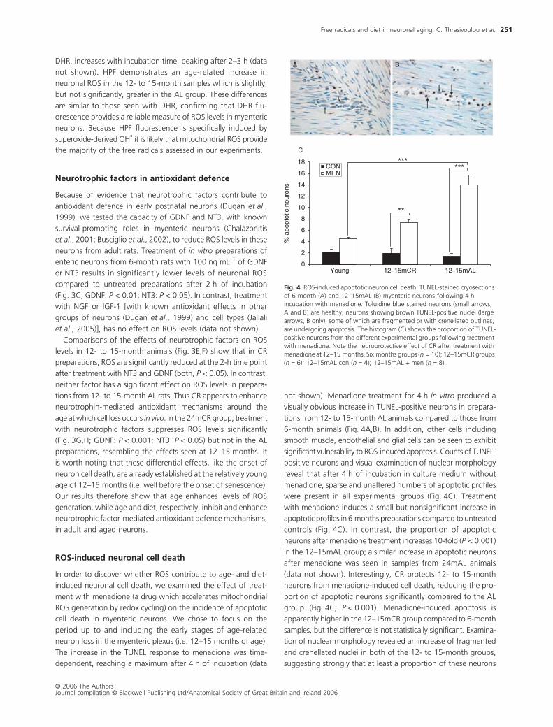
not shown). Menadione treatment for 4 h in vitro produced a
visually obvious increase in TUNEL-positive neurons in prepara-
tions from 12- to 15-month AL animals compared to those from
6-month animals (Fig. 4A,B). In addition, other cells including
smooth muscle, endothelial and glial cells can be seen to exhibit
significant vulnerability to ROS-induced apoptosis. Counts of TUNEL-
positive neurons and visual examination of nuclear morphology
reveal that after 4 h of incubation in culture medium without
menadione, sparse and unaltered numbers of apoptotic profiles
were present in all experimental groups (Fig. 4C). Treatment
with menadione induces a small but nonsignificant increase in
apoptotic profiles in 6 months preparations compared to untreated
controls (Fig. 4C). In contrast, the proportion of apoptotic
neurons after menadione treatment increases 10-fold (P < 0.001)
in the 12–15mAL group; a similar increase in apoptotic neurons
after menadione was seen in samples from 24mAL animals
(data not shown). Interestingly, CR protects 12- to 15-month
neurons from menadione-induced cell death, reducing the pro-
portion of apoptotic neurons significantly compared to the AL
group (Fig. 4C; P < 0.001). Menadione-induced apoptosis is
apparently higher in the 12–15mCR group compared to 6-month
samples, but the difference is not statistically significant. Examina-
tion of nuclear morphology revealed an increase of fragmented
and crenellated nuclei in both of the 12- to 15-month groups,
suggesting strongly that at least a proportion of these neurons
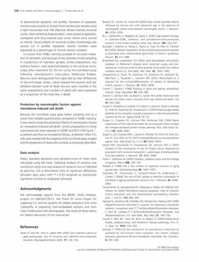
Fig. 4 ROS-induced apoptotic neuron cell death: TUNEL-stained cryosections of 6-month (A) and 12–15mAL (B) myenteric neurons following 4 h incubation with menadione. Toluidine blue stained neurons (small arrows, A and B) are healthy; neurons showing brown TUNEL-positive nuclei (large arrows, B only), some of which are fragmented or with crenellated outlines, are undergoing apoptosis. The histogram (C) shows the proportion of TUNEL-positive neurons from the different experimental groups following treatment with menadione. Note the neuroprotective effect of CR after treatment with menadione at 12–15 months. Six months groups (n = 10); 12–15mCR groups (n = 6); 12–15mAL con (n = 4); 12–15mAL + men (n = 8).
Free radicals and diet in neuronal aging, C. Thrasivoulou et al.
© 2006 The AuthorsJournal compilation © Blackwell Publishing Ltd/Anatomical Society of Great Britain and Ireland 2006
252
were dying apoptotically. Thus, age and ad libitum feeding
enhance vulnerability to ROS-induced death of myenteric neurons.
Next we wanted to find out whether menadione-induced
death of neurons could be ameliorated by in vitro treatment with
antioxidants and by neurotrophic factors. Preparations from
6-month and from 12- to 15-month AL and CR animals were
treated with menadione as before, following pretreatment
with the synthetic antioxidant carboxyfullerene also known as
C3 (Dugan et al., 1997) or with the neurotrophic factors GDNF
or NT3 (Fig. 5A–C). Fullerene compounds react avidly with free
radicals and are regarded as ‘radical sponges’. The trimalonic
acid derivative of fullerene, C3, is a synthetic water-soluble com-
pound that has been found to be an effective antioxidant both
in vivo and in vitro (Tsao et al., 2002). Preliminary experiments
using a ‘Live-Dead’ assay (Molecular Probes Inc., Eugene, OR,
USA) demonstrated improved imaging and sampling compared
to the TUNEL method and this was therefore used for these exper-
iments. The effects of menadione on cell death are qualitatively
similar using this assay compared with TUNEL staining in the
young and 12–15mAL groups. However, menadione induces a
greater extent of cell death as measured by this method com-
pared with TUNEL staining in the 12–15mCR group. We spec-
ulate that this is because the Live-Dead assay demonstrates
ethidium staining in compromised cells not yet committed to
apoptosis, i.e. still capable of being rescued (see below).
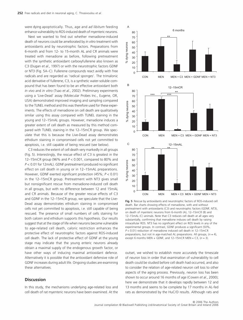
C3 reduces the extent of cell death very markedly in all groups
(Fig. 5). Interestingly, the rescue effect of C3 is greatest in the
12–15mCR group (96% and P < 0.001, compared to 80% and
P < 0.01 for 12mAL). GDNF pretreatment produced no significant
effect on cell death in young or in 12–15mAL preparations.
However, GDNF exerted significant protection (45%; P < 0.01)
in the 12–15mCR group. Pretreatment with NT3 gives small
but nonsignificant rescue from menadione-induced cell death
in all groups, but with no difference between 12 and 15mAL
and CR animals. Because of the greater rescue effects of C3
and GDNF in the 12–15mCR group, we speculate that the Live-
Dead assay demonstrates ethidium staining in compromised
cells not yet committed to apoptosis, i.e. still capable of being
rescued. The presence of small numbers of cells staining for
both calcein and ethidium supports this hypothesis. Our results
suggest that at the stage of life when neurons become vulnerable
to age-related cell death, caloric restriction enhances the
protective effect of neurotrophic factors against ROS-induced
cell death. The lack of protective effect of GDNF at the young
stage may indicate that the young enteric neurons already
obtain a maximal supply of the endogenous growth factor, or
have other ways of inducing maximal antioxidant defence.
Alternatively it is possible that the antioxidant defensive role of
GDNF increases during adult life. Ongoing studies are examining
these alternatives.
Discussion
In this study, the mechanisms underlying age-related loss and
cell death of rat myenteric neurons have been examined. At the
outset, we wished to establish more accurately the timescale
of neuron loss in order that examination of vulnerability to cell
death could be studied before cell death had occurred, and also
to consider the relation of age-related neuron cell loss to other
aspects of the aging process. Previously, neuron loss has been
shown to occur around 16 months of age (Cowen et al., 2000);
here we demonstrate that it develops rapidly between 12 and
13 months and seems to be complete by 17 months in AL-fed
rats as demonstrated by the HuC/D results. Although rats and
Fig. 5 Rescue by antioxidants and neurotrophic factors of ROS-induced cell death. Bar charts showing effects of menadione, with and without pretreatment with antioxidants (C3) and neurotrophic factors (GDNF, NT3), on death of myenteric neurons from 6-month (A), 12–15mCR (B) and 12–15mAL (C) animals. Note that C3 reduces cell death at all ages very substantially, confirming that menadione induces cell death by raising intracellular ROS. NT3 has no significant effect on ROS levels in any of the experimental groups. In contrast, GDNF produces a significant (50%, P < 0.01) reduction of menadione induced cell death in 12–15mCR preparations, but not in age-matched AL preparations. All groups, (n = 4), except 6 months MEN + GDNF, and 12–15mCR MEN + C3, (n = 3).
Free radicals and diet in neuronal aging, C. Thrasivoulou et al.
© 2006 The AuthorsJournal compilation © Blackwell Publishing Ltd/Anatomical Society of Great Britain and Ireland 2006
253
mice remain relatively healthy at 12 months of age, it is thought
that these rodents rarely live longer than 12 months in the wild
(Phelan & Austad, 1989). Selective pressures on neuropro-
tective mechanisms are therefore likely to be reduced or
nonexistent by this age, providing a possible explanation for
this apparently early and substantial neuron loss. Through the
use of alternative markers (PGP 9.5, HuC/D) and staining methods
(immunofluorescence, toluidine blue staining of resin-embedded
sections) we confirm that PGP 9.5 labelling provides an accurate
indication of age-related neuron loss. Resin-embedded sections
exhibited substantial ‘holes’ surrounded by enteric glial cells in
ganglia from AL-fed, aged rats. This arrangement resembles the
nodules of Nageotte seen in dorsal root ganglia following path-
ological neuron loss (Thomas et al., 1997).
Significant neuron loss is only seen in AL-fed animals at the
12- to 15-month age stage, confirming previous observations
that diet is a key coregulator of age-related neuron cell death;
however, a smaller but significant (P < 0.05) loss of neurons is seen
in CR animals at 24 months indicating the dietary neuroprotec-
tion is not complete. CR is known to increase the expression
of neuroprotective genes including those encoding protein
chaperones, neurotrophic factors (Prolla & Mattson, 2001),
mitochondrial gene expression (Nicoletti et al., 2005) and repres-
sors of apoptosis in the aging rat brain (Shelke & Leeuwenburgh,
2003). Our data confirm the protective effect of dietary restric-
tion against age-related neuron cell death. Furthermore, the
observation that cellular vulnerability to aging occurs relatively
early in life supports predictions based on the evolutionary theory
of aging (Kirkwood & Austad, 2000) that dietary restriction will
enhance investment in cellular maintenance and repair during
adult, reproductive life.
We examined the possibility that myenteric neurons express-
ing the calcium-binding proteins, calretinin and calbindin (Abalo
et al., 2005), might be particularly vulnerable to age-related
neurodegeneration as a result of calcium dyshomeostasis (Bu
et al., 2003), which may be linked to altered mitochondrial ROS
generation in old age (Mattson & Liu, 2002). Calretinin-positive
neurons form a far larger (25%) proportion of myenteric
neurons compared to calbindin (2.5%) but exhibited no age-
related loss. Calbindin-positive neurons, in contrast, exhibited
a substantial 50% loss which was fully rescued by CR. This is
an interesting addition to the literature on selective vulnerability
of subpopulations of neurons in the enteric nervous system
(Wade & Cowen, 2004), but does not further our understanding
of the contribution of calcium dyshomeostasis in this process,
nor does it tell us more about the phenotype of the large majority
of myenteric neurons which are vulnerable during aging.
Evidence is growing of an association between ROS and age-
related neurodegenerative disease (for review, see (Butterfield
& Lauderback, 2002; Poon et al., 2004). However, there are
few direct demonstrations of increased ROS levels in the non-
diseased aging nervous system, or of the protective effects of
CR against a possible age-related increase in ROS (Poon et al.,2004). Indeed, in Drosophila a lack of correlation between
mitochondrial ROS and lifespan was recently demonstrated
(Miwa et al., 2004). Our data show a significant increase in ROS
levels in 12–15 months in both AL and CR groups compared
to 6-month myenteric neurons in vitro which is maintained in
24-month neurons. ROS levels were not markedly influenced
by diet although the rate of increase in ROS levels at 1 h of
incubation (following 20 min of loading with DHR, see Exper-
imental procedures) was reduced in 12–15mCR neurons com-
pared to the parallel AL group. ROS levels showed similar trends
in both 24-month groups indicating that even after 50% of
neurons had died in the AL group, the remainder continue to
exhibit elevated ROS levels compared to young neurons. This
suggests that while some neurons are more vulnerable than
others to age-related neurodegeneration, all are exposed to
the damaging effects of elevated ROS.
The free radical-sensing dyes have advantages and dis-
advantages (for review see Tarpey et al., 2004). An important
advantage of DHR for the present study is its capacity to provide
a clear, neuron-specific signal which allows us to separate the
confounding effects of changes in ROS in smooth muscle, enteric
glial and vascular endothelial cells, all of which contribute to
the ROS signal. These considerations rule out the use of more
widely accepted biochemical assays for ROS in our preparations.
Our laboratory has recently published further methodology for
the use of DHR in cell-specific ROS assays, including effects of
age and growth factor treatments, which support the view
that with appropriate controls this method can provide reliable
information on superoxide generation (Jallali et al., 2005). Here,
the possibility that the DHR signal is affected by alterations in
ROS from sources other than superoxide (e.g. reactive nitrogen
species) or by alterations in mitochondrial membrane potential
(Hempel et al., 1999) was examined using the recently
developed dye, HPF (Setsukinai et al., 2003; Ooe et al., 2005),
which reacts specifically with hydroxyl radicals. We confirmed
the principal DHR results (i.e. the effects of age and diet on ROS)
by showing a significant age-related increase of over 50% in
ROS levels in 12- to 15-month neurons which is partially reduced
by CR. Because HPF responds primarily to OH•, derived princi-
pally from superoxide, this experiment indicates that DHR in this
model is probably measuring a ROS signal predominantly orig-
inating in mitochondrial superoxide.
Neurotrophic factors have been recently shown to exert
antioxidant effects in developing sympathetic neurons (Dugan
et al., 1999); however, their role in the adult and aging nervous
system has not been investigated. We demonstrate that two of
the principal endogenously expressed neurotrophic factors in
the gut, GDNF and NT3, reduce ROS levels significantly in 6 months
myenteric neurons showing that neurotrophic factors have anti-
oxidant effects in adult myenteric neurons. This neuroprotective
role was abolished in older (12–15 months and 24 months)
neurons by AL feeding but retained with CR, showing that diet
influences the capacity of neurotrophic factors to affect anti-
oxidant defence in the adult nervous system. Interestingly, the
effects of both GDNF and NT3 are maximal in young and CR-fed
groups during the first hour of treatment and then plateau out
between 1 and 2 h. This result suggests that neurotrophic factors
Free radicals and diet in neuronal aging, C. Thrasivoulou et al.
© 2006 The AuthorsJournal compilation © Blackwell Publishing Ltd/Anatomical Society of Great Britain and Ireland 2006
254
have a finite effect on ROS homeostasis, but does not indicate
whether they influence ROS generation by direct effects on mito-
chondrial function or through the up-regulation of antioxidants.
Further experiments are required to examine this question.
ROS are increasingly understood to be associated with
neurodegeneration, including neuron cell death, in a number
of pathological conditions (see Introduction). However, while
age is generally the strongest predictor of neurodegenerative
disease, there is little evidence that nonpathological aging is
accompanied by ROS-induced neuronal cell death. Recent data
(Cowen et al., 2000) demonstrated that rat myenteric neurons
are exceptionally vulnerable to cell death with over 50% of cells
dying in animals at a relatively early stage in the aging process
(16 m). Because cell death under these circumstances, even in
relatively short-lived animals, occurs over weeks and months it
is not possible to observe cell death directly in vivo or in ex vivopreparations. Stimulating the mitochondrial electron transport
chain using the redox-cycling drug, menadione, causes oxidative
stress and, in some cells, apoptotic cell death (Laux & Nel, 2001;
Gerasimenko et al., 2002). We therefore exposed myenteric
neurons to menadione in vitro in order to examine their vul-
nerability to ROS-induced apoptotic cell death. TUNEL-positive
neurons increased significantly following menadione treatment
in 12- to 15-month and 24-month neurons from AL-fed animals
in comparison with neurons from 6-month animals. The pro-
portional increases in apoptotic neurons were 21% and 43%,
respectively, while in contrast only 3% of 6-month neurons
responded in the same way. Interestingly, CR very largely pro-
tected neurons against ROS-induced apoptotic cell death since
the difference between the proportions of apoptotic neurons
in 12mCR and 6-month was not statistically significant. In addi-
tion to the characteristic brown nuclear staining characteristic
of the TUNEL reaction, many dying neurons exhibited fragmented
and pyknotic nuclei, indicating that they were indeed under-
going apoptotic cell death. In addition, other cells including
smooth muscle, endothelial and glial cells exhibited widespread
vulnerability to ROS-induced apoptosis.
Stimulation of ROS formation from the mitochondrial
electron transport chain therefore provokes apoptotic cell death
in older adult and aged neurons. Furthermore, like the ROS
levels seen in vitro in these neurons, ROS-induced apoptotic
neuron cell death is sensitive to diet, being largely prevented
by CR at 12–15 months (Fig. 4 and above) as well as at 24 months
(data not shown). This is the first demonstration that, during
nonpathological aging, neurons become vulnerable to ROS-
induced apoptosis and that CR plays a key role in protecting
neurons against this form of stress-induced cell death.
Adult neurons become relatively independent of neurotrophic
factors for their survival in vitro, as has been demonstrated
by studies of adult sensory (Vogelbaum et al., 1998) and
sympathetic (Easton et al., 1997; Orike et al., 2001b) neurons.
NGF-independent survival of adult sympathetic neurons appears
to be the result of an intrinsic up-regulation of PI3-kinase
signalling (Orike et al., 2001a), which may be set by early life
exposure to varying levels of neurotrophic factors (Gatzinsky
et al., 2004) (T. Cowen & M. Campioni-Noack, unpublished
observations). However, this observation does not rule out the
possibility that ongoing neurotrophin signalling influences in an
unknown way the responses of adult and aging neurons to
oxidative stress, hence protecting them against ROS-induced cell
death. We therefore attempted to discover whether menadione-
induced death of neurons could be ameliorated by in vitrotreatment with antioxidants and by neurotrophic factors. The
synthetic antioxidant carboxyfullerene, C3, reduced the extent of
cell death very significantly in young and 12- to 15-month groups,
with greatest effect in the 12–15mCR group, indicating that
menadione-induced cell death was indeed the result of elevated
ROS. Pretreatment with GDNF resulted in significant protection
from menadione-induced cell death in the 12–15mCR group
only, confirming our hypothesis from the ROS experiments that
CR enhances antioxidant defence by neurotrophic factors. In
contrast, NT3 effected a small but nonsignificant rescue of 12–
15mCR neurons, despite the fact that it significantly depressed
ROS levels in similar samples. The lack of protective effect of
GDNF on menadione-induced cell death in young neurons,
combined with the significant effect of C3, may be explained
by the presence of optimal levels of intrinsic GDNF and saturated
neuronal responses in the young myenteric plexus. Further
studies of the relation between specific neurotrophic factors,
antioxidant enzyme activity and cell death pathways are under-
way in attempts to resolve these apparent anomalies.
In summary, our results confirm the selective loss of rat mye-
nteric neurons during aging and suggest a plausible mechanism
whereby age-related neuron cell death is induced by elevated
intraneuronal levels of ROS, which are accentuated by ad libitumfeeding. In contrast, dietary restriction protects against ROS-
induced neuronal cell death through mechanisms which
include enhancement of a novel, antioxidant defensive role of
neurotrophic factors.
Experimental procedures
Animals
All experiments were carried out on male Sprague Dawley rats
from a colony maintained at Royal Free and University College
Medical School (RFUCMS). All animals were reared up to
6 months of age with ad libitum (AL) access to BEEKAY Rat and
Mouse standard 1 diet (B&K Universal Ltd, Hull, UK), composed
of 19% crude protein, 5% oil, 4% fibre and 72% dry matter.
The caloric content is 16.16 mJ/kg and the rats consumed an
average of 50–60 g per day (i.e. 0.80–0.96 mJ per day). From
6 months of age, some animals were transferred to a calorie-
restricted diet (CR) of 25 g per day (0.40 mJ), whereas others
remained on the AL dietary regime. Numbers of samples used
in each experiment are included in the figure legends.
Animals between the ages of 6 and 24 months were killed
by carbon dioxide inhalation. Caloric restriction and all other con-
ditions were undertaken in compliance with UK Home Office
legislation under the Animals (Scientific Procedures) Act 1986.
Free radicals and diet in neuronal aging, C. Thrasivoulou et al.
© 2006 The AuthorsJournal compilation © Blackwell Publishing Ltd/Anatomical Society of Great Britain and Ireland 2006
255
Tissue preparation
Immediately after respiration and heart beat ceased, the abdo-
minal cavity was opened and the small intestine removed in toto.
Dimensions of small intestineAfter the animals were killed, gut contents were flushed out
with Hanks balanced salt solution (HBSS; pH 7.4). The gut was
then inflated with HBSS to a pressure at which the gut began
to curl (Johnson et al., 1998) and placed on a sheet of trans-
parent plastic. Marks showing length and diameter were made
at several points along the gut and transferred to graph paper
allowing the total area of gut wall to be calculated.
Numbers of neuronsIleum was removed from AL and CR rats at 6, 12, 13, 15 and
24 months of age, inflated with HBSS, fixed in 4% paraformal-
dehyde for 2 h, washed in PBS, and treated overnight with PBS
+ 20% sucrose. Strip preparations of the muscularis externa
containing the myenteric plexus of neurons were dissected off,
opened and sections taken from 10 to 20 cm of distal ileum.
Samples were processed for immunocytochemistry using PGP
9.5 (UltraClone, Isle of Wight, UK), HuC/D (Molecular Probes) and
calretinin (Swant Antibodies, Bellinzona, Switzerland) polyclonal
antibodies and a monoclonal antibody against calbindin (Swant),
with appropriate Alexafluor (Molecular Probes) secondary anti-
bodies. PGP 9.5 and HuC/D are established as pan-neuronal
markers for rodent myenteric neurons (Johnson et al., 1998;
Phillips et al., 2004). Neurons were counted using a fluores-
cence microscope, expressed as numbers of neurons per mm2
and corrected for any age- or diet-related changes in size of the
gut wall. All procedures have been described previously in detail
(Johnson et al., 1998). To confirm that loss of PGP-immunoreactive
neuron numbers coincided with neuron loss, some specimens
were processed for resin embedding. One-micrometre sections
were cut on an ultramicrotome, stained for toluidine blue and
examined by light microscopy using a ×40 objective.
ROS levels in neurons
An in vitro assay of ROS levels in myenteric neurons has been
developed in our laboratory using the fluorescent probe, di-
hydrorhodamine (DHR; Molecular Probes) (Soubeyre et al., 2001;
Jallali et al., 2005). Comparisons of DHR with another ROS-
sensitive probe, dichlorofluorescein diacetate (DCF-DA) (Hempel
et al., 1999), demonstrated that the latter was not retained
adequately in cells (Jallali et al., 2005). It should be borne in mind
that these preparations contain many different cell types (muscle,
connective tissue cells, endothelium) as well as neurons, hence
more conventional biochemical assays for ROS are inappropriate.
Strip preparations of the myenteric plexus were prepared as
before and transferred to F14 culture medium in 12-well culture
plates maintained at 37 °C in a CO2 incubator. Samples were
treated with 10 µM DHR for 20 min then maintained under the
same conditions for a further 1–4 h. Graphs show the timescale
starting from the point of incubation of the tissues in DHR
(Fig. 3C–H). The first measurement was taken at 20 min when
DHR loading was complete, termed time zero. Eighty- and 140-
min time points indicate 1 and 2 h of incubation post-probe
loading, respectively. Preliminary experiments showed that
responses peaked at 2 h (data not shown). To ensure that the
changes in fluorescence signal obtained using DHR were not
confounded by alterations in mitochondrial membrane poten-
tial, ferric iron or other age-associated variables (Hempel et al.,1999), comparisons were made between 6-month and 24-month
AL preparations using the novel ROS-sensing dye, HPF (10 µM;
Alexis Biochemicals, Nottingham, UK) (Setsukinai et al., 2003),
in samples pre-incubated for 20–30 min (as for DHR) then main-
tained for up to 3 h in F14 medium containing HPF following
preliminary experiments showing that fluorescence was maximal
at 2–3 h (data not shown).
Free radical regulationIn order to examine the role of neurotrophic factors in reducing
free radical levels in enteric neurons from animals of different
ages and subjected to different dietary regimes, duplicate samples
were pretreated for 2 h with those neurotrophic factors with
established roles in the developing and adult ENS (Chalazonitis
et al., 2001; Busciglio et al., 2002), namely NT3, GDNF and IGF-1
at 100 ng mL−1. NGF was used at the same concentration as a
control since it has no known role in the adult ENS. ROS-sensing
dyes were added for the last 20–30 min of the pre-incubation.
Imaging and image analysisAt 0, 1 and 2 h after pre-incubations, samples were removed,
mounted in antifade mountant (Citifluor, UK) and imaged using
confocal microscopy. Single 2-µm optical slices were obtained
at a constant depth within the myenteric ganglia under strictly
standardized conditions. Laser current was monitored and
neutral density, aperture, black level, Kalman filtering and all other
settings were maintained constant. A minimum of six images
of non-overlapping ganglia containing a total of at least 50
neurons from each sample were stored and subjected to
image analysis using a Zeiss Kontron image analysis system
(KS400 V2/3, Carl Zeiss Ltd, Welwyn Garden City, UK). Nucleated
profiles of neurons were selected and the neuronal perimeter
traced interactively and filled to generate a mask. The masks of
all traced neurons within a ganglion were then used to measure
mean grey value (intensity) (GV/µm2) and integrated grey value
per cell (GV/cell), as well as cell size.
Apoptotic neuronal cell death
Preparations similar to those used previously were treated with
the redox-cycling drug, menadione (50 µM) (Iyanagi, 1990), for 4 h.
After treatment, samples were fixed in 4% paraformaldehyde
for 2 h, washed in PBS, followed by overnight treatment
with 20% sucrose in PBS, then frozen in cryoprotectant and
cryosectioned at 7 µm. TUNEL staining of the sections was
carried out using a kit (NeuroTACS II, TA900, R&D Systems, UK)
Free radicals and diet in neuronal aging, C. Thrasivoulou et al.
© 2006 The AuthorsJournal compilation © Blackwell Publishing Ltd/Anatomical Society of Great Britain and Ireland 2006
256
to demonstrate apoptotic cell profiles. Numbers of apoptotic
neurons were scored on at least three sections per sample using
a light microscope and ×40 objective. Brown-stained neuronal
nuclei, often exhibiting fragmentation, were scored as apoptotic,
compared with blue-stained oval nuclei which were scored
as normal. Visual analysis of neuronal nuclear morphology was
carried out in parallel. Apoptotic neuron numbers were
expressed as a percentage of normal neuron numbers.
To ensure that TUNEL staining provided an accurate indica-
tion of cell death, and because of the relatively limited sampling
in cryosections of myenteric ganglia, similar preparations, but
without fixation, were examined directly in the confocal micro-
scope after treatment for 20 min using the Live-Dead assay
following manufacturer’s instructions (Molecular Probes).
Neurons were distinguished from glial cells by clear differences
of size and shape. Green, calcein-stained live neurons and red,
ethidium-stained nuclei of dead neurons were counted in the
same preparations and numbers of dead cells were expressed
as a proportion of the total (live + dead).
Protection by neurotrophic factors against menadione-induced cell death
Because the Live-Dead assay gave better sampling and as a
result more reliable quantification compared to TUNEL staining,
it was used to study the protective effects of neurotrophic factors
on menadione-induced cell death. Similar preparations to those
used previously were exposed to GDNF and NT3 (100 ng mL−1,
as before) and then to menadione (50 µM, as before). After 4 h,
cells were treated with the reagents of the Live-Dead assay, imaged
and the proportion of dead cells counted, as previously described.
Data analysis
Mean, standard deviations and standard errors of mean were
calculated using MS Excel. Statistical analysis of variance was
carried out using two-way analysis of variance (ANOVA) followed
by post-hoc LSD or Bonnferoni tests of significant differences
between data pairs with P < 0.05 assigned as statistically
significant (Unistat or Graphpad software).
Acknowledgments
We acknowledge support from the BBSRC SAGE Initiative,
project no.108/SAG10013. We thank Dr Laura Dugan for
supplying C3, and are grateful for skilled assistance from Innis
Clatworthy in preparing resin-embedded sections and from
Clare Underwood with photography. We thank Dr Brian Merry
for helpful discussion of the manuscript.
References
Abalo R, Jose RA, Vera G, Isabel MM (2005) Ileal myenteric plexus inaged guinea-pigs: loss of structure and calretinin-immunoreactiveneurones. Neurogastroenterol. Motil. 17, 123–132.
Baquet ZC, Gorski JA, Jones KR (2004) Early striatal dendrite deficitsfollowed by neuron loss with advanced age in the absence ofanterograde cortical brain-derived neurotrophic factor. J. Neurosci.24, 4250–4258.
Bu J, Sathyendra V, Nagykery N, Geula C (2003) Age-related changesin calbindin-D28k, calretinin, and parvalbumin-immunoreactiveneurons in the human cerebral cortex. Exp. Neurol. 182, 220–231.
Busciglio J, Pelsman A, Wong C, Pigino G, Yuan M, Mori H, YanknerBA (2002) Altered metabolism of the amyloid beta precursor proteinis associated with mitochondrial dysfunction in Down’s syndrome.Neuron. 33, 677–688.
Butterfield DA, Lauderback CM (2002) Lipid peroxidation and proteinoxidation in Alzheimer’s disease brain: potential causes and con-sequences involving amyloid beta-peptide-associated free radicaloxidative stress(1,2). Free Radic. Biol. Med. 32, 1050–1060.
Chalazonitis A, Pham TD, Rothman TP, Distefano PS, Bothwell M,Blair-Flynn J, Tessarollo L, Gershon MD (2001) Neurotrophin-3 isrequired for the survival-differentiation of subsets of developingenteric neurons. J. Neurosci. 21, 5620–5636.
Cowen T, Gavazzi I (1998) Plasticity in adult and ageing sympatheticneurons. Prog. Neurobiol. 54, 249–288.
Cowen T, Johnson RJR, Soubeyre V, Santer RM (2000) Restricted dietrescues rat enteric motor neurones from age related cell death. Gut47, 653–660.
Cowen T, Woodhoo A, Sullivan CD, Jolly R, Crutcher K, Wyatt S, MichaelGJ, Orike N, Gatzinsky KP, Thrasivoulou C (2003) Reduced age-relatedplasticity of neurotrophin receptor expression in selected sympatheticneurons of the rat. Aging Cell 2, 59–70.
Dugan LL, Creedon DJ, Johnson EM, Holtzman DM (1999) Rapidsuppression of free radical formation by nerve growth factor involvesthe mitogen-activated protein kinase pathway. Proc. Natl Acad. Sci.U S A 94, 4086–4091.
Dugan LL, Du Turetsky DMC, Lobner D, Wheeler M, Almli CR, Shen CK,Luh TY, Choi DW, Lin TS (1997) Carboxyfullerenes as neuroprotectiveagents. Proc. Natl Acad. Sci. U S A 94, 9434–9439.
Easton RM, Deckwerth TL, Parsadanian AS, Johnson EM Jr (1997)Analysis of the mechanism of loss of trophic factor dependenceassociated with neuronal maturation: a phenotype indistinguishablefrom Bax deletion. J. Neurosci. 17, 9656–9666.
Finkel T, Holbrook NJ (2000) Oxidants, oxidative stress and the biologyof ageing. Nature 408, 239–247.
Gabella G (1989) Fall in the number of myenteric neurons in agingguinea pigs. Gastroenterology 96, 1487–1493.
Gatzinsky KP, Thrasivoulou C, Campioni-Noack M, Underwood C,Cowen T (2004) The role of NGF uptake in selective vulnerability tocell death in ageing sympathetic neurons. Eur. J. Neurosci. 20, 2848–2856.
Gerasimenko JV, Gerasimenko OV, Palejwala A, Tepikin AV, Petersen OH,Watson AJ (2002) Menadione-induced apoptosis: roles of cytosolicCa(2+) elevations and the mitochondrial permeability transitionpore. J. Cell Sci. 115, 485–497.
Hempel SL, Buettner GR, O’Malley YQ, Wessels DA, Flaherty DM (1999)Dihydrofluorescein diacetate is superior for detecting intracellularoxidants: comparison with 2′,7′-dichlorodihydrofluorescein diacetate,5 (and 6) -carboxy-2′,7′-dichlorodihydrofluorescein diacetate, anddihydrorhodamine 123. Free Radic. Biol. Med. 27, 146–159.
Huang X, Moir RD, Tanzi RE, Bush AI, Rogers JT (2004) Redox-activemetals, oxidative stress, and Alzheimer’s disease pathology. Ann. N.Y. Acad. Sci. 1012, 153–163.
Iyanagi T (1990) On the mechanism of one-electron reduction ofquinones by microsomal flavin enzymes: the kinetic analysisbetween cytochrome B5 and menadione. Free Radic. Res. Commun.8, 259–268.
Free radicals and diet in neuronal aging, C. Thrasivoulou et al.
© 2006 The AuthorsJournal compilation © Blackwell Publishing Ltd/Anatomical Society of Great Britain and Ireland 2006
257
Jallali N, Ridha H, Thrasivoulou C, Underwood C, Butler PE, Cowen T(2005) Vulnerability to ROS-induced cell death in ageing articularcartilage: the role of antioxidant enzyme activity. OsteoarthritisCartilage 13, 614–622.
Johnson RJR, Schemann M, Santer RM, Cowen T (1998) The effects ofage on the overall population and on subpopulations of myentericneurons in the rat intestine. J. Anat. 192, 479–488.
Jung C, Rong Y, Doctrow SR, Baudry M, Malfroy B, Xu Z (2001) Syntheticsuperoxide dismutase/catalase mimetics reduce oxidative stress andprolong survival in a mouse amyotrophic lateral sclerosis model.Neurosci. Lett. 304, 157–160.
Kirkwood TB, Austad S (2000) Why do we age? Nature 408, 233–238.LaFerla FM (2002) Calcium dyshomeostasis and intracellular signalling
in Alzheimer’s disease. Nat. Rev. Neurosci. 3, 862–872.Lau FC, Shukitt-Hale B, Joseph JA (2005) The beneficial effects of fruit
polyphenols on brain aging. Neurobiol Aging 26 (Suppl. 1), 128–132.Laux I, Nel A (2001) Evidence that oxidative stress-induced apoptosis by
menadione involves Fas-dependent and Fas-independent pathways.Clin. Immunol. 101, 335–344.
Mattson MP, Chan SL, Duan W (2002) Modification of brain aging andneurodegenerative disorders by genes, diet, and behavior. Physiol.Rev. 82, 637–672.
Mattson MP, Liu D (2002) Energetics and oxidative stress in synapticplasticity and neurodegenerative disorders. Neuromolecular Med. 2,215–231.
Miller FD, Kaplan DR (2001) On Trk for retrograde signaling. Neuron.32, 767–770.
Mills EM, Takeda K, Yu ZX, Ferrans V, Katagiri Y, Jiang H, Lavigne MC,Leto TL, Guroff G (1998) Nerve growth factor treatment prevents theincrease in superoxide produced by epidermal growth factor in PC12cells. J. Biol. Chem. 273, 22165–22168.
Miwa S, Riyahi K, Partridge L, Brand MD (2004) Lack of correlationbetween mitochondrial reactive oxygen species production and lifespan in Drosophila. Ann. N. Y. Acad. Sci. 1019, 388–391.
Morrison JH, Hof PR (1997) Life and death of neurons in the aging brain.Science 278, 412–419.
Nicoletti VG, Marino VM, Cuppari C, Licciardello D, Patti D, Purrello VS,Stella AM (2005) Effect of antioxidant diets on mitochondrial geneexpression in rat brain during aging. Neurochem. Res. 30, 737–752.
Nunomura A, Perry G, Aliev G, Hirai K, Takeda A, Balraj EK, Jones PK,Ghanbari H, Wataya T, Shimohama S, Chiba S, Atwood CS,Petersen RB, Smith MA (2001) Oxidative damage is the earliestevent in Alzheimer disease. J. Neuropathol. Exp. Neurol. 60, 759–767.
Ooe H, Taira T, Iguchi-Ariga SMM, Ariga H (2005) Induction of reactiveoxygen species by bisphenol A and abrogation of bisphenol A-inducedcell injury by DJ-1. Toxicol. Sci. 88, 114–126.
Orike N, Middleton G, Buchman VL, Cowen T, Davies AM (2001a) Roleof PI 3-kinase, Akt and Bcl-2-related proteins in sustaining the survivalof neurotrophic factor-independent adult sympathetic neurons. J. CellBiol. 154, 995–1005.
Orike N, Thrasivoulou C, Wrigley A, Cowen T (2001b) Differentialregulation of survival and growth in adult sympathetic neurons: anin vitro study of neurotrophin responsiveness. J. Neurobiol. 47, 295–305.
Phelan JP, Austad SN (1989) Natural selection, dietary restriction, andextended longevity. Growth Dev. Aging 53, 4–6.
Phillips RJ, Hargrave SL, Rhodes BS, Zopf DA, Powley TL (2004)Quantification of neurons in the myenteric plexus: an evaluation ofputative pan-neuronal markers. J. Neurosci. Meth. 133, 99–107.
Phillips RJ, Kieffer EJ, Powley TL (2003) Aging of the myenteric plexus:neuronal loss is specific to cholinergic neurons. Auton. Neurosci. 106,69–83.
Poon HF, Calabrese V, Scapagnini G, Butterfield DA (2004) Free radicalsand brain aging. Clin. Geriatr. Med. 20, 329–359.
Porter AJ, Wattchow DA, Brookes SJH, Schemann M, Costa M (1996)Choline acetyltransferase immunoreactivity in the human small andlarge intestine. Gastroenterology 111, 401–408.
Prolla TA, Mattson MP (2001) Molecular mechanisms of brain aging andneurodegenerative disorders: lessons from dietary restriction. TrendsNeurosciences 24, S21–S31.
Salinas M, Diaz R, Abraham NG, Ruiz de Galarreta CM, Cuadrado A(2003) Nerve growth factor protects against 6-hydroxydopamine-induced oxidative stress by increasing expression of heme oxygenase-1 in a phosphatidylinositol 3-kinase-dependent manner. J. Biol. Chem.278, 13898–13904.
Schapira AH (1998) Mitochondrial dysfunction in neurodegenerativedisorders. Biochimica Biophysica Acta 1366, 225–233.
Setsukinai K, Urano Y, Kakinuma K, Majima HJ, Nagano T (2003)Development of novel fluorescence probes that can reliably detectreactive oxygen species and distinguish specific species. J. Biol. Chem.278, 3170–3175.
Shelke RR, Leeuwenburgh C (2003) Lifelong caloric restriction increasesexpression of apoptosis repressor with a caspase recruitment domain(ARC) in the brain. FASEB J. 17, 494–496.
Sohal RS, Weindruch R (1996) Oxidative stress, caloric restriction, andaging. Science 273, 59–63.
Soubeyre V, Thrasivoulou C, Hoyle CH, Saffrey MJ, Cowen T (2001) Freeradical induced cell death in ageing rat enteric neurons. Abstr. -SocNeurosci 31, 169.
Tarpey MM, Wink DA, Grisham MB (2004) Methods for detection ofreactive metabolites of oxygen and nitrogen: in vitro and in vivo con-siderations. Am. J. Physiol. Regul. Integr. Comp. Physiol. 286, R431–R444.
Thomas PK, Landon DN, King RHM (1997) Diseases of peripheral nerves.In Greenfield’s Neuropathology (Graham DI, Lantos PL, eds). London:Arnold, pp. 367–432.
Tsao N, Luh T-Y, Chou C-K, Chang TY, Wu J-J, Liu C-C, Lei H-Y (2002)In vitro action of carboxyfullerene. J. Antimicrob Chemother 49, 641–649.
Turlejski K, Djavadian R (2002) Life-long stability of neurons: a centuryof research on neurogenesis, neuronal death and neuron quantifica-tion in adult CNS. Prog. Brain Res. 136, 39–65.
Vogelbaum MA, Tong JX, Rich KM (1998) Developmental regulation ofapoptosis in dorsal root ganglion neurons. J. Neuroscience 18, 8928–8935.
Wade PR, Cowen T (2004) Neurodegeneration: a key factor in the ageinggut. Neurogastroenterol. Motil. 16, 19–23.
Young HM, Bergner AJ, Mü Ller T (2003) Acquisition of neuronal andglial markers by neural crest–derived cells in the mouse intestine.J. Comp. Neurol. 456, 1–11.
Copyright © 2022 FDOKUMEN