Raul Sumuel - Estudos DPU
10
1976;36:4543-4551. Cancer Res Eugene F. Howard, David F. Scott and Carol E. Bennett Cells in Culture Embryo Fibroblasts by Conditioned Medium from Mammary Stimulation of Thymidine Uptake and Cell Proliferation in Mouse Updated version http://cancerres.aacrjournals.org/content/36/12/4543 Access the most recent version of this article at: E-mail alerts related to this article or journal. Sign up to receive free email-alerts Subscriptions Reprints and . [email protected] Department at To order reprints of this article or to subscribe to the journal, contact the AACR Publications Permissions . [email protected] Department at To request permission to re-use all or part of this article, contact the AACR Publications on April 16, 2014. © 1976 American Association for Cancer Research. cancerres.aacrjournals.org Downloaded from on April 16, 2014. © 1976 American Association for Cancer Research. cancerres.aacrjournals.org Downloaded from
-
Upload
independent -
Category
Documents
-
view
0 -
download
0
Transcript of Raul Sumuel - Estudos DPU

1976;36:4543-4551. Cancer Res Eugene F. Howard, David F. Scott and Carol E. Bennett Cells in CultureEmbryo Fibroblasts by Conditioned Medium from Mammary Stimulation of Thymidine Uptake and Cell Proliferation in Mouse
Updated version
http://cancerres.aacrjournals.org/content/36/12/4543
Access the most recent version of this article at:
E-mail alerts related to this article or journal.Sign up to receive free email-alerts
Subscriptions
Reprints and
To order reprints of this article or to subscribe to the journal, contact the AACR Publications
Permissions
To request permission to re-use all or part of this article, contact the AACR Publications
on April 16, 2014. © 1976 American Association for Cancer Research. cancerres.aacrjournals.org Downloaded from on April 16, 2014. © 1976 American Association for Cancer Research. cancerres.aacrjournals.org Downloaded from
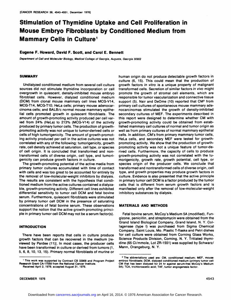
[CANCER RESEARCH 36, 4543-4551 , December 1976]
human origin do not produce detectable growth factors inculture (6, 15). This could mean that the production ofgrowth factors in vitro is a unique property of malignanttransformed calls. Secretion of similar factors in vivo mightpromote the growth of shromal cell elements, which areresponsible for tumor vasculanizahion and connective tissuesupport (5). Naimand DeOma (10) reported that CM2 fromprimary call cultures of spontaneous mouse mammary adanocancinomas stimulated the growth of density-inhibitedsecondary cultures of MEF. The experiments described inthis report ware designed to determine whether CM withgrowth-promoting activity could be obtained from aslablished mammary cell cultures of normal and tumor origin aswell as from primary cultures of normal mammary epithaliabcells. In addition, CM's from primary mammary tumor cells,HaLa calls, and secondary MEF wane tasted for growthpromoting activity. We show that the production of growthpromoting activity was not a unique feature of tumor-danived calls. Furthermore, the capacity of calls to producegrowth-promoting activity was not correlated with the tumonigenicity, growth rate, growth potential, call type, orspecies origin of the producer calls. We conclude thattransformed and nontransformed cells of diverse origin, calltype, and growth properties may produce growth factors inculture. Evidence is also presented that the active principlein primary tumor cell DCMis a factor produced by the tumorcalls that is different from serum growth factors and ismanifested only after the removal of low-molecular-weightinhibitory substances by dialysis.
MATERIALS AND METHODS
Fetal bovine serum, McCoy's Medium 5A (modified), Fungizona, penicillin, and shraptomycin were obtained from theGrand Island Biological Company, Grand Island, N. Y. Gollaganase (type I) was purchased from Sigma ChemicalCompany, Saint Louis, Mo. Plastic T-flasks and PaIn dishesfor call culture wane obtained from Conning Glass Works,Science Products Division, Conning, N. Y. Tnitiated thymidine (65 Ci/mmole, Lot ZR-i55i) was supplied by Schwamz/Mann, Onangabung, N. V.
2 The abbreviations used are: CM, conditioned medium; MEF, mouse
embryo fibroblasts; DCM, dialyzed conditioned medium (primary tumor cellCM that has been dialyzed for 24 hr against serum-free McCoy's Medium5A); TCA, trichloroacetic acid; TAF, tumor angiogenesis factor.
DECEMBER1976 4543
Stimulation of Thymidine Uptake and Cell Proliferation inMouse Embryo Fibroblasts by Conditioned Medium fromMammary Cells in Culture1
Eugene F. Howard, David F. Scott, and Carol E. Bennett
Departmentof CellandMolecularBiology,MedicalCollegeof Georgia,Augusta,Georgia30902
SUMMARY
Undialyzed conditioned medium from several call culturesources did not stimulate thymidina incorporation or callovergrowth in quiescent, density-inhibited mouse embryofibroblast calls. However, dialyzed conditioned medium(DGM) from cbonal mouse mammary call lines MCG-V14,MGG-Ti4, MGG-TiO; HaLa cells; primary mouse adenocarcinoma calls; and BALB/c normal mouse mammary apithahal cells promoted growth in quiescent fibroblasts. Theamount of growth-promoting activity produced pen cell vanied from 24% (HeLa) ho 213% (MCG-Vi 4) of the activityproduced by primary tumor calls. The production of growthpromoting activity was not unique to tumor-derived cells oncells of high lumonigenicihy. The amount of growth-promoling activity produced par cell in the active cultures was notcorrelated with any of the following: humonigenicity, growthmate,cell density achieved at saturation , call type, or speciesof call origin. It is concluded that transformed and nontransformed cells of diverse origin, call type, and humonigenicitycan producegrowthfactorsinculture.
The growth-promoting potential of the active media fromprimary tumor cultures accumulated with lime of contactwith cells and was too great to be accounted for entirely bythe removal of low-molecular-weight inhibitors by dialysis.The results are consistent with the hypothesis that condihoned medium from the active cultures contained a dialyzable, growth-promoting activity. Different cell lines exhibiteddifferential sensitivity to humor call DCM and fatal bovineserum. Furthermore, quiescent fibroblashsware stimulatedby primary tumor cell DCM in the presence of saturatingconcentrations of fetal bovine serum. These observationssupport the notion that the active growth-promoting pnincipie in primary tumor call DCM may not be a serum factor(s).
INTRODUCTION
Theme have been reports that cells in culture producegrowth factors that can be recovered in the medium [maviewed by Pandae (ii)]. In most cases, the producer cellshave been transformed in culture ordenived from tumors (1,5, 6, 8, iO, 13, 15). Primary normal fibmoblasts of murina or
, This work was supported by Contract CB 33909 and Program ProjectResearch Grant CA-i 7059 from the National Cancer Institute.
Received April 2, 1976; accepted August 31, 1976.
on April 16, 2014. © 1976 American Association for Cancer Research. cancerres.aacrjournals.org Downloaded from

E. F. Howard at al.
Preparation of CM from Primary Tumor Cultures
Spontaneous mammary adenocarcinomas were excisedfrom C3H/BiKi mica. Necrotic tissue was removed, and thetumors ware minced with scalpel blades. Aliquots of tissuemince equivalent to 2 tumors (1-cm diameter) wanedigestedseparately in 10 ml serum-free McCoy's Medium 5A thatcontained 1 mg collagenase, 5 @gFungizona, 400 unitspenicillin, and 400 j@gstraptomycin par ml. Each 10 mldigestion mixture was placed in a tightly stoppared, 50-mbEnlanmayen flask and agitated gently in a 37°waler bath for2 hr. The enzyme-digested tissue was then pipatted gentlyto dispense the cells. The cell suspensions were pooled,concentrated by cenlnifugation at 90 x g for 3 mm, and thanresuspended in McCoy's Medium 5A that was supplemenled with 10% fetal bovine serum.
Pmimarymonolayer cultures of tumor epithelial cells waneestablished by seeding the call suspensions into plastic,screw-capped culture flasks (75-sq cm growing funface).Each culture flask received all of the cells obtained from 2tumors. Accurate cell counts could not be made due ho apreponderance of call aggregates in these suspensions.The primary tumor cultures became confluent in 4 to 6 days.The spent medium was removed from these cultures andreplaced with antibiotic-free medium, which contained0.25% fatal bovine serum to prevent cell detachment. After6 days, this medium was removed, cantmifuged at 10,000 x gfor 30 mm, filtered through a 0.22-sm Millipoma filter, andfrozen at —80°until usedas GM. Forsomeexperiments, CMwas dialyzed for 24 hr against 100 volumes of serum-freeMcCoy's Medium 5A (2 changes) before filtration.
MEF Cultures
MEF ware obtained in the following manner. Embryoswane removed from 17-day-pregnant BALB/cKi mica,minced, and digested with collaganasa according to theprotocol described above for primary tumors. The cellsobtained from the enzyme digestion grew as fibroblastmonolayers in culture flasks. Confluent monolayens waredispersed with 0.05% trypsin in Puck's saline. The callsware then transferred to plastic Petni dishes (9 sq cm). Thegrowth curve of these secondary MEF cultures is shown inChart 1. Density-dependent inhibition of growth occurredafter 5 days in culture. After this time, there was no furtherincrease in cell number, and the incorporation of InitiatedIhymidine into acid-insoluble material had virtually ceased.Cell viability as measured by trypan blue dye exclusionremained greater than 90% during the entire 8-day cultureperiod. Chart 2 shows that thymidine incorporation couldbe stimulated in quiescent MEF culture by the addition of10% fatal bovine serum. Three different lots of fetal bovineserum wane tested; all stimulated thymidine uptake in MEFto the same extent. Addition of fresh, serum-free McGoy'sMedium 5A in this experiment did not cause a detectablestimulation of thymidine incorporation. Therefore, the observed stimulation was probably due to serum factors. Theincrease in thymidina incorporation was followed by callgrowth. Forty-eight hr after addition of serum, the numberof calls in the culture dish had doubled. No increase in cellnumber was seen in unfed cultures.
0 , 2 3 4 5 6 7 8
DAYS IN CULTURE
0 2 8 24 36 48
5l0
Tc@ I
@‘
‘n N
!@4 I
Chart 1. Cell growth and thymidine uptake in secondary cultures of MEF.Cells were seeded into plastic Petri dishes(9 sq cm) at a density of 104/sq cm.Cell number and thymidine incorporation into acid-insoluble material weredetermined in duplicate parallel cultures each day for 8 days. Cell numberand thymidine uptake were measured as described in “Materialsand Methode.―L@,incorporation of 3H into acid-insoluble material following a 1-hrexposure of [3H]thymidine (1 MCi/dish; final concentration, 7.5 nM).
..J
‘°‘-@
I
HOURS AFTER FEEDING
Chart 2. Comparison of the ability of fetal bovine serum and serum-freemedium to stimulate thymidine uptake into acid-insoluble material by density-inhibited MEF. The spent growth medium on quiescent fibroblasts wasreplaced on Day 7 (Chart 1) with fresh McCoy's Medium 5A that containediO% (v/v) fetal bovine serum or no serum. At the intervals indicated on theabscissa, tritium incorporation into acid-insoluble material was measuredfollowing a i-hr exposure to [3H]thymidine. Duplicate parallel cultures wereassayed at each time point for each additive.
Bioassay for Growth-promoting Activity of Tumor Cell CM
Secondary MEF cultures wane used ho assay growth-promoting activity in tumor cell GM. On the 7th day of culture(Ghart 1), the spent medium was removed from the quies
4544 CANCER RESEARCH VOL. 36
on April 16, 2014. © 1976 American Association for Cancer Research. cancerres.aacrjournals.org Downloaded from

Growth Stimulation by Mammary Cell CM
cent MEF, and was replaced with the solution ho be assayed. Twenty-four hr later, thymidina incorporation wasmeasured in treated and control cultures. A significant shimulation of thymidine uptake at 24 hr was interpreted as anindication of growth promotion. In some experiments, thisassumption was verified by measuring the increase in cellnumber 48 hr after addition of test solutions. Exposure ofthe MEF to serum-free McCoy's Medium 5A for 24 h prior tothe addition of test solutions did not alter the magnitude onthe timing of the stimulation of thymidina incorporation onthe increase in cell number.
Thymldine Incorporation into TCA-insoluble Material
Twenty-four hr after addition of the test solution to quiescent MEF, 1 pCi of Initiated thymidina (65 Gi/mmole; finalthymidine concentration, 7.5 nM) was added to each culturedish. One hr later, the medium was removed from the cubtunes and replaced with 2 ml of cold 5% TGA. After 30 mm at4°,the TCA was removed, and the acid-insoluble residue ineach culture dish was dissolved in 1 ml of 0.5 N NaOH (15mm, 37°).This solution was washed quantitatively from theculture dish into a centrifuge tube. Ten ml of cold 5% TGAwere added. After 20 mm at 4°,the precipitate was collectedby centnifugation at 10,000 x g . The pellet was washedtwice with cold 5% TCA and once with cold 95% ethanol,and then it was drained dry. The pellet was than hydrolyzedin 2.5 ml of 5% TGA at 90°for 15 mm. The acid-insolubleresidue was removed by centnifugation, and an aliquot ofthe supematant was assayed for radioactivity in a liquidscintillation counter. Another aliquol was assayed for DNAcontent with the diphenylamine method of Burton (3). Datawere expressed as tnihium dpm pen @gDNA per hr. In someexperiments, cells were removed from the culture dish withtrypsin and counted; their viability was measured by trypanblue dye exclusion before precipitation with 5% TGA. Datawemeexpressed as tnitium dpm per 10@viable calls pen hr.
Primary Normal Mammary Eplthelial Cultures
The 4th and 5th inguinab mammary glands wane dissectedfrom virgin BALB/cKi mice and carefully cleaned of connechive tissue, blood vassals, lymph nodes, and muscleunder a binocular dissecting microscope. During this openation, the glands were placed in serum-free McGoy's Madium 5A that contained excess antibiotics (400 units penicilun and 400 @Lgstreptomycin pen ml). The cleaned glandswere then minced and digested with collaganase as descnibed for primary tumor cells. The glands from 2 micewere digested separately, and the cells obtained wereseeded into 1 plastic PaIn dish (9 sq cm). A total of 12cultures (12 PaIn dishes) were prepared for each expemiment.
EstablIshed Mammary Cell Lines
All of the mammary cell lines used in this study wereestablished in this laboratory. A detailed description of theirproperties is in preparation.
MCG-V14. This cell line was derived from normal mammary tissue that was obtained from a 16-month-old virgin
A/Ki mouse. The primary cells, which were obtained fromcollagenase-digasted mammary tissue, were apithalioid.The cell line that was derived from the primary cells wascloned after 1 year and 50 passages. Clone 7 was used inthis study. These cells are fibroepithelioid and have a modalchromosome number of 125. They grow with a doublingtime of 33 hr in log phaseand achieve a saturation densityof 0.75 x 10@cells/sq cm. When 5 x 10@calls ware injecteds.c. into 12 A/Ki mica, a spindle call tumor appeared in 1mouse after 6 months. No humorshave appeared in any ofthe other mice after 12 months. MGG-V14-7 is considered tobe a bow-humoniganic cell clone.
MCG-T14. The source of this call line was a mammaryadanocarcinoma that had appeared spontaneously in aBALB/cKi mouse and had subsequently been propagatedas a s.c. implant. The primary cells from the tumor weresubculhured 76 times during a period of 14 months. At thispoint, the line was cloned. Clone 3 was used in this study.The cells are epithelioid and have a modal chromosomenumber of 50. The doubling time of MGG-T14-3 cells is 14 hrin log phase. The saturation cell density is 2.10 x 10@cells/sq cm. When 5 x 10@cells were injected s.c. into BALB/cKimica, palpable adenocancinomas wane formed in 10 ho 14days. MGG-T14-3 is considered to be a high-tumoniganiccell clone.
MCG-T1O. The source of this cell line was a spontaneousmammary adenocarcinoma from a CBA/StKi mouse. Thecells derived from this tumor were cloned after 45 passagesand 1 year in culture. The clone used in this study, clone10, is fibmoblastoid and has a modal chromosome number of40. MGG-T10-10 calls grow with a doubling time of 10 hr inlog phase and achieve a saturation density of 2.62 x 10@cells/sq cm. MGG-T10-10 cells produced spindle cell carcinomas in CBA/StKi mice within 2 weeks and are consideredto be highly tumonigenic.
MCG-T19. This tumor cell line was derived from a spontaneous mammary adenocancinoma from an A/Ki mouse.After 1 year and 30 passages in culture, the line was cloned.Clone 9 was used in this study. The chromosome number inclone 9 cells is bimodal with modes at 43 and 80 chromosomes. The doubling time of this clone is 10 hr in log phase.The saturation density is 1.89 x 10@cells/sq cm. MCG-T19-9produced spindle cell carcinomas 1 ho 2 weeks after injeclion s.c. into A/Ki mice.
The cbonal mammary cell lines used in this study will bereferred ho without the clone number; e.g., MGG-V14-7 willbe written as MGG-V14.
Culture of Established Cell Lines
All of the clonal cell lines used in this study were maintamed as monolayer cultures in McGoy's Medium 5A supplemented with 5% fetal bovine serum. Cultures weregrown at 37°in a 5% GO2-95% aimatmosphere. Subculturewas accomplished with standard trypsinization techniques.Cells in all cultures used in this study wane counted on ahemocytomehar after hrypsinization. Cells were renewedfrom frozen shocks at 3-month intervals. Thus, all of thecbonal cell lines used in this study were subcultuned nomore than 12 limes after cloning. All cells ware free ofcontaminating bacteria, fungi, and Mycoplasma.
DECEMBER1976 4545
on April 16, 2014. © 1976 American Association for Cancer Research. cancerres.aacrjournals.org Downloaded from

0 24 48 72 96
E. F. Howard at al.
RESULTS
Growth-promoting Activity of CM from Primary TumorCells. The ability of tumor call CM and those of otheradditives to stimulate thymidine incorporation into acidinsoluble malarial by quiescent MEF wane compared (Chart3). The acid-insoluble radioactivity, recovered from callsthat had bean exposed to fatal bovine serum [final concantmahion,10% (v/v)] for 24 hr and then labeled with Initialedthymidine for 1 hn, was given a normalized value of 1.0. Theabsolute value of the acid-insoluble radioactivity in theseserum-fed cultures averaged 4521 ±783 dpm/@g DNA (n= 8). Thymidine incorporation in cultures in which the spent
medium was not renewed or supplemented (unfed cultures)was 8% of that seen in serum-fed cullumes (Chart 3). Whenquiescent MEF were exposed to serum-free McCoy's Medium SA, thymidine incorporation 24 hr later was about 18%of the response induced by 10% serum. This indicated thatrenewal of medium constituents caused only a small shimulation of thymidine incorporation. The activity of McCoy'sMedium 5A supplemented with 0.25% fetal bovine serumwas identical to that of McCoy's Medium 5A alone. Furthermore, undialyzed tumor cell GM that contained 0.25% serum (sea “Materials and Methods―)had the same activity asserum-free medium. Therefore, 0.25% serum did not stimulate thymidina incorporation in quiescent MEF above thelevel exhibited by serum-free medium. In contrast to theprevious additives, DCM had a shimulatomy activity comparable to that of 10% fetal bovine serum.
In addition ho an apparent affect on DNA synthesis, DCMstimulated cellular overgrowth in density-inhibited MEF(Chart 4). The magnitude of cellular overgrowth (approxi
‘4
HOURS AFTER FEEDING
Chart 4. Cell overgrowth in density-inhibited MEF that were fed with primary tumor cell DCM or with medium that contained iO% (v/v) fetal bovineserum. Quiescent fibroblasts were fed with DCM or serum at 0 time. At theintervals indicated on the abscissa, cells were removed by trypsinization fromduplicate cultures in each category and counted on a hemocytometer. Ateach time point, unfed cultures in which the spent growth medium was notremoved were included as controls. Cell counts (ordinate) were expressed ascells x iO'/sq cm.
mately 2-fold) was equivalent to that caused by 10% fetalbovine serum.
Growth-promoting Activity of CM from Established CellLines, Normal Mammary Epithelium, and Normal MEF. GMand DGM from several cell lines ware assayed for theirability to stimulate hhymidine uptake into acid-insoluble matemial by density-inhibited MEF. The mouse mammary calllines were MGG-V14, MGG-T14, MGG-T10, and MGG-T19,described in “Materialsand Methods.―HaLa cells were alsoused as a source of DGM.In addition, medium from normalmammary apithelium and from secondary cultures of MEFwas lashed. For every call culture, the protocol for obtainingGM was the same as that described for primary humorcalls(see “Materialsand Methods―).Table 1 shows the results ofduplicate experiments. II can be seen that the shimubahomyactivity of GM (relative to 10% serum) from the various callcultures ranged from 0.02 (secondary MEF) to 0.28 (MCGV14). With the exception of cell line MCG-T19, dialysis ofGM from all of the other cell cultures caused an increase instimulatory activity from 1.8-fold (HeLa) ho 14-fold (MGGTb).
In these experiments, CM was obtained from cells in thestationary phase of growth. The plateau on saturation densily achieved by each cell line varied from 0.40 x 10@calls/sq cm (primary mammary epithalial cells) ho 2.62 x 10@cells/sq cm (MGG-Tb0). Consequently, GM was oblainedfrom different numbers of cells in each call line. In order hocompare the potential of each cell line to produce growhhpromoting activity, the relative activity of the various DGM'swas normalized for cell density (Table 2). The saturationdensity of each cell line (Table 2, Golumn A) is expressed asa multiple of the cell density achieved by primary tumorcells [relative cell density (Table 2, Golumn B)]. The relativegrowth-promoting activity of hhe DGM obtained from each
n= 10
.-J‘4J
I0
08
06
04
02
0% UNFED 0 25% SERUM-F@E PRIMARY PRIMARYSERUM SERUM MEI@UM TUMOR TUMOR
CELL SELLCM DCM
Chart 3. Comparison of the ability of several additives to stimulate thymidine uptake into acid-insoluble material by density-inhibited MEF. The protocol for this assay is described in “Materialsand Methods.―Thymidine uptakewas measured 24 hr after the spent growth medium on quiescent fibroblastswas replaced with a particular additive. In cultures in which the spentmedium was replaced with fresh medium that contained iO% (v/v) fetalbovine serum, the amount of radioactivity incorporated into acid-insolublematerial following a i-hr exposure to [3H]thymidine was given a normalizedvalue of i .0 [relative activity (ordinate)]. The amount of thymidine uptake inunfed cultures in which the spent growth medium was not replaced and incultures that were exposed to fresh medium supplemented with 0.25% (v/v)fetal bovine serum, serum-free medium, primary tumor cell CM, or DCM wasexpressed as a multiple of the normalized activity of iO% serum.
CANCERRESEARCHVOL. 364546
n5 n6 n@5
nz8@@FIJ
on April 16, 2014. © 1976 American Association for Cancer Research. cancerres.aacrjournals.org Downloaded from

Table 1
Stimulation of thymidine uptake in stationary MEFby CM fromvariouscell cultures
0CM and CM from several establishedcell lines (described in“Results―)plus primary, normal mammary epithelial cell culturesand secondaryMEFwere tested for their ability to stimulatehhymidine uptake into acid-insoluble material by density-inhibited MEF.The amount of thymidine uptake in MEFthat were fed with freshmedium that contained 10% (v/v) fetal bovine serum was given anormalized value of 1.0.
Source of mediumMcCoy'sMedium5A + 10%fetal bovine
serumMCG-T14
0CMCM
MCG-T19DCMCM
MCG-T100CMCM
MCG-V140CMCM
HeLaDCMCM
Primary normal mammaryepitheliumDCMCM
SecondaryMEF0CMCMa Mean ± S.E.
cell line is listed in Table 2, Column C. These values havebeen connected for the activity exhibited by fresh serum-freemedium (Chart 1).This isdona because0CM is obtained bydialysis of GM against serum-free medium and presumablysome of the activity of DGM(relative activity, 0.2 comparedto fetal bovineserum) is due to renewaland removalof lowmolecular-weight medium components. The connected nelativa growth-promoting activity of the 0GM obtained from acell line was divided by the relative cell density to estimatethe relative activity produced pancall (Table 2, Column 0). IIcan be seen that call line MGG-V14produced more thantwice as much growth-promoting activity per cell as didprimary tumor cells. The relative activity per cell in the othercell cultures ranged from zero (MEF and MCG-T19) to 0.56(MGG-T14). Primary mammary epithelial cells produced32% as much activity per call as did the primary adanocancinoma cells.
Further Observations on Primary Tumor Cell DCM. Ifprimary tumor cells constantly release a nondialyzablegrowth factor, one might expect that the growth-promotingactivity of CM would increase as a function of the lime themedium was in contact with cells. Chart 5 shows that CMthat was harvested after 24 hr and then dialyzed (Ghart 5,DCM 24 hr) had one-half the activity of DCM that had beenin contact with primary tumor cells for 6 days. Therefore,the stimulatory activity of 0CM does appear to increasewith lime. The primary tumor cells from which 24-hr CM hadbeen harvested were refad wihh fresh medium and 0.25%serum. Six days later, this medium was harvested and
Growth Stimulation by Mammary Cell CM
dialyzed (Chart 5, DCM 6 days-rinsed). The 0CM from therinsed cultures had the same activity as did 6-day 0CM fromunninsed cultures (i.e. , from confluent, primary tumor cultunes where the spent growth medium was replaced withMcCoy's Medium 5A and low serum without an intervening
Table 2Relativeactivity of DCM from variouscell cultures normalized for
cell densityA. Saturationcell densityachievedbyeachcell line. B. Forthe r
= relative cell density, the saturation density achieved by primary
tumor cells was given a normalizedvalue of 1.0. C. For the conmacted relative activity, the growth-promoting activity of the 0CMfrom each culture source was connected for the activity exhibited byserum-freemedium. This was done by subtracting the amount ofincorporation of thymidine into acid-insoluble material in quiescentMEF cultures that were fed with serum-free medium from theamountof incorporation in MEFcultures that werefed with variousDCM's.The corrected activity of primarytumor cell DCMwasgivena normalized value of 1 .0. 0. For the relative activity per cell, thecorrected relative growth-promoting activity of the 0CM obtainedfrom a particular cell source was divided by the relative cell densityho estimate the relative activity produced per cell.
Relativeactivity1.00
0.79 ±0.10―0.08 ±0.03
0.14 ±0.050.12 ±0.08
0.71 ±0.280.05 ±0.02
1.00 ±0.140.28 ±0.07
0.47 ±0.030.27 ±0.07
0.26 ±0.050.04 ±0.01
0.12 ±0.060.02 ±0.001
A. Sahuma C. Con 0. Relalion cellB. Rela nectedhive achy
0CM sourcedensity(x
10@cells/sqhivecell
densityrelativeactivityity/cell(C/B)Pnimaryhumor1.581.001.001.00MCG-V140.750.471
.002.13MCG-T142.101.330.740.56MCG-T102.621
.660.640.39Primarymammary0.400.250.080.32epitheliumHeLa2.541.610.390.24MCG-T19
MEF1.89 1.701.20 1.070.000.000.000.00
,q
0CM 0CM 0CMSERUM 24HR 6@YS 6OAYS-RINSED
ADDITIVES
Chart5. Accumulationof stimulahoryactivity in primarytumor cell DCM.The ability of DCM, which was obtained from primary mammary tumor cellsafter 24 hr or 6 days, to stimulate thymidine uptake into acid-insolublematerial by density-inhibited MEF was compared. In addition, confluentmonolayens of primary tumor cells were rinsed for 24 hr with fresh mediumthat contained 0.25% (v/v) fetal bovine serum. This medium was replaced byfresh medium that contained 0.25% serum. Six days later, this medium wasdialyzed and assayed as “DCM-6days-rinsed.―The stimulatory activity of thevariousDCMwasexpressedasa multipleof theactivity(1.0)of freshmediumthat contained 10% (v/v) fetal bovine serum.
4547DECEMBER1976
on April 16, 2014. © 1976 American Association for Cancer Research. cancerres.aacrjournals.org Downloaded from

E. F. Howard at al.
24-hr “rinse―).This result indicates that the active principlein 0GM is probably not derived from residual, spent growthmedium in the primary tumor cultures.
One possible explanation for the growth-promoting achyity in tumor cell 0GM is that the tumor cells sequester serumfactors from the growth medium and then releasethem hothe GM. If this occurs, than the active principle in tumor cell0CM might be a serum factor(s). This possibility is varydifficult to rule out. However, the following experimentsprovide indirect evidence that the growth-promoting activityin tumor cell 0CM is different from the growth factor(s) infetal bovine serum.
The ability of 10% fetal bovine serum and tumor cell DGMto stimulate thymidine uptake was compared in the celllines utilized previously (Chart 6). All cell cultures wareallowed horeach a quiescent, stationary phase during whichcall growth and DNA synthesis had ceased before they werefed with serum or with 0CM. The absolute magnitude of thebasal and stimulated levels of thymidina incorporation variad greatly between these call lines; therefore, normalizeddata wane not presented in this case. This experimentshowed that the response to serum and 0GM is very differant in some cell lines. For example, MCG-V14 is stimulatedby 10% serum but is apparently refractory ho DGM. In conInast to MGG-V14, MCG-T10 was 3.6 limes more sensitive toDCM than to 10% serum. MEF, HeLa cells (not shown), andMGG-T14 mammary tumor cells had a nearly equal masponsa to serum and DCM. Finally, quiescent MGG-T19calls were not stimulated by serum. However, 0GM causeda 3-fold stimulation of thymidina uptake compared to unfedcontrols. If the active principle in DCM is a serum factor(s),then one would expect that a given call type would respondsimilarly to 0CM or to serum. This does not occur in MCGV14,MCG-T10, on MCG-T19 calls.
The maximum serum stimulation of quiescent MEF occumred with 10% serum. Addition of more serum did notcause any further increase in thymidine incorporation (mesuits not shown). An experiment was performed to determine whether DCMcould stimulate MEF in the presenceofsaturating concentrations of serum (Chart 7). When MEFwere fed with DCM and 10% fetal bovine serum , the stimulalion of thymidine incorporation was greaten than when cellswere fed wihh serum or with DGM alone. The magnitude ofthe response in cells fed with DGM and 10% serum (5985dpm/@g DNA) was approximately 7% less than the sum(6419 dpm/@g DNA) of the response in cells that were fedwith serum alone or DCM alone. This implies that the affectof serum and DGMon thymidine incorporation is additive atsaturating concentrations of serum. These data are consistent with the hypothesis that the active principle in 0GM isdifferent from the active factor(s) in serum.
DISCUSSION
These experiments show that DGM but not CM from3 mouse mammary cbonal cell lines, HaLa cells, and primarymouse mammary epithelial cells stimulated thymidine uptake into acid-insoluble material by quiescent MEF. Cellsfrom 1 malignant cell line (MCG-T19) and MEF did notproduce any detectable growth-promoting activity. In addilion , we have confirmed the observation of Nair and DeOma(10) that DGM from primary monolayer culhures of mousemammary adenocancinoma cells stimulated hhymidina uptake and cell overgrowth in MEF cultures.
Our observations show that among the cell culturestested production of growth-promoting activity was not aunique property of tumor-derived cells. MGG-V14, which isa cell line derived from normal mammarytissue, producedmore than twice as much growth-promoting activity pencallas the primary humor calls. MGG-V14exhibited density
%@\
0@
@ 3OOC@
IMEF v-14 T-I0 T.14 T-19
Chart 6. Comparison of the ability of primary tumor cell 0CM and of freshmedium supplemented with 10% (v/v) fetal bovine serum to stimulate thymidine incorporation into acid-insoluble material by several different cell lines.The spent growth medium on density-inhibited cells was replaced with serumor 0CM. Twenty-four hr later, thymidine uptake was measured in replicatecultures in each category as described in “Materialsand Methods.―Thymidine uptake was also measured in unfed control cultures in which the spentgrowth medium was not replaced. Results are expressed as ‘Hdpm per @gDNA per hr (abscissa).
600C
500C
4000
3000
2000
I000
0.
0% DChi 0%SERUM +
0CM
Chart 7. Thymidlne uptake Into acid-insoluble material by density-inhibited MEF that were fed with iO% fetal bovine serum, primary tumor cellDCM, or DCM made iO% (v/v) in fetal bovine serum. Thymidine uptake wasmeasured 24 hr after the spent growth medium was replaced by the variousadditives. Results are expressed as 3H dpm per @gDNA per hr.
4548 CANCER RESEARCH VOL. 36
N UNFED
0 0% SERUM
@ PRIMARYTUMORCELL DCM
d@nJIi:
on April 16, 2014. © 1976 American Association for Cancer Research. cancerres.aacrjournals.org Downloaded from

Growth Stimulation by Mammary Cell CM
dependent inhibition of growth al low cell density and inthis respect resembled a nontransformed cell line. Howeven, MGG-Vb4 is an established cell line which has produced a tumor in 6 months when injected s.c. into homobogous mica (sea “Materialsand Methods―).Therefore, MGGVb4 is likely to be a transformed cell line with low humorigenicity. The production of an axtmacallulamTAF by astablished cell lines (WI-38 and BALB/c-3T3), which were demivedfrom normal tissue and exhibited “contactinhibition―of growth in culture, has been reported by Klagsbrunat a!.(6). In addition, a variety of humor-derived and viral-Iransformed cell lines produced TAF. However, primary culturesof MEFand low-passagecultures of human fibmoblastsproduced no TAF. These observations could mean that produclion of TAF is a general property of transformed calls inculture but is not a property of nontransformed calls. Thispossibility is supported by Suddith et al. (15), who reportedthat GM obtained from a variety of cbonabcell lines of tumororigin stimulated the proliferation of cultured endothelialcells, while GM from cultures of nontransfommed, primaryfibroblasts, nonmalignant peripheral lymphocytes, andcells derived from amniocentesis had no detectable activity.Although it is difficult to compare growth-promoting aclivities detected in different assay systems, our data have abearing on the proposition Ihat the production of growthfactors is a unique property of transformed cells in culture.As in the cited reports, primary MEF produced no growthpromoting activity. However, primary cultures of mousemammary apithelial cells produced approximately one-thirdas much growth-promoting activity pencall as did primarytumor cells. Thus, in this systemgrowth-promoting activitywas produced by nontransformed cells in culture. To determine whether this is an isolated phenomenon or a generalproperty of primary apithelial cells will require the tasting ofa broad spectrum of primary call cultures from a variety ofepihhelial tissues.
The amount of growth-promoting activity produced pancell by primary epithelial cells was considerably less thanthat produced by primary tumor cells. This could mean thatthe production of growth factors is amplified by the processof transformation. If this were the case,one might expect tofind a correlation between the amount of growth-promotingactivity produced par cell and those properties of cells inculture that amaassociated with the transformed state, suchas high tumonigenicity, increased growth mateduring logphase, and the capacity to achieve high-saturation calldensities. In our experiments none of these parameterswere correlated with the capacity of cells to producegrowth-promoting activity. Thus, MGG-V14 exhibited lowtumonigenicity when measured by the criteria of time oftumor production in homologous mica (6 to 8 months afterthe s.c. injection of 5 x 10@cells), low growth mate(doublingtime, 33 hr in log phase), and a high saturation density (0.75x 10@cells/sq cm). MGG-T19 exhibited high tumonigenicity(10 to 14 days after injection of 5 x 10⁶ cells), high growthrate (doubling time, 10 hr in log phase),and a high saturalion density (1.89 x 10@cells/sq cm). By these criteria,MCG-T19 appeared to be a cell line in which the degree oftransformation was greater than that of MGG-Vb4. However,MCG-Vb4 produced the greatest amounhof growth-promot
ing activity pen cell of all cell lines tested. MGG-Tb9 did notproduce detectable growth-promoting activity. Cell linesMGG-Tb4 and MGG-T10 were similar ho MGG-T19 with masped to tumonigenicity, growth rate, and saturation densityachieved in culture. These cells produced gnowth-promoting achivity but not at the level exhibited by MGG-V14 calls.These observations suggest that, among the culturestested, transformation, when measured by the criteria oftumonigenicity, growth rate, and growth potential, was notcorrelated with the capacity of cells to produce growthpromoting activity. Similarly, the extent of production ofgrowth-promoting activity was not correlated with cell typeor species origin. Active fibroblasts (MGG-T10) and inactiveMEF, active apithelial cells (MGG-T14, HaLa, primary mammary epithelium), and inactive epithelial cells (MGG-T19)were identified. In addition to the active munine call cultunas, cells of human origin (HeLa) produced an activeprinciple. These observations suggest that transformed andnontransfonmed cells of diverse origin, cell type, and tumorigenicity may produce growth factors in culture. However,the proposition that the growth-promoting activity in 0GM,obtained from active cultures, actually contained a growthfactor(s) of cellular origin requires further scrutiny.
Galderone and Unanue (4) have shown that macrophagerich penihoneal exudate cells from the mouse release adialyzable inhibitor and a nondialyzabla growth factor to themedium when they are placed in in vitro cullure. A similarphenomenon that would explain the relative inactivity of GMcompared to DGM could have occurred in our active cultunes. However, there are alternative explanations of ourdata to be considered. It is conceivable that GM because ofits contact with nongrowing cell monolayens acquires lowmolecular-weight inhibitory substances only. These couldbe derived from dead or dying cells and/on could be maleased as normal export products from healthy calls. GMthat contained the inhibitory substances would not promotegrowth. However, dialysis of the GM would remove inhibitons, and the DCM would then exhibit slimulalory activity.This explanation implies that themeis no growth factor; theactivity of DGM could be ascribed wholly to the removal of adialyzable inhibitor. If these were the case, dialysis of GMfrom any source should produce a 0CM with growth-promoting activity. Furthermore, since GM is dialyzed againstserum-free medium, the growth-promoting activity of DGMwould not be expected to exceed that of serum-free mediumon of medium that is supplemented with an amount of fetalbovine serum (0.25%) equivalent to the level that is presentduring the conditioning period. These predictions are notborne out by our results. Dialysis does not always produce a0GM in which relative growth-promoting activity is differentfrom untreated GM (call line MGG-Tb9). In most cases, therelative activity of 0CM is 50 to 100% that of fetal bovineserum. This is significantly greater than the relative activity(20%) of serum-free medium or medium supplemented with
0.25% serum. Thus, it would appear that the growth-promoting activity of DGM is too great to be accounted forentirely by the removal of a dialyzable inhibitor.
If the premise is accepted that active 0GM contains agrowth factor(s), then it is of interest to speculate on thenature and source of this material. One possibility is that the
DECEMBER1976 4549
on April 16, 2014. © 1976 American Association for Cancer Research. cancerres.aacrjournals.org Downloaded from

E. F. Howard at a!.
active principle in DCM is a serum growth factor(s) that isderived from the original spent-growth medium or from the0.25% serum present during the conditioning period. Wehave already discussed the observation that the relativepotency of active 0GM from primary tumors exceeds that of0.25% serum. If the growth-promoting activity of primarytumor cell DCM is due ho residual serum from spent medium, than the activity of DGM should not increase withprolonged exposure to the cell monolayer. However, wehave shown (Ghart 5) that the activity of primary humor cellDGM does increase with lime of exposure to cells. Theincrease in activity occurs with similar magnitude when thecell monolayer is washed to remove spent medium beforefresh CM is applied. Thus, it appears that the active pnincipIe in primary tumor call DCM accumulates in the cultureand that it is probably not derived from spent medium.
The active principle in 0GM also could be a serum growthfactor(s) that is sequestered by the calls during growth inhigh concentrations of serum and than is slowly released tothe GM. If this occurred, the relative activity of DCM wouldstill be expected to increase with time of contact with thecell monolayer. Two of our observations give indirect evidance that the active principle in primary tumor call DGM isdifferent from the growth factors in serum. First, some celllines react differentially to serum and primary tumor callDGM (Chart 6). For example, thymidine incorporation intodensity-inhibited MGG-V14 calls is stimulated by serum butnot by primary humor cell DGM. MGG-T10 is much moresensitive to 0CM than to serum. Quiescent MCG-T19 cellsare refractory to serum, but thymidine incorporation is slimulahed by primary tumor cell 0CM. If the active principle inprimary tumor cell DCM is the same as the active principlein fetal bovine serum, than one would expect that a givencell line would react similarly to each additive. However,Laffart (7) has shown that both relative and absolute levelsof serum factors influence the proliferation of cultured fatalrat hepatocytes. Furthermore, some serum growth factorsresemble peptide hormones (12), and some cell lines differin their hormone requirements for growth in vitro (2). II ispossible, therefore, that the differential response of somecall lines to DGM and to whole serum is due to differences inthe relative and absolute levels of paptide hormone-likegrowth factors in these additives.
The 2nd observation, which suggests that primary tumorcall 0GM contains growth factors that are different fromserum growth factors, was the response of quiescent MEFcalls to the addition of both 0CM and 10% serum. Theincrease of thymidine uptake into acid-insoluble material,following the simultaneous addition of both 0CM and sarum, was equal to the sum of the increase in cultures givenserum plus the increase in cultures given DGM (Chart 7).The response of MEF to serum plateauad at the 10% level.Further addition of serum to 25% did not cause a further inneasein thymidine uptake, nor did it result in cell toxicity(E. Howard, unpublished observation). Therefore, 10% sarum was a saturating dose with respect to the stimulation ofthymidine uptake by quiescent MEF. If serum and DCM conlain the same growth factors, these factors should interactwith the same cellular receptors that are responsible for theregulation of thymidina uptake, and the simultaneous addi
lion of 0GM and a saturating concentration of serum shouldnot result in a stimulation of thymidine uptake above thatseen with 10% serum. Thus, the observation that the mesponse to both DGM and 10% serum is additive stronglysuggests that the active principle in 0CM interacts withcellular receptors that are different from those involved inthe response ho serum growth factors and that the activeprinciple of DCM is different from the serum growth factors.However, alternative interpretations of these data, which involve an increased response of a cellular receptor to aunique combination of several serum growth factors in0CM and 10% serum, cannot be ruled out at this time.
On the basis of the experiments with primary tumor call0CM, we propose that our active cell cultures also pro
duced a growth factor(s) that could stimulate cell proliferalion in quiescent MEF calls. Our data do not reveal whetherthe various active call lines produced qualitatively differentgrowth factors, or whether the growth-promoting activity inprimary tumor cell DGM resembled the tumor-producedshromal growth factors that may function in vivo to stimulatethe proliferation of stromab tissues required for sustainedtumor growth (5). The fact that primary humor cell 0GMstimulated DNA synthesis and cell division in malignant calllines of epithelial origin (HeLa, MGG-T14, MGG-T19) as wallas it did in MEF, suggests that primary humor cell 0CMcontained a general growth-promoting activity not specificfor fibroblasts. The growth of all of the call lines tested,including the tumor calls, was completely arrested whenthey were exposed to DCM. Thus, the growth-promotingactivity of DGM in these experiments represented a reversalof growth arrest at high cell density. It will be interesting tosee whether 0GM can also promote or support the attachment of freshly plated target cells or the growth of thesecells at low density.
A definitive answer howhether or not active DGM containsdialyzable inhibitors on macromolaculam growth factorsawaits the isolation, purification, and biochemical charactanization of these substances. Although the literatureabounds with reports of factors that will stimulate or inhibitcell growth [reviewed by Pandee (11)], few of these factorshave been isolated or characterized. Instances in whichcultured cells produce readily identifiable growth factorsare name.Rat liver cell clones exist that apparently produce amultiplication-stimulating factor that resembles growth fachors in whole serum (14). Nerve growth factor, which is anatural product of the mouse submaxillary gland, is produced by mouse fibroblasts (16) and by mouse neumoblastoma cells in culture (9). Nainand DeOme(10) reported thatthe active principle in primary mammary tumor cell DCMcould be precipitated with ammonium sulfate. We haveconfirmed this observation in preliminary experiments.Thus, we are encouraged that further efforts to isolate andcharacterize the putative growth factors in DGM obtainedfrom active cell cultures will be successful.
ACKNOWLEDGMENTS
The authors acknowledge the capable technical assistance of JudyGreene.
4550 CANCERRESEARCHVOL. 36
on April 16, 2014. © 1976 American Association for Cancer Research. cancerres.aacrjournals.org Downloaded from

Growth Stimulation by Mammary Cell CM
Growth of Fibroblasts in Vitro. Cancer Res., 4: 694-703, 1944.9. Murphy, R. A., Pantazis, N. J., Amason, B. G. W., and Young, M.
Secretion of a Nerve Growth Factor by Mouse Neuroblastoma Cells inCulture. Proc. NahI.Acad. Sci. U. S., 72: i895-i898, 1975.
10. Nair,B.K.,andDeOme,K.B.AGrowth-stimulatingFactorReleasedbyCultured Mouse Mammary Tumor Cells. Cancer Res., 33: 2754-2760,1973.
i 1. Pardee, A. B. The Cell Surface and Fibroblast Proliferation Some CurrentResearch Trends. Biochim. Biophys. Acta, 417: i53-172, 1975.
i2. Pastan, I. Regulation of Cellular Growth. Advan. Metab. Disorders, 8: 7-16, i975.
13. Rubin, H. Overgrowth Stimulating Factor Released from Rous SarcomaCells. Science, 167: 127i-i272, 1970.
i4. Smith, G., and Temin, H. Purified Multiplication-Stimulating Activityfrom Rat Liver Cell Conditioned Medium: Comparison of BiologicalActivities with Calf Serum, Insulin, and Somatomedin. J. Cellular Physiol.,84:i8i-i92,1974.
is.Suddith,R.L.,Kelly,P.J.,Hutchison,H.T.,Murray,E.A.,andHaber,B.In Vitro Demonstrationof an EndothelialProliferativeFactor Producedby Neural Cell Lines. Science, 190: 682-684, i975.
i6. Young, M., Oger, J., Blanchard, M. H., Asdourian, H., Amos, H., andArnason, B. G. W. Secretion of a Nerve Growth Factor by Primary ChickFibroblast Cells. Science, 187: 361-362, i975.
4551DECEMBER1976
REFERENCES
i . Argyris, T. S. The Growth-Promoting Effects of Tumors on Tissues.Advan.Biol.Skin,6:55-73,1965.
2. Armelin, H. A., Nishikawa, K., and Sato, G. H. Control of Mammalian CellGrowth in Culture: The Action of Protein and Steroid Hormones asEffector Substances. In : B. Clarkson and R. Baserga (eds.), Control ofProliferation in Animal Cells, pp. 97-104. Cold Spring Harbor, N. Y.:Cold Spring Harbor Laboratory, 1974.
3. Burton, K. A Study of the Conditions and Mechanisms of the Diphenylamine Reaction for the Colonimetnic Estimation of Deoxyribonucleic Acid.Biochem. J., 62: 3i5-323, 1956.
4. Calderone, J., and Unanue, E. R. Two Biological Activities RegulatingCell Proliferation Found in Cultures of Pentoneal Exudate Cells. NatureNew Biol., 253: 359-361 , 1975.
5. Folkman, J. Tumor Angiogenesis. Advan. Cancer Res. 19: 331-358,1974.
6. Klagsbrun, M., Knighton, D., and Folkman, J. Tumor Angiogenesis Activity in Cells Grown in Tissue Culture. Cancer Res., 36: 1iO-i 13, 1976.
7. Leffert, H. L. Growth Control of Differential Fetal Rat Hepatocytes inPrimary Monolayer Culture. J. Cell Biol., 62: 767-779, 1974.
8. Ludford, R. J., and Barlow, H. The Influence of Malignant Cells upon the
on April 16, 2014. © 1976 American Association for Cancer Research. cancerres.aacrjournals.org Downloaded from