Training manual for fecal sludge-based compost production ...
w a t e r r e s e a r c h 4 3 ( 2 0 0 9 ) 4 8 5 0 – 4 8 5 9
Avai lab le at www.sc iencedi rect .com
journa l homepage : www.e lsev i er . com/ loca te /wat res
Rapid decay of host-specific fecal Bacteroidales cellsin seawater as measured by quantitative PCRwith propidium monoazide
Sungwoo Bae, Stefan Wuertz*
Department of Civil and Environmental Engineering, 2001 EU III, University of California, Davis, One Shields Avenue, Davis, CA 95616, USA
a r t i c l e i n f o
Article history:
Received 20 March 2009
Received in revised form
22 June 2009
Accepted 28 June 2009
Published online 3 July 2009
Keywords:
Persistence and survival
Host-specific Bacteroidales
Microbial source tracking
Fecal pollution
Propidium monoazide
* Corresponding author. Tel.: þ1 530 754 640E-mail address: [email protected] (S.
0043-1354/$ – see front matter ª 2009 Elsevidoi:10.1016/j.watres.2009.06.053
a b s t r a c t
We investigated the persistence of feces-derived Bacteroidales cells and their DNA in
seawater under natural conditions using an optimized chemical method based on co-ex-
traction of nucleic acids with propidium monoazide (PMA), which interferes with PCR
amplification of molecular markers from extracellular DNA and dead bacterial cells. The
previously validated Bacteroidales assays BacUni-UCD, BacHum-UCD, BacCow-UCD, and
BacCan-UCD were utilized to determine concentrations of Bacteroidales genetic markers
targeting all warm-blooded animals, humans, cows and dogs, specifically, over a period of
24 d. Microcosms containing mixed feces in dialysis tubing were exposed to seawater
under flow-through conditions at ambient temperature in the presence and absence of
sunlight. Using a two-stage plus linear decay model, the average T99 (two-log reduction) of
host-specific Bacteroidales cells was 28 h, whereas that of host-specific Bacteroidales DNA
was 177 h. Natural sunlight did not affect the survival of uncultivable Bacteroidales cells and
their DNA with the exception of the BacCow-UCD marker. Bacteroidales DNA, as measured
by quantitative PCR (qPCR) without PMA, persisted for as long as 24 d at concentrations
close to the limit of detection. Culturable Enterococcus cells were detected for only 70 h,
whereas Enterococcus cells measured by qPCR with and without PMA persisted for 450 h. In
conclusion, measuring Bacteroidales DNA without differentiating between intact and dead
cells or extracellular DNA may misinform about the extent of recent fecal pollution events,
particularly in the case of multiple sources of contamination with variable temporal and
spatial scales due to the relatively long persistence of DNA in the environment. In contrast,
applying qPCR with and without PMA can provide data on the fate and transport of fecal
Bacteroidales in water, and help implement management practices to protect recreational
water quality.
ª 2009 Elsevier Ltd. All rights reserved.
1. Introduction inadequate to identify the source of fecal contamination
For more than 100 years, fecal indicator bacteria (FIB) such as
total and fecal coliforms, E. coli and Enterococcus, have been
used to assess recreational water quality. However, FIB are
7; fax: þ1 530 752 7872.Wuertz).er Ltd. All rights reserved
because they exist in the feces of a variety of warm-blooded
animals; they are far less prevalent in cold-blooded animals
(Field and Samadpour, 2007). Microbial source tracking (MST)
is a rapidly emerging approach to discriminate between
.
w a t e r r e s e a r c h 4 3 ( 2 0 0 9 ) 4 8 5 0 – 4 8 5 9 4851
human and non-human fecal contamination from various
livestock and wildlife sources in water using microbiological
traits of source identifiers (Field and Samadpour, 2007; Santo
Domingo et al., 2007). Among MST techniques, cultivation-
and library-independent methods such as qPCR assays tar-
geting host-specific molecular makers have been highlighted
because of their simplicity, sensitivity, specificity, and ability
to generate quantitative data (Santo Domingo et al., 2007).
However, without information regarding the persistence of
source identifiers in the environment, it is difficult to develop
effective management and implementation plans for recrea-
tional waters.
Members of the order of Bacteroidales have been proposed
both as an alternative to the fecal indicator bacteria Entero-
coccus and E. coli and as an MST identifier because of their
abundance in the gastrointestinal tract with host-specific
distributions (Dick et al., 2005; Seurinck et al., 2005; Layton
et al., 2006; Reischer et al., 2006; Kildare et al., 2007). Previous
decay studies have been laboratory-based and the DNA of
fecal Bacteroides was detected by PCR for days and even weeks
in freshwater incubating in a batch reactor, whereas cultur-
able Bacteroides fragilis cells could survive only a few hours
(Kreader, 1998; Savichtcheva and Okabe, 2006). More recently,
the persistence of host-specific Bacteroides genetic markers
was examined at different incubation temperatures and
salinities (Seurinck et al., 2005; Okabe and Shimazu, 2007; Bell
et al., 2009). However, the fate of MST identifiers in water was
influenced by a complex mix of physical, chemical, and bio-
logical factors that are difficult to simulate in the laboratory
(Anderson et al., 2005). Consequently, even though the
persistence of some Bacteroidales genetic markers was inves-
tigated in laboratory experiments, little is known regarding
the survival and persistence of fecal Bacteroidales cells and
their DNA in the natural environment.
Enumeration of non-cultivable host-specific Bacteroidales
cells in feces was previously attempted using bromodeox-
yuridine (BrdU) DNA labeling and immunocapture followed by
PCR, but the technique was not suitable to evaluate the
persistence of Bacteroidales cells because of high detection
limits (Walters and Field, 2006). In recent times, an optimized
quantitative PCR method with propidium monoazide (PMA-
qPCR) successfully discriminated between viable (or intact)
and dead Bacteroidales cells (Bae and Wuertz, 2009). Propidium
monoazide, a DNA-intercalating dye, will enter only dead cells
with a compromised cell envelope and then bind to DNA, or
attach to extracellular DNA (Nocker et al., 2006). Upon expo-
sure to bright visible light, the photoactive azide group on the
dye is converted to a nitrene radical, which is readily attached
to a carbon atom of the DNA through a C–H insertion, resulting
in an inhibition of amplification of DNA from dead cells and
free DNA. Residual unbound PMA is simultaneously trans-
formed to a hydroxylamine, which is incapable of covalently
binding to DNA (Nocker et al., 2006, 2007a; Bae and Wuertz,
2009).
The objective of the present study was to determine the
survival and persistence in seawater of host-specific fecal
Bacteroidales cells and their DNA using PMA-qPCR, because
fecal Bacteroidales cells cannot be cultivated at present. Repli-
cate microcosms were constructed in flowing seawater and
incubated outdoors to simulate environmental conditions by
allowing exposure to diurnal cycles of natural sunlight and
ambient temperature. We also tested the hypothesis that
sunlight was the single most important contributor to survival
of Bacteroidales cells and their DNA because natural sunlight is
considered to greatly affect bacterial die-off in marine waters.
Decay rate models were developed for both host-specific
Bacteroidales cells and their DNA in seawater.
2. Materials and methods
2.1. Fecal sample collection and preparation of inocula
Individual fecal samples were collected with a sterile utensil
and placed in a sterile 50-mL tube. Human fecal samples were
donated from six healthy adults and one infant. A total of 15
fecal samples were obtained from a cattle farm (4 samples;
Winters, CA) and a dog park (11 samples; Davis, CA). Each fecal
sample was immediately transported on ice to the laboratory.
Ten milligrams of feces was diluted with 1� PBS solution, and
serially diluted samples were analyzed by qPCR with host-
specific Bacteroidales assays (Kildare et al., 2007) as described
below to verify the specificity of each assay. Once the speci-
ficity had been confirmed, 500 mg of each fecal sample from
different hosts was mixed with 30 ml of 1� sterile PBS solu-
tion. The mixed samples were immediately shipped on ice to
the Bodega Bay Marine Laboratory and then combined with
2 l of seawater before distribution into dialysis tubing.
2.2. Microcosm study design
Microcosms were designed to simulate environmental condi-
tions by allowing exposure to ambient insolation, seawater
temperature, salinity and other factors characteristic of marine
environments. Experiments were conducted at outdoor facilities
at the Bodega Bay Marine Laboratory (38�19.1100 N 123�04.2940
W). All microcosms were placed in a rectangular tank with
dimensions of 80 cm by 45 cm by 30 cm. The rectangular tank
was designed to be exposed to diurnal cycles of insolation with
all dialysis tubes receiving equivalent amounts of sunlight.
Tubes were flatly spread across the bottom of the tank and tubes
were randomly selected during sampling events.
Fresh seawater was continuously introduced into the tank
at a flow rate of a 1.9 l/min. The seawater exiting from the tank
was disinfected by UV irradiation before discharging into the
Pacific Ocean. Temperature, salinity, conductivity, light
intensity, meteorological, and oceanographic data of the
seawater were obtained from the Bodega Ocean Observing
Node for experimental periods from 10 October to 15
December 2007. (http://www.bml.ucdavis.edu/boon/index.
html). In order to check for the presence of host-specific Bac-
teroidales prior to the microcosm experiments, a 100-ml
seawater sample was subjected to direct DNA extraction using
the Ultraclean� Water DNA Isolation kit (Mo Bio Laboratories
Inc., Carlsbad, CA).
Microcosms were employed to monitor the decay rates of
culturable fecal indicator bacteria and host-specific Bacter-
oidales cells and their DNA. A ready-to-use Spectra/Por
7 membrane dialysis tubing (MWCO 50000, 28 mm flat width;
Spectrum Laboratories, Inc., Rancho Dominguez, CA) was cut
w a t e r r e s e a r c h 4 3 ( 2 0 0 9 ) 4 8 5 0 – 4 8 5 94852
into pieces of 8-cm length. The transparent dialysis tubes
were permeable to UV and visible light and contained the
same diluted mixture of feces from three hosts as described in
Section 2.1. One half of the 66 individual dialysis tubes were
placed inside a 6.4 cm long PVC pipe shielding them from
sunlight. The remaining dialysis tubes were exposed to
sunlight at a diurnal cycle. Triplicates of the dialysis tubes for
both light and dark conditions were randomly sampled for
24 days and immediately shipped on ice to the University of
California, Davis, for further analysis.
2.3. Enumeration of fecal indicator bacteria
Procedures for enumeration of Enterococcus were carried out
using membrane filtration techniques (USEPA, 2002).
Preliminary experiments were conducted to determine the
initial concentrations of Enterococcus using 100 ml of
seawater obtained at the Bodega Bay Marine Laboratory. For
the microcosms, between 5 ml and 10 ml of fecal sample
from dialysis tubes were diluted with a sterile buffer solu-
tion and filtered through 47-mm diameter sterile membrane
filter with a 0.45 mm pore size (Millipore Corp., Bedford,
Mass). The filters were subsequently placed on mEI agar and
MI agar plates. Triplicate plates were then incubated at
41 �C for 24 h. The TaqMan� qPCR assay for Enterococcus
targeting the 23S rRNA gene was used as previously
described in (Haugland et al., 2005) after optimizing primer
and probe concentrations at 600 nM and 200 nM, respec-
tively. The assay limit of detection (ALOD) was 10 gene copies
per reaction and calculated as previously described (Kildare
et al., 2007). Otherwise, qPCR procedures were performed as
explained in Section 2.7.
2.4. PMA treatment
PMA (Biotium Inc., Hayward, CA, USA) was dissolved in 20%
DMSO to create a stock concentration of 2 mM and stored in the
dark at �20 �C. Exposure time and PMA concentration were in
accordance with previous optimization experiments for fecal
Bacteroidales (Bae and Wuertz, 2009). PMA was added to
samples at a final concentration of 100 mM. Following a 5-min
incubation in the dark, samples were exposed for 10 min with
a 600-W halogen light source at a distance of 20 cm from the
light source. Microcentrifuge tubes were placed on ice prior to
light exposure to avoid excessive heating.
2.5. Nucleic acid extraction
A 400-ml aliquot from a microcosm was extracted using the
FastDNA� spin kit for soil (Biomedicals, Solon, OH, USA).
Cell lysis was achieved by bead beating using a bead mill
Minibead beater� (Biospec Products Inc., Bartlesville, OK,
USA) at 2400 rpm for 20 s. Cell debris was removed by
centrifugation for 5 min at 14,000 � g before adding 250 ml of
PPS solution (provided with the kit). DNA was eluted twice
with a volume of 80 ml DES buffer (provided in the kit).
Otherwise, DNA extraction was performed according to the
manufacturer’s instructions. For fresh seawater samples,
the UltraClean� Water DNA Isolation kit (Mo Bio Laborato-
ries Inc., Carlsbad, CA) was used with a 0.22-mm sterile filter
(provided with the kit) according to the manufacturer’s
protocol.
2.6. PCR amplification of host-specific Bacteroidales
The real-time 50-nuclease PCR assays for Universal
(BacUni-UCD), mixed human-specific (BacHum-UCD), dog-
specific (BacCan-UCD), and cow-specific (BacCow-UCD)
Bacteroidales were used to quantify host-specific Bacter-
oidales markers in microcosms and seawater samples.
Each 25-ml PCR reaction contained 12.5 ml of qPCR Master-
Mix Plus (Eurogentec, San Diego, CA) with 400 nM forward
and reverse primers and 80 nM probe. Ten microliters of
DNA extracts was assayed in 96-well plates in a ABI PRISM
7700 Sequence Detection system (Applied Biosystems,
Foster City, CA). The thermal protocol for Bacteroidales
consisted of 2 min at 50 �C, followed by 10 min at 95 �C,
and 40 cycles of 15 s at 95 �C and 60 s at 60 �C. Each sample
was analyzed in duplicate and a serial dilution approach
was employed for each duplicate to alleviate the effects of
potentially present PCR inhibitors.
The limit of detection (LOD) was calculated using the
following equation.
LOD ¼�
ALOD
VT
��Vel
Vex
�(1)
Here, V (ml) refers to the volume of sample that was extracted
(Vex), the volume of eluate from the nucleic acid extraction
(Vel), and the volume of nucleic acid template added to the PCR
reaction (VT). The assay limits of detection (ALOD) were previ-
ously reported as 2, 3.5, 3.7 and 1 gene copies per reaction for
the universal, human-specific, cow-specific and dog-specific
Bacteroidales genetic markers, respectively (Kildare et al.,
2007).
2.7. Decay rate calculations
The decay rates of culturable Enterococcus cells were calculated
by a firstorder decay model (N ¼ N0 � exp�k1t). A two-stage
exponential-plus-linear model was used to fit the experi-
mental data and to calculate the decay rates as well as T99
(time for 2-log reduction) for Bacteroidales.
The model equation is
N ¼ N1 � exp�k1t þN2 � exp�k2t � k3 � tþ y0 (2)
where N is the number of gene copies per milliliter of host-
specific Bacteroidales at time t; the sum of N1 and N2 is the
initial concentration of host-specific Bacteroidales; y0 is
a constant; and k1, k2, and k3 are the decay rate constants.
2.8. Statistical analysis
Statistical tools provided in MINITAB� version 15 were
used to perform ANOVA tests at each sampling point. All
standard deviations were calculated from three indepen-
dent replicates. The effects of light/dark and PMA/non-
PMA of the variables tested were analyzed by two-way
ANOVA. The non-linear regression analysis and fitting of
persistence data was performed using Origin Pro 8�. To
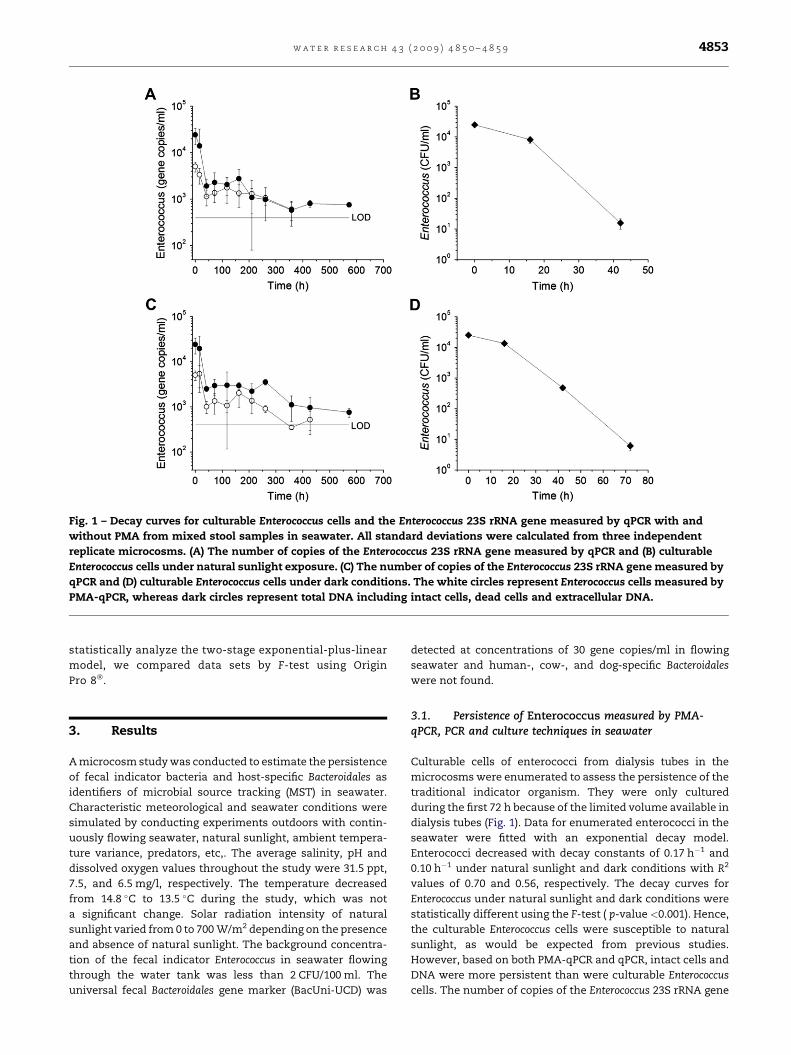
Fig. 1 – Decay curves for culturable Enterococcus cells and the Enterococcus 23S rRNA gene measured by qPCR with and
without PMA from mixed stool samples in seawater. All standard deviations were calculated from three independent
replicate microcosms. (A) The number of copies of the Enterococcus 23S rRNA gene measured by qPCR and (B) culturable
Enterococcus cells under natural sunlight exposure. (C) The number of copies of the Enterococcus 23S rRNA gene measured by
qPCR and (D) culturable Enterococcus cells under dark conditions. The white circles represent Enterococcus cells measured by
PMA-qPCR, whereas dark circles represent total DNA including intact cells, dead cells and extracellular DNA.
w a t e r r e s e a r c h 4 3 ( 2 0 0 9 ) 4 8 5 0 – 4 8 5 9 4853
statistically analyze the two-stage exponential-plus-linear
model, we compared data sets by F-test using Origin
Pro 8�.
3. Results
A microcosm study was conducted to estimate the persistence
of fecal indicator bacteria and host-specific Bacteroidales as
identifiers of microbial source tracking (MST) in seawater.
Characteristic meteorological and seawater conditions were
simulated by conducting experiments outdoors with contin-
uously flowing seawater, natural sunlight, ambient tempera-
ture variance, predators, etc,. The average salinity, pH and
dissolved oxygen values throughout the study were 31.5 ppt,
7.5, and 6.5 mg/l, respectively. The temperature decreased
from 14.8 �C to 13.5 �C during the study, which was not
a significant change. Solar radiation intensity of natural
sunlight varied from 0 to 700 W/m2 depending on the presence
and absence of natural sunlight. The background concentra-
tion of the fecal indicator Enterococcus in seawater flowing
through the water tank was less than 2 CFU/100 ml. The
universal fecal Bacteroidales gene marker (BacUni-UCD) was
detected at concentrations of 30 gene copies/ml in flowing
seawater and human-, cow-, and dog-specific Bacteroidales
were not found.
3.1. Persistence of Enterococcus measured by PMA-qPCR, PCR and culture techniques in seawater
Culturable cells of enterococci from dialysis tubes in the
microcosms were enumerated to assess the persistence of the
traditional indicator organism. They were only cultured
during the first 72 h because of the limited volume available in
dialysis tubes (Fig. 1). Data for enumerated enterococci in the
seawater were fitted with an exponential decay model.
Enterococci decreased with decay constants of 0.17 h�1 and
0.10 h�1 under natural sunlight and dark conditions with R2
values of 0.70 and 0.56, respectively. The decay curves for
Enterococcus under natural sunlight and dark conditions were
statistically different using the F-test ( p-value <0.001). Hence,
the culturable Enterococcus cells were susceptible to natural
sunlight, as would be expected from previous studies.
However, based on both PMA-qPCR and qPCR, intact cells and
DNA were more persistent than were culturable Enterococcus
cells. The number of copies of the Enterococcus 23S rRNA gene
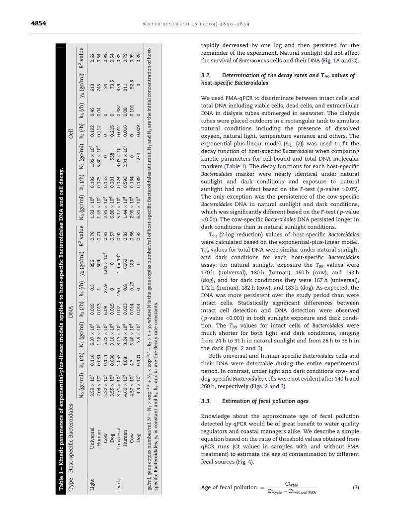
Ta
ble
1–
Kin
eti
cp
ara
mete
rso
fex
po
nen
tia
l-p
lus-
lin
ea
rm
od
els
ap
pli
ed
toh
ost
-sp
eci
fic
Ba
ctero
ida
les
DN
Aa
nd
cell
deca
y.
Ty
pe
Ho
st-s
peci
fic
Ba
ctero
ida
les
DN
AC
ell
N0
(gc/
ml)
k 1(/
h)
N1
(gc/
ml)
k 2(/
h)
k 3(/
h)
y 0(g
c/m
l)R
2v
alu
eN
0(g
c/m
l)k 1
(/h
)N
1(g
c/m
l)k 2
(/h
)k 3
(/h
)y 0
(gc/
ml)
R2
va
lue
Lig
ht
Un
ivers
al
3.5
9�
10
70.1
16
5.3
7�
10
60.0
15
0.5
856
0.7
61.9
2�
10
60.1
92
1.9
2�
10
60.1
92
0.4
5613
0.6
2
Hu
ma
n7.0
4�
10
60.0
81
1.1
8�
10
60.0
13
1609
0.7
11.4
5�
10
60.1
75
5.8
0�
10
40.2
12
0.0
4745
0.6
4
Co
w5.2
2�
10
50.1
11
5.2
2�
10
46.0
927.9
1.0
2�
10
40.9
32.9
5�
10
40.1
53
00
034
0.9
9
Do
g3.5
5�
10
50.0
98
9.1
0�
10
40.0
15
00
0.5
76.8
0�
10
40.2
01
538
0.2
15
073.5
0.5
4
Da
rkU
niv
ers
al
3.7
1�
10
72.0
55
8.1
3�
10
60.0
2250
1.9�
10
50.9
25.1
7�
10
60.1
24
9.0
3�
10
30.0
12
0.4
87
379
0.8
5
Hu
ma
n4.6
2�
10
61.0
83.2
4�
10
60.0
23
0.8
6062
0.8
21.4
4�
10
60.5
93
2.3
1�
10
40.0
16
0.0
8211
0.7
9
Co
w4.5
7�
10
54.7
6.4
0�
10
40.0
14
0.2
9183
0.8
62.9
5�
10
40.1
84
00
0.1
01
52.8
0.9
9
Do
g4.4�
10
50.1
01
5.9�
10
40.0
14
00
0.9
26.8
1�
10
40.1
89
273
0.0
09
00
0.8
9
gc/
ml,
gen
eco
pie
sn
um
ber/
ml.
N¼
N1�
ex
p�
k 1tþ
N2�
ex
p�
k 2t�
k 3�
tþ
y 0w
here
Nis
the
gen
eco
pie
sn
um
ber/
mlo
fh
ost
-sp
eci
fic
Ba
ctero
ida
les
at
tim
et,
N1
an
dN
2a
reth
ein
itia
lco
nce
ntr
ati
on
of
ho
st-
speci
fic
Ba
ctero
ida
les,
y 0is
con
sta
nt
an
dk 1
,k 2
,a
nd
k 3a
reth
ed
eca
yra
teco
nst
an
ts.
w a t e r r e s e a r c h 4 3 ( 2 0 0 9 ) 4 8 5 0 – 4 8 5 94854
rapidly decreased by one log and then persisted for the
remainder of the experiment. Natural sunlight did not affect
the survival of Enterococcus cells and their DNA (Fig. 1A and C).
3.2. Determination of the decay rates and T99 values ofhost-specific Bacteroidales
We used PMA-qPCR to discriminate between intact cells and
total DNA including viable cells, dead cells, and extracellular
DNA in dialysis tubes submerged in seawater. The dialysis
tubes were placed outdoors in a rectangular tank to simulate
natural conditions including the presence of dissolved
oxygen, natural light, temperature variance and others. The
exponential-plus-linear model (Eq. (2)) was used to fit the
decay function of host-specific Bacteroidales when comparing
kinetic parameters for cell-bound and total DNA molecular
markers (Table 1). The decay functions for each host-specific
Bacteroidales marker were nearly identical under natural
sunlight and dark conditions and exposure to natural
sunlight had no effect based on the F-test ( p-value >0.05).
The only exception was the persistence of the cow-specific
Bacteroidales DNA in natural sunlight and dark conditions,
which was significantly different based on the F-test ( p-value
<0.01). The cow-specific Bacteroidales DNA persisted longer in
dark conditions than in natural sunlight conditions.
T99 (2-log reduction) values of host-specific Bacteroidales
were calculated based on the exponential-plus-linear model.
T99 values for total DNA were similar under natural sunlight
and dark conditions for each host-specific Bacteroidales
assay: for natural sunlight exposure the T99 values were
170 h (universal), 180 h (human), 160 h (cow), and 193 h
(dog), and for dark conditions they were 167 h (universal),
172 h (human), 182 h (cow), and 183 h (dog). As expected, the
DNA was more persistent over the study period than were
intact cells. Statistically significant differences between
intact cell detection and DNA detection were observed
( p-value <0.001) in both sunlight exposure and dark condi-
tion. The T99 values for intact cells of Bacteroidales were
much shorter for both light and dark conditions, ranging
from 24 h to 31 h in natural sunlight and from 26 h to 38 h in
the dark (Figs. 2 and 3).
Both universal and human-specific Bacteroidales cells and
their DNA were detectable during the entire experimental
period. In contrast, under light and dark conditions cow- and
dog-specific Bacteroidales cells were not evident after 140 h and
260 h, respectively (Figs. 2 and 3).
3.3. Estimation of fecal pollution ages
Knowledge about the approximate age of fecal pollution
detected by qPCR would be of great benefit to water quality
regulators and coastal managers alike. We describe a simple
equation based on the ratio of threshold values obtained from
qPCR runs (Ct values in samples with and without PMA
treatment) to estimate the age of contamination by different
fecal sources (Fig. 4).
Age of fecal pollution ¼ CtPMA
Ctcycle � Ctwithout PMA(3)
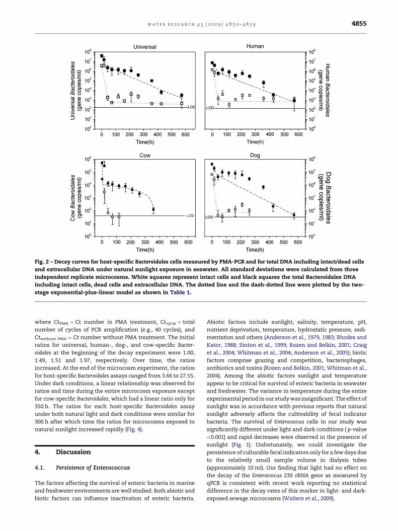
Fig. 2 – Decay curves for host-specific Bacteroidales cells measured by PMA-PCR and for total DNA including intact/dead cells
and extracellular DNA under natural sunlight exposure in seawater. All standard deviations were calculated from three
independent replicate microcosms. White squares represent intact cells and black squares the total Bacteroidales DNA
including intact cells, dead cells and extracellular DNA. The dotted line and the dash-dotted line were plotted by the two-
stage exponential-plus-linear model as shown in Table 1.
w a t e r r e s e a r c h 4 3 ( 2 0 0 9 ) 4 8 5 0 – 4 8 5 9 4855
where CtPMA ¼ Ct number in PMA treatment, Ctcycle ¼ total
number of cycles of PCR amplification (e.g., 40 cycles), and
Ctwithout PMA ¼ Ct number without PMA treatment. The initial
ratios for universal, human-, dog-, and cow-specific Bacter-
oidales at the beginning of the decay experiment were 1.00,
1.49, 1.51 and 1.97, respectively. Over time, the ratios
increased. At the end of the microcosm experiment, the ratios
for host-specific Bacteroidales assays ranged from 3.66 to 27.55.
Under dark conditions, a linear relationship was observed for
ratios and time during the entire microcosm exposure except
for cow-specific Bacteroidales, which had a linear ratio only for
350 h. The ratios for each host-specific Bacteroidales assay
under both natural light and dark conditions were similar for
300 h after which time the ratios for microcosms exposed to
natural sunlight increased rapidly (Fig. 4).
4. Discussion
4.1. Persistence of Enterococcus
The factors affecting the survival of enteric bacteria in marine
and freshwater environments are well studied. Both abiotic and
biotic factors can influence inactivation of enteric bacteria.
Abiotic factors include sunlight, salinity, temperature, pH,
nutrient deprivation, temperature, hydrostatic pressure, sedi-
mentation and others (Anderson et al., 1979, 1983; Rhodes and
Kator, 1988; Sinton et al., 1999; Rozen and Belkin, 2001; Craig
et al., 2004; Whitman et al., 2004; Anderson et al., 2005); biotic
factors comprise grazing and competition, bacteriophages,
antibiotics and toxins (Rozen and Belkin, 2001; Whitman et al.,
2004). Among the abiotic factors sunlight and temperature
appear to be critical for survival of enteric bacteria in seawater
and freshwater. The variance in temperature during the entire
experimental period in our study was insignificant. The effect of
sunlight was in accordance with previous reports that natural
sunlight adversely affects the cultivability of fecal indicator
bacteria. The survival of Enterococcus cells in our study was
significantly different under light and dark conditions ( p-value
<0.001) and rapid decreases were observed in the presence of
sunlight (Fig. 1). Unfortunately, we could investigate the
persistence of culturable fecal indicators only for a few days due
to the relatively small sample volume in dialysis tubes
(approximately 10 ml). Our finding that light had no effect on
the decay of the Enterococcus 23S rRNA gene as measured by
qPCR is consistent with recent work reporting no statistical
difference in the decay rates of this marker in light- and dark-
exposed sewage microcosms (Walters et al., 2009).
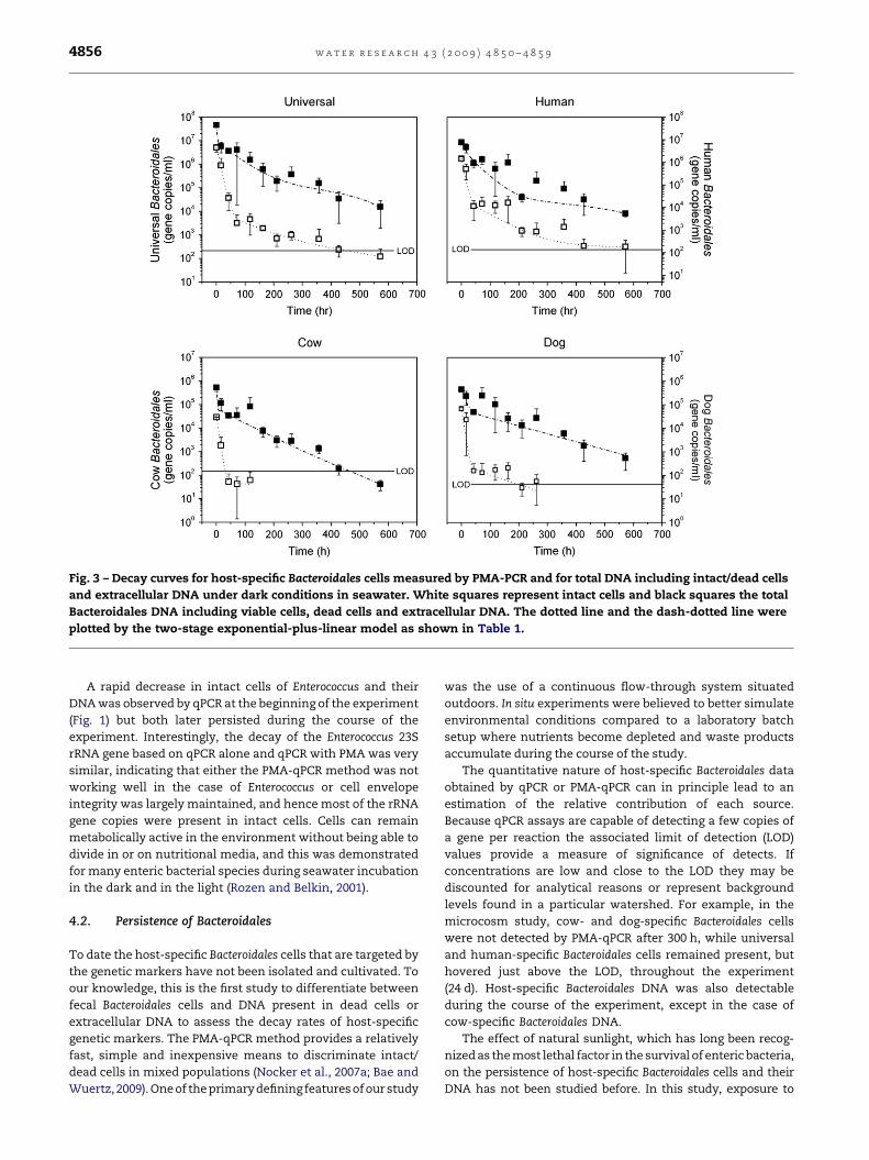
Fig. 3 – Decay curves for host-specific Bacteroidales cells measured by PMA-PCR and for total DNA including intact/dead cells
and extracellular DNA under dark conditions in seawater. White squares represent intact cells and black squares the total
Bacteroidales DNA including viable cells, dead cells and extracellular DNA. The dotted line and the dash-dotted line were
plotted by the two-stage exponential-plus-linear model as shown in Table 1.
w a t e r r e s e a r c h 4 3 ( 2 0 0 9 ) 4 8 5 0 – 4 8 5 94856
A rapid decrease in intact cells of Enterococcus and their
DNA was observed by qPCR at the beginning of the experiment
(Fig. 1) but both later persisted during the course of the
experiment. Interestingly, the decay of the Enterococcus 23S
rRNA gene based on qPCR alone and qPCR with PMA was very
similar, indicating that either the PMA-qPCR method was not
working well in the case of Enterococcus or cell envelope
integrity was largely maintained, and hence most of the rRNA
gene copies were present in intact cells. Cells can remain
metabolically active in the environment without being able to
divide in or on nutritional media, and this was demonstrated
for many enteric bacterial species during seawater incubation
in the dark and in the light (Rozen and Belkin, 2001).
4.2. Persistence of Bacteroidales
To date the host-specific Bacteroidales cells that are targeted by
the genetic markers have not been isolated and cultivated. To
our knowledge, this is the first study to differentiate between
fecal Bacteroidales cells and DNA present in dead cells or
extracellular DNA to assess the decay rates of host-specific
genetic markers. The PMA-qPCR method provides a relatively
fast, simple and inexpensive means to discriminate intact/
dead cells in mixed populations (Nocker et al., 2007a; Bae and
Wuertz,2009). One of the primary defining features of our study
was the use of a continuous flow-through system situated
outdoors. In situ experiments were believed to better simulate
environmental conditions compared to a laboratory batch
setup where nutrients become depleted and waste products
accumulate during the course of the study.
The quantitative nature of host-specific Bacteroidales data
obtained by qPCR or PMA-qPCR can in principle lead to an
estimation of the relative contribution of each source.
Because qPCR assays are capable of detecting a few copies of
a gene per reaction the associated limit of detection (LOD)
values provide a measure of significance of detects. If
concentrations are low and close to the LOD they may be
discounted for analytical reasons or represent background
levels found in a particular watershed. For example, in the
microcosm study, cow- and dog-specific Bacteroidales cells
were not detected by PMA-qPCR after 300 h, while universal
and human-specific Bacteroidales cells remained present, but
hovered just above the LOD, throughout the experiment
(24 d). Host-specific Bacteroidales DNA was also detectable
during the course of the experiment, except in the case of
cow-specific Bacteroidales DNA.
The effect of natural sunlight, which has long been recog-
nized as the most lethal factor in the survival of enteric bacteria,
on the persistence of host-specific Bacteroidales cells and their
DNA has not been studied before. In this study, exposure to
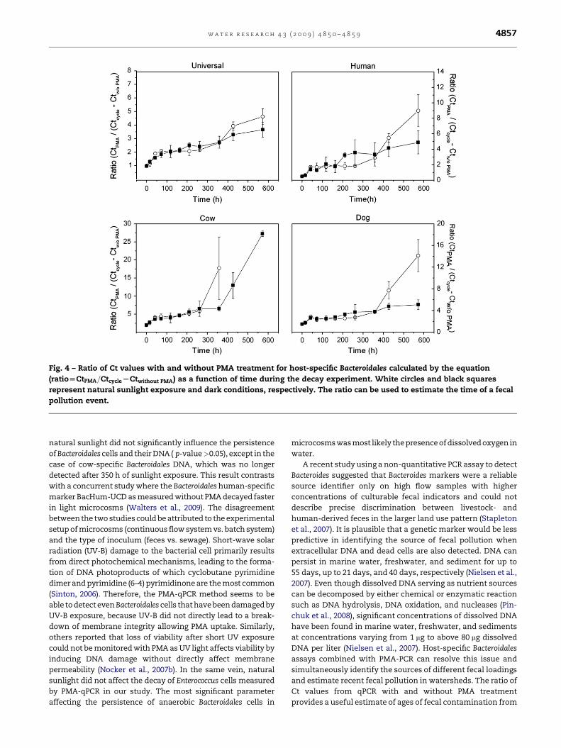
Fig. 4 – Ratio of Ct values with and without PMA treatment for host-specific Bacteroidales calculated by the equation
(ratio[CtPMA=CtcycleLCtwithout PMA) as a function of time during the decay experiment. White circles and black squares
represent natural sunlight exposure and dark conditions, respectively. The ratio can be used to estimate the time of a fecal
pollution event.
w a t e r r e s e a r c h 4 3 ( 2 0 0 9 ) 4 8 5 0 – 4 8 5 9 4857
natural sunlight did not significantly influence the persistence
of Bacteroidales cells and their DNA ( p-value>0.05), except in the
case of cow-specific Bacteroidales DNA, which was no longer
detected after 350 h of sunlight exposure. This result contrasts
with a concurrent study where the Bacteroidales human-specific
marker BacHum-UCD as measured without PMA decayed faster
in light microcosms (Walters et al., 2009). The disagreement
between the two studies could be attributed to the experimental
setup of microcosms (continuous flow system vs. batch system)
and the type of inoculum (feces vs. sewage). Short-wave solar
radiation (UV-B) damage to the bacterial cell primarily results
from direct photochemical mechanisms, leading to the forma-
tion of DNA photoproducts of which cyclobutane pyrimidine
dimer and pyrimidine (6–4) pyrimidinone are the most common
(Sinton, 2006). Therefore, the PMA-qPCR method seems to be
able to detect even Bacteroidales cells that have been damaged by
UV-B exposure, because UV-B did not directly lead to a break-
down of membrane integrity allowing PMA uptake. Similarly,
others reported that loss of viability after short UV exposure
could not be monitored with PMA as UV light affects viability by
inducing DNA damage without directly affect membrane
permeability (Nocker et al., 2007b). In the same vein, natural
sunlight did not affect the decay of Enterococcus cells measured
by PMA-qPCR in our study. The most significant parameter
affecting the persistence of anaerobic Bacteroidales cells in
microcosms was most likely the presence of dissolved oxygen in
water.
A recent study using a non-quantitative PCR assay to detect
Bacteroides suggested that Bacteroides markers were a reliable
source identifier only on high flow samples with higher
concentrations of culturable fecal indicators and could not
describe precise discrimination between livestock- and
human-derived feces in the larger land use pattern (Stapleton
et al., 2007). It is plausible that a genetic marker would be less
predictive in identifying the source of fecal pollution when
extracellular DNA and dead cells are also detected. DNA can
persist in marine water, freshwater, and sediment for up to
55 days, up to 21 days, and 40 days, respectively (Nielsen et al.,
2007). Even though dissolved DNA serving as nutrient sources
can be decomposed by either chemical or enzymatic reaction
such as DNA hydrolysis, DNA oxidation, and nucleases (Pin-
chuk et al., 2008), significant concentrations of dissolved DNA
have been found in marine water, freshwater, and sediments
at concentrations varying from 1 mg to above 80 mg dissolved
DNA per liter (Nielsen et al., 2007). Host-specific Bacteroidales
assays combined with PMA-PCR can resolve this issue and
simultaneously identify the sources of different fecal loadings
and estimate recent fecal pollution in watersheds. The ratio of
Ct values from qPCR with and without PMA treatment
provides a useful estimate of ages of fecal contamination from
w a t e r r e s e a r c h 4 3 ( 2 0 0 9 ) 4 8 5 0 – 4 8 5 94858
different animal sources based on the persistence of Bacter-
oidales genetic markers.
4.3. Modeling Bacteroidales decay
Understanding host-specific Bacteroidales decay rates is vital for
predicting their environmental persistence. We employed
exponential-plus-linear models to fit the experimental data and
calculated the decay rates of host-specific Bacteroidales. The
survival of culturable indicators such as E. coli and Enterococcus is
generally represented as a first-order decay function, asgiven by
Chick’s Law in water (Gonzalez, 1995; Pachepsky et al., 2006).
However, this exponential model may not fit well with experi-
mental data possessing random variations of field experiments.
Therefore, it was suggested that bacterial inactivation curves
are usually characterized by two or more phases, showing
‘‘shoulder’’ behavior (slow removal/die-off or perhaps growth
early in the experiment followed by apparent first-order decay
(Whitman et al., 2004). The third phase is an apparent period of
‘‘recovery’’ or ‘‘stability’’, which may or may not be quantified.
Usually, the decay rates are calculated within only certain
exponential periods and the shoulder phase or the third phase
in the experiment is ignored. Others have proposed that the
modelshouldcontain at least threeparameters, which is known
as the two-stage decay model. A realistic die-off model should
account for multiple stressors from abiotic or biotic factors and
even regrowth in the early stage; hence the linear portion is
explained by the uncertainty of the model due to experimental
errors or other marginal factors (Crane and Moore, 1986;
Pachepsky et al., 2006). At the molecular level microorganisms
do not follow a simple decay model, because various lethal
factors simultaneously affect their viability and the population
may separate into two subpopulations, namely, a rapid and
a slow decrease population. Therefore, the two-stage expo-
nential-plus-linear model provides more mechanistic insights
into the decay of Bacteroidales in water. The ‘‘shoulder’’ behavior
observed was not due to regrowth given that the prevailing
oxygen concentration of 6.5 mg l�1 would inhibit cell division,
but probably reflects the combined effects of predator grazing,
release of free DNA by cell lysis, desorption of DNA from sus-
pended solids, and other environmental variables.
Several questions have yet to be addressed. Sediment
resuspension represents a significant source of bacteria to the
water column (Fries et al., 2008). In the same way, Bacteroidales
might survive in anaerobic sediments for prolonged periods of
time or the DNA of Bacteroidales may persist while sorbed to
sediment. If true, the resuspension of sediments would
interfere in the analysis of microbial source tracking data.
Further, it is essential to determine the relationship between
the detection of a source identifier like Bacteroidales and the
presence of fecal pathogens as well as the incidence of illness
associated with recreational activities in impacted waters.
5. Conclusions
� The PMA-qPCR method to quantify host-specific Bacter-
oidales cells can detect a recent fecal contamination event
and significantly enhances the usefulness of source tracking
data obtained from watersheds experiencing inputs from
multiple fecal sources. The information is highly relevant
for the development of water quality criteria and traditional
FIB methods are unsuited to this purpose.
� Host-specific Bacteroidales cells had an average two-log
reduction time in seawater of 28 h whereas that of host-
specific Bacteroidales DNA was 177 h.
� Host-specific Bacteroidales showed similar decay rates in
seawater. Natural sunlight did not affect the survival of
uncultivated Bacteroidales cells and their DNA, with the
exception of cow-specific Bacteroidales DNA, which persisted
longer in the dark. A likely explanation in that the presence of
dissolved oxygen acts as principal factor in the decay of cells.
� The ratio of gene copies or Ct values calculated using qPCR
with and without PMA treatment can be used to estimate
the age of fecal pollution originating from different sources.
In an absolute sense, only one discrete source of host-
specific fecal contamination ought to be present when
applying the ratio. In many scenarios it is reasonable to
assume that there has been one type of major input of fecal
contamination, such as in form of runoff after a storm
event. The ratio can also serve to pinpoint human waste-
water inputs from sources not representing publicly owned
treatment works, based on the fact that treated and dis-
infected wastewater would have hardly any intact Bacter-
oidales cells. Hence, if a low ratio is detected for the human-
specific assay, the implication is that there are either septic
tank leakages or other recent human inputs.
� Decay rate constants for each host-specific Bacteroidales
genetic markerwerecalculatedfromatwo-stageexponential-
plus-linear model. These constants can provide valuable
information to establish fate and transport models of host-
specific Bacteroidales and to understand the different survival
stages in seawater.
� A rapid decrease in intact cells of Enterococcus and their DNA
was observed by qPCR at the beginning of the microscosm
decay experiment. However, the 23S rRNA gene remained
detectable throughout the study by both qPCR and qPCR with
PMA, necessitating further studies before this assay can be
used to inform and enforce new water quality criteria.
r e f e r e n c e s
Anderson, I.C., Rhodes, M., Kator, H., 1979. Sublethal stress inEscherichia coli: a function of salinity. Appl. Environ. Microbiol.38 (6), 1147–1152.
Anderson, I.C., Rhodes, M.W., Kator, H.I., 1983. Seasonal variationin survival of Escherichia coli exposed in situ in membranediffusion chambers containing filtered and nonfilteredestuarine water. Appl. Environ. Microbiol. 45 (6), 1877–1883.
Anderson, K.L., Whitlock, J.E., Harwood, V.J., 2005. Persistenceand differential survival of fecal indicator bacteria insubtropical waters and sediments. Appl. Environ. Microbiol. 71(6), 3041–3048.
Bae, S., Wuertz, S., 2009. Discrimination of viable and dead fecalBacteroidales bacteria by quantitative PCR with propidiummonoazide. Appl. Environ. Microbiol. 75, 2940–2944.
Bell, A., Layton, A.C., McKay, L., Williams, D., Gentry, D., Sayler, D.,2009. Factors influencing the persistence of fecal Bacteroides instream water. J. Environ. Qual. 38, 1224–1232.
Craig, D.L., Fallowfield, H.J., Cromar, N.J., 2004. Use of microcosmsto determine persistence of Escherichia coli in recreational
w a t e r r e s e a r c h 4 3 ( 2 0 0 9 ) 4 8 5 0 – 4 8 5 9 4859
coastal water and sediment and validation with in situmeasurements. J. Appl. Microbiol. 96 (5), 922–930.
Crane, S.R., Moore, J.A., 1986. Modeling enteric bacterial die-off –a review. Water, Air, Soil Pollut. 27 (3–4), 411–439.
Dick, L.K., Bernhard, A.E., Brodeur, T.J., Santo Domingo, J.W.,Simpson, J.M., Walters, S.P., Field, K.G., 2005. Hostdistributions of uncultivated fecal Bacteroidales bacteriareveal genetic markers for fecal source identification. Appl.Environ. Microbiol. 71 (6), 3184–3191.
Field, K.G., Samadpour, M., 2007. Fecal source tracking, theindicator paradigm, and managing water quality. Water Res.41 (16), 3517–3538.
Fries, J.S., Characklis, G.W., Noble, R.T., 2008. Sediment-waterexchange of Vibrio sp., fecal indicator bacteria: implicationsfor persistence and transport in the Neuse River Estuary,North Carolina, USA. Water Res. 42 (4–5), 941–950.
Gonzalez, J.M., 1995. Modelling enteric bacteria survival in aquaticsystems. Hydrobiologia 316 (2), 109–116.
Haugland, R.A., Siefring, S.C., Wymer, L.J., Brenner, K.P.,Dufour, A.P., 2005. Comparison of Enterococcus measurementsin freshwater at two recreational beaches by quantitativepolymerase chain reaction and membrane filter cultureanalysis. Water Res. 39 (4), 559–568.
Kildare, B.J., Leutenegger, C.M., McSwain, B.S., Bambic, D.G.,Rajal, V.B., Wuertz, S., 2007. 16S rRNA-based assays forquantitative detection of universal, human-, cow-, and dog-specific fecal Bacteroidales: a Bayesian approach. Water Res.41 (16), 3701–3715.
Kreader, C.A., 1998. Persistence of PCR-detectable Bacteroidesdistasonis from human feces in river water. Appl. Environ.Microbiol. 64 (10), 4103–4105.
Layton, A., McKay, L., Williams, D., Garrett, V., Gentry, R.,Sayler, G., 2006. Development of Bacteroides 16S rRNA geneTaqMan-based real-time PCR assays for estimation of total,human, and bovine fecal pollution in water. Appl. Environ.Microbiol. 72 (6), 4214–4224.
Nielsen, K.M., Johnsen, P.J., Bensasson, D., Daffonchio, D., 2007.Release and persistence of extracellular DNA in theenvironment. Environ. Biosafety Res. 6 (1–2), 37–53.
Nocker, A., Cheung, C.Y., Camper, A.K., 2006. Comparison ofpropidium monoazide with ethidium monoazide fordifferentiation of live vs. dead bacteria by selective removal ofDNA from dead cells. J. Microbiol. Methods 67 (2), 310–320.
Nocker, A., Sossa-Fernandez, P., Burr, M.D., Camper, A.K., 2007a.Use of propidium monoazide for live/dead distinction inmicrobial ecology. Appl. Environ. Microbiol. 73 (16), 5111–5117.
Nocker, A., Sossa, K.E., Camper, A.K., 2007b. Molecular monitoring ofdisinfectionefficacy using propidium monoazide incombinationwith quantitative PCR. J. Microbiol. Methods 70 (2), 252–260.
Okabe, S., Shimazu, Y., 2007. Persistence of host-specificBacteroides-Prevotella 16S rRNA genetic markers inenvironmental waters: effects of temperature and salinity.Appl. Microbiol. Biotechnol. 76 (4), 935–944.
Pachepsky, Y.A., Sadeghi, A.M., Bradford, S.A., Shelton, D.R.,Guber, A.K., Dao, T., 2006. Transport and fate of manure-bornepathogens: modeling perspective. Agric. Water Manage 86(1–2), 81–92.
Pinchuk, G.E., Ammons, C., Culley, D.E., Li, S.M.W., McLean, J.S.,Romine, M.F., Nealson, K.H., Fredrickson, J.K., Beliaev, A.S.,2008. Utilization of DNA as a sole source of phosphorus,carbon, and energy by Shewanella spp.: ecological andphysiological implications for dissimilatory metal reduction.Appl. Environ. Microbiol. 74 (4), 1198–1208.
Reischer, G.H., Kasper, D.C., Steinborn, R., Mach, R.L.,Farnleitner, A.H., 2006. Quantitative PCR method for sensitivedetection of ruminant fecal pollution in freshwater andevaluation of this method in alpine karstic regions. Appl.Environ. Microbiol. 72 (8), 5610–5614.
Rhodes, M.W., Kator, H., 1988. Survival of Escherichia coli andSalmonella spp. in estuarine environments. Appl. Environ.Microbiol. 54 (12), 2902–2907.
Rozen, Y., Belkin, S., 2001. Survival of enteric bacteria in seawater.FEMS Microbiol. Rev. 25 (5), 513–529.
Santo Domingo, J.W., Bambic, D.G., Edge, T.A., Wuertz, S., 2007.Quo vadis source tracking? Towards a strategic framework forenvironmental monitoring of fecal pollution. Water Res. 41(16), 3539–3552.
Savichtcheva, O., Okabe, S., 2006. Alternative indicators of fecalpollution: relations with pathogens and conventionalindicators, current methodologies for direct pathogenmonitoring and future application perspectives. Water Res. 40(13), 2463–2476.
Seurinck, S., Defoirdt, T., Verstraete, W., Siciliano, S.D., 2005.Detection and quantification of the human-specific HF183Bacteroides 16S rRNA genetic marker with real-time PCR forassessment of human faecal pollution in freshwater. Environ.Microbiol. 7 (2), 249–259.
Sinton, L.W., 2006. Biotic and abiotic effects. In: Belkin, S.,Colwell, R.R. (Eds.), Oceans and Health: Pathogen in the MarineEnvironment. Springer ScienceþBusiness Media, New York,pp. xiv, 464 pp.
Sinton, L.W., Finlay, R.K., Lynch, P.A., 1999. Sunlight inactivationof fecal bacteriophages and bacteria in sewage-pollutedseawater. Appl. Environ. Microbiol. 65 (8), 3605–3613.
Stapleton, C.M., Wyer, M.D., Kay, D., Crowther, J., McDonald, A.T.,Walters, M., Gawler, A., Hindle, T., 2007. Microbial sourcetracking: a forensic technique for microbial sourceidentification? J. Environ. Monit. 9 (5), 427–439.
USEPA, 2002. Method 1600: Enterococci in Water by MembraneFiltration Using Membrane- Enterococcus Indoxyl-b-D-Glucoside Agar (mEI). Office of Water, Washington, DC (EPA821-R-02-022).
Walters, S.P., Field, K.G., 2006. Persistence and growth of fecalBacteroidales assessed by bromodeoxyuridineimmunocapture. Appl. Environ. Microbiol. 72 (7), 4532–4539.
Walters, S.P., Yamahara, K.M., Boehm, A.B., 2009. Persistenceof nucleic acid markers of health-relevant organisms inseawater microcosms: implication for their use inassessing risk in recreational waters. Water Res. 43 (19),4929–4939.
Whitman, R.L., Nevers, M.B., Korinek, G.C., Byappanahalli, M.N.,2004. Solar and temporal effects on Escherichia coliconcentration at a Lake Michigan swimming beach. Appl.Environ. Microbiol. 70 (7), 4276–4285.
Copyright © 2022 FDOKUMEN