Radiodiagnosis Training Program Curriculum Version 2
241
Radiodiagnosis Training Program Curriculum Version 2 The Royal Australian and New Zealand College of Radiologists ® ®
-
Upload
khangminh22 -
Category
Documents
-
view
0 -
download
0
Transcript of Radiodiagnosis Training Program Curriculum Version 2

Radiodiagnosis Training Program
CurriculumVersion 2
The Royal Australian and New Zealand
College of Radiologists®
®

Radiodiagnosis Training Program
The Royal Australian and New Zealand College of Radiologists®
Level 9, 51 Druitt Street
Sydney, NSW, 2000
Australia
Telephone: +61 2 9268 9777
Fax: +61 2 9268 9799
Website: http://www.ranzcr.com
Copyright © 2014 The Royal Australian and New Zealand College of Radiologists®. All rights
reserved. This material may only be reproduced with the written permission of the RANZCR®.
Version History Summary of Amendments
Version 2.1 published October 2015 Inclusion of CanMEDS mapping table; p.20Inclusion of ETRs for BMD, CTCA & CTC; pp.77-79
Version 2.2 published October 2017 Removal of Radiology Adverse Events Register; pp.27, 28, 30Clarification of reporting requirements for ETRs; pp. 61-79RANZCR website links updated
How to reference this document:
The Royal Australian and New Zealand College of Radiologists®. 2014. Radiodiagnosis Training
Program Curriculum. Sydney: The Royal Australian and New Zealand College of Radiologists®.

Page 3© 2014 RANZCR. Radiodiagnosis Training Program – Curriculum Version 2.2
TABLE OF CONTENTS
• Foreword 5• Contributors to the Radiodiagnosis Curriculum and Training Program – Curriculum Version 2 6• Important Information for Trainees 10• Key Educational Principles 11• Training Program Structure & Assessments 13• Glossary of Learning Verbs 22• Patient Safety Syllabus 23 ¡ Patient Safety Cycle 23 ¡ Cultural Competence 24 ¡ Doctor-Patient Referral 24 ¡ Review of Request 24 ¡ Patient Preparation 25 ¡ Performance of An Examination 26 ¡ Management of Adverse Events – Open Disclosure 27 ¡ Review of Images/Quality Control Workup 27 ¡ Analysis, Verification and Interpretation (Clinical Reasoning) 27 ¡ Radiologist Report Writing & Communication of Results 28 ¡ The Role of Incident Monitoring 28 ¡ Contrast Media 28 ¡ Sedation 29 ¡ References Used in the Development of the Patient Safety Cycle 30 ¡ Report Writing Module 30 ¡ Key Elements of a Radiology Reporting Template 33 ¡ Mandatory In-Training Assessment for Patient Safety and Report Writing in Year 1 of Training 34 ¡ Lesion Choice and Measurement 36 ¡ Frequently Asked Questions 39 ¡ References 40 ¡ Appendix A 41• Key Conditions in Year 1 of Training 45 ¡ List of Key Conditions in Early Training 46 ¡ Training Requirements – Management of Contrast Reactions 47 ¡ Tools for Training – Key Conditions in Early Training 47 ¡ Early Training Matrix Examples 48 ¡ Advice for Training Sites – Teaching First Year Trainees Key Conditions in Early Training 54• Experiential Training Requirements 61 ¡ General X-Ray Experiential Training Requirements 62 ¡ Breast Imaging Experiential Training Requirements 64 ¡ Interventional Radiology Experiential Training Requirements 66 ¡ Magnetic Resonance Imaging Experiential Training Requirements 68 ¡ Nuclear Medicine Experiential Training Requirements 70 ¡ Obstetric & Gynecology Experiential Training Requirements 73 ¡ Paediatric & Neonatal Experiential Training Requirements 75 ¡ Bone Mineral Densitometry 77 ¡ CT Coronary Angiography 78 ¡ CT Colonography 79• Anatomy Syllabus 81 ¡ Categorisation of Required Skills and Knowledge for Anatomical Structures 81 ¡ A Note on Normal Variants 81 ¡ Category 1 81 ¡ Category 2 82 ¡ Category 3 82 ¡ Anatomy of the Head & Face (Excluding CNS) 83 ¡ Anatomy of the Central Nervous System 88

Page 4 © 2014 RANZCR. Radiodiagnosis Training Program – Curriculum Version 2.2
¡ Anatomy of the Neck (Non-Spinal) 94 ¡ Anatomy of the Upper Limb 98 ¡ Anatomy of the Lower Limb 105 ¡ Anatomy of the Spine & Back 110 ¡ Anatomy of the Thorax 112 ¡ Anatomy of the Abdomen 114 ¡ Anatomy of the Pelvis 117• Applied Imaging Technology Syllabus 119 ¡ Applied Imaging Technology Category Definitions 119 ¡ Category 1 119 ¡ Category 2 119 ¡ Category 3 119 ¡ Basic Concepts of Electromagnetic Radiation (BCER) 120 ¡ Production of X-Rays 120 ¡ X-Ray Generators 121 ¡ Interactions between X-Rays and Matter of Relevance to Medical Imaging 121 ¡ Filters, Collimators & Grids 122 ¡ Basic Digital Imaging Concepts 122 ¡ Radiographic Image Acquisition 123 ¡ Fluoroscopic Image Acquisition 124 ¡ Measures of Radiographic & Fluoroscopic Image Quality 125 ¡ Mammography 126 ¡ Computed Tomography 127 ¡ Magnetic Resonance Imaging 129 ¡ Nuclear Medicine 131 ¡ Ultrasound Imaging 133 ¡ Electrical Safety In Medicine 135 ¡ Dosimetry and Radiation Biology 136 ¡ Radiation Protection 138 ¡ Quality Assurance for Diagnostic Imaging Equipment 139 ¡ Contrast Agents 140• Pathology 141 ¡ Introduction 141 ¡ Pathology Recommended Reading List 141 ¡ General Pathology 141 ¡ Blood Vessels 143 ¡ The Heart 143 ¡ Haematologic Disorders, Lymph Nodes, Spleen and Thymus 144 ¡ The Lung and Pleura 145 ¡ Head and Neck 146 ¡ The Gastrointestinal Tract 147 ¡ LIver, Biliary Tract and Exorcrine Pancreas 148 ¡ The Kidney and Urinary Tract 149 ¡ Male Genital System 150 ¡ Female Genital System 151 ¡ The Breast 152 ¡ The Endocrine System 153 ¡ Skin 154 ¡ Bones 155 ¡ Joints 156 ¡ Soft Tissue 157 ¡ The Central and Peripheral Nervous System 158 ¡ The Eye 160• Body Systems Syllabus 154

Page 5© 2014 RANZCR. Radiodiagnosis Training Program – Curriculum Version 2.2
Foreword
Welcome to the second edition of the Radiodiagnosis
Training Program Curriculum. No iteration of the
curriculum represents a final complete statement, and we
are mindful that regular updates are required.
This document refines and updates the previous “new”
curriculum without altering the goals outlined in the first
edition. The basic underlying focus of this curriculum
remains to produce a general clinical radiologist, with
an understanding of not just the medical roles, but also
the non-medical roles. The first cohort of trainees in the
new curriculum, which sat the Part 2, had one of the most
successful pass rates in all components. The emphasis of
the “old” curriculum being exam centric is slowly being
diffused. In Phase 2, there are now systems focused
rotations, experiential training requirements and Project
2, to be now completed before being able to exit the
training program.
It is the content that has been updated, in particular
the experiential training requirements, after feedback
and consultation from departments and training sites.
Policies have been introduced to ensure that all trainees,
trainers and departments get a fair representation in the
education process. Examination processes have also been
adjusted to be more reliable, robust and valid. These
updates have been inserted in this new edition.
This is not a final document and the curriculum, will
continue to be regularly reviewed and updated
Finally, it must be emphasized that the trainees, as adult
learners, carry the primary responsibility that training
assessments, experiential requirements and appropriate
documentation are in order, and submitted in a timely
fashion. This will hopefully be more streamlined with the
introduction of Trainee Information Management System.
Dr Gabriel Lau
Chief Censor
Royal Australian and New Zealand College of Radiologists

Page 6 © 2014 RANZCR. Radiodiagnosis Training Program – Curriculum Version 2.2
Dr Gabriel Lau Chief Censor
Professor Alan Coulthard Member
Dr Chee-Yan Hiew Member
Dr Yvonne Ho Member
Dr Sanjay Jeganathan Member
Dr Alexander Rhodes Member
Dr Kenneth Lau Member
Dr Russell Metcalfe Member
Dr Meredith Thomas Member
Dr Jules Catt Trainee Representative
Dr Jash Agraval Trainee Representative
Dr Gabriel Lau Chief Censor
Dr Greg Slater Dean
Dr Michelle Bennett Member
Dr Melissa Lea Member
Dr Sam McCormack Member
Dr Neil Caplin Member
Dr Mark Phillips Member
Dr Murali Guduguntla Member
Professor Mark Khangure Member
Dr Steve Alatakis Member
Dr Martin Marshall Member
Dr Rahul Lakshmaman Member
Dr Michael Lau
Curriculum and Assessment Committee Members (2014)
Clinical Radiology Education & Training Committee Members 2014
Pathology Reviewer
Contributors to the Radiodiagnosis Training Program Curriculum Version 2
The RANZCR Radiodiagnosis Training Program Curriculum represents a large body of work achieved as cooperative effort by a
large people within and outside the Faculty. This document will continue to be reviewed and updated in order for it to retain
relevance and to meet the goals for which it was designed.

Page 7© 2014 RANZCR. Radiodiagnosis Training Program – Curriculum Version 2.2
Training Program Assessment Committee MembersDr Tony Peduto NSW
Dr Ivor Berman VIC
Dr Mike Ditchfield VIC
Professor John Heggie VIC
Dr Chee-Chung Hiew NSW
Dr Andrew Holden NZ
Dr Sarah Kremer VIC
Dr Rob Loneragan NSW
Dr Vince Mercuri VIC
Assoc. Professor Ross O’Neil ACT
Professor Alexander Pitman VIC
Dr Tony Smith SA
Dr Barry Soans NSW
Dr John Troupis VIC
Professor Shih-Chang Wang NSW
Radiology Education Board MembersDr Michael Bynevelt WA
Dr Steve Chyrissidis SA
Professor Alan Coulthard QLD
Dr Ian Cowan NZ
Dr Tim Dunshea VIC
Dr Robert Fabiny VIC
Dr Robin Harle TAS
Dr Robert Heng TAS
Professor Oliver Hennessy VIC Former Chief Censor Radiology
Dr Rajeev Jyoti ACT
Dr Lizbeth Kenny QLD Past President
Professor Mark Khangure WA President
Dr David Lisle QLD
Dr Robert Loneragan NSW
Dr Clement McCormick WA Chief Accreditation Officer
Professor Richard Mendelson WA
Professor John Slavotinek SA
Dr Tom Snow QLD
Dr Michelle Thong VIC Trainee Representative
Dr Greg van Schie WA
Professor Shih-Chang Wang NSW
Dr Patrick Ziesing SA

Page 8 © 2014 RANZCR. Radiodiagnosis Training Program – Curriculum Version 2.2
Anatomy ReviewersDr Chris Briggs VIC
Dr Deborah Bryce NSW
Professor Timothy Buckenham NZ
Dr Norman Eizenberg VIC
Professor Alexander Pitman VIC Head Anatomy Examiner
Dr Dzung Vu NSW
Professor Shih-Chang Wang NSW
Applied Imaging Technology ReviewersAssoc. Professor Lee Collins NSW
CMS John Cormack SA
Dr John Heggie VIC Head AIT Examiner
Dr Kieran Maher VIC
Patient Safety ReviewersDr D. Neil Jones SA
Dr Carmel Crock VIC
Assoc. Professor Brendan Flanagan VIC
Dr Julia Harrison VIC
Dr Catherine Mandel VIC
Radiodiagnosis Body System ReviewersDr John Andersen QLD
Dr Roger Bain QLD
Dr Diana Balog NZ
Dr Randell Brown SA
Dr Joga Chaganti NSW
Dr Winston Chong VIC
Professor Alan Coulthard QLD
Dr Mike Ditchfield VIC
Dr Piers Dugdale NSW
Dr Bruno Giuffre NSW
Dr Derek Glenn NSW
Dr Lavier Gomes NSW
Dr Robin Harle TAS
Dr Stefan Heinze VIC
Professor Oliver Hennessy VIC
Dr Chee-Yan Hiew VIC

Page 9© 2014 RANZCR. Radiodiagnosis Training Program – Curriculum Version 2.2
Dr Andrew Holden NZ
Dr Sarah Karunaratne NSW
Dr Sarah Kremer VIC
Dr Kenneth Lau VIC
Dr Diane Leighton NZ
Dr Robert Loneragan NSW
Dr Andrew Long NZ
Dr Mary Moss SA
Dr Parm Naidoo VIC
Assoc. Professor Ross O’Neill ACT
Dr Amanda Palmer NSW
Dr Amanda Palmer NSW
Dr Tony Peduto NSW
Dr Pramod Phadke NSW
Dr Clinton Pinto NZ
Dr Christine Shearman NSW
Dr Tony Smith SA
Dr Kelvin Stribley VIC
Dr Dirk Sweeney WA
Dr Glyn Thomas NZ
Dr Sven Thonell WA
Professor Shih-Chang Wang NSW
Dr John Waugh VIC
Dr Benjamin Wilson NZ
Dr David Wong QLD
Dr Rodney Wu NZ
Dr Allan Wycherley SA
RANZCR StaffMs Bianca Heggelund Manager, Speciality Training

Page 10 © 2014 RANZCR. Radiodiagnosis Training Program – Curriculum Version 2.2
Important Information for Trainees
The Radiodiagnosis Training Program Curriculum contains
both non-Medical Expert and Medical Expert content and
learning objectives, in keeping with the principles of the
CanMEDS 2000 medical professional framework.
The Medical Expert content of the Radiodiagnosis Training
Program curriculum will be used to develop questions for
all examinations and as such this content will apply to
ALL trainees.
Medical expert content includes the Body Systems
Syllabus, Applied Imaging Technology Syllabus, Anatomy
Syllabus, Patient Safety Module, Report Writing Module
and the Key Conditions in Early Training Module.
All trainees entering the RANZCR Radiology training
program from January 2010 (Australia) and December
2009 (New Zealand) are entering the “new” training
program.

Page 11© 2014 RANZCR. Radiodiagnosis Training Program – Curriculum Version 2.2
Key Educational Principles
There are a number of educational principles underpinning the Radiodiagnosis Curriculum and Training Program. These
principles are described in this section.
Contents
• Self-Directed Learning 12
• Training, Teaching and Supervision 12
KEY
ED
PR
INC
IPLE
S

Page 12 © 2014 RANZCR. Radiodiagnosis Training Program – Curriculum Version 2.2
teaching sessions, please refer to the Supervision,
Teaching & Training Policy, located on the College website
at –https://www.ranzcr.com/fellows/clinical-radiology/
professional-documents/supervision-training-and-
teaching-of-radiology-trainees
Self Directed Learning
Trainees are responsible for ensuring that they meet
the various assessment and progression requirements
throughout the training program. Trainees entering the
new training program will be provided with learning
and assessment materials that outline the key training
milestones throughout the 5 year Radiodiagnosis training
program.
Training Information Management System (TIMS) is a
software system for trainees to submit their assessments,
manage their progression through the training program,
manage their rotations and will allow their Director of
Training to access their submissions in order to evaluate
them.
All trainees will be issued with a User Guide and log
in details to access TIMS, ion commencement into the
training program.
Training, Teaching and Supervision
To enable trainees to learn it is essential that they are
provided with training, teaching and supervision that is
appropriate to their stage of training.
Examples of accredited training and teaching in the
Radiodiagnosis Curriculum and Training Program include:
• Observation of procedures/examinations
• Completion of in training assessments in the
Radiodiagnosis Curriculum and Training Program
• Attendance at formal teaching sessions
• Presenting at formal teaching sessions
• Participation in clinico-radiologic rounds and
meetings
• Participation in quality assurance activities
• Participation in journal clubs
• Completion of experiential training requirements in
the Radiodiagnosis Curriculum and Training Program
• Report Writing
• Reviewing archived teaching cases
It is essential for trainees to receive appropriate formal
supervision during service work. Formal supervision will
enable trainees to learn and accrue accredited training
time. Details of the appropriate numbers of supervision,

Page 13© 2014 RANZCR. Radiodiagnosis Training Program – Curriculum Version 2.2
Training Program Structure & Assessments
This curriculum document outlines the structure of the Radiodiagnosis training program, the knowledge and skills the
RANZCR expects diagnostic radiology trainees to develop and the nature of the various examinations and assessments that
occur throughout the training program.
Please refer to the RANZCR Radiodiagnosis Training Program diagram for a pictorial representation of the entire training
program.
An overview of the Training Program structure is provided in this section. Please refer to specific curriculum sections for
detailed information.
Contents
• Generalist Training with Subspecialty Exposure 15
• Roles of the Medical Professional 15
¡ Medical Expert Role
¡ Core Syllabi
¡ Non-Medical Expert Roles
• Experiential Training Requirements 18
• Examinations & Barriers 19
¡ Examination Attempts
¡ Barriers to Progression and Completion
TRA
ININ
G P
RO
GR
AM

Page 14© 2014 RANZCR. Radiodiagnosis Training Program – Curriculum Version 2.2
TRA
ININ
G P
RO
GR
AM
Ethi
cs &
Res
earc
h
Trai
ning
Prog
ress
ion
Barr
ier
DoT
6-m
onth
ly a
sses
smen
tD
oT 6
-mon
thly
ass
essm
ent
DoT
6-m
onth
ly a
sses
smen
t
DoT
6-m
onth
ly a
sses
smen
tD
oT 6
-mon
thly
ass
essm
ent
Syste
m-fo
cuse
d Ro
tatio
ns4
x 3
mon
ths
Syst
em-fo
cuse
d Ro
tatio
ns4
x 3
mon
ths
or 2
x 6
mon
ths
or N
M Y
ear
1
Ab
bre
via
tions:
A
IT: A
dvan
ced
Imag
ing
Tech
nolo
gy •
CAT
: Crit
ical
ly A
ppra
ised
Top
ics
• C
ME:
Con
tinui
ng M
edic
al E
duca
tion
C
PD: C
ontin
uing
Pro
fess
iona
l Dev
elop
men
t • E
BM: E
vide
nce-
base
d M
edic
ine
• D
OPS
: Dire
ctly
Obs
erve
d Pr
oced
ures
IP
X: In
divi
dual
Pat
ient
Exa
min
atio
n •
MSF
: Mul
tisou
rce
Feed
back
• N
M: N
ucle
ar M
edic
ine
• RD
: Rad
iodi
agno
sis

Page 15© 2014 RANZCR. Radiodiagnosis Training Program – Curriculum Version 2.2
The new Syllabi should not change what has previously
been taught and examined, but make the content more
explicit than in the past. The expectation is that these
syllabi will make the teaching, learning, study and
assessment processes more open and transparent for all
involved.
Within the medical expert role the following syllabi are
included:
• Patient Care & Safety
• Key Conditions in Early Training
• Radiological Anatomy & Normal Variants
• Applied Imaging Technology
• Abdominal Imaging
• Chest Imaging
• Neuroimaging
• Head & Neck Imaging
• Musculoskeletal Imaging
• Paediatric Imaging
• Breast Imaging
• Obstetric & Gynaecological Imaging
• Vascular Imaging & Intervention
Learning Competencies
Required skills and learning objectives are included for
each syllabus. Trainees are expected to acquire the skills
and achieve the learning competencies by the end of
training.
Categories of Knowledge
Conditions within each core syllabus have been divided
into three categories with category 1 representing “must
know” knowledge, category 2 “important to know”
knowledge and category 3 “useful to know” knowledge.
It is crucial not to confuse “Categories of Knowledge”
with “Degree of Difficulty”. Some uncommon or rare
conditions are so characteristic that they represent
easy “snap” diagnoses, and conversely many common
and critical conditions may be subtle, atypical and very
difficult to diagnose in some situations.
Core Syllabi
Patient Safety
Patient Safety is emphasised early in training with the
Generalist Training with Subspecialty Exposure
The final product of the training program is primarily a
broadly skilled, safe general radiologist. However, the
training program provides the opportunity for system-
focussed rotations in the latter stages of training. The aim
of such rotations is to provide more in-depth training for
specific subspecialty areas of radiology with appropriate
supervision, on a part-time basis in conjunction with
general radiology work, for defined training blocks of 3 or
6 months duration.
Trainees must complete the full 5 years of training before
full-time subspecialty fellowships can be commenced.
The training program consists of two phases:
• Phase 1: 3 years of general radiology training
• Phase 2: 2 years of systems-focusssed (as
distinguished from subspecialty) rotations for advanced
radiology training.
This 3/2 framework has been adopted by many other
training programs, especially those from the UK and the
EU.
For more information on implementing System Focussed
Rotations, please refer to the document ‘Implementing
Systems Focussed Rotations’ available here on the College
website - https://www.ranzcr.com/trainees/clinical-
radiology/curriculum
Roles of the Medical Professional
The Radiodiagnosis Curriculum consists of 7 roles which
encompass the competencies of the specialty. These roles
consist of the Medical Expert and 6 Non-Medical Expert
roles.
Medical Expert Role
No new content has been added to the Medical Expert
syllabi.
Rather, content that was previously used for teaching
and assessment has been explicitly defined, listed
and categorised into levels of knowledge by College
examiners across all areas.
TRA
ININ
G P
RO
GR
AM

Page 16 © 2014 RANZCR. Radiodiagnosis Training Program – Curriculum Version 2.2
in this updated Version of the Radiodiagnosis Curriculum.
Radiodiagnosis of Specific Body Systems
Each of the major body systems or subspecialty areas
that are assessed directly in the Part 2 Radiodiagnosis
examination has been divided into categories of
importance.
Non-Medical Expert Roles
The need for skills in communication, team work, patient
support and advocacy, professionalism, management
and administrative skills and research and education is
reflected in the content and learning objectives of the
Medical expert role.
Assessment of the trainee’s performance in these non
medical expert roles will be undertaken by completion
of Director of Training Assessments and Multi Source
Feedback at specific points in training.
Research and Education skills will also be assessed by
the trainee completing the project requirements of the
training program.
Communication Skills (Communicator)
Radiologists recognise the importance of communication
in best practice of medicine. They acknowledge the vital
role of good communication skills in all of their extended
roles.
Team Work (Collaborator)
Radiologists establish and maintain cooperative
relationships with patients, with radiology staff and with
other clinicians to optimise patient care and to facilitate
research and education.
Management & Administrative Skills (Manager)
Radiologists make decisions regarding effective utilisation
of finite health care resources in the context of individual
patient care. They provide leadership in healthcare
organisations and ensure effective work practices through
adequate staffing and development of policies and
procedures.
focus on knowledge and skills trainees need to have to
be able to practice safely. The Patient Care and Safety
Syllabus will be further developed over time to cover later
stages of training. Essentially, all Patient Care and Safety
is considered Category 1 knowledge.
Key Conditions in Early Training
Important conditions which may be life threatening
if undiagnosed over a period of 12 hours have been
identified by the Curriculum Assessment Committee
(CAC). Such conditions are important to recognise and
manage appropriately during daily practice and on call.
By definition, all these conditions are considered Category
1 knowledge.
Training sites will need to ensure that the list of key
conditions is covered in formal and informal teaching
sessions before a trainee may go on call.
Report Writing
The Report Writing Module focuses on the principles of
report writing and the importance of effective report
writing in radiology practice. Again, this is considered
Category 1 (Essential) knowledge.
Radiological Anatomy & Normal Variants
A detailed Anatomy syllabus has been developed with
anatomy knowledge divided into three categories of
importance.
Unrecognised normal variants may be misinterpreted as
possible pathology causing unnecessary clinical concern
and costly investigation. For this reason, an indicative list
of normal variants have been developed for each of the
body systems, and are to be learned and assessed within
the Anatomy syllabus in Phase 1, as well as with specific
body systems in Phase 2.
Applied Imaging Technology (AIT)
A detailed AIT syllabus has been developed with AIT
knowledge divided into categories of importance.
Pathology
A reviewed Pathology Syllabus now exsist and is included

Page 17© 2014 RANZCR. Radiodiagnosis Training Program – Curriculum Version 2.2
A brief description of the Curriculum Assessments is
provided in this section. The full details of each tool are
specified in Training Program, Assessments Section on the
College website and a summary of the assessments per
year of training are located in the Key Training Milestones
documents, also located on the College website here
- https://www.ranzcr.com/trainees/clinical-radiology/
assessment
Director of Training (DoT) Asessment
The Director of Training meets with all Clinical supervisors
involved in trainng to discuss the trainee’s performance,
the Director of Training then meets with the trainee
to discuss the assessment outcomes. The Director of
Training assessment reflects the 7 competency roles of the
curriculum.
This process is to be undertaken every 6 months of
calendar time.
Multi-Source Feedback (MSF)
This tool relates to capabilities within the areas of
communication skills, team work, professionalism and
management /administrative skills. The trainee should
ask the specified number of colleagues and co-workers
to independently and anonymously comment about his
or her strengths and weaknesses in these areas. The
assessors will complete an online survey generated by
TIMS, the College will then de-identify and analyse the
results and provide the trainee with a report. Using this
report, DoTs and Supervisors can provide meaningful
feedback to trainees.
This process is to be undertaken once a year.
Direct Observation of Procedural Skills
The DOPS is a focused observation of a trainee
undertaking a practical clinical procedure (e.g., PICC
line placement, barium examination etc), and to provide
formative and constructive feedback to a trainee for that
procedure from the supervising expert.
One DOPS must be satisfactorily performed every
6 months during years 1 to 4 and 4 DOPS must be
completed in the 5th year. All DOPS must be submitted on
TIMS.
Patient Support & Advocacy (Health Advocate)
As health advocates, radiologists responsibly use their
expertise and influence to advance the health and
well-being of individual patients, communities, and
populations. Their knowledge of the risks and costs of
imaging procedures, are balanced against the benefits
to individual patients and the community. Advocacy may
occur individually or as a group when influencing public
health policy.
Research & Education (Scholar)
Radiologists recognise the importance of participating in
and encouraging life long learning. They contribute to the
appraisal, collection, dissemination and comprehension
of health care knowledge. They assist and promote
the education of clinical colleagues, other health care
providers, students, patients, the community & other
bodies. Radiologists recognise the importance of research
and actively participate in advancing the knowledge of
their speciality.
Professionalism (Professional)
Radiologists aim to deliver the best possible quality
of health care with integrity, honesty and compassion.
Radiologists exhibit high standards of personal and
interpersonal behaviour and they practice ethically with
active concern for the patient, their profession and society.
Ongoing Development of Educational Components
The modules for Audit and Quality Skills, Presentation
Skills, Ethics and Research Skills and Management Skills
are currently under development.
Educational Resourses
In-Training Assessments
All curriculum content is assessable and or examinable.
The Radiodiagnosis Training Program Curriculum utilises
a range of assessment methods that are valid, reliable,
feasible and undertaken on multiple occasions throughout
all phases of training in order to provide timely feedback
and guidance to trainees.
TRA
ININ
G P
RO
GR
AM
Page 18 © 2014 RANZCR. Radiodiagnosis Training Program – Curriculum Version 2.2
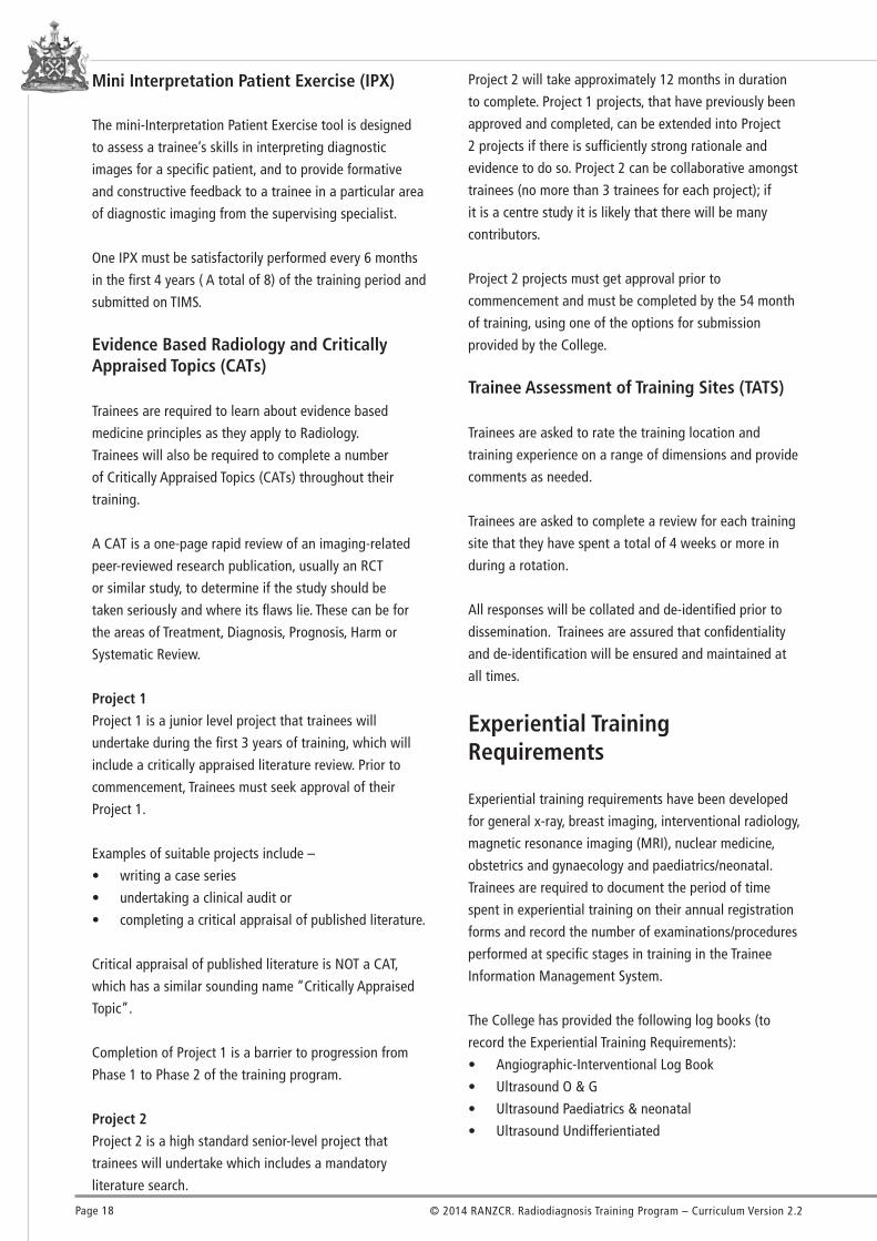
Mini Interpretation Patient Exercise (IPX)
The mini-Interpretation Patient Exercise tool is designed
to assess a trainee’s skills in interpreting diagnostic
images for a specific patient, and to provide formative
and constructive feedback to a trainee in a particular area
of diagnostic imaging from the supervising specialist.
One IPX must be satisfactorily performed every 6 months
in the first 4 years ( A total of 8) of the training period and
submitted on TIMS.
Evidence Based Radiology and Critically Appraised Topics (CATs)
Trainees are required to learn about evidence based
medicine principles as they apply to Radiology.
Trainees will also be required to complete a number
of Critically Appraised Topics (CATs) throughout their
training.
A CAT is a one-page rapid review of an imaging-related
peer-reviewed research publication, usually an RCT
or similar study, to determine if the study should be
taken seriously and where its flaws lie. These can be for
the areas of Treatment, Diagnosis, Prognosis, Harm or
Systematic Review.
Project 1
Project 1 is a junior level project that trainees will
undertake during the first 3 years of training, which will
include a critically appraised literature review. Prior to
commencement, Trainees must seek approval of their
Project 1.
Examples of suitable projects include –
• writing a case series
• undertaking a clinical audit or
• completing a critical appraisal of published literature.
Critical appraisal of published literature is NOT a CAT,
which has a similar sounding name “Critically Appraised
Topic”.
Completion of Project 1 is a barrier to progression from
Phase 1 to Phase 2 of the training program.
Project 2
Project 2 is a high standard senior-level project that
trainees will undertake which includes a mandatory
literature search.
Project 2 will take approximately 12 months in duration
to complete. Project 1 projects, that have previously been
approved and completed, can be extended into Project
2 projects if there is sufficiently strong rationale and
evidence to do so. Project 2 can be collaborative amongst
trainees (no more than 3 trainees for each project); if
it is a centre study it is likely that there will be many
contributors.
Project 2 projects must get approval prior to
commencement and must be completed by the 54 month
of training, using one of the options for submission
provided by the College.
Trainee Assessment of Training Sites (TATS)
Trainees are asked to rate the training location and
training experience on a range of dimensions and provide
comments as needed.
Trainees are asked to complete a review for each training
site that they have spent a total of 4 weeks or more in
during a rotation.
All responses will be collated and de-identified prior to
dissemination. Trainees are assured that confidentiality
and de-identification will be ensured and maintained at
all times.
Experiential Training Requirements
Experiential training requirements have been developed
for general x-ray, breast imaging, interventional radiology,
magnetic resonance imaging (MRI), nuclear medicine,
obstetrics and gynaecology and paediatrics/neonatal.
Trainees are required to document the period of time
spent in experiential training on their annual registration
forms and record the number of examinations/procedures
performed at specific stages in training in the Trainee
Information Management System.
The College has provided the following log books (to
record the Experiential Training Requirements):
• Angiographic-Interventional Log Book
• Ultrasound O & G
• Ultrasound Paediatrics & neonatal
• Ultrasound Undifferientiated

Page 19© 2014 RANZCR. Radiodiagnosis Training Program – Curriculum Version 2.2
Apart from the logbooks, such records should be obtained
through regular information system reports at the
appropriate training sites. Please refer to the section on
Experiential Training Requirements on the College website
to access the logbooks and for further information -
https://www.ranzcr.com/trainees/clinical-radiology/
assessment
Examinations and Barriers
The content of the Part 1 and part 2 examinations are
driven from the Radiodiagnosis Curriculum. Below are
descriptions of the examinations in the Radiodiagnosis
training program.
Part 1 Examination
The Part 1 examination will comprise of 4 Papers:
• Anatomy Paper 1
• Anatomy Paper 2
• AIT Paper 1
• AIT Paper 2
All part 1 examinations are delivered in an electronic
format over two days.
Full eligibility criteria and an explaination about the
components of the Part 1 examinations are pubished on
the website at -
https://www.ranzcr.com/trainees/clinical-radiology/exams
Part 2 Examination
The Part 2 examination will comprise of the following
components –
• e- Film Reading
• Radiology MCQ
• Pathology MCQ
• VIVA examination
Full eligibility criteria and an explaination about the
components of the Part 2 examinations are published on
the website at -
https://www.ranzcr.com/trainees/clinical-radiology/exams
TRA
ININ
G P
RO
GR
AM

Page 20 © 2014 RANZCR. Radiodiagnosis Training Program – Curriculum Version 2.2
CanMEDS Roles
Assessment Tool
DOT Assessment 3 3 3 3 3 3 3
Multi-Source Feedback (MSF) 3 3 3 3
Direct Observation of Procedural Skills (DOPS) 3 3 3 3
Mini Individual Patient Exercise (IPX) 3 3 3 3
Critically Appraised Topics (CAT) 3 3 3
Project 1 & 2 3 3 3 3 3
Examinations 3 3 3 3
Trainee Assessment of Training Site (TATS) 3
Experiential Training 3 3 3 3 3 3
Logbook 3 3 3
Medical Expert
Comm
unicator
Collaborator
Manager
Health Advocate
Scholar
Professional
Assessing the CansMEDS Roles
The following table details how the various assessment tools used within the training program relate to the roles of the
CansMEDS framework.

Page 21© 2014 RANZCR. Radiodiagnosis Training Program – Curriculum Version 2.2
Roles and Responsibilities
Trainees hold the key responsibility for their learning
and are responsible for ensuring that they meet the
assessment and progression requirements of the training
program.
Directors of Training are responsible for ensuring that
trainees are supervised and monitored appropriately, are
afforded appropriate learning opportunities and attend
relevant training and teaching sessions. Directors of
Training should also provide some direct face-to-face
teaching as appropriate.
Directors of Training are responsible for ensuring
that training networks and sites meet accreditation
standards and provide appropriate teaching, training and
supervision. Directors of Training must also complete the 6
monthly DoT assessments for trainees.
All consultant radiologists at accredited training sites
are expected to be involved in training and teaching and
to share the responsibility for performing the in training
assessments required in the training program. These are
referred to as Clinical Supervisors and are instrumental in
providing much needed DoT support.
TRA
ININ
G P
RO
GR
AM

Page 22 © 2014 RANZCR. Radiodiagnosis Training Program – Curriculum Version 2.2
Glossary of Learning Verbs
ComprehensionWhat you are expected to understand
Describe Communicate in writing the key features of
Distinguish Highlight the differences between
Explain Make clear or intelligible, or state the meaning of
Identify Recognise, establish or select after consideration
Illustrate Use an example to describe or explain something
ApplicationCan you apply your knowledge?
Apply Put to practical use
Demonstrate Prove with certainty or exhibit by practical means
AnalysisCan you analyse the detail of what you have learned?
Analyse Examine in detail the structure of
Categorise Place into a defined class or division
Compare and contrast Show the similarities and/or differences between
Construct Build up or compile
Discuss Examine in detail by argument
Interpret Translate into intelligible or familiar terms

Page 23© 2014 RANZCR. Radiodiagnosis Training Program – Curriculum Version 2.2
Patient Safety CyclePlease see the Patient Safety Cycle diagram for reference.
The trainee should be aware of the complete cycle of
safety associated with a patient referral for an imaging
examination or procedure. In general, the cycle is not
complete until the final approved report has been
received and understood by the referring clinical team and
its leader, and any appropriate clinical action has been
taken as a result.
All too often the trainee acts as if the patient safety
cycle ends after completion of the imaging procedure.
However, more incidents, errors and miscommunications
arise because of problems in the cycle that occur after the
imaging examination has been completed than through
problems that occur during the examination. The purpose
of this module is to ensure that trainees are fully aware of
the key central role of the radiologist in ensuring that the
cycle is completed safely for all patients.
Patient Safety Syllabus
In consultation with their Director of Training, clinical
supervisors and other trainees, the trainee is expected to
acquire the knowledge defined in this Syllabus and apply
it to all aspects of their practice of radiology in order to
maximise patient safety.
Trainees must have access to supervision and support
from their Director of Training, supervisor or consultant(s)
throughout their training.
The Patient Safety Syllabus should be read in conjunction
with the rest of the Radiodiagnosis Training program
Curriculum. The syllabus must be actively taught and
learned in training centres in the first 6 months of
training, and be referred to throughout the period of
training. The content of the syllabus is assessable at
various times in training through the DOPS, IPX, MSF, DoT
Assessments as well as at the Part 2 Examinations.
PATI
ENT
SAFE
TY

Page 24 © 2014 RANZCR. Radiodiagnosis Training Program – Curriculum Version 2.2
• previous key investigations
• Follows up with the referrer and their supervisor
regarding any missing information or questions
regarding the referral.
• Reviews the appropriateness of the referral in
relation to the suspected diagnosis.
• Seeks advice from their supervisor if the
appropriateness of the referral is in question.
Risk Factor Assessment
The trainee:
• Recognises at risk groups by conducting a risk factor
assessment taking into account the following key
areas:
o possible reactions to contrast media (see separate
section on administration of contrast media and
management of reactions to contrast media)
o need for sedation (see separate section on use of
sedation and management of complications from
sedation)
o radiation dose
o age-related risks (e.g. children, the elderly)
o modality-specific risks
o allergies
o diabetes
o renal function
o contraindications to MRI
o pregnancy status
o other contraindications
o anti-coagulation
o infection control (e.g. if patient has an infectious
disease)
• Recognises pitfalls and variations of examinations
and imaging modalities including indicative doses for
radiation.
• Identifies alternative examinations for at risk groups
and discusses these with their supervisor and the
referrer.
• Discusses safety issues related to the use of contrast
media with their supervisor and the referrer.
• Discusses alternative examinations with their
supervisor and the referrer.
Cultural Competence
The trainee:
• Identifies patients from Culturally and Linguistically
Diverse (CALD) backgrounds and provides culturally
sensitive care in accordance with the training site’s
policies and procedures.
Doctor-Patient Referral
The trainee demonstrates knowledge of the purposes of
the referral including:
• Communication between the referrer and the
radiologist
• Patient demographic and clinical information
• Evidence of patient consent
• Referrer’s diagnosis (confirmation, exclusion)
• Tests or procedures requested
• Information expected from the analysis, verification
and interpretation of the examination and images
• Provision of specific information required by the DI
service which is indicated
• Known allergies
• Risk Factors that could potentially impact on the
conduct of the imaging examination
• Recording of administrative and clinical information
relevant to the administration or conduct of the
imaging examination within the DI service
Review of Request
The trainee:
• Reads the referral to ascertain legibility of hand-
written forms (does legibility impact on data
management and test selection?).
• Reviews completeness of the request form — is
there any information missing such as:
• clinical information - details of the current problem,
co-existing and past medical and surgical history,
history of previous sedation and anaesthesia, current
medications (including non-prescribed medications),
allergies, fasting status
• patient consent
• suspected diagnosis
• clinical question(s) to be answered

Page 25© 2014 RANZCR. Radiodiagnosis Training Program – Curriculum Version 2.2
Patient Preparation
The trainee:
• Verifies the identification of the patient with the
patient (wherever possible) and other members of
the health care team.
• Verifies with the patient (wherever possible)
and other members of the health care team the
examination(s) and or procedure(s) to be performed.
• Demonstrates knowledge and application of the
protocol to provide information on pre-examination
preparation required by patients for particular
examinations to patients and referrers.
• Demonstrates knowledge and application of
procedures to confirm correct patient preparation
prior to an imaging examination, and procedures for
patients who are inappropriately prepared.
• Demonstrates knowledge of the significance of
clinical Information: details of the current problem,
co-existing and past medical and surgical history,
history of previous sedation and anaesthesia, current
medications (including non-prescribed medications),
allergies, fasting status.
• Explains the examination(s) and or procedure(s) to
be performed to the patient.
• Discusses the risk factor assessment with the patient.
• Identifies alternative examinations for at risk groups
and discusses these with the patient.
• Discusses radiation safety protocols and shielding
protocols with the patient.
• Demonstrates knowledge of the psychological, social
and cultural factors that influence the ways in which
patient’s interact with and respond to information
provided by health professionals.
Consent
The trainee:
• Discusses the examination(s) including alternative
examination(s) where appropriate, possible risks,
procedures and relevant imaging modalities with the
patient (points above to be covered in discussions)
and provides written information to the patient.
• Verifies that the patient has provided voluntary
informed consent in writing and that the evidence of
consent is recorded in the Patient’s medical record.
• Utilises qualified interpreters, including, including
Auslan and telephone interpreters when consulting
with patients, family and carers not fluent in English
or those with special communication needs.
• When using an interpreter the trainee addresses the
patient directly.
• Demonstrates knowledge and application of the
training site policies and procedures pertaining to
consent. (consent policies and procedures will differ
between training sites, states and territories).
Particular attention must be paid to patient consent for
Interventional procedures. Patients should receive written
information on the procedure in sufficient time to allow
them to review the information and seek a second opinion
if they wish.
For interventional procedures the patient should have
their informed consent confirmed at a separate discussion
with the radiologist who will be performing the procedure.
IF CONSENT IS IN DOUBT
If the trainee has any doubt as to the competence and
comprehension of the patient to voluntarily consent the
trainee must inform their supervisor and seek guidance as
to the appropriate steps.
Exception: the exception to obtaining informed consent is
emergency situations where the patient is unable to give
consent.
Improving Communication of Risk
Even although a body of knowledge exists on risk and
strategies for management of risk the communication of
risks to patients may be complicated if the assumption
is made that patients will rationally review evidence to
identify and choose the procedure or examination that
will maximise their health outcomes.
The trainee:
• Builds a relationship of trust with patients.
• Identifies the multiple and possibly conflicting
sources of risk information that patients may access.
• Demonstrates knowledge of the psychological, social
and cultural factors that influence the ways in which
patient’s respond to risk information.
PATI
ENT
SAFE
TY

Page 26 © 2014 RANZCR. Radiodiagnosis Training Program – Curriculum Version 2.2
Performance of an Examination
The trainee:
• Verifies the identification of the patient with the
patient (wherever possible) and other members of
the health care team.
• Verifies with the patient (wherever possible)
and other members of the health care team the
examination(s) and or procedure(s) to be performed.
• Reviews all appropriate records prior to performing
the examination to ensure that all relevant risk
factors have been identified, addressed and
discussed.
• Confirms that the patient has consented to the
examination in writing (with the exception of
emergency situations where the patient is unable to
give consent).
• Must seek advice from their supervisor or the
consultant on duty if there is any doubt whatsoever
as to the appropriateness of the examination in
terms of patient safety and requests for unnecessary
imaging.
The trainee demonstrates knowledge and application of:
Imaging protocols
Including but not limited to:
Fluoroscopy
• Collimation
• Frame rate
• Capture vs. Exposure
• Minimising radiation dose
CT
• Pitch, exposure, DLP
• Coverage
• Contrast injection timing
• Rate
• Number of phases
• Contrast dose
• Delays
• Mixes
Plain Radiography
• Appropriate views & projections for suspected
pathology
• Desired penetration & exposure for indication and
suspected pathology
Ultrasound
• Probe selection
• Image gain control & optimization
• Safe use of duplex, colour and power Doppler
• Appropriate search and image capture strategy
Paediatric Imaging Protocols
• Paediatric radiation exposure protocols
• ALARA, especially for CT, fluoroscopy and
angiography procedures
Radiation Safety Protocols
Including but not limited to:
Indicative doses
• Common diagnostic procedures
• Recognise pitfalls and variations
Shielding Protocols
• Shielding protocols appropriate to the imaging being
performed
Drug Administration Protocols
Including but not limited to:
• Right Patient
• Right route
• Right Dose
• Right Drug
• Right time
• Recognises the role of nurses, pharmacists & allied
health professionals in medication management.
Observation
• The trainee observes at least one of each type of
examination/procedure prior to undertaking it
themselves.

Page 27© 2014 RANZCR. Radiodiagnosis Training Program – Curriculum Version 2.2
Management of Adverse Events – Open Disclosure
The trainee:
• Advises their supervisor immediately about the
occurrence of an adverse event or near miss and
the affect of the adverse event or near miss on the
patient.
• Completes appropriate documentation in the
health care records, incident reports and records for
investigation.
• Demonstrates empathy for patients, families and
carers who are affected by an adverse event or near
miss.
• Demonstrates knowledge and application of the
training site’s policy and procedures on managing,
reporting and disclosing adverse events and near
misses.
• Identifies the information that should be provided to
patients immediately after an adverse event.
• Records adverse events on the training site Adverse
Events Register.
• Refers patients suffering an adverse event or near
miss to an appropriate senior health care worker or
an appropriate community based trauma support
program.
• Reviews the Australian Commission on Safety and
Quality in Healthcare (ACSQHC) Open Disclosure
Standard.
Review of Images/ Quality Control Workup
• The trainee demonstrates knowledge of the factors
that can have a negative impact on image quality
that may lead to errors in reporting (Refer to the AIT
Syllabus).
Analysis, Verification and Interpretation (Clinical Reasoning)
The trainee:
• Defines what is meant by the KEY FINDINGS in an
examination.
• Identifies KEY FINDINGS (i.e. may be a single positive
Complications
The trainee:
• Recognises the signs of potential complication(s)
relevant to the examination(s) being performed early
including but not limited to:
o Vasovagal
o Pneumothorax
o Haemorrhage
o (see separate sections on administration of
contrast media and management of reactions
to contrast media and use of sedation and
management of complications from sedation).
• Implements the relevant protocols to prevent or
manage complication(s).
• Seeks assistance of consultant(s) at the first sign of
potential complications.
• Holds a current CPR certification
Infection Control
The trainee:
• Demonstrates knowledge and application of training
site, and national, state/ territory policies and
procedures for infection control issues including but
not limited to:
o Hand washing Safety (Sterile Technique – Aseptic
technique)
o Use of personal protective equipment
o appropriate reprocessing of instruments and
equipment
o Set up of sterile trays
o Systems for handling blood (including dried
blood), other body fluids, secretions and excretions
(excluding sweat), nonintact skin and mucous
membranes.
o Disinfection of equipment and instruments
o Needle and waste disposal
• Demonstrates knowledge and application of
additional precautions in situations where standard
precautions may not be sufficient to prevent the
transmission of infectious agents (e.g. tuberculosis,
measles, Creutzfeldt–Jakob disease).
• Informs authorities of ‘notifiable diseases’.
PATI
ENT
SAFE
TY

Page 28 © 2014 RANZCR. Radiodiagnosis Training Program – Curriculum Version 2.2
Report structure is very much a matter of personal or
institutional style, but the most important role of the
report is to clearly convey the information that the
clinician most wants to know about a specific patient in a
particular clinical scenario.
The final approved report is the most important in
medicolegal and practical terms. Interim reports are all
very well, but carry little medicolegal status and may be
radically different from the final report. If a final report
differs substantially in content, diagnosis and meaning
from an initial, provisional or interim report, Patient
Safety is potentially compromised unless the referring
team is directly informed of the change in report findings.
Failure to do this directly may lead to prolonged delays
in appropriate management, and could even lead to
incorrect management.
The Role of Incident Monitoring
The trainee:
• Is able to report incidents using the system relevant
to their training site (e.g. AIMS).
• Trainees are required to report 1 incident per year on
an Adverse Events Register mandated by their local
jurisdiction.
• Demonstrates knowledge and application of the
relevant Medical Board(s) code of practice and AMA/
NZ equivalent code of conduct as they relate to
patient safety and care.
• Demonstrates knowledge and application of the
safety policies and procedures as required by the
relevant training site and national, state / territory
regulatory bodies.
Contrast Media
The trainee radiologist needs to have a working
knowledge of the protocols regarding appropriate use and
administration of contrast.
The emphasis should always be on PREVENTION OF
CONTRAST COMPLICATIONS
Required Skills
The trainee:
finding, a constellation of findings, or the absence of
positive findings).
• Distinguishes KEY FINDINGS from other (incidental or
ancillary) findings in a set of images (examination).
• Organises all findings in an examination in order
of importance relevant to the clinical question or
diagnosis AND with respect to relative acuity and
severity for the patient.
• Utilises clinical reasoning to judge a finding in terms
of its CLINICAL URGENCY.
• Identifies KEY CONDITIONS which may be life
threatening if undiagnosed over a period of 12 hours.
o Trainees must be aware of the level of urgency of
specific key findings, in certain clinical settings
• Reports findings of KEY CONDITIONS to the treating
team upon diagnosis.
o In the case of Key Conditions and other critical
findings, the patient safety cycle is not complete
until the referring team members have been
directly informed and have agreed to take the
relevant action for that patient
o Trainees need to be aware of their own limitations
in terms of knowledge of key conditions and know
when they should ask for help
• Recognises that some findings require immediate
attention to the patient.
• Organises multiple findings in relation to the degree
of CLINICAL URGENCY.
• Utilises cross checking and diagnostic timeout when
considering key findings.
• Identifies possible errors of perception and
interpretation specific to the examination.
• Seeks advice from their supervisor or consultant on
difficult examinations.
Radiologist Report Writing & Communication of Results
A formal interpretative imaging report is the work product
of diagnostic radiologists and is an official legal record.
Ensuring that reports are complete and accurate and that
reporting is prioritised according to the clinical level of
urgency should lead to reduced errors, improved outcomes
for patients, increased efficiency and decreased cost in
terms of unnecessary imaging.
Reports should be well structured, succinct and highlight
the major findings and if possible, the level of clinical
importance in clear, unambiguous language.

Page 29© 2014 RANZCR. Radiodiagnosis Training Program – Curriculum Version 2.2
The emphasis should always be on PREVENTION OF
COMPLICATIONS FROM procedural sedation and/or
analgesia.
Required Skills
The trainee:
• Demonstrates knowledge and application of
protocols in relation to administration of procedural
sedation and/or analgesia including screening all
patients for history of relevant allergies, current
medications, risk factors that increase the likelihood
of complications from sedation.
• Conducts a risk factor assessment which identifies
those patients at increased risk of cardiovascular,
respiratory or airway compromise during procedural
sedation and/or analgesia, as in such cases, an
anaesthetist should be present to care for the
patient.
• Identifies the potential risks of sedation and/or
analgesia including but not limited to:
o Depression of protective airway reflexes and loss of
airway patency.
o Depression of respiration.
o Depression of the cardiovascular system.
o Drug interactions or adverse reactions, including
anaphylaxis.
o Individual variations in response to the drugs used,
particularly in children, the elderly, and those with
pre-existing medical disease.
o The possibility of deeper sedation or anaesthesia
being used to compensate for inadequate analgesia
or local anaesthesia.
o Risks inherent in the wide variety of procedures
performed under procedural sedation and/or
analgesia. Unexpected extreme sensitivity to
the drugs used for procedural sedation and/or
analgesia which may result in unintentional loss
of consciousness, respiratory or cardiovascular
depression.
o Over-sedation, airway obstruction, respiratory or
cardiovascular complications may occur at any
time. Therefore, to ensure high standards of patient
care, the following guidelines are recommended.
• Demonstrates knowledge and application of the
training site’s protocols to manage adverse events
due to complications from administration of sedation
and/or analgesia.
• Demonstrates knowledge and application of
professional supervision protocols in relation to
administration of contrast including screening all
patients for history of relevant contrast allergy,
current medications, risk factors that increase the
likelihood of contrast-induced renal impairment, and
medical conditions that may result in life threatening
complications from contrast administration.
• Demonstrates knowledge and application of
protocols to determine when the radiologist
responsible for overseeing the study must be
contacted for advice before contrast is administered.
• Demonstrates knowledge and application of
protocols that determine the dose and type of
contrast medium that is administered taking
into consideration the patient’s age, how it is
administered, by whom it is administered (only by
medical personnel trained in venipuncture) and under
whose authorisation.
• Records that contrast has been administered to a
patient in the patient record.
• Recognises the signs of potential complication(s) of
administration of contrast early including but not
limited to:
o Extravasation
o Allergic reactions
o Contrast induced nephropathy
o Nephrogenic systemic fibrosis
o Delayed reactions
• Demonstrates knowledge and application of the
training site’s protocols to manage adverse events
due to contrast reactions.
• Demonstrates the ability to manage complications of
intravenous contrast administration or other medical
emergencies.
• Demonstrates knowledge and application of the
procedure for the use of contrast media which
complies with the current version of the RANZCR
Contrast Guidelines. https://www.ranzcr.com/
documents/573-iodinated-contrast-guidelines-2016/
file
• Holds current CPR certification to provide basic life
support.
Sedation
The trainee radiologist needs to have a working
knowledge of the protocols regarding appropriate use and
administration of procedural sedation and/or analgesia.
PATI
ENT
SAFE
TY

Page 30 © 2014 RANZCR. Radiodiagnosis Training Program – Curriculum Version 2.2
transmission of infectious diseases in the health care
setting; Endorsed by the Communicable Diseases
Network Australia, the National Public Health
Partnership and the Australian Health Ministers’
Advisory Council
January 2004
• The Royal Australian and New Zealand College of
Radiologists Radiology Adverse Events Register
(RAER)
• How can doctors communicate information about
risk more effectively?; Andy Alaszewski, Tom Horlick-
Jones; BMJ VOLUME 327 27 SEPTEMBER 2003
• National Patient Safety Education Framework, the
Australian Commission on Safety and Quality in
Healthcare (ACSQHC), formerly the Australian Council
for Safety and Quality in Health Care – July 2005
• Australian Curriculum Framework for Junior Doctors,
Confederation of Postgraduate Medical Education
Councils
• National Health and Medical Research Council
(NHMRC), Communicating with Patients, Advice for
medical practitioners, 2004
Report Writing Module
A formal interpretive report is the diagnostic radiologist’s
work product. This module defines the essential steps in
writing imaging reports and communicating the findings.
Learning Objectives
The trainee:
• Completes the University of Florida’s Curriculum
in Radiology Reporting (CCR). The Curriculum in
Radiology Reporting can be accessed at: http://
protocols.xray.ufl.edu/live_crr/prog/home/home.php
• Reads the Clinical Radiology Written Report
Guidelines, which can be accessed from the College
website at the following location:o https://www.ranzcr.com/college/document-
library/clinical-radiology-written-report-
guidelines
• Views the following lecture resources, available on
the College Learning Management System:
• Demonstrates the ability to manage complications
from administration of sedation and/or analgesia.
• Demonstrates knowledge and application of the
current Australian and New Zealand College of
Anaesthetists (ANZCA) Guidelines on Sedation
and/or Analgesia for Diagnostic and Interventional
Medical or Surgical Procedures – specifically sections
5.4 and 5.7.
• http://www.anzca.edu.au/resources/professional-
documents
• Holds current CPR certification to provide basic life
support
References Used in the Development of the Patient Safety Cycle
• RANZCR QR3.i Project – Review of Diagnostic
Imaging Requests
• Board of the Faculty of Clinical Radiology
The Royal College of Radiologists (2006)
Standards for the reporting and interpretation of
imaging investigations
Royal College of Radiologists, London
• Board of the Faculty of Clinical Radiology
The Royal College of Radiologists (2005)
Standards for patient consent particular to radiology
Royal College of Radiologists, London
• Curriculum in Radiology Reporting developed by
Doctors Linda Lanier, Chris Sistrom and Richard Rathe
from the University of Florida
• The Royal Australian and New Zealand College
of Radiologists Radiology Curriculum Advisory
Committee
• The Royal Australian and New Zealand College of
Radiologists Patient Safety Working Group
• The Royal Australian and New Zealand College of
Radiologists®
Standards of Practice for Diagnostic and
Interventional Radiology
Version 9.0
• Infection control guidelines for the prevention of

Page 31© 2014 RANZCR. Radiodiagnosis Training Program – Curriculum Version 2.2
• Essential knowledge:
o Age, sex, ethnicity, demographic characteristics/
clinical state of the patient including signs,
symptoms, results of other tests (e.g. previous
imaging studies should be sought, pathological,
laboratory and clinical tests).
• Advice:
o Awareness of the accuracy of the examination
in the particular patient related to the published
accuracy of the technique, and its applicability to
this particular examination. Level of certainty or
doubt needs to be clearly indicated in the report.
If a definitive diagnosis is given assume that this
will be used for patient management/if a definitive
diagnosis is not able to be provided advice on
further investigations should be given if necessary
taking into account the relative accuracy and
applicability of the suggested investigations and
patient safety (e.g. radiation exposure).
• Communication with the referrer:
o Report needs to be clear and written to match
the referrer’s expected level of knowledge and
familiarity with the issues raised. (e.g. the wording
of a report pertaining to a rare condition that is
provided to a general practitioner is likely to differ
compared to a report on the same rare condition
that is provided to a specialist in that particular
field). Processes must be in place that enables the
referring doctor to discuss the imaging findings
with the radiologists in complex cases.
• Taking Appropriate Action:
o Processes must be in place to allow direct
communication from the radiologist to the referrer
in cases that have urgent clinical priority. Examples
include medical conditions requiring emergency
treatment, findings of malignancy requiring
treatment, diagnosis of tuberculosis which has the
potential to harm others.
• Communication with the patient:
o Patients must always be treated with respect
and honesty. A thorough assessment of the
investigation must be completed prior to
speaking to the patient/if bad news needs to be
conveyed the radiologist conveying the bad news
should have undergone some form of training.
Radiologists must ensure that they follow up with
o 2012 AOCR/ASM RA - Learning from incident
reports in the Australian medical imaging setting
o 2012 AOCR/ASM RA - Implementation of the
radiology written report Guideline
• Explains the purpose of an imaging report in patient
care
• Identifies the core concepts of report writing
• Demonstrates knowledge and application of the
essential steps in writing imaging reports and
communicating the findings
• Clinical information:
o Information provided by the referrer, specialist
background of the referrer, medical signs/
symptoms pointing to a particular diagnosis, a
range of diagnoses, or possible diagnoses under
consideration.
• Technical knowledge:
o Ability of the radiologist to evaluate the quality of
images and their suitability to the diagnosis of the
condition(s) under consideration.
• Observation:
o Cross checking of patient identification,
confirmation that the type and date of the
examination are correct, normal findings,
unequivocal abnormal findings, both anticipated
and unanticipated, abnormal findings, normal
variants.
• Analysis:
o Further evaluation of definitive or equivocal
abnormalities for relevant imaging characteristics
(e.g. shape, contour, density, enhancement pattern,
signal intensity, and echogenicity) to formulate an
opinion – whether there is an active pathological
process present or whether the findings can
be encompassed within the range of normal
appearances.
• Medical Interpretation:
o Correlation of the image analysis with other
factors to interpret the radiographic findings and
their relevance to the patient. A wide medical
knowledge is required in order to reach a specific
diagnosis or appropriately ranked differential
diagnosis sufficient to allow clinical decisions to be
made.
PATI
ENT
SAFE
TY

Page 32 © 2014 RANZCR. Radiodiagnosis Training Program – Curriculum Version 2.2
the primary care doctor who will be involved in the
future care of the patient.
• Recognises when the findings constitute a medical
emergency and implements local protocols to alert
referrers in urgent cases.
• Distinguishes between synoptic reporting and regular
reporting.
• Identifies the advantages of synoptic reporting.
• Participates in or conducts an audit of reporting
discrepancies within the Radiology Department in
the first three years of training.
• Recognises the need to proofread and correct errors
in spelling, measurements, and L/R reference.
• Recognises that further investigations should only
be suggested if they are medically indicated and will
contribute to patient management.
• Utilises cross checking and diagnostic timeout when
there are any doubts as to the efficacy of the report
findings.

Page 33© 2014 RANZCR. Radiodiagnosis Training Program – Curriculum Version 2.2
PATI
ENT
SAFE
TY
Examples
Specific examples have not been provided as there is great variation in individual institutional and practitioner style in
reporting. Also, report style and type will depend on the exact type of examination being performed, and the referring
clinical discipline. For example, the report for an emergency physician searching for abdominal bleeding, an orthopaedic
surgeon enquiring about a subtle ligamentous injury or oncologist evaluating the outcome of the latest round of
chemotherapy can differ in style and structure greatly.
Key Elements of a Radiology Reporting Template
Referring Doctor
Radiologist
Location
Examination Date
Patient Name
Patient Address
Patient’s Age
Patient File Number
Gender
Date of Exam
Procedure(s) Performed
IntroductionDetail discussions with the patient regarding the procedure(s) and possible complications, risks – e.g. interventional procedures, contrast reactions must be discussed with the patient and the patient needs to give informed consent before the procedure can be undertaken
Technical Adequacy
Regional Normal Anatomy
Regional Normal Pathology
Associated Findings
Appropriate Progress
Incidental Findings including Normal Variants
Date of Report
Conclusion (answer the question)
Level of Urgency
Dispatch (method of delivery of report)
Synoptic Reporting:
The aim of synoptic reports is to provide a clinically relevant format for the particular type of exam (e.g. mammography, breast ultrasound, oncology imaging ), with constrained descriptive terms and consistent formatting for a report so that the reading clinician is better able to read the outcome and the reporting doctor is more consistent with the approach. In a sense synoptic reports are templated but the templates are more restrictive than the standardised templates for say, CT scan of the abdomen.Checklist – Normal/Abnormal

Page 34 © 2014 RANZCR. Radiodiagnosis Training Program – Curriculum Version 2.2
TNM Reporting
Structuring the report under the headings of ‘Primary
tumour’, ‘Lymph nodes’ and ‘Metastases’ follows the
pathological TNM format. As the radiology scan will
also visualize other pathology which may or may not
be related to the tumour, this is placed at the end of
the report under the heading of ‘Other findings’. If the
patient also has a brain scan, it can be useful to add this
as a separate heading at the beginning or the end of
the report as there are specific issues which need to be
mentioned in a brain report which do not easily fit into
the other headings.
Following the TNM format for the radiology report
reminds the reporting radiologist to ‘think like the tumour’
and to be aware of common disease patterns for that
particular tumour. It also reminds you to look carefully
for recurrent disease at the site of a resected primary.
Measurements and Response
Measuring lesions is very important in oncology
scans to allow an objective assessment of disease
response. Imaging forms a part of the patient’s overall
assessment and the importance the clinician will place on
measurements will depend upon whether the patient is in
a clinical trial and other clinical factors.
It is the radiologist’s responsibility to provide an accurate,
objective report that enables the clinician to use the scan
information appropriately.
There are a number of methods of determining response,
but the most commonly used at present is RECIST 1.1
(Response Evaluation Criteria In Solid Tumours version
1.1). This has specific definitions for measurements and
response. A summary of this is provided below and the
full details can be obtained from the original articles 2-5.
The choice of appropriate target lesions is important and
is further discussed below. All lesions should be measured
in mm.
The templates
Three templates have been produced to cover most
oncology reporting needs. These were developed for CT,
but can be used for any modality. They may be set up as
templates in a reporting system or the headings can be
dictated and typed for each report. The measurements
Mandatory In-Training Assessment for Patient Safety and Report Writing in Year 1 of Training
Trainees are required to report 1 incident per year on
an Adverse Events Register mandated by their local
jurisdiction.
Refer to section on Trainee Progression for further
information.
Curriculum in Radiology Reporting
All year 1 trainees must complete all modules of the
University of Florida’s Curriculum in Radiology Reporting.
All modules must be completed by the end of the first
year of training. Progress of trainees will be tracked by the
College. Failure to complete the modules will be a barrier
to progression to year 2.
Trainees are required to set up individual registrations. The
University of Florida’s Curriculum in Radiology Reporting
can be accessed at: http://protocols.xray.ufl.edu/live_crr/
prog/home/home.php
Standardized TNM oncology reports
The College would like to acknowledge Dr Gormley for
providing the following advice on Standardised TNM
Oncology Reports for Radiology trainees.
Introduction
Oncology patients usually have a number of CT scans;
at diagnosis and at multiple episodes of follow up.
Using a tumour, node, metastasis (TNM) structure for
the radiology report provides consistent reports that
cover all the important clinical areas. This is particularly
useful when patients are scanned at multiple different
locations. It is recommended that most oncology CT
reports are issued following this format with the inclusion
of measurements of target lesions. Lymphoma patients
are assessed clinically by different criteria and a specific
set of headings have been provided to reflect this. A
description of the templates is provided with additional
general information regarding oncology reporting, RECIST
and Cheson/IWG criteria.

Page 35© 2014 RANZCR. Radiodiagnosis Training Program – Curriculum Version 2.2
Metastases
• presence or absence of any suspected metastases
• describe distribution.
• measure target lesions
Provide subjective assessment of non-measurable lesions
Any questionable metastatic lesions are also discussed
here.
Other findings
• Comment on other relevant findings
• Can put findings related to the disease /
complications first
Do a geographic review of the scan from neck to pelvis to
ensure all areas assessed
Conclusion
• Give TNM stage as best as possible
• If primary not imaged can say Tx,N2,M1
2. FOLLOW-UP TEMPLATE
Primary tumour
• Location, size and local extension / invasion
• Comment on change of size and give measurements
for current and previous scans
Other appropriate statements under this heading may be
• not imaged
• previous resection with no evidence of recurrent
disease
• stable fibrosis
A dedicated look at the site of resected disease will help
identify early recurrence.
Lymph nodes
• presence or absence of any suspected lymph node
metastases
Work away from the primary tumour site superiorly and
inferiorly to ensure all nodal groups are assessed. Distant
nodes may occur outside the radiotherapy field
• describe the involved regions
• describe overall increase / decrease in size
• include target nodes in the measurement table
For RECIST purposes, only two nodes are required to be
measured.
can be included in tables under each heading or placed
as a table of ‘target lesions’ at the end of the report (see
examples).
It is important to include free text under each heading
and not rely solely on measurements as the radiologist’s
interpretation is still important and not all lesions will be
included in the table. Comment should be made on the
general trend of change and any indeterminate lesions.
The headings should not be removed in the absence of
disease as a negative statement is just as important.
GENERIC HEADINGSClinical History;
Comparison Scan; Date and regions scanned of
comparison scan(s)
Technique;
1. STAGING TEMPLATE
Primary tumour
• location
• size – uni or bi-dimensional. Give longest diameter in
any plane if needed to define the T stage as well as
the axial diameter.
• local extension / invasion
Enough information to provide a T stage as best as
possible with imaging.
Other appropriate statements under this heading may be
• ‘not imaged’
• ‘previous resection with no evidence of residual
disease’
Lymph nodes
• presence or absence of any suspected lymph node
metastases
• describe the involved regions.
Work away from the primary tumour site superiorly and
inferiorly to ensure all nodal groups are assessed
Loco-regional nodes are defined by the primary site. More
distant nodes are considered metastatic (i.e. M1)
• The largest node in several groups should be
included in the measurement table.
• For RECIST purposes, only two nodes are required to
be measured.
PATI
ENT
SAFE
TY

Page 36 © 2014 RANZCR. Radiodiagnosis Training Program – Curriculum Version 2.2
Splenic and liver nodules
• presence or absence of any intra splenic or intra
hepatic masses
• measure 2 or 3 per organ and include in
measurement table
• give an indication of distribution and progression /
response
Often will just say ‘normal liver and spleen with no
masses’
Splenic size
• longest diameter
Other findings
• Comment on other relevant findings
Do a geographic review of the scan from neck to pelvis to
ensure all areas assessed
Conclusion
• Give subjective impression of disease response
IMPORTANT POINTS
• Always describe the disease in the free text,
don’t just give measurements
• Save all measurements to PACS
• Be practical
Lesion Choice and Measurement
Target lesions
At the original scan a number of lesions should be
identified as target lesions. Ideally these will include
the primary tumour, some lymph nodes and metastases.
It is important to try to choose lesions that will be
reproducible on future scans. So if the primary is very
ill defined and difficult to accurately measure this is
not a good target lesion. Liver lesions are also difficult.
Preferably the largest lesions are chosen, but it may be
necessary to choose smaller lesions which are better
defined in the surrounding liver.
There are specific lesions which are defined as non-
measurable by RECIST 1.1, and are only assessed
subjectively (see below). Lung lesions can be measured
on lung or soft tissue windows, but this needs to be
consistent on follow up scans. Lesion measurements
should be saved on PACS to help with follow up.
Metastases
• presence or absence of any suspected metastases
• give an indication of distribution and progression /
response
• include target lesions in measurement table
Questionable metastatic lesions are also discussed here.
New lesions is included as a separate heading as a
reminder to look for these, as any new lesion indicates
progressive disease. This heading is optional.
Other findings
• Comment on other relevant findings
• Can put findings related to the disease /
complications first
Do a geographic review of the scan from neck to pelvis to
ensure all areas assessed
Conclusion
• Give summary of disease response
• Only use RECIST terms if calculating response
accurately
3. LYMPHOMA TEMPLATE
The template headings are based upon the IWG/Cheson
criteria 6,7. These criteria require bi-dimensional
measurements, with a maximum of six nodal / extra-nodal
masses and additional measurements of splenic and liver
nodules. The free text is important as there are often
more groups involved than will be measured. For ease of
radiologist use, we provide the measurements, and the
clinical team calculates the formal response if required.
Lymph nodes and masses
• describe all the nodal regions involved
• include extranodal lymphomatous masses, excluding
intrasplenic and intrahepatic lesions
• choose 6 lesions to include in the measurement table
Lesions should be chosen from disparate regions of
body if possible, but must include mediastinal and
retroperitoneal nodes / masses if involved
• bi-dimensional measurements in the axial plane
• give an indication of distribution and progression /
response in follow-up

Page 37© 2014 RANZCR. Radiodiagnosis Training Program – Curriculum Version 2.2
Calculating response
The uni-dimensional measurements of target lesions are
summated for the initial scan and the follow up scan. The
difference of these totals is calculated and this is divided
by the initial total to give a percentage change between
the two scans.
This enables the RECIST response to be given as per the
criteria for progressive disease, stable disease, partial
response and complete response (see RECIST 1.1).
Sometimes it is necessary to provide a subjective
assessment of response if there are no measurable
lesions. The response by radiologists is usually calculated
between the two most recent scans. In formal RECIST
measurements the baseline scan is usually used for
comparison. As long as the same lesions have been
measured and documented, the clinicians will be able
to check the response between any two different time
points. It is important to be clear which two scans have
been assessed in the report. The most important thing
is not to use the RECIST terminology of PR and PD if the
disease is stable. You can clarify in the conclusion as
‘Partial response since the previous scan’.
RECIST (Response Evaluation Criteria In Solid
Tumours)3
Version 1.1 2009 - Summary
Target lesions at baseline
o maximum 5 lesions total
o maximum 2 per organ
o includes primary, lymph nodes and metastases
o representative of all involved organs
o preferably longest lesion, but should also be best
for “reproducible repeated measurements”
• Measurements are conventionally taken in the axial
plane
• All measurements in mm.
Primary tumour – longest axial diameter. For staging
scans the longest diameter of the tumour in any plane
should be given as well if this will make a difference to the
T stage of the tumour. The axial measurement is used for
follow up. Many people give bi-dimensional measurements
of the primary, although this is not required for RECIST.
Lymph nodes – Short axis diameter. This is defined as the
‘waist’ of the lymph node and has been shown to be the
optimal predictor of malignancy when using size4. Assess-
ing a node by “CT size criteria’ provides an indication of the
risk of the lymph node being malignant. There are different
recommended cut off sizes for different sites of the body
(table1). This has a significant margin of error, but is a use-
ful guide. In trial cases, RECIST 1.1 provides a generalized
cut off of 10mm short axis for all regions for an abnormal
node and 15mm short axis for a target lesion. Nodal meas-
urements are still included in the measurement table when
they are <10mm.
LYMPHOMA – has a different set of criteria and uses bi-
dimensional lymph node measurements (see below)
Metastases – longest diameter in the axial plane.
Follow up measurements
• Same lesion, same plane (axial), same windows
At follow up the same lesions should be measured on the
same windows. In clinical trials this is very important.
Sometimes it can be difficult to define the boundaries
of a lesion at follow up, but it is important to try as best
as possible. Sometimes non-target lesions will have
significantly changed in size, while the target ones have
not. The target lesions should still be measured, but if
the non-target measurements will change the overall
response these can also be measured and added into the
measurement table, or mentioned in the free text. (see
FAQ)
The direction of measurement can be different as long
as the longest axis (or short axis for lymph nodes) is
measured.
PATI
ENT
SAFE
TY

Page 38 © 2014 RANZCR. Radiodiagnosis Training Program – Curriculum Version 2.2
Partial Response
• ��≥ 30% decrease sum (from baseline for trial)
Progressive Disease
• ��≥ 20% increase in sum (from smallest sum in study
for trial)
• Total sum increase must be at least 5mm
• OR Any new lesion
Stable Disease
• Neither PR or PD
CHESON/IWG CRITERIA FOR LYMPHOMA6,7
An international working group formulated criteria for the
assessment of lymphoma response. This includes clinical
and haematological analysis as well as imaging. The
updated criteria in 2007 also include PET. These criteria
use bi-dimensional measurements in disease response
assessment, calculating the sum of the products of the
diameters.
There are a number of factors to be satisfied for the
fulfillment of the Cheson response criteria. From a
radiological perspective it is important to identify
appropriate target lesions and to provide bi-dimensional
measurements of these.
• up to six of the largest dominant nodes or nodal
masses should be measured.
These nodes or masses should be
• clearly measurable in at least 2 perpendicular
dimensions
e.g “multiple small liver metastases”
“multiple enlarged retroperitoneal lymph nodes
<15mm short axis”
Bone: o soft tissue component of lytic lesions
can be measured if >10mm
o sclerotic = non measureable
Cystic lesions: o can measure if thought to be a
‘cystic met’ & >10mm
o use non-cystic lesions in preference
Lesions at site of previous radiotherapy:
o only measureable if demonstrates
progression of lesions
Response
This is calculated by adding up all the diameters for the
first scan (summated diameters) and also for the follow-
up scan. There will be a total measurement for each
scan. Subtract the follow up scan total from the initial
total, divide the difference by the initial total and x 100
to obtain a percentage change. The percentage change is
then used to define the disease response. There are four
categories of response by the RECIST 1.1 criteria.
Initial total – follow up total = change
change / initial total x 100 = %change
Complete Response
• disappearance of all target lesions
• LN short axis <10mm (i.e. nodes now ‘normal’, but
can be present)
• So CR sum can be >0 due to ‘normal’ lymph nodes
Measurable lesions
≥10mm longest diameter (primary tumour and metastases)Lymph nodes ≥15mm short axis diameter
Non-measurable lesions
<10mm longest diameter (primary tumour and metastasis)10 – 15mm short axis diameter lymph nodes (<10mm normal)AscitesPleural/pericardial effusionsLymphangitis carcinomatosaLeptomeningeal diseaseSclerotic bone lesions

Page 39© 2014 RANZCR. Radiodiagnosis Training Program – Curriculum Version 2.2
to re-measure to give an accurate comparison with the
current measurements. Describe what you have done in
the free text and you could put ‘(re-measured)’ after the
measurement in the table.
What if the non target lesions are much bigger?
Sometimes this will not change the overall response, so
calculate this with the target lesions first. If including the
non target lesions would give a different response, then
this can be done in two ways. Firstly you could add the
key lesions into the measurement table and calculate the
response using all lesions. Secondly you could give the
result using the target lesions, but also say that ‘several
other lesions have significantly increased in size and this
would make it progressive disease’. Remember any new
lesion is progressive disease regardless of what the rest of
the scan has done.
Should I put a new lesion into the measurement table?
Not routinely. However, if there have only been 2 or 3
lesions to follow it could be useful to include the new
lesion if it fits the criteria for a target lesion as it could be
useful at follow up.
• from disparate regions of the body if possible
• include mediastinal and retroperitoneal areas
whenever these sites are involved.
Splenic and hepatic nodules are assessed separately
They must regress by ≥ 50% in their SPD or, for single
nodules, in the greatest transverse diameter.
“Lymph nodes should be considered abnormal if the long
axis is more than 1.5 cm regardless of the short axis. If a
lymph node has a long axis of 1.1cm to 1.5 cm, it should
only be considered abnormal if its short axis is more than
1.0cm. Lymph nodes ≤1.0 x ≤1.0cm will not be
considered as abnormal for relapse or progressive
disease”.
(Quoted from ref 7 Cheson BD, Pfistner B, Juweid ME et al.
Revised Response Criteria for Malignant Lymphoma.
J Clin Oncol 2007;25:579-586)
FREQUENTLY ASKED QUESTIONS
What if there are two primary tumours?
If the second primary is old and irrelevant it can be
mentioned under other findings. If there are two active
primary tumours it may be necessary to use subheadings
under ‘Primary tumour’. Be clear about which tumour
you think the lymph nodes and metastases are related to,
although sometimes it isn’t obvious or doesn’t matter.
How do I report a CT brain?
Best to use an additional heading and report normally.
Can put before Primary tumour or after Other findings.
Can also include brain metastases in the measurements
table and mention them under the Metastasis heading.
Can I use this for MRI or CT-PET?
While the templates were designed for CT, they can be
used for any modality imaging cancer.
What if I don’t agree with the lesions previously measured?
You should still measure the same lesions, especially if the
patient is on a clinical trial. If you think they have been
significantly under or over measured it will be necessary
PATI
ENT
SAFE
TY

Page 40 © 2014 RANZCR. Radiodiagnosis Training Program – Curriculum Version 2.2
7. Cheson BD, Pfistner B, Juweid ME et al. Revised
Response Criteria for Malignant Lymphoma. J Clin Oncol
2007;25:579-586.
8. Hricak H. “Oncologic Imaging: Essentials of reporting
common cancers.” Saunders. Philadelphia. 2007
References
1. Sobin LH, Wittekind Ch. TMN classification of malignant
tumours, 6th edition. Wiley. ISBN:0-471-22288-7
2. Therasse P, Arbuck SG, Eisenhauer EA et al. New
guidelines to evaluate the response to treatment in solid
tumours. J Natl Cancer Inst 2000;92(3):205-216
3. Eisenhauer EA, Therasse P, Bogaerts J. New response
evaluation criteria in solid tumours: Revised RECIST
guideline (version 1.1) Eur J Cancer 2009 Jan;45(2):228-
247
4. Schwartz LH, Bogaerts J, Ford R et al. Evaluation
of lymph nodes with RECIST 1.1. Eur J Cancer 2009
Jan;45(2):261-7
5. Bogaerts J, Ford R, Sargent D. Individual patient data
analysis to assess modifications to the RECIST criteria. Eur
J Cancer 2009 Jan;45(2):248-260
6. Cheson BD, Horning SJ, Coiffier B et al. Report of
an International Workshop to Standardize Response
Criteria for Non-Hodgkin’s Lymphomas. J Clin Oncol
1999;17:1244-1253
Site Short axis size (mm)
Cervical 10
Axilla 10
Mediastinum 10
Subcarinal 12
Paracardiac 8
Retrocrural 6
Gastrohepatic Ligament, porta-hepatis 8
Upper ara-aortic 10
Lower para-aortic 12
Common iliac 9
External iliac 10
Internal iliac 7
Obturator 8
Inguinal 10
Table 1. Cut off values of lymph nodes for CT size criteria8

Page 41© 2014 RANZCR. Radiodiagnosis Training Program – Curriculum Version 2.2
PATI
ENT
SAFE
TY
Appendix A
Report Templates
ONCOLOGY STAGING REPORT
Clinical History:
Technique:
Report:PRIMARY TUMOUR;
[free text]
LYMPH NODES;
[free text]
METASTASES;
[free text]
OTHER FINDINGS;
CONCLUSION:
Location Size (short axis)
Location (Target Lesions) Size (longest diameter)

Page 42 © 2014 RANZCR. Radiodiagnosis Training Program – Curriculum Version 2.2
ONCOLOGY FOLLOW UP REPORT
Clinical History:
Comparison Scan:
Text:
Report:PRIMARY TUMOUR;
[free text]
LYMPH NODES;
[free text]
METASTASES;
[free text]
NEW LESIONS:
OTHER FINDINGS;
CONCLUSION:
Location Previous Size Current Size
(short axis)
Location Previous Size Current Size
(short axis)

Page 43© 2014 RANZCR. Radiodiagnosis Training Program – Curriculum Version 2.2
PATI
ENT
SAFE
TY
LYMPHOMA REPORT
Clinical History:
Comparison Scan:
Technique:
Report:LYMPH NODES & MASSES;
[free text]
LYMPH NODES;
[free text]
SPLEEN & LIVER NODULES;
[free text]
SPLENIC SIZE:
OTHER FINDINGS;
CONCLUSION:
Location Previous Scan Current Scan
(bi-dimensional)
Location (Target lesions) Previous Scan Current Size
(bi-dimensional)

Page 44 © 2014 RANZCR. Radiodiagnosis Training Program – Curriculum Version 2.2
This page is intentionally left blank

Page 45© 2014 RANZCR. Radiodiagnosis Training Program – Curriculum Version 2.2
Contents
• List of Key Conditions 46
• Management of contrast reactions
• Neuroimaging
• Head and neck imaging
• Obstetric & gynecologic imaging
• Musculoskeletal imaging
• Chest imaging
• Abdominal imaging
• Paediatric imaging
• Vascular and interventional radiology
Key Conditions in Year 1 of Training
Key Conditions in early training are defined as those conditions which may be life threatening if undiagnosed over a period
of 12 hours.
These key conditions must be documented and communicated to the treating team immediately upon diagnosis.
Trainees need to be aware of their own limitations in terms of knowledge of these conditions and know when they should
ask for help.
KEY
CO
ND
ITIO
NS
Y1

Page 46 © 2014 RANZCR. Radiodiagnosis Training Program – Curriculum Version 2.2
Key Conditions in Year 1 of Training
The list of key conditions below must, as a minimum
requirement, be covered in formal and informal teaching
sessions in the first four months of training before a
trainee goes on call in order to:
• Equip trainees with key skills and knowledge
of Radiology relevant to on call activities and
emergency radiology.
• Facilitate and guide learning of Radiological Anatomy
through clinically relevant principles.
• Facilitate and guide learning of basics in clinical
radiology in the areas of safety, complications,
consent & communication, reporting skills and
radiological procedures.
• Provide a sound foundation for future training and
learning of Clinical Radiology.
List of Key Conditions in Early Training
Management of Contrast Reactions
Neuroimaging
• Subarachnoid haemorrhage
• Extradural/subdural haemorrhage (including skull
fractures)
• Cerebral parenchymal haemorrhage
• Cerebral abscess, Empyema
• Venous sinus thrombosis
• Herniation
• Stroke
• Diffuse Axonal Injury
• Hypoxic brain injury
• Cord compression
• Cerebrovascular injury
Head and Neck
• Foreign bodies
• Retropharyngeal abscesses
• Tonsillar abscess
• Ruptured Oesophegus
O&G Imaging
• Viability scanning
• Ectopic pregnancy
• Tubo-ovarian masses
• Ovarian masses
• Ruptured Ovarian Cysts
Musculoskeletal System
• Fracture/Dislocation
• Spine fracture/dislocation
• Spinal Cord injuries
• Epidural haematomas
• Septic Arthritis/Osteomyelitis/Discitis
Chest
• Pneumothorax (especailly tension pneumothorax)
• Pulmonary embolus
• Pulmonary oedema
• Aortic/vascular trauma
• Aortic dissection
• Cardiac tamponade
• Typical/Atypical infections (TB)
• Flail Chest
Abdomen
• Free gas
• Bowel obstruction
• Volvulus
• Trauma to abdominal organs
• Abdominal aortic aneurysm and rupture
• Mesenteric ischaemia/other organ ischaemia/
infarction
• Testicular torsion
• Abdominal sepsis
• Abscess, collections and drainage
o Hydronephrosis/ pyonephrosis
o Acute abdomen e.g. appendicitis, pyelonephritis,
emphysematous cholecystitis, intra-abdominal
abscesses including subphrenic collections.
Paediatrics
• Airway foreign body
• Intussusception, pyloric stenosis, ,malrotation
• Epiglotitis/croup
• NEC
• NAI

Page 47© 2014 RANZCR. Radiodiagnosis Training Program – Curriculum Version 2.2
Training Site Responsibilities
Training departments need to prepare a checklist
containing the key conditions in early training and ensure
that trainees have had adequate teaching and exposure to
these conditions in the first four months of training prior
to going on call. The checklist should be fully completed
prior to trainees going on call.
Tools for Training - Key Conditions in Early Training
A template for key conditions in early training has been
developed as a tool to assist with teaching and learning
about these conditions.
The early training template, a number of completed
template examples and a suggested outline for teaching
year 1 trainees are provided on the following pages.
The early training template is structured as follows:
Vascular Imaging and Interventional Radiology
• Acute aortic syndromes
o Trauma (psuedoaneurysm/ intramural haematoma)
o Aneurysm (rupture/ inflammatory/ mycotic)
o Dissection
• Extravasation/ haemmorhage/thromboembolism
o Trauma
o GI bleed and management
o Peripheral, pulum and visceral
Trainee Requirements – Management of Contrast Reactions
Trainees are expected to complete training in
management of contrast reactions in their first year of
training.
Trainees are expected to complete an accredited CPR
course and hold a current CPR certification.
Condition
Definition
Clinical Presentation
Pathology
Where it Occurs
Complications
Differential Diagnosis
Radiology
Modality/Technique
Associated Conditions
Features
Artefacts
Clinical Impacts
Sensitivity/Specificity
Management Plan & Communications
Relevant Anatomy
See examples of completed early training matrix templates on the following pages.
KEY
CO
ND
ITIO
NS
Y1
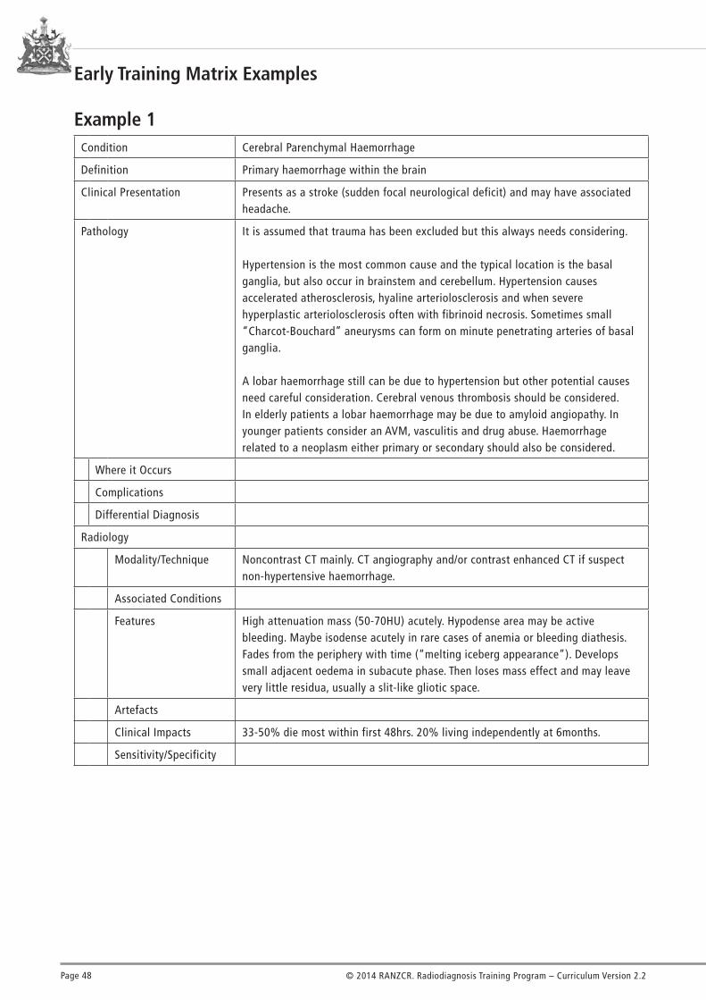
Page 48 © 2014 RANZCR. Radiodiagnosis Training Program – Curriculum Version 2.2
Condition Cerebral Parenchymal Haemorrhage
Definition Primary haemorrhage within the brain
Clinical Presentation Presents as a stroke (sudden focal neurological deficit) and may have associated headache.
Pathology It is assumed that trauma has been excluded but this always needs considering.
Hypertension is the most common cause and the typical location is the basal ganglia, but also occur in brainstem and cerebellum. Hypertension causes accelerated atherosclerosis, hyaline arteriolosclerosis and when severe hyperplastic arteriolosclerosis often with fibrinoid necrosis. Sometimes small “Charcot-Bouchard” aneurysms can form on minute penetrating arteries of basal ganglia.
A lobar haemorrhage still can be due to hypertension but other potential causes need careful consideration. Cerebral venous thrombosis should be considered. In elderly patients a lobar haemorrhage may be due to amyloid angiopathy. In younger patients consider an AVM, vasculitis and drug abuse. Haemorrhage related to a neoplasm either primary or secondary should also be considered.
Where it Occurs
Complications
Differential Diagnosis
Radiology
Modality/Technique Noncontrast CT mainly. CT angiography and/or contrast enhanced CT if suspect non-hypertensive haemorrhage.
Associated Conditions
Features High attenuation mass (50-70HU) acutely. Hypodense area may be active bleeding. Maybe isodense acutely in rare cases of anemia or bleeding diathesis. Fades from the periphery with time (“melting iceberg appearance”). Develops small adjacent oedema in subacute phase. Then loses mass effect and may leave very little residua, usually a slit-like gliotic space.
Artefacts
Clinical Impacts 33-50% die most within first 48hrs. 20% living independently at 6months.
Sensitivity/Specificity
Early Training Matrix Examples
Example 1

Page 49© 2014 RANZCR. Radiodiagnosis Training Program – Curriculum Version 2.2
KEY
CO
ND
ITIO
NS
Y1
Management Plan 1. Diagnose intracerebral haemorrhage on CT.2. Does it fit with a hypertensive haemorrhage based on typical location, age
and medical history? If not consider another cause based on these same factors and any other clues on the films e.g. any big or dense vessels? Always consider a non-hypertensive cause if a lobar (peripheral) haemorrhage.
3. Consider performing a CT angiogram (try to include as much of the brain as possible and especially the area of the haemorrhage) or a routine post contrast scan (or both).
4. Assess for intraventricular or extraaxial extension, mass effect, secondary hydrocephalus and herniation.
5. Contact clinical team.
Relevant Anatomy Important to assess epicentre of haemorrhage relative to key brain anatomical structures. Know basic venous anatomy to assess for dense thrombosed vein e.g. internal cerebral vein in basal ganglia haemorrhage. Ventricular system and basal cisterns for secondary effects of mass effect (Is anything missing because it is effaced?)
Example 1 cont.

Page 50 © 2014 RANZCR. Radiodiagnosis Training Program – Curriculum Version 2.2
Condition Subarachnoid Haemorrhage
Definition Haemorrhage into the subarachnoid space between the pia and arachnoid membranes.
Clinical Presentation Sudden “worst ever” headache. May have reduced GCS and may have neurological findings.
Pathology Berry aneurysm are saccular true aneurysm that tend to occur at branch points within or near the Circle of Willis. Thought to be due to the altered flow dynamics and areas of wall weakening at these sites.
They are relatively common being found in 2-5% of the population. 20-30% have multiple aneurysms. They are more common in females. There is a weak familial risk. They are associated with various connective tissue diseases especially adult polycystic kidney disease. Smoking is also a risk factor for aneurysmal SAH.
Where it Occurs At the common sites of Berry aneurysms but haemorrhage will spread widely through the subarachnoid space and into ventricles with time. Most common sites are anterior communicating artery, posterior aspect of terminal ICA either related to posterior communication artery or anterior choroidal artery, middle cerebral artery bifurcation, basilar tip and PICA origin from vertebral artery. Predominant site of haemorrhage may give clue to site of aneurysm or in the case of multiple aneurysms which one bleed.
Complications Associated intraparenchymal haemorrhage, rebleed, hydrocephalus, cerebral vasospasm.
Differential Diagnosis Pseudosubarachnoid hemorrhage due to cerebral edema effacing basal cisterns, Normal structures such as calcified choroid plexus extending through foramen of Lushka, Traumatic SAH, Vascular causes other than Berry aneurysm rupture such as intradural vertebral dissection or AVM, Benign perimesencephalic haemorrhage (diagnosis of exclusion, should have typical history and benign clinical course, low pressure bleed anterior to brainstem usually limited by Lilliquist’s membrane).
Radiology
Modality/Technique CT Brain C-, CT angiography, Conventional Angiography.
Associated Conditions
Features High attenuation acute haemorrhage within the basal subarachnoid cisterns. Small or subtle haemorrhages may only be seen in dependent areas such as interpeduncular cistern or occipital horns of lateral ventricles. Also evaluate posterior fossa carefully which often suffers from beam hardening artefact. Look for asymmetry in lateral and Sylvian fissures.
Artefacts Beam hardening artefactartifact.
Clinical Impacts
Sensitivity/Specificity Sensitivity is very good early after the onset of symptoms but will reduce will time after this and also depends on the volume of bleed. Patients suspected of having a SAH with a negative CT should have a lumber puncture looking for xanthochromia.
Example 2

Page 51© 2014 RANZCR. Radiodiagnosis Training Program – Curriculum Version 2.2
KEY
CO
ND
ITIO
NS
Y1
Management Plan 1) Diagnose SAH on CT.2) Look for complications such as hydrocephalus and if the presentation has been delayed ischemia from vasospasm.3) Perform a CT angiogram trying to optimise image quality and ensure good coverage of the most likely source of hemorrhage e.g. scan higher if you think there might be a pericallosal aneurysm.4) Contact the clinical / neurosurgical team.5) Look for and assess the aneurysm that caused the haemorrhage but don’t be satisfied with that, look for the 2nd or 3rd aneurysm. Is there any obvious vasospasm on the CTA?
Relevant Anatomy Cerebral arterial vasculature especially circle of Willis, Normal cerebral structures that can mimic haemorrhage through physiologic calcification for example.
Example 2 cont.

Page 52 © 2014 RANZCR. Radiodiagnosis Training Program – Curriculum Version 2.2
Condition Testicular torsion
Definition Torsion of the spermatic cord
Clinical Presentation Acute scrotal pain, onset frequently at night, scrotal swelling and œdema, testis difficult to palpate, thickened tender spermatic cord.
Atypical presentations may delay diagnosis leading to testicular necrosis:Abdominal, RIF or flank & groin pain, nausea, vomiting, undescended testis, normal testicular lie, bilateral torsion (10%), detorsion and hyperæmia.
Pathology 1. Extravaginal torsion in fœtus & neonate: the testis, epididymis and tunica vaginalis twist on the spermatic cord. Undescended testis may delay diagnosis.
2. Intravaginal torsion – most commonly peripubertal. Bell-clapper deformity in which the tunica vaginalis completely surrounds the testis. Congenitally absent posterior anchoring occurs in 12% and allows the testicle to twist.
Frequency Approx 1:125 males. < 10% are > 30 years of age.
Morbidity Salvage rate 80-100% if repaired within 6 hours.0% at 24 hours.
5-10% spontaneous detorsion: high risk of recurrence.
Differential Diagnosis Acute epididymitis/epididymo-orchitis; abscess; torsion of the epididymal appendix; incarcerated hernia; hæmatoma; trauma; ruptured varicocele; scrotal tumour; vasculitis; Fournier gangrene; acute idiopathic scrotal œdema.
Radiology
Modality/Technique For atypical cases: high frequency US and sensitive colour flow Doppler/power Doppler; straddle view.
Associated Conditions Undescended testis.
Features Swelling, hypoechogenicity; heterogeneity indicates necrosis, and echogenic areas may indicate hæmorrhage; torsion knot in cord; echogenic infarcted testis, tunica albuginea and mediastinum more common in neonatal torsion.
Pitfalls Flow not demonstrated in 10-20% of normal pre-pubertal testes; normal asymmetry may give false positive in young child; capsular flow; necrotic abscess has no central flow.
Clinical Impacts Immediate surgical detorsion/orchiectomy and bilateral orchidopexy: US should be reserved for those with atypical features.
Sensitivity/Specificity 86-100%/77-100%; unilateral absent flow in a symptomatic enlarged testicle is highly specific.
Management Plan A classic presentation at a typical age with consistent physical findings merits urgent operative repair without imaging confirmation. If imaging is equivocal, urgent surgical exploration should proceed.
Relevant Anatomy Spermatic cord, testis, epididymis, vascular supply and intratesticular arteries and veins, tunica albuginea, tunica vaginalis, inguinal herniæ.
Example 3

Page 53© 2014 RANZCR. Radiodiagnosis Training Program – Curriculum Version 2.2
KEY
CO
ND
ITIO
NS
Y1
Condition Pulmonary œdema
Definition Abnormal accumulation of fluid in the extravascular compartments of the lung.
Clinical Presentation Dyspnœa, hypoxæmia, tachypnœa, orthopnœa, tachycardia, crepitations and rales.
Pathology Hydrostatic pressure œdema.Permeability œdema with diffuse alveolar damage.Permeability œdema without DAD.Mixed œdema.
Where it Occurs Pulmonary interstitium and alveoli.
Complications Respiratory failure, hypoxæmia.
Differential Diagnosis Pneumonia, aspiration pneumonia.
Radiology
Modality/Technique CXR, HRCT
Associated Conditions Heart failure, fluid overload, renal failure, neurogenic, aspiration, lung injury, ARDS, chemotherapy, shock, hypersensitivity, pulmonary embolism, negative pressure (upper airway obstruction), postobstructive (airway), veno-occlusive disease, near-drowning, drowning, high altitude, reperfusion, reexpansion, postpneumonectomy, postreduction, air embolism, asthma, opiate overdose, marathon runners, decompression sickness, pregnancy & childbirth.
Features Septal lines, peribronchial cuffing, alveolar œdema.
Pitfalls Imaging findings lag behind clinical changes; COPD; gravitational effects; mitral regurgitation (RUL); underlying disease e.g. pneumonia, toxic injury.
Clinical Impacts Correlate imaging appearances with clinical features.
Sensitivity/Specificity Variable. Principal discriminatory features for the different causes of PO are distribution of pulmonary blood flow, distribution of pulmonary œdema and the width of the vascular pedicle. Ancillary findings are pulmonary blood volume, peribronchial cuffing, septal lines, pleural effusions, air bronchograms, lung volume and heart size. Accuracy 80-90%. 5% false positive and 11% false negative can be achieved.
Management Plan Depends on cause.
Relevant Anatomy Pulmonary alveoli, interstitium, vasculature, cardiac chambers and systemic veins.
Example 4

Page 54 © 2014 RANZCR. Radiodiagnosis Training Program – Curriculum Version 2.2
Non Medical Expert Role
The expectation of trainees and trainers is that the skills
defined in the non medical expert roles are incorporated
into all aspects of training and radiology practice. Non
medical expertise is in no way viewed as being mutually
exclusive from medical expertise.
Team Work
Radiologists establish and maintain cooperative
relationships with patients, with radiology staff and with
other clinicians to optimise patient care and to facilitate
research and education.
Key competencies: At the end of training Radiology
trainees are able to:
1. Develop their role in the provision of patient care and
the expectations of the patient and clinician requesting
imaging
a) Assist referring doctors to select the most
appropriate and effective investigative pathway.
b) Assist the radiology team to organise and prioritise
the workload.
c) Supervise investigations and procedures.
d) Advocate for patient needs and improve patient
satisfaction.
e) Produce a structured report that communicates
the important findings and addresses the clinical
question.
f) Research clinical/radiological problems through
literature and internet.
g) Check reports with supervising radiologist and
consult clinicians for additional information as
appropriate.
h) Prepare the report in a timely manner.
i) Alert referring clinicians to important or unexpected
findings.
j) Develop interventional skills through radiologist
mentoring and study.
k) Recognise competence and limitations and seek help
appropriately.
l) Participate in departmental and multidisciplinary
meetings.
m) Continually update knowledge through personal
study and teaching programs.
Advice for Training Sites
Teaching First Year Trainees Key Conditions in Early Training
Trainees should always be able to ask for help from senior
departmental staff in a timely manner and expect that
their request is responded to in a timely manner with
patient safety being paramount.
Training sites should aim to provide in-house supervision
and exposure to key conditions in early training, so the
trainee acquires the core knowledge and skills defined
by this module as well as any other skills relevant to that
institution’s needs and clinical practice environment.
Teachers should facilitate and promote self-learning
behaviour through for example discussion and
presentation of relevant case materials. Tutorials with
discussions, not lectures, should be the dominant learning
format.
Sessions should be structured using key educational
principles as follows:
• Provide context for topic & probe prior knowledge
• Ask WHY topic is important
• Explore WHAT should be learned
• Discuss HOW this can be achieved
• Provide EXAMPLES which illustrate, and guide
discussion
• Summarize key learning points, and identify areas for
further learning
Training sites should aim to provide the following items to
in-house training for first year trainees:
• Involvement in film-based tutorials at least twice a
week.
• Rostered session(s) off each week for either course
attendance or self-directed learning.
• Preparation of short specific anatomy or technique
presentations for other trainees, under guidance of a
named clinical supervisor or consultant on at least a
monthly basis.
• Presentation of basic and emergency cases in
radiological meetings.
• Attendance at clinico-radiological conferences and
other radiology rounds at least twice a week.

Page 55© 2014 RANZCR. Radiodiagnosis Training Program – Curriculum Version 2.2
KEY
CO
ND
ITIO
NS
Y1
d) Organise and maintain educational materials and
resources.
e) Promote the education of all staff groups and the
process of learning from them.
Learning opportunities
These capabilities may be gained through learning
activities such as:
• Reflective practice as a trainee.
• Observation of the behaviour of senior radiologists
and departmental staff.
• Discussion with senior staff and peers.
• Formal feedback in Appraisal sessions.
• Liaison with referring practitioners.
• Participation in conferences, inter-disciplinary
meetings and continuing education.
• Education in conflict management.
Communication Skills
Radiologists recognise the importance of communication
in best practice of medicine.
They acknowledge the vital role of good communication
skills in all of their extended roles.
Key competencies: At the end of training Radiology
trainees are able to:
1. Demonstrate good communication skills
a) Convey their expert opinion, degree of certainty in
the diagnosis and its implications effectively.
b) Listen to and discuss issues and concerns with
patients, colleagues and students.
c) Respect patient confidentiality, privacy, and
autonomy.
d) Demonstrate an awareness of the legal implications
of their actions.
e) Encourage discussion, questions and interaction in
imaging encounters with patients and/or their family.
f) Show initiative to alter the mode of communication
appropriately. For example, phone through urgent,
important or unexpected findings; confirm the receipt
of important findings.
2. Demonstrate knowledge and communication of the
strengths and limitations of the resources available for
patient care: equipment, personnel, infrastructure, time
constraints, cost
a) Demonstrate knowledge of departmental work flow,
equipment and the abilities of team members.
b) Recognise constraints and other commitments
and rationalise competing demands to deliver the
optimum sustainable system of patient care.
c) Discuss with patients and clinicians their
expectations in relation to the medical system,
clinical setting, test requested and report availability;
and if there are any expectations that are not
realistic, provide an explanation as to why.
d) Reach realistic agreements regarding service delivery
with relevant colleagues e.g. ‘CT at 4pm’ or ‘in-
patients will be done within 24hours’.
e) Communicate any significant changes from the
agreed management, minimising impact on the
patient and the service.
3. Manage working relationships effectively and
professionally
a) Obtain informed and willing consent from the
patient through communication with the patient and
referring doctor.
b) Discuss possible, common or serious complications
with patients and check that patients comprehend
the information being provided to them.
c) Ensure correct patient, correct procedure, correct site.
d) Ensure pre-procedure checks are satisfactory.
e) Give and receive feedback constructively.
f) Manage conflict with patients, staff and clinicians in
a professional manner seeking to comprehend other
points of view and exploring constructive solutions.
g) Act with integrity.
h) Help and support colleagues and staff.
i) Employ practices that promote a safe workplace.
j) Work cooperatively with managerial, governmental
and professional bodies.
4. Facilitate research and education
a) Attend departmental and interdepartmental
meetings, study groups and conferences.
b) Arrange, prepare and present meetings.
c) Participate in audit, peer review, research and quality
improvement.

Page 56 © 2014 RANZCR. Radiodiagnosis Training Program – Curriculum Version 2.2
c) Address challenging communication issues in a
competent manner, including: obtaining informed
consent, delivering bad news, addressing anger,
confusion and misunderstanding.
d) Demonstrate knowledge and application of the basic
principles of conflict resolution and negotiation.
Learning opportunities
These capabilities may be gained through learning
activities such as:
• Observation of the behaviour of Senior Staff with
discussion of this behaviour.
• Formal feedback in Appraisal sessions.
• Observation of departmental staff.
• Liaison with referring practitioners.
• Participation in Conferences/ inter-disciplinary
meetings/ continuing education.
• Attendance at communication skills workshops.
• Discussion of conflict management or training in
conflict management.
Patient Support & Advocacy
As health advocates, radiologists responsibly use their
expertise and influence to advance the health and
well-being of individual patients, communities, and
populations. Their knowledge of the risks and costs of
imaging procedures, are balanced against the benefits
to individual patients and the community. Advocacy may
occur individually or as a group when influencing public
health policy.
Key competencies: At the end of training Radiology
trainees are able to:
1. Advocate for individual patients
a) Participate in Multidisciplinary Team meetings.
b) Negotiate on behalf of patients during clinical
encounters for investigations that minimize
risk, radiation exposure and cost. For example:
negotiation to substitute non-ionising imaging
studies in patients, particularly children or pregnant
females.
c) Report adverse events and act in accordance with
the local quality and safety framework.
d) Participate in Quality Improvement Projects where
appropriate.
2. Participate in activities using feedback to ensure
good communication and continually develop their skills
a) Seek and accept the opinions of others when their
skills are being evaluated.
b) Provide effective feedback to others regarding their
abilities.
c) Show appreciation of the importance of non-verbal
behaviour and of trust in effective communication.
d) Demonstrate follow up to ensure comprehension by
the other party, and to ensure that communication
has been effective. For example, confirm that patients
comprehend the procedures to which they have
consented and that bad news has been delivered
with empathy and compassion.
e) Participate in positive relationships with those
who are in a position to observe and evaluate their
abilities and skills e.g. sonographers, radiographers,
clinical consultants, fellow trainees.
f) Demonstrate follow up to check their own and
other’s compliance with agreements.
3. Communicate effectively across different settings
a) Demonstrate ability to focus on important
information that must be conveyed, and to reconcile
this with the amount that patients, students,
colleagues or others can assimilate.
b) Adjust communication to meet the needs of different
audiences. For example, communication with a
colleague, communication with a patient, a lecture
to students, a lecture to the local radiology branch or
a large conference; local meeting; student settings;
public meetings or public setting.
c) Communicate effectively with people from different
social, cultural and linguistic backgrounds.
d) Use trained interpreters when they are required and
are available (and appreciate the risks of using family
/ friends in paramedical roles).
4. Demonstrate the ability to recognise difficult
communication situations, and to work within these
situations to achieve the best possible outcome
a) React professionally to inappropriate communication
or actions by others that arise from strong emotion,
such as frustration, grief or sadness.
b) Acknowledge the difficulties inherent in these
situations and work towards solutions.

Page 57© 2014 RANZCR. Radiodiagnosis Training Program – Curriculum Version 2.2
KEY
CO
ND
ITIO
NS
Y1
• Activities within local branch of the College.
• Participating in multidisciplinary staff meetings.
• Participating in change management process in
department.
• Public speaking to community groups.
• Review relevant sections of the National Patient
Safety Education Framework.
Management & Administrative Skills
Radiologists make decisions regarding effective utilisation
of finite health care resources in the context of individual
patient care. They provide leadership in healthcare
organisations and ensure effective work practices through
adequate staffing and development of policies and
procedures.
Key competencies: At the end of training Radiology
trainees are able to:
1. Participate in activities that contribute to the
effectiveness of healthcare organisations and
systems
a) Initiate, organise and complete an audit of
departmental practice as it relates to running of the
department (eg. Patient waiting times).
b) Demonstrate competence in workload management
in an area of the radiology department, e.g. CT. Take
responsibility, in conjunction with other department
staff, for smooth and efficient day to day operation of
this part of the department.
c) Manage clinical and other correspondence effectively.
d) Respond to correspondence promptly.
e) Document procedures/activities in case-notes
appropriately.
f) Respond to complaints appropriately.
2. Manage their practice and career effectively
a) Set priorities and manage time to ensure that outside
activities and personal life do not impact adversely
on patient care and practice requirements.
b) Allocate finite healthcare resources appropriately.
c) Triage requests for radiological examinations.
d) Work to help radiologists use their time effectively,
eg by managing, in conjunction with other staff, an
area of the department such as CT on a day-to-day
basis.
e) Ensure information is available for patients through
verbal &/or written communication.
f) Apply the practice privacy policies which govern
the use of patient personal information within the
service and its disclosure to other parties.
g) Apply protocols for gaining patients’ consent to the
use of their personal information.
h) The trainee demonstrates knowledge of the proposed
uses of personal information and communicates this
information to the patient.
2. Advocate for the profession
a) Provide support to the profession through Junior
Forum membership and participation in branch
meetings, ASM and other educational events as
appropriate to the stage of training.
3. Advocate for trainees
a) Comment responsibly during completion of
assessment of training site forms.
b) Provide support for other trainees that require
assistance.
c) Notify the supervisor of training or other College
officers of issues having significant adverse impact
on training.
d) Advocate for other health care team members.
e) Participate in training and educational activities of
other staff.
f) Contribute in oral or written form to the
development of manuals, procedural documents and
other resources required by other health care team
members.
g) Advocate for the community.
h) Provide accurate information to the community
and consumer groups regarding issues relevant to
radiology.
Learning Opportunities
These capabilities may be gained through learning
activities such as:
• Discussions with senior staff about health system
issues.
• Observation of more senior trainees and radiologists
in their tasks.
• Involvement in department planning activities.
• Conduct of patient surveys.

Page 58 © 2014 RANZCR. Radiodiagnosis Training Program – Curriculum Version 2.2
allowances in the form of additional time, resources
or altered practice for those from a different cultural
background.
c) Admit error and acknowledge the contributions of
others.
d) Identify and disclose personal interests, and act to
resolve potential conflicts of interest.
e) Recognise and appropriately respond to incompetent,
unprofessional or unethical behaviour.
f) Recognise and respect privacy and confidentiality
with regard to patients and colleagues.
g) Obtain appropriate and informed consent when
working with patients.
2. Give priority to patient care and to continuity of care
a) Act to ensure high standards of diagnosis,
management and safety for patients. Ensure that
imaging protocols, image interpretation, procedures
and other professional services are conducted
optimally.
b) Make time for urgent studies, retrieve previous
images for comparison, and rapidly communicate
unexpected results to clinical team members.
c) Maintain responsibility for patient care until it
is formally assumed by others. Ensure that the
transmission of information, including details of
treatment, images and reports, occurs in an effective
and timely fashion.
d) Answer letters, pages, e-mail, and phone calls
promptly.
3. Behave appropriately towards patients and colleagues
a) Behave in a manner that is inclusive of social,
ethnic and religious groups. Avoid interpersonally
destructive behaviour and encourage inclusive and
constructive behaviour in others.
b) Ensure that communication with the patient,
the patient’s social network and other health
professionals is directed toward the best possible
outcome. Ensure that conflicts and/or inadequate
communication do not impact adversely on patient
care.
4. Support accountability and the development of the
profession
a) Observe regulatory requirements as required by
government, medical defence bodies, medical boards
e) Serve in administration and leadership roles, as
appropriate.
f) Participate in department organisation, eg as
registrar representative on committees as required.
g) Create call and other rosters fairly.
h) Be aware of hospital guidelines relating to OHS, EEO
etc.
Resources: Clinical Audit in Radiology, 100+ recipes, ed
Godwin R et al. Published by RCR.
Learning opportunities
These capabilities may be gained through learning
activities such as:
• Regular supervisor review
• Review of outstanding reports
• Observation of senior staff
• Formal instruction in daily team activities
• Instruction such as workshops on team work and
roles
• Studying Management courses
• Progressive responsibility for conduct and reporting
of studies
• Time management workshops
• Discussion of budgeting and costs with senior staff
• Career mentorship
• Participation in departmental audit activities
• Career information form RANZCR website
• Participation in research projects
Professionalism
Radiologists aim to deliver the best possible quality
of health care with integrity, honesty and compassion.
Radiologists exhibit high standards of personal and
interpersonal behaviour and they practice ethically with
active concern for the patient, their profession and society.
Key competencies: At the end of training Radiology
Trainees are able to:
1. Behave ethically
a) Prioritise the interests of the patient. Identify their
own capacities and limitations, and seek advice or
direct assistance when appropriate.
b) Recognise the importance of tolerance and
respect for other value systems, and make realistic

Page 59© 2014 RANZCR. Radiodiagnosis Training Program – Curriculum Version 2.2
KEY
CO
ND
ITIO
NS
Y1
Research & Education
Radiologists recognise the importance of participating in
and encouraging life long learning. They contribute to the
appraisal, collection, dissemination and comprehension
of health care knowledge. They assist and promote
the education of clinical colleagues, other health care
providers, students, patients, the community & other
bodies. Radiologists recognise the importance of research
and actively participate in advancing the knowledge of
their speciality.
Key competencies: At the end of training Radiology
trainees are able to:
1. Adopt a scholarly approach to practice
a) Effectively manage their own learning and apply this
to their clinical decision making.
b) Design, implement and evaluate an audit cycle.
c) Demonstrate commitment to lifelong learning by
a process of enquiry in order to maintain current
knowledge of radiological literature.
d) Present at clinical meetings or grand rounds.
e) Participate in College learning activities.
f) Describe current thinking in the profession
concerning management of patient risk, risk/benefit
of investigation and resource allocation.
2. Apply the principles of evidence based medicine
a) Conduct literature searches.
b) Critically appraise published work relevant to their
practice.
c) Apply analysis from literature to radiological
environment and practice.
d) Interpret statistical results in order to assess validity
of findings in published works.
e) Participate in literature review meetings (eg journal
clubs).
f) Participate in a statistical skills course.
3. Develop educational skills and techniques
a) Outline principles of adult learning.
b) Employ self directed learning skills and activities.
c) Select the most effective means to deliver a message
clearly, accurately and efficiently to a given audience.
d) Teach junior trainees and other health workers.
and funding bodies such as Medicare. Maintain
medical registration and medical indemnity.
b) Recognise the basic legal aspects of medical
litigation and the potential for radiologists to be
defendants or consultants in litigation.
c) Recognise the importance of life long learning.
Arrange to participate in or conduct educational
sessions at a level appropriate to the stage of
training.
d) Balance personal and professional responsibilities to
ensure patient care, personal health and sustainable
practice.
Learning Opportunities
These capabilities may be gained through learning
activities such as:
• Study or review of ethical and professional codes of
practice of the College, Medical Boards and other
bodies
• Observation of others performing at a high standard
• Discussion of the trainee’s own practice and any
issues they may have experienced or wish to explore
with their supervisor
• Feedback from supervisors
• Negotiating a learning contract with a
supervisor concerning performance in the area of
professionalism
• Developing a portfolio of cases or situations
illustrating professional issues
• Participating in cultural awareness training
• Preparing a service profile for the department and
compare this with the surrounding community, or
levels of service generally by radiology
• Writing up reflective commentary of critical incidents
or cases where an ethical or professional issue
emerged
• Taking a course relating to ethics in medicine and/or
radiology
• Review legal aspects of medical litigation related to
radiology and membership of a MDT.
• Document incidents that may have medico legal
implications and notify and discuss these incidents
with supervisor or a senior colleague

Page 60 © 2014 RANZCR. Radiodiagnosis Training Program – Curriculum Version 2.2
e) Mentor junior trainees at a level appropriate to the
stage of training.
4. Develop research skills
a) Describe and participate in the process of study
design and evaluation.
b) Submit an original manuscript of publishable quality
reporting research in any area of radiology.
c) Participate in annual registrar research / audit
presentation.
d) Present scientific material at an academic meeting.
e) Describe conflict of interest issues.
f) Describe preparation and provision of advice for
grant application.
Learning Opportunities
These capabilities may be gained through learning
activities such as:
• Presentation at scientific meetings
• Presentation at departmental meetings
• Preparation of grant applications in collaboration
with senior colleagues
• Publication of peer reviewed papers
• Submission of posters or papers to conference
• Statistical skills courses
• Critical appraisal courses
• Journal clubs

Page 61© 2014 RANZCR. Radiodiagnosis Training Program – Curriculum Version 2.2
Contents
• General X-Ray Experiential Training Requirements 62
• Breast Imaging Experiential Training Requirements 64
• Interventional Radiology Experiential Training Requirements 66
• Magnetic Resonance Imaging Experiential Training Requirements 68
• Nuclear Medicine Experiential Training Requirements 70
• Obstetric & Gynecology Experiential Training Requirements 73
• Paediatric & Neonatal Experiential Training Requirements 75
• Bone Mineral Densitometry 77
• CT Coronary Angiography 78
• CT Colonography 79
EXPERIENTIAL TRAINING REQUIREMENTS
Introduction
Trainees are expected to meet the minimum requirements specified for each of the experiential training requirements.
Training sites/networks are expected to make every effort to ensure that trainee rosters accommodate experiential training
requirements.
Logbook Requirements: Please note that the requirements for the specific logbooks (Angiographic and Interventional,
Obstetrics & Gynaecological Ultrasound and Paediatrics and Neonate Ultrasound), must be completed in addition to
meeting the experiential training number requirements.
Submission in TIMS
We have tried to simplify the recording of the number of examinations/procedures. Trainees do not need to upload their RIS/
PACS printout into TIMS.
The trainee needs to enter the number of the specific experiential training examinations/procedures completed at least
once every year so their progress can be tracked. The trainee must press save each time they enter a number. The next time
they wish to update TIMS, they need to add to the number already displaying so there is an updated total number. When
the trainee has reached the minimum number required they can press submit and the assessment instance will show as
‘Pending 1st Reviewer Approval’.
For example, the requirement for General X-ray in Years 1-3 is 7000 plain films, which are to be read by the end of Year 3.
At the end of Year 1, the trainee must update TIMS with how many they have read by simply entering the number into the
’EXP: Gen Rad Years 1-3 completed to date’ field for this TIMS assessment.
The Director of Training (DoT) will then be auto-notified and will need to approve this assessment. Approving it indicates
that each experiential training requirement has been checked by the DoT who has witnessed the RIS/PACS system reports or
trainee log book.EX
PER
IEN
TIA
L R
EQ

Page 62 © 2014 RANZCR. Radiodiagnosis Training Program – Curriculum Version 2.2
General X-Ray Experiential Training Requirements
Type of Examinations to be studied
• Chest • Abdominal • Extremity • Emergency Department and ICU Imaging
Number of examinations to be studied by type
• Observe 5 of each type of examination within the initial 5 day attachment • Perform 1 of each type of examination within the initial 5 day attachment under
supervision • 7,000 plain films are to be reported by the end of year 3 of training o Assuming 40 weeks are worked each year: equates to 58 X-rays per week in
the first 3 years of training • 10,000 plain films minimum are to be reported by the end of training o Assuming 40 weeks are worked each year: equates to 38 X-rays per week in
the last 2 years of training
Total: 10,000
Note: The plain films can include images prepared and presented in clinical meetings, MDMs, seen in training sessions, or double read reports. Trainees should keep a record of all images they wish to count for purposes of future audits. The responsibility is placed on the trainees (as adult learners) to get a good mix of cases.
Expected level of knowledge and degree of independence to be obtained
• By the end of phase 1 trainees should be able to report plain films, interventional procedures and fluoroscopic studies independently under the supervision of a consultant
• Please refer to the Curriculum for Category conditions and Key conditions • Refer to Guidelines on Supervision of Registrars in Training Departments for
information on degree of independence
Duration of attachment: 5 days attached to a radiographer within the first three months of training then ongoing experience in fluoroscopic and general radiology.
Learning Outcome Suggested Content
Observe the role of the radiographer and understand the interaction and roles of radiographer and radiologist
• Appropriateness of positioning and views (e.g. erect abdominal, lateral chest X-ray)
• Fluoroscopic studies• Barium studies• Intravenous Urogram (IVU) • Tubograms• Spinal access
Identify the anatomy that is relevant to each examination
• Please refer to the Curriculum for the Anatomy syllabus • Radiographic anatomy • Surface anatomy with relevance to positioning

Page 63© 2014 RANZCR. Radiodiagnosis Training Program – Curriculum Version 2.2
Identify and discuss the radiation safety requirements for each examination
• Pregnancy status • Last menstrual period (LMP) • Radiation protection requirement• Paediatric vs. Adult patients • Organ sensitivity • Pulsed fluoroscopy • Digital vs. analogue • Screening time • Screening vs. images • Please refer to the Curriculum for the AIT Syllabus
Identify and discuss patient conditions that may impact on the safety and or effectiveness of the examination
• Patient mobility and spinal injury • Patients from Culturally and Linguistically Diverse Backgrounds (CALD) • ICU-artefacts
General X-Ray Experiential Training Requirements cont.
EXPE
RIE
NTI
AL
REQ

Page 64 © 2014 RANZCR. Radiodiagnosis Training Program – Curriculum Version 2.2
Breast Imaging
Type of Examinations to be studied
• Mammogram-standard views, extra views (diagnostic and screening) • Special Views –
o Extended CC o Lateral o Cleopatra o Spot Compression o Magnification
• Biopsy (ultrasound, stereotactic and MRI guidance) • US • MRI (diagnostic and screening)
Number of examinations to be studied by type
• 100 Diagnostic mammograms, to be reported with appropriate supervision and feedback (equates to 20 per year over five years)
• 500 screening mammograms, to be examined, not reported (assuming 40 weeks worked each year: equates to 2.5 per week over five years – observed)
• 100 ultrasounds to be reported, must be present at examinations (equates to 20 per year over five years)
• 20 MRI, to be examined, ideally live but can be archived cases, or cases presented in clinical meetings, MDMs or seen in training sessions (equates to 4 per year over five years: to be seen not reported – ideally are “live”, but can be archived teaching cases)
• 10 Biopsies, to be performed, including cyst aspiration and biopsies under US guidance, stereotactic biopsies (equates to 2 per year over five years)
Total: 730
Expected level of knowledge and degree of independence to be obtained
Breast imaging is an important part of both public hospital and private practice radiology. Trainees, by the end of their training, need to be competent in diagnostic breast imaging, including mammography and ultrasound, and to have been exposed to breast MRI, as well as the investigation and staging of metastatic breast cancer. This also includes basic interventional techniques and an ability to adequately investigate suspected breast pathology. The trainee needs to be able to safely interpret screening examinations, particularly mammography. A knowledge of benign and malignant breast pathology and epidemiology is essential.
• Please refer to the Curriculum for Category conditions and Key conditions • Refer to Guidelines on Supervision of Registrars in Training Departments for
information on degree of independence • By the end of their experiential training requirements in Breast Imaging, trainees
should be able to meet the requirements of Breast Screen Australia for screening mammogram reading. Trainees may achieve this level well before the end of their training, depending on jurisdictional permission and the local training environment.
Duration of attachment: Minimum 8 weeks in a breast screen and assessment unit, and / or diagnostic department. This needs to include attendance at multidisciplinary meetings (does not have to be consecutive)

Page 65© 2014 RANZCR. Radiodiagnosis Training Program – Curriculum Version 2.2
Learning Outcome Suggested Content
Observe the role of the radiographer and understand the interaction and roles of radiographer and radiologist
• Patient positioning • Family history and past history • Radiology exam and double read • Patient care • Importance of previous images
Identify the anatomy that is relevant to each examination
• Please refer to the Curriculum for the Anatomy syllabus • Surface imaging anatomy • Anatomy of the breast and axilla at different ages • Anatomy following treatment (therapeutic and or cosmetic)
Identify and discuss the radiation safety requirements for each examination
• Radiation induced breast cancers, including those induced by mammography and other medical imaging
• Surveillance – frequency screening vs. diagnostic • Please refer to the Curriculum for the AIT Syllabus
Identify and discuss patient conditions that may impact on the safety and or effectiveness of the examination
• Patient mobility issues • Implants • Dense breasts • Cyclic and age related hormonal effects • Patients from Culturally and Linguistically Diverse Backgrounds (CALD)
Breast Imaging cont.
EXPE
RIE
NTI
AL
REQ

Page 66 © 2014 RANZCR. Radiodiagnosis Training Program – Curriculum Version 2.2
Interventional Radiology
Type of examinations to be studied
• Ultrasound, Fluoroscopy and Computerised Tomography guided procedures • Vascular access – Arterial and Venous • Drainage – Effusions / Ascites / Abscess / Nephrostomy / Biliary • Biopsy and FNA targeted and general • Musculoskeletal biopsy and intervention • Angioplasty and stent (Understand principles and method and assist)• Embolisation (Understand principles and method and assist)
Number of examinations to be studied by type
• 50 biopsy / drainage / musculoskeletal interventions (e.g. joint injection) – to be performed and reported
• 50 vascular procedures including a minimum of 10 arterial punctures – to be performed and reported
Total: 100*
* The procedures are required to be recorded in the Angiographic and Interventional Radiology Procedures Logbook and uploaded into TIMS for verification on completion.
Expected Level of knowledge and degree of independence to be obtained
• Able to undertake procedures and report on all Category 1 conditions, Key conditions and identify all Category 2 conditions
• Please refer to the Curriculum for Category conditions and Key conditions • Refer to Guidelines on Supervision of Registrars in Training Departments for
information on degree of independence
Duration of attachment: 3 months minimum / 6 months recommended over duration of training program. (does not have to be consecutive)
Learning Outcome Suggested Content
• Observe the conduct of procedures by Consultant Radiologists (ensure patient consent is obtained)
• Carry out interventional procedures under supervision of a Consultant Radiologist
• Patient focus • Provision of information on procedure• Choice of procedure – risk / benefit / alternative• Patient comprehension• Risk / Complication • Informed consent• Awareness of own limitations and knowing when to seek help

Page 67© 2014 RANZCR. Radiodiagnosis Training Program – Curriculum Version 2.2
Identify the anatomy that is relevant to each examination
• Please refer to the Curriculum for the Anatomy syllabus• Surface imaging anatomy • Arterial and Venous anatomy • Peritoneal anatomy • Urinary tract anatomy • Biliary anatomy • Spinal anatomy
Identify and discuss the radiation safety requirements for each examination
• Screening time • Please refer to the Curriculum for the AIT Syllabus
Identify and discuss patient conditions that may impact on the safety and or effectiveness of the examination
• Allergy • Coagulopathy • Renal dysfunction • Contrast reactions • Anxiety • Analgesia and sedation • Body mass
Interventional Radiology cont.
EXPE
RIE
NTI
AL
REQ

Page 68 © 2014 RANZCR. Radiodiagnosis Training Program – Curriculum Version 2.2
Magnetic Resonance Imaging
Type of examinations to be studied
• Brain • Spine • Head and Neck • Musculoskeletal • Body • Cardiovascular • Breast
Number of examinations to be studied
750 – Of these, 500 are to be reported, 150 in phase 1 of training and 350 in phase 2 of training. Reporting includes input into protocolling of studies. 250 may be examined in clinical meetings, MDMs or training sessions, or may be archived cases
Total: 750
Expected level of knowledge and degree of independence to be obtained
• Be able to competently protocol MRI studies, problem solve sequences and artefacts
• Please refer to the Curriculum for Category conditions and Key conditions • Refer to Guidelines on Supervision of Registrars in Training Departments for
information on degree of independence
Duration of attachment: Recommended period is 6 months (may also include term(s) of System-Focused Rotations)(does not have to be consecutive)
Learning Outcome Suggested Content
Observe the role of the radiographer and understand the interaction and roles of radiographer and radiologist
• MRI sequence protocolling • Pre Screening Questionnaires • Use of sequences• Safety• Use and placement of coils• Contrast safety
Identify the anatomy that is relevant to each examination
• Please refer to the Curriculum for the Anatomy syllabus • Radiographic anatomy
Identify and discuss the safety requirements for each examination
• Implants and other Contra-Indications (CI) • Heating • Specific Absorption Rate (SAR) • Contrast Safety - Nephrogenic Systemic Fibrosis (NSF) • Projectiles • Pregnancy • Please refer to the Curriculum for the AIT Syllabus
Identify and discuss patient conditions that may impact on the safety and or effectiveness of the examination
As above

Page 69© 2014 RANZCR. Radiodiagnosis Training Program – Curriculum Version 2.2
Suggested types of studies and numbers
• Brain 200• Spine 200• Head and Neck 50• Musculoskeletal 200• Body including pelvis 50• Cardiovascular 30• Breast 20
Magnetic Resonance Imaging cont.
EXPE
RIE
NTI
AL
REQ

Page 70 © 2014 RANZCR. Radiodiagnosis Training Program – Curriculum Version 2.2
Nuclear Medicine
Recommended types of examinations to be studied
• Bone • Thyroid• Parathyroid (Sestamibi imaging)• Ventilation / Perfusion• VQ• GI Bleed scan• Renal: DTPA / MAG 3 • Renal: DMSA• Liver colloid• Liver Haemangioma • MIBG / OCTREOTIDE• Cardiac (including performing exercise and pharmacological stress tests under
supervision – also expect correlation of at least 2 cases with an anatomical modality such as CTCA or Catheter Angiography)
• Sentinel lymph node mapping• White Cell / Gallium• CT/PET – NSCLC, Colorectal Cancer, Lymphoma, head and neck tumours • Paediatric Nuclear Medicine, ESP Renal, (obstruction, infection, reflux) bone,
(fracture, non-accidental injury, infection) Liver,-biliary atresia versus neonatal hepatitis
• Therapy-Iodine 131 for thyrotoxicosis or thyroid cancer• Strontium for palliation of bony metastases
Number of examinations to be studied
250 includes 50 CT/PET (studies can be observed +/- reported) *
Total: 250
* It is recommended that the Nuclear Medicine and CT/PET requirements should be recorded by trainees for auditing purposes in a log book. A logbook has been created for trainees should they wish to utilize this. It is not mandatory, nor is there a breakdown for the total number of specific scans required.
Trainees should be aware that Nuclear medicine and CT/PET is an examinable content and trainees should ensure they obtain a broad range of scans and experiences.

Page 71© 2014 RANZCR. Radiodiagnosis Training Program – Curriculum Version 2.2
Expected level of knowledge and degree of independence to be obtained
• Review and correlate with other standard imaging modalities (e.g. Plain film, CT etc.)
• Be involved with the primary interview of the patients (e.g. asking a patient for a bone scan about previous surgery, tumour or symptoms)
• Appreciate radiation dose and precautions in common clinical situations e.g. lactating mother, young female for I131 therapy for thyrotoxicosis.
• Bone scan: infection, fracture, primary and secondary tumour, stress fracture, RSD, prosthetic complications
• Thyroid: thyrotoxicosis: Graves and autonomous nodule, cold nodule vs hot nodule Therapy for thyrotoxicosis and radiation precautions for patients
• Parathyroid: adenoma, • V/Q: PE, reverse mismatch indicative of parenchymal process • GI Bleed • DTPA: renal function / obstruction • DMSA: Scarring • Liver Colloid: Focal nodular hyperplasia • Liver Blood pool: Haemangioma • MIBG / Octreotide: Neuroblastoma, Phaeochromocytoma and carcinoid tumour. • WBC / Gallium: infection, lymphoma, sarcoidosis, inflammatory bowel disease,
PUO
Expected level of knowledge and degree of independence to be obtained continued
• Cardiac: Risk stratification Ischemia / infarction / hibernation / viability • PET: NSCLC, Colorectal malignancy, lymphoma, head and neck• Please refer to the Curriculum for Category conditions and Key conditions • Refer to Guidelines on Supervision of Registrars in Training Departments for
information on degree of independence
Duration of attachment: 4 weeks(does not have to be consecutive)
Learning Outcome Suggested Content
Observe the role of the radiographer and understand the interaction and roles of radiographer and radiologist
• The aim should be to be familiar with the basics of image acquisition and comprehend the principles of interpretation in diagnosing common conditions.
• Tumour• Nuclear medicine therapies-with I131 (thyroid), Strontium (bone pain palliation)
- risks, benefits and radiation precautions
Identify the anatomy that is relevant to each examination
• Please refer to the Curriculum for the Anatomy syllabus • Radiopharmaceutical distribution, dose
Identify and discuss the radiation safety requirements for each examination
• Half life • Excretion• Pregnancy• Relationship R to L Shunts • Please refer to the Curriculum for the AIT Syllabus
Nuclear Medicine cont.
EXPE
RIE
NTI
AL
REQ

Page 72 © 2014 RANZCR. Radiodiagnosis Training Program – Curriculum Version 2.2
Identify and discuss patient conditions that may impact on the safety and or effectiveness of the examination
As above
Nuclear Medicine cont.

Page 73© 2014 RANZCR. Radiodiagnosis Training Program – Curriculum Version 2.2
Type of examinations to be studied
• Pelvic Scans (transabdominal (TA) and transvaginal (TV)) • Early pregnancy and complication scans• Nuchal Translucency scans • Obstetric Morphology scans• Obstetric Growth Scans • Relevant MRI (foetal, placental, ovarian and uterine) • Body CT
Number of examinations to be performed
• 1st Rotation: Minimum-perform 50 US scans under supervision per rotation as a junior registrar, with an emphasis on first trimester scans and pelvic scans (including TV scans) as these would constitute most of the emergency situations that trainees should have to deal with on call and during working hours. The trainee is expected to spend most of their time with the Sonographers learning scanning techniques, and report / review their scans with the supervising Radiologist.*
• Subsequent Rotation: Minimum-perform 50 scans under supervision with an emphasis on reporting and interpreting findings.
• Obstetrics and Gynaecology MRI exposure as part of MRI rotation is desirable.
Total: 100 ultrasounds
* Trainees commencing from December 2013 (New Zealand) and from January 2014 (Australia and Singapore) will be required to record these scans in the Obstetrics and Gynaecology Ultrasound Logbook and upload it into TIMS for verification on completion. These 50 ultrasound scans are in addition to the 1st year training requirement that trainees are required to perform 50 undifferentiated ultrasound scans.
Expected Level of knowledge and degree of independence to be obtained
• Perform and interpret early pregnancy / pelvic scans• 2nd and 3rd trimester scans- perform basic biometry, document presentation of
foetus and placental position • Perform and interpret female pelvic scans – TA and TV analysis. • Interpret CT scanning gynaecological pathology• Interpret MR scanning gynaecological pathology• Understand appropriate use of placental and foetal MRI• Please refer to the Curriculum for Category conditions and Key conditions • Refer to Guidelines on Supervision of Registrars in Training Departments for
information on degree of independence• Understand appropriate use of placental and foetal MRI• Please refer to the Curriculum for Category conditions and Key conditions • Refer to Guidelines on Supervision of Registrars in Training Departments for
information on degree of independence
Duration of attachment: • Recommended: 2 rotations each of 4 weeks • Minimum: 8 weeks or equivalent
Obstetrics and Gynaecology
EXPE
RIE
NTI
AL
REQ

Page 74 © 2014 RANZCR. Radiodiagnosis Training Program – Curriculum Version 2.2
Learning Outcome Suggested Content
Observe the role of the sonographer and understand the interaction and roles of sonographer and radiologist
• 1st Rotation: By the end of their first rotation, ideally by the end of their 2nd year, trainees are expected to be able to perform and interpret pelvic and early pregnancy scans competently, have performed at least 50 scans (supervised – and recorded in a logbook) and be able to interpret 2nd and 3rd trimester scans (i.e. morphology, growth etc.).
• 2nd Rotation: There should be greater emphasis on 2nd and 3rd trimester scanning, performing fetal biometry, interpreting and reporting scan findings.
• Should be able to scan and interpret uterine, ovarian and pelvic studies and identify common pathology.
Identify the anatomy that is relevant to each examination
• Please refer to the Curriculum for the Anatomy syllabus • Normal developmental anatomy
Identify and discuss the safety requirements for each examination
• Please refer to the Curriculum for the AIT syllabus
Identify and discuss patient conditions that may impact on the safety and or effectiveness of the examination
• Contraindications of transvaginal scans • Cultural significance of TV scans • Relevant family history
Obstetrics and Gynaecology cont.

Page 75© 2014 RANZCR. Radiodiagnosis Training Program – Curriculum Version 2.2
Type of examinations to be studied
• CXR • AXR • Extremity XRs • Abdominal US • Neonatal head US • Hip US • Barium Swallow and meal • TPT placement under fluoroscopy • Contrast enema • IVU • MCU • Fluoroscopy for line checks • Brain CT • Skeletal survey for NAI • MR brain and spine
Number of examinations to be studied
• 20 Screening procedures to be performed and reported • 50 supervised ultrasounds to be performed and reported (includes abdominal
and hips)*• 10 Neonatal heads to be observed and reported• 300 Paediatric plain films to be reported
Total: 380
* Trainees commencing from December 2013 (New Zealand) and from January 2014 (Australia and Singapore) will be required to record these scans in the Paediatric and Neonatal Ultrasound Logbook and upload it into TIMS for verification on completion. These 50 ultrasound scans are in addition to the 1st year training requirement that trainees are required to perform 50 undifferentiated ultrasound scans.
Duration of attachment: 3 months minimum or equivalent in paediatric Radiology training. This can be taken as a single block of a minimum of 4 weeks and 12 weeks at a paediatric hospital radiology department. The remainder of paediatric training can be completed through training in community paediatric radiology, either within an adult hospital, or in other suitable and appropriate training settings as determined by training jurisdictions. Appropriate paediatric case-mix and supervision must be readily available in order for non-paediatric hospital training to be recognised.
Learning Outcome Suggested Content
Expected level of knowledge and degree of independence to be obtained
• Able to undertake procedures and report on all Category 1 conditions, Key conditions and identify all Category 2 and 3 conditions
• Please refer to the Curriculum for Category conditions and Key conditions• Refer to Guidelines on Supervision of Registrars in Training Departments for
information on degree of independence
Identify the anatomy that is relevant to each examination
• Please refer to the Curriculum for the Anatomy syllabus • Normal developmental anatomy
Paediatrics/Neonatal
EXPE
RIE
NTI
AL
REQ

Page 76 © 2014 RANZCR. Radiodiagnosis Training Program – Curriculum Version 2.2
Identify and discuss the radiation safety requirements for each examination
• Please refer to the Curriculum for the AIT syllabus• Paediatric Dose Reduction
Identify and discuss patient conditions that may impact on the safety and or effectiveness of the examination
• Increased patient sensitivity to radiation • Often limited co-operation and comprehension • Potential emotional stress and possible need for sedation • Need to empathise with anxious parents
Paediatrics/Neonatal cont.

Page 77© 2014 RANZCR. Radiodiagnosis Training Program – Curriculum Version 2.2
Type of examinations to be studied
• BMD
Number of examinations to be studied
• 50 BMD scans to be seen, not reported. Ideally all live cases, but can be archived teaching cases (equates to 10 per year over the five years of training).
Expected level of knowledge and degree of independence to be obtained
• Understand the pathophysiology of osteoporosis, as well as the principles and practice of bone density, body composition measurement and aspects of advanced bone measurement techniques.
• The trainee should reach a standard that allows them to appropriately report BMD studies.
• Please refer to the Curriculum for Category conditions and Key conditions • Refer to Guidelines on Supervision of Registrars in Training Departments for
information on degree of independence
Learning Outcome Suggested Content
Identify the anatomy that is relevant to each examination
• Please refer to the Curriculum for the Anatomy syllabus
Identify and discuss the radiation safety requirements for each examination
• Please refer to the Curriculum for the AIT syllabus
Identify and discuss patient conditions that may impact on the safety and or effectiveness of the examination
• Understand artefacts that may impact on imaging
Bone Mineral Densitometry
EXPE
RIE
NTI
AL
REQ

Page 78 © 2014 RANZCR. Radiodiagnosis Training Program – Curriculum Version 2.2
Type of examinations to be studied
• CTCA
Number of examinations to be studied
• 50 CTCA scans, to be seen, not reported but supervised by a CTCA qualified specialist. Ideally at least 10 live cases and no more than 40 archived teaching cases (equates to 10 per year over the five years of training).
• For supervision purposes, a qualified radiologist is credentialed to Level 1.
Expected level of knowledge and degree of independence to be obtained
• Be able to problem solve sequences and artefacts• Please refer to the Curriculum for Category conditions and Key conditions • Refer to Guidelines on Supervision of Registrars in Training Departments for
information on degree of independence
Learning Outcome Suggested Content
Identify the anatomy that is relevant to each examination
• Please refer to the Curriculum for the Anatomy syllabus • Coronary artery anatomy
Identify and discuss the radiation safety requirements for each examination
• Please refer to the Curriculum for the AIT syllabus
Identify and discuss patient conditions that may impact on the safety and or effectiveness of the examination
• Allergy • Coagulopathy • Renal dysfunction • Contrast reactions • Anxiety • Analgesia and sedation • Asthma• Taken Sildenafil within 24 hours• On verapamil• Use of beta blockers
CT Coronary Angiography

Page 79© 2014 RANZCR. Radiodiagnosis Training Program – Curriculum Version 2.2
EXPE
RIE
NTI
AL
REQ
Type of examinations to be studied
• CTC
Number of examinations to be studied
• 20 CTC scans, to be seen, not reported, and supervised by a qualified Radiologist. Ideally at least 10 live cases and no more than 10 archived teaching cases (equates to 4 per year over the five years of training). For supervision purposes, a qualified radiologist is credentialed in CT Colonography
Expected level of knowledge and degree of independence to be obtained
• Be able to problem solve sequences and artefacts• Please refer to the Curriculum for Category conditions and Key conditions • Refer to Guidelines on Supervision of Registrars in Training Departments for
information on degree of independence
Learning Outcome Suggested Content
Identify the anatomy that is relevant to each examination
• Please refer to the Curriculum for the Anatomy syllabus
Identify and discuss the radiation safety requirements for each examination
• Please refer to the Curriculum for the AIT syllabus
Identify and discuss patient conditions that may impact on the safety and or effectiveness of the examination
• Allergy • Coagulopathy • Buscopan• Anxiety • Analgesia and sedation • Body mass
CT Colonography

Page 80 © 2014 RANZCR. Radiodiagnosis Training Program – Curriculum Version 2.2
This page is intentionally left blank

Page 81© 2014 RANZCR. Radiodiagnosis Training Program – Curriculum Version 2.2
A Note on Normal Variants
Understanding and recognizing normal variants is
a crucial part of being a radiologist, so as to avoid
potentially damaging confusion with serious pathology.
This is to be distinguished from congenital anomalies,
although sometimes the distinction between these
categories is somewhat blurred. In general however,
normal variants are not the cause of significant disease
but may mimic significant abnormalities such as fractures,
tumours, dysplasias etc. As a result it is important for
trainees to become very familiar with these variants early
in their training, and consequently, these are examined
formally in the Part 1 examination.
Lists of radiologically relevant Normal Variants as they
pertain to clinical practice and specific body systems
are contained in the section on body systems. All Part 1
examination candidates should specifically refer to this list
to learn some of the common normal variants, especially
those listed in Category 1.
Category 1
Critical anatomical structures
Must recognise and interpret, must know and explain.
These structures comprise core basic radiologic anatomy,
and a deficiency of anatomical knowledge and skills
for these structures will jeopardise a radiology trainee’s
ability to perform to a satisfactory level during radiology
training.
Projectional identification: identifies confidently on all
common projectional modalities,
recognises normal variants and knows range of normality,
can identify and point out expected location, shape
and size even if not visible, and the adjacent category 1
structures.
Cross sectional identification: identifies confidently
on all common cross-sectional modalities, in standard
radiological planes and any dedicated planes (straight
or curved) commonly used for that structure, recognises
normal variants and knows range of normality, can trace
structure from plane to plane in an interactive stack;
can identify and point out expected location, shape
and size even if not visible, and the adjacent category 1
Anatomy Syllabus
Categorisation of Required Skills and Knowledge for Anatomical Structures
Knowledge of pathologically relevant anatomy needs
to be learned in the early stages of training to ensure
trainees have a baseline of knowledge to undertake the
Anatomy exam.
Application of anatomical knowledge to disease will be
learned throughout all stages of training.
Anatomical tasks of a radiologist that provide the basis
for the categories:
1. Identification of a structure in projectional modalities
and in cross sectional modalities. This includes
identification of the structure’s expected location and
relations and size even if not visible.
• Projectional modalities: radiography, fluoroscopy,
angiography (including spin angiography),
planar nuclear medicine studies. The multitude of
radiography and fluoroscopy specific adaptations
(e.g. OPG, planar arthrography, contrast studies, etc)
are all subsumed in this.
• Cross sectional modalities: CT, MR, US, SPECT and
PET nuclear medicine studies; includes CT variants
such as angio-CT.
2. Identification of a structure as normal or abnormal.
This requires the knowledge of the range of normality and
also of normal variants, particularly those that simulate
disease or are on the borderlands with disease.
3. Coherent communication with referrers, colleagues,
patients and the entire health care team regarding a
particular anatomical structure or structures (normal
or abnormal as the case may be) in a language that
is anatomically correct, radiologically relevant, and
meaningful to the intended audience.
This is anatomical competence required at a basic level of
Part 1 exit standard. Knowledge of pathologically relevant
anatomy is a competence to be developed during training
and to be assessed at the level of Part 2 exam. AN
ATO
MY

Page 82 © 2014 RANZCR. Radiodiagnosis Training Program – Curriculum Version 2.2
functional anatomy. Knows and can concisely describe
clinically important anatomical variants, particularly
those that endanger the structure or those that simulate
disease.
Category 3
Useful radiologic anatomical structures
Good to recognise, good to know.
Anatomical structures in this category must be known to
category 2 level for satisfactory sub-specialist radiology
performance. A radiology trainee at the end of radiology
training would not be expected to know these structures
to category 2 level, but is aware of their existence. A
radiology trainee at the beginning of radiology training is
unlikely to know these structures.
Projectional identification: with increasing training and
experience able to identify on all common projectional
modalities on which it is visible, and distinguish normal
structure from abnormality of either this or other
structures.
Cross sectional identification: with increasing training
and experience able to identify on all common cross-
sectional modalities on which it is visible in key working
standard planes (transverse and coronal), and distinguish
normal structure from abnormality of either this or other
structures.
Knowledge base: with increasing training and experience
aware of the structure’s existence, name, and functional
anatomy.
structures; can point out this expected location and size in
a interactive scrolling stack.
Knowledge base: can give a structured coherent verbal
account (oral or written) of the anatomical structure in
language applicable to radiology reporting and to inter-
specialty communication; this includes all common and
important anatomical characteristics of the structure, for
example course, parts, relations, distribution, etc. Knows
and can concisely describe normal anatomical variants,
particularly those that endanger the structure or other
structures, and those that simulate disease. Can draw a
basic diagram (artistic skills not required) to illustrate key
morphology, internal composition and external relations
of the structure in a way applicable to radiology image
analysis and identification.
Category 2
Important anatomical structures
Must recognise, must know.
Anatomical structures in this category must be known
for competent generalist radiologist performance. For
radiology trainees at the beginning of radiology training,
anatomical knowledge of these structures is needed to
permit the acquisition of skills and knowledge of imaging
manifestations of disease.
Projectional identification: identifies confidently on all
common projectional modalities, recognises normal
variants, can differentiate normal from abnormal
appearance, can describe and point out the nearest
category 1 structures to which it relates when not visible.
SAME AS FOR CATEGORY 1 STRUCTURES.
Cross sectional identification: identifies confidently on
all common cross sectional modalities in key working
standard planes (transverse and coronal), recognises
normal variants, can differentiate normal from abnormal
appearance, can describe and point out the nearest
category 1 structures to which it relates when not visible.
Knowledge base: can give a concise, coherent verbal
account (oral or written) of the anatomical structure in
language applicable to radiology reporting and to inter-
speciality communication; this includes all the clinically
important anatomical characteristics of the structure, for
example course, area of supply, location of vulnerability,

Page 83© 2014 RANZCR. Radiodiagnosis Training Program – Curriculum Version 2.2
Anatomy of the Head & Face (Excluding CNS)
Category 1 Category 2 Category 3
1. INTRACRANIAL CAVITY (EXTRA AXIAL)
Anterior Cranial Fossa
• Ethmoid bone• Frontal bone & sinus• Sphenoid: Lesser wing• Olfactory bulb and tract
• Meningeal coverings• Crista galli
Middle Cranial Fossa
• Sphenoid body, greater wing & sinus
• Temporal bone & apex• Middle meningeal artery
• Meningeal coverings
• Foramina of middle cranial fossa & contents
¡ Optic canal ¡ Superior orbital fissure ¡ Foramen rotundum ¡ Foramen ovale ¡ Carotid canal
• Vidian canal• Foramen spinosum• Foramen lacerum
Posterior Cranial Fossa
• Temporal bone• Occipital bone
• Meningeal coverings
• Foramina of posterior cranial fossa & contents
¡ Internal auditory meatus with CN VII & VIII
¡ Jugular foramen & contents ¡ Hypoglossal canal & CN XII ¡ Foramen magnum
• Ganglia of CN IX & X in jugular foramen
2. CRANIAL VAULT
Bones
• Layers of skull• Bones including prominences,
foramina and key vascular markings:
¡ Frontal ¡ Parietal ¡ Sphenoid ¡ Temporal ¡ Occipital
• Dural coverings• Arachnoid granulations• Sutures
AN
ATO
MY

Page 84 © 2014 RANZCR. Radiodiagnosis Training Program – Curriculum Version 2.2
Scalp
• Galea• Blood supply to scalp
• Frontalis muscle• Occipitalis muscle
Nerves
• Meningeal• Scalp
• Skull bones
3. THE ORBIT
Bony Orbit
• Boundaries & walls, including contributions from specific skull bones
• Foramina & contents• Optic canal• Superior orbital fissure• Inferior orbital fissure
• Lacrimal fossa/crest• Periorbita• Medial & lateral tubercles• Orbital septum
Preseptal Structures
• Lacrimal sac & duct • Lids & tarsal plates• Blood supply & venous drainage• Levator palpebrae superioris &
nerve supply• Conjunctival sac boundaries• Lacrimal canaliculi
• Lateral and medial check ligaments of the globe
Extraocular Muscle Cone
• Intraconal fat• Extraocular muscles & their nerve
supplies
• Tendon annulus
Extraconal Space
• Lacrimal gland • Nerve & blood supply to the lacrimal gland
• Extraconal fat
Globe & Contents
• Cornea & sclera• Choroid & retina• Iris & lens
• Canal of Schlemm• Macula position• Short ciliary arteries• Nerve supply ¡ Short and long ciliary nerves • Ciliary ganglion

Page 85© 2014 RANZCR. Radiodiagnosis Training Program – Curriculum Version 2.2
Optic Nerve Complex
• Fovea• Optic nerve• Central artery of retina• Central retinal vein• Optic nerve sheath
Arteries
• Ophthalmic artery • Infraorbital artery • Central artery of retina
• Supraorbital• Supratrochlear• Lacrimal• Dorsal nasal• Anterior & posterior ethmoidal
• Anterior ciliary• Posterior ciliary• Zygomaticotemporal • Zygomaticofacial
Veins
• Superior and inferior ophthalmic veins
• Facial-cavernous anastomoses
• Ophthalmic vein tributaries
Nerves
• Oculomotor nerve & divisions• Ciliary ganglion• Ophthalmic nerve• Maxillary nerve• Infraorbital nerve
• Branches of ophthalmic nerve• Zygomatic branches of maxillary
nerve
4. NASAL CAVITY & PARANASAL SINUSES
Bones & Foramina/Canals
• Key bones• Ethmoid bone• Palatine bone• Maxilla• Conchae & meati• Ostiomeatal complex & its
components• Sphenoid sinus • Sphenoethmoidal recess
• Other bones ¡ Premaxilla (incisive bone) ¡ Pterygoid plates of sphenoid ¡ Nasal bone ¡ Lacrimal bone ¡ Nasal septum & vomer ¡ Ethmodial cell variants ¡ Haller cell ¡ Agger nasii cell• Foramina ¡ Sphenopalatine foramen ¡ Palatine canals ¡ Incisive foramen• Variations of pneumatisation
• Mucosa
Blood Supply
• Sphenopalatine artery • Anterior and posterior ethmoidal
arteries• Venous drainage A
NA
TOM
Y

Page 86 © 2014 RANZCR. Radiodiagnosis Training Program – Curriculum Version 2.2
Nerve Supply
• Anterior ethmoidal• Nasopalatine• Branches of greater palatine nerve
Lymphatics
• Lymphatic drainage & nodal pathways
5. THE FACIAL BONES
• Bones, processes, articulations, sinuses, foramina/canals & their contents
¡ Sphenoid ¡ Palatine ¡ Ethmoid ¡ Nasal ¡ Vomer ¡ Zygoma ¡ Maxilla ¡ Mandible
6. THE TEMPORAL BONE
External Ear & Petrous Temporal
• External auditory meatus• Tympanic membrane• Mastoid air cells
• Auricle & its innervation• Tympanic ring
Middle Ear
• Floor & roof features, windows & foramina
• Ossicular Chain ¡ Malleus ¡ Incus ¡ Stapes• Nerves ¡ Facial nerve ¡ Chorda tympani
• Ossicular chain joints & ligaments• Muscles ¡ Tensor tympani ¡ Stapedius• Jacobson’s nerve
Inner Ear
• Bony & membranous labyrinth• Facial nerve canal, course & parts• Stylomastoid foramen
• Dorello’s canal, abducens n.• Cochlear aqueduct• Vestibular aqueduct

Page 87© 2014 RANZCR. Radiodiagnosis Training Program – Curriculum Version 2.2
7. TEMPOROMANDIBULAR JOINT
• Condylar fossa & eminence• Articular disc & components• Condyle & articular cartilage• Fully open & closed positional
anatomy
• Joint capsule• Normal motional variants
8. MANDIBLE
• Condyle, neck, ramus & body• Muscle attachments• Inferior alveolar artery & nerve• Canals & foramina ¡ Mandibular canal ¡ Lingula ¡ Inferior alveolar foramen ¡ Mental foramen
• Mental nerve• Dental nerves• Nerve to mylohyoid• Inferior alveolar vein
9. THE TEETH
• Dental terminology ¡ Mesial-distal, buccal-lingual,
crown-roots• Parts of tooth ¡ Crown, neck, root, root canal,
enamel, dentine, pulp cavity, roots
• Numbering and naming (FDI terminology)
10. SUPERFICIAL FACE
• Veins ¡ Facial vein ¡ Facial venous anastomoses
• Veins ¡ Supratrochlear & supraorbital
• Muscles of facial expression
AN
ATO
MY

Page 88 © 2014 RANZCR. Radiodiagnosis Training Program – Curriculum Version 2.2
Anatomy of the Central Nervous System
Category 1 Category 2 Category 3
1. THE BRAIN
White Matter
• Corpus callosum • Fornix and forniceal commissure • Corticospinal tracts (and
corticobulbar tract)• Optic tract, geniculocalcarine tract
and optic radiation• Internal capsule & components
• Middle thalamic radiation• Spinothalamic tract and spinal
lemniscus• Medial lemniscus system• Spinocerebellar tracts• Rubrospinal tract
• Anterior, posterior, habenular commissures
• Posterior & inferior thalamic radiations
• Auditory system ¡ Lateral lemniscus ¡ Inferior brachium ¡ Auditory radiation• Association tracts (subcortical WM)• Anterior thalamic radiation• Trigeminothalamic tract• Reticular formation• Reticulospinal tracts
Grey Matter Nuclei (Non-Cranial Nerve)
• Caudate nucleus• Putamen• Globus pallidus • Amygdala
Cerebral Cortex
• Frontal, temporal & occipital poles• Frontal, temporal, parietal, &
occipital lobes• Key gyri ¡ Precentral ¡ Postcentral ¡ Precuneus ¡ Calcarine ¡ Cingulate ¡ Operculum• Hippocampus & components
• Important gyri ¡ Frontal ¡ Orbital ¡ Superior parietal & paracentral
lobules ¡ Gyrus rectus ¡ Supramarginal and angular ¡ Cuneus ¡ Lingual ¡ Occipitotemporal ¡ Temporal • Parahippocampal gyrus &
subiculum
• Other gyri ¡ Pyriform cortex ¡ Insular gyri
Cerebral Sulci
• Interhemispheric fissure• Lateral (Sylvian) fissure• Central (Rolandic) sulcus• Callosal sulcus• Cingulate sulcus• Parietooccipital fissure • Calcarine sulcus
• Circular sulcus• Collateral sulcus• Superior & inferior frontal • Superior & inferior temporal • Postcentral
• Temporooccipital notch• Occipitotemporal • Fimbriodentate • Intraparietal• Subparietal

Page 89© 2014 RANZCR. Radiodiagnosis Training Program – Curriculum Version 2.2
Anatomic Basis of Functional Systems
• Cortical motor system • Cortical sensory system • Auditory system• Visual System
• Olfactory system • Speech: Broca & Wernicke areas
2. THE BRAINSTEM
White Matter
• Cerebral peduncle• Middle cerebellar peduncle• Inferior cerebellar peduncle• Pyramid and pyramidal
decussation
• Superior cerebellar peduncle
Grey Matter Nuclei (Non-Cranial Nerve)
• Thalamus ¡ Lateral and medial genicular
bodies • Pineal gland • Posterior pituitary
(neurohypophysis)• Substantia nigra• Superior and inferior colliculi
• Subthalamic nucleus• Thalamic nuclei ¡ Ventral posterior nucleus• Red nucleus• Pontine nuclei• Olivary nucleus• Hypothalamus ¡ Infundibulum ¡ Mammillary body
• All other thalamic nuclei
3. VENTRICULAR SYSTEM
• Lateral ventricles• Third ventricle & boundaries• Cerebral aqueduct• Fourth ventricle• Obex, median (Magendie) and
lateral (Luschka) foramina• Choroid plexus
• Septum pellucidum, velum interpositum
• Choroid fissures of lateral ventricles
• Superior medullary velum• Features of fourth ventricle floor
4. BASAL CSF CISTERNS
• Suprasellar cistern• Interpeduncular cistern• Ambient cistern• Quadrigeminal cistern• Prepontine cistern• Cerebellopontine cistern• Premedullary & perimedullary
cisterns• Cisterna magna
AN
ATO
MY

Page 90 © 2014 RANZCR. Radiodiagnosis Training Program – Curriculum Version 2.2
5. PITUITARY & RELATED STRUCTURES
• Sella turcica• Cavernous sinus, walls and
contents• Neurohypophysis & Stalk• Adenohypophysis• Pituitary blood supply & portal
system• Planum sphenoidale
• Diaphragma sellae • ICA dural rings
6. THE CRANIAL NERVES
Cranial Nerve Systems
• Olfactory bulb & tract• Retina, optic nerve & chiasm• Oculomotor nerve & nucleus,
ciliary ganglion• Trochlear nucleus & n.• Trigeminal nuclei, ganglion &
roots• Abducens nucleus & n.• Facial nucleus & n. • Vestibulocochlear nerve & spiral
ganglion• Glossopharyngeal nerve & ganglia• Vagus nerve & ganglia• Accessory nucleus & n.• Hypoglossal nucleus & n.
• Edinger-Westphal nucleus• Spinal trigeminal tract nucleus• Superior salivary nucleus, Lacrimal
nucleus, Facial motor nucleus, facial sensory components
• Vestibular nuclei, cochlear nucleus• Inferior salivary nucleus • Motor & dorsal nuclei• Multi-nerve nuclei ¡ Spinal nucleus of trigeminal
nerve ¡ Nucleus of tractus solitarius ¡ Nucleus ambiguus• Mesencephalic ganglion• Trigeminothalamic tract
Organisation of Cranial Nerve Nuclei
• Somatic motor efferent ¡ Hypoglossal, abducens,
trochlear, oculomotor • Brachiomotor efferent ¡ Motor nucleus of VII ¡ Motor nucleus of V• Somatic sensory ¡ Trigeminal sensory• Vestibular and cochlear nuclei
• Brachiomotor efferent ¡ Nucleus ambiguus• Somatic sensory ¡ Mesencephalic, spinal• Visceral sensory ¡ Nucleus of tractus solitarius• Visceral motor efferent ¡ Dorsal nucleus of vagus,
salivary, lacrimal, Edinger Westphal
7. THE MENINGES
• Pia mater (in general)• Arachnoid mater (in general) • Dura mater (in general)• Falx cerebri• Tentorium cerebelli• Falx cerebelli• Middle meningeal artery• Subarachnoid space (in general)• Subdural space (in general)• Extradural space (in general)
• Meningeal blood supply • Meningeal innervation

Page 91© 2014 RANZCR. Radiodiagnosis Training Program – Curriculum Version 2.2
8. THE CEREBELLUM
• Neocerebellum• Vermis• Cerebellar tonsils• Dentate nuclei• Superior, middle and inferior
peduncles
• Superior & inferior medullary velum
9. VASCULAR SUPPLY TO THE BRAIN
Arterial
• Internal carotid arteries, branches & segments
• Ophthalmic artery and branches• Circle of Willis configuration and
common variations• Middle cerebral artery (MCA),
segments & branches• Anterior cerebral artery (ACA),
segments & branches• Anterior communicating artery
(AComA)• Posterior cerebral artery (PCA),
segments & branches• Vertebral & basilar artery• Anterior & posterior spinal arteries• Posterior communicating artery
(PComA)• Cerebellar arteries (SCA, AICA,
PICA)• Arterial territories on cross-
sectional imaging, variations
• Extradural ICA branches ¡ Inferolateral trunk ¡ Menigohypophyseal trunk ¡ Artery of Vidian canal
• Anterior choroidal artery• Anterolateral and anteromedial
perforating arteries including artery of Heubner
• Intracranial – extracranial anastomoses
¡ Ophthalmic/facial ¡ Inferolateral & maxillary• Posterolateral perforating arteries• Posteromedial perforating arteries• Basilar and vertebral perforators
Venous
• Ophthalmic vein• Internal cerebral vein• Basal vein (of Rosenthal)• Great cerebral vein (of Galen)• Venous sinuses
• Venous territories and overlap• Thalamostriate vein• Septal veins• Anterior cerebral vein• Deep middle cerebral vein• Cortical veins• Superior anastomotic vein (of
Trolard) • Inferior anastomotic vein (of
Labbe)• Superficial middle cerebral vein
AN
ATO
MY

Page 92 © 2014 RANZCR. Radiodiagnosis Training Program – Curriculum Version 2.2
10. THE SPINAL CORD
Spinal Cord Structure
• Craniocervical junction• Cervical enlargement• Cervical cord• Thoracic cord• Lumbar enlargement• Conus medullaris• Filum terminale• Cauda equina
Spinal Grey Matter
• Anterior horn and motor neurons• Posterior horn and sensory
neurons• Dorsal root ganglion• Central canal
• Lateral horn and autonomic neurons
• Laminae of gray matter
Spinal White Matter Tracts
• Anterolateral funiculi (columns) ¡ Corticospinal tract ¡ Medial longitudinal fasciculus ¡ Spinothalamic tract• Lateral funiculi (columns) ¡ Corticospinal tract ¡ Corticorubral tract ¡ Spinocerebellar tracts • Dorsal funiculi (columns) ¡ Fasciculus and nucleus gracilis ¡ Fasciculus and nucleus
cuneatus
• Reticulospinal tract• Ventral white commissure
• All other tracts
Spinal CSF Spaces & Coverings
• Ventral nerve roots• Dorsal nerve roots• Denticulate ligament• Pia mater• Arachnoid mater• Dura mater• Subarachnoid space• Subdural space• Epidural (extradural) space
Functional Anatomical Systems of the Cord
• Lumbar enlargement• Pain & temperature sensation• Vibration & proprioception
sensation
• Thoracic autonomic outflow• Sacral autonomic outflow

Page 93© 2014 RANZCR. Radiodiagnosis Training Program – Curriculum Version 2.2
Spinal Vascular Supply
• Anterior spinal artery• Posterior spinal artery• Spinal segmental reinforcing
arteries (esp. Adamkiewicz)• Spinal venous plexus
• Spinal segmental veins
AN
ATO
MY

Page 94 © 2014 RANZCR. Radiodiagnosis Training Program – Curriculum Version 2.2
Anatomy of the Neck (Non-Spinal)
Category 1 Category 2 Category 3
1. MUSCLES OF THE NECK
• Suprahyoid muscles (digastric, sternohyoid, mylohyoid)
• Infrahyoid muscles (sternothyroid, thyrohyoid, sternohyoid, omohyoid)
2. VISCERAL AXIS OF THE NECK
• Hyoid bone and related muscles and ligaments
Larynx
• Laryngeal cartilages• Laryngeal divisions: supraglottic,
glottic and subglottic• Vestibule, ventricle/sinus• Pyriform recess/sinus/fossa
• Fibromuscular structures & folds• Intrinsic muscles
• Saccule
Pharyngeal Muscles
• Circular ¡ Superior constrictor &
components ¡ Middle constrictor ¡ Inferior constrictor &
components
• Longitudinal ¡ Stylopharyngeus ¡ Palatopharyngeus ¡ Salpingopharyngeus
Nasopharynx
• Palatine tonsil, its features and relations
• Rosenmüller fossa
• Boundaries• Auditory/Eustachian tube
Oropharynx
• Palatine tonsil, its features and relations
• Boundaries • Palatine tonsil, blood supply
Laryngopharynx (Hypopharynx)
• Boundaries
Thyroid gland
• Parts• Relations• Arteries and veins

Page 95© 2014 RANZCR. Radiodiagnosis Training Program – Curriculum Version 2.2
3. FASCIAE & SPACES OF THE NECK
Superficial Layer of Deep Cervical Fascia (DCF)
• Spaces & their contents ¡ Masticator space ¡ Parotid space ¡ Submandibular & sublingual
spaces ¡ Suprasternal space (of Burns)
Deep layer of DCF
• Perivertebral space • “Danger space” and its
significance
Middle layer of DCF
• Buccopharyngeal fascia• Pharyngobasilar fascia
• Cloison sagittale
Other spaces:
• Parapharyngeal space • Retropharyngeal space• Visceral space
• Pre-styloid & retrostyloid parapharyngeal spaces
• Anterior, posterior cervical, suboccipital triangles
4. INFRATEMPORAL & TEMPORAL FOSSAE
Temporal Fossa
• Temporalis muscle• Masseter muscle• Zygomatic arch
Parathyroid glands
• Location & Relations
AN
ATO
MY

Page 96 © 2014 RANZCR. Radiodiagnosis Training Program – Curriculum Version 2.2
Infratemporal Fossa
• Muscles ¡ Medial pterygoid ¡ Lateral pterygoid ¡ Pterygopalatine fossa ¡ Palatine plates ¡ Contents• Foramen ovale• Nerves ¡ Mandibular n. & branches ¡ Maxillary n. & branches ¡ Pterygopalatine ganglion• Vessels ¡ Maxillary artery & branches ¡ Deep maxillary vein ¡ Pterygoid venous plexus
5. THE TONGUE
• Muscles ¡ Genioglossus ¡ Mylohyoid ¡ Hyoglossus• Hyoid bone• Mylohyoid sling• Lingual artery, nerve and chorda
tympani• Facial artery & vein• Hypoglossal & glossopharyngeal
nerves• Lymphatic drainage
• Other Muscles ¡ Geniohyoid ¡ Intrinsic muscles ¡ Styloglossus ¡ Palatoglossus • Lingual vein
6. BLOOD VESSELS AND NERVES OF THE NECK
• Common carotid artery, course, relations, vertebral level of bifurcation
• Relations of common carotid artery
• Relations of external carotid and Internal carotid arteries in the neck
• All branches of external carotid artery and their relations
• Vertebral artery in the neck• Thyrocervical trunk and its
branches in the neck
• Branches (especially cutaneous branches & phrenic nerve)
• Internal jugular vein and its relations
• Anterior, external jugular veins, their course, origin and termination, relations

Page 97© 2014 RANZCR. Radiodiagnosis Training Program – Curriculum Version 2.2
• Cervical plexus ¡ Topography,relations with
scaleni anterior and medius ¡ Relations with sternomastoid
• Branches (especially cutaneous branches & phrenic nerve)
Brachial plexus in the neck
• Topography, relations • Formation, roots and trunks and branches in the neck
7. CAROTID SHEATH
• Internal carotid artery• External carotid artery and its
head branches• Internal jugular vein • Carotid body• Nerves ¡ Glossopharyngeal ¡ Vagus ¡ Accessory ¡ Hypoglossal
• Jugular tributaries• Carotid sympathetic plexus• Internal-external carotid
anastomoses
8. LYMPH NODES
• Traditional divisions ¡ Superficial cervical chain ¡ Spinal accessory chain ¡ Deep cervical chain ¡ Jugulo-digastric and jugulo-
omohyoid, Virchow nodes ¡ Retropharyngeal nodes ¡ Transverse cervical chain
• Imaging-based classification ¡ Levels I – VI
9. SECTIONAL ANATOMY
• Cross sections (horizontal sections) of the neck at all vertebral levels
• Median section (Mid-sagittal section) of the neck
• Cross sections of the larynx and pharynx
AN
ATO
MY

Page 98 © 2014 RANZCR. Radiodiagnosis Training Program – Curriculum Version 2.2
Anatomy of the Upper Limb
Category 1 Category 2 Category 3
1. BONE
Clavicle, Scapula and Humerus
• Bony features• Articular surfaces• Attachments of ligaments• Epiphyses – (sites, dates of
appearance/fusion)
• Attachments of muscles• Joint capsular attachments
Radius and Ulna
• Bony features• Articular surfaces• Attachments of ligaments• Epiphyses (sites, dates of
appearance/fusion)
• Attachments of muscles• Joint capsular attachments
Carpal Bones
• Names of all bones• Bony features• Ossification
• Articular surfaces • Attachments of muscles
Metacarpals & Phalanges
• Bony features• Articular surfaces• Epiphyses (sites, dates of
appearance/fusion)
• Sesamoids• Joint capsular attachments
• Attachments of muscles
2. JOINT
Joints of the Shoulder Girdle
• Acromioclavicular joint • Sternoclavicular joint
Shoulder Joint
• Articular surfaces• Fibrous capsule & joint cavity• Labrum
• Tendon of long head of biceps• Subacromial bursa
• Glenohumeral ligaments
Elbow Joint
• Articular surfaces• Fibrous capsule & joint cavity• Pads of fat
• Carrying angle • Olecranon bursa
Radioulnar Joints
• Proximal radioulnar joint• Distal radioulnar joint
• Articular disc

Page 99© 2014 RANZCR. Radiodiagnosis Training Program – Curriculum Version 2.2
Wrist Joint
• Articular surfaces• Capsule & ligaments
Joints of the Hand
• Intercarpal joints• 1st carpometacarpal joint• Metacarpophalangeal joints• Interphalangeal joints
• Carpometacarpal joints• Intermetacarpal joints
3. LIGAMENTS
Clavicular
• Coracoclavicular ligament • Costoclavicular ligament • Anterior & posterior sternoclavicular ligaments
Acromioclavicular
• Acromioclavicular ligament
Shoulder
• Coracoacromial ligament• Glenohumeral ligaments
• Coracohumeral ligament
Elbow
• Collateral ligaments
Radioulnar
• Annular ligament
Metacarpophalangeal
• Palmar ligaments (Plates)• Collateral ligaments
Interphalangeal
• Collateral ligaments• Palmar ligaments (Plates)
4. MUSCLE/GROUP
Muscles of the Shoulder (Pectoral) Girdle and Upper Arm
• Subscapularis• Supraspinatus• Infraspinatus• Teres minor• Biceps (brachii)
• Pectoralis major• Pectoralis minor• Serratus anterior• Deltoid• Teres major• Coracobrachialis• Brachialis• Triceps (brachii)
• Subclavius
AN
ATO
MY

Page 100 © 2014 RANZCR. Radiodiagnosis Training Program – Curriculum Version 2.2
Muscles of Forearm
• Flexor compartment superficial layer:
¡ Pronator teres ¡ Flexor carpi radialis ¡ Palmaris longus ¡ Flexor carpi ulnaris• Intermediate layer: ¡ Flexor digitorum superficialis• Deep layer: ¡ Flexor pollicis longus ¡ Flexor digitorum profundus
• Flexor compartment very deep layer:
• Pronator quadratus
• Extensor compartment superficial layer lateral group:
¡ Brachioradialis ¡ Ext. carpi radialis longus ¡ Ext. carpi radialis brevis
• Extensor compartment superficial layer posterior group:
¡ Extensor digitorum ¡ Ext. digiti minimi ¡ Extensor carpi ulnaris• Extensor compartment deep layer: ¡ Abductor pollicis longus ¡ Extensor pollicis brevis ¡ Extensor pollicis longus ¡ Extensor indicis ¡ Supinator
• Extensor compartment superficial layer posterior group:
¡ Anconeus
Deep Fascia
• Flexor Retinaculum • Interosseus membrane• Extensor retinaculum• Palmar aponeurosis• Fibrous flexor sheaths of digits• Deep transverse metacarpal
ligament
Long Tendons and Synovial Sheaths
• Flexor tendons• Extensor tendons

Page 101© 2014 RANZCR. Radiodiagnosis Training Program – Curriculum Version 2.2
Muscles of Hand
• Intrinsic muscles of palm 1st layer: ¡ Abductor pollicis brevis ¡ Flexor pollicis brevis ¡ Flexor digit minimi ¡ Abductor digit minimi• Intrinsic muscles of palm 2nd
layer: ¡ 4 lumbricals (from long
tendons)• Intrinsic muscles of palm 3rd
layer: ¡ Opponens pollicis ¡ Adductor pollicis ¡ Opponens digit minimi• Intrinsic muscles of palm 4th layer
• Intrinsic muscles of palm 4th layer:
¡ 3 Palmar interossei
5. ARTERIAL STRUCTURE
Axillary (all Category 1 except as shown)
• Subscapular artery• Circumflex humeral arteries
(anterior & posterior)
Brachial
• Profunda brachii artery
Radial & Ulnar
• Anterior & posterior interosseous• Superficial palmar arch• Deep palmar arch• Digital arteries
6. VENOUS STRUCTURE
Superficial Veins
• Cephalic• Basilic• Communications: Medial cubital
vein
• Dorsal venous arch
Deep Veins
• Axillary vein • Venae comitantes
7. LYMPHATICS
Axillary Lymph Nodes (all Category 1 except as indicated)
• Apical, central, lateral, posterior, subscapular groups
• Supratrochlear lymph nodes
AN
ATO
MY

Page 102 © 2014 RANZCR. Radiodiagnosis Training Program – Curriculum Version 2.2
8. NERVES
Axillary
• All category 1 except upper lateral cutaneous N. of arm
• Upper lateral cutaneous nerve of arm
Musculocutaneous
• Lateral cutaneous nerve of forearm
Median
• Median nerve course, relations and innervation
• Recurrent (Thenar) branch• All other median branches except
anterior interosseous
• Anterior interosseus nerve
Ulnar Nerve
• Ulnar nerve course, relations and innervation
• Deep (terminal) branch• Superficial (terminal) branch
• Dorsal branch
Radial Nerve
• Radial nerve course, relations and innervation
• Deep Branch (posterior interosseus nerve)
• Superficial (terminal) branch
Brachial Plexus (Infraclavicular Part) and Branches
Lateral Cord (all Category 2 except as indicated):
• Lateral pectoral nerve
Medial Cord (all Category 2 except as indicated)
• Medial pectoral nerve• Medial cut. nerves of arm &
forearm
Posterior Cord (all Category 2 except as indicated):
• Upper & lower subscapular• Thoracodorsal nerves
Branches from Supraclavicular Part of Brachial Plexus
• Long thoracic nerve • Suprascapular nerve• Nerve to subclavius• Nerve to rhomboids
9. REGIONS ANTERIOR
Pectoral Region
• Breast • Boundaries• Contents
• Others

Page 103© 2014 RANZCR. Radiodiagnosis Training Program – Curriculum Version 2.2
Axilla
• Boundaries• Contents
Anterior Compartment of Arm
• Boundaries• Contents
Cubital Fossa
• Boundaries• Contents
Anterior Compartment of Forearm
• Boundaries• Contents
Carpal Tunnel
• Boundaries• Contents
Palm of Hand
• Boundaries• Contents
Palmar Aspect of Digits
• Boundaries• Contents
10. REGIONS POSTERIOR
Scapular Region
• Boundaries• Contents
Deltoid Region
• Boundaries• Contents
Posterior Compartment of Arm
• Boundaries• Contents
Posterior Compartment of Forearm
• Boundaries• Contents
Anatomical Snuff Box
• Boundaries• Contents
AN
ATO
MY

Page 104 © 2014 RANZCR. Radiodiagnosis Training Program – Curriculum Version 2.2
Dorsum of Hand
• Boundaries• Contents
Dorsal Aspect of Digits
• Boundaries• Contents
11. COMMON VARIANTS(all Category 1 except as indicated)
• Bony/ligamentous• Vascular• Nervous
• Muscular

Page 105© 2014 RANZCR. Radiodiagnosis Training Program – Curriculum Version 2.2
Anatomy of the Lower Limb
Category 1 Category 2 Category 3
1. BONE
Hip Bone, Femur and Patella
• Parts• Bony features• Articular surfaces• Epiphyses (sites, dates of
appearance)
• Attachments of muscles• Attachments of ligaments
Tibia and Fibula
•Parts• Bony features• Articular surfaces• Attachments of ligaments• Epiphyses (sites, dates of
appearance)
• Attachments of muscles
Bones of the Foot
• Tarsal bones• Talus & calcaneus• Accessory bones and sesamoids
• Navicular & cuboid• Metatarsals• Phalanges
• Cuneiforms • Ossification• Epiphyses
2. JOINTS
Hip Joint
• Articular surfaces• Fibrous capsule and retinacular
fibres• Acetabular labrum
• Bursae • Pad of fat
Knee Joint
• Articular surfaces (patello-femoral & femoro-tibial)
• Fibrous capsule & deficiencies• Menisci (medial & lateral)
• Synovial membrane• Bursae: suprapatellar, prepatellar,
semimembranosus• Infapatellar pad of fat
• Intracapsular tendon of popliteus
Tibiofibular Joints
• Distal tibiofibular joint (Syndesmosis)
• Proximal tibiofibular joint
Ankle Joint
• Articular surfaces• Fibrous capsule
AN
ATO
MY

Page 106 © 2014 RANZCR. Radiodiagnosis Training Program – Curriculum Version 2.2
Joints of the Foot
• Subtalar & talocalcaneonavicular joints
• Other intertarsal joints (including calcaneocuboid)
• Tarsometatarsal & intermetatarsal joints
• Metatarsophalangeal & interphalangeal joints
3. LIGAMENTS
Hip Bone
• Inguinal ligament
Hip Joint
• Iliofemoral ligament • Transverse acetabular ligament• Ligament of head of femur
• Pubofemoral ligament• Ischiofemoral ligament
Knee Joint
• Ligamentum patellae• Collateral ligaments (medial &
lateral)• Cruciate ligaments (anterior &
posterior)
• Oblique popliteal• Intermeniscal ligaments• Patellar retinacula
• Arcuate popliteal, transverse• Coronary• Ligamentum mucosum
Deep Fascia
• Deep transverse metatarsal ligament
Ankle Joint
• Collateral ligaments (medial & lateral)
Joints of Foot
• Interosseous talocalcaneal ligament
• Spring ligament• Bifurcate ligament • Cervical ligament• Collateral ligaments
• Plantar plates
4. MUSCLE
Muscles of Hip and Thigh
• From posterior abdominal wall• Psoas major (& minor)• Iliacus

Page 107© 2014 RANZCR. Radiodiagnosis Training Program – Curriculum Version 2.2
Muscles of the Gluteal Region
• Piriformis • Gluteus maximus• Gluteus medius• Gluteus minimus• Obturator internus• Quadratus femoris
• Tensor fascia lata• Superior gemellus• Inferior gemellus
Anterior Compartment of Thigh
• Sartorius• Rectus femoris• Vastus lateralis• Vastus medialis• Vastus intermedius• Pectineus
Medial Compartment of Thigh
• Gracilis• Adductor longus• Adductor brevis• Adductor magnus• Obturator externus
Posterior Compartment of Thigh
• Semitendinosus• Semimembranosus• Biceps femoris
Muscles of Leg
• Anterior compartment ¡ Tibialis anterior ¡ Extensor hallucis ¡ Extensor digitorum ¡ Peroneus tertius• Lateral compartment ¡ Peroneus longus ¡ Peroneus brevis
Posterior Compartment of Leg
• Popliteus• Gastrocnemius• Soleus• Flexor digitorum longus• Flexor hallucis longus• Tibialis posterior
• Plantaris
Deep Fascia
• Flexor Retinaculum • Extensor retinacula• Plantar aponeurosis• Synovial sheaths
• Peroneal retinacula• Fibrous flexor sheaths of digits
AN
ATO
MY

Page 108 © 2014 RANZCR. Radiodiagnosis Training Program – Curriculum Version 2.2
Long Tendons
• Extensor tendons• Peroneal tendons• Flexor tendons
Muscles of the Foot
• Intrinsic muscle(s) of dorsum ¡ Extensor digitorum (& hallucis)
brevis• Intrinsic muscles of sole ¡ Flexor hallucis brevis ¡ Adductor hallucis ¡ Dorsal interossei
• Intrinsic muscles of sole ¡ Abductor hallucis ¡ Flexor digitorum brevis ¡ Abductor digiti minimi ¡ Flexor accessorius ¡ 4 lumbricals ¡ Flexor digiti ¡ Minimi brevis ¡ Plantar interossei
Arches of the Foot
• Longitudinal arch • (Medial & lateral)
• Transverse arch
5. ARTERIAL STRUCTURES
Femoral
• Profunda femoris artery• Dorsalis pedis
• Medial & lateral circumflex Femoral arteries
• Popliteal• Posterior tibial• Anterior tibial• Peroneal
• Perforating arteries• Genicular arteries• Plantar vascular arches• Lateral & medial plantar arteries• Digital arteries
6. VENOUS STRUCTURES
Superficial
• Great saphenous • Small saphenous
Deep Veins
• Femoral vein • Popliteal vein • Venae comitantes of arteries• Venous plexus (sinuses) in soleus
7. LYMPHATICS
• Superficial inguinal (horizontal & vertical)
• Deep inguinal
• Popliteal
8. NERVES
Thigh
• Saphenous nerve• Obturator nerve• Sciatic nerve

Page 109© 2014 RANZCR. Radiodiagnosis Training Program – Curriculum Version 2.2
Leg
• Common peroneal nerve• Superficial peroneal nerve• Deep peroneal nerve
• Sural nerve• Medial plantar nerve• Lateral plantar nerve• Superficial & deep terminal
branches
9. REGIONS ANTERIOR
Femoral Triangle & Subsartorial Canal
• Boundaries & contents
Anterior & Medial Compartments of Thigh
• Boundaries & contents
Anterior & Lateral Compartments of Leg
• Boundaries & contents
Dorsum of Foot & Digits
• Boundaries & contents
10. REGIONS POSTERIOR
Gluteal Region
• Boundaries & contents
Posterior Compartment of Thigh & Popliteal Fossa
• Boundaries • Contents
Posterior Compartment of Leg
• Boundaries & contents
Tarsal Tunnel & Sole of Foot
• Boundaries & contents
Plantar Aspect of Digits
• Boundaries & contents
AN
ATO
MY

Page 110 © 2014 RANZCR. Radiodiagnosis Training Program – Curriculum Version 2.2
Anatomy of the Spine & Back
Category 1 Category 2 Category 3
1. Bones
• Cervical vertebra including atlas and axis
• Thoracic vertebrae• Lumbar vertebrae• Sacrum• Coccyx• Features of vertebrae (body,
pedicle, facets etc)• Intervertebral foramina
2. Joints
• Atlantoaxial joints (median and lateral)
• Intervertebral discs• Zygapophyseal (facet) joints• Sacroiliac joints
• Atlantooccipital joints• Costovertebral and
costotransverse joints• Sacrococcygeal joint
3. Ligaments
• Ligamentum flavum• Transverse ligament of atlas
• Anterior and posterior longitudinal ligaments
• Apical and alar ligaments• Cruciform ligament
• Interspinous and supraspinous ligaments
• Intertransverse ligaments• Ligamentum nuchae (nuchal
ligament)• Tectorial membrane• Anterior and posterior
atlantooccipital membranes• Anterior and posterior atlantoaxial
membranes
4. Muscles
• Extrinsic back muscles• Intrinsic back muscles• Splenius capitis and cervicis• Erector spinae group• Transversospinalis group• Multifidus• Suboccipital muscles
• Spinalis• Longissimus• Iliocostalis • Semispinalis muscles• Rotatores• Intertransversarii and interspinales
muscles
5. Vertebral canal and contents
• Spinal cord and nerve roots including cauda equina
• Dura mater and dural sleeves• Subarachnoid space including
lumbar cistern• Epidural space

Page 111© 2014 RANZCR. Radiodiagnosis Training Program – Curriculum Version 2.2
6. Arteries
• Vertebral artery • Spinal arteries• Artery of Adamkiewicz
7. Veins
• Epidural venous plexus• Batson vertebral plexus
AN
ATO
MY

Page 112 © 2014 RANZCR. Radiodiagnosis Training Program – Curriculum Version 2.2
Anatomy of the Thorax
Category 1 Category 2 Category 3
1. BONE
• Ribs• Sternum• Typical thoracic vertebral bodies• Scapula
• Clavicle• Costal cartilages
2. JOINT
• Sternoclavicular joint• Manubriosternal joint
• Costochondral joint• Costocervical joint
3. LIGAMENT
• Arcuate ligament• Ligamentum arteriosum
• Central tendon of the diaphragm • Pulmonary ligament• Pericardial ligament
4. MUSCLE
• Diaphragm• Intercostal muscles• Pectoral muscles• Serratus muscle, anterior
• Scapular muscles• Paravertebral muscles
• Serratus muscles, posterior
5. ARTERIAL STRUCTURE
• Aorta• Brachiocephalic artery• Common carotid arteries• Subclavian arteries• Pulmonary arteries• Bronchial arteries• Right and left internal mammary
arteries• Coronary arteries• Intercostal arteries, posterior and
anterior
• Thyrocervical trunk• Costocervical trunk
• Lateral thoracic artery• Dorsal scapular artery• Thyroidea ima artery
6. VENOUS STRUCTURE
• SVC and IVC• Brachiocephalic veins• Subclavian veins• Azygous vein• Hemiazygous vein• Pulmonary veins• Coronary veins
• Accessory hemiazygous vein• Superior and supreme intercostal
veins• Lateral thoracic vein
• Internal mammary veins• Thebesian veins
7. LYMPHATICS
• Thoracic duct• Intrathoracic nodal groups
• Cisterna chyli

Page 113© 2014 RANZCR. Radiodiagnosis Training Program – Curriculum Version 2.2
8. NERVES
• Recurrent laryngeal nerve• Phrenic nerve• Spinal cord
• Intercostal nerves• Vagus nerves
• Cardiac plexus
9. RADIOLOGICAL SPACES
• Pleural spaces• Pericardial spaces
10. HOLLOW VISCUS
• Oesophagus• Trachea• Bronchial tree
11. SOLID VISCUS
• Lung• Heart• Thymus
12. CROSS SECTION
• Level of T5
13. UNCLASSIFIABLE
• Superior thoracic aperture (thoracic inlet)
AN
ATO
MY

Page 114 © 2014 RANZCR. Radiodiagnosis Training Program – Curriculum Version 2.2
Anatomy of the Abdomen
Category 1 Category 2 Category 3
1. Arterial structure
• Aorta • Parietal branches• Common iliacs
• Inferior phrenic • Lumbar arteries• Inferior and superior epigastric
arteries
• Median sacral
2. Arterial structure: Visceral branches
• Celiac• Common hepatic• SMA• IMA• Renals
• Adrenals• Gonadals• Splenic• Genital
• Duodenal• Pancreatic• Gastric• Gallbladder
3. Ligament
• Inguinal ligament and associated structures
4. Radiological Spaces
Retroperitoneal
• Renal fasciae and spaces• Anterior pararenal spaces
Intraperitoneal Spaces & Cavities
• Greater sac• Lesser sac• Right mesenteric space• Left mesenteric space• Supramesocolic• Inframesocolic• Right and left paracolic• Inguinal canal• Scrotal sac
5. Neural tract or nerve
• Lumbar nerves and plexus • Vagus nerves• Thoracoabdominal and subcostal
nerves in abdominal wall• Sympathetic trunk and ganglia
• Greater, lesser, least splanchnics• Autonomic plexuses and ganglia

Page 115© 2014 RANZCR. Radiodiagnosis Training Program – Curriculum Version 2.2
6. Hollow Viscus
• Oesophagus (abdo)• Stomach• Duodenum• Jejunum• Ileum• Caecum• Appendix• Colon• Renal pelves and ureters• Gallbladder• Biliary tree
7. Venous Structures
• Common iliacs• IVC and tributaries• Portal system• Portosystemic anastomoses
• Gonadal veins• Ascending lumbar vein
8. Cross Section
• Identifications at any level transverse or coronal
9. Bone
• Ribs
10. Muscle/group
• Rectus abdominis and fascias• Anterolateral abdominal muscles
and aponeuroses• Psoas
• Posterior abdominal muscles and fasciae
11. Fascias
• Properitoneal and retroperitoneal • Superficial abdominal fascia
12. Lymphatics
• Common iliac nodes• Paraaortic nodes• Preaortic nodes• Portal, portocaval nodes• Peripancreatic nodes• External iliac nodes
• Cisterna chyli
AN
ATO
MY

Page 116 © 2014 RANZCR. Radiodiagnosis Training Program – Curriculum Version 2.2
13. Solid viscus
• Liver, specifically • Couinaud segments • Venous anatomy• Spleen• Suprarenal glands• Kidneys• Pancreas• Testis (note: ovary is classified in
pelvis)

Page 117© 2014 RANZCR. Radiodiagnosis Training Program – Curriculum Version 2.2
Anatomy of the Pelvis
Category 1 Category 2 Category 3
1. BONE
• Ilium• Ischium• Pubis• Sacrum
2. JOINT
• Sacroiliac joints• Pubic symphysis• Lumbosacral joint
3. LIGAMENT
• Sacrotuberous ligament• Sacrospinous ligament• Sacroiliac ligaments
4. MUSCLE
• Levator ani and coccygeus (pelvic floor)
• Piriformis• Obturator internus
5. ARTERIAL STRUCTURE
• Internal iliac artery• Superior, middle and inferior
rectal arteries• Internal pudendal artery• Uterine artery• Median sacral artery
• Superior and inferior gluteal arteries
• Obturator artery• Vaginal artery
• Umbilical artery• Superior and inferior vesical
arteries• Ovarian artery• Iliolumbar artery and lateral
sacral arteries
6. VENOUS STRUCTURE
• Internal iliac vein• Internal pudendal vein
• Pelvic venous plexuses: prostate, bladder, uterus, vagina
7. LYMPHATICS
• Internal iliac lymph nodes
8. NERVES
• Sacral plexus• Lumbosacral trunk• Sciatic nerve• Pudendal nerve• Obturator nerve• Cauda equina
• Superior & inferior gluteal nerves• Hypogastric nerves• Inferior hypogastric plexus• Pelvic splanchnic nerves
(parasympathetic)• Sacral splanchnic nerves
(sympathetic)
AN
ATO
MY

Page 118 © 2014 RANZCR. Radiodiagnosis Training Program – Curriculum Version 2.2
9. RADIOLOGICAL SPACES AND FORAMINA
• Greater and lesser sciatic foramen• Rectouterine & rectovesical
pouches
• Superficial and deep perineal pouches
• Ischioanal fossae• Mesorectal fascia
• Presacral and rectovesical fascia
10. VISCERA
• Rectum and anal canal• Bladder and urethra (male and
female)• Uterus• Uterine tubes & broad ligament• Ovaries• Vagina• Pelvic ureters• Prostate• Ductus deferens and spermatic
cord• External genitalia (male and
female)• Testis and epididymis
• Seminal vesicles and ejaculatory ducts
11. CROSS SECTION
• Midline sagittal hemipelvis
12. UNCLASSIFIABLE
• Pelvic inlet and outlet

Page 119© 2014 RANZCR. Radiodiagnosis Training Program – Curriculum Version 2.2
Category 2
2.1
Expected knowledge for an expert but where a
lesser depth or degree of knowledge is required.
This includes information which is not essential for
image interpretation but is necessary to understand
possible limitations of equipment, technique and the
imaging setting.
An in principle understanding of these concepts is
required to interact effectively with other disciplines
in the patient’s interest. E.g. Effect of pixel size
on image resolution, parameters that influence
appropriate image viewing conditions (luminance,
monitor resolution, ambient light),major principles
of image generation and an understanding of the
principles of dose estimation and quality assurance.
Category 3
3.1
Rarely used knowledge: but with implications for
patient care and radiologist activities. E.g. electrical
micro/macro-shock, minor artefacts (ultrasound :
diaphragmatic mis-registration behind a hepatic
lipoma), and quality control equipment tests.
3.2
Those where the radiologist will rarely be the prime
(or even the preferred source) of information,
particularly if this information is unlikely to be used
by a candidate in the 5 years of training. It is felt
that if the knowledge has not been used in this time
period, it will have limited retention and uncertain
relevance when finally needed. E.g. X-ray generator
design, film screen combinations.
The AIT Syllabus subjects within each topic have been
assigned to one of the categories defined above. Note
that not every category is used for each topic. For
example all the subjects in Topic 1 have been assigned
to Category 1. Learning objectives have been developed
for the majority of subjects and are listed directly below
the subject. A very small number of Category 3 subjects
do not have learning objectives as the AIT reviewers felt
trainees only need to be aware of these subjects and not
actively learn them.
Applied Imaging Technology Syllabus
This syllabus identifies the topics and learning objectives
for Applied Imaging Technology that trainees are expected
to know and demonstate proficiency in by the end of
their training. The topics are categorised into levels of
knowledge as defined below.
Applied Imaging Technology Category Definitions
Category 1
1.1
Physical principles of image acquisition, processing
and display. These relate to artefacts, image
acquisition parameters and methods and their impact
upon patient safety and image quality. E.g. Influence
of kVp and mAs upon the image, effect of grid upon
scatter and dose, effect of ultrasound transducer
frequency upon resolution and penetration.
Knowledge of this type is important to patient care
and the radiologist role as expert and collaborator.
Ignorance in this regard would seriously affect
patient safety, the radiologist’s status as a competent
individual or a useful member of a multi-disciplinary
team. Knowledge in this category will facilitate
the radiologist in deciding the appropriateness of
referrals for radiology examinations. This knowledge
would be utilised in clinical practice several time a
year.
It is accepted that in some settings the radiologist
will not need / have the detailed knowledge
necessary for image acquisition but should
understand factors that impact on that image. E.g.
Artefacts: Beam Hardening/ Motion/ Esp. on 3D
recon. Algorithms with edge enhancement/ SPECT
recon with poor counting stats.
1.2
Special knowledge relevant to radiologist’s status as
expert in the arena of: Patient and Staff Safety. E.g.
management of radiation exposure to minimise risk
of carcinogenesis, an understanding of MRI safety
issues (missile effect, heating) relating to patient
implants or devices.
AIT

Page 120 © 2014 RANZCR. Radiodiagnosis Training Program – Curriculum Version 2.2
Topic 1: BASIC CONCEPTS OF ELECTROMAGNETIC RADIATION (BCER)
BCER: Category 1 Subjects
(a) Electromagnetic waves(b) Relationship between frequency & wavelength(c) The electromagnetic spectrum(d) Sources of electromagnetic radiation(e) Wave-particle duality - photons(f) Energy of photons
BCER: Category 1 Learning Objectives
• Describe the electromagnetic radiation spectrum.• Distinguish between the various components of that spectrum.• Explain the relationship between wavelength, frequency and energy.• Demonstrate knowledge of the wave-particle duality of photons.
Topic 2: PRODUCTION OF X-RAYS
Production of X-Rays: Category 1 Subjects
(a) Continuous Radiation or Bremsstrahlung(b) Characteristic Radiation(c) Effect of variation of: • kV • mA • Filtration • Voltage • Waveform
Production of X-Rays: Category 1 Learning Objectives
• Discuss the production of X-rays and the distinction between Bremsstrahlung and Characteristic radiation.• Describe and illustrate the spectrum of X-ray energies produced by an X-ray tube.• Discuss the impact of changes in kVp, anode material, mA and filtration on the X-ray spectrum, patient dose and
image quality.
Production of X-Rays: Category 2 Subjects
(a) X-ray tubes: • Basic design • Line focus principle • Heel effect
Production of X-Rays: Category 2 Learning Objectives
• Describe and illustrate the basic components of X-ray tube construction.• Describe and illustrate the line focus principle.• Describe and illustrate the heel effect and its implication for image quality.
Production of X-Rays: Category 3 Subjects
(a) X-ray tubes: • Tube ratings
Production of X-Rays: Category 3 Learning Objectives
• Explain the information on a tube rating chart.

Page 121© 2014 RANZCR. Radiodiagnosis Training Program – Curriculum Version 2.2
Topic 3: X-RAY GENERATORS
X-Ray Generators: Category 1 Subjects
(a) Generator Waveforms: • Effect of waveform on image quality • Automatic Exposure Control (AEC)
X-Ray Generators: Category 1 Learning Objectives
• Explain the impact that generator waveform has on image quality and patient dose.• Describe how an AEC system operates in generic terms.
X-Ray Generators: Category 2 Subjects
(a) Generator Waveforms: • Effect of waveform on radiation output
X-Ray Generators: Category 2 Learning Objectives
• Explain the impact that generator waveform has on radiation output.
Topic 4: INTERACTIONS BETWEEN X-RAYS AND MATTER OF RELEVANCE TO MEDICAL IMAGING
X-Rays & Matter: Category 1 Subjects
(a) Ionisation and excitation: • Photostimulable phosphors(b) Photoelectric effect & characteristic radiation(c) Compton scattering(d) Scattered radiation - impact of, field size, thickness & kVp
X-Rays & Matter: Category 1 Learning Objectives
• Distinguish between atomic ionisation and excitation.• Describe relevant luminescence processes in the context of photostimuable phosphors.• Describe the interaction processes of photoelectric effect & Compton scattering.• Discuss the impact of field size, kVp and patient thickness on scatter production.
X-Rays & Matter: Category 2 Subjects
(a) Ionisation and excitation: • Thermoluminescent Dosimeters (TLD)(b) Coherent scattering(c) Attenuation: • Linear attenuation coefficient and equation • Mass attenuation coefficient • Half-value layer (HVL) • Factors affecting attenuation • Polychromatic radiation attenuation
AIT

Page 122 © 2014 RANZCR. Radiodiagnosis Training Program – Curriculum Version 2.2
X-Rays & Matter: Category 2 Learning Objectives
• Distinguish between atomic ionisation and excitation.• Describe the coherent scattering interaction process.• Demonstrate knowledge of the process described by attenuation.• Describe the attenuation of monoenergetic and polychromatic radiation in terms of linear and mass attenuation
coefficients and HVLs.• Demonstrate knowledge of the factors that impact on attenuation.
X-Rays & Matter: Category 3 Subjects
(a) Ionisation and excitation: • Luminescent screens
X-Rays & Matter: Category 3 Learning Objectives
• Describe very simply how luminescent screens work.
Topic 5: FILTERS, COLLIMATORS & GRIDS
Filters, Collimators & Grids: Category 1 Subjects
(a) Filtration • Traditional (Al, Cu) • K-edge • Combination filters(b) Scatter reduction techniques: • Collimation • Compression • Grids (types, properties, implication for patient dose & image quality) • Air gaps
Filters, Collimators & Grids: Category 1 Learning Objectives
• Explain what is meant by filtration in its various guises.• Describe the impact of filtration on the spectrum from an X-ray tube.• Describe how and why the scatter reduction techniques work.• Demonstrate knowledge of the implication of these techniques on image quality and dose.
Topic 6: BASIC DIGITAL IMAGING CONCEPTS
Basic Digital Imaging Concepts: Category 1 Subjects
(a) General Terminology: • Pixels & voxels • Image matrix • Grey scale display levels [bits per pixel]
Basic Digital Imaging Concepts: Category 1 Learning Objectives
• Define what is meant by pixels, voxels and the grey scale.
Basic Digital Imaging Concepts: Category 2 Subjects
(a) Image compression (lossy and lossless) (b) Picture Archiving & Communication Systems (PACS) and (c) Teleradiology

Page 123© 2014 RANZCR. Radiodiagnosis Training Program – Curriculum Version 2.2
Basic Digital Imaging Concepts: Category 2 Learning Objectives
• Distinguish between lossless and lossy images.• Describe the key elements of PACS.
Basic Digital Imaging Concepts: Category 3 Subjects
(a) General Terminology: • Storage requirements
Topic 7: RADIOGRAPIC IMAGE ACQUISITION
Radiographic Image Acquisition: Category 1 Subjects
(a) Computed Radiography (CR) • Basics • Image processing • Image quality(b) Flat Panel Detectors (DR): • Indirect (a-Si) • Direct (a-Se) • Image quality • Detector elements (dels)
Radiographic Image Acquisition: Category 1 Learning Objectives
• Describe the key elements of the CR system that lead to image formation.• Discuss the impact of image processing on image quality.• Describe the key elements of the DR system that lead to image formation.• Differentiate between types of DR system.• Discuss the impact of image processing on image quality.
Radiographic Image Acquisition: Category 2 Subjects
(a) Image display and recording devices: • X-ray or laser film (optical density (OD) , Base+fog, contrast and Dmax) • Display monitors (resolution, brightness)
Radiographic Image Acquisition: Category 2 Learning Objectives
• Describe the key factors that contribute to image quality for both film and softcopy reporting.
Radiographic Image Acquisition: Category 3 Subjects
(a) Dual Energy Radiography: • Basic physics • DEXA
Radiographic Image Acquisition: Category 3 Learning Objectives
• Describe in very basic terms the concept of dual energy radiography.
AIT

Page 124 © 2014 RANZCR. Radiodiagnosis Training Program – Curriculum Version 2.2
Topic 8: FLUOROSCOPIC IMAGE ACQUISITION
Fluoroscopic Image Acquisition: Category 1 Subjects
(a) Image Intensification • Performance characteristics: – Pulsed fluoroscopy – Field size dependence – Automatic brightness control (ABC) modes: (control of kV, mA, pulse length, video voltage)(b) Flat Panel Detectors(c) Digital Fluoroscopy & Fluorography(d) Digital Subtraction Techniques: • The digitised image: – Logarithmic processing – Image noise – Effect of scatter – Mask subtraction(e) Digital image processing • Motion artefact reduction [remasking, pixel shifting]
Fluoroscopic Image Acquisition: Category 1 Learning Objectives
• Describe with illustrations the key components of image intensifiers.• Compare and contrast image intensifiers and flat panel detectors.• Explain the implications of field size, pulsed fluoroscopy on image quality and patient dose.• Describe the purpose of ABC and describe in general terms how it operates.• Describe the physical principles of DSA including why logarithmic processing is undertaken.• Describe the process of mask subtraction and understand the impact that the subtraction process has on image
noise.• Describe what is meant by image processing operations such as pixel shifting and remasking and explain why
they are important in minimising impact of motion artifact.
Fluoroscopic Image Acquisition: Category 2 Subjects
(a) Image Intensification • Basic principles • Performance characteristics: – Limiting resolution – Veiling glare(b) Digital image processing • Noise reduction including frame integration • Edge enhancement • Landmarking
Fluoroscopic Image Acquisition: Category 2 Learning Objectives
• In the context of image intensifiers, describe what is meant by veiling glare and define limiting spatial resolution.• In DSA, describe the image processing operations such as edge enhancement and landmarking and processes
that may be used to reduce image noise such as frame integration.

Page 125© 2014 RANZCR. Radiodiagnosis Training Program – Curriculum Version 2.2
Fluoroscopic Image Acquisition: Category 3 Subjects
(a) Image Intensification • Performance characteristics: – Contrast ratio – Distortion(b) Cone Beam CT with fluoroscopy equipment(c) Digital image processing • Road mapping
Fluoroscopic Image Acquisition: Category 3 Learning Objectives
• Define what is meant by the term contrast ratio in the context of image intensification.• Describe types of image distortion.• In DSA, explain what is meant by road mapping.
Topic 9: MEASURES OF RADIOGRAPHIC & FLUOROSCOPIC IMAGE QUALITY
Measures of Radiographic & Fluoroscopic Image Quality: Category 1 Subjects
(a) Contrast: • Subject contrast • Image contrast • Scatter (b) Spatial Resolution: • Blur • Limiting resolution (c) Noise • Random noise (quantum mottle) • Signal-to-noise ratio (d) Geometrical considerations: • Magnification • Focal spot size
Measures of Radiographic & Fluoroscopic Image Quality: Category 1 Learning Objectives
• Discuss in detail the key image descriptors, contrast, spatial resolution and noise.• Explain the impact of magnification and focal spot size on image quality.• Explain the impact of noise on image quality.• Explain what is meant by quantum mottle (random noise) and the SNR.
Measures of Radiographic & Fluoroscopic Image Quality : Category 2 Subjects
(a) Spatial Resolution: • Line-spread function (LSF) • Modulation transfer function (MTF) (b) Noise • Systematic (structured) noise (c) Geometrical considerations: • Distortion
AIT

Page 126 © 2014 RANZCR. Radiodiagnosis Training Program – Curriculum Version 2.2
Measures of Radiographic & Fluoroscopic Image Quality: Category 2 Learning Objectives
• Define the LSF and MTF.• Distinguish between quantum noise and other types of noise.• Explain the origin of image distortion arising from geometric effects.
Topic 10: MAMMOGRAPHY
Mammography: Category 1 Subjects
(a) Basic principles of soft-tissue imaging: • Contrast improvement at low kVp • Contrast versus radiation absorbed dose (b) Analogue and Digital detectors • Film/screen Systems • Computed Radiography Systems • Digital Radiography Systems • Scanning systems
Effect of detector design on spatial resolution(c) Magnification technique
Mammography: Category 1 Learning Objectives
• Describe the construction and operational principles of X-ray mammography equipment.• Discuss the advantages and disadvantages of magnification versus contact mammography.• Discuss the impact of kVp, filtration, glandular content and breast thickness on the Mean Glandular Dose.• Contrast various digital methods which have been applied to mammography.• Discuss the advantages and disadvantages of digital techniques when compared with film/screen.
Mammography: Category 2 Subjects
(a) Differential features of Mammographic X-ray units (b) Geometric considerations (magnification, focal spot size) as a limiting factor(c) Image Processing
Mammography: Category 2 Learning Objectives
• Discuss the performance characteristics of X-ray mammography equipment. • Explain the impact of system geometry on spatial resolution.• Describe the effect of image processing on image quality.
Mammography: Category 3 Subjects
(a) Stereotactic Techniques(b) Computer aided diagnostics(c) Tomosynthesis
Mammography: Category 3 Learning Objectives
• Describe the stereotactic imaging process.

Page 127© 2014 RANZCR. Radiodiagnosis Training Program – Curriculum Version 2.2
Topic 11: COMPUTED TOMOGRAPHY
Computed Tomography: Category 1 Subjects
(a) Principles of CT scanning (b) Scanner Geometry: • Helical single slice scanning • Multi-detector CT scanning • Collimation(c) Image reconstruction and display: • Filtered back projection • Voxels and pixels • CT-numbers • Window width and level (d) Image quality: • Spatial [high-contrast] resolution • Contrast discrimination [low-contrast detectability] • Noise • Impact of pixel size, imaged slice thickness, mAs, algorithm, sampling frequency, number of projections &
field of view on image quality and patient dose (e) Artefacts • Partial volume • Motion • Aliasing • Streak • Beam hardening • Ring artefact • Helical scanning • Multi planar reconstruction(f) CT dose considerations • Tube current modulation(g) CT Fluoroscopy: • Basic technical and radiation dose considerations
Computed Tomography: Category 1 Learning Objectives
• Describe various methods of reconstruction from projections with emphasis on filtered back projection.• Describe and contrast the various geometries used for CT scanning.• Discuss differences between single slice and multi slice CT, sequential vs helical.• Define the CT-numbers.• Discuss the quality of CT images in terms of resolution and noise, highlighting factors that affect each.• Describe the origin and appearance of common artifacts in CT images.• Discuss radiation dose features unique to CT scanning.• Explain in generic terms how tube current modulation works and its impact on patient dose.• Explain the impact of multi detector CT vs. single slice CT on patient dose.• Distinguish between collimated X-Ray beam width and imaged sliced width.
AIT

Page 128 © 2014 RANZCR. Radiodiagnosis Training Program – Curriculum Version 2.2
Computed Tomography: Category 2 Subjects
(a) Scanner Geometry: 3rd generation • Cone beam CT(b) Image reconstruction and display: • Other types of reconstruction algorithms [no mathematics] • Multi planar reconstruction(c) Image Quality • Line spread function • MTF(d) CT Fluoroscopy: • Image reconstruction
Computed Tomography: Category 2 Learning Objectives
• Describe key features of image reconstruction in CT Fluoroscopy.• Describe the principle and relevance of Scanned Projection Radiography (SPR). • Describe key features of the concept of CT Fluoroscopy.
Computed Tomography: Category 3 Subjects
(a) Data acquisition equipment: • X-ray tube
Computed Tomography: Category 3 Learning Objectives
• Describe the unique features of the X-ray tube used in CT.

Page 129© 2014 RANZCR. Radiodiagnosis Training Program – Curriculum Version 2.2
Topic 12: MAGNETIC RESONANCE IMAGING (MRI)
Magnetic Resonance Imaging (MRI): Category 1 Subjects
(a) Basic Nuclear Magnetic Resonance (NMR): • Magnetic susceptibility • Nuclear magnetic moments • Effect of external magnetic field • Nuclear precession • Equilibrium magnetization • Significance of Radio Frequency (RF) pulse • Resonance & Larmor frequency • Free Induction Decay (FID) • Chemical shift(b) Relaxation: • longitudinal (T1) relaxation time • Transverse (T2) relaxation time • Effect of field inhomogeneities, T2*(c) Pulse sequences: • Inversion recovery and STIR • Spin-echo • Characteristic features of Gradient Echo, Fast Spin Echo, Echo Planar Imaging (EPI) and other fast imaging
techniques(d) Production of the Image: • Gradient fields • Slice thickness and RF bandwidth • Phase-encoding gradient • Frequency-encoding (readout) gradient • Determinants of image acquisition time(e) Image Quality • Signal-to-noise ratios • Spatial resolution • Common artefacts (f) Hazards and bio-effects: • Static magnetic field • Time varying magnetic field • RF field • Contraindications for MRI • Environmental problems
AIT

Page 130 © 2014 RANZCR. Radiodiagnosis Training Program – Curriculum Version 2.2
Magnetic Resonance Imaging (MRI): Category 1 Learning Objectives
• Describe the Nuclear Magnetic Resonance (NMR) phenomenon from both classical physics and quantum mechanics perspectives.
• Discuss the significance and the uniqueness of the Larmor frequency for a nuclear species.• Describe the origin of the Free Induction Decay and discuss the key factors which determine its strength.• Describe the origin of the T1 and T2 relaxation mechanisms.• Describe the behaviour of T1 and T2 as the strength of the static field is changed.• Describe the spin-echo and inversion recovery pulse sequences – including multiple spin echo and STIR.• Outline the advantages and characteristic features of Gradient Echo, Fast Spin Echo, Echo Planar Imaging (EPI)
and other fast imaging techniques.• Discuss the physics behind the chemical shift phenomenon.• Describe how gradients may be applied to spatially encode the NMR signal. • Describe interleaved multislice imaging and indicate why it is utilised.• Discuss quality features of MR images including artifacts.• Discuss safety issues (patient and environmental) and contra-indications in the use of MRI.
Magnetic Resonance Imaging (MRI): Category 2 Subjects
(a) Production of the Image: • 2D Fourier transformation technique • 3D Fourier transformation technique(b) Fat suppression and fat imaging(c) Magnetic resonance spectroscopy (MRS)(d) Contrast imaging
Magnetic Resonance Imaging (MRI): Category 2 Learning Objectives
• Discuss the role of the Fourier Transform (FT) in MR image reconstruction.• Describe 2D-FT reconstruction methods in terms of the three time intervals (slice selection, phase encoding and
frequency encoding).• Compare the 3D-FT reconstruction technique with the 2D-FT method.• Discuss the advantages of the Gradient Echo, Fast Spin Echo, Echo Planar Imaging (EPI) and other fast imaging
techniques.• Explain the effects of preparatory inversion pulse on image contrast.• Compare and contrast fat suppression obtained by spectral, IR GRE and subtraction methods.• Identify the biomolecular species which may be analysed in clinical MRS.
Magnetic Resonance Imaging (MRI): Category 3 Subjects
(a) Instrumentation • Magnets • Gradient coils • RF coils and electronics • Functional MRI(b) Hybrid MR-PET(c) Intra operative(d) Flow effects: • Flow-void effect • Paradoxical enhancement • Magnetic Resonance Angiography (MRA) • Diffusion imaging
Magnetic Resonance Imaging (MRI): Category 3 Learning Objectives
• Describe the general construction and mode of operation of MRI scanners.• Describe in simple terms the effects of blood flow on MR image data.

Page 131© 2014 RANZCR. Radiodiagnosis Training Program – Curriculum Version 2.2
Topic 13: NUCLEAR MEDICINE
Nuclear Medicine: Category 1 Subjects
(a) Basic atomic structure and radioactivity: • Atomic structure • Isotopes • Radioactivity: – Alpha – Beta – Gamma – Radioactive decay law – Half-life & decay constant – Activity & specific activity(b) Measurement of radiation and radioactivity: • Scintillation systems (c) Imaging systems • Gamma camera - general principles • Performance parameters: – Efficiency / sensitivity – Uniformity – Spatial linearity – Resolution [intrinsic & extrinsic] • Single photon emission computed tomography (SPECT) - general principles • Positron emission tomography (PET) - general principles(d) Radiopharmaceuticals: • Desirable characteristics • Physiological clearance • Biological & effective half-life • Standardised uptake value (SUV)
Nuclear Medicine: Category 1 Learning Objectives
• Distinguish between the major forms of radioactive decay.• Express the radioactive decay law in mathematical terms.• Perform simple calculations using the concepts of physical, biological and effective half-lives.• Describe the construction and mode of operation of scintillation detectors.• Describe the main features and mode of operation of a gamma camera.• Describe the main features and mode of operation of a SPECT camera.• Discuss the performance characteristics of SPECT & gamma cameras.• Describe the physical, biological and chemical characteristics of radionuclides which are suitable for nuclear
imaging.• Discuss major indicators of the physical quality of SPECT images.• Describe the main features and mode of operation of a PET scanner.• Discuss issues that limit the performance of PET scanners.
AIT

Page 132 © 2014 RANZCR. Radiodiagnosis Training Program – Curriculum Version 2.2
Nuclear Medicine: Category 2 Subjects
(a) Measurement of radiation and radioactivity: • Detector types: – Gas-filled detectors (ionisation, Geiger) • Pulse-height analysis & energy spectra (b) Imaging systems • Collimators • Pulse height analysers • Data analysis and display • PET/CT systems • SPECT/CT(c) Statistics and mathematics of nuclear decay:(d) Radiopharmaceuticals: • Radionuclide production
Nuclear Medicine: Category 2 Learning Objectives
• Describe the construction and mode of operation of gas-filled detectors.• Discuss the concept of pulse height analysis.• Explain the significance of the Poisson distribution in the characterisation of image noise.• Describe the manufacturing processes used for the production of medical radioisotopes.
Nuclear Medicine: Category 3 Subjects
(a) Measurement of radiation and radioactivity: • Solid-state detectors (b) Statistics and mathematics of nuclear decay: • Poison and normal distribution • Summation of errors

Page 133© 2014 RANZCR. Radiodiagnosis Training Program – Curriculum Version 2.2
Topic 14: ULTRASOUND IMAGING
Ultrasound Imaging: Category 1 Subjects
(a) Fundamental physics of ultrasound and interaction with tissues: • Interference, diffraction, resonance • Reflection, refraction • Attenuation, absorption, scattering(b) Transducers and the ultrasonic field: • Piezoelectric effect • Linear & convex arrays • Phased arraysc) Pulse-echo imaging and instrumentation: • Grey-scale imaging • Receiver functions: – Time-gain compensation (TGC) • Digital processing: – Pre- & post-processing • Spatial resolution(d) Real-time systems • Basic principles(e) Doppler systems: • Doppler effect & Doppler shift equation: – Basic principles – Limitations on velocity measurement(f) Ultrasound artefacts: • Multiple reflections – reverberation • Attenuation: – Shadowing – Enhancement(g) Biological effects: • Mechanisms of interaction with tissues • Thermal and mechanical incices • Safety recommendations • Australasian Society for Ultrasound Medicine (ASUM) Safety Statements
Ultrasound Imaging: Category 1 Learning Objectives
• Demonstrate knowledge of the basic physical nature of ultrasound waves and the interactions that occur as it traverses through tissues and other media.
• Demonstrate knowledge of the various types of ultrasound transducers which are available, and to be able to choose a transducer on the basis of its physical characteristics and suitability for a given application.
• Demonstrate knowledge of the basic principles of ultrasound imaging and how various technical factors affect image quality.
• Describe how real-time systems work, and be aware of the interplay between temporal resolution, spatial resolution and depth of penetration.
• Describe the basic physical principles underlying the use of the Doppler effect in ultrasound imaging. Explain how choice of frequency affects attenuation, spatial resolution, and the maximum flow rate that can be detected. Describe the operation of a simple duplex transducer.
• Recognise simple ultrasound artefacts and explain how they are formed.• Discuss the main mechanisms by which ultrasound could damage tissue. Have a knowledge of safe levels of
exposure for imaging and safety recommendations.
AIT

Page 134 © 2014 RANZCR. Radiodiagnosis Training Program – Curriculum Version 2.2
Ultrasound Imaging: Category 2 Subjects
(a) Fundamental physics of ultrasound and interaction with tissues: • Wave motion and types of waves • Wave length, frequency, phase • Intensity, pressure, amplitude • Decibel notation - intensity and amplitude • Velocity in liquids and biological media • Acoustic impedance(b) Transducers and the ultrasonic field: • Beam pattern - near & far field • Focused transducers - types & techniques • Broad bandwidth transducersc) Pulse-echo imaging and instrumentation: • B-mode(d) Doppler systems: • Doppler effect & Doppler shift equation: – Direction detection – Spectral analysis • Colour and power Doppler(e) Compound imaging (f) Panoramic imaging(g) Ultrasound artefacts: • Beam width - side lobes • Instrumentation artefacts (colour Doppler aliasing)
Ultrasound Imaging: Category 2 Learning Objectives
• Demonstrate knowledge of some of the basic parameters which characterise a sound wave. Conduct simple calculations relating to frequency, wavelength and relative intensity in decibels. Demonstrate working knowledge of the relative magnitudes of sound velocity, acoustic impedance and attenuation in various biological media, and their implications for imaging.
• Describe details of the main physical parameters that characterise transducers, and their effect on the image.• Describe the basic principles of B-mode pulse-echo imaging.
Understand parameters such as pulse length, frequency, pulse repetition frequency and TGC affect the image.• Perform simple calculations using the Doppler shift equation and understand the concepts underlying spectral
analysis colour Doppler and power Doppler.• Describe the basic principles of compound imaging.• Describe the basic principles of panoramic imaging.• Explain the factors which produce more complex artefacts such as aliasing and side lobes.
Ultrasound Imaging: Category 3 Subjects
(a) Transducers and the ultrasonic field: • Special types of transducer: – Intracavity probes – Biopsy and surgical probes • A-mode(b) New techniques: • Harmonic imaging • Contrast agents • 3D/4D imaging with ultrasound(c) Ultrasound artefacts: • Refraction - sound speed errors

Page 135© 2014 RANZCR. Radiodiagnosis Training Program – Curriculum Version 2.2
Ultrasound Imaging: Category 3 Learning Objectives
• Demonstrate a working (although not necessarily detailed) knowledge of more complex technology involving: • Special transducers • Harmonic imaging, 3D imaging and ultrasound contrast agents.
Topic 15: ELECTRICAL SAFETY IN MEDICINE
Electrical Safety in Medicine: Category 3 Subjects
(a) Basic electrical safety issues.(b) Macroshock and microshock(c) Classification of areas and equipment.
Electrical Safety in Medicine: Category 3 Learning Objectives
• Identify basic electrical hazards and safety issues relating to imaging equipment and areas.• Distinguish between macroshock and microshock hazards.• Interpret electrical classification of equipment and areas.
AIT

Page 136 © 2014 RANZCR. Radiodiagnosis Training Program – Curriculum Version 2.2
Topic 16: DOSIMETRY AND RADIATION BIOLOGY
Dosimetry and Radiation Biology: Category 1 Subjects
(a) Radiation quantities and units • Exposure; Coulomb/kg • Air kerma; Gray • Absorbed dose; Gray • Equivalent dose; Sievert • Effective dose; Sievert(b) Basic dosimetry parameters • Skin dose • Organ dose • Effective dose • Genetically significant dose (GSD) • Natural background dose(c) Interaction mechanisms • Ionisation • Excitation • Free radicals • Introduction to concept of linear energy transfer (LET)(d) Mutation • Spontaneous • Radiation induced • Dose rate dependence • Relation to germ cell maturation • Chromosome damage (brief overview)(e) Radiation carcinogenesis & other stochastic effects • Mechanisms • Latent period • Effect of dose and dose rate • Organ sensitivity • Risk of carcinogenesis • Hereditary effects(f) Deterministic effects • Skin damage (including late effects) • Sterility • Cataract induction(g) Effects on embryonic and foetal development • Relation to stage of development • Implantation failure • Embryonic death and malformations • Growth retardation • Mental retardation • Therapeutic abortion • Carcinogenesis

Page 137© 2014 RANZCR. Radiodiagnosis Training Program – Curriculum Version 2.2
Dosimetry and Radiation Biology: Category 1 Learning Objectives
• Define the main radiation quantities and units used in diagnostic radiology and nuclear medicine, and to understand the parameters they measure.
• Demonstrate knowledge of the function and interpret the values of specific dose measurement methods used for radiological procedures. Explain the implications of measured dose parameters, both in terms of overall risk and the risk to specific tissues and organs. Be aware of the relative radiation doses from different radiological procedures, and how they compare to natural background radiation doses.
• Examine the mechanism of how radiation interacts with tissue to cause biological damage, and the parameters used to quantify this damage.
• Demonstrate knowledge of the hereditary and genetic implications of radiation exposure.• Demonstrate knowledge of the stochastic effects of radiation and the factors which influence it. Assess the
approximate risk from a radiation exposure and explain how to convey this risk in a simple manner to patients and other staff.
• Demonstrate knowledge of the deterministic effects of radiation and the factors which influence it.• Identify the procedures that may deliver large doses of radiation.• Demonstrate knowledge of the effects of radiation on the developing embryo and foetus at various stages of
gestation. To be aware of which procedures may deliver large doses to the embryo/foetus, and the actions to be taken in considering dose to a pregnant patient, prospectively or retrospectively.
Dosimetry and Radiation Biology: Category 2 Subjects
(a) Additional derived dosimetry parameters and indices • Dose-area product (DAP) • CT Dose Index (CTDI) • Dose length product (DLP)
Dosimetry and Radiation Biology: Category 2 Learning Objectives
•Explain the importance and application of the dose descriptors: • DAPS • CTDI • DLP
AIT

Page 138 © 2014 RANZCR. Radiodiagnosis Training Program – Curriculum Version 2.2
Topic 17: RADIATION PROTECTION
Radiation Protection: Category 1 Subjects
(a) Objectives(b) Biological aspects: • Radiation weighting factors(c) Measures of detriment • Nominal probability coefficients • Tissue weighting factors(d) ICRP framework of radiological protection practices • Justification • Optimisation (ALARA) • Limitation (see dose limits below) • Medical radiation including medical research(e) Dose limits • Occupational exposure • Risks from occupational exposure • Public • Occupational exposure of pregnant women • Diagnostic Reference Levels (DRLs)(f) Practical methods of reducing dose to • Occupationally exposed personnel • Publicfrom Diagnostic X-ray equipment: • Distance & time • Protective clothing [aprons, gloves, thyroid shields] • Barriers [calculations not required] • Radioactive material (nuclear medicine) • Monitoring and surveillance • Personal hygiene • Transport, storage and management of sources • Simple decontamination procedures • Distance, time & shielding(g) Practical methods of reducing dose to patients • Radiography and fluoroscopy • CT optimisation • Guidelines for potentially pregnant and pregnant patients • Examination of children • Interventional proceduresQuality Assurance programmes [see details below](h) Methods of assessing radiation dose • Film badge dosimeters • Thermoluminescent dosimeters • Optically stimulated luminescent dosimeters • Direct reading ionisation chamber dosimeters • Electronic dosimeters

Page 139© 2014 RANZCR. Radiodiagnosis Training Program – Curriculum Version 2.2
(i) Computational methods of assissing radiation dose • Patient radiation doses (skin and organ absorbed doses, effective dose) in diagnostic radiology • Typical doses to patients and foetus if applicable: – Chest X-ray – Abdomen X-ray – Lumbosacral spine X-ray – Mammography – Fluoroscopy procedures – CT(j) Patient radiation doses and foetal dose if applicable (organ absorbed dose and effective dose) in nuclear
medicine scans.(k) Specific issues associated with therapeutic administration of radioisotopes.
Radiation Protection: Category 1 Learning Objectives
• Articulate the objective of radiation protection.• Define radiation weighting factors and understand the various factor values.• Describe detriment, probability coefficients and tissue weighting factors, and differentiate between the factors
for various tissues.• Describe, compare and contrast the various radiation dose quantities, and how they relate to each other.• Describe the ICRP radiological protection principles, and how they relate to medical exposure, and to research
uses of radiation.• State and compare the ICRP dose limits for various groups.• Describe the concept of DRLs and explain how they are derived.• Describe, compare and contrast methods of occupational and public radiation dose reduction in both diagnostic
radiology and nuclear medicine environments.• Describe the principle of dose optimisation, and how it is applied to diagnostic and interventional radiology.• Describe, compare and contrast the various technologies used for personal measurement and assessment of
radiation dose in medical imaging.• Describe the various methods for calculation of patient radiation dose in radiology.• State approximate doses for common x-ray imaging examinations.• Describe the factors influencing patient dose in CT scanning.• Describe the methods of calculating patient and foetal radiation dose in nuclear medicine. State approximate
doses for common examinations.• Discuss the safety issues associated with therapeutic administration of radioisotopes in relation to: • Thyroid • Breast • Foetus as appropriate
Radiation Protection: Category 3 Subjects
(a) ICRP Framework of Radiological Protection(b) Interventions
Topic 18: QUALITY ASSURANCE FOR DIAGNOSTIC IMAGING EQUIPMENT
Quality Assurance for Diagnostic Imaging Equipment: Category 2 Subjects
(a) Overview, benefits and rationale for Quality Assurance in Imaging
Quality Assurance for Diagnostic Imaging Equipment: Category 2 Learning Objectives
• Describe the principles and benefits of quality assurance in imaging.
AIT

Page 140 © 2014 RANZCR. Radiodiagnosis Training Program – Curriculum Version 2.2
Quality Assurance for Diagnostic Imaging Equipment: Category 3 Subjects
(a) Quality Control (QC) tests on Radiographic equipment • X-ray generator & tube • Fluoroscopic imaging equipment • Computed Tomography • DSA equipment • Image receptor and associated equipment(b) QC tests in Nuclear Medicine • Radiopharmaceutical Integrity • Dose calibrator • Gamma camera • Computer image • Processing • Artefacts • SPECT • PET(c) QC tests on ultrasound imaging equipment • AIUM target and tests • Tissue-equivalent phantoms • Test phantoms(d) QC tests on MRI scanners
Topic 19: CONTRAST AGENTS
Contrast Agents: Category 2 Subjects
(a) Basic physical properties(b) Types of contrast studies • Iodine • Barium(c) Contrast in MRI studies(d) Ultrasound contrast agents(e) Safety in contrast agent use
Contrast Agents: Category 2 Learning Objectives
• Describe the physical principles of contrast agents used in radiology. Articulate in general terms how they improve subject contrast.
• Differentiate and contrast the use of iodine and barium agents in radiology examinations.• Describe the fundamental properties of MRI contrast agents. Articulate in general terms how they improve
contrast.• Describe the nature, function, and use of ultrasound contrast agents.• Articulate in general terms the safety issues involved in contrast agent use.

© 2014 RANZCR. Radiodiagnosis Training Program – Curriculum Version 2.2 Page 141
Pathology
Introduction
Knowledge of pathology is essential to the interpretation
of radiological imaging. Accordingly, the Pathology
Examinations are given equal emphasis with the
Radiodiagnosis Examinations. The standard of knowledge
expected from candidates is intended to approach that
required for an honours standard medical student. The
syllabus includes general and systemic pathology with
special emphasis on structural and functional pathology
that may be demonstrated by standard imaging
modalities.
Although emphasis is placed on the pathology of systems,
an understanding of general pathology is required, as
it forms the foundation on which the former can be
expanded. Knowledge about the aetiology, pathogenesis,
structural changes and implications important for clinical
management are necessary for systemic pathology.
A detailed knowledge of histopathology is not required.
Candidates are encouraged to attend any regular
multidisciplinary review meetings and tumour board
meetings held in their institution. In the larger centres,
formal pathology lectures and tutorials could be sought
by the candidates.
Pathology Recommended Reading List
General Pathology
Limited reference to the following texts is encouraged to
enable the candidate to more clearly understand the basic
concepts of general pathology.
• WaIter JB. Walter and Israel General Pathology, 7th
Edition. Churchill Livingston,
Special/Systemic Pathology
• Kumar V. Robbins and Cotran, Pathological Basis of
Disease, 8th Edition. Saunders
Most of the required knowledge is based on this textbook.
However, it should be noted that not all chapters are
comprehensive for Category 1 and 2 diseases relevant
to Medical Imaging and candidates are expected to read
more widely. Imaging journal articles often cover the
pathology relevant to topic of the article.
For example, neuronal tumours in the brain, fibrocystic
changes in the breast and bone tumours are inadequately
covered in the relevant chapters. Hydatid disease, an
important infection, needs more detailed knowledge than
found in Robbins.
Conversely, much of the new information on the finer
points of the genetic basis of disease is not a high priority
for Radiology Registrars to learn.
• Levison DA. Muir’s Textbook of Pathology, 14th Edition.
Hodder Arnold
• Flinders Medical Centre Medical Imaging© 2009
RANZCR. Pathology Course Notes – Curriculum
GENERAL PATHOLOGY
CELLULAR PATHOLOGY
Cellular Adaptations of Growth and Differentiation• Hyperplasia
• Hypertrophy
• Atrophy
• Metaplasia
Cell Injury and Cell Death• Necrosis
• Apoptosis
Intracellular Accumulations• Lipids
• Proteins
• Glycogen
• Pigments
Pathologic Calcification
Acute and Chronic Inflammation• Acute Inflammation
Morphologic patterns e.g. abscess
• Chronic Inflammation
Morphologic patterns e.g. granulomata
PATH
OLO
GY

Page 142 © 2014 RANZCR. Radiodiagnosis Training Program – Curriculum Version 2.2
Tissue Regeneration and Repair • Regeneration
• Repair: scar formation
• Fibrosis
• Healing in Specialised Tissue e.g. healing of fracture
Haemodynamic Disorders, Thrombosis and Shock• Oedema
• Hyperaemia and Congestion
• Haemorrhage
• Haemostasis and Thrombosis
• Embolism
• Infarction
• Shock
Diseases of the Immune System• Hypersensitivity Reactions
• Autoimmune Diseases
o Systemic lupus erythematosus
o Sjogren syndrome
o Systemic sclerosis (scleroderma)
• Immunodeficiency Syndromes
o Acquired immunodeficiency syndrome
• Amyloidosis
Neoplasia• Nomenclature
o Benign tumours
o Malignant tumours
• Characteristics of benign and malignant tumours
o Differentiation
o Local Invasion
o Metastasis
Pathways of Spread
• Epidemiology
o Cancer Incidence
o Geographic and Environmental Factors
o Carcinogenic Agents
o Age
o Genetic predisposition
o Acquired predisposing conditions
• Clinical Aspects of Neoplasia
o Local and Hormonal Effects
o Cancer Cachexia
o Paraneoplastic Syndromes
• Grading and Staging of Tumours
o Laboratory Diagnosis of Cancer
Histologic Methods:
Excision biopsy, Incisional biopsy,
Core biopsy
o Cytologic Methods:
Fine needle aspiration, Body cavity fluids
o Flow Cytometry
o Biochemical Tumour Markers
Infectious Diseases
Categories of Infectious Agents (knowledge about specific
agents should be acquired as per organ system)
• Viruses
• Bacteria
• Fungi
• Protozoa
• Helminths
Environmental and Nutritional Diseases
• Industrial and Agricultural Exposures
• Effects of Tobacco
• Effects of Alcohol
• Adverse Drug Reactions
• Drug Abuse
• Poisoning
• Radiation Injury
• Nutritional Deficiencies: Vitamins, Minerals
• Obesity

Page 143© 2014 RANZCR. Radiodiagnosis Training Program – Curriculum Version 2.2
Systemic PathologyBlood Vessels
Category 1 Category 2 Category 3
Hypertensive Vascular Disease
• Arteriolosclerosis• Atherosclerosis• Aneurysms and Dissection ¡ Abdominal aortic aneurysm ¡ Thoracic aortic aneurysm ¡ Aortic dissection• Veins and Lymphatics ¡ Varicose veins ¡ Thrombophlebitis and venous
thrombosis ¡ Lymphangitis and
lymphoedema• Tumours ¡ Haemangioma ¡ Lymphangioma
• Congenital Anomalies ¡ Arteriovenous fistula• Veins and Lymphatics ¡ Superior vena caval syndrome ¡ Inferior vena caval syndrome
• Vasculitides ¡ Large vessel vasculitis e.g.
giant cell (temporal) arteritis ¡ Medium-sized vessel vasculitis
e.g. polyarteritis nodosa ¡ Small vessel vasculitis e.g.
microscopic polyangiitis• Tumours ¡ Haemangioendothelioma ¡ Kaposi sarcoma ¡ Haemangiopericytoma ¡ Angiosarcoma
The Heart
Category 1 Category 2 Category 3
Heart Failure
• Ischaemic Heart Disease ¡ Myocardial Infarction• Hypertensive Heart Disease ¡ Systemic hypertensive heart
disease• Valvular Heart Disease ¡ Valvular degeneration caused
by calcification e.g. calcific aortic stenosis
¡ Valvular myxomatous degeneration e.g. mitral valve prolapse
¡ Infective endocarditis ¡ Noninfected vegetations
• Congenital heart disease ¡ Left to right shunts e.g. atrial
septal defect, ventricular septal defect
¡ Right to left shunts e.g. Tetralogy of Fallot, transposition of great arteries
¡ Obstructive congenital anomalies e.g. coarctation of aorta, pulmonary stenosis
• Hypertensive Heart Disease ¡ Pulmonary hypertensive heart
disease• Myocardial Disease ¡ Dilated cardiomyopathy ¡ Hypertrophic cardiomyopathy ¡ Restrictive cardiomyopathy• Pericardial Disease ¡ Pericardial effusion ¡ Pericarditis
• Primary cardiac tumours ¡ Myxoma ¡ Lipoma ¡ Papillary fibroelastoma
PATH
OLO
GY

Page 144 © 2014 RANZCR. Radiodiagnosis Training Program – Curriculum Version 2.2
Haematologic Disorders, Lymph Nodes, Spleen and Thymus
Category 1 Category 2 Category 3
• Haemorrhagic diatheses (overview)
¡ Disorders of primary haemostasis (platelet disorders) e.g. thrombocytopenia
¡ Disorders of secondary haemostasis (coagulation factor disorders) e.g. haemophilia
• Lymphadenopathy ¡ Reactive proliferations e.g.
lymphadenitis ¡ Lymphoma (overview) ¡ Metastatic tumour• Spleen ¡ Splenomegaly ¡ Congenital anomalies e.g.
accessory spleen ¡ Rupture
• Anaemia (overview) ¡ Anaemias of blood loss ¡ Haemolytic anaemias
e.g. sickle cell disease, thalassaemia syndromes
• Lymphoproliferative disorders ¡ Chronic lymphocytic
leukaemia/small lymphocytic lymphoma
¡ Follicular lymphoma ¡ Marginal zone lymphoma
(MALToma) ¡ Diffuse large B-cell lymphoma ¡ Burkitt lymphoma ¡ Plasma cell (multiple)
myeloma ¡ Hodgkin lymphoma • Myeloid neoplasms ¡ Polycythaemia vera ¡ Primary myelofibrosis
• Lymphoproliferative disorders ¡ Acute lymphoblastic
leukaemia ¡ Lymphoplasmacytic
lymphoma (Waldenstrom macroglobulinaemia)
¡ Mantle cell lymphoma ¡ Hairy cell leukaemia• Myeloid neoplasms ¡ Acute myeloid leukaemia ¡ Chronic myelogeneous
leukaemia• Histiocytic neoplasms ¡ Langerhans cell histiocytosis• Thymus ¡ Developmental diorders e.g.
hypoplasia, cysts ¡ Thymoma Noninvasive thymoma ¡ Invasive thymoma ¡ Thymic carcinoma

Page 145© 2014 RANZCR. Radiodiagnosis Training Program – Curriculum Version 2.2
The Lung and PleuraCategory 1 Category 2 Category 3
• Atelectasis• Pulmonary Congestion and
Oedema• Acute Lung Injury and Adult
Respiratory Distress Syndrome• Chronic Obstructive Pulmonary
Disease ¡ Emphysema ¡ Chronic bronchitis ¡ Asthma ¡ Bronchiectasis• Chronic Diffuse Interstitial
(Restrictive) Diseases ¡ Fibrosing diseases e.g.
idiopathic pulmonary fibrosis, pneumoconioses
¡ Granulomatous pulmonary diseases e.g. sarcoidosis, hypersensitivity pneumonitis
• Diseases of Vascular Origin ¡ Pulmonary embolism,
haemorrhage and infarction• Pulmonary Infections ¡ Community acquired acute
pneumonia e.g. Streptococcus pneumoniae
¡ Community acquired atypical (viral and mycoplasmal) pneumonia
¡ Hospital acquired pneumonia ¡ Aspiration pneumonia ¡ Chronic pneumonia ¡ Pulmonary abscess ¡ Pulmonary tuberculosis ¡ Pneumonia in
immunocompromised• Tumours ¡ Adenocarcinoma ¡ Squamous cell carcinoma ¡ Small cell neuroendocrine
carcinoma ¡ Large cell carcinoma ¡ Well differentiated
neuroendocrine neoplasm (carcinoid)
¡ Metastatic tumours• Pleura ¡ Pleural effusions ¡ Pneumothorax ¡ Pleural plaque ¡ Mesothelioma
• Congenital anomalies ¡ Cysts ¡ Bronchopulmonary
sequestration• Chronic Diffuse Interstitial
(Restrictive) Diseases ¡ Pulmonary eosinophilia ¡ Smoking-related interstitial
lung disease ¡ Pulmonary alveolar proteinosis• Diseases of Vascular Origin ¡ Pulmonary hypertension ¡ Diffuse pulmonary
haemorrhage syndromes e.g. Goodpasture’s syndrome
• Tumours ¡ Pulmonary hamartoma
• Pleura ¡ Solitary fibrous tumour
PATH
OLO
GY

Page 146 © 2014 RANZCR. Radiodiagnosis Training Program – Curriculum Version 2.2
Head and NeckCategory 1 Category 2 Category 3
• Oral Cavity and Oropharynx ¡ Inflammatory lesions e.g.
tonsillitis, abscess ¡ Squamous cell carcinoma• Upper Airways ¡ Inflammatory lesions e.g.
abscess ¡ Inflammatory nasal polyps ¡ Tumours ¡ Nasopharyngeal
carcinoma ¡ Squamous cell carcinoma
of the larynx• Ears ¡ Tumour-like lesion e.g.
cholesteatoma• Neck ¡ Congenital anomalies ¡ Second branchial cleft
cyst ¡ Thyroglossal duct cyst ¡ Lymphadenopathy ¡ Inflammatory / infectious
e.g. tuberculosis ¡ Tumour ¡ Lymphoma (overview) ¡ Metastatic tumour ¡ Tumours ¡ Paraganglioma
• Upper Airways ¡ Necrotizing lesions e.g.
microscopic polyangiitis (Wegener’s granulomatosis)
¡ Antrochoanal polyp ¡ Tumours ¡ Sinonasal papilloma• Neck ¡ Congenital ¡ Other branchial cleft cysts• Salivary glands ¡ Inflammatory lesions e.g.
sialadenitis ¡ Tumours ¡ Pleomorphic adenoma ¡ Papillary cystadenoma
lymphomatosum (Warthin tumour)
¡ Adenoid cystic carcinoma ¡ Mucoepidermoid
carcinoma
• Oral Cavity and Oropharynx ¡ Tumour ¡ Odontogenic ¡ Periapical cyst ¡ Odontogenic
keratocyst ¡ Dentigerous cyst ¡ Ameloblastoma ¡ Odontoma• Upper Airways ¡ Tumours ¡ Esthesioneuroblastoma ¡ (Juvenile) nasopharyngeal
angiofibroma• Salivary glands ¡ Tumours ¡ Acinic cell carcinoma• Ears ¡ Congenital e.g.otospongiosis,
malformation

Page 147© 2014 RANZCR. Radiodiagnosis Training Program – Curriculum Version 2.2
PATH
OLO
GY
The Gastrointestinal TractCategory 1 Category 2 Category 3
• Oesophagus ¡ Achalasia ¡ Oesophagitis ¡ Barrett oesophagus ¡ Tumours ¡ Adenocarcinoma ¡ Squamous cell carcinoma• Stomach ¡ Pyloric stenosis ¡ Gastritis ¡ Peptic ulcer disease ¡ Polyps ¡ Tumours ¡ Adenocarcinoma• Small and Large Intestine ¡ Meckel diverticulum ¡ Intestinal obstruction e.g.
volvulus, intussusception ¡ Angiodysplasia ¡ Infectious enterocolitis ¡ Ischaemic bowel disease ¡ Crohn disease ¡ Ulcerative colitis ¡ Diverticular disease ¡ Acute appendicitis ¡ Diverticular disease ¡ Polyps ¡ Familial syndromes e.g.
familial adenomatous polyposis
¡ Tumours ¡ Adenocarcinoma
ª Oesophagus ¡ Atresia, fistulae ¡ Varices• Stomach ¡ Tumours ¡ Lymphoma e.g. MALToma ¡ Well differentiated
neuroendocrine neoplasm (carcinoid)
¡ Gastrointestinal stromal tumour (GIST)
• Small and Large Intestine ¡ Hirschsprung disease• Anus ¡ Tumours ¡ Squamous cell carcinoma
• Oesophagus ¡ Tumours ¡ Leiomyoma

Page 148 © 2014 RANZCR. Radiodiagnosis Training Program – Curriculum Version 2.2
Liver, Biliary Tract and Exorcrine PancreasCategory 1 Category 2 Category 3
• Liver and biliary tract ¡ Hepatic failure ¡ Cirrhosis ¡ Infectious disorders e.g. viral
hepatitis, Echinococcosis ¡ Alcoholic liver disease ¡ Metabolic liver disease e.g.
steatosis, haemochromatosis, antitrypsin deficiency
¡ Anomalies of biliary tree and liver cysts
¡ Cholelithiasis, cholecystitis ¡ Choledocholithiasis,
choledochal cysts ¡ Nodules and tumours ¡ Nodular hyperplasia ¡ Hepatic adenoma ¡ Hepatocellular carcinoma ¡ Cholangiocarcinoma ¡ Metastatic tumoursPancreas Acute pancreatitisChronic pancreatitisNon-neoplastic cysts e.g. pseudocystCystic neoplasms e.g. serous cystadenomaTumours Ductal adenocarcinoma
• Liver and biliary tract ¡ Intrahepatic biliary tract
disease e.g. primary biliary cirrhosis
¡ Tumours ¡ Carcinoma of gallbladder• Pancreas ¡ Congenital anomalies e.g.
pancreas divisum ¡ Cystic fibrosis
• Liver and biliary tract ¡ Biliary atresia ¡ Circulatory disorders e.g.
veno-occlusive disease ¡ Hepatic disease associated
with pregnancy• Pancreas ¡ IgG4 disease ¡ Tumours ¡ Acinar cell carcinoma ¡ Pancreatoblastoma

Page 149© 2014 RANZCR. Radiodiagnosis Training Program – Curriculum Version 2.2
PATH
OLO
GY
The Kidney and Urinary TractCategory 1 Category 2 Category 3
• Kidney ¡ Tubular and Interstitial
Diseases ¡ Tubulointerstitial
nephritis e.g. pyelonephritis
¡ Vascular Diseases ¡ Renal artery stenosis ¡ Cystic Diseases ¡ Autosomal dominant
(adult) polycystic kidney disease
¡ Autosomal recessive (childhood polycystic kidney disease
¡ Cystic diseases of renal medulla e.g. medullary sponge kidney
¡ Acquired (dialysis-associated) cystic disease
¡ Urolithiasis ¡ Tumours ¡ Angiomyolipoma ¡ Oncocytoma ¡ Renal cell carcinoma e.g.
clear cell, papillary ¡ Nephroblastoma (Wilm’s
tumour) ¡ Urothelial carcinoma• Lower Urinary Tract ¡ Congenital anomalies of
ureter and bladder e.g. diverticulum, duplication
¡ Tumours and Tumour-like lesions
¡ Papilloma ¡ Papillary urothelial
carcinoma, non-invasive and invasive
• Kidney ¡ Vascular Diseases ¡ Benign nephrosclerosis ¡ Malignant hypertension
and accelerated hypertension
• Lower Urinary Tract ¡ Obstructive Lesions ¡ Fibroepithelial polyp of
ureter ¡ Sclerosing retroperitoneal
fibrosis
• Kidney ¡ Tubulointerstitial Diseases ¡ Analgesic nephropathy ¡ Vascular Diseases ¡ Thrombotic
microangiopathies e.g. haemolytic uraemic syndrome

Page 150 © 2014 RANZCR. Radiodiagnosis Training Program – Curriculum Version 2.2
Male Genital SystemCategory 1 Category 2 Category 3
• Testis and Epididymis ¡ Epididymitis and orchitis ¡ Torsion ¡ Germ cell tumour ¡ Seminoma ¡ Teratoma ¡ Spermatocytic seminoma ¡ Embryonal carcinoma ¡ Yolk sac tumour
(endodermal sinus tumour)
¡ Choriocarcinoma ¡ Mixed tumours ¡ Non-germ cell tumour ¡ Sex cord stromal tumours ¡ Lymphoma• Prostate ¡ Benign prostatic hyperplasia ¡ Adenocarcinoma
• Testis and Epididymis ¡ Cryptorchidism

Page 151© 2014 RANZCR. Radiodiagnosis Training Program – Curriculum Version 2.2
PATH
OLO
GY
Female Genital SystemCategory 1 Category 2 Category 3
• Uterine Cervix ¡ Cervical polyps ¡ Carcinoma of cervix• Uterine Corpus and Endometrium ¡ Menstrual cycle, dysfunctional
uterine bleeding, menopause ¡ Endometriosis and
adenomyosis ¡ Endometrial polyps ¡ Endometrial hyperplasia ¡ Carcinoma of endometrium ¡ Leiomyoma• Fallopian Tubes ¡ Pelvic inflammatory disease• Ovaries ¡ Non-neoplastic and functional
cysts ¡ Polycystic ovaries and stromal
hyperthecosis ¡ Tumours ¡ Surface (Mullerian)
epithelial ¡ Serous ¡ Mucinous ¡ Endometrioid ¡ Clear cell ¡ Brenner ¡ Germ cell ¡ Teratoma ¡ Dysgerminoma ¡ Yolk sac tumour
(endodermal sinus tumour)
¡ Choriocarcinoma ¡ Sex cord-stromal ¡ Granulosa-theca cell
tumour ¡ Fibroma, thecoma and
fibrothecoma ¡ Metastatic tumours• Gestational and Placental
Disorders ¡ Spontaneous abortion ¡ Ectopic pregnancy ¡ Twin gestation ¡ Twin placentas ¡ Abnormalities of placental
implantation ¡ Hydatidiform mole ¡ Choriocarcinoma
• Uterine Corpus and Endometrium ¡ Leiomyosarcoma• Fallopian Tubes ¡ Paratubal cysts • Gestational and Placental
Disorders ¡ Pre-eclampsia and eclampsia
• Vulva, Vagina ¡ Squamous cell carcinoma ¡ Embryonal rhabdomyosarcoma• Uterine Corpus and Endometrium ¡ Carcinosarcoma (malignant
mixed Mullerian tumour) ¡ Endometrial stromal sarcoma• Gestational and Placental
Disorders ¡ Placental-site trophoblastic
tumour

Page 152 © 2014 RANZCR. Radiodiagnosis Training Program – Curriculum Version 2.2
The BreastCategory 1 Category 2 Category 3
• Inflammatory Disorders ¡ Acute mastitis ¡ Periductal mastitis ¡ Mammary duct ectasia ¡ Fat necrosis ¡ Lymphocytic mastopathy• Fibrocystic change• Proliferative breast disease ¡ Usual ductal hyperplasia ¡ Sclerosing adenosis ¡ Complex sclerosing lesion ¡ Papilloma ¡ Atypical ductal hypeplasia ¡ Atypical lobular hyperplasia• Carcinoma ¡ Ductal carcinoma-in-situ ¡ Lobular carcinoma-in-situ ¡ Invasive ductal carcinoma, no
special type ¡ Invasive lobular carcinoma ¡ Medullary carcinoma ¡ Mucinous (colloid) carcinoma ¡ Tubular carcinoma• Stromal tumour ¡ Fibroadenoma ¡ Phyllodes tumour • Lymphoma
• Benign stromal lesions/tumours e.g. pseudoangiomatous stromal hyperplasia, diabetic mastopathy,
• Male breast ¡ Gynaecomastia ¡ Carcinoma
• Disorders of development e.g. milkline remnants, accessory axillary breast tissue
• Benign stromal lesions/tumours e.g. hamartoma
• Rare malignancies ¡ Invasive papillary carcinoma, ¡ Metaplastic carcinoma ¡ Post-radiation angiosarcoma

Page 153© 2014 RANZCR. Radiodiagnosis Training Program – Curriculum Version 2.2
PATH
OLO
GY
The Endocrine SystemCategory 1 Category 2 Category 3
• Pituitary Gland ¡ Tumours ¡ Pituitary adenoma e.g.
prolactinoma, benign invasive adenoma
¡ Hypopituitarism ¡ Pituitary apoplexy ¡ Sheehan syndrome ¡ Empty sella syndrome ¡ Hypothalamic
suprasellar tumours e.g. craniopharyngioma
• Thyroid Gland ¡ Hyperthyroidism ¡ Hypothyroidism ¡ Thyroiditis ¡ Hashimoto’s thyroiditis ¡ Grave’s disease ¡ Follicular adenoma ¡ Carcinoma ¡ Papillary carcinoma ¡ Follicular carcinoma ¡ Medullary carcinoma ¡ Anaplastic carcinoma• Parathyroid Glands ¡ Primary hyperparathyroidism ¡ Secondary
hyperparathyroidism ¡ Parathyroid adenoma• Endocrine Pancreas ¡ Diabetes mellitus ¡ Pancreatic endocrine
neoplasms (islet cell tumours) ¡ Insulinoma ¡ Gastrinoma• Adrenal Glands ¡ Hypercortisolism (Cushing
syndrome) ¡ Primary hyperaldosteronism ¡ Adrenocortical insufficiency ¡ Adrenocortical adenoma ¡ Adrenal myelolipoma ¡ Phaeochromocytoma ¡ Metastatic tumours• Multiple Endocrine Neoplasia
Syndromes
• Pituitary Gland ¡ Posterior pituitary syndromes ¡ Hypophysitis ¡ Lymphocytic hypophysitis ¡ Granulomatous
hypophysitis• Parathyroid Glands ¡ Tertiary hyperparathyroidism ¡ Parathyroid hyperplasia ¡ Hypoparathyroidism• Adrenal Glands ¡ Neuroblastoma ¡ Adrenocortical carcinoma
• Parathyroid Glands ¡ Pseudohyperparathyroidism• Endocrine Pancreas ¡ Pancreatic endocrine
neoplasms (islet cell tumours) ¡ Glucagonoma ¡ Somatostatinoma ¡ VIPoma• Adrenal Glands ¡ Adrenogenital syndromes

Page 154 © 2014 RANZCR. Radiodiagnosis Training Program – Curriculum Version 2.2
SkinCategory 1 Category 2 Category 3
• Common skin malignancies and patterns of spread
¡ Squamous cell carcinoma, melanoma
• Cutaneous T-cell lymphoma e.g. mycosis fungoides
• Systemic mastocytosis

Page 155© 2014 RANZCR. Radiodiagnosis Training Program – Curriculum Version 2.2
PATH
OLO
GY
BonesCategory 1 Category 2 Category 3
• Osteoporosis• Paget Disease (Osteitis deformans)• Rickets/osteomalacia,
hyperparathyroidism, renal osteodystrophy
• Fractures• Osteonecrosis (Avascular necrosis)• Osteomyelitis• Tumours, bone-forming ¡ Osteoma ¡ Osteoid osteoma,
osteoblastoma ¡ Osteosarcoma• Tumours, cartilage-forming ¡ Osteochondroma ¡ Chondroma ¡ Chondroblastoma ¡ Chondromyxoid fibroma ¡ Chondrosarcoma• Tumours, fibrous and fibro-osseus ¡ Fibrous cortical defect, non-
ssifying fibroma ¡ Fibrous dysplasia ¡ Fibrosarcoma• Tumours, miscellaneous ¡ Ewing sarcoma, primitive
neuroectodermal tumour ¡ Giant cell tumour ¡ Aneurysmal bone cyst• Metastatic tumours, including
haematolymphoid neoplasms
• Developmental abnormalities ¡ Achondroplasia,
hypochondroplasia ¡ Type 1 collagen disorders
(osteogenesis imperfecta) ¡ Osteopetrosis
• Other developmental abnormalities

Page 156 © 2014 RANZCR. Radiodiagnosis Training Program – Curriculum Version 2.2
JointsCategory 1 Category 2 Category 3
• Osteoarthritis• Rheumatoid arthritis• Juvenile idiopathic arthritis• Seronegative
spondyloarthropathies ¡ Ankylosing spondyloarthritis ¡ Reiter syndrome ¡ Enteritis-associated arthritis ¡ Psoriatic arthritis• Infectious arthritis ¡ Bacterial arthritis ¡ Tuberculous arthritis• Crystal-induced arthritis ¡ Gout ¡ Calcium pyrophosphate
crystal deposition disease• Osteochondral loose bodies• Ganglion, synovial cyst• Tenosynovial giant cell tumour
• Other arthritides ¡ Hydroxyapatite deposition
disease ¡ Haemophilia ¡ Acromegalic arthritis

Page 157© 2014 RANZCR. Radiodiagnosis Training Program – Curriculum Version 2.2
PATH
OLO
GY
Soft TissueCategory 1 Category 2 Category 3
• Overview of Diseases of Skeletal Muscle
¡ Denervation ¡ Muscular dystrophy ¡ Myopathies• Fatty tumours ¡ Lipoma ¡ Liposarcoma• Fibrous tumours ¡ Fibrosarcoma• Skeletal muscle tumours ¡ Rhabdomyosarcoma• Smooth muscle tumours ¡ Leiomyoma ¡ Leiomyosarcoma• Miscellaneous tumours ¡ Pleomorphic sarcoma
(formerly malignant fibrous histiocytoma)
¡ Synovial sarcoma
• Fibrous tumour-like lesions ¡ Nodular fasciitis ¡ Myositis ossificans ¡ Fibromatosis

Page 158 © 2014 RANZCR. Radiodiagnosis Training Program – Curriculum Version 2.2
The Central and Peripheral Nervous SystemCategory 1 Category 2 Category 3
• Cerebral oedema• Hydrocephalus• Raised Intracranial Pressure and
Herniation• Trauma ¡ Skull fractures ¡ Parenchymal injuries ¡ Traumatic vascular injury ¡ Sequelae of brain trauma ¡ Spinal cord trauma• Cerebrovascular Diseases ¡ Hypoxa, ischaemia and
infarction ¡ Hypertensive cerebrovascular
disease ¡ Intracranial haemorrhage• Infections ¡ Acute meningitis ¡ Acute focal suppurative
meningitis ¡ Chronic bacterial
meningoencephalitis ¡ Viral meningoencephalitis ¡ Fungal meningoencephalitis• Demyelinating diseases ¡ Multiple sclerosis ¡ Neuromyelitis optica ¡ Acute disseminated
encephalomyelitis• Degenerative diseases ¡ Alzheimer disease ¡ Frontotemporal dementia ¡ Vascular dementia ¡ Parkinson disease ¡ Huntingdon disease• Tumours ¡ Gliomas ¡ Astrocytoma ¡ Pilocytic astrocytoma ¡ Pleomorphic
xanthoastrocytoma ¡ Oligodendroglioma ¡ Ependymoma ¡ Neuronal tumours e.g.
ganglioglioma ¡ Medulloblastoma ¡ Primary CNS lymphoma ¡ Meningioma ¡ Cerebellar
haemangioblastoma
• Leukodystrophies e.g. CADASIL syndrome, adrenoleukodystrophy
• Metabolic diseases e.g. thiamine deficiency, vitamin B12 deficiency
• Toxic disorders e.g. combined methotrexate and radiation-induced injury, carbon monoxide
• Pineal Tumours ¡ Germ Cell Tumours ¡ Pineocytoma/Pineoblastoma ¡ Gliomas
• Transmissible Spongiform Encephalopathies (prion diseases)

Page 159© 2014 RANZCR. Radiodiagnosis Training Program – Curriculum Version 2.2
PATH
OLO
GY
The Central and Peripheral Nervous System cont.Category 1 Category 2 Category 3
¡ Metastatic tumours ¡ Paraneoplastic syndromes ¡ Peripheral nerve sheath
tumours ¡ Schwannoma ¡ Malignant peripheral
nerve sheath tumour ¡ Familial tumour syndromes ¡ Neurofibromatosis type 1 ¡ Neurofibromatosis type 2 ¡ Tuberous sclerosis
complex ¡ Von Hippel-Lindau
disease

Page 160 © 2014 RANZCR. Radiodiagnosis Training Program – Curriculum Version 2.2
THE EYECategory 1 Category 2 Category 3
• Thyroid Ophthalmopathy• Idiopathic Orbital Inflammation
(orbital pseudotumour)• Orbital Cellulitis• Carotico-cavernous fistula• Optic Nerve Glioma
• Retinal detachment• Orbital Neoplasms • Encapsulated venous
haemangioma• Ocular melanoma e.g. choroidal
melanoma• Retinoblastoma• Optic Nerve Sheath Meningioma• End-stage Eye: Phthisis Bulbi

Page 161© 2014 RANZCR. Radiodiagnosis Training Program – Curriculum Version 2.2
Contents• Definitions 161
¡ Learning Competencies 162
¡ Normal Variants 162
¡ Condition Categories 162
• Abdominal Imaging 162
¡ Normal Variants 165
¡ Adult Clinical Conditions 166
• Cardiothoracic Imaging 171
¡ Learning Competencies 171
¡ Normal Variants 174
¡ Adult Clinical Conditions 174
• Extracranial Head & Neck Imaging 178
¡ Learning Competencies 178
¡ Neuro/ENT imaging Normal Variants 180
¡ Extracranial Head & Neck Imaging Clinical Conditions 181
• Neuroradiology 188
¡ Learning Competencies 188
¡ Adult Clinical Conditions 190
• Musculoskeletal Imaging 193
¡ Learning Competencies 193
¡ Normal Variants 195
¡ Adult Clinical Conditions 196
• Paediatric Imaging 211
¡ Learning Competencies 211
¡ Paediatric Clinical Conditions 214
• Breast Imaging 222
¡ Learning Competencies 222
¡ Breast Normal Variants 225
¡ Breast Clinical Conditions 225
• Obstetric & Gynaecological Imaging 227
¡ Learning Competencies 227
¡ O&G Normal Variants 229
¡ Clinical Conditions 229
• Vascular Imaging & Interventional Radiology 236
¡ Learning Competencies 236
¡ VIR Normal Variants 238
¡ Adult Clinical Conditions 239
Body Systems Syllabus
This syllabus defines the learning competencies, the clinical conditions and normal variants for each body system that
trainees are expected to know and demonstrate proficiency in by the end of their training. The clinical conditions and
normal variants are categorised into levels of knowledge as defined below.
BO
DY
SYS
TEM
S

Page 162 © 2014 RANZCR. Radiodiagnosis Training Program – Curriculum Version 2.2
diagnosed in timely fashion. They may not be as common
as Category 1.2 conditions.
1.3) Less common conditions in which the radiological
appearance has an important role in diagnosis
These include rarer conditions with specific or
characteristic appearances where the patient & clinician
would be significantly assisted by their inclusion in a
report. The vast majority of truly rare conditions are not
included in this category, but a few rare pathologies (e.g.,
Osteogenic sarcoma) are included because of their clinical
importance and characteristic imaging findings.
Category 2
Conditions which are clinically relevant but of lesser
importance due to:
• Less urgency in their diagnosis
• Less frequency in their occurrence
The passing candidate should able to be to suggest the
correct disease type and /or diagnosis, however a lesser
level of knowledge is still acceptable. Findings should NOT
be diagnosed incorrectly as other unrelated conditions.
Category 3
Conditions which are rare, but which should be known to
prevent a more serious diagnosis being considered e.g.
mesoblastic nephroma is the most common renal mass in
a neonate (rather than Wilm’s tumour). For most Category
3 conditions the candidate need only know a few facts. It
is not an exhaustive list. Conditions given in Category 3
should have clinical relevance in a practical setting, and
it is fully accepted that many rare conditions will not be
included.
Abdominal Imaging Learning Competencies
The competent (trainee) radiologist will be able to:
Demonstrate a clinical, pathological and radiological
understanding of gastrointestinal disease.
Interpret common and important clinical presentations in
abdominal imaging practice for example;
Learning Competencies
Required skills and learning competencies are defined for
each body system.
Normal Variants — Definition
Best practice necessitates particular emphasis on
situations which may compromise patient care.
Unrecognised normal variants may be misinterpreted as
possible pathology causing unnecessary clinical concern
and costly investigation. For this reason, an indicative
list of normal variants has been developed for each of
the body systems with the exception of Paediatrics. The
following textbook is required reading for Paediatric
Normal Variants:
Atlas of Normal Variants that May Simulate Disease.
Theodore E. Keats, Mark W. Anderson. Mosby-Year Book
Inc 2007. ISBN: 10 0-323-04300-3, ISBN: 13 978-0323-
04300-7
It should be noted that the Part 1 Anatomy examination
now has a specific focus on clinically relevant normal
variants. This examination may contain questions that
are related to the specific normal variants listed in the
sections below. Please see the Anatomy syllabus for
further comments on normal variants and their place in
Anatomy learning and assessment.
Condition Categories – Definition
Condition Categories are defined as follows:
Category 1
1.1) Common Conditions
Those that would be encountered in a differential
diagnosis several times a year in a clinical practice.
Ignorance of these conditions would seriously affect
the radiologist’s status as a peer or useful member of a
multidisciplinary team.
1.2) Conditions in which the radiology has a major impact
on patient management
These are conditions that either could be potentially
fatal, or could have major clinical consequences if not

Page 163© 2014 RANZCR. Radiodiagnosis Training Program – Curriculum Version 2.2
• Abdominal Trauma
• Appendicitis
• Suspected bowel obstruction
• Suspected abdominal collection
• Jaundice
• Renal Colic
• GIT Haemorrhage
• Upper abdominal Pain (e.g. pancreatitis)
• Lower abdominal Pain (e.g. diverticulitis)
CORE KNOWLEDGE
Explain the strengths, weaknesses and risks (radiation
related and other) of the imaging modalities currently
used in diagnosis and assessment of abdominal
abnormalities.
Describe/direct assessment or work-up of a clinical
problem or imaging finding using imaging methods as
appropriate.
Be able to recommend follow-up imaging at later time
interval when appropriate.
Plain X-ray / Fluoroscopy
Knowledge
• Safety / Preparation / Contrast Use (IV/ Oral) / Radiation
Safety Dosimetry and Dose Reduction / Technical Factors
• Indications/ contraindications for Plain X-ray/ specific
protocols
• Limitations of Plain X-ray /Fluoroscopic Procedures
Equipment and Techniques
• AP Supine +/- Erect/ Decubitus /Erect chest for free gas /
Renal area (Insp/ Exp: re calculi) / Lateral
Skills
• Be able to perform and interpret -
• Contrast and Fluoroscopic Contrast Study
• Barium Swallow
• Barium Meal
• Barium Follow Through / Enteroclysis
• Barium Enema
• Urethrogram
• MCU
• Cystogram
• X-ray image Quality Assessment in Abdominal Imaging
BO
DY
SYS
TEM
S
Ultrasound
Knowledge
Safety
• Indications/contraindications for Abdominal Ultrasound/
specific protocols
• Limitations of Ultrasound in Abdominal Imaging
Equipment and Technique
• Ultrasound Assessment of Abdominal and Pelvic Viscera
abdominal wall
• Ultrasound / Doppler Assessment of Abdominal
Vasculature
• Contrast enhanced Ultrasound
• Endoluminal Ultrasound (Principles / Indications Only)
• Ultrasound Image Quality Assessment
CT Scanning
Knowledge
• Safety / Scan Preparation / Contrast Use (IV/ Oral)
/ Radiation Safety Dosimetry and Dose Reduction /
Technical Factors
• Indications/contraindications for Abdominal CT / Specific
Protocols
Equipment and Technique
• Limitations of CT in Abdominal Imaging
Skills
Be able to perform and interpret -
• CT Assessment of Abdominal and Pelvic Viscera
• CT Assessment of Abdominal Vasculature
• CT Assessment of the Retroperitoneum
• CT colonography, CT IVC and CT IVP
• CT Image Quality Assessment
MRI Scanning
• Safety / Scan Preparation / Contrast Use / MRI General
Safety / Technical Factors
• Indications/contraindications for Abdominal MRI /
Specific Protocols
• Limitations of MRI in Abdominal Imaging

Page 164 © 2014 RANZCR. Radiodiagnosis Training Program – Curriculum Version 2.2
Acts safely
• Recognise imaging signs related to acute emergency
abdominal conditions (e.g. pneumoperitoneum, acute
colitis, toxic megacolon, ischaemic gut, aortic rupture).
• Safely assign a class of diagnosis (benign / normal
variant / probable malignancy) and direct further
assessment where required.
• Correctly categorise indeterminate findings and guide
further investigation if required (e.g. renal cyst /
haemangioma of liver).
Convey findings
• Communicates relevant findings including diagnoses,
treament plans, complications and follow up to the
referring clinician, patient and or family/carers and other
health care team members and assists in formulating
an imaging and management plan as necessary (e.g.
surgical consultation, biopsy).
Radiological Interventions
• Demonstrate knowledge of abdominal interventional
procedures including the role of procedures, indications
associated complications and their management.
• Examples include:
• Percutaneous fine needle biopsy / core biopsy / abscess
drainage;
• Biliary and urological interventions such as
percutaneous transhepatic cholangiography /biliary
drainage/nephrostomy;
• Abdominal arterial interventions including embolisation,
chemoembolisation, angioplasty and stenting.
• Communicating effectively with patients and clinicians
regarding the benefits and risks associated with
different diagnostic modalities and procedures.
Skills
• Be able to perform and interpret -
• MRI Assessment of Abdominal and Pelvic Viscera
including liver specific contrast studies
• MRI Assessment of Abdominal Vasculature
• MRCP
• MRI Image Quality Assessment
Nuclear Medicine
Knowledge
• Safety / Radiopharmaceutical Use (Labelled RBC /DTPA
MAG III DMSA) / Radiation Safety Dosimetry and Dose
Reduction
• Nuclear Medicine GIT Bleeding Studies
• Meckels Scans
• DTPA / DMSA / MAG III
• Demonstrate knowledge of the principles/ limitations/
indications of MIBG / Octreotide Scanning/ PET Scans in
the abdomen.
• Equipment and Technique: Basics only
• NMCT Image Quality Assessment
• Limitations of NMCT in Abdominal Imaging
Non Imaging Investigations
• Demonstrate knowledge of the role and process of
other non-imaging investigations (e.g. endoscopy /
colonoscopy / capsular endoscopy / manometry).
KEY SKILLS
Interpret the examination accurately and safely
• Recognise the normal abdominal anatomy, and variants.
• Conduct a thorough and systematic review of
the imaging examination and perceive relevant
abnormalities.
• Correctly describe the likely cause of abnormalities in
broad context (e.g. neoplastic, traumatic, inflammatory)
• Integrate knowledge of pathology, clinical information
and imaging appearance to form an appropriate
differential diagnosis.
• Evaluate the imaging study in light of differential
diagnoses to identify clinically relevant findings.

Page 165© 2014 RANZCR. Radiodiagnosis Training Program – Curriculum Version 2.2
Abdominal Imaging – Normal Variants
Category 1 Category 2 Category 3
1. Gastrointestinal System
• Colonic interposition (Chilaiditi Syndrome)
• Mobile caecum• Duodenal diverticulum• Primary colonic pneumatosis• Cathartic colon
• Phrenic ampulla • Intramural pseudodiverticulosis• Functional megacolon and ogilvie
syndrome• Gastric diverticulum
• “Feline” oesophagus• Brunner gland hyperplasia• Ectopic pancreatic rest• Prominent lymphoid follicles
2. Hepatopancreatobiliary System
• Reidel’s lobe• Focal fatty infiltration• Replaced right hepatic artery• Variant left hepatic supply
• Milk of calcium bile• Agenesis of dorsal pancreas• Annular pancreas• Pancreas divisum• Biliary hamartoma
• Congenital absence of hepatic segments
• Variant hepatic venous drainage
3. Renal and Urinary Tract
• Junctional zone• Dromedullary hump• Column of bertin• Persistent fetal lobulation• Renal ectopia• Horseshoe kidney• Ureteral duplication• Urachal remnant• Bladder diverticulum
• Accessory renal arteries• Retroaortic renal vein• Parapelvic cysts• Retrocaval ureter• Congenital megacalices• Ureteritis cystica
4. Male Reproductive System
• Ectasia of rete testis• Epidermoid cyst of testis
• Scrotal pearl• Congenital prostatic cyst
5. Spleen / Haematological / Bone Marrow
• Spenuculi • Splenosis • Wandering spleen
6. Retroperitoneum
• Duplicated IVC • Pelvic lipomatosis
BO
DY
SYS
TEM
S

Page 166 © 2014 RANZCR. Radiodiagnosis Training Program – Curriculum Version 2.2
Abdominal Imaging – Adult Clinical Conditions (For Paediatric Conditions, See Paediatric Syllabus)
Category 1 Category 2 Category 3
1. Gastrointestinal System
Oesophagus
• Carcinoma• Trauma (tear and rupture)• Reflux esophagitis and hiatus
hernia
• Pharyngeal pouch• Oesophageal web• Oesophageal varices• Cricopharyngeal spasm• Motility disorders• Achalasia• Other esophagitis (infective,
corrosive, radiation and auto-immune)
• Diverticula• Schatzki ring
• Leiomyoma• Spontaneous intramural
haemorrhage• Scleroderma• Duplication cyst
Stomach
• Carcinoma• Peptic ulcer• Lymphoma• Gastritis, acute and chronic• Hyperplastic and inflammatory
polyps
• Acute gastric dilation• GIST tumours• Post surgical appearances and
complications• Gastric volvulus colon Organoaxial
colon Mesenteroaxial• Gastric outlet obstruction
• Gastric diverticulum• Leiomyoma / leiomyosarcoma• Menetrier’s disease• Corrosive injury
Small Bowel
• Crohn’s disease• Small bowel obstruction• Ischaemia• Intussusception• Small bowel trauma• Haemorrhage
• Coeliac disease• Metastatic disease• Lymphoma• Primary small bowel tumour
(adenocarcinoma and / or carcinoid)
• Post surgical appearances and complications
• Meckel’s diverticulum• Gallstone ileus• Radiation enteritis• TB
• Small bowel infections / infestations
• Whipple’s disease / Amyloid and mastocytosis
• Polyps & polyposis syndromes • Eosinophilic gastroenteritis• Yersinia• GIST tumours• Jejunal diverticulosis

Page 167© 2014 RANZCR. Radiodiagnosis Training Program – Curriculum Version 2.2
Large Bowel
• Carcinoma• Diverticular disease• Appendicitis• Inflammatory bowel disease• Obstruction and
pseudoobstruction• Ischaemic colitis• Volvulus• Toxic megacolon• Haemorrhage
• Polyps• Angiodysplasia• Infective Colitis• Carcinoid tumour• Perianal sepsis• Pseudo-obstruction• Post surgical appearances and
complications• Epiploic appendagitis• Mesenteric panniculiti
• Radiation Colitis• Metastases • Lymphoma• Pneumatosis intestinalis
Peritoneum / mesentery / abdominal wall
• Ascites• Hernias• Peritonitis• Metastases• Pneumoperitoneum
• Rectus sheath haematoma• Pseudomyxoma peritonei• TB peritonitis
• Mesenteric cyst• Desmoid tumour• Mesothelioma, other tumours• Sclerosing peritonitis
2. Hepatopancreatobiliary system
Diffuse Liver Disease
• Fatty infiltration and fatty sparing• Cirrhosis• Portal hypertension
• Acute & chronic hepatitis• Sclerosing cholangitis
• Primary biliary cirrhosis• Wilson disease• Haemochromatosis• a1 anti-trypsin deficiency• Glycogen storage disorder• Post transplant appearances and
complications• Caroli’s disease
Focal Liver Disease
• Cyst• Cavernous haemangioma• Metastatic disease• Hepatocellular carcinoma• Focal nodular hyperplasia• Hepatocellular adenoma
• Hydatid disease• Cholangiocarcinoma• Abscess
• Biliary hamartomas• Biliary cystadenoma
Trauma / Vascular
• Trauma• Portal vein thrombosis
• Budd-Chiari syndrome • Liver infarction• Peliosis hepatis• Veno-occlusive disease
Biliary
• Cholelithiasis• Acute Cholecystitis• Obstruction
• Adenomyosis • Gallbladder polyps• Gallbladder carcinoma• Cholangiocarcinoma• Cholangitis • Chronic cholecystitis / Porcelain
gallbladder
• Biliary infestation (including oriental cholangiohepatitis)
• Choledochal cyst
BO
DY
SYS
TEM
S

Page 168 © 2014 RANZCR. Radiodiagnosis Training Program – Curriculum Version 2.2
Pancreas
• Acute pancreatitis• Pancreatic carcinoma • Trauma
• Chronic pancreatitis• Pseudocyst• Ampullary tumour• Islet tumours• Intraductal neoplasia • Cystic pancreatic tumours
• Annular pancreas• Ectopic pancreas• Pancreas divisum
3. Renal and urinary tract
Renal Congenital / Developmental
• Congenital / Developmental:-• Horseshoe / Pancake / duplex
kidney• Crossed fused ectopia• Vesicoureteric reflux
• Medullary sponge kidney• Adult polycystic kidney disease
• Calyceal diverticulum
Renal Neoplasia
• Renal cell carcinoma• Transitional cell carcinoma• Angiomyolipoma
• Oncocytoma • Multilocular cystic nephroma
Renal Inflammation / Infection
• Renal abscess / carbuncle• Acute pyelonephritis
• Tuberculosis • Schistosomiasis• Xanthogranulomatous
pyleonephritis
Renal Vascular Conditions
• Renal infarction• Renal artery stenosis• Fibromuscular dysplasia• Renal vein thrombosis• Renal artery aneurysm
• Renal AV fistula • Polyarteritis nodosa
Miscellaneous Renal Conditions
• Obstruction (including PUJ)
• Renal trauma• Renal calculi• Simple renal cysts• Contrast media nephrotoxicity
• Acute tubular necrosis• Papillary necrosis• Analgesic nephropathy• Renal atrophy• Renal transplant complications• Nephrocalcinosis• Glomerulonephritis (acute and
chronic but not specific subtypes excepting Goodpastures)
• Acute and chronic renal failure
• Acute cortical necrosis• Acquired cystic disease of the
kidney• Amyloidosis• Diabetic nephropathy• Sickle cell nephropathy• Gouty nephropathy• SLE

Page 169© 2014 RANZCR. Radiodiagnosis Training Program – Curriculum Version 2.2
Bladder, prostate, ureters and urethra
• Cystitis (acute and chronic)• Transitional cell carcinoma• Trauma• Urethral strictures • Ureterocoele
• Neurogenic bladder• Diverticula
• Other tumours• Ureteropyelitis cystica• Schistosomiasis• Tuberculosis• Malacoplakia• Urachal remnants
Adrenals
• Adrenal metastasis• Adrenal adenoma (functioning and
non functioning)• Phaeochromocytoma• Adrenal carcinoma
• Cushings syndrome• Multiple endocrine neoplasia• Paragangliomas• Spontaneous adrenal
haemorrhage
• Addison’s disease and syndrome• Adrenal hyperplasia• Myelolipoma
4. Male reproductive system
• Benign prostatic hyperplasia• Prostatic carcinoma• Epididymo-orchitis• Testicular torsion• Testicular tumours including
lymphoma / leukemia• Urethral trauma
• Epididymal cysts• Spermatocoele• Varicocele• Spermatic granuloma• Testicular microlithiasis• Testicular trauma
• Testicular cysts• Prostatitis (acute and chronic)• Other epididimal / paraepididymal
abnormalities: TB / adenomatoid tumor
5. Splenunculi / Haematological / Bone Marrow
• Lymphoma• Trauma• Spontaneous and delayed rupture• Infarction• Metastases• Abscess
• Myeloma / plasmacytoma• Myeloproliferative /
myelodysplastic disorders• Leukemia• Haemoglobinopathies
(e.g. thalassemia)• Splenic cyst• Haemangioma• Langerhans cell histiocytosis• ITP• DIC• Splenic Infection
(e.g. mononucleosis)• Immunosuppression &
Opportunistic infection
• Sickle cell anaemia• Spherocytosis• Thorotrast
6. Retroperitoneum
• Lymphoma• Lymph node enlargement,
metastases • Sarcoma• Aortic aneurysm
• Retroperitoneal fibrosis • Inflammatory aortitis
BO
DY
SYS
TEM
S

Page 170 © 2014 RANZCR. Radiodiagnosis Training Program – Curriculum Version 2.2
Techniques and Applications (Adult Abdomen, Pelvis)
• AXR• IV Pyelogram• Barium swallow• ERCP• CT abdomen & pelvis• US abdomen & pelvis• US of urinary tract• US of appendix
• Barium meal• Barium enema• Small bowel follow through• Enteroclysis• MRCP• MRI of the liver• CT colonography• Biopsy• Drainage• Duodenal intubation• Abdominal angiography
• Intravenous (CT) cholangiography• MRI of the pelvis• PTC• Biliary stents• Ureteric stents• Cholecystostomy• Gastrostomy• TIPS

Page 171© 2014 RANZCR. Radiodiagnosis Training Program – Curriculum Version 2.2
BO
DY
SYS
TEM
S
Cardiothoracic Imaging Learning Competencies
The competent (trainee) radiologist will be able to:
• Demonstrate a clinical, pathological and radiological
understanding of cardiothoracic disease.
• Interpret common and important clinical presentations
in cardiothoracic imaging practice for example;
¡ Cough
¡ Shortness of breath
¡ Chest pain
¡ Chest trauma
¡ Haemoptysis
¡ Fever
¡ Part of multi-system disorder presentations
¡ Anatomical variants
CORE KNOWLEDGE
Explain the strengths, weaknesses and risks (radiation
related and other) of the imaging modalities currently
used in diagnosing and evaluating chest abnormalities.
Know the radiation doses of imaging modalities that
utilize ionizing radiation and understand the common
dose reduction methods.
Understand the risk of radiation in pregnancy and
alternative imaging pathways in pregnancy.
Describe/direct assessment or work-up of a clinical
problem or imaging finding using imaging methods as
appropriate.
Be able to recommend follow-up imaging at later time
interval when appropriate.
Recognize the radiologically guided diagnostic
and interventional procedures, their indications,
contraindications and basic procedural details.
Plain X-rays/Fluoroscopy
Knowledge
• Safety / Preparation / Contrast Use (IV/ Oral) / Radiation
Safety Dosimetry and Dose Reduction /Technical Factors/
conventional vs digitial plain radiography/use of grid
• Indications/contraindications for Plain X-ray/specific
protocols
• Limitations of Plain X-ray /Fluoroscopic Procedures
• X-ray image Quality Assessment in Chest Imaging
Equipment and Technique
• PA and lateral chest views
• Inspiratory and expiratory views and indications
• Portable chest views
• Lordotic view
• Decubitus view
Skills
• Be able to perform and interpret -
Diaphragm screening
• Aspirated foreign body
CT
Knowledge
• Safety / Preparation / Contrast Use (IV/ Oral) / Radiation
Safety /Dosimetry/Common methods of dose Reduction
/Technical Factors of improving spatial and temporal
resolution
• Indications/contraindications for CT/specific protocols/
CT-guided procedures
Equipment and technique
• Common CT artifacts
• Limitations of CT Procedures
• CT image Quality Assessment in Chest Imaging
Equipment and Technique
• CT/HRCT/Cardiac CT techniques for common clinical
situations
• Contrast media – intravenous: indications, benefits, risks
• IV contrast allergy – prevention
• IV contrast allergy – treatment of an acute reaction
• Contrast induced nephropathy and its prevention

Page 172 © 2014 RANZCR. Radiodiagnosis Training Program – Curriculum Version 2.2
Ultrasound
Knowledge
Safety
• Ultrasound of pleural effusion – diagnosis and
treatment planning
• Equipment and technique
• Limitations of ultrasound in chest imaging
• US image Quality Assessment in Chest Imaging
Angiography
Knowledge
• Safety/Preparation/Contrast Use/Radiation/Technical
• Factors/Indications/Contraindications
• Identify the strengths and weaknesses of angiography
in chest and vascular disease
• Equipment and technique
• Use of angiography in interventional procedures
• Limitations
• Image quality assessment
MRI in chest disease
Knowledge
• Safety / Preparation / Contrast Use (IV/ Oral) / Technical
Factors
• Indications/contraindications for MRI/specific protocols
• Identifies the strengths and weaknesses of MRI in chest/
cardiac disease and conducts examinations accordingly.
Equipment and technique
• MR sequences for chest use
• Limitations of MRI Procedures
• MR image Quality Assessment in Chest Imaging
Nuclear medicine in chest disease
Knowledge
• Safety / Radiopharmaceutical Use / Radiation Safety
Dosimetry and Dose Reduction
• Identifies the strengths and weaknesses of nuclear
medicine in chest/cardiac disease and conducts
examinations accordingly
• Equipment and technique
• Pulmonary embolism assessment
• PET and its role
• NMCT Image Quality Assessment
Non Imaging Investigations
• Demonstrate knowledge of the role and process of other
non-imaging investigations (e.g. bronchoscopy) used in
this area.
• Recognise and describe cardiac imaging techniques
and findings
• Knowledge in these topics is optional, and may be
examinable
During Phase 1
Topics to include:
• Acquisition techniques- i.e. difference between a
routine CT angiogram and a CT angiogram targeting the
coronary arteries, to include knowledge regarding:
• Contrast: Volume, Rate, Mix, dual v three phase
• Limitations of CT unit with clinical impact i.e. how low
does heart rate need to be, does the heart rate need to
be regular
• Expected radiation dose exposure for routine Coronary
CT with the variation expected with the different CT
units
• Acquisition technique variation:
¡ RV/LV
¡ Pulmonary arteries
¡ Triple rule out
• Anatomy:
¡ Cardiac chamber structure
¡ Trabeculations, bands, papillary muscles
¡ Venous anatomy and variation
¡ Arterial anatomy and variation
• Valvular structure
¡ AV – variation and implications
¡ MV- variation and implications
• Function:
¡ Left Ventricle
¡ Radiation dose variation with CT unit
¡ Data / image manipulation for post processing
¡ Validation of technique
¡ Right Ventricle
¡ Radiation dose variation with CT unit
¡ Data / Image manipulation for post processing
¡ Validation of technique

Page 173© 2014 RANZCR. Radiodiagnosis Training Program – Curriculum Version 2.2
surgical consultation, biopsy).
Radiological Interventions
• Demonstrate knowledge of chest interventional
procedures including the role of procedures, associated
complications and their management.
• Basic principles and indications for chest biopsy or
drainage:
¡ Fine needle aspiration
¡ Core biopsy
¡ Embolization for haemoptysis and AVM
¡ Tumour embolization and radio-frequency ablation
• Localisation techniques – CT, ultrasound and
fluoroscopy.
• Causes of sampling error
• Potential complications and their detection and
management
• Specimen handling
• Communicating effectively with patients and clinicians
regarding the benefits and risks associated with
different diagnostic modalities and procedures.
• Cardiac Magnetic Resonance Imaging
• Brief introductory knowledge
• Technique, sequences, Conditions which are optimally
assessed
• Myocardial structure, function
• Valvular structure, function
Phase 2
• Advanced study units – reporting credentialing, level A
• Fellowship (post 5th year) – full credentialing to
supervisor level, level B
KEY SKILLS
• Interpret the examination accurately and safely
• Recognise the normal chest anatomy, range of variants
and changing appearance with age and physiological
states, including recognising normal imaging studies or
requiring limited work-up to arrive at this conclusion.
• Conduct a thorough and systematic review of the
imaging examination and perceive and describe relevant
abnormalities. Judge likely category of abnormality, eg
air space disease, interstitial disease, mediastinal mass.
• Within limits of information available, integrate
broader knowledge of pathology, epidemiology, gross
morphology, ancillary tests, clinical presentations and
imaging appearances to form an appropriate differential
diagnosis.
• Evaluate the imaging study in light of differential
diagnoses to identify clinically relevant findings.
Acts safely
• Recognise and act appropriately upon imaging signs of
a chest emergency in conditions such as:
• Tension pneumothorax;
• Acute aortic dissection or traumatic aortic rupture.
• Pulmonary embolism
• Myocardial infarct
• Cardiac tamponade
Convey findings
• Communicates relevant findings including diagnoses,
treatment plans, complications and follow up to the
referring clinician, patient and or family/carers and other
health care team members and assists in formulating
an imaging and management plan as necessary (e.g.
BO
DY
SYS
TEM
S

Page 174 © 2014 RANZCR. Radiodiagnosis Training Program – Curriculum Version 2.2
Chest Imaging – Normal Adult Variants
Category 1 Category 2 Category 3
• Technical limitations- Physics and Position (Inspiration, Rotation….)
• Azygos lobe fissure• Cardiophrenic fat pads• Aberrant right subclavian artery• Eventration- partial / total• Pectus excavatum• Pectus carinatum• Rhomboid fossa• Normal thymus• Tracheal buckling• Bifid rib• Cervical rib
• Asymptomatic variations of aortic arch branching, including:
• Right arch with aberrant left subclavian artery
• Aortic diverticulum of Kommerell
• Aortic nipple / left superior intercostal vein
• Superior accessory fissure• Inferior accessory fissure• Inferior pulmonary ligament• Tracheal cartilage calcification• Variations of segmental and
subsegmental branches of bronchopulmonary tree
• Absence of the left pericardium• Common origin of brachiocephalic
and left common carotid arteries (“bovine arch”)
• Separate origin of vertebral artery from arch
• Congenital variations of coronary anatomy, including:
• Independent ositum of MRC and conus branch
• Circumflex from RCA • Circumflex from RC Sinus • LCA from RC Sinus• Poland’s syndrome
Chest Imaging – Adult Clinical Conditions(For Paediatric Conditions, See Paediatric Syllabus)
Category 1 Category 2 Category 3
1. Air Space/Ground-glass
• Diffuse alveolar damage / ARDS • Diffuse alveolar haemorrhage• Aspiration pneumonia• Atelectasis & patterns of collapse• Adenocarcinoma in lepidic
originPulmonary oedema (cardiogenic and other)
• Pneumonia (viral, bacterial & fungal) & complications (e.g. abscess)
• Mycobacterial infection• AIDS & other forms of
Immunocompromised host infection
• Eosinophilic lung disease• Pulmonary alveolar proteinosis• Toxic inhalation• Pulmonary sequestration• Cryptogenic organizing
pneumonitis• Acute hypersensitivity pneumonitis• Adenocarcinoma in-situ/minimally
invasive adenocarcinoma
• Alveolar microlithiasis• Amyloidosis• Lymphoma• Hydatid disease

Page 175© 2014 RANZCR. Radiodiagnosis Training Program – Curriculum Version 2.2
2. Airways
• Bronchiectasis• Cystic Fibrosis• Chronic Obstructive Airways
Disease• Allergic bronchopulmonary
aspergillosis
• Tracheal Stenosis• Bronchiolar Disease• Tracheobronchomalacia
• Tracheobronchomegaly• Kartageners syndrome• Tracheopathia osteoplastica• Relapsing polychondritis
3. Interstitial
• Sarcoidosis• Asbestosis• Lymphangitis carcinomatosa• Pulmonary fibrosis• Connective tissue disorders
(lung manifestations)• Smoking related interstitial lung
diseases
• Diffuse interstitial pneumonias• Langerhan’s Cell Histiocytosis• Hypersensitivity pneumonitis• Drug reactions• Pneumoconiosis, coal, silica• Lymphangiomyomatosis
4. Mediastinum
• Lymphoma• Superior Vena Cava Obstruction• Pneumomediastinum• Diaphragmatic hernias• Goitre• Metastatic lymph node
involvement• Oesophageal cancer & other
diseases
• Thymoma• Ectopic parathyroid• Ectopic thyroid• Germ cell tumours• Bronchogenic cyst• Extramedullary haematopoiesis• Nerve sheath tumours
• Sympathetic ganglion tumours• Fibrosing mediastinitis • Neurenteric cyst
5. Carcinoma & Nodules
• Bronchogenic carcinoma and staging
• Lung (tumour) Biopsy and complications
• Solitary pulmonary nodule: ¡ causes and management
(solid vs ground-glass nodule)• Metastasis (lung)• Radiation changes
• Wegener’s Granulomatosis• Pulmonary hamartoma• Pulmonary carcinoid • Rheumatoid nodule
6. Pleura
• Pleural effusion• Asbestos related pleural disease• Malignant mesothelioma• Pneumothorax• Haemothorax• Pleural metastasis• Pleural thickening• Empyema
• Actinomycosis • Fibrous tumour of pleura
BO
DY
SYS
TEM
S

Page 176 © 2014 RANZCR. Radiodiagnosis Training Program – Curriculum Version 2.2
7. Hyperinflation & Cysts
• Emphysema • Alpha 1 antitrypsin deficiency• Lymphangioleiomyomatosis• Langerhans cell histiocytosis• Neurofibromatosis• Tuberous sclerosis
8. Heart & Pericardium
• Cardiac size & contour• Pericardial effusion /
haemopericardium / pneumopericardium
• Valvular heart disease• Pericardial calcifications• Valvular heart disease• Left to right shunt (septal defects
and patent ductus arteriosus)
• Cardiac mass• Pacemaker & defibrillator
placement and artificial valves• Coronary artery atheromatous
disease• Cardiac aneurysm• Cardiomyopathy
• Pericardial tumours, • Coronary AV fistula• Right ventricular dysplasia• Takotsubo cardiomyopathy• Valsalva sinus aneurysm
9. Vessels
• Pulmonary embolism & venous thromboembolism
• Embolism (septic, air, fat & other) • Aneurysm• Dissection• Cor pulmonale• Atheromatous disease, including
coronary arteries
• Pulmonary arterial hypertension• AV malformation / angioma• Congenital variants of arteries
and veins in chest, including transposition of arteries, anomalous venous drainage
• Marfan’s / Ehlers-Danlos• Coarctation / pseudocoarctation• Anomalous origin of coronary
arteries• Management of acute hemoptysis
• Marfan’s / Ehlers-Danlos• Scimitar syndrome• Pulmonary varix
10. Chest Wall
• Pectus and Kyphoscoliosis• Rib lesions• Rib notching• Rib expansion• Multiple myeloma
• Haematopoietic disease (e.g. Sickle cell disease, Thalassaemia)
• Poland’s Syndrome

Page 177© 2014 RANZCR. Radiodiagnosis Training Program – Curriculum Version 2.2
11. Trauma & ICU
• Diaphragmatic rupture• Chest wall trauma• Tracheobronchial rupture• Rib/sterna/clavicular/thoracic
spine fractures & Complications• Median sternotomy• Endotracheal, intercostal tube,
chest drainage tube and catheter assessment
• Thoracotomy and complications• Pacemaker wire position• Central line malpositioning• Oesophageal rupture/Boerhaave’s
syndrome• Inhaled and swallowed foreign
bodies
12. Techniques & Applications (Adult Chest)
• CXR• CT/HRCT• CT Pulmonary Angiography• CT aortography, including its use
for TAVI• CT coronary angiography• CT of pulmonary masses• Chest CT in trauma and vascular
diseases
• Ultrasound of the chest• Diaphragmatic fluoroscopy• Venography of central veins• Angiography• CT-guided lung biopsy• Embolization• Tumour ablation
• MRI of mediastinum/certain parenchymal disease/heart
• MR venography of the chest• Bronchial angiography• Pulmonary angiography
BO
DY
SYS
TEM
S

Page 178 © 2014 RANZCR. Radiodiagnosis Training Program – Curriculum Version 2.2
Ultrasound
Knowledge
Safety
• Indications/contraindications for Extracranial Head &
Neck Imaging Ultrasound/specific protocols
• Ultrasound Image Quality Assessment
• Limitations of Ultrasound in Extracranial Head & Neck
Imaging
CT Scanning
Knowledge
• Safety / Scan Preparation / Contrast Use (IV/ Oral)
/ Radiation Safety Dosimetry and Dose Reduction /
Technical Factors
• Indications/contraindications for Extracranial Head &
Neck Imaging CT / Specific Protocols
Equipment and technique
• CT Image Quality Assessment
• Limitations of CT in Extracranial Head & Neck Imaging
Equipment and Technique
• Cone Beam CT, its advantages and limitations in Head
and Neck, ENT and Dental imaging.
MRI Scanning
Knowledge
• Safety / Scan Preparation / Contrast Use / MRI General
Safety / Technical Factors
• Indications/contraindications for Extracranial Head &
Neck MRI / Specific Protocols
Equipment and technique
• MRI Image Quality Assessment
• Limitations of MRI in Extracranial Head & Neck Imaging
Extracranial Head and Neck Imaging Learning Competencies
The competent (trainee) radiologist will be able to:
Demonstrate a clinical, pathological and radiological
understanding of extracranial head and neck disease
Interpret common clinical presentations in head and neck
imaging practice.
CORE KNOWLEDGE
• Explain the strengths, weaknesses and risks (radiation
related and other) and roles of the imaging modalities
currently used in diagnosing and evaluating head
and neck abnormalities (e.g. ultrasound: thyroid,
parathyroids, neck nodes and salivary glands; CT:
sinuses (pre FESS), neck (staging of SCC), salivary glands
and thyroid, basic temporal-bone pathology; MRI: IAM’s,
common skull base lesions, sinonasal malignancy, large
/ critically placed carcinoma in H & N (e.g. nasopharynx,
tongue base).
• Describe/direct assessment or work-up of a clinical
problem or imaging finding using imaging methods as
appropriate.
• Recognise and act upon an inadequate examination.
• Be able to recommend follow-up imaging at later time
interval when appropriate.
Plain X-ray / Fluoroscopy
Knowledge
• Safety / Preparation / Contrast Use (IV/ Oral) / Radiation
Safety Dosimetry and Dose Reduction / Technical Factors
• Indications/contraindications for Extracranial Head &
Neck Imaging/specific protocols.
• Limitations of Plain X-ray /Fluoroscopic Procedures
• X-ray image Quality Assessment in Extracranial Head &
Neck Imaging
Equipment and Techniques
• The Orthopantomogram (OPG), Indications, Artefacts
and limitations.

Page 179© 2014 RANZCR. Radiodiagnosis Training Program – Curriculum Version 2.2
• Evaluate the imaging study in light of differential
diagnoses to identify clinically relevant findings.
Acts Safely
• Recognise imaging signs suspicious of conditions
with significant patient implications (eg malignant or
infective lesions).
• Safely assign a benign diagnosis and discriminate such
cases from those that require further assessment.
• Correctly categorise indeterminate findings and guide
further investigation appropriately.
Convey findings
• Communicates relevant findings including diagnoses,
treatment plans, complications and follow up to the
referring clinician, patient and or family/carers and other
health care team members and assists in formulating
an imaging and management plan (e.g. surgical
consultation, biopsy).
• Conduct appropriate notifications in relation to
infectious diseases and other notifiable conditions.
Radiological Interventions
• Describe the techniques of imaging guided fine needle
aspirate cytology (e.g. thyroid nodules and neck nodes)
and core biopsy.
• Communicate effectively with patients regarding the
benefits and risks associated with different diagnostic
modalities and procedures.
Nuclear Medicine
Knowledge
• Safety / Radiopharmaceutical Use (Labelled RBC /DTPA
MAG III DMSA) / Radiation Safety Dosimetry and Dose
Reduction
• Dosimetry and Dose Reduction
Equipment and technique
• Demonstrate knowledge of the principles/ limitations/
indications of MIBG / Octreotide Scanning/ PET Scans in
Extracranial Head & Neck Imaging.
• NMCT Image Quality Assessment
• Limitations of NMCT in Extracranial Head & Neck
Imaging
• Role of PET and PET/CT in Head and Neck Cancer
imaging and its limitations.
Skills
• Be able to interpret -
¡ Nuclear Medicine GIT Bleeding Studies
¡ Meckels Scans
¡ DTPA / DMSA / MAG III
Non Imaging Investigations
• Demonstrate knowledge of the role and process of other
non-imaging investigations (e.g. nasendoscopy) used in
this topic.
KEY SKILLS
• Interpret the examination accurately and safely
• Recognise the normal head and neck anatomy, range of
clinically significant variants and changing appearance
with age and physiological states, including recognising
normal imaging studies. May require limited work-up to
arrive at this conclusion.
• Conduct a thorough and systematic review of
the imaging examination and perceive relevant
abnormalities.
• Correctly describe likely category of abnormalities on
imaging examination (eg. solid mass, lymphadenopathy,
cyst).
• Within limits of information available, integrate broader
knowledge of pathology (e.g. epidemiology, gross
morphology, ancillary tests), clinical presentations and
imaging appearances to form an appropriate differential
diagnosis.
BO
DY
SYS
TEM
S

Page 180 © 2014 RANZCR. Radiodiagnosis Training Program – Curriculum Version 2.2
Neuro/ENT Imaging – Normal Variants
Category 1 Category 2 Category 3
1. Brain
• Large Virchow-Robin spaces
• Asymmetry of hemispheres and ventricles
• Persistent cavum septum pellucidum
• Cavum vergae
• Basal ganglia calcification
• Normal appearance of brain at different ages in adulthood
• Vermian pseudotumour
• Calcarine pseudotumour
• Choroid plexus pseudotumour
• “Low hanging” cerebellar tonsils
• Mega-cisterna magna
• Dentate nucleus calcification
• Empty sella
• Normal appearance of brain in paediatrics
2. Skull
• Frontal hyperostosis • Normal vault thinning
• Parietal foramina
• Arachnoid granulations
• Large external occipital protruberance
• Arachnoid granulations
• Pseudofractures
¡ Accessory sutures
¡ Superficial temporal artery
¡ Occipitomastoid suture
¡ Spheno-occipital synchondrosis
3. ENT
• Neck and pharynx
¡ Asymmetry of internal jugular veins
¡ Persistent adenoids
• Paranasal sinuses
¡ Low cribriform plate
¡ Dehiscent lamina papyracea
¡ Onodi cell
¡ Carotid dehiscence into sphenoid sinus
• Temporal bone
¡ Dehiscent internal carotid artery
¡ Dehiscent internal jugular vein
• Neck and pharynx:
¡ Pyramidal lobe of thyroid
¡ Median thyroid vein
¡ Temporal bone:
¡ High jugular bulb
¡ Variation in mastoid development
• Aberrant retropharyngeal course of carotid artery
• Paranasal sinus developmental and pneumotisation variants and drainage patterns
• Calcified stylohyoid ligament

Page 181© 2014 RANZCR. Radiodiagnosis Training Program – Curriculum Version 2.2
4. VASCULAR
• Circle of Willis normal variants• Vertebral artery asymmetry &
dominance• Common carotid artery origin
variations• Asymmetric internal jugular veins• Jugular bulb variations
• Azygos anterior cerebral artery • Duplicated cerebral arteries• Persistent fetal cerebral arteries
¡ Hypoglossal
¡ Trigeminal
Extracranial Head & Neck Imaging – Clinical Conditions
Category 1 Category 2 Category 3
1. Imaging Techniques & Applications
• Facial x-rays• Routine views are
¡ Lateral view
¡ Caldwell view
¡ Water’s view
¡ Basal view• Lateral airway x-rays• CT face & sinuses• CT temporal bones• CT & MR of skull base• CT & MR of nasopharynx• CT of neck• US of Thyroid and Parathyroid• US of salivary glands• US of Neck
• MR of neck• MR of paranasal sinuses• Angiography of external carotid
branches• OPG• PET/CT• Image guided procedures
• Special x-ray views of temporal bones
• Sialography• Dacryocystography• Laryngography• Pharyngography• Videofluoroscopy of swallowing• Cone Beam CT of temporal bones,
sinus and dental pathology
2. Nose and sinuses; facial bones
Congenital / Developmental
• Frontoethmoidal / anterior skull base encephalocoele
• Choanal atresia• Anterior neuropore abnormalities
Trauma / Fractures
• Zygomaticomaxillary fractures
¡ Isolated fracture of the zygoma and zygomatic arch
¡ Tripod fracture• Maxillary fractures
¡ Isolated antral fractures
¡ Alveolar ridge fractures• Mid-face fractures
¡ LeFort types I To III
¡ Bilateral mid-face fractures
• Nasal fractures
¡ Nasal bone fractures
¡ Fractures of the nasal septum
¡ Fractures of the nasal spine
¡ Nasoorbitalethmoidal fractures
BO
DY
SYS
TEM
S

Page 182 © 2014 RANZCR. Radiodiagnosis Training Program – Curriculum Version 2.2
Inflammatory / Infection
• RhinoSinusitis, acute• RhinoSinusitis, chronic• Planning CT for functional
endoscopic sinus surgery
• Sinonasal polyposis• Fungal sinusitis• Mucocoele • Wegener’s granulomatosis
• Invasive fungal infection
Benign Tumours & Tumour-like Conditions
• Sinus osteoma• Fibrous dysplasia
• Juvenile angiofibroma• Inverting papilloma
• Mandibular and Maxillary Tori• Ossifying Fibroma• Stafne Cyst
Malignant Tumours
• Metastases • Adenocarcinoma• Squamous cell carcinoma• Lymphoma• Chrondo-Sarcoma• Osteo-Sarcoma• Rhabdomyosarcoma
• Melanoma• Esthesioneuroblastoma
3. Orbit
Congenital / Developmental
• Dermoid, epidermoid• High myopia changes in globe
• Vascular malformation (Cavernous haemangioma)
• Neurofibromatosis type 1
• Coloboma• Lymphangioma
Trauma / Fractures
• Orbital fractures
¡ Orbital rim fractures
¡ Blow-out fractures
¡ Blow-in fractures
¡ Orbital emphysema• Foreign body
• Ocular Injuries
¡ Ruptured globe
¡ Dislocated lens
¡ Retinal detachment
¡ Phthisis bulbi
Inflammatory / Infection
• Idiopathic orbital inflammatory disease (pseudotumour)
• Subperiosteal abscess• Orbital cellulitis
• Optic neuritis• Phthisis bulbi
• Dacroadenitis• Sjogren Syndrome
Benign Tumours & Tumour-like Conditions
• Capillary haemangioma• Optic nerve sheath meningioma
• Benign mixed lacrimal tumour
Malignant Tumours
• Retinoblastoma• Ocular melanoma• Optic pathway glioma
• Lymphoma • Rhabdomyosarcoma
• Lacrimal gland malignantcies

Page 183© 2014 RANZCR. Radiodiagnosis Training Program – Curriculum Version 2.2
Other Conditions
• Thyroid orbitopathy (Graves) • Venous varix of orbit (primary and secondary)
• Carotid-cavernous fistula
4. Temporal Bone and Cerebellopontine angle; Skull base
External auditory canal
• Atresia• Exostoses
• Necrotising external otitis• EAC cholesteatoma• Keratosis obturans• Squamous cell carcinoma
Middle ear, mastoid
• Acute mastoiditis + abscess• Aberrant internal carotid artery• Cholesteatoma, acquired
• Dehiscent jugular bulb• Cholesteatoma, congenital • Chronic mastoiditis• Glomus tympanicum
paraganglioma
• Cholesterol granuloma• Rhabdomyosarcoma• Post-operative ear
Inner ear; petrous apex
• Apical petrositis • Labyrinthitis ossificans• Cochlear implants• Otosclerosis• Cholesterol granuloma petrous
apex• Large endolympatic sac (LEDS)• Otosclerosis • Petrous apex pseudolesion
• Congenital labyrinthine abnormalities
• Cochlear dysplasia• Chondrosarcoma petrous apex• Labyrinthitis ossificans
Cerebellopontine angle and internal auditory canal
• Acoustic schwannoma• Meningioma• Viral labyrinthitis
• Epidermoid cyst• Arachnoid cyst• Neurofibromatosis 2
• Aneurysm• Metastases • Ramsay-Hunt syndrome• Superficial siderosis
General temporal bone lesions
• Temporal bone fractures• CSF leak
• Fibrous dysplasia• Paget’s disease
Skull base
• Glomus jugulare paraganglioma• Dural sinus thrombosis
• Chordoma of clivus• Skull base meningioma• Metastasis
• Jugular foramen schwannoma• Chondroid series tumour• Post radiation treatment
appearances and complications
BO
DY
SYS
TEM
S

Page 184 © 2014 RANZCR. Radiodiagnosis Training Program – Curriculum Version 2.2
5. Larynx, hypopharynx, trachea
• SCC hypopharynx• SCC larynx • Epiglottitis• Tracheal and Laryngeal infection/
inflammation• Inhaled foreign bodies
• Vocal cord paresis• Laryngeal trauma
• Laryngocele• Chondroid lesions
6. Oral cavity, Pharyngeal mucosal Space
Congenital
• Thornwaldt’s cyst • Lingual thyroid• Dermoid of floor of mouth
Inflammatory / Infective
• Tonsillar abscess• Ludwig’s angina: infection
extending into sublingual and submandibular spaces
• Simple ranula• Plunging ranula• Sialocoele • Retention cyst of pharynx
Benign Tumours
• Benign mixed tumour of submandibular gland
Malignant Tumours
• SCC of oral cavity• SCC of oropharynx / tonsil• SCC of nasopharynx / NPC• Lymphoma
• Adenoid cystic carcinoma, minor salivary glands
Miscellaneous
• Zenker’s diverticulum
7. Submandibular Space
Congenital / Developmental
• Dermoid/Epidermoid cyst• Lymphatic malformation
Infection / Inflammatory
• Obstruction / calculus • Acute infection• Chronic infection
Benign Tumours
• Pleomorphic adenoma • Reactive lymphadenopathy

Page 185© 2014 RANZCR. Radiodiagnosis Training Program – Curriculum Version 2.2
Malignant Tumours
• Salivary gland origin• Metastatic lymphadenopathy• Lymphoma
8. Parotid Space
Congenital / Developmental
• 1st branchial cleft cyst, sinus or fistula
Infection / Inflammatory
• Acute parotitis• Sjogren’s syndrome • Lymphoepithelial cysts of HIV
Benign Tumours
• Benign mixed tumour (pleomorphic adenoma)
• Warthin’s tumour • Haemangioma (children)
Malignant Tumours
• Mucoepidermoid carcinoma • Lymphoma
• Intraparotid node metastases • Adenoid cystic carcinoma
Miscellaneous
• Perineural spread via CN 5 • Benign masticator muscle hypertrophy
• Denervation atrophy of trigeminal nerve territories
9. Carotid Space
Congenital / Developmental
Infective / Inflammatory
• Lymphadenopathy• Node abscess
• Caseating necrosis due to granulomatosis infection
Vascular Lesions
• Internal jugular vein thrombosis• Carotid arterial dissection• Carotid artery pseudoaneurysm
Benign Tumours
• Paraganglioma (e.g. carotid body tumour and glomus vagale)
• Reactive lymphadenopathy
• Nerve Sheath Tumours • Castleman’s disease
BO
DY
SYS
TEM
S

Page 186 © 2014 RANZCR. Radiodiagnosis Training Program – Curriculum Version 2.2
Malignant Tumours
• Metastatic lymphadenopathy• Lymphoma
10. Masticator Space
Infective / Inflammatory
• Masticator space abscess / cellulitis
Malignant Tumours
• Spread of SCC• Perineural spread via trigeminal
nerve• Sarcoma• Lymphoma
Miscellaneous
• Benign masseteric hypertrophy • Schwannoma of trigeminal nerve
11. Retropharyngeal Space (RPS) and Prevertebral Spaces
Normal Variants & Pseudolesions
• Retropharyngeal effusion • Levator scapulae hypertrophy
Infective / Inflammatory
• Suppurative adenopathy or RPS abscess
• Prevertebral space infection
• Longus colli tendonitis (calcific HADD)
Malignant Tumours
• Nodal metastases in RPS • Lymphoma in RPS nodes
Degenerative
• Osteophytes and calcification in the anterior longitudinal ligament
12. Visceral Space Organs
Thyroid
• Multinodular goitre• Grave’s Disease• Thyroid adenoma• Papillary carcinoma• Assessment of solitary nodule (e.g.
colloid nodule, follicular lesion)• Sonographic Assessment of
thyroid nodules
• Hashimoto’s Thyroiditis • Follicular carcinoma• Anaplastic carcinoma

Page 187© 2014 RANZCR. Radiodiagnosis Training Program – Curriculum Version 2.2
Parathyroids
• Imaging in hyperparathyroidism • Developmental cysts – parathyroid or thymic origin
13. Neck – general
Congenital lesions
• 2nd branchial cleft cyst• Thyroglossal duct cyst
• Slow Flow Malformations: Venous, Lymphatic, Mixed Venous-Lymphatic
• High Flow Vascular malformations: AVM or AVF
• Infantile Hemangiomas (true neoplasms)
• Dermoid cyst• Thyroglossal duct fistula• 1st, 3rd and 4th Branchial
Anomalies
Lymph nodes of the neck
• Knowledge of common patterns of lymph node and lymphatic spread
• Lymph node groups in the Head and Neck, their nomenclature and definition
• Metastases in neck nodes (especially SCC and papillary thyroid Ca)
• Lymphoma
• Reactive lymphadenopathy• Suppurative lympadenitis
• Castleman’s disease
14. Maxillofacial / Dental
Basic Dental Disease
• Caries• Periodontal disease• Periapical sepsis
• Impacted teeth• Know common dental
nomenclature for teeth
• In maxilla, associated with antral disease
Odontogenic Lesions
• Periapical granuloma• Periapical cyst• Periapical abscess
• Dentigerous cyst • Common / important odontogenic cysts and tumours
• Ameloblastoma• Odontogenic Keratocyst (OKC)
Trauma
• Mandibular fractures
¡ Body and angle
¡ Symphyseal and parasymphyseal
¡ Condylar
¡ Ramus and coronoid process
• TMJ dysfunction• TMJ dislocation
BO
DY
SYS
TEM
S

Page 188 © 2014 RANZCR. Radiodiagnosis Training Program – Curriculum Version 2.2
Catheter angiography
Knowledge
• Basics of catheter angiography in neuroradiological
practice
• Safety / Preparation / IV Contrast Use / Radiation
Safety, Dosimetry and Dose Reduction / Technical Factors
• Indications/contraindications for Neuroradiological
Imaging/specific protocols.
• Equipment and Techniques: / standard projections
• Basic knowledge of therapeutic options e.g.
embolisation, aneurysm treatment
Ultrasound
Knowledge
Safety
• Indications/contraindications for Neuroradiology
Imaging Ultrasound/specific protocols
Equipment and Technique
• Basic knowledge of the role of trans cranial doppler
ultrasound
• Ultrasound Image Quality Assessment
• Limitations of Ultrasound in Neuroradiology Imaging
CT
Knowledge
• Safety / Scan Preparation / IV Contrast Use / Radiation
Safety, Dosimetry and Dose Reduction / Technical Factors
• Indications/contraindications for Neuroradiological CT
Imaging / Specific Protocols
• The use of CT angiography, CT venography and CT
perfusion in neurological practice
• CT Image Quality Assessment
• Limitations of CT in Neuroradiology Imaging
MRI
Knowledge
• Safety / Scan Preparation / IV Contrast Use / MRI
General Safety / Technical Factors
• Indications/contraindications for Neuroradiological
Imaging MRI / Specific Protocols
Neuroradiology Learning Competencies
The competent (trainee) radiologist will be able to:
• Demonstrate a clinical, pathological and radiological
understanding of neurological imaging practice
• Interpret common and important clinical presentations
in neurological imaging practice for example;
• Headache
• Suspected stroke/collapse
• Cranial and Spinal Trauma
• Focal neurological deficit
• Radicular/cranial nerve symptoms
• Myelopathy
CORE KNOWLEDGE
• Explain strengths, weaknesses and risks (radiation
related and other) and roles of the imaging modalities
currently used in diagnosing and evaluating
neurological abnormalities. (eg use of IV contrast agents
in CT and MRI, MRI sequence options, ultrasound in the
neonate).
• Describe/direct assessment or work-up of a clinical
problem or imaging finding using imaging methods as
appropriate.
• Recognise and act upon an inadequate examination.
• Recommend follow-up imaging when appropriate.
Plain X-ray / Fluoroscopy
Knowledge
• Safety / Preparation / IV Contrast Use / Radiation
Safety, Dosimetry and Dose Reduction / Technical Factors
• Indications/contraindications for Neuroradiological
Imaging/specific protocols
Equipment and Techniques
• Limitations of Plain X-ray /Fluoroscopic Procedures
• X-ray image Quality Assessment in Neuroradiology
Imaging

Page 189© 2014 RANZCR. Radiodiagnosis Training Program – Curriculum Version 2.2
and imaging appearances to form an appropriate
differential diagnosis
• Evaluate the imaging study in light of differential
diagnoses to identify clinically relevant findings
Acts Safely
• Recognise imaging signs suspicious of conditions with
significant patient implications (e.g.: subarachnoid
haemorrhage, isodense subdural haematoma, cord
compression, coning)
• Safely assign a benign diagnosis and discriminate such
cases from those that require further assessment
• Correctly categorise indeterminate findings and guide
subsequent investigation appropriately
Convey findings
• Communicates relevant findings including diagnoses,
treatment plans, complications and follow up to the
referring clinician, patient and or family/carers and other
health care team members and assists in formulating
an imaging and management plan as necessary (eg:
surgical consultation, biopsy)
Interventions and techniques
• Demonstrate competence in lumbar puncture and
myelography. Possess basic knowledge about
interventional techniques used during management
of aneurysms, dural AV fistulae, arteriovenous
malformations and arterial and venous thrombosis
(thrombolysis)
• Demonstrate knowledge of more advanced imaging
methods including perfusion imaging, and basic
knowledge of MR spectroscopy and functional MRI
• Communicate effectively with patients regarding the
benefits and risks associated with different diagnostic
modalities and procedures
• MR angiographic techniques
• MR Venography
• The basic principals and utility of MR perfusion, MR
diffusion
Equipment and Technique
• MRI Image Quality Assessment
• Limitations of MRI in Neuroradiology Imaging
Nuclear Medicine
Knowledge
• Safety / Radiopharmaceutical Use / Radiation Safety
Dosimetry and Dose Reduction
• Demonstrate knowledge of the principles/ limitations/
indications of SPECT and PET Scans in Neuroradiology
Imaging
• Knowledge of tracer options for neuro imaging (eg FDG,
DOTA)
Equipment and Technique: Basics only
• NMCT Image Quality Assessment
• Limitations of NMCT in Neuroradiology Imaging
• Non Imaging Investigations
Knowledge of the role of non-imaging investigations (e.g.:
EEG, nerve conduction studies, CSF examination).
KEY SKILL
Interpret the examination accurately and safely
• Recognise normal neurological anatomy, common
normal variants, expected changes with age and
physiological stages, including recognising normal
imaging studies or requires limited work-up to arrive at
this conclusion
• Conduct a thorough and systematic review of
the imaging examination and perceives relevant
abnormalities
• Correctly describe likely category of abnormalities on
imaging examination (e.g.: stroke, brain tumour, extra-
axial mass effect)
• Within limits of the information available, integrate
broader knowledge of pathology (e.g.: epidemiology,
gross morphology, ancillary tests), clinical presentations
BO
DY
SYS
TEM
S

Page 190 © 2014 RANZCR. Radiodiagnosis Training Program – Curriculum Version 2.2
Neuro Imaging – Adult Clinical Conditions(For Paediatric Conditions, See Paediatric Syllabus)
Category 1 Category 2 Category 3
1. Brain and intracranial structures
Congenital malformations (see Paeds Syllabus)
• Chiari malformations 1 & 2
• Agenesis of corpus callosum
• Dandy Walker spectrum
• Holoprosencephaly
• Heterotopic grey matter
• Lissencephaly
• Schizencephaly
• Pachygyria-polymicrogyria
• Septo-optic dysplasia
• Hemimegalencephaly
• Microcephaly
• Chiari malformation 3
Infection
• Meningitis
• Cerebral abscess
• Empyema
• Herpes encephalitis
• HIV / AIDS and the CNS• Ventriculitis
• Neurocysticercosis
• Tuberculosis
• Fungal infections
• Rickettsial infections
• Lyme disease
Tumours
• Intra-axial metastases
• Leptomeningeal metastases
• Meningioma
• Astrocytoma
• Glioblastoma multiforme
• Brainstem glioma
• Teratoma• Primary CNS lymphoma
• Other brain tumours
• Ependymoma
• Choroid plexus papilloma
• Choroid plexus carcinoma
• Medulloblastoma
• Haemangioblastoma
• Germinoma
• DNET
• Supratentorial PNET
• Pilocytic astrocytoma
• Oligodendroglioma
• Subependymal giant cell
astrocytoma
• Pleomorphic xanthoastrocytoma
• Gliomatosis cerebri
• Atypical meningioma
• Leukemia
• Even More Brain Tumours
¡ Central neurocytoma
¡ Ganglioglioma
¡ Gliosarcoma
¡ Pineoblastoma
¡ Pineocytoma
¡ Subependymoma
¡ Astroblastoma
¡ Dysplastic cerebellar
gangliocytoma
¡ Desmoplastic infantile
ganglioglioma
¡ Atypical teratoid-rhabdoid
tumour
¡ Angiocentric lymphoma
¡ Embryonal carcinoma
¡ Haemangiopericytoma
¡ Malignant meningioma
Non-neoplastic cysts
• Arachnoid cyst
• Colloid cyst
• Dermoid cyst
• Epidermoid cyst
• Pineal cyst
• Choroid plexus cyst
• Ependymal cyst
• Porencephalic cyst
• Neuroglial cyst

Page 191© 2014 RANZCR. Radiodiagnosis Training Program – Curriculum Version 2.2
Trauma
• Extradural haematoma
• Subdural haematoma
• Traumatic subarachnoid haemorrhage
• Cerebral contusion
• Diffuse axonal injury
• Non-accidental injury
• Intracranial herniation syndromes
• Carotico-cavernous fistula
• Traumatic subdural CSF hygroma
Demyelination; dementias
• Multiple sclerosis
• Tumefactive demyelination
• Multi-infarct dementia
• Alzheimer’s disease
• Parkinson’s disease
• ADEM
• Subacute sclerosing panencephalitis
Vascular lesions
• Thromboembolic infarction
• Carotid and vertebral dissection
• Subarachnoid haemorrhage, aneurysmal
• Subarachnoid haemorrhage, perimesencephalic (venous)
• Aneurysm
• Spontaneous intracerebral haemorrhage
• Hypotensive infarction
• Diffuse hypoxic injury
• Venous infarction
• Intracranial venous thrombosis
• Fibromuscular hyperplasia of the carotids
• Cavernous malformation
• Arteriovenous malformation
• Pseudoaneurysm
• Cerebral amyloid disease
• Vasculitis
• Cranial dural AV fistula
• Vascular loop compression (e.g.
trigeminal neuralgia, hemifacial
spasm)
• Developmental venous anomaly
• Superficial siderosis
• CADASIL
• Capillary telangiectasia
Toxic, metabolic, or degenerative disorders
• Acute hypertensive encephalopathy
(PRES)
• Mesial temporal sclerosis
• Osmotic demyelination syndrome
• Status epilepticus
• Carbon monoxide poisoning
• Alcoholic encephalopathy
• Hepatic encephalopathyEffects of
therapeutic radiation
• Effects of chemotherapeutic drugs
• Effects of drug abuse
• Fahr’s disease
• Idiopathic (benign) intracranial
hypertension
Miscellaneous
• Brain death
• Obstructive hydrocephalus – communicating and non-communicating
• Aqueduct stenosis
• Complications of CSF shunts
• Normal pressure hydrocephalus
• Neurosarcoidosis
• Intracranial hypotension
• Hypertrophic pachymeningitis
BO
DY
SYS
TEM
S

Page 192 © 2014 RANZCR. Radiodiagnosis Training Program – Curriculum Version 2.2
2. Pituitary and parasellar region
• Pituitary microadenoma
• Pituitary macroadenoma
• Pituitary apoplexy
• Craniopharyngioma
• Pituitary stalk anomalies
• Rathke’s cleft cyst
• Lymphocytic hypophysitis
• Empty sella
• Hamartoma of tuber cinereum
• Pituicytoma
3. Skull
• Skull fractures & complications
• Haemangioma
• Myeloma
• Metastases
• Fibrous dysplasia
• Paget’s disease
• Frontal hyperostosis
4. Spinal cord and related structures; peripheral nerves
• Ependymoma
• Astrocytoma
• Meningioma
• Epidural haemorrhage
• Epidural abscess
• Spinal / epidural metastases
• Intrathecal metastases
• Traumatic cord injury
• Spinal cord infarction
• Disc prolapse
• Neurofibroma
• Transverse myelitis
• Arachnoiditis
• Postoperative epidural fibrosis / scarring
• Syringomyelia
• Transverse myelitis
• Cord demyelination
• Arachnoid cyst
• Dorsal / ventral dural defect
• Spinal vascular malformations
• Spinal dural AV fistula
• Meningitis
5. Neurocutaneous syndromes
• Neurofibromatosis 1
• Neurofibromatosis 2
• Von Hippel Lindau
• Tuberous Sclerosis
• Basal cell naevus syndrome
• Cowden syndrome
6. Cranial Nerve
• Optic neuritis
• Schwannoma
• Vascular compression syndrome (trigeminal neuralgia)
• Vestibular neuritis
• Bell palsy
• Perineural spread
Imaging Techniques and Applications (Adult Neuro & Spine)
• Skull x-ray for trauma
• CT skull and brain
• MRI of brain
• MRI of pituitary
• CT of spine
• MRI of spine
• Myelography & CT myelography • Spinal interventions – bone, disc and facet joints (principles)

Page 193© 2014 RANZCR. Radiodiagnosis Training Program – Curriculum Version 2.2
Ultrasound
Knowledge
Safety
• Indications/contraindications for MSK Imaging
Ultrasound/specific protocols
Equipment and Technique
• Ultrasound Image Quality Assessment
• Limitations of Ultrasound in MSK Imaging
CT Scanning
Knowledge
• Safety / Scan Preparation / Contrast Use (IV/ Oral)
/ Radiation Safety Dosimetry and Dose Reduction /
Technical Factors
• Indications/contraindications for MSK Imaging CT /
Specific Protocols
Equipment and Technique
• CT Image Quality Assessment
• Limitations of CT in MSK Imaging
MRI Scanning
Knowledge
• Safety / Scan Preparation / Contrast Use / MRI General
Safety / Technical Factors
• Indications/contraindications for MSK Imaging MRI /
Specific Protocols
Equipment and Technique
• MRI Image Quality Assessment
• Limitations of MRI in MSK Imaging
Nuclear Medicine
Knowledge
• Safety / Radiopharmaceutical Use / Radiation Safety
Dosimetry and Dose Reduction
• Octreotide Scanning/ PET Scans in MSK Imaging
Musculoskeletal (MSK) Imaging Learning Competencies
The competent (trainee) radiologist will be able to:
• Demonstrate a clinical, pathological and radiological
understanding of MSK imaging
• Interpret common clinical presentations in MSK imaging
practice.
¡ Interpret clinical emergencies where intervention
e.g. surgical or oncological, is required. E.g.
fracture dislocation of spine, pathological or
complicated peripheral joints fractures with
potential neurovascular harm or alignment
problem e.g. stress fracture, infection.
¡ Acquire further relevant clinical information from
referring clinician which may affect radiological
diagnosis or differential diagnosis.
CORE KNOWLEDGE
• Explain the strengths, weaknesses and risks (radiation
related and other) of the imaging modalities currently
used in diagnosing and evaluating MSK abnormalities.
• Recognise and act upon an inadequate examination.
• Describe/direct assessment or work-up of a clinical
problem or imaging finding using imaging methods as
appropriate.
• Recognise and act upon an inadequate examination.
• Be able to recommend follow-up imaging at later time
interval when appropriate.
Plain X-ray / Fluoroscopy
Knowledge
• Safety / Preparation / Contrast Use (IV/ Oral) / Radiation
Safety Dosimetry and Dose Reduction / Technical Factors
• Indications/contraindications for MSK Imaging/specific
protocols.
Equipment and Techniques
• Limitations of Plain X-ray /Fluoroscopic Procedures
• X-ray image Quality Assessment in MSK Imaging
BO
DY
SYS
TEM
S

Page 194 © 2014 RANZCR. Radiodiagnosis Training Program – Curriculum Version 2.2
an imaging and management plan as necessary plan
(e.g. surgical consultation, and non-primary bone or soft
tissue sarcoma biopsy) with referring physician team.
Radiological Interventions
• Describe the imaging guided techniques and planning
of joint aspiration, diagnostic and therapeutic injections
and biopsy (excluding primary sarcoma of bone and
soft tissue) in the MSK system, including strengths and
weaknesses of different procedures.
• In relation to potential primary sarcoma of bone and
soft tissue biopsy, be able to refer to an orthopaedic
oncology and radiology service in a designated specialty
unit.
• Assess the adequacy of a biopsy specimen in relation
to pathology requirements and be aware of common
problems associated with biopsy adequacy.
• Communicate effectively with patients regarding the
benefits and risks associated with different diagnostic
modalities and procedures.
• Interact effectively with other health care team
members such as surgeons, rheumatologists, oncologists
and pathologists during management of MSK
conditions.
Equipment and Technique: Basics only
• NMCT Image Quality Assessment
• Limitations of NMCT in MSK Imaging
KEY SKILLS
• Interpret the examination accurately and safely
• Recognise the normal MSK anatomy, range of variants
and changing appearance with age and physiological
states, including recognising normal imaging studies or
requires limited work-up to arrive at this conclusion.
• Conduct a thorough and systematic review of
the imaging examination and perceives relevant
abnormalities.
• Correctly describe perceived abnormalities in terms of
location, appearance and possible differential diagnosis.
• Within limits of information available, integrate broader
knowledge of pathology (eg epidemiology, gross
morphology, ancillary tests), clinical presentations and
imaging appearances to form an appropriate differential
diagnosis.
• Evaluate the imaging study in light of differential
diagnoses to identify clinically relevant findings.
Acts Safely
• Recognise imaging signs which would lead to a change
in management, such as infection, fracture/dislocation,
tumor.
• Safely diagnose other lesions or normal anatomy
without excessive investigation or patient risk.
• Correctly categorise indeterminate findings and guide
investigation appropriately including knowing when
to refer for MSK subspecialist or multidisciplinary
team opinion particularly with respect to potentially
significant tumour or tumour-like bone or soft tissue
conditions. This is especially related to potentially
primary bone or soft tissue sarcoma, which requires
dedicated specialised team/unit referral. Have
awareness of anatomical compartment contamination
and effect on management and prognosis for the
individual patient.
Convey findings
• Communicates relevant findings including diagnoses,
treatment plans, complications and follow up to the
referring clinician, patient and or family/carers and other
health care team members and assists in formulating

Page 195© 2014 RANZCR. Radiodiagnosis Training Program – Curriculum Version 2.2
Musculoskeletal Imaging - Normal Variants
Category 1 Category 2 Category 3
1. Extraneous to Musculoskeletal System
• Superimposed hair braid• Superimposed soft tissue
2. Technique / Artifact
• Mach effect• Projectional variants
(e.g. lucency greater tuberosity humerus, tuberosity radius simulating bone lesion, epiphysis proximal humerus simulating fracture)
3. In relation to Musculoskeletal System
• Accessory Ossicles that may simulate fracture
• Sutural variants that may simulate fracture
• Vascular channels• Nutrient foramina• Bone island• Anomalies of segmentation• Transitional spine• Variants of epiphyseal fusion• Variants of ossification (bone,
epiphysis, apophysis, ossicles, sutures, synchondroses, ligaments)
• Vacuum phenomenon• Growth arrest lines• Fusion of carpal / other bones• Variants in bone marrow fat
content• Potentially symptomatic / painful
normal variants (awareness required for patient care)
• Awareness that some previous “normal variants” have been further investigated and are pathologic eg. many “os acetabulum” are infact stress fractures related to FAI.
• Hyperostosis frontalis interna• Variants of pneumatisation of
paranasal sinuses• Intracranial calcifications• Pseudosubluxation of C2/3• Accessory ribs• Variations in pedicle size• Notochordal remnants• Scoliosis of sacrum• Fibrous cortical defect• Coccygeal angulation• Physeal scar• Metaphyseal density• Bipartite patella / other bones• Especially with MRI, awareness
of normal red and white marrow pattern in growth and ageing
• Variants of glenoid labrum (e.g. Buford complex)
• Variants of acetabular labrum• Eg. fusion: Tarsal coalition• Eg. Ossicles / ossification centres:
Os tibial externum
Spinal Variants
• Limbus vertebra• Conjoined nerve roots• Bone island• Lumbar segmentation &
numbering anomalies
• Craniovertebral junction variants • Filum terminale fibrolipoma• Ventriculus terminalis
BO
DY
SYS
TEM
S

Page 196 © 2014 RANZCR. Radiodiagnosis Training Program – Curriculum Version 2.2
Ankle & Foot Variants
• Tarsal coalition• Ossicles around the ankle & foot• Accessory muscles of the hindfoot• Low soleal musculotendinous
junction• Accessory soleus
References:
Atlas of Normal Variants that May Simulate Disease. Theodore E. Keats, Mark W. Anderson. Mosby-Year Book Inc 2007. ISBN:
10 0-323-04300-3, ISBN: 13 978-0323-04300-7
Musculoskeletal Imaging – Clinical Conditions(For Paediatric Conditions, See Paediatric Syllabus)
Category 1 Category 2 Category 3
1. Shoulder and Clavicle
Normal appearances
• Plain film findings • Arthrography• Ultrasound findings• MR findings• CT findings• MR arthrography
Imaging Techniques & Applications
• Standard plain film views of the shoulder joint
¡ AP external rotation
¡ AP internal rotation
¡ True anatomical AP for GH joint space
¡ Axial view
¡ Outlet view for impingement• Standard projections for the
clavicle
• Additional special x-ray views:
¡ True AP and lateral views of the scapula
¡ Scapular neck
¡ Scapular spine
¡ Suprascapular notch
¡ Coracoid and acromion
¡ Scapulothoracic
¡ Bicipital groove
¡ Acromioclavicular joint
¡ Sternoclavicular joint• MRI of shoulder• Ultrasound of rotator cuff• CT of shoulder & scapula
• Westpoint view for instability• Arthrography and joint aspiration• MR arthrography• CT arthrography• Non-rotator cuff ultrasound
Impingement
• Plain film signs of subacromial impingement
• Ultrasound findings in subacromial impingement
• MRI signs of subacromial impingement
• Internal impingement syndrome • Coracoid impingement syndrome

Page 197© 2014 RANZCR. Radiodiagnosis Training Program – Curriculum Version 2.2
Bony Trauma
• Common fractures (Greater tuberosity, Humeral neck, Neck and body of scapula, Acromion)
• Clavicular fractures & related complications
• Sternoclavicular joint dislocation and subluxation
• Less common fractures (Coracoid process, scapular spine)
Joint Trauma
• Loss of alignment and congruity at the GHJ (dislocation and subluxation)
• Hill-Sachs defects• Posterior glenoid rim fracture• AC joint subluxation
• Glenoid hypoplasia• Bony Bankart lesion• Reverse Hill-Sachs defect
Arthritis and Bursitis
• Osteoarthritis• Rheumatoid arthritis• Septic arthritis, bacterial• Osteomyelitis
• Atypical infective arthritis• Psoriatic arthritis• Osteochondromatosis• Calcium pyrophosphate
arthropathy (CPPD)
• Other arthritides (Gout, amyloid)
• Other bursitis• Sternoclavicular hyperostosis
Glenohumeral Capsule & Ligament Injuries
• Bankart injuries • Adhesive capsulitis, including treatment options
• SLAP injuries in general
• TUBS (Traumatic Unilateral with Bankart lesion, usually requiring surgery)
• AMBRI (Atraumatic, Multidirectional, Bilateral, Rehabilitation, rarely needs an Inferior capsular shift)
• SLAP lesions – types I to IV• GLAD lesion• HAGL lesion• Inferior instability• IGL lesion• Microinstability (superior
instability)• Perthes and ALPSA lesions• Labral cysts• Rotator interval injuries and
synovitis
BO
DY
SYS
TEM
S

Page 198 © 2014 RANZCR. Radiodiagnosis Training Program – Curriculum Version 2.2
Tendon Injuries and Degeneration
• Calcific tendinopathy (HADD)• Rotator cuff tendinopathy / tears• Long head of biceps tendinopathy
/ tears• Subscapularis tear / avulsion• Infraspinatus tear / avulsion
• Pectoralis major tears / avulsion• Long head of biceps dislocation
• Long head of biceps instability• Other tendon injuries or avulsions
(e.g., coracobrachialis, short head of biceps, etc.)
Miscellaneous Disorders
•AVN & bone infarcts • Traumatic osteolysis of the outer clavicle
• Suprascapular nerve palsy• Post-op changes & complications
in humeral head prosthesis
• Osteochondral lesion OCD • Acromial apophysitis• Scapulothoracic bursitis • Axillary nerve palsy • Quadrilateral space syndrome • Parsonage-Turner Syndrome
2. Elbow and Forearm
Normal appearances
• Plain film findings• Elbow ossification centres
• MR findings• CT findings
• Ultrasound findings• Arthrography
Imaging Techniques & Applications
• Essential elbow views:
¡ AP, lateral
¡ Oblique• AP and lateral forearm views in
zero rotation
• Additional views• Axial • Radial head-capitellar • ‘Cubital tunnel’• Stress for MCL instability
• CT of the elbow• MR of the elbow• US of the elbow• US & MR of elbow / forearm
nerves
Bony Trauma
• Common elbow fractures (Epicondylar, lateral condylar, capitellar, radial head & neck, ulnar)
• Supracondylar fractures & related complications
• Fracture-Dislocations (Monteggia, Galeazzi)
• Nightstick fracture• Greenstick fractures
• Less common elbow fractures (Coronoid, olecranon)
• Coronoid process fracture• Plastic bowing of the forearm
bones
• Stress fracture of olecranon• Essex-Lopesti fracture/subluxation
Joint Injuries & Overuse syndromes
• Soft tissue signs of intra-articular fracture
¡ Supinator sign
¡ Fat fad displacement
• Loose bodies and os supratrochleare
• Synovial osteochondromatosis
• Radiocapitellar overload syndrome• Capitellar OCD• Valgus extension overload

Page 199© 2014 RANZCR. Radiodiagnosis Training Program – Curriculum Version 2.2
Infection & Inflammation
• Osteoarthritis• Rheumatoid arthritis • Gout• Septic arthritis• Osteomyelitis• Olecranon bursitis
• Psoriatic arthritis• CPPD
• Bicipital radial bursitis• Other arthritides
(Amyloid)
Tendinopathy & Tendon Injuries
• Posterior dislocation of elbow• Collateral ligament injuries• Lateral epicondylitis• Medial epicondylitis• Biceps tendinopathy and rupture• Triceps tendinopathy and rupture
• Brachialis injuries • Hyperextension injuries
Neuropathies & Nerve Injuries
• Ulnar neuropathy• Median neuropathy
• Radial (posterior interosseus) neuropathy
• Anconeus epitrochlearis• Synovial fringe syndrome
3. Hand and Wrist
Imaging Techniques & Applications
• Essential views• PA
¡ Oblique
¡ Lateral
¡ Scaphoid view
• MRI • Bone scan• Additional views
¡ Hook of hamate view
¡ Functional views for carpal instability (including stress, ulnar deviation, clenched fist)
¡ DRUJ instability views
• Additional views
¡ Pisotriquetral view
¡ Carpal boss view
¡ Anterior ridge of trapezium
Bony Trauma
• Common carpal fractures
¡ Scaphoid
¡ Triquetral
¡ Scaphoid AVN
¡ Thumb
¡ MCP joint (UCL and RCL)• Fractures at base MC1• Volar plate injuries• MCP fracture/dislocations• CMC joint injuries, esp. 5th
• Phalangeal fractures• Stress views for thumb MCPJ, &
indications• Lunatomalacia• Nonunion & repair of scaphoid
fractures• Stress fracture• Hook of hamate• Pisiform• Distal radial growth plate
• AVN/OCD of capitate• Less common carpal fractures• Anterior ridge of the trapezium• Pisiform• Capitate
BO
DY
SYS
TEM
S

Page 200 © 2014 RANZCR. Radiodiagnosis Training Program – Curriculum Version 2.2
Joint Injuries, Carpal Instability
• Scapholunate dissociation• Soft tissue signs of intra-articular
fracture
¡ Pronator quadratus fat plane displacement
¡ Scaphoid fat pad displacement
• SLAC & SNAC wrist• DISI & VISI• Lunate & perilunate dislocations• TFCC Injuries• DRUJ instability
Arthritis & Inflammation
• Osteoarthritis• Rheumatoid arthritis• Psoriatic arthritis• CPPD• Septic arthritis and osteomyelitis
• Soft tissue abscess• Septic tenosynovitis• Gout• Haemochromatosis• Reflex sympathetic dystrophy
• Ulnar impingement syndrome• Hamato-lunate impingement• Scleroderma• SLE• Acromegaly• Other arthritides
(e.g. amyloid)
Tendons
• Extensor tendinopathies (de Quervain’s and ECU)
• Flexor tendinopathies
• Tendon injuries
¡ FDP rupture – 3 types
¡ Bowstringing
¡ Extensor tendon injuries
¡ Extensor hood injuries
• Proximal and distal intersection syndrome
Miscellaneous
• Ganglia • Carpal boss syndrome• Carpal tunnel syndrome • Guyon’s canal syndrome
• Hypothenar hammer syndrome
4. Pelvis, Hip and Thigh
Imaging Techniques & Applications
• XR AP pelvis NB. int rotation of hips
• Lateral views of the hip
¡ Frog leg
¡ Cross-table• Sacroiliac joint views• CT of pelvic fractures
• MRI of pelvis & hips• US of hip joints• Bone scan of pelvis & hips
• US of groin• Additional views
¡ Judet
¡ Prone symphysis
¡ Obturator
¡ Flamingo
¡ ASIS
¡ Lesser trochanter
Bony Trauma
• Disruption of the pelvic ring• Anterior and posterior column
injury• Pubic rami fractures• Sacral stress fracture
• Apophyseal avulsions
¡ ASIS, PSIS
¡ Ischial tuberosity
¡ Lesser trochanter
¡ Tensor fascia lata
¡ Iliac crest
• Stress reaction and fractures
¡ Apophysitis
¡ Femoral shaft
¡ Thigh splints
¡ Groin strain – osteitis pubis

Page 201© 2014 RANZCR. Radiodiagnosis Training Program – Curriculum Version 2.2
Joint / Articular Injuries
• Dislocations of the hip• Loose hip fragment• Acetabular fractures• Femoral neck fractures
¡ Subcapital
¡ Stress / incomplete
¡ Intertrochanteric including classification
¡ Complications e.g, avascular necrosis (AVN)
• Transient osteoporosis• Regional migratory osteoporosis• Nontraumatic avascular necrosis
Arthritis and Inflammation
• Osteoarthritis• Rheumatoid arthritis• Septic arthritis• Sacroilitis
• Synovial chondromatosis• Other hip conditions
¡ Labral tears / cysts
¡ Transient osteoporosis
¡ Idiopathic chondrolysis
¡ AVN of the femoral head
¡ Snapping hip syndromes• Iliopsoas bursitis, infection• Other septic bursitis
• Ankylosing spondylitis• Acromegaly• DISH-related (diffuse idiopathic
skeletal hyperostosis)• Ankylosis, joint changes in
paraplegia & quadriplegia
Nerve & Muscle Pathology
• Muscle and tendon tears
¡ Adductor
¡ Quadriceps
¡ Hamstrings
¡ Gluteus• Muscle wasting• Myositis ossificans
(Heterotopic ossification)• Cellulitis
• Intramuscular and intermuscular hematoma
• Peripheral nerve sheath tumours• Course & injuries to femoral nerve• Necrotising fasciitis• Intramuscular abscess• Diabetic myonecrosis
• Inflammatory myopathies
¡ Polymyositis
¡ Dermatomyositis• Nerve entrapments
¡ Obturator neuropathy
¡ Sciatic nerve entrapment
¡ Meralgia paraesthetica
¡ Pudental nerve entrapment
¡ Groin hernias (“sports hernia”)
Post-Treatment Changes
• Hip joint prosthesis & complications
• Radiation necrosis
• Arthrodesis of hip• Pelvic fracture fixation
• Femoral rods, nails, plates & complications
5. Knee and Leg
Imaging Techniques & Applications
• AP, Lateral views• Intercondylar view (Rosenberg
view)• Patellofemoral skyline view
• Flexed weight-bearing view• MRI of knee • CT of knee
• XR & CT arthrography• MR arthrography• US of knee• Leg length assessment (CT, X-ray)• Knee alignment by CT
BO
DY
SYS
TEM
S

Page 202 © 2014 RANZCR. Radiodiagnosis Training Program – Curriculum Version 2.2
Bony Trauma
• Fracture healing• Long bone fractures• Growth plate fractures• Patellar fractures• Bone bruising (MR)• Salter-Harris type I fractures• Stress fractures of femur, patella,
tibia and fibula• Osteochrondritis dissecans
• Bone scan findings in fractures• Fibular head fracture• Stress fractures of the growth
plates• Evaluation of orthopaedic
prostheses and fixation• Spontaneous osteonecrosis of the
knee (SONK)
• Plastic bowing of the fibula• Transient osteoporosis of the knee
Knee Injuries
• Common avulsion injuries ¡ ACL, PCL, MCL, LCL• Osteochondral fractures ¡ Tibial condyle ¡ Femoral condyle ¡ Patella ¡ Tibial plateau fracture• Soft tissue abnormalities on plain
films ¡ Joint effusion ¡ Lipohaemarthrosis ¡ Chondrocalcinosis ¡ MCL soft tissue swelling ¡ Osteochondral loose bodies• Dislocations & related
complications ¡ Internal derangement ¡ Meniscal tears (all types) ¡ Ligament tears ¡ Posterolateral corner injury• Patellar tendinopathy and rupture• Patellar conditions
• Less common avulsions ¡ Segond and reverse Segond
fracture ¡ Medial retinacular avulsion
from patella ¡ Apophysis of tibial tuberosity ¡ Quadriceps avulsion ¡ Patellar sleeve fractures• Abnormal ossicles around the
knee• Less common meniscal conditions ¡ Discoid meniscus ¡ Meniscal cyst ¡ Post-operative meniscal
change ¡ Less common tendon
pathologies ¡ Popliteus tendon injury ¡ Iliotibial band tendinopathy
and rupture ¡ Quadriceps tendinopathy and
rupture• Ganglia – periarticular,
intraosseous and cruciate ligament
• Uncommon avulsions ¡ Gerdy’s tubercle avulsion ¡ Capsular avulsion ¡ Arcuate avulsion ¡ Biceps femoris avulsion• Vertical patellar fractures• Uncommon meniscal findings ¡ Meniscal ossicle ¡ Parameniscal cyst ¡ Meniscocapsular separation• Thickening of ligamentum
mucosum• Other patellar conditions ¡ Fong’s syndrome ¡ Excessive lateral pressure
syndrome (ELPS) ¡ Hoffa’s disease ¡ Patellar position & tracking
abnormalities ¡ Plica syndrome
Leg Trauma
• Ruptured popliteal cyst• Gastrocnemius tear
(tennis leg)• Other muscle tears
• Proximal tibiofibular joint ¡ Fractures and dislocations
• Proximal tibiofibular joint ¡ Instability due to hypoplastic
facets
Arthritis / Synovitis / Bursitis
• Osteoarthritis• Rheumatoid arthritis• Chondrocalcinosis, including CPPD• Popliteal (Baker) cysts
• Pigmented villonodular synovitis• Synovial osteochondromatosis• Bursal pathology ¡ Prepatellar bursitis ¡ Infrapatellar bursitis ¡ Pes anserine bursitis
• Lipoma arborescens, knee• Reflex sympathetic dystrophy,
knee• Haemophillia• Gout

Page 203© 2014 RANZCR. Radiodiagnosis Training Program – Curriculum Version 2.2
Neurovascular
• For vascular pathology, see VIR syllabus
• Proximal tibiofibular joint ganglion causing peroneal nerve entrapment
6. Ankle and Foot
Normal Appearances & Functional Asoects
• Anatomy of ankle ligaments ¡ Lateral ligament complex ¡ Deltoid ligament ¡ Distal tibiofibular syndesmosis• Biomechanics of a diastasis injury
Imaging Techniques & Applications
• AP, Lateral views• Mortise view• Views of toes• Oblique views• CT for complex fractures• Bone scan
• MRI of foot & ankle• US of the Achilles tendon• US of foot and ankle
• Additional Ankle views ¡ Impingement ¡ Weight bearing ¡ Stress ¡ Proximal fibular shaft for
Maisonneuve fracture• Additional foot views ¡ Axial heel ¡ Weightbearing ¡ Navicular ¡ Harris-Beath ¡ Plantodorsal midfoot ¡ Os tibiale externum ¡ Skyline sesamoids
BO
DY
SYS
TEM
S

Page 204 © 2014 RANZCR. Radiodiagnosis Training Program – Curriculum Version 2.2
Ankle Injuries
• Common Ankle Fractures
¡ Weber classification
¡ Dupuytrens fracture
¡ Maissoneuve fracture
¡ Posterior lip of distal tibia
¡ Medial malleolus• Commonly missed hindfoot
fractures
¡ Talar dome
¡ Tibial plafond
¡ Anterior process of calcaneum
¡ Posterior process of talus
¡ Lateral process of talus
¡ Talar neck• Calcaneal fracture classification &
analysis• Subtalar dislocation • Avulsion fractures & ligament
injuries
¡ LCL complex
¡ Deltoid ligament
¡ Distal tibiofibular syndesmosis
• Midfoot injuries• Lisfranc fracture-dislocation• Osteochondritis dissecans OCD
talar dome• AVN of the talus• Stress Fractures
¡ Distal fibula
¡ Distal tibia
¡ Talar neck and lateral process
¡ Distal tibial growth plate
¡ Other ankle ligament injuries
¡ Anterior talofibular ligament (ATFL)
• Ankle impingement
¡ Anterolateral
¡ Anteromedial• Posterolateral Ankle arthrofibrosis • Classification of talar dome
fractures• Lauge-Hansen classification of
ankle injuries
Foot & Ankle Tendon Injuries & Tendinopathies
• Achilles tendinosis & tear • Haglund syndrome (retrocalcaneal bursitis)
• Tibialis posterior tendon injuries & tenosynovitis
• Plantar fasciitis• Other heel conditions
¡ Plantar fibromatosis• Painful os tibiale externum• Other tendinopathy &
tenosynovitis
¡ FDL
¡ FHL
¡ Peroneal
¡ Tibialis anterior

Page 205© 2014 RANZCR. Radiodiagnosis Training Program – Curriculum Version 2.2
Foot Injuries
• Forefoot fractures & dislocations
¡ Base of fifth metatarsal
¡ Fractures of tarsal bones
¡ Midtarsal fractures & dislocations
¡ Metatarsal fractures (acute & stress)
• Fractures & dislocations of toes
• Other Hindfoot conditions
¡ Tarsal coalition
¡ Sinus tarsi syndrome
¡ Tarsal tunnel syndrome
¡ Other tarsal stress fractures
¡ 2nd MT base stress fracture
¡ Special 5th MT fractures
¡ Spiral distal shaft
¡ Jones fracture
¡ Fracture of the tubercle• Other forefoot injuries
¡ Turf toe
¡ Plantar plate rupture
¡ Toe deformities
¡ Sesamoid stress fractures, sesamoiditis
¡ Hallux valgus and metatarsus primus varus
¡ Hallux rigidus
¡ Freiberg’s infraction
• Mueller-Weiss syndrome (spontaneous atraumatic osteonecrosis of the tarsal navicular)
• Other conditions of the forefoot
¡ Bunionette formation
¡ Painful os intermetatarseum
¡ Morton’s neuroma
Arthropathy and Infection
• Osteoarthritis• Post-traumatic arthritis• Rheumatoid arthritis• Septic arthritis and osteomyelitis
• Midfoot ganglia • Neuropathic (Charcot) foot• Diabetic foot• Reiter’s syndrome• Reflex sympathetic dystrophy
• Acromegaly• Hemochromatosis
7. The Spine (For Spinal Cord & Nerves, See Neuro Syllabus)
Congenital and Developmental Disorders (Also see Paediatric Syllabus)
• Chiari I malformation• Neurofibromatosis Type 1• Neurofibromatosis Type 2• Idiopathic scoliosis• Congenital scoliosis and kyphosis• Schmorl node• Scheuermann Disease• Tethered cord• Diastematomyelia
• Failure of vertebral formation• Partial vertebral duplication• Vertebral segmentation failure• Klippel-Feil spectrum• Congenital spinal stenosis• Neuromuscular scoliosis• Idiopathic kyphosis• Connective tissue disorders• Achondroplasia• Posterior element incomplete
fusion• Sacrococcygeal teratoma
• Meningocoeles• Dural dysplasia• Mucopolysaccharidoses• Sickle cell disease• Osteopetrosis• Ochronosis• Caudal Regression Syndrome• Neurenteric cyst • Osteogenesis imperfecta
BO
DY
SYS
TEM
S

Page 206 © 2014 RANZCR. Radiodiagnosis Training Program – Curriculum Version 2.2
Spinal Infections and Inflammatory Disorders
Infections
• Discitis (pyogenic, TB)• Epidural abscess• Subdural abscess• Paraspinal abscess• Osteomyelitis
• Granulomatous osteomyelitis• HIV• Spinal meningitis• Septic facet joint arthritis• Pseudoinflammatory:
pseudoarthrosis of ankylosing spondylitis
• Echinococcus• Schistosomiasis• Cysticercosis
Spine Post-procedural Imaging
Post-operative Imaging and Complications
• Recurrent vertebral disc herniation• Post-operative infection• Peridural fibrosis• Hardware failure
• Pseudomeningocoele• Bone graft complications
Cervical Spine
• Routine Non-trauma Views
¡ Neutral lateral
¡ AP from C3 toT1
¡ AP atlantoaxial region
¡ AP obliques
¡ Acute trauma Views
¡ Cross-table lateral
¡ With clinical neuropathy – CT or MRI
¡ AP views after lateral view cleared
¡ Flexion and extension with patient UNAIDED!!
¡ Spinal alignment signs
¡ Prevertebral soft tissue swelling
• Hyperflexion injuries
¡ Wedge compression fracture
¡ “Clay shoveler’s” fracture
¡ Flexion teardrop fracture
¡ Unilateral or bilateral facet dislocation / fracture dislocation
¡ Acute disc protrusion• Hyperextension injuries
¡ Fracture of the posterior arch of the atlas
¡ Extension sprain injuries
¡ Extension teardrop fracture
¡ Hangman’s fractures• Odontoid process fracture• Jefferson fracture
• Additional non-trauma views
¡ Flexion and extension
¡ Additional trauma views
¡ “Swimmer’s view”
¡ Other Injuries
¡ Rotary atlantoaxial subluxation
¡ Radicular symptoms following cervical spine injury
• Neurocentral joint degeneration • Rheumatoid arthritis, esp. atlanto-
axial subluxation

Page 207© 2014 RANZCR. Radiodiagnosis Training Program – Curriculum Version 2.2
Thoracic Spine
• Routine views• AP ± weight bearing• Lateral (long exposure)• Acute trauma• Wedge compression fractures• Age of fractures• Burst fractures• Fractures and dislocations
• Additional views• Lateral centred on the
thoracolumbar junction• Cross-table lateral if unable to
stand• Other injuries• Thoracic disc protrusion
Lumbar Spine
• Routine views
¡ AP
¡ Lateral
¡ Coned lumbosacral junction
¡ 30° angled sacroiliac joints
¡ Additional views
¡ Oblique views
¡ Acute trauma
¡ Wedge compression
¡ Burst fracture
¡ Transverse process
¡ Chance injuries
¡ Injury to ring apophysis• Vertebral osteonecrosis• Bone stress and stress fractures of
the pars• Sacroiliitis• Discitis & vertebral osteomyelitis
• Bone scan, including SPECT• Stress fractures of the spinous
process• Interspinous bursopathy• Other conditions
¡ Limbus vertebra
¡ Schmorl’s nodes
¡ Scheuermann’s disease
¡ Lumbosacral pseudarthrosis
Degenerative Spinal Conditions & Arthritides
• Degenerative disc disease nomenclature
• Degenerative disc disease
¡ Bulge
¡ Disc annular tear
¡ Disc herniation, cervical
¡ Foraminal disc extrusion
¡ Degenerative endplate changes• Spondylolisthesis• Instability• Spondylosis• Acquired spinal stenosis• DISH• Facet arthropathy• Rheumatoid arthritis• Epidural lipomatosis
• OPLL• Degenerative scoliosis• Facet joint synovial cyst• Ankylosing spondylitis• GIT-related spondylitides• Septic arthritis sacroiliac joint
• Neurogenic (Charcot) arthropathy• Septic arthritis facet joint• Ossification of ligamentum flavum
BO
DY
SYS
TEM
S

Page 208 © 2014 RANZCR. Radiodiagnosis Training Program – Curriculum Version 2.2
8. Bone Dysplasias (also see Paediatric Syllabus)
• Multiple Hereditary Osteochondromas (diaphyseal aclasis)
• Neurofibromatosis
• Ollier disease• Maffucci syndrome• Achondroplasia • Osteogenesis imperfecta • Meleorrhostosis • Osteopoikilosis • Trevors disease
• Mucopolysaccharidoses• Spondyloepiphyseal dysplasias
9. Bone Marrow & Metabolic Diseases
• Osteoporosis• Osteomalacia• Renal osteodystrophy• Patchy red marrow• Leukemia• Radiation changes
• Hemochromatosis• Hemosiderosis• Myelofibrosis• Extramedullary hemopoiesis• Hyperparathyroidism• Immune suppression states• Treatment-related benign reactive
changes
• Osteopenia / osteoporosis related to primary bone tumour
• Gaucher’s Disease
Haemoglobinopathies
• Thalassaemia• Sickle cell anaemia
10. Peripheral Nerve Conditions
• Muscle denervation• Thoracic Outlet Syndrome• Common nerve entrapments – Median nerve
• Brachial plexus pathology
¡ Trauma
¡ Traumatic neuroma
¡ Radiation plexopathy
¡ Superior sulcus syndrome
¡ Other entrapment neuropathies
¡ Supracapsular n.
¡ Posterior interosseus n.
¡ Radial n.
¡ Ulnar n.
¡ Femoral n.
¡ Posterior tibial n.
¡ Common peroneal n.
• Idiopathic brachial plexus neuritis• Hypertrophic neuropathy• Peripheral neurolymphomatosis• Leprosy
11. Bone Tumours
Imaging Methods
• X-Ray • MRI• CT• Nuclear imaging
• Biopsy methods (appropriate surgical consultation first!)
Secondary Malignancy
• Metastases, lytic• Metastases, blastic• Multiple myeloma

Page 209© 2014 RANZCR. Radiodiagnosis Training Program – Curriculum Version 2.2
Primary Osteoblastic
• Osteoma• Osteoid osteoma• Osteosarcoma
• Osteoblastoma
Primary Cartilaginous
• Enchondroma• Osteochondroma• Chondroblastoma• Chondrosarcoma
• Chondromyxoid fibroma
Primary Fibrous
• Fibrous dysplasia• Nonossifying Fibroma• Fibrous cortical defect• Malignant fibrous histiocytoma
• Fibrosarcoma • Fibroxanthoma
Miscellaneous
• Giant cell tumour• Intraosseous haemangioma • Aneurysmal bone cyst• Multiple myeloma
• Ewing sarcoma• Lymphoma• Plasmacytoma• Chordoma
• Adamantinoma
Tumour-Like Lesions
• Unicameral bone cyst• Intraosseous lipoma• Heterotopic ossification• Paget disease
• Brown tumour• Bone infarct
• Thoracic elastofibroma
12. Soft Tissue Tumours
Imaging Methods
• X-Ray• MRI
• CT • Ultrasound (cyst vs. solid)• Nuclear imaging• Biopsy methods (appropriate
surgical consultation first!)
Secondary Malignancy
• Lymph node metastases • Metastases, muscle• Metastases, fat
Primary Fatty
• Lipoma• Liposarcoma
Primary Fibrous
• Fibromatosis / desmoid• Malignant fibrous histiocytoma
• Fibrosarcoma, soft tissue
Other Primary Soft Tissue Tumours
• Synovial sarcoma • Soft tissue chondroma• Soft tissue chondrosarcoma• Leiomyosarcoma• Rhabdomyosarcoma
BO
DY
SYS
TEM
S

Page 210 © 2014 RANZCR. Radiodiagnosis Training Program – Curriculum Version 2.2
Peripheral Nerve Tumours
• Benign peripheral nerve sheath tumour
• Neurofibroma, solitary
• Malignant peripheral nerve sheath tumour
• Neurofibroma, plexiform
Primary Vascular
• Capillary haemangioma• Lymphangioma• Capillary malformation
(e.g. Sturge Weber syndrome)• Venous malformation• Arteriovenous malformation
• Angiosarcoma
Miscellaneous Tumours and Tumour-Like Lesions
• Pigmented villonodular synovitis• Synovial osteochondromatosis• Myositis ossificans• Tumoral calcinosis• Soft tissue ganglion
• Giant cell tumour of the tendon sheath
• Aneurysm• Venous varix• Unrecognised muscle tear• Apophyseal avulsions
• Elastofibroma of the chest

Page 211© 2014 RANZCR. Radiodiagnosis Training Program – Curriculum Version 2.2
• Indications/contraindications for Paediatric Imaging/
specific protocols.
Equipment and Techniques
• Limitations of Plain X-ray /Fluoroscopic Procedures
• X-ray image Quality Assessment in Paediatric Imaging
Ultrasound
Knowledge
Safety
• Indications/contraindications for Paediatric Imaging
Ultrasound/specific protocols
Equipment and Technique
• Ultrasound Image Quality Assessment
• Limitations of Ultrasound in Paediatric Imaging
CT Scanning
Knowledge
• Safety / Scan Preparation / Contrast Use (IV/ Oral) /
Radiation Safety Dosimetry (ALARA principle) and Dose
Reduction / Technical Factors
• Indications/contraindications for Paediatric Imaging CT /
Specific Protocols
Equipment and Technique
• CT Image Quality Assessment
• Limitations of CT in Paediatric Imaging
MRI Scanning
Knowledge
• Safety / Scan Preparation / Contrast Use / MRI General
Safety / Technical Factors
• Indications/contraindications for Paediatric Imaging MRI
/ Specific Protocols
Equipment and Technique
• MRI Image Quality Assessment
• Limitations of MRI in Paediatric Imaging
Paediatric Imaging Learning Competencies
The competent (trainee) radiologist will be able to:
• Demonstrate a clinical, pathological and radiological
understanding of paediatric disease
• Interpret common and important clinical presentations
in Paediatric Imaging
• Demonstrate knowledge of the special conditions
related to the paediatric imaging examination including
increased patient sensitivity to radiation, often limited
co-operation and comprehension, potential emotional
stress and possible need for sedation. There is also a
need to empathise with anxious parents.
• Identify the likely paediatric abnormalities that may be
encountered and assessed in a general practice.
CORE KNOWLEDGE
• Interacts effectively with other health care team
members such as medical imaging staff, the child’s
parents/carers, paediatric clinicians, GPs and relevant
statutory authorities during management of paediatric
conditions.
• Explain the strengths, weaknesses, risks and roles of
the imaging modalities, currently used in diagnosing
and evaluating paediatric abnormalities (to the parents/
carers and sometimes to the child). The trainee needs
to demonstrate knowledge of the different range of
pathology depending on the child’s age and clinical
features on the interpretation of investigations. (E.g. a
vascular renal mass in a neonate vs. a child of 4 years
with a supra renal mass and elevated urinary VMA etc).
• Describe and direct assessment or work-up of a clinical
finding using imaging methods as appropriate.
• Recognise when referral to a tertiary centre is necessary.
• Acts effectively in the paediatric setting in order to
maximise the likelihood of a successful diagnostic study.
• Recognise and act upon an inadequate examination and
suggest further investigations or referral to a paediatric
centre’s.
Plain X-ray / Fluoroscopy
Knowledge
• Safety / Preparation / Contrast Use (IV/ Oral) / Radiation
Safety Dosimetry and Dose Reduction / Technical Factors
BO
DY
SYS
TEM
S

Page 212 © 2014 RANZCR. Radiodiagnosis Training Program – Curriculum Version 2.2
Nuclear Medicine
Knowledge
• Safety / Radiopharmaceutical Use (Labeled RBC /DTPA
MAG III DMSA) / Radiation Safety Dosimetry and Dose
Reduction
• Nuclear Medicine GIT Bleeding Studies
• Meckels Scans
• DTPA / DMSA / MAG III)
• Demonstrate knowledge of the principles/ limitations/
indications of MIBG / / PET Scans in Paediatric Imaging
Equipment and Technique: Basics only
• NMCT Image Quality Assessment
• Limitations of NMCT in Paediatric Imaging
KEY SKILLS
• Interpret the examination accurately and safely
• Recognise normal paediatric anatomy, range of variants
and changing appearances with age and physiological
states; including recognising normal imaging findings
or those that require limited work-up to arrive at the
diagnosis.
• Conduct a thorough and systematic review of the
imaging examination and to recognise relevant
abnormalities.
• Correctly describe the likely site of abnormalities on
imaging examination. (e.g. diaphyseal destructive
lesion, suprarenal mass). Within the limits of clinical
information available, to integrate broader knowledge
of pathology (e.g. epidemiology, gross morphology,
and possible ancillary tests), clinical presentations and
imaging appearances to form an appropriate differential
diagnosis.
• Evaluate the imaging study in light of differential
diagnoses to identify clinically relevant findings.
Activities may include eliciting clinically relevant
findings or appropriate further investigative pathways
including talking to and examining the patient. Discuss
the case with the referring clinicians, the parents/
carers, reviewing previous imaging, reviewing relevant
literature and providing suggestions for further imaging
which may include biopsy or other interventional
procedure.
Acts Safely
• Correctly assign the class of diagnosis based on the
imaging appearances when this is possible, (E.g. benign/
normal variant/ inflammatory processes/neoplasm),
suggesting appropriate diagnoses for age and to direct
further assessment where required.
• Recognise and act appropriately upon imaging signs of
a paediatric emergency.
• Correctly categorise indeterminate findings and to
guide investigation appropriately and this also includes
referral to a paediatric radiology centre.
• Appreciate the age of consent and the need for full
disclosure of the benefit and risk of a particular
examination and procedure. Consent is required
(either verbally or in writing) before an examination or
procedure is to be performed.
• Recognise the radiological findings of child abuse and
suggest relevant further investigations.
Convey findings
• Communicates relevant findings including diagnoses,
treatment plans, complications and follow up to the
referring clinician, parents/carers and other health care
team members and assists in formulating an imaging
and management plan as necessary.
Radiological Interventions & Paediatric procedures
• Describe, perform with systematic approach to imaging
and recognition of pathology and correctly evaluate:
¡ A micturating cystourethrogram – recognise vesico-
ureteric reflux, duplex system, trabeculated bladder
and posterior urethral valves
¡ An airways and chest screening – for diaphragmatic
movement and tracheomalacia.
¡ A barium swallow for vascular ring/sling and gastro-
oesophageal reflux
¡ A non-ionic contrast / barium meal – recognise to
assess malrotation or volvulus
¡ US renal tract
¡ US cranial
¡ US hip for DDH and joint effusion
¡ US pylorus
¡ US intussusception
• Describe the technique for, risks of and
contraindications to contrast media enema studies in
the evaluation of common paediatric distal small bowel

Page 213© 2014 RANZCR. Radiodiagnosis Training Program – Curriculum Version 2.2
and large bowel pathologies, including hirschsprung’s
disease, Microcolon, colonic and ileal atresia and
meconium ileus.
• Reference for Paediatric Imaging Radiation Dose
• The Image Gently Campaign (lower radiation dose in the
imaging of children)
http://www.imagegently.org/
Paediatric Normal Variants
There are numerous normal variants in children and
adolescents in virtually all body systems.
Atlas of Normal Variants that May Simulate Disease.
Theodore E. Keats, Mark W. Anderson. (most upto date
version available)
BO
DY
SYS
TEM
S

Page 214 © 2014 RANZCR. Radiodiagnosis Training Program – Curriculum Version 2.2
Paediatric Imaging – Clinical Conditions
Note there is extensive overlap with the adult syllabus in the various body systems. Some of the required paediatric syllabus
is included in the adult syllabus.
Category 1 Category 2 Category 3
NEONATES & FIRST 3 MONTHS
1. Head and spine
• Aqueduct Stenosis • Arachnoid cyst • Callosal dysgenesis • Chiari malformations • Choanal atresia • Hydrocephalus • Infections – meningitis and
encephalitis• Intracranial haemorrhage • Neonatal encephalopathy
(Hypoxic Ischemic Injury) • Neoplasms – supra and infra
tentorial tumours• Abusive Head Trauma• Periventricular Leukomalacia• Trauma
• Common Orbital lesions• Dandy Walker spectrum• EncephalocoeleHoloprosencephaly
spectrum• Neuronal migrational
abnormalities• Porencephaly• TORCH Infections• Vein of Galen Aneurysm
• Hydranencephaly • Septo-optic dysplasia• Hemimeganencephaly
2. Neck
• Congenital abnormalities e.g.
¡ Branchial abnormalities
¡ Thyroglossal duct cyst• Fibromatosis colli• Lymphadenitis• Neoplasm’s – e.g.
haemangioma, neuroblastoma, rhabdomyosarcoma
• Prevertebral abscess• Trauma e.g. Cervical spine
fractures and normal variants simulating fractures
• Lingual thyroid / / Thyroid agenesis
• Torticollis • Vascular Malformations e.g.
lymphangioma

Page 215© 2014 RANZCR. Radiodiagnosis Training Program – Curriculum Version 2.2
3. Chest
• Assessment of Intravascular lines and ET tubes
• Chronic Lung Disease of Prematurity
• Diaphragmatic Hernia• Hyaline membrane disease • Infections including bronchiolitis,
pneumonia, abscesses and aspiration
• Meconium Aspiration • Neonatal mediastinal masses
(Normal thymus / masses benign and malignant)
• Neonatal Pneumonia and Aspiration
• Oesophageal atresia and tracheo-oesophageal fistula
• Patent Ductus Arteriosis • Pulmonary Interstitial Emphysema • Pneumothorax/
Pneumomediastinum• Transient tachypnoea of the
Newborn
• Aortic Co-arctation• Bronchopulmonary foregut
malformations e.g. Bronchial Atresia, Congenital lobar overinflation, CPAM, Pulmonary Sequestration
• Dextrocardia and anomalies of situs
• Persistent Pulmonary Hypertension• Tetralogy of Fallot• Transposition of the Great vessels• Variants of aortic arch and major
branch anomalies• Vascular rings/slings
• Ebstein’s anomaly• Neonatal VSD /ASD / TAPVD• Pericardial defects• Pleuropericardial cyst • Pulmonary Haemorrhage• Pulmonary Hypoplasia/congenital
venolobar syndrome• Truncus Arteriosus
4. Abdomen & Pelvis
Stomach & Duodenum, Spleen
• Duodenal Atresia and Web• Gastroesophageal reflux • Malrotation with or without small
bowel volvulus• Pyloric Stenosis
• Duplication Cysts• Gastroschisis• Omphalocoele • Trauma
Small and Large Bowel
• Hirschsprung’s disease • Ileal Atresia & Stenosis • Meconium Ileus • Meconium peritonitis • Meconium Plug Syndrome/small
left colon • Microcolon • Necrotising Enterocolitis
• Anal atresia • Colonic atresia• Colonic Duplication
Pancreas & Retroperitoneum
• Adrenal haemorrhage • Neuroblastoma
• Annular pancreas • Pancreatitis
Hepatobiliary
• Neonatal hepatitis / Biliary atresia • Hepatoblastomas• Haemangioma/
haemangioendothelioma
• Choledochal cyst• Liver tumours – non
hepatoblastoma
• Bile plug syndrome• Liver Transplantation • Spontaneous bile duct perforation• Vascular Malformations
BO
DY
SYS
TEM
S

Page 216 © 2014 RANZCR. Radiodiagnosis Training Program – Curriculum Version 2.2
Kidneys & Urinary Tract
• Nephroblastomatosis• Non-obstructive/non-refluxing
(primary) megaureter • Posterior urethral valves • PUJ/ VUJ obstruction• Pyelonephritis • Urachal anomalies• Ureterocoele • Vesicoureteric Reflux • Wilms Tumour
• Anomalies of renal / position and fusion: Including Horseshoe/ Ectopia/ Duplex/ Crossed Fused Ectopia
• Autosomal dominant and recessive Polycystic Kidney Disease
• Mesoblastic Nephroma • Multicystic dysplastic Kidney• Nephrocalcinosis
• HUS • Neurogenic bladder • Renal vascular accident• Sacrococcygeal teratoma
Genital Systems (Some overlap with O&G, Abdomen curricula)
• Hernias • Abnormalities of uterine fusion / development and uterine obstruction
• Cryptorchidism• Epididymitis• Hydrocoele • Testicular/ovarian neoplasm • Testicular trauma• Torsion of the testicular appendix • Torsion testis/ovary • Varicocoele
• Foreign bodies
Vascular
• Haemangiomas• Vascular malformation e.g.
Lymphatic Malformation/ Venous Malformation
• AVM • Vascular tumours (other than haemangioma)
5. Neonatal Imaging Techniques and Applications
• CXR• AXR• Skeletal XR & survey• Skull x-ray• IVP• MCU• US of abdomen & pelvis• US of brain• Barium swallow• Barium meal• Barium enema
• CT brain, skull & spine• US of spine• MRI of brain & pituitary• MRI of spine
THE BABY, TODDLER, CHILD AND ADOLESCENT
1. Head, Neck & Spine
Normal – refer to adult syllabus
• Sutures on x-ray and CT • Normal appearances of the cerebrum at different ages & normal myelination

Page 217© 2014 RANZCR. Radiodiagnosis Training Program – Curriculum Version 2.2
Developmental (Some overlap with Neuro Syllabus)
• Benign macrocrania of infancy (increased subarachnoid fluid space of infancy)
• Craniosynostosis • Dermoid, epidermoid cyst
• Arachnoid cyst • Branchial cleft cyst• Colloid cyst• Malformations of cortical
development
¡ Heterotopic grey matter
¡ Lissencephaly
¡ Schizencephaly
¡ Pachygyria - polymicrogyria
• Fibrous dysplasia• Rathke’s cleft cyst
Neurocutaneous / Phakomatoses
• Neurofibromatosis 1• Neurofibromatosis 2
• Tuberous sclerosis• Von Hippel Lindau
Neoplastic and Hamartomatous
(Some overlap with Neuro syllabus; NB. Different categories for Adult vs. Children)
• Choroid Plexus Papilloma/ carcinoma
• Craniopharyngioma • Ependymoma • Gliomas (including brain stem and
optic nerve)• Medulloblastoma • Pilocytic Astrocytoma• Pineal tumours • Pituitary tumours • Retinoblastoma
• Haemangioblastoma • Langerhans cell histiocytosis• Oligodendroglioma • Rhabdomyosarcoma
• Hamartoma of the tuber cinereum
Vascular
• Vascular malformations • Venous thrombosis
• Moya moya• Sturge-Weber Syndrome
Infective
• Meningitis / Cerebral abscess • Retropharyngeal abscess • Sinusitis, tonsillitis and enlarged
adenoids
• ADEM• Transverse myelitis
Trauma
• Intra cranial haemorrhage • NAI – abusive head trauma• Skull fractures and complications
• Shearing injuries
Miscellaneous
• Dysmyelination syndromes MELAS• Leigh’s disease
Spine
• Scheuermann’s condition• Scoliosis – idiopathic/ congenital• Spinal dysraphism• Spinal Cord Tumours• Tethered cord
• Inflammatory Spinal diseases • Sacrococcygeal Teratomas• Spinal Bone Tumours
BO
DY
SYS
TEM
S

Page 218 © 2014 RANZCR. Radiodiagnosis Training Program – Curriculum Version 2.2
Imaging Techniques and Applications (Paediatric Neuro, H&N, Spine)
• Skull x-rays• Spinal x-rays• Airway x-rays• CT skull and brain• MRI of brain• MRI of pituitary• CT of spine• MRI of spine
• Angiography• MR angiography• Myelography• Lumbar puncture
2. Chest (Incl. Heart)
Normal – see adult chest syllabus
• Thymus
Developmental
• Bronchogenic cyst• Dextrocardia/Situs inversus • Diaphragmatic Hernia• Laryngomalacia• Tracheomalacia
• Agenesis/aplasia• Kartagener and immotile cilia
syndrome• Neurogenic and oesophageal
duplication cysts • Swyer-James syndrome
Traumatic
• Pneumatocoele
Neoplastic and hamartomatous
• Mediastinal lymphoma • Dermoids• Mediastinal germ cell tumour• Lymphangioma• Thoracic neuroblastoma
Infective
• Bronchiolitis • Croup • Epiglottitis • Non-resolving pneumonia
including tuberculosis / chronic aspiration / bronchiectasis/ foreign body/ abscess & empyema
• Typical and atypical pneumonias
Miscellaneous
• Foreign body • Asthma• Cystic fibrosis
• Histiocytosis• Tuberous sclerosis
Vascular
• ASD/ VSD/ PDA and left to right shunts
• Acute pulmonary oedema/ fluid overload
• Partial anomalous Pulmonary Venous drainage
• Reduced pulmonary vasculature• Tetralogy of Fallot

Page 219© 2014 RANZCR. Radiodiagnosis Training Program – Curriculum Version 2.2
Imaging Techniques and Applications (Paediatric Chest)
• CXR• Airway x-rays
• CT Chest • MR Chest
3. Musculoskeletal
Normal – see adult MSK syllabus
Developmental & Nutritional
• DDH • Fibrous dysplasia • Osgood-Schlatter’s• Perthes Disease • Slipped capital femoral epiphysis • Thalassemia• VACTERL Deformity
• Osteochondroses • Rickets• Talipes Equinovarus• Tarsal Coalition • Vertebral Segmentation anomaly,
including Klippel-Feil Syndrome• Discoid meniscus
• Congenital Pseudarthrosis of the tibia
• Heavy metal Poisoning• Scurvy • Vertical Talus
Inherited
• Achondroplasia• Haemophilia• Osteogenesis Imperfecta
• Cleidocranial dysostosis• MPS• Osteopetrosis• Osteopoikilosis• Sickle cell disease
Traumatic
• Fractures
¡ Greenstick
¡ Plastic bowing
¡ Salter harris
¡ Torus• Fracture – Dislocation –
monteggia • Epiphyseal/apophyseal avulsion • Injury to the physis • Non-Accidental injury• Stress and toddler’s fractures • Bone Infarction / osteonecrosis
Infective & Inflammatory
• Brodies abscess • Juvenile inflammatory arthritis • Osteomyelitis / Discitis • Septic Arthritis • Transient Synovitis
BO
DY
SYS
TEM
S

Page 220 © 2014 RANZCR. Radiodiagnosis Training Program – Curriculum Version 2.2
Neoplastic, hamartomatous and related conditions
• Aneurysmal Bone Cyst• Ewing’s Sarcoma • Cartilage tumours
¡ Enchondroma
¡ Olliers• Fibrous dysplasia• Langerhans Cell Histiocytosis • Osteosarcoma • Osteochondroma • Non-Ossifying Fibroma/ Fibrous
cortical defect• Osteoid Osteoma/ Osteoblastoma• Unicameral Bone Cyst• Vascular tumours
• Chondroblastoma • Fibromatoses• Metastases
• Adamantinoma• Chondromyxoid fibroma
Imaging Techniques and Applications (Paediatric MSK)
• X-rays• Skeletal survey• US of hip joint• Bone scan
• CT scan• MRI scan
• US of other joints
4. Abdomen & Pelvis
Normal
Developmental
• Duplex kidney and complications• Multicystic dysplastic kidney• PUJ obstruction
• Biliary atresia • Meckel’s diverticulum
• Mesenteric cyst• Urachal rest• Choledochal cyst
Traumatic
• Assessment of major blunt trauma and visceral perforation
• Torsion testis/ovarian
• Testicular trauma
Neoplastic and hamartomatous
• Liver Haemangioma• Nephroblastomatosis• Neuroblastoma and
ganglioneuroma • Ovarian teratoma • Wilm’s tumour
• Hepatoblastoma • Fibrolamellar hepatoma• Multilocular cystic nephroma
Infective / inflammatory
• Appendicitis & mesenteric adenitis • Bladder and upper tract urinary
infection including: Pyelonephritis and renal abscess
• Inflammatory bowel disease • Hydronephrosis • Reflux nephropathy
• Epididymitis • Cholecystitis and cholelithiasis

Page 221© 2014 RANZCR. Radiodiagnosis Training Program – Curriculum Version 2.2
• Henoch-Schonlein Purpura• Kawasaki’s disease
• Liver transplant • Renal transplant
Miscellaneous
• Intussusception • Reflux oesophagitis and hiatus
hernia
• Coeliac disease• Hydrometrocolpos
• Ingested foreign bodies and bezoar
• Caroli disease
Imaging Techniques and Applications (Paediatric Abdomen, Pelvis)
• AXR• IV Pyelogram• MCU• Barium swallow• Barium meal• Small bowel follow through• Barium enema• CT abdomen & pelvis• US abdomen & pelvis• US of urinary tract• US of appendix
• Enteroclysis• Renal scintigraphy• Intussusception reduction
• ERCP• MRCP• MRI of the liver• MRI of the pelvis• Abdominal angiography
5. Other Systemic Conditions
Chromosomal
• Down’s syndrome• Turner’s syndrome
• Marfan syndrome • Noonan syndrome
Other
• Leukaemia and Lymphoma • Mastocytosis
Vascular
BO
DY
SYS
TEM
S

Page 222 © 2014 RANZCR. Radiodiagnosis Training Program – Curriculum Version 2.2
Breast Imaging Learning Competencies
The competent (trainee) radiologist will be able to:
• Demonstrate a clinical, pathological and radiological
understanding of breast disease.
• Interpret common and important clinical presentations
in breast imaging practice for example
• Breast lump
• Breast pain
• Nipple discharge
• Describe the pathology of breast disease
• Incidence of carcinoma of the breast
Risk factors:
¡ racial
¡ genetic
¡ hormonal
¡ dietary
¡ Other
• Local spread
• Patterns of metastatic disease in breast carcinoma
• Staging of breast carcinoma
• Survival rates
• Other types of breast disease as listed in the level 2
document
• Explain basic principles and issues of screening related
to breast cancer in Australia and New Zealand
• Aim of Screening
•Target group
• Differences between opportunistic and population
screening
• Screening intervals including “at risk” screening
• Randomised controlled trials methodology
• False negative rates
• Interval cancer rates
• Screening artefacts:
¡ Lead-time bias
¡ Length bias
• Screening controversies
• Cost effective screening
• Genetic factors including BRCA-1 and BRCA-2 genes
and gene testing.
• Distinction between screening and diagnostic
mammography
• Role of Ultrasound Screening in dense breasts
• Role of MRI screening in high risk women
CORE KNOWLEDGE
• Explain the strengths, weaknesses and risks (radiation
related and other) of the imaging modalities
currently used in diagnosis and assessment of breast
abnormalities.
• Describe/direct assessment or work-up of a clinical
problem or imaging finding using imaging methods as
appropriate.
• Recognise and act upon an inadequate examination.
• Be able to recommend follow-up imaging at later time
interval when appropriate.
Mammography
Knowledge
• Basic physics of mammography including exposure
factors
• Basic knowledge of film-screen mammography units
• Comparison of film/screen vs digital mammography
• Comparison between CR vs full field Digital (DR)
• Standard mammography positioning techniques
• Rationale for compression and magnification
• Image quality assessment criteria
• Mammography quality control procedures
• Special mammography positions and techniques
• Imaging of breasts with implants
• Radiation related mammography risk
Ultrasound
Knowledge
• Indications/contraindications for use of breast
ultrasound
• Ultrasound equipment and technique
• Ultrasound image quality assessment
• Limitations of breast ultrasound

Page 223© 2014 RANZCR. Radiodiagnosis Training Program – Curriculum Version 2.2
MRI
Knowledge
• Indications/contraindications for use of breast MRI
• Standard MRI breast sequences
• Special sequences for assessment of breast implants
• MRI image quality assessment
• Limitations of breast MRI
CT
Knowledge
• Use of CT in breast cancer staging
• Common CT appearances of metastatic disease in
breast carcinoma
• Accuracy of CT compared with other modalities
• Role of CT in detection of incidental breast lesions
Nuclear Medicine
Knowledge
• Imaging findings in benign and malignant breast
disease using Tc-sestamibi or Tc-tetrofosmin
scintimammography
• Indications/contraindications for Nuclear Medicine bone
scanning in breast cancer staging
• Indications/contraindications for PET or PET/CT in breast
cancer imaging
• Accuracy of PET or PET/CT compared with other
modalities
• Describe assessment or work-up of a clinical problem
or mammographic finding using imaging methods as
appropriate
• Recognise and describe imaging findings in the
breast
• Normal mammographic anatomy and development;
patterns and variations with age
• Mammographic features of breast pathology:
¡ asymmetric density
¡ circumscribed mass
¡ stellate lesion
¡ architectural distortion
¡ micro-calcifications
¡ macro- calcifications
BO
DY
SYS
TEM
S
• Distinction between malignant and benign (invasive and
non invasive) mammographic features
• Lesion localisation techniques (Clock face vs quadrants)
• Unusual mammographic patterns of malignant breast
pathology
• Unusual mammographic patterns of benign breast
pathology
• Imaging the post-operative breast
• Commonly missed lesions on mammography
• Mammographic artefacts
• Implant mammography
• Normal ultrasound breast anatomy
• Ultrasound differentiation between benign and
malignant disease
• Ultrasound features of cysts
• Breast implant ultrasound
• Normal MRI breast anatomy
• MRI differentiation between benign and malignant
disease
• MRI features of cysts
• Breast implant MRI
KEY SKILLS
• Interpret the examination accurately and safely
• Recognise the normal breast anatomy, range of variants
and changing appearance with age and physiological
states; recognise normal imaging studies.
• Recognise breast pathology: benign, in-situ and invasive
lesions.
• Conduct a thorough and systematic review of the
imaging examination and perceive relevant findings,
positive and negative.
• Within limits of information available, form an
appropriate differential diagnosis by integrating
imaging findings with broader knowledge of clinical
presentation, epidemiology, pathology, and ancillary
tests.
• Evaluate the imaging study in light of differential
diagnoses to identify clinically relevant findings.
Acts Safely
• Recognise imaging signs suspicious of malignant breast
disease.
• Safely assign a benign diagnosis and discriminate such
cases from those that require further assessment.
• Correctly categorise indeterminate findings and guide
further investigation if required.

Page 224 © 2014 RANZCR. Radiodiagnosis Training Program – Curriculum Version 2.2
Triple Test
• Explain the importance of the triple test, i.e.
concordance between imaging, clinical and pathological
findings in formulating a management plan.
Convey findings
• Communicates relevant findings including diagnoses,
treatment plans, complications and follow up to the
referring clinician, patient and or family/carers and other
health care team members and assists in formulating an
imaging and management plan as necessary.
Radiological Interventions
• Demonstrate knowledge of breast interventional
procedures including the role of procedures, associated
complications and their management.
• Principles, indications and technique of:
• Fine needle aspiration
• Core biopsy
• Methods of stereotactic and vacuum assisted biopsy
• Ultrasound guided biopsy
• MRI guided biopsy
• Causes of biopsy sampling error
• Management of potential complications
• Specimen handling and radiography
• Localization techniques:
¡ Hook wire
¡ Carbon tracing
¡ Sentinel node mapping
¡ Clinical trials of isotope seeds for localising lesions
• Principles and indications of Ductography
• Communicating effectively with patients and clinicians
regarding the benefits and risks associated with
different diagnostic modalities and procedures.

Page 225© 2014 RANZCR. Radiodiagnosis Training Program – Curriculum Version 2.2
Breast Imaging - Normal Variants
Category 1 Category 2 Category 3
NORMAL VARIATIONS OF THE BREAST
• Extraneous to breast• Cassette artefacts• Deodorant artefact • Normal lymph nodes & lymphatic
drainage• Skin lesions• Sternalis• Normal breast • Normal asymmetry • Accessory glandular tissue • Variation in appearance over time
/ hormonal state• Normal chest wall
• Accessory nipples• Variations of lymphatic drainage
¡ Sentinel node(s)
¡ Axillary
¡ Internal mammary
¡ Infraclavicular
¡ Contralateral
• Poland’s syndrome
Breast Imaging Clinical Conditions
Category 1 Category 2 Category 3
1. Breast Cancer Screening
• Principles of mammographic screening
• Evidence for population screening• Population vs. sporadic screening• Categorisation in breast screening
• Indications for ultrasound and MRI in breast screening
• Alternative breast screening methods
• Computer aided diagnosis
2. Malignant Disease
• Invasive breast cancer• Ductal• Lobular• Subtypes (medullary, tubular,
mucinous)• DCIS
• Inflammatory carcinoma• Papillary carcinoma• Intracystic carcinoma• Atypical ductal hyperplasia • Columnar cell change• Phylloides tumour• Investigation of axillary
lymphadenopathy
• Sarcomas of the breast• Metastatic disease to the breast• Lymphoma of the breast• Male breast cancer• Paget’s disease of the breast
BO
DY
SYS
TEM
S

Page 226 © 2014 RANZCR. Radiodiagnosis Training Program – Curriculum Version 2.2
3. Benign Disease
• Fibroadenoma• Breast cysts• Simple• Complex• Breast abscess• Hamartoma and lipoma• Benign breast calcifications• Breast hematoma• Fat necrosis• Sclerosing adenosis and fibrocystic
change• Breast abscess and mastitis
• Post operative appearances e.g. seroma, scarring, breast reconstruction, reduction mammoplasty
• Giant fibroadenoma• LCIS• Radial scar• Ductal papilloma • Usual ductal hyperplasia• Lymphoedema of the breast• Gynaecomastia
¡ Breast implants – Types & Complications
• Management of breast pain• Cosmetic oil and gel injections• Diabetic mastopathy• Multiple papillomatosis• Granulomatous mastitis• Chronic breast abscess
Techniques & Applications for Breast Imaging
• Mammography, analogue• Mammography, CR & digital• Common mammographic artifacts• Mammography-ultrasound lesion
correlation• Ultrasound of the breast• Triple-test principle• Fine-needle biopsy• Core biopsy
• Vacuum-assisted biopsy• Hook wire localisation• Indications for breast MRI
• Features of cancer on breast MRI• Causes of false positives on breast
MRI• Principles of BI-RADS

Page 227© 2014 RANZCR. Radiodiagnosis Training Program – Curriculum Version 2.2
Obstetric and Gynaecological Imaging Learning Competencies
The competent (trainee) radiologist will be able to:
• Demonstrate a clinical, pathological and radiological
understanding of O&G disease.
• Interpret common and important clinical presentations
in O & G imaging
• Explain the basic principles of routine screening for
obstetric abnormality in first and second trimester
CORE KNOWLEDGE
• Explain the strengths, weaknesses and risks and roles of
the imaging modalities currently used in diagnosing and
evaluating obstetric and gynaecological abnormalities.
Explain the role and process of other non-imaging
investigations e.g the use of blood tests in First
Trimester Screening, Second Trimester Screening, the
implication of BRCA positivity etc
• Describe/direct assessment or work-up of a clinical
problem or finding using imaging methods as
appropriate.
• Recognise and act upon an inadequate examination.
Plain X-ray / Fluoroscopy
Knowledge
• Safety / Preparation / Contrast Use (IV/ Oral) / Radiation
Safety Dosimetry and Dose Reduction / Technical Factors
• Indications/contraindications for O&G Imaging/specific
protocols.
Equipment and Techniques
• Limitations of Plain X-ray /Fluoroscopic Procedures
• X-ray image Quality Assessment in O&G Imaging
Ultrasound
Knowledge
Safety
• Indications/contraindications for O&G Imaging
Ultrasound/specific protocols
BO
DY
SYS
TEM
S
Equipment and Technique
• Ultrasound Image Quality Assessment
• Limitations of Ultrasound in O&G Imaging
CT Scanning
Knowledge
• Safety / Scan Preparation / Contrast Use (IV/ Oral)
/ Radiation Safety Dosimetry and Dose Reduction /
Technical Factors
• Indications/contraindications for O&G / Specific
Protocols
Equipment and Technique
• CT Image Quality Assessment
• Limitations of CT in O&G Imaging
MRI Scanning
Knowledge
• Safety / Scan Preparation / Contrast Use / MRI General
Safety / Technical Factors
• Indications/contraindications for O&G Imaging MRI /
Specific Protocols
Equipment and Technique
• MRI Image Quality Assessment
• Limitations of MRI in O&G Imaging
Nuclear Medicine
Knowledge
• Safety / Radiopharmaceutical Use (Labelled RBC /DTPA
MAG III DMSA) / Radiation Safety Dosimetry and Dose
Reduction
• Demonstrate knowledge of the principles/ limitations/
indications of Nuclear Medicine Scanning/ PET Scans in
O&G Imaging
Equipment and Technique: Basics only
• NMCT Image Quality Assessment
• Limitations of NMCT in O&G Imaging

Page 228 © 2014 RANZCR. Radiodiagnosis Training Program – Curriculum Version 2.2
KEY SKILLS
• Interpret the examination accurately and safely
• Appreciate the normal anatomy, range of variants and
changing appearance with age and physiological states,
including recognising normal imaging studies or those
that require limited work-up to arrive at this conclusion.
• Conduct a thorough and systematic review of
the imaging examination and perceive relevant
abnormalities.
• Judge the likely site of abnormalities on imaging
examination. (eg. endometrial vs. myometrial /
myometrial vs. adnexal) and appropriate category of
abnormality (eg ovarian mass lesion / fetal abdominal
wall defect), in order to determine a differential
diagnosis
• Within limits of information available, integrate a
broader knowledge of clinical presentations, imaging
appearances and pathology (eg epidemiology, gross
morphology, ancillary tests), to form an appropriate
differential diagnosis.
• Re-evaluate the imaging study in light of differential
diagnoses to identify clinically relevant findings.
Activities may include eliciting clinically relevant
findings, or appropriate further investigative pathways
including talking to and examining the patient,
reviewing previous imaging, reviewing relevant
literature, or obtaining a second opinion, follow up,
biopsy, or further imaging.
Act safely
• Recognise imaging signs suspicious of conditions with
significant patient implications (e.g. fetal abnormality /
ectopic pregnancy/ abnormal placental site).
• Safely assign class of diagnosis (benign / normal variant/
probable malignancy / significant fetal abnormality) and
direct further assessment where required.
• Correctly categorise indeterminate findings and guide
further investigation appropriately.
Convey findings
• Appropriately communicate relevant findings
including diagnoses, their implications and assists in
formulating an applicable imaging and management
plan. (e.g. urgent obstetric or emergency service
referral, amniocentesis, IV cannulation and possible
laparoscopy). Particularly in this setting, where the
diagnoses may be distressing, care should be taken
to ensure the communication with the patient is
appropriate; meaning timely, using terms that the
patient can readily comprehend, with an appreciation of
their social support and social network, with empathy
for the content, and acceptance of the possible need for
repeat explanations.
Radiological interventions
• Describe the techniques of both routine and ultrasonic
hystero-salpingography, HyCoSi (hysteron salpingo
contrast sonography), amniocentesis and CVS,
appreciating their place in investigation and their
complications.
• Communicate effectively with patients regarding the
benefits and risks associated with different diagnostic
modalities and procedures, appreciating their cultural
circumstances, social support and social network.
• Interact effectively with other health care team
members such as medical imaging staff, obstetricians,
gynaecologists, neonatologists, midwives, nurses, GPs
and pathologists during management of obstetric and
gynaecological conditions.

Page 229© 2014 RANZCR. Radiodiagnosis Training Program – Curriculum Version 2.2
Obstetrics & Gynaecological Imaging – Normal Variants
Category 1 Category 2 Category 3
1. Gynaecology
• Variations of uterine version• Physiological ovarian follicle• Arcuate uterus
2. Obstetrics
• Braxton-Hicks contraction• Placental lakes• Debris/vernix in amniotic fluid• Normal gut herniation before 12
weeks• Transient rotated foot position• Dolichocephaly• Corpus luteum with increased
circumferential vascularity in first trimester (vs ectopic)
• Succenturiate lobe
• Physiological pericardial fluid• Brachycephaly• Decreased end diastolic velocity
in umbilical artery due to fetal respiration or movement
• Placental shelf
• Circumvallate placenta
Obstetric & Gynaecological Imaging – Clinical Conditions
Category 1 Category 2 Category 3
GYNAECOLOGICAL IMAGING
1. Ovarian Lesions
Physiological
• Physiological cysts & their complications including haemorrhagic cysts ie corpus luteal, follicular
• Corpus Luteum of pregnancy• Ruptured follicle midcycle
Developmental
• Pre / postmenopausal appearance • Absent / streak ovaries
Idiopathic
• Endometriosis• Polycystic ovary syndrome
Neoplastic
• Surface epithelial-stromal; Serous, mucinous & endometrioid
• Germ cell tumors
• Other surface epithelial-stromal tumors
• Secondary neoplasms• Sex cord stromal tumors
• Struma Ovarii• Ovarian carcinoid
BO
DY
SYS
TEM
S

Page 230 © 2014 RANZCR. Radiodiagnosis Training Program – Curriculum Version 2.2
Inflammatory / Vascular
• Tubo-ovarian abscess • Ovarian torsion • Ovarian vein thrombosis
Miscellaneous
• Ovarian Torsion • Ovarian Stimulation• Ovarian Hyper stimulation
syndrome• Theca lutein cysts
• Luteomas of Pregnancy• Meig’s Syndrome
2. Adenexal Non-ovarian lesions
Physiological / Developmental
• Broad ligament cysts• Wolffian duct remnants• Paraovarian & fimbrial
Inflammatory
• PID & hydrosalpinx • tubovarian abscess
• Tubal occlusions • Tuberculosis Salpingitis• Salpingitis Isthmica Nodosa
Neoplastic
• Carcinoma of the fallopian tube• Tubal metastases• Tubal leiomyoma
Idiopathic
• Tubal Endometriosis
Vascular
• Adenexal varices
3. Non-pregnant Uterus and Cervix
Physiological Changes related to age
• Age related physiological alterations
Congenital / developmental anomalies
• Mullerian Duct Uterine Anomalies (Subseptate / bicornuate etc.)
• Nabothian cysts
• Absent uterus • Intersex states
Benign tumours / hyperplastic lesions
• Leiomyoma• Adenomyosis• Cervical polyp
Malignant neoplasms / dysplasia
• SCC of the cervix • Leiomyosarcoma
Inflammatory / Post surgical / Iatrogenic
• IUD placement & complications • Asherrman’s syndrome• Endometrial adhesions

Page 231© 2014 RANZCR. Radiodiagnosis Training Program – Curriculum Version 2.2
Vascular
• Arteriovenous malformation
4. Endometrial Lesions
Physiological
• Changes related to age and the normal and abnormal menstrual cycle
Neoplastic / dysplastic
• Atypical endometrial hyperplasia and endometrial carcinoma
• Endometrial polyps
• Gestational Trophoblastic Disease • Malignant mixed müllerian tumors• Stromal tumors
Infective
• Endometritis
Hyperplastic / proliferative
• Simple endometrial hyperplasia• Endometrial polyps• Endometrial cysts
• Adenomyosis
5. Vagina & Labia
Malignant neoplasms / dysplasia:
• SCC of vagina • Adenocarcinoma• Rhabdomyosarcoma (Pediatric)• Paget’s disease• Vulval carcinoma• Vaginal Leimyoma
Infective/Inflammmatory
• Bartholin’s Cyst• Vaginal Foreign Body
• Bartholonitis• Vaginal Fistula
Congenital/developmental anomalies
• Mullerian Duct Fusion Anomalies (Hematometra/ colpos)
• Gartner duct cysts • Imperforate Hymen• Labial adhesions• Vaginal Atresia• Ambiguous genitalia
Techniques & Applications (Gynaecological)
• Transabdominal ultrasound• Transvaginal ultrasound• Pelvic CT• Hysterosalpingography
• Pelvic MRI• Sonohysterogram
• Pelvic angiography & embolisation• Pelvic venography & embolisation• HyCoSi
BO
DY
SYS
TEM
S

Page 232 © 2014 RANZCR. Radiodiagnosis Training Program – Curriculum Version 2.2
Abdomen and Renal Tract
1. First Trimester
• Miscarriage• Ectopic pregnancy• Gestational trophoblastic disease• Perisac haematoma• Physiological (e.g., extrusion
of the gut into the cord, mineralisation of the skull vault)
• Cervical incompetence • Chorio-amniotic separation
• Heterotopic pregnancy• Abdominal Ectopic
Nuchal Scan
• Scanning and interpretation of combined first trimester screening
• Cystic Hygroma• Cystic hygroma / hydrops• 2VC
• BOO• Limb defects• Acrania anencephaly spectrum• Gastroschisis• Omphalocele
• Ienencephaly• Encephalocele• Hydrocephalus
2. Second Trimester Morphology Scan
ASUM Guidelines For the Mid Trimester Obstetric Scan
June 1991, Reaffirmed May 1996, Revised October 1999, July 2005
CHECKLIST OF STRUCTURES TO BE SCANNED
Head
• Falx• Cavum Septum Pellucidum• Skull Bones• Lateral Ventricles• Choroid Plexus• Cerebellum/Vermis• Nuchal thickness• Cisterna Magna
Face
• Orbits• Nasal Bone• Jaw• Lips• Profile
Diaphragm
• Right• Left
Heart
• FHMD• Position• Axis • 4 Chambers • Intraventricular Septum • Foramen Ovale • Mitral Valve • Tricuspid Valve

Page 233© 2014 RANZCR. Radiodiagnosis Training Program – Curriculum Version 2.2
Great Vessels
• Left Ventricular Outflow Tract• Right Ventricular Outflow Tract • Aortic arch• Ductal Arch
Abdomen
• Stomach / Situs• Kidneys • Bladder • Abdominal Wall
Spine
• Ossification Centres • Skin Line
Extremities
• 12 Long bones • Hands/Fingers • Feet/Toes• Position of joints
Umbilical Cord
• Insertion • 3 Vessels
Other
• Cervical incompetence
Abnormal Second Trimester Scan
Sonographic Markers of Aneuploidy
• Nuchal thickening• Echogenic gut• Choroid Plexus cysts• Echogenic intracardiac foci• Single vessel cord• Short humerus• Short femur• Absent nasal bone
Neural Axis
• Anencephaly• Hydrocephalus• Choroid plexus cyst• Chiari II• Spina Bifida
• ACC• Holoprosencephaly spectrum
• Hydranencephaly• Encepholocele• Arachnoid Cysts• Dandy Walker Spectrum• Mega cisterna magna• Vein of Galen• Intracranial Tumours• Sacral Agenesis• SCT• Cerebellar hypoplasia
BO
DY
SYS
TEM
S

Page 234 © 2014 RANZCR. Radiodiagnosis Training Program – Curriculum Version 2.2
Face and neck
• Absent nasal bone• Cleft lip palate
• Cystig Hygroma• Micrognathia
Chest cardiac
• CDH• CPAM
• Pleural Effusion• Cardiac defects• Pericardial Effusion
• CHAOS• Teratoma• Pentralogy of Cantrell• Arrythmias
Abdomen and Renal Tract
• Omphalocoele• Gastroschisis• Renal Pelvis dilatation• BOO, posterior urethral valves• Renal agenesis• MCDK
• Bowel atresias including Duodenal atresia
• Abdominal cysts• Ascites• Abdominal calcifications• Echogenic kidneys• Horseshoe kidney• Ectopic kidney
• Bladder extrophy• Cloaca• Adrenal masses• Renal masses• Hepatic masses
MSk
• Club foot • Abnormalities of the hand / feet• Polydactyly• Radial ray
• Skeletal dysplasias• Arthrogryposis• Focal femoral deficiency• Scoliosis, segmental spine
anomalies
Other
• Fetal Hydrops • Eetal infection • Heterotaxy syndrome
3. Third Trimester
• Routine Assessment of fetal growth and wellbeing
• IUGR• macrosomia• Placental assessment• Umbilical Artery dopplers
• Fetal dopplers• Fetal anaemia
4. Placenta Membranes and Cord Abnormalities
• Placenta Praevia• Placental Haemorrhage &
Abruption• Anomalies of placental form,
thickness or cord insertion (e.g. succinturate lobe / marginal cord insertion etc.)
• Placental insufficiency & IUGR• Vasa praevia• Retained products of conception• SUA
• Placenta increta/percreta/acreta• Doppler studies relating to
IUGR and placental insufficiency including umbilical,
• Circumvallate Placenta• Velamentous cord insertion• Chrioamnion separation
• Chorioangioma• Cord cysts• Umbilical Vein Varix• Amniotic band syndrome

Page 235© 2014 RANZCR. Radiodiagnosis Training Program – Curriculum Version 2.2
5. Liquor abnormalities
• Assessment and evaluation of normal and abnormal liquor volumes
• Oligohydramnios• Polyhydramnios
6. Multiple Pregnancies
• First trimester determination of chorionicity and amnionicity, ie: DCDA, MCDA, MCMA
• Recognition of twin peak sign
• TTTS, staging• Discordant twin growth
• TRAPS (twin reverse arterial perfusion)
• Conjoined twins• Fetus-in-fetu• Acardia
7. Post partum complications
• Endometritis• RPOC• LSCS complcations eg collections
• Ovarian Vein Thrombosis
8. Aneuploidy
• Trisomy 21 • Trisomy 13• Trisomy 18
• Turner’s syndrome • Triploidy
BO
DY
SYS
TEM
S

Page 236 © 2014 RANZCR. Radiodiagnosis Training Program – Curriculum Version 2.2
Ultrasound
Knowledge
Safety
• Indications/contraindications for Vascular Imaging &
Interventional Radiology Ultrasound/specific protocols
Equipment and Technique
• Ultrasound Image Quality Assessment
• Limitations of Ultrasound in Vascular Imaging &
Interventional Radiology
CT Scanning
Knowledge
• Safety / Scan Preparation / Contrast Use (IV/ Oral)
/ Radiation Safety Dosimetry and Dose Reduction /
Technical Factors
• Indications/contraindications for Vascular Imaging &
Interventional Radiology CT / Specific Protocols
Equipment and Technique
• CT Image Quality Assessment
• Limitations of CT in Vascular Imaging & Interventional
Radiology
MRI Scanning
Knowledge
• Safety / Scan Preparation / Contrast Use / MRI General
Safety / Technical Factors
• Indications/contraindications for Vascular Imaging &
Interventional Radiology MRI / Specific Protocols
Equipment and Technique
• MRI Image Quality Assessment
• Limitations of MRI in Vascular Imaging & Interventional
Radiology
Vascular Imaging and Interventional Radiology Learning Competencies
The competent (trainee) radiologist will be able to
• Demonstrate a clinical, pathological and radiological
understanding of vascular disease.
• Interpret common and important clinical presentations
in vascular imaging and interventional radiology
• Construct management plans for common clinical
presentations in vascular imaging and interventional
radiology.
CORE KNOWLEDGE
• Explain the strengths, weaknesses and risks (radiation
related and other) and roles of the imaging modalities
and procedures currently used in vascular imaging and
interventional radiology.
• Demonstrate knowledge of the indications,
contraindications and complications of interventional
procedures as well as their relative efficacy compared
with other treatment options (including no treatment).
• Where appropriate participates in the clinical
assessment and counselling of a patient prior to the
performance of an interventional radiology procedure.
• Describe / direct assessment or work-up of a vascular
or interventional radiology problem using imaging
methods as appropriate.
• Explain the preparation, techniques and protocols for an
investigation or procedure.
• Recognises and act upon an inadequate examination or
procedure.
Plain X-ray / Fluoroscopy
Knowledge
• Safety / Preparation / Contrast Use (IV/ Oral) / Radiation
Safety Dosimetry and Dose Reduction / Technical Factors
• Indications/contraindications for Vascular Imaging &
Interventional Radiology/specific protocols
Equipment and Techniques
• Limitations of Plain X-ray /Fluoroscopic Procedures
• X-ray image Quality Assessment in Vascular Imaging &
Interventional Radiology

Page 237© 2014 RANZCR. Radiodiagnosis Training Program – Curriculum Version 2.2
health care team members and assists in formulating an
imaging and management plan as necessary.
Radiological interventions
• Competent in the techniques of basic diagnostic
angiography. Familiar with common vascular
interventional procedures such as angioplasty and
stenting.
• Competent at basic biopsy and image guided drainage
procedures. Familiar with the steps involved in
percutaneous nephrostomy insertion.
• Assess the adequacy of interventional procedures,
recognise and manage complications and
demonstrate knowledge and application of the follow-
up of procedures.
• Competent in the knowledge, clinical presentations,
assessment and management of interventional
radiological procedures.
• QA for procedural radiology.
• Communicating effectively with patients and clinicians
regarding the benefits and risks associated with
different diagnostic modalities and procedures.
Nuclear Medicine
Knowledge
• Safety / Radiopharmaceutical Use (Labelled RBC /DTPA
MAG III DMSA) / Radiation Safety Dosimetry and Dose
Reduction
• Nuclear Medicine GIT Bleeding Studies
• Meckels Scans
• DTPA / DMSA / MAG III)
• Demonstrate knowledge of the principles/ limitations/
indications of MIBG / Octreotide Scanning/ PET Scans in
Vascular Imaging & Interventional Radiology
Equipment and Technique: Basics only
• NMCT Image Quality Assessment
• Limitations of NMCT in Vascular Imaging &
Interventional Radiology
KEY SKILLS
• Interpret the examination accurately and safely
• Recognise the normal anatomy, range of variants and
changing appearance with age and physiological states.
• Conducts a thorough and systematic review of the
imaging examination and perceive the relevant
abnormalities.
• Within the limits of information available, integrates
broader knowledge of pathology (eg. Epidemiology,
gross morphology, ancillary tests), clinical presentations
and imaging appearances to form an appropriate
differential diagnosis.
• Evaluates the imaging study in light of differential
diagnoses to identify clinically relevant findings.
Acts Safely
• Recognises imaging signs suspicious of conditions with
significant patient implications (eg. traumatic aortic
injury, aortic dissection, embolism, aneurysm leak).
• Safely assign a class of diagnosis.
• Correctly categorises indeterminate findings and guides
investigation appropriately.
Convey findings
• Communicates relevant findings including diagnoses,
treatment plans, complications and follow up to the
referring clinician, patient and or family/carers and other
BO
DY
SYS
TEM
S

Page 238 © 2014 RANZCR. Radiodiagnosis Training Program – Curriculum Version 2.2
Vascular and Interventional Imaging – Normal Variants
Category 1 Category 2 Category 3
1. General and peripheral vascular
• Carotid tonsillar loops• Popliteal entrapments
• Persistent sciatic artery
2. Thoracic vascular
• Aortic arch variants• Co arctation
• Double SVC• Thoracic outlet syndrome
• Left SVC• Azygous continuation IVC
3. Abdominal vascular
• Double IVC• Accessory renal arteries
• Left IVC• Coeliac / mesenteric arterial
variants
• Hepatic arterial variants• Renal vein variants
4. Urointervention
• Horseshoe kidney• Pelvic kidney• Exrarenal pelvis• Parapelvic cysts
• Ureteric variants • Urachal variants
5. Gastrointestinal and hepatobiliary
• Chyladites • Billiary tree variants

Page 239© 2014 RANZCR. Radiodiagnosis Training Program – Curriculum Version 2.2
Vascular and Interventional Radiology – Clinical Conditions
Category 1 Category 2 Category 3
VASCULAR DIAGNOSTIC CONDITIONS
1. General Vascular – combine with peripheral vascular
• Aneurysms• Atherosclerosis• Fibromuscular hyperplasia• Arteriovenous malformation• Embolism• Patterns of vascular trauma /
injury
• Fibromuscular hyperplasia • Granulomatosis with polyangitis (Wegener’s Granulomatosis)
• Takayasu’s Arteritis• Giant Cell Arteritis• Vasculitis – other• Polyarteritis Nodosa• Hereditary Haemorrhagic
Telangectasia
2. Peripheral Vascular – combine with general vascular
• Vascular grafts and complications• Deep venous thrombosis
• Iatrogenic femoral Pseudoaneurysm and AV fistula
• Buerger disease (Thromboangitis obliterans)
• Raynaud phenomenon• Popliteal artery entrapment
syndrome• Adventitial cystic disease of
popliteal artery• Klippel-Trenauny-Weber Syndrome• May Turner syndrome
3. Chest and Neck Vascular
• Common congenital variants of thoracic aorta
• Spontaneous aortic dissection• Traumatic aortic injuries• Carotid artery stenosis• Carotid artery dissection • Pulmonary embolism
• Subclavian steal syndrome • Superior vena cava syndrome• Aortitis• Haemoptysis• Pulmonary AVM
• Thoracic outlet syndrome• Paget-Schroetter (effort) syndrome
4. Cardiac
• Principles of cardiac CT• Principles of echocardiography• Coronary artery anatomy and
variations• Coronary artery ischemic disease
• Principles of cardiac MRI• Indications for coronary
angiography• Principles of cardiac nuclear
imaging
• Cardiac catheterisation• Transoesophageal
echocardiography
5. Abdominal Vascular
• Abdominal aortic aneurysm• Mesenteric ischemia• GI haemorhage • Blunt abdominal and pelvic
trauma• Retroperitoneal and rectus sheath
haematoma
• Splenic and other visceral artery aneuryms
• Aortic endoleak• Vascular complications of
pancreatitis• Portal hypertension / varicies• Renal artery stenosis
• Coeliac artery compression syndrome
• Budd Chiari syndrome• Aortoenteric fistula • Mesenteric vein thrombosis
BO
DY
SYS
TEM
S

Page 240 © 2014 RANZCR. Radiodiagnosis Training Program – Curriculum Version 2.2
VASCULAR & NONVASCULAR INTERVENTIONS
1. Principles of Interventional Procedures
• Principles of conscious sedation, analgesia and anticoagulation
• Correct site / correct procedure• Pre-procedure blood tests• Sterile and aseptic practice• Taking consent• Risks of various common
interventions
• Principles of patient aftercare and followup
• Role of multidisciplinary care
2. Vascular Intervention
• Diagnostic arteriography• Dialysis fistulography• Percutaneous venous access• Central and PICC line insertion
• Percutaneous angioplasty• Vascular stent insertion• Embolisation• IVC filter insertion• Lumbar sympathectomy
• Complex arterial and venous access
• IVC filter retrieval• Foreign body retrieval• Thrombolysis / thrombectomy• Aortic stent grafts
3. Uroradiology Intervention
• Nephrostomy• Renal biopsy
• Antegrade stent insertion / ureteric dilatation
• Transrectal prostate biopsy
• Varicocele / ovarian vein embolisation
• Uterine fibroid embolisation• Fallopian tube recanalisation
4. Gastrointestinal & Hepatobiliary Intervention
• GI tract dilatations and stenting• Liver biopsy principles and
techniques• Other abdominal organ biopsy• Percutaneous abscess drainage
• Transjugular liver biopsy • Percutaneous transhepatic
cholangiogram and drainage• Biliary stenting• Percutaneous gastrostomy
• Hepatic chemoembolisation• Hepatic radioembolisation• Percutaneous local ablative
therapies (principles)• Transjugular intrahepatic
portosystemic shunt (principles)


![Fy2011 Foundry Manual of Training Opportunities(Version 2)[1]](https://static.fdokumen.com/doc/165x107/631a46321a1adcf65a0f017b/fy2011-foundry-manual-of-training-opportunitiesversion-21.jpg)