Radiation resistance due to high expression of miR-21 and G2/M checkpoint arrest in breast cancer...
12
RESEARCH Open Access Radiation resistance due to high expression of miR-21 and G2/M checkpoint arrest in breast cancer cells Nataša Anastasov 1* , Ines Höfig 1 , Iria Gonzalez Vasconcellos 1 , Kristina Rappl 2 , Herbert Braselmann 3 , Natalie Ludyga 2 , Gert Auer 4 , Michaela Aubele 2 and Michael J Atkinson 1,5 Abstract Background: There is evidence that the extent of the G2/M arrest following irradiation is correlated with tumour cell survival and hence therapeutic success. We studied the regulation of cellular response to radiation treatment by miR-21-mediated modulation of cell cycle progression in breast cancer cells and analysed miR-21 expression in breast cancer tissue samples with long-term follow up. Methods: The miR-21 expression levels were quantified (qRT-PCR) in a panel of 86 cases of invasive breast carcinomas in relation to metastasis free survival. The cellular radiosensitivity of human breast cancer cells after irradiation was determined comparing two cell lines (T47D and MDA-MB-361) by cell proliferation and colony forming assays. The influence of miR-21 overexpression or downregulation on cell cycle progression and G2/M checkpoint arrest after irradiation was assessed by flow cytometric analysis. Results: The expression of miR-21 was transiently increased 8 hours after irradiation in the radioresistant T47D cells and significantly changed with lower extent in radiosensitive MDA-MB-361 cells. Anti-miR-21 treated breast cancer cells failed to exhibit the DNA damage-G2 checkpoint increase after irradiation. Apoptotic activity was significantly enhanced from 7% to 27% in T47D cells and from 18% to 30% in MDA-MB-361 cells 24 hours after 5 Gy irradiation. Additionally, we characterized expression of miR-21 in invasive breast carcinomas. In comparison to non-cancerous adjacent breast tissue, tumours samples had increased miR-21 expression that inversely correlated with the distant metastases-free survival of patients (p = 0.029). Conclusions: Our data indicate that miR-21 expression in breast cancer cells contributes to radiation resistance by compromising cell cycle progression. These data point to the potential of combining radiotherapy with an anti-miR-21 as a potent G2/M check point inhibitor in overcoming radiation resistance of tumours. Keywords: MiR-21, Breast cancer, Radiation resistance, G2/M checkpoint arrest Background MicroRNAs (miRNAs) are functional small nucleic acids that regulate the stability and translational efficiency of target messenger RNAs [1]. Altered expression of mi- RNAs has been demonstrated in several human cancers where miRNA 'signatures' are found to be informative for tumour classification and clinical outcome [2,3]. Al- though several miRNAs are upregulated in specific tumour types [4], a global reduction of miRNA abun- dance is the more common trait in human cancers. This results in considerable influence on the transformed phenotype [5]. In human breast cancer the deregulation of miRNA expression was first demonstrated by Iorio et al. [6], who suggested a possible role for miRNAs as robust biomarkers for breast cancer diagnosis and prog- nosis. Recent studies [7] provide evidence that miRNAs are involved in many of the cellular regulatory processes, including activation of different signaling pathways and induction of apoptosis [8,9]. * Correspondence: [email protected] 1 Institute of Radiation Biology, Helmholtz Zentrum Muenchen, German Research Center for Environmental Health, Muenchen, Germany Full list of author information is available at the end of the article © 2012 Anastasov et al.; licensee BioMed Central Ltd. This is an Open Access article distributed under the terms of the Creative Commons Attribution License (http://creativecommons.org/licenses/by/2.0), which permits unrestricted use, distribution, and reproduction in any medium, provided the original work is properly cited. Anastasov et al. Radiation Oncology 2012, 7:206 http://www.ro-journal.com/content/7/1/206
-
Upload
helmholtz-muenchen -
Category
Documents
-
view
1 -
download
0
Transcript of Radiation resistance due to high expression of miR-21 and G2/M checkpoint arrest in breast cancer...

Anastasov et al. Radiation Oncology 2012, 7:206http://www.ro-journal.com/content/7/1/206
RESEARCH Open Access
Radiation resistance due to high expression ofmiR-21 and G2/M checkpoint arrest in breastcancer cellsNataša Anastasov1*, Ines Höfig1, Iria Gonzalez Vasconcellos1, Kristina Rappl2, Herbert Braselmann3, Natalie Ludyga2,Gert Auer4, Michaela Aubele2 and Michael J Atkinson1,5
Abstract
Background: There is evidence that the extent of the G2/M arrest following irradiation is correlated with tumourcell survival and hence therapeutic success. We studied the regulation of cellular response to radiation treatment bymiR-21-mediated modulation of cell cycle progression in breast cancer cells and analysed miR-21 expression inbreast cancer tissue samples with long-term follow up.
Methods: The miR-21 expression levels were quantified (qRT-PCR) in a panel of 86 cases of invasive breastcarcinomas in relation to metastasis free survival. The cellular radiosensitivity of human breast cancer cells afterirradiation was determined comparing two cell lines (T47D and MDA-MB-361) by cell proliferation and colonyforming assays. The influence of miR-21 overexpression or downregulation on cell cycle progression and G2/Mcheckpoint arrest after irradiation was assessed by flow cytometric analysis.
Results: The expression of miR-21 was transiently increased 8 hours after irradiation in the radioresistant T47D cellsand significantly changed with lower extent in radiosensitive MDA-MB-361 cells. Anti-miR-21 treated breast cancercells failed to exhibit the DNA damage-G2 checkpoint increase after irradiation. Apoptotic activity was significantlyenhanced from 7% to 27% in T47D cells and from 18% to 30% in MDA-MB-361 cells 24 hours after 5 Gy irradiation.Additionally, we characterized expression of miR-21 in invasive breast carcinomas. In comparison to non-cancerousadjacent breast tissue, tumours samples had increased miR-21 expression that inversely correlated with the distantmetastases-free survival of patients (p = 0.029).
Conclusions: Our data indicate that miR-21 expression in breast cancer cells contributes to radiation resistance bycompromising cell cycle progression. These data point to the potential of combining radiotherapy with ananti-miR-21 as a potent G2/M check point inhibitor in overcoming radiation resistance of tumours.
Keywords: MiR-21, Breast cancer, Radiation resistance, G2/M checkpoint arrest
BackgroundMicroRNAs (miRNAs) are functional small nucleic acidsthat regulate the stability and translational efficiency oftarget messenger RNAs [1]. Altered expression of mi-RNAs has been demonstrated in several human cancerswhere miRNA 'signatures' are found to be informativefor tumour classification and clinical outcome [2,3]. Al-though several miRNAs are upregulated in specific
* Correspondence: [email protected] of Radiation Biology, Helmholtz Zentrum Muenchen, GermanResearch Center for Environmental Health, Muenchen, GermanyFull list of author information is available at the end of the article
© 2012 Anastasov et al.; licensee BioMed CentCommons Attribution License (http://creativecreproduction in any medium, provided the or
tumour types [4], a global reduction of miRNA abun-dance is the more common trait in human cancers. Thisresults in considerable influence on the transformedphenotype [5]. In human breast cancer the deregulationof miRNA expression was first demonstrated by Iorioet al. [6], who suggested a possible role for miRNAs asrobust biomarkers for breast cancer diagnosis and prog-nosis. Recent studies [7] provide evidence that miRNAsare involved in many of the cellular regulatory processes,including activation of different signaling pathways andinduction of apoptosis [8,9].
ral Ltd. This is an Open Access article distributed under the terms of the Creativeommons.org/licenses/by/2.0), which permits unrestricted use, distribution, andiginal work is properly cited.
Anastasov et al. Radiation Oncology 2012, 7:206 Page 2 of 12http://www.ro-journal.com/content/7/1/206
The heterogeneity of human cancers requires the useof multiple therapeutic modalities, including radiationtherapy. However, the development of radioresistancepresents a problem over prolonged courses of treatment[7,10]. Only a few studies have described the effect of ra-diation on miRNA expression profiles [7,11,12]. Thereare indications that radiation sensitivity may be manipu-lated by influencing the expression of a single miRNAspecies [11,13,14]. However, little is known of the under-lying mechanisms. A more detailed knowledge about ra-diation influences on miRNA expression in tumour cellsis important for improving the effectiveness and redu-cing the side effects of radiotherapy. Overexpression ofmiR-21 has been previously reported [15-17]. Patientswith high tumour miR-21 expression have a worse clin-ical outcome than those with low tumour miR-21 ex-pression [18]. A possible explanation was provided by agenome wide search for miR-21 targets. This suggesteda functional link between miR-21 and the p53 tumoursuppressor pathway [17,19], where p53-induced proteinsprovoke apoptosis in response to DNA damage after ir-radiation in cancer. One possibility to improve therapeuticstrategy is the modulation of cell cycle progression. Thefact that the radiation-induced G2-phase block is a univer-sal event in tumour cells renders the G2/M checkpoint astarget for improved efficacy of radiation therapy [20].Most of the cancer cells have mutations in genes involvedin the G1 checkpoint such as p53, Rb, p16, MDM2 andcyclin D1 [21,22]. Interestingly the G2 checkpoint isusually retained in the cancer cells with impaired G1checkpoint. Therefore if the G2 checkpoint is selectivelydisrupted the cancer cells with impaired G1 checkpointwould become more sensitive to the DNA-damagingtreatment compared with normal cells because normalcells still retain G1 checkpoint intact [21].In this study, we characterized expression of miR-21 in
86 invasive mammary carcinomas, supporting poorprognostic effects with high miR-21 expression. Addition-ally, we identify changes in miR-21 levels and cellular re-sponse regulation after irradiation in breast cancer cells.Furthermore, we give evidence that modulating the miR-21 expression level would be an important milestone in ef-ficient breast cancer radiation therapy treatment.
MethodsGrowth and maintenance of cell linesThe breast cancer cell line MDA–MB–361 was culturedin DMEM (Dulbecco Modified Eagles medium) with20% FCS, (Invitrogen, Carlsbad, CA) and T47D wasmaintained in RPMI 1640 (Roswell Park Memorial Insti-tute medium) supplemented with 10% FCS and humaninsulin (10 μg/ml). The cell cultures were maintained ina water humidified 37°C incubator with 5% CO2.
Ionizing radiation treatmentIrradiation of cell cultures containing 1 × 10⁶ log phasecells was performed with a Cs-137 irradiator (HWM D-2000, Siemens, Germany) at a dose rate of 0.95 Gy/min.Doses of 2.5 Gy; 5.0 Gy or 7.5 Gy were administered atroom temperature and control cells were sham irradiated.The exposed and sham irradiated cells were subsequentlyincubated at 37°C and harvested after indicated timepoints for RNA and protein isolation. The experiment wasrepeated for each dose in triplicate.
Lentivirus production and infection of breast cancer celllinesReplication-defective lentiviral particles were producedby transient co-transfection of HEK293T cells in a 10cm petri dish with 16 μg, 8 μg and 4 μg of packagingplasmids pMDLg/pRRE, pRSV. Rev and pMD2.G (a kindgift from D. Trono, École polytechnique fédérale de Lau-sanne) and 8 μg of lentiviral transduction vectorpGreenPuro (pGP; System Biosciences, California) usingLipofectamine 2000 (Life Technologies, California)according to the manufacturer’s instructions. The pGPvector (named EV – empty virus in results section) wasused as the backbone for miR-21 overexpression andmiR-21 downregulation (anti-miR-21) by specificmiRNA oligo cloning (pmiRZIP-21 - Cat. Nr. MZIP21-PA-1-GVO-SB; Biocat, Heidelberg, Germany).The virus particles were harvested 48 hours after
transfection, cleared and concentrated as previouslydescribed [23]. According to virus titer determinationvirus productions ranged between 108 and 109 TU/ml(TU - Transduction Units). Viral infection of breast can-cer cells was performed using protocols previouslydescribed [24]. Briefly, 2 × 10⁵ cells per well were infectedwith 4 × 10⁵ TU/ml (defined as 2 MOI – multiplicity ofinfection) and three days after infection GFP expressionwas monitored. After infection 5 × 10⁵ cells were irra-diated for indicated time points. Microscopic analysis wasdone 48 hours post irradiation (HBO 50/AC and Axio-Cam MRC, Carl Zeiss AG, Germany).
RNA isolation for miRNA expression analysisParaffin-embedded tissue was microdissected with asterile needle from 5 μm thick sections using a stereomicroscope (Stemi 2000, Zeiss, Germany). A consecutiveH&E-stained section was used for guidance. Tumour cellmaterial (containing at least >80% tumour cells) was col-lected from all cases. Additionally, histologically normalductal epithelium material was collected from five casesas control tissue. Total RNA was isolated from microdis-sected tissues as previously described [25]. After diges-tion in lysis buffer and 500 μg/ml proteinase K the RNAwas purified by phenol/chloroform extraction, ethanolprecipitated, and dissolved in 20 μl RNase-free water.
Anastasov et al. Radiation Oncology 2012, 7:206 Page 3 of 12http://www.ro-journal.com/content/7/1/206
Five microlitres (100 ng) of RNA were reverse-transcribed using MultiScribeTM reverse transcriptase(Applied Biosystems; Foster City, CA, USA) [26]. Fur-ther processing and evaluation of the results was per-formed according to the manufacturer’s instructions.Total RNA was isolated from each of the breast cancer
cell lines (MDA-MB-361 and T47D) after irradiation. Cellswere pelleted by centrifugation at 1500 rpm for 5 min, andwashed with 1 ml Dulbecco’s phosphate-buffered saline(PBS) without MgCl2 and CaCl2 (Invitrogen, Carlsbad, CA,USA). Small RNAs (<200 nucleotides) were isolated fromthe cells using the mirVana™ miRNA isolation kit (AppliedBiosystems; Foster City, CA, USA) following the proto-col for total RNA isolation. The quantity and quality ofthe total RNA and miRNA was measured with the Nano-drop spectrophotometer (PeqLab Biotechnology; Germany)and by running 2% agarose gels stained with ethidiumbromide, respectively.
TaqMan-miRNA assays and data analysisA specific single TaqMan – miRNA assay (Applied Bio-systems, Forster City, CA, USA) was used for miR-21 ex-pression analysis (Cat.Nr. 4427975; Assay ID 000397) intotal RNA isolations from FFPE samples and from cellstreated with irradiation. Quantitative PCR was performedon StepOnePlus Detection System (Applied Biosystems,Foster City, CA) according to the manufacturer’s instruc-tions. The relative expression values of specific miRNAwere calculated by using the 2–ΔΔCT method [27] nor-malized to the control miRNA (RNU43 and RNU44 -Cat.Nr. 4427975; Assay ID 001094 and 001095) and tothe FFPE control or non-irradiated sample. All reactionswere performed at least twice in duplicate.
Cell Proliferation and survivalCell proliferation and viability was determined with a col-orimetric cell proliferation WST1 kit (Roche, Manheim,Germany). Twenty-four hours before irradiation, 1000 to2000 cells per well were seeded into 24-well plates. Threedays after irradiation, 200 μl fresh growth medium and20 μl WST1 labeling reagent were added and the cellswere incubated for 2 hours in a 37°C incubator with 5%CO2. After incubation the absorbance was determined at450 nm with reference length at 650 nm using a spectro-photometer plate reader (TECAN, Switzerland). For themeasurement of clonogenic survival, cells were seeded inrange of densities (500–2000 cells per plate) and 24 hlater irradiation was performed. After 10–14 days, thecolony formation capacity was assayed after ethanol fix-ation and Giemsa staining.
Cell cycle and subG1 fraction analysisDNA staining of isolated nuclei for cell cycle analysiswas performed using a modification of the method of
Nüsse et al., [28]. At each indicated time, the treated cellswere trypsinized and collected by centrifugation at 300 gfor 5 min, and the supernatant was carefully removed. Thecell pellet was gently resuspended in 500 μl of a solutioncontaining 10 mM NaCl, 4 mM Na-citrate, 10 μg/mlRNase, 0.3% Nonidet P-40, and 50 μg/ml propidiumiodide(PI). The cell suspensions were incubated for 60 min atroom temperature followed by the addition of 500 μl of so-lution containing 70 mM citric acid, 250 mM sucrose and50 μg/ml PI. The cell suspensions were mixed and storedat 4°C before flow cytometry. Cell cycle distributions wereanalyzed on a FACScan LSR II (Becton- Dickinson) (exci-tation wavelength: 488 nm; emission wavelength: 610 nm,LSR II, Becton Dickinson/FACS DIVA Software). Cellswith a DNA content less than that of cells in the G1 phaseof the cell cycle (<2n) were assigned to the subG1 fractionand were considered to be apoptotic.
Patients and tumour samplesFormalin-fixed and paraffin-embedded (FFPE) archivalmaterial, obtained from 86 patients with invasive ductalbreast carcinomas (IDC), was used for miRNA analysis.Forty-nine tumours were lymph node negative and 57tumours were small in size (≤2 cm). Nine of the tumourswere histological grade 1, 56 were grade 2, and 23 weregrade 3 [29,30]. The patients age ranged from 15 to 84years (median 66 years). All patients were surgically trea-ted, and no patient received preoperative adjuvant chemo-therapy treatment. Postoperative 29 patients receivedradiation therapy treatment and 4 patients received Noval-dex with radiation therapy. Detailed long-term clinicalfollow-up was available for all patients with a medianfollow-up period of 113 months (min. 5 months, max. 468months). Forty patients relapsed with distant metastaseswithin the total follow-up period. Ethical approval for thestudy was obtained from the Ethics Committee of theMedical Faculty of the Technical University of Munich.
StatisticsCorrelation between histopathological markers and miRNAexpression was examined by Spearman's rank correlationtest. For univariate survival analysis Kaplan-Meier curveswere calculated from 86 patients, and differences betweenstrata were tested with the log-rank Chi-Square value.Results obtained in the in vitro experiments were testedusing one- or two-way ANOVA and GraphPad Prism. Inall analysis statistical significance was considered at the p<0.05 levels.
ResultsBreast cancer cellular characterisation after irradiationTwo breast cancer cell lines (T47D and MDA-MB-361)were analysed for their radiation sensitivity. Seventy-two

C
Cel
l num
ber subG1: 7.8%
G2/M: 35.1%
subG1: 14.3%
G2/M: 47.3%
subG1: 17.1%
G2/M: 68.8%
subG1: 19.5%
G2/M: 69.8%
MDA-MB-361
DNA Content
0 Gy 2.5 Gy 5 Gy 7.5 Gy
subG1: 5.7%
G2/M: 34.7%
subG1: 8.7%
G2/M: 44.2%
subG1: 7.7%
G2/M: 65.9%
subG1: 8.9%
G2/M: 61.8%
T47D
Cel
l num
ber
DNA Content
92%
47%
39%
81%
Colorimetric MTT (WST1) Assay
Cel
l pro
lifer
atio
n in
dex
(%)
0 Gy 2.5 Gy 5 Gy
A
[**]
[*]
[*]
BClonogenic assay
1
10
100
0 Gy 2 Gy 4 Gy 6 Gy 8 Gy
T47D
MDA-MB-361
Lo
g s
urv
ival
(%
)
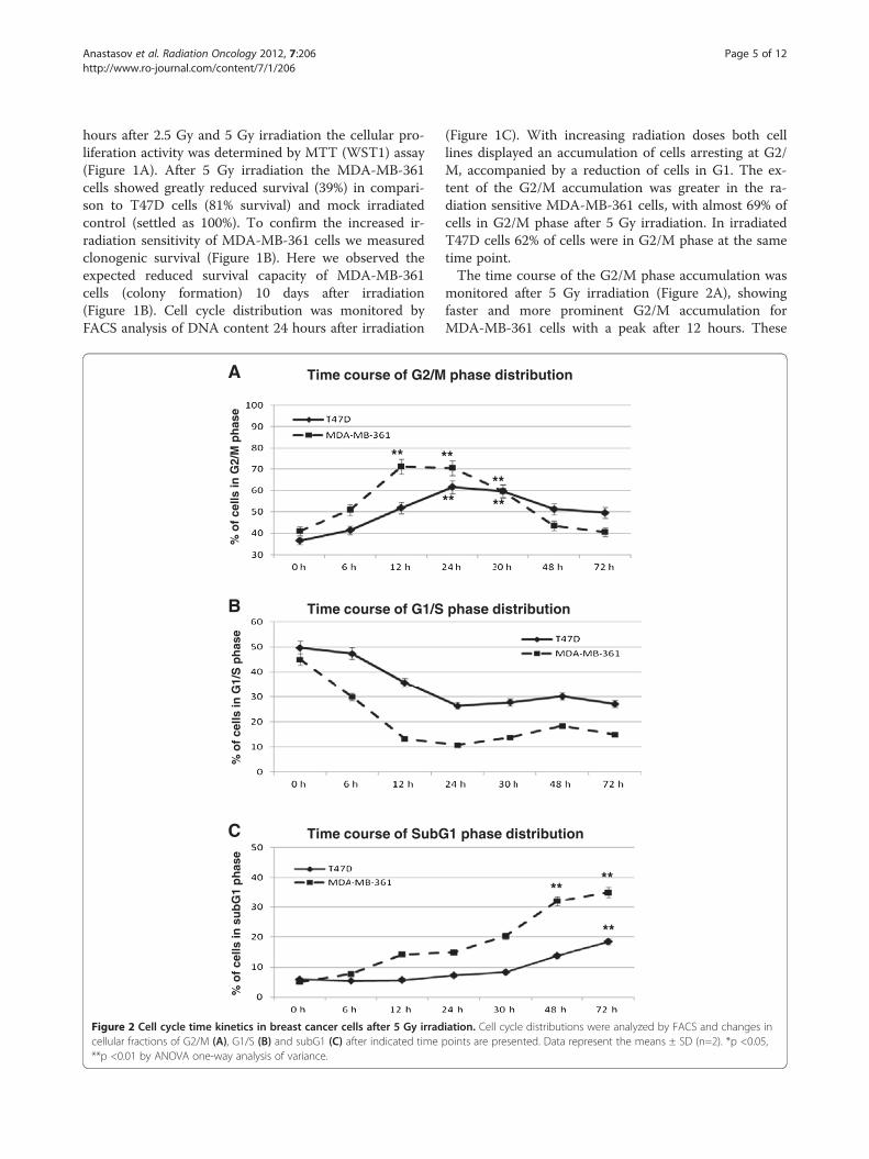
Figure 1 Breast cancer cell survival and cell cycle characterisation after irradiation. (A) Growth characteristics of T47D and MDA-MB-361breast cancer cells were determined by MTT (WST1) assay 72 hours after irradiation. Data represent the means ± SD (n=4). *p <0.05, **p <0.01 byANOVA one-way analysis of variance. (B) clonogenic survival of breast cancer cells 10 days after irradiation. (C) Cell cycle progression in breastcancer cells was evaluated by PI staining and flow cytometry 24 hours after irradiation at the indicated doses.
Anastasov et al. Radiation Oncology 2012, 7:206 Page 4 of 12http://www.ro-journal.com/content/7/1/206
Anastasov et al. Radiation Oncology 2012, 7:206 Page 5 of 12http://www.ro-journal.com/content/7/1/206
hours after 2.5 Gy and 5 Gy irradiation the cellular pro-liferation activity was determined by MTT (WST1) assay(Figure 1A). After 5 Gy irradiation the MDA-MB-361cells showed greatly reduced survival (39%) in compari-son to T47D cells (81% survival) and mock irradiatedcontrol (settled as 100%). To confirm the increased ir-radiation sensitivity of MDA-MB-361 cells we measuredclonogenic survival (Figure 1B). Here we observed theexpected reduced survival capacity of MDA-MB-361cells (colony formation) 10 days after irradiation(Figure 1B). Cell cycle distribution was monitored byFACS analysis of DNA content 24 hours after irradiation
A
B
C
% o
f ce
lls in
G2/
M p
has
e %
of
cells
in G
1/S
ph
ase
% o
f ce
lls in
su
bG
1 p
has
e
Time course of G2/M
Time course of G1/S
Time course of SubG
** *
Figure 2 Cell cycle time kinetics in breast cancer cells after 5 Gy irradcellular fractions of G2/M (A), G1/S (B) and subG1 (C) after indicated time**p <0.01 by ANOVA one-way analysis of variance.
(Figure 1C). With increasing radiation doses both celllines displayed an accumulation of cells arresting at G2/M, accompanied by a reduction of cells in G1. The ex-tent of the G2/M accumulation was greater in the ra-diation sensitive MDA-MB-361 cells, with almost 69% ofcells in G2/M phase after 5 Gy irradiation. In irradiatedT47D cells 62% of cells were in G2/M phase at the sametime point.The time course of the G2/M phase accumulation was
monitored after 5 Gy irradiation (Figure 2A), showingfaster and more prominent G2/M accumulation forMDA-MB-361 cells with a peak after 12 hours. These
phase distribution
phase distribution
1 phase distribution
****
**
*
******
iation. Cell cycle distributions were analyzed by FACS and changes inpoints are presented. Data represent the means ± SD (n=2). *p <0.05,

Anastasov et al. Radiation Oncology 2012, 7:206 Page 6 of 12http://www.ro-journal.com/content/7/1/206
changes were accompanied with faster reduction in G1phase (Figure 2B) and the appearance of a subG1 frac-tion of apoptotic cells already 12 hours after irradiation.In T47D cells the changes were less prominent, butslight increase in subG1 fraction was nevertheless de-tectable 72 hours after irradiation (Figure 2C). Theseresults establish the T47D cells as radioresistant andMDA-MB-361 cells as radiosensitive cell line.
Characterization of miR-21 expression after irradiationIncreased miR-21 expression levels were detected inboth cell lines (T47D and MDA-MB-361) compared to
B miR-21 expression in T47
Rel
ativ
e m
iR-2
1 ex
pre
ssio
n
miR-21 expression in MD
Rel
ativ
e m
iR-2
1 ex
pre
ssio
n
C
Rel
ativ
e m
iR-2
1 ex
pre
ssio
n
control tissue
MDA-MB-361
A miR-21 expression levels in c
**
**
**
*
Figure 3 Time kinetics of miR-21 expression in breast cancer cells aftcell lines compared to adjacent control mammary tissue. (B) Relative miR-25Gy irradiation and indicated time points in relation to the expression of sh*p <0.05, **p <0.01 by ANOVA.
control adjacent mammary tissue (Figure 3A). Interest-ingly the resistant T47D showed fivefold higher miR-21expression than MDA-MB-361 (Figure 4A).In order to determine if miR-21 expression is in-
fluenced by ionizing radiation, the miR-21 levels weremeasured in exponentially growing breast cancer cells(T47D and MDA-MB-361) following 5 Gy irradiation(Figure 3B and C). MiR-21 expression showed a pro-minent induction 8 hours after irradiation only in theradioresistant T47D cells. These transient changes inmiR-21 expression levels correlate with postponedlower apoptotic subG1 fraction accumulation in the
D cells after 5 Gy irradiation
A-MB-361 cells after 5 Gy irradiation
T47D
ell lines
**
**
*
*
er 5 Gy irradiation. (A) Relative miR-21 expression in breast cancer1 expression in T47D and (C) MDA-MB-361 breast cancer cells afteram irradiated controls. Data represent the means ± SD (n=4).
B
C
MDA-MB-361 + EV + miR-21 + anti-miR-21
Colorimetric MTT (WST1) Assay in MDA-MB-361 cells
Cel
l pro
lifer
atio
n in
dex
(%)
6%
T47D + EV + miR-21 + anti-miR-21
Colorimetric MTT (WST1) Assay in T47D cells
Cel
l pro
lifer
atio
n in
dex
(%)
17%
control
control
**
**
***
***
***
***
A
control + EV + miR-21 + anti-miR-21
miR-21 overexpression (miR-21) and downregulation (anti-miR-21)
Rel
ativ
e m
iR-2
1 ex
pre
ssio
n
****
***
***
2.2 0.2
5.7 5.5
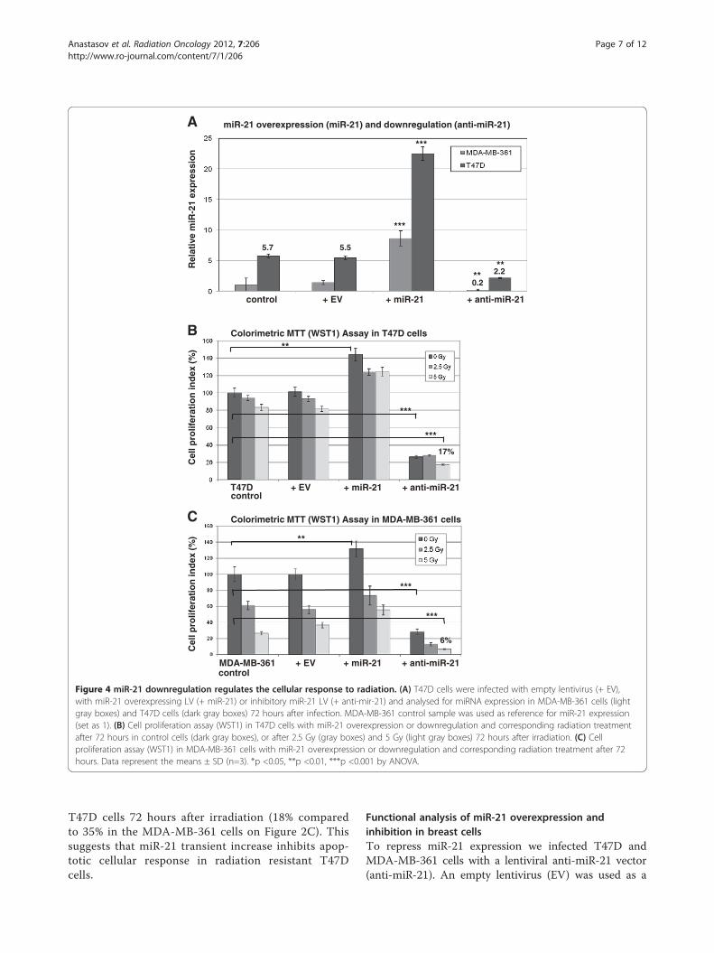
Figure 4 miR-21 downregulation regulates the cellular response to radiation. (A) T47D cells were infected with empty lentivirus (+ EV),with miR-21 overexpressing LV (+ miR-21) or inhibitory miR-21 LV (+ anti-mir-21) and analysed for miRNA expression in MDA-MB-361 cells (lightgray boxes) and T47D cells (dark gray boxes) 72 hours after infection. MDA-MB-361 control sample was used as reference for miR-21 expression(set as 1). (B) Cell proliferation assay (WST1) in T47D cells with miR-21 overexpression or downregulation and corresponding radiation treatmentafter 72 hours in control cells (dark gray boxes), or after 2.5 Gy (gray boxes) and 5 Gy (light gray boxes) 72 hours after irradiation. (C) Cellproliferation assay (WST1) in MDA-MB-361 cells with miR-21 overexpression or downregulation and corresponding radiation treatment after 72hours. Data represent the means ± SD (n=3). *p <0.05, **p <0.01, ***p <0.001 by ANOVA.
Anastasov et al. Radiation Oncology 2012, 7:206 Page 7 of 12http://www.ro-journal.com/content/7/1/206
T47D cells 72 hours after irradiation (18% comparedto 35% in the MDA-MB-361 cells on Figure 2C). Thissuggests that miR-21 transient increase inhibits apop-totic cellular response in radiation resistant T47Dcells.
Functional analysis of miR-21 overexpression andinhibition in breast cellsTo repress miR-21 expression we infected T47D andMDA-MB-361 cells with a lentiviral anti-miR-21 vector(anti-miR-21). An empty lentivirus (EV) was used as a
Anastasov et al. Radiation Oncology 2012, 7:206 Page 8 of 12http://www.ro-journal.com/content/7/1/206
control in parallel with a lentivirus overexpressing miR-21. The level of mature miR-21 in infected and controlcells was measured by quantitative RT-PCR and corre-lated to MDA-MB-361 as 1 fold expression control(Figure 4A). In T47D cells miR-21 expression was 5.5fold higher than in MDA-MB-361 cells. The miR-21overexpression produced 4 fold higher levels of miR-21when compared to untreated T47D cells and cellsinfected with control virus (EV) 72 hours post infection(dark gray boxes, Figure 4A). Consequently, miR-21 ex-pression was decreased by anti-miR-21 lentiviral infec-tion as compared to control cells (T47D and EV).Nevertheless the inhibition of miR-21 expression inT47D cells was 2.2 fold higher than miR-21 expressionlevels in untreated MDA-MB-361 cells. Accordingly,miR-21 overexpression and downregulation data wereanalyzed in MDA-MB-361 cells (light gray boxes,Figure 4A) with more prominent inhibition of miR-21
A
T47D control
+ EV
+ miR-21
+ anti-miR-21
SubG1: 16.8% G2/M: 22.9%
SubG1: 9.4%G2/M: 29.5%
SubG1: 5.9%G2/M: 36.6%
SubG1: 11.3%G2/M: 29.2%
0 Gy 5
T47D + EV + miR-21 + anti-miR-21
B
sub
G1
frac
tio
n (
%)
control
Apoptosis induction in T47D cells
**
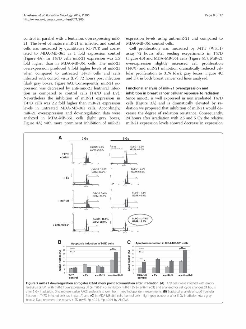
Figure 5 miR-21 downregulation abrogates G2/M check point accumlentivirus (+ EV), with miR-21 overexpressing LV (+ miR-21) or inhibitory miafter 5 Gy irradiation. One representative FACS analysis is shown from threefraction in T47D infected cells (as in part A) and (C) in MDA-MB-361 cells (cboxes). Data represent the means ± SD (n=3). *p <0.05, **p <0.01 by ANOV
expression levels using anti-miR-21 and compared toMDA-MB-361 control cells.Cell proliferation was measured by MTT (WST1)
assay 72 hours after seeding exeperiments in T47D(Figure 4B) and MDA-MB-361 cells (Figure 4C). MiR-21overexpression slightly increased cell proliferation(140%) and miR-21 inhibition dramatically reduced cel-lular proliferation to 31% (dark gray boxes, Figure 4Cand D), in both breast cancer cell lines analyzed.
Functional analysis of miR-21 overexpression andinhibition in breast cancer cellular response to radiationSince miR-21 is well expressed in non irradiated T47Dcells (Figure 3A) and is dramatically elevated by ra-diation we proposed that inhibition of miR-21 would de-crease the degree of radiation resistance. Consequently,24 hours after irradiation with 2.5 and 5 Gy the relativemiR-21 expression levels showed decrease in expression
SubG1: 27.4%G2/M: 18.6%
SubG1: 7.3% G2/M: 61.6%
SubG1: 7.8% G2/M: 46.9%
SubG1: 6.9%G2/M: 64.0%
Gy
MDA-361 + EV + miR-21 + anti-miR-21
C
sub
G1
frac
tio
n (
%)
control
Apoptosis induction in MDA-MB-361 cells
**
**
ulation after irradiation. (A) T47D cells were infected with emptyR-21 LV (+ anti-mir-21) and analysed for cell cycle changes 24 hoursindependent experiments. (B) Statistical analysis of subG1 cellularontrol cells - light gray boxes) or after 5 Gy irradiation (dark grayA.
A
B
n=8/2
n=25/15
p=0.091
miR-21 expression in IDC breast cancer with radiation therapy (33 cases)
Pro
bab
ility
distant metastasis free survival (months)
miR-21 expression in IDC breast cancer (86 cases)
distant metastasis free survival (months)
n=59/32
p=0.029
n=27/8
Pro
bab
ility
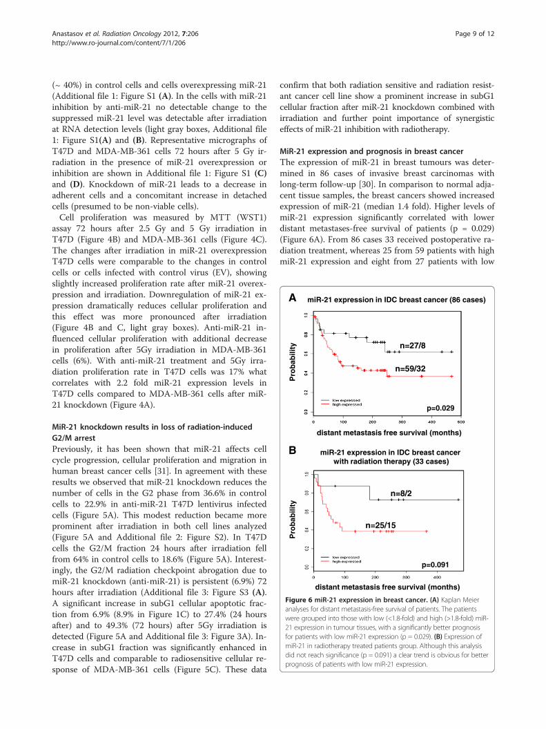
Figure 6 miR-21 expression in breast cancer. (A) Kaplan Meieranalyses for distant metastasis-free survival of patients. The patientswere grouped into those with low (<1.8-fold) and high (>1.8-fold) miR-21 expression in tumour tissues, with a significantly better prognosisfor patients with low miR-21 expression (p = 0.029). (B) Expression ofmiR-21 in radiotherapy treated patients group. Although this analysisdid not reach significance (p = 0.091) a clear trend is obvious for betterprognosis of patients with low miR-21 expression.
Anastasov et al. Radiation Oncology 2012, 7:206 Page 9 of 12http://www.ro-journal.com/content/7/1/206
(~ 40%) in control cells and cells overexpressing miR-21(Additional file 1: Figure S1 (A). In the cells with miR-21inhibition by anti-miR-21 no detectable change to thesuppressed miR-21 level was detectable after irradiationat RNA detection levels (light gray boxes, Additional file1: Figure S1(A) and (B). Representative micrographs ofT47D and MDA-MB-361 cells 72 hours after 5 Gy ir-radiation in the presence of miR-21 overexpression orinhibition are shown in Additional file 1: Figure S1 (C)and (D). Knockdown of miR-21 leads to a decrease inadherent cells and a concomitant increase in detachedcells (presumed to be non-viable cells).Cell proliferation was measured by MTT (WST1)
assay 72 hours after 2.5 Gy and 5 Gy irradiation inT47D (Figure 4B) and MDA-MB-361 cells (Figure 4C).The changes after irradiation in miR-21 overexpressionT47D cells were comparable to the changes in controlcells or cells infected with control virus (EV), showingslightly increased proliferation rate after miR-21 overex-pression and irradiation. Downregulation of miR-21 ex-pression dramatically reduces cellular proliferation andthis effect was more pronounced after irradiation(Figure 4B and C, light gray boxes). Anti-miR-21 in-fluenced cellular proliferation with additional decreasein proliferation after 5Gy irradiation in MDA-MB-361cells (6%). With anti-miR-21 treatment and 5Gy irra-diation proliferation rate in T47D cells was 17% whatcorrelates with 2.2 fold miR-21 expression levels inT47D cells compared to MDA-MB-361 cells after miR-21 knockdown (Figure 4A).
MiR-21 knockdown results in loss of radiation-inducedG2/M arrestPreviously, it has been shown that miR-21 affects cellcycle progression, cellular proliferation and migration inhuman breast cancer cells [31]. In agreement with theseresults we observed that miR-21 knockdown reduces thenumber of cells in the G2 phase from 36.6% in controlcells to 22.9% in anti-miR-21 T47D lentivirus infectedcells (Figure 5A). This modest reduction became moreprominent after irradiation in both cell lines analyzed(Figure 5A and Additional file 2: Figure S2). In T47Dcells the G2/M fraction 24 hours after irradiation fellfrom 64% in control cells to 18.6% (Figure 5A). Interest-ingly, the G2/M radiation checkpoint abrogation due tomiR-21 knockdown (anti-miR-21) is persistent (6.9%) 72hours after irradiation (Additional file 3: Figure S3 (A).A significant increase in subG1 cellular apoptotic frac-tion from 6.9% (8.9% in Figure 1C) to 27.4% (24 hoursafter) and to 49.3% (72 hours) after 5Gy irradiation isdetected (Figure 5A and Additional file 3: Figure 3A). In-crease in subG1 fraction was significantly enhanced inT47D cells and comparable to radiosensitive cellular re-sponse of MDA-MB-361 cells (Figure 5C). These data
confirm that both radiation sensitive and radiation resist-ant cancer cell line show a prominent increase in subG1cellular fraction after miR-21 knockdown combined withirradiation and further point importance of synergisticeffects of miR-21 inhibition with radiotherapy.
MiR-21 expression and prognosis in breast cancerThe expression of miR-21 in breast tumours was deter-mined in 86 cases of invasive breast carcinomas withlong-term follow-up [30]. In comparison to normal adja-cent tissue samples, the breast cancers showed increasedexpression of miR-21 (median 1.4 fold). Higher levels ofmiR-21 expression significantly correlated with lowerdistant metastases-free survival of patients (p = 0.029)(Figure 6A). From 86 cases 33 received postoperative ra-diation treatment, whereas 25 from 59 patients with highmiR-21 expression and eight from 27 patients with low
Anastasov et al. Radiation Oncology 2012, 7:206 Page 10 of 12http://www.ro-journal.com/content/7/1/206
miR-21 expression were treated with radiation therapy(Figure 6B). A trend towards better prognosis andincreased survival in patients with low miR-21 expressionreceiving radiation therapy was evident, but will requirevalidation with a much larger patient collective.
DiscussionUpregulation of miR-21 is a frequent miRNA alterationdescribed in human cancers [32]. The consequences ofoverexpression of miR-21 is that it acts as an “oncomir”blocking apoptosis [33], promoting cell proliferation[18,34] and causing invasion and metastasis [35,36]. Itappears that miR-21 targets multiple tumour-supressivepathways [31] and recent studies showed convincing evi-dence that miR-21 negatively regulates Cdc25A and cellcycle progression in colon cancer [37] and in humanglioblastoma cells [38]. According to miR-21 target ana-lysis, Lu et al., demonstrated that miR-21 promotes celltransformation by targeting the programmed cell death4 gene (PDCD4) in MCF7 breast cancer cells [39]. Thereis some evidence that miRNAs are also involved inmodulating radiation sensitivity in lymphoblastic celllines [7], endothelial cells [14] and for resistance to cyto-toxic anticancer therapy in lung cancer cells [11]. Datapublished recently from Gwak et al., [40] demonstratethe importance of miR-21 knockdown in radiosensita-tion of glioblastomas. In correlation with our resultsthey present importance of high miR-21 expressionlevels in conferring radiation resistance in glioblastomas.The roles of miR-21 expression in modulating responseof breast tumour cells to irradiation have not been pre-viously analyzed.Therefore, we have investigated miR-21 expression in
radiation resistant and radiation sensitive breast cancercells after exposure to γ-irradiation. We observed thatthe expression of miR-21 was not significantly changedafter 5 Gy exposure of the radiosensitive MDA-MB-361cells, but was transiently increased in radiation resistantT47D cells. This data support hypothesis that miR-21 isnot merely upregulated in association with oncogenesis,but rather can act as radioresistant miRNA when transi-ently overexpressed after radiation treatment [36].The G2/M checkpoint arrest is prominent after expos-
ure to DNA damage reagents such as γ-irradiation[21,41]. Our cell cycle data analysis showed that theanti-apoptotic action of miR-21 is also evident after ra-diation exposure and correlates with radiation resistance.In addition, miR-21 influence cell cycle progression viathe DNA damage-G2 checkpoint induction. In this mat-ter miR-21 inhibition (anti-miR-21) is able to reduce theG2/M block and to enhance apoptosis induction 24hours after radiation treatment (Figure 5A). All togetherthese data suggest the importance of combination the-rapy such as radiotherapy with efficient G2/M check
point inhibitor anti-miR-21. Supporting our results inthe manuscript of Li et al. [38], it is presented that miR-21 inhibitor reduces G2/M arrest what is inconsistentwith recently published data from Gwak et al., [40]showing G2/M induction after miR-21 knockdown inglioblastoma cells. This highlights the importance of G2/M arrest after radiation treatment to be studied in differ-ent tumour cell types to further support a general con-clusion about miR-21 function in radioresitance.The data presented in Figure 6 confirm previously
published data from Yan et al. [18], demonstratingincreased expression of miR-21 in breast cancer. Weidentify that patients with low expression levels of miR-21 have better clinical outcome. Previously it has beenreported that high levels of miR-21 expression correlatewith advanced clinical stage, lymph node metastasis andshortened survival of the patients [18,42]. This is con-firmed by the association we observe between low miR-21 expression and distant metastasis free survival. Therole of miR-21 in shaping the response to radiotherapyis suggested by the increased clinical survival seen forlow miR-21 patient group after radiation therapy.
ConclusionsTaken together, our results show that miR-21 expressiontransiently increases in response to irradiation treatmentin the T47D radiation resistant cell line. Furthermore,the miR-21 knockdown improved radiation inducedapoptosis and growth arrest in radiation resistant cellsalmost to the same extent as in sensitive breast cancercells (MDA-MB-361). These findings are important con-cerning the better clinical outcome for patients with lowmiR-21 expression levels and the use of miR-21 as po-tential target in breast cancer therapy.
Additional files
Additional file 1: Figure S1. qRT-PCR quantification of miR-21overexpression and downregulation 24 hours after irradiation. (A) T47Dand (B) MDA-MB-361 cells were infected with empty lentivirus (+ EV),with miR-21 overexpressing LV (+ miR-21) or inhibitory miR-21 LV (+ anti-mir-21) and analysed for miRNA expression changes in control cells (darkgray boxes), or after 2.5 Gy (gray boxes) and 5 Gy (light gray boxes) 24hours after irradiation. Data represent the means ± SD (n=3). *p <0.05, **p <0.01 by ANOVA. (C) Representative micrographs (scale bar = 50 μm)of T47D cells and (D) MDA-MB-361 cells 72 hours after 5 Gy irradiationwith miR-21 overexpression (+ miR-21) or inhibition (+ anti-miR-21).
Additional file 2: Figure S2. miR-21 downregulation inducesconsiderable cellular apoptosis 24 hours after irradiation in MDA-MB-361cells. MDA-MB-361 cells were infected with empty lentivirus (+ EV), withmiR-21 overexpressing LV (+ miR-21) or inhibitory miR-21 LV (+ anti-miR-21) and analysed for cell cycle changes 24 hours after 5 Gy irradiation.One representative FACS analysis is shown from three independentexperiments.
Additional file 3: Figure S3. miR-21 downregulation inducesconsiderable cellular apoptosis 72 hours after irradiation in T47D cells. (A)T47D cells were infected with empty lentivirus (+ EV), with miR-21overexpressing LV (+ miR-21) or inhibitory miR-21 LV (+ anti-mir-21) and
Anastasov et al. Radiation Oncology 2012, 7:206 Page 11 of 12http://www.ro-journal.com/content/7/1/206
analysed for cell cycle changes 72 hours after 5 Gy irradiation. Onerepresentative FACS analysis is shown. (B) Statistical analysis of subG1cellular fraction in T47D infected cells (control cells - light gray boxes) orafter 5 Gy irradiation (dark gray boxes). Data represent the means ± SD(n=3). *p <0.05 by ANOVA.
Competing interestsThe authors declare no potential conflict of interest.
Authors’ contributionsNA, MA and MJA coordinated the study and drafted the manuscript. NA, IH,IGV, KR, and NL performed experiments and analyzed data. GA contributedimportant material for analysis. HB performed statistical analysis. All authorsread and approved the final manuscript.
AcknowledgementsThe authors thank Katrin Lindner and Stefanie Winkler for excellent technicalassistance. Partially supported by a grant from the DeutscheWirtschaftsministerium (KF 2341803SB1) and by the European Commisson(Seventh Framework Programme) 'STORE' Project, no. 232628.
Author details1Institute of Radiation Biology, Helmholtz Zentrum Muenchen, GermanResearch Center for Environmental Health, Muenchen, Germany. 2Institute ofPathology, Helmholtz Zentrum Muenchen, German Research Center forEnvironmental Health, Muenchen, Germany. 3Research Unit of RadiationCytogenetics, Helmholtz Zentrum Muenchen, German Research Center forEnvironmental Health, Muenchen, Germany. 4Department of Oncology andPathology, Karolinska Institute and Hospital, Stockholm, Sweden. 5RadiationBiology, Institute of Radiation Oncology, Technical University Munich,Munich, Germany.
Received: 29 June 2012 Accepted: 23 November 2012Published: 5 December 2012
References1. Bartel DP: MicroRNAs: target recognition and regulatory functions.
Cell 2009, 136(2):215–233.2. Calin GA, Croce CM: MicroRNA signatures in human cancers. Nat Rev
Cancer 2006, 6(11):857–866.3. Ventura A, Jacks T: MicroRNAs and cancer: short RNAs go a long way.
Cell 2009, 136(4):586–591.4. Volinia S, Calin, Liu GA, Ambs CG, Cimmino S, Petrocca A, Visone F, Iorio R,
Roldo M, Ferracin C, et al: A microRNA expression signature of humansolid tumors defines cancer gene targets. Proc Natl Acad Sci USA 2006,103(7):2257–2261.
5. Lu J, Getz, Miska G, Alvarez-Saavedra EA, Lamb E, Peck J, Sweet-Cordero D,Ebert A, Mak BL, Ferrando RH, et al: MicroRNA expression profiles classifyhuman cancers. Nature 2005, 435(7043):834–838.
6. Iorio MV, Ferracin M, Liu CG, Veronese A, Spizzo R, Sabbioni S, Magri S,Pedriali M, Fabbri M, Campiglio M, et al: MicroRNA gene expressionderegulation in human breast cancer. Cancer Res 2005, 65(16):7065–7070.
7. Chaudhry MA: Real-time PCR analysis of micro-RNA expression inionizing radiation-treated cells. Cancer Biother Radiopharm 2009,24(1):49–56.
8. Ambros V: The functions of animal microRNAs. Nature 2004,431(7006):350–355.
9. Mendell JT: MicroRNAs: critical regulators of development, cellularphysiology and malignancy. Cell Cycle 2005, 4(9):1179–1184.
10. Jameel JK, Rao VS, Cawkwell L, Drew PJ: Radioresistance in carcinoma ofthe breast. Breast 2004, 13(6):452–460.
11. Weidhaas JB, Babar I, Nallur SM, Trang P, Roush S, Boehm M, Gillespie E,Slack FJ: MicroRNAs as potential agents to alter resistance to cytotoxicanticancer therapy. Cancer Res 2007, 67(23):11111–11116.
12. Maes OC, An J, Sarojini H, Wu H, Wang E: Changes in MicroRNA expressionpatterns in human fibroblasts after low-LET radiation. J Cell Biochem 2008,105(3):824–834.
13. Wagner-Ecker M, Schwager C, Wirkner U, Abdollahi A, Huber P, et al:MicroRNA expression after ionizing radiation in human endothelial cells.Radiat Oncol 2010, 5:25.
14. Kraemer A, Anastasov N, Angermeier M, Winkler K, Atkinson MJ, Moertl S:MicroRNA-mediated processes are essential for the cellular radiationresponse. Radiat Res 2011, 176(5):575–586.
15. Sempere LF, Christensen M, Silahtaroglu A, Bak M, Heath CV, Schwartz G,Wells W, Kauppinen S, Cole CN: Altered MicroRNA expression confined tospecific epithelial cell subpopulations in breast cancer. Cancer Res 2007,67(24):11612–11620.
16. Si ML, Zhu S, Wu H, Lu Z, Wu F, Mo YY: miR-21-mediated tumor growth.Oncogene 2007, 26(19):2799–2803.
17. Frankel LB, Christoffersen NR, Jacobsen A, Lindow M, Krogh A, Lund AH:Programmed cell death 4 (PDCD4) is an important functional target ofthe microRNA miR-21 in breast cancer cells. J Biol Chem 2008,283(2):1026–1033.
18. Yan LX, Huang XF, Shao Q, Huang MY, Deng L, Wu QL, Zeng YX, Shao JY:MicroRNA miR-21 overexpression in human breast cancer is associatedwith advanced clinical stage, lymph node metastasis and patient poorprognosis. RNA 2008, 14(11):2348–2360.
19. Yu Z, Baserga R, Chen L, Wang C, Lisanti M, Pestell R: microRNA, cell cycle,and human breast cancer. Am J Pathol 2010, 176(3):1058–1064.
20. Strunz AM, Peschke P, Waldeck W, Ehemann V, Kissel M, Debus J:Preferential radiosensitization in p53-mutated human tumour cell linesby pentoxifylline-mediated disruption of the G2/M checkpoint control.Int J Radiat Biol 2002, 78(8):721–732.
21. Suganuma M, Kawabe T, Hori H, Funabiki T, Okamoto T: Sensitization ofcancer cells to DNA damage-induced cell death by specific cell cycle G2checkpoint abrogation. Cancer Res 1999, 59(23):5887–5891.
22. Levine AJ: p53, the cellular gatekeeper for growth and division. Cell 1997,88(3):323–331.
23. Anastasov N, Klier M, Koch I, Angermeier D, Hofler H, Fend F, Quintanilla-Martinez L: Efficient shRNA delivery into B and T lymphoma cells usinglentiviral vector-mediated transfer. J Hematop 2009, 2(1):9–19.
24. Anastasov N, Bonzheim I, Rudelius M, Klier M, Dau T, Angermeier D, DuysterJ, Pittaluga S, Fend F, Raffeld M, Quintanilla-Martinez L: C/EBPbetaexpression in ALK-positive anaplastic large cell lymphomas is requiredfor cell proliferation and is induced by the STAT3 signaling pathway.Haematologica 2010, 95(5):760–767.
25. Aubele M, Walch AK, Ludyga N, Braselmann H, Atkinson MJ, Luber B, AuerG, Tapio S, Cooke T, Bartlett JM: Prognostic value of protein tyrosinekinase 6 (PTK6) for long-term survival of breast cancer patients. Br JCancer 2008, 99(7):1089–1095.
26. Chen C, Ridzon D, Broomer A, Zhou AJA, Lee Z, Nguyen DH, Barbisin JT, XuM, Mahuvakar NL, Andersen NL, et al: Real-time quantification ofmicroRNAs by stem-loop RT-PCR. Nucleic Acids Res 2005, 33(20):e179.
27. Klier M, Anastasov N, Hermann A, Meindl T, Angermeier D, Raffeld M, FendF, Quintanilla-Martinez L: Specific lentiviral shRNA-mediated knockdownof cyclin D1 in mantle cell lymphoma has minimal effects on cellsurvival and reveals a regulatory circuit with cyclin D2. Leukemia 2008,22(11):2097–2105.
28. Nusse M, Beisker W, Kramer J, Miller B, Schreiber GA, Viaggi S, Weller EM,Wessels JM: Measurement of micronuclei by flow cytometry. Methods CellBiol 1994, 42(Pt B):149–158.
29. Elston CW, Ellis IO: Pathological prognostic factors in breast cancer I. Thevalue of histological grade in breast cancer: experience from a largestudy with long-term follow-up. Histopathology 1991, 19(5):403–410.
30. Aubele M, Auer G, Walch AK, Munro A, Atkinson MJ, Braselmann H,Fornander T, Bartlett JM: PTK (protein tyrosine kinase)-6 and HER2 and 4,but not HER1 and 3 predict long-term survival in breast carcinomas.Br J Cancer 2007, 96(5):801–807.
31. Yan LX, Wu QN, Zhang Y, Li YY, Liao DZ, Hou JH, Fu J, Zeng MS, Yun JP, WuQL, et al: Knockdown of miR-21 in human breast cancer cell lines inhibitsproliferation, in vitro migration and in vivo tumor growth. Breast CancerRes 2011, 13(1):R2.
32. Krichevsky AM, Gabriely G: miR-21: a small multi-faceted RNA. J Cell MolMed 2009, 13(1):39–53.
33. Chan JA, Krichevsky AM, Kosik KS: MicroRNA-21 is an antiapoptotic factorin human glioblastoma cells. Cancer Res 2005, 65(14):6029–6033.
34. Meng F, Henson R, Wehbe-Janek H, Ghoshal K, Jacob ST, Patel T: MicroRNA-21 regulates expression of the PTEN tumor suppressor gene in humanhepatocellular cancer. Gastroenterology 2007, 133(2):647–658.
35. Asangani IA, Rasheed SA, Nikolova DA, Leupold JH, Colburn NH, Post S,Allgayer H: MicroRNA-21 (miR-21) post-transcriptionally downregulates
Anastasov et al. Radiation Oncology 2012, 7:206 Page 12 of 12http://www.ro-journal.com/content/7/1/206
tumor suppressor Pdcd4 and stimulates invasion, intravasation andmetastasis in colorectal cancer. Oncogene 2008, 27(15):2128–2136.
36. Iliopoulos D, Jaeger SA, Hirsch HA, Bulyk ML, Struh K: STAT3 activation ofmiR-21 and miR-181b-1 via PTEN and CYLD are part of the epigeneticswitch linking inflammation to cancer. Mol Cell 2010, 39(4):493–506.
37. Wang P, Zou F, Zhang X, Li H, Dulak A, Tomko RJ Jr, Lazo JS, Wang Z,Zhang L, Yu J: microRNA-21 negatively regulates Cdc25A and cell cycleprogression in colon cancer cells. Cancer Res 2009, 69(20):8157–8165.
38. Li Y, Zhao S, Zhen Y, Li Q, Teng L, Asai A, Kawamoto K: A miR-21 inhibitorenhances apoptosis and reduces G(2)-M accumulation induced byionizing radiation in human glioblastoma U251 cells. Brain Tumor Pathol2011, 28(3):209–214.
39. Lu Z, Liu M, Stribinskis V, Klinge CM, Ramos KS, Colburn NH, Li Y: MicroRNA-21 promotes cell transformation by targeting the programmed celldeath 4 gene. Oncogene 2008, 27(31):4373–4379.
40. Gwak HS, Kim TH, Jo GH, Kim YJ, Kwak HJ, Kim JH, Yin J, Yoo H, Lee SH, ParkJB: Silencing of MicroRNA-21 confers radio-sensitivity through inhibitionof the PI3K/AKT pathway and enhancing autophagy in malignant gliomacell lines. PLoS One 2012, 7(10):e47449.
41. Kawabe T: G2 checkpoint abrogators as anticancer drugs. Mol Cancer Ther2004, 3(4):513–519.
42. Lee JA, Lee HY, Lee ES, Kim I, Bae JW: Prognostic implications ofMicroRNA-21 overexpression in invasive ductal carcinomas of the breast.J Breast Cancer 2011, 14(4):269–275.
doi:10.1186/1748-717X-7-206Cite this article as: Anastasov et al.: Radiation resistance due to highexpression of miR-21 and G2/M checkpoint arrest in breast cancer cells.Radiation Oncology 2012 7:206.
Submit your next manuscript to BioMed Centraland take full advantage of:
• Convenient online submission
• Thorough peer review
• No space constraints or color figure charges
• Immediate publication on acceptance
• Inclusion in PubMed, CAS, Scopus and Google Scholar
• Research which is freely available for redistribution
Submit your manuscript at www.biomedcentral.com/submit