Quantification of Obstructive and Nonobstructive Coronary Lesions by 64Slice Computed Tomography
8
EXPEDITED REVIEWS Quantification of Obstructive and Nonobstructive Coronary Lesions by 64-Slice Computed Tomography A Comparative Study With Quantitative Coronary Angiography and Intravascular Ultrasound Alexander W. Leber, MD,* Andreas Knez, MD,* Franz von Ziegler, MD,* Alexander Becker, MD,* Konstantin Nikolaou, MD,† Stephan Paul, MD,* Bernd Wintersperger, MD,† Maximilian Reiser, MD,† Christoph R. Becker, MD,† Gerhard Steinbeck, MD,* Peter Boekstegers, MD* Munich, Germany OBJECTIVES The aim of the present study was to determine the diagnostic accuracy of 64-slice computed tomography (CT) to identify and quantify atherosclerotic coronary lesions in comparison with catheter-based angiography and intravascular ultrasound (IVUS). BACKGROUND Currently, the ability of multislice CT to quantify the degree of coronary artery stenosis and dimensions of coronary plaques has not been evaluated. METHODS We included 59 patients scheduled for coronary angiography due to stable angina pectoris. A contrast-enhanced 64-slice CT (Senation 64, Siemens Medical Solutions, Forchheim, Germany) was performed before the invasive angiogram. In a subset of 18 patients, IVUS of 32 vessels was part of the catheterization procedure. RESULTS In 55 of 59 patients, 64-slice CT enabled the visualization of the entire coronary tree with diagnostic image quality (American Heart Association 15-segment model). The overall correla- tion between the degree of stenosis detected by quantitative coronary angiography compared with 64-slice CT was r 0.54. Sensitivity for the detection of stenosis 50%, stenosis 50%, and stenosis 75% was 79%, 73%, and 80%, respectively, and specificity was 97%. In comparison with IVUS, 46 of 55 (84%) lesions were identified correctly. The mean plaque areas and the percentage of vessel obstruction measured by IVUS and 64-slice CT were 8.1 mm 2 versus 7.3 mm 2 (p 0.03, r 0.73) and 50.4% versus 41.1% (p 0.001, r 0.61), respectively. CONCLUSIONS Contrast-enhanced 64-slice CT is a clinically robust modality that allows the identification of proximal coronary lesions with excellent accuracy. Measurements of plaque and lumen areas derived by CT correlated well with IVUS. A major limitation is the insufficient ability of CT to exactly quantify the degree of stenosis. (J Am Coll Cardiol 2005;46:147–54) © 2005 by the American College of Cardiology Foundation A noninvasive method that would allow the evaluation of coronary stenoses with a comparable accuracy to catheter- based angiography would have enormous clinical value. The recent development of 16-slice computed tomography (CT) already constitutes an important step forward in noninvasive See page 155 angiography and, although remarkable results to identify high-grade coronary stenoses were reported from several groups, it is evident that this technology is still affected by numerous limitations (1–5). Because of spatial reso- lution, the reliable identification of coronary lesions is restricted to major coronary branches with a diameter of at least 2 mm. Partial volume effects caused by coronary calcifications frequently count for false-positive and false- negative results. Furthermore, it is consistently reported that because of the limited temporal resolution, motion artifacts can only be avoided in patients with heart rates of 65 beats/min (3–7). Those limitations probably contributed to the fact that all existing studies focused on the detection of high-grade stenosis only and, until now, no attempt was directed toward a CT-derived stenosis quantification. For clinical purposes, however, it also is necessary to accurately determine the severity of a lesion because the therapeutic consequence of a 50% or a 90% stenosis may be completely different. The recent intro- duction of a new scanner generation with improved spatial and temporal resolution generating 64 slices per rotation and covering the entire volume of the heart in 8 to 9 s promises a significant improvement of image quality that may allow a more precise evaluation of coronary stenosis. Thus, the aim of the present study was to determine the diagnostic accuracy of a 64-slice CT that offers an isotropic voxel resolution of 0.4 0.4 0.4 mm to identify and quantify atherosclerotic coronary lesions in comparison with catheter-based angiography and intravascular ultrasound (IVUS). From the Departments of *Cardiology and †Clinical Radiology, University of Munich, Munich, Germany. Both Drs. Leber and Knez contributed equally to this study. Manuscript received December 16, 2004; revised manuscript received February 28, 2005, accepted March 10, 2005. Journal of the American College of Cardiology Vol. 46, No. 1, 2005 © 2005 by the American College of Cardiology Foundation ISSN 0735-1097/05/$30.00 Published by Elsevier Inc. doi:10.1016/j.jacc.2005.03.071
-
Upload
independent -
Category
Documents
-
view
0 -
download
0
Transcript of Quantification of Obstructive and Nonobstructive Coronary Lesions by 64Slice Computed Tomography

E
QCACAKCM
Acbra
ahgblrac
Ms
2
Journal of the American College of Cardiology Vol. 46, No. 1, 2005© 2005 by the American College of Cardiology Foundation ISSN 0735-1097/05/$30.00P
XPEDITED REVIEWS
uantification of Obstructive and Nonobstructiveoronary Lesions by 64-Slice Computed TomographyComparative Study With Quantitative
oronary Angiography and Intravascular Ultrasoundlexander W. Leber, MD,* Andreas Knez, MD,* Franz von Ziegler, MD,* Alexander Becker, MD,*onstantin Nikolaou, MD,† Stephan Paul, MD,* Bernd Wintersperger, MD,† Maximilian Reiser, MD,†hristoph R. Becker, MD,† Gerhard Steinbeck, MD,* Peter Boekstegers, MD*unich, Germany
OBJECTIVES The aim of the present study was to determine the diagnostic accuracy of 64-slice computedtomography (CT) to identify and quantify atherosclerotic coronary lesions in comparison withcatheter-based angiography and intravascular ultrasound (IVUS).
BACKGROUND Currently, the ability of multislice CT to quantify the degree of coronary artery stenosis anddimensions of coronary plaques has not been evaluated.
METHODS We included 59 patients scheduled for coronary angiography due to stable angina pectoris. Acontrast-enhanced 64-slice CT (Senation 64, Siemens Medical Solutions, Forchheim,Germany) was performed before the invasive angiogram. In a subset of 18 patients, IVUS of32 vessels was part of the catheterization procedure.
RESULTS In 55 of 59 patients, 64-slice CT enabled the visualization of the entire coronary tree withdiagnostic image quality (American Heart Association 15-segment model). The overall correla-tion between the degree of stenosis detected by quantitative coronary angiography compared with64-slice CT was r � 0.54. Sensitivity for the detection of stenosis �50%, stenosis �50%, andstenosis �75% was 79%, 73%, and 80%, respectively, and specificity was 97%. In comparison withIVUS, 46 of 55 (84%) lesions were identified correctly. The mean plaque areas and the percentageof vessel obstruction measured by IVUS and 64-slice CT were 8.1 mm2 versus 7.3 mm2 (p � 0.03,r � 0.73) and 50.4% versus 41.1% (p � 0.001, r � 0.61), respectively.
CONCLUSIONS Contrast-enhanced 64-slice CT is a clinically robust modality that allows the identification ofproximal coronary lesions with excellent accuracy. Measurements of plaque and lumen areasderived by CT correlated well with IVUS. A major limitation is the insufficient ability of CTto exactly quantify the degree of stenosis. (J Am Coll Cardiol 2005;46:147–54) © 2005 by
ublished by Elsevier Inc. doi:10.1016/j.jacc.2005.03.071
the American College of Cardiology Foundation
ntaoctnqnbsdsrtqcttml
noninvasive method that would allow the evaluation oforonary stenoses with a comparable accuracy to catheter-ased angiography would have enormous clinical value. Theecent development of 16-slice computed tomography (CT)lready constitutes an important step forward in noninvasive
See page 155
ngiography and, although remarkable results to identifyigh-grade coronary stenoses were reported from severalroups, it is evident that this technology is still affectedy numerous limitations (1–5). Because of spatial reso-ution, the reliable identification of coronary lesions isestricted to major coronary branches with a diameter oft least 2 mm. Partial volume effects caused by coronaryalcifications frequently count for false-positive and false-
From the Departments of *Cardiology and †Clinical Radiology, University ofunich, Munich, Germany. Both Drs. Leber and Knez contributed equally to this
tudy.
aManuscript received December 16, 2004; revised manuscript received February 28,
005, accepted March 10, 2005.
egative results. Furthermore, it is consistently reportedhat because of the limited temporal resolution, motionrtifacts can only be avoided in patients with heart ratesf �65 beats/min (3–7). Those limitations probablyontributed to the fact that all existing studies focused onhe detection of high-grade stenosis only and, until now,o attempt was directed toward a CT-derived stenosisuantification. For clinical purposes, however, it also isecessary to accurately determine the severity of a lesionecause the therapeutic consequence of a 50% or a 90%tenosis may be completely different. The recent intro-uction of a new scanner generation with improvedpatial and temporal resolution generating 64 slices perotation and covering the entire volume of the heart in 8o 9 s promises a significant improvement of imageuality that may allow a more precise evaluation oforonary stenosis. Thus, the aim of the present study waso determine the diagnostic accuracy of a 64-slice CThat offers an isotropic voxel resolution of 0.4 � 0.4 � 0.4m to identify and quantify atherosclerotic coronary
esions in comparison with catheter-based angiography
nd intravascular ultrasound (IVUS).
M
Pcd1c6psccpatCHbspoasTgsh(wiHtwtsadttearh1�
athuhaiaaaIaIawbad
hcdeatnqf
ccsicdstolap
wfeism(atwsco
148 Leber et al. JACC Vol. 46, No. 1, 2005MSCT for the Detection of Coronary Plaques July 5, 2005:147–54
ETHODS
atients. From July to November 2004, we studied 59onsecutive patients (no previously known coronary arteryisease [n � 49], patients with previous angioplasty [n �0]) scheduled for conventional coronary angiography be-ause of stable angina pectoris who were suitable for a4-slice CT scan at least one day before the catheterizationrocedure. Patients with atrial fibrillation, previous bypassurgery, previous stenting of �1 vessel, an unstable clinicalondition, or a contraindication to the administration ofontrast agent were excluded. The 64-slice CT scan waserformed within two days before coronary angiography inll patients. The study protocol included the oral adminis-ration of 50 mg of metoprolol 60 min before the scheduledT scan in patients with heart rates �70 beats/min.owever, in the presence of contraindications for a beta-
locker or an unsatisfactory lowering of the heart rate, thecan was performed even at higher heart rates. The studyrotocol was approved by the institutional ethics committeef the Grosshadern Hospital of the University of Munich,nd all patients gave informed consent to participate in thetudy.he 64-slice CT scanning technique. Computed tomo-raphic angiography was performed using a 64-slice CTcanner (Sensation 64, Siemens Medical Solutions, Forch-eim, Germany). A bolus of 80 ml of contrast agentSolutrast 300, 300 mgI/ml�1, Altana, Konstanz, Germany)as injected intravenously (5 ml/s�). As soon as the signal
n the ascending aorta reached a predefined threshold of 100U, the scan started automatically and the entire volume of
he heart was acquired during one breathhold in 8 to 9 sith simultaneous recording of the electrocardiographic
race. The detector collimation was 0.6 mm, gantry rotationpeed was 330 ms per rotation, tube voltage was 120 kV atcurrent of 550 to 750 mAs (depending on patient size)
uring 55% of the cardiac cycle (diastole), and a reduction ofhe current by 80% was performed during the remainingime of the R-R interval, leading to an estimated meanffective radiation dose of approximately 10 to 14 mSv. Bypplying a half scan algorithm (only data from a 180° gantryotation is used for image reconstruction) in patients witheart rates �65 beats/min, acquisition time was reduced to65 ms (gantry rotation time/2 � 330 ms/2). For heart rates
Abbreviations and AcronymsCSA � cross-sectional areaCT � computed tomographyEEM � external elastic membraneIVUS � intravascular ultrasoundLAD � left anterior descending arteryLCX � left circumflex arteryMSCT � multislice computed tomographyQCA � quantitative coronary angiographyRCA � right coronary artery
65 beats/min, the adaptive cardio volume mode was o
ctivated, which automatically switches from a one-segmento a two-segment scan (two segments from consecutiveeart cycles that together provide data of a 180° rotation aresed for slice reconstruction). Depending on the patient’seart rate, a maximum temporal resolution of 83 ms can bechieved (8,9). Using retrospective electrocardiographic gat-ng, we performed routinely reconstructions at 500, 600,nd 700 ms before the R-wave (10). Because of motionrtifacts at these reconstructions, additional reconstructionst 300, 400, and 550 ms were performed in six patients.mage analysis of 64-slice CT. The CT data set wasnalyzed by two independent experienced readers using theNSIGHT (Neoimagery Co., City of Industry, California)nd the Leonardo (Siemens, Forchheim, Germany) soft-are packages. In a first step, image quality was determinedy the investigators on the basis of the presence of motionrtifacts and on the basis of the contrast-to-noise ratio, asescribed in previous studies (11).The grading criteria for image quality were as follows: A
igh-quality image was defined as no motion artifacts and aontrast-to-noise ratio of �8; a moderate-quality image wasefined as motion artifacts present but the vessel stillvaluable or a contrast-to-noise ratio between 4 and 8; andpoor-quality image was defined as motion artifacts present
hat made vessel delineation impossible or a contrast-to-oise ratio �4. Only patients who had high- and moderate-uality images of all coronary segments were considered forurther analysis.
Any discernible structure that could be assigned to theoronary artery wall, that had a CT density less than theontrast-enhanced coronary lumen but greater than theurrounding connective tissue, and that could be identifiedn at least two independent planes was defined as a noncal-ified coronary atherosclerotic plaque. Any structure with aensity of 130 HU or more that could be visualizedeparately from the contrast-enhanced coronary lumen (ei-her because it was “embedded” within noncalcified plaquer because its density was above the contrast-enhancedumen), that could be assigned to the coronary artery wall,nd that could be identified in at least two independentlanes was defined as a calcified atherosclerotic plaque (12).The display setting used for lumen and plaque quantification
as determined empirically in a subset of six patients (recruitedrom the cohort of patients in whom IVUS was performed). Inach patient, four different coronary sites that were easy todentify because of their location next to landmarks wereelected. The image display setting at each site was thenanipulated so that the multislice computed tomographic
MSCT) image equaled the IVUS image in size and patternnd allowed exact separation between vessel, surroundingissue, plaque, and lumen. The values for window width andindow level of the respective section were recorded and were
et in relation to the mean intensity within the lumen at theorresponding site. The results of this analysis revealed that theptimal setting to detect plaque and outer vessel boundaries is
btained on average at a width representing 155% (range, 395
tlHtvIac
(rndtpIa
FllvmL
149JACC Vol. 46, No. 1, 2005 Leber et al.July 5, 2005:147–54 MSCT for the Detection of Coronary Plaques
o 809 HU) of the mean intensity within the lumen and at aevel representing 65% of the mean intensity (range, 165 to 339
U). Keeping the window level (65% of mean intensity withinhe corresponding lumen) and reducing the width to a HUalue of one, measurements provided optimal matching withVUS (Fig. 1). Those initial measurements were performed byn independent investigator who was not involved in the lateromparative analysis.
igure 1. Angiographically nonobstructive lesion of the left anterior desceesion. (B) 64-slice computed tomography: arrowheads indicate a noncalcongitudinal reconstruction. (D) Intravascular ultrasound cross section: lumessel. (F) Window setting for lumen measurements: width is reduced toeasured in the lumen. (G) Window level to determine outer vessel boundaumen area is 4 mm2, plaque area is 1 mm2.
For comparison with quantitative coronary angiography u
QCA), the grade of diameter stenosis (maximum diametereduction) was determined in longitudinal curved multipla-ar reformatted reconstructions by dividing the minimaliameter in the diseased segment through the diameter inhe adjacent proximal disease-free section in the same tworojections that were used for QCA. For comparison withVUS, the percent area stenosis as well as plaque and vesselreas were determined in the cross sections of the coronaries
artery. (A) Invasive angiogram: arrow indicates a nonobstructive smoothlaque in the left anterior descending artery. (C) Intravascular ultrasound:ea 4 mm2, plaque area 11 mm2. (E) Cross-sectional view of the coronary, window level is set to 65% (210 HU in this case) of the mean intensityidth at 155% of mean value within the lumen, level at 65% of mean value).
ndingified pen ar
1 HUries (w
sing thin maximum intensity projections (Fig. 2).

CsCmcd3depl(spiAACtlcsCoAmoc
CIsppdcmmwltfiFpoScwS�rcolt(
Fot vasivp artery
150 Leber et al. JACC Vol. 46, No. 1, 2005MSCT for the Detection of Coronary Plaques July 5, 2005:147–54
atheter-based coronary angiography and IVUS. Inva-ive coronary angiograms were evaluated by QCA (Quant-or QCA, Siemens Medical Systems, Forchheim, Ger-any) by an independent, blinded investigator. For
oronary artery lesions, the mean diameter reduction wasetermined in two projections. In 18 patients, the IVUS of2 coronary arteries was performed as part of the invasiveiagnostic procedure (motorized pullback at 0.5 mm/s,lectronic 20-MHz eagle view transducer, Volcano Thera-eutics, Rancho Cordova, California). For each detectable
esion, the external elastic membrane cross-sectional areaEEM CSA) and the plaque area, defined as lumen CSAubtracted from EEM CSA, was defined by another inde-endent investigator. Those measurements were performedn accordance to the IVUS interpretation guidelines of themerican College of Cardiology and the American Heartssociation (13).omparison of QCA with MSCT. The determination of
he diagnostic accuracy of 64-slice CT was performed foresions with various degrees of luminal narrowing. Wealculated sensitivity and specificity for lesions causingtenoses �75%, for stenoses �50%, and for stenoses �50%.alculations for sensitivity and specificity were conductedn a segmental basis according to the American Heartssociation’s segment model (15 segments) (1). Further-ore, we correlated measurements for stenosis severity
btained by 64-slice CT to QCA using the Spearman rank
igure 2. Correlation of quantitative coronary angiography (QCA) and 64-f a high-grade stenosis in the left circumflex artery. (Diameter in the referehe stenotic section 0.6 mm on QCA, 0.5 mm on 64-slice CT). (A) Inrojection). (B) Multiplanar reformatted projection of the left circumflex
orrelation.
omparison of IVUS versus MSCT. We performedVUS in 32 coronary arteries without significant coronarytenoses of �50% on conventional angiography. The com-arison with IVUS was based on a site-by-site basis if singlelaques could easily be distinguished as the result of aisease-free section of at least 10 mm between them. In thease that a single plaque involved the entire segment or evenore segments, the comparison was performed on a seg-ental basis. Sections without evidence of atherosclerosisere divided into 10-mm intervals. The site and the
ongitudinal extent of coronary plaques were determined onhe basis of their distance to the ostium and on the basis ofduciary points like side branches or severe calcifications.or each plaque, the maximum EEM CSA, the maximumlaque area, as well as the maximum percent luminalbstruction were determined.tatistics. The diagnostic accuracy of 64-slice CT to detectoronary lesions with various degrees of luminal obstructionas evaluated regarding QCA as the standard of reference.ensitivity and specificity were calculated for stenosis75%, for stenosis �50%, and for lesions �50% diameter
eduction. Furthermore, we separately determined the ac-uracy for extensively calcified lesions (calcium area �50%f vessel area), for noncalcified or moderately calcifiedesions (calcium area �50% of vessel area), and for lesionshat were later targeted for a revascularization proceduree.g., angioplasty or bypass surgery).
omputed tomography (CT) angiography: Visualization and quantificationction 3.1 mm on QCA, 3.0 mm on 64-slice CT; minimal diameter within
e coronary angiogram of the left coronary artery (right anterior obliqueby 64-slice CT.
slice cnce se
The capability of 64-slice CT to detect and quantify
ctbwwEpdbMAcs
R
Astfbhb
sccp5gedo
t
B[2cfti7
fcos2urwmcbaCfiawecc
o�pcrpsdu
sisa
T6
N�5�
A
T
MDALSN
A nts 1,
151JACC Vol. 46, No. 1, 2005 Leber et al.July 5, 2005:147–54 MSCT for the Detection of Coronary Plaques
oronary plaques was further compared with IVUS. Sensi-ivity and specificity were calculated on a plaque-per-plaqueasis and were given for different coronary vessels. Sectionsithout atherosclerosis were divided in 10-mm sections andere the basis for specificity calculations. Correlations ofEM CSA, lumen CSA, and plaque CSA, as well asercent vessel obstruction (comparison with IVUS) andiameter stenosis (comparison with QCA) were determinedy calculating the Spearman rank correlation coefficient.ean values were compared using the double-tailed t test.p value �0.05 was considered to be statistically signifi-
ant. All calculations were performed using the NCSS 2000oftware package (NCSS, Kaysville, Utah).
ESULTS
ccording to the inclusion criteria, contrast-enhanced 64-lice CT was performed successfully without any complica-ions in 59 patients (age, 64 � 10 years) that were scheduledor conventional invasive coronary angiography. Beta-lockers were administered in 21 patients to reduce theireart rate. The mean heart rate during the scan was 62 � 13eats/min.Four of 59 patients had CT angiograms that were
everely affected by motion artifacts (n � 2) or poorontrast-to-noise ratio (n � 2), making an evaluation of alloronary segments impossible. The heart rates of these fouratients were 63, 72, 82, and 89 beats/min. In the remaining5 patients, all coronary segments were graded to provideood or intermediate image quality that was sufficient forvaluation. Nine patients had heart rates �70 beats/minuring the investigation, and diagnostic image quality wasbtained in six of them.A comparison between 64-slice CT and QCA was
heoretically available in 825 segments from 55 patients.
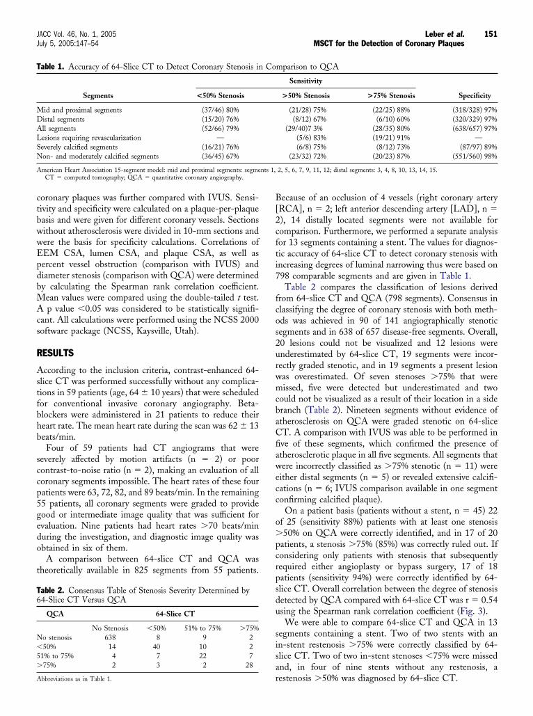
able 2. Consensus Table of Stenosis Severity Determined by4-Slice CT Versus QCA
QCA 64-Slice CT
No Stenosis �50% 51% to 75% �75%o stenosis 638 8 9 250% 14 40 10 2
1% to 75% 4 7 22 775% 2 3 2 28
able 1. Accuracy of 64-Slice CT to Detect Coronary Stenosis in
Segments <50% Stenosis
id and proximal segments (37/46) 80%istal segments (15/20) 76%ll segments (52/66) 79%esions requiring revascularization —everely calcified segments (16/21) 76%on- and moderately calcified segments (36/45) 67%
merican Heart Association 15-segment model: mid and proximal segments: segmeCT � computed tomography; QCA � quantitative coronary angiography.
rbbreviations as in Table 1.
ecause of an occlusion of 4 vessels (right coronary arteryRCA], n � 2; left anterior descending artery [LAD], n �), 14 distally located segments were not available foromparison. Furthermore, we performed a separate analysisor 13 segments containing a stent. The values for diagnos-ic accuracy of 64-slice CT to detect coronary stenosis withncreasing degrees of luminal narrowing thus were based on98 comparable segments and are given in Table 1.Table 2 compares the classification of lesions derived
rom 64-slice CT and QCA (798 segments). Consensus inlassifying the degree of coronary stenosis with both meth-ds was achieved in 90 of 141 angiographically stenoticegments and in 638 of 657 disease-free segments. Overall,0 lesions could not be visualized and 12 lesions werenderestimated by 64-slice CT, 19 segments were incor-ectly graded stenotic, and in 19 segments a present lesionas overestimated. Of seven stenoses �75% that wereissed, five were detected but underestimated and two
ould not be visualized as a result of their location in a sideranch (Table 2). Nineteen segments without evidence oftherosclerosis on QCA were graded stenotic on 64-sliceT. A comparison with IVUS was able to be performed inve of these segments, which confirmed the presence oftherosclerotic plaque in all five segments. All segments thatere incorrectly classified as �75% stenotic (n � 11) were
ither distal segments (n � 5) or revealed extensive calcifi-ations (n � 6; IVUS comparison available in one segmentonfirming calcified plaque).
On a patient basis (patients without a stent, n � 45) 22f 25 (sensitivity 88%) patients with at least one stenosis50% on QCA were correctly identified, and in 17 of 20
atients, a stenosis �75% (85%) was correctly ruled out. Ifonsidering only patients with stenosis that subsequentlyequired either angioplasty or bypass surgery, 17 of 18atients (sensitivity 94%) were correctly identified by 64-lice CT. Overall correlation between the degree of stenosisetected by QCA compared with 64-slice CT was r � 0.54sing the Spearman rank correlation coefficient (Fig. 3).We were able to compare 64-slice CT and QCA in 13
egments containing a stent. Two of two stents with ann-stent restenosis �75% were correctly classified by 64-lice CT. Two of two in-stent stenoses �75% were missednd, in four of nine stents without any restenosis, a
mparison to QCA
Sensitivity
Specificity>50% Stenosis >75% Stenosis
(21/28) 75% (22/25) 88% (318/328) 97%(8/12) 67% (6/10) 60% (320/329) 97%
(29/40)7 3% (28/35) 80% (638/657) 97%(5/6) 83% (19/21) 91% —(6/8) 75% (8/12) 73% (87/97) 89%
(23/32) 72% (20/23) 87% (551/560) 98%
2, 5, 6, 7, 9, 11, 12; distal segments: 3, 4, 8, 10, 13, 14, 15.
Co
estenosis �50% was diagnosed by 64-slice CT.

tcai6bO64c
EE�mvvc0
D
Iaflrtod
pisoawcgCitTerba1totwCcsasmbsrtarDg
F6d
Ti
RLLRT
Cdr
Fe[(cau0
152 Leber et al. JACC Vol. 46, No. 1, 2005MSCT for the Detection of Coronary Plaques July 5, 2005:147–54
Intravascular ultrasound was part of the invasive cathe-erization procedure in 18 patients and was performed in 32oronary vessels (left main and LAD, n � 16; left circumflexrtery [LCX], n � 14; RCA, n � 2). No stents werenvestigated by IVUS. The results for diagnostic accuracy of4-slice CT to detect coronary plaques in these vessels,ased on a site-by-site analysis, are given in Table 3.verall, 55 coronary plaques were detected on IVUS, and
4-slice CT identified 46 of them (sensitivity 84%); in 39 of3 disease-free 10-mm sections, the presence of plaque wasorrectly excluded (specificity 91%; Table 3).
The mean plaque CSA, the mean lumen CSA, the meanEM CAS, and the percent vessel obstruction (% plaque ofEM CSA) determined by IVUS and 64-slice CT were 8.1
3.8 mm2 versus 7.3 � 5.1 mm2 (p � 0.04), 8.4 � 4.5m2 versus 9.4 � 5.1 mm2 (p � 0.01), 16.4 � 5.8 mm2
ersus 16.7 � 7.1 mm2 (p � 0.6, NS), and 50.4 � 14.0%ersus 41.1 � 22.7% (p � 0.001). The correlation coeffi-ients for these measurements were r � 0.73, r � 0.81, r �.88, and r � 0.61, respectively (Fig. 4).
ISCUSSION
n recent publications using 16-slice CT scanners, remark-ble results identifying high-grade coronary stenoses wereound, although this technology is still affected by severalimitations explained by the given spatial and temporalesolution (1,3,7). Those limitations probably contributedo the fact that all existing studies focused on the detectionf high-grade stenosis only and, until now, no attempt wasirected toward a CT-derived stenosis quantification. In the
igure 3. Correlation of quantitative coronary angiography (QCA) and4-slice computed tomography (CT) measurements of diameter stenosis iniseased coronary segments. Pearson’s correlation coefficient r � 0.54.
able 3. Accuracy of 64-Slice CT to Detect Coronary Lesionsn Comparison to IVUS
Sensitivity Specificity
CA (5/6) 83% (6/6) 100%M (5/5) 100% (11/11) 100%AD (26/30) 87% (13/14) 93%CX (10/14) 71% (10/13) 77%otal (46/55) 84% (40/44) 91%
T � computed tomography; IVUS � intravascular ultrasound; LAD � left anterior
iescending artery; LM � left main artery; RCA � right coronary artery; RCX �ight circumflex artery.
resent study, we used a new generation 64-slice CT withmproved spatial and temporal resolution, and we demon-trated that this technology was feasible in identifyingbstructive and nonobstructive coronary lesions with highccuracy in major parts of all three coronaries. Furthermore,e found a good correlation to determine plaque areas
ompared with IVUS. However, the ability to quantify therade of luminal obstruction is limited.linical feasibility. Previous MSCT studies revealed that
ts clinical feasibility mainly is limited by motion artifactshat occurred in patients with heart rates �65 beats/min.herefore, in most studies CT angiography was performed
xclusively in subjects with lower heart rates (1,3). In aecent report, we demonstrated that by administering aeta-blocker, a satisfactory heart rate reduction could onlychieved in 80% of patients and that, even in these patients,0% of CT studies were affected by motion artifacts (7). Inhe present study, using a scanner with a temporal resolutionf 83 to 165 ms, we investigated all patients irrespective ofhe heart rate (although in 21 of 59 patients a beta-blockeras administered) and we found that complete, assessableT angiograms were obtained in 55 of 59 patients. When
onsidering only patients with heart rates �70 beats/min,ix of nine patients still exhibited diagnostic image quality ofll coronary arteries. Compared with 4-slice CT and 16-lice CT investigations, this constitutes a major improve-ent and is not only explained by improved acquisition time
ut also by the fact that the entire scan time is significantlyhortened to only 8 to 11 s, making this technology moreobust against respiratory and motion artifacts of the pa-ient. However, heart rate control by negative chronotropicgents seems still to be reasonable, even with a temporalesolution of 83 to 165 ms.
iagnostic accuracy of 64-slice CT in detecting angio-raphically stenotic lesions. In the case of diagnostic
igure 4. Correlation of the percentage of plaque area contributing tontire vessel area (ratio of plaque area to the external elastic membraneEEM] cross-sectional area [CSA]) determined by intravascular ultrasoundIVUS) and 64-slice computed tomography (CT). The correlation coeffi-ient between the measurements with both methods is r � 0.61. Onverage, the percentage of vessel area occupied by plaque is significantlynderestimated by 64-slice computed tomography (50.4% vs. 41.1%, p �.001).
mage quality, even with 4-slice and 16-slice CT scanners,

hiHgwwwbi6t7s
ia(a6(1beil4f(v
as(altiseetUnacteft
nsorc(
iasvTbolcbaepetmpfSunnosnar
lsspasaac
RMKE
R
153JACC Vol. 46, No. 1, 2005 Leber et al.July 5, 2005:147–54 MSCT for the Detection of Coronary Plaques
igh sensitivities and specifities were achieved in severalndependent investigations on various patient populations.
owever, those studies exclusively aimed to detect high-rade coronary stenosis predominantly in coronary segmentsith a diameter of at least 2 mm (2–5). The present studyas designed to determine the accuracy of 64-slice CTithout excluding distal coronary segments and sideranches. In the only existing 16-slice CT study thatnvestigated all coronary segments, an overall sensitivity of3% to detect high-grade stenoses was reported (14) and,hus, our results, demonstrating a sensitivity of 75% (57 of5 of �50% stenosis), reveal that 64-slice CT may beuperior to this technology.
In major segments of the LAD and the RCA, as well asn the proximal part of the LCX, we found an excellentccuracy to detect all degrees of stenosis. Therefore, 24 of 2789%) lesions and 94% of patients that later requiredngioplasty or bypass surgery were identified correctly by4-slice CT. However, in the distal segments of the LCXsegment 13), the marginal branches (segments 12, 14, and5) and the LAD (segment 8), sufficient accuracy could note obtained in most cases, indicating that spatial resolutionven with this scanner technology still is not high enough todentify stenosis in peripheral segments. However, distalesions are rarely a target for an intervention. Similar to-slice CT and 16-slice CT, extensive calcifications are arequent source for missclassifications even with 64 slice CTTable 1), although the degree of artifacts due to partialolume effects seem to be less severe.
The noninvasive follow-up of coronary stents is a desire-ble goal, and it is already speculated that 64-slice CTcanners may enable the visualization of in-stent restenosis15). However, the dense stent material and the resultingrtifacts still prevent a correct assessment of the in-stentumen and, therefore, 6 of 13 stents were missclassified inhe present study. Because a comparison only was availablen a small number of stents and because we did not useharper kernel reconstructions that may have improved stentvaluation by 64-slice CT, larger studies are needed tovaluate the true ability of 64-slice CT to assess differentypes of coronary stents.
se of 64-slice CT to detect intermediate and nonste-otic coronary lesions (<50% luminal stenosis). Becausengiography only allows imaging of the lumen contour oforonary vessels and provides no information concerninghe vessel wall, plaque size or compensatory vessel remod-lling, IVUS and not angiography is the reference standardor the detection of nonobstructive lesions and for quanti-ative measurements of lumen size and plaque size.
Furthermore, IVUS also is superior in determining lumi-al obstruction, especially in the presence of intermediatetenosis (13,16,17). Therefore, we performed a comparisonf IVUS and CT in a subset of patients. This comparisonevealed the high accuracy of 64-slice CT in identifyingoronary lesions in vessels without significant stenosis
�50%) on angiography.For CT-derived measurements of lumen and plaque, themage display setting may have important influence. Fun-bashi et al. (18) demonstrated that individually adaptedettings that were related to peak attenuation within theessel lumen are superior to standardized fixed settings (18).herefore, the measurements in the present study wereased on an empirically calculated formula determining theptimal image display settings dependent on the respectiveumen attenuation. By using these window settings, aorrelation to IVUS was good for lumen and plaque areasut only moderate for percent vessel obstruction because ofsignificant trend to overestimate lumen areas and under-
stimate plaque areas. This observation is explained byartial volume effects occurring at the lumen/plaque borderither caused by calcium or the dense contrast agent and byhe fact that density values of opacified lumen and plaqueay overlap in a certain range. In contrast to the latter,
artial volume effects will be diminished by scanners withurther increased spatial resolution.tudy limitations. The findings of the present study doc-ment that 64-slice CT is a clinically suitable and robustoninvasive method to detect and quantify obstructive andonobstructive coronary disease, although the quantificationf luminal obstruction is limited because of technical re-trictions. Therefore, this new technique may improve theoninvasive workup of symptomatic and probably evensymptomatic patients in terms of stenosis detection andisk stratification.
The results are influenced by the relatively high preva-ence of coronary artery disease in our patient cohort. Thus,ensitivity and specificity values may differ in asymptomaticubjects with a lower prevalence of atherosclerosis. Mostatients scheduled for catheterization at our hospital presentt the same day of the procedure, leaving no time for a CTcan. Therefore, we investigated only patients that werevailable for a CT scan at least one day before the invasivengiogram. This fact also explains the low number ofonsecutive patients recruited during a four-month period.
eprint requests and correspondence: Dr. Alexander W. Leber,D, University of Munich, Klinikum Grosshadern, Medizinische
linik I, Marchioninistra�e 15, 81377 München, Germany.-mail: [email protected].
EFERENCES
1. Kuettner A, Kopp AF, Schroeder S, et al. Diagnostic accuracy ofmultidetector computed tomography coronary angiography in patientswith angiographically proven coronary artery disease. J Am CollCardiol 2004;43:831–9.
2. Kuettner A, Trabold T, Schroeder S, et al. Noninvasive detection ofcoronary lesions using 16-detector multislice spiral computed tomog-raphy technology: initial clinical results. J Am Coll Cardiol 2004;44:1230–7.
3. Ropers D, Baum U, Pohle K, et al. Detection of coronary arterystenoses with thin-slice multi-detector row spiral computed tomogra-phy and multiplanar reconstruction. Circulation 2003;107:664–6.
4. Mollet NR, Cademartiri F, Nieman K, et al. Multislice spiral
computed tomography coronary angiography in patients with stableangina pectoris. J Am Coll Cardiol 2004;43:2265–70.
1
1
1
1
1
1
1
1
1
154 Leber et al. JACC Vol. 46, No. 1, 2005MSCT for the Detection of Coronary Plaques July 5, 2005:147–54
5. Nieman K, Cademartiri F, Lemos PA, Raaijmakers R, PattynamaPM, de Feyter PJ. Reliable noninvasive coronary angiography with fastsubmillimeter multislice spiral computed tomography. Circulation2002;106:2051–4.
6. Nieman K, Oudkerk M, Rensing BJ, et al. Coronary angiography withmulti-slice computed tomography. Lancet 2001;357:599–603.
7. Leber AW, Knez A, Becker A, et al. Accuracy of multidetector spiralcomputed tomography in identifying and differentiating the composi-tion of coronary atherosclerotic plaques: a comparative study withintracoronary ultrasound. J Am Coll Cardiol 2004;43:1241–7.
8. Flohr T, Stierstorfer K, Raupach R, Ulzheimer S, Bruder H. Perfor-mance evaluation of a 64-slice CT system with z-flying focal spot.Rofo 2004;176:1803–10.
9. Flohr T, Ohnesorge B. Heart rate adaptive optimization of spatial andtemporal resolution for electrocardiogram-gated multislice spiral CTof the heart. J Comput Assist Tomogr 2001;25:907–23.
0. Flohr T, Prokop M, Becker C, et al. A retrospectively ECG-gatedmultislice spiral CT scan and reconstruction technique with suppres-sion of heart pulsation artifacts for cardio-thoracic imaging withextended volume coverage. Eur Radiol 2002;12:1497–503.
1. Leber AW, Knez A, Becker C, et al. Non-invasive intravenouscoronary angiography using electron beam tomography and multislicecomputed tomography. Heart 2003;89:633–9.
2. Achenbach S, Ropers D, Hoffmann U, et al. Assessment of coronary
by multidetector spiral computed tomography. J Am Coll Cardiol2004;43:842–7.
3. Mintz GS, Nissen SE, Anderson WD, et al. American College ofCardiology clinical expert consensus document on standards foracquisition, measurement and reporting of intravascular ultrasoundstudies (IVUS). A report of the American College of Cardiology TaskForce on Clinical Expert Consensus Documents. J Am Coll Cardiol2001;37:1478–92.
4. Hoffmann U, Moselewski F, Cury RC, et al. Predictive value of16-slice multidetector spiral computed tomography to detect signifi-cant obstructive coronary artery disease in patients at high risk forcoronary artery disease: patient-versus segment-based analysis. Circu-lation 2004;110:2638–43.
5. Mollet NR, Cademartiri F. Images in cardiovascular medicine. In-stent neointimal hyperplasia with 16-row multislice computed tomog-raphy coronary angiography. Circulation 2004;110:e514.
6. Nissen SE. Application of intravascular ultrasound to characterizecoronary artery disease and assess the progression or regression ofatherosclerosis. Am J Cardiol 2002;89:24B–31B.
7. Schoenhagen P, White RD, Nissen SE, Tuzcu EM. Coronaryimaging: angiography shows the stenosis, but IVUS, CT, and MRIshow the plaque. Cleve Clin J Med 2003;70:713–9.
8. Funabashi N, Kobayashi Y, Kudo M, et al. New method of measuringcoronary diameter by electron-beam computed tomographic angiog-raphy using adjusted thresholds determined by calibration with aortic
remodeling in stenotic and nonstenotic coronary atherosclerotic lesions opacity. Circ J 2004;68:769–77.